HEMATOLOGY-ONCOLOGY Saulius Girnius 07/19/2013 Hem/Onc Emergencies.

Hematology-Oncology Emergencies in the
HospitalJ. Ben Davoren, MD, PhD
Clinical Professor of MedicineAssociate Chief of Staff, Clinical Informatics
SFVAMC

Hem/Onc Emergency Case #1
56 yo man with established metastatic prostate cancer to bone
PSA 166 at diagnosis 18 mos prior to admission
Prostate bx Gleason score 5+5 Bone scan positive diffusely PSA fell to 2.2 with LHRH analog
therapy

Hem/Onc Emergency Cases
14 mos after dx, PSA rising despite attempts at chemotherapy
Multiple painful bony areas, even soft tissue masses
Now presents with 5 days of gait difficulty, progressing to left foot drop and inability to walk
Admitted to…you guessed it…hospitalist service

More History
Mild foot drop progressed to complete inability to move leg in 36 hours (5 d ago)
No new (back) pain, taking percocet q 4 hr for pains in multiple places
No bowel movement x 4 days No problems with urination Right leg now starting to feel same as
left – weak with some numbness and tingling radiating down the whole leg
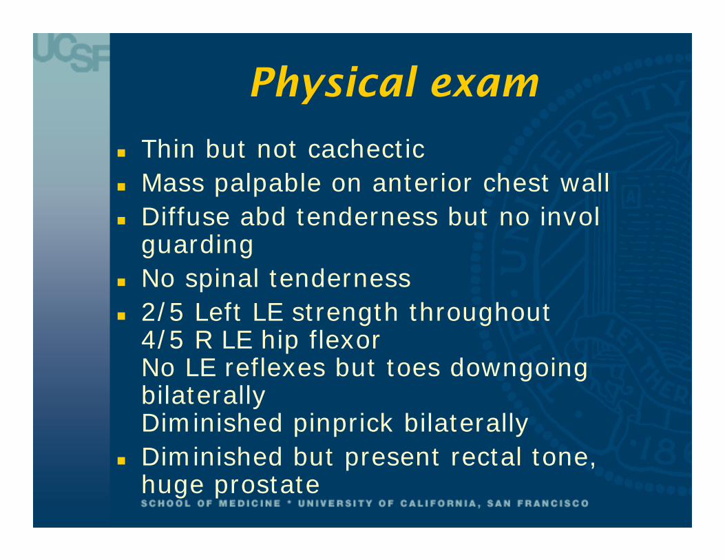
Physical exam
Thin but not cachectic Mass palpable on anterior chest wall Diffuse abd tenderness but no invol
guarding No spinal tenderness 2/5 Left LE strength throughout
4/5 R LE hip flexorNo LE reflexes but toes downgoing bilaterallyDiminished pinprick bilaterally
Diminished but present rectal tone, huge prostate
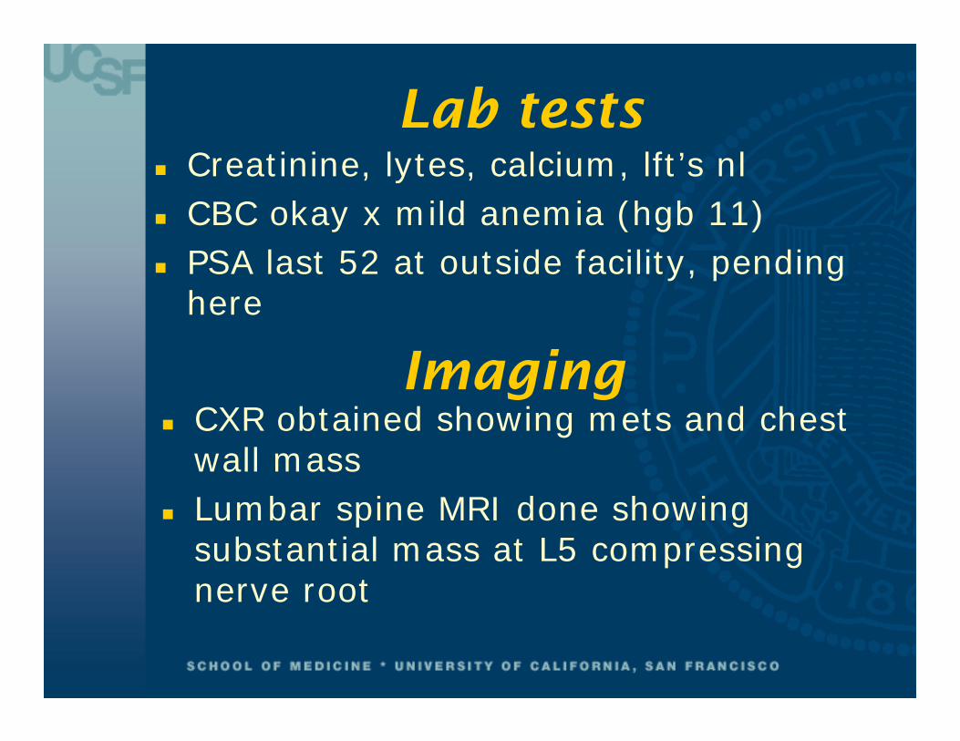
Lab tests Creatinine, lytes, calcium, lft’s nl CBC okay x mild anemia (hgb 11) PSA last 52 at outside facility, pending
here
CXR obtained showing mets and chest wall mass
Lumbar spine MRI done showing substantial mass at L5 compressing nerve root
Imaging

L4 - Normal

L5 – Extensive Tumor

Radiation Therapy Consult
Tell me when you’re ready to treat him Neurosurgery should have right of first
refusal
Neurosurgery consult No thecal sac compression Nerve roots involved directly by tumor No role for surgical decompression
without cord compression Recommend XRT and chemotherapy

Hem/Onc Consult
Neurosurgery has right of first refusal We’ll call radiation oncology Chemotherapy change later
Empiric Meds Patient comes on decadron 4 mg PO q 6
hours Given morphine PCA for bony pains

Clinical Course
Starts XRT urgently Day 1 is already feeling better Day 2 is even better Day 3 is getting weaker both
subjectively and objectively Rad Onc says “it’s not OUR fault”

There’s no additional history There are no new labs Patient says yes to DNR/DNI, but still wants to
be aggressive short of that Physical Exam:
1/5 strength both lower extremitiesSensation gone on left

More imaging
MRI SPINE SURVEY Multiple levels of metastases Significant cord compression at T4

Spine Survey

Empiric medications Casodex (bicalutamide) started Ketoconazole considered Decadron dose increased to 10 q 6 hrs
Outcome Patient taken to Anterior Corpectomy for
acute cord decompression by Neurosurgical and CT surgery teams
Slow recovery of sensation and motor function occurring
Will eventually get radiation therapy

Hem/Onc Emergencies:What you always need to
know Decision Maker: Patient, DPOA, Relative Tissue diagnosis: presumptive vs.
biopsy-proven The time course of the decompensation Status of other disease beyond the
currently involved site

Neurological Emergencies for Today
Spinal Cord Compression CNS Metastases with
symptoms

Neurological: Cord Compression I
In most patients, presents as pain weeks in advance of motor symptoms/findings
Autonomic dysfunction FOLLOWS motor/sensory, so if you see it, it’s late!
Usually occurs in diseases with vertebral body mets, not hematogenous duralmets
Rapid deterioration (days) predicts worse outcome than longer (weeks)

The MRI view

Neurological: Cord Compression II
You need to get: A diagnosis - clear primary, or a new biopsy Dexamethasone in the patient Pain control: Opiates likely to be required
along with bowel prophylaxis MRI [or CT myelogram] of the WHOLE spine Radiation Therapy IF multiple levels, poor KPS Neurosurgery most other circumstances,
especially no dx, or prior RT, or very rapid onset
A Medical Oncologist to remind you and help out
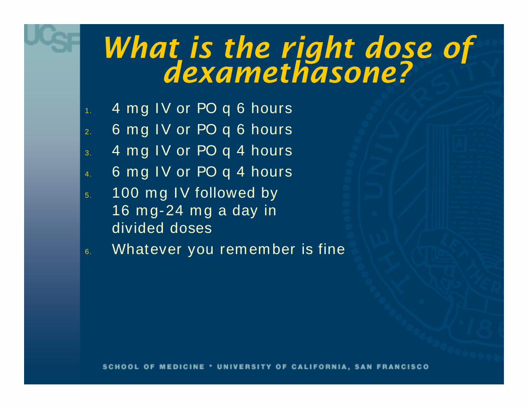
What is the right dose of dexamethasone?
1. 4 mg IV or PO q 6 hours2. 6 mg IV or PO q 6 hours3. 4 mg IV or PO q 4 hours4. 6 mg IV or PO q 4 hours5. 100 mg IV followed by
16 mg-24 mg a day individed doses
6. Whatever you remember is fine

Surgery + XRT vs.XRT alone
In patients with cord compression due to solid tumors:
Surgery + XRTpreserves ability to walk [84%]versus XRT alone [57%]
Surgery + XRT patientswalk longer [median 122 d]versus XRT alone [median 13 d]
Patchell et al, Lancet 2005; 366
Is Brain Different than Cord?
Headache, seizures, altered mental status, or focal deficits
Most commonly from lung or breast cancer if no prior known primary
GI malignancies, sarcomas, prostate cancer are unusual etiologies – look for another 1
If no dx, isolated to brain, consider lymphoma, HIV or not
Surgery followed by Radiation therapy for isolated metastases improves survival vs XRT or surgery alone

Neurological: Brain Metastases
You need to get: A diagnosis if no known malignancy Dexamethasone into the patient if there is
edema, focal sx, shift (~16 mg/day, taper at end of XRT)
Phenytoin into the patient only if seizure witnessed/suspected
Good imaging of the whole brain: MRI if at all possible
Neurosurgery if no diagnosis, or clearly an isolated met
Radiation oncology will consider stereotactic radiosurgery for up to 3 or so isolated mets

Case #2
51 yo African American male without PMHx presents with:
Acute onset of both right-sided rib and back pain
Cough Weight loss Confusion

Baseline evaluation shows
WBC 6.3, Hct 16.9, plts 340K Na 125, K 4.2, Cr 1.0, Ca 11, albumin
1.8, Phos 4.4 Tender ribs, no fracture on CXR Blood smear with rouleaux Total serum protein 10.5 g/dL Dx: symptomatic hypercalcemia, likely
from multiple myeloma

Treating Hypercalcemia: What is the first thing that
you would do?1. 80 mg IV furosemide2. 2L Normal Saline3. IV pamidronate4. IV calcitonin5. IV Solumedrol6. Something else

Hypercalcemiamanifestations
“Stones”[Renal: dehydration & nephrogenicDI; true stones rare]
“Bones”[Pain actually uncommon]
“Abdominal Groans”[Constipation, anorexia;less often nausea/vomiting]
“Psychic Moans”[Confusion, lethargy]
Ca++ Cai
mg/dL (mmol/L)
10.0 1.4
12.0 2.0
14.0 2.5
Mild
Moderate
Critical

Hypercalcemia of malignancy
Usually gradual in onset Fatigue, constipation, anorexia, apathy,
decreased LOC common manifestations Squamous cancers from multiple sites
often make PTH-RP even without bony mets
Breast, lung, myeloma mets common Prostate very rare despite bony disease Patients always volume depleted due to
calcium-induced renal tubular defects

Treating Hypercalcemia of malignancy
You need to: VOLUME REPLETE the patient Use furosemide ONLY to manage the
patient’s volume status Administer an IV bisphosphonate.
If CrCl >30 ml/min, either pamidronate 90 mg over 3 hrs, or zoledronic acid 3-4 mg over 15 mins (800 x pamidronate potency)
Know that calcitonin, steroids, gallium nitrate, mithramycin are not as effective (but calcitonin acts quickly)
Know that except for myeloma, hypercalcemia predicts short survival ~5mo

How to treat hypercalcemia when CrCl
<30?1. Use 30 mg pamidronate instead of 902. Use calcitonin 4 units/kg
SQ q 12 hr, hope renalfunction improves
3. If hypercalcemia severe,go ahead with full dosebisphosphonate
4. Use new osteoclast inhibitor denosumab, since common side effect is hypocalcemia

Case #3 77 year old woman with Chronic
Lymphocytic Leukemia, intermittently receiving chemotherapy for the last 3 years for symptoms
Admitted with dehydration, fatigue, mild renal insufficiency, anemia for fluids and transfusion (WBC = 18,000; hgb = 7.1; plts = 88,000; Cr = 2.7)
On hospital day #2, lab calls with panic value neutrophil count of 400/mm3 just as ward calls with patient temperature of 100.8F

Treating the Febrile Neutropenic Patient – First
Steps?1. Repeat labs to confirm, evaluate for a source,
consider empiric antibioticsif looks ill
2. Obtain blood and urine cultures,CXR stat.Empiric cefipime + vancomycinwithin 60 minutes
3. As #2 but vancomycin only ifhas indwelling catheter,specific concern for skin infection
4. As #2 but also add filgrastim (G-CSF)5 mcg/kg SQ daily

Neutropenic Fever I
Almost always from chemotherapy effect (“day 10-15”), not underlying malignancy
Duration of neutropenia predicts organisms Short-term = gram-negative>gram-positive Long-term (weeks) also fungal, viral,
opportunistic Top of the “right now, this minute” list of
common hem/onc emergencies(mortality rate 33% if antibiotics given later than 60 mins after presentation vs. 20% given sooner

Neutropenic Fever II You need to: Fully evaluate the patient for a source
Blood, line cx, CXR, sputum, urine, skin, ?LP If there’s a suspected source, treat it If there’s NOT a suspected source (>75% of
time), treat empirically for gut floraCefipime, imi/meropenem, moxifloxacin,
pip/tazo, Aztreonam for pen allergy Add vancomycin/linezolid if patient ill, concern
for line infection (PICC>>implanted port) Add coverage for lack of response (e.g.
voriconazole after 3-4 days if still febrile) Consider GCSF for ANC<100, sepsis, Age>65

Neutropenic Fever III – Are Hospitalists really going to
see it? Modern Oncology Treatment continues to
expand to more and more regimens for more and more cancers
Third, 4th, or 5th line therapy for relatively resistant cancers (lung, gastric, pancreatic, low-grade lymphomas) is now part of standard guidelines (so patients with less marrow reserve are treated more and more often)
The timing of chemotherapy administration is less and less related to physician/clinic visits with the advent of oral chemotherapeutic regimens so it is not always obvious when “day 10” of chemotherapy is, when doing medication reconciliation

Oral chemo causing neutropenia 6-mercaptopurine Altretamine Busulfan Capecitabine Carmustine Chlorambucil Crizotinib Cyclophosphamide Dasatinib Etoposide Everolimus Hydroxyurea Imatinib
Lenalidomide Lomustine Melphalan Methotrexate Pazopanib Procarbazine Sorafenib Sunitinib Temozolomide Thalidomide Topotecan Vandetanib Vemurafenib

Case #4
You have just received signout on a 51 year old female patient admitted for community-acquired pneumonia 2 days ago
Clinically she has yet to ‘turn the corner’ despite several days of antibiotics
Before she is seen, you get the stat page from the lab that her morning CBC shows a platelet count of 14,000 / mm3

What do you want to know right now?
1. PT/PTT/fibrinogen to rule out DIC2. Yesterday’s platelet count
to see how far it’s fallen3. Is the patient bleeding?4. Has this patient received
heparin in any form in thelast week?
5. LDH to rule out TTP

Thrombocytopenic bleeding
• Petechiae and mucosal bleeding (the image below) are common but by themselves don’t require systemic treatment
• Active bleedingwith a platelet countunder 50,000/mm3
requiresplatelet transfusionregardless of etiology

Acute Thrombocytopenia in Inpatients
Cause Severity Treatment KeysDisseminated Intravascular Coagulation (DIC)
Variable, can be severe (<20Kplts/mm3)
Treat the underlying disorder;Support with platelets and factors
Heparin-induced (HIT)
Moderate (typically 50-120K)
Stop all heparin products;Anticoagulate with direct thrombin inhibitor
Other drug-induced
Moderate Stop offending drug/drug class
Thrombotic Thrombocytopenic Purpura (TTP)
Moderate, rarely severe
Plasma exchange required +/- corticosteroids
HELLP (in Pregnancy)
Variable, can be severe
Delivery
Immune (ITP) Variable, can be severe
Diagnosis of exclusion, very unlikely to develop in inpatient

Acute Thrombocytopenia in Inpatients – the Big Three
Cause Bleeding vsClotting
Companion features
DIC Bleeding more common
Elevated PT, aPTT, D-dimersLow fibrinogenPatient usually quite ill
HIT Clotting more common (venous/arterial)
PT, fibrinogen should be normalHeparin exposure 4-10 days prior to >50% fall in platelet count
TTP Clotting more common (arterial)
Normal LDH excludes diagnosisHemolytic anemia usually modestSchistocytes on smear eventuallyprominentAt least one systemic sign or symptom should be present (mental status changes, renal insufficiency, fever)

If HIT is the leading diagnosis
A negative ELISA (Anti-PF4 Ab) is reassuring, a positive test is not diagnostic
Serotonin-release assay (SRA) is the gold standard and should be sent at the same time as the ELISA
A direct thrombin inhibitor (must adjust bivalirudin in renal insufficiency; adjust argatroban in hepatic insufficiency) must be given while tests are pending
Platelet count improves within days DVT should be looked for if not already known

How long should patients with likely HIT be anticoagulated
with warfarin after their platelet counts are normal?
1. If no clot, stop;If clot, 3 months
2. If no clot, 3-4 weeks;If clot, 3 months
3. If no clot, 3-4 weeks;If clot, indefinite(at least 6 months)
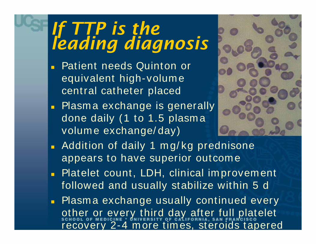
If TTP is theleading diagnosis Patient needs Quinton or
equivalent high-volumecentral catheter placed
Plasma exchange is generallydone daily (1 to 1.5 plasmavolume exchange/day)
Addition of daily 1 mg/kg prednisone appears to have superior outcome
Platelet count, LDH, clinical improvement followed and usually stabilize within 5 d
Plasma exchange usually continued every other or every third day after full platelet recovery 2-4 more times, steroids tapered

Back to our Patient: DIC, not bleeding
For patients in DIC, you need to: Check PT, PTT, Fibrinogen, D-dimers BID Give FFP 10-15 ml/kg for INR > 1.5 to 2 Give 10U cryoprecipitate for fibrinogen
<150 Give platelets to keep > 40,000 Give vitamin K 10 mg SQ (patients are
depleting liver-dependent factors) If use heparin, adjust dose to normalize PT
and fibrinogen but NOT elevate PTT(no bolus, usually 800-1000 units/hr IV)
INR 1.7, aPTT 1.5 x ULNFibrinogen normal, D-dimers up

In DIC, when should heparin be considered?
1. Once FFP, cryo and platelets given, give heparin to prevent further rise in PT/INR
2. When there is definite evidence of thromboembolism
3. When factor/platelet replacement are ongoing but DIC labs are still abnormal
4. When factor/platelet replacement are ongoing, DIC labs are abnormal, but only if patient having clotting/bleeding

SUMMARY
In general, hematologic and oncologic emergencies occur with already-known diagnoses
You need to know the diagnosis before instituting empiric therapy
Steroids are almost never wrong Neutropenic fever is as urgent as cord
compression and airway obstruction When in doubt, call Heme/Onc!

Suggested Real-Time References
Internet Free: Emedicine.medscape.com (reference section) www.merckmedicus.com (includes Harrison’s online &
Hospital Medicine online) Internet Cost:
UpToDate (www.utdol.com) MDConsult (www.mdconsult.com)
Mobile Device Applications (Free): Medscape (Diseases & Conditions) Clinical Care Options Oncology inPractice (Freter &
Haddadin Oncologic Emergencies section)
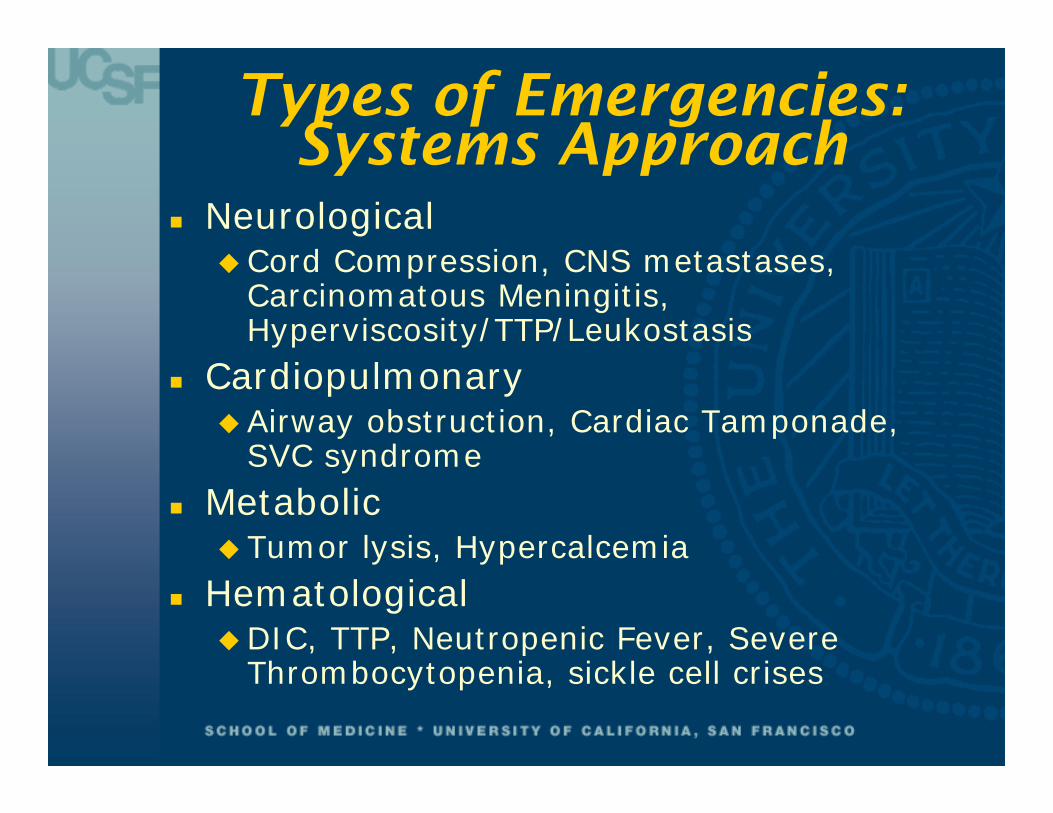
Types of Emergencies: Systems Approach
NeurologicalCord Compression, CNS metastases,
Carcinomatous Meningitis, Hyperviscosity/TTP/Leukostasis
CardiopulmonaryAirway obstruction, Cardiac Tamponade,
SVC syndrome Metabolic
Tumor lysis, Hypercalcemia Hematological
DIC, TTP, Neutropenic Fever, Severe Thrombocytopenia, sickle cell crises

Types of Emergencies: Urgency Approach
Right Now This MinuteAirway obstruction, neutropenic fever,
tamponade, cord compression, CNS metastases with symptoms
Today coagulopathies, tumor lysis, DIC,
leukostasis, TTP, hyperviscosity, severe thrombocytopenia, sickle cell complications
If Not Today, TomorrowSVC syndrome, most hypercalcemia, most
CNS mets without edema

Case #5
Previously healthy 64 yo man with fatigue, confusion SOB, easy bruising for 5 days
ER assessment: Pale, in some distress, bruising and some
gum bleeding, cardiac/lung/abd exam pretty benign
WBC 128,000, hgb 6.8, platelets 22KCreatinine 3.4, K 5.6, Ca 7.8, LFTs 2 x ULN T 100.2F, BP 100/50, HR 110, RR 26
02 sat 86% RA 92% on 4L NC ECG: sinus tach, CXR: mild increased
interstitium

Peripheral Blood Smear
Dx: Acute Leukemia NOS,70% blasts

What do you want to do first?
1) Evaluate for DIC / Transfuse platelets
2) Check tumor lysis labs and start allopurinol
3) Give antibiotics for functional neutropenic fever & sepsis
4) Call Renal to initiate dialysis for hyperkalemia
5) Call IR for central catheter for leukapheresis
6) Give chemotherapy to reduce WBC count

Leukostasis
Mostly in AML with WBC > 100,000 Fxn of cell number and size and
deformabilityCan occur at lower WBCs, this is a clinical dxCLL, CML uncommon even with WBC
>300,000 Altered mental status, coma common, but
other organs also involved Hypoxia, renal insufficiency concerning May worsen with induction chemo High one-week mortality left untreated

Leukostasis You need to get: The patient HYDRATED Quinton access (Interventional
Radiology or Renal Fellow) and arrange for possible leukapheresis, diagnosis and chemotherapy
The urge NOT to transfuse an anemic, hypoxic patient – total viscosity would increase
Consider LP for cytology to rule in / out CNS leukemia if altered mental status
Steroids may help demarginate WBCs

Leukostasis Additional issues in high WBC leukemia: Plan for Tumor Lysis – it will happen with
therapy or possibly on its own Monitor platelet counts carefully (automated
machine counts inaccurate with high WBC, and DIC is also likely, so check PT/PTT/fibrinogen)
Leukapheresis only useful short-term in combination with some cytoreductive chemo, indications still controversial
Critical that Hem/Onc involved to decide chemotherapy regimen vs count stabilization +/- apheresis

Tumor Lysis Syndrome
“A syndrome resulting from cytotoxic therapy, occurring generally in aggressive, rapidly proliferating lymphoproliferative disorders.”It is characterized by combinations of:Hyperuricemia Lactic acidosisHyperkalemiaHyperphosphatemiaHypocalcemiaAcute renal failure

Tumor Lysis Syndrome I
Occurs in tumors with high body burden and high chemosensitivity
Usually high-grade lymphomas or leukemias
Small cell, germ cell less common Usually due to therapy, so you know the
diagnosis already May occur at onset of therapy, or after a
day or two

Tumor Lysis Syndrome II Pre-treatment, you need to: Fix conditions that will make effects worse:
Dehydration, renal obstruction, avoid IV contrast
Get baseline labs: K, Ca, Phos, Uric Acid, LDH, Cr
Alkaline diuresis: D5•1/2NS with 2 or even 3 amps NaHCO3/l at 200+ cc/hr, keep urine pH > 7, urine output high; ? lasix, mannitol
Allopurinol 600 mg load, then 300 QD to keep uric acid down
Rasburicase? [recombinant urate oxidase$2500/d]
Especially high risk patients: call renal before starting therapy
Tumor Lysis Syndrome III
During treatment, you need to: Keep alkaline urine output high Check BID lytes, phos, UA, Ca, LDH, Cr Try to keep phosphate < 7, Ca > 6, K<6 If Ca low, remember to give Mg, too Give back Ca slowly if phosphate high

Tumor Lysis Syndrome IV
If phosphate >7, switch NaHCO3 to NS to prevent Ca-PO4 deposits in kidney (urate nephropathy now less of a concern)
Oral phosphate binders (Amphogel) help Acute K Rx is always good, but
insulin/D-50 preferred over IV Calcium if Ca-P product high. Kayexalate fine
May need dialysis

Neurological Emergencies
Spinal Cord Compression (already covered) CNS Metastases with symptoms (already covered) Carcinomatous Meningitis Vascular Events:
Hyperviscosity Leukostasis (already covered) TTP (already covered)

Neurological: Carcinomatous Meningitis I
Focal cranial/peripheral neuropathy, seizures common
Unlikely to occur without other obvious systemic disease
Hematogenous disease, lymphoma, leukemia
CT/MRI may be negative; need CSF cytology

Neurological: Carcinomatous Meningitis II
You need to get: CSF cytology for the diagnosis If high suspicion, intrathecal
chemotherapy at same LPMethotrexate or Ara-C ONLY!!
Radiation therapy to whole cranium Neurosurgery if Ommaya reservoir
needed for multiple courses

Neurological: Vascular Events
Hyperviscosity/ Leukostasis / TTP Hyperviscosity mostly with
Waldenstrom’s, less common myeloma or Polycythemia Vera, or Essential Thrombocythemia
Nonspecific sx: somnolence, headache, blurry vision, dizziness
Serum viscosity usually > 3 In PV, Hgb usually > 18 In ET, platelets well over 106

Neurological: Hyperviscosity
You need to get: The patient HYDRATED Apheresis for IgM, plus chemotherapy.
You will need the Hem/Onc Fellow for chemo and the Renal Fellow for a Quinton catheter!
Phlebotomy for PV: replace units with NS, want Hgb ~ 15
Hydroxyurea and aspirin for ET

Cardiopulmonary Emergencies
Airway obstruction Cardiac Tamponade SVC syndrome

Pulmonary: Airway Obstruction I
Most respiratory failure from cancer due to lymphangitic spread, chemotherapy toxicity, radiation, malignant effusions
Also need to consider cardiac failure from chemotherapy (doxorubicin, cyclo-phosphamide), radiation, cardiac tamponade
As with all airway emergencies, intubation, cricothyrotomy, Heliox may be necessary
Pulmonary: Airway Obstruction II
You need to get: Airway protection, corticosteroids for
edema A diagnosis (small cell lung cancer Rx
would be chemotherapy) Pulmonary, ENT, and CT surgery
consults for stenting or laser curettage Medical Oncology to evaluate for XRT
vs. chemotherapy or both

Cardiac: Tamponade I
Malignant effusions are common, just not commonly symptomatic
Presents with left or right sided failure, paradoxus, big heart on CXR
May be vague: hiccoughs, persistent cough
Lung, breast cancer most common primaries
Mortality probably will be from other aspects of patient’s disease

Cardiac: Tamponade II
You need to get: An Echo and cytology from
pericardiocentesis Catheter drainage of the pericardial space
(so leave it in at pericardiocentesis!) Oncology input re: chemotherapy CT surgery input re: subxiphoid pericardial
window or balloon pericardiotomy, especially for recurrent effusions in patients with good performance status

Cardiopulmonary: SVC Syndrome I
Usually from lung cancer;lymphoma, breast cancer, mediastinaltumors also possible
Facial, symmetric or asymmetric upper extremity edema common
Shortness of breath common but patients not hypoxic
Only a relative emergency, even with CNS symptoms; usually collaterals working

Cardiopulmonary: SVC Syndrome II
You need to get: A chest CT to outline the mass that will
need therapy A diagnosis hopefully from another site
(to avoid biopsy-induced bleeding) Oncology involved; chemotherapy for
small cell, lymphoma, germ cell Radiation oncology for almost
everything else No a priori need for heparin or steroids Call IR? New area of interest: stenting

SVC stenting for symptoms –before & after

Metabolic Emergencies
Tumor Lysis Syndrome (already covered)
Hypercalcemia(already covered)

Hematologic Emergencies
Neutropenic Fever (already covered)
DIC (already covered) Severe thrombocytopenia (already
covered) Sickle cell crises (covered by Brad
Lewis) Overanticoagulation (covered by
Tracy Minichiello)

Platelet vs. factor bleeding
CLINICAL CHARACTERISTIC
PLATELET DEFECT CLOTTING FACTOR DEFICIENCY
Site of bleeding Skin, mucous membranes Deep in soft tissue
Bleeding after minor cuts Yes Not usually
Petechiae Present Absent
Ecchymoses Small, superficial Large, palpable
Hemarthrosis, muscle hematomas
Rare Common
Bleeding after Surgery Immediate, mild Delayed, severe