Helicobacter pylori Induces Apoptosis of Macrophages in ... fileassociated with the development of...
11
10.1128/IAI.72.5.2889-2898.2004. 2004, 72(5):2889. DOI: Infect. Immun. Rena J. Menaker, Peter J. M. Ceponis and Nicola L. Jones Alterations in the Mitochondrial Pathway Macrophages in Association with Induces Apoptosis of Helicobacter pylori http://iai.asm.org/content/72/5/2889 Updated information and services can be found at: These include: REFERENCES http://iai.asm.org/content/72/5/2889#ref-list-1 at: This article cites 74 articles, 43 of which can be accessed free CONTENT ALERTS more» articles cite this article), Receive: RSS Feeds, eTOCs, free email alerts (when new http://journals.asm.org/site/misc/reprints.xhtml Information about commercial reprint orders: http://journals.asm.org/site/subscriptions/ To subscribe to to another ASM Journal go to: on February 23, 2013 by PENN STATE UNIV http://iai.asm.org/ Downloaded from
Transcript of Helicobacter pylori Induces Apoptosis of Macrophages in ... fileassociated with the development of...
10.1128/IAI.72.5.2889-2898.2004.
2004, 72(5):2889. DOI:Infect. Immun. Rena J. Menaker, Peter J. M. Ceponis and Nicola L. Jones Alterations in the Mitochondrial PathwayMacrophages in Association with
Induces Apoptosis ofHelicobacter pylori
http://iai.asm.org/content/72/5/2889Updated information and services can be found at:
These include:
REFERENCEShttp://iai.asm.org/content/72/5/2889#ref-list-1at:
This article cites 74 articles, 43 of which can be accessed free
CONTENT ALERTS more»articles cite this article),
Receive: RSS Feeds, eTOCs, free email alerts (when new
http://journals.asm.org/site/misc/reprints.xhtmlInformation about commercial reprint orders: http://journals.asm.org/site/subscriptions/To subscribe to to another ASM Journal go to:
on February 23, 2013 by P
EN
N S
TA
TE
UN
IVhttp://iai.asm
.org/D
ownloaded from

INFECTION AND IMMUNITY, May 2004, p. 2889�2898 Vol. 72, No. 50019-9567/04/$08.00�0 DOI: 10.1128/IAI.72.5.2889–2898.2004Copyright © 2004, American Society for Microbiology. All Rights Reserved.
Helicobacter pylori Induces Apoptosis of Macrophages in Associationwith Alterations in the Mitochondrial Pathway
Rena J. Menaker,1,2 Peter J. M. Ceponis,1,3 and Nicola L. Jones1,2,4*Research Institute, Hospital for Sick Children,1 and Department of Physiology,2 Department of Laboratory Medicine and
Pathobiology,3 and Department of Paediatrics,4 University of Toronto, Toronto, Canada
Received 21 July 2003/Returned for modification 15 September 2003/Accepted 19 January 2004
Helicobacter pylori is a gastric bacterial pathogen that evades host immune responses in vivo and isassociated with the development of gastritis, peptic ulcer disease, and gastric cancers. Induction of macrophageapoptosis is a method employed by multiple pathogens to escape host immune responses. Therefore, wehypothesized that H. pylori induces apoptosis of infected macrophages. RAW 264.7 cells were infected with H.pylori strain 60190, and apoptosis was assessed. Transmission electron microscopy and fluorescence micros-copy showed that infected macrophages displayed morphological features characteristic of apoptosis. Quan-tification by acridine orange-ethidium bromide fluorescent-dye staining showed that apoptosis was dose andtime dependent, and apoptosis was further confirmed by increased binding of annexin V-fluorescein isothio-cyanate (FITC) to externalized phosphatidylserine of infected but not of control macrophages. Macrophagesinfected with isogenic mutants of H. pylori strain 60190 deficient in either cagA or vacA induced significantlyless apoptosis than the parental strain, as assessed by increased binding of annexin V-FITC. Western blotanalysis of whole-cell protein lysates revealed that infection with strain 60190 induced a time-dependentincrease in cleavage of procaspase 8 and disappearance of full-length Bid compared with uninfected cells.Furthermore, pharmacological inhibition of caspase 8 caused a decrease in levels of apoptosis. Finally,infection caused a time-dependent increase in mitochondrial-membrane permeability and release of cyto-chrome c into the cytosol. These results suggest that H. pylori induces apoptosis of macrophages in associationwith alterations in the mitochondrial pathway. Elimination of this key immunomodulatory cell may representa mechanism employed by the bacterium to evade host immune responses.
Helicobacter pylori is a gram-negative microaerophilic bacte-rium that causes a lifelong infection in over half of the world’shuman population (50). Without specific antimicrobial treat-ment, all individuals infected with H. pylori exhibit chronicgastric inflammation, and a small percentage will develop pep-tic ulcers and gastric adenocarcinoma or mucosa-associatedlymphoid tissue lymphoma (14, 57). In response to infection,the host launches a vigorous immune response, including themucosal infiltration of neutrophils, lymphocytes, and macro-phages (18). However, this response is insufficient for clear-ance of the bacterium, suggesting that H. pylori is capable ofevading host immune responses.
H. pylori strains are classified based on carriage of the cagpathogenicity island (PAI) and expression of the vacuolatingcytotoxin (VacA) (9). Type I strains possess the cag PAI andsecrete VacA; in contrast, type II strains lack the cag PAI anddo not produce VacA. Controversy surrounds the role of cagAand vacA as bacterial virulence factors in vivo. For example,initial studies showed an increased risk of peptic-ulcer diseaseand gastric cancers in association with strains expressing CagA(4, 58); however, subsequent studies of both adults (24, 56) andchildren (43) provided conflicting results. Furthermore, infec-tion with type I, but not type II, strains in Western populationsis associated with increased risk for development of peptic-
ulcer disease and gastric cancers (8, 9, 57), with contradictorydata found in both Asian and Australian populations (47, 56).
Apoptosis can be signaled through a number of pathways,including the death receptor and mitochondrial pathways (2,25). First, ligation of FasL to three Fas molecules activates thedeath receptor pathway, recruiting the Fas-associated deathdomain and the proenzyme form of caspase 8 to form thedeath-inducing signaling complex (53). Formation of thedeath-inducing signaling complex may result in cleavage ofprocaspase 3, leading to activation of downstream effectors andresulting in genomic fragmentation and other characteristicfeatures of apoptosis (12, 17). Alternatively, apoptosis may besignaled through the mitochondrial pathway, where death sig-nals increase mitochondrial-membrane permeability, allowingthe subsequent release of proapoptotic factors, such as cyto-chrome c, into the cytosol (19), again leading to the inductionof apoptosis (41). In some instances, both pathways may worksynergistically when death receptor-dependent activation ofprocaspase 8 leads to cleavage and activation of Bid to damagemitochondrial-membrane permeability, release cytochrome c,and induce apoptosis involving both the mitochondrial anddeath receptor pathways.
Increasing evidence indicates that apoptosis of immune cellsplays an important role in modulating the pathogenesis of anumber of bacterially induced diseases (54). For example, thehost response can include induction of macrophage apoptosisto purposefully eliminate intracellular bacteria, such as Myco-bacterium tuberculosis and Mycobacterium bovis (49, 60), con-firming the importance of this immune cell. Alternatively, Shi-gella flexneri and Salmonella enterica serovar Typhimurium
* Corresponding author. Mailing address: Rm. 8409, Hospital forSick Children, 555 University Ave., Toronto, ON, Canada M5G 1X8.Phone: (416) 813-7734. Fax: (416) 813-6531. E-mail: [email protected].
2889
on February 23, 2013 by P
EN
N S
TA
TE
UN
IVhttp://iai.asm
.org/D
ownloaded from
trigger macrophage apoptosis to induce inflammation and per-sist within the host (54). Furthermore, macrophages play acentral role in the inflammatory response to infection with H.pylori (20, 23). For example, increased numbers of macro-phages are observed in the mucosa of gastric biopsy specimensobtained from H. pylori-infected children and correlate withthe severity of gastritis (71). Although H. pylori is consideredan extracellular pathogen, clinical isolates have been shown toinvade epithelial monolayers (1, 66), and a recent study byJhala et al. (32) showed the bacterium residing in the laminapropria in infected individuals, suggesting a possible point ofinteraction between the bacterium and mucosal macrophages.Alternatively, H. pylori secretes soluble bacterial products thattraverse the epithelial barrier and interact with macrophages inthe lamina propria. Indeed, H. pylori urease has been detectedin the lamina propria in patients with H. pylori-associated gas-tritis but not in uninfected subjects (46). Moreover, H. pyloricould interact with mucosal macrophages following the loss ofsurface epithelium during infection (1, 70), as H. pylori inducesapoptosis of epithelial cells both in vitro (51) and in vivo (35).
H. pylori modulates apoptotic signaling pathways in a num-ber of cell types in vivo and in vitro, including epithelial cells(33), polymorphonuclear leukocytes (28), and T lymphocytes(69). Recently, the ability of H. pylori to induce apoptosis inmacrophages was demonstrated (23, 72). However, neither theapoptotic signaling pathways nor the bacterial factors involvedhave been fully delineated. Therefore, the aim of our study wasto demonstrate that H. pylori induces apoptosis of macro-phages and to delineate both the bacterial factors and theapoptotic signaling pathways involved.
MATERIALS AND METHODS
Bacterial growth conditions. Wild-type H. pylori strain 60190 isolated from anadult with gastritis (ATCC 49503; cagA� cagE� VacA�) (36) and its isogeniccagA� and vacA� mutants (kindly provided by Richard Peek, Jr., Nashville,Tenn.) were grown under microaerophilic conditions (5% O2, 10% CO2, 85%N2) on Columbia blood agar plates containing 5% sheep blood for 72 h at 37°Cor supplemented with 10% heat-inactivated fetal calf serum and 20 �g of kana-mycin/ml (mutant strains) (36). The bacteria were then suspended in brucellabroth (Difco Laboratories, Detroit, Mich.) supplemented with 10% heat-inacti-vated fetal bovine serum (Gibco BRL Life Technologies, Gaithersburg, Md.) and20 �g of kanamycin/ml (mutant strains) (36) and shaken at 120 rpm overnight at37°C under microaerophilic conditions (36). The cells were subsequently pelletedand resuspended in sterile phosphate-buffered saline (PBS), and optical densitymeasurements were obtained by a spectrophotometric reading. Before the in-fection of eukaryotic cells, a droplet of culture was assessed for bacterial motilityunder bright-field microscopy, and after the infection of eukaryotic cells, themedium was cultured on blood agar plates to exclude contamination.
Eukaryotic-cell culture and conditions of infection. The RAW 264.7 murinemacrophage cell line, used as a model cell line to define H. pylori-induced signaltransduction and its interaction with immune cells (23, 70, 72), was grown at 37°Cin 5% CO2 in Dulbecco’s modified Eagle’s medium containing L-glutamine andglucose (Gibco BRL Life Technologies) and further supplemented with 10%heat-inactivated fetal bovine serum and 1% penicillin-streptomycin (Gibco BRLLife Technologies). Following the removal of antibiotics by rinsing the macro-phages with PBS, the macrophages were infected with H. pylori at differentmultiplicities of infection (MOI) (from 10 bacteria to 1 macrophage to 100bacteria to 1 macrophage) for 2 to 48 h. RAW 264.7 cells incubated withstaurosporine (1 �M; Sigma Aldrich, Oakville, Ontario, Canada) for 8 to 24 hserved as a positive control for the induction of apoptosis (6).
Assessment of apoptosis. (i) Transmission electron microscopy. Macrophageswere grown to 70% confluency and infected with H. pylori as described above,with uninfected cells serving as controls. After 24 h of infection, the adherentcells were scraped with a rubber policeman, pooled with cells in suspension, andpelleted by centrifugation. The cells were then fixed with 2% glutaraldehyde in
0.1 M phosphate buffer, postfixed in 2% osmium tetroxide, and dehydratedthrough a series of graded acetone washes (16). Samples were embedded inepoxy resin, sectioned, and placed onto 300-mesh copper grids. The grids werestained with uranyl acetate and lead salts, as previously described (16), andsamples were examined for the presence of apoptotic cells using a Philips trans-mission electron microscope at the Electron Microscopy Facility of the Hospitalfor Sick Children.
(ii) Fluorescent-dye staining. Staining cells with an acridine orange-ethidiumbromide mixture allows the determination of apoptosis based on nuclear mor-phology and membrane integrity (34). Acridine orange is a cell-permeable dyethat enters all cells and intercalates DNA to appear green (viable cells).Ethidium bromide, however, enters only nonviable cells that exhibit disruptedmembrane integrity, overriding the acridine orange to fluoresce orange. Thus,viable, necrotic, and early and late apoptotic cells can be distinguished by thedifferential uptake and binding of these dyes (11): the nuclei in viable cellsfluoresce green, while the nuclei in necrotic cells fluoresce orange. Early apo-ptotic cells are distinguished by the enhanced green fluorescence of their char-acteristic condensed chromatin, while late apoptotic cells that have lost mem-brane integrity display enhanced uniform orange fluorescence of their condensedchromatin.
Following infection, the suspended and scraped cells were pooled, pelleted bycentrifugation, washed once in PBS, and resuspended in 1 ml of antibiotic-freemedium at room temperature. Acridine orange-ethidium bromide (1 �M; SigmaAldrich) in PBS was mixed with a 0.1-ml cell suspension, a drop was applied toa microscope slide, and viability was assessed by counting 500 cells in fiverandomly selected fields. The percentage of apoptotic cells was then calculated(11).
In some experiments, the specific caspase 8 inhibitor Z-IETD-FMK (R&DSystems, Minneapolis, Minn.) was resuspended in dimethyl sulfoxide and prein-cubated with RAW 264.7 cells for 2 h at a concentration of 100 �M (11). Thecells were then infected with H. pylori for 24 h, and apoptosis was assessed byfluorescent-dye staining using acridine orange-ethidium bromide. Dimethyl sul-foxide alone served as a vehicle control.
(iii) Ann-V–PI flow cytometry. The binding of annexin V-fluorescein isothio-cyanate (Ann-V) to externalized phosphatidylserine was used as a measurementof apoptotic RAW 264.7 cells with an Ann-V–propidium iodide (PI) apoptosisdetection kit (BD Biosciences, Montreal, Quebec, Canada) according to themanufacturer’s instructions. Briefly, in viable cells, phosphatidylserine is strictlyconfined to the inner leaflet of the plasma membrane that faces the cytosol. Thesurface expression of this phospholipid is an early feature of apoptosis and occursbefore the loss of membrane integrity (30). Thus, early apoptotic cells bindannexin V, a Ca2�-dependent phospholipid-binding protein with high affinity forexternalized phosphatidylserine. Furthermore, living cells exclude PI, allowingspecific detection and quantification of apoptosis by fluorescence-activated cellsorter (FACS) analysis (38).
Suspended and scraped cells were pooled, pelleted by centrifugation, washedonce with ice-cold PBS, and resuspended in binding buffer (10 mM HEPES-NaOH [pH 7.4], 140 mM NaCl, 2.5 mM CaCl2) to a concentration of 106/ml.Next, 0.1 ml of this cell suspension was transferred to a 5-ml tube and incubatedwith 0.005 ml of Ann-V and 0.005 ml of PI for 15 min at 25°C in the dark. Finally,0.4 ml of binding buffer was added, and samples were analyzed by flow cytometrywithin 1 h on a FACScan flow cytometer (BD Biosciences) at the Core FlowCytometry Facility of the Hospital for Sick Children. Samples were gated on thebasis of forward versus side scatter for size, and the results are presented as thepercentage of cells that were viable (Ann-V� PI�), early apoptotic (Ann-V�
PI�), or nonviable (Ann-V� PI� or Ann-V� PI�).Western blotting. (i) Whole-cell protein extraction. Suspended and adherent
macrophages were pooled, pelleted, washed once with cold PBS, and pelleted bycentrifugation at 13,000 � g for 10 s. Subsequently, the cell pellet was resus-pended in 150 �l of RIPA buffer (1% NP-40, 0.5% sodium deoxycholate, and0.1% sodium dodecyl sulfate [SDS] in PBS) supplemented with 150 mM NaCl,50 mM NaF, 1 mM Na3VO4, 20 �g of phenlymethylsulfonyl fluoride/ml, 10 �gof aprotinin/ml, 2 �g of pepstatin A/ml, and 2 �g of leupeptin/ml (all obtainedfrom Sigma Aldrich) by being vortexed and was left at 4°C for 20 min. The lysateswere then centrifuged at 11,200 � g for 10 min at 4°C, and the supernatant(whole-cell protein extract) was stored at �70°C until immunoblotting was per-formed (10).
(ii) Cytosolic protein extraction. Cytosolic proteins were isolated as previouslydescribed (21). Briefly, suspended and adherent macrophages were pooled, pel-leted, washed twice in cold PBS, and incubated for 30 min on ice in lysis buffer(68 mM sucrose, 200 mM mannitol, 50 mM KCl, 1 mM EDTA, 1 mM EGTA, 1mM dithiothreitol, 10 �g of aprotinin/ml, 2 �g of pepstatin A/ml, 2 �g ofleupeptin/ml, and 0.5 mM phenylmethylsulfonyl fluoride [all obtained from
2890 MENAKER ET AL. INFECT. IMMUN.
on February 23, 2013 by P
EN
N S
TA
TE
UN
IVhttp://iai.asm
.org/D
ownloaded from
Sigma Aldrich]). The cells were then passed 40 times through a 25-gauge 5/8-in.needle and centrifuged at 1,500 � g for 10 min. The supernatant was transferredto a fresh Eppendorf tube and was further cleared by centrifugation at 22,000 �g for 15 min. The supernatant was stored at �70°C until the protein concentra-tion was determined by the Bio-Rad assay for use in immunoblotting.
(iii) Immunoblotting. Equal volumes of whole-cell protein extracts were addedto 10 �l of 2� loading buffer, and samples were boiled for 3 min and thensubjected to electrophoresis through an SDS–5% polyacrylamide gel electro-phoresis (PAGE) stacking gel and an SDS–15% PAGE separating gel at 111 Vfor 1.25 h at room temperature (or 1.5 h for caspase 8 immunoblotting). Forcytochrome c assays, 15 to 20 �g of cytosolic protein extract was loaded forSDS-PAGE. The proteins were subsequently transferred onto a nitrocellulosemembrane (BioTrace NT; Pall Corp., Ann Arbor, Mich.) at 4°C and 85 V for 2 h,rinsed in distilled H2O, and then blocked with Tris-buffered saline containing0.05% Tween 20 (TBST) supplemented with 5% low-fat milk for 30 min at roomtemperature. The membranes were probed overnight with shaking at 4°C with apolyclonal goat anti-mouse Bid antibody (1:4,000; R&D Systems), polyclonalrabbit anti-mouse caspase 8 antibody (1:1,000; Santa Cruz Biotechnology, SantaCruz, Calif.), monoclonal mouse anti-cytochrome c antibody (1:250; BD Bio-sciences-Pharmingen), polyclonal goat anti-mouse actin antibody (1:1,000; SantaCruz Biotechnology), or polyclonal rabbit anti-mouse actin antibody (1:1,000;Santa Cruz Biotechnology) in TBST-milk. The membranes were washed twicewith distilled water and three times in TBST (10 min each time) and incubatedwith horseradish peroxidase-conjugated donkey anti-goat immunoglobulin Gantibody (1:1,000; Santa Cruz Biotechnology) or horseradish peroxidase-conju-gated donkey anti-rabbit immunoglobulin G antibody (1:1,000; Santa Cruz Bio-technology) for 1 h at room temperature. The membranes were then washedagain with distilled water and TBST (three washes; 10 min each), and bands werevisualized by chemiluminescence detection (Santa Cruz Biotechnology) usingKodak Biomax MR film.
Mitochondrial-membrane sensor flow cytometry. A cationic dye that fluo-resces differently in apoptotic and nonapoptotic cells (ApoAlert MitochodrialMembrane Sensor Kit; BD Biosciences-Clontech) was utilized according to themanufacturer’s instructions. Briefly, in living cells, the dye is taken up in themitochondria to form red fluorescent aggregates. However, in apoptotic cells,increased mitochondrial-membrane permeability prevents the dye from accumu-lating inside mitochondria, and it remains in monomeric form in the cytosol,where it fluoresces green (13).
Uninfected and infected macrophages were pooled, pelleted by centrifugation,resuspended in 1 ml of diluted Mitosensor reagent/sample, and incubated at37°C in 5% CO2 for 20 min. One milliliter of incubation buffer was added to thesamples, followed by centrifugation and resuspension in incubation buffer ac-cording to the manufacturer’s instructions. Samples were then analyzed by flowcytometry within 1 h of preparation. The results are presented as percent in-crease in membrane permeability over controls.
Statistical analysis. Results are expressed as means � standard errors. To teststatistical significance among multiple groups, a one-way analysis of variance(ANOVA) was used, followed by post hoc comparisons with the Tukey-Krameror Bonferronni multiple-comparison test, as indicated in the text. Alternatively,a two-tailed unpaired Student’s t test was used where indicated.
RESULTS
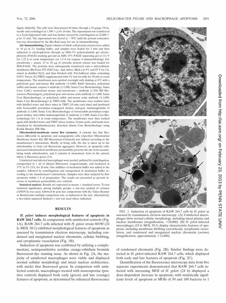
H. pylori induces morphological features of apoptosis inRAW 264.7 cells. In comparison with uninfected controls (Fig.1A), RAW 264.7 cells infected with H. pylori strain 60190 (24h; MOI, 50:1) exhibited morphological features of apoptosis asassessed by transmission electron microscopy, including con-densed and marginated nuclear chromatin, cellular blebbing,and cytoplasmic vacuolation (Fig. 1B).
Induction of apoptosis was confirmed by utilizing a comple-mentary, semiquantitative acridine orange-ethidium bromidefluorescent-dye staining assay. As shown in Fig. 2A, the ma-jority of uninfected macrophages were viable and displayednormal cellular morphology and intact nuclear architecture,with nuclei that fluoresced green. In comparison with unin-fected controls, macrophages treated with staurosporine (pos-itive control) displayed both early (green) and late (orange)features of apoptosis, as determined by enhanced fluorescence
of condensed chromatin (Fig. 2B). Similar findings were de-tected in H. pylori-infected RAW 264.7 cells, which displayedboth early and late features of apoptosis (Fig. 2C).
Quantification of the fluorescence microscopy data from fiveseparate experiments demonstrated that RAW 264.7 cells in-fected with increasing MOI of H. pylori (24 h) displayed adose-dependent increase in apoptosis, with statistically signif-icant levels of apoptosis at MOIs of 50 and 100 bacteria to 1
FIG. 1. Induction of apoptosis of RAW 264.7 cells by H. pylori asassessed by transmission electron microscopy. (A) Uninfected macro-phages show normal cellular morphology, including intact plasma andnuclear membranes (magnification, �20,000). (B) H. pylori-infectedmacrophages (24 h; MOI, 50:1) display characteristic features of apo-ptosis, including membrane blebbing (arrowhead), cytoplasmic vacuo-lation, and condensed and marginated nuclear chromatin (arrows)(magnification, approximately �12,000).
VOL. 72, 2004 HELICOBACTER PYLORI AND MACROPHAGE APOPTOSIS 2891
on February 23, 2013 by P
EN
N S
TA
TE
UN
IVhttp://iai.asm
.org/D
ownloaded from

macrophage compared to uninfected cells (23.1% � 8.4% and27.9% � 12.9%, respectively, versus 9.9% � 5.8%; ANOVA-Bonferroni multiple-comparisons test; P � 0.004; n � 5) (Fig.3A). Furthermore, H. pylori-induced apoptosis of RAW 264.7cells occurred in a time-dependent manner, with maximal pro-grammed cell death occurring 24 h postinfection comparedwith uninfected controls (13.5% � 2.9% versus 4.0% � 1.0%;unpaired t test; P � 0.0004) (Fig. 3B).
H. pylori induces extracellular exposure of phosphatidylser-ine. H. pylori-induced apoptosis of macrophages was furtherconfirmed by assessing the surface expression of phosphatidyl-serine using Ann-V in conjunction with PI and FACS analysis.A FACS analysis representative of three individual experi-ments is depicted in Fig. 4. As shown in Fig. 4A, 80% ofuninfected RAW 264.7 cells were viable (Annexin-V� PI�),while only 5% were apoptotic (Annexin-V� PI�). In contrast,only 51% of H. pylori-infected RAW 264.7 cells were viable(Annexin-V� PI�), while 16% were apoptotic (Fig. 4C). Fig-ure 4D displays the mean percentage of H. pylori-infectedRAW 264.7 cells undergoing apoptosis (Annexin V� PI�)from three separate experiments. In comparison with unin-fected cells, H. pylori-infected macrophages displayed anincrease in apoptosis similar to that detected in staurosporine-treated cells (16% � 3.8% versus 2.8% � 0.6%; ANOVA–Tukey-Kramer test; P � 0.05; n � 3) (Fig. 4D).
FIG. 2. Induction of apoptosis in RAW 264.7 cells by H. pylori asassessed by fluorescence microscopy using acridine orange andethidium bromide staining. (A) Untreated RAW 264.7 cells shownormal cellular morphology and display intact nuclear architecture, asdemonstrated by the green fluorescence of their nuclei (magnification,�250). (B) RAW 264.7 cells treated with staurosporine for 24 h (pos-itive control for apoptosis) show morphological features characteristicof apoptosis, as demonstrated by the enhanced fluorescence of theircondensed chromatin. Early apoptotic cells fluoresce green (arrows),and late apoptotic cells fluoresce orange (arrowheads) (magnification,�250). (C) H. pylori-infected cells (MOI, 50:1; 24 h) show features ofapoptosis similar to those of staurosporine-treated cells (magnifica-tion, �400).
FIG. 3. Quantification of fluorescence microscopy data using acri-dine orange and ethidium bromide staining. (A) Infection with H.pylori at MOI of 10:1, 30:1, 50:1, and 100:1 for 24 h resulted in adose-dependent induction of apoptosis, with increased levels of apo-ptosis observed at MOI of 50:1 and 100:1 compared with uninfectedcontrols (23.1% � 8.4% and 27.9% � 12.9%, respectively, versus 9.9%� 5.8%; ANOVA-Bonferroni multiple-comparison test; *P � 0.004; n� 5). (B) RAW 264.7 cells infected with H. pylori (solid bars) at anMOI of 50:1 for 6, 24, or 48 h showed a time-dependent increase inapoptosis at 24 h compared with time-matched uninfected controls(open bars) (13.5% � 2.9% versus 4.0% � 1.0%; unpaired t test; *, P� 0.0004; n � 4). The results are expressed as mean percentages ofapoptotic cells per 500 cells enumerated, and the error bars representstandard errors of the mean.
2892 MENAKER ET AL. INFECT. IMMUN.
on February 23, 2013 by P
EN
N S
TA
TE
UN
IVhttp://iai.asm
.org/D
ownloaded from

cagA and vacA are involved in H. pylori-induced apoptosis ofRAW 264.7 cells. To delineate the bacterial virulence factorsinvolved in H. pylori-induced apoptosis of macrophages, RAW264.7 cells were infected with the parental strain 60190 (cagA�
VacA�) (24 h; MOI, 100:1) or its isogenic mutant strains (cagAor vacA mutant) (24 h; MOI, 100:1) and were subsequentlyanalyzed by FACS for binding of annexin V to exteriorizedphosphatidylserine. RAW 264.7 cells infected with the parentalH. pylori strain 60190 displayed an �3-fold increase in apopto-sis over uninfected controls (13.8% � 1.8% versus 2.4% �1.1%; ANOVA–Tukey-Kramer test; P � 0.001; n � 3) (Fig. 5).In contrast, cells infected with isogenic mutant strains deficientin either cagA or vacA showed a reduction in apoptosis com-pared with cells infected with the wild-type strain (7.6% �0.35% and 5.7% � 1.5%, respectively; ANOVA–Tukey-Kramer test; P � 0.002; n � 3).
H. pylori induces cleavage of procaspase 8. RAW 264.7 cellsinfected with H. pylori strain 60190 (MOI, 100:1; 2 to 8 h)displayed a time-dependent increase in the appearance of thep20 caspase 8 subunit, beginning as early as 2 h postinfection,compared with uninfected controls (Fig. 6A).
In order to determine the importance of caspase 8 activa-tion, RAW 264.7 cells were preincubated with the specificcaspase 8 inhibitor Z-IETD-FMK for 2 h and infected with thebacterium (24 h; MOI, 100:1), and apoptosis was quantified byutilizing the acridine orange-ethidium bromide fluorescent-dye
staining assay. As shown previously, H. pylori induced an in-crease in apoptosis compared to uninfected controls (12.5% �1.5% versus 1.4% � 0.2%; ANOVA–Tukey Kramer test; P �0.001) (Fig. 6B). Dimethyl sulfoxide vehicle alone neither in-
FIG. 4. H. pylori induces apoptosis, as measured by annexin-V binding to externalized phosphatidylserine. RAW 264.7 cells were infected withH. pylori at an MOI of 50:1 for 24 h. Viable cells (annexin-V� PI�); nonviable, including late apoptotic or necrotic cells (annexin-V� PI� orannexin-V� PI�); and apoptotic cells (annexin-V� PI�) were detected by the binding of Ann-V to externalized phospatidylserine in conjunctionwith PI, a dye excluded from viable cells. (A, B, and C) One FACS analysis representative of three individual experiments. (A) Eighty percent ofuninfected RAW 264.7 cells were viable (annexin-V� PI�), while only 5% were apoptotic (annexin-V� PI�). (B) One percent of RAW 264.7 cellstreated with 1 �M staurosporine (positive control for apoptosis) were apoptotic (annexin-V� PI�), and 98% of RAW 264.7 cells were nonviable(annexin-V� PI�). (C) Sixteen percent of RAW 264.7 cells infected with H. pylori (60190) at an MOI of 50:1 for 24 h were apoptotic (annexin-V�
PI�), and 31% of the cells were nonviable (annexin-V� PI�). FL1, flow cytometry channel 1 to detect PI stain; FL2, flow cytometry channel 2 todetect annexin-V. (D) Combined results of three separate FACS analyses depicting the mean levels of apoptotic cells (annexin-V� PI�).Staurosporine-treated cells showed an increase in apoptosis over uninfected controls. RAW 264.7 cells infected with H. pylori (60190) at an MOIof 50:1 for 24 h showed an increase in apoptosis in comparison with uninfected controls (16.0% � 3.7% versus 2.8% � 0.6%; Tukey-Kramer test;*, P � 0.05).
FIG. 5. vacA and cagA are involved in H. pylori-induced apoptosis ofRAW 264.7 cells. RAW 264.7 cells infected with H. pylori strain 60190 (24h; MOI, 100:1) showed an increase in apoptosis (annexin-V� PI�) incomparison with uninfected controls (13.8% � 1.8% versus 2.4% � 1.1%;ANOVA; **, P � 0.001) as assessed by Ann-V–PI binding. However,isogenic mutant strains deficient in either cagA (24 h; MOI, 100:1) orvacA (24 h; MOI, 100:1) showed a reduction in apoptosis (annexin-V�
PI�) compared to RAW 264.7 cells infected with the wild-type strain(7.6% � 0.35% and 5.7% � 1.5%, respectively, versus 13.8% � 1.8%;ANOVA–Tukey-Kramer test; *, P � 0.002; n � 3).
VOL. 72, 2004 HELICOBACTER PYLORI AND MACROPHAGE APOPTOSIS 2893
on February 23, 2013 by P
EN
N S
TA
TE
UN
IVhttp://iai.asm
.org/D
ownloaded from

duced nor prevented bacterially induced apoptosis. In contrast,preincubation with the specific caspase 8 inhibitor (Z-IETD-FMK) caused a reduction in apoptosis of H. pylori-infectedRAW 264.7 cells (6.4% � 1.4% versus 12.5% � 1.5%;ANOVA–Tukey Kramer test; P � 0.05; n � 3).
H. pylori infection decreases expression levels of uncleavedBid protein. Since caspase 8 is the cysteine protease mostcommonly activated in association with the cleavage of Bid(42), we assayed protein expression levels of uncleaved Bid byimmunoblotting. Truncated-Bid (tBid) levels were constant at15 kDa among uninfected and H. pylori-infected cells. How-ever, in comparison with uninfected controls, RAW 264.7 cellsinfected with H. pylori strain 60190 (MOI, 100:1; 2 to 8 h)
showed a time-dependent decrease in expression levels of un-cleaved Bid protein (22 kDa). The decrease in expression ofthe uncleaved Bid protein was detectable 6 h postinfection(Fig. 7A). Densitometry analysis of the immunoblotting resultsfrom three separate experiments where uncleaved Bid expres-sion was normalized to actin levels demonstrates that there wasa significant decrease in protein expression levels of uncleavedBid at 2, 6, and 8 h postinfection (ANOVA–Tukey-Kramertest) (Fig. 7B).
H. pylori infection induces mitochondrial dysfunction andcytochrome c release. Since activation of Bid and caspase 8 canlead to increased mitochondrial-membrane permeability (67),we evaluated mitochondrial-membrane permeability after in-fection of RAW 264.7 cells. A time-dependent increase inmitochondrial-membrane permeability was observed in cellsinfected with H. pylori (60190) for 2 to 24 h (data not shown)(n � 1). A representative FACS analysis of three individualexperiments is depicted in Fig. 8, showing uninfected (Fig. 8A),staurosporine-treated (24 h; 1 �M) (Fig. 8B), and H. pylori-infected (24 h; MOI, 100:1) (Fig. 8C) RAW 264.7 cells. Asshown in Fig. 8A, 82.6% of uninfected RAW 264.7 cells dis-played intact mitochondrial membranes, while only 4.3%showed an increase in membrane permeability. In contrast,
FIG. 6. H. pylori infection of RAW 264.7 cells induces a time-dependent increase in cleavage of procaspase 8 (p20). (A) (Top) Im-munoblot analysis of whole-cell protein lysates from H. pylori (60190)-infected RAW 264.7 cells probed with caspase 8 antibody. UninfectedRAW 264.7 cells (�) do not express the active caspase 8 fragment.However, H. pylori-infected RAW 264.7 cells (�) (2 to 8 h; MOI,100:1) exhibit cleavage of procaspase 8 and appearance of caspase 8(p20) beginning 2 h postinfection compared to time-matched unin-fected controls (lanes 1 and 2). (Bottom) Actin levels were assayed tomonitor protein-loading levels between samples (n � 3). (B) Preincu-bation with the specific caspase 8 inhibitor Z-IETD-FMK leads to areduction in H. pylori-induced apoptosis of RAW 264.7 cells, as as-sessed by acridine orange-ethidium bromide fluorescent-dye staining.Similar to Fig. 2C, infection with H. pylori (24 h; MOI, 100:1) leads toan increase in levels of apoptosis compared to uninfected controls(12.5% � 1.5% versus 1.4% � 0.2%; ANOVA–Tukey-Kramer test; P� 0.001). However, preincubation with the caspase 8 inhibitor, but notthe dimethyl sulfoxide vehicle control, caused a decrease in levels ofapoptosis induced by H. pylori (6.4% � 1.4% versus 12.5% � 1.5%;ANOVA–Tukey-Kramer test; *, P � 0.05; n � 3). Error bars, standarderrors.
FIG. 7. Infection of RAW 264.7 cells with H. pylori leads to atime-dependent decrease in the expression of uncleaved Bid protein.(A) Whole-cell protein extracts from RAW 264.7 cells analyzed byimmunoblotting. (Top) Uninfected RAW 264.7 cells (�) expressedboth uncleaved (22-kDa) and cleaved (15-kDa) Bid protein. In con-trast, uncleaved Bid protein expression decreased following infectionwith H. pylori (MOI, 100:1) (�) beginning at 6 h. (Bottom) Actin levelswere assayed to monitor protein loading between samples (n � 3).(B) Densitometry corresponding to immunoblots from three separateexperiments reflects decreased uncleaved Bid protein expression in H.pylori-infected samples (solid bars) when normalized to actin levels tocontrol for protein loading (open bars) over time (*, P � 0.05; **, P �0.001; ANOVA; n � 3). Error bars, standard errors.
2894 MENAKER ET AL. INFECT. IMMUN.
on February 23, 2013 by P
EN
N S
TA
TE
UN
IVhttp://iai.asm
.org/D
ownloaded from

1.3% of staurosporine-treated macrophages displayed intactmitochondrial membranes, and 42.1% showed increased mito-chondrial-membrane permeability (Fig. 8B). As depicted inFig. 8C, 63.1% of RAW 264.7 cells infected with H. pyloristrain 60190 (24 h; MOI, 100:1) displayed intact mitochondrial-membrane permeability, while 12.6% of the cells displayedincreased mitochondrial-membrane permeability. Thus, an�3-fold increase in disrupted membrane integrity was detectedin H. pylori-infected RAW 264.7 cells (Fig. 8C) versus unin-fected controls (Fig. 8B) (n � 3).
When mitochondrial-membrane integrity is compromised,cytochrome c may be released into the cytosol (41). Thus, wedetermined by Western blot analysis if there was an increase incytosolic cytochrome c protein levels. As demonstrated in Fig.8D, in comparison with cytosolic extracts obtained from unin-fected RAW 264.7 cells, cells infected with H. pylori strain60190 (MOI, 100:1; 24 h) showed increased levels of cytosoliccytochrome c, similar to that induced by staurosporine (posi-tive control) (n � 3).
DISCUSSION
By utilizing several complementary techniques, includingtransmission electron microscopy, fluorescence microscopy,
FACS analysis, and immunoblotting, this study supported (23,72) and extended recent evidence that H. pylori induces apo-ptosis of murine macrophages in vitro. Mechanistically, wehave established that H. pylori-induced apoptosis of macro-phages is mediated by virulence factors encoded by vacA andcagA. Additionally, we showed that induction of macrophageapoptosis by H. pylori involves caspase 8 activation, disappear-ance of uncleaved Bid, increased mitochondrial-membranepermeability, and release of cytochrome c into the cytosol.Taken together, the results of this study indicate a role for themitochondrial pathway in the apoptotic signaling cascade trig-gered by infection of macrophages by specific H. pylori factors.
Many investigators have focused on the ability of H. pylori toinduce epithelial-cell apoptosis. Accelerated epithelial-cellapoptosis (33, 51) is hypothesized to result in peptic ulceration(37), while resistance to apoptosis has been observed and maylead to gastric malignancies (62). However, since infectioncauses the accumulation of immune cells in the stomach, un-derstanding the interaction of H. pylori with cells of the im-mune system is also important in delineating the pathogenesisof infection (65). Indeed, H. pylori induces apoptosis of mac-rophages (23) and T lymphocytes (69), while H. pylori lipopoly-saccharide impedes the apoptosis of polymorphonuclear leu-kocytes (28). Recent evidence demonstrates that apoptosis of
FIG. 8. H. pylori infection leads to increased mitochondrial-membrane permeability and increased cytosolic cytochrome c in RAW 264.7 cells.Macrophages were harvested after H. pylori (60190) infection (MOI, 100:1; 24 h), and mitochondrial-membrane permeability was assessed usingFACS analysis. (A, B, and C) One representative FACS analysis from three separate experiments is shown, where the x axis represents fluorescencelevels of monomeric dye remaining in the cytosol of cells with increased mitochondrial-membrane permeability (green emission). In contrast, they axis represents the fluorescence levels of the aggregated dye taken up and retained in the mitochondria of cells with intact mitochondrialmembranes (red emission). (A) A total of 4.3% of uninfected RAW 264.7 cells show increased mitochondrial-membrane permeability, while 82.6%show intact mitochondrial-membrane permeability. (B) A total of 42.1% of staurosporine-treated macrophages have increased mitochondrial-membrane permeability, while only 1.3% display intact mitochondrial-membrane permeability. (C) At 24 h postinfection, 12.6% of H. pylori-infected RAW 264.7 cells display increased mitochondrial-membrane permeability. FL1, flow cytometry channel 1 to detect FITC; FL2, flowcytometry channel 2 to detect PE. (D) Infection with H. pylori leads to an increase in cytosolic cytochrome c protein levels, as assessed by Westernblot analysis. In comparison with cytosolic extracts from uninfected RAW 264.7 cells, H. pylori strain 60190-infected RAW 264.7 cells (H. pylori)(MOI, 100:1; 24 h) show increased protein levels of cytosolic cytochrome c, similar to staurosporine-treated cells (Staurosporine) (1 �M; 24 h;positive control; n � 3).
VOL. 72, 2004 HELICOBACTER PYLORI AND MACROPHAGE APOPTOSIS 2895
on February 23, 2013 by P
EN
N S
TA
TE
UN
IVhttp://iai.asm
.org/D
ownloaded from
macrophages is a common event in the pathogenesis of anumber of microbial infections. Several bacterial pathogens,including S. flexneri (73), Legionella pneumophila (22), andYersinia enterocolitica (15, 61), induce apoptosis in macro-phages, in some cases decreasing the effectiveness of the im-mune response (74) or contributing to the inflammatory stateby the release of preformed proinflammatory cytokines, suchas interleukin-1 (29). In contrast, other intracellular bacteria,such as M. bovis (39), chlamydiae (19), and Brucella suis (26),inhibit macrophage apoptosis to maintain persistent infectionwithin the host cell. Thus, both accelerating and inhibitingapoptosis may enable the bacteria to shield itself from the hostimmune response and protect its intracellular sanctuary (1).Therefore, we speculate that H. pylori induces apoptosis ofmacrophages to modulate host immune responses and estab-lish chronic infection.
To delineate the bacterial factors involved in H. pylori-in-duced apoptosis of macrophages, we employed isogenic mu-tants deficient in CagA and VacA production. Since both mu-tants induced a reduction in apoptosis in comparison with thewild-type strain, our findings indicate roles for both the cagPAI and the vacuolating cytotoxin. It is known that CagA isinjected into macrophages, undergoes tyrosine phosphoryla-tion, and then binds and activates SRC homology 2 domain-containing tyrosine phosphatase (SHP-2) (48, 55). In epithelialcells, this leads to the induction of a growth factor-like mor-phological change called the “hummingbird” phenotype (5,63). However, the mechanism through which it may contributeto the induction of apoptosis remains largely unclear. Recently,Tsutsumi et al. have implicated a CagA-induced SHP-2 signal-ing mechanism in the induction of epithelial-cell apoptosis(68), but this possibility is undefined in macrophages. Both S.flexneri and S. enterica serovar Typhimurium induce macro-phage apoptosis by injecting invasion protein B (IpaB) andSalmonella invasion protein B (SipB), respectively, into hostcells by a type III secretion system (27, 73). These bacterialproteins specifically bind to and activate caspase 1 to induceapoptosis (27, 73). As cag� H. pylori strains are generally as-sociated with the ability to induce increased apoptosis in epi-thelial cells (52), whether the type IV-secreted CagA proteinacts in a similar fashion in macrophages remains to be deter-mined.
In addition to CagA, we found that VacA was also involvedin the induction of apoptosis. In epithelial cells, VacA has beenshown to target the mitochondria, cause cytochrome c release,activate caspase 3, and lead to cleavage of downstream apo-ptosis-inducing substrates, such as poly(ADP-ribose) polymer-ase (21). Although H. pylori can induce distinct signal trans-duction pathways in macrophages versus epithelial cells (44),since we observed activation of the mitochondrial pathway, wespeculate that VacA may also target mitochondria in macro-phages.
Here, we have demonstrated that induction of macrophageapoptosis during H. pylori infection occurs in association withcaspase 8 activation, decreased levels of uncleaved Bid, in-creased mitochondrial permeability, and cytochrome c release.Furthermore, preincubation with a caspase 8 inhibitor partiallyabrogated the apoptotic response to infection. Bid is a specificsubstrate of caspase 8 and is strongly dependent upon cleavagefor its proapoptotic activity (42). Temporally, the disappear-
ance of uncleaved Bid coincided with the largest increase in thecleaved caspase 8 fragment, and both occurred before apopto-sis was observed. tBid is known to translocate from the cytosolto the mitochondrial membrane and to interact with the pro-apoptotic protein Bax to increase the mitochondrial-mem-brane potential and allow cytochrome c release (40). We con-sistently observed high levels of tBid in uninfected and infectedRAW 264.7 cells. Thus, since the N terminus of Bid has aninhibitory effect on the proapoptotic activity of tBid (42), ourobservation of the disappearance of uncleaved Bid may beconsistent with an apoptotic response. Finally, we observed anincrease in mitochondrial-membrane permeability at 24 h byFACS analysis and cytochrome c immunoblotting, consistentwith prior caspase 8 and Bid activation. Overall, this suggests atemporal sequence of events, beginning with activation ofcaspase 8 and Bid cleavage and followed by mitochondrial-membrane dysfunction and cytochrome c release, implicatingBid as a link between activated caspase 8 and the mitochon-drial death pathway.
H. pylori induces epithelial-cell apoptosis through a type IIpathway in vitro through activation of caspases 8, 9, 6, and 3, aswell as a time-dependent activation of Bid and release ofcytochrome c (3, 45, 59, 64). Furthermore, the cleavage of Bidby activated caspase 8 can also link the death receptor pathwayto the mitochondrial pathway during infection of macrophageswith other bacteria, such as Mycobacterium avium (7). Indirectevidence suggests that Bid also plays a role in the apoptosis ofmacrophages during infection with Y. enterocolitica (15). In-creased Fas receptor expression during infection of epithelialcells in vitro (33) and the importance of the Fas death receptorpathway in the modulation of disease pathophysiology follow-ing infection with H. pylori in vivo have been demonstrated (31,35). Whether the activation of caspase 8 that we observed isdirectly stimulated by bacterial activation of the Fas receptorremains to be elucidated.
In summary, this study showed that H. pylori infection in-duces apoptosis in macrophages through a caspase 8-, Bid-,and mitochondrion-dependent mechanism. This apoptotic sig-naling cascade is mediated in part by VacA and the cag PAIgene cagA. Thus, in addition to other strategies to evade hostimmune responses, such as disruption of phagosome matura-tion (72) and disruption of cytokine signaling (10), induction ofmacrophage apoptosis may represent a mechanism by which H.pylori usurps the host immune response to establish chronicinfection in humans.
ACKNOWLEDGMENTS
We thank Danny Aguilar from the Graphics Center at the Hospitalfor Sick Children for assistance in preparing the figures and JoyceChing for helpful discussions regarding these experiments.
P.J.M.C. is supported by a Canadian Institutes of Health Research(CIHR)/Canadian Digestive Health Foundation Doctoral Award andis a CIHR Strategic Training fellow in Cell Signaling in MucosalInflammation and Pain (STP-53877). This work was funded by anoperating CIHR grant to N.L.J.
REFERENCES
1. Allen, L. A. 1999. Intracellular niches for extracellular bacteria: lessons fromHelicobacter pylori. J. Leukoc. Biol. 66:753–756.
2. Ashkenazi, A., and V. M. Dixit. 1998. Death receptors: signalling and mod-ulation. Science 281:1305–1308.
3. Ashktorab, H., M. Neapolitano, C. Bomma, C. Allen, A. Ahmed, A. Dubois,
2896 MENAKER ET AL. INFECT. IMMUN.
on February 23, 2013 by P
EN
N S
TA
TE
UN
IVhttp://iai.asm
.org/D
ownloaded from

T. Naab, and D. T. Smoot. 2002. In vivo and in vitro activation of caspase-8and -3 associated with Helicobacter pylori infection. Microb. Infect. 4:713–722.
4. Atherton, J. C. 1997. The clinical relevance of strain types of Helicobacterpylori. Gut 40:701–703.
5. Backert, S., S. Moese, M. Selbach, V. Brinkmann, and T. F. Meyer. 2001.Phosphorylation of tyrosine 972 of the Helicobacter pylori CagA protein isessential for induction of a scattering phenotype in gastric epithelial cells.Mol. Microbiol. 42:631–644.
6. Belmokhtar, C. A., J. Hillion, and E. Segal-Bendirdjian. 2001. Staurosporineinduces apoptosis through both caspase-dependent and caspase-independentmechanisms. Oncogene 20:3354–3362.
7. Bhattacharyya, A., S. Pathak, C. Basak, S. Law, M. Kundu, and J. Basu.2003. Execution of macrophage apoptosis by Mycobacterium avium throughapoptosis signal-regulating kinase 1/p38 mitogen-activated protein kinasesignaling and caspase 8 activation. J. Biol. Chem. 278:26517–26525.
8. Blaser, M. J., G. I. Perez-Perez, H. Kleanthous, T. L. Cover, R. M. Peek,P. H. Chyou, G. N. Stemmermann, and A. Nomura. 1995. Infection withHelicobacter pylori strains possessing cagA is associated with an increased riskof developing adenocarcinoma of the stomach. Cancer Res. 55:2111–2115.
9. Censini, S., C. Lange, Z. Xiang, J. E. Crabtree, P. Ghiara, M. Borodovsky,R. Rappuoli, and A. Covacci. 1996. Cag, a pathogenicity island of Helicobac-ter pylori, encodes type I-specific and disease-associated virulence factors.Proc. Natl. Acad. Sci. USA 93:14648–14653.
10. Ceponis, P. J. M., D. M. McKay, R. J. Menaker, E. Galindo-Mata, and N. L.Jones. 2003. Helicobacter pylori infection interferes with epithelial Stat6-mediated interleukin-4 signal transduction independent of cagA, cagE orvacA. J. Immunol. 171:2035–2041.
11. Ching, J. C., N. L. Jones, P. J. Ceponis, M. A. Karmali, and P. M. Sherman.2002. Escherichia coli Shiga-like toxins induce apoptosis and cleavage ofpoly(ADP-ribose) polymerase via in vitro activation of caspases. Infect. Im-mun. 70:4669–4677.
12. Cohen, G. M. 1997. Caspases: the executioners of apoptosis. Biochem. J.326:1–16.
13. Contreras, J. L., C. A. Smyth, G. Bilbao, C. T. Young, J. A. Thompson, andD. E. Eckhoff. 2002. 17-Estradiol protects isolated human pancreatic isletsagainst proinflammatory cytokine-induced cell death: molecular mechanismsand islet functionality. Transplantation 74:1252–1259.
14. Covacci, A., J. L. Telford, G. D. Giudice, J. Parsonnet, and R. Rappuoli.1999. Helicobacter pylori virulence and genetic geography. Science 284:1328–1333.
15. Denecker, G., W. Declercq, C. A. Geuijen, A. Boland, R. Benabdillah, M. vanGurp, M. P. Sory, P. Vandenabeele, and G. R. Cornelis. 2001. Yersiniaenterocolitica YopP-induced apoptosis of macrophages involves the apopto-tic signaling cascade upstream of bid. J. Biol. Chem. 276:19706–19714.
16. Dytoc, M. T., A. Ismaili, D. J. Philpott, R. Soni, J. L. Brunton, and P. M.Sherman. 1994. Distinct binding properties of eaeA-negative verocytotoxin-producing Escherichia coli of serotype O113:H21. Infect. Immun. 62:3494–3505.
17. Enari, M., H. Sakahira, H. Yokoyama, K. Okawa, A. Iwamatsu, and S.Nagata. 1998. A caspase-activated DNase that degrades DNA during apo-ptosis, and its inhibitor ICAD. Nature 391:43–50.
18. Ernst, P. 1999. Review article: the role of inflammation in the pathogenesisof gastric cancer. Aliment. Pharmacol. Ther. Suppl. 1:13–18.
19. Fan, T., H. Lu, H. Hu, L. Shi, G. A. McClarty, D. M. Nance, A. H. Greenberg,and G. Zhong. 1998. Inhibition of apoptosis in Chlamydia-infected cells:blockade of mitochondrial cytochrome c release and caspase activation. J.Exp. Med. 187:487–496.
20. Fiocca, R., O. Luinetti, L. Villani, A. M. Chiaravalli, C. Capella, and E.Solcia.1994. Epithelial cytotoxicity, immune responses, and inflammatorycomponents of Helicobacter pylori gastritis. Scand. J. Gastroenterol. Suppl.205:11–21.
21. Galmiche, A., J. Rassow, A. Doye, S. Cagnol, J. C. Chambard, S. Contamin,V. de Thillot, I. Just, V. Ricci, E. Solcia, E. Van Obberghen, and P. Boquet.2000. The N-terminal 34kDa fragment of Helicobacter pylori vacuolatingcytotoxin targets mitochondria and induces cytochrome c release. EMBO J.19:6361–6370.
22. Gao, L.-Y., and Y. Abu Kwaik. 1999. Apoptosis in macrophages and alveolarepithelial cells during early stages of infection by Legionella pneumophila andits role in cytopathogenicity. Infect. Immun. 67:862–870.
23. Gobert, A. P., C. Yulan, J.-Y. Wang, J.-L. Boucher, R. K. Iyer, S. D. Ceder-baum, R. A. Casero, Jr., J. C. Newton, and K. T. Wilson. 2002. Helicobacterpylori induces macrophage apoptosis by activation of arginase II. J. Immunol.168:4692–4700.
24. Graham, D. Y., R. M. Genta, D. P. Graham, and J. E. Crabtree. 1996. SerumCagA antibodies in asymptomatic subjects and patients with peptic ulcer:lack of correlation of IgG antibody in patients with peptic ulcer or asymp-tomatic Helicobacter pylori gastritis. J. Clin. Pathol. 49:829–832.
25. Green, D. R., and J. C. Reed. 1998. Mitochondria and apoptosis. Science281:1309–1312.
26. Gross, A., A. Terraza, S. Ouahrani-Bettache, J.-P. Liautard, and J. Dor-
nand. 2000. In vitro Brucella suis infection prevents the programmed celldeath of human monocytic cells. Infect. Immun. 68:342–351.
27. Hersh, D., D. M. Monack, M. R. Smith, N. Ghori, S. Falkow, and A. Zych-linsky. 1999. The Salmonella invasin SipB induces macrophage apoptosis bybinding to caspase 1. Proc. Natl. Acad. Sci. USA 96:2396–2401.
28. Hofman, V., V. Ricci, B. Mograbi, P. Brest, F. Luciano, P. Boquet, B. Rossi,P. Auberger, and P. Hofman. 2001. Helicobacter pylori lipopolysaccharidehinders polymorphonuclear leukocyte apoptosis. Lab. Investig. 81:375–384.
29. Hogquist, K. A., M. A. Nett, E. R. Unanue, and D. D. Chaplin. 1991.Interleukin 1 is processed and released during apoptosis. Proc. Natl. Acad.Sci. USA 88:8485–8489.
30. Homburg, C. H., M. de Haas, A. E. G. K. von dem Borne, A. J. Verhoeven,C. P. M. Reutelingsperger, and D. Roos. 1995. Human neutrophils lose theirsurface Fc- RIII and acquire annexin V binding sites during apoptosis invitro. Blood 85:532–540.
31. Houghton, J., R. M. Korah, M. R. Condon, and K. H. Kim. 1999. Apoptosisin Helicobacter pylori-associated gastric and duodenal ulcer disease is medi-ated via the Fas antigen pathway. Dig. Dis. Sci. 44:465–478.
32. Jhala, N. C., G. P. Siegal, K. Klemm, B. F. Atkinson, and D. N. Jhala. 2003.Infiltration of Helicobacter pylori in the gastric mucosa. Am. J. Clin. Pathol.119:101–107.
33. Jones, N. L., A. S. Day, H. A. Jennings, and P. M. Sherman. 1999. Helico-bacter pylori induces gastric epithelial cell apoptosis in association with in-creased Fas receptor expression. Infect. Immun. 67:4237–4242.
34. Jones, N. L., A. Islur, R. Haq, M. Mascarenhas, M. A. Karmali, M. H.Perdue, B. W. Zanke, and P. M. Sherman. 2000. Escherichia coli Shiga toxinsinduce apoptosis in epithelial cells that is regulated by the Bcl-2 family.Am. J. Physiol. Gastrointest. Liver Physiol. 278:G811–G819.
35. Jones, N. L., A. S. Day, H. Jennings, P. T. Shannon, E. Galindo-Mata, andP. M. Sherman. 2002. Enhanced disease severity in Helicobacter pylori-in-fected mice deficient in Fas signaling. Infect. Immun. 70:2591–2597.
36. Keates, S., S. Sougioultzis, A. C. Keates, D. Zhao, R. M. Peek, Jr., L. M.Shaw, and C. P. Kelly. 2001. cag� Helicobacter pylori induce transactivationof the epidermal growth factor receptor in AGS gastric epithelial cells.J. Biol. Chem. 276:48127–48134.
37. Kohda, K., K. Tanaka, Y. Aiba, M. Yasuda, T. Miwa, and Y. Koga. 1999.Role of apoptosis induced by Helicobacter pylori infection in the developmentof duodenal ulcer. Gut 44:456–462.
38. Koopman, G., C. P. M. Reutelingsperger, G. A. M. Kuijten, R. M. J. Keeh-nen, S. T. Pals, and M. H. J. van Oers. 1994. Annexin V for flow cytometricdetection of phosphatidylserine expression on B cells undergoing apoptosis.Blood 84:1415–1420.
39. Kremer, L., J. Estaquier, E. Brandt, J. C. Ameisen, and C. Locht. 1997.Mycobacterium bovis Bacillus Calmette Guerin infection prevents apoptosisof resting human monocytes. Eur. J. Immunol. 27:2450–2456.
40. Kuwana, T., M. R. Mackey, G. Perkins, M. H. Ellisman, M. Latterich, R.Schneiter, D. R. Green, and D. D. Newmeyer. 2002. Bid, Bax, and lipidscooperate to form supramolecular openings in the outer mitochondrialmembrane. Cell 111:331–342.
41. Li, P., D. Nijhawan, I. Budihardjo, S. M. Srinivasula, M. Ahmad, E. S.Alnemri, and X. Wang. 1997. Cytochrome c and dATP-dependent formationof Apaf-1/Caspase-9 complex initiates an apoptotic protease cascade. Cell91:479–489.
42. Li, H., H. Zhu, C. J. Xu, and J. Yuan. 1998. Cleavage of BID by caspase 8mediates the mitochondrial damage in the Fas pathway of apoptosis. Cell94:491–501.
43. Loeb, M., P. Jayaratne, N. Jones, A. Sihoe, and P. Sherman. 1998. Lack ofcorrelation between vacuolating cytotoxin activity, cagA gene in Helicobacterpylori, and peptic ulcer disease in children. Eur. J. Clin. Microbiol. Infect.Dis. 17:653–656.
44. Maeda, S., M. Akanuma, Y. Mitsuno, Y. Hirata, K. Ogura, H. Yoshida, Y.Shiratori, and M. Omata. 2001. Distinct mechanism of Helicobacter pylori-mediated NF-kappa activation between gastric cancer cells and monocyticcells. J. Biol. Chem. 276:44856–44864.
45. Maeda, S., H. Yoshida, Y. Mitsuno, Y. Hirata, K. Ogura, Y. Shiratori, andM. Omata. 2002. Analysis of apoptotic and antiapoptotic signalling pathwaysinduced by Helicobacter pylori. Mol. Pathol. 55:286–293.
46. Mai, U. E., G. I. Perez-Perez, J. B. Allen, S. M. Wahl, M. J. Blaser, and P. D.Smith. 1992. Surface proteins from Helicobacter pylori exhibit chemotacticactivity for human leukocytes and are present in gastric mucosa. J. Exp. Med.175:517–525.
47. Mitchell, H. M., S. L. Hazell, Y. Y. Li, and P. J. Hu. 1996. Serologicalresponse to specific Helicobacter pylori antigens: antibody against CagA an-tigen is not predictive of gastric cancer in a developing country. Am. J.Gastroenterol. 91:1785–1788.
48. Moese, S., M. Selbach, U. Zimny-Arndt, P. R. Jungblut, T. F. Meyer, and S.Backert. 2001. Identification of a tyrosine-phosphorylated 35 kDa carboxy-terminal fragment (p35CagA) of the Helicobacter pylori CagA protein inphagocytic cells: processing or breakage? Proteomics 1:618–629.
49. Molloy, A., P. Laochumroonvorapong, and G. Kaplan. 1994. Apoptosis, butnot necrosis, of infected monocytes is coupled with killing of intracellularbacillus Calmette-Guerin. J. Exp. Med. 180:1499–1509.
VOL. 72, 2004 HELICOBACTER PYLORI AND MACROPHAGE APOPTOSIS 2897
on February 23, 2013 by P
EN
N S
TA
TE
UN
IVhttp://iai.asm
.org/D
ownloaded from

50. Montecucco, C., and R. Rappuoli. 2001. Living dangerously: how Helicobac-ter pylori survives in the human stomach. Nat. Rev. Mol. Cell Biol. 2:457–466.
51. Moss, S. F., J. Calam, B. Agarwal, S. Wang, and P. R. Holt. 1996. Inductionof gastric epithelial apoptosis by Helicobacter pylori. Gut 38:498–501.
52. Moss, S. F., E. M. Sordillo, A. M. Abdalla, V. Makarov, Z. Hanzely, G. I.Perez-Perez, M. J. Blaser, and P. R. Holt. 2001. Increased gastric epithelialcell apoptosis associated with colonization with CagA� Helicobacter pyloristrains. Cancer Res. 61:1406–1411.
53. Nagata, S. 1997. Apoptosis by death factor. Cell 88:355–365.54. Navarre, W. W., and A. Zychlinsky. 2000. Pathogen-induced apoptosis of
macrophages: a common end for different pathogenic strategies. Cell Mi-crobiol. 2:265–273.
55. Odenbreit, S., B. Gebert, J. Puls, W. Fischer, and R. Haas. 2001. Interactionof Helicobacter pylori with professional phagocytes: role of the cag pathoge-nicity island and translocation, phosphorylation and processing of CagA.Cell Microbiol. 3:21–31.
56. Pan, Z. J., R. W. van der Hulst, M. Feller, S. D. Xiao, G. N. Tytgat, J.Dankert, and A. van der Ende. 1997. Equally high prevalences of infectionwith cagA-positive Helicobacter pylori in Chinese patients. J. Clin. Microbiol.35:1344–1347.
57. Parsonnet, J., G. D. Friedman, N. Orentreich, and H. Vogelman. 1997. Riskfor gastric cancer in people with CagA positive or CagA negative Helicobac-ter pylori infection. Gut 40:297–301.
58. Peek, R. M., Jr., M. F. Vaezi, G. W. Falk, J. R. Goldblum, G. I. Perez-Perez,J. E. Richter, and M. J. Blaser. 1999. Role of Helicobacter pylori cagA�
strains and specific host immune responses on the development of prema-lignant and malignant lesions in the gastric cardia. Int. J. Cancer 82:520–524.
59. Potthoff, A., S. Ledig, J. Martin, O. Jandl, M. Cornberg, B. Obst, W. Beil,M. P. Manns, and S. Wagner. 2002. Significance of the caspase family inHelicobacter pylori induced gastric epithelial apoptosis. Helicobacter 7:367–377.
60. Rojas, M., M. Olivier, P. Gros, L. F. Barrera, and L. F. Garcia. 1999. TNF-�and IL-10 modulate the induction of apoptosis by virulent Mycobacteriumtuberculosis in murine macrophages. J. Immunol. 162:6122–6131.
61. Ruckdeschel, K., A. Roggenkamp, V. Lafont, P. Mangeat, J. Heesemann, andB. Rouot. 1997. Interaction of Yersinia enterocolitica with macrophages leadsto macrophage cell death through apoptosis. Infect. Immun. 65:4813–4821.
62. Scotiniotis, I. A., T. Rokkas, E. E. Furth, B. Rigas, and S. J. Shiff. 2000.Altered gastric epithelial cell kinetics in Helicobacter pylori-associated intes-
tinal metaplasia: implications for gastric carcinogenesis. Int. J. Cancer 85:192–200.
63. Segal, E. D., J. Cha, J. Lo, S. Falkow, and L. S. Tompkins. 1999. Alteredstates: involvement of phosphorylated CagA in the induction of host cellulargrowth changes by Helicobacter pylori. Proc. Natl. Acad. Sci. USA 96:14559–14564.
64. Shibayama, K., Y. Doi, N. Shibata, T. Yagi, T. Nada, Y. Iinuma, and Y.Arakawa. 2001. Apoptotic signaling pathway activated by Helicobacter pyloriinfection and increase of apoptosis-inducing activity under serum-starvedconditions. Infect. Immun. 69:3181–3189.
65. Shirin, H., and S. F. Moss. 1998. Helicobacter pylori induced apoptosis. Gut43:592–594.
66. Su, B., S. Johansson, M. Fallman, M. Patarroyo, M. Granstrom, and S.Normark. 1999. Signal transduction-mediated adherence and entry of Heli-cobacter pylori into cultured cells. Gastroenterology 117:595–604.
67. Tafani, M., N. O. Karpinich, K. A. Hurster, J. G. Pastorino, T. Schneider,M. A. Russo, and J. L. Farber. 2002. Cytochrome c release upon Fas receptoractivation depends on translocation of full-length bid and the induction ofthe mitochondrial permeability transition. J. Biol. Chem. 277:10073–10082.
68. Tsutsumi, R., H. Higashi, M. Higuchi, M. Okada, and M. Hatakeyama. 2003.Attenuation of Helicobacter pylori CagA x SHP-2 signaling by interactionbetween CagA and C-terminal Src kinase. J. Biol. Chem. 278:3664–3670.
69. Wang, J., E. G. Brooks, K. B. Bamford, T. L. Denning, J. Pappo, and P. B.Ernst. 2001. Negative selection of T cells by Helicobacter pylori as a model forbacterial strain selection by immune evasion. J. Immunol. 167:926–934.
70. Whitney, A. E., T. S. Emory, A. M. Marty, P. A. O’Shea, and G. W. Newman.2000. Increased macrophage infiltration of gastric mucosa in Helicobacterpylori-infected children. Dig. Dis. Sci. 45:1337–1342.
71. Wu, K. C., L. M. Jackson, A. M. Galvin, T. Gray, C. J. Hawkey, and Y. R.Mahida. 1999. Phenotypic and functional characterisation of myofibroblasts,macrophages, and lymphocytes migrating out of the human gastric laminapropria following the loss of epithelial cells. Gut 44:323–330.
72. Zheng, P. Y., and N. L. Jones. 2003. Helicobacter pylori strains expressing thevacuolating cytotoxin interrupt phagosome maturation in macrophages byrecruiting and retaining TACO (coronin 1) protein. Cell. Microbiol. 5:25–40.
73. Zychlinsky, A., M. C. Prevost, and P. J. Sansonetti. 1992. Shigella flexneriinduces apoptosis in infected macrophages. Nature 358:167–169.
74. Zychlinsky, A., and P. J. Sansonetti. 1997. Apoptosis as a proinflammatoryevent: what can we learn from bacteria-induced cell death? Trends Micro-biol. 5:201–204.
Editor: B. B. Finlay
2898 MENAKER ET AL. INFECT. IMMUN.
on February 23, 2013 by P
EN
N S
TA
TE
UN
IVhttp://iai.asm
.org/D
ownloaded from


![Gastritis [Compatibility Mode]](https://static.fdocuments.in/doc/165x107/577d39881a28ab3a6b99f9d2/gastritis-compatibility-mode.jpg)