Head and Neck Cancer Imaging - ReadingSample · Medical Radiology / Diagnostic Imaging Head and...
15
Medical Radiology / Diagnostic Imaging Head and Neck Cancer Imaging Bearbeitet von Robert Hermans 1. Auflage 2012. Buch. XIV, 413 S. Hardcover ISBN 978 3 642 17868 9 Format (B x L): 0 x 0 cm Gewicht: 990 g Weitere Fachgebiete > Medizin > Sonstige Medizinische Fachgebiete > Radiologie, Bildgebende Verfahren Zu Inhaltsverzeichnis schnell und portofrei erhältlich bei Die Online-Fachbuchhandlung beck-shop.de ist spezialisiert auf Fachbücher, insbesondere Recht, Steuern und Wirtschaft. Im Sortiment finden Sie alle Medien (Bücher, Zeitschriften, CDs, eBooks, etc.) aller Verlage. Ergänzt wird das Programm durch Services wie Neuerscheinungsdienst oder Zusammenstellungen von Büchern zu Sonderpreisen. Der Shop führt mehr als 8 Millionen Produkte.
Transcript of Head and Neck Cancer Imaging - ReadingSample · Medical Radiology / Diagnostic Imaging Head and...
Medical Radiology / Diagnostic Imaging
Head and Neck Cancer Imaging
Bearbeitet vonRobert Hermans
1. Auflage 2012. Buch. XIV, 413 S. HardcoverISBN 978 3 642 17868 9Format (B x L): 0 x 0 cm
Gewicht: 990 g
Weitere Fachgebiete > Medizin > Sonstige Medizinische Fachgebiete > Radiologie,Bildgebende Verfahren
Zu Inhaltsverzeichnis
schnell und portofrei erhältlich bei
Die Online-Fachbuchhandlung beck-shop.de ist spezialisiert auf Fachbücher, insbesondere Recht, Steuern und Wirtschaft.Im Sortiment finden Sie alle Medien (Bücher, Zeitschriften, CDs, eBooks, etc.) aller Verlage. Ergänzt wird das Programmdurch Services wie Neuerscheinungsdienst oder Zusammenstellungen von Büchern zu Sonderpreisen. Der Shop führt mehr
als 8 Millionen Produkte.
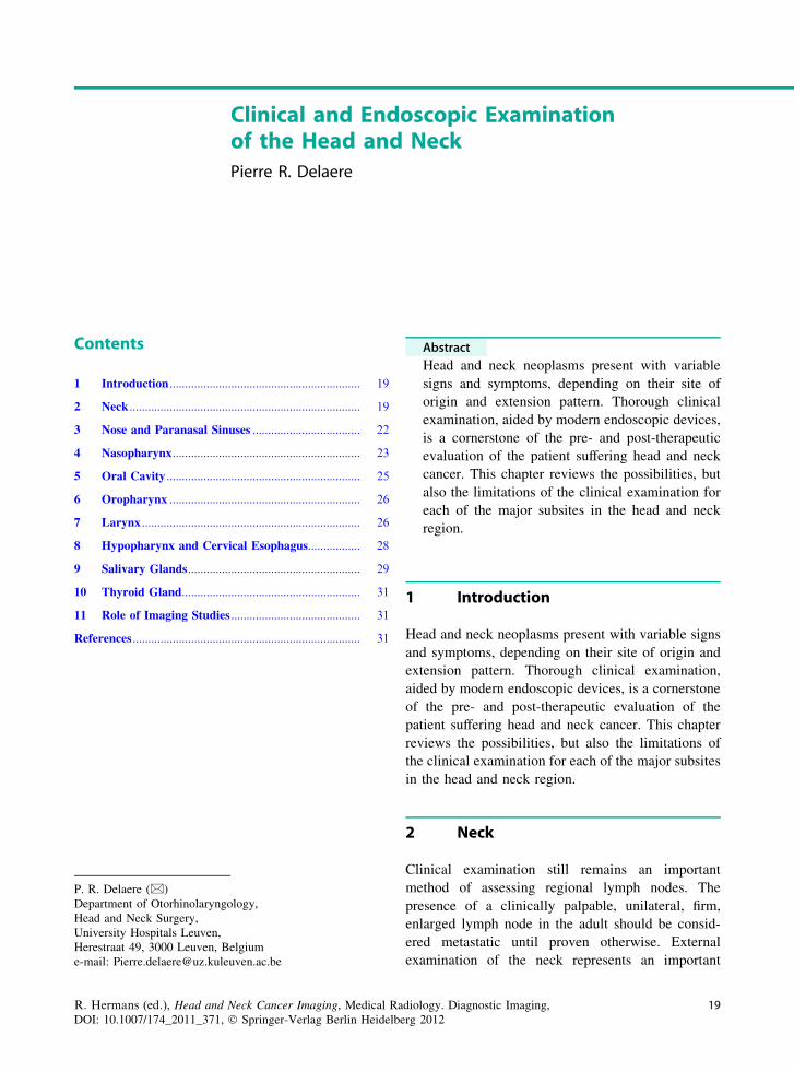
Clinical and Endoscopic Examinationof the Head and Neck
Pierre R. Delaere
Contents
1 Introduction .............................................................. 19
2 Neck ........................................................................... 19
3 Nose and Paranasal Sinuses ................................... 22
4 Nasopharynx............................................................. 23
5 Oral Cavity ............................................................... 25
6 Oropharynx .............................................................. 26
7 Larynx ....................................................................... 26
8 Hypopharynx and Cervical Esophagus................. 28
9 Salivary Glands........................................................ 29
10 Thyroid Gland.......................................................... 31
11 Role of Imaging Studies .......................................... 31
References .......................................................................... 31
Abstract
Head and neck neoplasms present with variablesigns and symptoms, depending on their site oforigin and extension pattern. Thorough clinicalexamination, aided by modern endoscopic devices,is a cornerstone of the pre- and post-therapeuticevaluation of the patient suffering head and neckcancer. This chapter reviews the possibilities, butalso the limitations of the clinical examination foreach of the major subsites in the head and neckregion.
1 Introduction
Head and neck neoplasms present with variable signsand symptoms, depending on their site of origin andextension pattern. Thorough clinical examination,aided by modern endoscopic devices, is a cornerstoneof the pre- and post-therapeutic evaluation of thepatient suffering head and neck cancer. This chapterreviews the possibilities, but also the limitations ofthe clinical examination for each of the major subsitesin the head and neck region.
2 Neck
Clinical examination still remains an importantmethod of assessing regional lymph nodes. Thepresence of a clinically palpable, unilateral, firm,enlarged lymph node in the adult should be consid-ered metastatic until proven otherwise. Externalexamination of the neck represents an important
P. R. Delaere (&)Department of Otorhinolaryngology,Head and Neck Surgery,University Hospitals Leuven,Herestraat 49, 3000 Leuven, Belgiume-mail: [email protected]
R. Hermans (ed.), Head and Neck Cancer Imaging, Medical Radiology. Diagnostic Imaging,DOI: 10.1007/174_2011_371, � Springer-Verlag Berlin Heidelberg 2012
19

starting point in the examination of the patient. It isimportant to remember that some cervical massesmay escape the very best surgical palpation. It isessential that an orderly and systematic examinationof the lymphatic fields on both sides of the neck isperformed (Stell and Maran 1972).
Regional lymphatic drainage from the mucosa ofthe upper aerodigestive tract, salivary glands, and thethyroid gland occurs to specific regional lymph nodegroups (Shah 1990). They should be appropriatelyaddressed in treatment planning for a given primarysite. The major lymph node groups of the head andneck are shown in Fig. 1. Cervical lymph nodes in thelateral aspect of the neck primary drain the mucosa ofthe upper aerodigestive tract. These include the sub-mental and submandibular group of lymph nodeslocated in the submental and submandibular trianglesof the neck. Deep jugular lymph nodes include thejugulodigastric, jugulo-omohyoid, and supraclavicu-lar group of lymph nodes adjacent to the internaljugular vein. Lymph nodes in the posterior triangle of
the neck include the accessory chain of lymph nodeslocated along the spinal accessory nerve and thetransverse cervical chain of lymph nodes in the floorof the posterior triangle of the neck. The retropha-ryngeal lymph nodes are at risk of metastatic dis-semination from tumors of the pharynx. The centralcompartment of the neck includes the Delphian lymphnode overlying the thyroid cartilage in the midlinedraining the larynx, and the perithyroid lymph nodesadjacent to the thyroid gland. Lymph nodes in thetracheoesophageal groove provide primary drainageto the thyroid gland as well as the hypopharynx,subglottic larynx, and cervical esophagus. Lymphnodes in the anterior superior mediastinum providedrainage to the thyroid gland and the cervicalesophagus.
The localization of a palpable metastatic lymphnode often indicates the potential source of a primarytumor. In Fig. 1 the regional lymph node groupsdraining a specific primary site as first echelon lymphnodes are depicted.
Lower lip, floor of mouth,lower gum
Face, nose, paranasal sinuses,
oral cavity, submandibular
gland
Anterior scalp, forehead,
parotid
Oral cavity, oropharynx,
nasopharynx, hypopharynx,
supraglottic larynx
Thyroid, larynx, hypopharynx,
cervical esophagus
Nasopharynx, thyroid, esophagus,
Lung, breast
Nasopharynx, thyroid, esophagus,
Lung, breast
Delphian node (larynx)
Submandibular
Jugulodigastric
(upper jugular)
Tracheoesophageal groove
Supraclavicular
(lower jugular)
Transverse cervical
Jugulo-omohyoid
(mid-jugular)
Spinal accessory chain
Fig. 1 The regional lymph nodes of the head and neck region; the major regional lymphatic chains are annotated on the left. Theseregional lymph node groups drain a specific primary site as first echelon lymph nodes (indicated on right)
20 P. R. Delaere

In order to establish a consistent and easilyreproducible method for description of regional cer-vical lymph nodes, providing a common languagebetween the clinician, the pathologist, and radiologist,the Head and Neck Service at Memorial Sloan–Kettering Cancer Center has described a levelingsystem of cervical lymph nodes (Fig. 2). This systemdivides the lymph nodes in the lateral aspect of theneck into five nodal groups or levels. In addition,lymph nodes in the central compartment of the neckare assigned Levels VI and VII.• Level I: Submental group and submandibular
group. Lymph nodes in the triangular area boundedby the posterior belly of the digastric muscle, theinferior border of the body of the mandible, and thehyoid bone.
• Level II: Upper jugular group. Lymph nodesaround the upper portion of the internal jugular veinand the upper part of the spinal accessory nerve,extending from the base of the skull up to thebifurcation of the carotid artery or the hyoid bone.Surgical landmarks: base of skull superiorly, pos-terior belly of digastric muscle anteriorly, posterior
border of the sternocleidomastoid muscle posteri-orly, and hyoid bone inferiorly.
• Level III: Mid-jugular group. Lymph nodes aroundthe middle third of the internal jugular vein.
• Surgical landmarks: hyoid bone superiorly, laterallimit of the sternohyoid muscle anteriorly, theposterior border of sternocleidomastoid muscleposteriorly, and the caudal border of the cricoidcartilage inferiorly.
• Level IV: Lower jugular group. Lymph nodesaround the lower third of the internal jugular.Surgical landmarks: cricoid superiorly, lateral limitof the sternohyoid muscle anteriorly, posteriorborder of the sternocleidomastoid muscle posteri-orly, and clavicle inferiorly.
• Level V: Posterior triangle group. Lymph nodesaround the lower portion of the spinal accessorynerve and along the transverse cervical vessels. It isbounded by the triangle formed by the clavicle,posterior border of the sternomastoid muscle, andthe anterior border of the trapezius muscle.
• Level VI: Central compartment group. Lymphnodes in the prelaryngeal, pretracheal, (Delphian),
Fig. 2 Level system ofcervical lymph nodes: sevenlevels are distinguished(labeled I–VII)
Clinical and Endoscopic Examination of the Head and Neck 21

paratracheal, and tracheoeophageal groove. Theboundaries are: hyoid bone to suprasternal notch andbetween the medial borders of the carotid sheaths.
• Level VII: Superior mediastinal group. Lymphnodes in the anterior superior mediastinum andtracheoesophageal grooves, extending from thesuprasternal notch to the innominate artery.Some nodes in the neck are more difficult to pal-
pate than others. Thus the retropharyngeal and highestparajugular nodes are almost impossible to detect bypalpation until they are very large.
Structures in the neck which may be mistaken forenlarged lymph nodes are the transverse process ofthe atlas, the carotid bifurcation and the sub-mandibular salivary gland.
Physical examination of the neck for lymph nodemetastasis has a variable reliability (Watkinson et al.1990). A meta-analysis comparing computed tomog-raphy (CT) with physical examination (PE) yieldedthe following results: sensitivity, 83 (CT) versus 74%(PE); specificity, 83 (CT) versus 81% (PE): andaccuracy, 83 (CT) versus 77% (PE). Overall, PEidentified 75% of pathologic cervical adenopathy; thisdetection rate increased to 91% with addition of CT(Merritt et al. 1997).
The American Joint Committee on Cancer and theInternational Union against Cancer has agreed upon auniform staging system for cervical lymph nodes. Theexact description of each N stage of lymph nodemetastasis from squamous carcinomas of the head andneck is described in Table 1. Squamous carcinomasof the nasopharynx and well-differentiated thyroidcarcinomas have a different biology and cervicalmetastases from these tumors are assigned differentstaging systems.
An enlarged metastatic cervical lymph node maybe the only physical finding present in some patientswhose primary tumors are either microscopic oroccult at the time of presentation. A systematic search
for a primary tumor should be undertaken in thesepatients prior to embarking upon therapy for themetastatic nodes. If a thorough head and neckexamination, including fiberoptic nasolaryngoscopy,CT or MRI-study, and PDG-PET scan, fails to show aprimary tumor, then the diagnosis of metastatic car-cinoma to a cervical lymph node from an unknownprimary is established.
3 Nose and Paranasal Sinuses
The nasal cavity is the beginning of the upper airwayand is divided in the midline by the nasal septum.Laterally, the nasal cavity contains the nasal conchae,the inferior concha being part of the nasal cavity, andthe superior and middle conchae being compositeparts of the ethmoid complex. The nasal cavity issurrounded by air containing bony spaces calledparanasal sinuses, the largest of which, the maxillaryantrum, is present on each side. The ethmoid air cellsoccupy the superior aspect of the nasal cavity, andseparate it from the anterior skull base at the level ofthe cribriform plate. Superoanteriorly, the frontalsinus contained within the frontal bone forms a bil-oculated pneumatic space. The sphenoid sinus at thesuperoposterior part of the nasal cavity is located atthe roof of the nasopharynx. The adult ethmoid sinusis narrowest anteriorly in a section known as theostiomeatal complex and this is the site of drainage ofthe maxillary and frontal sinuses (Fig. 3).
Since all of the paranasal sinuses are containedwithin bony spaces, primary tumors of epithelialorigin seldom produce symptoms until they are ofsignificant dimensions, causing obstruction, or untilthey have broken through the bony confines of theinvolved sinus cavity. Tumors of the nasal cavityoften produce symptoms of nasal obstruction, epi-staxis, or obstructive pansinusitis early during the
Table 1 N staging of lymph node metastasis from squamous cell carcinoma of the head and neck except nasopharynx (UICC,International Union Against Cancer 2009)
Nx Regional lymph nodes cannot be assessed
N0 No regional lymph node metastasis
N1 Metastasis in a single ipsilateral lymph node, \3 cm in greatest dimension
N2a Metastasis in single ipsilateral lymph node [3 cm but \6 cm in greatest dimension
N2b Metastasis in multiple ipsilateral lymph nodes, none [6 cm in greatest dimension
N3 Metastasis in a lymph node [6 cm in greatest dimension
22 P. R. Delaere

course of the disease. Unilateral epistaxis, obstruc-tion, or sinusitis should raise the index of suspicionregarding the possibility of a neoplastic process.Tumors of the maxillary antrum may present withsymptoms of obstructive maxillary sinusitis. Swellingof the upper gum or loose teeth may be the firstmanifestation of a malignant tumor of the maxillaryantrum. Locally advanced tumors may present withanesthesia of the skin of the cheek and upper lip,diplopia, proptosis, nasal obstruction, epistaxis, amass in the hard palate or upper gum, or a soft tissuemass in the upper gingivobuccal sulcus. Advancedtumors may present with trismus and visible or pal-pable fullness of the check. Trismus usually is a signof pterygoid musculature invasion. Epistaxis may bethe first manifestation of tumors of the ethmoid orfrontal sinus. This may be accompanied by frontalheadaches or diplopia. Occasionally anosmia may bepresent in patients with esthesioneuroblastoma.Anesthesia in the distribution of the fifth cranial nerveor paralysis of the third, fourth, or sixth cranial nervemay be the first manifestation of a primary tumor ofthe sphenoid sinus. Although sinonasal malignancy israre, persistent nasal symptoms should always beinvestigated, particularly if unilateral. Tumors of thenasal cavity and paranasal sinuses are the most chal-lenging to stage. Endoscopic evaluation of the nasalcavity is crucial in accurate clinical assessment of anintranasal lesion. Fiber optic flexible endoscopy pro-vides adequate visualization of the lower half of thenasal cavity. Therefore, lesions presenting in theregion of the inferior turbinate, middle meatus, and
the lower half of the nasal septum can be easilyvisualized by office endoscopy.
Rigid endoscopic evaluation with telescopes gen-erally requires adequate topical anesthesia as well asshrinkage of the mucosal surfaces of the interior ofthe nasal cavity with the use of topical cocaine. A setof 0, 30, 70, and 90� telescopes should be availablefor appropriate evaluation of the interior of the nasalcavity (Fig. 4). Diagnostic nasal endoscopy allows thecharacterization of intranasal anatomy and identifi-cation of pathology not otherwise visible by tradi-tional diagnostic techniques, such as the use of aheadlight, speculum, and mirror (Bolder and Kennedy1992; Levine 1990).
4 Nasopharynx
The nasopharynx is the portion of the pharynxbounded superiorly by the skull base and the sphenoidand laterally by the paired tori of the eustachian tubes,with the Rosenmüller’s fossa. Anteriorly the posteriorchoanae form the limit of the space, and inferiorly anartificial line drawn at the level of the hard palatedelimits the nasopharynx from the oropharynx.
Presenting symptoms of nasopharyngeal cancermay include a neck mass, epistaxis, nasal obstruction,otalgia, decreased hearing, or cranial neuropathies.Approximately 85 percent of patients have cervicaladenopathy and 50 percent have bilateral neckinvolvement (Lindberg 1972). Serous otitis mediamay occur due to Eustachian tube obstruction. Cranial
Frontal sinus
Ethmoid air cells
Uncinate process
Middle concha
Inferior concha
Maxillary antrum
Ostiomeatal complex
Fig. 3 Coronal sectionthrough maxillofacial region,showing proximity of orbitand anterior cranial fossa tonasal cavity and paranasalsinuses. Disease of the sinusesand nasal cavity may spreaddirectly into adjacentstructures with catastrophicresults
Clinical and Endoscopic Examination of the Head and Neck 23
nerve VI is most frequently affected but multiplecranial nerves may be involved.
Nasopharyngeal carcinoma has a tendency forearly lymphatic spread. The lateral retropharyngeallymph node (of Rouvier) is the first lymphatic filter
but is not palpable. The common first palpable node isthe jugulodigastric and/or apical node under thesternomastoid which are second echelon nodes.Bilateral and contralateral lymph node metastases arenot uncommon.
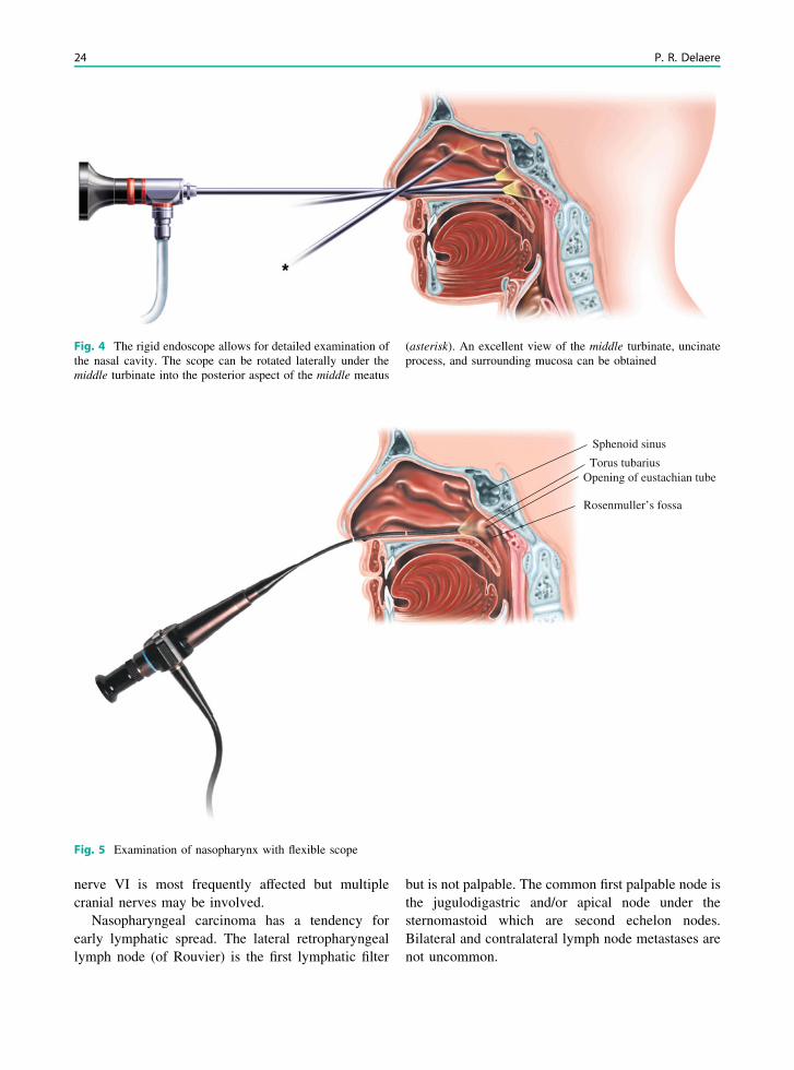
Fig. 4 The rigid endoscope allows for detailed examination ofthe nasal cavity. The scope can be rotated laterally under themiddle turbinate into the posterior aspect of the middle meatus
(asterisk). An excellent view of the middle turbinate, uncinateprocess, and surrounding mucosa can be obtained
Opening of eustachian tube
Sphenoid sinus
Rosenmuller’s fossa
Torus tubarius
Fig. 5 Examination of nasopharynx with flexible scope
24 P. R. Delaere

Nasendoscopy (Fig. 5) using the flexible scopegives a good view of the nasal floor, the walls of thenasopharynx and the fossa of Rosenmüller. Naso-pharyngeal tumors in any quadrant including the fossaof Rosenmüller can be seen and accurately biopsied.For the nasopharynx, also rigid 0 and 30� sinusendoscopes can be similarly used in the clinical set-ting. Under anesthesia, should this be necessary, theseare the scopes of choice for visual assessment andbiopsy.
Evidence of lower cranial nerve deficits may beapparent from palatal or glossal paralysis and atrophy.A full evaluation of the remaining cranial nervesshould include visual assessment and examination ofthe tympanic membranes.
5 Oral Cavity
The oral cavity extends from the vermilion borders ofthe lips to the junction of the hard and soft palatessuperiorly and to the line of the circumvallate papillaeinferiorly. Within this area are the lips, buccalmucosa, alveolar ridges with teeth and gingiva, ret-romolar trigone, floor of mouth, anterior two-thirds ofthe tongue, and hard palate (Fig. 6).
All mucosal surfaces of the mouth require thor-ough and systematic examination. The oral cavity islined by a mucous membrane which is a non-keratinizing stratified squamous epithelium and istherefore pink. It contains taste buds and many minorsalivary glands. All mucosal surfaces should beexamined using tongue blades under optimal lightingconditions.
The clinical features of the primary tumors arisingin the mucosal surface of the oral cavity are variable.The tumor may be ulcerative, exophytic, or endo-phytic, or it may be a superficial proliferative lesion.Most patients with a mouth cancer present with apainful ulcer. Squamous carcinomas with excessivekeratin production and verrucous carcinomas presentas white heaped-up keratotic lesions with varyingdegrees of keratin debris on the surface. Bleedingfrom the surface of the lesion is a characteristicfor malignancy and should immediately raise thesuspicion for a neoplastic process. Endophytic lesionshave a very small surface component but have asubstantial amount of soft tissue involvement beneaththe surface.
Oral salivary tumors may present as a nodule, anon-ulcerative swelling or more usually as an ulcer-ative lesion. Metastatic tumors may also present assubmucosal masses. Mucosal melanoma shows char-acteristic pigmentation.
Macroscopic lesions should be evaluated formobility, tenderness and be palpated with the glovedfinger to detect submucosal spread. This is particu-larly important in tongue lesions extending posteri-orly into the posterior third and tongue base. Thedistance from the tumor to the mandible and themobility of the lesion in relation to the mandible arecritical elements in determining the management ofperimandibular cancers. The indications for examina-tion under anesthesia include an inadequate assessmentof the extent of the disease by history and physicalexamination and imaging, or the presence of symptomsreferable to the trachea, larynx, hypopharynx, and
Circumvallate papillae
Foramen caecum
Fig. 6 Oral cavity and oropharynx. The posterior limits of theoral cavity are the anterior tonsillar pillars, the junction of theanterior two-thirds and posterior one-third of the tongue(i.e. the circumvallate papillae) and the junction of the hardand soft palate. The soft palate and the tonsil are therefore partof the oropharynx. Carcinoma of the anterior two-thirds of thetongue is the most frequent site for a mouth cancer and thelateral border (1) is the most common location. Carcinoma ofthe floor of the mouth most commonly occurs anteriorly eitherin the midline or more usually to one side of the midline (2).Carcinoma of the oropharynx most commonly occurs in the slitbetween tonsil and base of tongue, at the level of the anteriortonsillar pillar (3)
Clinical and Endoscopic Examination of the Head and Neck 25
esophagus that need endoscopic assessment. It is notcost-effective screening to perform panendoscopy onall patients with oral cavity cancer (Benninger et al.1993; Hordijk et al. 1989).
Palpation of the neck is of course essential in theassessment of a patient with mouth cancer. Necknodal disease is the single most important factordetermining the method of treatment, and also prog-nosis is determined by the presence of metastaticnodes. Full examination of the neck must be carriedout to detect any lymph node metastases and eachlevel must be carefully palpated, particularly theupper and middle deep cervical nodes deep to thesternomastoid, from behind the patient, using the tipsof the fingers. Carcinoma of the oral tongue has thegreatest propensity for metastasis to the neck amongall oral cancers. The primary echelon of drainage islevel II but other levels may be also involved.
6 Oropharynx
The oropharynx is that part of the pharynx whichextends from the level of the hard palate above to thehyoid bone below. The anterior wall of the orophar-ynx is formed by the base or posterior third of thetongue bounded anteriorly by the v-shaped line ofcircumvallate papillae (Fig. 6). When present, theinitial symptoms of oropharyngeal cancer are oftenvague and non-specific, leading to a delay in diag-nosis. Consequently, the overwhelming majority ofpatients present with locally advanced tumors.
Presenting symptoms may include sore throat,foreign-body sensation in the throat, altered voice orreferred pain to the ear that is mediated through theglossopharyngeal and vagus nerves. Over two-thirdsof patients present with a neck lump. As the tumorgrows and infiltrates locally, it may cause progressiveimpairment of tongue movement which affects speechand swallowing.
Most tumors of the oropharynx can be easily seenwith good lighting, but those originating in the lowerpart of the oropharynx and tongue base are bestviewed with a laryngeal mirror. The patient shouldbe asked to protrude the tongue, to rule out injury tothe hypoglossal nerve. Trismus is a sign of invasion ofthe masticator space. Sensory and motor functionshould be assessed, particularly mobility of the tongue
as well as fixation. Fiberoptic nasopharyngealendoscopy has greatly enhanced the ease of exami-nation of these tumors, particularly in assessing thelower extent of the tumor and also the superior extentif the nasopharynx is involved. The extent ofinvolvement is often underestimated on inspection,and bimanual palpation of the tumor must be under-taken in all patients. Careful palpation should becarried out to estimate the extent of infiltration, butthis examination may be limited by patient tolerance;thorough palpation under general anesthetic isadvisable. Advanced tumors that cause trismus mayalso be better assessed under a general anesthetic.A detailed examination and biopsy under generalanesthetic may be the only accurate method ofassessing the extent of tumors such as those of thetongue base that may be in a submucosal location.
Examination of the neck must be carried out sys-tematically and each level must be carefully palpatedto detect lymph node enlargement or deep invasion ofthe tumor.
Nodal metastases from squamous cell carcinomasare typically hard and when small are generallymobile. As they enlarge, those in the deep cervicalchain initially become attached to the structures in thecarotid sheath and the overlying sternomastoid mus-cle with limitation in vertical mobility, but laterbecome attached to deeper structures in the prever-tebral region with absolute fixation.
Lymphomas on the other hand have a rubberyconsistency and are generally larger and multiple withmatting together of adjacent nodes. Cystic degenera-tion in a metastatic jugulodigastric node from asquamous carcinoma of the oropharynx may have asimilar presentation to a brachial cyst but the latter isa far less likely diagnosed in the older patient.
7 Larynx
The larynx communicates with the oropharynx aboveand the trachea below. Posteriorly it is partly sur-rounded by the hypopharynx. It may be functionallydivided into three important areas. The supraglottiscontains epiglottis, aryepiglottic folds, arytenoids,false cords, and includes the laryngeal ventricle. Theglottis includes the vocal cords and anterior com-missure and posterior commissure. The subglottis is
26 P. R. Delaere
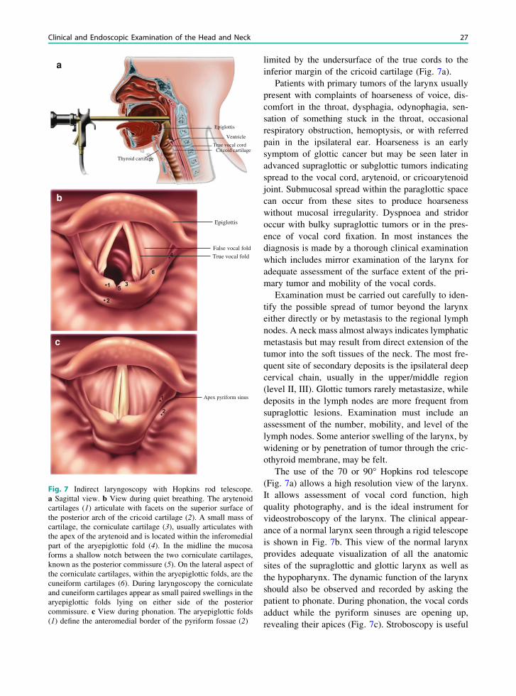
limited by the undersurface of the true cords to theinferior margin of the cricoid cartilage (Fig. 7a).
Patients with primary tumors of the larynx usuallypresent with complaints of hoarseness of voice, dis-comfort in the throat, dysphagia, odynophagia, sen-sation of something stuck in the throat, occasionalrespiratory obstruction, hemoptysis, or with referredpain in the ipsilateral ear. Hoarseness is an earlysymptom of glottic cancer but may be seen later inadvanced supraglottic or subglottic tumors indicatingspread to the vocal cord, arytenoid, or cricoarytenoidjoint. Submucosal spread within the paraglottic spacecan occur from these sites to produce hoarsenesswithout mucosal irregularity. Dyspnoea and stridoroccur with bulky supraglottic tumors or in the pres-ence of vocal cord fixation. In most instances thediagnosis is made by a thorough clinical examinationwhich includes mirror examination of the larynx foradequate assessment of the surface extent of the pri-mary tumor and mobility of the vocal cords.
Examination must be carried out carefully to iden-tify the possible spread of tumor beyond the larynxeither directly or by metastasis to the regional lymphnodes. A neck mass almost always indicates lymphaticmetastasis but may result from direct extension of thetumor into the soft tissues of the neck. The most fre-quent site of secondary deposits is the ipsilateral deepcervical chain, usually in the upper/middle region(level II, III). Glottic tumors rarely metastasize, whiledeposits in the lymph nodes are more frequent fromsupraglottic lesions. Examination must include anassessment of the number, mobility, and level of thelymph nodes. Some anterior swelling of the larynx, bywidening or by penetration of tumor through the cric-othyroid membrane, may be felt.
The use of the 70 or 90� Hopkins rod telescope(Fig. 7a) allows a high resolution view of the larynx.It allows assessment of vocal cord function, highquality photography, and is the ideal instrument forvideostroboscopy of the larynx. The clinical appear-ance of a normal larynx seen through a rigid telescopeis shown in Fig. 7b. This view of the normal larynxprovides adequate visualization of all the anatomicsites of the supraglottic and glottic larynx as well asthe hypopharynx. The dynamic function of the larynxshould also be observed and recorded by asking thepatient to phonate. During phonation, the vocal cordsadduct while the pyriform sinuses are opening up,revealing their apices (Fig. 7c). Stroboscopy is useful
Epiglottis
True vocal cordCricoid cartilage
Thyroid cartilage
Ventricle
Epiglottis
False vocal foldTrue vocal fold
Apex pyriform sinus
a
b
c
Fig. 7 Indirect laryngoscopy with Hopkins rod telescope.a Sagittal view. b View during quiet breathing. The arytenoidcartilages (1) articulate with facets on the superior surface ofthe posterior arch of the cricoid cartilage (2). A small mass ofcartilage, the corniculate cartilage (3), usually articulates withthe apex of the arytenoid and is located within the inferomedialpart of the aryepiglottic fold (4). In the midline the mucosaforms a shallow notch between the two corniculate cartilages,known as the posterior commissure (5). On the lateral aspect ofthe corniculate cartilages, within the aryepiglottic folds, are thecuneiform cartilages (6). During laryngoscopy the corniculateand cuneiform cartilages appear as small paired swellings in thearyepiglottic folds lying on either side of the posteriorcommissure. c View during phonation. The aryepiglottic folds(1) define the anteromedial border of the pyriform fossae (2)
Clinical and Endoscopic Examination of the Head and Neck 27

for the differentiation of functional from anatomicaldefects (Sercarz et al. 1992) and has been employedin the early detection of glottic cancer. In the lattersetting, preservation of the mucosal wave suggeststhat a lesion is not invasive (Zhao 1992).
Technological advance is producing increasinglysmaller diameter fiberoptic endoscopes for examina-tion of the human body. The flexible nasendoscope canbe used to examine the postnasal space, pharynx, andlarynx, down to the level of the vocal cords. Flexiblenasolaryngoscopy (Fig. 8) is generally carried out in anormal anatomical position and during normal respi-ration, unlike the rather distorted position achieved byindirect laryngoscopy or the use of the Hopkins rods.Additionally, flexible endoscopy can be used to directlyobserve the pharyngeal phase of swallowing, givingcomplementary information to that obtained by vid-eofluoroscopy. Test swallows of milk or colored foodcan be examined (Logemann 1983).
Direct laryngoscopy under general anesthesia isthe only reliable way to assess mucosal lesions of thelarynx and pharynx (Phelps 1992; Parker 1992), andmore often enables adequate biopsies to be sampledthan with flexible techniques (Ritchie et al. 1993). If atumor is detected, its limits in all directions should bedetermined both by sight and palpation.
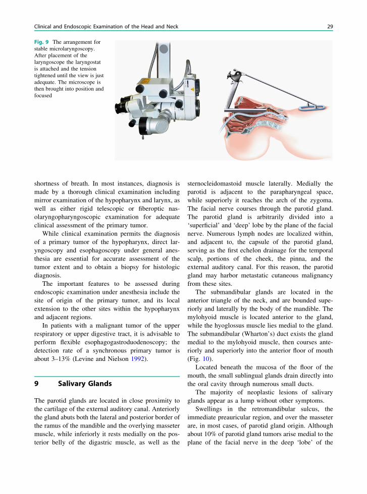
The introduction of the operating microscope hasfacilitated detailed examination of the larynx(Kleinsasser 1965) (Fig. 9). Use of various telescopes
(0, 30, 70, and 120�) provides an excellent anddetailed view of the lesion.
Clinical examination is limited by the fact thatcertain areas of the larynx are inaccessible to bothvisualization and palpation; nevertheless involvementof these structures has an important bearing on stag-ing as well as on management. Information fromradiological imaging and operative endoscopy mustbe utilized in conjunction with physical findings toobtain an accurate pretreatment TNM staging record.Particularly supraglottic tumors are frequentlyunderstaged because the pre-epiglottic and paraglotticspaces cannot be assessed clinically.
8 Hypopharynx and CervicalEsophagus
The hypopharynx links the oropharynx superiorly tothe larynx and esophagus below. Its boundaries areroughly the hyoid and valleculae above and the cri-coid below. Common sites for squamous cell cancersare the pyriform sinuses, the posterior pharyngealwall and the postcricoid space.
Patients with primary tumors of the hypopharynxusually present with the complaints of discomfort inthe throat, dysphagia, odynophagia, sensation ofsomething stuck in the throat, referred pain in theipsilateral ear, hemoptysis, hoarseness of voice, or
Fig. 8 Flexiblelaryngoscopy. Fiberopticlaryngeal nasendoscopyprovides a clear image of thelarynx, laryngopharynx, andbase of tongue
28 P. R. Delaere
shortness of breath. In most instances, diagnosis ismade by a thorough clinical examination includingmirror examination of the hypopharynx and larynx, aswell as either rigid telescopic or fiberoptic nas-olaryngopharyngoscopic examination for adequateclinical assessment of the primary tumor.
While clinical examination permits the diagnosisof a primary tumor of the hypopharynx, direct lar-yngoscopy and esophagoscopy under general anes-thesia are essential for accurate assessment of thetumor extent and to obtain a biopsy for histologicdiagnosis.
The important features to be assessed duringendoscopic examination under anesthesia include thesite of origin of the primary tumor, and its localextension to the other sites within the hypopharynxand adjacent regions.
In patients with a malignant tumor of the upperrespiratory or upper digestive tract, it is advisable toperform flexible esophagogastroduodenoscopy; thedetection rate of a synchronous primary tumor isabout 3–13% (Levine and Nielson 1992).
9 Salivary Glands
The parotid glands are located in close proximity tothe cartilage of the external auditory canal. Anteriorlythe gland abuts both the lateral and posterior border ofthe ramus of the mandible and the overlying massetermuscle, while inferiorly it rests medially on the pos-terior belly of the digastric muscle, as well as the
sternocleidomastoid muscle laterally. Medially theparotid is adjacent to the parapharyngeal space,while superiorly it reaches the arch of the zygoma.The facial nerve courses through the parotid gland.The parotid gland is arbitrarily divided into a‘superficial’ and ‘deep’ lobe by the plane of the facialnerve. Numerous lymph nodes are localized within,and adjacent to, the capsule of the parotid gland,serving as the first echelon drainage for the temporalscalp, portions of the cheek, the pinna, and theexternal auditory canal. For this reason, the parotidgland may harbor metastatic cutaneous malignancyfrom these sites.
The submandibular glands are located in theanterior triangle of the neck, and are bounded supe-riorly and laterally by the body of the mandible. Themylohyoid muscle is located anterior to the gland,while the hyoglossus muscle lies medial to the gland.The submandibular (Wharton’s) duct exists the glandmedial to the mylohyoid muscle, then courses ante-riorly and superiorly into the anterior floor of mouth(Fig. 10).
Located beneath the mucosa of the floor of themouth, the small sublingual glands drain directly intothe oral cavity through numerous small ducts.
The majority of neoplastic lesions of salivaryglands appear as a lump without other symptoms.
Swellings in the retromandibular sulcus, theimmediate preauricular region, and over the masseterare, in most cases, of parotid gland origin. Althoughabout 10% of parotid gland tumors arise medial to theplane of the facial nerve in the deep ‘lobe’ of the
Fig. 9 The arrangement forstable microlaryngoscopy.After placement of thelaryngoscope the laryngostatis attached and the tensiontightened until the view is justadequate. The microscope isthen brought into position andfocused
Clinical and Endoscopic Examination of the Head and Neck 29

gland, more than three-fourths of these deep lobetumors will present as a typical parotid mass.
In the parotid gland pleomorphic adenomas presentas round, firm, reasonable well-demarcated tumors,with a tendency to nodularity as they grow. Their siteof election is between the ascending ramus of themandible anteriorly, and the mastoid process andsternomastoid posteriorly, usually in the tail of thegland. Occasionally they arise in the immediatepreauricular region, where they tend to be small.Warthin’s tumors lie almost invariably in the lowerpole of the gland, are ovoid in shape, and vary inconsistency between soft and firm, depending onwhether or not they have been exposed to previousinflammation; these tumors can occur bilaterally.
Weakness or paralysis of the facial nerve in apreviously untreated patient almost always indicatesthat a tumor is malignant (Spiro et al. 1975; Borthuneet al. 1986). Careful assessment should be made of thefacial nerve and the nerves traversing the nearbycarotid space (cranial nerve IX and XII) if a deep lobeor parapharyngeal space tumor is suspected.
It is often difficult to distinguish between a tumorarising within the submandibular gland or an enlargednode close to the gland or on its outer surface.
Bimanual palpation is essential to differentiatebetween the two, since a node lying on the outersurface of the salivary gland is unlikely to be palpatedby a finger in the mouth, whereas a tumor of the glanditself is more readily compressible bimanually. Pleo-morphic adenomas of the submandibular gland areusually large, quite hard, and nodular, but may beconfused with a slowly growing malignancy such asan adenoid cystic carcinoma. Submandibular glandneoplasms also need evaluation of the lingual andhypoglossal nerves.
The assessment of intraoral minor salivary glandneoplasms depends on the location of the tumor.Palatal lesions are the most common, usually givingthe appearance of being fixed, whether they arebenign or malignant, because of the tight adherence ofthe mucous membrane to bone. Tumors of the hard orsoft palate are often fusiform, firm to hard in con-sistency and nodular. Again the distinction betweenmixed tumor and adenoid cystic carcinoma may bedifficult to make.
A salivary gland tumor arising from the deep lobeof the parotid gland, or from a minor salivary glandin the parapharyngeal space, may cause secondarydisplacement of the palatotonsillar region.
Stenon’s duct
Parotid gland
Submandibular gland
Sublingual gland
Wharton’s duct
Fig. 10 Anatomic relationsof the parotid, submandibular,and sublingual salivary gland
30 P. R. Delaere
Swelling detectable both in the pharynx and par-otid region indicates a very bulky tumor originatingfrom within the deep lobe of the parotid gland. Thisparotid swelling can be visible externally, but thetechnique of bimanual palpation will elicit the char-acteristic sign of ballottement between the examiningfingers, typical of masses occupying such a wide area.The absence of both a visible swelling in the parotidgland and ballottement suggests an origin exclusivelyin the parapharyngeal space.
10 Thyroid Gland
The thyroid gland lies within the pretracheal fascia inthe front of the neck, and consists of two symmetricallobes united in the midline by an isthmus that overliesthe second to fourth tracheal rings (Fig. 1). There isoften a pyramidal lobe, which may extend as high asthe top of the thyroid cartilage.
The incidence of palpable thyroid nodules is esti-mated at only 4–7% of the general adult population.They occur more frequently in women and they areincreasing with age (Mazzaferri et al. 1988). Slightlyless than 5% of thyroid nodules are found to bemalignant (Gharib and Goellner 1993). Most patientswith differentiated carcinoma present with a palpablenodule in the thyroid gland of varying size, consis-tency, and local extent. The primary tumor maypresent as a solitary, well-defined, intrathyroidal dis-crete palpable nodule or it may manifest with diffuseinvolvement of the thyroid gland with or withoutextrathyroid extension and fixation to the structures inthe central compartment of the neck, or it may presentas multiple palpable nodules. The most commonlocation of palpable metastatic lymph nodes fromthyroid cancer is at levels III, IV, or V in the lateralneck. Procedures commonly used for the initialevaluation of thyroid nodules are: ultrasound, radio-nuclide imaging, and fine-needle aspiration biopsy(FNAB).
Anaplastic carcinoma of the thyroid gland usuallymanifests in the older population with a very shorthistory of a rapidly enlarging thyroid mass. Physicalexamination reveals a diffusely enlarged firm to hardill-defined thyroid mass with significant extrathyroidextension to adjacent soft tissues. The mass appearsfixed and inseparable from the laryngotracheal–esophageal complex.
11 Role of Imaging Studies
The clinical evaluation allows to appreciate themucosal layer of the head and neck region quite well.However, the deep extent of potentially infiltratinglesions can only be judged indirectly. Some regions,such as the base of the skull, pterygopalatine andinfratemporal fossa, orbits and brain are beyondclinical evaluation, but critical management decisionshave to be made based on the involvement of thesestructures; imaging findings are of the utmostimportance in such cases. Perineural and/or perivas-cular spread, eventually leading to tumor progressionor recurrences at distance from the primary tumor canoften only be detected by imaging.
Metastatic adenopathies can be identified, some-times still in a subclinical stage or at places notaccessible for clinical examination, such as the ret-ropharyngeal or paratracheal lymph nodes. Also,information on extranodal tumor spread and therelation to critical structures such as the carotidarteries, is necessary for determining the optimalpatient management, and can be deduced fromimaging studies.
Imaging is needed in submucosal lesions, coveredby an intact mucosa. The origin and extent of suchlesions is often difficult to determine based on theclinical evaluation alone. Imaging may provideimportant clues to the diagnosis, as representativebiopsies may be difficult to obtain in deep-seatedlesions.
All these findings can profoundly influence thestaging and management of the patient with head andneck cancer. Finally, imaging may be used to monitortumor response and to try to detect recurrent or per-sistent disease before it becomes clinically evident,possibly with a better chance for successful salvage.
The single most important factor in the optimal useof all this information is the mutual cooperationbetween the radiologist and the physicians in chargeof patient care.
References
Benninger MS, Enrique RR, Nichols RD (1993) Symptom-directed selective endoscopy and cost containment forevaluation of head and neck cancer. Head Neck 15:532–536
Clinical and Endoscopic Examination of the Head and Neck 31
Bolder WE, Kennedy DW (1992) Nasal endoscopy in theoutpatient clinic. Otolaryngol Clin North Am 25:791–802
Borthune A, Kaalhus O, Vermund H (1986) Salivary glandmalignant neoplasms: treatment and prognosis. Int J RadiatOncol Biol Phys 12:747–754
Gharib H, Goellner JR (1993) Fine needle aspiration biopsy ofthe thyroid: an appraisal. Ann Int Med 118:282–289
Hordijk GJ, Bruggink T, Ravasz LA (1989) Panendoscopy: avaluable procedure? Otolaryngol Head Neck Surg101:426–428
Kleinsasser O (1965) Weitere technische entwicklung und ersteergebnisse der ‘endolaryngealen microchirurgie’. Zeitschriftfür Laryngologie, Rhinologie und Otologie 44:711–727
Levine HL (1990) The office diagnosis of nasal and sinusdisorders using rigid nasal endoscopy. Otolaryngol HeadNeck Surg 102:370–373
Levine B, Nielson EW (1992) The justifications and con-troversies of panendoscopy-a review. Ear Nose Throat J71:335–343
Lindberg RD (1972) Distribution of cervical lymph nodemetatstases from squamous cell carcinoma of the upperrespiratory and digestive tracts. Cancer 29:1446–1450
Logemann JA (1983) Evaluation and treatment of swallowingdisorders. College Hill Press, San Diego
Mazzaferri EL, de los Santos ET, Rofagha-Keyhani S (1988)Solitary thyroid nodule: diagnosis and management. MedClin North Am 72:1177–1211
Merritt RM, Williams MF, James TH et al (1997) Detection ofcervical metastasis. A meta-analysis comparing computedtomography with physical examination. Arch OtolaryngolHead Neck Surg 123:149–152
Parker R (1992) Laryngoscopy, microlaryngoscopy and lasersurgery. In: McGregor IA, Howard DJ (eds) Rob andsmith’s operative surgery: head and neck, part 2, 4th edn.Oxford, Butterworth, pp 451–463
Phelps PD (1992) Carcinoma of the larynx-the role of imagingin staging and pre-treatment assessments. Clin Radiol46:77–83
Ritchie AJ, McGuigan J, Stenvenson HM et al (1993)Diagnostic rigid and flexible oesophagoscopy in carcinomaof the oesophagus: a comparison. Thorax 48:115–118
Sercarz JA, Berke GS, Ming Y et al (1992) Videostroboscopyof human vocal fold paralysis. Ann Otol Rhinol Laryngol101:567–577
Shah JP (1990) Patterns of nodal metastases from squamouscarcinomas of the upper aerodigestive tract. Am J Surg160:405–409
Spiro RH, Huvos AW, Strong EW (1975) Cancer of the parotidgland: a clinicopathologic study of 288 primary cases. Am JSurg 130:452–459
Stell PM, Maran AGD (eds) (1972) Pre-operative considera-tions. In: Head and neck surgery. William HeinnemannMedical, London, p 6
UICC, International Union Against Cancer (2009) TNMclassification of malignant tumors, 7th edn. Wiley,New York, p 37
Watkinson JC, Johnston D, James D et al (1990) The reliabilityof palpation in the assessment of tumours. Clin Otolaryngol5:405–410
Zhao R (1992) Diagnostic value of stroboscopy in early glotticcarcinoma. Chung Rua Ehr Pi Yan Hou Ko Tsa Chih27:175–176
32 P. R. Delaere