
Handbook of critical_and_intensive_care_medicine__2nd_edition
418
-
Upload
mistela-qose -
Category
Health & Medicine
-
view
120 -
download
0
Transcript of Handbook of critical_and_intensive_care_medicine__2nd_edition


Handbook of Criticaland Intensive
Care Medicine
Handbook of Criticaland Intensive
Care Medicine
Second Edition
Joseph Varon, MD, FACP,FCCP, FCCM
Pilar Acosta, MD
University of Texas Health Science Center,Houston, TX, USA
With 26 Illustrations
123

Joseph Varon, MDDorrington Medical
Associates, P.A.2219 Dorrington St.University of Texas Health
Science CenterHouston TX [email protected]
Pilar Acosta, MDDorrington Medical
Associates, P.A.2219 Dorrington St.University of Texas Health
Science CenterHouston TX [email protected]
ISBN 978-0-387-92850-0 e-ISBN 978-0-387-92851-7DOI 10.1007/978-0-387-92851-7Springer New York Dordrecht Heidelberg London
Library of Congress Control Number: 2009937882
© Springer Science+Business Media, LLC 2010All rights reserved. This work may not be translated or copied in whole or in partwithout the written permission of the publisher (Springer Science+Business Media,LLC, 233 Spring Street, New York, NY 10013, USA), except for brief excerpts inconnection with reviews or scholarly analysis. Use in connection with any form ofinformation storage and retrieval, electronic adaptation, computer software, or bysimilar or dissimilar methodology now known or hereafter developed is forbidden.The use in this publication of trade names, trademarks, service marks, and similarterms, even if they are not identified as such, is not to be taken as an expression ofopinion as to whether or not they are subject to proprietary rights.While the advice and information in this book are believed to be true and accurateat the date of going to press, neither the authors nor the editors nor the publishercan accept any legal responsibility for any errors or omissions that may be made.The publisher makes no warranty, express or implied, with respect to the materialcontained herein.
Printed on acid-free paper
Springer is part of Springer Science+Business Media (www.springer.com)

This book is dedicated to my spouse Sara,and my children Adylle, Jacques, Daryelle,and Michelle for understanding those count-less days, nights, and weekends away fromhome.
Joseph Varon, MD, FACP, FCCP, FCCM
This book is dedicated to my family, for theirunconditional support.
Pilar Acosta, MD

Preface
Why write a book on the management of critically ill patients? Over the past fewdecades we have seen an enormous growth in the number of intensive care units(ICU) across the world. Indeed, it is estimated that a large proportion of health careexpenses are devoted to patients in these specialized units. Medical students, res-idents, fellows, attending physicians, critical care nurses, pharmacists, respiratorytherapists, and other health-care providers (irrespective of their ultimate field of prac-tice) will spend several months or years of their professional lives taking care ofcritically ill or severely injured patients. These clinicians must have special train-ing, experience, and competence in managing complex problems in their patients.Moreover, these clinicians must interpret data obtained by many kinds of monitoringdevices, and they must integrate this information with their knowledge of the patho-physiology of disease. Even more important is the fact that anyone working in an ICUor with a critically ill patient must approach patients with a multidisciplinary team.The phrase there is no I in TEAM comes to mind.
The second edition of this book∗ was written for every practitioner engaged inCritical Care Medicine across the world. We have attempted to present basic and gen-erally accepted clinical information, our own personal experiences, facts, and someimportant formulas as well as laboratory values and tables which we feel will be use-ful to the practitioner of Critical Care Medicine. The chapters of this book follow anoutline format and are divided by organ–system (i.e., neurologic disorders, cardio-vascular disorders), as well as special topics (i.e., environmental disorders, trauma,toxicology). Every chapter has been updated and many chapters are completely new.
It is important for the reader of this handbook to understand that Critical CareMedicine is not a static field and changes occur every day. Therefore, this handbookis not meant to define the standard of care, but rather to be a general guide to currentclinical practice used in Critical Care Medicine. We wrote this book hoping that itwill benefit thousands of critically ill patients, but more importantly that it will aidpracticing clinicians to assume a multidisciplinary approach.
Joseph Varon, MD, FACP, FCCP, FCCMPilar Acosta, MD
∗Note: the first edition of this book, published by Springer in 2002, was titled Handbook of PracticalCritical Care Medicine.

Contents
Preface vii
1. Approach to the Intensive Care Unit (ICU) 1
2. The Basics of Critical Care 11
3. Cardiovascular Disorders 47
4. Endocrinologic Disorders 85
5. Environmental Disorders 113
6. Gastrointestinal Disorders 137
7. Hematologic Disorders 149
8. Infectious Diseases 171
9. Neurologic Disorders 191
10. Nutrition 209
11. Critical Care Oncology 229
12. Critical Care of the Pregnant Patient 249
13. Pulmonary Disorders 267
14. Renal and Fluid–Electrolyte Disorders 297
15. Special Techniques 335
16. Toxicology 355
17. Trauma 375
18. Allergic and Immunologic Emergencies 393
19. Pharmacologic Agents Commonly Used in the ICU 399
20. Common Laboratory Values in the ICU 407
Index 413

1Approach to the
Intensive CareUnit (ICU)
� I. WELCOME TO THE ICU
What Is an ICU?
An intensive care unit (ICU) is an area of a hospital that provides aggressive therapy,using state-of-the-art technology and both invasive and noninvasive monitoring forcritically ill and high-risk patients. In these units the patient’s physiological variablesare reported to the practitioner on a continuous basis, so that titrated care can beprovided.
As a medical student, resident physician, attending physician, or other healthcareprovider, one is likely to spend several hundreds of hours in these units caring forvery sick patients. Knowing the function and organization of these specialized areaswill help the practitioner in understanding critical care.
Historical Development of the ICU
The origin of the ICU remains controversial. In 1863, Florence Nightingale wrote,“In small country hospitals there are areas that have a recess or small room leadingfrom the operating theater in which the patients remain until they have recovered, orat least recover from the immediate effects of the operation.” This is probably theearliest description of what would become the ICU. Recovery rooms were developedat the Johns Hopkins Hospital in the 1920s. In Germany in the 1930s, the first well-organized postoperative ICU was developed. In the United States, more specializedpostoperative recovery rooms were implemented in the 1940s at the Mayo Clinic.By the late 1950s, the first shock unit was established in Los Angeles. The initialsurveillance unit for patients after acute myocardial infarction was started in KansasCity in 1962.
J. Varon, P. Acosta, Handbook of Critical and Intensive Care Medicine,DOI 10.1007/978-0-387-92851-7_1,C© Springer Science+Business Media, LLC 2010

2 1. Approach to the Intensive Care Unit
Economical Impact of the ICU
Since their initial development, there has been a rapid and remarkable growth ofICU beds in the United States. There are presently more than 60,000 ICU beds inthe United States, and critical care consumes more than 2.5% of the gross nationalproduct.
Organization of the ICU
ICUs in the United States may be open or closed. Open ICUs may be utilized by anyattending physician with admitting privileges in that institution, and many subspe-cialists may manage the patient at the same time. These physicians do not need to bespecifically trained in critical care medicine. A different system is provided in closedICUs, in which the management of the patient on admission to the unit is providedby an ICU team and orchestrated by physicians with specialized training in criticalcare medicine. Although consultants may be involved in the patient’s care, all ordersare written by the ICU team and all decisions are approved by this team.
ICUs may also be organized by the type of patients whom they are intended totreat. In some studies, these “closed” units have shown shorter length of stay for theICU patients due to the standardization of care.
ICUs can also be divided on the basis of the patients they have. Examples includethe neurosurgical ICU (NICU), pediatric ICU (PICU), cardiovascular surgery ICU(CVICU), surgical ICU (SICU), medical ICU (MICU), and coronary care unit (CCU).
Most ICUs in the United States have a medical director who, with varying degreesof authority, is responsible for bed allocation, policy making, and quality assurance,and who may be, particularly in closed ICUs, the primary attending physician forpatients admitted to that unit.
� II. TEAM WORK
Care of the critically ill patient has evolved into a discipline that requires specializedtraining and skills. The physician in the ICU depends on nursing for accurate chartingand assessment of the patients during the times when he or she is not at the bedsideand for the provision of the full spectrum of nursing care, including psychologicaland social support and the administration of ordered therapies.
Complex mechanical ventilation devices need appropriate monitoring and adjust-ment. This expertise and other functions are provided by a professional team ofrespiratory therapy practitioners. The wide spectrum of the pharmacopeia used inthe ICU is greatly enhanced by the assistance of our colleagues in pharmacy. Manyinstitutions find it useful to have pharmacists with advanced training participate inrounding to help practitioners in the appropriate pharmacologic management of thecritically ill. Additionally, technicians with experience in monitoring equipment mayhelp in obtaining physiologic data and maintaining the associated equipment. Withoutthese additional healthcare professionals, optimal ICU management would not bepossible.
As many ICU patients remain in these units for prolonged periods of time,additional heathcare providers, such as the nutritional support team and physical/occupational therapy, remain important component of the management of thesepatients.
V. System-Oriented Rounds 3
� III. THE FLOWSHEET
ICU patients, by virtue of their critical illnesses, present with complex pathophysiol-ogy and symptomatology. In many cases, these patients are endotracheally intubated,with mental status depression, and cannot provide historical information. The physi-cal examination and monitoring of physiology and laboratory data must provide theinformation on which to base a diagnosis and initiate appropriate treatment in thesecases.
The flowsheet is the repository of information necessary for the recognition andmanagement of severe physiological derangements in critically ill patients. A well-organized flowsheet provides around-the-clock information regarding the differentorgan systems rather than just vital signs alone. In many institutions these flowsheetsare computerized, potentially improving accessibility and allowing real-time data.These devices are complex and in many instances expensive.
Major categories appropriate for an ICU flowsheet include
— Vital signs
— Neurological status
— Hemodynamic parameters
— Ventilator settings
— Respiratory parameters
— Inputs and outputs
— Laboratory data
— Medications
� IV. THE CRITICALLY ILL PATIENT
In general, ICU patients not only are very ill but also may have disease processes thatinvolve a number of different organ systems. Therefore, the approach to the criticallyill patient needs to be systematic and complete (see below).
Several issues need to be considered in the initial approach to the critically illpatient. The initial evaluation consists of assessment of the ABC (airway, breathing,circulation), with simultaneous interventions performed as needed. An organized andefficient history and physical examination should then be conducted for all patientsentering the ICU, and a series of priorities for therapeutic interventions should beestablished.
� V. SYSTEM-ORIENTED ROUNDS
In the ICU accurate transmission of clinical information is required. It is importantto be compulsive and follow every single detail. The mode of presentation duringICU rounds may vary based on institutional tradition. Nevertheless, because of mul-tiple medical problems, systematic gathering and presentation of data are needed forproper management of these patients. We prefer presenting and writing notes in a“head-to-toe” format (see Table 1.1).

4 1. Approach to the Intensive Care Unit
Table 1.1. Minimum Amount of Information Necessary forPresentation During Rounds (See Text for Details)
ICU survival guide for presentation during rounds
1. Identification/problem list2. Major events during the last 24 h3. Neurological:
—Mental status, complaints, detailed neurological exam (if pertinent)4. Cardiovascular:
—Symptoms and physical findings, record BP, pulse variability over thepast 24 h., ECG, echocardiogram results
—If CVP line and/or Swan-Ganz catheter in place, check CVP andhemodynamics yourself
5. Respiratory:—Ventilator settings, latest ABGs, symptoms and physical findings, CXR
(daily if the patient is intubated). Other calculations (e.g., compliance,minute volume, etc)
6. Renal/Metabolic:—Urine output (per hour and during the last 24 h), inputs/outputs with
balance (daily, weekly), weight, electrolytes, and if done, creatinineclearance. Acid–base balance interpretation
7. Gastrointestinal:—Abdominal exam, oral intake, coffee-grounds, diarrhea. Abdominal
x-rays, liver function tests, amylase, etc.8. Infectious diseases:
—Temperature curve, WBC, cultures, current antibiotics (number of dayson each drug), and antibiotic levels
9. Hematology:—CBC, PT, PTT, TT, BT, DIC screen (if pertinent), peripheral smear.
Medications altering bleeding10. Nutrition:
—TPN, enteral feedings, rate, caloric intake, and grams of protein11. Endocrine:
—Do you need to check TFTs or cortisol? Give total insulin needs per hourand 24 h
12. Psychosocial:—Is the patient depressed or suicidal? Is the family aware of his or her
present condition?13. Other:
—Check the endotracheal tube position (from lips or nostrils incentimeters), and check CXR position. Check all lines, transducers.Note position of the catheter, skin insertion sites
—All medications and drips must be known. All drips must be renewedbefore or during rounds
Abbreviations: ABG, arterial blood gas; BP, blood pressure; BT, bleeding time; CBC,complete blood count; CXR, chest x-ray; CVP, central venous pressure; DIC, dissemi-nated intravascular coagulation; ECG, electrocardiogram; PT, prothrombin time; PTT, partialthromboplastin time; TFT, thyroid function tests; TPN, total parenteral nutrition; TT, thrombintime; WBC, white blood cell count.

V. System-Oriented Rounds 5
The ICU progress note is system-oriented, which differs from the problem-orientedapproach commonly utilized on the general medicine-surgery wards. The assessmentand plan are formulated for each of the different organ systems as aids to organization,but like in the non-ICU chart, each progress note should contain a “problem list” thatis addressed daily. This problem list allows the healthcare provider to keep track ofmultiple problems simultaneously and enables a physician unfamiliar with a givencase to efficiently understand its complexities if the need arises.
The art of presenting cases during rounds is perfected at the bedside over manyyears, but the following abbreviated guide may get the new member of the ICU teamoff to a good start. A “how-to” for examining an ICU patient and a stylized ICUprogress note guide are also presented. Remember that for each system reviewed afull review of data, assessment, and management plan should be provided. Using thissimple technique avoids important data to be skipped or forgotten.
When you arrive in the ICU in the morning
1. Ask the previous night’s physicians and nurses about your patients.
2. Go to the patient’s room. Review the flowsheet. Then proceed by examining andreviewing each organ system as follows:
Identification
— Provide name, age, major diagnoses, day of entry to the hospital, and day ofadmission to the ICU.
Major Events over the Last 24 h
— Mention (or list in the progress note) any medical event or diagnostic endeavorthat was significant. For example, major thoracic surgery or cardiopulmonaryarrest, computed tomography (CT) scan of the head, reintubation, or changesin mechanical ventilation.
Systems Review
Neurologic
— Mental status: Is the patient awake? If so, can you perform a mental statusexamination? If the patient is comatose, is he or she spontaneously breathing?
— What is the Glasgow coma scale score?
— If the patient is sedated, what is the Ramsay score, or what is the score on anyother scales (i.e., RASS) used at the institution for patients who are sedated?
— If pertinent (in patients with major neurological abnormalities or whose majordisease process involves the central nervous system), a detailed neurologicalexam should be performed.
— What are the results of any neurological evaluation in the past 24 h, such as alumbar puncture or CT scan?

6 1. Approach to the Intensive Care Unit
Cardiovascular
— Symptoms and physical findings: It is important to specifically inquire forsymptoms of dyspnea and chest pain or discomfort, among others. Thephysical examination should be focused on the cardiac rhythm, presence ofcongestive heart failure, pulmonary hypertension, pericardial effusion, andvalvulopathies.
— Electrocardiogram (ECG): We recommend that a diagnostic ECG be con-sidered in every ICU patient on a frequent basis. Many ICU patientscannot communicate chest pain or other cardiac symptomatology, so thatan ECG may be the only piece of information pointing toward cardiacpathology.
— If the patient has a central venous pressure (CVP) line and/or a pulmonaryartery (Swan-Ganz) catheter in place, check the CVP and hemodynamicsyourself. Hemodynamic calculations of oxygen consumption and deliveryshould be noted if the patient has a pulmonary artery catheter or an oximet-ric intravascular device. A detailed list of hemodynamic parameters usefulin the management of critically ill patients can be found in Chapters 3,“Cardiovascular Disorders” and 13, “Pulmonary Disorders”.
— Note the blood pressure (BP) and pulse variability over the past 24 h. Calculatethe mean arterial pressure (MAP) changes over the time period.
— If the patient had an echocardiogram, review the findings in detail.
— If the patient is receiving assisted mechanical cardiac support (i.e., intra-aorticballoon pump) or has a temporary pacemaker, the settings need to be recordedand compared to prior days.
Respiratory
— If the patient is on mechanical ventilation, the current ventilator settings needto be charted, including the ventilatory mode, tidal volume, preset respira-tory rate and patient’s own respiratory rate, amount of oxygen being provided(FiO2), and whether or not the patient is receiving positive end-expiratorypressure (PEEP) and/or pressure support (PS) and their levels. When perti-nent, peak flow settings and inspiration–expiration (I:E) ratio should be noted.Mechanically ventilated patients should have a daily measurement of thestatic and dynamic compliance, minute volume, and other parameters (seeChapters 2, “The Basics of Critical Care” and 13, “Pulmonary Disorders”).If weaning parameters were performed, they need to be addressed.
— The most recent arterial blood gases (ABGs) should be compared with previ-ous measurements. Calculation of the alveolar-arterial oxygen gradient shouldbe performed in all ABGs.
— Symptoms and physical findings should be noted, and if pertinent, sputumcharacteristics should be mentioned.
— Generally, a portable chest x-ray is obtained in all intubated patients daily.Attention is paid to CVP lines, endotracheal tubes, chest tubes, pericardio-centesis catheters, opacities in the lung fields (infiltrates), pneumothoraces,pneumomediastinum, and subcutaneous air.

V. System-Oriented Rounds 7
Renal/Metabolic
— Urine output is quantified per hour and during the past 24 h. In patients requir-ing intensive care for more than 2 days, it is important to keep track of theirinputs, outputs, and overall daily and weekly fluid balance.
— Daily weights.
— If the patient underwent hemodialysis or is on peritoneal dialysis, it isimportant to include it on the daily note.
— Electrolytes are noted including magnesium, phosphorus, calcium (ionized),and if done, creatinine clearance, urine electrolytes, etc. Any changes in thesevalues need special consideration.
— The ABGs are used for acid–base balance interpretation. The formulas mostcommonly used for these calculations are depicted in Chapter 14, “Renal andFluid-Electrolyte Disorders”.
Gastrointestinal
— Abdominal examination: A detailed abdominal examination may uncover newpathology or allow one to assess changes in recognized problems.
— If the patient is awake and alert, mention his or her oral intake (e.g., determinewhether clear liquids are well tolerated).
— The characteristics of the gastric contents or stool (e.g., coffee-grounds,diarrhea, etc.) should also be mentioned and recorded.
— Abdominal x-rays, if pertinent, are reviewed with special attention to theduration of feeding tubes, free air under the diaphragm, and bowel gas pattern.
— Liver function tests (transaminases, albumin, coagulation measurements, etc.)and pancreatic enzymes (amylase, lipase, etc.) are mentioned and recordedwhen pertinent, as well as their change since previous measurements.
Infectious Diseases
— Temperature curve: Changes in temperature (e.g., “fever spike” or hypother-mia) should be noted as well as the interventions performed to controlthe temperature. Note fever character, maximum temperature (T-max), andresponse to antipyretics.
— The total white blood cell count (WBC) is recorded, when pertinent, withspecial attention to changes in the differential.
— Cultures: Culture (blood, sputum, urine, etc.) results should be checked dailywith the microbiology laboratory and recorded. Those positive cultures, whenmentioned, should include the antibiotic sensitivity profile, when available.
— Current antibiotics: Current dosages and routes of administration as well asthe number of days on each drug should be reported. If an adverse reactionoccurred related to the administration of antibiotics, it should be reported.
— Antibiotic levels are drawn for many antibiotics with known pharmacokineticsto adjust their dosage (e.g., peak and trough levels for vancomycin).
— If the patient is receiving a new drug, either investigational or FDA approved,side effects and/or the observed salutary effects are reported.
8 1. Approach to the Intensive Care Unit
Hematology
— Complete blood cell count (CBC): When presenting the results, it is importantto be aware of the characteristics of the peripheral blood smear.
— Coagulation parameters: The prothrombin time (PT), partial thromboplas-tin time (PTT), thrombin time (TT), bleeding time (BT), and disseminatedintravascular coagulation (DIC) screen (e.g., fibrinogen, fibrin split products,d-dimer, platelet count) should be addressed when pertinent.
— If the patient has received blood products or has undergone plasma exchange,this should be noted.
— In this context special attention is paid to all medications that alter bleed-ing, both directly (e.g., heparin, desmopressin acetate) and indirectly (e.g.,ticarcillin-induced thrombocytopathy, ranitidine-induced thrombocytopenia).
Nutrition
— Total parenteral nutrition (TPN): You need to state what kind of formulathe patient is receiving, the total caloric intake provided by TPN with thepercentage of fat and carbohydrates given. The total amount of protein is men-tioned with an assessment of the anabolic or catabolic state (see Chapter 10,“Nutrition”).
— Enteral feedings: These are reported similar to TPN, with mention of anygastrointestinal intolerance (e.g., diarrhea).
— For both of the above, the nutritional needs of the patient and what percentageof these needs is actually being provided must be reported.
Endocrine
— Special attention is paid to pancreatic, adrenal, and thyroid function. If needed,a cortisol level or thyroid function tests are performed. In most situations thesedeterminations are not appropriate in the ICU except under special circum-stances (e.g., hypotension refractory to volume resuscitation in a patient withdisseminated tuberculosis, Addisonian crisis), and the results are usually notavailable immediately.
— Glucose values: The data are clear that good glycemic control helps patientsin the ICU. Therefore, you must include the glycemic variation that the patienthas over the past 24 h.
— Insulin: The total insulin needs per hour and per 24 h as well as the blood sugarvalues should be reported. The type of insulin preparation being used shouldbe specified.
— In patients with hyperosmolal states and diabetic ketoacidosis, it is necessaryto determine calculated and measured serum osmolality as well as ketones.The values for these are charted and compared with previous results.

VI. Do Not Resuscitate (DNR) and Ethical Issues 9
Psychosocial
— Patients in the ICU tend to be confused and in many instances disoriented.Although these symptoms and signs are reviewed as part of the neurologi-cal examination, it is important to consider other diagnoses (e.g., depression,psychosis).
— For drug overdoses and patients with depression, specific questions need to beasked regarding the potential of new suicidal and homicidal ideations.
Other
Other parameters also must be checked daily before the morning (or evening)rounds:
— Check the endotracheal tube size and position (from the lips or nostrils incentimeters), and check its position on chest x-ray, as mentioned above.
— If the patient has a nasotracheal or orotracheal tube, a detailed ear, nose, andthroat examination should be performed (because patients with nasotrachealtubes may develop severe sinusitis).
— Check all lines with their corresponding equipment (e.g., transducers must beat an adequate level). Note the position of the catheter(s) both on physicalexamination and on x-ray, as well as the appearance of the skin insertion site(s)(e.g., infection).
— All medications and continuous infusions and their proper concentrations andinfusion rates must be known and recorded.
— At the time of “pre-rounding,” all infusions must be renewed. TPN orders needto be written early, with changes based on the most recent laboratory findings.
— At the end of rounds every morning, it is important to keep a list of the thingsthat need to be done that day, for example, changes in central venous lines orarterial lines, performing a lumbar puncture, etc.
� VI. DO NOT RESUSCITATE (DNR) AND ETHICALISSUES
Ethical issues arise every day in the ICU. For example, should a particular patient bekept on mechanical ventilation when he has an underlying malignancy? Should thepatient with acquired immune deficiency syndrome (AIDS) receive cardiopulmonaryresuscitation (CPR) in the event of a cardiorespiratory arrest? Should the family bepermitted to terminate mechanical ventilation or tube feedings?
These and similar questions are frequently asked and in reality may have nosingle correct answer. Patients must be allowed the opportunity to express theirwishes about resuscitation. ICU physicians need to educate the patient and the familyregarding prognosis. Physicians are not obliged to provide futile interventions, butcommunication is the key to avoiding conflicts in this arena.
10 1. Approach to the Intensive Care Unit
Do not resuscitate (DNR) orders have become widely used in US hospitals.A DNR order specifically instructs the patient’s healthcare provider to forego CPR ifthe patient undergoes cardiac or respiratory arrest. Various levels of support may beagreed upon by patients, their physicians, and family.
Different institutions have distinct categories of support. Examples include thefollowing:
— Code A or Code I: Full support, including CPR, vasopressors, mechanicalventilation, surgery, etc.
— Code B or Code II: Full support except CPR (no endotracheal intubation orchest compressions). However, vasopressor drugs are utilized in these cases.
— Code C or Code III: Comfort care only. Depending on the policies of theinstitution, intravenous fluids, antibiotics, and other medications may bewithheld.
A patient who is DNR may be in either of the last two groups. It is important thenthat a full description of a particular triage status is provided and carefully explainedto the patient and/or family and discussed as needed. Remember to document all yourdiscussions with the family on the medical record.
As mentioned, the level of resuscitative efforts will therefore depend on thepatient’s wishes. When the patient cannot express his or her wishes, then these ques-tions are asked to the closest family member or designated individual. For example,would the patient have wanted full mechanical ventilatory support for a cardiopul-monary arrest? Were provisions made for a healthcare surrogate if the patient becameincompetent?
Ethical problems often can be resolved by seeking consultation with a group ofindividuals who are experienced in dealing with these issues. In many institutionsan ethics committee is available to provide consultation to practitioners and familiesregarding moral and ethical dilemmas.

2The Basics of Critical
Care
Critical and intensive care medicine is an integrated discipline that requires theclinician to examine a number of important basic interactions. These include theinteractions among organ systems, between the patient and his or her environment,and between the patient and life-support equipment. Gas exchange within the lung,for example, is dependent on the matching of ventilation and perfusion—in quantity,space, and time. Thus, neither the lungs nor the heart are solely responsible; rather, itis the cardiopulmonary interaction that determines the adequacy of gas exchange.
Critical care often entails providing advanced life support through the applicationof technology. Mechanical ventilation is a common example. Why is it that positivepressure ventilation and positive end-expiratory pressure (PEEP) can result in oliguriaor reduction of cardiac output? Many times clinical assessments and your therapeu-tic plans will be directed at the interaction between the patient and technology; thisrepresents a unique “physiology” in itself.
� I. CARDIAC ARREST AND RESUSCITATION
Resuscitation from death is not an everyday event but is no longer a rarity. The goalof resuscitation is restoration of normal or near-normal cardiopulmonary function,without deterioration of other organ systems.
A. EtiologyThe most common causes of sudden cardiac arrest are depicted in Table 2.1.
B. Pathogenesis1. Ventricular fibrillation (VF) or pulseless ventricular tachycardia (VT).2. Asystole.3. Pulseless electrical activity (PEA) (electromechanical dissociation). Patients
arresting with PEA can have any cardiac rhythm but no effective mechanicalsystole (thus, blood pressure [BP] is unobtainable).
4. Cardiogenic shock: No effective cardiac output is generated.
J. Varon, P. Acosta, Handbook of Critical and Intensive Care Medicine,DOI 10.1007/978-0-387-92851-7_2,C© Springer Science+Business Media, LLC 2010

12 2. The Basics of Critical Care
Table 2.1. Common Causes of Sudden Nontraumatic CardiacArrest
1. Primary cardiac eventa. CADb. Dysrhythmias due to
(1) Hyperkalemia(2) Severe acidemia(3) Other electrolyte disturbances
c. Myocarditisd. Tamponade
2. Secondary to respiratory arrest (e.g., children)3. Secondary to acute respiratory failure
a. Hypoxemiab. Hypercapnia
4. Extreme alterations in body temperature5. Drug effects
a. Digitalisb. Quinidinec. Tricyclic antidepressantsd. Cocaine
5. The central nervous system (CNS) will not tolerate >6 min of ischemia atnormothermia.
C. Diagnosis1. Unexpected loss of consciousness in the unmonitored patient.2. Loss of palpable central arterial pulse.3. Respiratory arrest in a patient previously breathing spontaneously.
D. Differential Diagnosis1. Syncope or vasovagal reactions2. Coma3. “Collapse”4. Seizures
E. Management1. Cardiopulmonary resuscitation
a. The main indications for cardiopulmonary resuscitation (CPR) in the ICUinclude(1). Cardiovascular collapse(2). Respiratory arrest with or without cardiac arrest
b. Mechanisms of blood flow during CPR(1). Direct compression of the heart between the sternum and vertebral column
“squeezes” blood from the ventricles into the great vessels.(2). Changes in intrathoracic pressure generate gradients between the periph-
eral venous and arterial beds, resulting in forward flow.(3). During CPR, the dynamics of the chest compression process may play
a major role in determining outcome of the resuscitation effort. Indeed,chest compressions by themselves may provide ventilation.

I. Cardiac Arrest and Resuscitation 13
(4). Interposed abdominal compression CPR increases aortic diastolic bloodpressure, improving blood perfusion to the coronary arteries.
c. Technique(1). Establish an effective airway (see Chapter 15 , “Special Techniques”).
(a). Assess breathing first (open airway, look, listen, and feel).(b). If respiratory arrest has occurred, the possibility of a foreign body
obstruction needs to be considered and measures taken to relieve it.(c). If endotracheal intubation is to be performed, give two breaths during
a 2 s pause every 30 chest compressions.(d). The respiratory rate during cardiac or respiratory arrest should be
8–10 breaths per minute. Once spontaneous circulation has beenrestored, the rate should be 10–12 breaths per minute.
(e). Ventilations should be performed with a tidal volume of 5–7 mL/kgof ideal body weight.
(f). The highest possible concentration of oxygen (100%) should beadministered to all patients receiving CPR.
(2). Determine pulselessness (if no pulse, start CPR immediately).(3). Chest compressions, current advanced cardiac life support (ACLS) rec-
ommendations:(a). Rescuer’s hand located in the lower margin of sternum.(b). Heel of one hand is placed on the lower half of the sternum and the
other hand is placed on top of the hand on the sternum so that thehands are parallel.
(c). Elbows are locked in position, the arms are straightened, and the res-cuer’s shoulders are positioned directly over the hands, providing astraight thrust.
(d). The sternum is depressed 11/2–2 in. in normal-sized adults with eachcompression at a rate of 100/min.
(e). The American Heart Association addresses alternative techniquesto standard manual CPR, specifically mechanical devices (i.e., vestCPR, LUCAS). This new device is with the purpose to enhancecompression and diminish exhaustion of the person delivering CPR.
(4). Cardiac monitoring and dysrhythmia recognition (see also Chapter 3,“Cardiovascular Disorders”)(a). Distinguish between ventricular and supraventricular rhythms.
i. Most rapid, wide QRS rhythms are VT.ii. Initiate therapy immediately (see below).
(5). Defibrillation is the major determinant of survival in cardiac arrest due toVF or pulseless VT.(a). Integrating early defibrillation and CPR provides better outcome.(b). Resume chest compressions after delivering one shock.
(6). Drug therapy during CPR may be given by the following routes:(a). Peripheral vein (antecubital or external jugular are preferred).(b). Central venous line (subclavian or internal jugular): On occasion a
long line that extends above the diaphragm can be started in thefemoral vein.
(c). Intraosseous (IO) cannulation provides access that is safe and effec-tive for drug delivery, fluid resuscitation, and blood sampling.
(d). Endotracheal: Medications should be administered at 2–2.5 timesthe recommended intravenous (IV) dose and should be diluted in 10
14 2. The Basics of Critical Care
mL of normal saline or distilled water. A catheter should be passedbeyond the tip of the endotracheal tube, and the medication sprayedquickly followed by several quick insufflations.
(e). The different drug dosages utilized during CPR and in the immediatepostresuscitation period are depicted in the appendix.
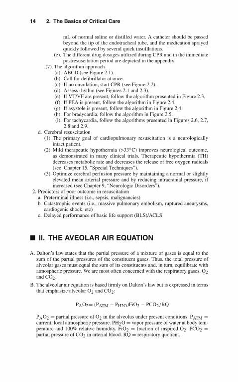
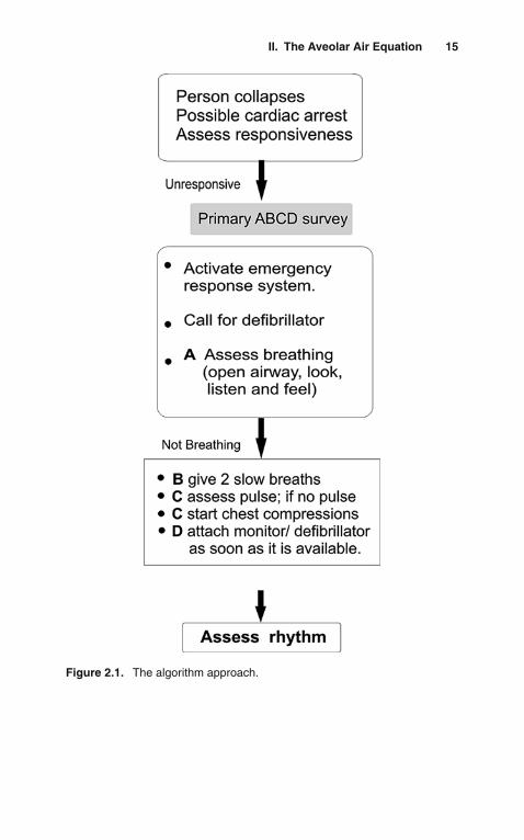
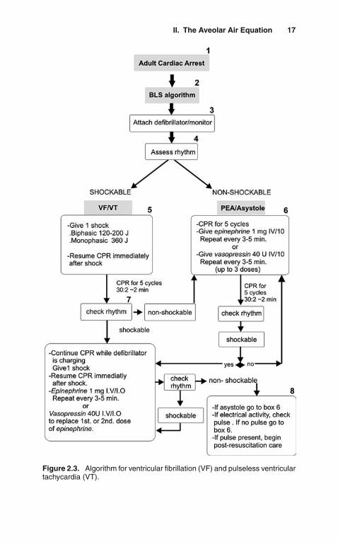
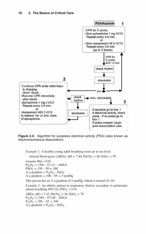
(7). The algorithm approach(a). ABCD (see Figure 2.1).(b). Call for defibrillator at once.(c). If no circulation, start CPR (see Figure 2.2).(d). Assess rhythm (see Figures 2.1 and 2.3).(e). If VT/VF are present, follow the algorithm presented in Figure 2.3.(f). If PEA is present, follow the algorithm in Figure 2.4.(g). If asystole is present, follow the algorithm in Figure 2.4.(h). For bradycardia, follow the algorithm in Figure 2.5.(i). For tachycardia, follow the algorithms presented in Figures 2.6, 2.7,
2.8 and 2.9.d. Cerebral resuscitation
(1). The primary goal of cardiopulmonary resuscitation is a neurologicallyintact patient.
(2). Mild therapeutic hypothermia (>33◦C) improves neurological outcome,as demonstrated in many clinical trials. Therapeutic hypothermia (TH)decreases metabolic rate and decreases the release of free oxygen radicals(see Chapter 15, “Special Techniques”).
(3). Optimize cerebral perfusion pressure by maintaining a normal or slightlyelevated mean arterial pressure and by reducing intracranial pressure, ifincreased (see Chapter 9, “Neurologic Disorders”).
2. Predictors of poor outcome in resuscitationa. Preterminal illness (i.e., sepsis, malignancies)b. Catastrophic events (i.e., massive pulmonary embolism, ruptured aneurysms,
cardiogenic shock, etc)c. Delayed performance of basic life support (BLS)/ACLS
� II. THE AVEOLAR AIR EQUATION
A. Dalton’s law states that the partial pressure of a mixture of gases is equal to thesum of the partial pressures of the constituent gases. Thus, the total pressure ofalveolar gases must equal the sum of its constituents and, in turn, equilibrate withatmospheric pressure. We are most often concerned with the respiratory gases, O2and CO2.
B. The alveolar air equation is based firmly on Dalton’s law but is expressed in termsthat emphasize alveolar O2 and CO2:
PAO2= (PATM − PH2O)FiO2 − PCO2/RQ
PAO2 = partial pressure of O2 in the alveolus under present conditions. PATM =current, local atmospheric pressure. PH2O = vapor pressure of water at body tem-perature and 100% relative humidity. FiO2 = fraction of inspired O2. PCO2 =partial pressure of CO2 in arterial blood. RQ = respiratory quotient.
II. The Aveolar Air Equation 15
Figure 2.1. The algorithm approach.

16 2. The Basics of Critical Care
Figure 2.2. The algorithm approach.
C. Many clinical and environmental influences are immediately obvious whenconsidering the terms of the equation:1. PATM: Altitude per se can clearly result in hypoxemia. A given patient’s PO2
must be considered in the context of location. A “normal” arterial PO2 is notthe same in Denver (average = 73 mmHg) as it is at sea level (average =95 mmHg).
2. FiO2: While atmospheric air is uniformly about 21% O2, one must ask: 21% ofwhat? The FiO2 on a mountaintop at 11,000 feet is also 21%, but there is notenough total O2 in the rarefied air to sustain an arterial PO2 above 60 mmHg.
3. PCO2: Although CO2 coming into the alveolus does not displace O2 (thiswould not obey Dalton’s law), the blood PCO2 does equilibrate with alveo-lar gases. Simultaneously, O2 is taken up from the alveolus. When patientshypoventilate, not only does CO2 accumulate but also alveolar O2 becomesdepleted. Thus, elevated PCO2 is associated with low PAO2 and sometimeshypoxemia. Similarly, hyperventilating patients (excess CO2 elimination, lowPCO2, frequent replenishment of alveolar O2) can have higher than normalPAO2 and arterial PO2.
4. RQ is the ratio of CO2 production to O2 consumption. The ratio of alveolargas exchange—CO2 coming into the alveolus and O2 leaving the alveolus—not unexpectedly, also reflects the RQ. Given a particular ratio of alveolar gasexchange, the ultimate value for PAO2 will also be affected by the rate of CO2elimination from the alveolus, i.e., alveolar ventilation.
D. The A-a Gradient1. While the alveolar air equation predicts the partial pressure of O2 in the alveolus
(PAO2) under current conditions, it is not necessarily true that arterial bloodwill have an identical partial pressure of O2 (PaO2). We can, however, measurethe PaO2 directly and compare it with the calculated value for PAO2. When wesubtract arterial from alveolar PO2, we obtain the A-a gradient.
II. The Aveolar Air Equation 17
Figure 2.3. Algorithm for ventricular fibrillation (VF) and pulseless ventriculartachycardia (VT).
18 2. The Basics of Critical Care
Figure 2.4. Algorithm for pulseless electrical activity (PEA) (also known aselectromechanical dissociation).
Example 1: A healthy young adult breathing room air at sea level:
Arterial blood gases (ABGs): pH = 7.40, PaCO2 = 40, PaO2 = 95
(assume RQ = 0.8)PAO2 = (760 – 47).21 – 40/0.8PAO2 = 150 – 50 = 100A-a gradient = PAO2 – PaO2A-a gradient = 100 – 95 = 5 mmHg
This person has an A-a gradient of 5 mmHg, which is normal (0–10)
Example 2: An elderly patient in respiratory distress secondary to pulmonaryedema breathing 40% O2 (FiO2 = 0.4):
ABGs: pH = 7.43, PaCO2 = 36, PaO2 = 70PAO2 = (760 – 47).40 – 36/0.8PAO2 = 285 – 45 = 240A-a gradient = PAO2 – PaO2
II. The Aveolar Air Equation 19
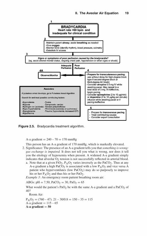
Figure 2.5. Bradycardia treatment algorithm.
A-a gradient = 240 – 70 = 170 mmHg
This person has an A-a gradient of 170 mmHg, which is markedly elevated.2. Significance: The presence of an A-a gradient tells you that something is wrong:
gas exchange is impaired. It does not tell you what is wrong, nor does it tellyou the etiology of hypoxemia when present. A widened A-a gradient simplyindicates that alveolar O2 tension is not successfully reflected in arterial blood.a. Note that at a given FiO2, PAO2 varies inversely as the PaCO2. Thus at any
A-a gradient a high PaCO2 is associated with a low PAO2 and vice versa Apatient who hyperventilates (low PaCO2) may do so purposely to improvehis or her PAO2 and thus his or her PaO2.
Example 3: An emergency room patient breathing room air:
ABGs: pH = 7.50, PaCO2 = 30, PaO2 = 65
What would the patient’s PaO2 be with the same A-a gradient and a PaCO2 of40?
Room Air:
PAO2 = (760 – 47). 21 – 30/0.8 = 150 – 35 = 115A-a gradient = 115 – 65A-a gradient = 50
20 2. The Basics of Critical Care
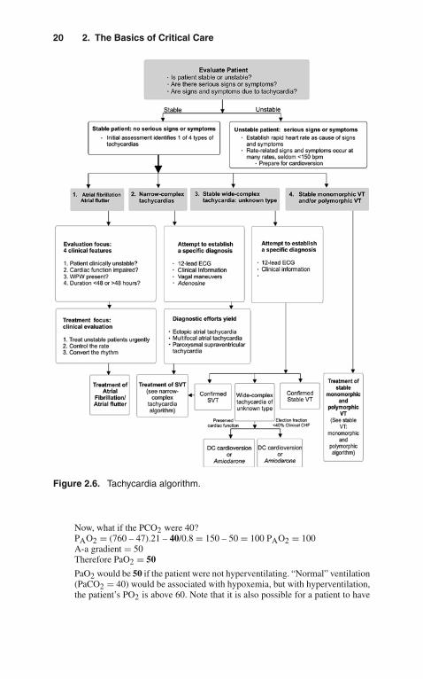
Figure 2.6. Tachycardia algorithm.
Now, what if the PCO2 were 40?PAO2 = (760 – 47).21 – 40/0.8 = 150 – 50 = 100 PAO2 = 100A-a gradient = 50Therefore PaO2 = 50
PaO2 would be 50 if the patient were not hyperventilating. “Normal” ventilation(PaCO2 = 40) would be associated with hypoxemia, but with hyperventilation,the patient’s PO2 is above 60. Note that it is also possible for a patient to have

II. The Aveolar Air Equation 21
Figure 2.7. Tachycardia algorithm.
hypoxemia without a widened A-a gradient. There are two important examples:high altitude and alveolar hypoventilation.
Example 4: A normal adult breathing room air at an altitude of 11,000 feet:
A-a gradient = 0PaO2 = (510 – 47).21 – 40/0.8 = 47A-a gradient = 0PaO2 = 47This patient has hypoxemia without an A-a gradient.

22 2. The Basics of Critical Care
Figure 2.8. Tachycardia algorithm.

II. The Aveolar Air Equation 23
Figure 2.9. Electrical-synchronized cardioversion algorithm.

24 2. The Basics of Critical Care
Example 5: A patient with pure alveolar hypoventilation secondary to narcoticoverdose breathing room air:PCO2 = 80; A-a gradient = 0PAO2 = (760 – 47).21 – 80/0.8PAO2 = 50A-a gradient = 0PaO2 = 50
This patient has hypoxemia without an A-a gradient.
3. Summary
a. The alveolar air equation shows the relationships among atmospheric pres-sure, FiO2, PaCO2, and alveolar O2 tension (PAO2).
b. When alveolar O2 tension (PAO2) is not reflected faithfully in arterial blood(PaO2)—i.e., a widened A-a gradient—the calculation indicates that gasexchange is impaired, but it does not tell you how or why.
c. Calculation of the A-a gradient is a useful bedside tool for evaluation ofpatients with respiratory distress or abnormal ABGs and to follow theirprogress.
d. It is possible to have hypoxemia without a widened A-a gradient. Highaltitude and hypoventilation (elevated PaCO2) are examples.
� III. OXYGEN TRANSPORT
A. Oxygen Delivery: Calculations1. Calculation of oxygen delivery (DO2) and oxygen consumption (VO2) are
useful bedside techniques in the ICU.2. DO2 = CO × CaO2
Oxygen delivery = Cardiac output × Arterial O2 content3. CaO2 = Hb × SaO2 × K
Arterial O2 content = Hemoglobin × Arterial O2 saturation × a constant∗∗We will use 1.34 mL O2/g Hb.4. Resolving the units:
DO2 [mL O2/min] = CO [mL/min] × Hb [g/100 mL] × 1.34 [mL O2/g] ×SaO2 [scalar]
5. Normal values (70-kg man at rest)DO2 = 5,000 mL/min [CO] × 15 g/100 mL [Hb] × 1.34 mLO2/g [constant] × 1.00 [SaO2]DO2 = 1,005 mL O2/min
6. This value does not take into account dissolved O2 in the plasma: 0.003 mLO2/100 cc/mmHg PaO2, which adds another 15 mL O2 of arterial O2 content.
7. Values to remember:Normal CaO2 (15 g Hb, 100% SaO2) = 20.4 mL O2/100 cc (20.4 vol %)Normal DO2 (70-kg man, at rest, CO = 5,000 mL/min) = 1,020 mL O2/min
B. Oxygen Transport: ConceptsOnly three clinical variables can affectDO2: cardiac output, hemoglobin, and oxygen saturation.

III. Oxygen Transport 25
Note that what looks very simple is not:1. Cardiac output entails all of normal cardiodynamics (preload, afterload,
contractility), hemodynamics, state of hydration, blood gas and electrolyteinfluences, the influence of mechanical ventilation and other technology,intrinsic cardiac disease, dysrhythmias, etc.
2. Hemoglobin is largely a quantitative problem (i.e., oxygen-carryingcapacity), but it also includes the effects of abnormal hemoglobins,massive transfusions, pH and temperature, other causes of shift in theoxyhemoglobin dissociation curve, and hemoglobin substitutes.
3. Arterial oxygen saturation embodies the pathophysiology of acute andchronic lung disease, management of mechanical ventilation, the car-diopulmonary interaction, venous admixture, intrapulmonary or intracar-diac shunting, etc.
4. If this is not complicated enough, recall that what you may be doing tosupport the lungs may have a detrimental effect on cardiac output (seebelow). Similarly, failure to correct severe blood gas abnormalities mayalso adversely affect cardiac function. This makes the bedside manage-ment of oxygen delivery in critically ill patients straightforward, althoughat times very difficult:a. Support oxygenation such that PO2 >60, SaO2 >0.9 on nontoxic FiO2
(≤0.5).b. Ensure hemoglobin concentration of at least 10 g/100 cc.c. Optimize cardiac output (CO) under current conditions (i.e., current
ventilator settings).
C. Physiologic Maintenance of Oxygen Delivery: Since DO2 is dependent on onlythree variables, how does a normal person respond to abnormalities of one of thevalues?1. Fall in SaO2:
If SaO2 falls to 0.5, a person can achieve normal O2 delivery by doubling CO:
DO2= CO × Hb × SaO2
DO2= 2CO × Hb × 1/2SaO2
a. Therefore, in the short term, increased CO can compensate for even severehypoxemia.
b. Note that when SaO2 = 0.5, PaO2 = 27! This is the definition of P50 fornormal adult hemoglobin A, namely, the PaO2 at which hemoglobin is 50%saturated (27 mmHg). Thus, even severe hypoxemia can be tolerated well aslong as hemoglobin is normal and CO can be enhanced.
c. In patients with chronically low SaO2 (high altitude, chronic lung dis-ease, cyanotic heart disease), they will also increase their hemoglobinconcentration.
2. Fall in Hemoglobin:If hemoglobin falls dramatically, DO2 is again maintained by increasing CO.a. Note that SaO2 can never increase beyond 100% and therefore cannot com-
pensate for low hemoglobin. The ability to increase and maintain CO is animportant mechanism by which anemia can be tolerated.
DO2= CO × Hb × SaO2

26 2. The Basics of Critical Care
DO2 = 2CO × 1/2Hb × SaO2
3. Fall in Cardiac Output:What if CO falls dramatically, then how is DO2 maintained? The answer is,DO2 in totality is not maintained, but tissue DO2 is maintained by enhancedextraction.a. If fewer liters of oxygenated blood are delivered, then the tissues must extract
more from every liter that is delivered.b. Normally, arterial blood is nearly 100% saturated with O2. Venous blood
returning to the heart is the same in terms of hemoglobin and quantitativelythe same as CO. Thus, it is the venous oxygen saturation (SvO2) that reflectsO2 extraction. Normal SvO2 = 0.75. Therefore, normal extraction is about25%.
c. Looking at extraction (i.e., A—V O2 difference) is therefore a good probe(under some circumstances, such as heart failure) of the adequacy of CO:High extraction implies inadequate CO.
4. Fall in Oxygen DeliveryHere is a general rule of thumb: A normal person can withstand a severeabnormality of any one of the O2 delivery variables (CO, Hb, SaO2) with-out developing lactic acidosis (lactic acidosis would indicate cellular O2deprivation with resultant anaerobic metabolism).a. Note that during cardiac arrest lactate is generated not because of hypoxemia
alone but, rather, because the cardiac output is also severely compromisedand unable to compensate for low PaO2 to maintain DO2.
D. Oxygen Consumption (VO2)1. DO2 (oxygen delivery) is what leaves the heart both quantitatively and quali-
tatively. What returns to the heart should be the same quantitatively, with thesame hemoglobin concentration, different only in terms of oxygen saturation.
2. If we know the DO2 (what left the heart) and we calculate what has returned tothe heart, we can then subtract to ascertain the amount consumed:
Oxygen Consumption
(VO2) = CO × (CaO2) − CO × (CvO2)∗what left what returned to
the heart the heart
Thus,VO2=CO(CaO2−CvO2)∗∗This is the Fick equation
∗CvO2 is the mixed venous O2 content.∗∗CaO2 – CvO2 is the arteriovenous O2 content difference.3. CvO2 (mixed venous O2 content) is calculated in exactly the same way as the
CaO2 (arterial content), namely:Hb × 1.34 × S vO2(SvO2 is the mixed venous O2 saturation).
4. If VO2 is known, the Fick equation can be used to calculate the cardiac output:
CO = VO2(CaO2 − CvO2)
III. Oxygen Transport 27
5. At the bedside, what do you need to calculate VO2?a. The patient needs a pulmonary artery (Swan-Ganz) catheter
(1). for CO determination(2). to obtain a true–mixed venous blood sample from the pulmonary artery
(SvO2)b. Arterial blood gas determination (or SaO2 determination)c. Hemoglobin determination
6. Example of normal values (70-kg man at rest)CaO2 = 15 [Hb] × 1.34 [constant] × 1.00 [SaO2] = 20.1CvO2 = 15 [Hb] × 1.34 [constant] × 0.75 [SvO2] = 15.1CO = 5,000 mL [5 L/min]VO2 = 5,000 mL/min (20.1 mL O2/100 mL – 15.1 mL O2/100 ccVO2 = 50 (20 – 15)
VO2 [normal, at rest] = 250 mL O2/min7. Bedside Application in the ICU: Human life depends on oxygen. This is a good
reason to assess the adequacy of DO2 in critically ill patients. Where there islife, there is O2 consumption.a. We are concerned about factors that increase resting O2 consumption such as
fever. Febrile patients increase their resting O2 consumption by 10–13%◦C(approximately 7%◦F).
b. We are also concerned when calculated O2 consumption is less than pre-dicted (for body surface area, temperature), such as may occur in sepsis.Many times in spite of high DO2, patients with sepsis have low calculatedVO2, lactic acidosis, oliguria, and other signs of poor parenchymal organfunction.
c. Instead of arbitrary endpoints, it is best to look for physiological end-points. When measured O2 consumption, SvO2, (A—V) O2 contentdifference, and serum lactate are all normal, then it is likely thatDO2 is adequate. Evidence that you have satisfied the body’s (tissues’)needs is better evidence of the adequacy of DO2 than any arbitrarynumber.
d. Make sure that you see DO2 as an integrated variable. If you change ventila-tor settings (see below)—for example, raise the PEEP to enhance SaO2—butin the process cause a fall in CO, you may not have achieved any over-all benefit in terms of DO2. Cardiac output, hemoglobin, and SaO2 requireindividual attention and management.
e. Look for opportunities to get the best results for each intervention. For exam-ple, a transfusion of packed red blood cells may increase hemoglobin andraise CO. This may be substantially better management than trying to raiseCO with crystalloid IV fluids to compensate for a borderline hemoglobinand/or SaO2.
f. Check CO and O2 transport variables often and measure the response to yourinterventions. Note that we often record heart rate and blood pressure everyhour although CO can vary over a wide range irrespective of these moretraditional signs. Cardiac output is a vital sign!
g. Current technology now provides continuous data for SaO2, SvO2,and even CO. These can obviate the necessity of repeated bloodgas determinations and facilitate frequent assessment of O2 transportvariables.

28 2. The Basics of Critical Care
� IV. MECHANICAL VENTILATION
Humans breathe for two reasons: to take in oxygen (oxygenation) and to eliminatecarbon dioxide (ventilation). A patient’s inability to perform either or both of thesefunctions defines respiratory failure.
A. Ventilation. Normal people produce CO2 continuously, thus, there is a constantneed for CO2 elimination. We all eliminate CO2 by a process that entails breathingin fresh air (essentially devoid of CO2), allowing it to equilibrate with the CO2dissolved in capillary blood and then exhaling it laden with CO2. We performthis process 10–14 times each minute with significant volumes of air, such thatunder normal conditions arterial CO2 (PaCO2) is kept nearly constant at 40 mmHg(torr).
More precisely, we move a tidal volume (Vt) in and out at a certain frequency(f) or respiratory rate (RR). The product of rate and tidal volume is the minute ven-tilation (Vmin). Thus, it is the minute ventilation that is fundamentally responsiblefor CO2 elimination.
Vmin= Vt×RR
1. The minute ventilation can be further divided into the gases that reach the alve-oli and are therefore available for exchange (the alveolar ventilation, VA) andthose gases that fill the airways or that reach unperfused (see below) alveoli andtherefore cannot exchange gases (the anatomical and physiological dead space,respectively, VD).
2. CO2 elimination is therefore directly proportional to the minute alveolarventilation at any level of CO2 production or blood PCO2.
CO2 elimination = (VA)min×PCO2
3. Since any physiologic parameter (i.e., serum creatinine, platelet count, PCO2)is ultimately the result of the balance between production and elimination, it fol-lows that PCO2 (under any conditions affecting production) can be controlledby adjusting minute ventilation.
B. Oxygenation. How people accomplish oxygenation is equally simple, but con-siderably different from how we accomplish ventilation. We purposely inhale anoxygen-enriched atmosphere all the way down to our alveoli to allow the oxygento be taken up by the capillary blood—both dissolved in proportion to its partialpressure (obeying Henry’s law) and in combination with hemoglobin. More pre-cisely, the air we inspire has a certain fraction that is oxygen—that is, a certainfraction of inspired O2 (FiO2).
Although we breathe only intermittently, we need to accomplish gas exchangecontinuously. If there were oxygen in our alveoli only when we inhaled, then bloodwould pass through the lungs unoxygenated in between breaths. Thus, we need tomaintain volume in our lungs even at end-exhalation. This is accomplished bymaintaining a pressure gradient across the lungs between breaths. The pressurein the pleural space (outside the lungs) is negative (approximately [–] 5 cmH2O)with respect to the atmospheric pressure present in our airways. If we subtract

IV. Mechanical Ventilation 29
vectorially, there is a 0 – (–5) = +5 cmH2O pressure gradient across the lungseven at end-exhalation—in effect, a PEEP. Thus, oxygenation is accomplished innormal people by purposely inspiring a certain FiO2 and maintaining a certainPEEP.
C. PEEP and Compliance1. Compliance
The volume in the lungs is related to the transpulmonary pressure. Indeed,volume and pressure are intimately related in many systems (such as ven-tilator tubing, cardiac filling, resting lung volume) through the variable ofcompliance (C)
C =�V
�P
Compliance is defined as the change in volume for a given change in pres-sure. Thus, in order for us to achieve a given volume change—such as a tidalvolume—in our lungs, we must make a pressure change. The precise pressurenecessary will be determined by the lung (and chest wall) compliance.
Mathematically, it is clear that, as compliance falls (as may occur in pul-monary edema, adult respiratory distress syndrome (ARDS), lung fibrosis, andmany other conditions), one must achieve ever-increasing �P just to achieve thesame �V. It is often the case that a patient’s inability to do the work required toincrease �P to maintain an adequate tidal exchange (�V) is the ultimate causeof respiratory failure.
The fundamental role that lung compliance plays in determining the relation-ship between clinically significant lung volumes (e.g., tidal volume) and thepressures required to achieve them has many important clinical implications:a. If there is no gradient of pressure (�P = 0), then there is no volume change.
When a patient develops a pneumothorax, the pressure in the pleural spaceequals the pressure in the airways. As a result, there is no transpulmonarypressure (�P) and thus no lung volume—i.e., the lung collapses—becauseof the lungs’ intrinsic compliance (and elastance, which is defined as1/compliance).Pneumothorax results in no lung volume (zero �V), because there is notranspulmonary pressure (zero �P).
b. To create a volume change, we must effect a pressure change. Thus, tidalvolume is determined by the �P generated as the chest wall expands andthe diaphragm contracts. Similarly, to increase our tidal volume, we mustgenerate a larger �P, or if compliance falls, we may need a larger �P just toachieve the same tidal volume.
c. If a person has low lung compliance (i.e., restrictive disease), then normalresting negative intrapleural pressure (�P) will result in lower resting lungvolume (�V).
d. If a person has high lung compliance (i.e., emphysema with destruc-tion of lung parenchyma), then normal resting negative intrapleuralpressure will result in high resting lung volume (e.g., “barrel chest”of emphysema).
e. Since the lungs are merely populations of alveoli, these relationships amongpressure, volume, and compliance apply to individual alveoli and specificlung regions as well as to whole lungs.
30 2. The Basics of Critical Care
f. Compliance contributes to the logical connection between the requirementsof gas exchange and the respiratory work:(1). CO2 production demands minute ventilation.(2). Minute ventilation requires a certain tidal volume.(3). This change in volume requires a change in pressure.(4). How much pressure for a given volume is determined by compliance.(5). The amount of pressure that must be generated is a major determinant of
the work of breathing.2. PEEP
From our description of oxygenation and ventilation, it should be clear thatwe are clinically concerned about maintaining the adequacy of two importantlung volumes: The tidal volume of each breath and the resting lung volumein between breaths. The pressure generated during active inspiration eitherby the ventilator or the patient will determine the tidal volume (mediated, ofcourse, through compliance). But what determines the resting lung volume?The answer is the resting transpulmonary pressure. In normal people (with nor-mal lung compliance), the vectorial difference between airway and intrapleuralpressures (�P) determines the resting lung volume, known more precisely asthe functional residual capacity (FRC):
Pairway − Ppleural = �Ptranspulmonary
0 − ( − 5) = + 5
�P = + 5cm H2O
Resting lung volume (FRC) is therefore determined by the AP and compliance:
C = �V
�P
C = FRC
Ptranspulmonary
FRC = C(Ptranspulmonary)
Since �P is positive and present at end-expiration, what we are talking aboutis positive end-expiratory pressure (PEEP). It should also be clear that PEEPdirectly determines FRC.
We have all had the experience of inflating a balloon. It’s difficult at first, thensuddenly gets easier once some volume is inside. As we reach the full inflation,it may again become difficult as we reach the limits of the balloon’s compliance.If we let go, the balloon recoils (elastance) and collapses.
Our alveoli, in many ways, are similar: If they start fully collapsed, theyare difficult to inflate at first. Once there is some volume, it becomes easier; thispoint of change in compliance is referred to as critical opening pressure (COP).
Unlike balloons, normal alveoli do not immediately lose all of their volumewhen pressure is released but may maintain some volume (thanks in large mea-sure to surfactant) until distending pressure is critically low and then collapse.The point at which this occurs is critical closing pressure (CCP).

IV. Mechanical Ventilation 31
If one could maintain end-expiratory pressure (i.e., PEEP) above CCP, thenalveoli would not collapse; their volume would be enhanced, and, in the aggre-gate, lung volume (FRC) would be enhanced. If low lung compliance resultsin high CCP, then PEEP must be increased above the CCP to prevent alveolarcollapse. This is precisely the rationale for PEEP in the management of acutelow-compliance lung disease (e.g., ARDS).a. Summary of the Effects of PEEP
(1). PEEP increases FRC.(2). PEEP increases compliance.(3). PEEP reduces shunt fraction (see below) by maintaining volume for gas
exchange in perfused lung units in between breaths.(4). PEEP increases dead space by overdistending normally compliant alve-
oli.(5). PEEP increases intrathoracic pressure, which can impede venous return
into the chest or specifically restrict cardiac filling, both of which mayresult in reduced cardiac output.
(6). PEEP may contribute to barotrauma because it represents the baseline(end-expiration) for all pressure changes, because it may cause overdis-tention of compliant lung regions, and because of the nature of the acutelung diseases in which PEEP is most frequently useful.
D. Modes of Mechanical Ventilation. Under routine conditions, when a patient devel-ops respiratory failure and is intubated, initial mechanical ventilatory support isprovided by some form of conventional volume-cycled ventilation (VCV). Volumecycled means that the endpoint for the ventilator is the delivery of a selectedtidal volume, leaving the machine itself to determine what pressure is neces-sary to deliver that volume, to that patient, at that time. Pressure-controlledventilation is a modality in which tidal volume that is delivered is variable anddependent of the peak pressure, inspiratory time, and patient’s compliance. Itreduces the risk for barotrauma by using lower peak pressures and longer inspira-tory times. This type of ventilation is preferred in patients with poor pulmonarycompliance.
In addition to the standard array of choices for how to deliver VCV, there arealso modes that do not use quasi-physiologic parameters, such as high-frequencyventilation. These modes of mechanical ventilatory support are beyond the scopeof this manual but have been reviewed in depth elsewhere.For conventional VCV, essentially four modes are commonly used, as depicted inTable 2.2.1. Controlled Mechanical Ventilation (CMV)
From our discussion above, it follows that the basic functions of ventilation andoxygenation can be accomplished by providing four basic settings: respiratoryrate, tidal volume, FiO2, and PEEP. Given these parameters, the ventilator willprovide the patient with a constant minute ventilation and oxygen. These are thesettings for CMV. The only gases these patients receive are from the machinebreaths. These patients cannot initiate a breath, change their rate, or access anyother source of fresh gases. This mode is therefore useful in a limited numberof settings, such as the following:a. In the operating room, when patients are fully anesthetizedb. When patients are apneic and likely to remain soc. When patients are sedated/anesthetized and paralyzed, in the intensive care
unit (ICU)

32 2. The Basics of Critical Care
Table 2.2. Commonly Used Modes of Volume-Cycled Ventilation
1.Controlled mechanical ventilation (CMV)
2.Assisted/controlled mechanical ventilation (A/C)
3.Synchronized intermittent mandatory ventilation (SIMV)
4.Continuous positive airway pressure (CPAP)
5.Pressure Support Ventilation (PSV) is not a separate mode but, rather, anadjunct that can be used with several other modes>>
It is important to note that in CMV, patients absolutely cannot breatheon their own. If these patients should awaken or attempt to breathe, theycan become agitated and dyspneic. It is extremely frightening to be unableto breathe and to experience essentially a chronically occluded airway.Worse yet, if these patients become detached from the ventilator, anes-thetized and/or paralyzed patients will be functionally apneic and may soonexperience full cardiopulmonary arrest.
2. Assisted/Controlled Mechanical Ventilation (A/C)The settings for A/C are the same four basic ones used for CMV. There is nodifference between CMV and A/C in anesthetized or apneic patients. The sin-gular difference is that in A/C mode patients can initiate breaths. Unlike in youand me, however, the amount of effort these patients make does not determinethe tidal volume. When these patients initiate a breath with sufficient force to“trigger” the ventilator, the tidal volume they receive is the one already pre-set to be delivered as a “controlled” breath. Moreover, the machine will usewhatever pressure is required to deliver the volume, and the patients’ lungs andchest must then accommodate that tidal volume. The set rate in A/C is essen-tially a default control rate; that is, it is the number of volume-cycled breathsthe machine will deliver on its own, even if the patient is apneic. Thus the “con-trolled” aspect of A/C is the guaranteed minute volume delivered regardless ofthe patient’s spontaneous efforts.
While this mode has some value in its ability to relieve dyspnea in the imme-diate postintubation period, it is not a good choice for prolonged mechanicalsupport. Evidence now indicates that in accommodating “assisted” breaths,the respiratory muscles may actually be fatigued rather than “rested,” as wasinitially intended by the design. Better choices are available for routine use.
3. Synchronized Intermittent Mandatory Ventilation (SIMV)Of currently available modes, SIMV is probably the most versatile and thereforethe most widely used. What does the term mean?a. Mandatory ventilation represents the same guaranteed minute ventilation
(respiratory rate × tidal volume) delivered by the machine as seen in CMVor A/C. Thus, if the patient becomes apneic while on SIMV, the mandatoryventilation will be provided.
b. Intermittent ventilation is used to emphasize that the machine will deliverthe desired number of breaths at intervals, leaving the patient free to breathespontaneously in between. If, for example, the set respiratory rate is 10breaths per minute, then the machine will deliver the selected tidal volumeabout every 6 s. The patient may need or desire to breathe more often than 10

IV. Mechanical Ventilation 33
times per minute and can do so in the intervals (approximately 6 s) betweenmachine breaths.
c. Synchronized IMV is a relatively new refinement of the original IMV design.In the example above, the machine will cycle every 6 s, but it would be unde-sirable for the ventilator to attempt delivery of a new breath if the patientwere in the process of exhaling a spontaneous breath. Without synchroniza-tion, this kind of “collision” could occur in the airways, causing very highairway pressure, a risk of barotrauma, ineffective ventilation, and enhanced(rather than relieved) dyspnea. The synchronizer looks at a “window” periodwhen the next machine breath is due. If the patient is exhaling, the ventila-tor can wait to begin inspiration. If the patient initiates a breath at the timethe ventilator is due to cycle, the machine breath and the spontaneous breathwill merge into one synchronized breath not unlike the “triggered” breathsin A/C mode. This represents a major improvement for IMV, especiallyin patients with prolonged expiratory times such as those with bronchialasthma.
d. SIMV was introduced into wide usage as a “weaning” modality. Weaningfrom mechanical ventilation is discussed below, but in this connection, theprinciple is simple. Initially, the mandatory ventilation provides the entireminute ventilation necessary to maintain the patient’s PCO2 within normallimits. As patients begin to breathe spontaneously, the mandatory ventilationis gradually reduced until they are essentially providing the entire minuteventilation (and therefore CO2 elimination) through their own spontaneousefforts. At this point these patients no longer require mechanical ventilatorysupport.
e. It should be noted here that on SIMV, as in all of the modes thus fardiscussed, oxygenation is still supported by the settings for PEEP and FiO2.
4. Continuous Positive Airway Pressure (CPAP)CPAP is a system for spontaneously breathing patients, in which the machineis providing only PEEP, FiO2, and humidification (maintained in all modes),but does not deliver any mechanical breaths. In this sense, it is the modethat would result if the patient were on either SIMV or A/C and themachine respiratory rate were set at zero. It may be provided via tight-fittingface mask or endotracheal/tracheostomy tube. Some of the applications forCPAP area. Patients with no ventilatory difficulty who require positive air-
way pressure (PEEP) to support oxygenation; it reduces alveolarcollapse.
b. Patients in the final stages of weaning who are being observed while theybreathe without ventilatory support.
5. Pressure Support Ventilation (PSV)Pressure support is not a mode of mechanical ventilation, rather it is an adjunctto other modes. All people can inhale a certain tidal volume based on theirability to create a significant negative intrapleural pressure. As noted in thedescription of PEEP above, the vectorial result of the negative intrapleural pres-sure is a positive-pressure gradient across the lungs. In simplest terms, PSV isthe delivery of gas flow (during a spontaneous breath) with a defined positivepressure that one selects on the ventilator. This positive pressure is vectoriallysummative with the negative pressure generated by the patient’s effort. The netresult is that the positive pressure gradient across the lung is enhanced, and asa result, so is the spontaneous tidal volume.

34 2. The Basics of Critical Care
NoPSV:0 − ( − 10) = + 10H2O transpulmonary pressureWith PSVof + 10+10(PSV) − ( − 10) = + 20cmH2O transpulmonary pressure
Respiratory muscles do not benefit from “rest,” defined as not contracting atall; in fact they may rapidly atrophy if not allowed to perform as they usu-ally do. However, with acute lung disease, the work of breathing may resultin fatigue. PSV should allow the respiratory muscles to perform a manageableamount of work, without the risk of atrophy on the one hand or fatigue on theother.
6. Airway Pressure Release Ventilation (APRV)APRV is a mode that delivers a high continuous positive airway pressurefor increased duration of time, and then pressure falls to lower levels for ashorter duration. This transition of pressures (from high to low) helps to elim-inate carbon dioxide. The high continuous positive pressure improves alveolarrecruitment, thus indicated in patients with acute lung injury or ARDS. Theunique feature of this mode of ventilation is that it is permissible on patientswith spontaneous breathing.
E. Initiation of Mechanical Ventilation1. Criteria for Initiation of Mechanical Ventilation
a. Physical assessment: The patient is apneic, severely tachypneic, or in res-piratory distress unresponsive to therapeutic interventions and supplementaloxygen.
b. Gas exchange: Hypoxemia (PO2 <50) despite high-flow oxygen, hypercarbia(acute, PCO2 >50 with acidic pH).
c. Clinical judgment: The constellation of laboratory and physical findings maybe the most compelling. A PCO2 of 60 and a respiratory rate of 35 may bethe usual baseline for some patients, but may represent a direct emergencyin others.
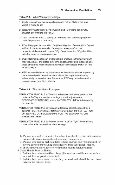
2. Initial Ventilator Settings (See Table 2.3)
F. General Principles of Ventilator Management (See Also Table 2.4)1. Therapeutic Endpoints
a. PaCO2: Ventilatory parameters are adjusted to achieve a PaCO2 of 35–45with the pH also in the normal physiologic range of 7.35–7.45.
b. PaO2: A PaO2 >60 that corresponds to an SaO2 >0.9 with the patient receiv-ing nontoxic FiO2 (≤0.5). If this is not achievable on physiologic PEEP, thePEEP can be raised in + 2-cm H2O increments to achieve this endpoint.(1). Note that this endpoint can be expressed as a PaO2/FiO2 ratio 60/0.4 =
150 (FiO2 = 0.4 is not associated with O2 toxicity).(2). PEEP will be most beneficial in acute low-compliance lung disease.
Patients with markedly asymmetrical lung disease, bullous emphysema,or asthma may actually have worsening gas exchange with significantPEEP.
2. When patients are in respiratory distress, it is acceptable to change any or all ofthe ventilator settings at one time; during weaning the same is not true.
3. Patient Comforta. Having an endotracheal tube in place is not comfortable, and because one
cannot speak, frustrating as well.
IV. Mechanical Ventilation 35
Table 2.3. Initial Ventilator Settings
1. Mode: Unless there is a compelling reason not to, SIMV is the mostversatile mode to use
2. Respiratory Rate: Generally between 8 and 12 breaths per minute,adjusted according to the PaCO2
3. Tidal Volume: In the ICU setting, 6–10 mL/kg lean body weight (do notcount adipose tissue or edema)
4. FiO2: Many people start with 1.00 (100% O2), but often 0.8 (80% O2) willsuffice. A phenomenon called “absorption atelectasis” occursproportionately more with higher FiO2. Regardless, the FiO2 should beadjusted down as soon as possible
5. PEEP: Normal people can create positive pressure in their airways withtheir lips, palate, and glottis. Since the endotracheal tube bypasses all ofthese structures, most initial setups include “physiologic” PEEP of +3 to+5 cm of H2O
6. PSV: 8–10 cmH2O can usually overcome the additional work imposed bythe endotracheal tube and ventilator circuit, but larger amounts maysubstantially reduce dyspnea. Remember, PSV only has relevance forspontaneously breathing patients
Table 2.4. The Ventilator Principles
VENTILATOR PRINCIPLE 1: To reach a desirable clinical endpoint for thepatient’s PaCO2, the ventilator settings you will adjust are theRESPIRATORY RATE (RR) and/or the TIDAL VOLUME (Vt) delivered bythe machine
VENTILATOR PRINCIPLE 2: To reach a desirable clinical endpoint for apatient’s PaO2, the ventilator settings you will adjust are the FRACTIONOF INSPIRED O2 (FiO2) and/or the POSITIVE END-EXPIRATORYPRESSURE (PEEP)
VENTILATOR PRINCIPLE 3: Patients do not “buck” or “fight” the ventilator;patients buck ill-conceived ventilator settings
b. Patients who will be intubated for a short time should receive mild sedationwith agents having no significant respiratory suppression.
c. Patients who require high ventilator settings and will likely be intubated forseveral days before weaning should receive more substantial sedation.
d. In our opinion, only a few selected patients require paralytic agents.4. Some Simple Rules of Thumb
a. Endotracheal tubes should be as large (diameter) as possible and cut as shortas possible once position is verified.
b. Endotracheal tubes must be carefully secured and should be out frombetween the patient’s teeth.

36 2. The Basics of Critical Care
c. Suctioning is important but should be minimal or strictly pm when the patientis on >+10 cmH2O PEEP, to minimize volume loss from within the lungs.
d. When setting up the ventilator, the peak inspiratory flow rate is best keptrelatively low (≤50 L/min [LPM]) but must be at least three times the minuteventilation or the patient may be dyspneic.
e. Generally, a PSV of 8–10 cmH2O overcomes the extra flow resistive work ofthe endotracheal tube, but the optimal level usually results in a spontaneousrespiratory rate <25 breaths per minute and absence of accessory muscle use.
f. Any intubated patient should have a nasogastric tube placed; this immedi-ately empties the stomach of liquid contents and of the air that is oftenswallowed during respiratory distress. It can then be used for stress ulcerprophylaxis and enteral nutritional support.
G. Weaning From Mechanical Ventilation. The ultimate goal of placing patients onmechanical ventilation is to take them off of mechanical ventilation.1. When reducing the ventilator settings, you may change two settings at once, but
not two that serve the same function: For example, you may reduce the FiO2and the respiratory rate at the same time, because one serves oxygenation andthe other serves ventilation; you would not reduce the respiratory rate and thePSV at the same time, since both serve the minute ventilation.
2. When beginning to wean PEEP from its peak setting, it is prudent to reduce itin 1-cmH2O increments for the first few changes.
3. When weaning PEEP, changes should not be made more frequently than every3–4 h; some alveoli may destabilize and collapse rapidly, others may take time;hence, whether or not the patient will tolerate the change may not be clear for3–4 h.
4. To be successfully weaned from the ventilator and extubated (removal of theendotracheal tube), patients must meet three sets of criteria: gas exchange,pulmonary mechanics, and circumstantial.a. Gas Exchange
(1). The FiO2 should be ≤0.5, and the PEEP should be physiologic beforeactive weaning is contemplated.
(2). The PaCO2 should be normal (or normal for the patient), and the pHshould be in the normal range (not acidic) before parameters that serveventilation are reduced.
(3). In general, the desirable therapeutic endpoints outlined above should bemet before the settings are reduced.
b. Pulmonary Mechanics(1). Although often referred to as “weaning parameters,” these are more
correctly thought of as extubation criteria.(2). The parameters measured and desirable values are depicted in Table 2.5.(3). The rapid shallow breathing index (RSBI) should be <105. This value is
obtained by dividing frequency (f) over tidal volume (Vt)
c. Circumstantial Criteria(1). The patient should be as awake and alert as possible, for extubation.(2). If the patient is neurologically impaired, careful testing of the gag and
cough reflexes is necessary.(3). Secretions should have minimized or the patient clearly demonstrated an
ability to manage them.(4). The airway should be patent and non-edematous, and the patient should
have control (voluntary or reflex) over it.

V. Hemodynamics 37
Table 2.5. Weaning Parameters
Tidal volume 4–7 mL/kgRespiratory rate <30 breaths/minNegative inspiratory force More negative than 20 cmH2OMinute ventilation <10 L/minRSBI <105Vital capacity >10 mL/kg
(5). Extraneous stresses placed on respiratory requirements must be cor-rected, such as metabolic acidosis, anemia, fever, bronchospasm, andcardiac dysrhythmia.
(6). Electrolyte abnormalities that compromise respiratory muscle func-tion must be corrected, such as low values for potassium, magnesium,phosphorus, and ionized calcium.
� V. HEMODYNAMICS
The subject of hemodynamics has two major components: Cardiodynamics, the phys-iology of heart function per se, and traditional hemodynamics, which embraces thepulmonary circulation, the systemic circulation, and the right and left sides of theheart as the functional pumps (respectively) of these circuits.
The bedside tool that is most useful for both continuous assessment of cardiacpump function and management of the hemodynamic state of the circulation is thepulmonary artery (PA) (Swan-Ganz) catheter. Although much useful information canbe derived from the measurements and calculations made possible through the use ofa PA catheter, the single most important reason to place one in a critically ill patient isto measure cardiac output. In the critical care unit, ensuring the adequacy of cardiacoutput and its integral role in life-sustaining DO2 is the principle rationale for place-ment of the catheter. Proper interpretation and manipulation of other values (e.g., thepulmonary capillary wedge pressure) depend on the cardiac output for context.
The PA catheter is not a therapy. It is a monitoring device. Like all interventions itcarries an inherent risk/benefit ratio. Unless the catheter is used actively to assess thepatient, guide management, and reassess the response to interventions, its placementis all risk and no benefit. Indeed, in recent years, the authors of this book have seldomused this device.
A. Physics and Physiology. Conceptually, the flow of any fluid through a conduit isgoverned by the following general principle:
Pressure = Flow × Resistance
Note that this applies to airway pressure, inspiratory flow rate, and airway resis-tance or, as in the present discussion, blood pressure, blood flow (cardiac output),and vascular resistance.

38 2. The Basics of Critical Care
Specifically
MAP = CO × SVRMean Arterial Cardiac SystemicPressure Output Vascular
ResistanceorPAP = CO × PVRPulmonary Cardiac PulmonaryArtery Output VascularPressure Resistance
We can solve these equations for the calculated resistances:
SVR = MAP × 79.9∗CO
PVR = PAP × 79.9∗CO
∗79.9 is a constant that converts the units according to the resistance equation ofPoiseuille.
Vascular Resistance: Conceptually, the resistance to flow of a fluid through aconduit is given by the equation of Poiseuille:
Resistance = 8 × length × velocity × viscosity
π (radius)4
B. Cardiodynamics. The pump function of the heart results from the interaction ofthree variables: preload, afterload, and contractility.1. Preload
In simplest terms, preload is the amount of cardiac filling during diastole.In this sense, one can think of it as the end-diastolic volume (EDV), whichis either the cause or the result of end-diastolic pressure (EDP). In purestterms, the preload is the resting fiber length of the myofibrils and forms thefundamental basis for the Frank–Starling curve.
2. AfterloadIn simplest terms, afterload can be thought of as the impedance to cardiacejection. If, for example, the diastolic blood pressure were high, the heartwould have to overcome that pressure just to open the aortic valve. In truth,afterload is much more complicated than simply blood pressure. In physio-logical terms, afterload is the developed wall tension during systole and isaffected by several factors. Generally speaking, anything that makes it easierfor the heart to eject blood is an afterload reducer, and anything that makes itmore difficult increases afterload.
3. ContractilityContractility is literally the force with which the heart contracts. Note that,given any amount of filling and a constant afterload, the amount of bloodejected by a given heart beat will depend on the force of contraction.
4. Cardiac OutputThe CO is the product of the amount of blood pumped each time the heartcontracts (stroke volume, SV) times the number of contractions per minute

V. Hemodynamics 39
(HR).
CO = HR × SV
SV = CO
HR
It is clear from the above that a person can have a CO of 5 LPM with a HR of70 or a HR of 140. Part of the clinical assessment of the CO includes lookingat the efficiency of cardiac performance. In general, better SV and lower HRrepresents greater efficiency.
5. IndexingNormal adults come in all shapes and sizes. The absolute CO for a healthy
45-kg woman may be the same as the value for an NFL football player inhemorrhagic shock. How can we compare the values? The answer is to indexthe values to body surface area (BSA). Thus, raw values for CO in adults mayvary widely, while indexed values are usually closely comparable:a. 45-kg female: CO = 4.0 LPM, BSA = 1.387 m2
CI = CO
BSA= 4.0 LPM
1.387 m2= 2.88LPM/m2
b. 145-kg male: CO = 8.0 LPM, BSA = 2.77 m2
CI = CO
BSA= 8.0 LPM
2.774 m2= 2.88LPM/m2
Notice that in the above example a 4-LPM CO yields an excellent cardiacindex (CI) for the smaller patient but would result in a value (1.44) for thelarger patient consistent with a diagnosis of shock.
Any of the values calculated as part of a hemodynamic profile can beindexed: CI, stroke volume index (SVI), systemic vascular resistance index(SVRI), etc. The normal values for these indices are useful, since they arethe standard for comparison of all patients, not just those of average size.
6. Basic Approach to Hemodynamic ManagementBedside pulmonary artery catheterization in the ICU is not the same proce-dure as diagnostic cardiac catheterization in the laboratory. The fundamentalpurpose in the ICU is to measure, monitor, and manage the CO as an integralpart of oxygen delivery. Since many of the patients are intubated, on posi-tive pressure ventilation (VCV), and may have multiple other problems thataffect cardiac performance (even if not of cardiac origin per se), the CO mustbe managed to meet O2 transport endpoints, not to make “the numbers” looklike textbook normal.a. Do not place a pulmonary artery catheter unless you are going to use it: The
actual bedside methodology for catheter placement is reviewed elsewhere.The important principles to remember follow:(1). The catheter is placed to measure CO; all of the other values and cal-
culations are only useful for management in the context of a knownCO.

40 2. The Basics of Critical Care
(2). Record and evaluate all of the values during placement of the catheter:central venous pressure (CVP), right atrial pressure (RA), right ventric-ular pressure (RV), pulmonary artery pressure (PA systolic, diastolic,and mean), and the pulmonary capillary occlusion (wedge) pressure(PCWP). All of these can be helpful in assessment, diagnosis, and,most importantly, management.
(3). Understand the clinical circumstances of the catheterization.Interpretation of the measured pressures in a patient on PEEP (seebelow) and positive-pressure ventilation differs markedly frominterpretation of the same values in a non-intubated, spontaneouslybreathing patient.
b. If the CO is lower than you desire for good O2 transport, or is accept-able but achieved inefficiently such as with low SV and a significanttachycardia, then the following interventions are recommended:(1). Optimize the Preload: In general, the higher the filling pressure
(PCWP) the higher the EDV; thus, the higher the preload. Remember,the endpoint is optimized CO as part of optimized O2 delivery. On anyStarling curve, maximal CO (or SV, or stroke work index [SWI]) isachieved at an optimal filling pressure of about 18–22 mmHg. This istrue only as a general rule of thumb. It does not take into account othermitigating factors such as oncotic pressure, capillary permeability, orthe transmural pressure (which may be markedly different in a patienton significant PEEP) (see below).
(2). Inotropic Support: If CO is still not adequate or still achieved ineffi-ciently, then the addition of an inotrope is justified. An inotrope enablesthe heart to achieve a higher function curve at the same filling pressure.A good choice for this intervention is dobutamine at between 5 and30 μg/kg/min.
(3). Reduced Afterload: If CO is still less than desired, further enhance-ment may be achieved by afterload reduction. Dobutamine itself hassome peripheral vasodilating properties. Amrinone is a phosphodi-esterase inhibitor with both inotropic and vasodilating properties andmay work well in tandem with dobutamine. If the patient’s bloodpressure is frankly elevated, afterload reduction can be achieved byadequate blood pressure control with a variety of agents.
(4). Perform a Two-Dimensional Echocardiogram: If available, a bedsidetwo-dimensional echocardiogram can further enhance your assess-ment.(a). Evaluate chamber size/ventricular volume.(b). Evaluate contractility/regional wall motion.(c). Evaluate valvular function, which may be hemodynamically
important.(d). Estimate ejection fraction.
(5). Reevaluation: After each incremental intervention (volume loading,inotropic support, afterload reduction), reevaluate CO, oxygen trans-port, and the derived hemodynamic profile.
c. Transmural Pressures: In our discussion of transpulmonary pressure, welooked at the gradient across the lungs from the airways to the pleu-ral space. An exactly analogous gradient exists across the wall of theheart between intracavitary pressure (inside the atria or ventricles) andthe intrathoracic (intrapleural) pressure (ITP). Again, it is the vectorial

V. Hemodynamics 41
difference across the wall of the heart that is the true transmural pressureand therefore the real determinant of ventricular volume. This concept hasmany important physiologic implications:(1). A “normal“ filling pressure for the right ventricle of 5 cmH2O (i.e., a
normal CVP) is not really 5 but, rather, 10, i.e., 5 cmH2O filling fromwithin and –5 cmH2O pulling from without:
CVP − ITP = Ptransmural5 − ( − 5) = + 10cmH2O
(2). This is a fundamental observation of the Starling relationship: Whenpeople inhale (more negative ITP), right ventricular filling is enhancedand, as a result, so is cardiac output.
(3). Similarly, a “normal” PCWP (which serves as left ventricular fillingpressure) is not the measured intracavitary pressure (normal ≈ 8–12 mmHg) but, rather, the transmural pressure, which includes theeffect of negative ITP.
(4). Note that the heart (or any of its chambers) has compliance. Just astranspulmonary pressure determines lung volume through compliance,so does transmural filling pressure determine ventricular volume (EDV,i.e., true preload).
(5). Since ventricular volume (EDV) is the preload and a major determi-nant of CO, appreciation of transmural pressures may help you in themanagement of CO and DO2.
d. Spontaneously breathing patients with negative ITP make it easy to usereadily measured values for CVP or PCWP. When patients are on positive-pressure ventilation (any conventional mode of VCV) and especially whenthey are on PEEP, ITP may no longer be negative and may even be sub-stantially positive. Thus, instead of enhancing transmural filling pressures,positive (+) ITP may reduce transmural gradients, ventricular filling, andtherefore, cardiac output.
NoPEEP:CVP − ITP = Ptransmural
+5 − ( − 5) = + 10cmH2O
PEEP = + 10cmH2O:
CVP − ITP = Ptransmural
+12 − ( + 10) = + 2cmH2O
(1). Note that even though the measured CVP (inside the vena cava) ishigher (+12) in the patient on PEEP, the transmural filling pressureis substantially lower, and as a result, so is the preload (and CO).
(2). Normally, if the CVP or PCWP rises, one presumes that theRVEDV and the LVEDV also increase (respectively). This isbecause the normally negative ITP vectorially enhances the trans-mural pressures and therefore end-diastolic volumes.
(3). If positive ITP actually “squeezes” the heart from without, themeasured filling pressure within the chambers will go up as thevolume (EDV) is going down!
(4). A patient on high PEEP who has high measured values for CVPand PCWP may have any of the following:

42 2. The Basics of Critical Care
(a). High end-diastolic volume, good preload, and therefore goodCO, as would normally be predicted by elevated fillingpressures.
(b). Less than expected EDVs and CO because of restrictedcardiac filling secondary to increased ITP.
(c). Very small EDVs because of “squeezing” from positive ITPwith resultant poor CO.
(5). No algorithm can tell you to what degree PEEP is affecting themeasured filling pressures nor the degree to which positive airwaypressure (PEEP) is transmitted to the pleural space and medi-astinum. The very best indicator of adequate filling pressures(CVP and/or PCWP) is a good CO achieved efficiently with a goodSV.
(6). A bedside echocardiogram maybe of value to look at chamber size(EDV) and correlate it with measured filling pressures (from thePA catheter). This will help you decide whether a given PCWPdoes indeed yield a good preload (EDV) and whether the CO maybenefit from further volume (preload) enhancement. The findingscan also help you decide whether inotropic support should beadded.
C. Summary1. The PA catheter can tell you a great deal about the patient’s condition but
nothing more important than the cardiac output. Its use in recent years iscontroversial.
2. The CO is one of only three parameters you can manipulate to optimize life-sustaining oxygen delivery.
3. The derived hemodynamic calculations and profiles may help you cat-egorize shock states and suggest specific management (see Chapter 3,“Cardiovascular Disorders”).
4. Regardless, in the ICU setting, particularly with patients on positive-pressureventilation, all manipulations of the patient will likely have hemodynamiceffects, and thus you must use the catheter as a monitoring device and updatedata frequently.
5. Ventilator setting changes, diuresis, IV fluids, fever, anemia, acido-sis/alkalosis, electrolyte abnormalities, medications, anesthesia/analgesics—all can and will affect cardiac performance, CO, and as aresult, DO2.
� VI. THE CARDIOPULMONARY INTERACTION
A. Proper gas exchange within the lungs depends on the matching of ventilation (V)and perfusion (QT). It is immediately obvious that both pulmonary and cardiacfunction are intimately involved.
B. By far the most common cause of arterial hypoxemia is mismatching of V andQT. If ventilation and perfusion are not matched in space, in quantity, or in time,then some mixed venous blood will pass from the right side of the heart to the leftside of the heart without being oxygenated.

VI. The Cardiopulmonary Interaction 43
1. Matching in SpaceIf there is a region of lung that receives blood flow but there is little or noventilation in the same region, then mixed venous blood flowing through thatarea will reach the left side of the heart without picking up oxygen. A goodclinical example of this problem is acute atelectasis.
2. Matching in QuantitySuppose a region of lung receives normal capillary blood flow but a reducedamount of ventilation. When fresh gas is present in the alveoli, the capillaryblood is initially well oxygenated, but because of reduced ventilation, the O2tension in the alveolus falls and subsequent blood flow is poorly oxygenateduntil alveolar gas is replenished. This kind of V/QT abnormality is very com-mon in both acute and chronic lung disease. It follows that supplemental O2(which allows capillary oxygen uptake to take place over a longer period oftime) improves the hypoxemia, which results from this V/QT abnormality.
3. Matching in TimeImagine a region of lung that only has ventilation during the inspiratory phaseof respiration but no residual gas after exhalation (see discussion of PEEPabove and Figure 2.1). Gas exchange would take place only during inspira-tion. Note that ventilation and perfusion would not be present at the same time,and mixed venous blood would be allowed to pass unoxygenated through thelungs between active inspirations. Such severe mismatch can occur in acutelow-compliance lung disease, such as hyalin membrane disease or ARDS.
4. Overall V/QT MatchingNote that any of the scenarios above—V and QT not in the same place, not inequal quantities, not present together at the same time—could all be representedmathematically as a low V/QT ratio. However, such a depiction would hide theindividual pathogenesis and the accompanying rationale for therapy.
5. Venous AdmixtureIn the examples above of mismatching of V and QT, note that the low V/QTratio means that some venous blood is not oxygenated and is returned as suchto the left side of the heart. Thus, either as a result of disproportionately highperfusion for the ventilation, perfusion that exhausts available oxygen, or bloodthat passes an alveolus that is only intermittently ventilated, some quantity ofmixed venous blood is added to oxygenated blood with resultant decrease inthe final PaO2 or frank hypoxemia.
6. True ShuntA true shunt occurs when there is zero ventilation and some measurable per-fusion. This shunted blood never comes in contact with air and thus reachesthe left side of the heart as unaltered mixed venous blood. No change in FiO2will improve the resultant hypoxemia, since oxygenated blood cannot saturatebeyond 100% and a fixed quantity of unoxygenated blood (shunt) will be addedto the final mix.
C. General Principles1. Venous Admixture
The venous admixture will show improvement in hypoxemia with enhancedFiO2. The component that is the result of low V/Q will be corrected and thetrue shunt component will not.
2. True ShuntA true shunt does not show any improvement of hypoxemia with enhancedFiO2. If you increase the FiO2 by 10% increments three times (0.21–0.30

44 2. The Basics of Critical Care
to 0.40–0.50) and the PO2 increases <10 torr, you are dealing with a fixedshunt.
3. Clinical EndpointsPO2 >60 torr, SaO2 >0.90 achieved a nontoxic FiO2 (≤0.5) are acceptable.If the patient requires either higher FiO2 or fails to reach these endpointsregardless of FiO2, then therapy directed at shunt reduction per se should beinstituted.
4. Shunt ReductionShunt reduction may occasionally be achieved rapidly, such as by relievingatelectasis or collapse, but it often requires positive-pressure ventilation andPEEP. The rationale for this approach is the recruitment and stabilization ofalveoli such that there is now measurable ventilation in perfused lung regionswhere previously there was none.a. Normal people have <5% shunt resulting from some venous blood being
returned directed to the left heart (most importantly, the bronchial circula-tion).
b. Patients with >15% usually require mechanical ventilation.c. It is interesting to note that the desirable clinical endpoints (PO2 >60, SaO2
>90 on FiO2 ≤.5 and PO2/FiO2 ratio ≥150) all are achieved when Qs/QT isreduced to <15%.
d. Shunt reduction is often achieved by mechanical ventilation per se, some-times by the application of PEEP (above physiologic), but it ultimatelydepends on reversal of the pathologic condition (e.g., atelectasis, pulmonaryedema, ARDS).
5. Shunt EquationIn a modified form, the shunt equation can be used at the bedside to calculatethe “shunt fraction” and followed during the course of management:
Qs
QT= 1 − SaO2
1 − SvO2
D. The Cardiopulmonary Interaction. From the above it is clear that management ofV/QT means the management of V and the management of QT. It is of paramountimportance that you realize that the two are intimately and inextricably related.1. Cardiac Output and Hypoxemia2. Ventilators and Cardiac Output
Patients who require shunt reduction to reach clinical endpoints are likely to beon positive-pressure ventilation and PEEP. While positive airway pressure mayhelp recruit and stabilize alveoli, it may also significantly impede venous returnto the chest, restrict cardiac diastolic filling, and thus reduce CO (see discussionabove). Note that in the scheme of optimizing DO2, raising the SaO2 with amaneuver (e.g., raising PEEP),.that results in loss of CO achieves nothing interms of DO2:a. DO2 = 10(CO) × 12(Hb) × 1.34 × 0.80(SaO2) = 1286b. DO2 = 8(CO) × 12(Hb) × 1.34 × 0.99(SaO2) = 1273
DO2 is the same in both cases: What has been gained in SaO2 has been lostin CO—perhaps secondary to the application of PEEP.

VII. Integrated Cardiopulmonary Management Principles 45
� VII. INTEGRATED CARDIOPULMONARYMANAGEMENT PRINCIPLES
A. Practical Suggestions. Based on the acknowledged interdependence of ventilatormanagement and hemodynamics, below are some practical suggestions:
1. Adjust the ventilator settings to reach clinical endpoints as outlined previously.2. If the patient requires PEEP >physiological, and especially ≥15 cmH2O, the
patient probably should have a PA catheter placed to assist management.3. CO (CI) should be optimized to (at least) within normal limits on the current
ventilator settings.4. Do not detach the patient from the ventilator to try to obtain “true readings.”
The relevant hemodynamics are those measured on the ventilator.5. Optimize CO first by preload enhancement followed by use of inotropes or
afterload-reducing agents, as indicated.6. Reassess. You may see that the ABGs improve with good hemodynamic
management and no further escalation of ventilator support will be needed.7. If the filling pressures are elevated or suggest high preload with poor cardiac
output in a patient on PEEP, an echocardiogram may help define ventricularvolume and ventricular function and help you to interpret the measured fillingpressures.
8. PEEP (like everything else) should be increased as much as necessary and aslittle as possible.
9. If the patient is breathing spontaneously, you may reduce the number of IMVbreaths as tolerated and maintain Vmin with spontaneous (usually with appro-priate PSV) breaths. The fewer positive-pressure breaths, the less impedanceto venous return and ventricular filling and, therefore, cardiac output.
10. There are 1,440 min in a day. A reduction in IMV breaths from 10 to 8 sparesthe heart 2,880 positive-pressure breaths and translates into many liters of COand DO2.
11. Do not forget to make measurements and calculations frequently. DO2,VO2, Qs/QT, CO, PCWP, SV, SVR, and others can all be assessed andreassessed anytime. These are much more precise insights into why a patient ishypoxemic or hypotensive than the simple measure of PO2 or blood pressure.
12. Patients with respiratory failure who are on mechanical ventilation should alsoreceivea. Stress ulcer prophylaxisb. Nutritional support (preferably enteral)c. Deep venous thrombosis prophylaxisd. Appropriate sedation/analgesia
13. Noninvasive and/or continuous monitors such as pulse oximetry, end-tidalCO2 monitoring, and SvO2 fiberoptic catheters can all help guide dynamicmanagement and reduce the need for arterial and mixed venous blood gasdeterminations.
14. Invasive arterial and central venous lines are needed for monitoring, bloodsampling, and administration of vasoactive substances, concentrated elec-trolytes, or emergency medications.
15. Such patients should have a daily chest x-ray to verify positions of tubesand catheters, follow lung pathology, and monitor for complications such asbarotrauma, atelectasis, or ICU-acquired disease.

3Cardiovascular Disorders
Cardiovascular disease is the number one cause of death in the United States, killingmore than 1,000 patients every day. Recent advances in our understanding of thepathogenesis of some of these disorders, as well as new therapeutic techniques, havegreatly improved our ability to treat these patients.
� I. ISCHEMIC HEART DISEASE
I. Unstable Angina PectorisA. Definition. Angina pectoris is chest discomfort that occurs when myocar-
dial oxygen demand exceeds supply. Unstable angina is the manifestationof coronary artery disease that falls somewhere between angina pectoris andmyocardial infarction. It is characterized by:1. Recent onset of ischemic chest pain2. Increase of severity, duration, or frequency or chronic anginal chest pain,
or3. Angina pain that occurs at rest
The term acute coronary syndrome is used to describe the spectrumof acute unstable manifestations of coronary disease including unsta-ble angina and myocardial infarction (Prinzmetal angina is also includedhere).
B. Risk Factors1. Increased age2. Male gender3. Family history4. Smoking history5. Hypertension6. Diabetes mellitus7. Dyslipidemia
C. Pathophysiology. Coronary artery atherosclerosis most commonly under-lies unstable angina. Unstable atheromatous plaque with the development
J. Varon, P. Acosta, Handbook of Critical and Intensive Care Medicine,DOI 10.1007/978-0-387-92851-7_3,C© Springer Science+Business Media, LLC 2010

48 3. Cardiovascular Disorders
of thrombus is thought to cause the transformation of a stable angina pic-ture into that of unstable angina. Up to 7–9% of hospitalized patients withunstable angina will develop myocardial infarction (MI). Coronary arteryspasm, hemorrhage, and increased platelet aggregation also play a role inthis syndrome.
D. Clinical Presentation. Substernal pain (pressure, heaviness, tightness, and/orburning) that is new in onset, prolonged, or occurring at rest is common.Shortness of breath, diaphoresis, nausea, and pain in the left arm may bepresent. On occasion, back and jaw pains are the cardinal features. However,many patients may have unusual symptoms, such as abdominal or back pain.
E. Differential Diagnosis1. Acute MI2. Acute aortic dissection3. Pulmonary disorders including pulmonary embolism, pleurisy, pneu-
mothorax, and pneumonia4. Peptic ulcer disease, pancreatitis, esophageal reflux and spasm, cholecys-
titis, and biliary colic5. Musculoskeletal conditions, chest wall pain, costochondritis6. Herpes zoster
F. Diagnostic Studies1. Diagnosis is made primarily by history.
Physical examination is usually unraveling. However, one should lookfor evidence of dyslipidemia, hypertension, and congestive heart failure(CHF) as well as the presence of murmurs.
2. Electrocardiograms (ECGs) during episodes of pain may show tangentrepolarization abnormalities. Normal tracing may be present also. Chestradiographs should be obtained and may show evidence of cardiomegalyand/or pulmonary edema.
G. Treatment. A patient with unstable angina should be placed on bed rest inthe intensive care unit (ICU).1. Pharmacotherapy
a. NitratesThis class of agents causes relaxation of vascular muscle and ven-odilatation. Diastolic ventricular wall tension is reduced by decreasedvenous return following administration of these agents, thus decreas-ing myocardial oxygen consumption. Therapy may be started withsublingual nitroglycerin, 0.4 mg (1/150) q5 min × 3. Topical ther-apy with 2% nitroglycerin ointment (1/2–2 in.) q6 h may also beinstituted. Recurrent bouts of pain should prompt institution of intra-venous nitroglycerin beginning at 10 μg/min and titrated upward tothe desired effect (absence of pain, systolic blood pressure no less than90–100 mmHg). These agents may cause headache and are commonlyassociated to tachyphylaxis.
b. Beta-adrenergic Blocking AgentsThese agents reduce myocardial oxygen demand by decreasing heartrate, blood pressure, and contractility. They also decrease the systemicvascular resistance and cardiac output. Patients with bradycardia of<50 beats per minute, systolic blood pressure <100 mmHg, chest x-rayevidence of pulmonary edema, second- or third-degree atrioventric-ular (A-V) block, or a PR interval ≥0.24 s, ejection fraction below

I. Ischemic Heart Disease 49
Table 3.1. Commonly Used Beta-Blockers in Unstable Angina
Drug Acute intravenous dose Oral dose
Atenolol 5 mg over 5 min, repeat × 1after 10 min
50 mg q12 h or 100 mgq24 h
Metoprolol 5 mg q5 min × 3 doses 50 mg q6 h; after 48 h,100 mg q12 h
Labetalol 20–80 mg bolus 2 mg/mininfusion, titrate to effect
100 mg bid
Nadolol 40–80 mg qid
Propanolol 0.5–3 mg slow IV bolus, repeatas necessary
40–800 mg/d given bidto qid
Table 3.2. Oral Calcium-Channel Antagonists Usedin Unstable Angina
Agent Oral dose
Verapamil 240–480 mg/dayDiltiazem 180–360 mg/day
25%, and those with bronchospastic lung disease should not receivethese agents. Intravenous and oral beta-blocker dosage schedules aredepicted in Table 3.1.
c. Calcium-Channel AntagonistsThese agents decrease myocardial ischemia by coronary and peripheralvasodilatation, negative effects on contractility and also heart rate. Theyare not first-line agents but may be used in refractory cases.
Oral doses of representative calcium channel antagonists are notedin Table 3.2. Because of their negative inotropic effect, these agentsshould be administered in patients with congestive heart failure verycautiously.
d. AspirinAspirin has been shown to decrease the rate of myocardial infarctionand coronary death in patients with unstable angina. Various dosingregimens ranging from 81 to 325 mg qd have been advocated. Somestudies have demonstrated a 50% reduction in cardiovascular death ornonfatal MI.
e. AnticoagulantsIntravenous heparin has been a useful adjunct in unstable angina,demonstrating reduced incidence of MI and refractory angina in somestudies. Intermediate- and high-risk presentations are usually treatedwith heparin. Doses of 60 IU/kg intravenous (IV) bolus followed by12 IU/kg/h infusion are recommended. Enoxaparin or dalteparin arelow molecular weight heparins that may be superior to unfractionatedheparin dosed at 1 mg/kg q12 h. Fondaparinux, a synthetic heparin

50 3. Cardiovascular Disorders
pentasaccharide is used in some patients. If an invasive strategy isplanned, a direct thrombin inhibitor such as bivalirudin can also beused.
f. Thrombolytic TherapyDespite the benefits in acute myocardial infarction, thrombolytic ther-apy has not been shown to improve outcome in patients with unstableangina.
g. Glycoprotein IIb/IIIa Receptor InhibitorsThese agents block the receptors that lead to platelet aggregation. Tworelatively short-acting (4–8 h) agents (eptifibatide, tirofiban) and onelonger acting antibody (abciximab) are available. These agents arebeneficial during percutaneous coronary interventions, and the shorteracting agents are approved for use in non-Q MI and unstable anginabeing medically managed. Decreased combined endpoints of death,infarction, and urgent intervention have been reported. These agentsshould be administered to high-risk patients (ST depression 1 mm, per-sistent or recurrent symptoms, widespread electrocardiographic [ECG]abnormalities, depressed left ventricular [LV] function, positive cardiacmarkers).(1). Eptifibatide, 180 μg/kg IV bolus followed by a 2-μg/kg/min infu-
sion. A double-bolus regimen has been shown to improve plateletinhibition in some clinical studies (180 mg/kg × 2, 10 min apart).
(2). Tirofiban, 0.4 μg/kg/min × 30 min followed by 0.1 μg/mininfusion.
(3). Abciximab, 0.25 mg/kg IV bolus followed by 0.125 μg/kg/mininfusion.
h. Clopidogrel—Given (75–300 mg loading) in combination with aspirinlead to benefit.
i. Morphine—Intravenous morphine sulfate (2–4 mg initially) should begiven for the relief of chest pain and anxiety.
j. Statins—Many authors recommend early administration of theseagents (i.e., atorvastatin 80 mg/day).
k. ACE inhibitors and Angiotensin II receptor blockers are utilized alsoin patients with concomitant diabetes, heart failure, and a documentedejection fraction <40%.
2. Nonpharmacologic TherapyEvery patient with angina must be placed on supplemental oxygen.Persistent chest pain despite maximal therapy with nitrates, beta-blockers,aspirin, heparin, etc., may require early cardiac catheterization, with theview toward potential mechanical intervention (percutaneous translumi-nal coronary angioplasty (PTCA) or coronary bypass surgery). Intraaorticballoon pump (IABP) insertion should be performed with the goal ofstabilizing the patient when needed. The IABP relieves pain and mayprovide relative stability for evaluation before intervention.
II. Myocardial InfarctionA. Definition. Myocardial infarction (MI) is necrosis of the cardiac muscle
resulting from insufficient supply of oxygenated blood.1. Q-Wave MI
Q-wave MI presents with ST-segment elevation and the subsequentdevelopment of pathologic Q waves in the ECG.

I. Ischemic Heart Disease 51
2. Nonacute MIMore than 50% of acute MIs in the United States do not present withST-segment elevation but rather have nonspecific ECG changes or evennormal ECGs.
B. Pathophysiology1. MI is nearly universally the result of coronary artery atherosclerosis.2. Atherosclerotic lesions reduce and limit the flow through coronary arter-
ies, resulting in ischemic myocardial cells.3. The formation of thrombi plays a significant role in acute MI and almost
all ST-segment elevation infarcts will have an occlusive thrombus in theinfarct-related artery if examined early enough in the course of the MI.
4. Occlusion of the right coronary artery (RCA) generally results in infe-rior/posterior MI.
5. Occlusion of the left anterior descending artery (LAD) generally leads toanterior infarctions, while blockage of the left circumflex artery (LCA)results in lateral and/or inferior/posterior MI.
6. Spasm of the coronary arteries may also play a role in MI. As many as 2%of all MI patients, a significantly higher percentage of those patients whoare less than 35 years of age, will have angiographically normal coronaryarteries, and presumably spasm is a significant pathophysiologic event.
C. Risk Factors. Risk factors for coronary artery disease including MI are age,male gender, family history, smoking, hypertension, elevated cholesterol,and diabetes mellitus. Cocaine use is a significant risk factor for MI.
D. Clinical Presentation1. Patients present with chest pain (typically substernal) lasting 30 min or
longer, which is unrelieved by rest or nitroglycerin, and pain that mayradiate from left or right arm into the jaw. The pain is typically non-pleuritic and may be associated with dyspnea, diaphoresis, nausea, orvomiting.
2. As many as 25% of all MIs are painless.3. “Burning” discomfort is as predictive of acute MI as pressure-type
discomfort.
E. Physical Findings1. Skin may be cool. Diaphoresis may be evident.2. Heart may demonstrate an apical systolic murmur, mitral regurgitation
secondary to papillary muscle dysfunction. S3 or S4 gallop sounds maybe present.
3. Advanced signs of congestive heart failure (CHF) with pulmonary edemamay be present with rales auscultated in lung fields.
4. In many instances the physical examination will not reveal specificabnormalities.
F. Diagnostic Studies1. The diagnosis of MI must be presumptive, based on history, physical
examination, and ECG.2. Electrocardiogram (see Table 3.3)
a. Q-Wave MI: The classic description of the evolution of Q-wave MIincludes the following:
(1). ST-segment elevation is indicative of an area of injury.(2). T-wave inversion, a sign of ischemia.

52 3. Cardiovascular Disorders
Table 3.3. ECG Localization of Infarcts
Infarct location ECG abnormality
Anterior V1–V4Anteroseptal V1–V2Anterolateral I, aVL, V4, V5, V6Lateral I and AVLInferior II, III, aVFPosterior R > S in V1
(3). Q waves indicate areas of infarction. Development of Q waves maybe early or may not occur for several days during the evolution of aMI.
b. Non Q-wave infarction: ST-segment depression and T-wave inversionmay be seen.
3. Enzyme StudiesNecrotic heart muscle cells release enzymes into the bloodstream.Classically, creatine kinase (CK or CPK) has been used in laboratorydiagnosis of MI.a. CK becomes elevated within 24 h. CK is also present in the skeletal
muscle and brain and thus may be released in other clinical conditions.To increase specificity, the assay of the MB isoenzyme of CK is used.This enzyme is found primarily in the myocardium.
b. Cardiac-specific Troponin I and Troponin T are regulatory compo-nents of the contractile apparatus of the heart. These proteins are veryspecific for myocardial injury and are released into the blood in thehours following myocardial infarction. They remain elevated for sev-eral days and are thus quite useful in patients presenting late afterinfarct. Troponin elevation, however, can occur in the absence of MIin patients with renal dysfunction.
c. CK-MB Subforms: The MB isoenzyme of CK exists in only oneform in the myocardial cell. After it is released into the blood stream,enzymatically mediated cleavage of a terminal lysine residue occurs,creating two subforms of CK-MB. The ratio of the freshly releasedCK-MB to the old cleaved CK-MB is a very sensitive and specific earlymarker of myocardial injury.
4. Nuclear Medicine TechniquesThallium 201 is taken up by perfused viable cardiac myocytes and mayindicate areas of infarction by the presence of “cold spots”. Unfortunately,this technique may not distinguish between acute MI and previous scar.Technetium 99 (Tc99) results in “hot spots” as the tracer accumulates indamaged myocardial cells.
5. Other diagnostic studies in patients with suspected MI that should beobtained include blood counts, electrolytes, glucose, blood urea nitrogen(BUN) and creatinine, and lipid profile.
G. Treatment of Acute MI. Several goals exist in the management of acute ST-elevation MI (STEMI): Minimizing the amount of infarcted myocardium,optimizing function, and controlling the complications of acute MI.1. Patients with suspected MI should have continuous ECG monitoring and
an IV line established. They should also receive supplemental oxygen tomaintain adequate oxygen saturation.

I. Ischemic Heart Disease 53
2. If the clinical condition of the patient permits, sublingual nitroglycerin(0.4 mg q5 min × 3) should be given to help differentiate those patientswho may be suffering from angina rather than MI.
3. Aspirin (ASA) should be given to all patients without contraindications(160–325 mg PO).
4. Thrombolytic Therapy.a. Patients with ST-segment elevation without contraindications for
thrombolytic therapy and who present within 6–12 h of the onsetof their symptoms should be considered for thrombolytic therapy. Ifa cardiac catheterization laboratory is available on-site, we recom-mend primary percutaneous coronary intervention (see below) for allpatients with an STEMI. It is presumed that the procedure will be doneexpeditiously (door-to-balloon time less than 60 min) in such hospitals.
b. Patients presenting perhaps up to 24 h may also be considered forthrombolysis, as some studies have reported improved outcomes (ISISII trial LATE trial).
c. Guidelines for Thrombolytic Agent Administration(1). Symptoms suggestive of acute MI not resolved with sublingual
nitroglycerin, lasting 20 min and <12 h(2). ST-segment elevation in two or more contiguous ECG leads or left
bundle branch block not known to be old.(3). Exclusion Criteria
(a). Bleeding diathesis(b). Active gastric or duodenal ulcers(c). Significant surgery within 3 weeks(d). Severe trauma within 6 months(e). History of cardiovascular accident (CVA) within 1 year or
other central nervous system (CNS) processes or hemorrhagewith a potential for bleeding
(f). Severe, poorlycontrolled hypertension (180/110)(g). Poor underlying prognosis (i.e., malignancy) where risk/
benefit assessment may not favor treatment(4). Thrombolytic Agents
(a). Tissue plasminogen activator (rt-PA, Activase) dosage 100 mg(accelerated dosing improves patency rates without increas-ing complications). Give 15 mg as an IV bolus followed by50 mg infused over 30 min and the remaining 35 mg infusedover 60 min. Concomitantly administer heparin intravenously(HART trial evidence suggests unacceptable reocclusion ratesif rt-PA is given without heparin). ASA should be given aswell.
(b). Streptokinase (Streptase)1.5 million units IV over 1 h. Because of the systemic stateinduced by streptokinase and high levels of fibrin-split prod-ucts, the need for heparin therapy with streptokinase has beenquestioned.
(c). Anistreptolase anisoylated (plasminogen/streptokinase activa-tor complex, Eminase). This drug is given as a bolus of30 U over 5 min. Like streptokinase, heparin therapy isuncertain.
(d). Reteplase: This agent is a modified rt-PA. In Gusto III reteplasehad similar efficacy to rt-PA. It is given as two IV boluses of

54 3. Cardiovascular Disorders
10 U over 2 min, 30 min apart. Heparin and ASA should beused.
(e). Tenecteplase (TNK): This is a modified rt-PA that is givenas a single IV bolus (0.5 mg/kg). In ASSENT-II, this drugwas equivalent to rt-PA in overall mortality, with fewer ratesof some bleeding complications. Heparin and ASA should beused.
5. Percutaneous Transluminal Coronary Angioplasty (PTCA)/PercutaneousCoronary Intervention (PCI)PTCA/PCI is an excellent alternative revascularization technique whereavailable quickly (door-to-needle insertion time of less than 30 min). Thisshould be strongly considered in patients with contraindications to throm-bolytic therapy. If skilled operators and facilities for rapid institution areavailable, the outcome of patients with primary PTCA appears equivalentto or better than that obtained with thrombolytic therapy.
6. Beta-blockersBeta-blockers are useful in preventing tachydysrhythmias and in reduc-ing myocardial oxygen consumption. Early intravenous beta-blockadefollowed by oral maintenance therapy reduces recurrent ischemia andinfarction, even in patients receiving thrombolytic therapy. Patients with-out contraindications should receive these agents, as they also preventrecurrent ischemia and life-threatening ventricular dysrhythmias. (SeeTable 3.1.)
7. Angiotensin-Converting Enzyme (ACE) InhibitorsACE inhibitors in the setting of large acute MI may have an impact on LVremodeling and improve survival in patients with LV dysfunction. Thesedrugs should probably not be administered in the first few hours afterinfarction.
8. Patients should be classified clinically for prognosis as well as to deter-mine therapy (see Tables 3.4 and 3.5).
9. Additional management of patients may be based on the hemodynamicsubset in which they fall.a. Uncomplicated MI
(1). In addition to the therapeutic regimens mentioned above, IV nitro-glycerin should be used in pain control. Clinical studies havesuggested that mortality and infarct size may be reduced by the useof nitrates. Therapy should be started at 10 μg/min and increaseduntil the patient is free of pain, or the systolic blood pressurefalls below 100 mmHg, or a maximal dose of approximately 200μg/min has been achieved.
Table 3.4. Killip Classification of Acute MI
Class I No heart failure: mortality <10%.
Class II Mild heart failure: mortality 10–20%.
Class III Severe heart failure: rales 50% of lung fields. Mortality 35–50%.
Class IV Cardiogenic shock: mortality 80%.
Adapted from Killip PT: Am J Cardiol 1967;20:457.

I. Ischemic Heart Disease 55
Table 3.5. Hemodynamic Subsets After Acute MI
Subset Cardiac index(L/m2)
Wedge pressure(torr)
No pulmonary congestion orperipheral hypoperfusion.
2.7 + 0.5 12 ± 7
Isolated pulmonarycongestion.
2.3 ± 0.4 23 ± 5
Isolated peripheralhypoperfusion.
1.9 ± 0.4 12 ± 5
Pulmonary congestion andhypoperfusion.
1.6 ± 0.6 27 ± 8
Adapted from Forrester GA: Am J Cardiol 1977;39:137.
(2). Morphine sulfate in 2-mg (IV) increments as needed for painunrelieved by nitroglycerin.
(3). Heparin 5,000 SQ q8–12 h or low molecular weight heparin indeep venous thrombosis (DVT) prophylaxis doses in patients with-out contraindications and who are not receiving full-dose heparinshould be given. Note: Patients with anterior wall MI have a lowerincidence of LV thrombosis if full heparinization is used.
(4). Statin therapy to lower cholesterol.(5). Strict bed rest for 24 h followed by gradual increase in activity.(6). Stool softener, commonly docusate sodium (Colace) 100 mg
PO qd.(7). A low-cholesterol, no-added salt diet should be prescribed.
b. Complicated MI (see Table 3.5)(1). Left Ventricular Dysfunction Manifested by Pulmonary
Congestion(a). Decrease left ventricular end-diastolic pressure with IV nitro-
glycerin, consider dobutamine, diuretics, or other vasodilators(see dosages below).
(2). Patients with Hypoperfusion Without Pulmonary Congestion(a). Careful IV hydration with normal saline. Pulmonary capillary
wedge pressure is targeted at approximately 18 mmHg.(b). Right ventricular MI accompanying inferior infarct may
present in this manner. Diagnosis may be made using right-sided precordial chest ECG leads. Significant volume adminis-tration may be required for adequate LV preload.
(3). Severe LV DysfunctionPulmonary artery cannulation should be performed.(a). If the systolic blood pressure is >100 mmHg, dobutamine up to
20 μg/kg/min intravenously should be started. Milrinone canalso be used. If the patient demonstrates hypotension with sys-tolic blood pressure (BP) <70–100 mmHg, dopamine in alpha-agonist doses or vasopressin 1–4 U/h IV or norepinephrine0.5–30 μg/min IV should be administered.

56 3. Cardiovascular Disorders
(b). Hypertensive patients should be treated with IV nitroglyc-erin beginning at 10 μg/min. Dihydropyridine calcium-channelblockers (i.e., clevidipine, nicardipine) can also be used.
(c). Mechanical support can be done withi. IABP
ii. Left ventricular- and biventricular-assist devicesiii. Percutaneous left atrial-to-femoral arterial ventricular-
assist deviceiv. Extracorporeal membrane oxygenation
c. Other Complications Following MI(1). Mitral Regurgitation: This is characterized by the sudden appear-
ance of a systolic murmur (radiating to the axilla) and worseningCHF.(a). Diagnostic Studies: Physical examination will demonstrate a
systolic murmur and worsening pulmonary congestion. Cardiaccatheterization will demonstrate giant V waves in the pul-monary wedge tracing.
(b). Therapy: Afterload reduction (i.e., IV sodium nitroprusside)to decrease pulmonary capillary wedge pressure. Hypotensivepatients may require catecholamines (i.e., dopamine and/ordobutamine).
(c). IABP may improve coronary perfusion and ventricular empty-ing.
(d). Surgical Repair.(2). Ventricular Septal Defect (VSD): VSD is an event occurring in
<1% of Q-wave MIs and may occur at any point from several hoursto several days after the onset of symptoms. It is most commonlyseen during the first 7 days.(a). Diagnosis: Acute VSD results in a loud holosystolic mur-
mur and sudden severe CHF with cardiogenic shock. Rightheart catheterization with oxygen saturation measurements willexhibit an oxygen saturation step-up between the right atriumand right ventricle, and “contrast” echocardiography will manytimes identify the defect.
(b). Treatment: Acute afterload reduction with IV sodium nitro-prusside and IABP is required for acute VSD with subsequentsurgical repair.
d. Dysrhythmias Following MI: 90% of the patients suffering from acuteMI will have dysrhythmias during the first 24 h.
(1). Sinus Bradycardia: The most commonly seen dysrhythmia in acuteMI. It should be treated only when signs of diminished cardiacoutput are present. Atropine 0.5–1 mg IV q3–5 min until a totaldose of 0.04 mg/kg has been given. If this proves ineffective,dopamine up to 20 μg/kg/min and epinephrine 10 μg/min shouldbe considered.
(2). Supraventricular Dysrhythmias: Sinus tachycardia should beaddressed by treating the underlying cause. Pain relief and sedationmany times are all that is required. Patients with atrial fibrillationor flutter in emergent conditions may require acute cardioversion.Stable patients should be treated with calcium-channel blockers,beta-blockers, etc.

I. Ischemic Heart Disease 57
(3). Paroxysmal Supraventricular Tachycardia: This should beapproached initially with vagal maneuvers and if these areunsuccessful, with adenosine 6 mg rapid IV push; if unsuccessful,adenosine 12 mg rapid IV push; followed by verapamil 2.5–5 mg IV push. Beta-blockers, amiodarone, or procainamide arealternatives.
(4). Ventricular Dysrhythmias: Prophylactic therapy with lidocainedoes not result in improvement of overall survival and thus is notindicated in patients with acute MI. In the patient showing sta-ble ventricular tachycardia with normal LV function, amiodaroneis given (250 mg over 10 min, followed by an intravenous infu-sion 1 mg/h for 6 h and then 0.5 mg/min). Other choices includeprocainamide, administered at 20–30 mg/min to a maximum doseof 17 mg/kg/h. Endpoints for therapy include abolition of dys-rhythmia, 50% widening of the QRS complex, and/or hypotension.Amiodarone is a preferred agent if depressed LV function ispresent.
(5). Magnesium sulfate has also been demonstrated to be useful,particularly in polymorphic ventricular tachycardia: 1–2 g over1–2 min, IV.
e. Conduction Disturbances Accompanying Acute MI(1). Atrioventricular (A-V) Conduction Disturbances
(a). First-Degree A-V Block: Occurs in 4–14% of acute MIs.(b). Second-Degree A-V block, Mobitz Type I: This is progres-
sive prolongation of the PR interval with intermittent non-conduction of an atrial beat. It is commonly seen in inferiorinfarction and rarely progresses to complete heart block.
(c). Second-Degree A-V block, Mobitz Type II: Represents 10%of all second-degree blocks during acute MI. This is com-monly seen in anterior infarction and infrequently progressesto complete heart block.
(d). Third-Degree A-V Block: Occurs in 6% of patients. Mortalitywith inferior MI is 20–25%; mortality with anterior MI is evengreater.
(e). Intraventricular Block: Refers to abnormalities within the threedivisions of the intraventricular conduction system. Theseblocks may progress to higher degrees of heart block. One infive patients with bundle branch block in acute MI will developsecond-degree or third-degree A-V block. Mortality rates aredouble of those who do not.
(f). Complete Heart Block: Occurs frequently in MIs with rightbundle branch block plus block of the anterior fascicle orposterior fascicle and less frequently, an isolated left or rightbundle branch block. Similarly, patients with alternating formsof bundle branch block have a high incidence of complete heartblock.
(2). Therapy(a). Atrioventricular Block
i. First-Degree A-V Block: no specific therapy.ii. Mobitz Type I Second-Degree A-V Block: Unless unusu-
ally slow ventricular rates occur, therapy is not needed.

58 3. Cardiovascular Disorders
Atropine is given (as for bradycardia) followed by tempo-rary transvenous pacemaker insertion in those patients whoare symptomatic.
iii. Mobitz Type II Second-Degree A-V Block: Particularlywhen associated with anterior MI, should result in place-ment of transvenous pacemaker.
iv. Complete Heart Block: Temporary transvenous pacemaker(some would advocate pacemaker therapy in inferior MI forhemodynamically compromised individuals only).
v. Intraventricular Conduction Disturbances: A transvenouspacemaker should be inserted for right bundle branch blockplus either anterior fascicular, posterior fascicular, or alter-nating bundle branch blocks. Patients with first-degree A-Vblock and new-onset right or left bundle branch block alsoshould receive transvenous pacing.
III. Cardiac PacemakersA. Definition. Cardiac pacemakers are complicated devices that may be used
to accelerate cardiac rate, bypass, block conduction tissue, and/or disruptdysrhythmias. Advancing technology has resulted in new modes of oper-ation, with dual-chamber pacing being used more often. A five-positioncode has been developed to describe clinical pacing mode (see Table 3.6),The North American Society of Pacing and Electrophysiology/British PacingElectrophysiology Group Generic Pacemaker Code.
B. Pacemaker Evaluation. Rhythm strips and 12-lead ECGs can be useful indetermining the mode of functioning of cardiac pacemakers placed in ICUpatients. Patients should be examined for failure to sense, as indicated byinappropriate pacemaker spikes, and failure to capture, as indicated by pace-maker spikes without subsequent chamber depolarizations. More detailedinformation can be obtained by querying appropriately equipped pacemakersand examining pulse characteristics with appropriate devices.
IV. Congestive Heart FailureA. Definition. CHF is the clinical state that occurs when the heart cannot pump
sufficient oxygenated blood to meet the metabolic needs of the tissues.
B. Etiology. CHF may result from the failure of either the left ventricle or theright ventricle. In many instances, both pumping chambers of the heart fail.Common causes of left ventricular failure include heart disease (aortic steno-sis [AS], aortic regurgitation [AR], mitral regurgitation [MR], hypertension,ischemic heart disease, cardiomyopathy, myocarditis). Common causes ofright ventricular failure include pulmonary hypertension (primary and sec-ondary), cardiomyopathy, and right ventricular infarction. Biventricular fail-ure commonly results from left ventricular failure. Additional causes of CHFinclude dysrhythmias, anemia, thyrotoxicosis, medication, and arteriovenousfistulas.
C. Clinical Manifestations1. Shortness of breath2. Orthopnea
Increased venous return associated with a recumbent position leads toworsening shortness of breath.

I. Ischemic Heart Disease 59
Tab
le3.
6.P
acem
aker
Cod
es
Cha
mbe
rpa
ced
Cha
mbe
rse
nsed
Res
pons
eto
sens
ing
Pro
gram
mab
ility
Ant
itach
ycar
dic
func
tions
O=
none
O=
none
O=
none
O=
none
O=
none
A=
atriu
mA
=at
rium
I=in
hibi
ted
S=
sim
ple
prog
ram
mab
leP
=an
titac
hyca
rdia
V=
vent
ricle
V=
vent
ricle
T=
trig
gere
dM
=m
ultip
rogr
amm
able
S=
shoc
kC
=co
mm
unic
atin
g
D=
dual
D=
dual
D=
dual
R=
rate
mod
ulat
ion
D=
dual
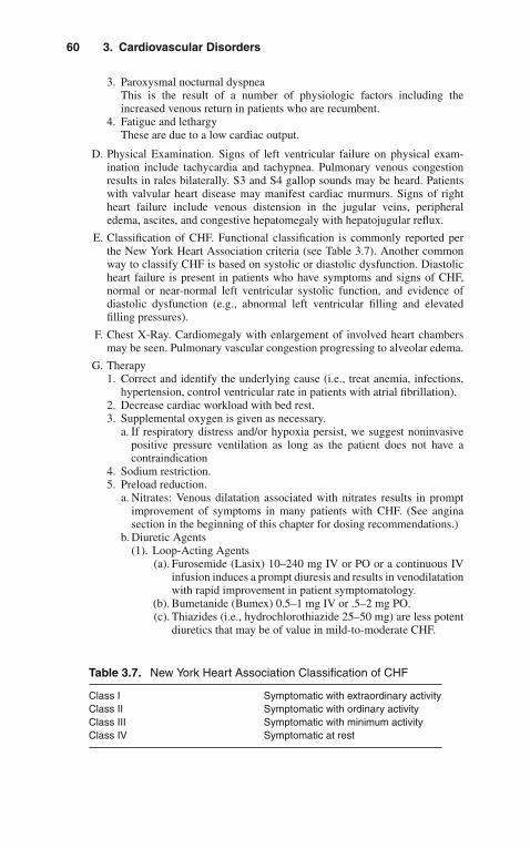
60 3. Cardiovascular Disorders
3. Paroxysmal nocturnal dyspneaThis is the result of a number of physiologic factors including theincreased venous return in patients who are recumbent.
4. Fatigue and lethargyThese are due to a low cardiac output.
D. Physical Examination. Signs of left ventricular failure on physical exam-ination include tachycardia and tachypnea. Pulmonary venous congestionresults in rales bilaterally. S3 and S4 gallop sounds may be heard. Patientswith valvular heart disease may manifest cardiac murmurs. Signs of rightheart failure include venous distension in the jugular veins, peripheraledema, ascites, and congestive hepatomegaly with hepatojugular reflux.
E. Classification of CHF. Functional classification is commonly reported perthe New York Heart Association criteria (see Table 3.7). Another commonway to classify CHF is based on systolic or diastolic dysfunction. Diastolicheart failure is present in patients who have symptoms and signs of CHF,normal or near-normal left ventricular systolic function, and evidence ofdiastolic dysfunction (e.g., abnormal left ventricular filling and elevatedfilling pressures).
F. Chest X-Ray. Cardiomegaly with enlargement of involved heart chambersmay be seen. Pulmonary vascular congestion progressing to alveolar edema.
G. Therapy1. Correct and identify the underlying cause (i.e., treat anemia, infections,
hypertension, control ventricular rate in patients with atrial fibrillation).2. Decrease cardiac workload with bed rest.3. Supplemental oxygen is given as necessary.
a. If respiratory distress and/or hypoxia persist, we suggest noninvasivepositive pressure ventilation as long as the patient does not have acontraindication
4. Sodium restriction.5. Preload reduction.
a. Nitrates: Venous dilatation associated with nitrates results in promptimprovement of symptoms in many patients with CHF. (See anginasection in the beginning of this chapter for dosing recommendations.)
b. Diuretic Agents(1). Loop-Acting Agents
(a). Furosemide (Lasix) 10–240 mg IV or PO or a continuous IVinfusion induces a prompt diuresis and results in venodilatationwith rapid improvement in patient symptomatology.
(b). Bumetanide (Bumex) 0.5–1 mg IV or .5–2 mg PO.(c). Thiazides (i.e., hydrochlorothiazide 25–50 mg) are less potent
diuretics that may be of value in mild-to-moderate CHF.
Table 3.7. New York Heart Association Classification of CHF
Class I Symptomatic with extraordinary activityClass II Symptomatic with ordinary activityClass III Symptomatic with minimum activityClass IV Symptomatic at rest
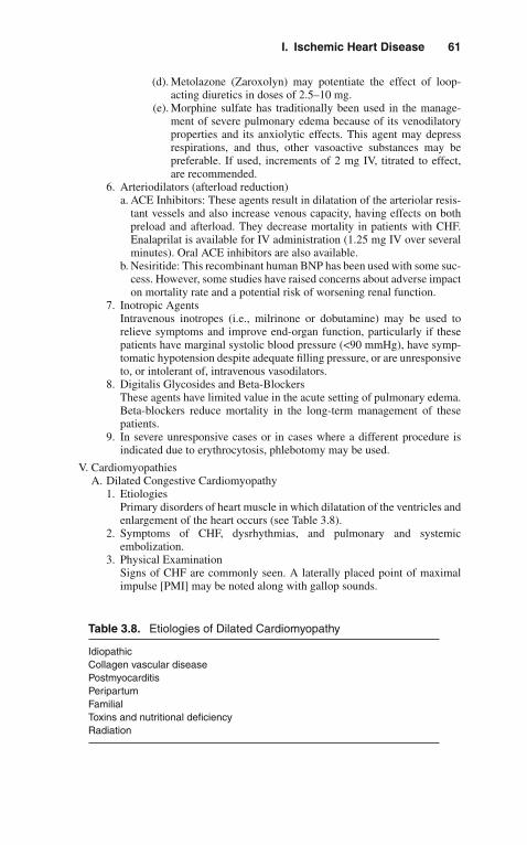
I. Ischemic Heart Disease 61
(d). Metolazone (Zaroxolyn) may potentiate the effect of loop-acting diuretics in doses of 2.5–10 mg.
(e). Morphine sulfate has traditionally been used in the manage-ment of severe pulmonary edema because of its venodilatoryproperties and its anxiolytic effects. This agent may depressrespirations, and thus, other vasoactive substances may bepreferable. If used, increments of 2 mg IV, titrated to effect,are recommended.
6. Arteriodilators (afterload reduction)a. ACE Inhibitors: These agents result in dilatation of the arteriolar resis-
tant vessels and also increase venous capacity, having effects on bothpreload and afterload. They decrease mortality in patients with CHF.Enalaprilat is available for IV administration (1.25 mg IV over severalminutes). Oral ACE inhibitors are also available.
b. Nesiritide: This recombinant human BNP has been used with some suc-cess. However, some studies have raised concerns about adverse impacton mortality rate and a potential risk of worsening renal function.
7. Inotropic AgentsIntravenous inotropes (i.e., milrinone or dobutamine) may be used torelieve symptoms and improve end-organ function, particularly if thesepatients have marginal systolic blood pressure (<90 mmHg), have symp-tomatic hypotension despite adequate filling pressure, or are unresponsiveto, or intolerant of, intravenous vasodilators.
8. Digitalis Glycosides and Beta-BlockersThese agents have limited value in the acute setting of pulmonary edema.Beta-blockers reduce mortality in the long-term management of thesepatients.
9. In severe unresponsive cases or in cases where a different procedure isindicated due to erythrocytosis, phlebotomy may be used.
V. CardiomyopathiesA. Dilated Congestive Cardiomyopathy
1. EtiologiesPrimary disorders of heart muscle in which dilatation of the ventricles andenlargement of the heart occurs (see Table 3.8).
2. Symptoms of CHF, dysrhythmias, and pulmonary and systemicembolization.
3. Physical ExaminationSigns of CHF are commonly seen. A laterally placed point of maximalimpulse [PMI] may be noted along with gallop sounds.
Table 3.8. Etiologies of Dilated Cardiomyopathy
IdiopathicCollagen vascular diseasePostmyocarditisPeripartumFamilialToxins and nutritional deficiencyRadiation
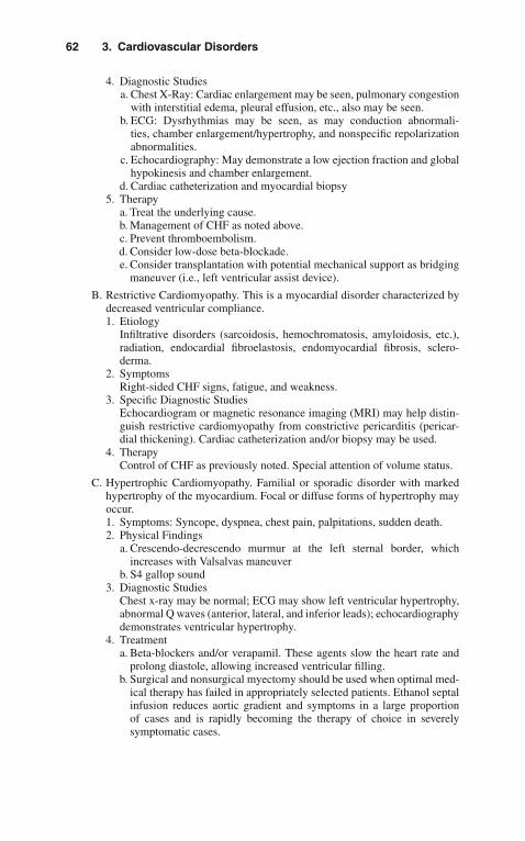
62 3. Cardiovascular Disorders
4. Diagnostic Studiesa. Chest X-Ray: Cardiac enlargement may be seen, pulmonary congestion
with interstitial edema, pleural effusion, etc., also may be seen.b. ECG: Dysrhythmias may be seen, as may conduction abnormali-
ties, chamber enlargement/hypertrophy, and nonspecific repolarizationabnormalities.
c. Echocardiography: May demonstrate a low ejection fraction and globalhypokinesis and chamber enlargement.
d. Cardiac catheterization and myocardial biopsy5. Therapy
a. Treat the underlying cause.b. Management of CHF as noted above.c. Prevent thromboembolism.d. Consider low-dose beta-blockade.e. Consider transplantation with potential mechanical support as bridging
maneuver (i.e., left ventricular assist device).
B. Restrictive Cardiomyopathy. This is a myocardial disorder characterized bydecreased ventricular compliance.1. Etiology
Infiltrative disorders (sarcoidosis, hemochromatosis, amyloidosis, etc.),radiation, endocardial fibroelastosis, endomyocardial fibrosis, sclero-derma.
2. SymptomsRight-sided CHF signs, fatigue, and weakness.
3. Specific Diagnostic StudiesEchocardiogram or magnetic resonance imaging (MRI) may help distin-guish restrictive cardiomyopathy from constrictive pericarditis (pericar-dial thickening). Cardiac catheterization and/or biopsy may be used.
4. TherapyControl of CHF as previously noted. Special attention of volume status.
C. Hypertrophic Cardiomyopathy. Familial or sporadic disorder with markedhypertrophy of the myocardium. Focal or diffuse forms of hypertrophy mayoccur.1. Symptoms: Syncope, dyspnea, chest pain, palpitations, sudden death.2. Physical Findings
a. Crescendo-decrescendo murmur at the left sternal border, whichincreases with Valsalvas maneuver
b. S4 gallop sound3. Diagnostic Studies
Chest x-ray may be normal; ECG may show left ventricular hypertrophy,abnormal Q waves (anterior, lateral, and inferior leads); echocardiographydemonstrates ventricular hypertrophy.
4. Treatmenta. Beta-blockers and/or verapamil. These agents slow the heart rate and
prolong diastole, allowing increased ventricular filling.b. Surgical and nonsurgical myectomy should be used when optimal med-
ical therapy has failed in appropriately selected patients. Ethanol septalinfusion reduces aortic gradient and symptoms in a large proportionof cases and is rapidly becoming the therapy of choice in severelysymptomatic cases.

I. Ischemic Heart Disease 63
c. Digitalis, nitrates, diuretics, and vasodilators may worsen the clinicalcondition of this subset of patients.
VI. MyocarditisMyocarditis is an inflammatory condition of the myocardium.
A. Etiology1. Infection
a. Viral (echovirus, adenovirus, etc.)b. Bacterialc. Mycoplasmad. Mycotice. Rickettsialf. Spirochetalg. Parasitic (trichinella, Trypanosoma cruzi)
2. Toxins and drugs (i.e., cocaine)3. Collagen vascular disease (scleroderma, systemic lupus erythematosus,
rheumatic fever, sarcoidosis)
B. Symptoms1. Dyspnea2. Chest discomfort
C. Physical Examination1. Tachycardia2. Pericardial friction rub (in the presence of coexistent pericarditis)3. Evidence of CHF
D. Therapy1. Supportive Care
a. Treatment of CHFb. Treatment of dysrhythmias, as necessaryc. Anticoagulation to prevent thromboembolism
2. Treat the underlying cause (the use of corticosteroids, immunoglob-ulins, and immunosuppressive therapy in selective populations withinflammatory infiltrates on endomyocardial biopsy may be warranted).
VII. PericarditisInflammation of the pericardium associated with many different etiologicfactors.A. Etiology (see Table 3.9)
B. Symptoms1. Anterior chest pain, commonly radiating to arms and back, which clas-
sically increases with inspiration and is relieved by sitting up or leaningforward. Palpitations and tachycardia may also occur.
C. Physical Examination1. Pericardial friction rub is best heard with the patient upright and leaning
forward.2. Tachycardia or other dysrhythmias may be auscultated.3. If pericardial tamponade occurs, low blood pressure, narrow pulse pres-
sure, and accentuated pulsus paradoxus may be seen.
D. Diagnostic Studies1. ECG (see Figure 3.1)
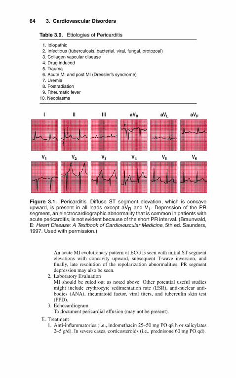
64 3. Cardiovascular Disorders
Table 3.9. Etiologies of Pericarditis
1. Idiopathic2. Infectious (tuberculosis, bacterial, viral, fungal, protozoal)3. Collagen vascular disease4. Drug induced5. Trauma6. Acute MI and post MI (Dressler’s syndrome)7. Uremia8. Postradiation9. Rheumatic fever
10. Neoplasms
Figure 3.1. Pericarditis. Diffuse ST segment elevation, which is concaveupward, is present in all leads except aVR and V1. Depression of the PRsegment, an electrocardiographic abnormality that is common in patients withacute pericarditis, is not evident because of the short PR interval. (Braunwald,E: Heart Disease: A Textbook of Cardiovascular Medicine, 5th ed. Saunders,1997. Used with permission.)
An acute MI evolutionary pattern of ECG is seen with initial ST-segmentelevations with concavity upward, subsequent T-wave inversion, andfinally, late resolution of the repolarization abnormalities. PR segmentdepression may also be seen.
2. Laboratory EvaluationMI should be ruled out as noted above. Other potential useful studiesmight include erythrocyte sedimentation rate (ESR), anti-nuclear anti-bodies (ANA), rheumatoid factor, viral titers, and tuberculin skin test(PPD).
3. EchocardiogramTo document pericardial effusion (may not be present).
E. Treatment1. Anti-inflammatories (i.e., indomethacin 25–50 mg PO q8 h or salicylates
2–5 g/d). In severe cases, corticosteroids (i.e., prednisone 60 mg PO qd).
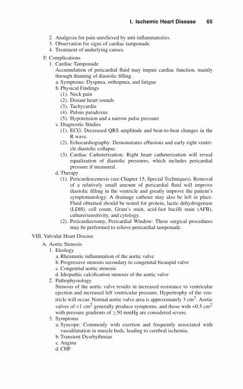
I. Ischemic Heart Disease 65
2. Analgesia for pain unrelieved by anti-inflammatories.3. Observation for signs of cardiac tamponade.4. Treatment of underlying causes.
F. Complications1. Cardiac Tamponade
Accumulation of pericardial fluid may impair cardiac function, mainlythrough thinning of diastolic filling.a. Symptoms: Dyspnea, orthopnea, and fatigueb. Physical Findings
(1). Neck pain(2). Distant heart sounds(3). Tachycardia(4). Pulsus paradoxus(5). Hypotension and a narrow pulse pressure
c. Diagnostic Studies(1). ECG: Decreased QRS amplitude and beat-to-beat changes in the
R wave.(2). Echocardiography: Demonstrates effusions and early right ventri-
cle diastolic collapse.(3). Cardiac Catheterization: Right heart catheterization will reveal
equalization of diastolic pressures, which includes pericardialpressure if measured.
d. Therapy(1). Pericardiocentesis (see Chapter 15, Special Techniques). Removal
of a relatively small amount of pericardial fluid will improvediastolic filling in the ventricle and greatly improve the patient’ssymptomatology. A drainage catheter may also be left in place.Fluid obtained should be tested for protein, lactic dehydrogenase(LDH), cell count, Gram’s stain, acid-fast bacilli stain (AFB),culture/sensitivity, and cytology.
(2). Pericardiectomy, Pericardial Window: These surgical proceduresmay be performed to relieve pericardial tamponade.
VIII. Valvular Heart Disease
A. Aortic Stenosis1. Etiology
a. Rheumatic inflammation of the aortic valveb. Progressive stenosis secondary to congenital bicuspid valvec. Congenital aortic stenosisd. Idiopathic calcification stenosis of the aortic valve
2. PathophysiologyStenosis of the aortic valve results in increased resistance to ventricularejection and increased left ventricular pressure. Hypertrophy of the ven-tricle will occur. Normal aortic valve area is approximately 3 cm2. Aorticvalves of <1 cm2 generally produce symptoms, and those with <0.5 cm2
with pressure gradients of ≥50 mmHg are considered severe.3. Symptoms
a. Syncope: Commonly with exertion and frequently associated withvasodilatation in muscle beds, leading to cerebral ischemia.
b. Transient Dysrhythmiasc. Anginad. CHF

66 3. Cardiovascular Disorders
4. Physical Findingsa. Slow-rising, delayed carotid upstroke with decreased amplitudeb. Narrowing of pulse pressurec. Loud systolic ejection murmur heard at the base of the heart and
radiating to the neck, often with a palpable thrill5. Diagnostic studies
a. ECG(1). Left ventricular hypertrophy(2). Nonspecific repolarization abnormalities
b. Chest x-ray(1). Pulmonary congestion in patients with CHF(2). Aortic dilatation(3). Calcification of the aortic valve
c. Echocardiography(1). Hypertrophy of the left ventricular wall(2). Visualization of the abnormal aortic valve
d. Cardiac catheterization documents severity of disease and calculationof valve area
6. Therapya. Judicious management for CHF and angina as they occur (see appro-
priate sections as above. These patients may be very preload sensitive).b. Valve replacement should be reserved as palliative therapy for patients
who are poor surgical risks.
B. Aortic Insufficiency1. Etiology
a. Infective endocarditisb. Trauma with valvular rupturec. Congenital bicuspid aortic valved. Rheumatic fibrosise. Myxomatous degenerationf. Accompanying aortic dissection
2. PathophysiologyLeft ventricular pressure increases secondary to regurgitation of bloodfrom the aorta, resulting in diastolic volume overload and subsequentdecompensation.
3. Symptomsa. Many patients remain asymptomatic for many years.b. Symptoms during decompensation include dyspnea on exertion, syn-
cope, chest pain, and CHF.4. Physical Findings
a. Widening pulse pressure with bounding pulses. Rapid rise and suddenfall in arterial pressure may result in head bobbing, capillary pulsa-tions in the nail beds (Quincke’s pulse), and “water-hammer” pulse. Inaddition, a murmur can be heard over the femoral arteries.
b. PMI may be displaced laterally, and S3 gallop may be heard. A diastolicblowing decrescendo murmur occurs along the left sternal border.
c. Austin-Flint murmur (apical diastolic rumble of low pitch secondary toaortic regurgitation, which affects the anterior mitral leaflet).
d. Systolic apical ejection murmur may also be heard.
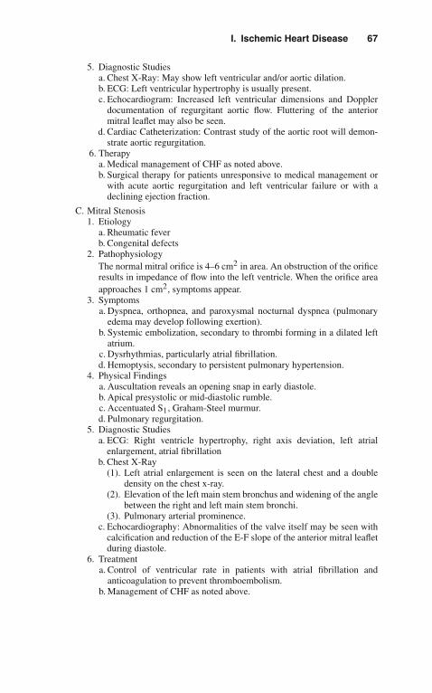
I. Ischemic Heart Disease 67
5. Diagnostic Studiesa. Chest X-Ray: May show left ventricular and/or aortic dilation.b. ECG: Left ventricular hypertrophy is usually present.c. Echocardiogram: Increased left ventricular dimensions and Doppler
documentation of regurgitant aortic flow. Fluttering of the anteriormitral leaflet may also be seen.
d. Cardiac Catheterization: Contrast study of the aortic root will demon-strate aortic regurgitation.
6. Therapya. Medical management of CHF as noted above.b. Surgical therapy for patients unresponsive to medical management or
with acute aortic regurgitation and left ventricular failure or with adeclining ejection fraction.
C. Mitral Stenosis1. Etiology
a. Rheumatic feverb. Congenital defects
2. PathophysiologyThe normal mitral orifice is 4–6 cm2 in area. An obstruction of the orificeresults in impedance of flow into the left ventricle. When the orifice areaapproaches 1 cm2, symptoms appear.
3. Symptomsa. Dyspnea, orthopnea, and paroxysmal nocturnal dyspnea (pulmonary
edema may develop following exertion).b. Systemic embolization, secondary to thrombi forming in a dilated left
atrium.c. Dysrhythmias, particularly atrial fibrillation.d. Hemoptysis, secondary to persistent pulmonary hypertension.
4. Physical Findingsa. Auscultation reveals an opening snap in early diastole.b. Apical presystolic or mid-diastolic rumble.c. Accentuated S1, Graham-Steel murmur.d. Pulmonary regurgitation.
5. Diagnostic Studiesa. ECG: Right ventricle hypertrophy, right axis deviation, left atrial
enlargement, atrial fibrillationb. Chest X-Ray
(1). Left atrial enlargement is seen on the lateral chest and a doubledensity on the chest x-ray.
(2). Elevation of the left main stem bronchus and widening of the anglebetween the right and left main stem bronchi.
(3). Pulmonary arterial prominence.c. Echocardiography: Abnormalities of the valve itself may be seen with
calcification and reduction of the E-F slope of the anterior mitral leafletduring diastole.
6. Treatmenta. Control of ventricular rate in patients with atrial fibrillation and
anticoagulation to prevent thromboembolism.b. Management of CHF as noted above.
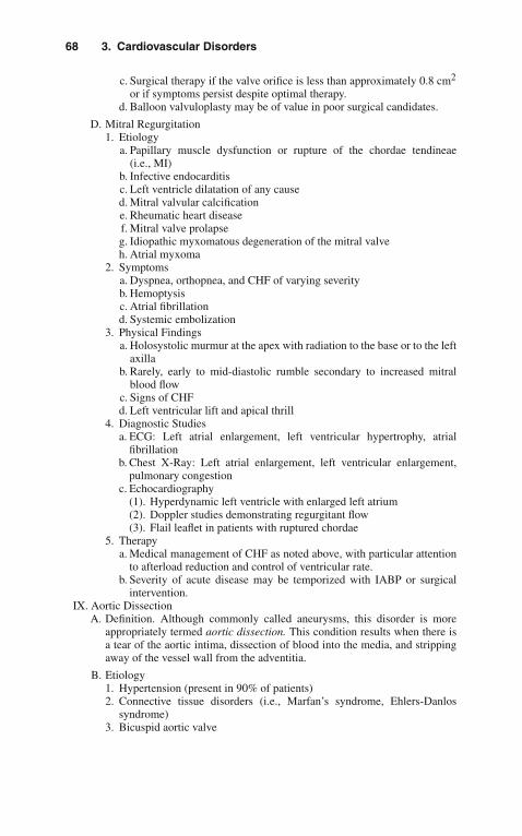
68 3. Cardiovascular Disorders
c. Surgical therapy if the valve orifice is less than approximately 0.8 cm2
or if symptoms persist despite optimal therapy.d. Balloon valvuloplasty may be of value in poor surgical candidates.
D. Mitral Regurgitation1. Etiology
a. Papillary muscle dysfunction or rupture of the chordae tendineae(i.e., MI)
b. Infective endocarditisc. Left ventricle dilatation of any caused. Mitral valvular calcificatione. Rheumatic heart diseasef. Mitral valve prolapseg. Idiopathic myxomatous degeneration of the mitral valveh. Atrial myxoma
2. Symptomsa. Dyspnea, orthopnea, and CHF of varying severityb. Hemoptysisc. Atrial fibrillationd. Systemic embolization
3. Physical Findingsa. Holosystolic murmur at the apex with radiation to the base or to the left
axillab. Rarely, early to mid-diastolic rumble secondary to increased mitral
blood flowc. Signs of CHFd. Left ventricular lift and apical thrill
4. Diagnostic Studiesa. ECG: Left atrial enlargement, left ventricular hypertrophy, atrial
fibrillationb. Chest X-Ray: Left atrial enlargement, left ventricular enlargement,
pulmonary congestionc. Echocardiography
(1). Hyperdynamic left ventricle with enlarged left atrium(2). Doppler studies demonstrating regurgitant flow(3). Flail leaflet in patients with ruptured chordae
5. Therapya. Medical management of CHF as noted above, with particular attention
to afterload reduction and control of ventricular rate.b. Severity of acute disease may be temporized with IABP or surgical
intervention.IX. Aortic Dissection
A. Definition. Although commonly called aneurysms, this disorder is moreappropriately termed aortic dissection. This condition results when there isa tear of the aortic intima, dissection of blood into the media, and strippingaway of the vessel wall from the adventitia.
B. Etiology1. Hypertension (present in 90% of patients)2. Connective tissue disorders (i.e., Marfan’s syndrome, Ehlers-Danlos
syndrome)3. Bicuspid aortic valve

I. Ischemic Heart Disease 69
Figure 3.2. Classification of Aortic Dissection (Stanford). In type A, theascending aorta is dissected (A). The intimal tear has always been at point1, but can occur at position 2 or 3. In type B dissection, the dissection islimited to the descending aorta (B). (Thubrikar, M: Vascular Mechanics andPathology, New York, Springer, 2007. Used with permission.)
4. Granulomatous arteritis and syphilitic aortitis5. Pregnancy6. Aortic injury
C. Classification. These lesions are commonly classified by their location (SeeFigure 3.2). Type A dissections involve the proximal aorta, from the aorticvalve to the aortic arch, and Type B dissections arise beyond the takeoff ofthe left subclavian artery.
D. Symptoms1. Chest pain (almost always present and usually abrupt, severe, and tearing
or burning)2. Syncope3. CHF4. Cerebrovascular accidents
E. Physical Findings1. Hypotension or hypertension2. Pulse deficits

70 3. Cardiovascular Disorders
3. Aortic regurgitation murmur4. Pericardial friction rub5. Neurologic signs6. Horner’s Syndrome and/or hoarseness
F. Diagnostic Studies1. Chest X-Ray
a. Abnormal in 90% of aortic dissectionsb. Widened aortic shadowc. Pleural effusionsd. Aortic calcification
2. ECG: Frequently abnormal (90%); however, nondiagnostic.3. Computed tomography of the chest with or without contrast will reveal
the lesion.4. Echocardiogram: Transthoracic and transesophageal studies may reveal
the dissection.5. Aortogram: The old “gold standard” for diagnosis.6. Magnetic resonance imaging
G. Therapy1. Surgical
a. Proximal dissections (Type A)b. Distal dissection, particularly if vital organs are compromised or
persistent pain occurs despite medical management2. Medical
a. Aggressive control of blood pressure. The typical regimen is adminis-tration of IV beta-blocker (traditionally, propranolol 1 mg IV q5 minuntil evidence of beta-blockade) followed by sodium nitroprusside tomaintain systolic blood pressure of approximately 100–120 mmHg.Alternative regimens using clevidipine or nicardipine combined witha beta-blocker have also been used.
b. Transcatheter stenting techniques are becoming more common andused in some centers.
X. Shock StatesShock can be defined as a state of inadequate tissue perfusion, which, unlessreversed, results in progressive organ dysfunction, damage, and death. Mortalityrates for shock of many causes still exceed 50%. In early stages of shock,the patient may be relatively asymptomatic. Sympathetic discharge and othercompensatory mechanisms may cause tachycardia and mild peripheral vaso-constriction in attempts to maintain blood pressure. When the state of shockworsens, organ hypoperfusion continues and blood pressure declines; signs oforgan dysfunction including restlessness and agitation, decreased urine output,and cool and clammy skin become evident.A. Classification and Etiology. A number of classification schemes for shock
states have been devised. These include cardiogenic shock; myopathic(reduced systolic function, i.e., acute MI), mechanical (mitral regurgitation,ventricular septal defect), extracardiac obstructive shock (i.e., pericardialtamponade, massive pulmonary embolus, or severe pulmonary hyperten-sion); oligemic shock (i.e., hemorrhage or fluid depletion); and distributiveshock (i.e., septic shock, anaphylaxis, neurogenic shock, etc.).

I. Ischemic Heart Disease 71
B. Diagnostic Evaluation1. Physical Examination
Tachycardia, hypotension, and evidence of hypoperfusion (i.e., alteredmental status, decreased urine output, cool and clammy skin) are gener-ally present. Other manifestations may be seen on physical examination,depending upon the etiology of the shock state.
2. Laboratory Evaluationa. ECG: Useful for identifying dysrhythmias and acute MI.b. Chest X-Ray: Pneumothorax, abnormal cardiac silhouette, and pul-
monary edema.c. Hematology and Chemistry: CBC, BUN, creatinine, electrolytes, glu-
cose, liver function tests (LFTs), and arterial blood gases should beobtained in the evaluation of any patient with shock.
3. Monitoringa. Foley Catheter: Patients in shock without contraindication should
receive catheter insertion for the monitoring of urine output.b. Arterial Line: For direct intra-arterial pressure determination and to
allow easy vascular access for laboratory and arterial blood gas moni-toring.
c. Central Venous Pressure (CVP) Monitoring Catheter, Oximetric CVPCatheter, or (rarely) PA Catheter: These are commonly employed. Mostpatients can be managed without pulmonary artery cannulation, and thePA catheter is being used less frequently.
C. Therapy. Primary treatment goals include restoring oxygen transport andorgan perfusion (i.e., urine output 1/2–1 mL/kg/h and the absence of lacticacidosis).1. Airway, breathing, and circulation (ABCs), as in all critically ill patients.2. Supportive Measures
a. Two large-bore IV catheters for those patients requiring volume resus-citation. For patients in whom fluid status is normal or elevated,a central IV line for administration of medication will usually berequired. For those patients not volume overloaded, the initial man-agement of hypotension and shock is volume administration. Volumechallenges of 250 cc to 1 L at a time should be rapidly administeredwith reassessment of the patient’s clinical condition.
b. Supplemental oxygen appropriate for the patient’s clinical status.c. Those patients not responding to volume administration should receive
beta-receptor stimulants. Dopamine, although commonly used inhypotensive patients, is not ideal as it produces significant shunts andcan decrease cerebral perfusion pressure. In the authors’ experience,vasopressin 1–6 U/h is the agent of choice in these conditions. Otheragents, such as norepinephrine, can also be used.
d. Mechanical support of the circulation may be necessary in patients withrefractory cardiogenic shock and amenable lesions.
e. Additional interventions and therapeutic goals for management ofshock must be based on specific etiologies.
XI. Infective EndocarditisA. Definition. Infection of the endocardial structures of the heart.
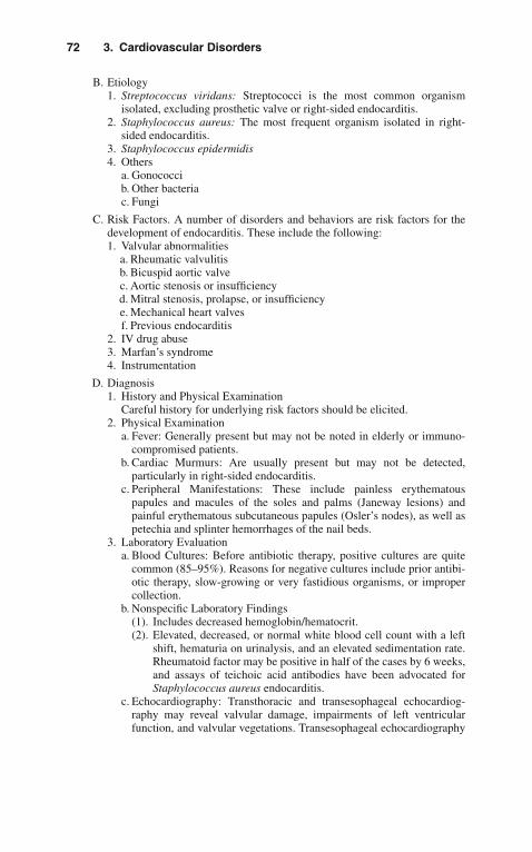
72 3. Cardiovascular Disorders
B. Etiology1. Streptococcus viridans: Streptococci is the most common organism
isolated, excluding prosthetic valve or right-sided endocarditis.2. Staphylococcus aureus: The most frequent organism isolated in right-
sided endocarditis.3. Staphylococcus epidermidis4. Others
a. Gonococcib. Other bacteriac. Fungi
C. Risk Factors. A number of disorders and behaviors are risk factors for thedevelopment of endocarditis. These include the following:1. Valvular abnormalities
a. Rheumatic valvulitisb. Bicuspid aortic valvec. Aortic stenosis or insufficiencyd. Mitral stenosis, prolapse, or insufficiencye. Mechanical heart valvesf. Previous endocarditis
2. IV drug abuse3. Marfan’s syndrome4. Instrumentation
D. Diagnosis1. History and Physical Examination
Careful history for underlying risk factors should be elicited.2. Physical Examination
a. Fever: Generally present but may not be noted in elderly or immuno-compromised patients.
b. Cardiac Murmurs: Are usually present but may not be detected,particularly in right-sided endocarditis.
c. Peripheral Manifestations: These include painless erythematouspapules and macules of the soles and palms (Janeway lesions) andpainful erythematous subcutaneous papules (Osler’s nodes), as well aspetechia and splinter hemorrhages of the nail beds.
3. Laboratory Evaluationa. Blood Cultures: Before antibiotic therapy, positive cultures are quite
common (85–95%). Reasons for negative cultures include prior antibi-otic therapy, slow-growing or very fastidious organisms, or impropercollection.
b. Nonspecific Laboratory Findings(1). Includes decreased hemoglobin/hematocrit.(2). Elevated, decreased, or normal white blood cell count with a left
shift, hematuria on urinalysis, and an elevated sedimentation rate.Rheumatoid factor may be positive in half of the cases by 6 weeks,and assays of teichoic acid antibodies have been advocated forStaphylococcus aureus endocarditis.
c. Echocardiography: Transthoracic and transesophageal echocardiog-raphy may reveal valvular damage, impairments of left ventricularfunction, and valvular vegetations. Transesophageal echocardiography
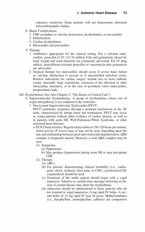
I. Ischemic Heart Disease 73
enhances sensitivity. Some patients will not demonstrate abnormalechocardiographic studies.
E. Major Complications1. CHF secondary to valvular destruction, dysrhythmias, or myocarditis2. Embolization3. Cardiac dysrhythmias4. Myocarditis and pericarditis
F. Therapy1. Antibiotics appropriate for the clinical setting. For a valvular endo-
carditis, penicillin G IV (12–24 million U/d) and gentamicin (dosed bybody weight and renal function) are commonly advocated. For IV drugaddicts, penicillinase-resistant penicillin or vancomycin plus gentamicinare advocated.
2. Surgical therapy for endocarditis should occur if severe heart failureor valvular obstruction is present or if uncontrolled infection exists.Relative indications for cardiac surgery include two or more embolicevents, unusually large vegetations, extension of the infection to otherintracardiac structures, or in the case of prosthetic valve endocarditis,periprosthetic leak.
XII. Dysrhythmias (See Also Chapter 2, “The Basics of Critical Care”)A. Supraventricular Dysrhythmias. A group of dysrhythmias whose site of
origin and pathway is not confined to the ventricles.1. Paroxysmal Supraventricular Tachycardia (PSVT)
PSVT commonly originates through a reentrant mechanism in the AVnode, characterized by abrupt onset and termination. PSVT may occurin young patients without other evidence of cardiac disease, as well asin patients with acute MI, Wolf-Parkinson-White Syndrome, or otherstructural heart diseases.a. ECG Characteristics: Regular tachycardia of 150–220 beats per minute.
Atrial activity (P waves) may or may not be seen, depending upon therate and relationship between atrial and ventricular depolarization. QRScomplex is frequently narrow. However, a wide QRS complex may beseen.(1). Symptoms
(a). Palpitations(b). May produce hypotension during acute MI or may precipitate
CHF.(2). Therapy
(a). ABCs.(b). For patients demonstrating clinical instability (i.e., cardio-
genic shock, ischemic chest pain, or CHF), synchronized DCcountershock should be used.
(c). Treatment of the stable patient should begin with a vagalmaneuver. Valsalva or carotid sinus massage following exclu-sion of carotid disease may abort the dysrhythmia.
(d). Adenosine should be administered to those patients who donot respond to vagal maneuvers, 6 mg rapid IV bolus. A sec-ond bolus of 12 mg rapid IV may be given. Methylxanthines(i.e., theophylline, aminophylline, caffeine) are competitive

74 3. Cardiovascular Disorders
antagonists, and dipyridamole enhances the pharmacologicef-fect of adenosine.
(e). Verapamil 5–10 mg over 5 min (may repeat dose in 20–30 min if ineffective) may be used if adenosine is ineffectivein patients with narrow complex PSVT. Pretreatment witha slow injection of 10 mL of 10% calcium chloride maydecrease the common hypotensive effects of this drug. Patientswith wide-complex tachycardia that cannot be confidentlydiagnosed as supraventricular should not receive verapamil,nor should patients with depressed ejection fraction. Digoxinor amiodarone as noted above for stable PSVT should beconsidered.
(f). Additional considerations include digoxin, beta-blockers,propafenone, diltiazem, pace termination, and synchronizedcardioversion for patients with preserved ejection fraction.
B. Atrial Fibrillation. Atrial fibrillation is characterized by chaotic atrial activ-ity without an organized atrial rhythm. This dysrhythmia may accompanycoronary artery disease, mitral and aortic valvular disease, thyrotoxicosis,peri- and myocarditis, alcoholic heart disease, and MI without evidence ofother organic cardiac disease.1. Electrocardiogram
Irregular, chaotic atrial activity without an organized rhythmic pattern.Conductive QRS complexes will have an irregularly irregular pattern.However, atrial ventricular block with emergence of a lower pacemakersite may result in irregular ventricular response.
2. Other Diagnostic Studiesa. Thyroid function testsb. Echocardiography
3. Therapya. In unstable patients, as in PSVT, DC cardioversion is indicated.b. Digoxin Loading: Digoxin has been the traditional therapy for new-
onset atrial fibrillation. Administer 0.5-mg IV loading dose followedby 0.25 mg q3–4 h until the ventricular rate is controlled. For patientswith impaired ejection fraction, this remains a good choice.
c. Alternatives for rapid rate control include(1). Diltiazem 20–25 mg IV over 2 min with continuous infusion of
5–15 mg/h.(2). Beta-blockers (i.e., propranolol 0.5 mg IV slowly followed by
boluses of 1 mg q5 min to a total of 0.1 mg/kg). Atenolol 5 mgIV slowly × 2. Metoprolol 5 mg slow IV q5 min × 3.
(3). Amiodarone in a dose of 150 mg over 10 min followed by 360 mgover 6 h and 0.5 mg/min is a good choice, particularly withdepressed ejection fraction.
d. Anticoagulation is advisable pre- and postcardioversion, particularlyin patients with mitral valve disease or a history of embolic phe-nomenon. Patients with a duration of atrial fibrillation >48 h shouldnot be converted acutely, if avoidable, because of embolization risk.Three to four weeks of anticoagulation precardioversion is recom-mended, with at least 4 weeks of anticoagulation postcardioversionsuggested.

I. Ischemic Heart Disease 75
C. Atrial Flutter. Atrial flutter is characterized by a rapid regular atrial rate of280–340 beats per minute, generally associated with varying degrees of AVblock. This dysrhythmia may occur with coronary artery disease, includingMI, thyrotoxicosis, pulmonary embolism, and mitral valve disease.1. ECG: Atrial depolarization classically has a “saw-tooth” pattern with
varying AV conduction block. Vagal maneuvers may slow the ventricularresponse rate, making atrial flutter waves more readily apparent.
2. Therapya. As for atrial fibrillation. In patients who are compromised, cardiover-
sion with DC countershock is indicated.b. Commonly responds to the pharmacologic interventions previously
denoted for atrial fibrillation. In addition, atrial pacing may alsoterminate atrial flutter.
D. Multifocal Atrial Tachycardia. In multifocal atrial tachycardia, a chaoticirregular atrial activity is seen, with rates between 100 and 180 and varyingp-wave morphology (three consecutive different p-wave morphologies). Thisdisorder commonly accompanies chronic obstructive pulmonary disease,theophylline toxicity, hypoxemia, and/or other metabolic disturbances.1. ECG (See Figure 3.3)
Varying P-to-P intervals and beat-to-beat variability in P-wave morphol-ogy.
2. Therapya. Treat the underlying cause. Rate may be controlled, if necessary, with
diltiazem or amiodarone. In the absence of depressed ejection frac-tion, beta-blockers may be used. In addition, IV magnesium has beenadvocated by some authorities.
E. Bradycardias and AV Conduction Blocks. These are characterized by a lowintrinsic rate from the sinus node or blockade of sinus impulses between theAV node, which result in slow ventricular rates.1. Etiology
a. Vagotoniab. Ischemic heart diseasec. Cardiomyopathiesd. Drugse. Degenerative diseases of the AV conduction system
2. TreatmentPatients who are symptomatic due to low ventricular response rate maybe treated with the following:
Figure 3.3. Multifocal atrial tachycardia. (Criner, G: Critical Care StudyGuide, Springer, 2002. Used with permission.)
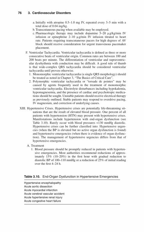
76 3. Cardiovascular Disorders
a. Initially with atropine 0.5–1.0 mg IV, repeated every 3–5 min with atotal dose of 0.04 mg/kg.
b. Transcutaneous pacing when available may be employed.c. Pharmacologic therapy may include dopamine 5–20 μg/kg/min IV
infusion or epinephrine 2–10 μg/min IV infusion titrated to heartrate. Patients requiring transcutaneous pacers for high degrees of AVblock should receive consideration for urgent transvenous pacemakerplacement.
F. Ventricular Tachycardia. Ventricular tachycardia is defined as three or moreconsecutive beats of ventricular origin. Common rates are between 100 and200 beats per minute. The differentiation of ventricular and supraventric-ular dysrhythmia with conduction may be difficult. A good rule of thumbis that wide-complex QRS tachycardia should be considered ventriculartachycardia until proven otherwise.1. Monomorphic ventricular tachycardia (a single QRS morphology) should
be treated as noted in Chapter 3, “The Basics of Critical Care”.2. Polymorphic ventricular tachycardia or “torsade de pointes” may be
caused by agents frequently used in the treatment of monomorphicventricular tachycardia. Electrolyte disturbances including hypokalemia,hypomagnesemia, and the presence of cardiac and psychotropic medica-tions should be sought. Unstable patients should receive electrical therapyas previously outlined. Stable patients may respond to overdrive pacing,IV magnesium, and correction of underlying causes.
XIII. Hypertensive Crises. Hypertensive crises are potentially life-threatening sit-uations that are the result of elevated blood pressure. One percent of allpatients with hypertension (HTN) may present with hypertensive crises.Manifestations include hypertension with end-organ dysfunction (seeTable 3.10). Rarely occur with blood pressures <130 mmHg diastolic.Hypertensive crises can be further classified into: Hypertensive urgen-cies (when the BP is elevated but no active organ dysfunction is found)and hypertensive emergencies (when there is evidence of organ dysfunc-tion). The management of hypertensive urgencies differs from that ofhypertensive emergencies.
A. Treatment1. Blood pressure should be promptly reduced in patients with hyperten-
sive emergencies. Most authorities recommend reductions of approx-imately 15% (10–20%) in the first hour with gradual reduction todiastolic BP of 100–110 mmHg or a reduction of 25% of initial readingover the first 4–24 h.
Table 3.10. End-Organ Dysfunction in Hypertensive Emergencies
Hypertensive encephalopathyAcute aortic dissectionAcute myocardial infarctionAcute cerebral vascular accidentAcute hypertensive renal injuryAcute congestive heart failure

I. Ischemic Heart Disease 77
Table 3.11. Intravenous Antihypertensive Medications
Clevidipine Start at 2 mg/h and double to the dose every3 min (max 32 mg/h)
Labetalol 20 mg bolus, 2 mg/min (max 300 mg/day)
Nicardipine 5–20 mg/h
Fenoldopam 0.1–2 μg/kg/min
Nitroglycerin 5 μg/min (increase by 5–10 μg q3–5 min asneeded)
Diazoxide 25–150 mg IV over 5 min or infusion of30 mg/min to effect
2. Reductions in BP may result in ischemia, and thus, these patients mustbe carefully followed. Parenteral therapy with short-acting agents isinitially recommended (see Table 3.11).
3. Patients receiving parenteral therapy commonly need continuous arte-rial pressure monitoring.a. Cyanide poisoning may occur with IV administration of sodium
nitroprusside to renal failure patients. Consider this if such patientsdevelop CNS depression, seizures, lactic acidosis, or cardiovascularinstability.(1). May occur with infusion rate 2 μg/kg/min.(2). Infusion rates of 10 μg/kg/min should not be continued for
prolonged periods of time because of this hazard.(3). If cyanide intoxication suspected, discontinue infusion and treat
as described in Chapter 16, “Toxicology.”
4. Oral therapy with clonidine (Catapres) (0.1 mg PO q20 min) or a hostof other agents may be used in less severe cases of hypertension in theICU patient.
XIV. Useful Facts and FormulasA. Pressure = Flow × Resistance: This is true in the airways as well as in the
cardiovascular system. For example:
Mean arterial pressure = cardiac output × systemic vascular resistance.Mean pulmonary arterial pressure = cardiac output × pulmonary vascular
resistance.
The unmeasured resistance term is usually calculated by solving theequations:
systemic vascular resistance = mean arterial pressurecardiac output
B. The Primary Determinants of Cardiovascular Performance1. Heart rate2. Preload3. Afterload4. Contractility

78 3. Cardiovascular Disorders
C. Other Principles and Conversion FactorsFluid flow:
Fluid flow = (pressure difference)(radius)4
(vessel length)(fluid viscosity)8
Conversion to mmHg
Pressure in mmHg = Pressure in cm H2O/1.36
LaPlace’s law:
Wall tension = distending pressure × vessel radius
wall thickness
Ohm’s law:
Current (I) = electromotive force (E)
resistance (R)
Poiseuille’s law:
Q = vπ r2
where Q = rate of blood flow (mm/sec); πr2 = cross-sectional area (cm2);v = velocity of blood flow.
Vascular capacitance:
Vascular compliance (capacitance) = increase in volume
increase in pressure
Vascular distensibility:
Vascular distensibility = increase in volume
increase in pressure × original volume
D. Direct measurements of the heart rate are relatively easy. Preload, afterload, andcontractility are more difficult to assess clinically. In assessment of cardiovas-cular performance, the following hemodynamic measurements are commonlymeasured or calculated:
1. Arteriovenous Oxygen Content Difference (avDO2)This is the difference between the arterial oxygen content (CaO2) and thevenous oxygen content (CvO2).

I. Ischemic Heart Disease 79
2. Body Surface Area (BSA)Calculated from height and weight, BSA is generally used to index mea-sured and derived values according to the size of the patient.
3. Cardiac Index (CI)Calculated as cardiac output/BSA, CI is the prime determinant of hemody-namic function.
4. Left Ventricular Stroke Work Index (LVSWI)LVSWI is the product of the stroke index (SI) and (mean arterial pressure[MAP]—pulmonary artery occlusion pressure [PAOP]), and a unit correc-tion factor of 0.0136. The LVSWI measures the work of the left ventricle(LV) as it ejects into the aorta.
5. Mean Arterial Pressure (MAP)The MAP is estimated as one-third of pulse pressure plus the diastolic bloodpressure.
6. Oxygen Consumption (VO2)Calculated as C(a–v)O2 H CO H 10, it is the amount of oxygen extracted inmL/min by the tissue from the arterial blood.
7. Oxygen Delivery (DO2)Calculated as (CaO2) H CO H 10, it is the total oxygen delivered by thecardiorespiratory systems.
8. Pulmonary Vascular Resistance Index (PVRI)Calculated as (MAP—PAOP)/CI, it measures the resistance in the pul-monary vasculature.
9. Right Ventricular Stroke Work Index (RVSWI)RVSWI is the product of the SI and (mean pulmonary artery pressure[MPAP]—central venous pressure [CVP]), and a unit correction factor of0.0136. It measures the work of the right ventricle as it ejects into thepulmonary artery.
10. Stroke Index (SI)Calculated as CI/heart rate, SI is the average volume of blood ejected by theventricle with each beat.
11. Systemic Vascular Resistance Index (SVRI)Calculated as (MAP—CVP)/CI, SVRI is the customary measure of theresistance in the systemic circuit.
12. Venous Admixture (Qva/QT)Calculated as (CcO2—CaO2)/(CcO2—CvO2), it represents the fraction ofcardiac output not oxygenated in an idealized lung.
E. Cardiac Output Formulas
Output of left ventricle = O2 consumption (mL/min)
[AO2] − [VO2]
It may also be measured by thermodilution techniques:
Q = V(Tb − Ti)K/
∫Tb(t)dt
where Q = cardiac output; V = volume of injectate; Tb = blood temperature;Ti = injectate temperature; K = a constant including the density factor and

80 3. Cardiovascular Disorders
catheter characteristics;∫
Tb(t)dt = area under the blood-temperature-timecurve.The same principle is applicable for the pulmonary blood flow:
Q = B/(Cv − Ca)
where Q = pulmonary blood flow; B = rate of loss of the indicator of alve-olar gas; Cv = concentration of the indicator in the venous blood; Ca =concentration of the indicator in the arterial blood.
Q = V/(CaO2 − cO2)
where Q = total pulmonary blood flow; VO2 = oxygen uptake; CaO2 = arterialoxygen concentration; CVO2 = mixed venous oxygen concentration.
F. Other Cardiovascular Performance Formulas/TablesAlveolar-arterial O2 difference or A-a gradient
= Alveolar pO2—arterial pO2Normal <10 torr (10 mmHg)
Alveolar PO2 at sea level (PAO2)= (FiO2 × 713) – 1.2 × PaCO2
Arterial blood O2 content (CaO2)= (PaO2 × 0.003) + (1.34 × Hb in g × arterial blood Hb O2 sat %)
Normal = 18–20 mL/dL
Arteriovenous oxygen difference (avDO2)= (CaO2) – (CvO2)
Normal = 4–5 mL/dL
Cardiac index (CI) = cardiac output/body surface areaNormal = 3.0–3.4 L/min-m2
Ejection Fraction (EF)
= [end diastolic volume] − [end systolic volume]
end − diastolic volume=%
Mean arterial (or pulmonary) pressure= DBP + 1/3 (SBP–DBP)
Mean pulmonary arterial pressure= DPAP + 1/3 (SPAP–DPAP)
O2 delivery index (DO2I) = CaO2 x cardiac index × 10Normal = 500–600 mL/min-m2
O2 consumption index (O2I)= Arteriovenous O2 difference × cardiac index × 10
Normal = 120–160 mL/min2
O2 extraction (O2 Ext)= (arteriovenous O2 difference/arterial blood O2 content) × 100
Normal = 20–30%
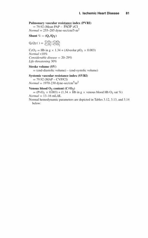
I. Ischemic Heart Disease 81
Pulmonary vascular resistance index (PVRI)= 79.92 (Mean PAP – PAOP )/CI
Normal = 255–285 dyne-sec/cm/5·m2
Shunt % = (Qs/QT)
QsQT( ) = CcO2−CaO2CcO2−CvO2
CcO2 = Hb in g × 1.34 + (Alveolar pO2 × 0.003)Normal <10%Considerable disease = 20–29%Life-threatening 30%
Stroke volume (SV)= (end-diastolic volume) – (end-systolic volume)
Systemic vascular resistance index (SVRI)= 79.92 (MAP – CVP/CI)
Normal = 1970-230 dyne-sec/cm5m2
Venous blood O2 content (CvO2)= (PvO2 × 0.003) + (1.34 × Hb in g × venous blood Hb O2 sat %)
Normal = 13–16 mL/dLNormal hemodynamic parameters are depicted in Tables 3.12, 3.13, and 3.14
below:

82 3. Cardiovascular Disorders
Tab
le3.
12.
Nor
mal
Hem
odyn
amic
Par
amet
ers,
Adu
lt
Par
amet
erE
quat
ion
Nor
mal
rang
e
Art
eria
lblo
odpr
essu
re(B
P)
Sys
tolic
(SB
P)
90–1
40m
mH
g
Dia
stol
ic(D
BP
)60
–90
mm
Hg
Mea
nar
teria
lpre
ssur
e(M
AP
)[S
BP
+(2
×D
BP
)]/3
70–1
05m
mH
g
Rig
htat
rialp
ress
ure
(RA
P)
2–6
mm
Hg
Rig
htve
ntric
ular
pres
sure
(RV
P)
Sys
tolic
(RV
SP
)15
–25
mm
Hg
Dia
stol
ic(R
VD
P)
0–8
mm
Hg
Pul
mon
ary
arte
rypr
essu
re(P
AP
)S
ysto
lic(P
AS
P)
15–2
5m
mH
g
Dia
stol
ic(P
AD
P)
8–15
mm
Hg
Mea
npu
lmon
ary
arte
rypr
essu
re(M
PAP
)[P
AS
P+
(2×
PAD
P)]
/310
–20
mm
Hg
Pul
mon
ary
arte
ryw
edge
pres
sure
(PA
WP
)6–
12m
mH
g
Left
atria
lpre
ssur
e(L
AP
)6–
12m
mH
g
Car
diac
outp
ut(C
O)
HR
×S
V/1
,000
4.0–
8.0
L/m
in
Car
diac
inde
x(C
I)C
O/B
SA
2.5–
4.0
L/m
in/m
2
Str
oke
volu
me
(SV
)C
O/H
R×
1,00
060
–100
mL/
beat
Str
oke
volu
me
inde
x(S
VI)
CI/H
R×
1,00
033
–47
mL/
m2/b
eat
Sys
tem
icva
scul
arre
sist
ance
(SV
R)
80×
(MA
P–
RA
P)/
CO
800–
1,20
0dy
ness
/cm
5
Sys
tem
icva
scul
arre
sist
ance
inde
x(S
VR
I)80
×(M
AP
–R
AP
)/C
I19
70–2
390
dyne
ss/c
m5/m
2
Pul
mon
ary
vasc
ular
resi
stan
ce(P
VR
)80
×(M
PAP
–PA
WP
)/C
O<
250
dyne
ss/c
m5
Pul
mon
ary
vasc
ular
resi
stan
cein
dex
(PV
RI)
80×
(MPA
P–
PAW
P)/
CI
255–
285
dyne
ss/c
m5/m
2

I. Ischemic Heart Disease 83
Tab
le3.
13.
Hem
odyn
amic
Par
amet
ers,
Adu
lt
Par
amet
erE
quat
ion
Nor
mal
Ran
ge
Left
vent
ricul
arst
roke
wor
k(L
VS
W)
SV
×(M
AP
–PA
WP
)×
0.01
3658
–104
g-m
/bea
t
Left
vent
ricul
arst
roke
wor
kin
dex
(LV
SW
I)S
VI×
(MA
P–
PAW
P)×
0.01
3650
–62
g-m
/m2/b
eat
Rig
htve
ntric
ular
stro
kew
ork
(RV
SW
)S
V×
(MPA
P–
RA
P)×
0.01
368–
16g-
m/b
eat
Rig
htve
ntric
ular
stro
kew
ork
inde
x(R
VS
WI)
SV
×(M
PAP
–R
AP
)×
0.01
365–
10g-
m/m
2/b
eat
Cor
onar
yar
tery
perf
usio
npr
essu
re(C
PP
)D
iast
olic
BP
-PA
WP
60–8
0m
mH
g
Rig
htve
ntric
ular
end-
dias
tolic
volu
me
(RV
ED
V)
SV
/EF
100–
160
mL
Rig
htve
ntric
ular
end-
syst
olic
volu
me
(RV
ES
V)
ED
V-S
V50
–100
mL
Rig
htve
ntric
ular
ejec
tion
frac
tion
(RV
EF
)S
V/E
DV
40–6
0%
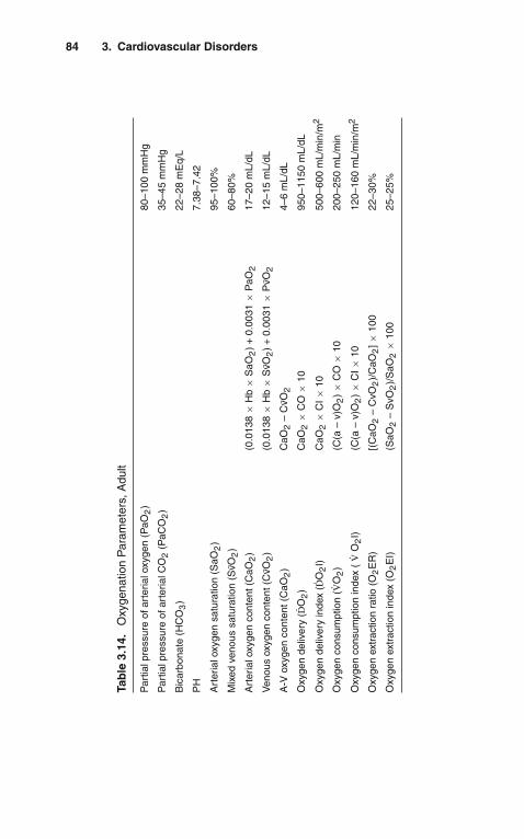
84 3. Cardiovascular Disorders
Tab
le3.
14.
Oxy
gena
tion
Par
amet
ers,
Adu
lt
Par
tialp
ress
ure
ofar
teria
loxy
gen
(PaO
2)
80–1
00m
mH
g
Par
tialp
ress
ure
ofar
teria
lCO
2(P
aCO
2)
35–4
5m
mH
g
Bic
arbo
nate
(HC
O3)
22–2
8m
Eq/
L
PH
7.38
–7.4
2
Art
eria
loxy
gen
satu
ratio
n(S
aO2)
95–1
00%
Mix
edve
nous
satu
ratio
n(S
vO2)
60–8
0%
Art
eria
loxy
gen
cont
ent(
CaO
2)
(0.0
138
×H
b×
SaO
2)
+0.
0031
×P
aO2
17–2
0m
L/dL
Ven
ous
oxyg
enco
nten
t(C
vO2)
(0.0
138
×H
b×
SvO
2)
+0.
0031
×P
vO2
12–1
5m
L/dL
A-V
oxyg
enco
nten
t(C
aO2)
CaO
2–
CvO
24–
6m
L/dL
Oxy
gen
deliv
ery
( DO
2)
CaO
2×
CO
×10
950–
1150
mL/
dL
Oxy
gen
deliv
ery
inde
x(D
O2I)
CaO
2×
CI×
1050
0–60
0m
L/m
in/m
2
Oxy
gen
cons
umpt
ion
(VO
2)
(C(a
–v)
O2)×
CO
×10
200–
250
mL/
min
Oxy
gen
cons
umpt
ion
inde
x(
VO
2I)
(C(a
–v)
O2)×
CI×
1012
0–16
0m
L/m
in/m
2
Oxy
gen
extr
actio
nra
tio(O
2E
R)
[(C
aO2
–C
vO2)/
CaO
2]×
100
22–3
0%
Oxy
gen
extr
actio
nin
dex
(O2E
I)(S
aO2
–S
vO2)/
SaO
2×
100
25–2
5%

4Endocrinologic Disorders
� I. ADRENAL INSUFFICIENCY
A. Definition. Adrenal insufficiency is a deficiency of glucocorticoid production,which can result from adrenal gland failure (primary adrenal insufficiency) orfailure of hypothalamic–pituitary secretion of corticotropin-releasing hormone(CRH) or adrenocorticotropic hormone (ACTH) (secondary adrenal insuffi-ciency).
B. Pathophysiology1. Glucocorticoid synthesis is regulated by the hypothalamic–pituitary–adrenal
(HPA) axis (hypothalamus secretes CRH, which stimulates the pituitaryto release ACTH, which increases release of cortisol by the adrenalcortex).
2. Cortisol is a negative feedback for further CRH and ACTH production.3. Mineralocorticoid production is regulated primarily by the renin-angiotensin
system, blood pressure, and extracellular potassium level.4. Catecholamines are synthesized by the adrenal cortex and medulla.5. The adrenal cortex is composed of the zona glomerulosa (aldosterone synthe-
sis), zona fasciculata (glucocorticoid synthesis), and zona reticularis (androgenand glucocorticoid synthesis).
6. Primary adrenal insufficiency usually results in loss of both glucocorticoid andmineralocorticoid secretion.
7. Patients with secondary adrenal insufficiency maintain normal secretion of themineralocorticoids.
C. Etiology1. Infection (acquired immune deficiency syndrome [AIDS], tuberculosis [TB],
cytomegalovirus, meningococcemia, fungus, pseudomonas septicemia)2. Adrenal hemorrhage (coagulopathies, anticoagulant therapy, sepsis, trauma,
pregnancy)3. Withdrawal of exogenous steroids or HPA axis suppression from recent (up to
1 year prior) treatment with exogenous corticosteroids4. Drugs that interfere with adrenal steroid synthesis (ketoconazole, etomidate,
aminoglutethimide)5. Tumor destruction
J. Varon, P. Acosta, Handbook of Critical and Intensive Care Medicine,DOI 10.1007/978-0-387-92851-7_4,C© Springer Science+Business Media, LLC 2010

86 4. Endocrinologic Disorders
6. Adrenal infarction (arteritis, thrombosis)7. Autoimmune disorders (sarcoidosis, amyloidosis)
D. Clinical Manifestations1. Weakness2. Weight loss3. Anorexia4. Hyperpigmentation (only in primary adrenal insufficiency with ACTH produc-
tion)5. Circulatory collapse6. Gastrointestinal disturbances (nausea/vomiting, abdominal pain, diarrhea, con-
stipation)7. Dehydration8. Fever9. May have only vague symptoms (malaise, arthralgias)
E. Associated Laboratory Abnormalities1. Increased potassium and calcium2. Decreased sodium and chloride3. Decreased glucose4. Metabolic acidosis5. Increased blood, urea, nitrogen/creatinine (BUN/Cr) ratio6. Normocytic, normochromic anemia, neutropenia, lymphocytosis, eosinophilia
F. Diagnostic Evaluation1. Physical examination
a. Hypotensionb. Hyperpigmentation (not in acute or secondary adrenal insufficiency)c. Females may have loss of axillary hair
2. Laboratory Evaluationa. Measurement of a single plasma cortisol level usually does not allow reliable
appraisal of pituitary and adrenal function; however, in stressed, critically illpatients, it is sufficient to rule out or to suggest the diagnosis of adrenalinsufficiency (a serum cortisol level ≥20 μg/dL indicates adequate adrenalglucocorticoid secretion following stress, ACTH, or CRH).
b. If the patient is hemodynamically unstable and adrenal insufficiency issuspected, a random cortisol level should be drawn and treatment withhydrocortisone (see “Therapy” section below) should be initiated immedi-ately.
c. If the patient is hemodynamically stable and adrenal insufficiency is sus-pected, a random cortisol level may be measured and the patient treated withdexamethasone (stress dose of 2–10 mg IV q6 h) until the result is avail-able; then, if the cortisol level is <20 μg/dL, a screening synthetic ACTHstimulation test may be performed:(1). Measure baseline cortisol level.(2). Give cosyntropin (synthetic ACTH) 0.25 mg IV.(3). Measure cortisol level again after 60 min.(4). An increase of <7 μg/dL is suggestive of primary adrenal insufficiency
if the basal cortisol level is <20 μg/dL.(5). If diagnosis of adrenal insufficiency is confirmed by plasma cortisol
<20 μg/dL, further testing (3-day ACTH infusion test) may be requiredto establish the exact etiology, but this should only be done when thepatient is out of the intensive care unit (ICU).

II. Diabetes Insipidus 87
d. Diagnostic imaging may help determine the etiology (bilaterally enlargedadrenal glands by computed tomography (CT) scan suggest adrenal hem-orrhage, neoplastic disease, TB, or fungal infection, whereas small adrenalglands suggest autoimmune disease or lack of ACTH).
e. High ACTH levels associated with low cortisol levels or failure of theadrenal gland to respond to prolonged ACTH stimulation are consistent withprimary adrenal insufficiency.
f. Measurement of plasma renin (PRA) and aldosterone (ALDO) levels mayhelp distinguish primary from secondary adrenal insufficiency (PRA/ALDOratios are high in primary adrenal insufficiency and low in secondary adrenalinsufficiency).
g. Patients with primary adrenal insufficiency have decreased 24-h urinarycortisol, 17-OHCS, and 17-KS, and increased ACTH (assessment of thesesubstances, impractical in most ICU situations, should not be undertakenbefore or in lieu of treatment).
G. Therapy1. Treat patients with circulatory collapse and suspected adrenal insufficiency
immediately (draw random cortisol level if possible first, but do not wait totreat).
2. Administer hydrocortisone 100 mg IV q8 h for 24 h; if the patient shows agood clinical response (or cortisol level confirms diagnosis), the dosage may betapered gradually and eventually changed to oral.
3. Provide adequate volume replacement with D5NS, until hypotension, dehydra-tion, and hypoglycemia are corrected.
4. Identify and treat any precipitating factors (infections, hemorrhage).
H. Complications1. Short-term corticosteroid therapy is safe.2. When there is a question of adrenal insufficiency during an emergency, short-
term supplemental corticosteroids should be given until adrenal integrity can beassessed.
3. It is of the utmost importance to treat patients in these situations, becauseadrenal crisis will progress to death if untreated.
� II. DIABETES INSIPIDUS
A. Definitions1. Diabetes insipidus is a disorder of fluid homeostasis resulting from inadequate
vasopressin or antidiuretic hormone (ADH) secretion or action.2. Neurogenic diabetes insipidus may be caused by a lack of production of ADH
precursor molecule by the hypothalamus or inadequate secretion of ADH bythe posterior pituitary.
3. Nephrogenic diabetes insipidus is caused by unresponsiveness of renal tubulesto ADH.
4. Both types of diabetes insipidus result in excretion of large volumes ofhypotonic fluid.
B. EtiologySee Table 4.1.
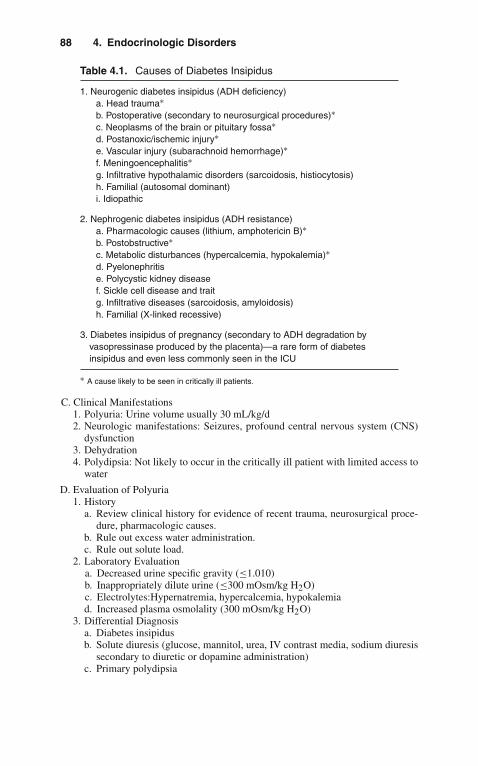
88 4. Endocrinologic Disorders
Table 4.1. Causes of Diabetes Insipidus
1. Neurogenic diabetes insipidus (ADH deficiency)a. Head trauma∗b. Postoperative (secondary to neurosurgical procedures)∗c. Neoplasms of the brain or pituitary fossa∗d. Postanoxic/ischemic injury∗e. Vascular injury (subarachnoid hemorrhage)∗f. Meningoencephalitis∗g. Infiltrative hypothalamic disorders (sarcoidosis, histiocytosis)h. Familial (autosomal dominant)i. Idiopathic
2. Nephrogenic diabetes insipidus (ADH resistance)a. Pharmacologic causes (lithium, amphotericin B)∗b. Postobstructive∗c. Metabolic disturbances (hypercalcemia, hypokalemia)∗d. Pyelonephritise. Polycystic kidney diseasef. Sickle cell disease and traitg. Infiltrative diseases (sarcoidosis, amyloidosis)h. Familial (X-linked recessive)
3. Diabetes insipidus of pregnancy (secondary to ADH degradation byvasopressinase produced by the placenta)—a rare form of diabetesinsipidus and even less commonly seen in the ICU
∗ A cause likely to be seen in critically ill patients.
C. Clinical Manifestations1. Polyuria: Urine volume usually 30 mL/kg/d2. Neurologic manifestations: Seizures, profound central nervous system (CNS)
dysfunction3. Dehydration4. Polydipsia: Not likely to occur in the critically ill patient with limited access to
water
D. Evaluation of Polyuria1. History
a. Review clinical history for evidence of recent trauma, neurosurgical proce-dure, pharmacologic causes.
b. Rule out excess water administration.c. Rule out solute load.
2. Laboratory Evaluationa. Decreased urine specific gravity (≤1.010)b. Inappropriately dilute urine (≤300 mOsm/kg H2O)c. Electrolytes:Hypernatremia, hypercalcemia, hypokalemiad. Increased plasma osmolality (300 mOsm/kg H2O)
3. Differential Diagnosisa. Diabetes insipidusb. Solute diuresis (glucose, mannitol, urea, IV contrast media, sodium diuresis
secondary to diuretic or dopamine administration)c. Primary polydipsia

II. Diabetes Insipidus 89
E. Diagnostic Approach1. First exclude solute diuresis as etiology (check urine glucose, osmolality, and
specific gravity).2. If the patient has hypernatremia and either an inappropriately low plasma
ADH level or an inappropriately low urine osmolality, the diagnosis of dia-betes insipidus is made (further testing is needed only to distinguish the type ofdiabetes insipidus):a. Administer a therapeutic trial of 1 μg DDAVP (desmopressin) subcuta-
neously.b. If urine osmolality increases ≥50%, the diagnosis of neurogenic diabetes
insipidus is confirmed and therapy can be instituted.c. If urine osmolality does not increase in response to DDAVP, renal resistance
to ADH is suggested and nephrogenic causes of diabetes insipidus should beaddressed.
3. If the plasma sodium is <145 mEq/L and the patient is hemodynamically stable,a water-deprivation test may be initiated (to stimulate secretion of ADH and todetermine if the patient will respond appropriately to ADH):a. Fluid restriction should be continued until plasma osmolality is
≥295 mOsm/kg or sodium level is ≥145 mEq/L (attempt to decrease fluidintake to ≤1–2 L/d and follow serial urine output, osmolality, and plasmasodium).
b. If plasma sodium ≥145 mEq/L while urine osmolality remains≤300 mOsm/kg, DDAVP may be given (as above) to distinguish betweenneurogenic and nephrogenic diabetes insipidus.
c. If plasma sodium ≥145 mEq/L and urine osmolality ≤300 mOsm/kg,secondary diabetes insipidus is suggested (nondiagnostic).
d. If the patient tolerated the fluid restriction well, it may be continued on thesuspicion that the cause is excessive water administration.
e. If the patient develops thirst or evidence of hypovolemia before polyuriaresolves, DDAVP therapy may be started with close monitoring of waterbalance until further testing can be performed safely (testing of plasma ADHlevels may be needed when the patient is out of the critical care setting) todistinguish between partial neurogenic diabetes insipidus and nephrogenicdiabetes insipidus.
F. Therapy1. Neurogenic Diabetes Insipidus
a. Desmopressin or DDAVP (vasopressin analog desamino-D-arginine-8-vasopressin) is preferred because it can be administered easily (parenterally,intranasally, or directly onto the buccal mucosa); it has a relatively long dura-tion of action; and this preparation does not have vasoconstrictor or intestinalmotility effects (note that others have advocated use of aqueous vasopressinin critically ill patients because of the shorter duration of action allowing forflexibility as the patient’s status changes). (See Table 4.2.)
b. Regardless of initial sodium levels, avoid excess water administration ariddevelopment of iatrogenic syndrome of inappropriate antidiuretic hormone(SIADH), especially after neurosurgical procedures, which may have shortduration of diabetes insipidus.
c. Patients with trauma or postoperative related diabetes insipidus should havetherapy withheld every 3–5 days for assessment of potential resolution ofdiabetes insipidus.
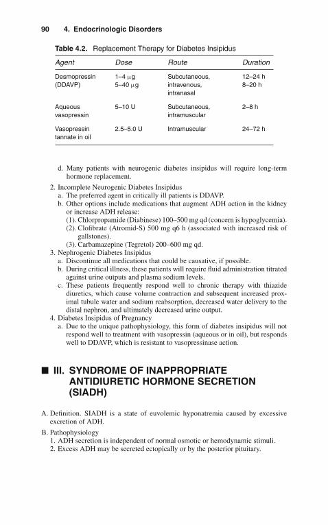
90 4. Endocrinologic Disorders
Table 4.2. Replacement Therapy for Diabetes Insipidus
Agent Dose Route Duration
Desmopressin 1–4 μg Subcutaneous, 12–24 h(DDAVP) 5–40 μg intravenous,
intranasal8–20 h
Aqueousvasopressin
5–10 U Subcutaneous,intramuscular
2–8 h
Vasopressintannate in oil
2.5–5.0 U Intramuscular 24–72 h
d. Many patients with neurogenic diabetes insipidus will require long-termhormone replacement.
2. Incomplete Neurogenic Diabetes Insipidusa. The preferred agent in critically ill patients is DDAVP.b. Other options include medications that augment ADH action in the kidney
or increase ADH release:(1). Chlorpropamide (Diabinese) 100–500 mg qd (concern is hypoglycemia).(2). Clofibrate (Atromid-S) 500 mg q6 h (associated with increased risk of
gallstones).(3). Carbamazepine (Tegretol) 200–600 mg qd.
3. Nephrogenic Diabetes Insipidusa. Discontinue all medications that could be causative, if possible.b. During critical illness, these patients will require fluid administration titrated
against urine outputs and plasma sodium levels.c. These patients frequently respond well to chronic therapy with thiazide
diuretics, which cause volume contraction and subsequent increased prox-imal tubule water and sodium reabsorption, decreased water delivery to thedistal nephron, and ultimately decreased urine output.
4. Diabetes Insipidus of Pregnancya. Due to the unique pathophysiology, this form of diabetes insipidus will not
respond well to treatment with vasopressin (aqueous or in oil), but respondswell to DDAVP, which is resistant to vasopressinase action.
� III. SYNDROME OF INAPPROPRIATEANTIDIURETIC HORMONE SECRETION(SIADH)
A. Definition. SIADH is a state of euvolemic hyponatremia caused by excessiveexcretion of ADH.
B. Pathophysiology1. ADH secretion is independent of normal osmotic or hemodynamic stimuli.2. Excess ADH may be secreted ectopically or by the posterior pituitary.
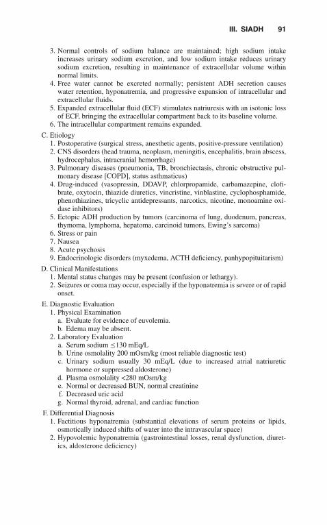
III. SIADH 91
3. Normal controls of sodium balance are maintained; high sodium intakeincreases urinary sodium excretion, and low sodium intake reduces urinarysodium excretion, resulting in maintenance of extracellular volume withinnormal limits.
4. Free water cannot be excreted normally; persistent ADH secretion causeswater retention, hyponatremia, and progressive expansion of intracellular andextracellular fluids.
5. Expanded extracellular fluid (ECF) stimulates natriuresis with an isotonic lossof ECF, bringing the extracellular compartment back to its baseline volume.
6. The intracellular compartment remains expanded.
C. Etiology1. Postoperative (surgical stress, anesthetic agents, positive-pressure ventilation)2. CNS disorders (head trauma, neoplasm, meningitis, encephalitis, brain abscess,
hydrocephalus, intracranial hemorrhage)3. Pulmonary diseases (pneumonia, TB, bronchiectasis, chronic obstructive pul-
monary disease [COPD], status asthmaticus)4. Drug-induced (vasopressin, DDAVP, chlorpropamide, carbamazepine, clofi-
brate, oxytocin, thiazide diuretics, vincristine, vinblastine, cyclophosphamide,phenothiazines, tricyclic antidepressants, narcotics, nicotine, monoamine oxi-dase inhibitors)
5. Ectopic ADH production by tumors (carcinoma of lung, duodenum, pancreas,thymoma, lymphoma, hepatoma, carcinoid tumors, Ewing’s sarcoma)
6. Stress or pain7. Nausea8. Acute psychosis9. Endocrinologic disorders (myxedema, ACTH deficiency, panhypopituitarism)
D. Clinical Manifestations1. Mental status changes may be present (confusion or lethargy).2. Seizures or coma may occur, especially if the hyponatremia is severe or of rapid
onset.
E. Diagnostic Evaluation1. Physical Examination
a. Evaluate for evidence of euvolemia.b. Edema may be absent.
2. Laboratory Evaluationa. Serum sodium ≤130 mEq/Lb. Urine osmolality 200 mOsm/kg (most reliable diagnostic test)c. Urinary sodium usually 30 mEq/L (due to increased atrial natriuretic
hormone or suppressed aldosterone)d. Plasma osmolality <280 mOsm/kge. Normal or decreased BUN, normal creatininef. Decreased uric acidg. Normal thyroid, adrenal, and cardiac function
F. Differential Diagnosis1. Factitious hyponatremia (substantial elevations of serum proteins or lipids,
osmotically induced shifts of water into the intravascular space)2. Hypovolemic hyponatremia (gastrointestinal losses, renal dysfunction, diuret-
ics, aldosterone deficiency)
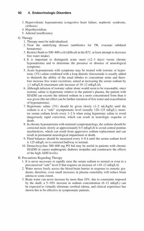
92 4. Endocrinologic Disorders
3. Hypervolemic hyponatremia (congestive heart failure, nephrotic syndrome,cirrhosis)
4. Hypothyroidism5. Adrenal insufficiency
G. Therapy1. Therapy must be individualized.2. Treat the underlying disease (antibiotics for TB, evacuate subdural
hematoma).3. Restrict fluids to 500–800 cc/d (difficult in the ICU, at least attempt to decrease
free water intake).4. It is important to distinguish acute onset (<2–3 days) versus chronic
hyponatremia and to determine the presence or absence of neurologicalsymptoms.
5. Acute hyponatremia with symptoms may be treated with isotonic or hyper-tonic (3%) saline combined with a loop diuretic (furosemide is usually addedto diminish the ability of the renal tubules to concentrate urine and there-fore increase free water excretion), aimed at increasing the serum sodium by≤1 mEq/L/h (maximum safe increase of 10–12 mEq/L/d).
6. Although infusion of isotonic saline alone would seem to be reasonable, sinceisotonic saline is hypertonic relative to the patient’s plasma, the patient withSIADH can excrete the infused sodium in a more concentrated form than itwas given (the net effect can be further retention of free water and exacerbationof hyponatremia).
7. Hypertonic saline (3%) should be given slowly (1–2 mL/kg/h) until thesodium is at a “safe” asymptomatic level (usually 120–125 mEq/L); moni-tor serum sodium levels every 1–2 h when using hypertonic saline to avoiddangerously rapid correction, which can result in neurologic sequelae ordeath.
8. In chronic hyponatremia with minimal symptomatology, the sodium should becorrected more slowly at approximately 0.5 mEq/L/h to avoid central pontinemyelinolysis, which can result from aggressive sodium replacement and canresult in permanent neurological impairment or death.
9. Fluid balances should be measured every 4–8 h until the serum sodium levelis 125 mEq/L or is corrected halfway to normal.
10. Demeclocycline 300–600 mg PO bid may be useful in patients with chronicSIADH (it causes nephrogenic diabetes insipidus and counteracts the effectsof the high ADH levels).
H. Precautions Regarding Therapy1. It is never necessary to rapidly raise the serum sodium to normal or even to a
preconceived “safe” level if that requires an increase of >10–12 mEq/L/d.2. Water moves freely across the blood-brain barrier in response to osmotic gra-
dients; therefore, even small increases in plasma osmolality will reduce brainedema to some extent.
3. Brain water can never increase by more than 10%, due to constraints imposedby the skull; a 5–10% increase in sodium concentration (6–12 mEq/L) canbe expected to virtually eliminate cerebral edema, and clinical experience hasshown this to be effective in symptomatic patients.

IV. DKA and HNKC 93
� IV. DIABETIC KETOACIDOSIS ANDHYPEROSMOLAR NONKETOTIC COMA
A. Definitions1. Diabetic ketoacidosis (DKA) and hyperosmolar nonketotic coma (HNKC)
are hyperglycemic states in patients with diabetes mellitus characterized byinsulin deficiency and relative excess of glucagon and other counterregulatoryhormones.
2. DKA is also characterized by ketosis.
B. Pathophysiology1. Insulin deficiency leads to
a. Increased glucagon, which causes excessive hepatic glucose production(gluconeogenesis/glycogenolysis)
b. Decreased glucose clearance from peripheral tissuesc. Development of hyperglycemia causing an osmotic diuresis (loss of
Na+/H2O), hypovolemia, and decreased glomerular filtration rate (GFR)d. Increased muscle proteolysis and decreased protein synthesis leading to loss
of nitrogen and electrolytes from intracellular fluid (ICF) to ECF to urine.2. In DKA, insulin deficiency also leads to
a. Increased cortisol, epinephrine, and growth hormone, which stimulateexcessive adipose tissue lipolysis and free fatty acid delivery to liverand subsequent ketogenesis and hyperketonemia (beta-hydroxybutyrate andacetoacetate) in DKA
b. Development of an anion gap metabolic acidosis (secondary to ketoacids,which neutralize the bicarbonate buffering system) and loss of K+, Na+,which buffer urinary excretion of ketoacids
3. In HNKC, insulin levels may be sufficient to prevent lipolysis and ketogenesis.
C. Etiology1. Common precipitating events include
a. Infectionb. Acute stress (myocardial infarction, stroke, trauma)c. Discontinuation of insulind. Discontinuation of parenteral nutritione. Use of some medications (glucocorticoids, diphenylhydantoin, propranolol)
2. Rarely, nondiabetics may develop HNKC.a. Iatrogenic (hypertonic intravenous hyperalimentation, hyperosmolar peri-
toneal dialysis)b. Severe fluid losses in burn patientsc. Feeding infants hypertonic oral formulas
D. Diagnostic Evaluation1. History
a. DKA develops in patients of all ages (75% of DKA patients are adults) withinsulin-dependent diabetes mellitus (IDDM) and infrequently with non-insulin-dependent diabetes mellitus (NIDDM), in the setting of coexistingsevere medical problems.
b. HNKC patients are typically elderly with a history of NIDDM.c. Duration of symptoms averages 12 days in HNKC and 3 days in DKA.

94 4. Endocrinologic Disorders
d. Gastrointestinal symptoms are common (abdominal pain ≈50%, nausea orvomiting ≈75%).
e. Seizures are common in HNKC.2. Physical Examination
a. Altered mental status (severely depressed sensorium in HNKC, associatedwith osmolalities of 340–350 mOsm/kg)
b. Dehydrated (dry mucous membranes, orthostatic hypotension, tachycardia)c. Patients with DKA may also have
(1). Fruity breath(2). Hyperventilation (Kussmaul respirations)(3). Abdominal tenderness
3. Laboratory Evaluationa. Table 4.3 lists the common diagnostic laboratory findings in DKA and
HNKCb. DKA may also be associated with
(1). Low serum Na+ (if corrected for hyperglycemia, may be normal orslightly elevated).
(2). Normal to slightly elevated serum K+ (note total body K+ depleted).(3). Leukocytosis (with neutrophilia) may occur secondary to stress or
dehydration instead of infection.(4). Serum amylase is often elevated (even without pancreatitis).
E. Management1. Initial Evaluation
a. Rule out infection as precipitant(1). Complete blood count (CBC), urine analysis (UA), chest x-ray, appro-
priate cultures(2). Altered mental status: Consider lumbar puncture to rule out meningitis(3). Abdominal pain: Consider appendicitis, diverticulitis, pelvic inflamma-
tory diseaseb. Rule out (R/O) myocardial infarction (electrocardiogram) in older patients
or those with long-standing DM (even without complaint of chest pain)2. Institute insulin therapy: 0.1 U/kg regular insulin IV bolus and begin infusion
of regular insulin at rate of 0.1 U/kg/h. (If IV access unobtainable, may use 0.1U/kg IM q1 h.)a. Expect blood glucose to decrease by ~75 mg/dL/h on this regimen.b. Follow blood glucose q1 h.
Table 4.3. Laboratory Values in DKA and HNKC
Lab Test DKA HNKC
Blood glucose (mg/dL) 200–2,000 Usually 600Blood ketones∗ Present AbsentArterial pH <7.4 Normal†
Anion gap Elevated (usually 18) Normal or elevatedOsmolality Slightly elevated ElevatedUrine dipstick Glucose and ketones Glucose
∗ Beta-hydroxybutyrate/acetoacetate.† May be low if hypovolemia causes poor tissue perfusion.
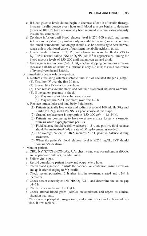
IV. DKA and HNKC 95
c. If blood glucose levels do not begin to decrease after 4 h of insulin therapy,increase insulin dosage every hour until blood glucose begins to decrease(doses of 100 U/h have occasionally been required in a rare, extraordinarilyinsulin-resistant patient).
d. Continue infusion until blood glucose level is 250–300 mg/dL and serumketones are negative (or positive only in undiluted serum) or urine ketonesare “small or moderate”; anion gap should also be decreasing to near-normalrange unless additional cause of persistent metabolic acidosis exists.
e. Lower insulin infusion to 2 U/h, and change intravascular fluid (IVF) toD5 0.45% normal saline (NS) or D5NS (add K+ if appropriate), aiming forblood glucose levels of 150–200 until patient can eat and drink.
f. Give regular insulin dose (5–10 U SQ) before stopping continuous infusion(because half-life of insulin via infusion is only 6–8 min) to avoid recurrenceof hyperglycemia and ketosis.
3. Immediately begin volume repletion.a. Restore circulating volume (isotonic fluid: NS or Lactated Ringer’s [LR]).
(1). First liter IV over the first 30 min.(2). Second liter IV over the next hour.(3). Then reassess volume status and continue as clinical situation warrants.(4). If the patient presents in shock:
(a). May use colloid for volume expansion(b). May require 2–3 L (or more) over first 1–2 h
b. Replace intracellular and total body fluid losses.(1). Patients typically lose water and sodium at around 100 mL H2O/kg and
7 mEq Na+/kg, so 0.45% NS is a good choice at this stage.(2). Gradual replacement is appropriate (150–300 cc/h × 12–24 h).(3). Patients are continuing to have excessive urinary losses via osmotic
diuresis while hyperglycemia persists.(4). Fluid balance should be followed every 1–2 h, and positive fluid balance
should be maintained (adjust rate of IV replacement as needed).(5). The average patient in DKA requires 5–7 L positive balance during
treatment.(6). When the patient’s blood glucose level is ≤250 mg/dL, IVF should
contain 5% dextrose.4. Monitor patient.
a. CBC, Na+/K+/Cl–/HCO3–/Cr, UA, chest x-ray, electrocardiogram (ECG),and appropriate cultures, on admission.
b. Follow vital signs.c. Record cumulative patient intake and output every hour.d. Check blood glucose q1 h while the patient is on continuous insulin infusion
and q4 h after changing to SQ insulin.e. Check serum potassium 2 h after insulin treatment started and q2–4 h
thereafter.f. Check serum electrolytes (Na+/HCO3–/Cl–), and determine the anion gap
q4–6 h.g. Check the serum ketone level q4 h.h. Check arterial blood gases (ABGs) on admission and repeat as clinical
situation warrants.i. Check serum phosphate, magnesium, and ionized calcium levels on admis-
sion. If low, replace.

96 4. Endocrinologic Disorders
j. Check urine dipstick q1 h for presence of glucose/ketones until they arenegative/small.
5. K+ Supplementationa. Serum K+ level decreases as soon as insulin action begins.b. K+ administration should begin when serum K+ ≤5.0 mEq/L and urine
output is documented.c. Goal of K+ supplementation at this point is to keep serum K+ ≥3.5 mEq/L
to help prevent cardiac arrhythmias.d. Patients typically have average total K+ deficit of ~5 mEq/kg (but it can be
much greater).e. Total K+ repletion can be completed gradually (after DKA is resolved).f. If admission K+ is <3.5, immediately add 40 mEq KCl to each liter of IVF
(KCl usually used initially; if phosphate is low, may use KPhos).g. Use IV K+ until DKA is reversed and patient can take potassium orally.
6. Metabolic Acidosisa. Insulin inhibits lipolysis and ketogenesis.b. Fluid replacement restores perfusion.c. Bicarbonate administration has not been shown to accelerate resolution of
acidosis and is generally not advisable. The exception is in severe hyper-kalemia with characteristic ECG changes, i.e., widened QRS or peakedT waves.(1). If needed, use 50–100 mEq NaHCO3 in 500–1,000 cc 0.45% saline over
1–2 h.(2). The main risk of bicarbonate therapy is the induction of severe
hypokalemia as potassium enters cell in exchange for hydrogen ions.7. Phosphate Supplementation
a. Phosphate depletion in DKA averages ~1.0 mmol/kg.b. Prospective randomized studies showed no beneficial effect of phosphate
treatment on recovery from DKA.c. Frequently, phosphate levels plummet to ~1.5 mg/dL with insulin treatment.d. IV replacement is usually not essential.e. However, if phosphorus levels are ≤1.0 mg/dL, then replacement is gener-
ally given (potassium phosphate IV is usually the best choice with concomi-tant hypokalemia), since severe hypophosphatemia may cause respiratoryfailure.
8. Magnesium Supplementationa. Mild magnesium depletion occurs in DKA and also falls with insulin
treatment.b. Typically does not need parenteral supplementation.c. If the patient develops ventricular irritability with hypomagnesemia, mag-
nesium sulfate 1–2 g IV may be given.
F. ComplicationsCerebral edema may develop; its therapy is not clear, but mannitol may be helpful.
� V. TIGHT GLYCEMIC CONTROL IN THE ICU
Recent evidence from randomized clinical trials demonstrates that tight glycemiccontrol in some critically ill patients may improve outcomes. In order to achieve this,

VI. Myxedema 97
the use of intravenous fluids that contain glucose should be avoided. The administra-tion of insulin therapy should be considered, either using sliding scale or continuousdrip. The recommended target blood sugar is around 180 mg/dL. The use of oralhypoglycemic agents should be discontinued to avoid hypoglycemia. Opponents ofthis therapy cite a high incidence of unrecognized hypoglycemic events as a seriousconcern. Close monitoring of the glycemic index, is therefore, necessary.
� VI. MYXEDEMA
A. Definition and Epidemiology1. Myxedema is severe thyroid hormone deficiency, which can lead to a decreased
level of consciousness, even coma.2. It has a reported fatality rate as high as 80%.3. Actual coma associated with severe hypothyroidism is rare (only about 200
cases reported in the literature).4. The incidence of myxedema is three times higher in females than males; elderly
females seem most susceptible to myxedema coma.
B. Pathophysiology1. Thyroid hormone is essential for normal metabolism of all cells2. Thyroid-stimulating hormone (TSH) secreted by the pituitary (under regula-
tion of the hypothalamus) stimulates the thyroid to secrete thyroxine (T4) andsmaller amounts of triiodothyronine (T3), which is the active form of thyroidhormone.
3. Most T3 is produced in the peripheral tissues by monodeiodination ofcirculating T4.
4. T3 and T4 circulate bound to serum proteins; the free T3 and T4 aremetabolically active.
5. T3 feeds back on the pituitary gland to inhibit production of TSH.6. In myxedema coma, the cause of coma is multifactorial (decreased cerebral
perfusion associated with low cardiac output from bradycardia and reducedstroke volume, decreased circulating levels of thyroid hormones resulting indecreased mental responsiveness).
7. Hypothermia may result from decreased T3 or T4, leading to reducedmetabolic rate in addition to an inability to shiver.
8. Hypoventilation (alveolar) is secondary to respiratory center depression (exac-erbated by use of analgesics, sedatives, and anesthesia), defective respiratorymuscle function, and occasionally airway obstruction (enlarged tongue).
9. Hyponatremia often accompanies myxedema and may have associatedhypochloremia (may contribute to altered mental status).
10. Decreased plasma volume and intense peripheral vasoconstriction are typical.
C. Etiology1. Occasionally, myxedema may be the result of chronic, severe primary thyroid
failure; patients with classic signs and symptoms lapse into stupor, coma, anddeath.
2. Myxedema may be precipitated in patients with moderate or unrecognizedhypothyroidism by a superimposed acute illness (infection) or following admin-istration of narcotics or sedatives.
3. See Table 4.4.

98 4. Endocrinologic Disorders
Table 4.4. Causes of Hypothyroidism
1. Autoimmune thyroid disease (Hashimoto’s thyroiditis)2. Thyroid ablation (radioactive iodine or surgical)3. Surgical thyroid resection4. Pituitary disease (secondary hypothyroidism)5. Hypothalamic disease (tertiary hypothyroidism)6. Chemical agents (antithyroid drugs, e.g., lithium, organic goitrogens,
iodine-containing drugs, e.g., amiodarone)7. Congenital thyroid agenesis8. Iodine deficiency or excess9. Thyroid hormone resistance
D. Risk Factors1. Infection2. Surgery3. Anesthesia4. Myocardial infarction5. Sedating drugs6. Cerebrovascular accidents7. Bleeding8. Cold exposure9. Trauma
10. Hyponatremia
E. Symptoms1. Decreased mental acuity2. Hoarseness3. Increased somnolence4. Cold intolerance5. Dry skin6. Brittle hair
F. Diagnostic Evaluation1. History
a. Usually, the patient has a long history of gradual deterioration.b. Gradual weight gain or inability to lose weight.c. May have been noncompliant with thyroid replacement therapy.d. May have history of Graves’ disease or other thyroid dysfunction.
2. Physical Examinationa. Hypothermia (core or rectal temperature <35◦C)b. Bradycardiac. Hypoventilation (slow respiratory rate, shallow breaths)d. Hypotensione. Physical features consistent with long-standing hypothyroidism
(1). Thick and doughy-appearing skin (may have orange or yellow tint)(2). Facial and general puffiness, periorbital edema(3). Large tongue(4). Alopecia, loss of lateral aspects of eyebrows

VI. Myxedema 99
f. Palpable thyroid (present in <50%) or thyroidectomy scarg. Cardiac examination may be consistent with pericardial effusion (e.g.,
muffled heart tones, cardiomegaly)h. Neurologic examination rarely reveals focal findings
(1). Altered level of consciousness(2). Delayed relaxation phase of deep tendon reflexes may be present but
difficult to detect(3). Disorders of muscular function (paralytic ileus, urinary retention, atonic
bowel with fecal impaction)3. Laboratory Evaluation
a. Confirmation of diagnosis relies on thyroid function tests to documenthypothyroidism (measure TSH, T4, free T4, reverse T3, T3RU) (seeTable 4.5).(1). TSH level is elevated in primary hypothyroidism.(2). In secondary and tertiary hypothyroidism, TSH is not elevated,
and diagnosis will rely on other laboratory parameters and clinicaljudgment.
b. Serum cortisol level should be drawn initially to evaluate for concomitantadrenal insufficiency.
c. CBC, UA, blood, and urine cultures should be sent.d. Serum cholesterol is usually elevated.e. Chest x-ray may reveal signs of pleural or pericardial effusion or of
infection.f. ECG is often abnormal (sinus bradycardia, small voltage QRS complexes,
prolonged Q-T intervals, isoelectric T-wave changes, supraventriculartachycardia).
g. ABG may reveal hypoxemia, hypercarbia, and respiratory acidosis.h. Serum glucose or sodium may be low.i. Normochromic, normocytic anemia is typical.
G. Differential Diagnosis1. Other causes of altered mental status (e.g., stroke, electrolyte disturbance such
as hyponatremia)2. Sepsis3. Hypothermia (especially with associated anemia)4. Hypopituitarism5. Hypoglycemia6. Renal failure
H. Therapy1. Initiate thyroid hormone replacement upon suspicion (awaiting confirmation of
the diagnosis may be too late).a. Intravenous administration of replacement hormone is necessary due to
unreliable gastrointestinal absorption in the myxedematous state.b. We prefer T3 and T4 combination therapy (T3 20 μg IV bolus followed by
10 μg q8 h with T4 200 μg IV followed by 100 μg IV q24 h) for 1–2 days,followed by T4 alone.
c. T3 for IV administration recently became available in the United States, somany authors recommend treatment with IV T3 alone.
d. Peripheral conversion of T4 → T3 requires the presence of some T3 forenzyme activity.

100 4. Endocrinologic Disorders
Tab
le4.
5.T
hyro
idF
unct
ion
Test
sin
Thy
roid
Dis
orde
rs
Test
Hyp
othy
roid
Hig
hT
4sy
ndro
me
Hyp
erth
yroi
dLo
wT
3sy
ndro
me
Low
T3/T
4sy
ndro
me
TS
HH
igh∗
Nl/l
owLo
wLo
wto
slLo
wto
sl
↑↑
Tota
lT4
Low
Hig
hH
igh
Nl
Low
Tota
lT3
Low
tolo
wN
lLo
w/N
l/hig
hH
igh
Low
Low
Rev
erse
T3
Nl/l
owN
l/hig
hH
igh
Hig
hH
igh
Free
T4
Low
Nl/h
igh
Hig
hN
lN
l
T3R
ULo
wN
l/low
Hig
hN
l/hig
hH
igh
Abb
revi
atio
ns:N
l,no
rmal
;sl,
slig
ht.
∗ Exc
eptio
ns:T
SH
islo
win
hypo
thyr
oidi
smof
seco
ndar
yan
dte
rtia
ryca
uses
.

VI. Myxedema 101
e. An advantage of IV T3 includes a more rapid onset of action than T4; also,peripheral conversion of T4 is not required for activity.
f. T3 is more arrhythmogenic than T4, and careful cardiac monitoring is essen-tial, especially since the risk of coronary artery disease is high in thesepatients.
g. If IV T3 is not immediately available, IV T4 (as above) may be given withoral T3 (25 μg q12 h) until the patient can be treated with oral T4 alone.
h. Previously, T4 alone was frequently used (200–500 μg IV bolus followed by50–100 μg IV q24 h).
i. Controversy exists regarding the best therapeutic regimen, and controlledtrials will probably never be performed because the disease is so rare.
j. Monitor T3 and T4 levels after 5 days and adjust doses accordingly, if thepatient remains unconscious.
2. Metabolic Supporta. Hypothermia is best treated with passive rewarming (active rewarming can
cause peripheral vasodilatation and worsening of shock) (See Chapter 5,“Environmental Disorders”).
b. Hyponatremia generally responds well to free water restriction.c. Hypoglycemia should be treated with IV dextrose.d. Seizures may be treated with standard anticonvulsant drugs.e. Identify and treat precipitating causes (e.g., infection, stroke, myocardial
infarction, narcotics, gastrointestinal bleeding).3. Supportive Care
a. Respiratory support with mechanical ventilation may be required.b. Hypotension should be treated aggressively with IV fluids (avoid free water)
and vasopressor therapy (dopamine is preferable over norepinephrine, as itmay better maintain coronary blood flow and renal/mesenteric blood flow).
c. Hypotension is poorly responsive to vasopressors until thyroid hormonereplacement is initiated.
d. Hydrocortisone (100 mg IV q8 h) should be given until initial cortisol levelis available (if this indicates a normal stress response to the acute medicalillness, this therapy may be stopped) or for 3–7 days, followed by a rapidtaper in the absence of hypothalamic-pituitary-adrenal disease; this therapymay be life-saving in patients with secondary/tertiary hypothyroidism.
e. Monitor for presence of arrhythmias (decrease dosage of thyroid hormonereplacement if arrhythmias occur); hypothyroidism is associated with a highincidence of coronary artery disease, and these patients should be monitoredfor evidence of myocardial ischemia exacerbated by increased myocardialoxygen consumption with T3/T4 treatment.
f. Avoid sedatives.I. Complications
1. Euthyroid patients tolerate short-term administration of thyroid hormone well.2. Delay of treatment in a myxedematous patient can make the difference between
survival and death.3. This treatment regimen should not routinely be instituted in hypothyroid
patients without clinical evidence of myxedema coma because of potentialcardiac complications (myocardial infarction, arrhythmias).
4. Severe hypothermia (<32◦C) is thought to have prognostic significance.

102 4. Endocrinologic Disorders
� VII. THYROTOXIC CISIS
A. Definitions1. Thyrotoxic crisis or thyroid storm, is a life-threatening complication of hyper-
thyroidism characterized by a severe, sudden exacerbation of thyrotoxicosis.2. The general term thyrotoxicosis refers to the clinical and biochemical manifes-
tations of excess thyroid hormone at the tissue level.3. True hyperthyroidism refers to disorders of thyroid glandular hyperfunction
with increased synthesis and secretion of thyroid hormone.4. There are states of thyrotoxicosis without true hyperthyroidism (e.g., factitious
ingestion of thyroid hormone, chronic thyroiditis with transient thyrotoxicosis,ectopic thyroid hormone production, subacute thyroiditis) that are associatedwith decreased thyroidal synthesis of new hormone; these states are rare inthe critically ill patient with the possible exception of T4 or T3 overdose (seeTable 4.6).
B. Pathophysiology1. The actual mechanisms by which a patient with thyrotoxicosis decompensates
into thyroid crisis are poorly understood.2. A crisis develops most often after a stressful precipitating event (e.g., trauma,
infection, DKA, surgical emergency, parturition, or myocardial infarction).3. Whatever the cause, the resulting syndrome resembles that of prolonged, severe
beta-adrenergic agonist overload.4. Catecholamine levels appear to be normal despite the hypermetabolic
state.
C. Etiology1. Undiagnosed hyperthyroidism (most commonly Graves’s disease or toxic
multinodular goiter) in a patient with major stress.
Table 4.6. Thyrotoxicosis: Etiologies and Differentiation by RAIU
RAIU high RAIU low RAIU low or high
TSH excess (e.g.,pituitary tumors)
Destructive thyroiddisease (e.g.,subacute thyroiditis,postpartum thyroiditis)
Iodine-inducedthyrotoxicosis (e.g.,food or medicationsuch as radiocontrastdye or amiodarone)
Abnormal thyroidstimulators (e.g.,thyroid-stimulatingantibodies—Graves’sdisease)
Ectopic thyroid tissue(e.g., metastaticfollicular carcinoma)
Thyroid autonomy(e.g., toxic multinodulargoiter or toxicadenoma)
Exogenous sources(e.g., medication orfood)
Abbreviation: RAIU, 24-h radioactive iodine uptake study.

VII. Thyrotoxic Cisis 103
2. Other etiologies of thyrotoxicosis may be distinguished by a 24-h radioactiveiodine uptake study when the patient is stable (see Table 4.6).
3. Inadequate therapy in a hyperthyroid patient.D. Symptoms
1. Abnormal mental states (agitation, confusion, psychosis).2. Fever (T 38.3◦C) is almost always present.3. Heat intolerance, diaphoresis.4. Palpitations (sinus tachycardia and atrial fibrillation are the most common
dysrhythmias).5. Gastrointestinal disturbances (diarrhea, nausea, vomiting, abdominal pain).6. Muscle wasting and weakness.7. Dyspnea.
E. Diagnostic Evaluation1. History
a. Marked and rapid recent weight loss may warn of impending storm.b. Unexplained fever in a thyrotoxic patient may precede a storm.c. A precipitating event may be evident.
2. Physical Examinationa. Goiter (palpate cautiously, since vigorous massage may cause release of
more hormone into the circulation)b. Tachycardia—may have tachydysrhythmiasc. Hyperthermiad. Mental status changese. Tremorf. Warm, moist sking. Ophthalmic signs of hyperthyroidism (proptosis, lid lag, lid retraction)h. Signs of congestive heart failure (high-output failure or cardiomyopathy)
3. Laboratory Evaluationa. Confirmation of the diagnosis relies on thyroid studies (see Tables 4.5
and 4.6).(1). Elevated T4(2). Elevated T3(3). Decreased TSH
b. Routine studies include CBC, electrolytes, urinalysis, chest x-ray, and ECG.c. Evaluate for infection as indicated by history and physical examination.d. Associated laboratory abnormalities..
(1). Hypercalcemia(2). Hypokalemia(3). Hyperglycemia(4). Hypocholesterolemia(5). Mild microcytic anemia(6). Lymphocytosis(7). Granulocytopenia(8). Hyperbilirubinemia(9). Elevated alkaline phosphatase
F. Differential Diagnosis1. Hypermetabolic states (sepsis, pheochromocytoma, Cushing’s syndrome)2. Thyrotoxicosis without crisis/storm

104 4. Endocrinologic Disorders
G. Therapy. Do not wait for lab values to begin treatment; diagnosis is made onclinical suspicion, and treatment should be initiated immediately.1. Supportive Measures
a. IV fluids for volume replacement.b. Acetaminophen for hyperthermia. Avoid aspirin, since it displaces T4 from
thyroid-binding globulin, thereby increasing the level of free T4.c. Cooling blankets, hydrogel pads.
2. Inhibition of Thyroid Hormone Synthesisa. Propylthiouracil (PTU) (200–300 mg oral or via nasogastric tube q6 h)b. Methimazole (20–25 mg oral or via nasogastric tube q4 h)
3. Inhibition of Thyroid Hormone Release With Iodide Therapy (beginning 1 hafter PTU therapy)a. Sodium iodide (1 g IV q8 h)b. Potassium iodide and iodine—Lugol’s solution (10 drops q8 h orally)
4. Inhibition of Peripheral Beta-Adrenergic Activitya. Most beta-blockers also block peripheral conversion of T4 to T3.b. Propranolol (Inderal) 0.5–1.0 mg/min IV to total dose of 2–10 mg IV q3–4 h;
may treat with 20–40 mg PO q6 h after initial control with IV administra-tion. An occasional patient has required up to 2 g/d PO due to variability ofhepatic metabolism in thyrotoxic individuals.
c. Esmolol (Brevibloc) 0.5- to 1.0-mg/kg bolus followed by infusion with50 μg/kg/min; if inadequate effect within 5 min, repeat bolus and increaseinfusion to 100 μg/kg/min; can repeat procedure, to 200–300 μg/kg/min.
d. Titrate beta-blockade to achieve heart rate of 80 beats per minute.e. If the patient has a history of reactive airway disease, use caution and a
short-acting cardioselective agent such as esmolol.f. Caution is also required in patients with CHF. Controlling the heart rate may
be of benefit; however, these agents are negative inotropes and may worsenlow-output failure.
g. If beta-blockers are contraindicated, other sympatholytic drugs (reserpine, adepleter of catecholamines, or guanethidine, an inhibitor of catecholaminerelease) may be useful as second-line agents.
5. Inhibition of Peripheral Conversion of T4 to T3a. PTU (see above dosages).b. Beta-blockade (see above).c. Dexamethasone 1–2 mg IV or PO q6 h.
6. Definitive therapy may require surgery or radioactive iodine therapy, afteretiology determined by RAIU.
7. Diagnose and treat underlying precipitating disorders (i.e., infection, othermajor stresses).
� VIII. SICK ETHYROID SYNDROME
A. Definitions1. Sick euthyroid syndrome is characterized by thyroid hormone alterations
associated with acute non-thyroidal illness.2. Two syndromes are described:

VIII. Sick Ethyroid Syndrome 105
a. Low T3 syndromeb. Low T3/T4 syndrome
3. These syndromes are thought to represent euthyroid states by many; however,specific tissues may actually be hypothyroid.
B. Review of Normal Thyroid Hormone Physiology1. Thyrotropin-releasing hormone (TRH) is released from the hypothalamus into
the blood stream.2. TRH stimulates synthesis and release of thyrotropin (TSH) from the pituitary
gland.3. TSH stimulates the thyroid gland to produce and secrete thyroxine (T4) and
smaller amounts of triiodothyronine (T3).4. The thyroid gland secretes predominantly T4 (80%), with smaller amounts of
T3 (20%).5. The remainder of T3 (the physiologically active form of thyroid hormone)
is produced in extrathyroidal tissues (primarily, the liver and kidneys) bymonodeiodination of circulating T4.
6. T3 and T4 circulate bound to serum proteins; the free T3 and T4 aremetabolically active.
7. T3 feeds back on the pituitary gland to inhibit production of TSH.8. Some of the circulating T4 is metabolized to the inactive product reverse T3.9. Both T3 and reverse T3 are rapidly cleared from the serum by further
deiodination.10. Thyroid hormone activity begins with binding of T3 to receptors on cell nuclei.11. Post-binding effects of T3 are needed for normal cellular function.12. The T3 resin-uptake test is commonly performed to give an approximation of
binding proteins (primarily thyroxine-binding globulin).
C. Pathophysiology1. In acute non-thyroidal illness, peripheral thyroid hormone metabolism is
altered.2. The exact mechanism of decreased T3 production is unknown.
D. Etiology1. Systemic illness (sepsis, cardiac or respiratory failure, neoplastic processes,
stroke)2. Surgery3. Caloric deprivation4. Drug-induced (glucocorticoids, iodides, amiodarone, propylthiouracil)
E. SymptomsNo specific symptoms are associated with the thyroidal hormone alterations.
F. Laboratory Evaluation1. Serum T3 is low (see Table 4.5).2. Serum T4 is low or normal.3. Reverse T3 is elevated (usually this is the most useful discriminator between
the sick euthyroid syndrome and hypothyroidism).4. Serum TSH is normal.
G. Therapy1. Attempts should be made to distinguish sick euthyroid syndrome from hypothy-
roidism.2. Currently, there is no proven benefit in using thyroid hormone to treat patients
with sick euthyroid syndrome.

106 4. Endocrinologic Disorders
3. Indeed, some researchers believe that the sick euthyroid state may be protectiveby conserving energy under stress.
� IX. HYPOGLYCEMIA
A. Definition. Hypoglycemia is defined by plasma glucose <40–50 mg/dL. This def-inition does not include the presence of associated symptoms, since critically illpatients may not reliably demonstrate classic symptomatology.
B. Pathophysiology1. Clinical situations that result in increased insulin, inability of the liver to gen-
erate glucose from its glycogen stores, or problems with the counterregulatorysystem may lead to hypoglycemia.
2. Insulin suppresses hepatic glucose production and stimulates glucose utilizationby peripheral tissues such as muscle.
3. Insulin secretion lowers plasma glucose concentration.4. In response to onset of hypoglycemia, the major counterregulatory hormones
glucagon and epinephrine increase and cause an acceleration of glycogenolysis.5. Catecholamines are glucose-counterregulatory hormones but do not play essen-
tial roles as long as secretion of glucagon is initiated; however, in the presenceof glucagon deficiency (long-standing diabetes mellitus or patients with totalpancreatectomy), catecholamines become major counterregulatory hormones.
6. Patients treated with nonselective beta-adrenergic-blocking agents may havesevere impairment of counterregulation mechanisms.
7. Patients with long-standing DM (10–15 years) may lose the ability to secreteepinephrine in response to hypoglycemia (becoming virtually defenselessagainst even moderate degrees of hyperinsulinemia).
C. EtiologySee Table 4.7.
D. Symptoms1. Adrenergic activation
a. Palpitationsb. Tremorc. Diaphoresisd. Pallore. Anxiety
2. Neuroglycopeniaa. Fatigueb. Faintnessc. Dizzinessd. Hungere. Inappropriate behaviorf. Visual symptomsg. Focal neurologic symptomsh. Seizuresi. Coma
E. Diagnostic Evaluation1. History
a. Check for history of DM.

IX. Hypoglycemia 107
Table 4.7. Causes of Hypoglycemia
1. Hyperinsulin statesa. Exogenous insulin administration∗b. Endogenous insulin excess (e.g., insulinomas)
2. Ethanol-induced (after ingestion in otherwise healthy patients or chronicalcoholics)∗
3. Drug-induced (e.g., sulfonylureas, quinine, propranolol, pentamidine)∗
4. Systemic disorders∗a. Hepatic disease (e.g., cirrhosis, fulminant viral hepatitis)b. Renal disease (e.g., chronic renal disease associated with
liver disease, CHF, sepsis)c. Sepsis (e.g., gram-negative sepsis, empyema of gallbladder)d. AIDS
5. Extensive thermal burns∗
6. Total parenteral nutrition∗
7. Insulin treatment of hyperkalemia∗
8. Factitious (insulin injection or sulfonylurea ingestion)∗
9. Endocrine causes (hypopituitarism, hypoadrenalism, hypothyroidism)
10. Autoimmune causes (insulin antireceptor antibodies, anti-insulinautoantibodies)
11. Starvation (severe caloric restriction)
12. Alimentary—following gastric surgery (e.g., gastrectomy)
13. Idiopathic (functional)—typically postprandial; this is a diagnosis ofexclusion and is usually not seen in the critically ill patient
∗ Causes likely to be seen in the critically ill.
(1). Recent insulin or oral hypoglycemic therapy?(2). If DM is long-standing, the patient may also have glucagon defi-
ciency and therefore be at higher risk for hypoglycemia, since thecounterregulatory mechanisms may also be ineffective.
b. Check for history of alcohol ingestion.2. Physical Examination
a. Tachycardiab. Pupillary dilationc. Cold, moist skind. Changes in body temperature (hypothermia, hyperthermia)e. Between hypoglycemic episodes, the examination may be normal.
3. Laboratory Evaluationa. Plasma glucose level <40–50 mg/dL. Whole blood glucose is usually about
15% less than the corresponding plasma glucose level.

108 4. Endocrinologic Disorders
b. The patient may have changes in blood counts.(1). Acute lymphocytosis is followed later by neutrophilia.(2). May have increased hemoglobin, total red blood cell (RBC) count, or
packed RBC volume.c. ECG changes (ST depression, flat T waves, QT-interval prolongation).d. Electroencephalogram (EEG) changes (diminished frequency of alpha
waves, increased delta waves).e. Artifactual hypoglycemia must be ruled out, especially if laboratory results
indicate hypoglycemia with no apparent cause.(1). Samples in serum separator tubes left at room temperature for extended
periods of time (blood glucose levels may decrease by 10–20 mg/dL/hdue to ongoing blood cell metabolism).
(2). Patients with increased numbers of blood cells (polycythemia vera,leukemia, leukemoid reactions) may have low measured plasma glucoselevels secondary to increased metabolism.
(3). These problems may be avoided by collecting blood in tubes contain-ing oxalate and fluoride (gray tubes), since fluoride acts as a glycolyticenzyme poison.
4. Diagnostic Approacha. Measure simultaneous blood glucose and plasma insulin levels during an
episode of hypoglycemia. The best way is to demonstrate insulin secretioninappropriate to the prevailing blood glucose concentration.(1). Relative hyperinsulinemia can be demonstrated by the simultaneous
determination of blood glucose and plasma insulin levels after anovernight fast or during a 24- to 72-h fast.
(2). Diagnosis of hyperinsulinism can further be supported by elevatedlevels of plasma C-peptide and proinsulin concentrations.
(3). Insulinomas can be localized by ultrasonography, CT scanning,magnetic resonance imaging (MRI), arteriography, and transhep-atic percutaneous venous sampling, and intraoperative high-frequencysonography.
b. Factitious hypoglycemia resulting from administration of insulin or sul-fonylurea agents is typically characterized by inappropriately high plasmainsulin levels (similar to insulinomas).(1). C-peptide levels remain low in insulin-induced factitious hypo-
glycemia; the presence of insulin antibodies in patients who have noreason to take insulin injections also suggests this etiology.
(2). C-peptide levels are elevated in sulfonylurea-induced factitious hypo-glycemia (similar to insulinomas); screening for plasma or urinesulfonylureas may confirm the diagnosis.
c. In spontaneous hypoglycemia, alcohol ingestion should be ruled out as acause (alcohol levels may not be helpful, since hypoglycemia may not occurfor as long as 36 h after ingestion).
d. Associated systemic disorders must be ruled out as appropriate for eachpatient (evaluate for liver, renal, or endocrine dysfunction, or sepsis).
F. Therapy1. If the patient is comatose, glucose should be administered intravenously (25–
50 cc of 50% dextrose followed by infusion of 10% dextrose) until persistentor mild hyperglycemia is present.
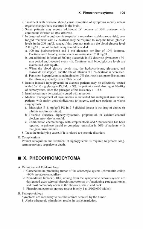
X. Pheochromocytoma 109
2. Treatment with dextrose should cause resolution of symptoms rapidly unlessorganic changes have occurred in the brain.
3. Some patients may require additional IV boluses of 50% dextrose withcontinuous infusion of 10% dextrose.
4. In drug-induced hypoglycemia (especially secondary to chlorpropamide), pro-longed treatment with IV dextrose may be required to keep the blood glucoselevels in the 200 mg/dL range; if this does not maintain the blood glucose level200 mg/dL, one of the following should be added:a. 100 mg hydrocortisone and 1 mg glucagon per liter of 10% dextrose.
Continue until blood glucose levels are maintained 200 mg/dL.b. An additional infusion of 300 mg diazoxide in 5% dextrose given over a 30-
min period and repeated every 4 h. Continue until blood glucose levels aremaintained 200 mg/dL.
c. When the blood glucose levels rise, the hydrocortisone, glucagon, anddiazoxide are stopped, and the rate of infusion of 10% dextrose is decreased.
d. Persistent hyperglycemia maintained on 5% dextrose is a sign to discontinuethe infusion gradually over a 24-h period.
5. Insulin-induced hypoglycemia in diabetic patients may be effectively treatedwith 0.5–1.0 mg glucagon IV, IM, or SQ; the patient should also ingest 20–40 gof carbohydrate, since the glucagon effect lasts only 1–1.5 h.
6. Insulinomas may be surgically cured with resection.7. Medical management of insulinomas is indicated for malignant insulinoma,
patients with major contraindications to surgery, and rare patients in whomsurgery fails.a. Diazoxide (3–8 mg/kg/d PO in 2–3 divided doses) is the drug of choice (it
inhibits insulin secretion).b. Thiazide diuretics, diphenylhydantoin, propranolol, or calcium-channel
blockers may also be useful.c. Combination chemotherapy with streptozotocin and 5-fluorouracil has been
reported to achieve partial or complete remission in 60% of patients withmalignant insulinomas.
8. Treat the underlying cause, if it is related to systemic disorders.
G. ComplicationsPrompt recognition and treatment of hypoglycemia is required to prevent long-term neurologic sequelae or death.
� X. PHEOCHROMOCYTOMA
A. Definition and Epidemiology1. Catecholamine-producing tumor of the adrenergic system (chromaffin cells);
~90% are adrenomedullary.2. Non-adrenal tumors (~10%) arising from the sympathetic nervous system are
designated extra-adrenal pheochromocytomas or functioning paragangliomasand most commonly occur in the abdomen, chest, and neck.
3. Pheochromocytomas are rare (occur in only 1 to 2/100,000 adults).
B. PathophysiologySymptoms are secondary to catecholamines secreted by the tumor:1. Alpha-adrenergic stimulation results in vasoconstriction.
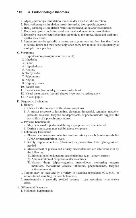
110 4. Endocrinologic Disorders
2. Alpha2-adrenergic stimulation results in decreased insulin secretion.3. Beta1-adrenergic stimulation results in cardiac inotropy/chronotropy.4. Beta2-adrenergic stimulation results in bronchodilation and vasodilation.5. Dopa1-receptor stimulation results in renal and mesenteric vasodilation.6. Excessive levels of catecholamines are toxic to the myocardium and cardiomy-
opathy may result.7. Symptoms may be episodic in nature; paroxysms may last from less than 1 min
to several hours and may occur only once every few months or as frequently asmultiple times per day.
C. Symptoms1. Hypertension (paroxysmal or persistent)2. Headache3. Pallor4. Hyperhidrosis5. Anxiety6. Tachycardia7. Palpitations8. Angina9. Hyperglycemia
10. Weight loss11. Paresthesias (second-degree vasoconstriction)12. Visual disturbances (second-degree hypertensive retinopathy)13. Dilated pupils
D. Diagnostic Evaluation1. History
a. Check for the presence of the above symptoms.b. A pressor response to histamine, glucagon, droperidol, tyramine, metoclo-
pramide, saralasin, tricyclic antidepressants, or phenothiazides suggests thepossibility of a pheochromocytoma.
2. Physical Examinationa. May be normal if performed during a symptom-free time interval.b. During a paroxysm, may exhibit above symptoms.
3. Laboratory Evaluationa. Plasma or urinary catecholamine levels or urinary catecholamine metabolite
(VMA or metanephrine) levels.b. Rarely, suppression tests (clonidine) or provocative tests (glucagon) are
needed.c. Measurement of plasma and urinary catecholamines are interfered with by
the following:(1). Stimulation of endogenous catecholamines (e.g., surgery, stroke)(2). Administration of exogenous catecholamines(3). Various drugs (alpha2-agonists, methyldopa, converting enzyme
inhibitors, monoamine oxidase inhibitors, phenothiazines, tricyclicantidepressants)
4. Tumors may be localized by a variety of scanning techniques (CT, MRI, orvenous blood sampling for catecholamines).
5. Arteriography is generally avoided because it can precipitate hypertensivecrisis.
E. Differential Diagnosis1. Malignant hypertension

X. Pheochromocytoma 111
2. Thyrotoxic crisis3. Hypertensive response (to stress, surgery, anesthesia)4. Cushing’s syndrome
F. Therapy1. Surgical excision is the definitive treatment.2. Preoperative management is important in determining surgical outcome.3. Preoperative goals:
a. Control blood pressureb. Provide adequate intravascular volumec. Treat tachyarrhythmiasd. Treat heart failuree. Treat glucose intolerance
4. Alpha-adrenergic blockade is the basis of hypertensive therapy, and patientsare generally treated 1–2 weeks before surgery with phenoxybenzamine (orlabetalol or prazosin) that is tapered to produce a 10- to 15-mmHg orthostaticblood pressure decrease; 1–2 weeks of pretreatment allows for intravascularvolume repletion (diminished by excess endogenous catecholamines).
5. Tachycardia may occur from unopposed beta-adrenergic receptor activation,and if the heart rate is 110 beats per minute, a beta-blocker should be added;beta-blockers should not be used before institution of alpha-adrenergic block-ade. Elimination of vasodilatory effects of beta-receptors result in unopposedalpha-adrenergic vasoconstriction and may provoke hypertensive crisis, andthe negative inotropic effects of beta-blockers may precipitate heart failure inthe face of hypertension.
6. Reflex tachycardia is less with prazosin or labetalol.7. Alpha-methylparatyrosine (an inhibitor of tyrosine hydroxylase, the rate-
limiting enzyme in catecholamine production) may be used in patients whocannot tolerate alpha-adrenergic blockade (postural hypotension) and can beespecially useful in patients with cardiomyopathy. Indeed, phenoxybenzamineand alpha-methylparatyrosine in combination result in less tachycardia andless postoperative hypotension.
8. Dysrhythmias are treated with standard antidysrhythmic agents.9. Blood should be available for transfusion in the perioperative period, since
pheochromocytomas are vascular tumors and bleeding is frequent.10. Diuretics should be used sparingly, since intravascular volume is already
constricted by catecholamines; conversely, over-administration of fluids canworsen heart failure.
11. Perioperatively, drugs that cause catecholamine release or potentiate cate-cholamine action are to be avoided (e.g., morphine can cause histamine-induced catecholamine release).
12. Postoperatively, patient’s need cardiovascular and metabolic monitoring.13. Hypoglycemia may occur following tumor removal (loss of catecholamine-
induced hyperglycemia).14. Pain is best treated with meperidine, and benzodiazepines are useful for
sedation.15. Recurrent hypertension should suggest the possibility of residual tumor.
5Environmental Disorders
� I. BURNS
It is estimated that 2 million people are burned annually in the United States. Ofthem, over 130,000 are children under the age of 15. Although most of these burnsare minor, approximately 3–5% of burn injuries are life threatening.
A. Pathophysiology1. Partial-thickness burns involve heat damage to the epidermis or a portion of
the dermis. The dermis contains sensory nerve endings, vascular supply, hairfollicles, and sweat glands.
2. Full-thickness burns involve injury to tissue deep to the sweat glands and hairfollicles. The presence of thrombosed blood vessels or charring is characteristicof full-thickness burns. However, final determination of burn depth may not bepossible for several days following the injury.
3. Shock may develop due to transudation and sequestration of fluid in the burnedareas and elsewhere in the body. Cardiac output may drop in major burns, dueto myocardial dysfunction.
B. Predisposition1. Burn injury is the second leading cause of death among children under age 12
years.2. Children are more likely to suffer burns than adults because of their greater
vulnerability to accidents due to inability to recognize or react to hazardoussituations. Half of all victims of hot water burns are children under age 5 years.
C. Clinical Presentation1. Partial-Thickness Burns
a. First-degree burn involves only the epidermis; there is blanching erythemabut no bullae formation.
b. Second-degree burn involves a portion of the dermis and produces edemaand fluid exudation. Bullae formation is characteristic. These developquickly after burn injury. Consider infection if bullae appear 18 h or laterafter a burn occurs.
2. Full-Thickness Burnsa. Third-Degree Burn. Surface is dry and inelastic. Skin surface may become
white or gray. This burn will not regenerate from unburned edges.
J. Varon, P. Acosta, Handbook of Critical and Intensive Care Medicine,DOI 10.1007/978-0-387-92851-7_5,C© Springer Science+Business Media, LLC 2010
114 5. Environmental Disorders
b. Fourth-Degree Burn: Extends beyond the depth of the skin to involveunderlying muscle, tendons, vascular structures, periosteum, or bone.
3. Survival depends on the extent and depth of the burn, the age of the patient,and associated injuries.
4. Vascular effects in burned skin are immediate vasoconstriction followed byincreased capillary permeability and plasma extravasation. Burned skin alsopermits increased insensible water loss.
D. Complications1. Coagulation necrosis at the burn site produces an advantageous setting for bac-
terial growth. Infection is one of the most important causes of death in severeburn injury.
2. Gastric dilatation and adynamic ileus occur in major burns.3. Acute hemolysis may occur due to heat damage of red blood cells.4. Acute renal failure may occur as a result of shock.5. Hypertension may be present in burn victims, especially children.6. Multiorgan failure is the leading cause of death in patients with burns.7. Manipulation of burn wounds has been shown to result in bacteremia in 20%
of cases.
E. Treatment1. Evaluate airway (see smoke inhalation, below) and perform endotracheal intu-
bation, if indicated by upper airway edema or deterioration of arterial bloodgases. Endotracheal intubation is appropriate for any patients with acute burninjury who display respiratory distress.
2. Establish intravenous access.3. Evaluate burned areas under sterile technique.4. Assess for presence of other injuries, especially with burns associated with
explosions.5. Insert nasogastric tube (to treat ileus) and urinary catheter to monitor urine
output.6. Obtain baseline laboratory values: complete blood count (CBC), electrolytes,
serum urea nitrogen (BUN), creatinine, glucose, arterial blood gases, andcarboxyhemoglobin level.
7. Estimate extent of burned area.a. Rule of nine for body surface area (BSA)
(1). Adults: arms 9% each; legs 18% each; head 9%; trunk 18% anterior,18% posterior; genitalia 1%
(2). Children: arms 9% each; legs 16% each; head 13%; trunk 18% anterior,18% posterior; genitalia 1%
(3). Infants: arms 9% each; legs 14% each; head 18%; trunk 18% anterior,18% posterior; genitalia 1%
b. Lund and Browder chart(1). More accurate in children
8. Classify severity of burn.a. Major burns
(1). Partial-thickness burns >20% BSA in children or the elderly or 25%BSA in adults
(2). Full thickness burns >10% BSA(3). Burns involving the face, hands, feet, or perineum that may produce
functional or cosmetic impairment(4). Caustic chemical burns

II. Decompression Illness and Air Embolism 115
(5). High-voltage electrical injury (see below)(6). Burns complicated by inhalation injury (see below) or major trauma
b. Moderate burns(1). Partial-thickness burns of 10–20% BSA in children or elderly or 15–
25% BSA in adults(2). Full-thickness burns <10% BSA
9. Fluid resuscitation.a. Within the first 24 h, administer isotonic balanced crystalloid solution
according to the recommended formula:(1). Multiple formulas for fluid administration exist, which recommend 1–4
mL/kg/% burn to be administered within the first 24 h.(2). One recommendation (the Parkland formula) is to administer 4 mL of
crystalloid solution per kilogram per percent BSA in the first 24 h oftreatment. Administer half this quantity within the first 8 h followingthe burn, one fourth during the second 8 h, and the remaining one fourthin the last 8 h.
b. May administer colloid-containing solution as needed after 24 h, at least12.5 g of albumin for every liter of crystalloid administered.
c. Monitor urine output. The patient should have at least 50 mL/h in the adult(or 1 mL/kg ideal body weight) and 1 mL/kg/h in the child.
d. Inhalation injury may significantly increase fluid requirements.10. Topical care
a. Initially, cover burned areas with dry sterile sheets.b. Clean burned areas with water and mild soap (Dial or Ivory), and remove
particulate matter from burn.c. Debride any overtly necrotic skin.d. Apply topical agents, such as silver sulfadiazine or mafenide acetate.e. Apply biologic dressings and synthetic skin substitutes to achieve temporary
wound closure.11. Nutrition
a. Early enteral feeding may attenuate the hypermetabolic response by preserv-ing the intestinal mucosal barrier.
b. High-protein diets appear preferable to conventional diets.12. Escharotomy
a. Circumferential burns producing a constricting eschar may result in respira-tory (thorax) or circulatory (extremities) impairment.
b. Escharotomy may be necessary:(1). Chest: diminished respiratory excursion, hypoxemia, diminished tidal
volume.(2). Extremities: diminished pulse and deterioration of circulation distal to
the burn.c. Fasciotomy may be necessary with high-voltage electrical burns or with
associated crush injury.
� II. DECOMPRESSION ILLNESS AND AIREMBOLISM
The occurrence of decompression illness has paralleled the increased popularity ofsport diving and may also occur in commercial divers and tunnel workers.

116 5. Environmental Disorders
A. Pathophysiology1. Decompression illness occurs when gas dissolved in body fluids separates to
form bubbles as ambient pressure decreases. Nitrogen accumulates in tissueduring a dive, the amount being dependent on the dive’s depth and dura-tion. When a diver ascends too rapidly and ambient pressure decreases, thereis insufficient time for nitrogen to equilibrate, and gas bubbles form in tis-sue and the venous circulation. Symptoms of decompression illness thenoccur.
2. Flying shortly after diving increases the risk of decompression illness, asdecreased ambient pressure at high altitude promotes gas bubble formation.
B. Predisposition1. Patients who are older or obese have a higher incidence of decompression
illness.2. Prior joint injury predisposes to development of decompression illness in that
joint.3. Air embolism tends to occur more frequently in less-experienced divers.
C. Clinical Presentation1. Decompression Illness
a. The vast majority of patients exhibit symptoms immediately followingdiving, though the onset may be delayed by 12–36 h.
b. The clinical manifestations of decompression sickness (DCS) can be dividedinto two major groups:(1). Type I DCS: pain only, usually involving the joints.(2). Type II DCS: involvement of the central nervous system (CNS). This
type accounts for 10–25% of instances in the United States.c. The most frequent presentation (two-thirds to three-quarters of cases)
involves joint pain.(1). Knees, shoulders, and elbows are most commonly involved.(2). There may be slight tenderness or edema, but the severity of pain is out
of proportion to objective clinical findings.d. Cutaneous decompression illness produces pruritus, erythematous eruption,
or mottling.e. Pulmonary involvement.
(1). Symptoms include pleuritic substernal chest pain, dyspnea, and cough.(2). Physical examination is usually unremarkable.
f. Circulatory collapse and death may result from serious decompressionillness.
g. Spinal cord decompression illness produces back pain, paresthesias, weak-ness, or paralysis due to obstruction of epidural vertebral veins.
h. Cerebral decompression illness may present with headache, confusion, hallu-cinations, delirium, seizures, visual disturbance, or cranial nerve involvement(especially, for unknown reasons, cranial nerve VIII, producing vomiting,vertigo, tinnitus, and nystagmus).
2. Air Embolisma. Cerebral air embolism is an important cause of death in divers.b. Focal neurologic deficit occurring within 15–20 min of ascent is the most
common presentation. Consider air embolism if immediate loss of con-sciousness occurs upon ascent.
c. Mediastinal emphysema, subcutaneous emphysema, or pneumothorax maybe associated with cerebral air embolism.

III. Near-Drowning 117
d. Coronary embolization can produce acute myocardial infarction or cardiacarrest.
D. Complications1. Persistent neurologic defect may result from CNS decompression illness.2. Osteonecrosis may be a late consequence of bone involvement, but this is less
commonly seen now than previously.
E. Treatment1. For air embolism, maintain patient in left lateral decubitus Trendelenburg
position.2. Apply 100% oxygen by mask or via endotracheal intubation.3. Administer normal saline intravenously as required.4. Treat seizures and cardiac dysrhythmias in standard fashion.5. Treatment of all patients with decompression illness and air embolism is with
recompression in a hyperbaric chamber. Recompression may be beneficialfor decompression illness even if treatment is delayed several days. Majorcomplications of hyperbaric oxygen therapy:a. Barotrauma (e.g., pneumothorax, sinus trauma)b. Oxygen toxicityc. Psychiatric (e.g., claustrophobia)
6. For the location of the closest hyperbaric chamber, contact the Divers AlertNetwork (DAN) at +1-919-684-4DAN (4326)
� III. NEAR-DROWNING
Drowning accounts for 6,000–8,000 deaths each year in the United States. It rep-resents the third most common cause of accidental death. Children under age 4comprise 40% of these fatalities. The peak incidence is during warm-weather months.Drowning is defined as suffocation in a liquid medium. Near-drowning is defined asat least temporary survival from such submersion.
A. Pathophysiology1. Near-drowning may be accompanied by aspiration of fluids or may be pri-
marily the consequence of asphyxia due to laryngospasm. Aspiration of largequantities of seawater produces hypovolemia and concentration of extracel-lular electrolytes, whereas substantial fresh water aspiration produces acutehypervolemia with dilution of extracellular electrolytes.
Although the early medical literature emphasized pathophysiologic dif-ferences between fresh and salt water drowning, aspiration of significantquantities of fluid required to produce the hemodilution or electrolyte changesdescribed does not occur in most human survivors. At least 85% of survivorsof drowning are thought to aspirate <22 mL of water per kilogram of bodyweight.
2. The adverse respiratory effects of near-drowning include the following:a. Intrapulmonary shuntingb. Ventilation-perfusion mismatchc. Decreased complianced. Intra-alveolar influx of fluid due to capillary-alveolar leake. Hypoxemia resulting from all of the above

118 5. Environmental Disorders
3. Hypothermia associated with immersion may predispose patients to cardiacdysrhythmias and exhaustion.
B. Predisposition1. Age distribution peaks of near-drowning victims.
a. Children <4 years.b. Males aged 14–24 years.c. Risk-taking behavior people.
2. Alcohol intoxication is related to a large proportion of adult drowning.
C. Clinical Presentation1. Respiratory findings usually predominate and include shortness of breath, air
hunger, and tachypnea.a. Auscultatory findings may include rales, rhonchi, or wheezes.b. Respiratory deterioration may occur insidiously.
2. Changes in mental status may be due to cerebral hypoxemia or head injury.3. Cardiac dysrhythmias (premature beats, bradycardia, atrial fibrillation, asys-
tole, ventricular fibrillation) result from hypoxemia or hypothermia.4. Consider the presence of hypothermia or trauma, especially cervical spine
injury in appropriate settings.
D. Complications1. Cerebral end-organ damage due to hypoxemia mediated through cerebral
edema and increased intracerebral pressure.2. Renal failure may be produced by acute tubular necrosis, hemoglobinuria, or
myoglobinuria.3. Bronchial hyperreactivity may follow near-drowning.4. Hypoglycemia is common.
E. Admission Criteria1. Need for assisted ventilation2. Coma or obtundation3. Cardiac arrest or dysrhythmias4. Hypothermia
F. Treatment1. Clear airway and ensure adequate respirations.
a. Apply 100% oxygen by mask to patients with spontaneous respirations.b. Perform endotracheal intubation and mechanical ventilation in apnea and
as directed by hypoxemia or hypercarbia or for protection of airwayin coma.
c. Initiate positive end-expiratory pressure (PEEP) at 3–5 cmH2O in mechan-ically ventilated patients. Increase as required to maintain arterial O2saturation (SaO2) ≥90%.
d. Monitor arterial saturation in all patients.e. Administer bronchodilators in standard fashion in patients with bronchocon-
striction.2. Treat cardiac dysrhythmias as indicated. Obtain an electrocardiogram (ECG)
on all patients with near-drowning.3. Monitor body temperature. If the patient is a victim of cold water
near-drowning consider slow rewarming due to the protective effects ofhypothermia.
4. Obtain chest radiograph at presentation and repeat as indicated.5. Treat circulatory shock, if present.
IV. Electrical Injuries 119
6. Consider bronchoscopy in patients who have aspirated particulate matter orcontaminated water.
G. Prognosis1. Classification of the victim’s neurological status allows systematic assessment
of prognosis.a. Category A: awake, fully consciousb. Category B: stuporous but arousablec. Category C: comatose
2. Eighty percent of near-drowning victims recover without sequelae, 2–9%survive with brain damage, and 12% die.
� IV. ELECTRICAL INJURIES
Approximately 3% of burn injuries that require hospital admission are the result ofelectrical injury. Of these, approximately 40% are fatal.
A. Pathophysiology1. Injuries are classified as high voltage (>1,000 volts) or low voltage (<1,000
volts). High voltages are generally more severe injuries, but the degree of injuryalso depends on tissue resistance and type of current.
2. Lightning produces current with many millions of volts, but exposure may beextremely brief.
3. Electrical energy causes injury through release of heat within tissues.4. The severity of tissue damage is directly related to duration of contact.
B. Predisposition1. There is a high incidence of electrical injury among children <6 years; most are
related to injury by electrical outlets and cords.2. Two-thirds of injuries among adults (mostly males) involve electrical and
construction workers.
C. Clinical Presentation1. Wounds occurring at sites where electricity enters and exits the body are typi-
cally small areas of full-thickness burn. There may be considerable underlyingtissue damage that is poorly reflected by the extent of visible skin damage.
2. Musculoskeletal injury results from tetanic contraction of skeletal muscle duringexposure. Fractures or dislocations may occur. Heat injury may produce bonyor periosteal destruction.
3. Electrical injury may produce vascular endothelial disruption, hemorrhage,arterial or venous thrombosis, or peripheral ischemia.
4. Cardiac dysrhythmias are a frequent cause of immediate death. Dysrhythmiasmay continue to appear within 24 h of injury.a. Sinus tachycardia, supraventricular tachycardia, ventricular tachycardia,
atrial fibrillation, atrioventricular (A–V) block, and intraventricular conduc-tion delay may occur.
b. Myocardial injury resulting from high-voltage injury is uncommon but mayoccur.
c. The most common ECG alterations are nonspecific ST-T-wave changes andsinus tachycardia.

120 5. Environmental Disorders
5. Gastrointestinal tract injuries include intestinal perforation and stress ulcerformation with gastrointestinal hemorrhage.
6. Signs of CNS injury include coma, confusion, disorientation, and seizures.Central respiratory center depression may occur.
D. Complications1. Renal failure may result from rhabdomyolysis.2. Delayed or long-term motor paralysis may be caused by injury to the central or
peripheral nervous system. Other long-term neural deficits include personalitychanges, memory or concentration impairment, and depression.
3. Cataracts may develop 6–12 months following electrical injury to the head.Other ophthalmic sequelae include corneal ulceration and retinal or optic nervedamage.
E. Admission CriteriaICU admission criteria include1. Thermal injury ≥20% BSA2. Suspicion of inhalation injury3. History of loss of consciousness4. Cardiac dysrhythmias5. Cardiopulmonary arrest6. Rhabdomyolysis
F. Treatment1. Clear the airway and support respiration, if necessary.2. Administer intravenous fluids as required to maintain a urine output of at least
50–100 mL/h or 1 mL/kg ideal body weight/h. Traditional burn formulas forfluid replacement are not applicable to electrical burns.
3. Monitor the cardiac rhythm.4. Obtain CBC, electrolytes, BUN, creatinine, prothrombin time (PT), par-
tial thromboplastin time (PTT), myoglobin, creatine kinase (CK), urinalysis,12-lead ECG.
5. If myoglobinuria appears, treat with mannitol and sodium bicarbonate (seeChapter 14, “Renal and Fluid-Electrolyte Disorders”).
6. Obtain surgical consultation for debridement of deep tissue, as necessary.Technetium scanning may identify areas of muscular injury.
7. Observe for development of sepsis, especially with Pseudomonas or Clostridiumspp. (in instances of myonecrosis). Antibiotics should be given prophylactically,as should tetanus prophylaxis.
� V. HEAT EXHAUSTION AND HEAT STROKE
Approximately 5,000 deaths due to heat-related illness occur annually in the UnitedStates. Mortality rate of heat stroke may be 30–80%. Heat exhaustion and heat strokedo not represent distinct pathophysiologic entities but rather constitute heat-inducedillnesses of varying severity.
A. Pathophysiology. Radiation of heat from the body accounts for approximately65% of cooling, provided that the air temperature is lower than body tempera-ture. This mechanism becomes less effective as ambient temperature approaches35◦C. Sweating with the ensuing evaporation produces approximately 30% of
V. Heat Exhaustion and Heat Stroke 121
cooling. As ambient temperature rises, this becomes the primary method of cool-ing. When air temperature exceeds body temperature, heat gain by radiation ispossible. As relative humidity rises, evaporative heat loss diminishes. Heat illnessis exacerbated by excessive fluid loss and electrolyte depletion.Physiologic responses to increased body heat:1. Cutaneous vasodilatation2. Increased cardiac output to maintain blood pressure3. Splanchnic vasoconstriction4. Increased sweat volume
B. Predisposition1. Neonates, who have poor thermoregulatory capability2. The elderly, especially those with cardiac disease3. Obese individuals4. Underlying illnesses: congestive heart failure, coronary artery disease, hyper-
thyroidism, dermatologic disorders, major burns5. Medications and drugs: beta-adrenergic blockers, phenothiazines, lithium,
cyclic antidepressants, antihistamines, amphetamines, cocaine, phencyclidine
C. Clinical Presentation1. Heat Exhaustion
a. Temperature elevation, but temperature is generally <39◦C.b. Symptoms: nausea, vomiting, headache, light-headedness, malaise, muscular
cramping.c. Diaphoresis, tachycardia, hypotension, or orthostatic hypotension.d. Mental status is unimpaired.
2. Heat Strokea. Temperature is generally >40.5◦C.b. Signs: tachypnea (respiratory rate as high as 60/min), tachycardia, hypo
tension.c. Dry, hot skin is classic, but not necessary to make the diagnosis. Lack of
sweating may be a late finding, and diaphoretic skin is seen in about half ofcases.
d. Major diagnostic point: CNS dysfunction (confusion, bizarre behavior,delirium, obtundation, coma, seizures). Ataxia may result from cerebel-lar involvement. Most CNS deficits are reversible with treatment but maybecome fixed.
D. Complications1. Heat Stroke
a. Muscular injury causing rhabdomyolysis.b. Hypoglycemia.c. Hypocalcemia (does not usually require specific treatment).d. Renal failure resulting from acute tubular necrosis or rhabdomyolysis (see
Chapter 14, “Renal and Fluid-Electrolyte Disorders”).e. Hepatocellular injury producing elevation of liver enzymes.f. Disseminated intravascular coagulation (DIC) may occur 1–3 days following
onset of heat stroke.g. Adult respiratory distress syndrome (see Chapter 13, Pulmonary Disorders).h. Neurologic: ataxia, dementia, cerebral edema, brain death.
E. Treatment1. Heat Exhaustion

122 5. Environmental Disorders
a. Place patient in a cool environment.b. Fluid replacement with normal saline or half-normal saline; amount to be
guided by vital signs. Invasive cardiovascular monitoring may be required,especially in older patients.
2. Heat Strokea. Maintain airway and breathing. Administer intravenous fluids, as directed by
vital signs.b. Maintain continuous core temperature monitoring with rectal, vaginal, or
esophageal probe.c. Initiate immediate vigorous cooling by one or more of the following mea-
sures:(1). Evaporative technique: Spray patient with warm water (15◦C) and cool
with fan. This method prevents cutaneous vasoconstriction and does notinduce shivering. Requires low-humidity environment.
(2). Ice packs to neck, axilla, and groin. Produces cooling in area of majorvessels and is useful as an adjunct to other techniques.
(3). Ice water bath: May cause cutaneous vasoconstriction. It is also difficultto monitor an immersed patient.
(4). Iced gastric lavage.(5). Cool (6–10◦C) peritoneal lavage.(6). Intermittent positive-pressure breathing (IPPB) with cold inhaled air.(7). Cardiopulmonary bypass (this method produces the most rapid cooling).
d. Do not administer antipyretics (e.g., salicylates or acetaminophen).e. Administer D5-half-normal saline to replace fluid loss initially while awaiting
electrolyte results.f. Monitor closely blood pressure, urine output, and urine for myoglobin.g. Follow oxygen saturation and chest radiograph to determine the development
of pulmonary complications, coagulation studies for DIC, liver enzymes,creatine kinase, BUN, creatinine.
h. May require paralysis and mechanical ventilation (especially if associatedwith amphetamine use).
i. If shivering occurs with cooling, may administer chlorpromazine (25–50 mgIV). However, this must be done with care, as chlorpromazine may producecardiac dysrhythmias, hypotension, or a neuroleptic malignant syndrome,which in itself produces hyperthermia.
j. Dantrolene may be used, 1 mg/kg IV (may be increased to as high as10 mg/kg). The mechanism appears to involve calcium-release inhibition inskeletal muscle. Muscular weakness may be produced. Further research intothe use of this agent in heat stroke is required.
� VI. HYPOTHERMIA
Normal core temperature is 36–37.5◦C. Thermoregulation is controlled in thehypothalamus. Shivering is initiated in the anterior hypothalamus and is the body’smost effective method to raise temperature. Hypothermia may be divided into mild(body temperature 33–35◦C), moderate (27–32◦C), and severe (<27◦C) forms. It isseen in a variety of circumstances.
Primary (accidental) hypothermia is caused by exposure to low environmentaltemperature. Secondary hypothermia occurs when heat conservation mechanisms

VI. Hypothermia 123
are abnormal due to underlying disease. Induced hypothermia has been used as atherapeutic measure in certain neurosurgical and cardiovascular surgical procedures.
A. Pathophysiology. In secondary hypothermia, there may be interference with thehypothalamic temperature-regulating center, or an inability to shiver, redistributeblood flow or move from a cold environment.
B. Predisposition1. Infants and the elderly are most susceptible.
a. Infants: large surface area to mass ratio, inability to protect selves from coolenvironment
b. Elderly: decreased sensory temperature appreciation, deficient centrallymediated response to cold, failure to shiver, limited ability to vasoconstrictperipheral vessels
2. Drug use and intoxication:a. Agents that depress the level of consciousness (especially ethanol) are often
related to exposure-related hypothermia.b. Barbiturates depress core temperature by central effect.c. Phenothiazines inhibit response to cold by alpha-adrenergic blocking activ-
ity and by direct suppression of the thermoregulatory center.
3. Preexisting illness: see Table 5.1.
C. Clinical Presentation1. Initial sympathetic response (producing peripheral vasoconstriction and tachy-
cardia) causes transient elevation of blood pressure and cardiac output.a. Increased renal perfusion produces increased urine production (“cold diure-
sis”), which may lead to hypovolemia, hemoconcentration, and increasedblood viscosity.
b. Response is ablated below 30◦C.
Table 5.1. Illness Predisposing to Hypothermia
Pathologic reduction of metabolic rateHypothyroidismHypopituitarismHypoadrenalism
Alteration of hypothalamic functionAnorexia nervosaHypothalamic tumorsHead traumaCerebrovascular accidentWernicke’s encephalopathySarcoidosis (involving the hypothalamus)
Other mechanismsHypoglycemiaSpinal cord transectionExtensive body surface burnsExfoliative erythrodermasSepsisSevere protein malnutrition

124 5. Environmental Disorders
2. Clouding of sensorium regularly occurs at 30–32◦C.3. Mild hypothermia: Tachypnea, tachycardia, shivering, amnesia, ataxia,
dysarthria.4. Moderate hypothermia: Decreased level of consciousness, mydriasis, atrial
fibrillation, bradycardia.5. Severe hypothermia: Absent reflexes and response to pain, coma, hypotension.6. Cardiac complications:
a. Dysrhythmias: atrial fibrillation is common.b. Patients are increasingly susceptible to ventricular fibrillation with
diminishing temperature. Below 28◦C, even minor procedures,such as moving or repositioning the patient, may initiate ventricularfibrillation.
c. Ventricular fibrillation is resistant to pharmacologic treatment or electricaldefibrillation until temperature has increased.
d. Susceptibility to ventricular dysrhythmias may persist for several days, evenafter normal body temperature has been restored.
e. ECG changes include PR, QT, and QRS prolongation, and J-point elevation(“Osborn wave”).
D. Complications1. Morbidity and mortality are related to the degree and duration of hypothermia
and to appropriate therapy.2. Bronchorrhea and depressed cough reflexes lead to bronchopneumonia and
aspiration pneumonitis.3. Punctate hemorrhages may occur in the gastrointestinal tract, but significant
bleeding is rare.4. Pancreatitis is common.5. Fatal disseminated intravascular coagulation may occur.
E. Treatment1. Cardiovascular
a. Employ basic life support protocols to support ventilation and circulation.b. Perform endotracheal intubation and mechanical ventilation as indicated.c. Initiate cardiopulmonary resuscitation (CPR), if cardiac arrest is present.
Continue until core temperature of 32–35◦C is reached or responseachieved.
d. Ventricular fibrillation may be resistant to defibrillation in severe hypother-mia. Attempt single defibrillation; if this is not successful, continue rewarm-ing until temperature >30◦C and administer bretylium 5–10 mg/kg IV over5–10 min, then infusion of 2 mg/min.
2. Rewarminga. For mild and moderate hypothermia (including most cases of secondary
hypothermia), passive rewarming is indicated.(1). Remove patient from cold environment.(2). Apply dry, unheated blankets.
b. For some cases of moderate hypothermia, use active surface rewarming.(1). Heating blankets, hot-water bottles, heat cradles, heat-fluidized beds,
warm-water immersion.(2). Moderate hypothermia may be associated with afterdrop of core temper-
ature as peripheral vasoconstriction is reversed, producing hypovolemicshock and ventricular fibrillation.
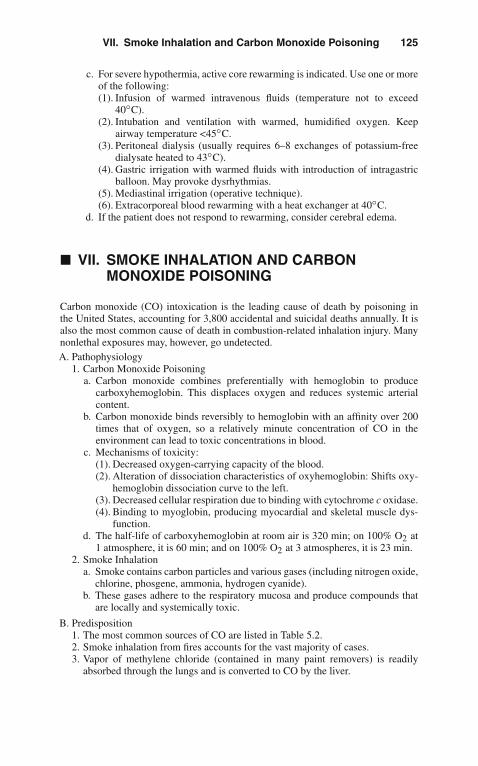
VII. Smoke Inhalation and Carbon Monoxide Poisoning 125
c. For severe hypothermia, active core rewarming is indicated. Use one or moreof the following:(1). Infusion of warmed intravenous fluids (temperature not to exceed
40◦C).(2). Intubation and ventilation with warmed, humidified oxygen. Keep
airway temperature <45◦C.(3). Peritoneal dialysis (usually requires 6–8 exchanges of potassium-free
dialysate heated to 43◦C).(4). Gastric irrigation with warmed fluids with introduction of intragastric
balloon. May provoke dysrhythmias.(5). Mediastinal irrigation (operative technique).(6). Extracorporeal blood rewarming with a heat exchanger at 40◦C.
d. If the patient does not respond to rewarming, consider cerebral edema.
� VII. SMOKE INHALATION AND CARBONMONOXIDE POISONING
Carbon monoxide (CO) intoxication is the leading cause of death by poisoning inthe United States, accounting for 3,800 accidental and suicidal deaths annually. It isalso the most common cause of death in combustion-related inhalation injury. Manynonlethal exposures may, however, go undetected.A. Pathophysiology
1. Carbon Monoxide Poisoninga. Carbon monoxide combines preferentially with hemoglobin to produce
carboxyhemoglobin. This displaces oxygen and reduces systemic arterialcontent.
b. Carbon monoxide binds reversibly to hemoglobin with an affinity over 200times that of oxygen, so a relatively minute concentration of CO in theenvironment can lead to toxic concentrations in blood.
c. Mechanisms of toxicity:(1). Decreased oxygen-carrying capacity of the blood.(2). Alteration of dissociation characteristics of oxyhemoglobin: Shifts oxy-
hemoglobin dissociation curve to the left.(3). Decreased cellular respiration due to binding with cytochrome c oxidase.(4). Binding to myoglobin, producing myocardial and skeletal muscle dys-
function.d. The half-life of carboxyhemoglobin at room air is 320 min; on 100% O2 at
1 atmosphere, it is 60 min; and on 100% O2 at 3 atmospheres, it is 23 min.2. Smoke Inhalation
a. Smoke contains carbon particles and various gases (including nitrogen oxide,chlorine, phosgene, ammonia, hydrogen cyanide).
b. These gases adhere to the respiratory mucosa and produce compounds thatare locally and systemically toxic.
B. Predisposition1. The most common sources of CO are listed in Table 5.2.2. Smoke inhalation from fires accounts for the vast majority of cases.3. Vapor of methylene chloride (contained in many paint removers) is readily
absorbed through the lungs and is converted to CO by the liver.

126 5. Environmental Disorders
Table 5.2. Exogenous Sources of Carbon Monoxide
Smoke from all types of firesPaint-remover containing methylene chlorideFurnacesGasoline-powered enginesSwimming pool heatersSterno fuelTobacco smokeVehicular exhaust fumesWater heaters
Table 5.3. Acute CO Poisoning
COHb level Symptoms
10% Headache20% Dizziness, nausea, dyspnea30% Visual disturbances40% Confusion, syncope50% Seizures, coma>60% Cardiopulmonary failure and death
C. Clinical Presentation1. Carbon Monoxide Poisoning
a. Symptoms and signs in acute CO poisoning depend on carboxyhemoglobin(COHb) level (see Table 5.3).(1). Patients with COHb levels <10% are usually asymptomatic.(2). Patients with levels >25% should be considered for hospital admission.(3). Those with levels >50% commonly have coma and seizures resulting
from cerebral edema.(4). Death is likely if levels >60%.(5). Determination of treatment only on COHb level is inappropriate, though,
because the level alone is a poor predictor of degree of injury. Therefore,clinical assessment is vital to planning treatment.
b. Chronic or subacute poisoning may present with less characteristicsymptoms.
2. Smoke InhalationSuspect ifa. Exposure occurred in a closed space.b. There are facial or pharyngeal burns or burned nasal vibrissae.c. There is carbonaceous sputum or hoarseness.
D. Complications1. Neuropsychiatric problems, such as memory loss, personality changes, mutism,
and parkinsonism may occur as long-term sequelae to CO poisoning.2. Indicators of a poor prognosis include altered level of consciousness, advanced
age, metabolic acidosis, and structural abnormalities on computed tomography(CT) or magnetic resonance (MR) scanning.

VIII. Scorpion Envenomation 127
E. Treatment1. Carbon Monoxide Poisoning
a. Obtain the COHb level in all patients suspected of having CO exposure (e.g.,all victims at a fire scene).
b. Apply supplemental oxygen 100% by mask while awaiting results.(1). The goal is to improve blood oxygen content by maximizing the fraction
dissolved in plasma.(2). Monitor patients with serial COHb levels. Continue 100% oxygen until
the COHb level is <5%. Continue to monitor COHb level thereafter toguard against undetected release of CO from tissue sites.
c. Obtain an ECG and monitor with serial CK levels if the ECG is suggestiveof ischemia.
d. Consider treatment with hyperbaric oxygen (HBO) in the following patients:(1). Patients with serious CO poisoning (e.g., coma, neurologic deficit,
cardiac or hemodynamic instability, history of loss of consciousness)(2). Pregnant women(3). Patients with underlying heart disease or abnormal ECG
e. Whether HBO alters outcome is still controversial and some authors haverecommended its use up to 24 h following the acute exposure.
f. If HBO treatment is unavailable, consider intubation and mechanical venti-lation with administration of 100% oxygen, if clinical findings merit.
g. Consider transfusion with packed red blood cells in severe poisoning.2. Smoke Inhalation
a. If findings of smoke inhalation are present, evaluate the airway with fiberop-tic bronchoscopy.
b. Obtain arterial blood gases and chest radiograph.c. Administer humidified oxygen.d. Perform early endotracheal intubation in the presence of upper airway
edema.
� VIII. SCORPION ENVENOMATION
Scorpions are arachnids that typically inhabit temperate climates. The only dangerousspecies of scorpion in the United States is the Centruroides genus, which is foundprimarily in the southwestern states (Arizona, California, New Mexico, Texas).
A. Pathophysiology. The venom is complex and contains a number of proteins,polypeptides, and enzymes (e.g., hyaluronidases, phospholipases), and neurotox-ins. The venom affects sodium channels to prolong action potentials and pro-duce spontaneous neural depolarizations of the sympathetic and parasympatheticsystems.
B. Predisposition. Most serious envenomations occur in children.
C. Clinical Presentation1. The most common finding is severe local pain at the site of the sting. In most
cases, this resolves within several hours. There is severe tenderness to palpationor percussion over the site.

128 5. Environmental Disorders
2. The sting may be accompanied by wheal and flare reaction, paresthesias at thesite of sting.
3. Significant envenomation produces tachycardia, hypertension, restlessness,hyperexcitability, diaphoresis, piloerection, nystagmus, diplopia, opisthotonos,muscular fasciculations, or hypersalivation.
4. The most severe cases produce seizures, pulmonary edema, muscular paralysis,respiratory arrest, cardiovascular collapse, and death.
D. Complications. These include DI, pancreatitis, jaundice, and renal failure.
E. Treatment1. Maintain airway and assist ventilations, if necessary.2. Clean the wound and administer tetanus prophylaxis if indicated. Do not cool
or incise wound.3. Classify Centruroides sting by severity of involvement, according to Table 5.4.
a. Grade I and II envenomation: Wound care as above and provide oralanalgesics.
b. Grade III and IV: An antivenin has been produced but is not generallyavailable. Contact your regional poison control center. Sedate and providesupportive care. High doses of phenobarbital have been recommended, butcare must be taken to prevent respiratory failure.
c. Treat severe tachycardia with beta-blockers.
F. Prognosis1. Though deaths may occur, none has been reported in the United States since
1968.2. If untreated, the sting may be lethal in 1% of adults and as many as 25% of
children under age 5 years.
� IX. SNAKEBITE
Approximately 8,000 instances of poisonous snakebite occur annually in the UnitedStates, with 9–15 deaths resulting. Most bites occur in the southern and southwesternstates, with a peak incidence during the summer months.
Table 5.4. Severity of Centruroides Scorpion Envenomation
Grade Signs and symptoms
I Local pain or paresthesias at site; tenderness to touch orPercussion
II Local findings and pain and paresthesias remote from sting site
III Symptomatic skeletal neuromuscular dysfunction (muscularjerking or shaking) or cranial nerve dysfunction (e.g., blurredvision, difficulty swallowing, hypersalivation, slurred speech,tongue fasciculations)
IV Somatic skeletal and cranial nerve dysfunction
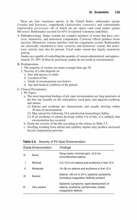
IX. Snakebite 129
There are four venomous species in the United States: rattlesnakes (genusCrotalus and Sistrurus), copperheads (Agkistrodon contortrix), and cottonmouths(Agkistrodon piscivorus)—all of which are pit vipers—and coral snakes (genusMicrurus). Rattlesnakes account for 65% of reported venomous snakebites.A. Pathophysiology. Snake venoms are complex mixtures of toxins that have cyto-
toxic, hemotoxic, and neurotoxic components. Cytotoxic effects produce tissuenecrosis. Hemotoxic venoms interfere with the coagulation system. Rattlesnakesare classically considered to have cytotoxic and hemotoxic venom, but neuro-toxic activity may also be present. Coral snake venom has largely neurotoxicactivity.Snakes are capable of controlling the quantity of venom administered, and approx-imately 25–30% of bites by poisonous snakes do not result in envenomation.
B. Predisposition1. The majority of victims are males younger than age 20.2. Severity of a bite depends on
a. Size and species of snakeb. Location of bitec. Grade of envenomation (see below)d. Age and medical condition of the patient
C. Clinical Presentation1. Pit Vipers
a. The most important findings of pit viper envenomation are fang punctures atthe bite site (usually on the extremities), local pain, and adjacent erythemaand edema.(1). Edema and erythema are characteristic and usually develop within
30 min of envenomation.(2). May spread for following 24 h and develop hemorrhagic bullae.(3). If no erythema or edema develops within 4 h of bite, it is unlikely that
envenomation has occurred.b. Grade the severity of the bite according to the criteria in Table 5.5.c. Swelling resulting from edema and capillary rupture may produce increased
fascial compartment pressure.
Table 5.5. Severity of Pit Viper Envenomation
Grade Envenomation Findings
O NoneFang marks; minimal pain; <2.5 cmcircumferential edema
I Minimal 2.5–12.5 cm edema and erythema in first 12 h
II Moderate 15–36 cm edema and erythema in first 12 h
III SevereEdema >36 cm in 24 h; systemic symptoms(including coagulation defects) present
IV Very severeSystemic symptoms; rapid development ofedema, erythema; ecchymoses, bullae;coagulation defects
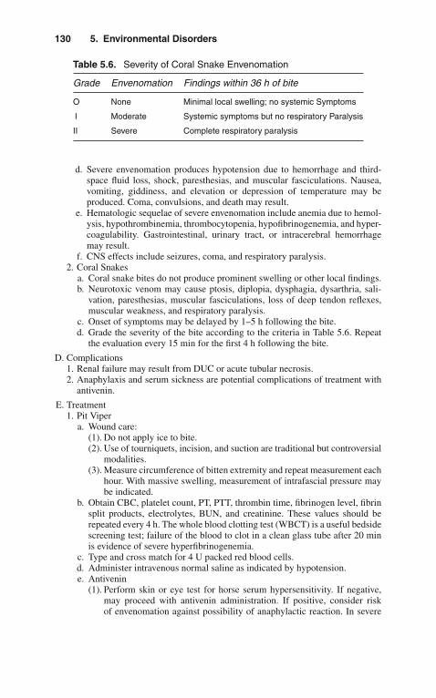
130 5. Environmental Disorders
Table 5.6. Severity of Coral Snake Envenomation
Grade Envenomation Findings within 36 h of bite
O None Minimal local swelling; no systemic Symptoms
I Moderate Systemic symptoms but no respiratory Paralysis
II Severe Complete respiratory paralysis
d. Severe envenomation produces hypotension due to hemorrhage and third-space fluid loss, shock, paresthesias, and muscular fasciculations. Nausea,vomiting, giddiness, and elevation or depression of temperature may beproduced. Coma, convulsions, and death may result.
e. Hematologic sequelae of severe envenomation include anemia due to hemol-ysis, hypothrombinemia, thrombocytopenia, hypofibrinogenemia, and hyper-coagulability. Gastrointestinal, urinary tract, or intracerebral hemorrhagemay result.
f. CNS effects include seizures, coma, and respiratory paralysis.2. Coral Snakes
a. Coral snake bites do not produce prominent swelling or other local findings.b. Neurotoxic venom may cause ptosis, diplopia, dysphagia, dysarthria, sali-
vation, paresthesias, muscular fasciculations, loss of deep tendon reflexes,muscular weakness, and respiratory paralysis.
c. Onset of symptoms may be delayed by 1–5 h following the bite.d. Grade the severity of the bite according to the criteria in Table 5.6. Repeat
the evaluation every 15 min for the first 4 h following the bite.
D. Complications1. Renal failure may result from DUC or acute tubular necrosis.2. Anaphylaxis and serum sickness are potential complications of treatment with
antivenin.
E. Treatment1. Pit Viper
a. Wound care:(1). Do not apply ice to bite.(2). Use of tourniquets, incision, and suction are traditional but controversial
modalities.(3). Measure circumference of bitten extremity and repeat measurement each
hour. With massive swelling, measurement of intrafascial pressure maybe indicated.
b. Obtain CBC, platelet count, PT, PTT, thrombin time, fibrinogen level, fibrinsplit products, electrolytes, BUN, and creatinine. These values should berepeated every 4 h. The whole blood clotting test (WBCT) is a useful bedsidescreening test; failure of the blood to clot in a clean glass tube after 20 minis evidence of severe hyperfibrinogenemia.
c. Type and cross match for 4 U packed red blood cells.d. Administer intravenous normal saline as indicated by hypotension.e. Antivenin
(1). Perform skin or eye test for horse serum hypersensitivity. If negative,may proceed with antivenin administration. If positive, consider riskof envenomation against possibility of anaphylactic reaction. In severe

X. Spider Bite 131
envenomation, may precede antivenin infusion with 250 mg methylpred-nisolone IV. Be prepared to treat anaphylaxis (see Chapter 18, “Allergicand Immunologic Emergencies”).
(2). No universally accepted standard recommendations for antivenin admin-istration exist. Controversy about its use exists.
(3). No antivenin is required for grades 0 or I pit viper bites.(4). Administer to grades II–IV bites. Test dose: 0.2–0.5 mL IV over 5 min; if
no adverse effect, administer remainder over 30 min to 2 h. Individualizedosage to severity and rate of progression: grade II, up to 5 vials; gradeIII, 5–15 vials; grade IV, 15–20 vials.
(5). Observe the patient for 3–5 h after initial administration; administer anadditional vial every 1–2 h if pain persists or swelling progresses.
f. Use of fasciotomy in severe envenomation with increased intrafascialpressure is controversial. This may be necessary if intrafascial pressure>30 torr. Obtain surgical consultation as required in instances of massiveedema.
g. The routine use of antibiotics is controversial and depends, in part, upon thelocal rates of infection.
2. Coral Snakesa. An antivenin is available for bites of Eastern coral snakes. Administer
antivenin to all bite victims of this snake, even before appearance ofsymptoms. Dosage is 3–6 vials in 300–500 mL normal saline.
b. There is no antivenin available for bites of the Arizona coral snake.c. Perform endotracheal intubation if there are any signs of bulbar paralysis,
such as diplopia or dysphagia.d. Institute respiratory support with mechanical ventilation as required.
Antivenin may not completely reverse respiratory depression.
� X. SPIDER BITE
The spiders that can cause medically significant bites include widow and falsewidow spiders (worldwide), recluse spiders (mostly North and South America),Australian funnel web spiders (eastern coastal Australia), and Phoneutria spiders(Brazil).
A. Black Widow Spider. The black widow spider is a member of the Latrodectusgenus and is found throughout the United States. The female, larger and moredangerous than the male, is black or brown, with a characteristic red hourglassmarking on the ventral abdomen.1. Pathophysiology
Black widow venom is extremely potent. The major activity of the venom liesin provoking the release of catecholamines at adrenergic synaptic terminals anddepletion of acetylcholine from motor nerve endings.
2. PredispositionThere is a high risk for mortality among the following patients:a. Patients younger than 16 or older than 65 years.b. Those with hypertension or cardiovascular disease (envenomation may cause
heart failure, cerebrovascular accident, or myocardial ischemia).

132 5. Environmental Disorders
3. Clinical Presentationa. Pain at the bite site may be minimal, but severe pain may develop in the
extremity, along with painful lymphadenopathy, erythema, swelling, andpiloerection 20–60 min following the bite.
b. Cramping and painful muscular contractions in the back, thigh, abdomen,and chest produce abdominal rigidity, tonic contractions, and tremor.
c. There may be restlessness, weakness, dizziness, urinary retention, diaphore-sis, salivation, nausea, vomiting, priapism, and hypertension.
d. Distinction must be made from the acute abdomen: despite rigidity, there isminimal tenderness and no rebound tenderness.
4. Complicationsa. Hypertensive crisisb. Paralysis and respiratory arrest (especially in children)c. Severe envenomation may produce shock and/or coma
5. Treatmenta. Clean wound and apply cool compresses (not ice) to the bite site.b. Administer diazepam (5–10 mg IV q3 h as needed) and methocarbamol
(1000 mg IV no faster than 100 mg/min and 1,000 mg additionally as IVinfusion) for symptomatic treatment of muscular spasm.
c. Calcium gluconate 10% (1–2 mL/kg IV over 20 min, maximum 10 mL) mayalso provide relief of muscular spasm, but this is transient, lasting <30 min.
d. Treat elevated blood pressure (see Chapter 3, “Cardiovascular Disorders”).e. Antivenin
(1). Administration of antivenin is recommended for symptomatic patients<16 or >65 years old, those with preexisting hypertension or cardio-vascular disease, and those displaying respiratory distress, pronouncedhypertension, or persistent severe muscular symptoms despite abovetreatment.
(2). Before administration, skin test for horse serum sensitivity (includedwith antivenin).
(3). Administer 1 vial antivenin diluted in 50 mL normal saline over 15–30 min. A second vial will be required only in severe cases.
6. Prognosisa. Prognosis for recovery with treatment is good and deaths are rare.b. There is a 0.5% incidence of anaphylaxis and a 2% incidence of serum
sickness associated with antivenin use.
B. Brown Recluse Spider. The Brown Recluse spider is of the Loxosceles speciesand is present throughout the southern United States. It is tan or brown, with aviolin-shaped mark on the dorsum of the cephalothorax.1. Pathophysiology
The venom contains a variety of enzymes, including hyaluronidase, protease,collagenase, and sphingomyelinase D, which are thought to be responsible fordermal necrosis and hemolysis.
2. PredispositionMost severe envenomations occur in children.
3. Clinical Presentationa. Local pain and burning, bulla formation at site of bite, becoming necrotic
over hours to days.b. Systemic signs and symptoms: fever, chills, petechiae, nausea, vomiting, and
weakness.

XI. Useful Facts and Formulas 133
c. Severe cases may produce hemolysis, disseminated intravascular coagula-tion, thrombocytopenia, jaundice, or shock.
4. Complicationsa. Severe hemolysis may result in death.b. Renal failure may result from hemoglobinuria or myoglobinuria.
5. Treatmenta. There is no commercially available antivenin available in the United States.b. Begin intravenous infusion with normal saline.c. Obtain CBC, platelet count, electrolytes, BUN, creatinine, PT, PTT, and
urinalysis.d. Clean the wound and administer tetanus prophylaxis, if indicated. Do not
cool or incise wound.e. Some recommend the use of dapsone (50–200 mg/d), a leukocyte inhibitor,
which relieves pain and reduces erythema and induration. Do not administerto children.
f. Obtain surgical consultation for wound care.
� XI. USEFUL FACTS AND FORMULAS
A. Temperature. Temperature conversion calculations are often done in the man-agement of critically ill patients. Degrees Celsius (◦C) and Fahrenheit (◦F) aremost commonly utilized:
◦Cto◦F◦F = (◦C × 9/5) + 32◦Fto◦C◦C = (◦F − 32) × 5/9
Occasionally, the Kelvin (K) temperature scale is used, primarily in gas lawcalculations:
Kto◦CK =◦ C + 273
B. Humidity. Relative humidity (RH) is usually measured by hygrometers, thus,eliminating the need of extracting and measuring the humidity content of the airsamples:
RH = Content[mg/L or mmHg]
Capacity[mg/L or mmHg]= %
The humidity deficit (HD) represents the maximum humidity capacity at bodytemperature:
HD = Capacity − content = mg/L
where capacity = the amount of water the alveolar air can hold at body temper-ature (also known as absolute humidity) and content = the humidity content ofinspired air.

134 5. Environmental Disorders
Table 5.7. Humidity Capacity of Saturated Gases From 0 to 43◦C
Gas temperature(◦C)
Water content(mg/L)
Water vaporpressure (mmHg)
0 4.9 4.65 6.8 6.6
10 9.4 9.317 14.5 14.618 15.4 15.619 16.3 16.520 17.3 17.521 18.4 18.722 19.4 19.823 20.6 21.124 21.8 22.425 23.1 23.826 24.4 25.227 25.8 26.728 27.2 28.329 28.8 30.030 30.4 31.831 32.0 33.732 33.8 35.733 35.6 37.734 37.6 39.935 39.6 42.236 41.7 44.637 43.9 47.038 46.2 49.839 48.6 52.540 51.1 55.441 53.7 58.442 56.5 61.6
The humidity capacity of saturated gases is depicted in Table 5.7
C. Pressure. Pressure is defined as force per unit area, and there are various waysof measuring this force. One way that force can be recorded is in a form of theheight of a column, as in the mercury barometer; therefore, it can be recordedin milliliters of mercury (mmHg) pressure or centimeters of water pressure. Toconvert cmH2O to mmHg:
cmH2O × 0.735 = mmHg
To convert mmHg to cmH2O:
mmHg × 1.36 = cmH2O
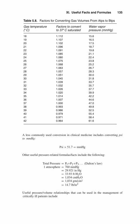
XI. Useful Facts and Formulas 135
Table 5.8. Factors for Converting Gas Volumes From Atps to Btps
Gas temperature(◦C)
Factors to convertto 37◦C saturated
Water vaporpressure (mmHg)
18 1.112 15.619 1.107 16.520 1.102 17.521 1.096 18.722 1.091 19.823 1.085 21.124 1.080 22.425 1.075 23.826 1.068 25.227 1.063 26.728 1.057 28.329 1.051 30.030 1.045 31.831 1.039 33.732 1.032 35.733 1.026 37.734 1.020 39.935 1.014 42.236 1.007 44.637 1.000 47.038 0.993 49.839 0.986 52.540 0.979 55.441 0.971 58.442 0.964 61.6
A less commonly used conversion in clinical medicine includes converting psito mmHg:
Psi × 51.7 = mmHg
Other useful pressure-related formulas/facts include the following:
Total Pressure = Pl+P2+P3 . . . (Dalton’s law)1 atmosphere = 760 mmHg
= 29.921 in Hg= 33.93 ft H2O= 1,034 cmH2O= 1,034 gm/cm2
= 14.7 lb/in2
Useful pressure/volume relationships that can be used in the management ofcritically ill patients include

136 5. Environmental Disorders
Table 5.9. Changes in Density With Altitude, Assuming a ConstantTemperature
Altitude (feet) Standardtemperature(◦C)
Density ratioconstanttemperature
Density ratiostandardtemperature
0 15.00 1.0000 1.00005,000 5.09 0.8320 0.8617
10,000 –4.81 0.6877 0.738515,000 –14.72 0.5643 0.6292
VolumeBTPS= VolumeATPS × Factor
where VolumeBTPS = the gas volume saturated with water at body temperature(37◦C) and ambient pressure (BTPS = barometric temperature pressure satu-ration); VolumeATPS = the gas volume saturated with water at ambient (room)temperature and pressure (ATPS = ambient temperature pressure saturation);Factor = the factors for converting gas volumes from ATPS to BTPS:
Conversion factor = PB − PH2O
PB − 47× 310
(273 +◦ C)
Table 5.8 depicts the conversions factors required to convert Atps to Btps.
D. Altitude. As altitude varies, changes in atmospheric pressure produce alterationsin gas density (see Table 5.9).

6Gastrointestinal
Disorders
� I. GASTROINTESTINAL BLEEDING
A. Classification1. Upper gastrointestinal (GI) bleeding is above the ligament of Treitz.2. Lower GI bleeding is below the ligament of Treitz.
B. Etiology. The most common causes of acute GI bleeding requiring admission tothe intensive care unit (ICU) are depicted in Table 6.1. The most common sourcesof GI bleeding in the ICU are gastroduodenal stress ulcerations.
C. Diagnostic Evaluation1. History
Although the history and physical in a critically ill patient with acute GI bleed-ing may be limited by the patient’s clinical condition, the following are pointsthat need to be investigated:a. History of hematemesis or melenab. Time of onsetc. Amount of bloodd. Color and charactere. Drug or alcohol use (i.e., nonsteroidal anti-inflammatory drug [NSAID],
prednisone, warfarin)f. Past medical history (i.e., cirrhosis, peptic ulcer disease [PUD], inflammatory
bowel disease [IBD], etc.)2. Physical Examination
The precise cause of acute GI bleeding is unlikely to be evident from phys-ical examination alone (except in chronic liver disease, Osler-Rendu-Webersyndrome, or hemorrhoids).a. General Appearance: This may vary from the patient in no acute distress to
the patient in hypovolemic shock.b. Vital Signs: Tachycardia, postural hypotension. An increase in heart rate of
10–20 beats per minute and drop in blood pressure of >20 mmHg uponassumption of an upright position are generally indicative of significant,acute volume loss.
J. Varon, P. Acosta, Handbook of Critical and Intensive Care Medicine,DOI 10.1007/978-0-387-92851-7_6,C© Springer Science+Business Media, LLC 2010

138 6. Gastrointestinal Disorders
Table 6.1. Etiologies of Acute GI Bleeding
Upper Lower
Esophagus Small intestineMucosal tear Arteriovenous malformationsEsophageal rupture Inflammatory bowel disease ischemiaEsophagitis Meckel’s diverticulumNeoplasms NeoplasmsVarices
Stomach Large intestineArteriovenous malformations DiverticulosisGastritis (any etiology) HemorrhoidsNeoplasms Inflammatory bowel disease infectionsPeptic ulcer disease IschemiaStress ulcers Neoplasms
DuodenumArteriovenous malformationsNeoplasms (rare)Peptic ulcer disease
Table 6.2. Advantages and Disadvantages of NG Tubes in Acute GIBleeding
Advantages Disadvantages
1. Document the presence orabsence of blood.
1. Patient discomfort.
2. Monitor rate of bleeding.2. Irritation of esophageal and/or
gastric mucosa.
3. To lavage and decompress thestomach.
3. Increased incidence of sinusitis.
4. Possible esophageal or gastricperforation.
c. Other Signs of Hypovolemia: Altered mental status, low urine output.d. Associated Findings: Petechiae, jaundice, hepatomegaly, and splenomegaly.e. Rectum: Look for hemorrhoids, fissures, etc. Examine stools for blood even
if the patient has an upper GI source.3. A nasogastric (NG) tube should be placed in all patients with acute GI bleeding.
The major advantages and disadvantages of NG tubes are shown in Table 6.2.4. Laboratory Evaluation
All patients admitted to the ICU with GI bleeding should undergo the laboratorytests depicted in Table 6.3.
5. Radiologic EvaluationAll patients should undergo chest radiograph and abdominal x-rays. These mayshow evidence of perforation or obstruction and may indicate ischemic changes.

I. Gastrointestinal Bleeding 139
Table 6.3. Initial Laboratory Evaluation in GI Bleeding
Complete blood count (H/H should be repeated every 4 h until patient isstable or bleeding has been controlled)
BUN, creatinine, and electrolytes
PT, PTT
Type and cross-match for 2–8 U of PRBCs, FFP
Other tests are ordered according to suspected or known underlying disease(i.e., LFTs, CK, etc.)
Abbreviations: BUN, blood urea nitrogen; CK, creatine kinase; FFP, fresh-frozen plasma;H/H, hemoglobin/hematocrit; LFT, liver function tests; PRBCs, packed red blood cells; PT,prothrombin time; PTT, partial thromboplastin time.
Contrast studies have a low diagnostic yield and may be hazardous for the crit-ically ill patient. They may also interfere with other diagnostic studies (i.e.,endoscopy, angiography). Special tests may be required in the evaluation ofacute GI bleeding. These includea. Selective angiography may be used as a diagnostic as well as therapeutic
tool (e.g., embolization). A bleeding rate at the time of the procedure ≥0.5mL/min is needed for diagnosis.
b. Radionuclide scans are sensitive in detecting lesions with lower bleedingrates.
6. Endoscopy is indicated in the vast majority of patients requiring ICU admissionfor GI bleeding.a. Upper endoscopy is indicated when blood is obtained from the NG tube or
when frank hematemesis is present.b. Flexible sigmoidoscopy should be performed initially if lower GI bleeding
is suspected. If this is not diagnostic, colonoscopy should be considered.c. Special endoscopic procedures may be required (i.e., wireless video capsule
endoscopy, push enteroscopy, double balloon enteroscopy)
D. Initial ICU Management1. As in any critically ill patient, the management of acute GI bleeding starts with
assessment of the airway, breathing, and circulation (ABCs). A low thresh-old for endotracheal intubation is recommended in the event of clouding ofconsciousness or overt shock, to prevent aspiration.
2. Insert at least two large-bore (16-gauge) IV catheters.3. Infuse blood, plasma expanders, and/or normal saline to maintain a mean
arterial pressure ≥65 mmHg.4. Some authors still recommend NG placement in all patients with GI bleeding
and lavage of the stomach until the return is clear. This practice is considereduseful only in settings where emergency endoscopy is not available.
5. Correction of preexisting coagulopathy (i.e., fresh frozen plasma [FFP], vitaminK, etc.).
6. Proton-pump inhibitors and H2-receptor blockers to prevent further hemor-rhage. Continuous infusions are preferred (i.e., pantoprazole [Protonix] 8 mg/h,ranitidine [Zantac] 150–300 mg/24 h IV infusion if the renal function is normal,or famotidine [Pepcid] 20 mg IV q12 h).

140 6. Gastrointestinal Disorders
7. Endoscopic and/or angiographic verification of the source of bleeding willallow more definitive therapy (i.e., thermal coagulation, injection therapy, fibrinsealant, endoclips, surgery).
E. Specific Management of Selected Conditions1. Variceal Hemorrhage
a. Vasopressin Infusion: Start at 0.2–0.4 U/min (up to 1 U/min). Some ofthe major complications of vasopressin (i.e., myocardial ischemia) can beprevented by the co-administration of nitroglycerin.
b. Alternatively, somatostatin-analogs can be utilized. Somatostatin inhibitsthe release of vasodilator hormones, such as glucagon, indirectly causingsplanchnic vasoconstriction and decreased portal inflow:(1). Octreotide is a long-acting analog of somatostatin.(2). Dosing: 25–50 mcg I.V. bolus followed by continuous I.V. infusion of
25–50 mcg/hc. Sclerotherapy: Indicated at the time of diagnostic endoscopy. Two or three
treatments are usually done within a 10-day period.d. Balloon Tamponade: Temporizing measure only. It is usually reserved for
hemorrhage that fails to stop after therapy with vasopressin and sclerother-apy. The routine use of Sengstaken–Blakemore or Minnesota tubes hasalmost disappeared with the advances in endoscopic therapy.
e. Surgical Therapy: Every patient with a major esophageal bleed shouldreceive surgical consultation in case an emergent intervention is needed.Indications for surgical therapy include the following:(1). Child’s class A or B patient in whom vital signs cannot be stabilized
medically.(2). Continuous bleeding for ≥48 h despite sclerotherapy and balloon
tamponade.(3). Third acute episode of esophageal bleeding in spite of previous scle-
rotherapy.2. Hemorrhage From Ulcers and Erosive Lesions
a. Endoscopy therapy with sclerosing agents, or laser coagulation, or heaterprobe.
b. Surgical intervention is indicated in cases of(1). Visible vascular pedicle on endoscopy(2). Transfusion of 6 U blood in 24 h(3). Arterial spurting
3. Active Lower GI Bleedinga. If a lesion is reachable with sigmoidoscopy or colonoscopy, local therapy
may be attempted (e.g., laser coagulation).b. Arterial embolization is indicated if the above fails.c. All patients with active lower GI bleeding should receive surgical consulta-
tion in case an emergent intervention is needed.
� II. ACUTE MESENTERIC ISCHEMIA
A. Definition. Acute mesenteric ischemia (AMI) is an acute reduction in blood flowto the intestine leading to inadequate perfusion. AMI may be a reflection ofgeneralized poor perfusion, or it may result from local pathology.

II. Acute Mesenteric Ischemia 141
Table 6.4. Risk Factors for the Development of AMI
— Age ≥50 years
— Atherosclerotic heart disease
— Congestive heart failure
— Recent myocardial infarction
— Valvular heart disease
B. Epidemiology. The incidence of AMI has increased over the past few decades.The rising incidence may be attributable to advances in medical technology and tonew therapies extending the life of critically ill patients who are prone to developAMI (e.g., elderly). The mortality in AMI is between 55 and 100%.
C. Etiology1. Occlusive
a. Atherosclerotic narrowing of the mesenteric bedb. Systemic emboli from any source (e.g., endocarditis)c. Vasculitisd. Hypercoagulable states
2. Nonocclusivea. Splanchnic Vasoconstriction
(1). Hypovolemia(2). Hypotension(3). Low cardiac output(4). Vasopressor agents use
D. Risk Factors for AMI. The most common predisposing conditions are depicted inTable 6.4.
E. Diagnostic Evaluation1. History and Physical Examination
The classic complaint of severe abdominal pain that is out of proportion tothe findings of physical examination, in our experience, is rarely seen. If peri-toneal signs are present (e.g., rebound tenderness), intestinal infarction is likelyto have occurred. Abdominal distention, emesis, and other signs of intestinalobstruction may occur in patients with AMI in situ. Lower GI bleeding mayoccur.
2. Laboratory Studies Reveala. Leukocytosis in 75% of patientsb. Metabolic acidosisc. Elevated amylase, creatine kinase (CK) (6–12 h after infarction has
occurred), lactate, and phosphate3. Radiologic Evaluation should be done as soon as the patient has been ade-
quately resuscitated (including measures aimed at relieving acute congestiveheart failure and hypotension, correction of hypovolemia and cardiac dysrhyth-mias)a. Plain Abdominal X-Rays
(1). Useful in excluding other causes of abdominal pain (i.e., mechanicalobstruction, perforation)

142 6. Gastrointestinal Disorders
(2). Seventy percent of patients will show at least one of the following:(a). Ileus(b). Ascites(c). Small bowel dilation(d). Separation of small bowel loops(e). Thickening of valvulae conniventes(f). Thumb printing
b. Barium studies are contraindicated in these patients and they interfere witharteriography.
c. Computed tomography (CT) may be particularly valuable when mesentericvein thrombosis is being considered. It may show focal or segmental bowelwall thickening or intestinal pneumatosis
d. Arteriography: For adequate study, the patient needs to be hemodynamicallystable. Early use of this test is the key to diagnosis. It provides a “road map”for the surgeon.
e. Laparotomy
F. Therapy1. As in any critically ill patient, the management of AMI starts with
assessment of the ABCs.2. Adequate hydration. If necessary, provide invasive hemodynamic moni-
toring to maximize cardiac output, oxygen delivery, and volume status.3. Patients with suspected embolic or thrombotic occlusion should undergo
urgent laparotomy for possible resection. Heparin and broad-spectrumantibiotic are indicated before surgery. Most patients will undergo a“second-look” operation within 24 h of the initial laparotomy.
4. In those cases with nonocclusive AMI, intra-arterial infusions of vasodila-tors (e.g., papaverine 30–60 mg/h) are advocated by some.
� III. FULMINANT HEPATIC FAILURE ANDENCEPHALOPATHY
A. Definition1. Acute Fulminant Hepatic Failure
Acute fulminant hepatic failure (FHF) is defined as acute liver failure associatedwith the development of hepatic encephalopathy within 8 weeks of the onset ofsymptoms attributable to hepatocellular dysfunction. This definition assumesthat there is no preexisting liver disease.
2. Hepatic EncephalopathyHepatic encephalopathy (HE) is a complex neuropsychiatric syndrome precip-itated by abnormal liver function. This syndrome is a feature of acute and/orchronic hepatocellular failure.
B. EtiologyCommon causes of FHF and HE are depicted in Table 6.5.
C. Diagnostic Evaluation1. History
A detailed history should be obtained from family members. The followingpoints need to be investigated:a. History of preexisting liver disease

III. Fulminant Hepatic Failure and Encephalopathy 143
Table 6.5. Causes of Acute Liver Failure
1. Viral hepatitis (i.e., A, B, C)
2. Drugs or toxinsAcetaminophenAcute alcohol intoxicationCarbon tetrachlorideHalothaneIsoniazidMonoamine oxidase inhibitorsMushroom poisoning
3. Fatty liver of pregnancy
4. Shock of any etiology
5. Massive liver infiltration (i.e., leukemia)
6. Decompensation of chronic liver failure
Table 6.6. Clinical Stages of Hepatic Encephalopathy
Stage Neurological findings
I Confusion, mild changes in personality, psychometric defectsII Drowsiness to lethargyIII Somnolent but arousableIV Coma
b. Drug or alcohol usec. Toxin exposure or ingestion
2. Physical ExaminationThis may vary from the patient in no distress to the patient in overt shock.a. Vital signs: tachycardia, hypotension.b. Associated findings: petechiae, jaundice, hepatomegaly, splenomegaly.c. The encephalopathy may begin with confusion, disorientation, and irrational
behavior. Coma may develop rapidly. (See Table 6.6.)3. Laboratory and Radiologic Evaluation
All patients with HE and/or FHF should undergo the following tests:a. Chest x-ray, abdominal x-rays.b. Blood glucose may reveal hypoglycemia.c. Serum bilirubin: A value >23 mg/dL is the best predictor of nonsurvival.d. AST and ALT have little prognostic value as levels tend to fall as the patient’s
condition worsens.e. Serum albumin: Its decrease reflects poor outcome.f. Serum electrolytes.g. Complete blood count.h. Head computed tomography (CT) scan to rule out a structural lesion (e.g.,
hemorrhage).i. Lumbar puncture needs to be considered and performed if meningitis is
suspected.

144 6. Gastrointestinal Disorders
j. If the etiology of FHF is unknown, the following need to be ordered:(1). Acetaminophen level(2). Hepatitis profile
(a). Viral hepatitis A is diagnosed by detection of HAV-IgM in thepatient’s serum.
(b). Viral hepatitis B is diagnosed byi. Detection of HBsAg
ii. Anti-HBc IgM(c). Viral hepatitis C is diagnosed by detection of anti-HCV.(d). Delta virus hepatitis is diagnosed by detection of anti-HDV in a
patient co-infected with hepatitis B virus.(3). Alkaline phosphatase(4). Amylase
k. Serum ammonia levell. Electroencephalograms (EEGs) are used to assess clinical response and
prognosis in patients with HE.
D. Complications of FHFWhen the liver fails acutely, all organ systems are involved to some extent.1. Central Nervous System (CNS)
Hepatic encephalopathy, cerebral edema2. Cardiovascular
Dysrhythmias (particularly in patients with advanced FHF), hypotension3. Pulmonary
Hypoxemia advancing to adult (acute) respiratory distress syndrome (ARDS)4. Renal
The development of renal failure with FHF carries a poor prognosis.a. In most instances the renal failure is related to “prerenal” causes.b. The hepatorenal syndrome is a diagnosis of exclusion. It is associated with a
normal urine sediment, a urinary sodium concentration of <20 mmol/L, andresolution if liver function improves.
5. HematologicThrombocytopenia, diminished clotting factors with episodes of severe bleed-ing.
6. InfectionSusceptibility to infection is increased in patients with FHF.
7. MetabolicHypoglycemia, metabolic acidosis, hypokalemia, hyponatremia.
E. Management1. Supportive Therapy
a. As in any critically ill patient, the management of AMI starts with assess-ment of the ABCs.
b. The usual indications for endotracheal intubation and assisted mechanicalventilation apply to these patients.
2. The use of corticosteroids for patients with FHF has not been proven to improvesurvival and, indeed, may worsen the clinical picture.
3. Some authors suggest avoiding parenteral nutrition, as protein and amino acidsmay worsen the clinical picture. However, new total parenteral nutrition solu-tions with “branched-chain” amino acids are probably efficacious and helpmaintain a positive-nitrogen balance.

IV. Pancreatitis 145
4. The management of FHF-associated cerebral edema is no different from that fornon-hepatic-related causes (see Chapter 9, “Neurologic Disorders”). In recentyears, emphasis on the use of therapeutic hypothermia for these patients seemsencouraging.
5. Some clinical and experimental evidence shows that the benzodiazepine antag-onist flumazenil (Romazicon) may have some role in improving the signs andsymptoms of HE.
6. Investigational data have shown some improvement in the hemodynamics ofpatients with FHF treated with n-acetylcysteine.
7. Liver transplantation may be an alternative form of therapy (in a few special-ized transplant centers) for some patients with no known contraindication to theprocedure.
8. Liver “dialysis”: A few specialized centers are currently exploring this form oftherapy.
9. Agents aimed at stimulating ammonia metabolism have also been tried (e.g.,ornithine-aspartate, sodium benzoate)
� IV. PANCREATITIS
A. Definition. Acute pancreatitis is an inflammatory process of the pancreas witha wide range of clinical severity ranging from self-limited to a lethal disease,complicated by multiple organ system failure (10% of cases).
B. Etiology. The most common causes of pancreatitis are1. Alcoholism2. Gallstones3. Hyperlipidemia4. Trauma (blunt or penetrating)5. Infections (i.e., mumps, mycoplasma)6. Hypoperfusion states (i.e., shock, cardiopulmonary bypass)7. Hypercalcemia8. Drugs (i.e., sulfonamides, thiazides)
C. Diagnostic Evaluation1. History
Ninety-five percent of patients with acute pancreatitis present with abdominalpain, of which 50% will present with upper abdominal discomfort radiating tothe back. Nausea and vomiting are also present.
2. Physical ExaminationDepending on the severity of the situation, the patient may have overt signs ofshock or may be hemodynamically stable. Other findings include the following:a. Abdominal tenderness and distentionb. Abdominal ileusc. Low-grade fever (Note: a fever >39◦C should suggest cholangitis, peritonitis,
or a pancreatic abscess.)d. Mild jaundicee. Ascitesf. Pleural effusion

146 6. Gastrointestinal Disorders
3. Laboratory Evaluationa. Complete Blood Count (CBC): Shows marked leukocytosis. Thrombo-
cytopenia may be present in those cases complicated by disseminatedintravascular coagulation (DIC).
b. Amylase: Elevated initially, but may decrease after 2–3 days if necrosis ofthe pancreas is widespread. False-positive results may occur in perforationof the esophagus, stomach, intestine, gynecologic disorders, renal failure,severe burns, diabetic ketoacidosis (DKA), salivary gland disorders, andmacroamylasemia.
c. Lipase: Hyperlipasemia persists longer than hyperamylasemia. However, ifnecrosis of the pancreas is widespread, these values may be normal.
d. Serum calcium is usually low. When levels are <8 mg/dL, the prognosis ispoor.
e. Other electrolyte imbalances as well as hyperglycemia are usually present.f. Metabolic acidosis may be present.g. C-reactive protein: Usually elevated.h. Urinalysis may reveal proteinuria, casts (25% of the cases), and glycosuria.
4. Radiologic EvaluationEvery patient with suspected acute pancreatitis should get a chest x-ray (to ruleout free air under the diaphragm, evidence of pleural effusions, etc.) and anabdominal x-ray (signs of intestinal obstruction, ileus, gallstones, the so-called“sentinel loop” of pancreatitis, or the colon “cut-off” sign, etc.). In addition,when the diagnosis remains in doubt, especially in the more severely ill, thefollowing can be obtained:a. Ultrasonography (US) is the modality of choice in patients with edema-
tous pancreatitis or suspected biliary pancreatitis and to follow up phlegmonor abscesses. Unfortunately, US cannot be accurately performed in obesepatients and in those with moderate-to-severe ileus.
b. CT is the most useful tool in assessing the retroperitoneum. Its use in acutepancreatitis is mainly to follow up on significant complications (i.e., abscess,phlegmon, pseudoaneurysms).
D. Management1. As in any critically ill patient, the management of acute pancreatitis starts with
assessment of the ABCs.2. Adequate hydration.3. Correct underlying factors.4. Control pancreatic enzyme secretion.
a. Nasogastric suctionb. Proton-pump inhibitors (e.g., pantoprazole 40 mg IV q12 h) or H2-receptor-
blocking agents (e.g., ranitidine [Zantac] 300-mg/24 h IV infusion if renalfunction is normal or famotidine 20 mg IV q12 h).
c. Many clinicians use the following agents in acute pancreatitis; however,clinical studies have not supported the routine use of these agents:(1). Calcitonin (300 IU/24 h)(2). Somatostatin (250 μg IV bolus, then 250 μg/h as IV drip).(3). Glucagon
d. Of interest is the use of intramuscular (IM) clonidine (not yet available in theUnited States) for patients hemodynamically stable with acute pancreatitis.Preliminary data show encouraging results.

IV. Pancreatitis 147
5. Sedation and analgesia: Patients may require substantial amounts of analgesia,usually with meperidine (Demerol).
6. Adequate parenteral nutrition (see Chapter 10, “Nutrition”).7. Correct hypocalcemia only if there is clinical evidence of tetany.
E. Complications. The most common complications of acute pancreatitis aredepicted in Table 6.7.1. Those patients who demonstrate fever >39◦C with a white blood cell count
>20,000/mm3 should be evaluated for the presence of a pancreatic abscess(with the use of CT). If there are any fluid collections, CT-guided fine-needleaspiration is then indicated (for Gram’s stain and cultures).
2. If the suspected diagnosis is pancreatic abscess, broad-spectrum antibioticsshould be started and an emergent surgical consultation obtained.
3. Some authors advocate necrosectomy in patients with necrotizing pancreatitis.
F. Prognosis1. In assessing the severity of the disease and prognosis, several classifications
have been used. The most commonly utilized is the Ranson’s criteria (initiallydeveloped for patients with alcoholic pancreatitis):a. Three or more of the following criteria must be met:
(1). Age >55 years(2). White blood cell count >16,000/mm3
(3). Glucose >200 mg/dL(4). Base deficit >4 mEq/L(5). Lactic dehydrogenase (LDH) >350 IU/L(6). AST (serum glutamate pyruvate transaminase [SGPT]) >250 IU/L)
b. Development of the following within 48 h indicates a worsening prognosis:(1). Hematocrit drop >10%(2). Serum urea nitrogen (BUN) rise >5 mg/dL(3). Partial pressure of O2 in arterial blood (PaO2) <60 torr (mmHg)(4). Calcium <8 mg/dL(5). Fluid sequestration >6 L
c. Mortality rates correlate with the number of criteria present:(1). 0–2 criteria, 1% mortality(2). 3–4 criteria, 16% mortality
Table 6.7. Complications of Acute Pancreatitis
1. Intravascular fluid depletiona. Prerenal azotemiab. Shock
2. ARDS (3–7 days after the onset)
3. Cardiac dysfunction
4. Pancreatic abscess
5. Pancreatic pseudocysts
6. Chronic pancreatitis
7. Permanent diabetes mellitus
8. Multiorgan system failure

148 6. Gastrointestinal Disorders
(3). 5–6 criteria, 40% mortality(4). 7–8 criteria, 100% mortality
2. Intensive care management and prompt surgical consultation have lowered themortality of acute pancreatitis.
� V. USEFUL FACTS AND FORMULAS
A. Intestinal Transit. The normal 24-h intestinal fluid and electrolyte transport isdepicted in Table 6.8.
Table 6.8. Normal 24-h Intestinal Fluid and Electrolyte Transport
Site Fluidreceived (L)
Amountabsorbed (L)
Electrolyte absorption
Na+ K+ Cl–
Duodenum/jejunum
9.0 4.0 Passive Passive Passive
Ileum 5.0 3.5 Active Passive PassiveColon 1.5 1.35 Active Passive Active
Table 6.9. Child’s Classification of Portal Hypertension
Class A B C
Serum bilirubin (mg/dL) <2 2–3 >3Serum albumin (g/dL) >3.5 3–3.5 <3Ascites None Easily controlled Poorly controlledEncephalopathy None Minimal AdvancedNutrition Excellent Good Poor
B. Stool Formulas. As part of the diagnostic workup of patients with diarrhea, stoolosmolal gap (SOG) is usually calculated utilizing the following formula:
SOG = stool osmolality − 2 × (stoolNa++stoolK+)
Normal stool osmolality is <290 mOsm/L. If the SOG >100, it indicates anosmotic diarrhea.
C. Liver Facts. The Child’s classification for portal hypertension is commonly usedin critically ill patients and is depicted in Table 6.9.

7Hematologic Disorders
� I. ANEMIA
A. Definition. Anemia is defined as an absolute decrease in the circulating red bloodcell (RBC) mass.
B. Etiology1. Decreased RBC Production
a. Deficiency of hematinic agents (i.e., iron, vitamin B12, folate)b. Bone marrow failure
2. Increased RBC Destruction or Lossa. Hemolysisb. Hemorrhage
C. Diagnostic Evaluation. The approach to the anemic patient in the intensive careunit (ICU) will differ depending on whether the patient was admitted with anemiaor if the anemia has developed during the ICU stay.1. In the Patient Admitted With Anemia
a. The symptoms of anemia will depend on the degree of anemia, the rapidityof development, cardiopulmonary reserve, and underlying disease.
b. As a general rule, a hemoglobin <7 g/dL represents severe anemia, andsuch patients may present with dyspnea on exertion, lightheadedness, angina,and/or fatigue.
c. The absence of symptoms in patients with hemoglobin <7 g/dL suggests agradual onset.
2. HistoryInquire about previous hematologic values, family, and ethnic history (i.e.,sickle cell, thalassemia), history of splenectomy, cholelithiasis at an earlyage, medications, drugs, alcohol use, dietary habits, gastrectomy, and bleedinghistory.
3. Physical Examinationa. General Appearance: Nutritional status or evidence of specific deficiencies,
evidence of chronic illness.b. Vital Signs: Tachycardia, postural hypotension, other signs of hypoperfusion
(i.e., decreased mental status, low urine output), petechiae, purpura.
J. Varon, P. Acosta, Handbook of Critical and Intensive Care Medicine,DOI 10.1007/978-0-387-92851-7_7,C© Springer Science+Business Media, LLC 2010

150 7. Hematologic Disorders
c. Associated Findings: Jaundice, glossitis (i.e., pernicious anemia, iron defi-ciency), neurologic abnormalities (i.e., vitamin B12, folate deficiency).Lymphadenopathy, hepatomegaly, splenomegaly (i.e., hemolysis, neo-plasms, infiltrative disorders). Heart: Listen for flow murmurs, prostheticvalves (i.e., increased RBC destruction). Rectal: Examine stools for blood.
4. Laboratory EvaluationLaboratory evaluation usually provides a diagnosis and should always be donein a stepwise manner unless the patient’s condition requires emergent transfu-sion. In this case, a blood sample for RBC indices, peripheral blood smear, andiron, folate, and vitamin B12 studies should be obtained before transfusion.a. Hemoglobin and hematocrit (Hct) estimate RBC mass and severity of ane-
mia, and in the patient suspected to have active bleeding, they should bemonitored serially. Acute blood loss does not influence the Hct immediately.
b. Mean corpuscular volume (MCV) is a measure of the average size of theRBCs. Classification of the anemia according to the MCV is helpful in gen-erating the differential diagnosis and workup. The smear must be examinedto determine whether multiple cell populations are present.(1). Low MCV (<80): Generally limits the diagnosis to a few disorders: Iron
deficiency, thalassemia, sideroblastic anemia, other hemoglobinopathies,and some cases of anemia of chronic disease.
(2). High MCV (<100): Megaloblastic anemias, liver disease, alcoholism,drugs (i.e., methotrexate, AZT), and myelodysplastic syndrome.
(3). Normal MCV: Acute blood loss, hemolytic anemia, pituitary or thyroidfailure, aplastic anemia, myelofibrosis, and anemia of chronic disease.
c. Reticulocyte count is also essential in the evaluation of the anemic patient.It reflects the rate of production of RBCs by the bone marrow. According tothe reticulocyte count, anemia can be classified into(1). Increased RBC destruction (i.e., bleeding, hemolysis) reflected in a high
reticulocyte count(2). Decreased RBC production (i.e., iron deficiency, anemia of chronic
disease) reflected in an abnormally low reticulocyte count5. Bleeding should be the first concern in patients who develop anemia while
in the ICU. Common sites of bleeding are the gastrointestinal (GI) tract,venipunctures, pulmonary tree, genitourinary tract, and the retroperitoneum.Development of anemia in the ICU should prompt additional investigation (i.e.,gastric aspiration to look for blood, stools checked for gross or occult blood,prothrombin time [PT], partial thromboplastin time [PTT], and platelet count).
D. Therapy1. Patient Acutely Bleeding
a. General Measures(1). Airway management: Assess the need for intubation to prevent aspira-
tion, especially in upper GI bleeding.(2). Obtain adequate venous access. Large-bore peripheral catheters allow
greater volume administration rates than long central lines.(3). Obtain blood for type and cross and diagnostic laboratories, as discussed
above.(4). Fluid resuscitation: Start with colloids or crystalloids, and continue with
whole blood or packed red blood cells as required:
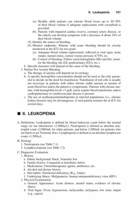
II. Leukopenia 151
(a). Healthy adult patients can tolerate blood losses up to 20–30%of their blood volume if adequate replacement with crystalloid isprovided.
(b). Patients with impaired cardiac reserve, coronary artery disease, orthe elderly can develop symptoms with a decrease of about 10% oftheir blood volume.
(5). Identify the source of bleeding.(6). Monitor endpoints: Patients with acute bleeding should be closely
monitored in the ICU for two goals:(a). Adequate blood volume replacement: reflected in vital signs, urine
output, mental status, central venous pressure (CVP), etc.(b). Control of bleeding: Follow serial hemoglobin (Hb) and Hct, moni-
tor the bleeding site (GI, genitourinary [GU], etc.).b. Specific measures will depend on the cause of the bleeding.
2. Patient Not Acutely Bleedinga. The therapy of anemia will depend on its etiology.b. A specific hemoglobin concentration should not be used as the only param-
eter to decide on the need for transfusion. Transfusion of red cells is usuallynot necessary in patients with either chronic stable anemia or anemia ofacute blood loss unless the patient is symptomatic. Patients with chronic ane-mia, with hemoglobin levels >7 g/dL rarely require blood transfusion, unlesscardiopulmonary or cerebrovascular disease is present.
c. The use of erythropoietin/darbepoetin in selected populations (i.e., chronickidney disease) may be advantageous, if such patient remains the in ICU forseveral days.
� II. LEUKOPENIA
A. Definitions. Leukopenia is defined by blood leukocyte count below the normalrange (in our laboratories <3,800/μL). Neutropenia is defined as absolute neu-trophil count <2,000/mL for white patients, and below 1,500/mL for patients whoare black or are Yemenite Jews. Lymphopenia is defined as an absolute lymphocytecount <1,500/μL.
B. Etiology1. Neutropenia (see Table 7.1)2. Lymphocytopenia (see Table 7.2)
C. Diagnostic Evaluation1. History
a. Ethnic background: black, Yemenite Jewb. Family history: Congenital or hereditary defectc. Medications: Chemotherapeutic agents, antibiotics, etc.d. Alcohol and dietary historye. Diet habits: Nutritional deficiency (B12, folate)f. Underlying illness: Malignancies, human immunodeficiency virus (HIV)
2. Physical Examinationa. General Appearance: Acute distress, mental status, evidence of chronic
illnessb. Vital Signs: Fever, hypotension, tachycardia, tachypnea, low urine output
(e.g., sepsis)
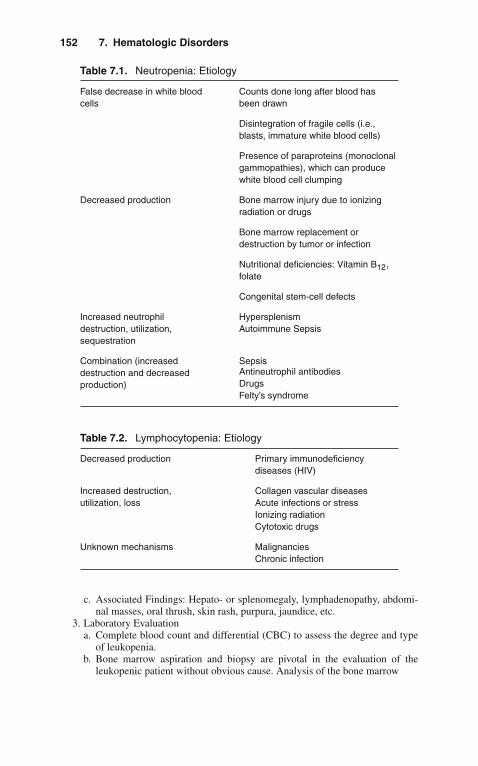
152 7. Hematologic Disorders
Table 7.1. Neutropenia: Etiology
False decrease in white bloodcells
Counts done long after blood hasbeen drawn
Disintegration of fragile cells (i.e.,blasts, immature white blood cells)
Presence of paraproteins (monoclonalgammopathies), which can producewhite blood cell clumping
Decreased production Bone marrow injury due to ionizingradiation or drugs
Bone marrow replacement ordestruction by tumor or infection
Nutritional deficiencies: Vitamin B12,folate
Congenital stem-cell defects
Increased neutrophildestruction, utilization,sequestration
HypersplenismAutoimmune Sepsis
Combination (increaseddestruction and decreasedproduction)
SepsisAntineutrophil antibodiesDrugsFelty’s syndrome
Table 7.2. Lymphocytopenia: Etiology
Decreased production Primary immunodeficiencydiseases (HIV)
Increased destruction,utilization, loss
Collagen vascular diseasesAcute infections or stressIonizing radiationCytotoxic drugs
Unknown mechanisms MalignanciesChronic infection
c. Associated Findings: Hepato- or splenomegaly, lymphadenopathy, abdomi-nal masses, oral thrush, skin rash, purpura, jaundice, etc.
3. Laboratory Evaluationa. Complete blood count and differential (CBC) to assess the degree and type
of leukopenia.b. Bone marrow aspiration and biopsy are pivotal in the evaluation of the
leukopenic patient without obvious cause. Analysis of the bone marrow

III. Thrombocytopenia 153
(1). Will classify the leukopenia, by revealing the degree of bone marrowcellularity: decreased production, decreased survival, or a combineddefect
(2). May indicate the etiology of the leukopenia as in aplastic anemia, bonemarrow infiltration (e.g., leukemia), infection, etc.
c. Other laboratory tests that may help in identifying the cause of leukopeniaare blood and tissue cultures, vitamin levels, and autoantibodies.
4. TherapyThe mainstay of therapy for the leukopenic patient is to treat the underlyingdisease. For example, in the patient with suspected drug-induced leukopenia,the offending medications should be discontinued. Vitamin deficiency shouldalso be treated when suspected.a. Colony-stimulating factors (G-CSF and GM-CSF) represent a line of ther-
apy for the treatment of leukopenia secondary to a decrease in bone marrowproduction.
b. Transfusions of white blood cell (WBC) concentrates have not been provento be of benefit in many controlled trials
c. Supportive therapy for the leukopenic patient requires special considera-tion, particularly in the ICU environment, where there is a higher risk fornosocomial infections:(1). Granulocyte counts <1,000/μL result in severe immunocompromise.(2). Patients who are immunocompromised should not receive rectal manip-
ulations. Strict hand washing for caregivers should be enforced. Avoidintramuscular (IM) or subcutaneous (SQ) routes.
(3). If the temperature rises >100.5◦F (38.5◦C), the patient should be fullyexamined, pan-cultured, and started on broad-spectrum antibiotics.
� III. THROMBOCYTOPENIA
A. Definition. Thrombocytopenia is defined as a platelet count <150,000/pL.
B. Etiology (see Table 7.3)
C. Diagnostic Evaluation1. History
Inquire about bleeding, thrombotic events, mental status changes, alcohol use,drugs and medications, and associated illness.a. Heparin-induced thrombocytopenia
More common with the use of unfractionated heparin (UFH) rather than lowmolecular weight heparin (LMWH). More common among surgical ratherthan medical patients.
2. Physical Examinationa. Vital Signs: Fever, tachycardia, hypotension, tachypnea, oliguria (i.e., sepsis)b. Skin: Purpura, hematomas, gingival bleeding, lymphadenopathy, hepato- or
splenomegaly, abdominal masses3. Laboratory Evaluation
a. Complete Blood Count and Platelet Count(1). Platelet counts >50,000/μL in isolation are not associated with signifi-
cant bleeding problems, and severe spontaneous bleeding is unusual inpatients with counts >g20,000/μL in the absence of coagulation factorabnormalities.

154 7. Hematologic Disorders
Table 7.3. Thrombocytopenia: Etiology
Decreased survival orsequestration
Autoimmune—Primary (ITP);Secondary: collagen vascular disease(SLE); viral infections; drug-induced(heparin, quinidine, sulfas);post-transfusionHypersplenism (portal hypertension,infiltrative disorders)Thrombotic thrombocytopenic purpura(TTP/HUS)Disseminated intravascularcoagulationSepsis
Decreased production Primary bone marrow disorders(aplastic anemia, primarythrombocytopenia)Bone marrow infiltration by tumor,infection, etc.Drug induced (alcohol, thiazides,alkylating agents)Infection
Developed while in ICU Drug-induced (heparin, H2-blockers,diuretics, antibodies)Disseminated intravascularcoagulationSepsisPost-transfusion
Abbreviations: HUS, hemolytic uremic syndrome; ITP, idiopathic thrombocytopenic purpura;SLE, systemic lupus erythematosus; TTP, thrombotic thrombocytopenic purpura.
(2). Thrombocytopenia associated with anemia suggests thrombotic throm-bocytopenic purpura (TTP), hemolytic uremic syndrome (HUS), dis-seminated intravascular coagulation (DIC), or other microangiopathicprocess.
(3). Pancytopenia should suggest leukemia, aplastic anemia, or other bonemarrow disorder.
b. Peripheral Blood Smear: Note platelet size and other abnormalities (i.e.,fragmented RBCs may indicate TTP or DIC; increased platelet size suggestsaccelerated destruction).
c. Coagulation Evaluation: PT, PTT, D-dimer, fibrin degradation products mayindicate the presence of consumption coagulopathy (e.g., DIC).
d. Bone Marrow Aspiration and Biopsy (not always necessary): To assess thenumber of megakaryocytes and the presence of bone marrow disorders (i.e.,leukemia, aplastic anemia, tumor infiltration).
e. For patients with suspected heparin-induced thrombocytopenia 14C-serotonin release assay remains the gold standard. Heparin-induced plateletaggregation tests can also be used as well as the solid phase ELISAimmunoassay.

IV. Anticoagulation and Fibrinolysis 155
4. TherapyDetailed treatment for the various causes of thrombocytopenia is beyond thescope of this chapter. We will concentrate on relevant topics for the acutemanagement of patients in ICU setting.
1. As hemostasis approaches normal at platelet counts >50,000/μL, patients withactive bleeding should be transfused (6–10 U) to attempt to achieve levels>50,000/μL.
2. Lumbar puncture and needle organ biopsies (lung, liver, kidneys, etc.) aremore hazardous than thoracentesis, paracentesis, and bone marrow biopsy.Transfusion to 50,000/μL before such procedures is indicated.
3. Patients with platelet counts <20,000/μL are at higher risk for hemorrhage.However, there is no clear threshold for prophylactic platelet transfusion.
4. Discontinue all nonessential medications including heparin. Consider chang-ing H2-blockers to coating agents or antacids. Avoid agents known to inhibitplatelet function (i.e., nonsteroidal anti-inflammatory drugs [NSAIDs], ticar-cillin, etc.).
5. Avoid trauma, IM and SQ injections, rectal manipulations, hard toothbrush,razors, etc. in thrombocytopenic patients.
6. TTP and HUS deserve special consideration because their management differs.a. TTP is a syndrome characterized by microangiopathic hemolytic anemia,
fever, fluctuating neurologic deficits, and renal insufficiency. HUS is feltto be a variant of this syndrome in which renal failure is the predominantfeature. TTP/HUS should be considered a medical emergency.
b. Patients with TTP/HUS should not receive platelet transfusions, unless life-threatening bleeding occurs.
c. Therapy for TTP/HUS includes plasma exchange by plasmapheresis ifavailable, otherwise fresh-frozen plasma (FFP) transfusions. Intravenous(IV) steroids can be used, and in unresponsive cases, vincristine has beenrecommended.
d. Heparin-induced thrombocytopenia: The first maneuver is to stop all heparinand heparin-containing products (i.e., heparin-flush). There are a numberof alternative anticoagulants to heparin in these patients, such as directthrombin inhibitors like lepirudin [recombinant hirudin], bivalirudin, orargatroban; fondaparinux; or danaparoids.
� IV. ANTICOAGULATION AND FIBRINOLYSIS
Anticoagulants and thrombolytic agents are potentially life-saving drugs when usedprophylactically or employed therapeutically in critically ill patients.
A. Anticoagulation1. Heparin
a. Mechanism of ActionHeparin acts by potentiating the activity of the plasma protease inhibitorantithrombin III, which rapidly inhibits the activity of factors XIIa, XIa, Xa,IXa, and thrombin (factor II).
b. Heparin prolongs the thrombin time (TT), bleeding time, PTT, and to a lesserextent the PT.

156 7. Hematologic Disorders
c. Heparin half-life is 1–3 h, but in patients with pulmonary embolism,clearance is accelerated (20–40%) compared to normals.
d. Indications(1). Prophylaxis of deep venous thrombosis (DVT) and pulmonary embolism
(PE). All patients in the ICU should be on some form of DVT prophy-laxis (commonly, heparin 5,000 U SQ q12 h or low molecular doseheparin (e.g., enoxaparin 40 mg SQ qd). Heparin has proved to bean effective agent in DVT prophylaxis, except after major orthopedicprocedures (particularly hip and knee replacement) and after prostatesurgery.
(2). Full anticoagulation (PTT approximately 2 times normal) in pulmonaryembolism or DVT, arterial thrombosis, and other disorders. A 5,000–8,000 U IV bolus is commonly used and infusion rates of 1,000 U/h(12–25 U/kg/h).
e. Monitoring of Anticoagulation(1). Not required for prophylactic doses.(2). For full anticoagulation, the heparin dose should be adjusted to maintain
the PTT at 1.5–2.0 times control. To avoid the tendency to under-anticoagulate patients on heparin therapy, a standardized dosing regimenhas been developed (Table 7.4).
(3). Low molecular weight heparin for DVT and acute coronary syndromes(ACS).EnoxaparinFor DVT 1 mg/kg SQ q12 h or
1.5 mg/kg SQ qdFor ACS 1 mg/kg SQ q12 h
f. Complications(1). Bleeding: Occurs in 7–20% of patients during full-dose heparinization.
Hemorrhage typically occurs from the GI, urinary tract, or surgical
Table 7.4. Standardized Protocol for Dosing of Intravenous Heparin
PTT∗ Dose adjustment† Repeat PTT
<50 5,000-U bolus, increase infusion by2,400 U/24 h
6 h
50–59 Increase infusion by 2,400 U/24 h 6 h
60–85 Therapeutic range, no change Next morning
86–95 High therapeutic range, decreaseinfusion by 1,920 U/24 h
Next morning
96–120 Stop infusion for 30 min, decreaseinfusion by 1,920 U/24 h
6 h
120 Stop infusion for 60 min, decreaseinfusion by 3,840 U/24 h
6 h
∗ Normal PTT range is 27–35 s.† Dosing protocol is based on an initial IV bolus of 5,000–8,000 U, followed by continuousinfusion of 24,000 U/24 h. The first PTT should be obtained 6 h after the bolus of heparin.

IV. Anticoagulation and Fibrinolysis 157
incisions. Less common sites are intracranial, retroperitoneal, soft tis-sues, nose, and pleural space. Bleeding is associated with the intensityof the anticoagulation (e.g., when the PTT is more than 3 times normal,the risk of hemorrhage is substantially higher).
(2). Thrombocytopenia: Heparin use is associated with thrombocytopenia in5–30% of patients, and its incidence is higher with the use of bovinelung heparin.(a). If thrombocytopenia is mild (>100,000/μL), not associated
with bleeding or thrombotic events, heparin therapy can becontinued.
(b). Severe thrombocytopenia can occur but is rare. It may be associ-ated with bleeding or paradoxic thrombotic events. Diagnosis ismade by detection of heparin-dependent immunoglobulin G (IgG).Treatment consists of discontinuation of all heparin use and avoidplatelet transfusion.
(3). Osteoporosis can be seen with long-term use of heparin.(4). Hypoaldosteronism rarely is seen.(5). Antidote: Heparin is generally undetectable in patient’s plasma within
3 h after discontinuation of therapy. In the rare instance in which anti-coagulation must be reversed more rapidly, protamine sulfate can beused.
2. WarfarinWarfarin (Coumadin) is the most frequently used oral anticoagulant in theUnited States. Sometimes the transition to chronic oral therapy is begun beforethe patient leaves the ICU or must be initiated because of heparin-inducedthrombocytopenia, or as DVT prophylaxis in certain cases. The physician car-ing for the critically ill can also encounter patients who have accidentally orpurposely overdosed with warfarin.a. Mechanism of Action: Warfarin interferes with the hepatic vitamin
K-dependent carboxylation of factors II, VII, IX, and X. It also inhibits thesynthesis of the anticoagulant factors protein C and S and may thereby bethrombogenic. The antithrombotic effects of warfarin occur only after sev-eral days of treatment. In patients on IV anticoagulation therapy, who requirechronic oral anticoagulation, heparin and warfarin should be overlapped forat least 48 h.
b. Indications(1). Prophylaxis of DVT and PE, especially in those patients in whom
heparin has not proved to be effective(2). For chronic anticoagulation therapy
c. Dosing(1). Loading Dose: 5–10 PO qd for 2–4 days(2). Maintenance Dose: 2–15 mg PO qd to keep INR (International standard-
ized ratio) therapeuticd. Complications
(1). Bleeding occurs in 2.4–8.1% of patients chronically anticoagulated.The risk is dose related and proportional to the prolongation of thePT. Treatment consists of FFP transfusions. Vitamin K replacementis only recommended for warfarin overdose, because of its delayedonset of action, and because it makes reinstitution of warfarin therapycomplicated.
(2). Warfarin skin necrosis secondary to a paradoxic hypercoagulable statedue to the warfarin-induced protein C reduction.

158 7. Hematologic Disorders
B. Fibrinolysis1. Fibrinolytic therapy has an expanding role in the treatment of many throm-
boembolic disorders. Many fibrinolytic drugs are currently marketed: streptoki-nase (SK), anisoylated plasminogen-streptokinase activator complex (APSAC),urokinase (UK), recombinant human tissue-type plasminogen activator (rt-PA),reteplase, and TNK among others. All drugs activate the fibrinolytic system byconverting plasminogen to the active enzyme plasmin. Plasmin degrades fibrinand dissolves the thrombus.
2. Indicationsa. Acute Myocardial Infarction (AMI): Thrombolytic therapy for AMI is
discussed in Chapter 3, “Cardiovascular Disorders.”b. Pulmonary Embolism: While the effectiveness and role of thrombolytic
agents in AMIs are firmly established, their use in venous thromboembolismremains infrequent and controversial, mainly because of the fear of a nega-tive benefit/risk ratio. SK, UK, and tPA have been shown to be more effectivethan heparin alone in accelerating clot lysis and improving pulmonary tis-sue perfusion. Current recommendations for the use of fibrinolysis in PEare for patients with massive pulmonary embolism and persistent systemichypotension in whom rapid resolution of pulmonary obstruction is desired.It is still questionable if there is significant improvement to survival in thosepatients.
c. Deep Venous Thrombosis: Even more debated is the use of thrombolyticagents in the treatment of DVT. Potential advantages of fibrinolysis overanticoagulation include prevention of PE by lysing the source of thrombus insitu, rapid restoration of normal venous circulation with a prompt resolutionof symptoms, and prevention of valve damage, which would otherwise resultin chronic venous insufficiency. Risks include a much higher incidence ofbleeding.
3. Dosage for Selected Thrombolytic Regimensa. Pulmonary Embolism
UK: 4,400 U/kg bolus, followed by 4,400 U/kg/h for 24 hUK: 15,000 U/kg bolus over 10 minSK: 250,000 U over 30 min, followed by 100,000 U/h for 24 hrt-PA: 100 mg as continuous peripheral infusion over 2 h
Selective use of lysis in the pulmonary artery directed by catheter (the Varon-Strickman procedure) can also be used.
b. Deep Venous ThrombosisSK: 250,000 U over 30 min, followed by 100,000 U/h for 48–72 hrt-PA: 0.5 mg/kg over 4–8 hrt-PA: 0.05 mg/kg/h for 24 h
c. Myocardial Infarction (See Section III)4. Monitoring
a. Clinical monitoring should include serial neurologic examinations to detectcentral nervous system (CNS) bleeding and frequent vital signs to detectbleeding. All puncture sites should be examined frequently.
b. Laboratory monitoring should include Hb/Hct, platelet, fibrinogen, PT, PTT.5. Complications
a. Bleeding: The greatest limitation of the thrombolytic drugs and the factorthat has limited their acceptance for the treatment of DVT and PE is theincidence of bleeding.

V. Blood and Blood Products Transfusion 159
b. Allergic Reactions: Reactions including skin rashes, fever, and hypotensionare rare and usually are associated with the use of SK and APSAC. Theinduction of antibodies against streptococcal antigens can occur after theadministration of SK or APSAC or after streptococcal infection, which mayneutralize the fibrinolytic activity of SK.
� V. BLOOD AND BLOOD PRODUCTSTRANSFUSION
Transfusion therapy may be associated with several immediate and delayed adverseeffects. Therefore, risks and benefits must be carefully weighed before any bloodproduct is administered. The use of blood components should be guided by a rationaldiagnostic and therapeutic approach.
A. Whole Blood. Whole blood stored >24 h contains few viable platelets or granulo-cytes; factors V and VIII are decreased, but stable clotting factors are maintained.One unit is 450 mL and transfused to an average-sized adult will increase thehemoglobin by 1.0 g/dL and Hct by 3%.1. Indications: Symptomatic anemia with massive hemorrhage2. Risks
a. Allergic reactionsb. Infectious diseases (i.e., HIV, hepatitis B)c. Febrile reactionsd. Volume overloade. Noncardiogenic pulmonary edema
B. Packed Red Blood Cells. Removal of 200–250 mL of plasma from whole bloodresults in packed RBCs (PRBCs). Transfusion of 1 U of PRBCs will increase theHb and Hct by the same amount as will 1 U of whole blood. One unit is 250–300mL.1. Indications: Symptomatic anemia2. Risks: Same as for whole blood (see above).
C. Packed Red Blood Cells, Leukocyte Poor. Most of the WBCs are removed fromthe packed RBCs by saline washing.1. Indications
a. Symptomatic anemia and allergic or febrile reaction from leukocyte antibod-ies
b. Patients with paroxysmal nocturnal hemoglobinuria2. Risks
Same as for whole blood (see above).
D. Fresh-Frozen Plasma. FFP is separated from freshly drawn whole blood and thenfrozen, with a volume of 200–250 mL. Rich in all coagulation factors; 1 mL sup-plies approximately 1 U of coagulation activity. FFP should be ABO compatible;Rh type or cross-matching is not required.1. Indications
a. Bleeding due to coagulation factor deficiencyb. Treatment of TIT and HUSc. Rapid reversal of vitamin K deficiency or warfarin overdose

160 7. Hematologic Disorders
2. RisksSame as for whole blood (see above).
E. Cryoprecipitate. Cryoprecipitate is made by thawing a unit of FFP at 4◦C. A whiteprecipitate forms; most of supernatant plasma is removed and refrozen. The vol-ume is 10 mL. A pack of cryoprecipitate contains von Willebrand’s factor, lesseramounts of factor VIII, fibrinogen, factor XIII, and fibronectin. ABO compatibilityis preferred.1. Indications
a. von Willebrand’s diseaseb. Mild-to-moderate hemophilia Ac. Factor XIII deficiencyd. Fibrinogen deficiency
2. Risksa. Infectious diseaseb. Hyperfibrinogenemiac. Allergic reactions
F. Platelets. Platelet packs are obtained from whole blood; 1 U contains at least5.5 × 1010 platelets/mm3 in approximately 50 mL volume. In a normal 70-kgadult, 1 U of platelets should increase platelet count by 5,000–10,000/mm3.1. Indications
a. To correct bleeding secondary to thrombocytopenia or abnormal plateletfunction
b. Prophylactically (e.g., in patients with chemotherapy-induced thrombocy-topenia, the threshold is somewhat controversial: 10,000–20,000/mm3), andbefore invasive procedures (target counts of 50,000/mm3)
2. Risksa. Infectious diseasesb. Allergic reactionsc. Alloimmunization
G. Complications of Transfusion Therapy1. Disease transmission: HIV, hepatitis, cytomegalovirus (CMV), Epstein-Barr
virus (EBV), Chagas’ disease, malaria.2. Allergic reactions characterized by fever, chills, urticaria, and respiratory
distress. These events are secondary to antileukocytic antibodies or anti-bodies against antigenic proteins in donor plasma. Therapy is symptomatic(acetaminophen; antihistamines; rarely, epinephrine or glucocorticoids areneeded).
3. Red Cell Transfusion Relateda. Acute Hemolytic Reactions: Fever, chills, back pain, nausea, vomiting,
hypotension, dark urine, chest pain. Acute renal failure with hemoglobinuriaand DIC may occur. If suspected(1). Inform the blood bank.(2). Stop transfusion.(3). Replace all IV tubing.(4). Send clotted and ETDA-treated blood samples from patient’s blood
along with the remainder of the unit of blood to the blood bank forcross-match.
(5). Send blood samples for DIC screen, bilirubin, and free hemoglobin.

VI. Disseminated Intravascular Coagulation 161
(6). Management(a). Intravascular volume expansion plus mannitol to keep urine output
>100 mL/h or 1 cc/kg/h.(b). Alkalization of urine with IV bicarbonate to keep urine pH >7.0 to
avoid hemoglobin tubular precipitation.(c). Treatment of DIC
b. Delayed Hemolytic Transfusion Reactions: 24 h to 25 days post-transfusion.These are secondary to an amnestic (1–3 days) or primary (7–25 days) anti-body response to RBC antigens. Patients usually develop a drop in the Hband Hct with an increase in bilirubin. Coombs’ test is positive.
c. Noncardiogenic pulmonary edema (acute respiratory distress syndrome[ARDS]), caused by antileukocytic antibodies.
d. Coagulopathy associated with a large volume of PRBC transfusions, sec-ondary to dilution of platelet and coagulation factors. Treatment consists ofFFP and platelet transfusions.
e. Citrate intoxication, also seen with large-volume transfusion of PRBCs.Patients present with hypocalcemia, hypotension, and drop in cardiac output.Treatment: IV calcium.
4. Volume Overload: Especially in patients with congestive heart failure (CHF).Diuretics may be needed after transfusion.
5. Platelet alloimmunization develops in patients who have received multipletransfusions. Approximately 75% of patients receiving platelets on a regu-lar basis will become alloimmunized to platelet antigens. Increments <20%of expected generally indicate alloimmunization. Patients may respond tosingle-donor platelets, but HLA-matched platelets may be needed.
� VI. DISSEMINATED INTRAVASCULARCOAGULATION
A. Definition. Disseminated intravascular coagulation (DIC) is a dynamic pathologicprocess triggered by activation of the clotting cascade with resultant generation ofexcess thrombin within the vascular system. Most consider DIC to be a systemichemorrhagic syndrome; however, this is only because hemorrhage is obvious andoften impressive. What is less commonly appreciated is the significant amountof microvascular thrombosis and, in some instances, large-vessel thrombosis thatoccurs. This thrombosis is usually the more life-threatening insult.
B. Etiology (see Table 7.5)
Table 7.5. Disseminated Intravascular Coagulation: Etiology
Obstetric accidents (amniotic fluid embolism, abruptio placentae)Intravascular hemolysisSepsisMalignancyTraumaVascular disorders

162 7. Hematologic Disorders
C. Diagnostic Evaluation. Since DIC is associated with an underlying disease state,the clinical evaluation will be directed toward identifying (1) primary illness; (2)the status of the coagulation system; and (3) the focal and systemic consequencesof the DIC-associated hemorrhage and/or thrombosis.1. Clinical Findings Associated With the Primary Illness
These findings will vary according to the precipitating event: obstetricalaccident, infection, malignancy, etc.
2. Clinical Findings Associated With the Coagulation Statusa. Bleeding from venipuncture sites, mucous membranes, hemorrhagic bullae,
hematuria, GI bleeding, etc.b. Purpura, petechiae, subcutaneous hematomas
3. Clinical Findings Associated With End-Organ Thrombosis and Hemorrhagea. Lungs: respiratory distress, hypoxia, ARDSb. Kidneys: proteinuria, renal insufficiencyc. Liver: Budd-Chiari syndrome, hepatitis, hepatic failured. Skin: necrosis, acrocyanosise. CNS: mental status changes, neurologic deficits
4. Laboratory Evaluationa. Peripheral blood smear will show fragmented RBCs, thrombocytopenia,
with large platelets.b. Prolonged PT and PTT.c. Thrombocytopenia is usually around 60,000/μL, but values ranging between
3,000 and 100,000/μL can be seen.d. Decreased fibrin level.e. Decreased anti-thrombin III level.f. Elevated levels of fibrin degradation products (FDP).g. Elevation of D-dimer neoantigens is a specific test for degradation products
of fibrin, whereas nonspecific FDP may be either fibrinogen or fibrin derived.
D. Therapy. The treatment of DIC is confusing and still controversial. Therapy mustbe individualized according to the cause of DIC, severity of hemorrhage, severityof thrombosis, hemodynamic status, and age.1. The most important and effective treatment for DIC is removal of the triggering
disease process (i.e., evacuate the uterus, control of shock, control of infection,removal of tumors, chemo- or radiotherapy, or other indicated therapy).
2. In cases of obstetric complications, anticoagulation is rarely needed,and evacuation of the uterus usually stops the intravascular clottingprocess.
3. If the patient continues to bleed or clot significantly after 6 h of initiation oftherapy directed to stop or blunt the triggering event, anticoagulation therapymay be indicated. There is general agreement on the need for anticoagula-tion in acute promyelocytic leukemia and perhaps DIC with solid tumors. Wefavor the use of low-dose, subcutaneous heparin at doses of 80–100 U/kg q6 h.Other anticoagulant modalities available are IV heparin and anti-thrombin IIIconcentrates.
4. If the patient continues to bleed after reasonable attempts to treat the triggeringevent of the DIC, and if anticoagulation therapy has been initiated, clottingfactor depletion is the most probable cause of bleeding and replacement therapyshould be considered.
5. The experimental use of thrombomodulin appears promising.

VII. Hemolytic Syndromes 163
� VII. HEMOLYTIC SYNDROMES
A. Definition. Premature destruction of red blood cells. This process may occureither because of abnormal factors in the intravascular environment or becauseof defective red blood cells.
B. Etiology (see Table 7.6)
C. Diagnostic Evaluation1. History and Physical Examination
Clinical manifestations will depend on the underlying disorder, on the severityof the anemia, and on whether the hemolysis is intravascular or extravascular.a. Intravascular hemolysis can present as an acute event with back pain, dysp-
nea, chills, fever, tachycardia, dark urine, and hypotension, and it may resultin renal failure.
b. Extravascular hemolysis is usually less dramatic and may be accompaniedonly by jaundice and splenomegaly.
2. Laboratory Evaluationa. Elevated reticulocyte count.b. Peripheral blood smear can provide a diagnosis in cases of spherocytosis;
microangiopathic disorders will show the presence of fragmented RBCs;Heinz bodies suggest enzymatic defects; or the presence of anisocytosis orsickle cells is consistent with hemoglobinopathies.
Table 7.6. Hemolytic Syndromes: Etiology
Acquired haemolyticdisorders
Immune hemolytic anemiaWarm-antibody (idiopathic, neoplasia,collagen vascular disorder, drugs)
Cold-antibody (idiopathic, mycoplasmainfection, lymphoproliferative disorder,paroxysmal cold hemoglobinuria
Microangiopathic hemolytic anemia(TTP, DIC, eclampsia)
Direct toxic effect (malaria, clostridialinfection)
SplenomegalyMembrane defectsParoxysmal nocturnal hemoglobinuriaSpur cell anemia
Hereditary hemolyticdisorders
Membrane defects (spherocytosis,elliptosis)
Enzyme defects (G6PD deficiency)ThalassemiasHemoglobinopathies

164 7. Hematologic Disorders
c. Other laboratory data suggestive of hemolysis are(1). Hemoglobinuria (indicative of intravascular hemolysis)(2). Hemoglobinemia (indicative of intravascular hemolysis)(3). Low levels of haptoglobin(4). Elevated lactic dehydrogenase (LDH)(5). Positive Coombs’ test
D. Sickle Cell Disease. Sickle cell disease is a heterogeneous group of defects ofhemoglobin synthesis, all of which can cause clinically significant illness dueto sickling of red cells. Sickle hemoglobin (Hb S) is less soluble when deoxy-genated and forms polymers that precipitate inside the RBCs, leading to membraneabnormalities, decreased deformability, and increased blood viscosity.1. Clinical Manifestations
The clinical manifestations of sickle cell disease are secondary to vasoocclusivephenomena, which may lead to microinfarctions with resultant painful crises,and eventually, chronic organ damage.
2. DiagnosisDemonstration of sickling under reduced oxygen tension. Hemoglobin elec-trophoresis should be performed to discriminate homozygous SS from AS andto determine the presence of other abnormal hemoglobins.
3. TreatmentThe treatment of sickle cell disease is supportive and limited to management ofacute and chronic complications. Frequently, these patients need to be admittedto the ICU due to the severity and life-threatening dimension of their acuteattacks.a. Early antibiotic treatment at the first evidence of infection. Pneumococcal
sepsis is a leading cause of mortality. Other prevalent pathogens include:Escherichia coli, Haemophilus influenzae, Salmonella sp., Shigella sp., andMycoplasma pneumoniae.
b. Painful crises: IV hydration; adequate analgesia (usually, a regular scheduleof opioids is necessary); oxygen administration is indicated when hypoxemiais present; correction of acidosis.
c. Look for precipitating events (i.e., infections, surgery, dehydration, trauma,cold temperatures, alcohol ingestion). When abdominal pain is one of themanifestations, other causes of abdominal pain must be ruled out (i.e., acuteabdomen, hepatobiliary disease).
d. Acute chest syndrome: Characterized by pleuritic chest pain, fever, cough,hypoxia, and pulmonary infiltrates. Lung scans and pulmonary angiogramsare usually of no help. In addition, the latter test is associated with addedrisk because of possible induction of sickling by the hypertonic contrastmedia. Differentiation between pneumonia and infarction is often difficult.Features favoring infarction include painful bone crisis, clear chest radio-graph at onset, lower lobe disease, and negative blood cultures. Treatmentincludes oxygen therapy, mechanical ventilation when indicated, empiricantibiotic therapy, and correction of acidosis.
e. Sickle cell crisis associated with cerebrovascular accidents or repeatedvenoocclusive crisis may benefit from transfusion or exchange transfusionto keep the Hb S levels <39%.
E. Autoimmune Hemolytic Anemia1. Warm-Antibody Autoimmune Hemolytic Anemia
Warm-antibody autoimmune hemolytic anemia (AHA) is usually extravascularand IgG mediated. This type of hemolytic anemia can be seen in the ICU not
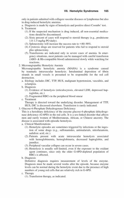
VII. Hemolytic Syndromes 165
only in patients admitted with collagen vascular diseases or lymphomas but alsoin drug-induced hemolytic anemias.a. Diagnosis is made by signs of hemolysis and positive direct Coombs’ test.b. Treatment
(1). If the suspected mechanism is drug induced, all non-essential medica-tions should be discontinued.
(2). Sixty percent of cases will respond to steroid therapy (e.g., prednisone1.0–1.5 mg/kg PO daily).
(3). Splenectomy will increase the success rate to ≈80–90%.(4). Cytotoxic drugs are reserved for patients who fail to respond to steroid
plus splenectomy.(5). Transfusions are indicated only in severe cases of anemia. In emer-
gency situations, most patients can be managed with careful transfusion(ABO- & Rh-compatible blood) administered slowly while watching forreactions.
2. Microangiopathic Hemolytic AnemiaMicroangiopathic hemolytic anemia (MAHA) is a syndrome causedby traumatic intravascular hemolysis. Intraluminal deposition of fibrinstrands in small vessels is presumed to be responsible for the red celldestruction.a. Etiology includes DIC, TTP, HUS, malignant hypertension, vasculitis, and
eclampsia.b. Diagnosis
(1). Evidence of hemolysis (reticulocytosis, elevated LDH, depressed hap-toglobin, etc.)
(2). Fragmented RBCs in the peripheral blood smearc. Treatment
Therapy is directed toward the underlying disorder. Management of TTP,HUS, DIC is discussed elsewhere. Transfusion is rarely indicated.
3. Glucose-6-Phosphate Dehydrogenase DeficiencyThis is a hereditary deficiency of the enzyme glucose-6-phosphate dehydroge-nase deficiency (G-6PD) in the red cells. It is a sex-linked disorder that affectsmen and rarely women of Mediterranean, African, or Chinese ancestry. Thedisease is associated with episodic hemolysis.a. Clinical Manifestations
(1). Hemolytic episodes are sometimes triggered by infections or the inges-tion of some drugs (e.g., sulfonamides, antimalarials, nitrofurantoin,nalidixic acid, etc.).
(2). Patients present with acute intravascular hemolysis associatedwith hemoglobinemia, hemoglobinuria, decreased haptoglobin, andjaundice.
(3). Peripheral vascular collapse can occur in severe cases.(4). Hemolysis is usually self-limited, even if the exposure to the oxidant
agent continues, since only the older G-6PD-depleted population ofRBCs is affected.
b. DiagnosisDefinitive diagnosis requires measurement of levels of the enzyme.Diagnosis must be made several weeks after the episode, because enzymelevels can be normal during the hemolytic event due to the presence of highnumbers of young red cells that are relatively rich in G-6PD.
c. Therapy(1). Transfusion therapy, as indicated.

166 7. Hematologic Disorders
(2). Protection of renal function during hemolytic episodes: IV hydration tomaintain a good urine output, alkalinization of urine (to keep urine pH>7.0).
(3). Prevention of hemolytic episodes can be accomplished by identifyingdeficient individuals, treating infections promptly, and avoiding exposureto oxidant agents.
� VIII. USEFUL FACTS AND FORMULAS
Patients in the ICU frequently have hematologic problems. These include anemia,coagulopathies, and thrombocytosis, to name just a few. In evaluation of thesepatients, many laboratory tests and indices are obtained. The following formulas willaid the critical care practitioner in evaluating these hematologic parameters:
A. Red Blood Cells. The mean corpuscular volume (MCV) indicates the averagevolume of a single RBC in a given blood sample and is calculated as follows:
MCV = Hct(%) × 10
RBC(1012/L)
The mean corpuscular hemoglobin (MCH) indicates the average weight ofhemoglobin per erythrocyte:
MCH = Hb(g/dL) × 10
RBC(1012/L)
The mean corpuscular hemoglobin concentration (MCHC) indicates the averageconcentration of hemoglobin in the RBCs of any specimen:
MCHC = Hb(g/dL)
Hct(%)× 100
The red blood cell volume can be calculated via a radionuclide study:
RBC volume = cpm of isotope injected
cpm/mL RBC in sample
where cpm = counts per million.
B. Reticulocyte Counts. To calculate the percentage of reticulocytes, usually basedon counting 1,000 RBCs, the following formula is commonly utilized:
Reticulocytes(% ) = Number of reticulocytes
Number of RBCs observed× 100

VIII. Useful Facts and Formulas 167
The actual reticulocyte count (ARC) reflects the actual number of reticulocytes in1 L of whole blood:
ARC = Reticulocytes(% )
100× RBC count ( × 1012/L) × 1,000
The corrected reticulocyte count (CRC) is calculated as
CRC = Reticulocytes (% ) × Hct(L/L)
0.45L/L
The reticulocyte count is usually viewed in relation to the degree of anemia. Thereticulocyte production index (RPI) is a frequently used correction method:
RPI = (Measured Hct/Normal Hct) × Reticulocyte count
Maturation time in peripheral blood
The maturation factor varies according to the hematocrit in the manner shown inTable 7.7.
A normal RPI is 1.0; an RPI of 3.0 or more represents adequate response ofthe marrow to anemia. An RPI of < 2.0 represents an inadequate response in thepresence of anemia.
C. Anemias. The RBC indices (MCV, MCHC, MCH) are frequently utilized toclassify anemias (see Table 7.8).
Table 7.9 depicts the laboratory differentiation of microcytic anemias.
D. Hemolytic Disorders. Table 7.10 depicts some of the common RISC morphologicabnormalities encountered in patients with hemolytic disorders:
Table 7.7. Maturation of Reticulocytes in Peripheral Blood
Hematocrit Maturation time in days
0.41–0.50 10.30–0.40 1.50.20–0.39 20.10–0.19 2.5
Table 7.8. RBC Indices in Hypochromic and Microcytic Anemias
MCV (fl) MCHC (g/dL) MCH (pg)
Normal 83–96 32–36 28–34Hypochromic 83–100 28–31 23–31Microcytic 70–82 32–36 22–27Hypochromic-microcytic 50–79 24–31 11–29

168 7. Hematologic Disorders
Table 7.9. Differentiation of Microcytic Anemias
Abnormality Ferritin Serum iron TIBC RDW
Chronic disease N/↑ ↓ ↓ NIron deficiency ↓ ↓ ↑ ↑Sideroblastic anemia N/↑ ↑ N NThalassemia N/↑ N/↑ N N/↑
N = Normal; ↑ = increased; ↓ = decreasedAbbreviations: RDW, red cell distribution width; TIBC, total iron-binding capacity.
Table 7.10. RBC Morphologic Abnormalities in Hemolytic Disorders
Abnormality Hemolytic disorder
Congenital Acquired
Fragmented cells(schistocytes)
Unstable hemoglobins(Heinz body anemias)
Microangiopathicprocesses
Prosthetic heartvalves
Permanently sickledcells
Sickle cell anemia
Spur cells(acanthocytes)
Abetalipoproteinemia Severe liverdisease
Spherocytes Hereditary spherocytosis Immune,warm-antibodytype
Target cells Thalassemia Liver diseaseHemoglobinopathies(Hb C)
Agglutinated cells Immune, coldagglutinin disease
E. Human Hemoglobins. Table 7.11 depicts the normal human hemoglobins atdifferent stages of life.
To convert colorimetric readings into grams of Hb/dL (g/dL) using a standardcurve set up with the same equipment and reagents used for specimen or calcu-late specimen concentration (Cu) based on Beer’s law, the following formula isutilized:
Cu(g/dL) = 301(Au × Cs)
As× 1
1,000= 0.301(Au × Cs)
As
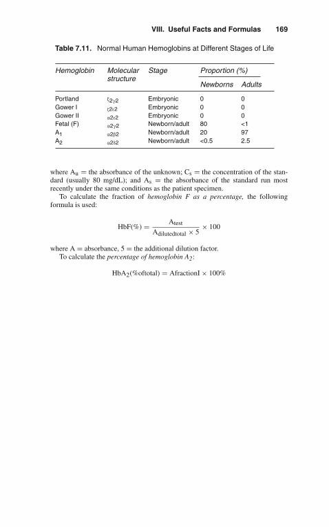
VIII. Useful Facts and Formulas 169
Table 7.11. Normal Human Hemoglobins at Different Stages of Life
Hemoglobin Molecularstructure
Stage Proportion (%)
Newborns Adults
Portland ζ2γ2 Embryonic 0 0Gower I ζ2ε2 Embryonic 0 0Gower II α2ε2 Embryonic 0 0Fetal (F) α2γ2 Newborn/adult 80 <1A1 α2β2 Newborn/adult 20 97A2 α2δ2 Newborn/adult <0.5 2.5
where Au = the absorbance of the unknown; Cs = the concentration of the stan-dard (usually 80 mg/dL); and As = the absorbance of the standard run mostrecently under the same conditions as the patient specimen.
To calculate the fraction of hemoglobin F as a percentage, the followingformula is used:
HbF(%) = Atest
Adilutedtotal × 5× 100
where A = absorbance, 5 = the additional dilution factor.To calculate the percentage of hemoglobin A2:
HbA2(%oftotal) = AfractionI × 100%

8Infectious Diseases
The number of infectious complications encountered in the intensive care unit (ICU)continues to increase. Patients who otherwise would have not survived in the past arenow improving due to new technical advancements. However, the length of stay, aswell as the large number of devices employed for this purpose, predisposes patients todifficult and often fatal infections. Clinical characteristics of patients who are treatedin the ICU have evolved in recent years. Those who are immunocompromised, post-transplant, and the geriatric population are now regularly treated in the ICU with theconsequent increase in morbidity, mortality, and cost.
From the infectious diseases point of view, the approach to a critically ill patientwho is admitted to the ICU should immediately differentiate if the patient was trans-ferred from the floor versus if the patient was directly admitted to the ICU fromthe community. This constitutes a paramount parameter to categorize the etiologicagents, to understand the pathophysiology of their processes, and mostly to decidewhich therapeutic antimicrobial interventions are needed.
� I. PNEUMONIA (NOSOCOMIAL)
A. If the patient is transferred to the ICU after being in the hospital for several days,then treatment should address the nosocomial aspect of infection and the followingimportant facts:1. Mortality rates among these patients are 20–60%.2. These patients represent 15% of all hospital deaths.3. Successful treatment depends upon underlying disease, specific causative
organisms, and timely institution of therapy.
B. Predisposing Factors1. Intubation.2. ICU: Especially the patient who is receiving sedation.3. Antibiotics: Broad-spectrum agents will rapidly change normal flora of the
mouth and gastrointestinal (GI) tract.4. Surgery: Especially thoracic, abdominal, or neurosurgery, which increases the
risk of aspiration.5. Chronic lung disease.
J. Varon, P. Acosta, Handbook of Critical and Intensive Care Medicine,DOI 10.1007/978-0-387-92851-7_8,C© Springer Science+Business Media, LLC 2010
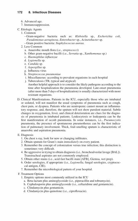
172 8. Infectious Diseases
6. Advanced age.7. Immunosuppression.
C. Etiologic Agents1. Common
– Gram-negative bacteria such as: Klebsiella sp., Escherichia coli,Pseudomonas aeruginosa, Enterobacter sp., Acinetobacter sp.
– Gram positive bacteria: Staphylococcus aureus.2. Less Common
a. Anaerobic mouth flora (i.e., streptococci)b. Other gram-negative bacilli (i.e., Serratia sp., Xanthomonas sp.)c. Haemophilus influenzaed. Legionella sp.e. Candida sp.f. Aspergillus sp.g. Influenza virush. Streptococcus pneumoniaei. Miscellaneous: according to prevalent organisms in each hospitalj. Tuberculosis (TB, typical and atypical)
Another helpful approach is to consider the likely pathogens according to thetime after hospitalization the pneumonia developed. Late-onset pneumonia(after more than 5 days of hospitalization) is usually characterized with moreresistant organisms.
D. Clinical Manifestations. Patients in the ICU, especially those who are intubatedor sedated, will not manifest the usual symptoms of pneumonia such as cough,chest pain, or dyspnea. Patients who are neutropenic cannot mount an inflamma-tory response, and, therefore, the sputum will not show purulent material. Subtlechanges in oxygenation, fever, and clinical deterioration are clues for the diagno-sis of pneumonia in intubated patients. Leukocytosis or leukopenia can be thefirst manifestation of occult pneumonia. In some instances, i.e., Pneumocystispneumonia, the presence of spontaneous pneumothorax can be the first indica-tion of pulmonary involvement. Thick, foul-smelling sputum is characteristic ofanaerobic and aspiration pneumonia.
E. Diagnosis1. On chest x-ray, look for new or changing infiltrates.2. Obtain sputum for Gram’s stain immediately on every patient.3. Remember the concept of colonization versus true infection; this distinction is
sometimes very difficult.4. Be aggressive in trying to obtain diagnosis (i.e., bronchoalveolar lavage [BAL]).
Transtracheal aspirates are not commonly employed.5. Obtain other stains (i.e., acid-fast bacilli stain [AFB], Giemsa, wet prep).6. Order serologies, if appropriate (i.e., Legionella, fungal serologies, cryptococ-
cal antigen, CIE).7. Remember the microbiological pattern of your hospital.
F. Treatment Options1. Empiric options most commonly utilized in the ICU
a. Beta-lactam plus aminoglycoside (i.e., piperacillin and tobramycin).b. Cephalosporin plus aminoglycoside (i.e., ceftazidime and gentamicin).c. Clindamycin plus gentamicin.d. Clindamycin plus quinolone (i.e., ciprofloxacin).

II. Community-Acquired Pneumonia 173
e. Imipenem/cilastatin plus aminoglycoside.f. Cephalosporin plus fluoroquinolone.g. Add trimethoprim-sulfamethoxazole [TMP-SMX] if Pneumocystis carinii
pneumonia is suspected.h. Add erythromycin or azithromycin 500 mg IV qd or erythromycin 0.5–1 g
IV q6 h if Legionella is suspectedi. TMP-SMX 15–20 mg/kg/d TMPj. Doxycycline 100 mg IV q12 hk. Rifampin 300 mg IV q12 hl. Amphotericin B 0.6–1 mg/kg/d
Duration of therapy is not well defined, but most authors agree on treat-ing gram-negative and anaerobic pneumonia for 10–21 days. Gram-positiveprocesses are usually treated between 10 and 14 days, and atypical pneumo-nias receive 2 weeks of antimicrobial therapy. Candida pneumonia requiresprolonged treatment with up to 1.5 g of amphotericin B as a total dose.
G. Prevention1. Preoperative and postoperative measures for prevention of pneumonia
a. Identification of high-risk patientsb. Treatment of respiratory infections, removal of respiratory secretionsc. Instruction and therapy to expand patients’ lungs (i.e., chest physiotherapy,
incentive spirometry)2. Proper hand washing3. Appropriate maintenance of in-use respiratory therapy equipment
a. Use of sterile fluids in nebulizersb. Proper use of single-dose and multidose medications for respiratory therapy
4. Proper sterilization and disinfection of reusable respiratory equipment5. Proper suctioning of the respiratory tract6. Protection of patients from other infected patients or staff
� II. COMMUNITY-ACQUIRED PNEUMONIA
A. Common Organisms1. Streptococcus pneumoniae2. Mycoplasma pneumoniae3. Haemophilus influenzae4. Klebsiella sp.5. Respiratory viruses (Influenza A and B, Adenovirus, Respiratory syncytial
virus, Parainfluenza)6. Legionella sp.
B. Other Less Common Organisms1. Pneumocystis carinii2. Mycobacterium tuberculosis3. Cryptococcus sp.4. Chlamydia psittaci5. Histoplasma sp.6. Nocardia sp.

174 8. Infectious Diseases
C. Common Manifestations1. Fever, cough, dyspnea, sputum production usually purulent but not in all cases.2. Hypoxemia is common.3. Anxiety.4. Leukocytosis; also leukopenia in severe infections.
D. Uncommon Presentations in Patients Who Are1/41. Elderly2. Immunocompromised (especially neutropenic)3. Post-transplantation
E. Clinical Clues for Diagnosis1. Acute onset: bacterial, viral, aspiration, tularemia, Pneumocystis2. Subacute onset: viral, Legionella, Haemophilus sp., Mycoplasma, Q fever,
Psittacosis, Chlamydia, Pneumocystis3. Aerogenous route: any segment4. Hematogenous: most commonly in both bases, as blood flow is preferential to
these areas
F. Associations1. Birds: psittacosis2. Turtles: typhoid3. Dogs: Pasteurella multocida4. Cattle: Q fever5. Rabbits: tularemia6. Air conditioners: Legionella7. COPD and smoking: H. Influenzae, Pseudomonas aeruginosa8. Hides: anthrax9. Foreign travel: Echinococcus, paragonimiasis
10. Barracks: Neisseria meningitidis, group A Streptococcus
G. Treatment. Empiric treatment is usually dictated by the geographical background,clinical presentation, and host status.
◦ Levofloxacin 750 mg.or
◦ Ceftriaxone 1 g IV plus azithromycin 500 mg IV1. Streptococcus pneumonia and Haemophilus influenza
a. Quinolone (moxifloxacin or levofloxacin)b. Ertapenem (1 g q24 h)c. Ceftriaxone (1 g q24 h)
2. Legionella sp., Mycoplasma pneumoniae, Chlamydia pneumoniaea. Moxifloxacin (400 mg IV qd)b. Levofloxacin (500 mg IV qd)c. Doxycycline (200 mg IV qd)
3. Pseudomonas aeruginosaa. Meropenem (2 g IV q8 h)b. Cefepime (2 g IV q8 h) and Amikacin (1 g IV q24 h)c. For multidrug resistant p. aeruginosa Colistin (80 mg IV q8 h)
4. Influenza A/B, Avian Influenzaa. Oseltamivir (Tamiflu) 75 mg PO q24 h plus Rimantadine 100 mg PO.b. Avian Influenza (Influenza virus type A H5N1)–Influenza following
close contact with infected poultry. Several outbreaks in humans havebeen identified in Asia: flu-like symptoms with vague gastrointestinal

IV. Sepsis 175
complaints that rapidly progress to acute respiratory failure. Diagnosis isby hemagglutinin-specific RT-PCR for avian influenza. Treatment withantivirals should be given early, and it includes: Oseltamivir (150 mg),with amantadine and rimantadine.
H. Complications After 72 h1. Persistent fever2. Empyema3. Obstruction4. Lung abscess5. Resistant organism6. Focus of infection
� III. SEVERE ADULT RESPIRATORY SYNDROME(SARS)
Term given by the World Health Organization, describes a rapidly progressive respi-ratory illness with documented outbreaks in China, Hong Kong, Vietnam, Singapore,and Canada. The presumable pathogen is a coronavirus that spreads person to personvia droplets, sewage, and water and potentially through human feces. It is hypoth-esized that bats are the primary reservoir for the disease. The mortality rate fromSARS is high (up to 20%). This is a two-stage illness:
1. Prodrome: includes fever, malaise, headache, myalgias, and diarrhea may occur.
2. Respiratory phase: non-productive cough and dyspnea that rapidly progress torespiratory failure.
There is no current treatment available for this illness, except for supportive care inthe intensive care unit. Preventive measures against SARS are mainly focused ontravel advisories to countries and cities with active outbreaks. Efforts are underwayto prepare a vaccine for the prevention of SARS.
� IV. SEPSIS
A. More than 750,000 cases of sepsis, with an associated mortality of 20–60%, areestimated to occur annually. Despite improvements in antimicrobial therapy andsupportive care, the incidence of and mortality associated with sepsis have notdeclined. This is, in part, a consequence of an array of medical advances thatcan place patients at increased risk for development of infection and, potentially,sepsis.
B. Sepsis and Related Disorders1. Definitions
a. Bacteremia: Positive blood cultures (may be transient)b. Sepsis: Clinical evidence suggestive of infection plus signs of a systemic
response to the infection (all of the following):

176 8. Infectious Diseases
(1). Tachypnea (respiration 20 breaths per minute; if patient is mechanicallyventilated, minute volume 10 L/min)
(2). Tachycardia (heart rate >90 beats per minute)(3). Hyperthermia or hypothermia (core or rectal temperature >38.4◦C
[101◦F] or <35.6◦C [96.1◦F])c. Sepsis Syndrome (may also be considered incipient septic shock in patients
who later become hypotensive): Clinical diagnosis of sepsis outlined above,plus evidence of altered organ perfusion (one or more of the following):(1). PaO2/FiO2 no higher than 280 (in the absence of other pulmonary or
cardiovascular disease).(2). Lactate level above the upper limit of normal.(3). Oliguria (documented urine output <0.5 mL/kg body weight for at least
1 h in patients with urinary catheters in place).(4). Acute alteration in mental status.(5). Positive blood cultures are not required.
d. Early Septic Shock: Clinical diagnosis of sepsis syndrome as outlinedabove, plus hypotension (systolic blood pressure <90 mmHg or a 40-mmHgdecrease below baseline systolic blood pressure) that lasts for <1 h andis responsive to conventional therapy (intravenous fluid administration orpharmacologic intervention)
e. Refractory Septic Shock: Clinical diagnosis of the sepsis syndrome outlinedabove, plus hypotension (systolic blood pressure <90 mmHg or a 40-mmHgdecrease below baseline systolic blood pressure) that lasts for >1 h despiteadequate volume resuscitation and that requires vasopressors
C. Pathophysiology. Cell walls of gram-negative bacteria contain proteins, lipids,and lipopolysaccharides. Endotoxin (lipopolysaccharide) has three components:an O-specific polysaccharide, the R-core, and lipid A. Lipid A may be themajor culprit in initiating the endotoxic symptoms. It is this component of endo-toxin that stimulates the release of tissue necrosis factor (TNF) and can alsoactivate the complement pathway. The sepsis syndrome is caused by endothe-lial damage following endotoxin-stimulated activation of neutrophils, coagu-lation, complement, and macrophages. Macrophages are stimulated to releaseTNF, interleukins, leukotrienes, thromboxane, and other cardioactive substances.Endotoxemia markedly increases the risk of myocardial depression and multi-ple organ failure. In patients who have positive blood cultures, those with severeendotoxemia have 5 times the mortality of those who do not have endotoxemia.
D. Priorities in the Treatment of Sepsisa. Early recognition.b. Cardiovascular/pulmonary support.c. Fluid resuscitation.d. Pressor agents.e. Empiric antibiotic therapy.f. Other immunotherapeutic agents (investigational).g. Corticosteroids are not effective.h. Drainage of any foci of infection.
E. Prognosis. Mortality in sepsis is a function of the severity of physiologic derange-ments, the duration of illness, and the number of organ system failures. Theseorgan systems include, but are not limited to, the lungs, kidneys, and liver.When the pulmonary system becomes dysfunctional, the resultant clinical entity isknown as the adult respiratory distress syndrome (ARDS). The sequence has been

V. Toxic Shock Syndrome 177
termed the multiple-organ dysfunction syndrome (MODS). MODS is the mostcommon cause of demise in patients who experience uncontrolled inflammationand infection.
F. Activated protein C. The presence of coagulation abnormalities is a major prob-lem in patients with severe sepsis and septic shock. Some reports suggest thatsupplementation of activated protein C may produce clinical benefit, by decreas-ing mortality rate. The greater benefit from activated protein C was observed inacutely ill patients (APACHE II score 25). Some evidence shows that patientsreceiving activated protein C have lower incidence of multi-organ system failure,but greater risk of serious bleeding (including fatal intracranial hemorrhage). Acareful selection of patients should be done in order to identify those that willbenefit the most from this therapy.
� V. TOXIC SHOCK SYNDROME
A. Clinical Case Definition (See Table 8.1)1. Severe febrile (38.9◦C) illness with rash (erythroderma followed by desqua-
mation), hypotension or syncope, and multiple organ system involvement (atleast four of the following: mucous membrane, GI, muscular, central nervoussystem [CNS], renal, hepatic, hematologic, cardiopulmonary, metabolic).
2. Hypotension: probably due to small-vessel and capillary leakage with extravas-cular accumulation of fluid (edema).
3. Blood cultures are usually negative.4. Acute episode followed by desquamation5. No evidence of other causes: scarlet fever, Kawasaki’s disease, Rocky
Mountain spotted fever, etc.
B. Epidemiology and Other Clinical Features1. Affects mostly young menstruating women. Tampon use, especially contin-
uous use and Rely brand in some studies. S. aureus colonization of vagina.Recurrence rate of 30%. Decrease in the number of reported cases.
Table 8.1. Toxic Shock Syndrome
Criteria for DiagnosisTemperature <38.9◦CSystolic blood pressure <90 mmHgRash with subsequent desquamation, especially on palms and soles
Involvement of >3 of following organ systems:Gastrointestinal: vomiting or severe diarrheaMuscular: severe myalgias or fivefold increase in creatine kinaseMucous membranes: frank hyperemiaRenal insufficiency: serum urea nitrogen, creatinine, double of normalLiver: enzymes, twice upper limits of normalBlood: thrombocytopenia <100,000/mm3
CNS: disorientation without focal findingsNegative tests for leptospirosis, Rocky Mountain spotted fever, and measles

178 8. Infectious Diseases
2. Also occurs in non-menstruating women, men, and children (colonization orfocal infection with S. aureus, including postoperative infections). Commonoccurrence after surgery. Fatality rate: 5–10%.
C. Etiology. Exotoxin(s) of S. aureus appear to cause the disease. Recently, strepto-cocci have been shown to cause the same syndrome.
D. Differential Diagnosis. Kawasaki’s disease, scarlet fever, leptospirosis, RockyMountain spotted fever, measles.
E. Treatment. The most important treatment is volume expansion and correction ofhypotension; removal of the tampon, if present, in menstruating women; debride-ment of wounds, etc.; and administration of antistaphylococcal antibiotics (aftercultures have been obtained). Steroids have not been proven to be effective or toalter outcome.
� VI. MENINGITIS
A. Acute meningitis is a medical emergency that requires early recognition, rapiddiagnosis, precise antimicrobial therapy, and aggressive ICU support.1. Etiologic Agents
a. Streptococcus pneumoniae: The most common cause in adults.b. Neisseria meningitis: Common among groups of young individuals and
children.c. Haemophilus influenzae: Common in children up to 12 years of age.d. Staphylococcus aureus and S. epidermidis: Seen in the elderly or postopera-
tively (CNS shunts).e. Listeria monocytogenes: Usually mistaken with diphtheroids or contami-
nants.f. Streptococci other than S. pneumoniae: Especially group B in neonatal
disease.g. Gram-negative bacilli: After surgery or trauma.h. Mycobacterium tuberculosis: Increasing in frequency.i. Cryptococcus: Usually in immunosuppressed patients (i.e., those with
acquired immune deficiency syndrome [AIDS], or impaired cell-mediatedimmunity).
j. Syphilis: presentation variable.k. Herpes simplex.l. Toxoplasma: can present as meningoencephalitis or brain abscess.
m. Naegleria: epidemiological history is paramount.n. Other viruses (i.e., echovirus, St. Louis, equine, and Western encephalitis).
2. Associations: Epidemiology and Organismsa. Summer and fall: Coxsackie or echovirus; leptospirab. Previous meningitis: S. pneumoniaec. Alcoholism: S. pneumoniaed. Young adults: N. meningitise. Elderly: S. pneumoniae, listeria, gram-negative bacillif. Lymphoma: Cryptococcus sp.g. Petechia: N. meningitidis, echovirush. Sinusitis: H. influenzae, S. pneumoniae, anaerobic bacteria

VI. Meningitis 179
i. Cellulitis: aerobic, gram-positive coccij. Brain abscess: mixed florak. Swimming in fresh water: amoebasl. Other family members with meningitis: N. meningitidis
m. Water contact: leptospiran. Hospital acquired: gram-negative bacilli, staphylococcus, candidao. Head trauma
(1). Close fracture: S. pneumoniae, gram-negative bacilli(2). Craniotomy:gram-negative bacilli, staphylococci(3). Cerebrospinal fluid rhinorrhea: S. pneumoniae
3. Cerebrospinal Fluid (CSF) Findings (See Table 8.2)4. Diagnostic Approach
a. Order antigen detection for H. influenzae, S. pneumoniae, N. meningitidis.b. Obtain high-volume CSF for AFB concentrate and fungal cultures (20–30
mL).c. If CSF is normal or viruses are suspected, repeat lumbar puncture (LP) in
24–36 h.d. Upon admission, obtain serologies for viral infections (i.e., St. Louis
encephalitis, California encephalitis).e. Obtain serologies in serum and CSF for fungal infections.f. Polymerase chain reaction (PCR) may be helpful (especially for TB and
cytomegalovirus [CMV] infections).5. Treatment
In acutely ill patients, the goal of therapy is to institute treatment before thepathologic process of inflammation can produce irreversible progression and/ordeath. Time is essential in this situation. Empiric therapy is instituted immedi-ately after diagnosis is made, and it is based on the recognition of a communityversus hospital and/or postoperative process. For community-acquired menin-gitis, usual treatment includes a third-generation cephalosporin (i.e., cefotaxime
Table 8.2. CSF Findings in Meningitis According to Etiology
Bacterial Tuberculous Viral Chronic
Glucose>40 mg/dL (Bloodratio <0.4)
30–45 mg/dL 20–40 mg/dL 30–40 mg/dL
Protein100–500 mg/dL
100–500 mg/dL 50–100 mg/dL 100–500 mg/dL
White blood cells 100–400/cc3 10–1,000/cc3 100–500/cc3
1000–10,000/cc3
Gram’s stain (+)60–80%(untreated)40–50%(previously treated)
AFB smear (+) inup to 40%
Smears areusuallynegative
Special stainsneeded: India ink(+)75% AFB(+)30%

180 8. Infectious Diseases
3 g IV q6 h or ceftriaxone 2–4 g q12–24 h). Vancomycin should be added tothis regimen until culture and susceptibility results are available.
B. Pneumococcal Meningitis1. Pneumococcal meningitis is still the most common cause of bacterial meningi-
tis in adults. Underlying diseases: sickle cell disease, splenectomy and splenicdysfunction, hypogammaglobulinemia, alcoholism, head trauma (CSF fistula),and chronic pulmonary, hepatic, or renal disease.
2. Associated infections: pneumonia, otitis, bacteremia, endocarditis, mastoiditis.3. Therapy: Ceftriaxone 4 g/d, Vancomycin 2 g/d should be given if there has been
beta-lactam resistance noted locally.
C. Haemophilus Meningitis1. Underlying disease (adults): alcoholism, compromised host defenses, head
trauma.2. Associated infections: pneumonia, sinusitis, otitis. Secondary cases can occur
in close contacts.3. Therapy: Cefotaxime (2 g IV q6 h), ceftriaxone (2 g IV q12 h), and chloram-
phenicol (500 mg PO q6 h for 2 weeks) as IV to PO switch.
D. Meningococcal Meningitis1. Meningococcal meningitis is seen primarily in children, adolescents, and young
adults. Secondary infection in close contacts can occur. Predisposing factorsinclude complement defects.
2. Disseminated neisserial infection (often recurrent in persons with C5–C8 defi-ciency). Waterhouse-Frederickson syndrome is an acute, often fatal, syndromeof septic shock associated with massive adrenal necrosis, associated with bac-teremia due to this organism. It requires early recognition, antibiotic therapy,and especially aggressive ICU/hemodynamic support.
3. Early antimicrobial therapy is needed. Ceftriaxone 2 g IV q12 h is the preferredIV therapy; as an alternative Meropenem 2 g q8 h can be administered.
E. Listeria Meningitis. Listeria is an important cause of bacteremia and meningitis,particularly in the elderly. Epidemiological history is important. Therapy is withampicillin (2 g q4 h) or Meropenem (excellent in vitro activity against Listeria).
F. Staphylococcus aureus and Staphylococcus epidermidis. Infection with theseorganisms is common after neurosurgery and/or ventricular peritoneal shuntplacement.1. Therapy
a. Methicillin sensitive: Cefotaxime (3 g IV q6 h) or cefepime (2 g IV q8 h).b. Methicillin resistant: Linezolid (600 mg IV q12 h) or vancomycin (2 g IV
q12 h).c. An infected shunt may need to be removed early in the course of ther-
apy if the patient is not responding. Repeat LP at 2–3 days is needed inorder to reach this decision (persistent growth of organisms, despite adequatetherapy).
G. Gram-Negative Bacilli1. Infections with gram-negative bacilli are challenging to treat due to their high
morbidity and mortality. Development of resistance can occur while on therapy(especially with Enterobacter sp.). Most organisms will respond to ceftriaxone,cefotaxime, or ceftazidime. For Pseudomonas aeruginosa, ceftazidime 2 g IVq8 h is the drug of choice. It should be given with gentamicin (1–2 mg/kg/8 h).

VII. Infections in Patients with Aids 181
H. Complications of Bacterial Meningitis1. Brain Abscess: Usually follows trauma, contiguous infection, hematogenous
dissemination.2. Subdural Empyema: Primarily disease of the young but, in elderly, may
complicate neurosurgery or subdural hematoma.3. Epidural Abscess: Usually accompanied by focal osteomyelitis and subdural
empyema.4. All of the above are caused by mixed bacteria and usually require drainage, as
well as prolonged IV antibiotic therapy.
I. Herpes Meningitis/Encephalitis. Herpes meningitis/encephalitis is a devastat-ing necrotizing type of encephalitis. Temporal spikes on electroencephalogram(EEG) are characteristic. Treatment is given with acyclovir 15 mg/kg q8 h (highdose) for 2 weeks. Careful attention to hydration is mandatory to avoid renalinsufficiency.
� VII. INFECTIONS IN PATIENTS WITH AIDS
A. Opportunistic infections are the most common causes of morbidity and mortalityin patients with human immunodeficiency virus (HIV). Patients with CD4 cells<250 are at risk for developing severe infectious complications. Their approach isdepicted in Table 8.3.
B. Summary of Current Therapeutic Approaches1. Pulmonary Disease
a. Disease due to Pneumocystis carinii (Pneumocystis jirovecii) pneumonia(PCP) (Table 8.4)
b. Disease due to M. Tuberculosis(1). Start with at least four drugs, preferably five; INH 300 mg/d, rifampin
600 mg/d, pyrazinamide 15/kg/d, ciprofloxacin 750 mg PO bid, etham-butol 15–20 mg/kg/d.
(2). If TB is sensitive to INH and/or rifampin, continue for 12–18 months(not in the ICU).
(3). If TB is resistant to either or both drugs (INH and Rifampin), mul-tiple drug resistant, continue with 5–6 drugs, and adjust according tosensitivities. Prognosis is very poor.
(4). Follow liver function tests, initially weekly and later monthly.(5). If patient cannot use PO drugs, give IV INH and rifampin (same dose)
and IM streptomycin (1 g/d).c. Pulmonary Disease Due to Histoplasma capsulatum
(1). Initiate therapy with amphotericin B at 0.8–1 mg/kg/d.(2). Search for other sites of involvement (i.e., bone marrow biopsy, lumbar
puncture, chest x-ray, barium enema, and small bowel series).(3). Once the patient is stable, switch to fluconazole 200 mg PO bid.
d. Pulmonary Disease Due to Legionella sp.(1). Initiate therapy with erythromycin 3–4 g IV/d.(2). If the patient is not responding, add rifampin (600 mg/d) and/or
ciprofloxacin 400 mg IV q12 h.
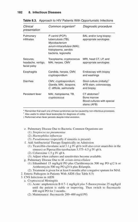
182 8. Infectious Diseases
Table 8.3. Approach to HIV Patients With Opportunistic Infections
Clinicalpresentation
Common organism∗ Diagnostic procedure
Pulmonaryinfiltrates
P. carinii (PCP);tuberculosis (TB);Mycobacteriumavium-intracellulare (MAI);histoplasma, aerobicbacteria, legionella
BAL and/or lung biopsy;appropriate serologies
Seizures,headache, vertigo,facial palsy
Toxoplasma, cryptococcusMAI, herpes, CMV
MRI, head CT, LP, andappropriate serologies
Esophagitis Candida, herpes, CMV,cryptosporidium
Endoscopy with biopsyand washings
Diarrhea CMV, cryptosporidium,Giardia, MAI, Isospora,C. difficile, salmonella
Stool culture (initially)†
AFB stain, colonoscopy,and biopsy
Persistent fever MAI, histoplasma, TB,cryptococcus
CT abdomen‡
Bone marrowBlood cultures with specialstains (AFB)
∗ Remember that each one of these syndromes can be caused by non-infectious processes.† Also useful to obtain fecal leukocytes for diagnosis of colitis.‡ Performed when fever persists despite initial evolution.
e. Pulmonary Disease Due to Bacteria. Common Organisms are(1). Streptococcus pneumoniae(2). Haemophilus influenzae(3). Pseudomonas (especially if sinusitis is present)
f. Add Antibacterial Therapy Empirically on Admission(1). Ticarcillin-clavulanic acid 3.1 g IV q6 h (will also cover anaerobes in the
sinuses) or Piperacillin-tazobactam 3.375–4.5 g IV q6 h.(2). Cefuroxime 1.5 g IV q8 h.(3). Adjust when cultures and sensitivities become available.
g. Pulmonary Disease Due to M. avium-intracellulare(1). Ethambutol 15 mg/kg/d PO plus Clarithromycin 500 mg PO q12 h or
Azithromycin 500 mg PO q24 h plus Rifampin.(2). Treatment is given for at least 6 months after a negative sputum for MAI.
2. Enteric Pathogens in Patients With AIDS (See Table 8.5)3. CNS Infections in AIDS
a. Cryptococcal Meningitis(1). Acute: amphotericin B 0.7–1 mg/kg/d plus 5-fluorocytosine 25 mg/kg/d
until the patient is stable or improving. Then switch to fluconazole400 mg/d PO for 3 months.
(2). Maintenance: fluconazole 200–400 mg/d PO.

VII. Infections in Patients with Aids 183
Table 8.4. Recommended Management for PCP
Antibiotic Mild to moderate Severe (Usually in ICU)
TMP-SMX 2–3 double-strength tabsPO tid for 14–21 days
5 mg/kg IV q6 h for3 weeks
Pentamidine 3–4 mg/kg IV-IM qd 4 mg IV qd (once a day)
Trimethoprim-dapsone
Trimethoprim 100 mgPO tid Dapsone 100 mgPO Qid
?
Clindamycin-primaquine
Clindamycin 600 mgPO tid Primaquine 30 mgPO qid
900 mg IV q8 h for 3 weeks
Atovaquone 750 mg PO bid For 2–3 weeks
Trimetrexate-leucovorin
Trimetrexate 45 mg/m2/dIV for 21 days Leucovorin30 mg/m2 IV q6 h for 10days, and then PO q6 h for14 days
Same as mild to moderate
Solumedrol
Corticosteroidsadjunctive therapy
? 40 mg IV or (equivalentPO bid) for 5 daysWean gradually over10 days
b. Toxoplasmosis(1). Pyrimethamine 200 mg PO: loading dose followed by 75 mg PO daily
with folinic acid 5 mg PO daily. (No IV presentation available.)(2). Sulfadiazine 1.5 g PO q6 h; plus Leucovorin 10 mg PO
q24 h.c. CMV (Including Retinitis)
(1). Ganciclovir 5–10 mg/kg IV q12 h for 14 days (initial therapy)(2). Foscarnet 60 mg/kg IV q8 h for 14 days (initial therapy)(3). Lifelong suppressive therapy with valganciclovir 900 mg PO q24 h.
d. Herpes Simplex(1). Acyclovir 10–15 mg/kg IV q8 h
e. Syphilis(1). Crystalline penicillin 24 million U/d for 14 days(2). Ceftriaxone 2–4 g/d IV for 14 days
C. Important Facts to Remember in Treating HIV-Infected Patients in the ICU1. Patients may have more than one infection at the same time.2. Blood precautions should be instituted immediately to avoid unnecessary
exposure.3. Noninfectious processes (i.e., tumors) can mimic infections.

184 8. Infectious Diseases
Table 8.5. Enteric Pathogens Commonly Seen in Patients WithAIDS
Organism Antimicrobial agent Direction oftherapy (days)
G. lamblia Metronidazole 250 mg PO tid 5
E. histolytica Metronidazole 750 mg tid anddiiodohydroxyquin 650 mg PO tid
10
Shigella sp. Fluoroquinolone IV or PO 3–7
C. jejuni Ciprofloxacin 500 mg IV q12 h 7
I. belli TMP-SMX 1 double-strength qd 14
CMV Ganciclovir 5 mg/kg IV q12 h 30
Herpes simplex Fluconazole 100 mg PO q24 h 14
Oral thrush Ketoconazole 200–400 mg/d PO 10
Candidaesophagitis
Fluconazole 200–400 mg/d IV 7–10
4. Patients require a full physical examination daily, including mouth, perirectalarea, and eyes.
5. Superinfections are common (i.e., fungal and resistant bacteria).6. When fever persists, consider lumbar puncture, liver, and bone marrow biopsy.7. Obtain CD4-CD8 counts if not recently clone.8. Code status needs to be established early.9. Privacy of and respect toward patient are essential and mandatory.
� VIII. INFECTIONS IN THEIMMUNOCOMPROMISED HOST
A. The number of critically ill patients with impaired host defense mechanisms whoare admitted to the ICU has dramatically increased in recent years. The knowledgeand recognition of the basic deficiency enable the physician to predict the type andsite of infection and allow the institution of early empiric therapy (see Tables 8.6and 8.7).
B. Immunocompromised patients admitted to the ICU should be categorized accord-ing to the time of acquisition of infection.
Hospital-acquired infections have different etiologic agents compared to thosefrom the community, despite having the same basic immunologic defect.
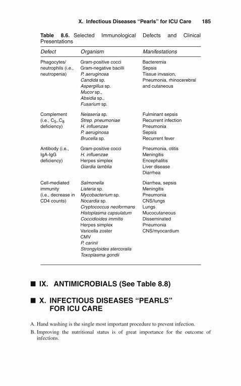
X. Infectious Diseases “Pearls” for ICU Care 185
Table 8.6. Selected Immunological Defects and ClinicalPresentations
Defect Organism Manifestations
Phagocytes/neutrophils (i.e.,neutropenia)
Gram-positive cocciGram-negative bacilliP. aeruginosaCandida sp.Aspergillus sp.Mucor sp.,Absidia sp.,Fusarium sp.
BacteremiaSepsisTissue invasion,Pneumonia, rhinocerebraland cutaneous
Complement(i.e., C5–C8deficiency)
Neisseria sp.Strep. pneumoniaeH. influenzaeP. aeruginosaBrucella sp.
Fulminant sepsisRecurrent infectionPneumoniaSepsisRecurrent fever
Antibody (i.e.,IgA-IgGdeficiency)
Gram-positive cocciH. influenzaeHerpes simplexGiardia lamblia
Pneumonia, otitisMeningitisEncephalitisLiver diseaseDiarrhea
Cell-mediatedimmunity(i.e., decrease inCD4 counts)
SalmonellaListeria sp.Mycobacterium sp.Nocardia sp.Cryptococcus neoformansHistoplasma capsulatumCoccidioides immitisHerpes simplexVaricella zosterCMVP. cariniiStrongyloides stercoralisToxoplasma gondii
Diarrhea, sepsisMeningitisPneumoniaCNS/lungsLungsMucocutaneousDisseminatedPneumoniaCNS/myocardium
� IX. ANTIMICROBIALS (See Table 8.8)
� X. INFECTIOUS DISEASES “PEARLS”FOR ICU CARE
A. Hand washing is the single most important procedure to prevent infection.
B. Improving the nutritional status is of great importance for the outcome ofinfections.
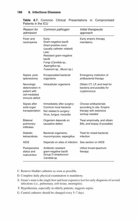
186 8. Infectious Diseases
Table 8.7. Common Clinical Presentations in CompromisedPatients in the ICU
Reason foradmission
Common pathogen Initial therapeuticapproach
Fever andneutropenia
Early: Early empiric therapymandatoryGram-negative bacilli
Gram-positive cocci(usually catheter related)Late:Resistant gram-negativebacilliFungi (Candida sp.,Aspergillus sp.,Fusarium sp., Mucor sp.)
Sepsis: post-splenectomy
Encapsulated bacterialorganisms
Emergency institution ofantibacterial therapy
Neurologicdeterioration inpatient withcell-mediatedimmune deficit
Intracellular organisms Obtain CT, LP, and treat forbacteria and possibly forcryptococcus
Sepsis aftersolid organtransplantation
Immediately after surgery:Common local bacteria
Choose antibacterialsaccording to site. Empirictherapy with extensiveworkup needed
Not related to surgery:Virus, fungus, nocardia
Bilateralpulmonaryinfiltrates
Organism depends oncausative defect
Treat empirically, and obtainBAL and biopsy (if possible)
Diabeticketoacidosis
Bacterial organisms,mucormycosis, aspergillus
Treat for mixed bacterialinfection
AIDS Depends on sites of infection See section on AIDS
Postoperativestatus andmalnutrition
Antibiotic-resistantgram-negative bacilli
Utilize broad-spectrumtherapy
Group D streptococciCandida sp.
C. Remove bladder catheters as soon as possible.
D. Complete daily physical examination is mandatory.
E. Gram’s stain is the single best and least expensive test for early diagnosis of severalinfections (i.e., pulmonary, soft tissue, meningitis).
F. Hypothermia, especially in elderly patients, suggests sepsis.
G. Central catheters should be changed every 5–7 days.

X. Infectious Diseases “Pearls” for ICU Care 187
Table 8.8. Selected Antimicrobials Commonly Used in the ICU
Drug Dose Renaladjustment:Creatinineclearance
Comments andside effects
>8050–10<10
Aminoglycosides (i.e.,gentamicin)
1–2 mg/kg IVq8 h
8–12 h Monitor levels, renalfunction, and hearing12 h
24–48 h
Broad-spectrumpenicillin (i.e.,piperacillin)
3–4 g IV q8 h 4–6 h Monitor Na+ andcoagulation profile8–12 h
12–24 h
Imipenem 500 mg to 1 g 6 h12 h24 h
Seizures, twitching,facial palsies
Cephalosporins (i.e.,ceftazidime)
2 g IV q8 h 6–12 h12 h14 h
Penetrates CSF well
Aztreonam 2 g IV q8 h 6–12 h12 h24 h
Tolerated inpenicillin-allergicpatients
Vancomycin 1 g IV q12 h 6–12 h2–3 dweekly
Monitor levels;interstitial nephritis
Oxacillin 6–12 g IV 4–6 h6–8 h8–12 h
Infuse in at least 1 h
Acyclovir 2–3 g/d IV 8 h12–24 h24–48 h
Monitor WBC andrenal function
Ganciclovir 5 mg/kg IV 12 h12 h24–48 h
Monitor bone marrowdepression
Clindamycin 600–900 mg IV 8 h8 h8 h
Diarrhea
Chloramphenicol 3–4 g IV or PO 6 h6 h6 h
Monitor bone marrowfunction

188 8. Infectious Diseases
Table 8.8. Continued
Drug Dose Renaladjustment:Creatinineclearance
Commentsandside effects
>8050–10<10
Metronidazole 30 mg/kg/d IVor PO
6 h6 h6 h
Metallic taste
Amphotericin B 0.5–1 mg/kg IV
once a day
24 h24 h48 h
Monitor renalfunction
Fluconazole 200–400 mgq12 h IV or PO
12 h24 h48 h
Interacts withanticoagulants
Itraconazole 2–4 g PO 12–24 h24 h24 h
TMP-SMX 4–5 mg/kg IV(TMP) orhigher
6–12 h12–24 h24–48 h
Monitor WBC;skin rash
Doxycycline 100–200 mg IV 12–24 h12–24 h12–24 h
Impairsneutrophilfunction
Levofloxacin 500–750 mg IV 24 h Do not use inchildren
Azithromycin 500 mg IV 24 hErythromycin 1–4 g/d IV 6 h
6 h6 h
Preferablygiven throughcentral IV line
Ribavirin Aerosolized190 mg/mL at12.5 L/min over18 h and therest over 6 h.Repeat dailyfor 10 days
???
Requiresspecial devicefor medicationdelivery

XI. Useful Facts and Formulas 189
H. Peripheral lines should be changed every 2–3 days.
I. If prolonged ICU stay is expected, early placement of subcutaneous catheters isrecommended.
J. Patients with high fever require special attention to fluid management.
K. Antibiotics interact with many other drugs. (See previous tables.)
L. Drug-induced fever is not uncommon (common agents are antibiotics, H2-antagonists, and phenytoin).
M. Fever may last for several days, even when appropriate antimicrobial therapy hasbeen instituted.
N. Closely follow the clinical situation, which is more important than laboratoryresults.
� XI. USEFUL FACTS AND FORMULAS
A. Antibiotic Kinetics. The pharmacokinetics of antibiotics depends on severalfactors.The volume of distribution (VD) of an antimicrobial is calculated as
VD = A
Cp
where A = total amount of antibiotic in the body; Cp = antibiotic plasmaconcentration.
Repetitive dosing of antibiotics depends on the principle of minimal plasmaconcentrations (Cmin):
Cmin = D
(VD)(2n − 1)
where D = dose; n = dosing interval expressed in half-lives.The plasma concentration at steady state (Css) of an antimicrobial can be
estimated utilizing the following formula:
Css = Dose per half - life
(0.693)(VD)
B. Antibiotic Adjustments. Renal dysfunction in critically ill patients is common.In those patients receiving aminoglycosides, dosage modification is requiredaccording to the aminoglycoside clearance:
Aminoglycoside clearance = (Ccr) (0.6) + 10
where Ccr = creatinine clearance in mL/min.

190 8. Infectious Diseases
Table 8.9. Selected Antibiotics Levels
Antibiotic Level (μg/mL)
Amikacin Peak 20–30 Through <8Gentamicin Peak 10–20 Through 5–10Chloramphenicol Peak 5–10 Through <2Tobramycin Peak 5–10 Through <2Vancomycin Peak 30–40 Through 5–10
Table 8.10. Selected Atypical Mycobacteria
Category Runyongroup
Mycobacterial species
Photochromogens I M. kansasiiM. marinum
Scotochromogens II M. scrofulaceumNonchromogens III M. avium-intracellulareRapid growers IV M. fortuitum
M. chelonae ssp. chelonae M. chelonaessp. abscessus M. ulcerans
To estimate the creatinine clearance, the Cockcroft and Gault formula isutilized:
Ccr(mL/min) = (140 − age) × weight
Cr × 72
where Cr = serum creatinine in mg/dL. Another modification to this formula isthe Spyker and Guerrant method:
Ccr(mL/min) = (140 − age) × (1.03 − 0.053 × Cr)
Cr
C. Antibiotic Levels. Some of the clinically employed antibiotic levels are depictedin Table 8.9.
D. Other Facts. Some of the atypical mycobacteria commonly encountered in thecritical care setting are depicted in Table 8.10.
9Neurologic Disorders
� I. BRAIN DEATH
A. Definition. Traditionally, death has been defined as the absence of spontaneousrespirations and spontaneous pulse. A more contemporary definition of deathincludes the concept of brain death, defined as the permanent cessation of allbrain function. This concept has evolved coincident with the increasing use oftransplantation and, thus, is an important concept for the critical care practitionerto master.
B. Legal Status of Brain Death. In the United States, the concept of brain death andcriteria for its diagnosis has been codified in the vast majority of states. Physiciansoperating outside of areas with specific legislation on brain death often rely oncommon law for legal certification of death, but the judicial acceptance of thebrain death concept is universal in the United States.
C. Determination of Brain Death. Specific requirements for determination of braindeath vary from institution to institution. In most institutions, specific sets of cri-teria are established. The recognition of irreversibility in most instances requiresthat the cause of the coma be established and be sufficient to account for the loss ofbrain function observed. For example, when drugs or toxins have been implicated,blood levels of these agents must be absent or below therapeutic levels before thedetermination of brain death by clinical examination.1. Clinical Determination of Brain Death
a. A common checklist for the diagnosis of brain death is depicted in Table 9.1.b. The specific brain stem functions tested vary from site to site, but every
institutional protocol includes a number of simple bedside tests demonstrat-ing the absence of brain stem function. One such example is the cold watercaloric test (Table 9.2).
c. In most institutions, two clinical observers must concur with diagnosis ofbrain death and so note in the patient’s chart.
d. The final component of the clinical evaluation of brain death is usually theapnea test (Table 9.3).
J. Varon, P. Acosta, Handbook of Critical and Intensive Care Medicine,DOI 10.1007/978-0-387-92851-7_9,C© Springer Science+Business Media, LLC 2010

192 9. Neurologic Disorders
Table 9.1. Clinical Determination of Brain Death
A. Coma of established cause• Temperature ≥32◦C• Absence of significant central nervous system depressants or
significant metabolic disturbances• Patient not in shock
B. Absence of spontaneous movements, decerebrate or decorticate posturing
C. Absence of brain stem responses• Pupils fixed• Corneal reflex absent• Unresponsiveness to pain in the distribution of the cranial nerves (i.e.,
supraorbital pressure)• Absence of cough or gag reflex• Absence of “doll’s eyes”• No eye movement with cold water (caloric test) bilaterally
D. Absence of respiratory activity for at least 3 min (See apnea test)
Table 9.2. Cold Water Caloric Test
A. Elevate patient’s head at 30-degree angle
B. Inject 50 mL of ice water into each external ear canal using an IVcatheter (after determination that the ear canal is free of cerumen).The patient should be observed for several minutes for the presenceof eye movements
Table 9.3. The Apnea Test
1. Oxygenate with 100% FiO2 for 5–10 min before the test
2. Keep O2 at 4–8 L/min delivered through a cannula in the endotracheal tubewhile the patient is disconnected from the ventilator.∗ (If hypotension and/or dysrhythmias develop, immediately reconnect to the ventilator.Consider other confirmatory tests.)
3. Observe for spontaneous respirations
4. After 10 min, obtain ABG. Patient is apneic if PCO2 ≥60 torr (mmHg) andthere are no respiratory movements
∗ In patients with COPD, the PaO2 must be <50 torr at the end of the apnea test.Abbreviations: ABG, arterial blood gas; COPD, chronic obstructive pulmonary disease; FiO2, fractionof inspired O2; PaO2, partial pressure of O2 in arterial blood PCO2, partial pressure of CO2 inarterial blood.
e. Ancillary Tests for Brain Death: Other tests used in determining brain deathinclude the following:(1). Electroencephalogram (EEG): An isoelectric EEG is not required as a
criterion for brain death in most institutions. However, it may be used asa confirmatory test.

II. Coma 193
(2). Cerebral Angiography: In the presence of toxic substances or sedativeagents, the irreversibility of coma may not be determined clinically. Thefour-vessel cerebral angiogram may be used to determine the absence ofbrain blood flow; and thus, the irreversible nature of coma, confirmingthe diagnosis of brain death.
(3). Cerebral Radionuclide Studies: Technetium 99 (T99) nuclear imagingstudies of the cerebral circulation have been used in some centers ascorroborative tests in determination of brain death. This procedure isnot as sensitive for cerebral blood flow as four-vessel arteriography indetermination of brain death.
f. The patient who is brain dead is DEAD. The physician does not require anypermission of the family or other individuals to remove a dead patient frommechanical ventilation or other life support maneuvers.
� II. COMA
A. Definition. Coma is a term denoting neurologic unresponsiveness. It representspart of a continuum from normal functioning to the absence of neurologicfunctioning with intermediate states of drowsiness and stupor. Consciousness isseparated into two components: level of arousal and the content of consciousness.1. Level of Arousal
Level of arousal depends upon the interaction between the reticular activatingsystem of the brain stem and the cerebral hemispheres bilaterally.
2. Content of Consciousness Exists Within the Cerebrum. These twocomponents—content and consciousness—may be affected individually. Forexample, dyskinetic mutism is a term applied to patients who appear awake(with open eyes, which, on occasion, may even track movements within theroom) but have an absence of the content of consciousness. An individualwho suffers basilar artery occlusion may develop the “locked-in” syndrome.In this syndrome, the content of consciousness is preserved, but the ability tocommunicate directly with the environment is absent.
B. Etiology. Coma is a frequent cause of hospital admission. Common causes ofcoma are depicted in Table 9.4.
Table 9.4. Common Causes of Coma
— Cerebrovascular accidents
— Central nervous system (CNS) trauma
— CNS infections
— Drug intoxication
— Metabolic
— Metastatic or primary CNS neoplasia
— Systemic infection (sepsis)
— Unknown

194 9. Neurologic Disorders
C. Diagnosis1. Careful History and Physical Examination
History should include information leading up to the discovery of the comatosepatient. Pertinent points in the physical examination include evidence of headtrauma (e.g., hemotympanum), cerebrospinal fluid (CSF) rhinorrhea, contu-sions, or lacerations. A complete neurologic examination looking for focal signsshould be recorded.
2. Toxic-Metabolic PhenomenaToxic-metabolic phenomena are found eventually to be responsible for themajority of patients with coma without obvious cause, and thus, evalua-tion for hypo- or hyperglycemia, hypo- or hypernatremia, renal failure, liverdysfunction with subsequent hepatic encephalopathy, and toxin ingestions asthe cause of coma should be sought.
3. Computed Tomography (CT) Scan of the HeadMass lesions, supratentorial or in the posterior fossa, may be found unexpect-edly at CT scan and account for unconsciousness. Thus, all patients with comaof unknown etiology should have neuroimaging studies completed.
4. Lumbar PuncturePatients without evidence of mass lesion should undergo CSF examination pri-marily to rule out infection. Specific diagnostic studies are noted in Table 9.5.
Table 9.5. CSF Studies in Patients With Coma of Unknown Etiology
— Tube ICell count with differential
— Tube IIGlucose, protein
— Tube IIIGram’s stain, acid fast bacilli (AFB) stain, routine cultures, India ink orcryptococcal antigen, pneumococcal antigen, meningococcal antigen,VDRL
— Tube IVSpecial studies as indicated (lactic acid, rheumatoid factor, etc.)
D. Treatment. Always remember the ABCs (airway, breathing, and circulation).1. Comatose patients with absent airway-protective reflexes should undergo endo-
tracheal intubation (with assisted mechanical ventilation in those patients withinadequate spontaneous ventilations).a. Circulation: Assessment of blood pressure and pulse rate to determine the
adequacy of cardiovascular function.2. Specific management is dictated by the clinical condition of the patient.
a. The patient with an infectious source should be treated aggressively withintravenous (IV) antibiotics. Patients with mass lesions should be consideredfor early surgical intervention.
b. Patients with toxic-metabolic events should receive appropriate therapy withclose monitoring of electrolytes and/or drug levels.
III. Intracranial Hypertension 195
c. Many clinicians recommend empiric therapy of the comatose patient withnaloxone, a narcotic antagonist (2–8 mg IV), and dextrose (50 g IV push).However, some data suggest that high levels of glucose may be deleterious toinjured neurons, and thus, with the advent of bedside glucose testing, manywould advocate the determination of blood glucose before the administrationof dextrose.
d. Flumazenil: A specific benzodiazepine antagonist is also available; however,in the absence of specific knowledge of benzodiazepine overdose, we do notrecommend its administration because of its potential for seizures in patientswith tricyclic antidepressant overdose.
3. Nonspecific Managementa. Intravenous access for the administration of medications.b. Nasogastric (NG) decompression should be considered through an NG tube.c. A urinary catheter should be placed for urine monitoring and ease of nursing
care for the comatose patient.d. Deep venous thrombosis prophylaxis should be administered to all patients
in whom no contraindications exist (i.e., heparin 5,000 U SQ q12 h or lowmolecular weight heparin 0.5 mg/kg).
e. Stress ulcer prophylaxis should be administered in every comatose patient(H2-blockers, sucralfate, proton-pump inhibitors, etc.).
f. Care and comfort measures (including lubrication of conjunctival spaces andeye taping).
g. Passive range of motion of upper and lower extremities, for the preventionof contractures.
h. Skin care (including frequent turning and positioning).
� III. INTRACRANIAL HYPERTENSION
A. Physiology1. The contents of the cranial vault include the brain, CSF, and the cerebral blood
volume. These contents are constrained by the skull itself.2. The brain is a highly metabolic organ and very dependent on continued blood
supply.3. Because of the closed nature of the cranial vault, cerebral blood flow is
dependent upon the difference between mean arterial pressure and intracranialpressure (ICP).
B. Etiology. A large number of intracranial processes may result in a rise in ICP andimpairment of cerebral blood flow (see Table 9.6). These specific entities mayrequire individualized therapy, and they are discussed in other sections of thisbook.
C. Management1. ABCs2. Positioning of the Patient
A 30-degree head-up tilt is recommended for those patients who do not have acontraindication (i.e., hypotension).
3. Hyperventilation

196 9. Neurologic Disorders
Table 9.6. Causes of Intracranial Hypertension
— Brain tumors
— Fulminant hepatic failure
— Head injury
— Meningitis and/or encephalitis
— Subarachnoid hemorrhage
— Vasculitis
— Other
The fastest way to control intracranial pressure is hyperventilation. Acutereductions in arterial PCO2 result in vasoconstriction and decrease in intracra-nial blood volume. Specific PCO2 values of approximately 25–35 torr (mmHg)are commonly advocated, although to our knowledge, no controlled studieshave demonstrated the utility of these specific target values.
4. Osmotic AgentsMannitol, at doses of 0.25 g–1 g/kg of ideal body weight (IBW) intravenouslyover 10–20 min pulls water from the brain and results in a decrease in ICP.Plasma osmolality should be maintained below 340 mOsm/L. Note: The ini-tial administration of mannitol may result in paradoxical increases in ICP;thus, many authors recommend prior therapy with a loop-acting diuretic (i.e.,furosemide).
5. Anesthetics and SedativesBarbiturates at first were popular because of their capacity to decrease brainmetabolism and cerebral blood flow, thus lowering ICP. The therapeutic valueof barbiturates is controversial, as its side effects may outweigh benefits (severehypotension requiring vasopressors). Patients requiring mechanical ventilationrequire sedation; a good agent to use is Propofol. It is easily titrated, has a shorthalf-life, permitting a frequent neurologic assessment.
6. CorticosteroidsThe only clear role of corticosteroids in the management of intracranialhypertension is in cerebral edema secondary to certain neoplasms. Their usein trauma, cerebrovascular accidents (CVAs), and metabolic causes has notbeen demonstrated to improve outcome and therefore cannot be routinelyrecommended.
7. CSF Drainage and ICP monitoringAn intraventricular catheter may be placed percutaneously at the bedsideand permit simultaneous monitoring and therapy of ICP. Sustained eleva-tions of ICP >20 cmH2O can be managed by withdrawal of CSF through theintraventricular catheter.
8. Positive End-Expiratory Pressure (PEEP)Some authors have advocated not using PEEP; however, in conditions in whichthis therapy is required (i.e., low lung compliance), routinely used levels ofPEEP (3–7 cmH2O) are not expected to impair cerebral blood drainage.
9. Therapeutic HypothermiaAlthough still no universally accepted and certainly not the standard of care,therapeutic hypothermia has been used with success in cases of intracranial

IV. Cerebrovascular Disease 197
hypertension refractory to medical management. For further details about thistechnique please see Chapter 15, “Special Techniques”.
� IV. CEREBROVASCULAR DISEASE
A. Epidemiology. Approximately 700,000 people have a new or recurrent cerebrovas-cular disease each year. Stroke is still the third leading cause of death and leadingcause of disability in the United States.
B. Classification. A number of different syndromes comprise the disorders labeledcerebral vascular accidents (CVAs). These disorders can be broadly grouped intotwo large categories: (1) those that produce vascular insufficiency (secondary tothrombosis, embolism, or stenosis leading to focal areas of ischemia) and (2) thosethat produce ruptures of the vascular tree, causing intracranial hypertension andsecondary cerebral ischemia.1. Vascular Insufficiency
a. Transient Ischemic Attacks (TIAs): TIAs are defined as the sudden orrapid onset of neurologic deficits secondary to cerebral ischemia that lastsfrom a few minutes to up to 24 h without residual signs or symptoms.Atherosclerosis is the most frequent cause.
b. Stroke(1). Definition: Stroke is the rapid onset of neurologic deficits involving a set
vascular territory with neurologic signs and symptoms lasting >24 h.(2). Risk Factors: Similar to those for TIA. An increasing frequency of stroke
related to vasospasm secondary to cocaine abuse has been noted in thelast decade in the United States.
(3). Classification of Stroke: Both thrombosis and embolism may result invascular insufficiency and the phenomena of stroke. Clinically, the differ-entiation of thrombosis and embolism is quite difficult. However, someclinical characteristics of each are noted in Table 9.7.
(4). Embolic Stroke: The most common causes of embolic stroke includeemboli secondary to atrial fibrillation, valvular heart disease, bacterialand nonbacterial endocarditis, trauma, secondary to myocardial infarc-tion or ventricular aneurysm, atrial myxoma, and paradoxical embolismsecondary to endocardial disease.
Table 9.7. Clinical Characteristics of Embolic and ThromboticStrokes
Embolism Thrombosis
Predisposing Factors Valvular heart disease AtherosclerosisEndocarditis DiabetesMyocardial infarction HypertensionAtrial fibrillation Arteritis
History of prior TIA Uncommon Common
Onset of symptoms Rapid onset Progression over hours

198 9. Neurologic Disorders
(a). Thrombotic Stroke: Occurs when a clot develops in a cerebralvessel. Intrinsic or extrinsic diseases of the cerebral vessels maycontribute to thrombotic strokes. These includei. Arteriosclerosis.
ii. Fibromuscular dysplasia.iii. Extension of embolism or dissection into cerebral arteries
because of arteritis (Takayasu’s disease, giant cell arteritis, andother vascular diseases).
iv. Increased viscosity secondary to proteins or increased cellu-lar elements (i.e., Waldenstrom’s macroglobulinemia, leukemiaswith elevated blast counts, and erythrocytosis of any cause).
v. TIAs are a risk factor for completed stroke, with the highest riskbeing in the first 3 months immediately following the onset ofTIAs.
(5). Initial evaluation and the management of cerebral ischemic syndromes.(a). ABCs: Secure the airway and assist with breathing and circula-
tion as with any other patient presenting with a potentially criticalillness.
(b). Careful Examination of the Patient: Emphasis should be on the neu-rologic examination to localize the area of deficits and on other areasof the physical examination to rule in or rule out secondary causesfor the ischemic syndrome.
(c). Laboratory Evaluation: Complete blood count, prothrombin time(PT), partial thromboplastin time (PTT), glucose, electrolytes,serum urea nitrogen (BUN), and creatinine are routinely ordered.Chest radiograph, ECG, and CT scan of the head should bedone without delay (to rule out hemorrhage, infarction, subduralhematoma, or intracranial masses).
(d). In any patient with new neurologic abnormalities, lumbar punctureshould be considered to rule out infectious causes and for the com-pletion of the evaluation for subarachnoid hemorrhage (after headCT scan has ruled out increased intracranial pressure).
(e). Echocardiography: For patients with a history or physical examina-tion suggestive of cardiac abnormality, echocardiography should beordered.
(f). Other useful studies may include duplex ultrasonography and cere-bral angiography.
(6). In patients with embolic CVAs with progressively worsening neu-rologic deficits (stroke in evolution), anticoagulation, beginning withheparin, is recommended. In addition, heparin is commonly prescribedfor patients with recurrent TIAs despite antiplatelet therapy. Note:Anticoagulation should be started (with heparin) in patients with worsen-ing neurologic deficits (stroke in evolution or suspected embolic source).Anticoagulation is contraindicated in patients with CT or LP evidenceof hemorrhage, and it is relatively contraindicated in patients with gas-trointestinal (GI) bleeding or coagulation disorders and in patients withhypertension.
(7). Blood Pressure Control: Therapy to maintain systemic blood pressure atapproximately 150/100 mmHg is advocated. Caution must be exercised,as reductions in blood pressure may worsen the clinical condition byproducing ischemia in poorly perfused regions of the central nervoussystem (CNS).

IV. Cerebrovascular Disease 199
(a). Thrombolytic therapy for ischemic stroke is now the standard ofcare. Intravenous recombinant tissue plasminogen activator (rtPA) isthe only FDA-approved medical therapy proven to reduce the effectsof an ischemic stroke. The best results from rtPA are seen whengiven within 3 h of onset of symptoms.
2. Rupture of the Vascular Treea. Subarachnoid hemorrhage (SAH) accounts for about 10% of all strokes and
16–20% of cerebral vascular deficits. The etiology of SAH includes rupturedaneurysms of cerebral vessels, bleeding from arteriovenous malformationsof the CNS, and trauma.(1). Clinical Manifestations
(a). Neurologic deficits may include focal neurologic signs as well ascoma.
(b). Generalized excruciating headache with neck stiffness is classicallydescribed.
(2). Evaluation and Management(a). ABCs, as noted previously.(b). CT scan of the head demonstrating subarachnoid blood is seen in
approximately 90% of the cases.(c). Lumbar puncture should be performed in those patients whose CT
scan is negative and for whom clinical suspicion of SAH is still high.(d). The patient should be kept at bed rest. Cardiac monitoring and
frequent (q1–2 h) neurologic assessments should be ordered.(e). Analgesia for headache should be prescribed (acetaminophen and
codeine are commonly used).(f). Glycemic control is desired in patients with ICH; hyperglycemia
may worsen brain injury. It is recommended to treat with insulinpatients who reach blood sugar levels >145 mg/dL.
(g). Stool softeners and mild laxatives should be prescribed to preventconstipation (and thus increased ICP).
(h). Blood pressure control: Keep blood pressure in ranges that maintainCPP >60 mmHg (CPP = MAP–ICP). If needed, an antihypertensiveagent may be used; intravenous labetalol, intravenous nicardipine,or intravenous clevidipine are the drugs of choice in this clinicalsetting.
(i). Surgical management: With the evolution of microsurgical tech-niques the surgical management of cerebral aneurysms is an effec-tive and safe procedure.
(j). Endovascular therapy: Intraluminal approach (using platinum coils)is an effective alternative to surgical clipping. A coil is inserted intothe lumen of the aneurysm, a local thrombus then forms around thecoil, obliterating the aneurysmal sac.
b. Intracerebral Hemorrhage: Intracerebral hemorrhage commonly occurs fol-lowing trauma. When it occurs spontaneously, it is frequently accompaniedby hypertension. Neurologic abnormalities, as seen in other types of strokes,are usually present, and the specific diagnosis requires neuroimaging studies.(1). Management
(a). ABCs, as required for every critically ill patient.(b). Control severe hypertension: As noted above, cerebral ischemia
may occur with reductions in blood pressure. However, control ofhypertension may reduce cerebral edema and improve neurologicfunction.

200 9. Neurologic Disorders
(c). Additional management of intracranial hypertension as noted abovemay be required.
(d). Supportive therapy as required for all intensive care unit (ICU)patients should continue.
(e). Activated recombinant factor VIIa has been used by some centers forexpanding hematomas. The data of large trials, however, has mixedresults.
c. Surgical evacuation of the hematoma should occur in patients with accessiblelesions, who have progressive signs of deterioration.
� V. STATUS EPILEPTICUS
A. Definition. Status epilepticus is defined as seizure activity continuing for 5 or10 min or frequent clinical seizures without an interictal return to the baselineclinical state. It is a condition that may lead to permanent neurologic damage oreven death.
B. General Approach. The management of seizure disorders is based on clinicalinformation.1. Most seizures stop spontaneously within 30–90 s.2. The diagnosis of status epilepticus is straightforward and can be determined
through observation of the patient in most cases.3. Generalized seizure disorders without motor findings may lead to changes in
mental status or coma and may not be clinically apparent, and thus furtherdiagnostic testing (i.e., EEG) may be required.
4. Continued seizure disorders may result in enzyme elevation (creatine kinase[CK]), making the diagnosis of other clinical conditions more difficult (i.e.,myocardial infarction).
C. Specific Management1. ABCs: As in all critically ill patients, airway, breathing, and circulation must
be maintained. Patients should be positioned so they cannot harm themselvesfrom their motor activity. Oxygen should be administered, and continuousobservation of the patient should ensue.
2. Blood glucose, calcium, magnesium, and other electrolytes, as well as BUN,liver functions, anticonvulsant levels, complete blood count, and toxicologicscreen should be obtained.
3. A normal saline infusion should be established, and 50 cc of 50% glucose and100 mg of thiamine should be administered intravenously.
4. ECG and blood pressure monitoring should be established.5. Diazepam (Valium) is the first-line agent, 5 mg over 1–2 min IV, repeated
every 5–10 min, or lorazepam (Ativan) 2–4 q5 min can be administered inthose patients continuing to seize.
6. Reoccurrence of seizures within 15–20 min following administration ofbenzodiazepines is quite frequent, and other antiepileptic agents should beinstituted. Phenytoin is used because of its proven efficacy in preventing thefrequency of seizures given IV, at a dose of 20 mg/kg and a rate of 50 mg/min,should be administered as a loading dose for patients not previously receiv-ing phenytoin. If dysrhythmias and/or hypotension ensue, the infusion

VI. Neuromuscular Disorders 201
should be stopped and resumed at a slower rate. Phosphenytoin is anotheralternative.
7. Persistent seizures following phenytoin administration should result in admin-istration of phenobarbital IV at rates of 50–100 mg/min until the seizure stopsor until a loading dose of 20 mg/kg IBW total has been given.
8. Continued seizures should prompt the administration of other medications:Intravenous propofol (5–30 cc/h) has been used in some cases of refractoryseizures with success. Valproic acid has also been used with success, at a doseof 10 mg/kg at a rate of 20 mg/min. In some cases, therapeutic hypothermiahas been used for intractable status epilepticus.
9. EEG monitoring is appropriate for patients receiving general anesthetic con-trol of status epilepticus. Continuous EEG monitoring is also granted forpatients in whom the initial EEG is not diagnostic.
10. Review of laboratory data and additional history and physical examination forunderlying disorders that may have resulted in the status epilepticus shouldbe undertaken. Mainstay of treatment is the identification and correction ofpredisposing factors.a. Antiepileptic drug non-complianceb. Withdrawal syndromes (alcohol, barbiturates, baclofen, benzodiazepines)c. Acute structural injuryd. Metabolic abnormalities (hypoglycemia, hepatic encephalopathy, uremia,
etc.)e. Use or overdose of drugs that lower seizure threshold (theophylline,
imipenem, tricyclic antidepressants, lithium, clozapine, flumazenil, lido-caine)
11. Following control of the status epilepticus, careful physical and laboratoryevaluation for underlying disease processes should ensue. Patients withouta clear etiology should undergo head CT scan and lumbar puncture unlesscontraindicated.
12. Continuous motor seizures may lead to muscle breakdown and thus releaseof myoglobin and other intracellular components into the circulation. Onemust be concerned about maintenance of adequate hydration as well as pro-tection from pigment-induced renal failure. (See Chapter 14, “Renal andFluid-Electrolyte Disorders.”)
� VI. NEUROMUSCULAR DISORDERS
A. Guillain-Barré Syndrome (GBS)1. Definition. GBS is an acute immune demyelinating disorder of the peripheral
nervous system that results in motor and sensory symptoms with few sensorysigns. In the vast majority of cases, it results in complete recovery. However,in up to 25% of patients, respiratory failure due to weakness of the respiratorymuscles ensues and mechanical ventilation is required for a period of time.Peaks of occurrence are in the 15- to 35- and 50- to 75-year-old age groups.
2. Clinical Manifestations. GBS presents in a typical pattern. The usual historyis that of a patient with a normal previous health status interrupted by a mildupper respiratory or GI illness followed by ascending weakness and numbness.

202 9. Neurologic Disorders
Table 9.8. Major Clinical Manifestations of Guillain-Barré Syndrome
— Distal paresthesias (initially lower extremities)
— Rapidly progressive motor weakness (ascending neuropathy)
— Symmetry is seldom absolute
— Facial weakness is common (one-third of cases)
— Recovery usually begins 2–4 weeks after progression stops
— Sinus tachycardia and labile blood pressure are common
— CSF protein elevation (after the first week)
— Nerve conduction abnormalities are detectable
Other factors include recent vaccination or surgery. Major clinical manifesta-tions are depicted in Table 9.8. Atypical presentations may include a descendingparalysis.
3. Diagnostic Evaluationa. Careful physical examination should be performed, attempting to rule out
other causes of neuropathology (spinal cord lesions, infection, metabolic ortoxic, etc.).
b. Lumbar puncture usually reveals elevated protein. There are usually fewmononuclear leukocytes in the CSF with lymphocyte counts <10/cc.
c. Eighty percent of all patients show slow nerve conduction.4. Management
a. ABCs.b. Supportive measures; close monitoring of respiratory function with frequent
measurements of the vital capacity and/or negative inspiratory force (NIF)are indicated.
c. A vital capacity of <20 mL/kg is an indication to consider intubation andassisted mechanical ventilatory support.
d. Active and passive range of motion of lower and upper extremities should beperformed to prevent the formation of contractures.
e. Bladder and bowel care should be done as many of these patients developadynamic ileus and urinary retention.
f. Decubitus ulcer prevention and care should be instituted.g. Prevention of thromboembolism with appropriate therapy (i.e., fractionated
or unfractionated heparin and support stockings).h. Plasma exchange presumably removes or dilutes circulating factors impli-
cated in the pathogenesis of GBS. It has been shown to decrease ventilatorydependence and earlier recovery in GBS.
i. Intravenous immunoglobin (IVIG) is as effective as plasma exchange inGBS.
j. Corticosteroids have not been proven to be of value in this syndrome.
B. Other Chronic Neurologic Disorders1. A number of chronic progressive neurologic disorders may result in patient
admission to the ICU for physiologic support. These include amyotrophic lat-eral sclerosis, multiple sclerosis, severe Parkinson’s disease, etc. Many of thesepatients are admitted because of their need for aggressive tracheobronchialtoilet or mechanical ventilation.

VII. Delirium in the ICU 203
2. Major concerns in managing these patients involve the decision to instituteaggressive therapy. It is preferable that these decisions be addressed with thepatient and their family before the need for these services, so that unwantedsupportive measures are not forced upon them.
� VII. DELIRIUM IN THE ICU
A. Epidemiology. Ten percent of medical and surgical patients become delirious dur-ing their hospitalization. These patients are at risk for harm to themselves (by thediscontinuation of an IV line, arterial line, NG tubes, etc.) and others. Patientswho develop delirium are at greater risk of mortality. Patients at high risk for thedevelopment of delirium are1. Those at the extremes of age (elderly and children)2. Patients with preexisting brain injury3. Drug-dependent patients or poly-pharmacy4. Postcardiotomy patients5. Patients with human immunodeficiency virus (HIV) disease
B. Clinical Features1. A prodromal state manifested by restlessness, irritability, anxiety, or sleep
difficulty.2. A rapidly fluctuating course. Patients are intermittently clear thinking and
coherent or grossly confused, disoriented, and disorganized.3. Reversed sleep-wake cycles and increased activity and confusion during the
nighttime h.
C. Evaluation and Management1. ABCs, as required for all patients with a critical illness.2. Careful attention to metabolic problems that may produce CNS disturbances
should be sought.3. Laboratories including blood glucose, electrolytes, calcium, BUN, and liver
function tests should be obtained as well as arterial blood gases or pulseoximetry to rule out hypoxemia (a common cause of mental status changein the ICU).
4. ECG should also be obtained to help rule out myocardial ischemia.5. For delirium assessment, the use of Confusion Assessment Method (CAM)
is useful as a screening tool. The modified version CAM-ICU is helpfulscreening tool in patients that are mechanically ventilated and unable tocommunicate verbally.
6. Patients with unexplained mental status changes should receive CT scanningor magnetic resonance imaging of the brain followed by lumbar puncture torule out infectious or other causes.
7. Careful review of the medications prescribed for the patient should beundertaken. Drugs commonly associated with delirium are depicted inTable 9.9.
8. Whether or not the etiology is known, some simple interventions that may helpcontrol the patient’s confusion and behavior are often missed. For example, ifthe patient normally wears eyeglasses or a hearing aid, return these items. Theold practice of placing delirious patients together is not helpful, and indeed, itmay increase the aggressive behavior of both patients, thus, making orientation
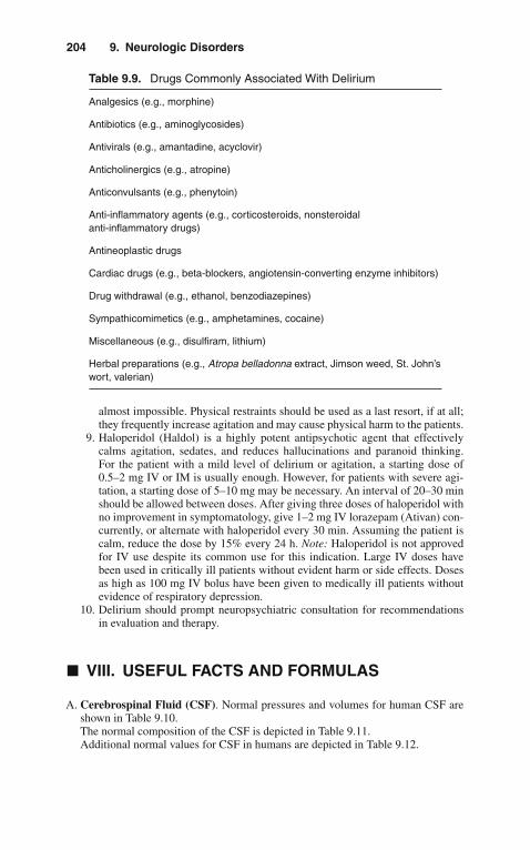
204 9. Neurologic Disorders
Table 9.9. Drugs Commonly Associated With Delirium
Analgesics (e.g., morphine)
Antibiotics (e.g., aminoglycosides)
Antivirals (e.g., amantadine, acyclovir)
Anticholinergics (e.g., atropine)
Anticonvulsants (e.g., phenytoin)
Anti-inflammatory agents (e.g., corticosteroids, nonsteroidalanti-inflammatory drugs)
Antineoplastic drugs
Cardiac drugs (e.g., beta-blockers, angiotensin-converting enzyme inhibitors)
Drug withdrawal (e.g., ethanol, benzodiazepines)
Sympathicomimetics (e.g., amphetamines, cocaine)
Miscellaneous (e.g., disulfiram, lithium)
Herbal preparations (e.g., Atropa belladonna extract, Jimson weed, St. John’swort, valerian)
almost impossible. Physical restraints should be used as a last resort, if at all;they frequently increase agitation and may cause physical harm to the patients.
9. Haloperidol (Haldol) is a highly potent antipsychotic agent that effectivelycalms agitation, sedates, and reduces hallucinations and paranoid thinking.For the patient with a mild level of delirium or agitation, a starting dose of0.5–2 mg IV or IM is usually enough. However, for patients with severe agi-tation, a starting dose of 5–10 mg may be necessary. An interval of 20–30 minshould be allowed between doses. After giving three doses of haloperidol withno improvement in symptomatology, give 1–2 mg IV lorazepam (Ativan) con-currently, or alternate with haloperidol every 30 min. Assuming the patient iscalm, reduce the dose by 15% every 24 h. Note: Haloperidol is not approvedfor IV use despite its common use for this indication. Large IV doses havebeen used in critically ill patients without evident harm or side effects. Dosesas high as 100 mg IV bolus have been given to medically ill patients withoutevidence of respiratory depression.
10. Delirium should prompt neuropsychiatric consultation for recommendationsin evaluation and therapy.
� VIII. USEFUL FACTS AND FORMULAS
A. Cerebrospinal Fluid (CSF). Normal pressures and volumes for human CSF areshown in Table 9.10.The normal composition of the CSF is depicted in Table 9.11.Additional normal values for CSF in humans are depicted in Table 9.12.

VIII. Useful Facts and Formulas 205
Table 9.10. Normal CSF Pressures and Volumes
CSF pressureChildren 3.0–7.5 mmHgAdults 3.5–13.5 mmHg
VolumeInfants 40–60 mLYoung children 60–100 mLOlder children 80–120 mLAdult 100–160 mL
Table 9.11. Normal Composition of the CSF
CSF Concentration (mean)
Specific gravity 1.007
Osmolality (mOsm/kg H2O) 289
PH 7.31
PCO2 (mmHg) 50.5
Na+ (mEq/L) 141
K+ (mEq/L) 2.9
Ca++ (mEq/L) 2.5
Mg++ (mEq/L) 2.4
Cl– (mEq/L) 124
Glucose (mg/dL) 61
Protein (mg/dL) 28
Table 9.12. Normal CSF Values
CSF parameter Newborns Infants, older children,and adults
Leukocyte count <32/μL <6/μL
Differential white cellcount
<60% polymorphs <1 polymorph
Proteins <170 mg/dL <45 mg/dL
Glucose >30 mg/dL >45 mg/dL
CSF: blood/glucoseratio
>0.44 >0.5
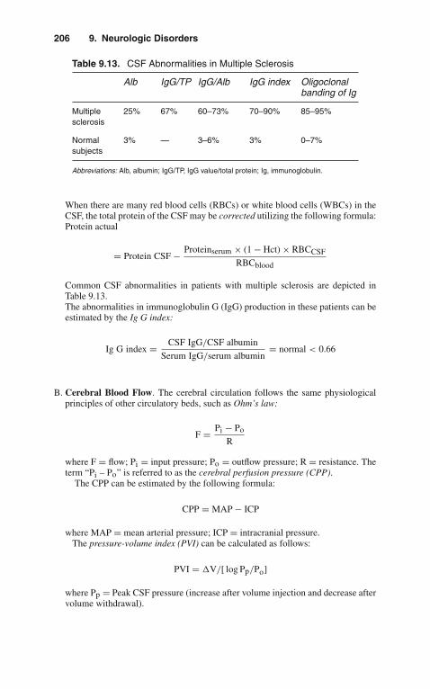
206 9. Neurologic Disorders
Table 9.13. CSF Abnormalities in Multiple Sclerosis
Alb IgG/TP IgG/Alb IgG index Oligoclonalbanding of Ig
Multiplesclerosis
25% 67% 60–73% 70–90% 85–95%
Normalsubjects
3% — 3–6% 3% 0–7%
Abbreviations: Alb, albumin; IgG/TP, IgG value/total protein; Ig, immunoglobulin.
When there are many red blood cells (RBCs) or white blood cells (WBCs) in theCSF, the total protein of the CSF may be corrected utilizing the following formula:Protein actual
= Protein CSF − Proteinserum × (1 − Hct) × RBCCSF
RBCblood
Common CSF abnormalities in patients with multiple sclerosis are depicted inTable 9.13.The abnormalities in immunoglobulin G (IgG) production in these patients can beestimated by the Ig G index:
Ig G index = CSF IgG/CSF albumin
Serum IgG/serum albumin= normal < 0.66
B. Cerebral Blood Flow. The cerebral circulation follows the same physiologicalprinciples of other circulatory beds, such as Ohm’s law:
F = Pi − Po
R
where F = flow; Pi = input pressure; Po = outflow pressure; R = resistance. Theterm “Pi – Po” is referred to as the cerebral perfusion pressure (CPP).
The CPP can be estimated by the following formula:
CPP = MAP − ICP
where MAP = mean arterial pressure; ICP = intracranial pressure.The pressure-volume index (PVI) can be calculated as follows:
PVI = �V/[ log Pp/Po]
where Pp = Peak CSF pressure (increase after volume injection and decrease aftervolume withdrawal).
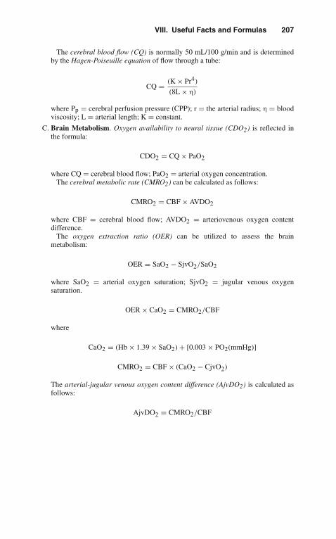
VIII. Useful Facts and Formulas 207
The cerebral blood flow (CQ) is normally 50 mL/100 g/min and is determinedby the Hagen-Poiseuille equation of flow through a tube:
CQ = (K × Pr4)
(8L × η)
where Pp = cerebral perfusion pressure (CPP); r = the arterial radius; η = bloodviscosity; L = arterial length; K = constant.
C. Brain Metabolism. Oxygen availability to neural tissue (CDO2) is reflected inthe formula:
CDO2 = CQ × PaO2
where CQ = cerebral blood flow; PaO2 = arterial oxygen concentration.The cerebral metabolic rate (CMRO2) can be calculated as follows:
CMRO2 = CBF × AVDO2
where CBF = cerebral blood flow; AVDO2 = arteriovenous oxygen contentdifference.
The oxygen extraction ratio (OER) can be utilized to assess the brainmetabolism:
OER = SaO2 − SjvO2/SaO2
where SaO2 = arterial oxygen saturation; SjvO2 = jugular venous oxygensaturation.
OER × CaO2 = CMRO2/CBF
where
CaO2 = (Hb × 1.39 × SaO2) + [0.003 × PO2(mmHg)]
CMRO2 = CBF × (CaO2 − CjvO2)
The arterial-jugular venous oxygen content difference (AjvDO2) is calculated asfollows:
AjvDO2 = CMRO2/CBF

10Nutrition
� I. AIMS OF NUTRITIONAL SUPPORT
A. Preserve tissue mass and decrease usage of endogenous nutrient stores.
B. Decrease catabolism.
C. Maintain/improve organ function.1. Immune2. Renal3. Hepatic4. Muscle
D. Improve wound healing.
E. Decrease infection.
F. Maintain gut barrier (decrease translocation).
G. Decrease morbidity/mortality.1. Decrease ICU/hospital stay.2. Decrease hospital costs.
� II. TIMING OF NUTRITIONAL SUPPORT
A. Optimal timing remains controversial.1. Some patients tolerate short periods of starvation by using endogenous stores
to support body functions.2. Well-nourished patients (non-stressed) have actually survived without food for
6 weeks (ingesting only water).3. Hypermetabolic and hypercatabolic critically ill patients can probably only
tolerate a few weeks of starvation before death.4. There appears to be no benefit of total starvation.
B. Accumulating data suggest that outcome can be improved with early and optimalnutritional support.
J. Varon, P. Acosta, Handbook of Critical and Intensive Care Medicine,DOI 10.1007/978-0-387-92851-7_10,C© Springer Science+Business Media, LLC 2010

210 10. Nutrition
1. Early nutritional support blunts the hypercatabolic/hypermetabolic response toinjury.
2. In a growing number of studies, patients randomized to receive early vs. delayedfeeding had decreased infection rates, fewer complications, and shorter lengthof stay in the hospital.
3. Animal studies report improved wound healing and improved hepatic functionin several injury models with early feeding.
� III. ROUTE OF NUTRITIONAL SUPPORT
A. Parenteral Nutrition1. Nutrients
Amino acids, dextrose, soy-based lipids, vitamins, minerals and trace elements(see Table 10.1)
2. Delivery: via peripheral or central vein3. Major complications
a. Central line placement (pneumothorax, hemothorax, carotid arteryperforation)
b. Metabolic derangements (hyperglycemia, electrolyte disturbances)c. Immune suppressiond. Increased infection rates (catheter-related sepsis, pneumonia, abscesses)e. Liver dysfunction (fatty infiltration, cholestasis, liver failure)f. Gut atrophy (diarrhea, bacterial translocation)g. Venous thrombosish. Overfeeding
4. Other problems include lack of some conditionally essential amino acids thatare not stable in solution (i.e., glutamine, cysteine)
Table 10.1. Comparison of Nutrients in Enteral vs. Parenteral
Nutrient Enteral Parenteral
Nitrogen source Intact proteins, peptides, oramino acids
Amino acids∗
Carbohydrate Simple sugars or complexcarbohydrates (i.e., starchand fiber)
Simple sugar (dextrose)
Lipids Long- and medium-chaintriglycerides, or long-chainfatty acids (ω-3 or ω-6)
Soy-based lipids
Vitamins Present Can be added
Minerals andtrace elements
Present Can be added
∗ Lacks some conditionally essential amino acids (i.e., glutamine and cysteine).

III. Route of Nutritional Support 211
5. Glucose/fat ratioa. Usually 60:40 to 40:60 (ratio of calories from each source)b. Large amounts of glucose (>60% of calories) can
(1). Increase energy expenditure.(2). Increase CO2 production and increase pulmonary workload (may delay
ventilator weaning).(3). Produce liver steatosis.(4). Lead to immune compromise.
B. Enteral Nutrition1. Nutrients (see Table 10.1)
a. Nitrogen sources: amino acids, peptides, or intact proteins (e.g., casein,whey, soy, lactalbumin)
b. Carbohydrates: simple sugars or complex carbohydrates (i.e., starch andfiber)
c. Lipids: long- or medium-chain triglycerides, ω-3 or ω-6 long-chain fattyacids
d. Vitaminse. Minerals and trace elements
2. Deliverya. Oralb. Gastric tube (i.e., nasogastric, gastric)c. Small-bowel feeding tube (i.e., nasoduodenal, gastroduodenal, jejunal)
3. Major Complicationsa. Aspiration (pneumonia, chemical pneumonitis, adult respiratory distress
syndrome [ARDS])b. Metabolic derangements (e.g., electrolyte disturbances, hyperglycemia);
these are less common than with parenteral nutritionc. Diarrhead. Misplaced feeding tubes (e.g., pneumothorax, empyema, bowel perforation)e. Overfeeding
C. Enteral vs. Parenteral Nutrition1. Enteral nutrition is required for optimal gut function (i.e., maintenance of gut
barrier, gut-associated immune system, immunoglobulin A [Ig A] secretion,mucin layer)
2. Total parenteral nutrition (TPN) is associated witha. Immunosuppression (thought to be related to intravenous [IV] lipids, which
are high in w-6 long-chain fatty acids)b. Increased infection rates (compared to enteral) in patients following trauma,
burns, surgery, and cancer chemo/radiotherapyc. Higher mortality in patients receiving chemo/radiotherapy and after burn
injury3. TPN is not superior to enteral nutrition in patients with inflammatory bowel
disease or pancreatitis.4. TPN may be beneficial in patients with short-gut syndromes and some types of
gastrointestinal (GI) fistulas.5. Enteral nutrition is the preferred method of feeding in patients receiving
chemo/radiotherapy, and following surgery, burns, trauma, sepsis, renal failure,liver failure, and respiratory failure.
6. Parenteral nutrition is indicated when enteral nutrition is not possible (i.e.,inadequate small bowel function).

212 10. Nutrition
� IV. GASTROINTESTINAL FUNCTION DURINGCRITICAL ILLNESS
A. Oral nutrition remains the best form of nutritional support; however, in manycritically ill patients, this is not possible.
B. Decreased motility of stomach and colon are common and typically last 5–7 daysin critically ill patients (longer if the patient remains critically ill).1. Gastric paresis is best assessed and monitored by measuring gastric residuals.2. Gastric residuals of ≥150 mL are usually considered abnormal.3. Patients with gastric residuals ≥150 mL should be fed in the small bowel
(postpyloric) to decrease risk of aspiration.
C. Motility and nutrient absorptive capability of small bowel is usually preserved(even after severe trauma, burns, or major surgery).
D. Bowel sounds are a poor index of small bowel motility.
� V. NUTRIENT REQUIREMENTS (QUANTITY)
A. Energy1. Caloric Content of Major Nutrients
a. Lipids provide 9 kcal/g.b. Carbohydrates provide 4 kcal/g.c. Proteins provide 4 kcal/g.
2. Studies show that most critically ill patients expend 25–35 kcal/kg/d.3. One can estimate resting metabolic expenditure (RME) using the Harris-
Benedict equation (see Table 10.2).4. One can also measure RME by indirect calorimetry (metabolic cart).5. Some recommend adjusting RME by multiplying by a correction factor
(see Table 10.3); however, correction factors frequently overestimate energyneeds.
6. We prefer to initially administer 25 kcal/kg/d (see Table 10.4).a. ≈20% protein (percent refers to percentage of total daily calories)b. ≈30% lipidsc. ≈50% carbohydrates
7. Patients with organ failure/disease states may have increased or decreased needsand should be considered individually.
Table 10.2. Harris-Benedict Equations
Men RME (kcal/d) = 66 + (13.7 × W) + (5 × H) – (6.8 × A)
Women RME (kcal/d) = 665 + (9.6 × W) + (1.7 × H) – (4.7 × A)
Abbreviations: A, age in years; H, height in cm; RME, resting metabolic expenditure; W,weight in kg.

V. Nutrient Requirements (Quantity) 213
Table 10.3. Energy Expenditure Correction Factors
Starvation 0.7Confined to bed 1.2Out of bed 1.3Fever 1 + 0.13/◦CElective surgery 1.0–1.2Multiple fractures 1.2–1.4Sepsis 1.4–1.8Burns
<20% BSB 1–1.520–40% BSB 1.5–1.940–100% BSB 1.9–2.1
Abbreviation: BSB, body surface area burned.
Table 10.4. Macronutrient Nutritional Requirements
Nutrient % of totalcalories
Quantity ofnutrients
Example for70-kg patient
Total calories 25 kcal/kg/d 1,750 kcal/d
Protein/aminoacids
15–25 1.2–2.0 g/kg/d 95 g/d (380 kcal/d)(based on1.35 g/kg/d)
Carbohydrates 30–65 50% of calories(avg pt)
220 g/d(880 kcal/d)
Fats 15–30 30% of calories(avg pt)
55 g/d (495 kcal/d)
8. Overfeeding (with either enteral or parenteral nutrients) is associated with moreadverse side effects than slightly underfeeding during most critical illnesses.
B. Protein1. Most critically ill patients need 1.2–2.0 g/kg/d.2. Protein requirements increase in patients with severe trauma, burns, and with
protein-losing enteropathies.
C. Water1. Must be individualized, as needs vary greatly between patients (differences in
insensible losses, GI losses, and urine losses).2. Initially estimate: 1 mL water per kilocalorie of energy in adults.
D. Vitamins1. Fat-soluble vitamins: A, D, E, K.2. Water-soluble vitamins: ascorbic acid (C), thiamine (B1), riboflavin (B2),
niacin, folate, pyridoxine (B6), B12, pantothenic acid, biotin.3. Published recommended daily allowances (RDAs) are based on oral intake in
healthy individuals.

214 10. Nutrition
Table 10.5. Micronutrient Nutritional Requirements
Micronutrient Enteral nutrition Parenteralnutrition
Example forTPN for a70-kg patient
MineralsSodium 60–140 mmol/d 60–120 mmol/d 80 mmol/dPotassium 50–140 mmol/d 50–120 mmol/d 50 mmol/dMagnesium 8–15 mmol/d 8–12 mmol/d 10 mmol/dPhosphorous 25 mmol/d 14–16 mmol/d 15 mmol/dCalcium 20 mmol/d 7–10 mmol/d 10 mmol/d
Trace elementsIron 10 mg/d 1–2 mg/d noneZinc 15 mg/d 2–5 mg/d 5 mg/dCopper 2–3 mg/d 0.5–1.5 mg/d 1 mg/dChromium 50–200 μg/d 10–20 μg/d 10 μg/dSelenium 50–200 μg/d 80–150 μg/d 100 μg/dIodine 150 μg/d 120 μg/d 120 μg/dManganese 2.5–5.0 mg/d 0.2–0.8 mg/d 0.5 mg/d
Vitamins∗Vitamin A RDA =
4,000–5,000 IU/dND 3,300 IU/d
Vitamin D RDA = 200–400 IU/d ND 200 IU/dVitamin E RDA = 12–15 IU/d ND 10 IU/dVitamin K RDA = 60–80 μg/d ND 10 mg/wk†
Thiamine RDA = 1.1–1.4 mg/d ND 3 mg/dRiboflavin RDA = 1.2–1.7 mg/d ND 5 mg/dNiacin RDA = 13–19 mg/d ND 40 mg/dPantothenicacid
4–7 mg/d‡ ND 15 mg/d
Pyridoxine RDA = 1.6–2.0 mg/d ND 4 mg/dFolic acid RDA = 0.4 mg/d ND 0.4 mg/dVitamin B12 RDA = 3 μg/d ND 5 μg/dVitamin C RDA = 40 mg/d ND 100 mg/dBiotin RDA = 30–100 μg/d ND 60 μg/d
Abbreviation: ND, not defined.∗ Enteral requirements should always exceed parenteral requirements; most recommendsupplying 1–3 times the RDA of each vitamin to patients with critical illness.† None if anticoagulation used.‡ RDA not established.
4. Vitamin needs for critically ill patients have not been determined.5. See Table 10.5 for estimates of nutritional requirements of the vitamins.6. Commercial enteral formulas generally supply the RDA of the vitamins (if
patients receive their caloric needs).7. An adult parenteral vitamin formulation was approved by the FDA in 1979
and is available for addition to TPN solutions; this should be added just beforeadministration, since degradation can occur.

VI. Role of Specific Nutrients (Quality) 215
E. Minerals (Na, K, Ca, PO4, Mg)1. See Table 10.5 for estimates of daily nutritional requirements of the minerals.2. Minerals are present in sufficient quantities in enteral products (special formu-
las limit electrolytes for renal failure).3. Must be supplemented in TPN.
F. Trace Elements (iron, copper, iodine, zinc, selenium, chromium, cobalt,manganese)1. Needs in critically ill patients have not been determined. (See Table 10.5 for
estimates of requirements.)2. Sufficient quantities are thought to be present in enteral products.3. Must be supplemented in TPN (all except iron can be added to solution).
a. Deficiency states have been reported in long-term TPN patients.b. Specifics are best managed by specially trained nutritional support teams.
� VI. ROLE OF SPECIFIC NUTRIENTS (QUALITY)
A. Nitrogen Sources1. Choices
a. Amino acidsb. Hydrolyzed protein (peptides)c. Intact proteins
2. Evidence suggests that peptides generated from the diet possess specificphysiologic activities.
3. Nitrogen is best delivered as intact protein (if digestion and absorption intact)or hydrolyzed protein (impaired digestion).
4. Protein is absorbed primarily as peptides (60%) and amino acids (33%).5. Essential amino acid formulas should not be used.6. Some amino acids become essential during critical illness.
a. These are called conditionally essential amino acids.b. Examples include glutamine, cysteine, arginine, and taurine.
7. Some amino acids appear to have specific roles.a. Glutamine is a fuel source for rapidly dividing cells, such as the GI tract
and immune system (patients with bone marrow transplant, supplementationwith glutamine was associated with lower rates of infection).
b. Arginine is required for optimum wound healing and is important in immunefunction. (Target patients are critically ill adults and preterm infants.)
c. Cysteine is needed for synthesis of glutathione.d. Branched-chain amino acids (BCAA) may improve mental status in patients
with hepatic encephalopathy; it is primarily metabolized by peripheralmuscle instead of the liver.
e. Note that glutamine and cysteine are not stable (or present) in TPN solution.
B. Lipids1. Linoleic Acid
a. Essential fatty acid (need 7–12% of total calories supplied as linoleic acid)b. ω-6 Polyunsaturated, long-chain fatty acid (immunosuppressive)c. Precursor to membrane arachidonic acid
2. ω-3 Polyunsaturated Fatty Acids (PUFA)a. Fish oils and linolenic acid.

216 10. Nutrition
b. Profound effects upon cell membrane fluidity and stability. Decreaseproduction of dienoic prostaglandins (i.e., PGE2), tumor necrosis factor,interleukin-1, and other proinflammatory cytokines.
c. Supplementation of omega-3 fatty acids in patients with acute lung injury,improves oxygenation and shortens length of mechanical ventilation.
3. Medium-Chain Triglyceridesa. Good energy source.b. Water-soluble.c. Enter circulation via GI tract.
4. Short-Chain Fatty Acids (SCFA)a. Examples: butyric and propionic acidb. Major fuel for the gut (especially the colon)c. Derived from metabolizable fiber
5. High-Fat Formulasa. If the patient is not overfed, these have little effect on CO2 production
(despite being marketed for decreasing the respiratory quotient [RQ]).b. Poor GI tolerance.
C. Carbohydrates1. Starches and sugars: good for energy2. Fiber
a. Metabolizable fiber (i.e., pectin, guar) is converted to SCFA in the colon bybacteria.
b. Bulk increases stool mass, softens stool, adds body to stool, and providessome stimulation of gut mass.
D. Dietary nucleic acids may be important for immune function.
� VII. MONITORING RESPONSES TONUTRITIONAL SUPPORT
A. Visceral Proteins1. Prealbumin
a. Half-life 2 days.b. Normal range is 10–40 mg/dL.
2. Transferrina. Half-life 8–9 days.b. Normal range is 160–355 mg/dL.
3. Albumina. Half-life 20 days.b. Normal range is 3.2–5.0 mg/dL.
B. Visceral protein levels are affected by nutritional intake as well as the disease state(especially presence of inflammation).
C. Increasing levels of visceral proteins suggest that nutritional support is adequate.
D. Nitrogen Balance1. Determined from 12- to 24-h urine collections and measurements of total
urinary nitrogen (more accurate than total urea nitrogen), compared to totalnitrogen intake.

VIII. Nutrition for Specific Disease Processes 217
2. May be inaccuratea. In patients with renal failureb. If urine is not correctly collected by staffc. If the patient has increased nitrogen losses in stool or from wounds (i.e.,
burns)3. N-balance = protein intake (g/d)/6.25 – {total urinary nitrogen (g/d) + 2}.4. Negative nitrogen balance is not necessarily detrimental over the short term
(i.e., 1–2 weeks).5. Improvement in nitrogen balance suggests that nutritional support is adequate.6. Be aware that nitrogen balance may improve as catabolism decreases despite
inadequate nutritional support.
E. Caloric goals: caloric needs can be determined using 25 kcal/kg of ideal bodyweight as an estimate. Another option is indirect calorimetry:1. Measures oxygen consumption and CO2 production for 15–30 min, estimates
energy expenditure, then extrapolates to 24 h.2. Keep RQ <1. Values >1 suggest lipogenesis from excessive caloric intake;
values ≈ 0.7 are found in starvation and reflect fat oxidation.
F. Other Nutritional Parameters Not Generally Useful in the Critically Ill1. Weight2. Skin-fold thickness3. Delayed cutaneous hypersensitivity (DCH)4. Lymphocyte counts
� VIII. NUTRITION FOR SPECIFIC DISEASEPROCESSES
A. Acute Renal Failure1. Use intact protein or peptide formula with moderate fat.2. Do not restrict protein (it is required for healing and for other organ functions).3. May limit fluid intake with double-strength formula (2 cal/mL).4. Watch K, Mg, and PO4 levels.
B. Hepatic Failure1. Use intact protein or peptide formula.2. Usually 1.0–1.2 g/kg/d of protein are needed to support repair and immune
function.3. BCAA may be of value if encephalopathy persists following use of intact
protein or peptide diets.
C. Inflammatory Bowel Disease/Pancreatitis1. Enteral nutrition is possible if a jejunal tube is placed (endoscopically or
radiologically) distal to Treitz ligament.2. Enteral nutrition should be attempted before initiating TPN.
D. Multiple Organ Failure1. Nutritional support is usually of marginal value.2. Nutritional support needs to be started before organ failure develops.

218 10. Nutrition
� IX. NASODUODENAL FEEDING TUBEPLACEMENT
A. Use in patients who do not tolerate oral or gastric feeding.
B. Patients with abdominal surgery should have the tube placed during surgery underdirect visualization.1. The anesthesiologist inserts the tube into the stomach.2. The surgeon locates the tube and directs it into the duodenum or jejunum.3. Eliminates need for confirmatory x-rays.4. Allows immediate feeding upon admission into intensive care unit (ICU).5. Feeding tubes may also be placed into the small bowel using a gastrostomy or
jejunostomy.
C. Tubes placed into the stomach will rarely (5–15%) migrate spontaneously into thesmall bowel in critically ill patients (due to gastroparesis).
D. Bedside Method1. Place patient in left lateral decubitus position (if possible).2. Lubricate the nostril with generic lubricant or 2% viscous lidocaine.3. Insert an 8–10 French small-bore feeding tube (containing wire stylet) into the
nostril, and gently advance it through the nasopharynx into the esophagus andthen the stomach.
4. If resistance is met or the patient coughs, becomes agitated, or decreases oxygensaturation, thena. Pull the tube back into the nasopharynx.b. Repeat step 3 and reinsert the tube into the stomach.c. Or, change the position of the patient’s neck (slightly flex or extend) before
reattempting insertion.5. Confirm position of the tube in the stomach.
a. Auscultate over the abdomen.b. Aspirate gastric contents (pH ≈ 2–5, unless on H2 blocker).
6. Remove wire stylet, and place a 45-degree bend approximately 1 in. from thedistal end of the wire.
7. Gently reinsert the wire stylet (should not meet resistance).8. Slowly advance the tube while rotating it in a clockwise direction.9. Check the position every 10–15 cm.
a. Auscultation will reveal higher pitched sounds when the tube is in thepylorus and proximal small bowel.
b. Bile may be aspirated from the tube in the small bowel.c. Bile/small bowel secretions have pH ≈ 6–7.d. Abdominal x-ray.
(1). Can confirm small bowel location.(2). May not be cost-effective.(3). Will avoid feeding into lung in rare case of misplaced feeding tube.
E. With this bedside method, we (faculty, residents, and medical students) suc-cessfully place >90% of attempted small bowel tubes into the duodenum orjejunum.
F. Aggressive surgical and bedside placement allows us to feed >97% of ourcritically ill patients enterally within 24–48 h of admission into the ICU.
XI. Approach to Enteral Feeding 219
G. If bedside placement is not possible, place the feeding tube into small intestineusing1. Endoscopy2. Fluoroscopy
� X. RECOMMENDATIONS FOR TPN USE
1. Use only when enteral nutrition is not possible (e.g., short-gut syndrome, chylo-thorax).a. Failure of the stomach to empty is not an indication for TPN but rather for a
small bowel feeding tube.b. Most patients with diarrhea can be managed with enteral nutrition.
2. Initial TPN orders may be based on recommendations in Tables 10.4 and 10.5.
3. Overall TPN management is best performed by specially trained nutritionalsupport teams.
4. For more specifics, the reader is referred to entire texts written about TPN.
� XI. APPROACH TO ENTERAL FEEDING
A. Enteral nutritional support should be initiated within 12–48 h of admission to theICU.
B. The oral route is preferred (but frequently not possible).
C. The gastric route is second choice and should be tried before placing a small boweltube in most patients.
D. Patients at high risk for aspiration or known gastric paresis should be fed with asmall bowel tube.
E. Feeding formulas should not be diluted.
F. Keep the head of the patient’s bed elevated 30◦ to decrease the risk of aspiration.
G. Feeding should be started at 25–30 mL/h and increased by 25 mL/h every 1–4 h astolerated by gastric residuals (<150 mL) until the caloric goal (25–30 kcal/kg/d)is achieved.
H. If the protein goal is not achieved, use a formula with a higher protein/calorie ratioor add protein to the formula.
I. Gastric residuals should be monitored every 4 h.
J. If the gastric residual is >150 mL, hold feeds for 2 h and then resume.
K. May increase feeds at slower rate (i.e., =10 mL/h every 6–12 h), but often this isnot necessary.
L. The goal rate of infusion should be met by the third day of therapy (frequentlyearlier).
M. Monitor nutritional response by measuring visceral protein levels.

220 10. Nutrition
1. Prealbumin and transferrin levels should be measured on day 1 and every 3 daysthereafter during initial therapy.
2. Increasing levels suggest that the patient is receiving adequate nutritionalsupport.
3. Levels usually normalize in 1–2 weeks if the disease process is controlled andnutritional support is adequate.
4. If levels fail to increase,a. Consider underlying infection, inflammation, or other disease processes.b. Reevaluate the adequacy of nutritional support.c. Nitrogen balance and energy balance (i.e., indirect calorimetry) may be
informative.d. Consult the nutritional support service.
Isolated head injury
Yes
High nitrogenformula
High nitrogenformula
with fiber
Impaireddigestion/
absorption orsepsis
Yes
Tolerated× 3 Days
?
Fatmalabsorption
?
Yes 1. Oral diet2. Standard formula a. Osmolite HN b. Jevity(Fiber)
Lowerfat
content1. Consider nutrition consult2. Trial of 50:50 mix of Reabilan HN: Criticare HN Peptamen: Criticare HN3. Trial of Criticare HN
Yes
Examples: - Reabilan HN - Peptamen
- Perative
Peptidebased formula
No
Example: - Jevity
Oraldiet
Standardformula
with fiber
Standardformula
No
No
cont
inue
feed
ing
No
Evaluate forcause of diarrhea
Hypermetabolic(acute injury)
Example: - Osmolite
Examples: - Replete
Example: - Repletewith fiber
HN
- Nitrolan
Figure 10.1. Flow diagram for nutritional support in patients with isolatedhead injury.

XI. Approach to Enteral Feeding 221
*Shock, resuscitation, major abdominal surgery, gut orpancreatic injury, sepsis, acute hypoalbuminemia (<2g/dL)
Multitrauma/burns
Septic shock
Abdominal surgery
No
Example: - Replete with fiber
1. Oral diet2. Standard nitrogen formula Examples: a. Osmolite HN b. Jevity (fiber)
Example: - Nitrolan - Replete
High nitrogenformula
High nitrogenplus fiber
No
Fatintolerance
?
Toleraled× 3 Days
?
continue feeding
No
Evaluate for causesof diarrhea
Impaireddigestion orabsorption*
Peptide-basedformula
Examples: - Reabilan HN - Perative
- Peptamen
Yes
Yes
Yes
1. Consider dietary consult2. 50:50 Mix Reabilan HN: Criticare HN Peptamen: Criticare HN3. Criticare HN
Lower fatcontent
Figure 10.2. Flow diagram for nutritional support in patients with multipletrauma, burn injury, sepsis/septic shock, or abdominal surgery.
N. Several flow diagrams (for specific patient populations, using enteral productscurrently on our formulary) are given as examples (see Figures 10.1, 10.2, and10.3).1. Isolated head injury (see Figure 10.1)2. Multitrauma, burn, sepsis, and abdominal surgery (see Figure 10.2)3. Severe malnutrition (see Figure 10.3)
O. If peptide-based diets are not available, intact protein diets should be used.
P. Formula Osmolality1. 300–600 mOsm/kg H2O.2. Rarely causes intolerance/diarrhea.
Q. Diarrhea is unfortunately encountered in patients on enteral and parenteralnutrition.1. Generally defined as >300–500 mL stool output per day.2. Most common etiologies are medications and infections. See Table 10.6 for a
partial list of etiologies and suggestions for preliminary workup.

222 10. Nutrition
Severe malnutritionwith probable impairmentof digestion, absorption
Examples: - Reabilan HN - Peptamen
- Peralive
Peptide-based formula
No
Nutrition consultto estimate needs
Tolerated× 3 Days
continue peptide formula
NoContinue peptides
trial intactprotein formula
Yes
Intact proteinformula
Yes
No
- Diarrhea evaluation- Nutrition consult
Continue peptides;check for other
intolerence reasons
Tolerated 1 weekprealbumin increasing
Yes
Example: - Criticare HN
Low-fatformula
Fat intolerance?
Oral dietas tolerated
Examples: - Osmolite HN - Nitrolan
- Jevity
Figure 10.3. Flow diagram for nutritional support in patients with severemalnutrition.
3. Note that many elixir forms of medications contain sorbitol.4. Once the specific etiologies of diarrhea have been evaluated, diarrhea may be
treated with antimotility agents (i.e., narcotics). We prefer to add paregoricdirectly to the feeding formula (30–60 cc q4–6 h).

XII. Useful Facts and Formulas 223
Table 10.6. Etiologies and Evaluation of Diarrhea
Causes Examples Workup
Drugs Sorbitol, antacids, H2blockers, antibiotics,lactulose, laxatives,quinidine, theophylline
No specific workup;discontinue any of thesemedications that are notabsolutely necessary
Infections 1. Clostridium difficile 1. Specific stool culture, C.difficile toxin assay, sigmoi-doscopy/colonoscopy forevidence ofpseudomembranes
2. Infectious diarrhea (e.g.,typhoid fever, shigellosis)
2. Fecal leukocytes, culture
3. Other: bacterialovergrowth, parasites,systemic infection, HIV
3. As relevant (e.g., lookfor ova and parasites;rarely causes new diarrheain critically ill)
Osmotic Measure stool osmotic gap(SOG);∗SOG >100 suggestsosmotic diarrhea
Impaction May be secondary tonarcotics
Rectal exam
Other causes Inflammatory boweldisease, pancreaticinsufficiency, short gutsyndrome
∗ SOG = stool osmolality – 2(stool Na+ + K+).
� XII. USEFUL FACTS AND FORMULAS
A. Nutritional Assessment. The total daily energy (TDE) requirements for a patientcan be calculated using the following formula:
TDE for men (kcal/d) = (66.47 + 13.75 W + 5.0H − 6.76A)× (Activity factor) × (Injury factor)
TDE for women (kcal/day) = (655.10 + 9.56 W + 1.85H4.68A)× (Activity factor) × (Injury factor)
where W = weight (kg), H = height (cm), A = age (years); the activity factor isderived from Table 10.7.
The injury factors can be estimated based on the Table 10.8.
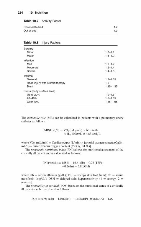
224 10. Nutrition
Table 10.7. Activity Factor
Confined to bed 1.2Out of bed 1.3
Table 10.8. Injury Factors
SurgeryMinor 1.0–1.1Major 1.1–1.2
InfectionMild 1.0–1.2Moderate 1.2–1.4Severe 1.4–1.8
TraumaSkeletal 1.2–1.35Head injury with steroid therapy 1.6Blunt 1.15–1.35
Burns (body surface area)Up to 20% 1.0–1.520–40% 1.5–1.85Over 40% 1.85–1.95
The metabolic rate (MR) can be calculated in patients with a pulmonary arterycatheter as follows:
MR(kcal/h) = VO2(mL/min) × 60 min/h×1L/1000mL × 4.83 kcal/L
where VO2 (mL/min) = Cardiac output (L/min) × [arterial oxygen content (CaO2,mL/L) – mixed venous oxygen content (CmO2, mL/L)].
The prognostic nutritional index (PNI) allows for nutritional assessment of thecritically ill patient and is calculated as follows:
PNI (%risk) = 158% − 16.6 (alb) − 0.78 (TSF)−0.2(tfn) − 5.8(DSH)
where alb = serum albumin (g/dL); TSF = triceps skin fold (mm); tfn = serumtransferrin (mg/dL); DSH = delayed skin hypersensitivity (1 = anergy, 2 =reactive).
The probability of survival (POS) based on the nutritional status of a criticallyill patient can be calculated as follows:
POS = 0 .91 (alb) − 1.0 (DSH) − 1.44 (SEP)+0.98 (DIA) − 1.09
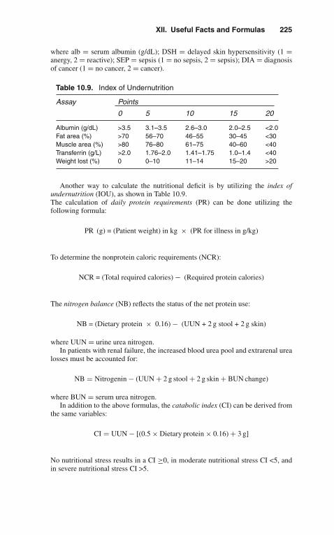
XII. Useful Facts and Formulas 225
where alb = serum albumin (g/dL); DSH = delayed skin hypersensitivity (1 =anergy, 2 = reactive); SEP = sepsis (1 = no sepsis, 2 = sepsis); DIA = diagnosisof cancer (1 = no cancer, 2 = cancer).
Table 10.9. Index of Undernutrition
Assay Points
0 5 10 15 20
Albumin (g/dL) >3.5 3.1–3.5 2.6–3.0 2.0–2.5 <2.0Fat area (%) >70 56–70 46–55 30–45 <30Muscle area (%) >80 76–80 61–75 40–60 <40Transferrin (g/L) >2.0 1.76–2.0 1.41–1.75 1.0–1.4 <40Weight lost (%) 0 0–10 11–14 15–20 >20
Another way to calculate the nutritional deficit is by utilizing the index ofundernutrition (IOU), as shown in Table 10.9.The calculation of daily protein requirements (PR) can be done utilizing thefollowing formula:
PR (g) = (Patient weight) in kg × (PR for illness in g/kg)
To determine the nonprotein caloric requirements (NCR):
NCR = (Total required calories) − (Required protein calories)
The nitrogen balance (NB) reflects the status of the net protein use:
NB = (Dietary protein × 0.16) − (UUN + 2 g stool + 2 g skin)
where UUN = urine urea nitrogen.In patients with renal failure, the increased blood urea pool and extrarenal urea
losses must be accounted for:
NB = Nitrogenin − (UUN + 2 g stool + 2 g skin + BUN change)
where BUN = serum urea nitrogen.In addition to the above formulas, the catabolic index (CI) can be derived from
the same variables:
CI = UUN − [(0.5 × Dietary protein × 0.16) + 3 g]
No nutritional stress results in a CI ≥0, in moderate nutritional stress CI <5, andin severe nutritional stress CI >5.
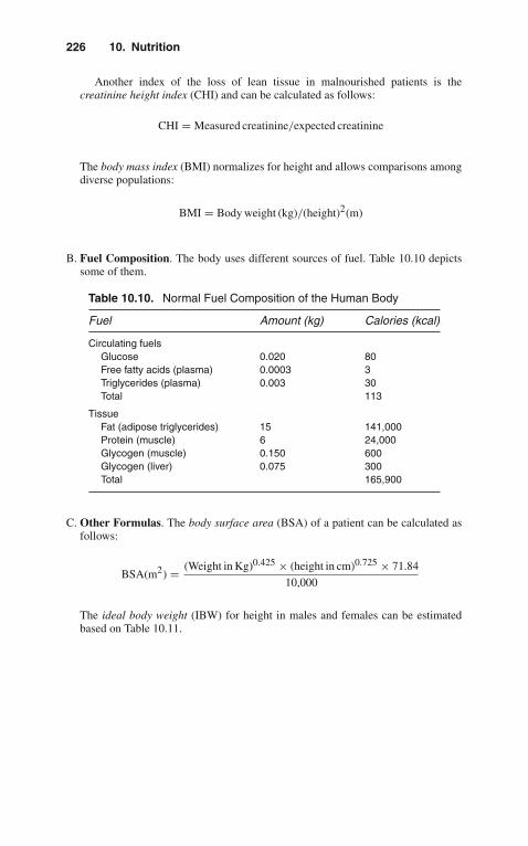
226 10. Nutrition
Another index of the loss of lean tissue in malnourished patients is thecreatinine height index (CHI) and can be calculated as follows:
CHI = Measured creatinine/expected creatinine
The body mass index (BMI) normalizes for height and allows comparisons amongdiverse populations:
BMI = Body weight (kg)/(height)2(m)
B. Fuel Composition. The body uses different sources of fuel. Table 10.10 depictssome of them.
Table 10.10. Normal Fuel Composition of the Human Body
Fuel Amount (kg) Calories (kcal)
Circulating fuelsGlucose 0.020 80Free fatty acids (plasma) 0.0003 3Triglycerides (plasma) 0.003 30Total 113
TissueFat (adipose triglycerides) 15 141,000Protein (muscle) 6 24,000Glycogen (muscle) 0.150 600Glycogen (liver) 0.075 300Total 165,900
C. Other Formulas. The body surface area (BSA) of a patient can be calculated asfollows:
BSA(m2) = (Weight in Kg)0.425 × (height in cm)0.725 × 71.84
10,000
The ideal body weight (IBW) for height in males and females can be estimatedbased on Table 10.11.

XII. Useful Facts and Formulas 227
Table 10.11. Ideal Body Weight in Males and Females
Height in cm Males (Weight in kg) Females (Weight in kg)
145 51.8 47.5
150 54.5 50.4
155 57.2 53.1
160 60.5 56.2
165 63.5 59.5
175 70.1 66.3
180 74.2
185 78.1
The percentage of ideal body weight (%IBW) is calculated as follows:
%IBW = 100 × (height in cm)
IBW

11Critical Care Oncology
Cancer is becoming the leading cause of death in the United States. Enhanced criti-cal care capabilities have contributed substantially to improved survival. Critical caremay be needed on a short-term basis for the complications of the underlying malig-nancy or of aggressive antineoplastic therapy. Postoperative critical care has greatlyfacilitated major extirpative cancer surgery and is an implicit part of other approachessuch as bone marrow transplantation.
The present chapter considers different types of cancer patients likely to need andbenefit from treatment in the ICU. Clinical judgment regarding the appropriate useof critical care services is required in all patient populations, not just in patients withcancer. The decision to admit and technologically support critically ill cancer patientsshould be individualized.
� I. CENTRAL NERVOUS SYSTEM
A. Altered Mental Status. Alteration in mental status is the most common centralnervous system (CNS) presentation for cancer patients in the intensive care unit(ICU). The common differential diagnoses are considered below. If these can beexcluded, and the patient has not received excessive sedative or narcotic-analgesicagents, the patient should be treated presumptively for sepsis. Altered mental statusis a reliable, though nonspecific, sign of sepsis, which carries a high mortality ratein cancer patients.1. Intracranial Mass Lesions
A history of headache, nausea, vomiting, or seizure activity together withpapilledema and other signs of raised intracranial pressure suggest an intracra-nial mass lesion. A moderate increase in intracranial pressure by itself isrelatively well tolerated; however, when intracranial pressure becomes criti-cal, brain substance will shift in the direction of least resistance, with resultantherniation through the tentorium or foramen magnum.
2. Primary Tumors of the CNSThese present with focal neurologic signs, depending on location.
J. Varon, P. Acosta, Handbook of Critical and Intensive Care Medicine,DOI 10.1007/978-0-387-92851-7_11,C© Springer Science+Business Media, LLC 2010

230 11. Critical Care Oncology
3. Secondary (Metastatic) TumorsApproximately 15–30% of secondary tumors will present with new onsetseizures. Common malignancies associated with cerebral metastases includebreast, lung, kidney, and melanoma.
4. Cerebral HemorrhageCerebral hemorrhage is associated with acute promyelocytic leukemia, as adirect complication of brain metastases or related thrombocytopenia.
5. Subdural HematomaAcute subdural hematomas present with fluctuation in the level of conscious-ness and hemiparesis.
6. Brain AbscessBrain abscess accounts for 30% of CNS infections in cancer patients.a. Clinically apparent raised intracranial pressure and neurologic deficits are
late signs.b. Usually present with fever, headache, drowsiness, confusion, and seizures.c. Typically seen in patients with leukemias or head and neck tumors.
B. Other Causes of Altered Mental Status in Critically Ill Cancer Patients1. Leptomeningeal Metastases
a. May present with signs of raised intracranial pressure and hydrocephalus.b. Acute leukemias, lymphomas, and breast carcinomas are frequent causes.
2. Cerebrovascular accident (CVA)Commonly occurs in cancer patients. As in all patients, CVA may be throm-botic, hemorrhagic, or embolic in nature.a. Most patients present with focal neurologic signs and headaches.b. Seizures are common, especially in hemorrhagic CVA.c. Embolic CVA in cancer patients may be related to septic emboli, especially
in patients with known fungal infection (i.e., aspergillosis).3. Metabolic Encephalopathies
Lethargy, weakness, somnolence, coma, agitation or psychosis, focal orgeneralized seizures can all result from metabolic abnormalities. Lackof focal neurologic signs suggests a metabolic encephalopathy. Examplesincludea. Hypercalcemia (see below)b. Hyponatremiac. Hypomagnesemiad. Hypoglycemiae. Uremia
4. Seizures/Postictal StatePatients with primary and secondary tumors (especially hemispheric) com-monly present with seizures.a. Differential diagnoses includes CVA, CNS infection, or narcotic withdrawal
as causes of seizures.b. In the immediate postictal period, findings may include evidence of tongue
biting, loss of bladder/bowel control, extensor plantar responses.c. The presence of lateralized focal signs suggests that seizures may have a
focal origin.d. Prolonged coma after a generalized seizure or transient hemiparesis (Todd’s
paralysis) following a Jacksonian, focal, or generalized seizure is more com-mon in patients with seizures secondary to mass lesions than in those withseizures secondary to other conditions.

I. Central Nervous System 231
5. Cerebral LeukostasisPatients with hyperleukocytosis (defined as a peripheral white blood cell[WBC] count >100,000/mm3) may present with blurred vision, dizziness,ataxia, stupor or coma, or with an intracranial hemorrhage.a. Hemorrhage results from leukostatic plugging of arterioles and capil-
laries with endothelial cell damage, capillary leak, and small vesseldisruption.
b. Retinal hemorrhages are suggestive of intracranial hemorrhage, and thusfundoscopic examination should be performed frequently.
6. Hyperviscosity Syndrome (HVS)Excessive elevations of serum paraproteins or marked leukocytosis can resultin elevated serum viscosity, sludging, and decreased perfusion of the microcir-culation, with stasis. HVS can affect any organ system; however, characteristicclinical findings occur in the lungs and CNS.a. Patients may present with visual disturbances or visual loss.b. Characteristic retinopathy is present with venous engorgement (with
“sausage-link” or “boxcar” segmentation), microaneurysms, hemorrhages,exudates, and occasionally papilledema.
c. Similar vascular changes may be seen in the bulbar conjunctivae.d. Other clinical findings may include headache, dizziness, Jacksonian and
generalized seizures, somnolence, lethargy, coma, and auditory distur-bances, including hearing loss.
7. CNS InfectionsPatients with cancer are susceptible to a variety of CNS infections, includingmeningitis, brain abscess (see above), and encephalitis.a. Meningitis is most frequently encountered in patient(s) with impaired cell-
mediated immunity and is typically caused by Cryptococcus neoformans orListeria monocytogenes.
b. Patients with meningitis present with fever, headache, and altered mentalstatus.
c. All cancer patients with fever and altered mental status should have a lumbarpuncture preceded by a computed tomography (CT) scan of the head (if acerebral mass lesion is suspected).
d. Encephalitis is most often caused by herpes viruses (simplex or zoster) orToxoplasma gondii.
e. Patients with encephalitis commonly present with signs of meningeal irrita-tion (fever, headache, nuchal rigidity) and evidence of altered mental status.Confusion may progress to stupor and coma; focal neurologic signs andseizures are common.
C. Spinal Cord Compression. Significant cord compression results from epiduralmetastases and is most frequently seen in breast, lung, or prostate cancer withdisseminated disease.
Classically, the chief complaint is back pain (90% of patients), which may beassociated with weakness, autonomic dysfunction, sensory disturbances, ataxia,and flexor spasms. The neurologic deficit is determined by the level of the involvedspinal cord.1. Compression from metastases typically arises from three locations.
a. Vertebral column (85%)b. Paravertebral spaces (10–15%)c. Epidural space (rare)
232 11. Critical Care Oncology
2. The distribution throughout the spine is approximately as follows:a. Thoracic (70%)b. Lumbar (20%)c. Cervical (10%)
D. Central Nervous System: Diagnostic Evaluation in the ICU1. History, physical examination, and careful neurologic evaluation, empha-
sizing lateralizing signs, fundoscopy, and evidence of raised intracranialpressure.
2. Laboratory tests should includea. Arterial blood gasesb. Serum electrolytes, glucosec. Calcium, magnesium, phosphorusd. Renal and hepatic function testse. Determination of serum viscosity, especially in cases of multiple myeloma
or other paraprotein-producing tumors3. Computed Tomography
Head CT is the diagnostic test of choice for mass lesions, midline shift,intracranial hemorrhage, or hydrocephalus.
4. Magnetic Resonance Imaging (MRI)MRI is a sensitive test for detection of intracerebral metastases and to differen-tiate between vascular and tumor-related masses. It is also the examination ofchoice for the evaluation of intramedullary, intradural, and extramedullary spinelesion(s).
5. MyelographyMyelography provides an indirect image of the spinal cord and nerve roots fromthe foramen magnum to the sacrum. It is the “gold standard” in the evaluationof spinal cord involvement by tumor.
6. Lumbar Puncture (LP)LP is most useful for the diagnosis of meningeal carcinomatosis, CNS leukemia,and CNS infections.
E. Central Nervous System: Acute Management in the ICU (See also Chapter 9,“Neurologic Disorders”)1. Raised Intracranial Pressure With Impending Herniation
a. Glucocorticoid therapy will improve neurologic deficits in 70% of patientswith symptomatic brain metastases by reduction of vasogenic brain edema.An initial dose of 10 mg dexamethasone may be given intravenously, fol-lowed by 16 mg/d in 3 or 4 divided doses by the most appropriate route.Patients who do not respond to the standard dose may improve when thedose is increased to 100 mg/d.
b. Osmotherapy with agents such as urea or mannitol is initiated to producerapid reduction of intracranial pressure in patients with known or suspectedintracranial metastases showing signs of herniation.
Mannitol 1.5–2.0 g/kg as a 20% solution can be administered by slowintravenous (IV) infusion. The total dose should not exceed 120 g/d.
c. Hyperventilation may be instituted in patients who present with signs ofbrain herniation. They should be intubated expeditiously and ventilated tomaintain an arterial PCO2 of 25–30 torr (mmHg). However, the use of thistechnique is controversial. Some authors believe that the beneficial effect ofhyperventilation lasts only 6 h.
d. Neurosurgical consultation is needed in the vast majority of patients.

II. Pulmonary 233
2. Seizuresa. Position the patient laterally to prevent aspiration and protect the airway.b. Correct any metabolic alteration or hypoxemia.c. If the seizure is sustained, acute control is achieved with lorazepam (Ativan)
1–10 mg IV or continuous infusion can be used. Alternatively, IV diazepam(Valium) 5–10 mg can repeated in 5–10 min up to 30 mg. Another usefulagent that permits rapid cessation of seizures is the administration of IVpropofol (Diprivan).
d. Long-term seizure control can usually be established with IV phenytoin(Dilantin). The loading dose is 15 mg/kg IV (50 mg/min). Phosphenytoincan also be used.
e. Intracerebral metastases should be treated with corticosteroids, chemother-apy, radiation, or surgery as indicated by the specific lesion.
3. Spinal Cord CompressionPalliation is generally accepted as a reasonable goal in the management of thesepatients.a. Radiotherapy and surgical decompression are the cornerstones of
management.b. Chemotherapy with nitrogen mustard or cyclophosphamide has been effec-
tively used, generally in combination with radiation, for the management ofcord compression caused by lymphoma or Hodgkin’s disease.
4. Other Modalitiesa. Leukophoresis is one of the therapeutic options for severe symptomatic
leukocytosis with leukostasis.b. If hydrocephalus is present, it should be managed by emergent relief and
shunting.c. Radiotherapy is currently the most commonly employed therapeutic modal-
ity for palliation of cerebral metastases.5. General Supportive Care
a. Stress ulcer prophylaxis in the form of antacids, sucralfate, or H2-receptorantagonists.
b. Prophylaxis for deep venous thrombosis (DVT) should include, if no con-traindication exists, the use of subcutaneous heparin (or low-molecular doseheparin) and/or the use of sequential compressive devices (SCDs) on thelower extremities.
c. Nutritional support should be provided for repletion of malnourishedpatients, as well as for maintenance of good nutrition in patients at risk formalnutrition due to cancer or its therapy.
d. Appropriate antimicrobial therapy (see below).
� II. PULMONARY
The lungs are involved commonly in cancer patients, with 75–90% of pulmonarycomplications being secondary to infection. Non-infectious complications includethose due to chemotherapy (i.e., bleomycin), thoracic irradiation, and pulmonaryresections. Respiratory failure in cancer patients requiring mechanical ventilation isassociated with a 75% mortality rate.
A. Pulmonary Infiltrates. In patients with systemic cancer, the differential diagnosesof pulmonary infiltrates seen on a routine chest film is extensive.

234 11. Critical Care Oncology
1. Localized infiltrates that are confined to a lobe or segment in a patient with acompatible history most frequently represent a bacterial process.
2. Diffuse bilateral infiltrates are more suggestive of opportunistic infection,treatment-induced lung injury, or lymphangitic spread of carcinoma.
3. Bilateral perihilar infiltrates in patients who have rapidly gained weightsupport a diagnosis of fluid overload.
4. Pulmonary infiltrates following bone marrow transplantation.a. Life-threatening infections generally occur within the first 100 days post-
transplant.b. Within the initial 30 days post-transplant, the most common pathogens for
pneumonia are bacterial or fungal.c. Interstitial pneumonia (diffuse nonbacterial pneumonia) is the predomi-
nant problem following transplantation, with the syndrome consisting ofdyspnea, nonproductive cough, hypoxemia, diffuse bilateral infiltrates andoccurring within 30–100 days after transplant.
d. Cytomegalovirus (CMV) pneumonia comprises the majority of intersti-tial pneumonitides. The incidence of CMV infection appears to be relatedto the loss of immunity during pretransplant conditioning and to thedevelopment of graft-versus-host disease.
5. Diagnosisa. Chest x-ray is never diagnostic of any single entity.b. Cultures of sputum and special stains of tracheobronchial secretions
(KOH, India ink) should be obtained routinely. Colonization of the upperrespiratory tract as well as the inadequacy of sputum production maymake identification of the offending organism(s) difficult.
c. Blood cultures for fungal and bacterial organisms.d. Viral titers (especially CMV).e. Daily determination of serum lactate levels may be of some value in
patients with respiratory failure. An increase in the serum lactate levelmay precede the deterioration of arterial blood gases and the develop-ment of diffuse infiltrates typical of adult respiratory distress syndrome(ARDS).
f. Bronchoscopy with bronchoalveolar lavage (BAL) has a diagnostic sen-sitivity of 80–90% and is the procedure of choice in cancer patients withdiffuse infiltrates.(1). BAL is most helpful in diagnosing opportunistic infection
(i.e., Pneumocystis carinii, viruses such as CMV, fungus, andmycobacteria).
(2). This procedure is also useful for the diagnosis of intraparenchymalpulmonary hemorrhage.
(3). BAL is safe in thrombocytopenic and mechanically ventilated patientswho may not tolerate transbronchial biopsy.
g. Open lung biopsy is reserved for selected patients due to its attendantmorbidity, discomfort, and financial cost.
6. Managementa. Early empirical use of broad-spectrum antibiotics (see Chapter 8,
“Infections”).b. In patients who remain persistently febrile despite the use of antibiotics,
amphotericin B and liposomal amphotericin have been shown to reduce themortality rate due to infection.

II. Pulmonary 235
c. Ganciclovir and hyperimmune globulin have been shown to improvesurvival in patients with interstitial pneumonia.
B. Pulmonary Leukostasis. Leukostasis, with obstructed flow in small pulmonaryvessels, is the consequence of the intravascular accumulation of immature,rigid myeloblasts, observed predominantly in acute myelogenous leukemia(AML) and chronic myelogenous leukemia (CML) patients in blast phase.Vascular stasis and distention result in local hypoxia. The release of intracel-lular enzymes and procoagulants leads to vascular and pulmonary parenchy-mal damage.
1. Signs and Symptoms: Progressive dyspnea and/or altered mental status (seediscussion on CNS).
2. Diagnosisa. CBC: WBC count is usually >150,000/mm3.b. Arterial Blood Gases (ABGs): True hypoxemia develops as a result of
impaired pulmonary gas exchange. Spurious low values for PaO2 may beconsistently obtained because the large number of blasts consume oxygenwithin the ABG specimen itself. The longer the interval between the collec-tion and analysis, the lower the measured PaO2. This may make assessmentof gas exchange difficult.
c. Pulse oximetry may be of benefit to follow the adequacy of arterialoxygenation.
d. Chest x-ray may be normal or show diffuse nodular infiltrates.3. Management
a. Myeloblast counts >50,000/mm3 warrant prompt treatment for reductionof the total WBC count to 20–60% within hours of recognition of thesyndrome.
b. Leukophoresis.c. Chemotherapy (i.e., daunorubicin, cytosine arabinoside, hydroxyurea).d. Adequate hydration.e. Urate nephropathy prevention should be initiated with allopurinol and urine
alkalinization.f. Hemodynamic monitoring is suggested.g. When ARDS results from leukostasis, the following should be carried out
expeditiously:(1). Fluid resuscitation to restore blood volume.(2). Cardiac output and hemodynamics should be optimized through vol-
ume enhancement and inotropic agents as needed.(3). Pulmonary vasoconstriction should be treated with a combination of
volume expansion, inotropic agents, and supplemental O2.(4). Mechanical ventilation should be instituted when needed to achieve
normal pH, pCO2, and PO2 >60 on nontoxic FiO2 (see Chapter 2,“The Basics of Critical Care”).
C. Treatment-Induced Lung Injury1. Chemotherapy-Induced Lung Injury
A large number of chemotherapeutic agents can produce pulmonary toxi-city, either actively or delayed years after therapy. Commonly used agentswith known pulmonary toxicity include alkylating agents (i.e., cyclophos-phamide, carmustine, chlorambucil, melphalan, busulfan), antimetabolites(i.e., methotrexate, azathioprine), antitumor antibiotics (i.e., bleomycin,
236 11. Critical Care Oncology
mitomycin), and alkaloids (i.e., vincristine). Pulmonary toxicity may takethe following forms:a. Noncardiogenic pulmonary edema (ARDS)b. Chronic pneumonitis and fibrosisc. Hypersensitivity pneumonitis (i.e., procarbazine, methotrexate,
bleomycin).2. Radiation-Induced Lung Toxicity
Radiation pneumonitis is a clinical syndrome of dyspnea, cough, and feverdeveloping in association with indistinct, hazy pulmonary infiltrates that mayprogress to dense alveolar consolidation following treatment with ionizingradiation.a. The likelihood of developing radiation-induced lung injury is influ-
enced by a number of variables including the total dose, fractionation ofdoses, volume of lung irradiated, and a history of prior irradiation andchemotherapy.
b. Pathophysiology(1). Direct effect of ionizing particles on alveolar structure.(2). Generation of high-energy oxygen-free radicals in excess of what
normal enzymatic systems (peroxidase, superoxide dismutase) canremove.
(3). The release of vasoactive substances such as histamine andbradykinin affects capillary permeability and pulmonary vascularresistance. The resultant pulmonary damage can exceed the area ofradiation.
c. From 5–15% of patients develop radiation pneumonitis.d. Symptoms may occur 1–6 months following completion of thoracic
irradiation.
� III. CARDIOVASCULAR
A. Cardiac Tamponade (See Also Chapter 3, “Cardiovascular Disorders”). Cardiactamponade is a life-threatening condition caused by increased intrapericardialpressure, resulting in limitation of ventricular diastolic filling and decreased strokevolume and cardiac output.1. Common Etiologies in Cancer Patients
a. Metastatic tumors of the pericardium(1). Much more commonly produce tamponade than primary tumors of the
pericardium.(2). Cause tamponade by either producing effusions or constriction.(3). Cancer of the lungs and breast, lymphoma, leukemia, and
melanoma account for 80% of metastatic causes of cardiactamponade.
b. Primary tumors of the pericardium.c. Postirradiation pericarditis with fibrosis. The pericardium is the most fre-
quent site injured by radiation. The latent period between radiotherapy andonset of clinical pericardial disease may be years.
d. Encasement of the heart by the tumor.
III. Cardiovascular 237
2. Clinical Findingsa. Symptoms are often nonspecific but commonly include sensation of fullness
in the chest, pericardial pain or interscapular pain, apprehension, dyspnea,and orthopnea.
b. Clinical signs include altered mental status, hypotension, tachycardia, nar-row arterial pulse pressure, distant heart tones with diminished apicalimpulse, tachypnea, oliguria, and diaphoresis. Other signs include thefollowing:(1). Pulsus paradoxus(2). Ewart’s sign (area of dullness at angle of left scapula)(3). Kussmaul’s sign (neck veins bulge on inspiration)
3.Diagnosisa. Clinical Suspicion: The key to recognizing tamponade is considering the
diagnosis.b. Chest X-Ray
(1). Large globular heart shadow (“water-bottle” configuration). If the peri-cardial fluid is <250 mL, the cardiac silhouette may be normal.
(2). Lung fields are usually clear.(3). Pleural effusions are common associated findings.
c. Electrocardiogram (ECG)(1). Sinus tachycardia.(2). Low voltage QRS (<5 mV).(3). Electrical alternans, which result from the heart oscillating in the filled
pericardial sac. Alternation of the QRS complexes is most specific forpericardial effusion.
d. Echocardiography makes a quick and definitive diagnosis of tamponade.Two-dimensional echo is more sensitive than M-mode. Findings include thefollowing:(1). Prolonged diastolic collapse or inversion of right atrial free wall.(2). Early diastolic collapse of the right ventricular free wall.(3). Effusions as small as 30 mL are early detected by echocardiography (seen
as an echo-free space).e. Pulmonary Artery (Swan-Ganz) Catheterization
(1). Elevated pulmonary capillary wedge and right atrial pressures with aprominent x descent with no significant y descent (“square root sign”).
(2). Decreased cardiac output, stroke volume, systemic arterial pressure, andmixed venous oxygen saturation (SvO2).
(3). Equalization of all pressures in diastole.f. MRI is also diagnostic but is expensive and time-consuming compared with
echocardiography.g. Diagnostic Pericardiocentesis
(1). Cytology to detect presence of malignant cells(2). Gram’s stain and acid fast bacilli (AFB) smear, culture and sensitivity,
cell count, and differential(3). Protein, lactic dehydrogenase (LDH) content
4.Therapya. Therapeutic pericardiocentesis should be performed immediately in hemody-
namically compromised patients.(1). Two-dimensional echocardiography-guided pericardiocentesis is suc-
cessful in 95% of cases with no major complications.
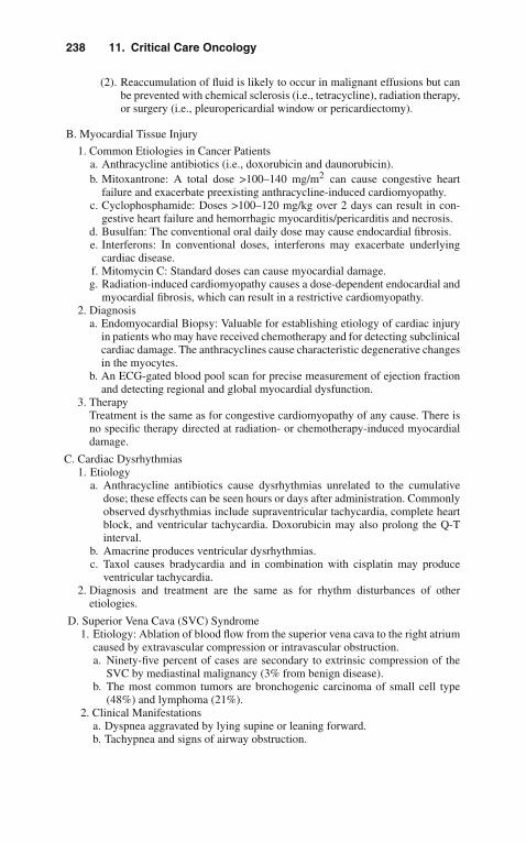
238 11. Critical Care Oncology
(2). Reaccumulation of fluid is likely to occur in malignant effusions but canbe prevented with chemical sclerosis (i.e., tetracycline), radiation therapy,or surgery (i.e., pleuropericardial window or pericardiectomy).
B. Myocardial Tissue Injury
1. Common Etiologies in Cancer Patientsa. Anthracycline antibiotics (i.e., doxorubicin and daunorubicin).b. Mitoxantrone: A total dose >100–140 mg/m2 can cause congestive heart
failure and exacerbate preexisting anthracycline-induced cardiomyopathy.c. Cyclophosphamide: Doses >100–120 mg/kg over 2 days can result in con-
gestive heart failure and hemorrhagic myocarditis/pericarditis and necrosis.d. Busulfan: The conventional oral daily dose may cause endocardial fibrosis.e. Interferons: In conventional doses, interferons may exacerbate underlying
cardiac disease.f. Mitomycin C: Standard doses can cause myocardial damage.g. Radiation-induced cardiomyopathy causes a dose-dependent endocardial and
myocardial fibrosis, which can result in a restrictive cardiomyopathy.2. Diagnosis
a. Endomyocardial Biopsy: Valuable for establishing etiology of cardiac injuryin patients who may have received chemotherapy and for detecting subclinicalcardiac damage. The anthracyclines cause characteristic degenerative changesin the myocytes.
b. An ECG-gated blood pool scan for precise measurement of ejection fractionand detecting regional and global myocardial dysfunction.
3. TherapyTreatment is the same as for congestive cardiomyopathy of any cause. There isno specific therapy directed at radiation- or chemotherapy-induced myocardialdamage.
C. Cardiac Dysrhythmias1. Etiology
a. Anthracycline antibiotics cause dysrhythmias unrelated to the cumulativedose; these effects can be seen hours or days after administration. Commonlyobserved dysrhythmias include supraventricular tachycardia, complete heartblock, and ventricular tachycardia. Doxorubicin may also prolong the Q-Tinterval.
b. Amacrine produces ventricular dysrhythmias.c. Taxol causes bradycardia and in combination with cisplatin may produce
ventricular tachycardia.2. Diagnosis and treatment are the same as for rhythm disturbances of other
etiologies.
D. Superior Vena Cava (SVC) Syndrome1. Etiology: Ablation of blood flow from the superior vena cava to the right atrium
caused by extravascular compression or intravascular obstruction.a. Ninety-five percent of cases are secondary to extrinsic compression of the
SVC by mediastinal malignancy (3% from benign disease).b. The most common tumors are bronchogenic carcinoma of small cell type
(48%) and lymphoma (21%).2. Clinical Manifestations
a. Dyspnea aggravated by lying supine or leaning forward.b. Tachypnea and signs of airway obstruction.

IV. Gastroenterology 239
c. Signs and symptoms of increased intracranial pressure (i.e., dizziness,headache, visual disturbance, seizure, altered mental status).
d. Dysphagia, hoarseness.e. Neck vein distention, facial plethora, and edema.f. Numerous, dilated, vertically oriented, and tortuous cutaneous venules or
veins above the rib cage margin.g. Upper body edema, with cyanosis and ruddy complexion.h. Immediate causes of death are airway obstruction and intracranial hemor-
rhage. Thrombosis at the SVC may occur in 30% of these patients.
3. Diagnosisa. Clinical suspicion.b. CT scan with IV contrast is the diagnostic procedure of choice.c. Transesophageal echocardiography is a safe bedside procedure excellent for
evaluating the SVC and surrounding structures.d. Angiography and radionuclide venography help to localize the obstruction.
4. Therapya. Symptomatic relief is the rule.b. Operative bypass relieves symptoms faster than radiation and is indicated in
patients with life-threatening respiratory compromise or advanced cerebraledema.
c. Endovascular therapy (stents) has been tried successfully in many patients.d. Radiation therapy is the mainstay of treatment for most malignant SVC
obstruction, although in small cell carcinoma and lymphomas chemother-apy, it is particularly useful.
e. Temporizing measures may be used in patients without significant airwayor neurologic compromise and include corticosteroids to decrease cerebraland laryngeal edema, diuretics, and elevation of the head.
f. Anticoagulation has no definitive role.
� IV. GASTROENTEROLOGY
A. Neutropenic Enterocolitis (Ileocecal Syndrome).1. Incidence
Neutropenic enterocolitis commonly occurs in patients with hematologic malig-nancies (leukemia is the most common, with an incidence of 10–40%) receivingchemotherapy.
2. PathophysiologyNeutropenic enterocolitis results from mucosal ulceration and necrosis of theileum, cecum, or ascending colon with overgrowth and mural invasion of bac-teria and/or fungi. Thrombocytopenia may predispose patients to hemorrhageinto the bowel wall. Enterocolitis typically presents on the seventh day of severeneutropenia.
3. Clinical Manifestationsa. Abdominal distentionb. Right-sided abdominal tendernessc. Watery diarrhead. Fevere. Thrombocytopenia and neutropenia
240 11. Critical Care Oncology
4. Diagnosisa. Clinical suspicion.b. Plain radiographs of the abdomen may show ileus with distended cecum and
pneumatosis coli.c. CT of the abdomen: thickened bowel wall containing air.d. Sigmoidoscopy.
5. Differential Diagnosisa. Appendicitisb. Pseudomembranous colitisc. Diverticulitisd. Other acute abdominal disorders
6. Medical Therapya. Nutritional supportb. Nasogastric suctionc. Broad-spectrum antibiotics with anaerobic, gram-negative, and Clostridium
difficile coverage7. Indications for Surgical Exploration
a. Perforationb. Severe bleedingc. Abscessd. Uncontrolled sepsise. Failure to improve after 2–3 days of intensive conservative management
B. Gastrointestinal (GI) Tract Hemorrhage and Perforation1. GI hemorrhage
a. The most common cause is hemorrhagic gastritis (32–48%), followed bypeptic ulcer disease.
b. Only 12–17% of bleeding is from the tumor per se (most commonly seen inGI lymphomas).
c. Less common causes include esophageal varices, Mallory-Weiss tears, can-dida esophagitis, and enteritis.
2. PerforationLymphomas are the most common malignancies to perforate duringchemotherapy.
3. DiagnosisA standard diagnostic workup should be performed to identify the source ofbleeding, with emphasis upon endoscopy.
4. Therapya. Surgicalb. Temporizing modalities to control bleeding include
(1). Angiography, with or without embolization.(2). Endoscopic intervention.(3). These modalities may also be useful in patients with carcinomatosis and
previously unresectable disease.
� V. RENAL/METABOLIC
Many cancer patients develop metabolic abnormalities caused by tumor-producedfactors (hormones or locally acting substances) or from tumor destruction byantineoplastic therapy.

V. Renal/Metabolic 241
A. Hypercalcemia1. Causes of Hypercalcemia in Cancer Patients
a. Secondary to malignancy 4%b. Etiologies other than the malignancy 77%c. With coexistent hyperparathyroidism 2%d. Vitamin D intoxication 16%e. Idiopathic
2. It is the most common metabolic abnormality of cancer patients (10%).3. May occur with or without bone metastases.4. Breast cancer is associated with hypercalcemia in 27–35% of patients.
Mechanisms include widespread osteolytic metastases, production ofparathyroid-like hormone, prostaglandin E2 (PGE2) (following hormonaltherapy with estrogens or anti-estrogens), humoral osteoclast-activating fac-tor, and coexisting primary hyperparathyroidism.
5. Lung cancer is associated with hypercalcemia in 12.5–35% of patients. Itis frequently seen in squamous cell carcinoma and is rare in small cellcarcinoma. It may occur early or late, with or without bone metastases.Mechanisms include production of osteoclast-activating factor, transforminggrowth factor alpha, interleukin 1, and tumor necrosis factor.
6. Multiple myeloma produces hypercalcemia in 20–40% of patients.Hypercalcemia develops secondary to extensive osteolytic bone destruction,osteoclast-activating factor, and lymphotoxin. Fifty percent develop renalinsufficiency, which can aggravate the hypercalcemia.
7. Lymphoma causes hypercalcemia by humoral mediation and local bonedestruction.
8. Head and neck malignancies have an incidence of hypercalcemia of 6%,which is humorally mediated. Hypercalcemia is associated with malignanciesof the oropharynx (37%), hypopharynx (24.3%), and tongue (21.5%).
9. Squamous cell, transitional cell, bladder, renal, and ovarian carcinomas mayalso produce humoral hypercalcemia.
10. Clinical Presentationa. Severity of illness depends on the degree of hypercalcemia, concurrent
illness or debility, age, and associated metabolic disturbances.b. Hypercalcemia of malignancy usually has a rapid onset.c. Neuromuscular manifestations often predominate and include lethargy,
confusion, stupor, and coma (occurs when serum calcium level is>13 mg/dL). Hallucinations and psychosis, weakness, and decreased deeptendon reflexes (DTRs) are also common.
d. Cardiovascular manifestations include increased cardiac contractility,increased sensitivity to digitalis, and dysrhythmias.
e. Renal manifestations include polyuria and polydipsia (earliest symptoms),dehydration, decreased glomerular filtration, loss of urinary concentratingability, and renal insufficiency.
f. Gastrointestinal signs and symptoms include nausea and vomiting,anorexia, obstipation/constipation, ileus, and abdominal pain.
g. Skeletal involvement is the hallmark of hypercalcemia from osteolyticmetastases or humorally mediated bone resorption resulting in pain, patho-logic fractures, deformities, or necrosis.
11. Diagnosisa. Laboratory Studies
(1). Total and ionized serum calcium
242 11. Critical Care Oncology
(2). Electrolytes, serum urea nitrogen (BUN), creatinine(3). Serum phosphorus and alkaline phosphatase(4). Measures of urinary calcium excretion and cyclic adenosine
monophosphate (cAMP).(5). High alkaline phosphatase level(6). Increased urinary calcium excretion
b. Radiologic Studies(1). Radionuclide bone scan(2). Skeletal surveys(3). Baseline chest x-ray
c. ECGs should be performed looking for characteristic changes, includingprolonged PR and QRS intervals, and shortened QT.
12. TreatmentHypercalcemia is often fatal if left untreated, especially when symptomatic orif serum calcium is >13 mg/dL. Treatment goals include promoting urinarycalcium excretion, inhibiting bone resorption, and reducing entry of calciuminto the extracellular fluid compartment.a. Hydration: To restore intravascular volume and increase the urinary output.
(1). Initially, 5–8 L of normal saline IV over the first 24 h; then, adequateIV fluids to maintain a urine output of 3–4 L/day.
(2). Electrolytes should be monitored during normal saline infusion.(3). Monitor urine output and cardiac status to avoid fluid overload.
b. Diuretics: Loop diuretics, such as furosemide, promote calciuresis by block-ing calcium reabsorption in the ascending loop of Henle and augment thecalciuretic effect of normal saline.(1). Furosemide in doses of 40–80 mg IV may be given after adequate
hydration.(2). Monitor electrolytes and urine output to avoid overdiuresis.
c. Inhibitors of bone resorption should be initiated promptly in symptomatichypercalcemia.(1). Mithramycin is an antitumor antibiotic with a direct toxic effect on
osteoclasts. The usual dose is 25 μg/kg IV over 6 h. It generallydecreases serum calcium within 6–48 h; it may be repeated if thepatient does not respond within 2 days. Use should be restricted toemergency treatment of severe hypercalcemia. Complications includethrombocytopenia, myelosuppression, hypotension, and hepatic andrenal toxicity.
(2). Disodium etidronate (EHDP) is an analog of pyrophosphate that blocksosteoclastic bone resorption and formation of bone crystals. The dose is7.5 mg/kg/d in 250 mL saline infused over 2–6 h for 3–7 days, followedby 20 mg/kg/d orally. The onset of action is slow, with normocalcemiaachieved in 4–7 days, 75% of the time. EHDP is contraindicated inpatients with renal failure.
(3). Glucocorticoids (i.e., prednisone) are most effective in hematologicmalignancies (especially multiple myeloma) and breast carcinoma andare of little value in solid tumors. These agents lower serum calcium byinhibition of calcium absorption and the action of vitamin D. Prednisonein a dose of 1–2 mg/kg/d has an onset of action in 3–5 days. Adverseeffects include GI bleeding, hyperglycemia, and osteopenia.

V. Renal/Metabolic 243
(4). Calcitonin inhibits osteoclastic bone resorption and enhances calciumexcretion. The dose is 4–8 IU/kg q6 h IM or SQ. It may lower cal-cium by 2–3 mg/dL over 2–3 h. Adverse reactions include nausea andvomiting, flushing, and hypersensitivity reactions (initial skin testing isrecommended before administration).
d. Hemodialysis is useful in patients who present with renal failure or whocannot be treated with normal saline diuresis.
e. Specific antineoplastic therapy should be initiated in patients for whom treat-ment exists. It is the most effective means of achieving long-term correctionof cancer-related hypercalcemia.
B. Tumor-Lysis Syndrome. Tumor-lysis syndrome is seen when cytotoxic che-motherapy induces rapid tumor cell lysis in patients with a large malignant cellburden of an exquisitely chemosensitive tumor. Intracellular metabolites arereleased in quantities that exceed the excretory capacity of the kidneys.1. This syndrome classically occurs in patients with Burkitt’s and non-Hodgkin’s
lymphoma, acute lymphoblastic and nonlymphoblastic leukemia, and chronicmyelogenous leukemia.
2. It may occur spontaneously in patients with lymphomas and leukemias, orfollowing treatment with chemotherapy, radiation, glucocorticoids, tamoxifen,and/or interferon.
3. Manifestationsa. Related to metabolic abnormalities.
(1). Hyperkalemia: Generalized weakness, irritability, decreased DTRs,paresthesias, paralysis, cardiac dysrhythmias, and cardiac arrest. Theclassic ECG changes include peaked T waves, diminished R waves pro-gressing to widened QRS, prolonged PR, loss of P wave, and sine wavepattern as terminal event.
(2). Hypocalcemia (related to hyperphosphatemia): Muscle spasms, car-popedal spasms, facial grimacing, laryngeal spasm, irritability, depres-sion, psychosis; intestinal cramps, chronic malabsorption; seizures andrespiratory arrest. Chvostek’s and Trousseau’s signs are present in somepatients. ECG reveals a prolonged QT interval.
(3). Hyperuricemia: Gouty arthritis, nephrolithiasis, and urate nephropathy.
b. Precipitation of calcium salts in tissues.c. Acute renal failure.
4. Prevention and Treatment (See Table 11.1).a. To prevent acute renal failure, patients who are to undergo treatment for
malignancies should receive the following:(1). Vigorous IV hydration, often with diuretics or renal-dose dopamine to
ensure adequate urine output(2). Alkalinization of the urine during the first 1–2 days of cytotoxic therapy
to increase the solubility of uric acid(3). Allopurinol to decrease the formation of uric acid
C. Other Common Metabolic Abnormalities in Cancer Patients1. Syndrome of Inappropriate Secretion of Antidiuretic Hormone (SIADH)
a. Occurs in 1–2% of cancer patients.b. Common in small cell carcinoma of the lungs as well as prostatic, pancreatic,
ureteral, and bladder carcinomas.c. Occasionally seen in lymphomas and leukemias.

244 11. Critical Care Oncology
Table 11.1. Management of Patients at Risk for Tumor-LysisSyndrome
I. When no metabolic aberration exists:1. Allopurinol 500 mg/m2 BSA/d; reduce to 200 mg/m2BSA/d, 3 days into
chemotherapy2. Hydration, 3,000 mL/m2BSA/d3. Chemotherapy initiated within 24–48 h of admission4. Monitor electrolytes, BUN, creatinine, uric acid, calcium, phosphorous
every 12–14 h
II. When metabolic aberration exists:1. Allopurinol initiated as above, reduce dose if hyperuricemia controlled,
reduce dose for renal insufficiency2. Hydration as above, add non-thiazide diuretics as needed3. Urinary alkalinization (urine pH >7)
—Sodium bicarbonate 100 mEq/L IV solution initially, adjust as needed—Discontinue when uric acid is normal
4. Chemotherapy postponed until uric acid controlled or dialysis begun5. Monitor same studies, every 6–12 h until stable (at least 3–5 days)6. Replace calcium as Ca++ gluconate by slow IV infusion for
symptomatic hypocalcemia or severe ECG changes7. Treat hyperkalemia with exchange resins, bicarbonate
III. Criteria for hemodialysis in patients unresponsive to the above measures:1. Serum potassium ≥6 mEq/L2. Serum uric acid ≥10 mg/dL3. Serum phosphorus rapidly rising or ≥10 mg/dL4. Fluid overload5. Symptomatic hypocalcemia
Abbreviation: BSA, body surface area.
2. Hypoglycemiaa. Insulinomas: Insulin-secreting, benign, islet-cell tumorsb. Non-islet-cell tumors (i.e., mesothelioma, fibrosarcoma, hemangiopericy-
toma, hepatoma, adrenocortical carcinoma, leukemia and lymphoma, pseu-domyxoma, pheochromocytoma, anaplastic carcinoma)
� VI. HEMATOLOGY
Cancer itself, antineoplastic therapy, and the acute conditions that occur in cancerpatients all result in hematologic abnormalities. Red blood cells, white blood cells,platelets, and coagulation factors may all be adversely affected quantitatively, qual-itatively, or both. Bleeding and infection are the primary life-threatening events incritically ill cancer patients, and they are both the cause and result of hematologicabnormalities. An extensive discussion of these entities can be found in Chapter 7,“Hematological Disorders.”

VIII. Immune Compromise 245
� VII. CHEMOTHERAPY-INDUCEDHYPERSENSITIVITY REACTIONS
A. Etiology and Presentation1. Asparaginase has the highest incidence of hypersensitivity reactions (6–43%).
The incidence is higher when the drug is given intravenously and as a singleagent. Common manifestations includea. Hypotension or hypertension.b. Laryngospasm, respiratory distress.c. Agitation.d. Facial edema.e. Reactions can be life-threatening and are more likely to occur after 2 or more
weeks of treatment.2. Cisplatin is the second most common antineoplastic agent causing hypersensi-
tivity reactions (1–20%). Potentially fatal reactions occur in 5% of patients.3. Alkylating agents are much less commonly a cause of hypersensitivity reac-
tions.a. Melphalan causes anaphylactic reactions in approximately 2–3% of patients.b. Bleomycin causes febrile illness in 20–25% of patients, which in some cases
progresses to a life-threatening syndrome (confusion, chills, respiratory dis-tress, hypotension), seen especially when administered IV to lymphomapatients.
c. Doxorubicin may also cause anaphylaxis.B. Therapy
1. Severe Reactionsa. Stop the antineoplastic drug infusion immediately.b. Epinephrine 0.5–0.75 mL (1:1,000 in 10 mL normal saline) IV push every
5–15 min.c. Aminophylline for acute bronchospasm.d. Diphenhydramine (or other antihistaminic agent) 25–50 mg IV.e. Hydrocortisone 500 mg IV initially and repeated every 6 h for prolonged
reactions.
� VIII. IMMUNE COMPROMISE
The patient with cancer (especially while undergoing chemotherapy) must be consid-ered an immunocompromised host.
A. Types of Immune Defects Recognized in Cancer Patients1. Defects in cellular and humoral immunity
a. T-lymphocyte mononuclear phagocyte defect: Hodgkin’s disease, lym-phoma, cytotoxic chemotherapy
b. Decreased or absent B-cell function in patients with multiple myeloma,chronic lymphocytic leukemia
2. Neutropeniaa. Neutropenia is the most common immunologic defect in patients with
neoplastic diseases.b. The risk for bacteremia and fungal infection increases with absolute neu-
trophil counts (ANCs) <1,000/mm3.

246 11. Critical Care Oncology
c. The most common cause of neutropenia is myelotoxic chemotherapy; neu-tropenia is also seen with leukemia, aplastic anemia, drug reactions, andwhen the bone marrow is destroyed by tumor or radiation.
3. Disruption of the Integument or Mucosal Surfacesa. Diagnostic procedures entailing skin puncture and biopsiesb. Invasive procedures such as the placement of indwelling central venous and
pulmonary artery catheters, urinary catheters, or endotracheal tubesc. Loss of the physical, chemical, and immunologic barrier functions of the gut
lining4. Hyposplenic or Postsplenectomy States
a. Decreased host responses to infections from encapsulated organisms such asS. pneumoniae, Haemophilus influenza, and Neisseria meningitides
B. Clinical Evaluation1. Careful attention to patient’s history of antineoplastic therapies.2. Investigate recurring infections, exposure to contagious diseases, and recent
travel.3. The presence of fever without an obvious source should be investigated
thoroughly by evaluating the following:a. Blood, urine, sputumb. Indwelling cathetersc. Surgical or other skin woundsd. Cerebrospinal fluid (CSF)e. Stoolf. Possibility of undrained collections, abscesses
4. Skin lesions should be inspected carefully. Ecthyma gangrenosum is a charac-teristic skin lesion associated with bacterial and fungal sepsis.
5. The oral cavity is another potential source in the immunocompromised hosts.Sinusitis and periodontitis may be sources, especially in orotracheally ornasotracheally intubated patients and those with nasogastric tubes.
6. Fundoscopic examination is essential for detection of fungal infection, espe-cially in patients with central venous and urinary catheters.
7. Perianal lesions may cause severe infection.8. Panculture is indicated in all febrile patients. All indwelling vascular appliances
should be removed and replaced.The diagnosis and treatment of specific infections in the immunocompromisedhost is covered in Chapter 8, Infections.”
� IX. USEFUL FACTS AND FORMULAS
A. Basic Oncology Formulas. Although not clinically useful, these formulas allowa better understanding of the oncogenesis process, its complications, and responseto therapy.The rapidly proliferating component of human tumors is known as the growthfactor (GF) and is calculated as follows:
GF = Observed fraction of cells in S
Expected fraction of cells in S
where S = part of cell cycle where DNA synthesis occurs predominantly.

IX. Useful Facts and Formulas 247
The fraction of cells in the “S” phase can be assessed by titrated thymidinelabeling and autoradiography. The fraction of labeled cells is known as thethymidine-labeling index (TLI):
TLI = Number of labeled cells
Total number of cells
B. Nutrition in Cancer. Also refer to Chapter 10, “Nutrition.”Cancer patients are frequently malnourished and require close nutritional monitor-ing. To assess the amount of weight loss (percent weight change) in these patients,the following formula is utilized
Percent weight change
= (Usual weight − Actual weight)(Usual weight) × 100
The evaluation of weight change based on the percent weight change formula isdepicted in Table 11.2.A useful formula in the nutritional assessment of these patients relates to thenitrogen balance:
Nitrogen balance = Protein intake (g)
6.25− (24 - h urine urea nitrogen + 4 g)
The catabolic index (ID) aids in the identification of the amount of “nutritionalstress” that these patients have:
CI = 24 h urine nitrogen excretion − [_ dietary nitrogen (g) intake + 3]
The interpretation of the catabolic index is depicted in Table 11.3.The arm muscle circumference (AMC) is another sensitive measure of proteinnutritional status in cancer patients:
AMC = Armcircumference − (TSF)
where TSF = triceps skinfold measurement.
Table 11.2. Evaluation of Weight Change Based on the PercentWeight Change Formula
Significant weight loss (%) Severe weight loss (%)
7 days 1–2 >21 month 5 >53 months 7.5 >7.56 months 10 >10

248 11. Critical Care Oncology
Table 11.3. Interpretation of the Catabolic Index
Catabolic index Interpretation
0 No significant stress1–5 Mild stress>5 Moderate to severe stress
Table 11.4. CSF Findings in Patients With CarcinomatousMeningitis
Percent of abnormal patients Range
Opening pressure 50 60–450WBC count 52 0–1800Glucose 30–38 0–244Protein 30–81 24–2485Cytology 41–70 24–2485
C. Other Facts. The CSF findings in patients with carcinomatous meningitis aredepicted in Table 11.4.The body surface area (BSA) of a patient can be calculated as:
BSA(m2) = (Weightinkg)0.425 × (heightincm)0.725 × 71.84
10,000

12Critical Care of the
Pregnant Patient
Many patients presenting to an intensive care facility will be pregnant. Many patientswill have diseases peculiar to pregnancy and will need critical care support. Others,however, will have underlying medical diseases (Table 12.1). Some of those diseaseswill require consideration of the passenger (fetus) who has created many changesin maternal physiology. The hormonal milieu created by the placenta—progesteroneand to a lesser extent estrogen—is responsible for the multifaceted changes in systemfunction.
One change seen early in pregnancy is in pulmonary function. Table 12.2 depictsthese modifications. Another organ system with significant change is the kidney.Table 12.3 reflects the serial changes in function. Table 12.4 demonstrates thedifferential risk of both acquired and congenital heart disease during pregnancy.
Since many books on obstetrical critical care have been published, and a full reviewof the many changes and diseases is beyond the scope of this chapter, a disease pro-cess that reflects the complexity of the severely ill gravida within the intensive careunit (ICU) has been chosen.
Hemodynamic changes during pregnancy and maternal physiologic changes occur-ring during labor should be kept in mind (see Tables 12.5 and 12.6).
� I. PREGNANCY-INDUCED HYPERTENSION
A. Definition. Pregnancy-induced hypertension (PIH) is the presence of elevatedblood pressure with evidence of end organ dysfunction, most commonly seen asedema, proteinuria, and elevated blood pressure. Table 12.7 presents many of thesynonyms for this process. The classification of preeclampsia (PIH) is depicted inTable 12.8.
B. Etiology. No causative agent has been isolated for the development of preeclamp-sia (PIH). It is most commonly believed to be an end product of antigen–antibodyinteraction with abnormal ratios of vasoactive agents such as prostacyclins andthromboxanes. The relationship of other agents such as lipid peroxides is under
J. Varon, P. Acosta, Handbook of Critical and Intensive Care Medicine,DOI 10.1007/978-0-387-92851-7_12,C© Springer Science+Business Media, LLC 2010

250 12. Critical Care of the Pregnant Patient
Table 12.1. Preexistent Medical Diseases∗
AsthmaCardiac disease, NYHA class 3/4
Prosthetic valve replacementsCritical mitral stenosisAortic stenosisEisenmenger’s syndrome
Cystic fibrosisDiabetes mellitus (insulin dependent)Chronic renal failureHypertensionRenal, hepatic, cardiac transplantsSystemic lupus erythematousThyrotoxicosis/thyroid storm
∗Treatment of these conditions during pregnancy remains unchanged.
active investigation. Regardless of the exact etiology, the process reflects a diffusesystemic endothelial dysfunction intimately associated with platelet dysfunction.This is seen in the classic HELLP (hemolysis, elevated liver enzymes, low platelet)syndrome, in which many organ systems have reflected the disease, and if assid-uous management does not occur, severe maternal morbidity or mortality mayoccur. Table 12.9 reflects the panorama of disease manifestations. Table 12.10depicts those subsets of pregnant women at greatest risk for the disease process.Table 12.11 presents the frequency of preeclampsia in the highest risk populations.
Patients with prior PIH who have underlying chronic hypertension have a 50–75% probability of developing PIH in pregnancy. No method exists to predict theseverity or to time the onset of the PIH process.
C. Approach to the Preeclamptic Patient1. Obtain Patient History
a. Current gestational age (calculated by LMP, last menstrual period)b. Past medical history: renal or chronic hypertensive diseases, systemic lupus
erythematosusc. Family history of preeclampsia/eclampsiad. Symptoms of disease
(1). Headache, blurred vision, scotoma(2). Blindness, diplopia(3). Weight gain (>2 lb/wk)(4). Right upper quadrant pain, epigastric pain, diffuse abdominal pain
(ruptured liver)(5). Tetanic contractions (abruptio placentae)(6). Nausea, emesis(7). Unconsciousness or seizure activity(8). Vaginal bleeding(9). Fetal movement
2. Physical Examinationa. Maternal blood pressure, pulse, respiratory rateb. Fetal heart rate (FHR) by continuous electronic monitoringc. Extensive cardiopulmonary examination

I. Pregnancy-Induced Hypertension 251
Tab
le12
.2.
Lung
Vol
umes
and
Cap
aciti
esin
Pre
gnan
cy
Defi
nitio
nC
hang
ein
Pre
gnan
cy
Res
pira
tory
rate
(RR
)N
umbe
rof
brea
ths
per
min
ute
Unc
hang
ed
Vita
lcap
acity
(Vc)
Max
imum
amou
ntof
air
that
can
befo
rcib
lyex
pire
daf
ter
max
imum
insp
iratio
n(I
C+
ER
V)
Unc
hang
ed
Insp
irato
ryca
paci
ty(I
C)
Max
imum
amou
ntof
air
that
can
bein
spire
dfr
omre
stin
gex
pira
tory
leve
l(T
V+
IRV
)In
crea
sed
5%
Tid
alvo
lum
e(V
T)
Am
ount
ofai
rin
spire
dan
dex
pire
dw
ithno
rmal
brea
thIn
crea
sed
30–4
0%
Insp
irato
ryre
serv
evo
lum
e(I
RV
)M
axim
umam
ount
ofai
rth
atca
nbe
insp
ired
aten
dof
norm
alin
spira
tion
Unc
hang
ed
Fun
ctio
nalr
esid
ualc
apac
ity(F
RC
)A
mou
ntof
air
inlu
ngs
atre
stin
gex
pira
tory
leve
l(E
RV
+R
V)
Dec
reas
ed20
%
Exp
irato
ryre
serv
evo
lum
e(E
RV
)M
axim
umam
ount
ofai
rth
atca
nbe
expi
red
from
rest
ing
expi
rato
ryle
vel
Dec
reas
ed20
%
Res
idua
lvol
ume
(RV
)A
mou
ntof
air
inlu
ngs
afte
rm
axim
umex
pira
tion
Dec
reas
ed20
%
Tota
llun
gca
paci
ty(T
LC)
Tota
lam
ount
ofai
rin
lung
sat
max
imal
insp
iratio
n(V
C+
RV
)D
ecre
ased
5%

252 12. Critical Care of the Pregnant Patient
Tab
le12
.3.
Ser
ialC
hang
esin
Ren
alH
emod
ynam
ics
Non
preg
nant
16w
k26
wk
36w
k29
wk
37w
k
Effe
ctiv
e48
0±
7284
0±
145
891
771
±17
574
8±
8567
7±
82re
nal
plas
ma
flow
279
(mL/
min
)
Glo
mer
ular
filtr
atio
n99
±18
149
±17
152
±18
150
±32
145
±19
138
±22
rate
(mL/
min
)
Filt
ratio
nfr
actio
n0.
210.
180.
180.
200.
190.
21
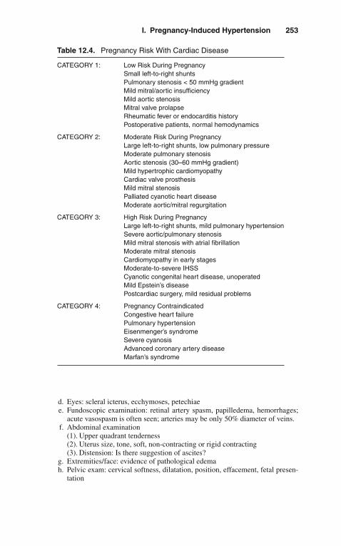
I. Pregnancy-Induced Hypertension 253
Table 12.4. Pregnancy Risk With Cardiac Disease
CATEGORY 1: Low Risk During PregnancySmall left-to-right shuntsPulmonary stenosis < 50 mmHg gradientMild mitral/aortic insufficiencyMild aortic stenosisMitral valve prolapseRheumatic fever or endocarditis historyPostoperative patients, normal hemodynamics
CATEGORY 2: Moderate Risk During PregnancyLarge left-to-right shunts, low pulmonary pressureModerate pulmonary stenosisAortic stenosis (30–60 mmHg gradient)Mild hypertrophic cardiomyopathyCardiac valve prosthesisMild mitral stenosisPalliated cyanotic heart diseaseModerate aortic/mitral regurgitation
CATEGORY 3: High Risk During PregnancyLarge left-to-right shunts, mild pulmonary hypertensionSevere aortic/pulmonary stenosisMild mitral stenosis with atrial fibrillationModerate mitral stenosisCardiomyopathy in early stagesModerate-to-severe IHSSCyanotic congenital heart disease, unoperatedMild Epstein’s diseasePostcardiac surgery, mild residual problems
CATEGORY 4: Pregnancy ContraindicatedCongestive heart failurePulmonary hypertensionEisenmenger’s syndromeSevere cyanosisAdvanced coronary artery diseaseMarfan’s syndrome
d. Eyes: scleral icterus, ecchymoses, petechiaee. Fundoscopic examination: retinal artery spasm, papilledema, hemorrhages;
acute vasospasm is often seen; arteries may be only 50% diameter of veins.f. Abdominal examination
(1). Upper quadrant tenderness(2). Uterus size, tone, soft, non-contracting or rigid contracting(3). Distension: Is there suggestion of ascites?
g. Extremities/face: evidence of pathological edemah. Pelvic exam: cervical softness, dilatation, position, effacement, fetal presen-
tation

254 12. Critical Care of the Pregnant Patient
Table 12.5. Hemodynamic Changes of Pregnancy
Cardiac output Increases 30–40%Heart rate Increases 10–15%Stroke volume IncreasesBlood volume Increases 30–40%Systemic blood pressure DecreasesPulse pressure IncreasesSystemic resistance DecreasesPulmonary artery pressure No ChangePulmonary resistance DecreasesMyocardial function Improves
Table 12.6. Hemodynamic Effects of Labor and Delivery
Cardiac output Increases with contractionsBlood volume IncreasesHeart rate VariablePeripheral resistance No changeSystemic artery pressure Increases
Table 12.7. Synonyms for Pregnancy-Induced Hypertension
Toxemia of pregnancyPIHPreeclampsiaEclampsiaPeripartum hypertensionEPH (edema, proteinuria, hypertension) gestosis
i. Patellar reflexes: Persistent clonus reflects central nervous system (CNS)hyperactivity and significant potential for seizure activity.
j. Please note: Blood pressure may be taken in both supine and lateral positions.Arm elevation when the patient is turned to the lateral decubitus results in afall in blood pressure commensurate with the distance in centimeters abovethe atrial level, roughly, 13.6 mmHg/10 cm of hydrostatic pressure. Thischange in pressure is frequently suggested to be the “real blood pressure.”A patient may then be considered to be normal when in fact hypertensionexists. Table 12.12 reflects these changes in the best study done in normalpregnant patients. (Mean arterial pressure [MAP] is unchanged.)The intensivist should remember that because of the vasodilation of preg-nancy and decreased systemic vascular resistance (SVR), patients, especiallyteenagers, with blood pressures (BPs) of 140/90 mmHg or lesser bloodpressures may be significantly hypertensive.
3. Laboratory Evaluationa. Type/Rh, indirect Coombs’, rapid plasma reagin (RPR), hepatitis B surface
antigen (HbsAg), rubella (if not previously obtained)

I. Pregnancy-Induced Hypertension 255Ta
ble
12.8
.C
lass
ifica
tion
ofP
reec
lam
psia
Mild
Sev
ere∗
Blo
odpr
essu
re13
0/80
–140
/95
mm
Hg
>16
0/11
0m
mH
gA
bsol
ute
Sys
tolic
≥140
mm
Hg
Dia
stol
ic≥9
0m
mH
gR
elat
ive
Sys
tolic
incr
ease
d>
30m
mH
gD
iast
olic
incr
ease
d>
15m
mH
gC
linic
alfin
ding
s1
+ed
ema
3–4
+ed
ema
Nor
mal
refle
xes
3–4
+re
flexe
sN
ovi
sual
sym
ptom
sS
coto
ma/
papi
llede
ma
dipl
opia
May
have
seiz
ures
,alte
red
cons
ciou
snes
sS
ever
ehe
adac
hes
Sev
ere
right
uppe
rqu
adra
ntab
dom
inal
pain
orte
nder
ness
Con
gest
ive
hear
tfai
lure
Pul
mon
ary
edem
aO
ligur
ia<
400
cc/2
4h
Wei
ghtl
ab>
5po
unds
/wee
kP
rote
inur
ia30
0m
g/24
h≥5
g/d;
3+/4
+se
miq
uant
itativ
eP
late
lets
Nor
mal
May
be<
150,
000
Live
rfu
nctio
nN
orm
alE
leva
ted
AS
T/A
LTC
lotti
ngst
udie
sN
orm
alM
aybe
prol
onge
dB
iliru
bin
Nor
mal
May
beel
evat
edA
ntic
onvu
lsan
tsIn
trap
artu
m:y
esA
ntep
artu
m:y
esIn
trap
artu
m:y
esA
bdom
inal
pain
Abs
ent
May
bepr
esen
tin
epig
astr
ium
orR
UQ
∗H
ELL
Psy
ndro
me,
refle
cted
byhe
mol
ysis
,el
evat
edliv
eren
zym
es,
and
low
plat
elet
coun
t,co
mpr
ises
the
grea
test
risk
grou
pfo
rm
orta
lity
and
mor
bidi
ty.
Abb
revi
atio
ns:A
LT,a
lani
neam
inot
rans
fera
se;A
ST,
aspa
rtat
eam
inot
rans
fera
se.

256 12. Critical Care of the Pregnant Patient
Table 12.9. Pregnancy-Induced Hypertension: AssociatedComplications
Hypertensive crisisPulmonary edema: ARDSEclampsiaIntracranial hemorrhageAmaurosisCerebral edemaAcute renal failureCortical necrosisRuptured liverMicroangiopathic hemolytic anemiaThrombocytopeniaDICHELLP syndrome
Abbreviations: ARDS, adult respiratory distress syndrome; DIC, disseminated intravascularcoagulation; HELLP, hemolysis, elevated liver enzymes, low platelets.
Table 12.10. Factors Associated with PIH
NulligravidaPrior preeclampsia/eclampsiaFamily history of preeclampsia, eclampsiaMaternal age >40 yearsAntiphospholipid antibody syndromeMultifetal pregnanciesMolar pregnanciesHydrops fetalis (any etiology)Chronic hypertension/renal diseaseDiabetes mellitus, insulin-dependent
Table 12.11. Frequency of Preeclampsia
7% of all pregnancies70% Nulligravidas30% Multigravidas
Twins 30%Molar pregnancies up to 70%Hydrops fetalis up to 50%Diabetes mellitus up to 50%Chronic hypertension 20%Prior severe PIH up to 50%
b. Complete blood count (CBC), platelets, microscopic examinationc. Serum urea nitrogen (BUN), creatinine, aspartate aminotransferase (AST),
alanine aminotransferase (ALT), glucose, electrolytes, uric acidd. Coagulation studies: prothrombin time (PT), partial thromboplastin time
(PTT), fibrinogen, fibrin split products (FSPs)

I. Pregnancy-Induced Hypertension 257
Table 12.12. Hemodynamic Alterations in Response to PositionChange Late in Third Trimester of Pregnancy
Hemodynamicparameter
Position
LL SUP SIT ST
MAP (mmHg) Hg 90 ± 6 90 ± 8 90 ± 8 91 ± 14CO (L/min) 6.6 ± 1.4 6.0 ± 1.4∗ 6.2 ± 2.0 54 ± 2.0∗P (beats/min) 82 ± 10 84 ± 10 91 ± 11 107 ± 17∗SVR (dyne cm s–5) 1210 ± 266 1437 ± 338 1217 ± 254 1319 ± 394PVR (dyne cm s–5) 76 ± 16 101 ± 45 102 ± 35 117 ± 35∗PCWP (mmHg) Hg 8 ± 2 6 ± 3 4 ± 4 4 ± 2CVP (mmHg) Hg 4± 3 3± 2 1 ± 1 1± 2LVSWI (g/m/m2/beat) 43 ± 9 40 ± 9 44 ± 5 34 ± 7∗
Abbreviations: CO, cardiac output; P, pulse; SVR, systemic vascular resistance; PVR, pul-monary vascular resistance; PCWP, pulmonary capillary wedge pressure; CVP, centralvenous pressure; LVSWI, left ventricular stroke work index; LL, left lateral; SUP, supine;SIT, sitting; ST, standing.∗P < 0.05, compared with left lateral position.Adapted from Clark et al., Am J Obstet Gynecol 1991;164:883–887.
Table 12.13. Colloid Osmotic Pressure
Non-pregnant 28 mmHgPregnant, term 23 mmHgPostpartum 17 mmHgPreeclampsia (PIH) 13.7 mmHg
e. Plasma oncotic pressure (colloid osmotic pressure [COP]) decreases inpregnancy secondary to hemodilution (see Table 12.13)
f. Urinalysis with rapid screen for proteinuria in intensive care unit (ICU)g. Chest x-ray only if pulmonary symptoms or physical examinations suggest
its needh. Obstetrical ultrasound (in ICU) for fetal age and number, estimated fetal
weight, position of fetuses, and placental positioni. Continuous FHR monitoring. Fetal well-being is evaluated by a non-stress
test or biophysical profile.j. 24-h urine collection for creatinine clearance and protein excretion.
D. Medical Therapy1. The presentation of a patient with PIH may range from a mild to a life-
threatening disease process. The process can only be ended by delivery. Thedecision to continue or to deliver the pregnancy will be made by consultationbetween medical and obstetrical personnel.
2. A true rule is that the disease may rapidly progress. Follow up of all maternaland fetal biophysical parameters on a routine basis is required. The frequencywill be determined by the disease severity.
3. Most preeclamptic patients are vasoconstricted and hemoconcentrated. Afterinitial therapy, volume expansion and hemodilution occurs.

258 12. Critical Care of the Pregnant Patient
Table 12.14. Magnesium Toxicity
Manifestations Level (mEq/L)
Loss of patellar reflex (8–12 mEq/L)Feeling of warmth, flushing (9–12 mEq/L)Somnolence (10–12 mEq/L)Slurred speech (10–12 mEq/L)Muscular paralysis (15–17 mEq/L)Respiratory difficulty (15–17 mEq/L)Cardiac arrest (30–35 mEq/L)
Adapted from Sibai BM: Preeclampsia-eclampsia: Valid treatment approached.Contemporary OB/GYN 1990; 35(No.8):84–100.
4. MgSO4 (magnesium sulfate) is considered the standard of therapy as a prophy-laxis for seizure activity. The loading dose is 4–6 g MgSO4·7H2O in 100 ccD5 1/4 NS over 15–20 min. A constant infusion of MgSO4 1–2 g/h will bemaintained depending on urine output and reflex activity, which are checkedon an hourly basis. Table 12.14 lists the potential effects of the magnesiumion and the average serum level at which they may occur. When MgSO4 isinfused, an infusion pump normally will be used to enhance patient safety,thus preventing a massive infusion of MgSO4, which could cause maternaldeath or severe morbidity.
5. Detailed intake and output (I&O) records must be maintained. Since renalfunction is frequently impaired, an increase in total body water can result inpulmonary edema. In rare cases, if hyponatremia is allowed to occur, cerebraledema may be observed.
6. Postdelivery, I&O must be assiduously maintained to prevent hypovolemiaand renal hypoperfusion.
7. Even in mild disease, diplopia may indicate the development of cerebraledema. Standard therapy with mannitol plus or minus Furosemide (Lasix) (seeChapter 9, “Neurologic Disorders”) may be used.
8. Severe pulmonary edema and adult respiratory distress syndrome (ARDS)may occur in preeclamptic patients, as in any other acutely ill patient.Indications for ventilator support are unchanged in this population.
9. Disseminated intravascular coagulation, especially associated with the HELLPsyndrome may require extensive blood product transfusion. In preeclampsia,because of vasoconstriction and the increased risk of pulmonary edema, cry-oprecipitate is often preferred over fresh frozen plasma (FFP). This reducesthe volume of infused blood products. Note: There is a higher risk of hepatitiswith increasing number of donor exposures.
10. When platelets and FFP are required, it is always advisable to use jumbo packsof each to decrease multiple-donor exposure.
11. Swan-Ganz catheter: Invasive pulmonary artery catheter monitoring is rarelyrequired with preeclampsia, but has been used when PIH is complicated bycardiac disease, severe renal disease, or ARDS/pulmonary edema. Knowledgeof the hemodynamic changes in pregnancy is required in considering selectionof therapy (see Tables 12.15 and 12.16).
12. Development of seizures (eclampsia) may occur pre- or post-treatmentwith MgSO4. If seizures develop pretherapy, 4 g of MgSO4 (8 cc of

I. Pregnancy-Induced Hypertension 259
Table 12.15 Acquired Obstetrical Disease That May RequireInvasive Hemodynamic Monitoring
Amniotic fluid embolism
Hemorrhagic shockAbruptio placentaPlacenta previaAbdominal pregnancy
PneumoniasViralBacterial
Septic shock
Chorioamnionitis
Pyelonephritis
Septic abortion
MgSO4·7H2O—50% solution) may be rapidly infused. If seizures developpost-treatment, a second dose of MgSO4 may be given, or some physicianschoose to give diazepam (Valium) 2.5–5 mg as an IV push as the agent ofchoice. If the patient does not awaken and become responsive within 60 min,then the possibility of an intracranial hemorrhage must be considered andworked up (see Chapter 9, “Neurologic Disorders”).
13. Pulse oximeters and recording dynamaps may be used in many patients. If anyconcern for an atypical or severe manifestation of PIH is present, an arterialcatheter should be placed. Repetitive laboratory studies can be drawn, andcontinuous blood pressure recording can be achieved.
14. Interactive dialogue between all members of the healthcare team will achieveoptimum outcome for both mother and child.
E. Major Complications of Preeclampsia/Eclampsia1. Hypertensive Crisis
a. BPs >200 systolic or 120 diastolicb. May be associated with pulmonary edema, intracranial hemorrhage, or
cerebral edemac. Rapid treatment is critical (see Table 12.16). An acute reduction of elevated
BP should initially be limited to a 20% reduction in mean arterial pressure. Amore substantial reduction may create severe uteroplacental hypoperfusionand precipitate acute fetal death or asphyxia. This is especially true if thediastolic blood pressure is acutely dropped to 90 mmHg or less.
d. These patients frequently warrant placement of invasive monitoring systemssuch as pulmonary artery catheters and peripheral arterial cannulas.
2. Cerebral Edemaa. Initiate fluid restrictionb. Invasive monitoringc. IV mannitol 1–2 g/kg of a 20% solution (100 g) of mannitol in 500 mL
of 5% D/W given over 10–20 min followed by a maintenance dose of50–300 mg/kg IV q6 h is effective. The serum osmolality should not beallowed to exceed 330–340 mOsm.

260 12. Critical Care of the Pregnant Patient
Tab
le12
.16.
Ant
ihyp
erte
nsiv
eD
rugs
inP
regn
ancy
Dru
gO
nset
ofac
tion
Dur
atio
nD
osag
eM
echa
nism
Sid
eef
fect
s
Cle
vidi
pine
30s
2m
inIV
1–2
mg/
hfo
llow
edby
repe
ated
incr
emen
tald
oubl
ing
ofth
edo
seat
90s
inte
rval
Art
eria
lvas
odila
tor
Non
e
(Cle
vipr
ex)
The
desi
red
ther
apeu
ticre
spon
sefo
rm
ostp
atie
nts
occu
rat
dose
s4–
6m
g/h
Hyd
rala
zine
15–2
0m
in3–
6h
5–10
mg
q30
min
Dire
ctsm
ooth
mus
cle
Refl
exta
chyc
ardi
a(A
pres
olin
e)R
elax
atio
nH
eada
che
Nic
ardi
pine
∗(C
arde
ne)
1–5
min
3–6
hIV
drip
2m
g/h,
incr
ease
by2
mg/
hea
chho
ur;m
axdo
sage
10m
g/h
Vas
odila
tion
Ca+
chan
nelb
lock
erH
eada
che,
naus
ea,
vom
iting
,hyp
oten
sion
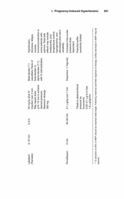
I. Pregnancy-Induced Hypertension 261
Labe
talo
l(T
rand
ate)
5–10
min
3–6
h20
mg
bysl
owIV
inje
ctio
nov
er2
min
.M
ayre
peat
dose
q10–
15m
into
achi
eve
desi
red
pres
sure
.M
axim
umdo
sage
300
mg
Bet
a-bl
ocke
r7-
1va
sodi
lato
rR
atio
ofbe
ta-b
lock
erto
alph
a-bl
ocke
r(7
:1)
with
IVad
min
istr
atio
n
Hyp
oten
sion
,di
zzin
ess,
fatig
ue,
naus
ea,
bron
choc
onst
rictio
nin
som
epa
tient
sw
ithas
thm
a.F
etal
side
effe
cts
may
incl
ude
brad
ycar
dia,
poor
tem
pera
ture
cont
rol,
hypo
glyc
emia
,and
decr
ease
dsh
ort-
term
varia
bilit
y
Fen
oldo
pam
5m
in30
–60
min
0.1
μg/
kgov
er1
min
Dop
amin
e–1
Ago
nist
Incr
ease
sin
tra-
ocul
arpr
essu
re,d
ose
depe
nden
t
Titr
ate
tode
sire
dbl
ood
pres
sure
byin
crem
ents
0.05
–0.1
μg
toa
max
1.6
μg/
kg/m
in.
Pot
entia
lsul
fite
sens
itivi
tytit
rate
d
∗A
redu
ctio
nof
20%
inM
AP
shou
ldbe
max
imal
initi
alta
rget
.F
ollo
win
gm
ater
nal
and
feta
lre
spon
seto
ther
apy,
furt
her
decr
ease
inM
AP
may
bede
sire
d.
262 12. Critical Care of the Pregnant Patient
d. Complications of osmotic agent use include(1). Osmotic diuresis with dehydration and hypernatremia(2). Rebound increase in intracranial pressure(3). Acute volume expansion
e. If a patient is intubated, hyperventilation may be used. (See Chapter 9,“Neurologic Disorders”.)
3. Hepatic Rupturea. Massive intraabdominal hemorrhage results with need for
(1). Massive blood volume support(2). Corrections of disseminated intravascular coagulation (DIC)(3). Invasive cardiovascular monitoring
b. Exploration and surgical repair when necessary. Because of liver dysfunc-tion/damage, packing of the rupture site is often accomplished.
c. Potential for automatic cell saver at operation can reduce total transfusionrequirements.
4. Abruptio Placentaea. Frequently associated with fetal distress.b. Often accompanied by coagulopathy with prolonged PT, PTT, low fibrino-
gen, and low platelet count.c. Vigorous/massive transfusion support may be required.d. Four major complications of hypovolemia and shock in these patients follow:
(1). Acute tubular necrosis(2). Cortical renal necrosis(3). Sheehan’s syndrome with acute pituitary insufficiency(4). ARDS: This has been a major cause of death in our obstetrical ICU.
F. Therapy1. Magnesium Sulfate
a. Distribution: extracellular space, bones, and intracellular space.b. Unbound to protein.c. Excretion by kidney; filtered load excretes T max for reabsorption in most
patients treated.d. Excretion half-life is ∼4 h.e. When used in normal patients for treatment of preterm labor, the earliest
manifestations of excessive Ca++ antagonism are ocular symptoms of visualdisturbance: blurring of vision, diplopia, and difficulty in focusing.
f. MgSO4 does not usually change blood pressure.g. Measurements of magnesium levels can be achieved in most clinical labs.
There is a poor correlation between levels observed and clinical effect.Therefore, no precise level can be stated to be therapeutic.
G. Antihypertensive Therapy (See Table 12.16)1. In most circumstances, drug therapy in PIH is reserved for those patients with
a. Persistent systolic BPs >180 mmHg orb. Persistent diastolic BPs >110 mmHg (105 mmHg in some institutions)
2. Before delivery, it is desired to maintain the diastolic blood pressure>90 mmHg. This allows for continued perfusion pressure to provide adequateuteroplacental perfusion.
3. If diastolic blood pressure decreases <90 mmHg, frequently, the decreaseduteroplacental perfusion will precipitate acute fetal distress, which mayprogress to an in utero death or to perinatal asphyxia.

II. Amniotic Fluid Embolism 263
4. Postdelivery, an acute, rapid decrease in blood pressure usually means sub-stantial blood loss and not cure of the disease process. Likewise, a nadir of90 mmHg diastolic blood pressure is desired.
5. Medical control of hypertension is often required only for a short period (usu-ally days). No study has ever demonstrated a beneficial long-term outcome withprolonged antihypertensive therapy.
6. The use of calcium-channel blockers in a setting of MgSO4 therapy should beconsidered a significant therapeutic step that may create an adverse impact oncardiovascular function; therefore, an intensive care setting with knowledge-able personnel (internal medicine, OB/GYN maternal fetal medicine, or OBanesthesia) capable of responding to these problems should be present.
� II. AMNIOTIC FLUID EMBOLISM
A. DefinitionAmniotic fluid embolism is the vascular transfer of amniotic fluid containinglanugo hairs, vernix, meconium, thromboplastic substances to the pulmonary cir-culation. This is a rare (1 in 8,000 pregnancies) event that is catastrophic and oftenassociated with death.1. It is unpreventable and is most frequent in the second stage of labor.2. The mortality rate is 80%; 25% of deaths occur in the first 60 min post event.3. Old animal models of disease are not applicable to human pathology
B. PresentationSudden onset of maternal distress:1. Severe dyspnea, bronchospasm, tachypnea2. Tachycardia, hypotension, arrhythmias3. Cyanosis, severe hypoxia4. Acute cardiovascular collapse, often cardiac arrest5. DIC6. Seizures, coma
C. Predisposing Factors1. Multiple pregnancy2. Large fetuses3. Short, tumultuous labors4. Pitocin administration5. Late rupture of membranes, with unengaged presenting part
D. Symptoms1. Restlessness2. Sweating3. Anxiety4. Coughing5. Air hunger
E. Pathophysiology1. Pulmonary hypertension; decreased cardiac output2. Cor pulmonale with pulmonary edema3. Severe hypoxemia and tissue hypoxia4. If acute respiratory distress allows survival, thromboplastins yield DIC

264 12. Critical Care of the Pregnant Patient
5. Swan-Ganz data reveal predominant left heart failure/dysfunction: 2◦ tohypoxic injury
F. Differential Diagnosis1. Acute pulmonary embolism2. Air embolism3. Myocardial infarction4. Acute aspiration of gastric contents5. Massive pneumothorax: uni/bilateral6. Reaction to local anesthetic
G. Acute Treatment1. Endotracheal tube placement2. Mechanical ventilation/positive end expiratory pressure (PEEP)3. Volume support with or without blood products4. Peripheral and pulmonary arterial catheterization5. Central venous pressure (CVP) monitoring6. No specific drug therapy: vasopressors and/or bronchodilators of choice
H. Hemodynamic Observations in Humans1. Mild-to-moderate increase in pulmonary artery pressure2. Variable increase in CVP3. Elevated pulmonary capillary-wedge pressure
� III. USEFUL FACTS AND FORMULAS
Uterine oxygen consumption can be calculated by the following formula:
O2 Uptake by Gravid Uterus = (A − V) × F
where A = material arterial blood oxygen content; V = uterine venous blood oxygencontent; F = uterine blood flow.
The oxygen saturation of the uterine venous blood flow (Sv) is another importantparameter to follow and is calculated as follows:
Sv = SaO2 − V
F × (O2Cap)
where SaO2 = maternal oxygen saturation; VO2 = oxygen consumption rate; F =uterine blood flow; O2 Cap = oxygen capacity of maternal blood.
If the last menstrual period (LMP) is known, the probable delivery date (DD) canbe approximated utilizing Naegele’s rule:
DD = First day of LMP + 7 days − 3 months
The approximate weight gain by a pregnant woman can be calculated after thesecond trimester as follows:
WG = 225 g × weeks of gestation
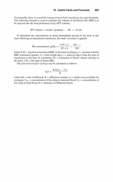
III. Useful Facts and Formulas 265
Occasionally, there is a need for intraperitoneal fetal transfusion in a gravid patient.The following formula is used to calculate the volume of red blood cells (RBCs) tobe injected into the fetal peritoneal cavity (IPT volume):
IPT volume = (weeks’ gestation − 20) × 10 mL.
To determine the concentration of donor hemoglobin present in the fetus at anytime following an intrauterine transfusion, Bowman’s formula is applied:
Hb concentration (g/dL) = 0.55 × a
85 × b× 120 − c
120
where 0.55 = fraction of transfused RBC in the fetal circulation; a = amount of donorRBC transfused (grams); b = fetal weight (kg); c = interval (days) from the time oftransfusion to the time of calculation; 85 = estimation of blood volume (mL/kg) inthe fetus; 120 = life span of donor RBC.
The placental transfer of drugs can be calculated as follows:
Q/t = KA(Cm − Cf)
D
where Q/t = rate of diffusion; K = diffusion constant; A = surface area available forexchange; Cm = concentration of free drug in maternal blood; Cf = concentration offree drug in fetal blood; D = thickness of diffusion barrier.

13Pulmonary Disorders
� I. CHRONIC OBSTRUCTIVE PULMONARYDISEASE (COPD)
A. Definition. COPD is a disorder characterized by expiratory flow limitation thatdoes not change markedly over periods of several months of observation. Theterm COPD includes the following:1. Chronic Bronchitis
Chronic bronchitis is a clinical diagnosis made when chronic cough with spu-tum production is present on most days for at least 3 months of the yearfor at least 2 consecutive years. Major pathologic findings include airwaysinflammation and enlargement of the submucosal mucus glands.
2. EmphysemaEmphysema is defined pathologically as an abnormal permanent enlargementof the air spaces distal to the terminal bronchiole, accompanied by destructionof their wall without obvious fibrosis. Clinically, it correlates with a reductionin the diffusing capacity (DLCO).
3. Various degrees of both chronic bronchitis and emphysema coexist in mostpatients with COPD. The term COPD should not be used for other forms ofobstructive lung disease such as bronchiectasis, cystic fibrosis, or major airwayobstruction.
B. Etiology and Risk Factors. The pathogenesis of most cases of COPD remainsunclear. The main risk factor associated with COPD is cigarette smoking, but mostsmokers do not develop COPD. Less than 1% of patients with emphysema havealpha1-antitrypsin deficiency (serum A1AT <5 μM; normal values 20–48 μM).
C. Diagnostic Evaluation1. Clinical Presentation
a. Cough, sputum production, and dyspnea that usually have been present forseveral years. Symptoms consistent with severe COPD in a young and/ornonsmoking adult should prompt the consideration of other conditions suchas alpha1-antitrypsin deficiency, uncontrolled asthma, or other less com-mon causes of obstructive lung disease (i.e., cystic fibrosis, immotile ciliasyndrome, Young’s syndrome [obstructive azoospermia with chronic bron-chitis/bronchiectasis], congenital or acquired immunoglobulin deficiency).
J. Varon, P. Acosta, Handbook of Critical and Intensive Care Medicine,DOI 10.1007/978-0-387-92851-7_13,C© Springer Science+Business Media, LLC 2010

268 13. Pulmonary Disorders
Table 13.1. Common Conditions Associated with COPDDecompensation
Respiratory Infections: Viral upper and lower respiratory tract (i.e., pharyngitis,tracheobronchitis, pneumonitis), aspiration, and bacterial pneumonia
Narcotics and sedatives
Inappropriately high fraction of inspired O2 (FiO2) (mainly in “CO2 retainers”)
Heart failure
Excessive diuresis with metabolic alkalosis and compensatory CO2 retention
Pneumothorax (rupture of a bleb)
Hypophosphatemia, hypomagnesemia
Hypermetabolic states (i.e., sepsis, fever)
b. During an acute decompensation of COPD, there is an increase in dyspneaand cough, and there are changes in sputum volume, color, and consistency.Physical examination may reveal(1). Pursed-lip breathing(2). Rapid shallow breathing(3). Use of respiratory accessory muscles (i.e., sternocleidomastoid, pec-
toralis, abdominal muscles)(4). Thoracoabdominal paradoxical breathing pattern(5). Wheezes, coarse crackles, and almost undetectable breath sounds in
severe cases(6). Increased jugular venous distention, hepatomegaly, peripheral edema,
and right-sided S3 and increased P2 sounds are characteristic of patientswith cor pulmonale due to severe COPD
(7). Various degrees of changes in mental status may be present and relatedto hypoxemia, hypercapnia, infection, and/or drugsOther conditions that are commonly associated with or precipitate aworsening of COPD patients are depicted in Table 13.1.
2. Laboratory Findingsa. Pulmonary Function Testing (PFT)
(1). Spirometry reveals an obstructive pattern: reduction in the ratio of forcedexpiratory volume in the first second to forced vital capacity (FEV1/FVCratio); normal for a 50-year-old person is 70%). The severity of the expi-ratory airflow limitation can be assessed by the FEV1 (as percent ofnormal predicted according to sex, race, and height). Commonly usedvalues to assign severity of functional impairment are based on theGlobal Initiative for Chronic Obstructive Pulmonary Disease (GOLDcriteria) shown in Table 13.2.
(2). In intubated, mechanically ventilated patients, the interrupter techniquecan be used to diagnose airflow limitation and assess the improvementof expiratory flow in response to bronchodilators.

I. Chronic Obstructive Pulmonary Disease (COPD) 269
Table 13.2. Classification of Severity of COPD—The GOLD Criteria
Stage Physiological characteristics
I: Mild COPD FeV1/FVC <70%FeV1 ≥80% of predicted
II: Moderate COPD FeV1/FVC <70%50%≤ FeV1 <80% of predicted
III: Severe COPD FeV1/FVC <70%30%≤ FeV1 <50% of predicted
IV: Very severe COPD FeV1/FVC <70%FeV1 <30% of predicted
b. Radiologic Studies(1). Chest x-ray may demonstrate evidence of emphysema:
(a). Flattening of the diaphragm(b). Increased retrosternal air space(c). Irregular areas of vascular attenuation and radiolucency (bullae:
lucent areas in the lung parenchyma >1–2 cm in diameter).(d). Typical smoker’s emphysema is mainly of apical distribution.
Predominant lower lung zones changes are consistent with emphy-sema due to alpha1-antitrypsin deficiency.
(2). Computed tomography (CT) of the chest is the most sensitive way todetect emphysema, although it is not routinely recommended as an initialdiagnostic test.
(3). The roentgenographic features of chronic bronchitis are nonspecific andmay include increased lung markings (“dirty lungs”) and thickening ofbronchial walls.
(4). Chest x-ray during an acute COPD exacerbation can be helpful in thedetection of associated processes such as pneumonia, atelectasis, orpneumothorax.
c. Arterial Blood Gases (ABGs)(1). Various degrees of hypoxemia with increased P(A-a)O2 gradient are
typical of COPD patients.(2). Chronic hypercapnia with compensatory metabolic alkalosis is seen in
severe cases (“CO2 retainers”).(3). Finding chronic CO2 retention in moderate COPD with FEV1 >1–1.3 L
is unusual and should raise the question of concomitant neuromuscularor sleep apnea disorders.
(4). Common acid–base disturbances seen during an acute exacerbation ofCOPD include(a). Acute respiratory acidosis.(b). Partially compensated respiratory acidosis (acute-on-chronic).(c). Chronic respiratory acidosis (mild exacerbation in “CO2 retainers”).(d). Metabolic alkalosis induced by diuretics or continuous nasogastric
aspiration may be a cause of persistent or worsening hypercapnia inCOPD.

270 13. Pulmonary Disorders
D. Management of Acute COPD Exacerbation1. Ensure Adequate Oxygenation and Ventilation
a. For most patients, the goal is to maintain a PaO2 of 55–60 mmHg (arte-rial oxyhemoglobin saturation of 88–90%). In patients with concomitantcoronary artery disease, an arterial saturation >90% is desirable.(1). Spontaneously breathing patients with acute COPD exacerbation can
usually achieve those levels using a Venturi mask set to deliver 24–35%O2 (preferred in “mouth breathers”) or a nasal cannula with an O2 flowof 1–2 L/min. (See Chapter 2, “The Basics of Critical Care.”)
(2). Some COPD patients will develop or worsen hypercapnia during O2therapy. A reduction of the hypoxic respiratory drive and a worsening ofV/Q mismatch are the underlying mechanisms thought to mediate thatresponse.
b. Patients with significant acidemia, inadequate PaO2, hypercapnia withchanges in mental status, or hemodynamic instability should be assisted withmechanical ventilation (MV).(1). Noninvasive positive-pressure ventilation (NIPPV) as the initial form
of ventilatory assistance has been reported effective in selected patientswith acute respiratory failure. Candidates for NIPPV should(a). Tolerate a facial or nasal mask.(b). Cooperate with this form of therapy.(c). Have an intact upper airway function without excessive secretions,
regurgitation, or vomiting.(d). Be hemodynamically stable.(e). It may also be offered to patients requiring endotracheal intubation
who decline invasive procedures.NIPPV can be administered using a volume-cycled or pressure-controlled ventilator (i.e., BiPAP in S/T mode inspiratory positiveairway pressure [IPAP] 10 cmH2O, expiratory positive airway pres-sure [EPAP] 5 cmH2O, rate 10 beats per minute). Close observationwith ABGs and continuous monitoring of arterial O2 saturation(SaO2) are recommended to determine NIPPV efficacy and to avoiddelays in endotracheal intubation.
(2). When volume-cycled MV is instituted in intubated COPD patients, amajor goal is to minimize dynamic hyperinflation (auto-positive end-expiratory pressure [PEEP]) and its hemodynamic consequences. Ingeneral, the ventilator should be set to lower the mean expiratory flow(VT/Te) through increases in inspiratory flow (i.e., 90 L/min) and expi-ratory time (reductions in machine rate, I:E ratio, or even sedation thatleads to failure to trigger) and reductions in tidal volume (i.e., 6 mL/kg).
2. Bronchodilatorsa. Inhaled Beta2-Agonists: When delivered by metered dose inhalers (MDI),
these drugs are as effective as nebulized in intubated or spontaneouslybreathing patients (used with a spacer device). Albuterol (Proventil,Ventolin) 2–4 puffs may be administered initially q20 min × 3, followed byq1–2 h until improvement occurs, and then q4–6 h. The dose for albuterolnebulization is 2.5 mg (0.5 cc of 0.5% solution in 2–3 cc of normal saline).
b. Anticholinergics: Ipratropium bromide (Atrovent) has shown to beas effective as beta2-agonists with potentially fewer side effects.Ipratropium bromide should be added to inhaled albuterol during COPDexacerbations.
II. Asthma 271
3. CorticosteroidsUnconfirmed trials have shown benefits from administration of steroids in acuteCOPD exacerbations. Methylprednisolone (Solu-Medrol) 0.5 mg/kg IV q6 h orprednisone 40–60 mg/d PO for 3 days and then tapered over a 2-week period isrecommended.
4. AntibioticsA recognized upper or lower respiratory infection should be treated ade-quately. The initial antibiotic regimen should target likely bacterial pathogens(H. influenzae, M. catarrhalis, and S. pneumoniae in most patients) and takeinto account local patterns of antibiotic resistance Empiric antibiotic therapy(i.e., trimethoprim-sulfamethoxazole, levofloxacin, doxycycline, or amoxicillinfor 7–10 days) in acute COPD exacerbation has been associated with an earlierresolution and fewer relapses.
5. Correct precipitating or associated problems (Table 13.1).
� II. ASTHMA
A. Definition1. Asthma
Asthma is a clinical syndrome characterized by increased responsiveness of thetracheobronchial tree to a variety of stimuli with slowing of forced expirationthat changes in severity either spontaneously or as a result of therapy.
2. Status AsthmaticusStatus asthmaticus is a severe episode of asthma that does not respond to usuallyeffective treatment requiring more aggressive therapy for reversal.
B. Pathophysiology. The key feature of asthma is airway inflammation with hyperre-sponsiveness leading to airway obstruction and in severe cases to hyperinflation,increased VD/VT, and V/Q mismatch, with subsequent hypoxemia and respiratoryinsufficiency.
C. Diagnostic Evaluation1. Clinical Presentation
Dyspnea, wheezing, and coughing are the most common symptoms during anasthma attack. Other diagnostic considerations, especially when a prior historyof asthma is absent, should include the following:a. Heart failure and ischemia with diastolic dysfunctionb. Aspiration of foreign bodiesc. Epiglottitis and croupd. Pulmonary embolism (rare)
Table 13.3 shows several adverse prognostic indicators obtained by history,physical examination, and routine tests in acute life-threatening asthma.
2. Laboratory Evaluationa. Spirometry: Bedside spirometry shows an obstructive pattern (see “COPD,”
section C2a). Serial FEV1 determinations are indicated to objectively evalu-ate the response to treatment. If spirometry is not available, monitoring peakexpiratory flow using a peak flowmeter is recommended.
b. Arterial Blood Gases: Hypoxemia may be seen in cases complicated by res-piratory failure, pneumonia, or pneumothorax. The most common acid–base

272 13. Pulmonary Disorders
Table 13.3. Factors Associated with Severe Acute AsthmaAttacks
Previous episode(s) of severe asthma (especially if associated withrespiratory failure)
Changes in mental status
Use of accessory muscles of respiration
Very diminished or absent breath sounds
Pulsus paradoxus >10 mmHg
Tachycardia >130 beats per min
Cyanosis
Hypoxemia
Hypercapnia or normocapnia in the setting of tachypnea
FEV1 <20% predicted
abnormality is acute respiratory alkalosis. Normocapnia or acute respiratoryacidosis indicates impending or established respiratory failure.
c. Chest X-Ray: May show evidence of hyperinflation, increased bronchialmarkings, or associated conditions such as pneumonia or pneumothorax.
d. Other Tests: In addition to the usual admission tests, theophylline level andblood and sputum cultures should be done if clinically indicated.
D. Management of Asthma Attacks1. Ensure Adequate Oxygenation
a. Most asthma patients will maintain SaO2 >90–92% during an acute attackwith a low concentration of supplemental O2 (Venturi mask or nasal can-nula 2 L/min). Monitor patient with pulse oximeter and supplement O2 asnecessary.
b. Mechanical Ventilation: Few patients with severe asthma will not respondto aggressive medical management and will require ventilatory support. Theventilatory strategy in patients with severe airway obstruction should provideadequate oxygenation and at the same time minimize the risk of barotraumathrough the use of small tidal volumes (i.e., 5–8 mL/kg) and minute ventila-tion (even if PaCO2 is allowed to climb: “controlled hypoventilation”). As inthe case of COPD with expiratory flow limitation, reducing the Ventilator’smean expiratory flow (VT/Te) will improve air trapping and its deleteriouseffects.
2. Beta-Adrenergic AgonistsBeta-adrenergic agonists are first-line therapy for acute asthma episodes.Selective beta2-agonists such as albuterol and terbutaline administered by MDIwith a holding chamber or by nebulization titrated to maximum effect are pre-ferred. For dosing see COPD, section D2a. When drug delivery by aerosol isinadequate, SC epinephrine (0.3 mL 1:1,000 q20 min × 3 max.) or terbutaline(0.25 mg q20 min × 2 max.) can be used.
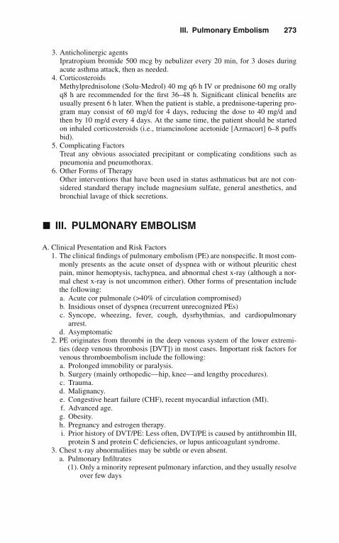
III. Pulmonary Embolism 273
3. Anticholinergic agentsIpratropium bromide 500 mcg by nebulizer every 20 min, for 3 doses duringacute asthma attack, then as needed.
4. CorticosteroidsMethylprednisolone (Solu-Medrol) 40 mg q6 h IV or prednisone 60 mg orallyq8 h are recommended for the first 36–48 h. Significant clinical benefits areusually present 6 h later. When the patient is stable, a prednisone-tapering pro-gram may consist of 60 mg/d for 4 days, reducing the dose to 40 mg/d andthen by 10 mg/d every 4 days. At the same time, the patient should be startedon inhaled corticosteroids (i.e., triamcinolone acetonide [Azmacort] 6–8 puffsbid).
5. Complicating FactorsTreat any obvious associated precipitant or complicating conditions such aspneumonia and pneumothorax.
6. Other Forms of TherapyOther interventions that have been used in status asthmaticus but are not con-sidered standard therapy include magnesium sulfate, general anesthetics, andbronchial lavage of thick secretions.
� III. PULMONARY EMBOLISM
A. Clinical Presentation and Risk Factors1. The clinical findings of pulmonary embolism (PE) are nonspecific. It most com-
monly presents as the acute onset of dyspnea with or without pleuritic chestpain, minor hemoptysis, tachypnea, and abnormal chest x-ray (although a nor-mal chest x-ray is not uncommon either). Other forms of presentation includethe following:a. Acute cor pulmonale (>40% of circulation compromised)b. Insidious onset of dyspnea (recurrent unrecognized PEs)c. Syncope, wheezing, fever, cough, dysrhythmias, and cardiopulmonary
arrest.d. Asymptomatic
2. PE originates from thrombi in the deep venous system of the lower extremi-ties (deep venous thrombosis [DVT]) in most cases. Important risk factors forvenous thromboembolism include the following:a. Prolonged immobility or paralysis.b. Surgery (mainly orthopedic—hip, knee—and lengthy procedures).c. Trauma.d. Malignancy.e. Congestive heart failure (CHF), recent myocardial infarction (MI).f. Advanced age.g. Obesity.h. Pregnancy and estrogen therapy.i. Prior history of DVT/PE: Less often, DVT/PE is caused by antithrombin III,
protein S and protein C deficiencies, or lupus anticoagulant syndrome.3. Chest x-ray abnormalities may be subtle or even absent.
a. Pulmonary Infiltrates(1). Only a minority represent pulmonary infarction, and they usually resolve
over few days

274 13. Pulmonary Disorders
(2). A pleural-based triangular infiltrate (Hampton’s hump) may be seen withinfarction. It usually persist for weeks.
b. Pleural effusion(s)c. Elevated hemidiaphragmd. Platelike atelectasise. Oligemia (Westermark’s sign)
4. Arterial Blood Gasesa. Hypoxemia in most cases (but 15% of PEs have PaO2 >80 mmHg)b. P(A-a)O2 gradient widenedc. Hypocapnia
5. D-dimerThis test has a good sensitivity and negative predictive value, but poor speci-ficity and positive predictive value.
6. Electrocardiogram (ECG)a. Nonspecific QRS and ST-T changesb. Sinus tachycardiac. Atrial dysrhythmias (multifocal atrial tachycardia [MAT], atrial flutter)d. S1-Q3-T3 pattern (only 10% of cases)e. Pulseless electrical activity in massive PE
7. EchocardiographyUp to 40% of patients with PE have echocardiographic anomalies. Some ofthese anomalies area. Increase in RV sizeb. Decreased RV functionc. Tricuspid regurgitationd. RV Thrombus
B. Diagnostic Tests1. Ventilation/Perfusion (V/Q) Scan
A normal V/Q scan practically rules out pulmonary embolism. On the otherhand, an abnormal V/Q scan is nonspecific and should be considered in the con-text of the clinical probability (see Figure 13.1). The V/Q scan and simultaneouschest x-ray findings are categorized as normal/very low, low, intermediate, orhigh probability, as depicted in Table 13.4 .
2. Lower Extremities Venous Studies (LEs)a. Duplex Ultrasound (DU): DU is Doppler ultrasound combined with real-time
two-dimensional ultrasound to study the venous system. When available, itis the method of choice for diagnosing proximal DVT (positive predictedvalue of 94%). Diagnostic criteria of DVT include the inability to collapsethe vein and to visualize the clot. DU can also assess flow augmentation,valvular incompetence, and other causes of pain and swelling (i.e., poplitealcysts and hematomas).
b. Impedance Plethysmography (IPG): Rarely used anymore, IPG determinesthe changes in electrical impedance of the calf in response to blood volumechanges produced by inflating a pneumatic thigh cuff. It is very sensitive forocclusive proximal DVT but insensitive for calf vein thrombosis.
c. Venography: Once considered the gold standard for the diagnosis of leg DVT,is nowadays less frequently used. Disadvantages include its invasiveness,cost, and potential allergic reactions to contrast media. Definitive diagnos-tic findings include filling defects in a well-opacified vein and/or partiallyoccluding defects surrounded by contrast media.
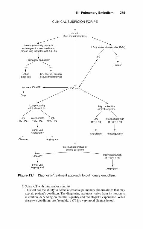
III. Pulmonary Embolism 275
CLINICAL SUSPICION FOR PE
Heparin(if no contraindications)
LEs (duplex ultrasound or IPGs)
Heparin
V/Q scan
IVC filter +/– heparindiscuss thrombolytics
High probabilityclinical suspicion
Low probabilityclinical suspicion
Stop
Pulmonary angiogram
Hemodynamically unstableAnticoagulation contraindicated
Diffuse lung infiltrates with (–) LEs
Otherdiagnosis
Normal(<1% + PE)
Low56% + PE
Low4% + PE
High40% + PE
Intermediate15% + PE
Low16% + PE
Serial LEsAngiorgram?
Intermediate/high88–96% + PE
Intermediate/high28 – 66% + PE
Angiogram
Intermediate probabilityclinical suspicion
Angiogram
Observe Angiogram
Serial LEsAngiogram? Anticoagulation
(–) (+)
(–) (+)
Figure 13.1. Diagnostic/treatment approach to pulmonary embolism.
3. Spiral CT with intravenous contrastThis test has the ability to detect alternative pulmonary abnormalities that mayexplain patient’s condition. The diagnosing accuracy varies from institution toinstitution, depending on the film’s quality and radiologist’s experience. Whenthese two conditions are favorable, a CT is a very good diagnostic tool.

276 13. Pulmonary Disorders
Table 13.4. V/Q Scan Interpretation Categories
Normal and very low probability• No perfusion defects present (ventilation study and/or chest x-ray may be
abnormal)• Three small segmental (<25% of a segment) perfusion defects with
normal chest x-ray
Low probability• >Three small segmental perfusion defects with normal chest x-ray• Large or moderate segmental perfusion defect involving no >4 segments
in one lung and no >3 segments in one lung region with matchingventilation defects and chest x-ray normal or with abnormalities smallerthan the perfusion defects
• Nonsegmental perfusion defect (small pleural effusion, cardiomegaly,enlarged aorta, mediastinum, hila)
• One moderate segmental (>25 to <75%) perfusion defect withnormal chest x-ray mismatch)
Intermediate probability• All V/Q scans not included in the above categories (borderline or difficult
to categorize)
High probability• Two large segmental (>75%) perfusion defects without corresponding
ventilation or chest x-ray abnormality or smaller ventilation or chestx-ray abnormalities (mismatch)
• Two moderate segmental and one large segmental perfusion-ventilation• Two moderate segmental and one large segmental perfusion-ventilation
mismatches• Four moderate segmental perfusion-ventilation mismatches
4. Pulmonary AngiographyStill considered the gold standard for the diagnosis of pulmonary embolism.In general, pulmonary arteriography is a safe procedure (mortality <0.2%, mor-bidity 4%), even in patients with significant pulmonary hypertension. Definitiveangiographic signs include the presence of intraluminal filling defects or cutoffsof pulmonary arteries. It is indicated in patients with suspected PE anda. Contraindications for anticoagulation (considering IVC filter)b. Hemodynamic instability (considering thrombolytic therapy or embolec-
tomy)c. High clinical suspicion for PE with other than high-probability V/Q scan and
negative leg venous studiesd. Extensive pulmonary parenchymal disease or CHF
C. Treatment of Acute Thromboembolism1. Anticoagulation
Anticoagulation should be started as soon as the clinical suspicion for PE ishigh enough to initiate a diagnostic evaluation.a. Heparin: Give an initial bolus of 10,000 U intravenously followed by a con-
tinuous infusion of 1,300 U/h (20,000 U in 500 mL D5W at 33 mL/h). Check
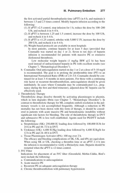
III. Pulmonary Embolism 277
the first activated partial thromboplastin time (aPTT) in 6 h, and maintain itbetween 1.5 and 2.5 times control. Modify heparin infusion according to thefollowing:(1). If aPTT >2.5 control, stop infusion for 1 h, reduce the dose by 100–200
U/h, and recheck it in 4–6 h.(2). If aPTT is between 1.25 and 1.5 control, increase the dose by 100 U/h,
and recheck it in 4–6 h.(3). If aPTT is <1.25 control, rebolus with 5,000 U IV, increase the dose by
200 U/h, and recheck it in 4–6 h.(4). Weight-based protocols are available in most hospitals
In most patients, continue heparin for at least 5 days (provided thatCoumadin was started on day 1 or 2). Seven to ten days of heparininfusion is recommended for patients with massive PE or extensiveiliofemoral thrombosis.
Low molecular weight heparin (1 mg/kg IBW q12 h) has beenused instead of unfractionated heparin in PE with excellent results (seeChapter 7, “Hematological Disorders”).
b. Coumadin: Oral anticoagulation started on day 1 or 2 at a dose of 5–10 mg/dis recommended. The goal is to prolong the prothrombin time (PT) to anInternational Normalized Ratio (INR) of 2.0–3.0. Coumadin should be con-tinued for at least 3–6 months in most patients. For those with a continuingrisk factor or recurrent thromboembolism, anticoagulation should be givenindefinitely. In cases where Coumadin may be contraindicated (i.e., preg-nancy during the first and third trimester), adjusted-dose SC heparin can beeffectively used.
2. Thrombolytic TherapyThrombolytic drugs dissolve thrombi by activating plasminogen to plasmin,which in turn degrades fibrin (see Chapter 7, “Hematologic Disorders”). Incontrast to thrombolytic therapy for MI, complete emboli resolution in the pul-monary vessels is not accomplished frequently. Although a reduction in PEmortality has not been shown with this form of therapy, it should be consid-ered in patients with acute massive PE and hemodynamic instability withoutsignificant risk factors for bleeding. The role of thrombolytic therapy in DVTand submassive PE is less well established. Agents used for PE/DVT includethe following:a. Streptokinase (SK): 250,000 IU loading dose followed by 100,000 IU/h for
24 h in PE and 48–72 h in DVT.b. Urokinase (UK): 4,400 IU/Kg loading dose followed by 4,400 IU/kg/h for
12 h in PE and 24–48 h in DVT.c. Tissue Plasminogen Activator (tPA): 100 mg over 2 h.
The efficacy and bleeding complications of SK, UK, and tPA are equivalent.When using SK or UK, checking a thrombin time or an aPTT 2–4 h intothe infusion is recommended to verify a fibrinolytic state. Heparin should berestarted when the aPTT is <2 times control.
3. IVC FilterIndications for placement of an IVC filter (Greenfield, Mobin-Uddin, Bird’snest) include the following:a. Contraindications to anticoagulationb. Acute massive PEc. Recurrent PE on adequate anticoagulation therapyd. Chronic thromboembolism with pulmonary hypertension

278 13. Pulmonary Disorders
e. Following pulmonary embolectomy or thromboendarterectomy4. Embolectomy
Embolectomy may be considered for documented massive PE with documentedocclusion of main pulmonary artery.
Catheter embolectomy: Intrapulmonary arterial techniques, using rheolyticembolectomy catheter (i.e., AngioJet embolectomy system), have beenused with success in some cases. This procedure has been called by somethe Varon-Strickman procedure.
Surgical embolectomy: This procedure is advised only if an experiencedsurgical team is immediately available.
� IV. ADULT RESPIRATORY DISTRESSSYNDROME (ARDS)
A. Definition. ARDS is a form of acute lung injury characterized by a high-permeability (noncardiogenic) pulmonary edema. In clinical practice, it is definedby the presence of the following:1. Acute respiratory distress in a patient with predisposing conditions2. Diffuse bilateral infiltrates on chest x-ray (pulmonary edema pattern)3. Hypoxemia (PaO2 <55 mmHg with FiO2 >0.5)4. Reduced respiratory system static compliance (<40–50 mL/cmH2O)5. Low or normal pulmonary artery occlusion pressure (pulmonary capillary
wedge pressure [PCWP] <16 cmH2O)
B. Etiology. ARDS is most commonly associated with1. Sepsis2. Bronchial aspiration of gastric content3. Trauma4. Nosocomial pneumonia
Major risk factors for the development of ARDS are listed in Table 13.5.
Table 13.5. Conditions Associated with ARDS
Air embolismAspiration of gastric contentsBurnsCardiopulmonary bypassDisseminated intravascular coagulationDrugs (cocaine, heroin, methadone, acetylsalicylic acid)Multiple fractures (fat embolism)Multiple transfusionsNear drowningPancreatitisPneumonia (bacterial, viral, fungal)Prolonged hypotensionSepsisToxin inhalationTrauma
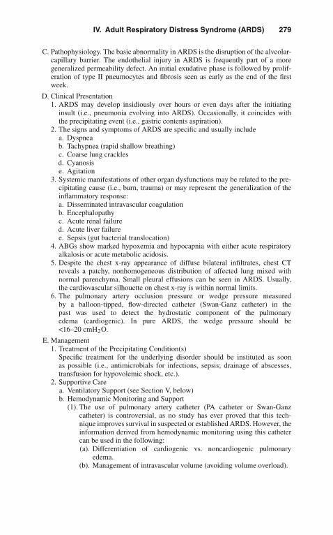
IV. Adult Respiratory Distress Syndrome (ARDS) 279
C. Pathophysiology. The basic abnormality in ARDS is the disruption of the alveolar-capillary barrier. The endothelial injury in ARDS is frequently part of a moregeneralized permeability defect. An initial exudative phase is followed by prolif-eration of type II pneumocytes and fibrosis seen as early as the end of the firstweek.
D. Clinical Presentation1. ARDS may develop insidiously over hours or even days after the initiating
insult (i.e., pneumonia evolving into ARDS). Occasionally, it coincides withthe precipitating event (i.e., gastric contents aspiration).
2. The signs and symptoms of ARDS are specific and usually includea. Dyspneab. Tachypnea (rapid shallow breathing)c. Coarse lung cracklesd. Cyanosise. Agitation
3. Systemic manifestations of other organ dysfunctions may be related to the pre-cipitating cause (i.e., burn, trauma) or may represent the generalization of theinflammatory response:a. Disseminated intravascular coagulationb. Encephalopathyc. Acute renal failured. Acute liver failuree. Sepsis (gut bacterial translocation)
4. ABGs show marked hypoxemia and hypocapnia with either acute respiratoryalkalosis or acute metabolic acidosis.
5. Despite the chest x-ray appearance of diffuse bilateral infiltrates, chest CTreveals a patchy, nonhomogeneous distribution of affected lung mixed withnormal parenchyma. Small pleural effusions can be seen in ARDS. Usually,the cardiovascular silhouette on chest x-ray is within normal limits.
6. The pulmonary artery occlusion pressure or wedge pressure measuredby a balloon-tipped, flow-directed catheter (Swan-Ganz catheter) in thepast was used to detect the hydrostatic component of the pulmonaryedema (cardiogenic). In pure ARDS, the wedge pressure should be<16–20 cmH2O.
E. Management1. Treatment of the Precipitating Condition(s)
Specific treatment for the underlying disorder should be instituted as soonas possible (i.e., antimicrobials for infections, sepsis; drainage of abscesses,transfusion for hypovolemic shock, etc.).
2. Supportive Carea. Ventilatory Support (see Section V, below)b. Hemodynamic Monitoring and Support
(1). The use of pulmonary artery catheter (PA catheter or Swan-Ganzcatheter) is controversial, as no study has ever proved that this tech-nique improves survival in suspected or established ARDS. However, theinformation derived from hemodynamic monitoring using this cathetercan be used in the following:(a). Differentiation of cardiogenic vs. noncardiogenic pulmonary
edema.(b). Management of intravascular volume (avoiding volume overload).

280 13. Pulmonary Disorders
(c). Assessment of the cardiovascular effects of PEEP titration (cardiacindex, stroke volume).
(d). Unfortunately, overtreating or confusing parameters is commonwhile using a PA catheter.
(2). In severe cases of ARDS, where high levels of extrinsic PEEP ordynamic hyperinflation (auto-PEEP) are necessary to maintain adequateoxygenation, a reduced cardiac index should be corrected with the useof inotropes (i.e., dobutamine or dopamine) to maintain an adequate O2delivery.
c. Nutritional Support (see Chapter 10, “Nutrition”)d. Diagnosis and Treatment of Complications
(1). Barotrauma (i.e., tube thoracostomy for pneumothorax)(2). Acute renal failure (i.e., hemodialysis)(3). DIC (i.e., transfusions)(4). Infections: line sepsis, urinary tract infection (UTI), cellulitis (i.e.,
antibiotics, change central lines)3. Other Therapeutic Modalities
a. Pharmacologic and immunologic agents targeted to arrest a specific stepin the inflammatory cascade or pathophysiologic process characteristic ofARDS and sepsis have been extensively evaluated without success: i.e., mon-oclonal antibodies against bacterial lipopolysaccharide and tumor necrosisfactor (TNF), soluble interleukin-1 (IL-1) and TNF receptors, prostaglandinE1, pentoxifylline, nonsteroidal anti-inflammatory drugs (NSAIDs) (i.e.,ibuprofen), synthetic surfactant mixtures (Exosurf), inhaled nitric oxide, etc.
b. Extracorporeal oxygenation and CO2 removal (IVOX, ECCO2R, ECMO),as currently implemented in some centers.
F. Prognosis1. The mortality of ARDS has remained unchanged over the past two decades in
spite of advances in supportive therapy.2. Early mortality is usually related to the underlying condition(s); later, mortality
is mainly related to multiple organ failure rather than pulmonary dysfunction.3. Most ARDS survivors surprisingly have minimal long-term impairment of
lung function (mild restrictive and diffusion capacity [DLCO] defects).Occasionally, reversible airway obstruction may develop).
� V. ACUTE RESPIRATORY FAILURE
A. Definition. Acute respiratory failure is the inability to maintain adequate bloodoxygenation and/or alveolar ventilation in the absence of an intracardiac shunt.Provided the baseline ABGs are close to predicted normal values, this usuallymeans an acute increase in PaCO2 >50 mmHg with arterial acidemia and/or aPaO2 <55 mmHg while breathing room air.
B. Classification and Etiology. Two clinical and pathophysiologic distinct types ofacute respiratory failure can be described:1. Hypoxemic Respiratory Failure
The hallmark of this type of respiratory failure is the inability to adequatelyoxygenate the blood. The main pathophysiologic mechanisms involved are

V. Acute Respiratory Failure 281
V/Q mismatch (response to 100% O2) and intrapulmonary shunting (no sig-nificant improvement with 100% O2). The patients exhibit a rapid shallowbreathing pattern and a low or normal PaCO2. This form of respiratory failureis commonly the result of a diffuse acute lung injury with high-permeabilitypulmonary edema (ARDS), severe pneumonic infiltrates, or cardiogenic pul-monary edema.
2. Hypercapnic Respiratory Failure (Pump Failure)The hallmark of ventilatory pump failure is hypercapnia with acute respira-tory acidosis. The P(A-a)O2 gradient is useful in determining if the hypoxemiapresent in this form of respiratory failure is due to hypoventilation (normal gra-dient) only or to additional parenchymal lung disease (elevated gradient). Thehypercapnia is the result of abnormalities in one or more of the determinants ofthe PaCO2
PaCO2= kVCO2/VE(1 − VD/VT)
a. Increased CO2 production (VCO2) in patients with fever, sepsis, agitation, orexcessive carbohydrate load, associated with a limited ventilatory capacity(high VD/VT, low VE)
b. Increased dead space (VD/VT) in severe COPD, cystic fibrosis, and severeasthma
c. Decreased total minute ventilation (VE) due to ventilatory pump dysfunc-tion:(1). Decreased Central Respiratory Drive: CVA, drugs (narcotics, sedatives,
anesthetics), central hypoventilation, hypothyroidism(2). Abnormal Respiratory Efferents
(a). Spinal Cord: trauma, poliomyelitis, amyotrophic lateral sclerosis,tetanus, rabies
(b). Neuromuscular: myasthenia gravis, multiple sclerosis, botulism,Guillain-Barré syndrome, hypophosphatemia, hypomagnesemia,drugs (streptomycin, amikacin, neuromuscular blockers), polyneu-ropathy of critical illness, bilateral phrenic nerve injury
(3). Abnormal Chest Wall and/or Muscles: severe kyphoscoliosis, anky-losing spondylitis, massive obesity, muscular dystrophy, polymyositis,respiratory muscles fatigue, acid maltase deficiency
(4). Airways, Upper Airway Obstruction: epiglottitis, fixed and variableupper airway obstruction due to tumors, post-extubation, tracheomala-cia, bilateral vocal cord paralysis
C. Management. The management of acute respiratory failure is initially support-ive, aimed at the correction of hypoxemia or hypercapnia until specific actionsare implemented to correct, if possible, the factors that lead to the respira-tory failure (i.e., antibiotics for pneumonia; diuretics, morphine, nitroglycerin,and afterload-reducing agents for cardiogenic pulmonary edema; naloxone fornarcotics overdose).1. Hypoxemic Respiratory Failure
a. Patients with V/Q mismatch abnormalities without significant intrapul-monary shunt will usually respond to noninvasive O2 supplementation (i.e.,nasal cannula, Venturi mask).

282 13. Pulmonary Disorders
b. In patients with cardiogenic pulmonary edema, the use of continuous posi-tive airway pressure (CPAP) (5–10 cmH2O) via a face mask in addition toO2 supplementation can be beneficial by reducing the transmural pressure ofthe left ventricle, and therefore afterload, but also by decreasing the preload.
c. The ventilatory management of patients with diffuse acute lung injury (i.e.,ARDS) requires mechanical ventilation and should be viewed as a balancebetween adequate oxygenation on one hand, and the risk for barotrauma andcardiovascular compromise on the other. The following section pertains tospecific aspects of the ventilatory management of ARDS.(1). Adequate Oxygenation: For most patients with ARDS, this means a
PaO2 of 55–60 mmHg or O2 saturation of 88–90% with a cardiac index>2.5 L/min/m2 and hemoglobin 10 g/dL. Arterial O2 saturation can beincreased in ARDS by the following:(a). Raising the fraction of inspired oxygen (FiO2): To avoid poten-
tial O2 toxic effects, it is recommended not to use 100% O2 formore than a few hours and to maintain an FiO2 ≤0.6. A particulareffort should be made to decrease FiO2 to the minimum accept-able in patients exposed to drugs that may increase O2 toxicity (i.e.,bleomycin, amiodarone).
(b). Increasing the end-expiratory lung volume to recruit collapsed orflooded alveoli. This can be achieved by adding extrinsic PEEPand/or setting the ventilator to create dynamic hyperinflation (auto-PEEP). It is unclear if one strategy is more effective than the otherin ARDS. The goal is to maximize oxygenation while at the sametime avoiding hypotension, reduced cardiac pump function and aplateau pressure >35 cmH2O. With these considerationsi. Extrinsic PEEP is usually started at a level of 5 cmH2O and
titrated up by 2 cmH2O increments to a level of 15–20 cmH2Oalong with the use of other strategies to minimize barotrauma (see“Avoiding Barotrauma,” below).
ii. Extended-ratio ventilation (prolonged I:E ratio, known as inverseratio ventilation when I/E >1:1) is a technique used to increasemean alveolar pressure and transpulmonary pressure. It canbe implemented with either volume–controlled or pressure-controlled ventilators. Increasing the inspiratory time (see Table13.6) increases the mean airway pressure (MAP) and allows therecruitment of lung units with long time constants, therefore,improving oxygenation. Extended-ratio ventilation is more easilyimplemented with volume–controlled than pressure–controlledventilators. Both require heavy sedation with or without paral-ysis; i.e., morphine sulfate, midazolam (Versed), lorazepam(Ativan), or propofol (Diprivan) by continuous infusion with orwithout cisatracurium (Nimbex). Monitoring of plateau pressureto keep it <35 cmH2O, auto-PEEP (end-expiration occlusionmethod).
(2). Avoiding barotrauma: Barotrauma in the form of extra-alveolar air orworsening of acute lung injury is the result of alveolar overdistention(increased transmural pressure or alveolar pressure [Palv]—pleural pres-sure [Ppl]). Thus, it seems reasonable to avoid lung volumes above totallung capacity (TLC) to prevent lung damage.

V. Acute Respiratory Failure 283
Table 13.6. Prolonging Inspiratory Time (Ti) and I/E Ratio inVolume-Controlled Ventilators∗
Reduce inspiratory flow (i.e., to 40 L/min)Use decelerating inspiratory flow waveformAdd an inspiratory pause (i.e., 0.2–0.5 s)Increase the percent inspiratory time†
∗ Applying these changes in a stepwise manner will allow progressive extension of Ti andI:E ratio to the degree that is tolerated or needed. Adjustment in tidal volume (VT) as rec-ommended (see below) and monitoring the plateau pressure (Pplateau), auto-PEEP, andits hemodynamic effects are required to avoid complications.† Direct way of setting the I:E ratio (i.e., Siemens Servo Ventilator).
(a). Because Palv, Ppl, and lung volumes are difficult to determine at thebedside, monitoring the plateau pressure (end-inflation hold pres-sure) is recommended as the best approximation of the peak alveolarpressure.
(b). A plateau pressure of 35 cmH2O or more, in the absence ofsignificantly decreased chest wall compliance, should be avoided.
(c). Peak airway pressure (Ppeak) reflects not only the elastic but alsothe flow-resistive pressures of the respiratory system, and it shouldbe used only as a gross estimate for the risk of barotrauma (i.e.,high Ppeak may be due to a small endotracheal tube, bronchospasm,secretions, high inspiratory peak flow, or worsening of lung or chestwall compliance) that may or may not mean alveolar overinflation.
(d). Determining the tidal volume (VT): ARDS is a nonhomogeneousprocess with collapsed and flooded areas mixed with relatively nor-mal aerated lung, resulting in a reduction in the TLC. Thus, it makessense to ventilate ARDS patients with smaller than conventionalVT. The VT chosen should be one that prevents lung overinflation(i.e., plateau pressure <35 cmH2O) and alveolar derecruitment at theend of expiration (inadequate oxygenation). This usually means aninitial VT of 5–6 mL/kg.
(e). Setting the respiratory rate: The machine rate should be deter-mined considering the patient’s metabolic demands, intrinsic rate,and desired I:E ratio. It is usually set between 25 and 40 breathsper minute. Even with these rates (plus the low VT used), minuteventilation may not be high enough for ARDS patients. AllowingCO2 retention (i.e., 60 mmHg) and respiratory acidemia (i.e., pH7.2–7.25) in an effort to limit barotrauma is referred as permissivehypercapnia.
(3). Controlling the hemodynamic effects of mechanical ventilation: Theincrease in end-expiratory lung volume and mean alveolar pressure pro-duced by the ventilatory strategies described above can have deleterioushemodynamics consequences. It is important to document that a venti-lator change aimed at increasing O2 saturation does not reduce the totalamount of O2 delivered to the tissues via a reduction in cardiac index. Insevere cases, the cardiac index should be maintained >2.5 L/min/m2 withthe use of inotropic agents (vasopressin 1–6 units per hour if hypotensionis present).
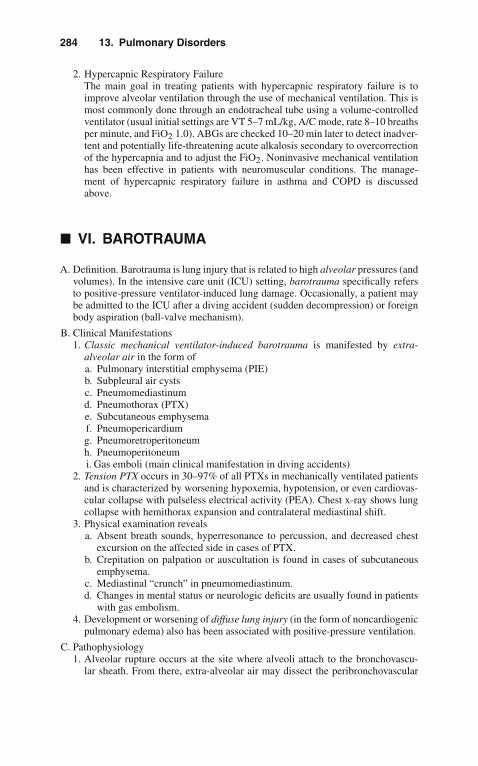
284 13. Pulmonary Disorders
2. Hypercapnic Respiratory FailureThe main goal in treating patients with hypercapnic respiratory failure is toimprove alveolar ventilation through the use of mechanical ventilation. This ismost commonly done through an endotracheal tube using a volume-controlledventilator (usual initial settings are VT 5–7 mL/kg, A/C mode, rate 8–10 breathsper minute, and FiO2 1.0). ABGs are checked 10–20 min later to detect inadver-tent and potentially life-threatening acute alkalosis secondary to overcorrectionof the hypercapnia and to adjust the FiO2. Noninvasive mechanical ventilationhas been effective in patients with neuromuscular conditions. The manage-ment of hypercapnic respiratory failure in asthma and COPD is discussedabove.
� VI. BAROTRAUMA
A. Definition. Barotrauma is lung injury that is related to high alveolar pressures (andvolumes). In the intensive care unit (ICU) setting, barotrauma specifically refersto positive-pressure ventilator-induced lung damage. Occasionally, a patient maybe admitted to the ICU after a diving accident (sudden decompression) or foreignbody aspiration (ball-valve mechanism).
B. Clinical Manifestations1. Classic mechanical ventilator-induced barotrauma is manifested by extra-
alveolar air in the form ofa. Pulmonary interstitial emphysema (PIE)b. Subpleural air cystsc. Pneumomediastinumd. Pneumothorax (PTX)e. Subcutaneous emphysemaf. Pneumopericardiumg. Pneumoretroperitoneumh. Pneumoperitoneumi. Gas emboli (main clinical manifestation in diving accidents)
2. Tension PTX occurs in 30–97% of all PTXs in mechanically ventilated patientsand is characterized by worsening hypoxemia, hypotension, or even cardiovas-cular collapse with pulseless electrical activity (PEA). Chest x-ray shows lungcollapse with hemithorax expansion and contralateral mediastinal shift.
3. Physical examination revealsa. Absent breath sounds, hyperresonance to percussion, and decreased chest
excursion on the affected side in cases of PTX.b. Crepitation on palpation or auscultation is found in cases of subcutaneous
emphysema.c. Mediastinal “crunch” in pneumomediastinum.d. Changes in mental status or neurologic deficits are usually found in patients
with gas embolism.4. Development or worsening of diffuse lung injury (in the form of noncardiogenic
pulmonary edema) also has been associated with positive-pressure ventilation.
C. Pathophysiology1. Alveolar rupture occurs at the site where alveoli attach to the bronchovascu-
lar sheath. From there, extra-alveolar air may dissect the peribronchovascular
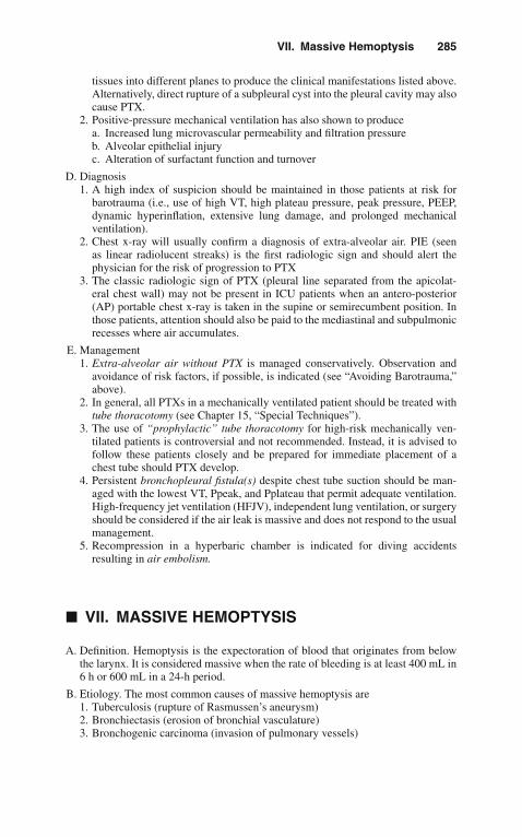
VII. Massive Hemoptysis 285
tissues into different planes to produce the clinical manifestations listed above.Alternatively, direct rupture of a subpleural cyst into the pleural cavity may alsocause PTX.
2. Positive-pressure mechanical ventilation has also shown to producea. Increased lung microvascular permeability and filtration pressureb. Alveolar epithelial injuryc. Alteration of surfactant function and turnover
D. Diagnosis1. A high index of suspicion should be maintained in those patients at risk for
barotrauma (i.e., use of high VT, high plateau pressure, peak pressure, PEEP,dynamic hyperinflation, extensive lung damage, and prolonged mechanicalventilation).
2. Chest x-ray will usually confirm a diagnosis of extra-alveolar air. PIE (seenas linear radiolucent streaks) is the first radiologic sign and should alert thephysician for the risk of progression to PTX
3. The classic radiologic sign of PTX (pleural line separated from the apicolat-eral chest wall) may not be present in ICU patients when an antero-posterior(AP) portable chest x-ray is taken in the supine or semirecumbent position. Inthose patients, attention should also be paid to the mediastinal and subpulmonicrecesses where air accumulates.
E. Management1. Extra-alveolar air without PTX is managed conservatively. Observation and
avoidance of risk factors, if possible, is indicated (see “Avoiding Barotrauma,”above).
2. In general, all PTXs in a mechanically ventilated patient should be treated withtube thoracotomy (see Chapter 15, “Special Techniques”).
3. The use of “prophylactic” tube thoracotomy for high-risk mechanically ven-tilated patients is controversial and not recommended. Instead, it is advised tofollow these patients closely and be prepared for immediate placement of achest tube should PTX develop.
4. Persistent bronchopleural fistula(s) despite chest tube suction should be man-aged with the lowest VT, Ppeak, and Pplateau that permit adequate ventilation.High-frequency jet ventilation (HFJV), independent lung ventilation, or surgeryshould be considered if the air leak is massive and does not respond to the usualmanagement.
5. Recompression in a hyperbaric chamber is indicated for diving accidentsresulting in air embolism.
� VII. MASSIVE HEMOPTYSIS
A. Definition. Hemoptysis is the expectoration of blood that originates from belowthe larynx. It is considered massive when the rate of bleeding is at least 400 mL in6 h or 600 mL in a 24-h period.
B. Etiology. The most common causes of massive hemoptysis are1. Tuberculosis (rupture of Rasmussen’s aneurysm)2. Bronchiectasis (erosion of bronchial vasculature)3. Bronchogenic carcinoma (invasion of pulmonary vessels)
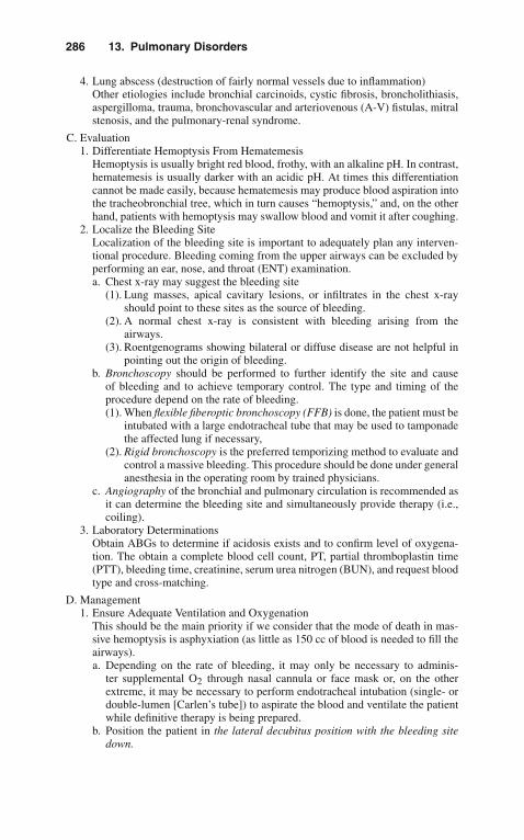
286 13. Pulmonary Disorders
4. Lung abscess (destruction of fairly normal vessels due to inflammation)Other etiologies include bronchial carcinoids, cystic fibrosis, broncholithiasis,aspergilloma, trauma, bronchovascular and arteriovenous (A-V) fistulas, mitralstenosis, and the pulmonary-renal syndrome.
C. Evaluation1. Differentiate Hemoptysis From Hematemesis
Hemoptysis is usually bright red blood, frothy, with an alkaline pH. In contrast,hematemesis is usually darker with an acidic pH. At times this differentiationcannot be made easily, because hematemesis may produce blood aspiration intothe tracheobronchial tree, which in turn causes “hemoptysis,” and, on the otherhand, patients with hemoptysis may swallow blood and vomit it after coughing.
2. Localize the Bleeding SiteLocalization of the bleeding site is important to adequately plan any interven-tional procedure. Bleeding coming from the upper airways can be excluded byperforming an ear, nose, and throat (ENT) examination.a. Chest x-ray may suggest the bleeding site
(1). Lung masses, apical cavitary lesions, or infiltrates in the chest x-rayshould point to these sites as the source of bleeding.
(2). A normal chest x-ray is consistent with bleeding arising from theairways.
(3). Roentgenograms showing bilateral or diffuse disease are not helpful inpointing out the origin of bleeding.
b. Bronchoscopy should be performed to further identify the site and causeof bleeding and to achieve temporary control. The type and timing of theprocedure depend on the rate of bleeding.(1). When flexible fiberoptic bronchoscopy (FFB) is done, the patient must be
intubated with a large endotracheal tube that may be used to tamponadethe affected lung if necessary,
(2). Rigid bronchoscopy is the preferred temporizing method to evaluate andcontrol a massive bleeding. This procedure should be done under generalanesthesia in the operating room by trained physicians.
c. Angiography of the bronchial and pulmonary circulation is recommended asit can determine the bleeding site and simultaneously provide therapy (i.e.,coiling).
3. Laboratory DeterminationsObtain ABGs to determine if acidosis exists and to confirm level of oxygena-tion. The obtain a complete blood cell count, PT, partial thromboplastin time(PTT), bleeding time, creatinine, serum urea nitrogen (BUN), and request bloodtype and cross-matching.
D. Management1. Ensure Adequate Ventilation and Oxygenation
This should be the main priority if we consider that the mode of death in mas-sive hemoptysis is asphyxiation (as little as 150 cc of blood is needed to fill theairways).a. Depending on the rate of bleeding, it may only be necessary to adminis-
ter supplemental O2 through nasal cannula or face mask or, on the otherextreme, it may be necessary to perform endotracheal intubation (single- ordouble-lumen [Carlen’s tube]) to aspirate the blood and ventilate the patientwhile definitive therapy is being prepared.
b. Position the patient in the lateral decubitus position with the bleeding sitedown.

VIII. Upper Airway Obstruction 287
2. Ensure Stable Hemodynamic Conditionsa. Obtain adequate venous access.b. Administer fluids as needed (normal saline or blood).
3. Suppress Cougha. Codeine 60 mg PO q6 h.b. Sedatives may be added (i.e., midazolam 2 mg or lorazepam 1 mg, IV q2 h
pm).4. Control the Bleeding Site
a. Bronchoscopic procedures include(1). Bronchial packing through a rigid bronchoscope(2). Tamponade of airway with a balloon-tipped (Fogarty) catheter through
a rigid bronchoscope or alongside a flexible bronchoscope(3). Bronchial lavage with cold saline through a rigid bronchoscope(4). Coagulation of visible lesions with a Neodynium-YAG laser (not helpful
in very active, brisk bleeding)b. Bronchial artery embolization is the method of choice for patients with mas-
sive or submassive hemoptysis and contraindications for surgery. Given itshigh success rate (90%), embolization has become a first-line treatment forall patients with massive or recurrent hemoptysis.
c. Surgical resection is recommended for localized lesions that can beremoved. It should not be offered to patients with(1). Metastatic lung cancer(2). Severe pulmonary or cardiovascular status
d. Correct coagulopathy, if present (i.e., administer FFP and vitamin K if PT isprolonged, transfuse platelets in severe thrombocytopenia).
E. Prognosis1. Although the underlying medical condition(s) affect the prognosis in massive
hemoptysis, the best estimator of mortality is the rate of bleeding. A study byCracco showed that hemoptysis of 600 cc occurring overa. 4 h has 71% mortalityb. 4–16 h has 45% mortalityc. 16–48 h has 5% mortality
2. Median operative mortality in massive hemoptysis is 17%. Actively bleedingpatients at the time of the surgery have a higher mortality when compared withnonactively bleeding patients.
� VIII. UPPER AIRWAY OBSTRUCTION
A. Acute upper airway obstruction can be a life-threatening situation that may leadto cardiac arrest. This situation requires immediate intervention. This is importantamong the pediatric population, having smaller airways that can quickly progressto complete obstruction.
B. An initial rapid assessment is pertinent to evaluate upper airway patency (audi-ble speech, cough, drooling), respiratory distress (retractions, nasal flaring), andhypoxemia.
C. Immediate treatment: Chin tilt and lift maneuvers should be attempted, as wellas removal of foreign body if suspected. Direct laryngoscopy can also be done,
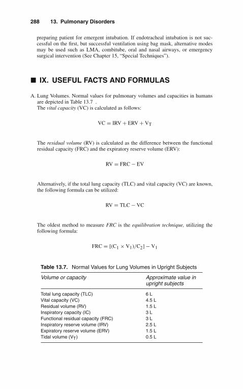
288 13. Pulmonary Disorders
preparing patient for emergent intubation. If endotracheal intubation is not suc-cessful on the first, but successful ventilation using bag mask, alternative modesmay be used such as LMA, combitube, oral and nasal airways, or emergencysurgical intervention (See Chapter 15, “Special Techniques”).
� IX. USEFUL FACTS AND FORMULAS
A. Lung Volumes. Normal values for pulmonary volumes and capacities in humansare depicted in Table 13.7 .The vital capacity (VC) is calculated as follows:
VC = IRV + ERV + VT
The residual volume (RV) is calculated as the difference between the functionalresidual capacity (FRC) and the expiratory reserve volume (ERV):
RV = FRC − EV
Alternatively, if the total lung capacity (TLC) and vital capacity (VC) are known,the following formula can be utilized:
RV = TLC − VC
The oldest method to measure FRC is the equilibration technique, utilizing thefollowing formula:
FRC = [(C1 × V1)/C2] − V1
Table 13.7. Normal Values for Lung Volumes in Upright Subjects
Volume or capacity Approximate value inupright subjects
Total lung capacity (TLC) 6 LVital capacity (VC) 4.5 LResidual volume (RV) 1.5 LInspiratory capacity (IC) 3 LFunctional residual capacity (FRC) 3 LInspiratory reserve volume (IRV) 2.5 LExpiratory reserve volume (ERV) 1.5 LTidal volume (VT) 0.5 L

IX. Useful Facts and Formulas 289
where C1 = known concentration of a test gas in the spirometer; V1 = volumeof gas in the spirometer; C2 = the fractional value of the gas after the subjectbreathes in the spirometer until the concentration of the test gas equals that in thespirometer.
Another way to measure FRC is by utilizing the nitrogen washout procedureand the following formula:
FRC = (VB × CB)/CX
where VB = amount of exhaled nitrogen volume in the bag; CB = fractionalconcentration of nitrogen in the bag; CX = subject initial fractional concentrationof nitrogen (0.80).
Alternatively, FRC can be calculated using body plethysmography asfollows:
FRC = (�/�P)(PB+�P)
where �V = change in volume; �P = change in pressure; PB = atmosphericpressure minus water vapor pressure (PH2O).
The tidal volume (VT) is the sum of the dead space volume (VD) and thealveolar volume (VA):
VT= VD+VA
The average dead space volume (VD) is estimated as 1 mL/lb body weight. For anaverage 70-kg man
VD= 70 × 2.2 × 1 = 154mL
B. Pulmonary Ventilation. The easiest way of estimating minute ventilation (VE) isby using the following formula:
VE= VT × RR = mL/min
where VT = tidal volume; RR = respiratory rate.Minute ventilation is also the sum of dead space (VD) and alveolar ventilation
(VA):
VE= VA+VD
The alveolar ventilation (VA) can be calculated as
VA= (VT − VD) × N

290 13. Pulmonary Disorders
where N = frequency of breathing in breaths per minute; VD = dead spaceventilation.
An alternative method requires knowledge of the CO2 production by thepatient. The production of CO2 ( VCO2) can be calculated as follows:
VCO2= VA × FACO2
where FACO2 = fractional concentration of CO2 in the alveolar gas; and VA =VCO2/FACO2
Dead space ventilation (VD) can be calculated if the minute ventilation (VE)is known:
VD= VE([PaCO2 − PECO2])/PaCO2
The partial pressure of alveolar CO2 (PACO2) is more convenient for thesecalculations and for practical purposes:
PACO2= FACO2 × (PB − 47)
In normal lungs, the arterial CO2 (PaCO2) approximates the PACO2. Therefore,the VA formula can be rewritten as
VA= K(VCO2/PaCO2)
where K = a factor (0.863) that converts CO2 concentrations to pressure (mmHg).
C. Gas Transport in Blood. The difference between the inspired and expired frac-tional concentration of O2 represents the oxygen uptake ( V O2):
VO2= (VI × FiO2) − (VE × FEO2)
where VI = volume of gas inhaled; FiO2 = fractional concentration of inspiredoxygen; VE = volume of gas exhaled; FEO2 = fractional concentration of expiredoxygen.
The amount of O2 in solution in 100 mL of blood is calculated as (assuming apartial O2 pressure of 70 mmHg)
(PO2/760) × αO2= 70/760 × 2.3 = 0.21mL/100mL
The PaO2 at which hemoglobin is 50% saturated (P50) can be calculated from thevenous pH and arterial blood gases as
P50 = antilogLog(1/k)
n= normal 22 − 33 mm Hg

IX. Useful Facts and Formulas 291
where
(1/k) = (antilog [n × log PaO27.4]) × (100 − SaO2/SaO2)
antilog [n × log PaO27.4] = log PaO2 − 0.5(7.4 − venous pH)
n= Hill constant = 2.7 for hemoglobin A
The Fick equation for oxygen consumption (VO2) is calculated as follows:
VO2= Q(CaO2 − CvO2)
where Q = cardiac output (L/min); CaO2 = arterial oxygen content; CFA~vO2 =mixed venous oxygen content.
The volume of carbon dioxide exhaled per unit time (CO2 production or VCO2) is calculated as follows:
VCO2= (VE × FECO2) − (VI × FICO2)
where VE = volume of gas exhaled per unit time; FECO2 = fractional concen-tration of carbon dioxide in the exhaled gas; VI = volume of gas inhaled per unittime; FICO2 = fractional concentration of inspired carbon dioxide.
Since the inspired gas usually contains negligible amounts of carbon dioxide,another representation of this formula is
VCO2= VE × FECO2
D. Pulmonary Circulation. The mean pulmonary artery pressure (PAP) can becalculated utilizing the following formula:
PAP = (PVR × PBF) + PAOP
where PVR = pulmonary vascular resistance; PBF = pulmonary blood flow(which typically equals the cardiac output). Reorganizing the above formula, thepulmonary vascular resistance (PVR) is then calculated as
PVR = (Mean PAP − PAOP)/CO
where PAOP = pulmonary artery occlusion pressure; CO = cardiac output.The pressures that surround the vessels in the pulmonary circulation contribute
to the transmural pressure (Ptm) represented as
Ptm = Pvas − Pis
where Pvas = vascular pressure; Pis = perivascular interstitial pressure.

292 13. Pulmonary Disorders
Table 13.8. Pulmonary Blood Flow Zones
Blood flow zones Pressures
I Palv > Ppa > PpvII Ppa > Palv > Ppv
III Ppa > Ppv > PalvIV Ppa > Ppv > Palv
Abbreviations: Palv, pressure surrounding the alveolar vessels; Ppa, mean pulmonaryarterial pressure; Ppv, mean venous (left atrial) pressure.
When the left atrial pressure (Pla) is available, the driving pressure responsiblefor producing pulmonary blood flow is then calculated as
(Ppa − Pla) = Q × Rvas
where Ppa = mean pulmonary arterial pressure; Pla = mean left atrial pressure;Q = pulmonary blood flow; Rvas = pulmonary vascular resistance.
The pulmonary vascular compliance (Cvas) can be calculated utilizing thefollowing formula:
Cvas =�Vvas/�Pvas
where �Vvas = change in blood volume; �Pvas = change in vascular pressure.The blood flow zones in an idealized upright lung with normal pressure
differences are depicted in Table 13.8 .
E. Mechanics and Gas Flow. The pressure inside the lungs relative to the pressureoutside is known as the transpulmonary pressure (TP) and is calculated as
TP = Palv − Ppl
where Palv = alveolar pressure; Ppl = pleural pressure.The change in volume (�V) for a unit pressure (�P) under conditions of no
flow is the static compliance:
Static compliance (Cs) = �V
�P
Clinically, this formula can be simplified as follows:
Cs = VT
Plateau airway pressure − (PEEP + autoPEEP)
where VT = tidal volume. Normal Cs value is 100 mL/cmH2O.The dynamic compliance (Cdyn) can be calculated utilizing the following
formula:

IX. Useful Facts and Formulas 293
Cdyn = VT
Plateau airway pressure − (PEEP + autoPEEP)
Normal Cdyn value is 100 mL/cmH2O.The specific compliance (Cspec) is calculated utilizing the following formula:
Cspec= Cstat/FRC
The chest wall compliance (Cw) can be calculated as
CW = VT
Airway pressure − Atmospheric pressure
Another formula that can be used under special circumstances (i.e., lung trans-plantation) is the separate lung compliance (CX) and is calculated as
CX = VT
Airway pressure − Intrapleural pressure
The type of gas flow in the lung is laminar flow and is described mathematicallyby the Poiseuille equation:
�P = 8μlV
πr4
where �P = hydrostatic pressure drop; V = gas flow; μ = gas viscosity; 1 = pathlength; r = radius of the tube. From this equation, resistance (R = �P/V) can becalculated:
R = 8μl
πr4
On the other hand, the pressure drop during turbulent flow can be calculatedutilizing the following formula:
�P = μl−ρ−r19/4
where �P = pressure drop during turbulent flow; μ = viscosity; ρ = density.The Reynolds number (Re) is the ratio of the pressure loss due to density-
dependent or inertial flow vs. the pressure loss due to viscous flow. This numberis used to predict the nature of a particular flow and is calculated as follows:
Re = 2ρrV
μA
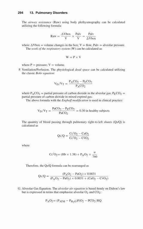
294 13. Pulmonary Disorders
The airway resistance (Raw) using body plethysmography can be calculatedutilizing the following formula:
Raw = �Vbox
V× Palv
V= Palv
�Vbox
where �Vbox = volume changes in the box; V = flow; Palv = alveolar pressure.The work of the respiratory system (W) can be calculated as
W = P × V
where P = pressure; V = volume.
F. Ventilation/Perfusion. The physiological dead space can be calculated utilizingthe classic Bohr equation:
VD/VT = PACO2 − PECO2
PACO2
where PACO2 = partial pressure of carbon dioxide in the alveolar gas; PECO2 =partial pressure of carbon dioxide in mixed expired gas.
The above formula with the Enghoff modification is used in clinical practice:
VD/VT = PaCO2 − PECO2
PaCO2= 0.30 in healthy subjects
The quantity of blood passing through pulmonary right-to-left shunts (Qs/Q) iscalculated as
Qs/Q = Cc′O2 − CaO2
Cc′O2 − CvO2
where
Cc′O2= (Hb × 1.38) + PAO2 × α
760
Therefore, the Qs/Q formula can be rearranged as
Qs/Q = (PAO2 − PaO2) × 0.0031
(PAO2 − PaO2) × 0.0031 + (CaO2 − CvO2)
G. Alveolar Gas Equation. The alveolar air equation is based firmly on Dalton’s lawbut is expressed in terms that emphasize alveolar O2 and CO2:
PAO2= (PATM − PH2O)FiO2 − PCO2/RQ

IX. Useful Facts and Formulas 295
where PAO2 = partial pressure of O2 in the alveolus under present conditions;PATM = current, local atmospheric pressure. PH2O = vapor pressure of waterat body temperature and 100% relative humidity; FiO2 = fraction of inspired O2;PCO2 = partial pressure of CO2 in arterial blood; RQ = respiratory quotient.At sea level, this equation can be simplified to
PAO2= 150 − 1.25 × PaCO2
The arterial oxygen tension (PaO2) corrected for age is calculated as
PaO2age − corrected = 100 − 1/3 age (in years)
The alveolar-arterial O2 gradient is age-corrected according to the followingformula:
Age correction = 2.5 + (0.25 × [age in years])

14Renal and Fluid–
Electrolyte Disorders
� I. ACID–BASE DISTURBANCES
An acidosis is any process that tends (in the absence of buffering or compensation)to cause the accumulation of hydrogen ions (H+). If the pH of the blood is lower thannormal (<7.35), acidemia is present. Similarly, any process that tends to cause theaccumulation of bicarbonate (HCO3) is an alkalosis. An elevated blood pH (>7.45) isreferred to as alkalemia. When the acid–base disturbance arises as a result of changesin the carbon dioxide tension (PCO2) of the blood, a respiratory process is present.Conversely, when the acid–base disturbance occurs as a result of accumulation ofnonvolatile acids or loss of HCO3 (or substances metabolized to HCO3), a metabolicprocess is present. Acidosis and alkalosis can be either primary or compensatory fora different acid–base problem. Many ICU patients have double and triple acid–basedisorders.
A. Approach to Acid–Base Disturbances
1. The initial evaluation of acid–base disorders requires the simultaneous exami-nation of arterial blood gases (ABGs) and serum electrolytes.
2. The typical patterns of blood pH, PCO2, and HCO3 in various acid–baseproblems are listed in Table 14.1.
3. The adequacy of compensation for a primary acid–base abnormality can also beassessed.a. For patients with metabolic acidosis, the expected PCO2 can be calculated as
PCO2= ([HCO3] × 1.5) + 8 ( ± 2)
(1). If the actual PCO2 is greater than expected, a simultaneous respiratoryacidosis is present.
J. Varon, P. Acosta, Handbook of Critical and Intensive Care Medicine,DOI 10.1007/978-0-387-92851-7_14,C© Springer Science+Business Media, LLC 2010

298 14. Renal and Fluid–Electrolyte Disorders
Table 14.1. Systematic Evaluation of Acid–Base Disorders
Compensated processMetabolic acidosis pH ↓ PCO2 ↓ HCO3 ↓Respiratory acidosis pH ↓ PCO2 ↑ HCO3 ↑Metabolic alkalosis pH ↑ PCO2 ↑ HCO3 ↑Respiratory alkalosis pH ↑ PCO2 ↓ HCO3 ↓
Uncompensated processMetabolic acidosis pH ↓ PCO2—HCO3 ↓Respiratory acidosis pH ↓ PCO2 ↑ HCO3—Metabolic alkalosis pH ↑ PCO2—HCO3 ↑Respiratory alkalosis pH ↑ PCO2 ↓ HCO3—
Mixed processMetabolic/Respiratory Acidosis pH ↓ PCO2 ↑ HCO3 ↓Metabolic/Respiratory Alkalosis pH ↑ PCO2 ↓ HCO3 ↑
(2). If the measured PCO2 is less than expected, a simultaneous respiratoryalkalosis is present.
b. For patients with metabolic alkalosis, the expected PCO2 can be calculated as
PCO2= {([HCO3] − 25) × 0.7} + 40 ( ± 2)
(1). Respiratory compensation for a primary metabolic alkalosis is limited to aPCO2 of about 55 mmHg. Even this limit may not be attained in patientswith chronic pulmonary or hepatic disease or congestive heart failure.
c. Metabolic compensation for respiratory acid–base disturbances depends inpart on the duration of the problem.(1). For patients with chronic respiratory acidosis, the expected [HCO3] can
be calculated as
[HCO3] = (PCO2 − 40) × 0.35 + 25 ( ± 2)
(a). The upper limit for this process is [HCO3] = 55 mEq/L.(b). If the degree of compensation is inadequate, the possibility of
a mixed acid–base disturbance or a superimposed acute processshould be entertained.
(2). The expected degree of compensation for acute respiratory acidosis iscalculated as
[HCO3] = (PCO2 − 40) × 0.10 + 25 ( ± 2)
The upper limit of compensation for acute respiratory acidosis is [HCO3]= 30 mEq/L.

I. Acid–Base Disturbances 299
(3). The expected metabolic response to chronic respiratory alkalosis can beestimated by
[HCO3] = 25 − (40 − PCO2) × 0.5 ( ± 2)
Inadequate compensation for chronic respiratory alkalosis should raisethe possibility of a superimposed acute respiratory alkalosis or a mixedacid–base problem.
(4). The expected metabolic response to acute respiratory alkalosis can becalculated as
[HCO3] = 25 − (40 − PCO2) × 0.25 ( ± 2)
B. Metabolic Acidosis. The clinical consequences of metabolic acidosis are due toabnormalities of intracellular pH, transcellular ionic shifts, or both.1. Signs and symptoms include tachypnea, depressed cardiac function, fatigue,
weakness, and altered mental status.2. Laboratory findings: hyperkalemia, calciuresis, and changes in carbohydrate
and lipid metabolism.3. Other specific findings may occur due to the etiology of the underlying cause
of the acidosis.4. Metabolic acidoses are classified based on whether the plasma anion gap (AG)
is normal or elevated. Its value is calculated as
AG = [Na+] − ([Cl−] + [HCO3]) (nl = 10 − 15 mEq/L)
5. The differential diagnosis of primary metabolic acidosis is listed in Table 14.2.a. In general, high AG metabolic acidoses are caused by the accumulation
of an acid in the plasma whose anion is something other than Cl–. Theseacids can be endogenous (i.e., lactic acid, ketoacids, uremic acids), exoge-nous (i.e., salicylates), or the endogenous metabolic products of exogenoustoxins (i.e., paraldehyde, methanol, ethylene glycol).
b. The accumulation of these unmeasured acid anions should be stoichiomet-rically equal to the observed decrease in [HCO3].
c. This equality will be preserved when the normal difference is maintained.Thus, if a normal anion gap = 12 mEq/L, and a normal [HCO3] =25 mEq/L, in a pure high AG metabolic acidosis
[Na+ − ([Cl−+25) = 12; or[Na+ − ([Cl−) = 37 ± 3.
If ([Na+ – [Cl–]) is significantly >37, then a coexistent metabolic alkalosisshould be suspected.If the difference is significantly <37, then a coexistent normal anion gapacidosis is probably present.

300 14. Renal and Fluid–Electrolyte Disorders
Table 14.2. Causes of Metabolic Acidosis
Elevated anion gap Normal anion gap
Methanol Urinary–enteric fistulaUremia Saline volume expansionKetoacidosis EndocrinopathiesParaldehyde DiarrheaLactic acidosis Carbonic anhydrase inhibitionEthylene glycol Acid–chloride administrationSalicylates Renal tubular acidosis
Mineralocorticoid antagonists
d. The causes of metabolic acidosis with a normal anion gap are listed inTable 14.2. These so-called hyperchloremic acidoses occur either due tothe administration of exogenous acids whose anion is chloride (e.g., HCl,NH4C1, CaC12) or the loss of body fluid that is relatively low in chloridebut high in bicarbonate or its metabolic equivalent.(1). In cases of normal AG metabolic acidosis, measurement of the urinary
AG can be diagnostically useful. It is calculated as
AGU= UNa+UK − UCl
The AGU is inversely related to urinary ammonium (NH4) excretion.Its value should be less than zero in acidotic patients with gastroin-testinal (GI) bicarbonate losses, since renal ammoniagenesis ought tobe well preserved. In patients with acidosis due to urinary bicarbonatelosses, especially if caused by renal tubular acidosis (RTA), the AGUshould be greater than zero.
6. The primary therapy of metabolic acidosis is to treat the underlying disease.7. The use of exogenous NaHCO3 in the therapy of metabolic acidosis (espe-
cially lactic acidosis) is somewhat controversial, since evidence exists thatsuch treatment can actually accelerate lactate production. However, it is gen-erally accepted that severe acidemia (pH <7.20) of metabolic origin shouldbe corrected with NaHCO3. The amount of bicarbonate required can becalculated by
HCO3 deficit= body wt(kg) × 0.5 × (target − actual [HCO3])
a. Administration of NaHCO3 as an isotonic solution (i.e., 3 ampules ofNaHCO3 per liter of sterile water or 5% dextrose in water) will reducethe risk of hypernatremia in patients at particular risk.
8. Dialysis is sometimes indicated not only for the correction of acidosis but alsofor the rapid elimination of certain toxins that cause acidosis, such as methanoland ethylene glycol, even if the serum urea nitrogen (BUN) and creatinine arenormal.
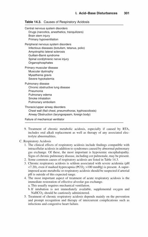
I. Acid–Base Disturbances 301
Table 14.3. Causes of Respiratory Acidosis
Central nervous system disordersDrugs (narcotics, anesthetics, tranquilizers)Brain stem injuryPrimary hypoventilation
Peripheral nervous system disordersInfectious diseases (botulism, tetanus, polio)Amyotrophic lateral sclerosisGuillain-Barré syndromeSpinal cord/phrenic nerve injuryOrganophosphates
Primary muscular diseaseMuscular dystrophyMyasthenia gravisSevere hypokalemia
Pulmonary diseaseChronic obstructive lung diseasePneumoniaPulmonary edemaSmoke inhalationPulmonary embolism
Thoracic/upper airway disordersChest wall (flail chest, pneumothorax, kyphoscoliosis)Airway Obstruction (laryngospasm, foreign body)
Failure of mechanical ventilator
9. Treatment of chronic metabolic acidosis, especially if caused by RTA,includes oral alkali replacement as well as therapy of any associated elec-trolyte abnormalities.
C. Respiratory Acidosis1. The clinical effects of respiratory acidosis include findings compatible with
intracellular acidosis in addition to syndromes caused by abnormal pulmonarygas exchange. Of these, the most important is hypoxemic encephalopathy.Signs of chronic pulmonary disease, including cor pulmonale, may be present.
2. Some common causes of respiratory acidosis are listed in Table 14.3.3. Chronic respiratory acidosis is seldom associated with severe acidemia (pH
<7.20), even if marked hypercapnia (PCO2 >100 mmHg) is present. A super-imposed acute metabolic or respiratory acidosis should be suspected if arterialpH is outside of this expected range.
4. The most important aspect of treatment of acute respiratory acidosis is theimmediate restoration of effective alveolar gas exchange.a. This usually requires mechanical ventilation.b. If intubation is not immediately available, supplemental oxygen and
NaHCO3 should be cautiously administered.5. Treatment of chronic respiratory acidosis depends mainly on the prevention
and prompt recognition and therapy of intercurrent complications such asinfections and congestive heart failure.

302 14. Renal and Fluid–Electrolyte Disorders
D. Metabolic Alkalosis1. The clinical features of metabolic alkalosis are nonspecific.
a. Evidence of neuromuscular irritability or latent tetany may be present.b. Cardiac dysrhythmias can occur.
2. Hypokalemia and hypochloremia are usually present.3. The plasma AG is frequently elevated, due partly to a slight increase in lactate
levels but mainly due to release of H+ ions from plasma proteins (especiallyalbumin) as part of the buffering process.
4. Urine pH measurements are not helpful.5. The most useful biochemical determination is the level of urinary chloride
(UCl), which forms the basis for classification of metabolic alkalosis.a. Metabolic alkalosis associated with an UCl <10 mEq/L are termed chloride-
responsive, whereas those with a UCl > 20 mEq/L are termed chloride-resistant.
b. In some cases, UCl falls between these levels, in which case, no definiteclassification is possible.
6. Causes of metabolic alkalosis are listed in Table 14.4.7. Since the kidney can ordinarily excrete a vast amount of HCO3, acid loss (or
base accumulation) alone does not usually result in a sustained alkalosis.a. If coexistent volume depletion is present, alkalosis can be persistent. This is
by far the most common pathogenesis for metabolic alkalosis. This combi-nation occurs when body fluids rich in NaCl and poor in NaHCO3 are lost,usually from the intestinal tract or kidneys. Rarely, other sites of NaCl loss(e.g., skin losses in cystic fibrosis) may be present.
b. Extracellular fluid volume contraction causes avid NaCl and NaHCO3reabsorption in all nephron sites with attendant decreases in UCl andperpetuation of the alkalosis.
Table 14.4. Causes of Metabolic Alkalosis
Chloride responsiveGastrointestinal disorders
Gastric: vomiting, nasogastric suctionColonic: villous adenoma, chloride diarrhea
Renal disordersDiuretic therapyPost-hypercapnic alkalosis
Cystic fibrosis
Chloride resistantHypermineralocorticoid statesSevere potassium depletionBartter’s syndrome
MiscellaneousAlkali administration (antacids, transfusions)HypercalcemiaPoorly absorbable anion administration (antibiotics)
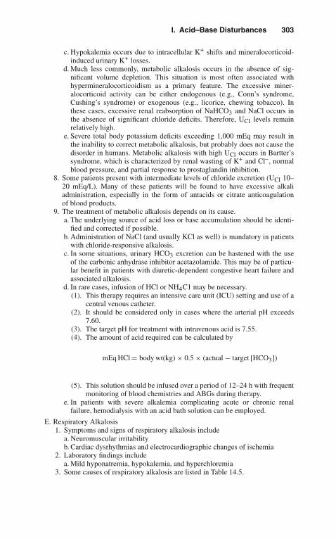
I. Acid–Base Disturbances 303
c. Hypokalemia occurs due to intracellular K+ shifts and mineralocorticoid-induced urinary K+ losses.
d. Much less commonly, metabolic alkalosis occurs in the absence of sig-nificant volume depletion. This situation is most often associated withhypermineralocorticoidism as a primary feature. The excessive miner-alocorticoid activity can be either endogenous (e.g., Conn’s syndrome,Cushing’s syndrome) or exogenous (e.g., licorice, chewing tobacco). Inthese cases, excessive renal reabsorption of NaHCO3 and NaCl occurs inthe absence of significant chloride deficits. Therefore, UCl levels remainrelatively high.
e. Severe total body potassium deficits exceeding 1,000 mEq may result inthe inability to correct metabolic alkalosis, but probably does not cause thedisorder in humans. Metabolic alkalosis with high UCl occurs in Bartter’ssyndrome, which is characterized by renal wasting of K+ and Cl–, normalblood pressure, and partial response to prostaglandin inhibition.
8. Some patients present with intermediate levels of chloride excretion (UCl 10–20 mEq/L). Many of these patients will be found to have excessive alkaliadministration, especially in the form of antacids or citrate anticoagulationof blood products.
9. The treatment of metabolic alkalosis depends on its cause.a. The underlying source of acid loss or base accumulation should be identi-
fied and corrected if possible.b. Administration of NaCl (and usually KCl as well) is mandatory in patients
with chloride-responsive alkalosis.c. In some situations, urinary HCO3 excretion can be hastened with the use
of the carbonic anhydrase inhibitor acetazolamide. This may be of particu-lar benefit in patients with diuretic-dependent congestive heart failure andassociated alkalosis.
d. In rare cases, infusion of HCl or NH4C1 may be necessary.(1). This therapy requires an intensive care unit (ICU) setting and use of a
central venous catheter.(2). It should be considered only in cases where the arterial pH exceeds
7.60.(3). The target pH for treatment with intravenous acid is 7.55.(4). The amount of acid required can be calculated by
mEq HCl = body wt(kg) × 0.5 × (actual − target [HCO3])
(5). This solution should be infused over a period of 12–24 h with frequentmonitoring of blood chemistries and ABGs during therapy.
e. In patients with severe alkalemia complicating acute or chronic renalfailure, hemodialysis with an acid bath solution can be employed.
E. Respiratory Alkalosis1. Symptoms and signs of respiratory alkalosis include
a. Neuromuscular irritabilityb. Cardiac dysrhythmias and electrocardiographic changes of ischemia
2. Laboratory findings includea. Mild hyponatremia, hypokalemia, and hyperchloremia
3. Some causes of respiratory alkalosis are listed in Table 14.5.
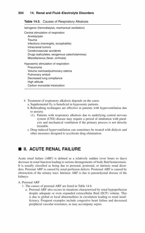
304 14. Renal and Fluid–Electrolyte Disorders
Table 14.5. Causes of Respiratory Alkalosis
Iatrogenic (hemodialysis, mechanical ventilation)
Central stimulation of respirationAnxiety/painTraumaInfections (meningitis, encephalitis)Intracranial tumorsCerebrovascular accidentsDrugs (salicylates, exogenous catecholamines)Miscellaneous (fever, cirrhosis)
Hypoxemic stimulation of respirationPneumoniaVolume overload/pulmonary edemaPulmonary emboliDecreased lung complianceHigh altitudeCarbon monoxide intoxication
4. Treatment of respiratory alkalosis depends on the cause.a. Supplemental O2 is beneficial in hypoxemic patients.b. Rebreathing techniques are effective in patients with hyperventilation due
to anxiety.(1). Patients with respiratory alkalosis due to underlying central nervous
system (CNS) disease may require a period of intubation with paral-ysis and mechanical ventilation if the primary process is not directlytreatable.
c. Drug-induced hyperventilation can sometimes be treated with dialysis andother measures designed to accelerate drug elimination.
� II. ACUTE RENAL FAILURE
Acute renal failure (ARF) is defined as a relatively sudden (over hours to days)decrease in renal function leading to serious derangements of body fluid homeostasis.It is usually classified as being due to prerenal, postrenal, or intrinsic renal disor-ders. Prerenal ARF is caused by renal perfusion defects. Postrenal ARF is caused byobstruction of the urinary tract. Intrinsic ARF is due to parenchymal disease of thekidneys.
A. Prerenal ARF1. The causes of prerenal ARF are listed in Table 14.6.
a. Prerenal ARF also occurs in situations characterized by renal hypoperfusiondespite adequate or even expanded extracellular fluid (ECF) volume. Thisis due to global or local abnormalities in circulation leading to renal insuf-ficiency. Frequent examples include congestive heart failure and decreasedperipheral vascular resistance, as may accompany sepsis.

II. Acute Renal Failure 305
Table 14.6. Causes of Prerenal ARF
Absolute ECF volume depletionExtrarenal volume losses
GI lossesThird-space lossesInadequate fluid intakeHemorrhage
Renal volume losses (diuretics)
Relative ECF volume depletionCongestive heart failureDecreased peripheral vascular resistance
Changes in renal vascular toneNonsteroidal anti-inflammatory drugsAngiotensin-converting enzyme inhibitorsHepatorenal syndrome
Abbreviation: ECF, extracellular fluid.
b. Renal blood flow may be selectively impaired by certain drugs (especiallynonsteroidal anti-inflammatory drugs [NSAIDs] and angiotensin inhibitors)or in the hepatorenal syndrome.
c. Whenever renal blood flow is severely curtailed, ischemic acute renal failureis a possibility.
2. The diagnosis of prerenal ARF can often be made on clinical grounds.a. The history and physical examination are crucial.b. Assessment of serial weights and intake/output records are valuable, if
available.c. Oliguria is a customary finding.d. The urinalysis reveals urinary concentration (specific gravity >1.020), but is
typically otherwise benign.e. A ratio of BUN/creatinine >20:1 is suggestive but not diagnostic of prerenal
ARF.f. The most helpful feature is evidence of avid sodium reabsorption in an
oliguric patient.(1). Sodium avidity can be assessed by determining the fractional excretion
of sodium (FENa), calculated as
FENa= (UNa × Pcreat)/(PNa × Ucreat) × 100
(2). Values <1% suggest prerenal azotemia in the appropriate clinicalsetting.
g. In occasional cases when doubt remains, measurement of central venouspressure or pulmonary capillary wedge pressure may be useful in guidingtherapy.
3. Treatment of prerenal ARF depends on correction of the underlying cause andreplacement of any volume deficits.a. The cause of fluid losses should be identified and treated if possible.

306 14. Renal and Fluid–Electrolyte Disorders
Table 14.7. Causes of Postrenal ARF
Urethral obstructionUrethral valvesProstatic hypertrophy
Bladder obstructionNeurogenic bladderBladder tumorsCystitis
Ureteral obstruction
IntrinsicUreteral stonesPapillary necrosis
ExtrinsicTumorsRetroperitoneal fibrosisAortic aneurysmPregnancy
b. Left ventricular function should be maximized if congestive heart failure(CHF) is playing a role.
c. Offending drugs should be discontinued.d. Hepatic transplantation may restore renal function to normal in patients with
hepatorenal syndrome.e. In addition to treatment of the underlying disease, volume expansion with
appropriate intravenous fluids (usually normal saline or blood products) isrequired in patients unable to ingest sufficient sodium and water.
B. Postrenal ARF1. Some causes of postrenal ARF are listed in Table 14.7.2. The diagnosis of postrenal ARF can be suspected on history and physical
examination.a. Alternating polyuria and oligoanuria is suggestive of subtotal urinary
obstruction, as is a very large postvoiding residual urine volume.3. Laboratory tests are usually nonspecific.
a. The urinalysis may be normal or disclose evidence of hematuria, pyuria, orcrystalluria.
b. The best screening test for obstruction is renal ultrasound, which has aspecificity of >90%.
c. In cases in which obstruction is still suspected despite a negative ultrasound,retrograde pyelography can be performed.
4. Therapy of postrenal ARF largely depends on the site of obstruction.a. The coexistence of obstruction and urinary tract infection is a urologic
emergency mandating broad-spectrum antibiotic coverage and immediatedecompression of the urinary tract.(1). Upper tract disease can be effectively approached with indwelling stents
or percutaneous nephrostomy.(2). Lower urinary obstruction can be relieved with urethral or suprapubic
catheterization.

II. Acute Renal Failure 307
Table 14.8. Causes of Intrinsic ARF
Glomerular diseasesAcute glomerulonephritisRapidly progressive glomerulonephritis
Tubulointerstitial diseasesAcute tubular necrosis
Drug-inducedIschemic
Acute interstitial nephritisAllergic/drug-inducedIdiopathic
Vascular diseasesRenal artery
Thrombosis/embolusDissectionTrauma
Renal microcirculationVasculitisMalignant hypertensionDisseminated intravascular coagulation (DIC)Thrombotic thrombocytopenic purpura (UP)Cholesterol atheroemboli
Renal vein thrombosis
b. After the patient’s medical condition has stabilized, it is often possible toundertake definitive repair of the obstructing lesion. Relief of even long-standing obstruction is generally indicated, since some functional recoverymay occur, even weeks or months later.
C. Intrinsic ARF1. Some causes of intrinsic ARF are listed in Table 14.8.2. Acute tubular necrosis (ATN) is by far the most common cause of intrinsic ARF
among hospitalized patients.a. Multiple drugs and toxins can cause ATN, including aminoglycoside antibi-
otics, certain chemotherapeutic agents (i.e., cisplatinum, mithramycin), andradiographic contrast materials.
b. Ischemia is another major factor causing ATN.c. ATN is especially common in patients with major trauma, recent major
surgery (particularly vascular operations), sepsis, or crush injury.3. Acute interstitial nephritis (AIN) is usually caused by exposure to a drug or
allergen. Rare idiopathic cases are encountered.a. Probably the most common drugs causing AIN are NSAIDs.b. Other frequent agents include antibiotics (particularly penicillins,
cephalosporins, and sulfa derivatives), loop and thiazide diuretics, andcimetidine.
c. Recognition of this entity is especially important, since effective therapydepends on withdrawal of the offending drug.

308 14. Renal and Fluid–Electrolyte Disorders
4. The diagnosis of intrinsic ARF depends heavily on the history and physicalexamination with subsequent directed laboratory and radiographic evaluation.a. Medications in particular should be carefully reviewed.b. The urine sediment should be examined; it is valuable to note that intrinsic
ARF is almost invariably accompanied by abnormalities on urinalysis andtheir absence should raise the suspicion of pre- or postrenal causes.
c. The presence of urinary eosinophils can be used to support a diagnosis ofAIN or cholesterol microemboli.
d. The FENa is typically >3% in intrinsic ARF.e. Serologic evaluation of patients with suspected vasculitis should include
antinuclear antibodies (ANA), complements, hepatitis B surface antigen,cryoglobulins, and rheumatoid factor. If there is a suspicion of a pulmonary–renal syndrome (i.e., Goodpasture), anti-basal membrane antibodies shouldbe obtained.
f. More invasive studies (e.g., angiography, renal biopsy) may be appropriatedepending on the clinical circumstances.
5. Therapy of intrinsic ARF is largely supportive.a. Offending drugs should be identified and withdrawn or substituted if
possible.b. Control of fluid and electrolyte balance, modification of drug dosages, and
dialysis should be entertained.c. Specific therapy directed at a particular disease process is possible in
occasional circumstances. A major aspect of the management of ARF isrecognition of the situations in which it is likely to occur, with appropriatemeasures to reduce its probability.(1). Medications with the least risk of nephrotoxicity should be chosen in
patients liable to ARF.(2). Volume depletion should be avoided or corrected.(3). Calcium-channel blockers may be helpful in ameliorating ARF if given
prophylactically (i.e., before angiography or renovascular surgery).(4). The use of hemodialysis acutely is believed by some to accelerate
the rate of recovery in patients with ARF. However, hemodialysis hasseveral potential side effects.
6. Despite many advances in the diagnosis and treatment of ARF, mortalityremains at least 50% in critically ill patients.
� III. ELECTROLYTE ABNORMALITIES
Certain electrolyte abnormalities that occur frequently pose a serious risk to ICUpatients, even if the disorder has been present for a long time.
A. Calcium. Calcium exists in three forms in the circulation: free ionized calcium(iCa++); in soluble complexes with phosphate, citrate, and bicarbonate; and boundto plasma proteins (principally albumin). Of these, the iCa++ is physiologicallymost important. The normal value for iCa++ is 4.0–4.9 mg/dL, 2.4–2.6 mEq/L, or1.2–1.3 mmol/L (note difference in units).Maintenance of normal iCa++ levels depends on the interaction between GIabsorption, bone fluxes, and renal excretion, governed by parathyroid hormone(PTH), 1,25-dihydroxyvitamin D (1,25-D3), and calcitonin.

III. Electrolyte Abnormalities 309
Table 14.9. Disorders of Calcium Homeostasis
Hypercalcemia Hypocalcemia
Hyperparathyroidism HypoparathyroidismHyperthyroidism PseudohypoparathyroidismAcute renal failure Vitamin D deficiencyMalignancy MalignancyExcessive vitamin A or D HyperphosphatemiaGranulomatous diseases PancreatitisThiazide diuretics Neonatal tetanyImmobilization Calcium complex formation
1. Hypercalcemiaa. Most patients with hypercalcemia are asymptomatic at the time of diagnosis.
However, numerous signs and symptoms may occur depending on the severityand rate of development of hypercalcemia. These include
(1). Nausea(2). Constipation(3). Anorexia(4). Pancreatitis(5). Peptic ulcers(6). Renal insufficiency(7). Polyuria(8). Urolithiasis(9). Bone pain
(10). Weakness(11). Confusion and coma
b. Symptomatic patients require urgent treatment.c. Some causes of hypercalcemia are listed in Table 14.9.d. Hypercalcemia is a medical emergency whose acute treatment is largely
independent of the cause.e. Therapy is outlined in Table 14.10.
(1). Intravenous (IV) normal saline should be administered at a rate of 150–250 mL/h.
(2). Furosemide is indicated to prevent volume overload and to decreasecalcium reabsorption (a minor effect).
Table 14.10. Treatment of Hypercalcemia
IV Fluids: Normal saline 150–250 mL/h(± Furosemide 40–80 mg IV q4–6 h)
Corticosteroids: Prednisone (or equivalent) 1 mg/kg/dCalcitonin: 4 U/kg SC q12 hEtidronate: 7.5 mg/kg/d IV q day × 1–4 daysMithramycin: 25 μg/kg IV q day × 3–4 daysIndomethacin: 25–50 mg PO q8 hDialysis
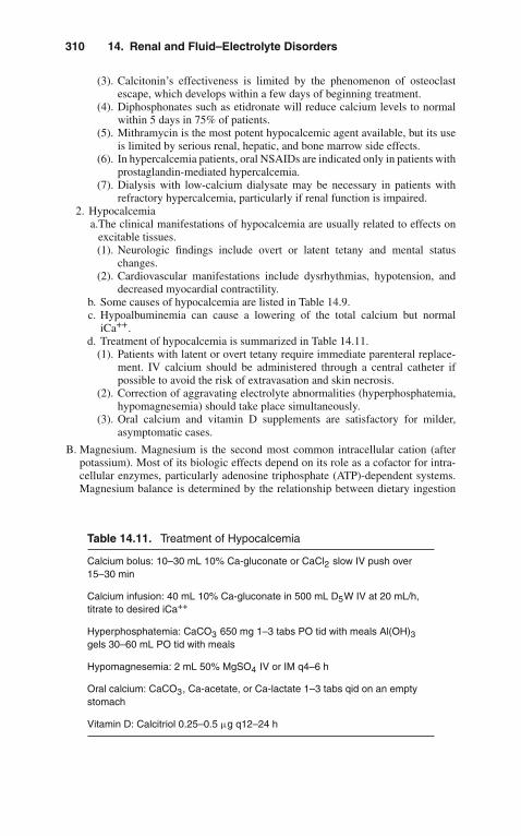
310 14. Renal and Fluid–Electrolyte Disorders
(3). Calcitonin’s effectiveness is limited by the phenomenon of osteoclastescape, which develops within a few days of beginning treatment.
(4). Diphosphonates such as etidronate will reduce calcium levels to normalwithin 5 days in 75% of patients.
(5). Mithramycin is the most potent hypocalcemic agent available, but its useis limited by serious renal, hepatic, and bone marrow side effects.
(6). In hypercalcemia patients, oral NSAIDs are indicated only in patients withprostaglandin-mediated hypercalcemia.
(7). Dialysis with low-calcium dialysate may be necessary in patients withrefractory hypercalcemia, particularly if renal function is impaired.
2. Hypocalcemiaa.The clinical manifestations of hypocalcemia are usually related to effects on
excitable tissues.(1). Neurologic findings include overt or latent tetany and mental status
changes.(2). Cardiovascular manifestations include dysrhythmias, hypotension, and
decreased myocardial contractility.b. Some causes of hypocalcemia are listed in Table 14.9.c. Hypoalbuminemia can cause a lowering of the total calcium but normal
iCa++.d. Treatment of hypocalcemia is summarized in Table 14.11.
(1). Patients with latent or overt tetany require immediate parenteral replace-ment. IV calcium should be administered through a central catheter ifpossible to avoid the risk of extravasation and skin necrosis.
(2). Correction of aggravating electrolyte abnormalities (hyperphosphatemia,hypomagnesemia) should take place simultaneously.
(3). Oral calcium and vitamin D supplements are satisfactory for milder,asymptomatic cases.
B. Magnesium. Magnesium is the second most common intracellular cation (afterpotassium). Most of its biologic effects depend on its role as a cofactor for intra-cellular enzymes, particularly adenosine triphosphate (ATP)-dependent systems.Magnesium balance is determined by the relationship between dietary ingestion
Table 14.11. Treatment of Hypocalcemia
Calcium bolus: 10–30 mL 10% Ca-gluconate or CaCl2 slow IV push over15–30 min
Calcium infusion: 40 mL 10% Ca-gluconate in 500 mL D5W IV at 20 mL/h,titrate to desired iCa++
Hyperphosphatemia: CaCO3 650 mg 1–3 tabs PO tid with meals Al(OH)3gels 30–60 mL PO tid with meals
Hypomagnesemia: 2 mL 50% MgSO4 IV or IM q4–6 h
Oral calcium: CaCO3, Ca-acetate, or Ca-lactate 1–3 tabs qid on an emptystomach
Vitamin D: Calcitriol 0.25–0.5 μg q12–24 h
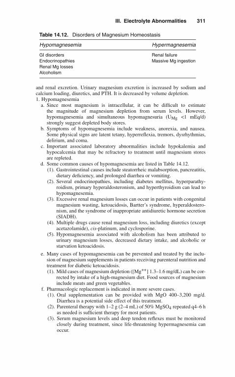
III. Electrolyte Abnormalities 311
Table 14.12. Disorders of Magnesium Homeostasis
Hypomagnesemia Hypermagnesemia
GI disorders Renal failureEndocrinopathies Massive Mg ingestionRenal Mg lossesAlcoholism
and renal excretion. Urinary magnesium excretion is increased by sodium andcalcium loading, diuretics, and PTH. It is decreased by volume depletion.1. Hypomagnesemia
a. Since most magnesium is intracellular, it can be difficult to estimatethe magnitude of magnesium depletion from serum levels. However,hypomagnesemia and simultaneous hypomagnesuria (UMg <1 mEq/d)strongly suggest depleted body stores.
b. Symptoms of hypomagnesemia include weakness, anorexia, and nausea.Some physical signs are latent tetany, hyperreflexia, tremors, dysrhythmias,delirium, and coma.
c. Important associated laboratory abnormalities include hypokalemia andhypocalcemia that may be refractory to treatment until magnesium storesare repleted.
d. Some common causes of hypomagnesemia are listed in Table 14.12.(1). Gastrointestinal causes include steatorrheic malabsorption, pancreatitis,
dietary deficiency, and prolonged diarrhea or vomiting.(2). Several endocrinopathies, including diabetes mellitus, hyperparathy-
roidism, primary hyperaldosteronism, and hyperthyroidism can lead tohypomagnesemia.
(3). Excessive renal magnesium losses can occur in patients with congenitalmagnesium wasting, ketoacidosis, Bartter’s syndrome, hyperaldostero-nism, and the syndrome of inappropriate antidiuretic hormone secretion(SIADH).
(4). Multiple drugs cause renal magnesium loss, including diuretics (exceptacetazolamide), cis-platinum, and cyclosporine.
(5). Hypomagnesemia associated with alcoholism has been attributed tourinary magnesium losses, decreased dietary intake, and alcoholic orstarvation ketoacidosis.
e. Many cases of hypomagnesemia can be prevented and treated by the inclu-sion of magnesium supplements in patients receiving parenteral nutrition andtreatment for diabetic ketoacidosis.(1). Mild cases of magnesium depletion ([Mg++] 1.3–1.6 mg/dL) can be cor-
rected by intake of a high-magnesium diet. Food sources of magnesiuminclude meats and green vegetables.
f. Pharmacologic replacement is indicated in more severe cases.(1). Oral supplementation can be provided with MgO 400–3,200 mg/d.
Diarrhea is a potential side effect of this treatment.(2). Parenteral therapy with 1–2 g (2–4 mL) of 50% MgSO4 repeated q4–6 h
as needed is sufficient therapy for most patients.(3). Serum magnesium levels and deep tendon reflexes must be monitored
closely during treatment, since life-threatening hypermagnesemia canoccur.

312 14. Renal and Fluid–Electrolyte Disorders
2. Hypermagnesemiaa. The clinical features of magnesium excess are frequently confused with
those of hypercalcemia. They include(1). Nausea(2). Altered mental status(3). Weakness and diminished tendon reflexes(4). Hypotension(5). Dysrhythmias(6). Respiratory paralysis
b. The most frequent condition predisposing patients to hypermagnesemia ischronic renal failure. Sustained hypermagnesemia only occurs in the settingof renal insufficiency, although acute magnesium excess can occur in thesetting of overzealous replacement of magnesium deficiency.
c. Therapy of hypermagnesemia involves discontinuing magnesium intake,infusion of Ca-gluconate 15 mg/kg over 4 h (which acts as a direct mag-nesium antagonist), and dialysis with a magnesium-free bath in severecases.
C. Phosphate. Phosphate is an abundant intracellular anion, critically important inenergy metabolism and structural integrity of literally all cells. The overwhelm-ing majority of dietary phosphate is normally absorbed, but a significant amount(approximately 200 mg/d) is secreted in the stool. Renal excretion eliminates theremainder, about 400–1,500 mg/d depending on intake. Urinary phosphorus excre-tion is increased by PTH, volume expansion, corticosteroids, and calcitonin, andis decreased by insulin, thyroid hormone, and vitamin D. Movement of phosphateinto cells is enhanced during alkalemia, by glucose and some other carbohydrates,and by hormones including insulin, epinephrine, and corticosteroids.1. Hypophosphatemia
a. Hypophosphatemia is characterized by altered mental status, weakness ormyopathy progressing to rhabdomyolysis, osteomalacia, dysfunction of allblood cell types, anorexia, respiratory failure, and decreased cardiac contrac-tility. However, most cases of phosphate depletion are asymptomatic unlesssevere.
b. Some causes of hypophosphatemia are listed in Table 14.13.(1). Respiratory and metabolic alkalosis are associated with intracellular
uptake of phosphate.(2). Refeeding after prolonged starvation precipitates hypophosphatemia,
largely due to intracellular shifts mediated by glucose and insulin.(3). Administration or excessive activity of other hormones (epinephrine,
growth hormone, steroids, gastrin, and glucagon) can cause hypophos-phatemia by similar mechanisms.
Table 14.13. Disorders of Phosphate Homeostasis
Hypophosphatemia Hyperphosphatemia
Intracellular shifts Massive cell lysisInadequate ingestion Increased ingestionGI losses Renal failureRenal losses

III. Electrolyte Abnormalities 313
(4). Malnutrition alone does not typically cause severe hypophosphatemia.However, GI losses due to malabsorption of phosphate or use ofphosphorus-binding antacids can lead to marked phosphate depletion.
(5). Excessive renal losses of phosphate are present in Fanconi’s syndrome,after renal transplantation in some patients, following recovery fromurinary obstruction or ATN, and with vitamin D-resistant rickets.
c. The best treatment of hypophosphatemia is often prevention.(1). Phosphate deficiency can be avoided by preventive supplementation
in patients with malnutrition undergoing hyperalimentation or refeed-ing, or who are chronic alcoholics, on long-term antacids, or who haveuncontrolled diabetes mellitus or ketoacidosis.
(2). Treatment of established hypophosphatemia depends on its severity andcause.(a). Patients with mild hypophosphatemia due to intracellular shifts can
be followed closely without active intervention unless the serumphosphorus drops <1.5 mg/dL.
(b). Dietary supplementation in the form of increased intake of dairyproducts is adequate in most asymptomatic patients.
(c). Oral supplementation can be provided with sodium and/or potas-sium phosphate, although diarrhea is often a dose-limiting sideeffect.
(d). Severe symptomatic hypophosphatemia is treated with elementalphosphorus (as sodium or potassium phosphate) 2 mg/kg IV q6h until oral repletion can begin, usually at a phosphorus level of2.0 mg/dL.
(e). Use of parenteral phosphate is relatively contraindicated in patientswith oliguric renal failure.
(f). Possible complications of IV phosphorus include dysrhythmias,hyperphosphatemia, hypocalcemia, hyperkalemia, and volumeoverload.
2. Hyperphosphatemiaa. Many of the symptoms of hyperphosphatemia can be attributed to the
reciprocal fall in iCa++ that generally occurs (see above).b. Severe hyperphosphatemia can also cause ARF, particularly in the setting
of massive cell lysis. Metastatic calcifications caused by precipitation ofCa-PO4 crystals in essentially any tissue or organ can lead to widespreadsymptoms and signs.
c. The causes of hyperphosphatemia are summarized in Table 14.13.d. Treatment of hyperphosphatemia involves decreasing ingestion with
phosphorus-restricted diets as well as increasing elimination via the GI tractand kidneys.(1). Phosphate binders containing magnesium, calcium, or aluminum will
accelerate stool phosphorus losses even in patients who are NPO.(2). Magnesium-containing agents should be avoided in patients with renal
failure.(3). Renal excretion of phosphate is enhanced by acetazolamide, volume
expansion, and alkaline diuresis. Unfortunately, the frequent coexis-tence of renal failure with hyperphosphatemia often makes this routeunreliable.
(4). Hemodialysis or peritoneal dialysis is effective for acute or chronichyperphosphatemia.
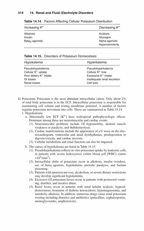
314 14. Renal and Fluid–Electrolyte Disorders
Table 14.14. Factors Affecting Cellular Potassium Distribution
Increasing K+ Decreasing K+
Alkalosis AcidosisInsulin GlucagonBeta2-agonists Alpha-agonists
Hyperosmolarity
Table 14.15. Disorders of Potassium Homeostasis
Hypokalemia Hyperkalemia
Pseudohypokalemia PseudohyperkalemiaCellular K+ uptake Cellular K+ lossPoor dietary K+ intake Excessive K+ intakeGI losses Inadequate renal excretionRenal losses Cell lysis
D. Potassium. Potassium is the most abundant intracellular cation. Only about 2%of total body potassium is in the ECF. Intracellular potassium is responsible formaintaining cell volume and resting membrane potential. A number of factorsregulate potassium movement into cells. These are summarized in Table 14.14.1. Hypokalemia
a. Abnormally low ECF [K+] have widespread pathophysiologic effects.Dominant among these are neuromuscular and cardiac events.(1). Neuromuscular problems include GI hypomotility, skeletal muscle
weakness or paralysis, and rhabdomyolysis.(2). Cardiac manifestations include the appearance of a U wave on the elec-
trocardiogram, ventricular and atrial dysrhythmias, predisposition todigoxin toxicity, and cardiac necrosis.
(3). Cellular metabolism and renal function can also be impaired.
b. The causes of hypokalemia are listed in Table 14.15.(1). Pseudohypokalemia reflects in vitro potassium uptake by leukemic cells
in patients with severe leukocytosis (white blood cell [WBC] counts>105/mm3).
(2). Intracellular shifts of potassium occur in alkalosis, insulin overdose,use of beta2-agonists, hypokalemic periodic paralysis, and bariumpoisoning.
(3). Patients with anorexia nervosa, alcoholism, or severe dietary restrictionsmay develop significant hypokalemia.
(4). Excessive GI potassium losses occur in patients with protracted vomit-ing, diarrhea, and laxative abuse.
(5). Renal losses occur in patients with renal tubular acidosis, hyperal-dosteronism, treatment of diabetic ketoacidosis, hypomagnesemia, andmetabolic alkalosis. In addition, numerous drugs cause renal potassiumwasting including diuretics and antibiotics (penicillins, cephalosporins,aminoglycosides, amphotericin).

III. Electrolyte Abnormalities 315
c. Treatment of hypokalemia is imprecise, since the degree of total body K+
depletion is usually impossible to calculate.(1). Ongoing potassium losses should be halted if possible, and aggravating
abnormalities (alkalosis, hypomagnesemia) corrected.(2). The particular potassium salt employed depends on the specific clini-
cal problem. KCl is preferred in patients with concomitant metabolicalkalosis and is effective in all forms of hypokalemia. Bicarbonate orphosphate salts of potassium may be preferred if acidosis or hypophos-phatemia is present.
(3). Oral potassium preparations are effective but can have an unpalatabletaste and cause GI irritation.
(4). Potassium can be given by slow IV infusion (10–20 mEq/h) througha central or peripheral line; concentrations above 40 mEq/L should beavoided in peripheral IVs to decrease the risk of phlebitis.
(5). Frequent monitoring of serum [K+] is essential during parenteral potas-sium repletion.
(6). Potassium-sparing diuretics are occasionally useful, but should be usedwith caution in patients receiving potassium supplements.
2. Hyperkalemiaa. The principal clinical abnormalities of hyperkalemia are neuromuscular and
cardiac. Weakness, paresthesias, and paralysis can occur but are usuallyovershadowed by cardiac disturbances. These include(1). Progressive electrocardiogram (ECG) appearance of peaked T waves.(2). Flattened P waves.(3). Prolonged PR interval.(4). Widening of the QRS complex.(5). The development of a sine-wave pattern presages the onset of ventricu-
lar fibrillation or asystole.b. Some causes of hyperkalemia are presented in Table 14.15.
(1). Pseudohyperkalemia is due to in vitro release of potassium from redblood cells, leukocytes, or platelets.
(2). Release of potassium from cells contributes to hyperkalemia in acido-sis, poorly controlled diabetes mellitus, beta-blockade, hyperkalemicperiodic paralysis, hyperosmolar states, and digitalis toxicity.
(3). Potassium ingestion seldom results in hyperkalemia if renal function isnormal except when excessive parenteral potassium supplements havebeen administered.
(4). Inadequate renal excretion occurs in patients with advanced renal fail-ure, deficiencies of adrenal hormones, and numerous drugs. Theseinclude potassium-sparing diuretics, NSAIDs, angiotensin-convertingenzyme inhibitors, and cyclosporine.
(5). Severe hyperkalemia is a frequent finding in patients with massive invivo hemolysis or tumor lysis syndrome.
c. The treatment of hyperkalemia is outlined in Table 14.16.(1). Calcium does not affect the serum potassium but rather antagonizes the
cardiac toxicity of hyperkalemia.(2). Glucose/insulin and bicarbonate infusions lower serum [K+] by stimu-
lating cellular potassium entry.(3). Kayexalate is used to augment fecal potassium excretion; it is relatively
ineffective unless the patient develops diarrhea or loose stools.(4). Dialysis is extremely effective for life-threatening hyperkalemia.

316 14. Renal and Fluid–Electrolyte Disorders
Table 14.16. Therapy of Hyperkalemia
10% Calcium gluconate: 10–30 mL IV
Onset <5 min
50% dextrose 50 mL + regular insulin 5 U IV q30 min
Onset 15–30 min
NaHCO3 50 mL (50 mEq) IV q30 min x 4 doses
Onset 15–30 min
Kayexalate in sorbitol 30–60 g PO/enema 4–6 h
Onset 1–2 h
Dialysis
(5). Diuretics and aldosterone analogues are occasionally useful adjunctivemeasures.
(6). The use of beta-adrenergic agents (i.e., albuterol) via nebulizations hasreceived considerable attention in recent years for the acute manage-ment of hyperkalemia in endstage renal disease patients.
E. Sodium. Hyponatremia and hypernatremia are disorders of water balance. Theosmolarity of a solution depends on the number of dissolved particles per liter. Inclinical practice, this can be measured, or calculated, as follows:
Posm= 2 × [Na+] + [glucose]/18 + [BUN]/2.8
Solutes restricted to one side of the plasma membrane are termed effectiveosmoles; changes in the quantity of effective osmoles in a body fluid obli-gate transmembrane water movement to maintain balance. Freely membrane-permeable substances such as urea do not cause water movement and are hencetermed ineffective osmoles. The tonicity, or effective osmolarity, of a solution canbe calculated by the following:
Eosm= 2 × [Na+] + [glucose]/18
Regulation of plasma osmolarity depends on the interplay between water inges-tion and renal water excretion. Renal mechanisms of water balance require theadequate delivery of salt and water to distal nephron sites as well as manipulationof tubular water permeability under the influence of antidiuretic hormone (ADH).ADH is secreted by the posterior pituitary in response to hypertonicity as wellas other nonosmotic stimuli, including hypovolemia, nausea, pain, and severaldrugs (i.e., nicotine, narcotics, vincristine, cyclophosphamide, chlorpropamide,and clofibrate). ADH increases renal water permeability, leading to increasedwater reabsorption and hypertonic urine. Normal thirst is even more responsiveto changes in osmolarity than ADH is; consequently, thirst should be consideredthe primary guardian of plasma tonicity.
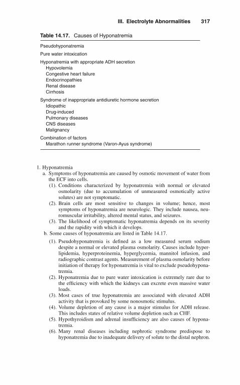
III. Electrolyte Abnormalities 317
Table 14.17. Causes of Hyponatremia
Pseudohyponatremia
Pure water intoxication
Hyponatremia with appropriate ADH secretionHypovolemiaCongestive heart failureEndocrinopathiesRenal diseaseCirrhosis
Syndrome of inappropriate antidiuretic hormone secretionIdiopathicDrug-inducedPulmonary diseasesCNS diseasesMalignancy
Combination of factorsMarathon runner syndrome (Varon-Ayus syndrome)
1. Hyponatremiaa. Symptoms of hyponatremia are caused by osmotic movement of water from
the ECF into cells.(1). Conditions characterized by hyponatremia with normal or elevated
osmolarity (due to accumulation of unmeasured osmotically activesolutes) are not symptomatic.
(2). Brain cells are most sensitive to changes in volume; hence, mostsymptoms of hyponatremia are neurologic. They include nausea, neu-romuscular irritability, altered mental status, and seizures.
(3). The likelihood of symptomatic hyponatremia depends on its severityand the rapidity with which it develops.
b. Some causes of hyponatremia are listed in Table 14.17.
(1). Pseudohyponatremia is defined as a low measured serum sodiumdespite a normal or elevated plasma osmolarity. Causes include hyper-lipidemia, hyperproteinemia, hyperglycemia, mannitol infusion, andradiographic contrast agents. Measurement of plasma osmolarity beforeinitiation of therapy for hyponatremia is vital to exclude pseudohypona-tremia.
(2). Hyponatremia due to pure water intoxication is extremely rare due tothe efficiency with which the kidneys can excrete even massive waterloads.
(3). Most cases of true hyponatremia are associated with elevated ADHactivity that is provoked by some nonosmotic stimulus.
(4). Volume depletion of any cause is a major stimulus for ADH release.This includes states of relative volume depletion such as CHF.
(5). Hypothyroidism and adrenal insufficiency are also causes of hypona-tremia.
(6). Many renal diseases including nephrotic syndrome predispose tohyponatremia due to inadequate delivery of solute to the distal nephron.

318 14. Renal and Fluid–Electrolyte Disorders
(7). Hyponatremia in untreated cirrhosis is caused by excessive ADHsecretion and impaired distal sodium delivery.
(8). SIADH has multiple causes including drugs, pulmonary diseases, CNSdiseases, and cancer. See also Chapter 4, "Endocrinologic Disorders."
(9). Hyponatremia in marathon runners (Varon-Ayus Syndrome). This well-described syndrome occurs not only in marathon runners but also in avariety of extreme sports and military exercises.
c. Symptomatic hyponatremia is a medical emergency regardless of duration.d. After the plasma osmolarity has been determined, therapy with 3% NaCl
should be instituted. The amount of NaCl required to correct hyponatremiato a specified target level is
mEq NaCl= 0.6 × (weight [kg]) × (target Na+ − plasma Na+)
The amount of 3% saline required to achieve this goal is
3%NaCl (mL) = (1,000) × (mEq NaCl)/513
The target [Na+] should be an increase of 20 mEq/L above the actual [Na+]or 130 mEq/L, whichever is lower. The rate of correction is 1.0–1.5 mEq/L/h.These calculations often underestimate the actual rate of correction due toongoing urinary sodium losses.
e. In addition to hypertonic saline infusion, all IV fluids including medicationsshould be given in 0.9% NaCl, and a fluid restriction of 1,000–1,500 mL/dinstituted if possible.
f. Demeclocycline 150–300 mg PO q12 h has been used successfully in thetreatment of SIADH but is contraindicated in cirrhosis.
g. Central pontine myelinolysis is a rare complication occurring after correc-tion of chronic (but not acute) hyponatremia if the serum [Na+] is raisedby >25 mEq/L during the first 48 h of therapy.
h. A new agent, conivaptan (Vaprisol) is indicated in some patients for the treat-ment of euvolemic and hypervolemic hyponatremia. It is an antagonist of thevasopressin V1A and V2 receptors.
2. Hypernatremiaa. Symptoms of hypernatremia are caused by cellular dehydration, particu-
larly of neurons. They include altered mental status, nausea, seizures, andintracranial hemorrhage. Myoclonus, metabolic acidosis, and hyperglycemiadue to peripheral insulin resistance are also common.
b. Most hypernatremic patients are volume-depleted; the finding of volumeoverload should suggest the possibility of acute salt poisoning.
c. Causes of hypernatremia are listed in Table 14.18.(1). Diabetes insipidus (DI) can be of central or nephrogenic origin (see
Chapter 4, “Endocrinologic Disorders”).(2). The most common cause of hypernatremia is excessive water loss.
(a). Excessive renal water loss with hypernatremia has been reported inpatients with renal failure, hypercalcemia, hypokalemia, sickle cell

III. Electrolyte Abnormalities 319
Table 14.18. Causes of Hypernatremia
Diabetes insipidus
H2O lossesRenalGIInsensible
Salt poisoning
H2O deprivation
Primary hypodipsia
Mineralocorticoid excess
disease, osmotic diuresis, postobstructive diuresis, sickle cell dis-ease, and drugs (including alcohol, lithium, demeclocycline, oralhypoglycemics, and others).
(b). GI water losses due to gastroenteritis are an especially commoncause of hypernatremia in children.
(c). Insensible water losses via the skin or respiratory tract occur withprolonged exposure to hot climates, thermal burns, and fever.
(3). Salt poisoning is a rare cause of outpatient hypernatremia but ismore common in hospitalized patients as a complication of hypertonicNaHCO3 therapy for severe acidosis.
(4). Water deprivation is a fundamental feature of practically all cases ofhypernatremia but is rarely the sole cause of the problem.
(5). Entities characterized by increased mineralocorticoid activity (Conn’ssyndrome and Cushing’s syndrome) are sometimes accompanied bymild-to-moderate hypernatremia.
d. The treatment of hypernatremia is replacement of free water deficits andcorrection of contributing electrolyte problems and hypovolemia, if present.(1). The water deficit is calculated as
H2O deficit (L)= 0.6 × weight (kg) × [(target Na+)/(actual Na+)) − 1]
The target sodium is either 148 mEq/L or a decrease of 20–25 mEq/Lin the plasma [Na+] from its initial value whichever is higher. The goalof therapy is to reduce the plasma [Na+] by 1.0–1.5 mEq/L/h.
(2). Faster rates of correction of chronic hypernatremia may precipitaterehydration seizures.
(3). The choice of fluid and route of administration depend on the clinicalcircumstances. Distilled or tap water given PO or via nasogastric (NG)tube is preferred when feasible. If the enteral route is unavailable, 0.45%NaCl can be administered by peripheral vein without significant risk ofhemolysis.
(4). Patients with clinical evidence of volume overload who cannot toler-ate the sodium load of 0.45% NaCl should be given distilled water IVthrough a central catheter.

320 14. Renal and Fluid–Electrolyte Disorders
(5). D5W is at least relatively contraindicated in many patients becauseof the coexistence of insulin resistance and the consequent risk ofworsening hyperosmolarity due to non-metabolized dextrose.
(6). Diuretics or dialysis can be employed for patients with salt poisoning.(7). Vasopressin analogues are useful in the long-term management of
central DI.
� IV. FLUID AND ELECTROLYTE THERAPY
In addition to the more specific treatments outlined above, some general guidelinesregarding fluid and electrolyte therapy are useful. Administration of fluids and nutri-tion are essential but frequently overlooked considerations in the care of the ICUpatient. Special care must be exercised in the selection and administration of IV flu-ids to the critically ill with ongoing renal or extrarenal fluid and electrolyte losses. IVfluids are potentially the most dangerous drugs used in the hospital; constant vigilanceis required.
A. For essentially all ICU patients, some IV access is necessary.
B. Whenever possible, the GI tract should be employed for maintenance fluids,nutrition, and medications.
C. The type of fluid used depends on the clinical situation. Some general commentscan be made:1. For routine maintenance, crystalloid solutions are employed.
a. In an otherwise well NPO patient, obligatory water losses amount to about1,000 mL/d.
b. Sodium losses are minimized by virtually complete renal sodium reclama-tion, but some urinary potassium excretion continues (30–60 mEq/d) andmust be replaced.
c. Excessive protein catabolism and starvation ketosis can be prevented byinclusion of glucose 150–200 g daily in maintenance fluids.
d. Supplements of other vitamins and minerals may be necessary if parenteraltherapy lasts a week or more.
2. Colloid solutions such as blood products, albumin, and plasma are indicatedfor the rapid expansion of intravascular volume with minimal effects on othercomponents of the ECF.
� V. DIALYSIS
ICU patients frequently develop homeostatic abnormalities that cannot be managedconservatively. In such a case, dialysis or a related modality becomes necessary.
A. Dialysis is indicated in many different situations.1. Volume overload manifested by pulmonary edema or severe hypertension unre-
sponsive to diuretics can be effectively treated with dialysis or ultrafiltration.2. Dialysis is useful for the treatment of several electrolyte abnormalities,
including severe acidosis or alkalosis, hyperkalemia, hypo- or hypernatremia,hypercalcemia, hyperphosphatemia, and hypermagnesemia.

V. Dialysis 321
3. Symptoms caused by accumulation of uremic toxins are best treated withdialysis.
4. Poisonings with ethylene glycol, methanol, salicylates, and others can beeffectively treated with dialysis.
B. The most frequently used dialysis modality in the ICU is hemodialysis (HD) orone of its variants. The choice between HD and peritoneal dialysis (PD) is usuallya matter of the physician’s preference, but in some cases a clear preference isevident.1. HD is the therapy of choice in severely catabolic patients due to more efficient
removal of urea and other low molecular weight nitrogenous wastes.a. A large-bore dual-lumen central venous catheter is required for vascular
access.b. For standard hemodialysis orders, the physician specifies the type of mem-
brane to be used (more biocompatible membranes such as cellulose acetateare preferred in the ICU), the duration of therapy, blood flow speed, typeof anticoagulation, composition of the dialysate with respect to Na+, K+,Ca++, and HCO3, the desired amount of fluid removal, and any additionalmedications or treatments required (e.g., blood products, antibiotics, orerythropoietin).
c. The most common complication of HD is hypotension, generally treatedwith fluid boluses of normal saline or albumin. In some cases, alter-nate treatments must be considered if hypotension is severe. Other seriouspotential complications of dialysis include dysrhythmias (presumably dueto acute electrolyte fluxes) and hypoxemia (caused by membrane-inducedcomplement activation and leukocyte sequestration in pulmonary capillar-ies as well as a decrease in minute ventilation due to removal of CO2 bydialysis).
2. To accomplish fluid and solute removal in hypotensive patients, several variantsof hemodialysis have been developed.a. Continuous arteriovenous hemofiltration (CAVH) with or without dialysis
is used in many ICUs. It has the specific advantage of allowing removalof large amounts of fluid even in patients with serious hemodynamic com-promise. The patient’s own arterial pressure is used to drive ultrafiltrationacross a highly permeable membrane. Fluid removal may exceed 500 mL/hwith CAVH, so provision must be made for a pump to limit ultrafiltrationor replacement with an adequate amount of a balanced salt solution. Soluteremoval can be accelerated by the passing peritoneal dialysate across theultrafiltrate side of the membrane. CAVH generally requires a femoral arte-rial access and thus may not be possible in patients with severe vasculardisease.
b. A modification of this technique, known as continuous venovenous hemofil-tration (CVVHD), has recently been introduced and is gaining favor.Vascular access is simplified, since an arterial access is not required, butCVVHD does require additional equipment in the form of pumps andalarms.
3. PD has less efficient clearance of low molecular weight solutes than HD; so,PD is generally not first-line therapy in hypercatabolic ICU patients.a. PD is much better tolerated from a hemodynamic standpoint and may be
preferred in unstable patients.

322 14. Renal and Fluid–Electrolyte Disorders
b. It is technically easier than HD in small children. Access to the peritonealcavity is obtained with a flexible catheter that can be placed at the bedside.
c. PD can be done continuously or intermittently with a minimum of equipmentand staff. Ultrafiltration is controlled by changing the glucose concentration(and hence osmolarity) of the dialysate.
d. PD orders should include the number and duration of exchanges, the com-position of the dialysate with respect to glucose, sodium, and calcium, andwhether any additives such as antibiotics, heparin, insulin, or potassium areto be included.
e. The most common complication of PD is peritonitis. Parenteral or intraperi-toneal antibiotics should result in clinical improvement within 24–48 h; ifnot, consideration should be given to catheter removal.
f. Hyperglycemia resulting from absorption of dialysate glucose can be man-aged by intraperitoneal or subcutaneous insulin administration.
� VI. RHABDOMYOLYSIS
Rhabdomyolysis is a condition characterized by release of muscle cell contents intothe circulation caused by skeletal muscle necrosis.
A. Signs and symptoms occur due to toxicity of pigment globin proteins andaccompanying fluid and electrolyte shifts.
B. Some causes of rhabdomyolysis are listed in Table 14.19.1. Trauma causes muscle injury both by direct pressure and muscle hypoperfusion
due to shock and vasospasm.2. Ischemic events such as arterial thrombosis and compartment syndromes can
lead to muscle necrosis.3. Numerous drugs and toxins including heroin, phencyclidine, cocaine, succinyl-
choline, and lipid-lowering agents have also been associated with rhabdomyol-ysis.
4. Infections due to coxsackie, influenza, and measles viruses as well as bacte-rial infections with Clostridium, Staphylococcus, and Legionella spp. can causeskeletal muscle injury.
5. Excessive muscle activity (seizures, status asthmaticus, marathon running) hasbeen reported as infrequent causes of rhabdomyolysis. In cases of heat stroke,some degree of muscle injury is invariably present.
Table 14.19. Causes of Rhabdomyolysis
TraumaIschemiaDrugs/toxinsInfectionsExcessive muscle activityHeat stroke
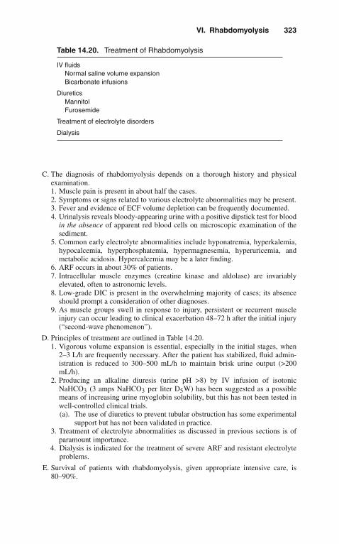
VI. Rhabdomyolysis 323
Table 14.20. Treatment of Rhabdomyolysis
IV fluidsNormal saline volume expansionBicarbonate infusions
DiureticsMannitolFurosemide
Treatment of electrolyte disorders
Dialysis
C. The diagnosis of rhabdomyolysis depends on a thorough history and physicalexamination.1. Muscle pain is present in about half the cases.2. Symptoms or signs related to various electrolyte abnormalities may be present.3. Fever and evidence of ECF volume depletion can be frequently documented.4. Urinalysis reveals bloody-appearing urine with a positive dipstick test for blood
in the absence of apparent red blood cells on microscopic examination of thesediment.
5. Common early electrolyte abnormalities include hyponatremia, hyperkalemia,hypocalcemia, hyperphosphatemia, hypermagnesemia, hyperuricemia, andmetabolic acidosis. Hypercalcemia may be a later finding.
6. ARF occurs in about 30% of patients.7. Intracellular muscle enzymes (creatine kinase and aldolase) are invariably
elevated, often to astronomic levels.8. Low-grade DIC is present in the overwhelming majority of cases; its absence
should prompt a consideration of other diagnoses.9. As muscle groups swell in response to injury, persistent or recurrent muscle
injury can occur leading to clinical exacerbation 48–72 h after the initial injury(“second-wave phenomenon”).
D. Principles of treatment are outlined in Table 14.20.1. Vigorous volume expansion is essential, especially in the initial stages, when
2–3 L/h are frequently necessary. After the patient has stabilized, fluid admin-istration is reduced to 300–500 mL/h to maintain brisk urine output (>200mL/h).
2. Producing an alkaline diuresis (urine pH >8) by IV infusion of isotonicNaHCO3 (3 amps NaHCO3 per liter D5W) has been suggested as a possiblemeans of increasing urine myoglobin solubility, but this has not been tested inwell-controlled clinical trials.(a). The use of diuretics to prevent tubular obstruction has some experimental
support but has not been validated in practice.3. Treatment of electrolyte abnormalities as discussed in previous sections is of
paramount importance.4. Dialysis is indicated for the treatment of severe ARF and resistant electrolyte
problems.
E. Survival of patients with rhabdomyolysis, given appropriate intensive care, is80–90%.
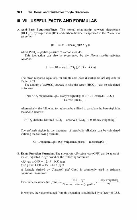
324 14. Renal and Fluid–Electrolyte Disorders
� VII. USEFUL FACTS AND FORMULAS
A. Acid–Base Equations/Facts. The normal relationship between bicarbonate(HCO3
–), hydrogen ions (H+), and carbon dioxide is expressed in the Hendersonequation:
[H+] = 24 × (PCO2/[HCO−3 ])
where PCO2 = partial pressure of carbon dioxide.This interaction can also be represented by the Henderson-Hasselbalch
equation:
pH = 6.10 + log([HCO−3 ]/0.03 × PCO2)
The mean response equations for simple acid–base disturbances are depicted inTable 14.21.
The amount of NaHCO3 needed to raise the serum [HCO3–] can be calculated
as follows:
NaHCO3 required (mEq)= Body weight (kg) × 0.7 × (Desired [HCO−3 ]
−Current [HCO−3 ])
Alternatively, the following formula can be utilized to calculate the base deficit inmetabolic acidosis:
HCO−3 deficit= (desired HCO3 − observed HCO3) × 0.4(body weight (kg))
The chloride deficit in the treatment of metabolic alkalosis can be calculatedutilizing the following formula:
Cl−Deficit (mEq)= 0.5 (weight in Kg)(103 − measured Cl−)
B. Renal Function Formulas. The glomerular filtration rate (GFR) can be approxi-mated, adjusted to age based on the following formulas:
<45 years: GFR = 12.49 – 0.37 (age)≥45 years: GFR = 153 –1.07 (age)
A formula derived by Cockcroft and Gault is commonly used to estimatecreatinine clearance:
Creatinine clearance (mL/min) = 140 − age
Serum creatinine (mg/dL)× Body weight (kg)
72
In women, the value obtained from this equation is multiplied by a factor of 0.85.

VII. Useful Facts and Formulas 325
Table 14.21. Selected Response Equations for Simple Acid–BaseDisturbances
Acid–base disturbance Equation
Metabolic acidosis �PaCO2 ≈ 1.2�[HCO3–]
Metabolic alkalosis �PaCO2 ≈ 0.7�[HCO3–]
Respiratory acidosis
Acute �[HCO3–] ≈ 0.1�paCO2
�[H+] ≈ 0.75�PaCO2Chronic �[HCO3
–] ≈ 0.3�PaCO2
�[H+] ≈ 0.3�PaCO2
Respiratory alkalosis
Acute �[HCO3–] ≈ 0.2�PaCO2
�[H–] ≈ 0.75�PaCO2
Chronic �[HCO3–] ≈ 0.5�PaCO2
�[H+] ≈ 0.5�PaCO2
This formula can also be adjusted for lean body weight (LBW) calculated fromthe followingLBW (male) = 50 kg + 2.3 kg/in. >5 feetLBW (female) = 45.5 kg + 2.3 kg/in. >5 feetAlternatively, the creatinine clearance (Ccr) can be calculated as follows:
Ccr = (Ucr · V)
Pcr
where Ucr = concentration of creatinine in a timed collection of urine; Pcr =concentration of creatinine in the plasma; V = urine flow rate (volume divided byperiod of collection).
Another commonly employed formula to calculate the creatinine clearance isJelliffe’s formula:
Ccr = 98 − 0.8 (age − 20)
Pcr
In this formula, age is rounded to nearest decade. In females, the above result ismultiplied by a factor of 0.9.
A more complicated and potentially more accurate way to calculate creatinineclearance is Mawer’s formula:
Ccr (males) = LBW [29.3 − (0.203 × age)][1 − (0.03 × Pcr)]
14.4(Pcr)

326 14. Renal and Fluid–Electrolyte Disorders
Table 14.22. Creatinine Clearance Values Under SelectedConditions
Condition Value
Normal >100 mL/minMild renal failure 40–60 mL/minModerate renal failure 10–40 mL/minSevere renal failure <10 mL/min
Ccr (females) = LBW [25.3 − (0.174 × age)][1 − (0.03 × Pcr)]
14.4(Pcr)
Hull’s formula for creatinine clearance is calculated as
Ccr= [(145 − age)/Pcr] − 3
In females the result is multiplied by a factor of 0.85.Ranges for creatinine clearance under selected conditions are depicted in
Table 14.22.
C. Selected Electrolytes. The transtubular potassium gradient (TTKG) allows oneto estimate the potassium secretory response in the cortical collecting duct. Thisindex corrects for water reabsorption in the cortical and medullary collectingducts:
TTKG = Corrected urine K+/Serum K+
Corrected urine K+ = Urine K+Uosm/Posm
The normal renal conservation of potassium is reflected by a TTKG <2.The percentage of magnesium retention (MR) can be calculated by the
following formula:
MR(%) = 1 −
Postinfusion
24 − hour
urine Mg++−(Preinfusion × Postinfusion)
urine Mg++/Cr ratio urine Cr
Total elemental magnesium infused× 1
The fractional tubular reabsorption of phosphate (TRP) allows for quantificationof renal phosphate wasting and is calculated as
TRP = 1 − Cp04/Ccr
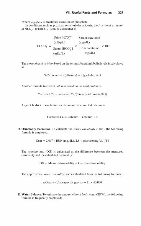
VII. Useful Facts and Formulas 327
where Cp04/Ccr = fractional excretion of phosphate.In conditions such as proximal renal tubular acidosis, the fractional excretion
of HCO3– (FEHCO3
–) can be calculated as
FEHCO−3 =
Urine [HCO−3 ]
(mEq/L)
Serum [HCO−3 ]
(mEq/L)
×
Serum creatinine
(mg/dL)
Urine creatinine
(mg/dL)
× 100
The correction of calcium based on the serum albumin/globulin levels is calculatedas
%Ca bound = 8 (albumin) + 2 (globulin) + 3
Another formula to correct calcium based on the total protein is
Corrected Ca = measured Ca/(0.6 + (total protein/8.5)
A quick bedside formula for calculation of the corrected calcium is
Corrected Ca = Calcium − albumin + 4
D. Osmolality Formulas. To calculate the serum osmolality (Osm), the followingformula is employed:
Osm = 2Na++BUN (mg/dL)/2.8 + glucose (mg/dL)/18
The osmolar gap (OG) is calculated as the difference between the measuredosmolality and the calculated osmolality:
OG = Measured osmolality − Calculated osmolality
The approximate urine osmolality can be calculated from the following formula:
mOsm ∼ (Urine specific gravity − 1) × 40,000
E. Water Balance. To estimate the amount of total body water (TBW), the followingformula is frequently employed:

328 14. Renal and Fluid–Electrolyte Disorders
TBW = Body weight (kg) × 60%
The water deficit of a patient can be estimated by the following equation:
Water deficit = 0.6 × Body weight in kg × (PNa/140 − 1)
where PNa = plasma sodium concentration.Alternatively, the free water deficit from the osmolality can be calculated as
H2O deficit (L) = Total body weight (kg) × 0.6
(1 − normal osm
observed osm
)
To calculate the free water clearance based on the osmolar clearance, thefollowing formula can be utilized:
Free water clearance = Urine volume − Osmolar clearance
where the osmolar clearance is calculated as
Osmolar clearance = Urine osmolarity × urine volume
Plasma osmolarity
The excess water (EW) of a patient is calculated as
EW= TBW − [Actual plasma Na/Desired plasma Na] × TBW
F. Urinary/Renal Indices. The most common urinary indices used in the differentialdiagnosis of acute renal failure are depicted in Table 14.23.
Table 14.23. Commonly Used Urinary Indices in Acute RenalFailure
Index Prerenal Acute tubular necrosis
Specific gravity >1.020 <1.010Urinary osmolality (mOsm/kg H2O) >500 <350Uosm/Posm >1.3 <1.1Urinary Na+ (mEq/L) <20 >40U/P Cr >40 <20RFI <1 >1FENa (%) <1 >1
Abbreviations: Cr, creatinine; P, plasma; RFI, renal failure index; U, urine; FENa, fractionalexcretion of sodium.

VII. Useful Facts and Formulas 329
To calculate the renal failure index (RFI), the following formula is commonlyemployed:
RFI = U Na+U/P Cr
The fractional excretion of sodium (FENa) is calculated as
FENa (%) = Quantity of Na+excreted
Quantity of Na+filtered× 100
or
FENa (%) = U/P Na+ × 100
U/P Cr
or
FENa (%) = UNa × V
PNa × (UCr × V/PCr)× 100
or
FENa (%) = UNa × PCr
PNa × UCr× 100
where UNa = urine sodium concentration; V = urine flow rate; PNa = plasmasodium concentration; UCr = urine creatinine concentration; PCr = plasmacreatinine concentration.
G. Hemodialysis Formulas. The following are useful equations in the managementof the chronic hemodialysis patient.The protein catabolic rate (PCR) is calculated as:
PCR (g/kg/d) = 0.22 + 0.036 × ID BUN × 24
ID interval
where ID BUN = interdialytic rise in blood urea nitrogen (BUN) in mg/dL; IDinterval = interdialytic interval in hours.Alternatively, if blood urea is measured, the PCR can be calculated utilizing thefollowing formula:
PCR (g/kg/d) = 0.22 + 0.01 × IDurea × 24
ID interval
where ID urea = interdialytic rise in blood urea in mmol/L.If the patient has a significant urine output, the contribution of the urinary ureaexcretion must be added to the PCR calculation and is calculated as follows:

330 14. Renal and Fluid–Electrolyte Disorders
Urine contribution to PCR = Urine urea N (g) × 150
ID interval (h) × Body weight (kg)
Alternatively, if urine urea is measured
Urine contribution to PCR = Urine urea (mmol) × 4.2
ID interval (h) × Body weight (kg)
To calculate the percentage of recirculation during hemodialysis, the followingformula is utilized:
%Recirculation = A2 − A1
A2 − V× 100
where A2 = blood urea or creatinine concentration in arterial blood line afterblood pump is stopped; Al = arterial line blood urea or creatinine concentration;V = venous line urea or creatinine concentration.
The volume of distribution of urea can be calculated as follows:
Males: V = 2.447 – 0.09516 A + 0.1074 H + 0.3362 WFemales: V = –2.097 + 0.1069 H + 0.2466 W
where V = volume in liters; A = age in years; H = height in centimeters; W =weight in kilograms.
The calculation of residual renal function for dialysis 3 times per week
GFR = V × U
t × (0.25U1 + 0.75U2)
where V = urine volume in interdialytic period; U = urine urea nitrogen or ureaconcentration; t = interdialytic period in minutes; U1 = postdialysis BUN orblood urea on first dialysis of the week; U2 = predialysis BUN or blood ureaon second dialysis of the week.The percent reduction of urea (PRU) can be calculated utilizing the followingformula:
PRU = Pre urea − Post urea
Pre urea× 100
The urea reduction ratio (URR) is calculated as
URR = 100 ×(
1 − Posturea
Preurea
)
H. Urinalysis. Please refer also to Chapter 20, “Common Laboratory Values in theICU” for additional laboratory values. The most common urinalysis manifesta-tions of renal diseases are depicted in Table 14.24.

VII. Useful Facts and Formulas 331
Table 14.24. Urinalysis in Different Conditions
Condition Findings
Prerenal failure SG: >1.015pH: <6Prot: trace to 1+
Sed:sparse hyaline, finegranular cases orbland
Postrenal failure SG: 1.010pH: >6Prot: trace to 1+Hb: +Sed: RBCs, WBCs
Acute tubularnecrosis (ATN)
“Muddy” brown urine
SG: 1.010pH: 6–7Prot: trace to 1+Blood: +
Sed:RBCs, WBCs, RTEcells, RTE casts,pigmented casts
Glomerulardiseases
SG: >1.020
pH: >6Prot: 1–4+
Sed:
RBCs, RBC casts,WBC, oval fatbodies, free fatdroplets, fatty casts
Vascular diseases SG:>1.020, ifpreglomerular
pH: <6Prot: trace to 2+
Sed:
RBCs and RBCcasts withglomerularinvolvement
Interstitial diseases SG: 1.010pH: 6–7Prot: trace to 1+
Sed:WBCs, WBC casts,eosinophils, RBCs,RTE cells
Abbreviations: RBC, red blood cells; RTE, renal tubular epithelial cells; WBC, white bloodcells; SG, urine specific gravity; Prot, protein; Sed, urinary sediment.

332 14. Renal and Fluid–Electrolyte Disorders
Table 14.25. Urine Color Based on the Presence of Elements orSubstances
Elements/substances Characteristic color
White blood cells Milky whitePrecipitated phosphatesChyle
Bilirubin Yellow/amberChloroquineSulfasalazineNitrofurantoinUrobilin
Phenazopyridine Brown/redHemoglobin myoglobinRed blood cellsPhenothiazinesPhenytoinPorphyrinsBeetsRed-colored candies
Melanin Brown/blackPhenolMethyldopaMetronidazoleQuinine
Pseudomonas infection Blue/greenAmitriptylineMethylene blueBiliverdinPropofol
Table 14.26. Normal Urinary Excretion of Selected Amino acids
Amino acid Normal excretion (mg/g of creatinine)
Cystine 18Lysine 130Arginine 16Ornithine 22
Some elements and substances can modify the color of urine in humans, asdepicted in Table 14.25.
I. Other Formulas/Facts. To determine whether a patient has aminoaciduria or not,the fractional reabsorption of an amino acid (FRA) is determined utilizing thefollowing formula:

VII. Useful Facts and Formulas 333
FRA = 1 − [Urine]A/[Plasma]A÷[Urine]Cr/[Plasm]Cr × 100%
The normal urinary excretion of amino acids in patients older than 2 years isdepicted in Table 14.26.When acute renal failure (ARF) is due to uric acid nephropathy (UAN), thefollowing equation is generally >1:
Index = spot urine uric acid (mg/dL)
spot urine creatinine (mg/dL)=> 1.0

15Special Techniques
� I. AIRWAY MANAGEMENT
A. The first technique in the management of patients with airway problems is manualopening of the airway (i.e., head-tilt, chin-lift). See Chapter 2, “The Basics ofCritical Care.”
B. Adjuncts for Artificial Airway1. Oropharyngeal Airways
a. Oropharyngeal airways are available in a number of different sizes andstyles.
b. These devices routinely should be sized from the angle of the jaw to thecentral incisors.
c. Techniques for insertion include the following:(1). The Inverted Technique: The oral airway is placed upside down and
rotated to the appropriate position after negotiating the tongue.(2). Tongue Depressors Technique: A tongue depressor is used to manip-
ulate the base of the tongue to prevent occlusion of the airway byimpeachment of the tongue on the end of the oral airway.
2. Nasopharyngeal Airwaysa. Nasopharyngeal airways are also available in a number of sizes, which
should be measured from the tragus of the ear to the tip of the nose.(1). Great care should be exercised to ensure that the angled opening of the
distal portion of the airway does not traumatize nasal passages, resultingin epistaxis.
(2). Well-lubricated nasopharyngeal airways appear to be better tolerated inthe alert patient as compared to oropharyngeal airways.
3. Laryngeal Mask Airway (LMA)a. LMAs are used by anesthesiologists in the operating room on a routine basis.
These devices can be utilized when concerns for difficult airway arise. Theyare quite simple to use.
C. Endotracheal Intubation1. Common indications for endotracheal intubation are depicted in
Table 15.1.2. Orotracheal Intubation
J. Varon, P. Acosta, Handbook of Critical and Intensive Care Medicine,DOI 10.1007/978-0-387-92851-7_15,C© Springer Science+Business Media, LLC 2010
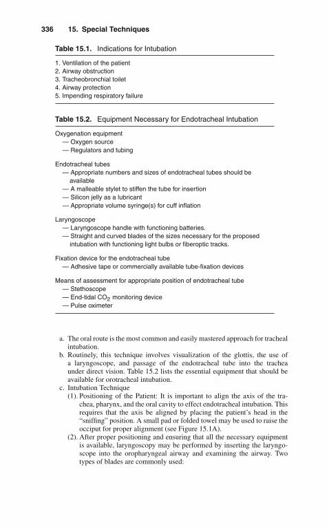
336 15. Special Techniques
Table 15.1. Indications for Intubation
1. Ventilation of the patient2. Airway obstruction3. Tracheobronchial toilet4. Airway protection5. Impending respiratory failure
Table 15.2. Equipment Necessary for Endotracheal Intubation
Oxygenation equipment— Oxygen source— Regulators and tubing
Endotracheal tubes— Appropriate numbers and sizes of endotracheal tubes should be
available— A malleable stylet to stiffen the tube for insertion— Silicon jelly as a lubricant— Appropriate volume syringe(s) for cuff inflation
Laryngoscope— Laryngoscope handle with functioning batteries.— Straight and curved blades of the sizes necessary for the proposed
intubation with functioning light bulbs or fiberoptic tracks.
Fixation device for the endotracheal tube— Adhesive tape or commercially available tube-fixation devices
Means of assessment for appropriate position of endotracheal tube— Stethoscope— End-tidal CO2 monitoring device— Pulse oximeter
a. The oral route is the most common and easily mastered approach for trachealintubation.
b. Routinely, this technique involves visualization of the glottis, the use ofa laryngoscope, and passage of the endotracheal tube into the tracheaunder direct vision. Table 15.2 lists the essential equipment that should beavailable for orotracheal intubation.
c. Intubation Technique(1). Positioning of the Patient: It is important to align the axis of the tra-
chea, pharynx, and the oral cavity to effect endotracheal intubation. Thisrequires that the axis be aligned by placing the patient’s head in the“sniffing” position. A small pad or folded towel may be used to raise theocciput for proper alignment (see Figure 15.1A).
(2). After proper positioning and ensuring that all the necessary equipmentis available, laryngoscopy may be performed by inserting the laryngo-scope into the oropharyngeal airway and examining the airway. Twotypes of blades are commonly used:

I. Airway Management 337
Figure 15.1. Endotracheal intubation. (A) Proper use of the laryngoscopeduring intubation. (B) View of the larynx during direct laryngoscopy. (FromAllison EJ Jr; Advanced Life Support Skills, St. Louis, 1994, Mosby. Usedwith permission.)

338 15. Special Techniques
(a). The Miller blade is used to lift the epiglottis to obtain visualizationof the tracheal opening.
(b). The Mackintosh blade fits into the vallecula, resulting in adherenceof the epiglottis to the back of the blade (see Figure 15.1A). Thetongue and other oral contents are displaced to the left side (seeFigure 15.1B). The Miller blade is inserted more midline, elevatingthe tongue upward.
(c). Care should be exercised in the use of the laryngoscope. Propertechnique is to lift the laryngoscope upward and not to use it as afulcrum (see Figures 15.1A and 15.1B).
(d). A Sellick maneuver (pressure on the cricoid to help occlude theepiglottis during manipulation of the airway) is commonly per-formed to help prevent aspiration and to stabilize the glottis duringthe intubation procedure.
(3). After identifying the laryngeal opening (see Figure 15.1B), the tracheais entered under direct visualization by placing the endotracheal tubethrough the vocal cords.(a). This can be most easily accomplished by placing the endotracheal
tube in the right corner of the mouth, directing the tip into the glotticopening. This technique does not require interruption of the view ofthe vocal cords during intubation.
(b). Insertion should be stopped when the cuff is displaced 2 cmfrom the glottic opening (external markings are typically at 21 or23 cm from the central incisors of average size women and men,respectively).
(c). The cuff is blown up to a moderate tension of the pilot balloon andventilation with 100% oxygen begun.
(4). Tube placement is ascertained by auscultation of chest and abdomen,examination of the rise and fall of the chest, condensation of therespiratory gas mixture in the endotracheal tube, maintenance of ade-quate saturation on pulse oximetry, and when available end-tidal CO2indicators.
3. Nasotracheal Intubationa. Nasotracheal intubation can be performed under direct visualization using
the laryngoscope.(1). The tube is placed through the nares, and the tip is visualized in the
pharynx.(2). McGill forceps can be used to manipulate the end of the endotracheal
tube through the vocal cords to achieve proper positioning.b. Blind nasal insertion.
(1). The location of the endotracheal tube is ascertained through ausculta-tion.
(2). This technique is reserved for those patients who have spontaneousventilation.
(3). An endotracheal tube of appropriate size is inserted through the narisand advanced to the pharynx.
(4). Auscultation using the unaided ear and listening at the nasal end is usedas the endotracheal tube is advanced.
(5). The tube is inserted through the glottic opening during inspiration andappropriate position is confirmed as noted above.
II. Cardioversion/Defibrillation 339
(6). Nasal intubation may result in severe epistaxis in patients with coagu-lopathy or if performed with excessive force.
(7). Patients with mid-face fractures should not be nasally intubated.(8). Sinusitis is a recognized complication of nasal intubation and should be
considered when determining the route of intubation.4. No matter what the route of intubation of the trachea is, all endotracheal tubes
should be secured with adhesive tape or other securing devices to preventdislodgment.
5. Common Complications of Endotracheal Intubationa. During Laryngoscopy and Intubation
(1). Dental and oral soft tissue trauma(2). Dysrhythmias, hypertension/hypotension(3). Aspiration of gastric contents
b. While Endotracheal Tube Is in Place(1). Tube obstruction(2). Esophageal intubation(3). Accidental extubation(4). Tracheal mucosal ischemia
c. Delayed Complications(1). Tracheal stenosis(2). Vocal cord paralysis(3). Laryngeal edema
D. Cricothyroidotomy1. The cricothyroid membrane can be identified by palpation below thyroid
cartilage.2. A large-bore (14- or 16-gauge) catheter may be placed through the cricothy-
roid membrane into the trachea and used to ventilate and oxygenate patients inwhom other airway maneuvers are unsuccessful.
3. Free release of air from the catheter will confirm tracheal position.4. The tip of the catheter should be angled inferiorly, and after tracheal penetration
with the plastic cannula, it should be advanced.5. The cannula may be adapted to fit the 15-mm opening of a standard Ambu bag,
or alternatively, a portable high-frequency jet ventilator device may be used toprovide oxygenation to the patient.
6. Surgical cricothyroidotomya. Percutaneous technique: A needle is used, passing into the trachea, and a
guide wire is positioned in the trachea through the needle. A dilator is thenpassed and a cricothyroidotomy tube with an internal obturator is inserted.
b. Surgical technique: A small midline incision is made over the cricothyroidmembrane, which is then opened, and an appropriate cannula is placed inthe trachea.
� II. CARDIOVERSION/DEFIBRILLATION
A. The major indications for the utilization of these techniques are covered in Chapter2, “The Basics of Critical Car” and Chapter 3, “Cardiovascular Disorders.”
B. Preparation. An appropriately functioning monitor/defibrillator and conductivepads or gel must be available.
340 15. Special Techniques
C. Procedure for Defibrillation1. Institute basic life support, if not already begun.2. Determine cardiac rhythm. If the patient is not already placed on a cardiac
monitor, then the quick-look capabilities of the monitor/defibrillator may beused.
3. Turn on the monitor section of the monitor/defibrillator.4. Select the paddle lead for the monitor/defibrillator.5. Place the paddles on the right upper sternal and left lateral position or
anteroposteriorly.6. Observe rhythm.
a. If ventricular fibrillation is observed,(1). Turn on the power to the defibrillator unit and make sure that the unit is
in the defibrillation (defib) mode.(2). Select the appropriate energy level. See Chapter 2, “The Basics of
Critical Care” and Chapter 3, “Cardiovascular Disorders.”(3). Place electrode gel or other conductive media and position the paddles
as mentioned above. Firm pressure should be applied.(4). Perform discharge of the defibrillator by simultaneously depressing both
discharge buttons located on the defibrillator paddles.
D. Procedure for Synchronized Cardioversion. After determining cardioversion isappropriate,1. Turn on the power to the defibrillator unit.2. Make sure that the defibrillator unit is in the synchronized (sync) mode.3. Apply conductive gel or other material to the paddles and position them as
noted above.4. Confirm that an acceptable electrocardiogram (ECG) signal is being received
from the monitor/defibrillator unit.5. Discharge the energy by depressing both discharge buttons located on the
defibrillator paddles and by observing the unit to ensure that the shock isdelivered.
E. Avoid administering countershocks directly over implanted pacemakers or defib-rillators and over nitroglycerin patches on the surface of the patient’s skin. Thepotential for serious injury with this device exists. You need to make sure that otherrescuers/healthcare providers are clear of the victim, before delivering shocks.
F. Complications1. An adverse rhythm may be produced by administering electrical countershocks.2. Burns of the skin may result, particularly when poor electrical conduction has
been established. The use of gel or other conductive material is mandatory andfirm pressure (approximately 25 pounds) should be applied to the paddles.
3. Myocardial injury.4. Systemic embolization.
� III. VASCULAR ACCESS
A. Modified Seldinger’s Technique. This technique is a simple method of obtainingaccess to vascular spaces.

III. Vascular Access 341
Figure 15.2. Modified Seldinger technique for vascular access. (A) Single-lumen catheter. (B) Double-lumen catheter.
1. After appropriate preparation, draping, and positioning, a needle is percuta-neously placed into the vascular structure. A guide wire with a flexible end(either J or straight) is inserted through the needle and into the lumen of thevessel (see Figure 15.2).
2. The needle is subsequently removed, and the catheter is inserted over the extra-luminal end of the guide wire and subsequently passed over the wire into thevessel.
3. The catheter is advanced and the guide wire removed. When appropriatelypositioned, the catheter is secured with suture or tape.
B. Central Venous Access1. The major indications for central venous access are depicted in Table 15.3.2. No absolute contraindications to central venous access exist. Relative con-
traindications may include bleeding diathesis and central venous thrombosis.3. Choice of Central Cannulation Route
a. Subclavian, internal jugular, and femoral routes have all been used exten-sively for central cannulation.
b. The specific site chosen is dependent upon the clinical circumstances andthe skill of the operator.
Table 15.3. Indications for Central Venous Cannulation
Difficult peripheral venous cannulationDrug administrationEmergency dialysisTotal parental nutritionHemodynamic monitoring

342 15. Special Techniques
Table 15.4. Equipment Necessary for Central VenousCannulation
Appropriate intravenous fluid with administration tubingPrep solution (routinely lodophor)Sterile towels10 m1 syringe with Luer-lok25-gauge needle for local anesthesia1% lidocaineAppropriate size and gauge introducer needleSpring guide wireNumber 11 bladeVessel dilatorSelected catheterSuture material
c. Subclavian insertion has a higher risk of pneumothorax. It also presents anoncompressible vascular puncture site.
d. There is a small but significant incidence of carotid puncture during internaljugular cannulation.
4. Table 15.4 displays the equipment necessary for central venous cannulation.5. Internal Jugular Catheterization (Anterior Approach)
a. In the non-emergent setting, informed consent should be obtained.b. Position the patient in a 15- to 20-degree Trendelenburg position and remove
the headboard of the bed.c. Wash, gown, and glove.d. Prepare the operative site gently with Iodophor solution, and drape the region
with sterile towels.e. The internal jugular vein lies beneath the sternocleidomastoid muscle and
slightly in front of the carotid artery, as shown in Figure 15.3.
Figure 15.3. Central venous cannulation. (See text for details.)
III. Vascular Access 343
f. In the anterior approach, the carotid artery is palpated (left index and middlefinger), the puncture site is infiltrated with 1% lidocaine, and a finding nee-dle is passed immediately lateral to the carotid pulsation beneath the medialedge of the sternocleidomastoid muscle at the level of the thyroid cartilage.The needle is advanced at an angle of approximately 30◦ to the skin (directedtoward the ipsilateral nipple). The vessel should be encountered within 3 cm.When gentle suction on the syringe produces a rush of venous blood, theneedle is removed, and the procedure is repeated with the larger gauge intro-ducer needle on a 5- or 10-mm Luer-lok syringe. Once the venous puncturehas been achieved, a guide wire is passed into the vessel (a vessel dilator canbe used), and the venous catheter is inserted over the wire through a verysmall skin incision made over the wire. The wire subsequently is removedand intravenous (IV) extension tubing is attached.
6. Internal Jugular Catheterization (Middle Approach)a. With the patient supine in the 20-degree Trendelenburg position and the
patient’s head slightly turned to the contralateral side, local anesthetic isinfiltrated at the junction of the sternal and clavicular heads of the sternoclei-domastoid muscle. The needle is inserted with an angle of approximately 30degrees to the skin and again directed toward the ipsilateral nipple. The ves-sel should be entered within 2–3 cm of insertion (see Figure 15.3A). Oncevascular access has been obtained, the procedure is repeated with the intro-ducer needle, the guide wire is passed through the needle, and cannulationis completed as noted above.
7. Internal Jugular Catheterization (Posterior Approach)a. The patient is again positioned in the 20-degree Trendelenburg position with
the head facing the contralateral shoulder. After preparation of the skin andlocal anesthesia as noted above, the needle is inserted through the skin atthe posterolateral margin of the sternocleidomastoid muscle (approximately4 cm above the sternoclavicular junction). This is the approximate pointwhere the external jugular vein crosses the posterior margin of the stern-ocleidomastoid muscle, a commonly used landmark. The needle is advancedin a caudal and medial direction, aiming at the contralateral nipple (seeFigure 15.3B). Once venous access with the introducer needle has beenobtained, a guide wire is placed into the catheter and cannulation proceedsas noted above.
C. Subclavian Vein Cannulation1. The patient is prepped and positioned in a manner analogous to that for inter-
nal jugular vein cannulation; however, a rolled-up towel should be placedlongitudinal between the scapulas to allow the shoulders to drop back.
2. The patient’s head is turned 45 degrees away from the side of intendedplacement.
3. The puncture site is identified approximately 1 cm below the inferior mar-gin of the clavicle at the junction of the medial and middle two thirds (seeFigure 15.3C).
4. Infiltration with 1% lidocaine of the region is accomplished. In addition,lidocaine is also injected into the periosteum of the clavicle.
5. The anesthesia needle is removed and the introducer needle is inserted intothe skin at this point. The tip is aimed at the suprasternal notch, passing justbeneath the clavicle. The bevel of the needle should be pointed toward the head(cephalad). When free flow of blood is obtained from the introducer needle,

344 15. Special Techniques
the bevel can be rotated 180 degrees, helping to facilitate thoracic placementof the guide wire. The catheter is threaded, the wire is removed, fluid flow isestablished, and the catheter is then secured.
D. Femoral Vein1. This approach is easily performed in most patients.2. The patient is placed supine, knees extended, and the foot of the anticipated
cannulation site is rotated outward 15–30 degrees.3. The site of insertion is cleaned and prepped, as noted previously, and the region
is draped.4. The insertion point is identified, lying 2–3 cm inferior to the inguinal liga-
ment (1–2 cm medial to the femoral pulse. (The reader is reminded of the navlmnemonic of the structures in this region: nerve, artery, vein, lymphatics.)
5. As for internal jugular cannulation, a 22-gauge finder needle is commonly usedfor local anesthesia infiltration as well as localization of the vessel.
6. After the femoral vein has been found, the introducer needle is placed on asyringe and inserted into the femoral vein.
7. Next, a flexible guide wire is placed and the needle is exchanged for thevascular cannula. The catheter is then secured.
E. Intraosseous (IO) access1. Gather Equipment
a. The recommended site for insertion of an IO needle is the anterior tibia withalternative sites including the distal femur, medial malleolus, and anteriorsuperior iliac spine
b. This technique can be used in all ages of patients2. Using sterile technique, locate the site of cannulation. Identify the tibial
tuberosity by palpation. The site for IO cannulation of the tibia is approx-imately 1–3 cm below the tibial tuberosity. At this site, the tibia usually isimmediately beneath the skin surface and is readily palpable as a flat, smoothsurface.
3. Position the patient supine and place sandbag or towel behind knee4. Cleanse skin over the insertion site with antibacterial agent5. Palpate the landmarks again and insert the needle through the skin over the flat
anteromedial surface of the tibia6. Using a gentle but firm twisting motion advance the needle through the bony
cortex of the proximal tibia; direct the needle perpendicular to the long axis ofthe bonea. Directing the needle at a slight angle of 10 degrees avoids puncturing the
epiphysial plateb. When placing an IO needle in other locations, aim slightly away from the
nearest joint space to reduce the risk of injury to the epiphysis or joint7. Stop advancing the needle when you feel a sudden decrease in resistance to
forward motion8. Unscrew the cap and remove the stylet from the needle. Slowly inject 3 cc
of normal saline, checking for any signs of increased resistance or increasedcircumference of extremitya. Insertion is successful and the needle is clearly in the marrow cavity if:
i. A sudden decrease in resistance to insertion occurs; the needle canremain upright without support
ii. Marrow can be aspirated into a syringeiii. Fluids flow freely through the needle
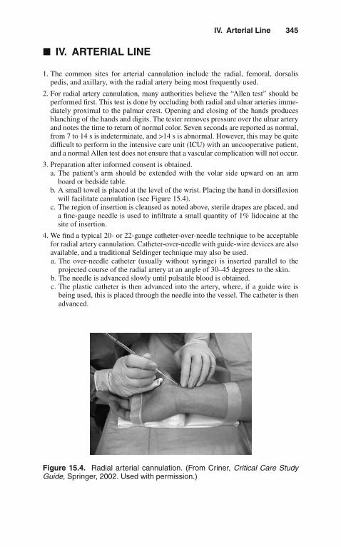
IV. Arterial Line 345
� IV. ARTERIAL LINE
1. The common sites for arterial cannulation include the radial, femoral, dorsalispedis, and axillary, with the radial artery being most frequently used.
2. For radial artery cannulation, many authorities believe the “Allen test” should beperformed first. This test is done by occluding both radial and ulnar arteries imme-diately proximal to the palmar crest. Opening and closing of the hands producesblanching of the hands and digits. The tester removes pressure over the ulnar arteryand notes the time to return of normal color. Seven seconds are reported as normal,from 7 to 14 s is indeterminate, and >14 s is abnormal. However, this may be quitedifficult to perform in the intensive care unit (ICU) with an uncooperative patient,and a normal Allen test does not ensure that a vascular complication will not occur.
3. Preparation after informed consent is obtained.a. The patient’s arm should be extended with the volar side upward on an arm
board or bedside table.b. A small towel is placed at the level of the wrist. Placing the hand in dorsiflexion
will facilitate cannulation (see Figure 15.4).c. The region of insertion is cleansed as noted above, sterile drapes are placed, and
a fine-gauge needle is used to infiltrate a small quantity of 1% lidocaine at thesite of insertion.
4. We find a typical 20- or 22-gauge catheter-over-needle technique to be acceptablefor radial artery cannulation. Catheter-over-needle with guide-wire devices are alsoavailable, and a traditional Seldinger technique may also be used.a. The over-needle catheter (usually without syringe) is inserted parallel to the
projected course of the radial artery at an angle of 30–45 degrees to the skin.b. The needle is advanced slowly until pulsatile blood is obtained.c. The plastic catheter is then advanced into the artery, where, if a guide wire is
being used, this is placed through the needle into the vessel. The catheter is thenadvanced.
Figure 15.4. Radial arterial cannulation. (From Criner, Critical Care StudyGuide, Springer, 2002. Used with permission.)
346 15. Special Techniques
d. The needle is removed and appropriate tubing is attached. The catheter is thensecured in place.
5. Complications include thrombosis, infection, aneurysm formation (especially infemoral arterial lines).
� V. PULMONARY ARTERY CATHETERIZATION
A. Intravenous access must be obtained using one of the techniques described above.The introducer sheath is then placed into the desired vessel.
B. The appropriate monitoring system must be in place, including pressure transduc-ers for all ports that will be monitored.1. High-pressure tubing must be flushed with appropriate solution.2. The clinician should observe and test the dynamic response by watching the
pressure wave on the bedside monitor while activating the flush device andquickly releasing it.
3. Each lumen of the catheter should be flushed and connected to the appropri-ate monitoring line and the balloon inflated to ensure an intact symmetricalballoon.
4. Rapid whipping of the catheter tip will convince the operator that the mon-itoring system is functioning properly when the catheter is inserted into theintroducer.
C. Continuous recording of distal port should be instituted.
D. The catheter is advanced approximately 20 cm (in the adult patient) for eithersubclavian or internal jugular insertions, at which point a central venous pressurewaveform should be seen (see Figure 15.5).
E. The balloon is inflated with 1–1.5 cc of air, and the catheter is further advancedinto the vascular system.
F. Within 30 or 40 cm of catheter insertion, the right ventricular (RV) pressurewaveform is usually seen (see Figure 15.5). This is easily identified by the steepupstroke, typically 2–3 times larger than the right atrial (RA) pressure.
G. Passage in the PA generally occurs at 40–50 cm of catheter and may be identifiedby the dicrotic notch of the downslope.
H. A pulmonary artery wedge pressure is usually noted at 50–60 cm of catheter andlooks much like a RA waveform (see Figure 15.5).
I. Complications include dysrhythmias, valvular damage, knotting of the catheter,atrial or ventricular perforation, air embolism, pulmonary embolism, pulmonaryarterial injury, and catheter-related sepsis.
� VI. TUBE THORACOSTOMY
A. The drainage system should be prepared before the chest tube is placed.1. All the couplings and tubing should be inspected, and appropriate fluid levels
should be maintained.

VI. Tube Thoracostomy 347
Figure 15.5. Pulmonary artery catheterization and waveforms. (See text fordetails.)
2. A “three-bottle system” (all of which may be maintained in a single commercialthoracostomy drainage system) is depicted in Figure 15.6D. The first bottle isthe trap bottle, which collects the fluid emanating from the chest tube itself. Thesecond bottle represents the water-seal bottle. Air is precluded from enteringthe pleural space through the system by the water in the water-seal bottle. Thethird bottle represents the manometer bottle. Suction applied to the manometerbottle is regulated by the distance that the center tube lies below the surface ofthe water. For example, if the central tube lies 20 cm below the surface of thewater, suction levels producing pressure in excess of –20 cm of water simplyresult in bubbling of ambient air in the manometer bottle, thus, maintainingthe –20-cm water pressure limit.
B. Surgical Technique1. The contents of a chest tube tray are depicted in Table 15.5.2. The patient should be positioned with the side for tube insertion uppermost.3. The operator should be gowned and gloved.4. The chest tube is usually inserted in the anterior axillary line in the fifth or
sixth intercostal space.5. The incision site should be prepped and draped, and lidocaine should be infil-
trated one intercostal space below the rib of the selected intercostal space ofinsertion. The periosteum, subcutaneous tissue, and pleural space should beinfiltrated. Aspiration of fluid or air will confirm infiltrated pleural space.
6. A small skin incision, appropriate to the size of the chest tube, is made with ascalpel over the anesthetized rib.

348 15. Special Techniques
Table 15.5. Contents of the Chest Tube Tray
— Sterile towels and drapes— 1% or 2% lidocaine— 10 mL syringe— 22- and 25-gauge needles— 1-0 silk suture with cutting needle (2 packages)— 2 large Kelly clamps— 2 medium Kelly clamps— Suture scissors— 4-inch-square gauze pads— Chest drainage, suction system, and appropriate chest tube
7. Extend the incision into the subcutaneous tissue and muscle at the intercostalspace, preferably using the blunt side of the scalpel or trocar.
8. A large clamp with an open end using spreading maneuvers is used until thepleural space is reached (see Figure 15.6A).
9. The index finger of the operator is used to explore the pleural space toensure that the lung, diaphragm, or another structure is not adherent (seeFigure 15.6B).
Figure 15.6. Tube thoracostomy. (Used with permission. Yeston N, Kirby R.Atlas of Anesthesia: Critical Care. Edited by Ronald Miller (series editor),Robert R. Kirby. ©1997 Current Medicine Group LLC.)
VIII. Pericardiocentesis 349
10. The tube is inserted, generally toward the apex of the pleural space fortreatment of pneumothoraces, by grasping the tube with a medium clamp,maneuvering it through the dissected tunnel (see Figure 15.6C), and ensur-ing that the last hole of the thoracostomy tube lies within the pleural space.The tube is fixed at the insertion site with 1-0 silk suture.a. A number of techniques are used, but commonly the suture ends are not cut
but wrapped around the tube and secured with tape. These may be used toclose the wound when the tube is pulled out.
11. The external end of the chest tube is then attached to the system and theconnections are taped over.
12. A sterile dressing with tape is applied to the skin.
C. Complications include tube malposition, empyema or wound infection, blockageof tube by blood or fibrin clot, and lung injuries.
� VII. INTRA-AORTIC BALLOON PUMP (IABP)
A. Indications for the Use of IABP1. Pump failure
a. After acute myocardial ischemic eventb. Cardiogenic shockc. Postcardiac transplant patientd. In the pre- or postoperative period of cardiac surgery
2. Acute mitral valvular regurgitation3. Unstable angina pectoris4. Other
B. Insertion should normally be accomplished by those with experience. The tech-nique is also dependent upon the particular catheter and approach to be used.1. Once the catheter is in place, its function is rather easy to visualize (see
Figure 15.7).2. During ventricular systole, the balloon (present in the proximal aorta) deflates,
decreasing afterload on the heart and improving ventricular performance.3. During diastole, the inflated balloon occludes 75–90% of the cross-sectional
area of the descending aorta, thereby increasing coronary perfusion.4. Helium is most commonly used for inflation and deflation of the balloon.
C. Complications include balloon membrane rupture or perforation, limb ischemia,aortic dissection, renal failure, thrombocytopenia, and infection.
� VIII. PERICARDIOCENTESIS
A. Blind pericardiocentesis should be performed in life-threatening situations (i.e.,decompensated cardiac tamponade).1. We prefer the subxiphoid approach.
a. If possible, the patient is placed upright or if necessary in a semirecliningposition.
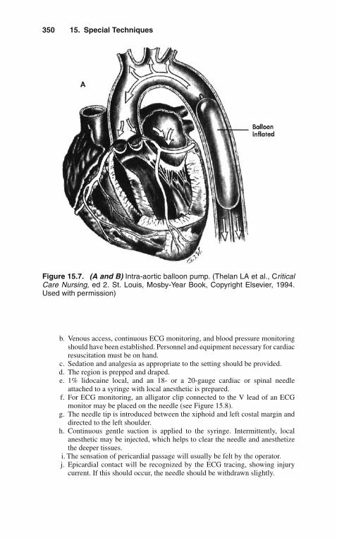
350 15. Special Techniques
Figure 15.7. (A and B) Intra-aortic balloon pump. (Thelan LA et al., CriticalCare Nursing, ed 2. St. Louis, Mosby-Year Book, Copyright Elsevier, 1994.Used with permission)
b. Venous access, continuous ECG monitoring, and blood pressure monitoringshould have been established. Personnel and equipment necessary for cardiacresuscitation must be on hand.
c. Sedation and analgesia as appropriate to the setting should be provided.d. The region is prepped and draped.e. 1% lidocaine local, and an 18- or a 20-gauge cardiac or spinal needle
attached to a syringe with local anesthetic is prepared.f. For ECG monitoring, an alligator clip connected to the V lead of an ECG
monitor may be placed on the needle (see Figure 15.8).g. The needle tip is introduced between the xiphoid and left costal margin and
directed to the left shoulder.h. Continuous gentle suction is applied to the syringe. Intermittently, local
anesthetic may be injected, which helps to clear the needle and anesthetizethe deeper tissues.
i. The sensation of pericardial passage will usually be felt by the operator.j. Epicardial contact will be recognized by the ECG tracing, showing injury
current. If this should occur, the needle should be withdrawn slightly.
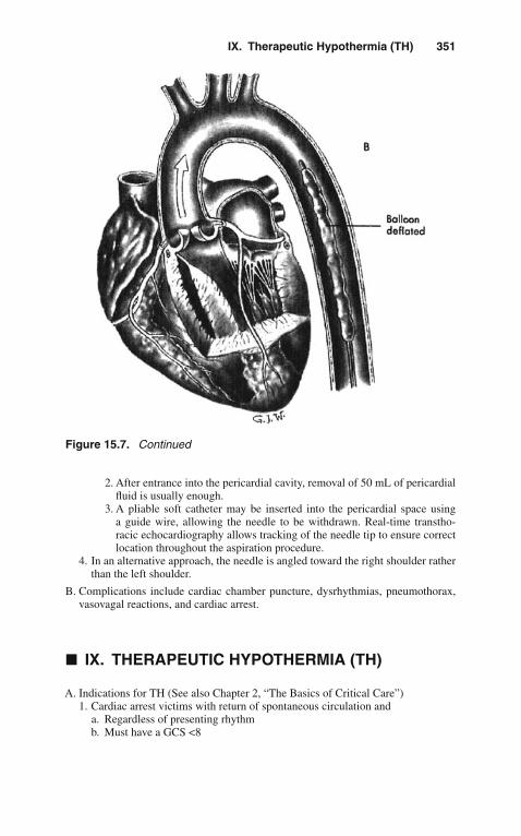
IX. Therapeutic Hypothermia (TH) 351
Figure 15.7. Continued
2. After entrance into the pericardial cavity, removal of 50 mL of pericardialfluid is usually enough.
3. A pliable soft catheter may be inserted into the pericardial space usinga guide wire, allowing the needle to be withdrawn. Real-time transtho-racic echocardiography allows tracking of the needle tip to ensure correctlocation throughout the aspiration procedure.
4. In an alternative approach, the needle is angled toward the right shoulder ratherthan the left shoulder.
B. Complications include cardiac chamber puncture, dysrhythmias, pneumothorax,vasovagal reactions, and cardiac arrest.
� IX. THERAPEUTIC HYPOTHERMIA (TH)
A. Indications for TH (See also Chapter 2, “The Basics of Critical Care”)1. Cardiac arrest victims with return of spontaneous circulation and
a. Regardless of presenting rhythmb. Must have a GCS <8

352 15. Special Techniques
Figure 15.8. Pericardiocentesis. (Sabiston DC, and Spencer FC. Gibbon’sSurgery of the Chest, 4th ed. Philadelphia WB, Saunders 1983. Used withpermission.)
2. Anoxic–hypoxic encephalopathy of the newborn3. Cerebrovascular accidents with increased intracranial pressure4. Others (i.e., traumatic brain injury, near-drowning, liver failure)
B. This procedure should normally be accomplished by those with experience. Thetechnique is also dependent upon the particular setting and materials available.Core temperature must be measured (i.e., bladder, vaginal, esophageal)

X. Bronchoscopy 353
1. Goal temperature is 32–33◦ C for a minimum of 24 h.2. Cold IV fluids help achieve this temperature faster and should be used in
patients without contraindications (i.e., end-stage renal disease).3. Magnesium sulfate (2 g IV) provides vasodilation and increases rate of temper-
ature drop.4. Sedative agents and neuromuscular blockers are utilized to decrease oxygen
consumption and to minimize shivering.5. A variety of devices exist to provide this technique. The simplest way to per-
form it is by utilizing ice packs applied to the surface (avoid direct contactwith skin). Cooling blankets can be used (the “Sandwich technique”). Hydrogeladhesive pads and special thermal wraps can also be utilized. Endovascularcooling can be accomplished by catheters that accelerate temperature drop.In neonate, selective brain hypothermia with cooling helmets is commonlyutilized. Selective brain hypothermia devices for adults are also available inselected centers.
6. Rewarming is accomplished after 24 h either by stopping the cooling device oractively rewarming the patient over a period of 12–24 h.
C. Complications include dysrhythmias, skin burns, increased infection rate, andrarely coagulopathy.
� X. BRONCHOSCOPY
A. Indications for bronchoscopy in the ICU1. Clearance of secretions and mucus plugs2. To obtain selective samples of tracheobronchial tree3. Hemoptysis4. Therapeutic (i.e., tumor destruction, atelectasis resolution)
B. This procedure should normally be accomplished by those with experience. If thepatient has an endotracheal tube in place, the technique is simpler.
C. Complications include dysrhythmias (many premedicate with Atropine prior tothe procedure), airway injury, pneumothorax.

16Toxicology
� I. GENERAL MANAGEMENT
A. History. Obtain as accurate a history as possible, including the substance ingestedor inhaled and the amount and time of ingestion. Always consider possibleingestion or inhalation of multiple substances.
B. Cardiorespiratory Care. The most important aspects of initial management arebasic care of respiratory and cardiovascular function: Maintain a patent airway,ensure adequate respiration (support ventilation when necessary), and treat shockif present.1. Airway
a. Loss of airway patency and reflexes may lead to obstruction, aspiration, orrespiratory arrest.
b. Maintain proper airway position; suction; use oropharyngeal or nasopharyn-geal adjuncts as needed.
c. Absent or depressed gag reflex in an unconscious or obtunded patientindicates an inability to protect the airway; endotracheal intubation isindicated.
2. Respirationsa. Respiratory failure is the most frequent cause of death in poisoned patients,
and usually it is a result of (CNS) central nervous system depression.b. Assist ventilation, and perform endotracheal intubation as required.c. Obtain and follow arterial blood gases.
3. Circulationa. Monitor blood pressure, pulse, and cardiac rhythm.b. Initiate intravenous line.c. If hypotension is present, administer fluid challenge with normal saline
10–20 mL/kg.d. If hypotension persists, administer vasopressin 1–6 U/hr.e. Other agents such as norepinephrine can also be used.
C. Gastrointestinal Decontamination. The traditional sequence for gastrointestinaldecontamination is emptying of stomach by induced emesis or gastric lavage fol-lowed by administration of activated charcoal. However, there is no data to supportits use anymore.
J. Varon, P. Acosta, Handbook of Critical and Intensive Care Medicine,DOI 10.1007/978-0-387-92851-7_16,C© Springer Science+Business Media, LLC 2010

356 16. Toxicology
1. Induced Emesis with Ipecaca. Should not be administered routinely in the management of poisoned
patients since there is no evidence from clinical studies that ipecac improvesoutcome. If a clinician, however, decides to use, then administer syrup ofipecac (30 mL for adults, 15 mL for children <5 years old, 10 mL for children<1 year old). Repeat if no emesis in 20 min.(1). Not recommended in children <9 months old.(2). If the second dose does not produce vomiting, perform gastric lavage.
b. Contraindications(1). Comatose or seizing patient(2). Ingestion of corrosive agents or petroleum distillates(3). Ingestion of substance likely to produce coma or seizures rapidly
2. Gastric LavageGastric lavage is contraindicated if the patient is obtunded or comatose untilendotracheal intubation is performed to protect the airway. Performance in anobtunded patient may lead to vomiting and aspiration of gastric contents. Othercomplications include perforation of the nasal mucosa, production of epistaxis,tracheal intubation, and stimulation of ventricular dysrhythmias. Lavage is con-troversial in corrosive ingestion. As with other methods of GI decontamination,the efficacy of gastric lavage decreases as the time between ingestion and treat-ment increases. Large amounts of unabsorbed drug will be removed in only aminority of patients who undergo gastric lavage
Utilize a 36–40 Fr tube in adults, 24–26 Fr in children. Check for the positionof the tube by injecting air and auscultating the stomach. Administer lavagefluid in aliquots of 200–300 mL, then allow return of fluid by gravity drainage.Continue until the lavage fluid is clear of ingested fragments or substance color(or longer if the circumstances of the ingestion dictate).
3. Activated Charcoala. It is, by far, the preferred means of gastric decontamination. Limits absorp-
tion of virtually all ingested substances. Administer 1 g/kg orally or viagastric tube after emesis or lavage is completed.
b. Repeat doses of 15–20 g at 1- or 2-h intervals, or continuous activatedcharcoal instillation may be useful in some instances (i.e., theophyllineoverdose).
c. Contraindicated in corrosive ingestion, ileus, or intestinal obstruction.4. Cathartics
a. Frequently used, but their effectiveness is not well established.b. Administer along with activated charcoal.c. Magnesium citrate 10% 3–4 mL/kg or sorbitol 70% 1–2 mL/kg. If no
charcoal is present in stool after 6 h, repeat half dose.5. Whole Bowel Irrigation
a. Effectiveness is not fully conclusive. May be useful in substantial inges-tion of substances poorly adsorbed by activated charcoal or enteric-coatedtablets.
b. Administer polyethylene glycol electrolyte solution (CoLyte or GoLYTELY)2 L/h by gastric tube until stool is free of particulate material.
c. Do not use in unconscious or obtunded patients.
D. Forced Diuresis and Control of Urine pH. May increase urinary excretion of someagents for which renal excretion is the major route of elimination. Do not use inpatients with renal failure or congestive heart failure.

II. Acetaminophen 357
1. Alkalinizationa. May be useful in overdose with salicylates, phenobarbital.b. Administer sodium bicarbonate 1–2 mEq/kg IV to achieve a urinary ph ≥7.0.c. Monitor serum pH.
2. Acidificationa. May be useful in overdose with phencyclidine, amphetamines, quinine,
quinidine, strychnine, cyclic antidepressants.b. Administer ammonium chloride 75 mg/kg IV in 4–6 divided doses to achieve
a urinary pH ≤6.0.c. Do not use in the presence of rhabdomyolysis, myoglobinuria, or hepatic
failure.d. Monitor serum pH.
E. Hemodialysis. May be useful in severe intoxication with amphetamines, methanol,ethylene glycol, isopropyl alcohol, lithium, salicylates. If fluid or acid–baseabnormalities are present, these can also be corrected.
F. Charcoal Hemoperfusion1. Utilizes an extracorporeal circuit through an activated charcoal column.2. May be useful in severe intoxication with barbiturates, doxorubicin, digoxin,
some beta-blockers, ethchlorvynol, meprobamate, phenytoin, salicylates, andtheophylline.
3. Potential complications: hypotension and thrombocytopenia.
G. Toxicology Screen1. Should be used to confirm diagnosis in all cases.2. Specific levels may be necessary in overdose with certain substances to guide
therapy (i.e., acetaminophen, iron, lithium, methanol, salicylates, theophylline).
H. Poison Control Centers. Notify poison control centers with management questionsor complicated or unusual poisonings. In the United States, the telephone numberfor these centers is 1-800-222-1222 or on the internet at http://www.aapcc.org
� II. ACETAMINOPHEN
Acetaminophen is a widely used analgesic and antipyretic. It is found in combina-tion with other analgesics and in various cold remedies (i.e., Comtrex, Congesprin,Excedrin PM, 4-Way Cold Tablets). Toxicity of significant overdose lies in pro-duction of hepatic necrosis. This is probably related to overwhelming of hepaticglutathione capacity to detoxify acetaminophen metabolic products. Toxicity is likelyto occur with single ingestions greater than 250 mg/kg or those greater than 12 g overa 24-h period. The diagnosis may be overlooked, especially in patients with alcoholicliver disease.
A. Clinical Effects1. In the first 24 h after overdose (Stage I), patients often manifest nausea,
vomiting, diaphoresis, pallor, lethargy, and malaise.2. From 24–72 h after the ingestion (Stage II), there is clinical and laboratory
evidence of hepatotoxicity. Of those patients that develop hepatic injury, overhalf will demonstrate ALT/AST elevation within 24 h and all have elevations by36 h. As stage II progresses, patients develop liver tenderness and right upper

358 16. Toxicology
quadrant pain. Elevations of prothrombin time (PT) and total bilirubin, oliguria,and renal function abnormalities may become evident.
3. Liver function abnormalities peak from 72 to 96 (Stage III) hours afteringestion. Jaundice, confusion (hepatic encephalopathy), a marked elevationin hepatic enzymes, hyperammonemia, and a bleeding diathesis are present.Plasma ALT and AST levels often exceed 10,000 IU/L. Prolongation of thePT/INR is evident, along with hypoglycemia, lactic acidosis, and a totalbilirubin concentration above 4.0 mg/dL.
4. Patients who survive stage III enter a recovery phase (Stage IV) that usuallybegins by day 4 and is complete by 7 days after overdose.
B. Diagnostic Studies1. Obtain serum acetaminophen as soon as 4 h and as late as 24 h following inges-
tion and plot the level on a nomogram (Figure 16.1). The nomogram is usefulonly in acute ingestion.
2. There is good correlation between timed serum and subsequent hepatotoxicity.If the level is in the hepatotoxic range, administer n-acetylcysteine.
3. Obtain prothrombin time and transaminase levels, serum urea nitrogen (BUN),creatinine.
C. Management1. Induce emesis or perform gastric lavage.2. Antidote: n-acetylcysteine (NAC).
Probable Hepatic Toxicity
Hepatic Toxicity Unlikely
1000
500
200
100
50
20
10
5
2
1000
500
200
100
50
20
10
Ace
tam
inop
hen
Pla
sma
Con
cent
ratio
n (µ
g/m
L)
5
2
244 8 12 16
Hours After Ingestion
20
Possible Hepatic Toxicity
25%
Figure 16.1. Rumack-Matthew nomogram for acetaminophen poisoning.(Rumack BH, Mathew H: Acetaminophen poisoning and toxicity. Pediatrics1975;55:871–876.)

III. ALCOHOL 359
a. Prevents liver injury when administered early following intoxication.b. In the United States, two NAC treatment regimens are currently approved
by the Food and Drug Administration (FDA): The first is a 72 h oral coursegiven as a 140 mg/kg loading dose followed by 17 doses of 70 mg/kg every4 h (total dose 1,330 mg/kg). The second is a 21 h intravenous infusion (total300 mg/kg).
c. Both oral and IV formulations are available.3. Cimetidine 300 mg q6 h has been used in the past, but its efficacy is uncertain.
� III. ALCOHOL
Ethyl alcohol is found in beverages, as well as perfumes, mouthwashes, andpharmaceutical preparations.
A. Overdose1. Clinical Effects
a. Variable, depending on the individual: slurred speech, impaired judgment,behavioral changes, combativeness, ataxia.
b. At very high levels, somnolence and respiratory depression occur. Whendeath occurs as a result of intoxication, it is usually due to respiratorydepression.
c. Also occurring: cardiac dysrhythmias, hypertension, hypoglycemia, hypo-magnesemia, hypophosphatemia, seizures, hypothermia, rhabdomyolysis,Wernicke’s encephalopathy.
d. Alcoholic ketoacidosis: metabolic acidosis with increased anion and osmolargaps, and ketosis. Associated nausea, vomiting, abdominal pain.
2. Metabolism of AlcoholRate is 12–50 mg/dL/h (average of 20 mg/dL/h).
3. Admission CriteriaAdmission to the intensive care unit may be required for trauma, seizures,hypothermia, and severe metabolic abnormalities.
4. Managementa. Management is mainly supportive, with observation predominant. Intubate
and ventilate if necessary.b. Administer IV glucose 50 g and thiamine 100 mg to all obtunded patients
after serum glucose level is drawn.c. Correct volume depletion as needed.d. If the patient is violent or very agitated, use physical restraints as needed.
Sedation may be necessary.e. Treatment of alcoholic ketoacidosis: Fluid replacement with D5%/normal
saline. Observe for appearance of hypoglycemia, hypophosphatemia, andhypokalemia.
B. Withdrawal1. Withdrawal following chronic use produces a number of signs and symp-
toms, including anxiety, insomnia, tremulousness, nausea, vomiting, tachycar-dia, hyperthermia, delirium, hallucinations, and seizures. The typical time ofappearance of withdrawal findings are listed in Table 16.1.
2. The hallmark of delirium tremens is significant alteration of the sensorium(global confusion, hallucinations, delusions, disorientation) accompanied byautonomic CNS hyperreactivity.

360 16. Toxicology
Table 16.1. Appearance of Findings of Alcohol Withdrawal
Findings Hours elapsed sincecessation of alcohol intake
Tremor, agitation, sleep disturbance,hyperexcitability
6–8
Hallucinations 24–36Seizures 7–48
Confusion, delusions, autonomichyperreactivity, disorientation
72–120
3. Managementa. Sedation
(1). Benzodiazepines: Administer diazepam 5 min as needed, then 5–20 mgIV or PO q6 h, or chlordiazepoxide 25–100 mg IV or PO q6 h, orlorazepam 1–2 mg IV, IM, or PO, or
(2). Phenobarbital 260 mg slow IV, repeat as needed to produce light sedation,or
(3). Haloperidol: Begin with 0.5–1.0 mg IV or IM q1–6 h, but much largerdoses may be required.
(4). Propofol: 5–20 cc/h to achieve required sedation level.
b. Thiamine 100 mg IVc. If severe hypertension or tachycardia is present, administer clonidine
0.1–0.2 mg q6 h.d. Other agents that have been used include beta-blockers, baclofen, and
carbamazepine.e. The use of ethanol to control withdrawal is discouraged in the setting of acute
alcohol withdrawal.
C. Other Alcohols1. Methanol
a. Methanol (methyl alcohol) is widely used as a solvent, is found in windshieldwasher solution and antifreeze, and is used as a solid canned fuel.
b. It produces CNS intoxication similar to that of ethanol. Other toxic effectsinclude acidosis and retinal cell toxicity.
c. Ethanol acts as a competitive substrate for alcohol dehydrogenase, theenzyme that produces toxic metabolites from methanol.
d. Treatment(1). Alkalinization(2). Inhibition of alcohol dehydrogenase (ADH) blocks bio-activation of the
parent alcohol to its toxic acid metabolites. Fomepizole and ethanol canaccomplish this. Fomepizole is loaded at 15 mg/kg intravenously, fol-lowed by 10 mg/kg every 12 h, with adjustments for hemodialysis orafter more than 2 days of therapy. Alternatively, IV ethanol (0.6 mg/kginitially and infusion of 66 mg/kg/h to maintain a blood alcohol level of100 mg/dL. Administer as 10% ethanol diluted in D5%W.
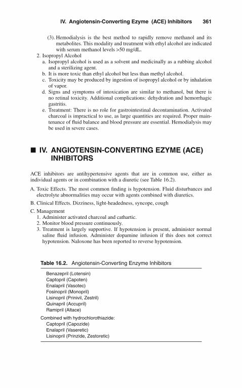
IV. Angiotensin-Converting Ezyme (ACE) Inhibitors 361
(3). Hemodialysis is the best method to rapidly remove methanol and itsmetabolites. This modality and treatment with ethyl alcohol are indicatedwith serum methanol levels >50 mg/dL.
2. Isopropyl Alcohola. Isopropyl alcohol is used as a solvent and medicinally as a rubbing alcohol
and a sterilizing agent.b. It is more toxic than ethyl alcohol but less than methyl alcohol.c. Toxicity may be produced by ingestion of isopropyl alcohol or by inhalation
of vapor.d. Signs and symptoms of intoxication are similar to methanol, but there is
no retinal toxicity. Additional complications: dehydration and hemorrhagicgastritis.
e. Treatment: There is no role for gastrointestinal decontamination. Activatedcharcoal is impractical to use, as large quantities are required. Proper main-tenance of fluid balance and blood pressure are essential. Hemodialysis maybe used in severe cases.
� IV. ANGIOTENSIN-CONVERTING EZYME (ACE)INHIBITORS
ACE inhibitors are antihypertensive agents that are in common use, either asindividual agents or in combination with a diuretic (see Table 16.2).
A. Toxic Effects. The most common finding is hypotension. Fluid disturbances andelectrolyte abnormalities may occur with agents combined with diuretics.
B. Clinical Effects. Dizziness, light-headedness, syncope, cough
C. Management1. Administer activated charcoal and cathartic.2. Monitor blood pressure continuously.3. Treatment is largely supportive. If hypotension is present, administer normal
saline fluid infusion. Administer dopamine infusion if this does not correcthypotension. Naloxone has been reported to reverse hypotension.
Table 16.2. Angiotensin-Converting Enzyme Inhibitors
Benazepril (Lotensin)Captopril (Capoten)Enalapril (Vasotec)Fosinopril (Monopril)Lisinopril (Prinivil, Zestril)Quinapril (Accupril)Ramipril (Altace)
Combined with hydrochlorothiazide:Captopril (Capozide)Enalapril (Vaseretic)Lisinopril (Prinzide, Zestoretic)

362 16. Toxicology
4. In severe overdose, consider hemodialysis in overdose with captopril, enalapril,or lisinopril.
� V. BETA-BLOCKERS
Common beta-blockers in use include propranolol, atenolol, metoprolol, pindolol,and nadolol. Agents are used for a variety of medical indications, including controlof hypertension, cardiac dysrhythmias, angina pectoris, and glaucoma. Duration oftoxicity varies greatly, depending on the agent ingested.
A. Clinical Effects1. Toxic effects include beta-adrenergic blockade, producing bradycardia,
hypotension, bronchospasm.2. Effects resulting from sodium-dependent membrane depression are seen pri-
marily with lipid-soluble agents (i.e., propranolol, metoprolol) and includehypotension, atrioventricular (A-V) block, QRS widening.
3. Seizures and coma may also occur.4. Metabolic effects include hyperkalemia and hypoglycemia.
B. Management1. ABCs like in any other critically ill patient. Activated charcoal may be useful.2. If the patient is hypotensive, administer fluids, pressor agents, as required.3. Administer atropine (0.01–0.03 mg/kg IV) and isoproterenol (start at 4 μg/min)
for bradycardia.4. In unresponsive hypotension and bradycardia, administer glucagon, 5–10 mg
IV. Follow with 1–5 mg/min IV infusion.5. Sodium bicarbonate has been used for patients with QRS widening.6. Magnesium sulfate can be administered when ventricular dysrhythmias are
present.7. Intravenous pacing may be required in some patients with refractory bradycar-
dia.8. Hemoperfusion may be useful for acebutolol, atenolol, and nadolol overdose.9. Monitor serum glucose and electrolytes, particularly potassium.
� VI. COCAINE
Cocaine produces clinical symptoms on the basis of adrenergic stimulation, CNSstimulation, and increased metabolic rate. It is most commonly inhaled or injectedparenterally. Use of free-base “crack” cocaine is very prevalent, as is the con-comitant use of other drugs. Complications of cocaine abuse include hypertensionwith resulting intracerebral hemorrhage or aortic dissection, myocardial infarction,cerebrovascular accident, hyperthermia, and rhabdomyolysis.
A. Clinical Effects1. The onset of toxic symptoms is within 30–60 min of injection or inhalation.2. Patients present most often with hypertension, dysrhythmias, pupillary dilata-
tion, agitation, anxiety, or psychosis.3. Seizures, hyperthermia, and rhabdomyolysis may occur.4. Hypertension may produce intracranial hemorrhage or thoracic aortic rupture.

VII. Cyanide 363
5. Chest pain may occur and is sometimes of myocardial origin. Myocardialinfarction may occur. Cocaine is dysrhythmogenic and may produce sinustachycardia, premature ventricular contractions, ventricular tachycardia, andventricular fibrillation.
B. Management1. ABCs. We recommend avoiding succinylcholine as a rapid sequence intubation
agent due to the risk of hyperkalemia and rhabdomyolysis that these patientshave.
2. Sedate with IV benzodiazepines or haloperidol.a. For hypertension, treat with phentolamine 0.02–0.2 mg/kg. Beta-adrenergic
antagonists (i.e., labetalol) are contraindicated in the treatment of cocaine-related cardiovascular complications as they create unopposed alpha-adrenergic stimulation and are associated with coronary vasoconstriction andend-organ ischemia.
3. Urine toxicology screen will confirm the diagnosis.
� VII. CYANIDE
Cyanide is a chemical with a variety of industrial uses. Sodium nitroprusside containscyanide, which is released into solution in increased quantity by exposure to light.The gas hydrogen cyanide is a product of combustion of plastics and a variety of othermaterials. A third of all smoke inhalation victims have increased cyanide levels.
A. MechanismToxicity is via chemical asphyxia, cyanide-blockade of cellular oxygen utiliza-tion. Exposure to small amounts of hydrogen cyanide gas or ingestion of a smallquantity of cyanide salt can be rapidly fatal.
B. Clinical Effects1. Respiratory failure and cardiovascular collapse. Rapidly developing coma and
severe lactic acidosis.2. Syncope, seizures, headache, nausea, confusion.3. Cyanide poisoning may occur with prolonged IV administration of sodium
nitroprusside. Consider this if such patients develop CNS depression, seizures,lactic acidosis, or cardiovascular instability.
C. Management1. Administer oxygen, maintain airway, and assist respirations.2. Treat hypotension and seizures in standard fashion.3. Administer cyanide antidote kit.
a. Have patient inhale amyl nitrite capsule.b. Administer sodium nitrite 300 mg IV.c. These two produce methemoglobinemia, which may itself be toxic.d. Administer sodium thiosulfate, which converts cyanide to thiocyanate,
150 mg/kg of a 25% solution IV (relatively contraindicated in renal failure).4. If cyanide salt is ingested, lavage stomach and administer activated charcoal.5. Hydroxocobalamin, a form of vitamin B12, is also used as an antidote.
a. Administer 4 g intramuscularly (IM).b. The IV formulation is not available in the United States.

364 16. Toxicology
6. Other cyanide antidotes, dicobalt-EDTA and 4-dimethylamineophenol, areoccasionally used.
� VIII. CYCLIC ANTIDEPRESSANTS
There is an estimated annual incidence of 500,000 cases of overdose withcyclic antidepressants. Many agents are in common use, including amitriptyline,desipramine, nortriptyline, doxepin, and imipramine (see Table 16.3). They may befound in medication combinations with other psychotropic agents. Toxicity occursvia anticholinergic effects, alpha-adrenergic blockade, inhibition of norepinephrinereuptake, and inhibition of the fast sodium channel. Some drugs, such as haloperidol,morphine, and disulfiram, may prolong cyclic antidepressant toxicity by interferingwith hydroxylation.
A. Clinical Effects1. Tachycardia, myoclonus, delirium, coma, hyperthermia, pupillary dilatation,
hypertension or hypotension, prolongation of QRS and QT, A-V block, andmyocardial depression.
2. The most important toxic effects are cardiovascular. A QRS duration >0.12 indi-cates serious toxicity. Sinus tachycardia is typical; supraventricular tachycardia,ventricular tachycardia, torsades des pointes, and ventricular fibrillation mayoccur. Bradycardia is a poor prognostic sign.
3. Rapid deterioration with the development of cardiovascular collapse, coma,or seizures may occur. Persistent seizures may produce hypothermia orrhabdomyolysis.
B. Management1. Administer activated charcoal 100 g. Do not administer syrup of ipecac. Repeat
doses of activated charcoal may be useful, and some have advocated continuousnasogastric instillation.
2. Continuous electrocardiographic monitoring is essential.3. Treat hyperthermia (see Chapter 5, “Environmental Disorders”) and seizures
in usual fashion (diazepam 0.1 mg/kg IV per dose as needed, or phenytoin15 mg/kg IV over 30 min).
Table 16.3. Cyclic Antidepressants
TricyclicAmitriptyline (Elavil, Endep, Etrafon, Limbitrol, Triavil)Amoxapine (Asendin)Desipramine (Norpramin)Doxepin (Sinequan)Imipramine (Tofranil)Nortriptyline (Pamelor)Protriptyline (Vivactil)Trimipramine (Surmontil)
TetracyclicMaprotiline (Ludiomil)
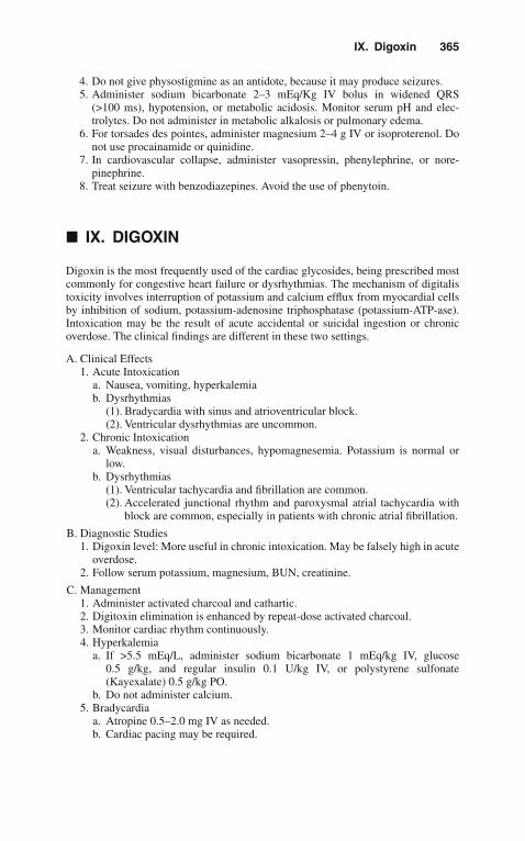
IX. Digoxin 365
4. Do not give physostigmine as an antidote, because it may produce seizures.5. Administer sodium bicarbonate 2–3 mEq/Kg IV bolus in widened QRS
(>100 ms), hypotension, or metabolic acidosis. Monitor serum pH and elec-trolytes. Do not administer in metabolic alkalosis or pulmonary edema.
6. For torsades des pointes, administer magnesium 2–4 g IV or isoproterenol. Donot use procainamide or quinidine.
7. In cardiovascular collapse, administer vasopressin, phenylephrine, or nore-pinephrine.
8. Treat seizure with benzodiazepines. Avoid the use of phenytoin.
� IX. DIGOXIN
Digoxin is the most frequently used of the cardiac glycosides, being prescribed mostcommonly for congestive heart failure or dysrhythmias. The mechanism of digitalistoxicity involves interruption of potassium and calcium efflux from myocardial cellsby inhibition of sodium, potassium-adenosine triphosphatase (potassium-ATP-ase).Intoxication may be the result of acute accidental or suicidal ingestion or chronicoverdose. The clinical findings are different in these two settings.
A. Clinical Effects1. Acute Intoxication
a. Nausea, vomiting, hyperkalemiab. Dysrhythmias
(1). Bradycardia with sinus and atrioventricular block.(2). Ventricular dysrhythmias are uncommon.
2. Chronic Intoxicationa. Weakness, visual disturbances, hypomagnesemia. Potassium is normal or
low.b. Dysrhythmias
(1). Ventricular tachycardia and fibrillation are common.(2). Accelerated junctional rhythm and paroxysmal atrial tachycardia with
block are common, especially in patients with chronic atrial fibrillation.
B. Diagnostic Studies1. Digoxin level: More useful in chronic intoxication. May be falsely high in acute
overdose.2. Follow serum potassium, magnesium, BUN, creatinine.
C. Management1. Administer activated charcoal and cathartic.2. Digitoxin elimination is enhanced by repeat-dose activated charcoal.3. Monitor cardiac rhythm continuously.4. Hyperkalemia
a. If >5.5 mEq/L, administer sodium bicarbonate 1 mEq/kg IV, glucose0.5 g/kg, and regular insulin 0.1 U/kg IV, or polystyrene sulfonate(Kayexalate) 0.5 g/kg PO.
b. Do not administer calcium.5. Bradycardia
a. Atropine 0.5–2.0 mg IV as needed.b. Cardiac pacing may be required.
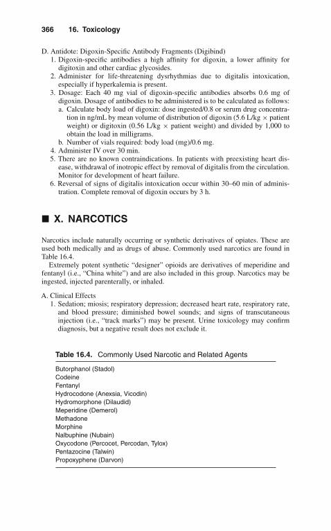
366 16. Toxicology
D. Antidote: Digoxin-Specific Antibody Fragments (Digibind)1. Digoxin-specific antibodies a high affinity for digoxin, a lower affinity for
digitoxin and other cardiac glycosides.2. Administer for life-threatening dysrhythmias due to digitalis intoxication,
especially if hyperkalemia is present.3. Dosage: Each 40 mg vial of digoxin-specific antibodies absorbs 0.6 mg of
digoxin. Dosage of antibodies to be administered is to be calculated as follows:a. Calculate body load of digoxin: dose ingested/0.8 or serum drug concentra-
tion in ng/mL by mean volume of distribution of digoxin (5.6 L/kg × patientweight) or digitoxin (0.56 L/kg × patient weight) and divided by 1,000 toobtain the load in milligrams.
b. Number of vials required: body load (mg)/0.6 mg.4. Administer IV over 30 min.5. There are no known contraindications. In patients with preexisting heart dis-
ease, withdrawal of inotropic effect by removal of digitalis from the circulation.Monitor for development of heart failure.
6. Reversal of signs of digitalis intoxication occur within 30–60 min of adminis-tration. Complete removal of digoxin occurs by 3 h.
� X. NARCOTICS
Narcotics include naturally occurring or synthetic derivatives of opiates. These areused both medically and as drugs of abuse. Commonly used narcotics are found inTable 16.4.
Extremely potent synthetic “designer” opioids are derivatives of meperidine andfentanyl (i.e., “China white”) and are also included in this group. Narcotics may beingested, injected parenterally, or inhaled.
A. Clinical Effects1. Sedation; miosis; respiratory depression; decreased heart rate, respiratory rate,
and blood pressure; diminished bowel sounds; and signs of transcutaneousinjection (i.e., “track marks”) may be present. Urine toxicology may confirmdiagnosis, but a negative result does not exclude it.
Table 16.4. Commonly Used Narcotic and Related Agents
Butorphanol (Stadol)CodeineFentanylHydrocodone (Anexsia, Vicodin)Hydromorphone (Dilaudid)Meperidine (Demerol)MethadoneMorphineNalbuphine (Nubain)Oxycodone (Percocet, Percodan, Tylox)Pentazocine (Talwin)Propoxyphene (Darvon)

XI. Phencyclidine 367
2. In significant overdose: coma, pinpoint pupils, severe respiratory depression,apnea.
3. Complications of overdose include rhabdomyolysis and noncardiogenic pul-monary edema.
4. Death is typically due to respiratory failure.
B. Management1. Maintain and assist ventilation as necessary.2. Antidote: Naloxone (Narcan)
a. Opioid antagonist, competitively blocks CNS opiate receptors.b. Administer 0.4–2.0 mg IV (may also be given IM, subcutaneously, intratra-
cheally). Repeat as needed. No response to total of 10 mg is evidence againstnarcotic overdose.
c. Duration of action is 1–4 h. Repeated administration may be required, oradminister as an infusion of 0.4–0.8 mg/h in 5% dextrose.
d. Use in opiate-dependent patients may produce narcotic withdrawalsyndrome.
e. Higher doses are frequently required with pentazocine and designer opioids:Begin with an initial dose of 4 mg.
3. If respiratory distress continues, monitor with chest radiograph and arterialblood gases (ABGs).
4. If noncardiogenic pulmonary edema (which may also be produced by naloxone)is present, treat according to the guidelines for treating adult respiratory distresssyndrome (ARDS) (see Chapter 13, “Pulmonary Disorders”).
� XI. PHENCYCLIDINE
Phencyclidine (PCP), formerly used as a legal anesthetic agent, is now an illicit drug.
A. Clinical Effects1. PCP produces hallucinations, alteration of mental status, and bizarre or violent
behavior. Clinical status tends to wax and wane, and severe symptoms maypersist for as long as 2 weeks.
2. The patient′s level of consciousness ranges from fully alert to comatose.3. The most common physical findings are nystagmus and hypertension. Pupils
may be dilated or miotic.
B. Medical Complications1. Complications may be due to direct effect of the drug or injury sustained during
intoxication.2. Major complications are indications for intensive care unit admission: seizures,
hyperthermia, rhabdomyolysis, and acute renal failure.
C. Diagnostic Studies1. Serum and urine PCP levels correlate poorly with clinical effects.2. Check urine for myoglobin, which may indicate rhabdomyolysis.
D. Management1. Management is largely supportive.2. PCP is frequently smoked, so gastric decontamination is not useful in these
cases. However, gastric lavage and activated charcoal may be indicated if largeamounts have been ingested. Do not induce emesis.

368 16. Toxicology
3. Hemodialysis and charcoal hemoperfusion are not effective in eliminating PCP.4. Acid diuresis may speed elimination but is rarely indicated.5. Severe agitation or violence: Utilize physical restraints. May administer
haloperidol 10 mg IM or IV or benzodiazepines.6. Seizures: If persistent, treat with IV benzodiazepines or propofol.
� XII. PHENYTOIN
Phenytoin toxicity may be due to acute overdose or chronic over-ingestion.
A. Clinical Effects1. Nausea, vomiting, lethargy, ataxia, agitation, irritability, hallucinations, and
seizures.2. Horizontal nystagmus is characteristic of overdose.3. At very high levels: coma, respiratory arrest.4. Cardiac toxicity occurs only with iatrogenic IV overdose, not with oral
ingestion.
B. Diagnostic Studies1. Phenytoin levels
a. Therapeutic: 10–20 mg/L.b. Levels >20 mg/L: nystagmus.c. Levels >30 mg/L: ataxia.d. Levels >40 mg/L: lethargy is common.
2. Serum Glucose: Hyperglycemia may occur.
C. Management1. Administer activated charcoal. Multiple doses of charcoal may enhance
elimination.2. Some recommend the use of charcoal hemoperfusion for severe intoxication.3. Remainder of treatment is supportive.4. Monitor cardiac rhythm in IV overdose.
� XIII. SALICYLATES
Salicylates are used for analgesic, antipyretic, and anti-inflammatory properties andare found in a variety of both prescription and over-the-counter preparations (i.e.,Alka-Seltzer, Ascriptin, Bufferin, Excedrin Extra Strength). Poisoning may be theresult of acute ingestion or chronic overdose.
A. Clinical Effects1. The clinical effects result from CNS respiratory stimulation, uncoupling of
oxidative phosphorylation, and interference with platelet function and bleedingtime.
2. Cerebral and pulmonary edema occur by uncertain mechanisms.3. Acute overdose
a. Tachypnea, tinnitus, vomiting, lethargy, respiratory alkalosis, metabolicacidosis
b. Severe: hypoglycemia, hyperthermia, seizures, coma, pulmonary edema

XIII. Salicylates 369
4. Chronic overdosea. Confusion, dehydration, metabolic acidosis. This presentation may mimic
sepsis.b. Pulmonary edema is more common than in acute overdose.
B. Diagnostic Studies1. In acute ingestion, obtain the salicylate level and plot it on a Done nomogram
(Figure 16.2).a. Multiple determinations may be necessary with sustained-release prepara-
tions. Obtain every 2–3 h for 12 h following ingestion.b. Usual therapeutic levels in arthritis patients: 100–300 mg/L (10–30 mg/dL).c. A nomogram is not as useful in chronic intoxication.
2. Follow ABGs, serum glucose and electrolytes, and chest radiographs.
C. Management1. Administer activated charcoal and cathartic.2. Monitor for development of pulmonary edema.3. Treat metabolic acidosis with IV sodium bicarbonate 1 mEq/kg to maintain pH
at 7.40–7.50.4. Rehydrate with IV crystalloid solution if dehydration has resulted from vomit-
ing or hyperventilation.5. Urinary alkalinization enhances excretion
200180
150
120
MILD
ASYMPTOMATIC
10090807060
50
40
30
20
Sal
icyl
ate
Pla
sma
Con
cent
ratio
n (m
g/dL
)
106 12 24 36
Hours After Ingestion48 60
200180
150
120
10090807060
50
40
30
20
10
SEVERE
MODERATE
Figure 16.2. A Done nomogram for acute salicylate poisoning. (Done AK:Salicylate intoxication: significance of measurements of salicylate in blood incases of acute ingestion. Pediatrics 1960;26:800–807.)
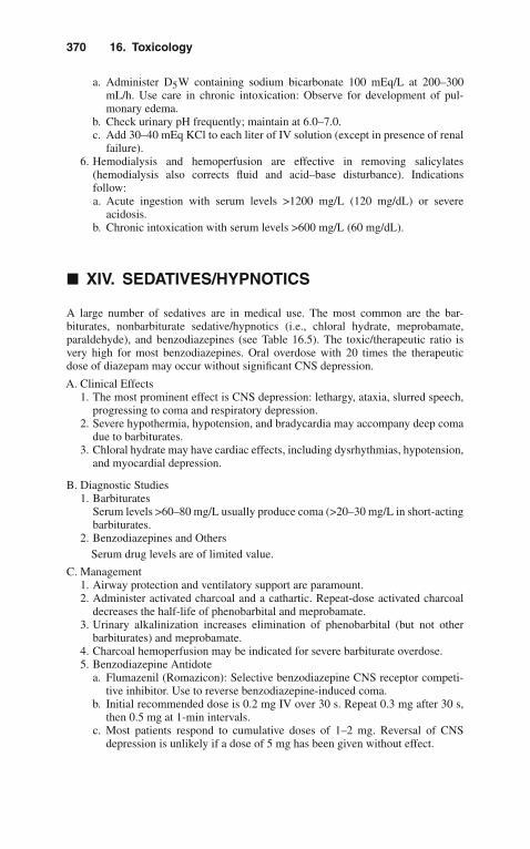
370 16. Toxicology
a. Administer D5W containing sodium bicarbonate 100 mEq/L at 200–300mL/h. Use care in chronic intoxication: Observe for development of pul-monary edema.
b. Check urinary pH frequently; maintain at 6.0–7.0.c. Add 30–40 mEq KCl to each liter of IV solution (except in presence of renal
failure).6. Hemodialysis and hemoperfusion are effective in removing salicylates
(hemodialysis also corrects fluid and acid–base disturbance). Indicationsfollow:a. Acute ingestion with serum levels >1200 mg/L (120 mg/dL) or severe
acidosis.b. Chronic intoxication with serum levels >600 mg/L (60 mg/dL).
� XIV. SEDATIVES/HYPNOTICS
A large number of sedatives are in medical use. The most common are the bar-biturates, nonbarbiturate sedative/hypnotics (i.e., chloral hydrate, meprobamate,paraldehyde), and benzodiazepines (see Table 16.5). The toxic/therapeutic ratio isvery high for most benzodiazepines. Oral overdose with 20 times the therapeuticdose of diazepam may occur without significant CNS depression.
A. Clinical Effects1. The most prominent effect is CNS depression: lethargy, ataxia, slurred speech,
progressing to coma and respiratory depression.2. Severe hypothermia, hypotension, and bradycardia may accompany deep coma
due to barbiturates.3. Chloral hydrate may have cardiac effects, including dysrhythmias, hypotension,
and myocardial depression.
B. Diagnostic Studies1. Barbiturates
Serum levels >60–80 mg/L usually produce coma (>20–30 mg/L in short-actingbarbiturates.
2. Benzodiazepines and OthersSerum drug levels are of limited value.
C. Management1. Airway protection and ventilatory support are paramount.2. Administer activated charcoal and a cathartic. Repeat-dose activated charcoal
decreases the half-life of phenobarbital and meprobamate.3. Urinary alkalinization increases elimination of phenobarbital (but not other
barbiturates) and meprobamate.4. Charcoal hemoperfusion may be indicated for severe barbiturate overdose.5. Benzodiazepine Antidote
a. Flumazenil (Romazicon): Selective benzodiazepine CNS receptor competi-tive inhibitor. Use to reverse benzodiazepine-induced coma.
b. Initial recommended dose is 0.2 mg IV over 30 s. Repeat 0.3 mg after 30 s,then 0.5 mg at 1-min intervals.
c. Most patients respond to cumulative doses of 1–2 mg. Reversal of CNSdepression is unlikely if a dose of 5 mg has been given without effect.

XV. Theophylline 371
Table 16.5. Commonly Used Sedative/Hypnotic Agents
BarbituratesShort-acting
Secobarbital (Seconal)Pentobarbital (Nembutal)
Intermediate-actingAmobarbital (Amytal)Apobarbital (Alurate)Butabarbital (Butisol)
Long-actingPhenobarbital
NonbarbituratesChloral hydrateEthchlorvynol (Placidyl)Meprobamate (Equagesic, Equanil, Miltown)Paraldehyde
BenzodiazepinesUltrashort-acting
Midazolam (Versed)Temazepam (Restoril)Triazolam (Halcion)
Short-actingAlprazolam (Xanax)Lorazepam (Ativan)Oxazepam (Serax)
Long-actingChlordiazepoxide (Librium)Chlorazepate (Tranxene)Diazepam (Valium)Flurazepam (Dalmane)Prazepam (Centrax)
d. Adverse effects: Nausea and vomiting are most common. In patients depen-dent on benzodiazepines, flumazenil may induce withdrawal syndrome (agi-tation, tachycardia, seizures). Do not use in patients who have concomitantTCA overdose or who are taking chronic TCAs.
e. The duration of action of a single dose is 1–2 h. If prolonged reversal isneeded, give repeated doses or administer as an IV infusion of 0.1–0.5 mg/h.
� XV. THEOPHYLLINE
The mechanism of theophylline toxicity is via release of endogenous catecholamines,stimulation of beta2-receptors, and inhibition of adenosine receptors. Clinical effectsmay be delayed for several hours following acute ingestion if a sustained-releaseformulation is involved. Toxicity may be acute or chronic.
372 16. Toxicology
A. Clinical Effects1. Common toxic effects include nausea, vomiting, tremor, tachycardia, and
hypotension.2. Hypokalemia, hyperglycemia, and metabolic acidosis may occur (only in acute
intoxication).3. Seizures and ventricular dysrhythmias may occur, especially with very high
serum levels and with chronic intoxication.
B. Diagnostic Studies1. Serum theophylline level.
a. Therapeutic: 15–20 mg/Lb. In acute overdose with level >100 mg/L, seizures, hypotension, and ventric-
ular dysrhythmias are common. Seizures may occur at levels of 40–60 mg/Lin chronic overdose. Repeat levels q2–4 h during treatment and monitor for12–16 h.
2. Monitor serum pH, glucose, and potassium levels.
C. Management1. Administer activated charcoal 100 g and re-administer 20–30 g q2–3 h.2. Treat seizures and ventricular dysrhythmias in standard fashion as required.
Magnesium has been successful in some cases. For supraventricular tachycardiaor rapid sinus tachycardia, ventricular dysrhythmias, or hypotension, administeresmolol 0.05 mg/kg/min or propranolol 0.01–0.03 mg/kg IV. Use with cautionif wheezing is present.
3. If theophylline level >100 mg/L or seizures or dysrhythmias do not respond totreatment, institute charcoal hemoperfusion.
� XVI. USEFUL FACTS AND FORMULAS
A. Basic Formulas. The therapeutic index (TI) of a drug can be calculated as follows:
TI = LD50
ED50
where LD50 = median lethal dose; ED50 = median effective dose.The margin of safety (MS) of a drug uses the ED99 for the desired effect and
the LD1 for the undesired effect:
MS = LD1
ED99
The apparent volume of distribution (Vd) can be calculated by the followingequation:
Vd = Doseiv
C0

XVI. Useful Facts and Formulas 373
where Doseiv = the IV dose; C0 = the extrapolated plasma concentration at timezero.
For those agents that follow a two-compartment model, several Vd exist and thefollowing formula is used to assess the volume of the central compartment (Vc):
Vc = Doseiv
A + B
where A and B represent disposition constants of a two-compartment model.In addition, the peripheral compartment (Vp) can be calculated as follows:
Vp = Doseiv
B
where B is derived from the elimination or equilibrium phase of a two-compartment model.
The total body clearance (Cl) of a drug can be calculated as the sum ofclearances by individual organs:
Cl = Clr + Clh + Cli + . . .
where Clr = renal clearance; Clh = hepatic clearance; Cli = intestinal clearance.
B. Osmolality Formulas. To calculate serum osmolality, the following formula isusually applied:
Calc. Osmolality (mOsm/kg) = 2Na + BUN/2.8 + Glucose/18
The osmolal gap (OG) is useful in several intoxications and is calculated asfollows:
OG = Measured osmolality − calculated osmolality
To calculate the contribution to measured osmolality of alcohols (also knownas osmol ratios), the alcohol concentration (mg/dL) is divided by the numbersdepicted in Table 16.6.
Table 16.6. Osmolal Ratios of Different Alcohols
Ethanol Ethylene glycol Isopropanol Methanol
4.6 6.2 6.0 3.2

374 16. Toxicology
C. Digitalis Intoxication. To treat digitalis poisoning appropriately, it is important toassess the digitalis body load:
Body load (mg) = (serum digoxin concentration)×5.6 × (bodyweightinkg) ÷ 100
The dose of digitalis antibodies (Digibind) is determined by dividing the bodyload by 0.6 mg/vial:
Dose (number of vials) = Body load (mg) + 0.6 (mg/vial)
17Trauma
Trauma is the leading cause of death in persons aged 1–44 years and the fourth lead-ing cause of death overall. About 140,000 traumatic deaths occur annually. Sepsis,adult respiratory distress syndrome, and multiple organ system failure are the leadingcause of death in trauma patients who survive the initial resuscitation and surgicalrepair of their injuries.
� I. MULTISYSTEM TRAUMA
A. Establishment of Priorities1. Of highest priority in the initial evaluation are
a. Airway maintenanceb. Breathing and ventilationc. Circulation and shock management
2. Secondary evaluation includes vital signs, complete physical (including rectal)examination. Nasogastric and urinary catheters (unless contraindicated) shouldgenerally be inserted to diagnose gastric or urinary tract hemorrhage and toallow for monitoring of urinary output.
3. Rapid normalization of vital signs is one of the goals in trauma management.
B. Severity of Injury Scoring Systems1. The Glasgow Coma Scale (see Table 17.1) is used for assessing neurologic
status in head injury.2. The Trauma Score (see Table 17.2) estimates physiologic severity of injury. It
combines the Glasgow Coma Scale with other clinical indices of cardiovascularand pulmonary function.
C. Airway Management1. Clear airway of debris or secretions2. Avoid chin lift and neck lift/tilt if cervical spine injury is considered. Obtain
cervical spine radiographs as soon as possible.
D. Oxygenation and Ventilation1. If adequate respirations appear to be present, obtain baseline blood gases as
soon as possible. Apply 100% O2 by mask. If no spontaneous respirations,assist with bag and mask.
J. Varon, P. Acosta, Handbook of Critical and Intensive Care Medicine,DOI 10.1007/978-0-387-92851-7_17,C© Springer Science+Business Media, LLC 2010

376 17. Trauma
Table 17.1. Glasgow Coma Scale
SCORE
Eye openingSpontaneous 4To verbal command 3To pain 2None 1
Best motor responseObeys verbal command 6Localizes painful stimuli 5Flexion withdrawal from painful stimuli 4Decorticate (flexion) response to painful stimuli 3Decerebrate (extension) response to painful stimuli 2None 1
Best verbal responseOriented conversation 5Disoriented conversation 4Inappropriate words 3Incomprehensible sounds 2None 1
Total Score 3–15
2. Endotracheal Intubationa. When necessary, intubation should usually be done by the oral route with
manual in-line stabilization.b. Rapid-sequence induction is often indicated in patients with major trauma,
head or facial injury, diminished level of consciousness, respiratory impair-ment: Preoxygenation, application of cricoid pressure, administration ofinduction agents (vecuronium 0.2 mg/kg IV, followed 3 min later bysuccinylcholine 1.0–1.5 mg/kg; succinylcholine is contraindicated in pen-etrating eye injuries, massive crush injury), followed by intubation.
3. When a surgical airway is necessary, cricothyrotomy is the preferred procedure.4. Most common causes for respiratory compromise: tension pneumothorax, open
pneumothorax, flail chest with pulmonary contusion.
E. Circulation and Shock Management1. Evaluation includes assessment of vital signs, level of consciousness, skin color,
character of pulse, and capillary refill.2. Shock in trauma is most commonly due to hypovolemia.
a. Likely sites for occult hemorrhage: thorax, abdomen, pelvis, retroperi-toneum, thigh.
b. In addition, conditions producing shock that should be considered includetension pneumothorax, cardiac tamponade, myocardial contusion, spinaltrauma.
3. Classification of hemorrhagic shock: see Table 17.3.4. Treatment

I. Multisystem Trauma 377
Table 17.2. Trauma Score
Points
Respiratory rate/minute10–24 425–35 3>35 2<10 10 0
Respiratory effortNormal 1Shallow or retractive 0
Systolic blood pressure>90 mmHg 470–90 mmHg 350–69 mmHg 2<50 mmHg 10 0
Capillary refillNormal 2Delayed 1None 0
Glasgow coma score14–15 511–13 48–10 35–7 23–4 1
Total –
a. Class I hemorrhage: replacement of primary fluid loss with electrolytesolution.
b. Class II hemorrhage: initial stabilization with intravenous (IV) fluids; mayrequire blood transfusion.
c. Class III hemorrhage: almost always requires transfusion.5. The patient should have at least two large-caliber (14- to 16-gauge or larger) IV
lines established. Initial fluid administration should be with isotonic electrolytesolution.a. The administration rate should be commensurate with the clinical condition
and vital signs. Fluid overload should be avoided, but adequate intravascularvolume, hematocrit, and tissue perfusion must be achieved.
b. In the hypovolemic patient, at least 2 L (20 mL/kg in a child) can be rapidlyinfused and the patient reassessed.
6. If shock persists despite resuscitation with IV fluids, blood replacement isindicated.a. Maintain a hematocrit of at least 30%.b. Cross-matched blood is preferable if the patient’s condition permits.

378 17. Trauma
Table 17.3. Classification of Hemorrhage
Blood volume lost Clinical signs
Class I Up to 15% Increased heart rate
Class II 15–30% Increased heart rate, decreasedpulse pressure, minor delay incapillary refill, anxiety
Class III 30–40% Increased heart rate, decreasedblood pressure, delayed capillaryrefill, clouded
Class IV >40% Markedly increased heart rate,sensorium decreased bloodpressure, negligible urine output,markedly depressed mentalstatus, skin cold and pale
c. If there is insufficient time for a full cross-match to be performed, type-specific (ABO and Rh compatible) blood should be administered.
d. In patients in severe, life-threatening shock for whom type-specific blood isnot available, administer type O blood (Rh negative in women of childbear-ing age). Subsequent cross-matching may be more difficult, however.
e. Autotransfusion (especially administration of autologous blood from chesttube drainage) should be employed when feasible.
7. Monitor for possible complications of transfusion.a. Hemolytic transfusion reaction: fever, chills, and chest, back, and joint pain.
Terminate transfusion, administer IV fluids and furosemide. Monitor urineoutput.
b. Hypothermia may follow massive transfusion with refrigerated blood. Giveblood through a warmer if possible. Monitor body temperature with a coreprobe.
c. Coagulopathy may result following massive transfusion, probably on thebasis of quantity and function of platelets, as well as consumption ofcoagulation factors. Administration of platelets is advised following rapidtransfusion of each 10 U of blood. If evidence of coagulopathy exists,consider administration of fresh-frozen plasma and cryoprecipitate.
d. Banked blood is acidemic and high in potassium and preservative anticoagu-lant binds calcium. Monitor serum pH, potassium, magnesium, and calciumlevels.
8. Monitor urine output: Volume replacement should produce urine output of atleast 1–1.5 mL/kg/h.
F. Complications of Hemorrhagic Shock and Volume Resuscitation. Peripheraledema, hypothermia, cerebral edema, cardiac dysfunction (usually right ventricu-lar failure), pneumonia, adult respiratory distress syndrome (ARDS), multisystemorgan failure.
G. Cardiac Arrest1. Perform immediate thoracotomy in patients in extremes, especially with pene-
trating chest trauma.
II. Head Trauma 379
2. Therapeutic objectives include relief of cardiac tamponade, open cardiac mas-sage, control of cardiac injuries, vascular control of major vessel or hilarinjuries, and aortic occlusion for treatment of shock.
� II. HEAD TRAUMA
Head injury is a major entity, often encountered in acute care, the head being the mostfrequently injured part of the body in trauma patients. Over 80,000 persons annuallysustain permanent disabling injuries of the head or spinal cord.
A. Assessment1. History
a. Important components are mechanism of injury and loss of conscious-ness. High-speed trauma (e.g., with ejection from vehicle, impact withwindshield) produces a greater chance of significant injury.
b. Incomplete recollection by the patient of details of injury may imply a tran-sient loss of consciousness. This symptom is not as useful if the patient isintoxicated.
2. Physical Examinationa. Examine the scalp and face for signs of trauma, such as lacerations, ecchy-
moses, hemotympanum, and bleeding or clear fluid from the nostrils orears.
b. Palpate the spine for tenderness or deformity. Always consider the possibilityof concomitant spinal cord injury.
c. Focus on other injuries that affect the airway or produce respiratory orcirculatory impairment.
3. Neurologic Examinationa. This is the best tool for identifying the presence of significant intracranial
injury.(1). Assess the mental status (most important aspect of the neurologic
examination).(2). Determine focal neurologic deficit, abnormal posturing, and pathologic
reflexes. Evaluate brain stem reflexes (light, corneal, gag) and ventilatorydrive.
(3). The absence of brain stem function usually indicates the need for urgentairway intervention.
b. Frequent repetition of neurologic examination is necessary, especially withinthe first 48 h of injury.
4. Glasgow Coma Scale (GCS)a. Determine the GCS score (see Table 17.1).
(1). Score of 13–15 = mild injury(2). Score of 9–12 = moderate injury(3). Score of 8 or less = severe injury
b. GCS is of limited usefulness in children <3 years.
B. Management1. Position
When associated injuries permit, elevate the head of the bed 30–45◦, as thisreduces intracranial pressure.

380 17. Trauma
2. Airways and Ventilationa. The highest initial priority is prevention or reduction of secondary injury due
to swelling or compression of cerebral tissue by cerebral edema and raisedintracranial pressure (ICP).
b. Proper airway management reduces increased ICP. Diminished mental sta-tus (particularly a GCS score < 9) is an indication for early endotrachealintubation.
c. Chin lift and neck lift maneuvers are inappropriate if the patient with headinjury is suspected of having cervical spine injury.
d. Initial management of increased ICP includes hyperventilation.(1). It is often recommended to maintain arterial pCO2 between 25 and
30 torr (mmHg), but there is no uniform agreement on this.(2). Many recommend not to hyperventilate patients with pCO2 < 20 torr or
pH >7.60.e. Endotracheal intubation.
(1). Nasal intubation is relatively contraindicated; use orotracheal intubationwith manual in-line stabilization.
(2). Precede intubation with bag-valve-mask ventilation, cricoid pressure,and pharmacologic induction: Etomidate 0.3 mg/kg IV and succinyl-choline 1.0–1.5 mg/kg (watch out for patients with renal failure orhyperkalemia, as may worsen it), or vecuronium 0.1–0.2 mg/kg IV.
f. When mechanical ventilation is instituted, avoid high levels (>10 cm) ofpositive end-expiratory pressure (PEEP), as this may increase ICP. Chestphysiotherapy may also increase ICP.
3. Osmotic Therapy and Diuresisa. Reduce increased ICP by reducing intracranial volume.b. Mannitol is the osmotic diuretic of choice. It is generally administered in
rapidly deteriorating patients. It is often used to arrest neurologic deteriora-tion when the patient is being prepared for urgent craniotomy.(1). Give as a 20% solution, 0.25–1.0 g/kg via rapid IV infusion.(2). ICP reduction usually occurs within 10–20 min.(3). Duration is limited to 2–6 h following initial bolus. Continuous infusion
may be required.(4). Monitor blood pressure, serum electrolytes, and osmolarity.
c. Diuretics may be used alone or in combination with osmotic diuretics toreduce intracerebral fluid volume.(1). Furosemide is the loop diuretic of choice. Administer an IV bolus of
1 mg/kg. The onset of action is slower than mannitol, but concomitantuse enhances the duration of ICP reduction by mannitol and decreasesthe risk of rebound ICP elevation. Repeated doses may be required.
(2). Acetazolamide is a carbonic anhydrase inhibitor. It decreases cere-brospinal fluid (CSF) production. Administer 250 mg qid. Monitor forproduction of acidosis.
d. Corticosteroids are frequently administered, but whether they diminish ICPin head trauma is controversial.
e. Therapeutic hypothermia has been used in these patients with conflictingresults. In the author’s experience, this is a beneficial therapeutic modality.
4. Cardiovascular Supporta. As a rule, cerebral injury does not produce hypotension (except in the agonal
state).b. Look for an extracranial source if hypotension is present.

III. Crush Injury 381
c. Avoid fluid overload, but provide adequate intravascular volume and hemat-ocrit. A pulmonary artery catheter may be required for monitoring.
C. Monitoring1. Intracranial Pressure
a. Indicated in patients with severe head injury with computed tomography(CT) evidence of raised ICP. Monitoring of ICP, however, has not beenproven to affect survival.
b. Monitor by intraventricular catheter, subarachnoid bolt, or extradural pres-sure sensor.
c. It is generally advised to maintain ICP at ≥20 mmHg.d. Cerebral perfusion pressure (CPP): monitor this (CPP = mean arterial pres-
sure [MAP] – ICP) and maintain at > 60 mmHg. If hypotension occurs,elevate arterial pressure to maintain CPP above this level, as ischemicdamage may occur.
2. Intravascular PressureArterial catheterization to allow monitoring of MAP as well as frequent bloodgas determinations.
3. Head trauma may produce syndrome of inappropriate secretion of antidiuretichormone (SIADH). This results in hyponatremia with a relatively concentratedurine (see also Chapters 9, “Neurologic Disorders” and 14, “Renal and Fluid-Electrolyte Disorders”).
D. Diagnostic Studies1. Skull Radiographs
a. Plain skull radiographs may demonstrate skull fracture but have poorsensitivity and specificity for identifying intracranial lesions.
b. May be useful in children < 2 years of age, in whom skull fracture mayidentify risk of hypovolemia due to extracranial bleeding, formation ofleptomeningeal cyst, and child abuse.
2. Computed Tomographya. CT is the diagnostic procedure of choice in assessing acute head injury.
Perform CT without contrast material. CT is indicated in the patient witha decreased level of consciousness (GCS score ≤14), deteriorating mentalstatus, focal neurologic deficit, seizures, or persistent vomiting.
b. If the GCS score is < 9, obtain CT immediately after endotracheal intubationwhen the patient is hemodynamically stable.
3. Magnetic Resonance Imaging (MRI)a. MRI is superior to CT in diffuse axonal injury.b. Limitations in acute injury: duration of scanning, interference of monitoring
and life support equipment with magnetic field.4. Ultrasound
May be an option in smaller children with suspected intraventricularhemorrhage. In addition, vascular blood flow calculations may be useful inthe management of these patients.
� III. CRUSH INJURY
A. Traumatic Asphyxia1. Mechanism: direct massive thoracoabdominal compression.

382 17. Trauma
2. Clinical findings: blanching cyanosis of upper chest, neck and head; petechiae;edema; subconjunctival hemorrhage.
3. Associated injuries: chest wall injury, pulmonary contusion, cardiac contusion,diaphragm rupture, intraabdominal solid and hollow viscus injury.
4. Sequelae: brachial plexus injury, spinal cord injury, transient neurologic impair-ment.
B. Abdominal and Pelvic Injury1. Crush injury accounts for 5% of pelvic fractures. May result in bladder
laceration.2. Crush mechanism to the abdomen results in a high proportion of hollow viscus
injury.
C. Skeletal Muscle Injury1. Results in myonecrosis. May produce rhabdomyolysis, hyperkalemia, hyper-
phosphatemia, hypocalcemia, and myoglobinuria.2. Sequelae include acute renal failure, disseminated intravascular coagulation.3. Follow creatine kinase (CK), electrolytes, creatinine, urine output.4. CK levels reach maximum 24–36 h after injury. Level should decline by 50%
each 48 h thereafter. If there is an increase in CK during this time, considerrecurrence of muscle necrosis.
5. Treat acute renal failure with fluid infusion, osmotic diuresis, alkalinization (seeChapter 14, “Renal and Fluid-Electrolyte Disorders”).
� IV. CHEST TRAUMA
Chest trauma is the cause of death in up to a quarter of cases of multisystem trauma.Injury may occur to the chest wall, lung, great vessels, and mediastinal viscera.
Most injuries can be initially managed with chest tube insertion and othernonoperative management.
Indications for thoracotomy include cardiac tamponade, massive hemothorax (seebelow); pulmonary air leak >15–20 L/min; aortic arch, esophageal, tracheal, or majorbronchial disruption; systemic air embolism; bullet embolism; cardiac arrest.
A. Chest Wall Trauma1. Rib fracture is the most common chest wall injury. It is an important indicator
of underlying injury.a. First to Third Ribs: great vessel and bronchial injury. Diminished pulse or
blood pressure in arms or radiographic evidence of mediastinal hematoma(see below) are indications for arteriography.
b. Lower Ribs: kidney, liver, spleen laceration.2. Flail chest occurs when three or more ribs are fractured in two places or when
multiple fractures are associated with sternal fracture.a. Clinical significance varies, depending upon the size and location of the flail
segment and the extent of underlying pulmonary contusion.b. Obtain and follow arterial blood gases (ABGs).c. Splint thorax with weights, traction, or skeletal fixation.d. Patients with severe hypoxemia will require endotracheal intubation and
positive-pressure ventilation. Observe for late development of pneumotho-rax, especially tension pneumothorax, in the mechanically ventilated patient.

V. Abdominal Trauma 383
3. Sternal fracture is associated with myocardial contusion, cardiac rupture andtamponade, and pulmonary contusion. Early surgical fixation is often necessary,and urgent surgery may be indicated when costosternal dislocations producecompromise of the trachea or neurovascular structures at the thoracic inlet.
4. Analgesic methods that may be required for treatment of major chest wallinjuries include parenteral opiates, epidural analgesia, and intercostal nerveblock.
B. Pneumothorax1. Usually results from penetrating trauma or blunt trauma with rib fracture. May
be caused by positive-pressure ventilation (barotrauma).2. Presence of pneumothorax requires 28–40 Fr chest tube insertion. Smaller tubes
may be utilized if pneumothorax is not accompanied by hemothorax.3. Open pneumothorax requires covering of chest wall injury with airtight dress-
ing and insertion of chest tube.4. Tension pneumothorax requires immediate needle decompression and chest
tube insertion. Clinical findings include unilateral absence of breath sounds,severe dyspnea, tracheal shift, jugular venous distention, cyanosis.
C. Hemothorax1. Initial treatment requires insertion of chest tube to evacuate hemothorax,
reexpand lung, and monitor rate of bleeding.2. Indications for thoracotomy include initial chest tube drainage of >1,500 mL or
continued bleeding of more than 300 mL/h for more than 2–3 h.
D. Major Vessel Injury1. Common cause of death in major trauma.2. Consider when there is radiographic evidence of mediastinal hematoma:
widened mediastinum, aortic knob obliteration, tracheal or nasogastric tubedeviation.
3. Arteriography or CT is required for diagnosis.
E. Cardiac Tamponade1. Most frequently occurs with penetrating injures. Suspect in chest trauma with
shock and jugular venous distention.2. Requires thoracotomy and pericardial decompression. Pericardiocentesis may
be performed if the diagnosis is uncertain or as a temporizing measure duringpreparation for thoracotomy.
F. Pulmonary Contusion. Management consists of supplemental oxygen administra-tion and mechanical ventilation with the addition of PEEP, if indicated in patientswith worsening hypoxemia.
G. Myocardial Contusion. Management consists of cardiac monitoring, echocardio-graphy, and treatment of dysrhythmias, as necessary.
� V. ABDOMINAL TRAUMA
A. Evaluation1. Physical findings may be unreliable if abdominal trauma is complicated by head
or other injury, or intoxication.
384 17. Trauma
2. Findings most consistently associated with internal abdominal injury areabdominal tenderness and guarding.
3. Examine thorax for rib fractures, palpate flanks and pelvis, and perform rectaland pelvic examination.
4. Obtain baseline hemogram, blood coagulation screen, and urinalysis.
B. Diagnostic Peritoneal Lavage (DPL)1. There is considerable variation in use of DPL from institution to institution.2. Indications may include equivocal abdominal findings, possible abdominal
injury in the face of diminished sensation due to head or spinal injury, or alcoholintoxication.
3. The major advantage is the ability to obtain a rapid indication of intraperitonealhemorrhage.
4. Relative contraindications: previous abdominal surgery, significant obesity,pregnancy, preexisting coagulopathy.
5. Positive lavage consists of aspiration of >10 mL of blood, aspiration of entericcontents, or lavage fluid with red blood cell levels >100,000/mm3, white bloodcells >500/mm3, amylase ≤20 IU/L, or bile.
C. Abdominal CT1. Indicated in stable patients with possible intraabdominal injury, and where DPL
is being considered but is contraindicated.2. Advantages include ability to visualize urinary tract and retroperitoneum.
Abdominal Ultrasound: Useful to detect free intraperitoneal blood after blunttrauma.
D. Penetrating Injury1. Antibiotics
a. Second- or third-generation cephalosporin, e.g., cefoxitin 2 g IV q6 h, orceftazidime 1–2 g IV q8 h, or
b. Combination of gentamicin (1.5–2.0 mg/kg IV loading and 3 mg/kg/d in3 maintenance doses) or tobramycin (l mg/kg q8 h IV) and clindamycin600–900 mg q8 h IV
E. Indications for Laparotomy1. Gunshot wounds.2. Stab wounds with shock, signs of peritoneal irritation, gastrointestinal bleeding,
or evisceration of bowel.3. Blunt trauma with unstable vital signs, gastrointestinal bleeding, peritoneal
irritation, pneumoperitoneum, evidence of diaphragmatic injury.4. There may be a role for laparoscopy in the stable patient with penetrating injury,
but this method has not been used extensively in this setting.
F. Postoperative Complications1. Intraabdominal hemorrhage
a. May be due to recurrent bleeding from sites not identified during surgery dueto hypotension.
b. Identify hemostasis deficiencies (thrombocytopenia, clotting dysfunction),especially if patient has had massive transfusions.
c. If prothrombin time (PT) and partial thromboplastin time (PTT) are pro-longed, administer fresh-frozen plasma.
2. Fever: Consider wound infection, necrotizing fascitis, peritonitis, and intraab-dominal abscess.

VI. Multiple Fractures 385
3. Missed intraabdominal injury: diaphragm, biliary tree, duodenum, pancreas,ureter, colon, and rectum.
G. Nonoperative Management1. May be suitable in patients who remain hemodynamically stable following ini-
tial resuscitation with 1–2 L of IV fluids. Laparoscopic repair may be anotheroption.a. Normal vital signs, urine output >1.0–1.5 mL/h, and no blood requirement.b. Patient must be alert.c. No coagulation defects.
2. CT should establish extent of injury.3. Suitable injuries include
a. Isolated splenic trauma with minor capsular tear or parenchymal injuryb. Stab wounds without shock, peritoneal irritation, or gastrointestinal bleeding
4. Monitoringa. Repeat abdominal examination for signs of peritoneal irritation at least every
4–6 h.b. Vital signs monitored every 1–2 h during the first 24 h.c. Serial hematocrit every 4 h. Also monitor amylase.d. Repeat abdominal CT after 12 h of observation and thereafter as indicated
by clinical signs.
H. Urinary Tract Injury1. Evaluate with IVP or abdominal CT, if gross hematuria, flank hematoma or
mass, penetrating trauma with suspected urinary injury.2. Bladder and urethral injury: see pelvic fracture.
� VI. MULTIPLE FRACTURES
A. General Considerations1. Identify fractures and dislocations, and assess distal neurocirculatory function.2. Complications of fractures include arterial and neural injury, hemorrhage, com-
partment syndrome, ARDS, fat embolization, infection, and thromboembolism.3. Fractures of the pelvis and femur are particularly significant because of
hemorrhage potential.4. Dislocations of the hip and knee require prompt reduction to avoid neurovascu-
lar complications.
B. Initial Management1. Immobilize any injured extremity.2. Stabilize femur fracture in Hare or comparable traction device.
C. Arterial Injury1. Arterial injury may be due to transection, arterial spasm, occlusion by
hematoma, external compression, arteriovenous fistula formation.2. Acute loss of vascular function requires emergent surgical exploration or
angiography.
D. Compartment Syndrome1. Circulatory supply can be lost due to increased muscular compartment pressure.
Most common in leg but also occurs in forearm.

386 17. Trauma
2. Earliest sign is pain with passive stretching of the extremity. Suspect if there issevere, constant pain despite reduction and immobilization.
3. Measure compartment pressure by inserting a needle connected to a manomet-ric pressure measurement system into the soft tissue of the extremity involved;should be <30 mmHg in the normotensive pressure.
4. Treatment is fasciotomy.
E. Infection in Open Injuries1. Tetanus prophylaxis should be given, especially for wounds with deep tissue
penetration, devitalized tissue, and burns.2. IV antibiotics.
a. IV first-generation cephalosporin (e.g., cefazolin 1 g IV q6 h). Some authorsprefer broader coverage.
b. If the wound is large or heavily contaminated, add gentamicin(1.5–2.0 mg/kg IV loading dose and 3 mg/kg/d in 3 maintenance doses) ortobramycin (1 mg/kg q8 h IV or 3 mg/kg IV single dose) if the patient hasnormal renal function.
3. Observe for appearance of gas gangrene (appearance of subcutaneous crepita-tion or soft tissue gas on radiographs).a. Occurs within 12–72 h of injury and requires broad-spectrum antibiotics and
aggressive wound debridement.b. Most often occurs in grossly contaminated open fractures with soft tissue
damage.
F. Fat Embolism1. Occurs most commonly in multiple fractures, especially involving femur, tibia,
pelvis.2. Onset of symptoms occurs 1–5 days following injury: tachypnea, hemoptysis,
fever, petechiae, mental status change.3. Hypoxemia (PaO2 <60 mmHg on room air) is the most consistent finding.4. Treatment includes correction of hypoxemia and ventilatory support with
PEEP used if arterial oxygenation cannot be maintained. Management is sim-ilar to that of ARDS (see Chapter 13, “Pulmonary Disorders”). Maintaina negative fluid balance. A pulmonary artery catheter may be required formanagement.
G. Pelvic Fracture. Pelvic fractures caused by high-energy forces have signifi-cant mortality. Hemorrhage is a major cause, as are associated injuries, sepsis,multiple-organ failure, and ARDS.Infection of pelvic hematoma may occur even in closed injuries due to hematoge-nous spread of bacteria.1. Evaluation
a. Physical Examination(1). Palpate pelvis to determine tenderness and instability.(2). Perform pelvic and rectal examination to ascertain open injury and
sphincter tone.(3). Blood at urethral meatus or high-riding prostate on rectal examination
indicates urethral tear.b. Radiographs
(1). Obtain pelvic radiographs in patients suspected of sustaining pelvicfracture and in patients with multiple trauma.
(2). CT is superior in demonstrating certain aspects of pelvic injury. Utilizein the stable patient.

VII. Spinal Cord Injury 387
2. Hemorrhagea. Usually retroperitoneal, the result of venous injury to pelvic venous plexus.
May be massive, exceeding several liters.b. Signs of persistent bleeding are an indication for external fixation of unstable
pelvic fractures.c. Application of pneumatic antishock trousers may be helpful in temporar-
ily controlling hemorrhage (although there is considerable controversy overefficacy). Once antishock trousers are in place, do not remove until vascularaccess is established and blood pressure stabilized.
d. Consider arteriography and selective embolization for patients with contin-ued severe hemorrhage.
3. Associated Injuriesa. Injuries to consider: urinary bladder perforation, vaginal or rectal laceration,
posterior urethral tear.b. If signs of urethral injury are present, a retrograde urethrogram should be
used to ascertain integrity of the urethra before urinary catheter insertion.
� VII. SPINAL CORD INJURY
Spinal injury should be considered in any patient with multisystem injury, head orfacial injury, and those who are unconscious. Maintain spinal immobilization untilradiographs exclude spinal injury.
A. Evaluation1. Perform a neurologic examination to determine the extent and level of injury.
a. Examine and document motor function and sensory level.b. Determine if there is sacral sensory sparing or anal sphincter contraction,
signs that cord injury is incomplete.c. Sacral reflexes (anal wink, bulbocavernosus reflex) are the first to return after
spinal shock, usually within 24 h following injury.
B. Management1. Endotracheal Intubation
a. Patients may require endotracheal intubation because of paralysis of respira-tory muscles.
b. If intubation is necessary, it should be done with in-line cervical immobiliza-tion.
c. The orotracheal approach is preferred, preceded by 100% oxygenation bymask and application of gentle cricoid pressure.
d. Administer thiopental 25–200 mg and vecuronium 0.1–0.2 mg/kg IV oretomidate 0.3–0.6 μg/kg before intubation.
2. Respiratory Carea. Monitor breathing with frequent vital capacity measurement. Vital capacity
<10 mL/kg is an indication for ventilatory assistance.b. Monitor arterial blood gases for hypoxemia or hypercarbia.
3. Corticosteroidsa. High-dose steroid therapy is generally accepted as reducing secondary injury
if started within 8 h of injury.

388 17. Trauma
b. Administer methylprednisolone initial IV bolus 30 mg/kg followed by5.4 mg/kg/h for the next 23 h.
c. Naloxone (5.4 mg/kg IV followed by infusion of 4 mg/kg/h for the following23 h) is frequently used in addition to corticosteroids, though its effectivenesshas not been proven.
4. Neurogenic Shocka. Results from injury to descending sympathetic pathways (usually in cervi-
cal and thoracic cord injuries) with loss of vasomotor tone and sympatheticcardiac innervation.
b. Result is hypotension due to vasodilation and bradycardia, which may lastfor days to weeks.
c. Administer IV fluids for initial treatment of hypotension. If hypotensionpersists, administer phenylephrine or dopamine.
d. Atropine (0.5 mg) or isoproterenol may be given if heart rate <45 beats perminute.
5. Abdominala. Insert a nasogastric tube for ileus and acute gastric dilatation.b. In patients with abdominal trauma, consider diagnostic testing (DPL or CT)
to determine intraabdominal injury.6. Urologic
a. A bladder catheter should be in place for at least 4 days following injury,or until other injuries are stabilized and urine output is no longer beingfollowed. After this, intermittent catheterization may be started.
b. Tape the catheter up over the pubis to prevent urethral traction injury.7. Traction Immobilization
a. Traction with Garner-Wells tongs may be required to immobilize and aligncervical injuries.(1). Weight required varies with injury.(2). Perform a neurologic examination, and take radiographs frequently to
determine that the alignment is correct and overdistraction does notoccur.
8. Autonomic Hyperreflexiaa. Autonomic hyperreflexia is the result of increased autonomic nervous system
(primarily sympathetic) activity caused by noxious stimulus from below thelevel of the cord lesion.
b. Produces paroxysmal hypertension, headache, sweating, bradycardia ortachycardia, and anisocoria.
c. Most common cause: bladder distention. Others: fecal impaction, urinarytract infection, ureterolithiasis.
d. Treatment: Treat underlying cause (e.g., catheterize bladder).Anticholinergic drugs may be used.
Therapeutic Hypothermia in spinal cord injuries.Under investigation, this therapy has been explored for this type of patients with
some success. More profound investigation is granted. See Chapter 15, “SpecialTechniques.”
� VIII. USEFUL FACTS AND FORMULAS
A. Hemorrhage. To assess the intravascular volume resuscitation needed in a traumapatient, normal blood volumes according to age need to be known (see Table 17.4).
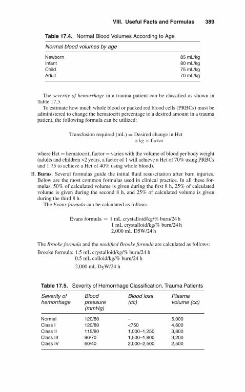
VIII. Useful Facts and Formulas 389
Table 17.4. Normal Blood Volumes According to Age
Normal blood volumes by age
Newborn 85 mL/kgInfant 80 mL/kgChild 75 mL/kgAdult 70 mL/kg
The severity of hemorrhage in a trauma patient can be classified as shown inTable 17.5.
To estimate how much whole blood or packed red blood cells (PRBCs) must beadministered to change the hematocrit percentage to a desired amount in a traumapatient, the following formula can be utilized:
Transfusion required (mL) = Desired change in Hct×kg × factor
where Hct = hematocrit; factor = varies with the volume of blood per body weight(adults and children >2 years, a factor of 1 will achieve a Hct of 70% using PRBCsand 1.75 to achieve a Hct of 40% using whole blood).
B. Burns. Several formulas guide the initial fluid resuscitation after burn injuries.Below are the most common formulas used in clinical practice. In all these for-mulas, 50% of calculated volume is given during the first 8 h, 25% of calculatedvolume is given during the second 8 h, and 25% of calculated volume is givenduring the third 8 h.
The Evans formula can be calculated as follows:
Evans formula = 1 mL crystalloid/kg/% burn/24 h1 mL crystalloid/kg/% burn/24 h2,000 mL D5W/24 h
The Brooke formula and the modified Brooke formula are calculated as follows:
Brooke formula: 1.5 mL crystalloid/kg/% burn/24 h0.5 mL colloid/kg/% burn/24 h
2,000 mL D5W/24 h
Table 17.5. Severity of Hemorrhage Classification, Trauma Patients
Severity ofhemorrhage
Bloodpressure(mmHg)
Blood loss(cc)
Plasmavolume (cc)
Normal 120/80 – 5,000Class I 120/80 <750 4,600Class II 115/80 1,000–1,250 3,800Class III 90/70 1,500–1,800 3,200Class IV 60/40 2,000–2,500 2,500

390 17. Trauma
Modified formula = 2 mL Ringer’s lactate/kg/% burn/24 h
The Parkland formula is calculated as
Parkland formula = 4 mL crystalloid/kg/% burn/24 h
In addition to these formulas, the evaporative water losses in patients withburns need to be calculated and replaced. Evaporative water loss (EWL) iscalculated as
EWL(mL/h) = (25 + % BSA burned) × BSA
where BSA = body surface area.
C. Other Trauma Scoring Systems. Several trauma scoring systems are in usethroughout the world. Of them, the abbreviated injury scale (AIS) is commonlyutilized (see Table 17.6).
Table 17.6. The Abbreviated Injury Scale
AIS score Injury severity
1 Minor2 Moderate3 Serious, non-life-threatening4 Severe, life-threatening5 Critical6 Maximal (correlates with death)
The trauma score (TS) is another commonly utilized system and is depicted inTable 17.7.
The revised trauma score (RTS) eliminates the assessment of capillary refilland respiratory effort and is calculated as follows:
RTS = 0.9368 GCS + 0.7326 SBP + 0.2908 RR coded values × revised score
coefficientwhere GCS = Glasgow Coma Scale; SBP = systolic blood pressure; RR =respiratory rate.
For children and infants, the pediatric trauma score is utilized (see Table 17.8).
D. Neurological Trauma. Within the primary survey, an early neurological traumaevaluation can be accomplished using the AVPU method:
A = alertV = responds to verbal stimulationP = responds to painful stimulationU = unresponsiveIn those patients with severe head injuries and ICP monitoring, cerebralperfusion pressure (CPP) is commonly utilized in management:
CPP = MAP = ICP
where MAP = mean arterial blood pressure; ICP = intracranial pressure.Another useful formula in neurological trauma is that of the calculation
of the pressure-volume index (PVI), which is defined as the volume (in
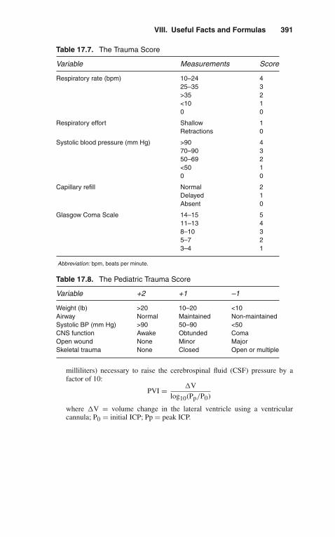
VIII. Useful Facts and Formulas 391
Table 17.7. The Trauma Score
Variable Measurements Score
Respiratory rate (bpm) 10–24 425–35 3>35 2<10 10 0
Respiratory effort Shallow 1Retractions 0
Systolic blood pressure (mm Hg) >90 470–90 350–69 2<50 10 0
Capillary refill Normal 2Delayed 1Absent 0
Glasgow Coma Scale 14–15 511–13 48–10 35–7 23–4 1
Abbreviation: bpm, beats per minute.
Table 17.8. The Pediatric Trauma Score
Variable +2 +1 –1
Weight (lb) >20 10–20 <10Airway Normal Maintained Non-maintainedSystolic BP (mm Hg) >90 50–90 <50CNS function Awake Obtunded ComaOpen wound None Minor MajorSkeletal trauma None Closed Open or multiple
milliliters) necessary to raise the cerebrospinal fluid (CSF) pressure by afactor of 10:
PVI = �V
log10(Pp/P0)
where �V = volume change in the lateral ventricle using a ventricularcannula; P0 = initial ICP; Pp = peak ICP.

18Allergic and Immunologic
Emergencies
� I. ANAPHYLAXIS
A. Definition. Anaphylaxis is an immediate, generalized, life-threatening reactionresulting from the release of bioactive substances from mast cells and basophils.Anaphylaxis can occur in more than one time period. The so-called biphasicanaphylaxis is defined as a recurrence of symptoms that develops following the“resolution” of the initial anaphylactic reaction. It can occur in up to 20% of ana-phylactic reactions and typically occurs within 8 h after resolution of the initialsymptoms. Recurrences up to 72 h later can occur.
B. Etiology. The most common causes of anaphylaxis in medical practice aredepicted in Table 18.1.
C. Clinical Manifestations1. The onset may vary from individual to individual depending on the sensitivity
of the person and the route, quantity, and rate of administration of the allergen.2. Early signs and symptoms that require a high index of suspicion may include
a. Agitationb. Dizzinessc. Headached. Nausea, vomiting
3. Cutaneous involvementa. Generalized pruritusb. Flushingc. Urticaria
4. Upper airway obstruction as a consequence of edema of the larynx, swelling oftongue and lips (angioedema). This may result in stridor and suffocation.
5. Respiratory failure (manifestations range from tachypnea to apnea) that maybe related to the factors mentioned above as well as bronchoconstriction of thelower airways manifested by wheezing. These patients may also develop adultrespiratory distress syndrome (ARDS).
J. Varon, P. Acosta, Handbook of Critical and Intensive Care Medicine,DOI 10.1007/978-0-387-92851-7_18,C© Springer Science+Business Media, LLC 2010

394 18. Allergic and Immunologic Emergencies
Table 18.1. Common Causes of Systemic Anaphylactic Reactions
1. Drugs—Antibiotics (i.e., penicillins, cephalosporins, sulfonamides, vancomycin)— Local anesthetics (i.e., lidocaine, procaine)— Muscle relaxants— Others (i.e., insulin, protamine)
2. Foods— Nuts and seeds— Fish, shellfish— Milk, eggs
3. Food additives— Aspartame— Monosodium glutamate
4. Diagnostics— Iodinated radiographic materials
5. Insect and snakes (stings and bites)
6. Exercise
7. Other— Latex gloves— Heterologous serum (i.e., tetanus antitoxin)
6. Cardiovascular collapse: The pathophysiology is thought to be related toenhanced vascular permeability, peripheral vasodilation, and intravascular vol-ume depletion. A heart rate increase >20 bpm from baseline and a decrease ofmean arterial pressure >20 torr (mmHg) are characteristic.
7. Dysrhythmias: Both supraventricular and ventricular rhythm disorders havebeen described in patients with anaphylaxis.
D. Laboratory Findings1. Do not wait for laboratory data to institute therapy!2. Patients with anaphylaxis may present with leukocytosis or leukopenia.3. Thrombocytopenia may appear in severe cases.4. Immunoglobulin E (IgE) measurements may not be helpful, because many
patients may manifest non-IgE-mediated anaphylaxis.
E. Management1. ABCs
Secure the airway, and assist with breathing and circulation as with any otherpatient presenting with a potentially critical illness.
2. The drug of choice for patients with acute anaphylaxis is epinephrine. Thedosage is 0.3–0.5 mL of 1:1000 dilution (0.3–0.5 mg) subcutaneously every10–20 min or intravenously as described below. Endotracheal administrationcan be attempted when no other route is available.
3. AntihistaminesTraditionally, H1-receptor antihistamines have been used; i.e., diphenhy-dramine (Benadryl) 25–50 mg intramuscularly (IM), intravenously (IV), or POq6–8 h. In theory, the combination of H1- and H2-receptor antihistamines might

II. Stevens-Johnson Syndrome 395
be a better chance of preventing further histamine-mediated reactions than H1blockers alone; i.e., cimetidine (Tagamet) 300 mg IV or PO q6 h.
4. Corticosteroids have an uncertain place in the management of acute reaction,since there is a 4–6 h latent period before such agents are pharmacologicallyeffective. The current recommended agents are hydrocortisone (Solu-Cortef)250 mg IV q6 h or methylprednisolone (Solu-Medrol) 50 mg IV q6 h for 2–4doses.
5. In cases of severe bronchospasm, the following drugs can be used:a. Metaproterenol 0.3 mL (5% solution) in 2.5 mL of saline, inhaled through a
nebulizerb. Aminophylline loading dose of 6 mg/kg IV over 30 min followed by 0.3–
0.9 mg/kg/h6. In patients with profound hypotension
a. Adequate IV fluid administration (up to 1 L every 20–30 min as needed).b. Epinephrine 1 mL of 1:1000 dilution in 500 mL of D5W at a rate of 0.5–5 μg
(0.25–2.5 mL)/min.c. Norepinephrine (Levophed): 4 mg in 1 L of D5W at a rate of 2–12 μg (0.5–3
mL)/min.d. Glucagon may be particularly useful in patients taking beta-adrenergic
blockers. The recommended dose is 1 mg in 1 L of D5W at a rate of 5–15 μg(5–15 mL)/min.
F. Preventive measures for patients at high risk of anaphylaxis are depicted inTable 18.2.
Table 18.2. Preventive Measures for Patients at High Risk forAnaphylaxis
1. Avoid exposure2. Slow administration of suspected agents under medical supervision in
adequate facility (i.e., ICU)3. Optimal management of underlying disorders4. Short- and long-term desensitization (i.e., penicillin, aspirin)
� II. STEVENS-JOHNSON SYNDROME(ERYTHEMA MULTIFORME)
A. Definition. Erythema multiforme (EM) is an erythematous maculopapular cuta-neous eruption of variable form. When EM grades into a more serious clinicalstate, the term Stevens-Johnson syndrome (SJS) is used.
B. Etiology. Common causes of EM and SJS are depicted in Table 18.3.
C. Clinical Manifestations1. Prodromal symptoms may include
a. Malaise, headacheb. Pharyngitis, rhinorrheac. Diarrhead. Arthralgias

396 18. Allergic and Immunologic Emergencies
Table 18.3. Causes of Erythema Multiforme/Stevens-JohnsonSyndrome
1. Infections— Viral (i.e., herpes simplex, measles, hepatitis B)— Bacterial (i.e., streptococcus, pseudomonas)— Mycobacterial (i.e., tuberculosis)— Spirochetes (i.e., syphilis)— Fungal (i.e., histoplasmosis)
2. Drugs— Analgesics (i.e., aspirin, nonsteroidal anti-inflammatory drugs)— Antibiotics (i.e., sulfonamides, penicillins, tetracycline)— Anticonvulsants (i.e., ethosuximide)— Antihypertensives (i.e., minoxidil)— Glucocorticoids— H2-blockers (i.e., cimetidine)
3. Immunizations— Horse serum— Polio vaccine— Pertussis vaccine
4. Neoplasms (i.e., lymphomas)
5. Connective tissue disorders (i.e., lupus erythematosus)
6. Physical agents— Radiation therapy— Sunlight
7. Others— Inflammatory bowel disease— Sarcoidosis
2. The earliest lesions in EM are often red, edematous papules surrounded byblanching. They enlarge to form small plaques with concentric alterations incolor and morphology.
3. The so-called Target lesions are areas of central epidermal necrosis with orwithout bullae formation.
4. Patients admitted to the ICU with SJS usually present with extensive tissuenecrosis and severe fluid depletion.
D. Laboratory Findings1. Usually nondiagnostic.2. Skin biopsy reveals a perivascular lymphocytic infiltrate in the upper dermis,
subepidermal bullae formation, and endothelial cell swelling.
E. Management1. Immediately discontinue suspected drugs or agents as well as all nonessential
drugs.2. The usefulness of systemic corticosteroids in this setting is controversial. In
the absence of controlled clinical trials, some authors recommend beginningtherapy with prednisone 1 mg/kg/d (or IV equivalent).

III. Angioneurotic Laryngeal Edema 397
3. Fluid replacement as indicated by severity of the disease.4. Evaluate for infection and treat appropriately.5. Obtain consultation depending on the degree and sites of involvement (i.e.,
ophthalmology, plastic surgery).6. Transfer the patient to a burn unit.7. Intravenous gammaglobulin has been utilized with conflictive results.8. Plasmapheresis has been used in some patients with success.
� III. ANGIONEUROTIC LARYNGEAL EDEMA
A. Definition. Angioneurotic laryngeal edema (ALE) is characterized by nonpruriticlocal swelling involving the face, larynx, and skin of the extremities.
B. Etiology1. Allergic
Related to foods (i.e., fish), drugs (i.e., angiotensin-converting enzyme [ACE]inhibitors), inhaled substances, insect stings (i.e., bees).
2. HereditaryCaused by a deficiency in C1-esterase inhibitor. Autosomal dominant.Precipitating events may include trauma and emotional stress.
C. Clinical Manifestations1. Swelling of face, larynx, and skin of extremities.2. Depending on the progression, stridor may be a prominent feature with ensuing
respiratory distress.3. Abdominal pain, nausea, and vomiting.
D. Laboratory Findings. Nondiagnostic except in cases of hereditary ALE.
E. Management1. ABCs
Secure the airway, and assist with breathing and circulation as with any otherpatient presenting with a potentially critical illness.
2. Avoid precipitating allergens.3. If ALE is thought to be allergic in origin, administer parenteral epinephrine and
antihistamines (as noted in “Anaphylaxis,” above).4. Intubation is only rarely required for patients with allergic ALE, while in
patients with hereditary ALE, the treatment of the acute episode may requireurgent intubation or tracheostomy.
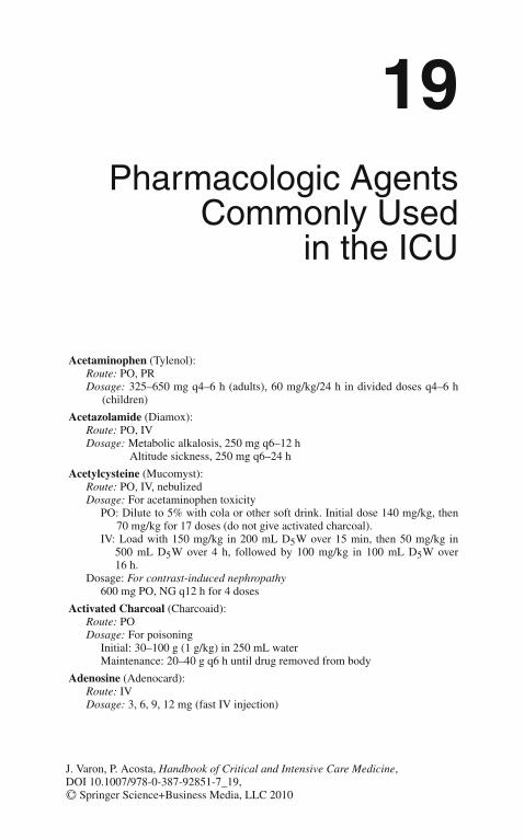
19Pharmacologic Agents
Commonly Usedin the ICU
Acetaminophen (Tylenol):Route: PO, PRDosage: 325–650 mg q4–6 h (adults), 60 mg/kg/24 h in divided doses q4–6 h
(children)
Acetazolamide (Diamox):Route: PO, IVDosage: Metabolic alkalosis, 250 mg q6–12 h
Altitude sickness, 250 mg q6–24 h
Acetylcysteine (Mucomyst):Route: PO, IV, nebulizedDosage: For acetaminophen toxicity
PO: Dilute to 5% with cola or other soft drink. Initial dose 140 mg/kg, then70 mg/kg for 17 doses (do not give activated charcoal).
IV: Load with 150 mg/kg in 200 mL D5W over 15 min, then 50 mg/kg in500 mL D5W over 4 h, followed by 100 mg/kg in 100 mL D5W over16 h.
Dosage: For contrast-induced nephropathy600 mg PO, NG q12 h for 4 doses
Activated Charcoal (Charcoaid):Route: PODosage: For poisoning
Initial: 30–100 g (1 g/kg) in 250 mL waterMaintenance: 20–40 g q6 h until drug removed from body
Adenosine (Adenocard):Route: IVDosage: 3, 6, 9, 12 mg (fast IV injection)
J. Varon, P. Acosta, Handbook of Critical and Intensive Care Medicine,DOI 10.1007/978-0-387-92851-7_19,C© Springer Science+Business Media, LLC 2010

400 19. Pharmacologic Agents Commonly Used in the ICU
Alteplase (Activase):Route: IVDosage: Acute pulmonary embolism: 100 mg over 2 h. Acute ischemic stroke
0.9 mg/kg.
Amphotericin B (Amphotec):Route: IVDosage: 3–4 mg/kg/day
Ammonium Chloride (generic):Route: PO, IVDosage: Urine acidification, 4–12 g/d PO in divided doses q4–6 h
Amiodarone (Cordarone, Pacerone)Route: PO, IVDosage: Pulseless VF/VT 300 mg. If VF/VT recurs a supplemental dose 250 mg
followed by infusion of 1 mg/min for 6 h, then 0.5 mg/min.
Amrinone (Inocor):Route: IVDosage: Bolus, 0.75–3 mg/kg over 2–3 min, followed by infusion of
5–20 μg/kg/min
Atropine (generic):Route: PO, IVDosage: Bronchospasm, 1.5–2.0 mg by nebulizer q6 h
Bradycardia, 0.5–1 mg IV push, repeat q5 min to max. 2 mgProphylaxis for bradycardia pre-endoscopy 0.6 mg I.M.
Bivalirudin (Angiomax):Route: IVDosage: Acute coronary syndromes undergoing PTCA/PCI 0.1 mg/kg followed by
0.25 mg/kg/h.
Bretylium (Bretylol):Route: IV, IMDosage: Bolus, 5–10 mg/kg over 10–20 min IV, followed by a continuous infusion
of 1–5 mg/min
Carbicarb (Carbicarb):Route: IVDosage: Severe acidosis, initial dose 1 mEq/kg, followed by 0.5 mEq/kg (adjust
dose as indicated by clinical condition and blood pH)
Chlordiazepoxide (Librium):Route: PO, IV, IMDosage: 15–100 mg/d in 3–4 divided doses
Chlorpromazine (Thorazine):Route: PO, PR, IMDosage: Severe psychosis with agitation, 25–100 mg IM q1–4 h until control is
achieved
Cisatracurium (Nimbex):Route: IVDosage: Bolus: 0.1 mg /kg Infusion: 1–3 mcg/kg/min

19. Pharmacologic Agents Commonly Used in the ICU 401
Clevidipine (Cleviprex):Route: IVDosage: Hypertensive crisis, postoperative hypertension when the oral route is not
feasible 1–2 mg/h as start dose and double every 3 min to a maximum of32 mg/h.
Clonidine (Catapres):Route: PO, transdermal (in some countries IM and PR used)Dosage: Hypertensive emergencies and urgencies, 0.2 mg PO, then 0.1 mg/
q20 min to 0.8 mg or until BP is controlled
Dalteparin (Fragmin):Route: SubcutaneousDosage: DVT prophylaxis: 2,500–5,000 Units subcutaneous once a day. DVT
treatment: 200 Units/kg
Dantrolene (Dantrium):Route: PO, IVDosage: Malignant hyperthermia, initial dose 1–2 mg/kg IV via rapid infusion;
may repeat to total 10 mg/kg, if needed
Daptomycin (Cubicin):Route: IVDosage: 4–6 mg/ kg once a day.
dDAVP (generic):Route: Intranasal, IV, SCDosage: Hemostasis, 0.3 μg/kg in NS over 15–30 minDiabetes insipidus, 0.5–1 mL IV/SC bid
Diazepam (Valium):Route: PO, IV, IMDosage: Status epilepticus, 5–10 mg IV (1–2 mg/min)
Diazoxide (Hyperstat):Route: PO, IVDosage: Hypertensive crisis, 1–3 mg/kg IV (max 150 mg) q5–15 min until BP is
controlled
Digoxin (Lanoxin):Route: PO, IV, IMDosage: Digitalization, 0.4–0.6 mg IV (may require up to 1.25 mg total);
maintenance, 0.125–0.25 mg/d PO or IV
Dobutamine (Dobutrex):Route: IVDosage: 2.5–15 μg/kg/min most commonly used
Dopamine (Inotropin):Route: IVDosage: Dopaminergic stimulation, 0.5–2.0 μg/kg/minAlpha-, beta-dopaminergic effects, >10 μg/kg/min
Drotrecogin alfa-activated protein C (Xigris):Route: IVDosage: 24 mcg/kg/h for a total of 96 h.

402 19. Pharmacologic Agents Commonly Used in the ICU
Enoxaparin (Lovenox):Route: Subcutaneous, IV.Dosing: DVT prophylaxis 40 mg daily, DVT treatment acute 1 mg/kg/q12 hor 1.5 mg/kg/daily.
Epinephrine (Epinephrine Injection):Route: IV, SCDosage: Beta1-, beta2-effect, 1–4 μg/min
Alpha-effect, 4 μg/min
Epoprostenol (Flolan)Route: IV, can be inhaled.Dosage: 1–2 ng/kg/min
Ertapenem (Invanz):Route: IV, IMDosage: 1 g daily
Esmolol (Brevibloc):Route: IVDosage: Bolus 0.5–1.0 mg/kg, followed by infusion at 50 μg/kg/min; maintenance
50–200 μg/kg/min
Etomidate (Amidate):Route: IVDosage: Intubation: 0.2–0.6 mg/kg
Fenoldopam (Corlopam):Route: IVDosage: 0.1–1.0 μg/kg/min, titrate to achieve desired blood pressure
Fentanyl (Sublimaze):Route: IV, IMDosage: Sedation/analgesia, 1 μg/kg IV/IM
Flumazenil (Romazicon):Route: IVDosage: 0.3 mg IV
Furosemide (Lasix):Route: PO, IV, IMDosage: 10–120 mg IV/IM, adjusted as necessary until desired response obtained.
May use continuous infusions.
Fosphenytoin (Cerebyx):Route: IV, IMDosage: Status epilepticus 15–20 mg/kg (loading dose)
Glucagon (generic):Route: SC, IM, IVDosage: Hypoglycemia, 0.5–1.0 mg SC/IM/IV, may repeat in 15 min
Bradycardia, 1–20 mg/h
Haloperidol (Haldol):Route: PO, IM, IV (not FDA approved)Dosage: Acute psychosis, 2–5 mg IM q1–2 h until symptoms are controlled
Heparin (Liquaemin):Route: IV, SC

19. Pharmacologic Agents Commonly Used in the ICU 403
Dosage: DVT prophylaxis, 5,000 U SC q8–12 hDVT/Pulmonary emboli therapy, bolus with 100 U/kg followed by a continuous
infusion of 800–1,200 U/h, titrated to maintain aPTT 11/2–2 times control
Hydralazine (Apresoline):Route: PO, IVDosage: 5 mg IV bolus; 5–10 mg IV q6 h maintenance
Hydromorphone (Dilaudid)Route: IV, PO, IM, SCDosage: 0.7–2 mg every 1–2 h as needed for pain
Imipenem and Cilastatin (Primaxin):Route: IV, IMDosage: 500–1,000 mg every 6 h.
Isoproterenol (Isuprel):Route: IV, SC, PO, inhaledDosage: Infusion 1–10 μg/min
Ketorolac Tromethamine (Toradol):Route: PO, IMDosage: Initial dose 30–60 mg IM, then 15–30 mg q6 h
Labetalol (Normodyne):Route: PO, IVDosage: Rapid BP control, IV bolus 5–20 mg (slowly); repeat after 5 min if
needed. Continuous infusion of 1 mg/mL started at 1–2 mg/min and titrated toeffect.
Lepirudin (Refludan):Route: IVDosage: Bolus 0.4 mg/kg followed by continuous infusion at 0.15 mg/kg/h with a
maximum dose of 44 mg as a bolus and 16.5 mg/h as infusion
Lidocaine (Xylocaine):Route: IV, IM, SCDosage: Bolus 1.0–1.5 mg/kg followed by 1–4 mg/min
Linezolid (Zyvox):Route: PO, IVDosage: 600 mg every 12 h.
Lorazepam (Ativan):Route: PO, IV, IMDosage: 2–10 mg/d in divided doses PO/IV/IM (some patients may require
continuous infusions).
Mannitol (Osmitrol):Route: IVDosage: For cerebral edema, IV infusion 0.15–2.0 g/kg as 15–25% solution over
30–60 min; max dose is up to 6 g/kg/24 h.
Meperidine (Demerol):Route: PO, IM, IV, SCDosage: 50–150 mg q3–4 h
Meropenem (Merrem):Route: IVDosage: 1–2 g every 8 h.
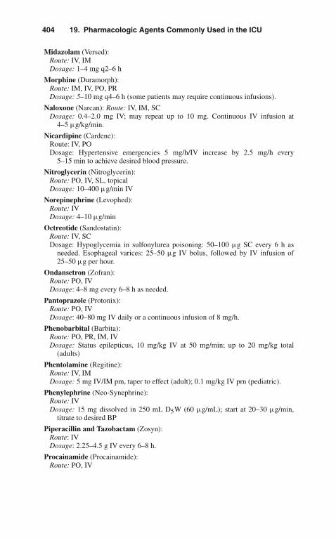
404 19. Pharmacologic Agents Commonly Used in the ICU
Midazolam (Versed):Route: IV, IMDosage: 1–4 mg q2–6 h
Morphine (Duramorph):Route: IM, IV, PO, PRDosage: 5–10 mg q4–6 h (some patients may require continuous infusions).
Naloxone (Narcan): Route: IV, IM, SCDosage: 0.4–2.0 mg IV; may repeat up to 10 mg. Continuous IV infusion at
4–5 μg/kg/min.
Nicardipine (Cardene):Route: IV, PODosage: Hypertensive emergencies 5 mg/h/IV increase by 2.5 mg/h every
5–15 min to achieve desired blood pressure.
Nitroglycerin (Nitroglycerin):Route: PO, IV, SL, topicalDosage: 10–400 μg/min IV
Norepinephrine (Levophed):Route: IVDosage: 4–10 μg/min
Octreotide (Sandostatin):Route: IV, SCDosage: Hypoglycemia in sulfonylurea poisoning: 50–100 μg SC every 6 h as
needed. Esophageal varices: 25–50 μg IV bolus, followed by IV infusion of25–50 μg per hour.
Ondansetron (Zofran):Route: PO, IVDosage: 4–8 mg every 6–8 h as needed.
Pantoprazole (Protonix):Route: PO, IVDosage: 40–80 mg IV daily or a continuous infusion of 8 mg/h.
Phenobarbital (Barbita):Route: PO, PR, IM, IVDosage: Status epilepticus, 10 mg/kg IV at 50 mg/min; up to 20 mg/kg total
(adults)
Phentolamine (Regitine):Route: IV, IMDosage: 5 mg IV/IM pm, taper to effect (adult); 0.1 mg/kg IV prn (pediatric).
Phenylephrine (Neo-Synephrine):Route: IVDosage: 15 mg dissolved in 250 mL D5W (60 μg/mL); start at 20–30 μg/min,
titrate to desired BP
Piperacillin and Tazobactam (Zosyn):Route: IVDosage: 2.25–4.5 g IV every 6–8 h.
Procainamide (Procainamide):Route: PO, IV

19. Pharmacologic Agents Commonly Used in the ICU 405
Dosage: 100 mg/min IV to effect or to a total dose of 1,000 mg, followed byinfusion 2–6 mg/min
Prochlorperazine (Compazine):Route: PO, IM, IV, PRDosage: 5–10 mg PO tid/qid; 5–10 mg IV q3–4 h; 25 mg PR bid
Propofol (Diprivan):Route: IVDosage: 2–15 cc/h by continuous infusion to achieve the desired level of sedation
Propranolol (Inderal):Route: PO, IVDosage: Titrate 0.5–1.0 mg IV q5 min to effect.
Protamine (Protamine sulfate):Route: IVDosage: 1 mg for each 90 U of lung heparin or 1 mg for each 115 U of intestinal
heparin, by slow injection over 1–3 min; max dose 50 mg in 10 min.
Rocuronium (Zemuron):Route: IVDosage: Intubation 0.6 mg/kg.
Sodium Bicarbonate (Sodium Bicarbonate Injection):Route: IV, PODosage: Severe acidosis, initial dose 1 mEq/kg, followed by 0.5 mEq/kg; adjust
dosage as indicated by clinical condition and blood pH.
Sodium Nitroprusside (Nipride):Route: IVDosage: Mix 50 mg in 250 mL D5W. Start at 0.5 μg/kg/min and titrate toeffect.
Sodium Polystyrene Sulfonate (Kayexalate):Route: PO, PRDosage: 15 g, PO q6–24 h
Succinylcholine (Anectine):Route: IV, IMDosage: 1–1.5 mg/kg IV; 2–4 mg/kg IM (pediatric use only)
Thiopental (Pentothal sodium):Route: IV, PRDosage: General anesthetic, 2–3 mL 2.5% solution (50–75 mg) IV q20–40 s until
desired effect reachedSeizures, 75–125 mg IV
Tigecycline (Tygacil):Route: IVDosage: initial dose 100 mg, then 50 mg every 12 h.
Trimethaphan (Arfonad):Route: IVDosage: Start at 0.3 mg/min and titrate to effect.
Valproic Acid (Depakote):Route: PO, IVDosage: Status epilepticus 15–45 mg/kg (loading dose), infusion 1–4 mg/kg/h.
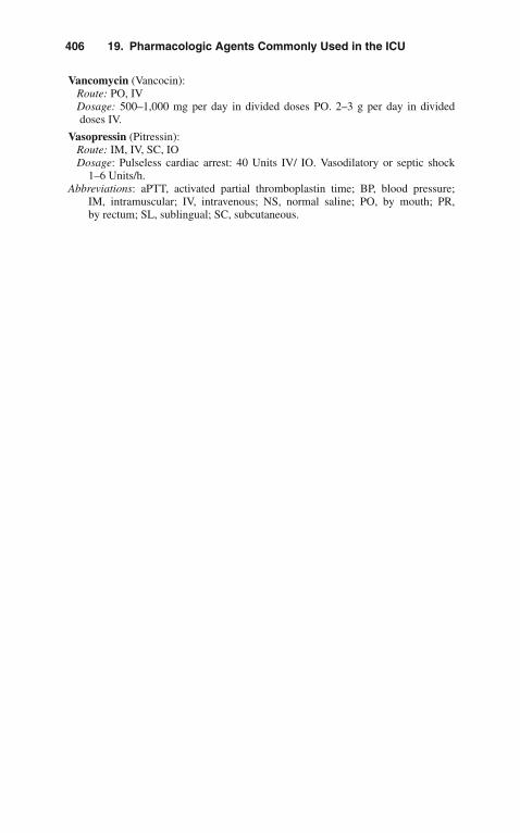
406 19. Pharmacologic Agents Commonly Used in the ICU
Vancomycin (Vancocin):Route: PO, IVDosage: 500–1,000 mg per day in divided doses PO. 2–3 g per day in divideddoses IV.
Vasopressin (Pitressin):Route: IM, IV, SC, IODosage: Pulseless cardiac arrest: 40 Units IV/ IO. Vasodilatory or septic shock
1–6 Units/h.Abbreviations: aPTT, activated partial thromboplastin time; BP, blood pressure;
IM, intramuscular; IV, intravenous; NS, normal saline; PO, by mouth; PR,by rectum; SL, sublingual; SC, subcutaneous.
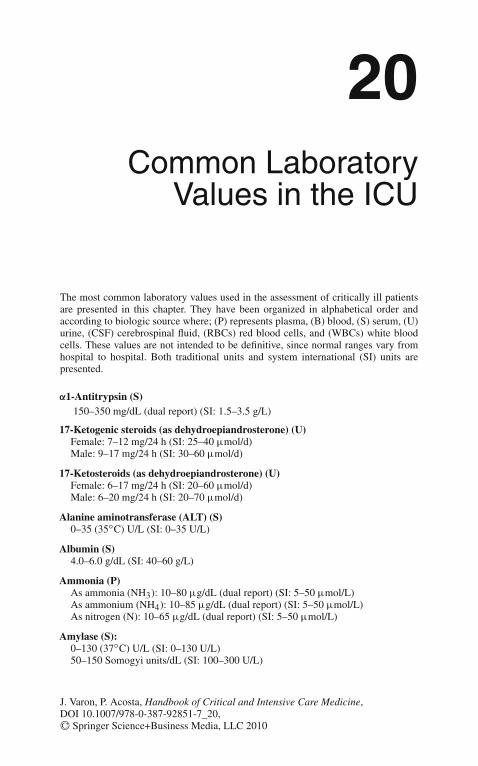
20Common Laboratory
Values in the ICU
The most common laboratory values used in the assessment of critically ill patientsare presented in this chapter. They have been organized in alphabetical order andaccording to biologic source where; (P) represents plasma, (B) blood, (S) serum, (U)urine, (CSF) cerebrospinal fluid, (RBCs) red blood cells, and (WBCs) white bloodcells. These values are not intended to be definitive, since normal ranges vary fromhospital to hospital. Both traditional units and system international (SI) units arepresented.
α1-Antitrypsin (S)150–350 mg/dL (dual report) (SI: 1.5–3.5 g/L)
17-Ketogenic steroids (as dehydroepiandrosterone) (U)Female: 7–12 mg/24 h (SI: 25–40 μmol/d)Male: 9–17 mg/24 h (SI: 30–60 μmol/d)
17-Ketosteroids (as dehydroepiandrosterone) (U)Female: 6–17 mg/24 h (SI: 20–60 μmol/d)Male: 6–20 mg/24 h (SI: 20–70 μmol/d)
Alanine aminotransferase (ALT) (S)0–35 (35◦C) U/L (SI: 0–35 U/L)
Albumin (S)4.0–6.0 g/dL (SI: 40–60 g/L)
Ammonia (P)As ammonia (NH3): 10–80 μg/dL (dual report) (SI: 5–50 μmol/L)As ammonium (NH4): 10–85 μg/dL (dual report) (SI: 5–50 μmol/L)As nitrogen (N): 10–65 μg/dL (dual report) (SI: 5–50 μmol/L)
Amylase (S):0–130 (37◦C) U/L (SI: 0–130 U/L)50–150 Somogyi units/dL (SI: 100–300 U/L)
J. Varon, P. Acosta, Handbook of Critical and Intensive Care Medicine,DOI 10.1007/978-0-387-92851-7_20,C© Springer Science+Business Media, LLC 2010
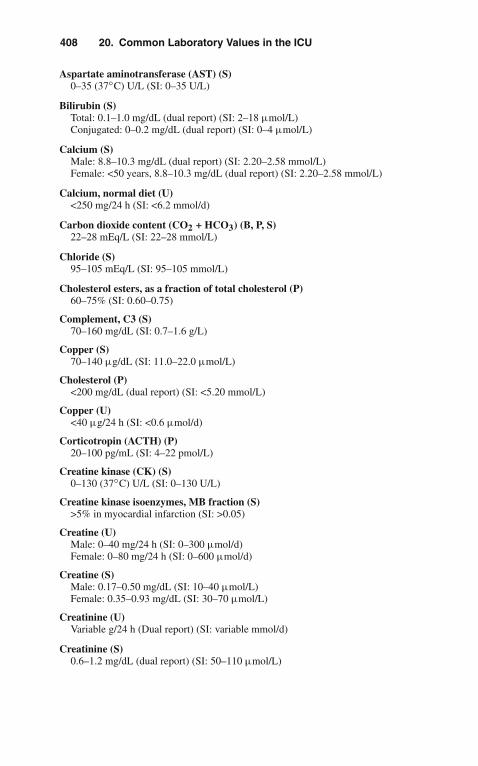
408 20. Common Laboratory Values in the ICU
Aspartate aminotransferase (AST) (S)0–35 (37◦C) U/L (SI: 0–35 U/L)
Bilirubin (S)Total: 0.1–1.0 mg/dL (dual report) (SI: 2–18 μmol/L)Conjugated: 0–0.2 mg/dL (dual report) (SI: 0–4 μmol/L)
Calcium (S)Male: 8.8–10.3 mg/dL (dual report) (SI: 2.20–2.58 mmol/L)Female: <50 years, 8.8–10.3 mg/dL (dual report) (SI: 2.20–2.58 mmol/L)
Calcium, normal diet (U)<250 mg/24 h (SI: <6.2 mmol/d)
Carbon dioxide content (CO2 + HCO3) (B, P, S)22–28 mEq/L (SI: 22–28 mmol/L)
Chloride (S)95–105 mEq/L (SI: 95–105 mmol/L)
Cholesterol esters, as a fraction of total cholesterol (P)60–75% (SI: 0.60–0.75)
Complement, C3 (S)70–160 mg/dL (SI: 0.7–1.6 g/L)
Copper (S)70–140 μg/dL (SI: 11.0–22.0 μmol/L)
Cholesterol (P)<200 mg/dL (dual report) (SI: <5.20 mmol/L)
Copper (U)<40 μg/24 h (SI: <0.6 μmol/d)
Corticotropin (ACTH) (P)20–100 pg/mL (SI: 4–22 pmol/L)
Creatine kinase (CK) (S)0–130 (37◦C) U/L (SI: 0–130 U/L)
Creatine kinase isoenzymes, MB fraction (S)>5% in myocardial infarction (SI: >0.05)
Creatine (U)Male: 0–40 mg/24 h (SI: 0–300 μmol/d)Female: 0–80 mg/24 h (SI: 0–600 μmol/d)
Creatine (S)Male: 0.17–0.50 mg/dL (SI: 10–40 μmol/L)Female: 0.35–0.93 mg/dL (SI: 30–70 μmol/L)
Creatinine (U)Variable g/24 h (Dual report) (SI: variable mmol/d)
Creatinine (S)0.6–1.2 mg/dL (dual report) (SI: 50–110 μmol/L)

20. Common Laboratory Values in the ICU 409
Creatinine clearance (S, U)75–125 mL/min (dual report) (SI: 1.24–2.08 mL/s)
Cystine (U)10–100 mg/24 h (SI: 40–420 μmol/d)
Dehydroepiandrosterone (U)Female: 0.2–1.8 mg/24 h (SI: 1–6 μmol/d)Male: 0.2–2.0 mg/24 h (SI: 1–7 μmol/d)
Digoxin, therapeutic (P)0.5–2.2 ng/mL (dual report) (SI: 0.6–2.8 mmol/L)0.5–2.2 μg/L (dual report) (SI: 0.6–2.8 mmol/L)
Erythrocyte sedimentation rate (B)Female: 0–30 mm/h (SI: 0–30 mm/h)Male: 0–20 mm/h (SI: 0–20 mm/h)
Estradiol, male >18 years (S)15–40 pg/mL (dual report) (SI: 55–150 pmol/L)
Ethyl alcohol (P)<100 mg/dL (SI: <22 mmol/L)
EtiocholanoloneFemale: 0.8–4.0 mg/24 h (SI: 2–14 μmol/d)Male: 1.4–5.0 mg/14 h (SI: 4–17 μmol/d)
Fibrinogen (P)200–4,300 mg/dL (SI: 2.0–4.0 g/L)
Follicle-stimulating hormone (FSH) (P)Female: 2.0–15.0 mIU/mL (SI: 2–15 IU/L)Peak production: 20–50 mIU/mL (SI: 20–50 IU/L)Male: 1.0–10.0 mIU/mL (SI: 1–10 IU/L)
Follicle-stimulating hormone (FSH) (U)Follicular phase: 2–15 IU/24 h (SI: 2–15 IU/d)Midcycle: 8–40 IU/24 h (SI: 8–40 IU/d)Luteal phase: 2–10 IU/24 h (SI: 2–10 IU/d)Menopausal women: 35–100 IU/24 h (SI: 35–100 IU/d)Male: 2–15 IU/24 h (SI: 2–15 IU/d)
Gamma-glutamyltransferase (GGT) (S)0–30 (30◦C) U/L (SI: 0–30 U/L)
Glucose (P)70–110 mg/dL (dual report) (SI: 3.9–6.1 mmol/L)
Hematocrit (B)Female: 33–43% (SI: 0.33–0.43)Male: 39–49% (SI: 0.39–0.49)
Hemoglobin (B)Male: 14.0–18.0 g/dL (SI: 140–180 g/L)Female: 11.5–15.5 g/dL (SI: 115–155 g/L)

410 20. Common Laboratory Values in the ICU
Hemoglobin (B)Female: 12.0–15.0 g/dL (SI: 120–150 g/L)Male: 13.6–17.2 g/dL (SI: 136–172 g/L)
Immunoglobulins (S)IgG: 500–1,200 mg/dL (SI: 5.00–12.00 g/L)IgA: 50–350 mg/dL (SI: 0.50–3.50 g/L)IgM: 30–230 mg/dL (SI: 0.30–2.30 g/L)IgD: <6 mg/dL (SI: <60 mg/L)IgE:
0–3 years: 0.5–1.0 U/mL (SI: 1–24 μg/L)3–80 years: 5–100 U/mL (SI: 12–240 μg/L)
Iron (S)Male: 80–180 μg/dL (dual report) (SI: 14–32 μmol/L)Female: 60–160 μg/dL (dual report) (SI: 11–29 μmol/L)
Iron-binding capacity (S)250–460 μg/dL (dual report) (SI: 45–82 μmol/L)
Ketosteroid fractions (U)Androsterone:Female: 0.5–3.0 mg/24 h (SI: 1–10 μmol/d)Male: 2.0–5.0 mg/24 h (SI: 7–17 μmol/d)
Lactate dehydrogenase (S)50–150 (37◦C) U/L (SI: 50–150 U/L)
Lactate dehydrogenase isoenzymes (S)LD1: 15–40% (SI: 0.15–0.40)LD2: 20–45% (SI: 0.20–0.45)LD3: 15–30% (SI: 0.15–0.30)LD4 and LD5: 5–20% (SI: 0.05–0.20)LD1: 10–60 U/L (SI: 10–60 U/L)LD2: 20–70 U/L (SI: 20–70 U/L)LD3: 10–45 U/L (SI: 10–45 U/L)LD4 and LD5: 5–30 U/L (SI: 5–30 U/L)
Lead, toxic (B)>60 μg/dL (dual report) (SI: >2.90 μmol/L)
Lead, toxic (U)>80 μg/24 h (dual report) (SI: >0.40 μmol/d)
Lipids, total (P)400–850 mg/dL (dual report) (SI: 4.0–8.5 g/L)
Lipoproteins (P)Low-density (LDL), as cholesterol: 50–190 mg/dL (dual report) (SI: 1.30–
4.90 mmol/L)High-density (HDL), as cholesterol:Male: 30–70 mg/dL (dual report) (SI: 0.80–1.80 mmol/L)Female: 30–90 mg/dL (dual report) (SI: 0.80–2.35 mmol/L)

20. Common Laboratory Values in the ICU 411
Magnesium (S)1.8–3.0 mg/dL (dual report) (SI: 0.80–1.20 mmol/L)
Mean corpuscular hemoglobin concentration (B)Mass concentration: 33–37 g/dL (SI: 330–370 g/L)Substance concentration (Hb[Fe]): 33–37 g/dL (SI: 20–23 mmol/L)
Mean corpuscular hemoglobin (B)Mass concentration: 27–33 pg (SI: 27–33 pg)Substance concentration (Hb[Fe]): 27–33 pg (SI: 1.68–2.05 fmol)
Mean corpuscular volume (B)Erythrocyte volume: 76–100 μm3 (SI: 76–100 fL)
Phenytoin, therapeutic (P)10–20 mg/L (SI: 40–80 μmol/L)
Phosphatase, acid (prostatic) (P)0–3 King-Armstrong U/dL (SI: 0–5.5 U/L)
Phosphatase, alkaline (S)30–120 U/L (SI: 30–120 U/L)
Phosphate (as phosphorus) (S)2.5–5.0 mg/dL (dual report) (SI: 0.80–1.60 mmol/L)
Platelets (B)150–450 103/mm3 (SI: 150–450 109/L)
Potassium (S)3.5–5.0 mEq/L (SI: 3.5–5.0 mmol/L)
Progesterone (P)Follicular phase: <2 ng/mL (SI: <6 nmol/L)Luteal phase: 2–20 ng/mL (SI: 6–64 nmol/L)
Protein, total (U)<150 mg/24 h (SI: <0.15 g/d)
Protein, total (S)6–8 g/dL (SI: 60–80 g/L)
Protein, total (CSF)<40 mg/dL (SI: <0.40 g/L)
Red blood cell count (erythrocytes) (B)Female: 3.5–5.0 106/mm3 (SI: 3.5–5.0 1012/L)Male: 4.3–5.9 106/mm3 (SI: 4.3–5.1 1012/L)
Red blood cell count (CSF)0/cu mm (SI: 0 106/L)
Reticulocyte count (adult) (B)10,000–75,000/cu mm (dual report) (SI: 10–75 106/L)Number fraction: 1–24 0/00 (No. per 1,000 erythrocytes) (SI: 1–24 10–3)0.1–2.4% (SI: 1–24 10–3)
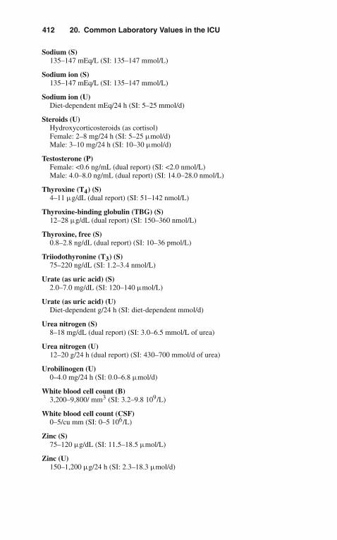
412 20. Common Laboratory Values in the ICU
Sodium (S)135–147 mEq/L (SI: 135–147 mmol/L)
Sodium ion (S)135–147 mEq/L (SI: 135–147 mmol/L)
Sodium ion (U)Diet-dependent mEq/24 h (SI: 5–25 mmol/d)
Steroids (U)Hydroxycorticosteroids (as cortisol)Female: 2–8 mg/24 h (SI: 5–25 μmol/d)Male: 3–10 mg/24 h (SI: 10–30 μmol/d)
Testosterone (P)Female: <0.6 ng/mL (dual report) (SI: <2.0 nmol/L)Male: 4.0–8.0 ng/mL (dual report) (SI: 14.0–28.0 nmol/L)
Thyroxine (T4) (S)4–11 μg/dL (dual report) (SI: 51–142 nmol/L)
Thyroxine-binding globulin (TBG) (S)12–28 μg/dL (dual report) (SI: 150–360 nmol/L)
Thyroxine, free (S)0.8–2.8 ng/dL (dual report) (SI: 10–36 pmol/L)
Triiodothyronine (T3) (S)75–220 ng/dL (SI: 1.2–3.4 nmol/L)
Urate (as uric acid) (S)2.0–7.0 mg/dL (SI: 120–140 μmol/L)
Urate (as uric acid) (U)Diet-dependent g/24 h (SI: diet-dependent mmol/d)
Urea nitrogen (S)8–18 mg/dL (dual report) (SI: 3.0–6.5 mmol/L of urea)
Urea nitrogen (U)12–20 g/24 h (dual report) (SI: 430–700 mmol/d of urea)
Urobilinogen (U)0–4.0 mg/24 h (SI: 0.0–6.8 μmol/d)
White blood cell count (B)3,200–9,800/ mm3 (SI: 3.2–9.8 109/L)
White blood cell count (CSF)0–5/cu mm (SI: 0–5 106/L)
Zinc (S)75–120 μg/dL (SI: 11.5–18.5 μmol/L)
Zinc (U)150–1,200 μg/24 h (SI: 2.3–18.3 μmol/d)
Index
Abbreviated injury scale, 390Abdominal surgery, nutritional support in
patients with, 218Abdominal trauma, 383–385A/C (assisted/controlled mechanical
ventilation), 32Acetaminophen (Tylenol), 357–359, 399Acetazolamide (Diamox), 399Acetylcysteine (Mucomyst), 399Acid-base disturbances, 297–304Acid-base equations/facts, 324Acidemia, 297Acidosis, 297
metabolic, 298respiratory, 308, 309
Activated charcoal (Charcoaid), 356Activated protein C, 177Acyclovir, 187Adenosine (Adenocard), 399Adrenal insufficiency, 85–87Adult respiratory distress syndrome (ARDS),
176, 278–280AIDS, infections in patients with, 181–184Air embolism, 115–117Airway management, 335–339Airway pressure release ventilation
(APRV), 34Airway resistance, 294Alanine aminotransferase, 407Albumin, 407Alcohol, 359–361Alkalemia, 297Alkalosis, 297
metabolic, 302–303respiratory, 303–304
Allergic emergencies, 393–397Altered mental status, 229–230Altitude, 136Alveolar air equation, 14–17, 294Alveolar ventilation, 289Amino acids, urinary excretion of, 332
Aminoglycoside clearance, 189Aminoglycosides, 187Ammonia, 407Ammonium chloride, 400Amniotic fluid embolism, 263–264Amphotericin B, 173Amrinone (Inocor), 400Amylase, 407Anaphylaxis, 393–395Anectine (succinylcholine), 405Anemia, 149–151Aneurysms, 68Angina pectoris, 47
unstable, 47–50Angioneurotic laryngeal edema, 397Angiotensin-converting enzyme (ACE)
inhibitors, 361Antibiotic adjustments, 189–190Antibiotic kinetics, 189Anticoagulants, 49Anticoagulation, 155–159Antidepressants, cyclic, 364–365Antidiuretic hormone (ADH), 90–92Antihypertensive drugs, 77
in pregnancy, 260–261, 263Antimicrobials, 187–188Antitrypsin, 407Aortic dissection, 68–70Aortic insufficiency, 66–67Aortic stenosis, 65–66Apnea test, 192Apresoline (hydralazine), 260, 403Arfonad (trimethaphan), 405Arm muscle circumference (AMC), 247Arterial-jugular venous oxygen content
difference, 207Arterial line, 345–346Arteriovenous oxygen content difference, 78Aspartate aminotransferase, 408Aspirin, 49
J. Varon, P. Acosta, Handbook of Critical and Intensive Care Medicine,DOI 10.1007/978-0-387-92851-7,C© Springer Science+Business Media, LLC 2010
414 Index
Assisted/controlled mechanical ventilation(A/C), 32
Asthma, 271–273Asystole treatment algorithm, 14Atherosclerosis, coronary artery, 51Ativan (lorazepam), 403ATPS to BTPS, converting gas volumes
from, 136Atrial fibrillation, 74Atrial flutter, 75Atrial tachycardia, multifocal, 75Atrioventricular block, 57–58Atrioventricular conduction blocks,
75–76Atropine, 400Austin-Flint murmur, 66Autoimmune hemolytic anemia,
164–165AVPU method, 390Azithromycin, 188Aztreonam, 187
Bacteremia, 180Barbita (phénobarbital), 404Barotrauma, 284–285Beer’s Law, 168Beta-blockers, 54, 362Bilirubin, 408Black widow spider bite, 131–132Bleeding
gastrointestinal, 137–140See also Hemorrhage
Bloodgas transport in, 290–291whole, 159
Blood products transfusion, 159–161Blood transfusion, 159–161Blood volumes, normal, 388–389Body mass index (BMI), 226Body surface area (BSA), 39, 79, 226Body weight, ideal, 227Bohr equation, 294Bowman’s formula, 265Bradycardias, 75–76Bradycardia treatment algorithm, 19Brain death, 191–193
clinical determination of, 191–193Brain metabolism formulas, 207Bretylium (Bretylol), 400Brevibloc (esmolol), 402Broad-spectrum penicillin, 187Bronchitis, chronic, 267Bronchodilators, 270Brooke formula, 389Brown recluse spider bite, 132BTPS to ATPS, converting gas volumes from,
135, 136
Burn formulas, 389–390Burns, 113–115
nutritional support in patients with, 221
Calcium, 308–310, 408normal diet, 408
Calcium-channel antagonists, 49Cancer, 229
nutrition in, 247–248Carbicarb, 400Carbohydrates, 216Carbon dioxide content, 408Carbon monoxide poisoning, 125–127Carcinomatous meningitis, 248Cardene (nicardipine), 260Cardiac arrest, 11–14Cardiac disease, pregnancy risk with, 253Cardiac dysrhythmias, 238Cardiac index (CI), 79Cardiac output (CO), 38–39Cardiac output formulas, 79–80Cardiac pacemakers, 58Cardiac tamponade, 65, 236–238Cardiodynamics, 38–42Cardiomyopathies, 61–63Cardiopulmonary interaction, 42–44Cardiopulmonary management principles,
integrated, 45Cardiopulmonary resuscitation (CPR), 12–14Cardiovascular disorders, 47–84Cardiovascular oncology, critical care,
236–239Cardiovascular performance formulas/tables,
80–81Cardiovascular performance, primary
determinants of, 77Cardiovascular systems review, 6Cardioversion, 339–340Catabolic index, 247, 248Catapres (clonidine), 401Celsius temperature, 133Central nervous system oncology, critical
care, 229–233Central venous cannulation, 341–342Cephalosporins, 187Cerebral blood flow, 206–207Cerebral leukostasis, 231Cerebral metabolic rate, 207Cerebral perfusion pressure (CPP), 206, 381Cerebrospinal fluid (CSF), 204–206Cerebrovascular accident (CVA), 196, 230Cerebrovascular disease, 197–200Charcoaid (activated charcoal), 399Chemotherapy-induced hypersensitivity
reactions, 245Chest trauma, 382–383Chest tube placement, 347

Index 415
Chest wall compliance, 293Chloramphenicol, 190Chlordiazepoxide (librium), 400Chloride, 408Chloride deficit, 324Chlorpromazine (thorazine), 400Cholesterol, 408Cholesterol esters, 408Chronic obstructive pulmonary disease
(COPD), 267–271Clevidipine, 56, 70, 77, 199, 260, 401Clindamycin, 187Clonidine (catapres), 401CMV (controlled mechanical ventilation),
31–32Cocaine, 362–363Cockcrof and Gault formula, 190, 324Cold water caloric test, 192Colloid osmotic pressure, 257Coma, 193–195
hyperosmolar nonketotic, 93–96Community-acquired pneumonia, 173–175Compartment syndrome, 385–386Compazine (prochlorperazine), 405Complement, 408Compliance, 29–30Conduction disturbances accompanying acute
MI, 57, 58Congestive heart failure, 58–61Continuous positive airway pressure
(CPAP), 33Controlled mechanical ventilation (CMV), 31Converting gas volumes from ATPS to BTPS,
135, 136COPD (chronic obstructive pulmonary
disease), 267–271Copper, 408Corlopam (fenoldopam), 402Coronary artery atherosclerosis, 47Coronary syndrome, acute, 47Corticotropin, 408Coumadin (warfarin), 157CPAP (continuous positive airway
pressure), 33CPR (cardiopulmonary resuscitation), 12–14CPR, mechanical devices, 13Creatine, 408Creatine kinase, 408Creatine kinase isoenzymes, 408Creatinine, 408Creatinine clearance, 243–244, 409Creatinine height index (CHI), 226Cricothyroidotomy, 339Critical care oncology, see Oncology, critical
careCritical care, vii
basics of, 11–45
of pregnant patients, 249–265special techniques in, 335–353
Critical illness, gastrointestinal functionduring, 212
Critically ill patients, 3Crush injury, 381–382Cryoprecipitate, 160Cyanide, 363–364Cyclic antidepressants, 364–365Cystine, 409
Dalton’s law, 14, 135Dantrolene (Dantrium), 401DDAVP (generic), 401Dead space ventilation, 290Dead space volume, 289Death, 191Decompression illness, 115–117Defibrillation, 339–340Dehydroepiandrosterone, 407Delirium, 203–204Delivery, hemodynamic effects of, 254Demerol (meperidine), 403Diabetes insipidus, 87–90Diabetic ketoacidosis, 93–96Diagnostic peritoneal lavage, 384Dialysis, 320–322Diamox (acetazolamide), 399Diarrhea, etiologies and evaluation of, 223Diazepam (Valium), 401Diazoxide (Hyperstat), 401Digitalis intoxication, 374Digoxin (Lanoxin), 365–366, 401, 409Dilated congestive cardiomyopathy, 61–63Diprivan (propofol), 405Disease processes, specific, nutrition for, 217Disseminated intravascular coagulation,
161–162Diuresis, forced, 356Diuretic agents, 60–61Dobutamine (Dobutrex), 401Do not resuscitate (DNR) orders, 9–10Dopamine (Inotropin), 401Doxycycline, 188Drowning, 117Drugs
antihypertensive, see Antihypertensivedrugs
margin of safety of, 372placental transfer of, 265therapeutic index of, 372total body clearance of, 373
Duramorph (morphine), 404Dynamic compliance, 292Dysrhythmias, 73–76
following myocardial infarction, 52
416 Index
Eclampsia, see Pregnancy-inducedhypertension
ECMO (extracorporeal membraneoxygenation), 56, 280
Ecthyma gangrenosum, 246Effective osmoles, 316Ejection fraction (EF), 80Electrical injuries, 119–120Electrical synchronized cardioversion
algorithm, 22Electrolyte abnormalities, 308–320Electrolyte disorders, 297–333Electrolyte therapy, 320Electromechanical dissociation, 18Electronic medical record, 10Emesis, induced, 356Emphysema, 267Endocarditis, infective, 71–73Endocrine systems review, 8Endocrinologic disorders, 85–111Endotracheal intubation, 335–339Energy expenditure correction factors, 213Enghoff modification, 294Enteral feeding, approach to, 219–223Enteral nutrition, 211Enterocolitis, neutropenic, 239–240Environmental disorders, 113–136Epinephrine, 402Equilibration technique, 288Erosive lesions, hemorrhage from, 140Erythema multiforme, 395–397Erythrocyte sedimentation rate, 409Erythromycin, 188Erythropoietin, 151, 321Esmolol (Brevibloc), 402Estradiol, 409Ethical issues, 9–10Ethyl alcohol, 359–360, 409Etiocholanolone, 409Evans formula, 389Evaporative water loss, 390Excess water, 328
Factor VII, 200Factor VIII, 160Fahrenheit temperature, 133Fat embolism, 386Feeding tube placement, nasoduodenal,
218–219Femoral vein cannulation, 344Fenoldopam (Corlopam), 77, 261, 402Fentanyl (Sublimaze), 402Fetal transfusion, intraperitoneal, 265Fibrinogen, 409Fibrinolysis, 155–159Fick equation for oxygen consumption,
26, 291
Flowsheet, intensive care unit, 3Fluconazole, 188Fluid-electrolyte disorders, 297–333Fluid flow, 78Fluid therapy, 320Flumazenil (Romazicon), 402Follicle-stimulating hormone, 409Fomepizole, 360Fractional excretion of sodium, 329Fractures
multiple, 385–387pelvic, 386–387
Free water clearance, 328Free water deficit, 328Fresh-frozen plasma, 159Fuel composition, 226Fulminant hepatic failure, acute, 142–145Furosemide (Lasix), 402
Gamma-glutamyltransferase, 409Ganciclovir, 187Gas transport in blood, 290–291Gastric lavage, 356Gastroenterology oncology, critical care,
239–240Gastrointestinal bleeding, 137–140Gastrointestinal decontamination, 355–356Gastrointestinal disorders, 137–148Gastrointestinal function during critical
illness, 212Gastrointestinal systems review, 7Gastrointestinal tract hemorrhage and
perforation, 240Gas volumes from ATPS to BTPS, converting,
135, 136Glasgow Coma Scale, 375, 376Glomerular filtration rate, 324Glucagon, 402Glucose, 409Glucose-6–phosphate dehydrogenase
deficiency, 165–166Glycemic control, 8, 96–97, 199Growth factor (GF), 241, 246Guillain-Barré syndrome (GBS), 201–202
Haemophilus meningitis, 180Hagen-Poiseuilte equation, 207Haloperidol (Haldol), 402Harris-Benedict equations, 212Head injury, isolated, nutritional support in
patients with, 220Head trauma, 379–381Heart disease
ischemic, 47–84valvular, 65–68
Heart failure, congestive, 58–61Heat exhaustion, 120–122

Index 417
Heat stroke, 120–122Hematemesis, 286Hematocrit, 409Hematologic disorders, 149–169Hematology oncology, critical care, 244Hematology systems review, 8Hemodialysis, 308Hemodialysis formulas, 329–330Hemodynamic changes of pregnancy, 254Hemodynamic effects of labor and
delivery, 254Hemodynamic management, basic approach
to, 39, 45Hemodynamic monitoring in obstetrical
disease, 259Hemodynamic parameters, 82, 83Hemodynamics, 37–42Hemodynamic subsets after acute MI, 55Hemoglobins, 411
human, 168–169Hemolytic syndromes, 163–166Hemoptysis, massive, 285–287Hemorrhage
classification of, 378from erosive lesions, 140gastrointestinal, 137–140gastrointestinal tract, 124intracerebral, 199in pelvic fractures, 387subarachnoid, 199from ulcers, 140variceal, 140
Hemorrhage formulas, 388–389Hemothorax, 383Henderson equation, 324Henderson-Hasselbalch equation, 324Heparin (Liquaenim), 155–157, 402Hepatic encephalopathy, 142Hepatic failure
acute fulminant, 142nutrition for, 217
Herpes meningitis/encephalitis, 181Hull’s formula, 326Humidity, 133Humidity capacity of saturated
gases, 134Hydralazinc (Apresoline), 103, 260Hypercalcemia, 241–243, 309–310Hypercapnic respiratory failure, 281Hyperkalemia, 315–316Hypermagnesemia, 312Hypernatremia, 318–320Hyperosmolar nonketotic coma, 93–96Hyperphosphatemia, 313Hypersensitivity reactions,
chemotherapy-induced, 245Hyperstat (diazoxide), 401
Hypertensionintracranial, 195–197portal, classification of, in children, 148pregnancy-induced, see Pregnancy-induced
hypertensionHypertensive crises, 76–77Hyperthermia, 176Hyperthyroidism, true, 102Hypertrophic cardiomyopathy, 62Hyperviscosity syndrome (HVS), 231Hypnotics, 370–371Hypocalcemia, 243Hypoglycemia, 106–109, 244Hypokalemia, 314–315Hypomagnesemia, 311Hyponatremia, 317–318Hypophosphatemia, 312–313Hypothermia, 122–125, 176Hypothyroidism, causes of, 98Hypoxemic respiratory failure, 281–282
Ideal body weight, 226Ileocecal syndrome, 239–240Imipenem, 187Immune compromise, 245–246Immunocompromised hosts, infections in,
184–185Immunoglobulins, 410Immunologic emergencies, 393–397Incipient septic shock, 176Inderal (propranolol), 405Index of undernutrition (IOU), 225Induced emesis, 356Ineffective osmoles, 316Infections, 181–185
in immunocompromised hosts,184–185
in patients with AIDS, 181–184Infectious diseases “pearls” for ICU care,
185–189Infectious diseases systems review, 7Infective endocarditis, 71–73Inflammatory bowel disease, nutrition
for, 217Injury scale, abbreviated, 390Inocor (amrinone), 400Inotropin (dopamine), 401Intensive care unit (ICU), vii
approach to, 1–10closed, 2common laboratory values in, 407–412defined, 1economical impact of, 2flowsheet, 3historical development of, 1–2open, 2organization of, 2

418 Index
Intensive care unit (cont.)pharmacologic agents commonly used in,
399–406system-oriented rounds in, 3–9team work on, 2
Intestinal transit, 148Intra-aortic balloon pump (IABP), 349, 350Intracerebral hemorrhage, 199–200Intracranial hemorrhage, 91, 177, 231, 232,
239, 256, 259, 318, 362Intracranial hypertension, 195–197Intraperitoneal fetal transfusion, 265Iron, 410Iron-binding capacity, 410Ischemic heart disease, 47–84Isolated head injury, nutritional support in
patients with, 220Isopropyl alcohol, 361Isoproterenol (Isuprel), 403Itraconazole, 188
Jelliffe’s formula, 325Jugular catheterization, internal, 343
Kayexalate (sodium polystyrenesulfonate), 405
Kelvin temperature, 133Ketoacidosis, diabetic, 93–96Ketorolac tromethamine (Toradol), 403Ketosteroid fractions, 41017–Ketosteroids, 407Killip classification of acute MI, 54
Labetalol (Normodyne, Trandate), 261, 403Laboratory values, common, in intensive care
units, 407–412Labor, hemodynamic effects of, 254Lactate dehydrogenase, 410Lanoxin (digoxin), 365–366, 401, 409Laplace’s law, 78Laryngeal edema, angioneurotic, 397Lasix (furosemide), 402Lead, toxic, 410Left ventricular stroke work index
(LVSWI), 79Leukopenia, 151–153Levofloxacin, 174Levophed (norepinephrine), 404Librium (chloidiazepoxide), 400Lidocaine (Xylocaine), 403Lipids, 211
total, 410Lipoproteins, 410Liquaemin (heparin), 155–157, 402Listeria meningitis, 180Lorazepam (Ativan), 403Lung compliance, separate, 293
Lung injury, treatment-induced, 235–236Lung volumes, 288–289
and capacities in pregnancy, 251Lymphocytopenia, 152Lymphopenia, 151
Macronutrient nutritional requirements, 213Magnesium, 310–312, 411Magnesium retention, 326Magnesium toxicity, 258Malnutrition, severe, nutritional support in
patients with, 221Mannitol (Osmitrol), 403Marathon, 317, 318, 322Margin of safety of drugs, 372Massive hemoptysis, 285–287Mawer’s formula, 325Mean arterial pressure (MAP), 79Mean corpuscular hemoglobin concentration
(MCHC), 166, 411Mean corpuscular hemoglobin (MCH),
166, 411Mean corpuscular volume (MCV), 166, 411Mean pulmonary artery pressure, 291Mechanical ventilation, 28–37Meningitis, 178–181
carcinomatous, 248Meningococcal meningitis, 180Mental status, altered, 229–230Meperidine (Demerol), 403Mesenteric ischemia, acute, 140–142Metabolic acidosis, 96, 299–301Metabolic alkalosis, 302–303Metabolic oncology, critical care, 240–244Metabolic rate (MR), 224Methanol, 360–361Metronidazole, 188Microangiopathic hemolytic anemia, 165Micronutrient nutritional requirements, 214Midazolam (Versed), 404Minimal plasma concentrations, 189Minute ventilation, 289Mitral regurgitation, 56, 68Mitral stenosis, 67–68Monitoring responses to nutritional support,
216–217Morphine (Duramorph), 404Mucomyst (acetylcysteine), 399Multifocal atrial tachycardia, 75Multiple fractures, 385–387Multiple-organ dysfunction syndrome
(MODS), 177Multiple organ failure, nutrition for, 217Multiple trauma, nutritional support in
patients with, 221Multisystem trauma, 375–379Myocardial infarction, 50–58

Index 419
Myocardial tissue injury, 238Myocarditis, 63Myxedema, 97–101
Naegele’s rule, 264Naloxone (Narcan), 404Narcotics, 366–367Nasoduodenal feeding tube placement,
218–219Nasopharyngeal airways, 335Nasotracheal intubation, 338–339Near-drowning, 117–119Neo-Synephrine (phenylephrine), 404Neurological trauma, 390–391Neurologic disorders, 191–207
chronic, 202–203Neurologic systems review, 5Neuromuscular disorders, 201–203Neutropenia, 151, 152Neutropenic enterocolitis, 239–240Nicardipine, 56, 70, 77, 199, 260, 404Nipride (sodium nitroprusside), 363, 405Nitrates, 48Nitrogen balance (NB), 216–217,
225, 247Nitrogen sources, 215Nitrogen washout procedure, 289Nitroglycerin, 404Norepinephrine (Levophed), 404Normodyne (labetalol), 327, 403Nosocomial pneumonia, 171–173Nutrient requirements, 212–215Nutrients, specific, role of, 215–216Nutrition, 209–227
in cancer, 247–248enteral, 211parenteral, 210–211, 214for specific disease processes, 217
Nutritional assessment, 223–226Nutritional requirements
macronutrient, 213micronutrient, 214
Nutritional supportaims of, 209monitoring responses to, 216–217in patients with abdominal
surgery, 221in patients with burn injury, 221in patients with isolated head injury, 220in patients with multiple trauma, 221in patients with sepsis, 221in patients with septic shock, 221in patients with severe malnutrition, 222route of, 210–211timing of, 209–210
Nutrition systems review, 8
Obstetrical disease, hemodynamic monitoringin, 259
Octreotide, 140, 404Ohm’s law, 78Oncology, critical care, 229–248
cardiovascular, 236–239central nervous system, 229–233chemotherapy-induced hypersensitivity
reactions, 245gastroenterology, 239–240hematology, 244immune compromise, 245–246metabolic, 240–244pulmonary, 233–236renal, 240–244
Oncology formulas, basic, 246–248Oropharyngeal airways, 335Osmitrol (mannitol), 403Osmolal gap, 373Osmolality formulas, 327, 373Osmolar clearance, 328Osmol ratios, 373Oxacillin, 187Oxygenation, 28–29Oxygenation parameters, 84Oxygen consumption, 26–27, 84
Fick equation for, 26, 291uterine, 264
Oxygen delivery, 84Oxygen extraction ratio, 207Oxygen saturation of uterine venous blood
flow, 264Oxygen transport, 24–27
Pacemakers, cardiac, 58Packed red blood cells, 159Pancreatitis, 145–148
nutrition for, 217Parenteral nutrition, 210–211, 214Parkland formula, 390Paroxysmal supraventricular tachycardia
(PSVT), 73–74PCP (phencyclidine), 367–368Pediatric trauma score, 390, 391PEEP (positive end-expiratory pressure),
30–31Pelvic fracture, 386–387Pentothal sodium (thiopental), 405Percent weight change, 247Percutaneous transluminal coronary
angioplasty (PTCA), 50Pericardiocentesis, 349–351Pericarditis, 63–65Peritoneal dialysis, 321Peritoneal lavage, diagnostic, 384Pharmacologic agents commonly used in
intensive care units, 399–406

420 Index
Phencyclidine (PCP), 367–368Phenobarbital (Barbita), 404Phentolamine (Regitine), 404Phenylephrine (neo-Synephrine), 404Phenytoin, 368, 411Pheochromocytoma, 109–111Phosphatase
acid, 411alkaline, 411
Phosphate, 312–314, 411fractional tubular reabsorption of, 326
Placental transfer of drugs, 265Plasma, fresh-frozen, 159–160Platelet alloimmunization, 161Platelets, 160, 411Pneumococcal meningitis, 180Pneumonia
community-acquired, 173–175nosocomial, 171–173
Pneumothorax, 383Poiseuilie’s law, 78Poiseuille’s equation, 293Poison control centers, 357Portal hypertension, classification of, in
children, 148Positive end-expiratory pressure (PEEP),
30–31Postresuscitation care, 14Potassium, 314–316, 411Potassium gradient, transtubular, 326Preeclampsia, see Pregnancy-induced
hypertensionPregnancy
antihypertensive drugs in, 260–261,262–263
hemodynamic changes of, 254lung volumes and capacities in, 251weight gain in, 264
Pregnancy-induced hypertension, 249–263associated complications, 256classification of, 255factors associated with, 256frequency of, 256major complications of, 259–262synonyms for, 254
Pregnancy risk with cardiac disease, 253Pregnant patients, critical care of, 249–265Pressure conversions, 135Pressure support ventilation (PSV), 33–34Pressure-volume index (PVI), 206, 390Probability of survival (POS), 224Problem list, 4Procainamide, 404Prochlorperazine (Compazine), 405Progesterone, 411Prognostic nutritional index (PNI), 224Propofol (Diprivan), 405
Propranolol (Inderal), 405Protamine, 405Protein catabolic rate, 329Protein, total, 411PSV (pressure support ventilation), 33–34PSVT (paroxysmal supraventricular
tachycardia), 73–74Psychosocial systems review, 9PTCA (percutaneous transluminal coronary
angioplasty), 54Pulmonary artery catheterization, 346Pulmonary artery (PA) catheter, 37Pulmonary blood flow zones, 292Pulmonary circulation formulas, 291–292Pulmonary disease, chronic obstructive
(COPD), 267–271Pulmonary disorders, 267–295Pulmonary embolism, 273–278Pulmonary infiltrates, 233–235Pulmonary leukostasis, 235Pulmonary oncology, critical care, 233–236Pulmonary vascular compliance, 292Pulmonary vascular resistance, 291Pulmonary vascular resistance index
(PVR1), 81Pulmonary ventilation, 289–290Pulseless electrical activity (PEA), 18
Radial arterial cannulation, 345Recovery room, 1Red blood cell count, 411Red blood cells, 166
packed, 159Red blood cell volume, 166Regitine (phentolamine), 404Relative humidity, 133Renal disorders, 297–333Renal failure, acute, 304–308
nutrition for, 217Renal failure index, 328, 329Renal function formulas, 324Renal indices, 328–329Renal oncology, critical care, 240–244Renal systems review, 7Residual volume (RV), 288Respiratory acidosis, 301, 325Respiratory alkalosis, 303–304Respiratory failure, acute, 280–284Respiratory systems review, 6Resting metabolic expenditure (RME), 212Restrictive cardiomyopathy, 62Resuscitation from death, 11–14Reticulocyte counts, 167, 411Reticulocyte production index (RPI), 167Revised trauma score, 390Reynolds number, 293Rhabdomyolysis, 322–323
Index 421
Ribavirin, 188Right ventricular stroke work index
(RVSWI), 83Rocuronium, 405Romazicon (ffumazenil), 402Rumack-Matthew nomogram for
acetaminophen poisoning, 358Rupture of vascular tree, 199–200
Salicylates, 368–370Saturated gases, humidity capacity of, 134Scorpion envenomation, 127–128Sedatives, 370–371Seizure disorders, 200Seldinger technique for vascular access,
modified, 341Separate lung compliance, 293Sepsis, 175–177
nutritional support in patients with, 221Sepsis syndrome, 176Septic shock, nutritional support in patients
with, 221Serum osmolality, 327, 373Shock states, 70–71SIADH (syndrome of inappropriate
antidiuretic hormone secretion), 90–92,317
Sick euthyroid syndrome, 104, 105Sickle cell disease, 164SIMV (synchronized intermittent mandatory
ventilation), 32–33Smoke inhalation, 125–127Snakebite, 128–131Sodium, 316–320, 412
fractional excretion of, 329Sodium bicarbonate, 405Sodium ion, 412Sodium nitroprusside (Nipride), 363, 405Sodium polystyrene sulfonate (Kayexelate),
405Special techniques in critical care, 335–353Spider bite, 131–133Spinal cord compression, 231–232Spinal cord injury, 387–388Spyker and Guerrant method, 190Starvation, 209Static compliance, 292Status asthmaticus, 271Status epilepticus, 200–201Steroids, 412Stevens-Johnson syndrome, 395–397Stool osmolal gap (SOG), 148Stroke, 197–199Stroke index (SI), 79Subarachnoid hemorrhage, 199Subclavian vein cannulation, 343–344Sublimaze (fentanyl), 402
Succinylcholine (Anectine), 405Superior vena cava (SVC) syndrome,
238–239Supraventricular dysrhythmias, 73–74Swan-Ganz catheter, 37Synchronized intermittent mandatory
ventilation (SIMV), 32–33Syndrome of inappropriate antidiuretic
hormone secretion (SIADH),90–92, 243
Systemic vascular resistance index(SVRI), 82
System-oriented rounds in intensive careunit, 3–9
Tachycardia, 176Tachycardia algorithms, 19–21Tachypnea, 176Team work on intensive care unit, 2–3Temperature conversion calculations, 133Testosterone, 412Theophylline, 371–372Therapeutic hypothermia, 14, 145, 196, 201,
351–353, 380, 388Therapeutic index of drugs, 372–373Thiopental (Pentothal sodium), 405Thoracostomy, tube, 346–349Thorazine (chlorpromazine), 400Thrombocytopenia, 153–155Thrombolytic agents, 53–54Thrombolytic therapy, 53–54Thymidine-labeling index (TLI), 247Thyroid function tests in thyroid
disorders, 100Thyroid storm, 102Thyrotoxic crisis, 102–104Thyrotoxicosis, 102Thyroxine, 412Thyroxine-binding globulin, 412Tidal volume, 289TMP-SMX, 183Toradol (ketorolac tromethamine), 403Total body clearance of drugs, 373Total body water, 327Total daily energy (TDE) requirements, 223Total parenteral nutrition (TPN), 211
recommendations for use of, 219Toxicology, 355–374
general management, 355–357Toxic shock syndrome, 177–178TPN, see Total parenteral nutritionTrandate (labetalol), 261, 403Transfusion therapy, 159–161Transient ischemie attacks (TIAs), 197Transmural pressure, 40–41, 291Transpulmonary pressure, 292Transtubular potassium gradient, 326

422 Index
Trauma, 375–391abdominal, 383–385chest, 382–383head, 379–381multisystem, 375–379neurological, 390–391
Trauma Score, 375, 377, 390, 391Triiodothyronine, 412Trimethaphan (arfonad), 405True hyperthyroidism, 102Tube thoracostomy, 346–349Tubular reabsorption of phosphate,
fractional, 326Tumor-lysis syndrome, 243Tylenol (acetaminophen), 357–359, 399
Ulcers, hemorrhage from, 140Undernutrition, index of (IOU), 225Unstable angina pectoris, 47–50Urate, 412Urea nitrogen, 412Urea reduction ratio, 330Uric acid nephropathy, 333Urinalysis, 330–332Urinary excretion of amino acids, 333Urinary indices, 328–329Urine color, 332Urine osmolality, 327Urobilinogen, 412Uterine oxygen consumption, 264Uterine venous blood flow, oxygen saturation
of, 264
Valium (diazepam), 401Valproic acid, 201, 405Valvular heart disease, 65–68Vancomycin, 180Vaprisol, 318Variceal hemorrhage, 140Varon-Ayus syndrome, 317, 318
Vascular access, 340–344Vascular capacitance, 78Vascular distensibility, 78Vascular insufficiency, 197–199Vascular resistance, 38Vascular tree, rupture of, 199–200Vasopressin, 55, 71, 87, 88, 89, 90, 91, 140,
283, 318, 320, 355, 365, 406VCV (volume-cycled ventilation), 31, 32Venous admixture, 79Ventilation, 28
mechanical, 28–37Ventilation/perfusion formulas, 294Ventilation/perfusion (V/Q) scan, 274Ventilator management, general principles of,
34–36Ventilator principles, 35Ventilator settings, initial, 35Ventricular septal defect (VSD), 56Ventricular tachycardia, 76Versed (midazolam), 404Vital capacity (VC), 288Volume-cycled ventilation (VCV), 31, 32
Warfarin (Coumadin), 157Warm-antibody autoimmune hemolytic
anemia, 164–166Water balance, 327–328Water deficit, 328Water loss, evaporative, 390Weaning from mechanical ventilation, 36–37Weight change, percent, 247Weight gain in pregnancy, 264White blood cell count, 412Whole blood, 159
Xylocaine (lidocaine), 403
Zinc, 412