Haemocompatibility and cytotoxic studies of non … · Acta of Bioengineering and Biomechanics...
10
Acta of Bioengineering and Biomechanics Original paper Vol. 17, No. 3, 2015 DOI: 10.5277/ABB-00076-2014-02 Haemocompatibility and cytotoxic studies of non-metallic composite materials modified with magnetic nano and microparticles MARIA SZYMONOWICZ 1 *, ZBIGNIEW RYBAK 1 , ANETA FRACZEK-SZCZYPTA 2 , DANUTA PALUCH 1 , AGNIESZKA RUSAK 1 , KATARZYNA NOWICKA 2 , MARTA BLAZEWICZ 2 1 Department of Experimental Surgery and Biomaterials Research, Medical University, Wrocław, Poland. 2 Department of Biomaterials, AGH University of Science and Technology, Kraków, Poland. Purpose: Preventing the formation of blood clots on the surface of biomaterials and investigation of the reasons of their formation are the leading topics of the research and development of biomaterials for implants placed into the bloodstream. Biocompatibility and stabil- ity of a material in body fluids and direct effect on blood cell counts components are related both to the structure and physico-chemical state of an implant surface. The aim of this study was to determine haemocompatibility and cytotoxicity of polysulfone-based samples containing nano and micro particles of magnetite (Fe 3 O 4 ). Methods: The polysulfone-based samples modified with nanometric and mi- crometric magnetite particles were examined. Physicochemical properties of the composites were determined by testing their wettability and surface roughness. The action of haemolytic, activation of coagulation system and cytotoxicity of composites was evaluated. Results: Wettability and roughness of materials were correlated with nanoparticles and microparticles content. In the tests of plasma coagulation system shortening of activated partial thromboplastin time for polysulfone with nano magnetite and with micro magnetite particles was observed in comparison with pure polysulfone. Prothrombine time and thrombine time values as well as fibrinogen concentration were unchanged. Haemolysis values were normal. Morphology and viability of cells were normal. Conclusions: Composites made from poly- sulfone modified with nanoparticles and microparticles of magnetite cause neither haemolytic nor cytotoxic reaction. These composites evoke plasma endogenous system activation. Key words: nanocomposite, magnetite particles, polysulfone, haemolysis, coagulation system, cell viability 1. Introduction Nanomedicine is a new domain of medicine deal- ing with the methods of design and manufacturing of nanomaterials designed for diagnosis and the treat- ment of diseased tissues [1], [11]. The world of medi- cine has high hopes connected with that field, espe- cially due to the potential possibilities for the treatment of diseases, regarded as incurable so far. At present, many centres in Poland and in the world de- velop research devoted to nanomaterials for medical applications. Research is being conducted on the de- velopment of a new generation of materials for the treatment and diagnosis of diseases and enforcement of mechanisms reconstructing and regenerating dis- eased or damaged human tissues [10]. Composites based on polymers and magnetic par- ticles can be obtained in various forms depending on their use and type of tissue. In the case of elements for contact with blood, coatings for artificial heart valves or stent surfaces, are the appropriate forms. The introduction of the magnetic component causes changes of physicochemical properties of the surface of the obtained material composite, including wettability, surface energy, roughness and mechanical properties as well. Magnetic particles modifying biostable polymer matrices improve their visualization ______________________________ * Corresponding author: Maria Szymonowicz, Department of Experimental Surgery and Biomaterials Reasearch, Medical Univer- sity, ul. Poniatowskiego 2, 50-326 Wrocław, Poland. Tel: +48 71/7840135, e-mail: [email protected] Received: April 7th, 2014 Accepted for publication: October 1st, 2014
Transcript of Haemocompatibility and cytotoxic studies of non … · Acta of Bioengineering and Biomechanics...
Acta of Bioengineering and Biomechanics Original paperVol. 17, No. 3, 2015 DOI: 10.5277/ABB-00076-2014-02
Haemocompatibility and cytotoxic studiesof non-metallic composite materials
modified with magnetic nano and microparticles
MARIA SZYMONOWICZ1*, ZBIGNIEW RYBAK1, ANETA FRACZEK-SZCZYPTA2,DANUTA PALUCH1, AGNIESZKA RUSAK1, KATARZYNA NOWICKA2, MARTA BLAZEWICZ2
1 Department of Experimental Surgery and Biomaterials Research, Medical University, Wrocław, Poland.2 Department of Biomaterials, AGH University of Science and Technology, Kraków, Poland.
Purpose: Preventing the formation of blood clots on the surface of biomaterials and investigation of the reasons of their formation arethe leading topics of the research and development of biomaterials for implants placed into the bloodstream. Biocompatibility and stabil-ity of a material in body fluids and direct effect on blood cell counts components are related both to the structure and physico-chemicalstate of an implant surface. The aim of this study was to determine haemocompatibility and cytotoxicity of polysulfone-based samplescontaining nano and micro particles of magnetite (Fe3O4). Methods: The polysulfone-based samples modified with nanometric and mi-crometric magnetite particles were examined. Physicochemical properties of the composites were determined by testing their wettabilityand surface roughness. The action of haemolytic, activation of coagulation system and cytotoxicity of composites was evaluated. Results:Wettability and roughness of materials were correlated with nanoparticles and microparticles content. In the tests of plasma coagulationsystem shortening of activated partial thromboplastin time for polysulfone with nano magnetite and with micro magnetite particles wasobserved in comparison with pure polysulfone. Prothrombine time and thrombine time values as well as fibrinogen concentration wereunchanged. Haemolysis values were normal. Morphology and viability of cells were normal. Conclusions: Composites made from poly-sulfone modified with nanoparticles and microparticles of magnetite cause neither haemolytic nor cytotoxic reaction. These compositesevoke plasma endogenous system activation.
Key words: nanocomposite, magnetite particles, polysulfone, haemolysis, coagulation system, cell viability
1. Introduction
Nanomedicine is a new domain of medicine deal-ing with the methods of design and manufacturing ofnanomaterials designed for diagnosis and the treat-ment of diseased tissues [1], [11]. The world of medi-cine has high hopes connected with that field, espe-cially due to the potential possibilities for thetreatment of diseases, regarded as incurable so far. Atpresent, many centres in Poland and in the world de-velop research devoted to nanomaterials for medicalapplications. Research is being conducted on the de-velopment of a new generation of materials for the
treatment and diagnosis of diseases and enforcementof mechanisms reconstructing and regenerating dis-eased or damaged human tissues [10].
Composites based on polymers and magnetic par-ticles can be obtained in various forms depending ontheir use and type of tissue. In the case of elements forcontact with blood, coatings for artificial heart valvesor stent surfaces, are the appropriate forms.
The introduction of the magnetic componentcauses changes of physicochemical properties of thesurface of the obtained material composite, includingwettability, surface energy, roughness and mechanicalproperties as well. Magnetic particles modifyingbiostable polymer matrices improve their visualization
______________________________
* Corresponding author: Maria Szymonowicz, Department of Experimental Surgery and Biomaterials Reasearch, Medical Univer-sity, ul. Poniatowskiego 2, 50-326 Wrocław, Poland. Tel: +48 71/7840135, e-mail: [email protected]
Received: April 7th, 2014Accepted for publication: October 1st, 2014

M. SZYMONOWICZ et al.50
in MRI (Magnetic Resonance Imaging), and suchmaterials can be used for preparation of the im-plants, the position of which is monitored in tissuemagnetic resonance (coating, layer, fibers) [13],[25]. Such particles have also received attention forapplications in magnetic drug targeting, contrastenhancement in MRI, and hyperthermia treatment[5], [8].
Biomaterials for temporary and permanent contactwith the organism should be biocompatible and pos-sess optimal physicochemical properties matched toreplaced or reconstructed tissue. Blood in the livingbody is the most complex dynamic biological system.Athrombogenity, haemocompatibility, absence of cy-totoxic action of materials in contact with blood areprerequisites for the proper functioning of such a sys-tem [23], [24], [14].
The aim of this study was to determine haemo-compatibility and cytotoxicity of various compositesamples in the form of thin foils obtained from biosta-ble polysulfone containing magnetite nano and microparticles.
Polysulfone is known to be a biocompatible poly-mer widely used for the manufacture of membranes,for haemodialysis and also for water purification [2].Polysulfone-based biomaterials were studied in con-tact with bone tissue cells. Their potential as drugcarriers or in therapies of neoplasms was also ana-lysed [15], [20], [21]. An addition of biocompatibleand bioactive fibers or inert ceramic particles alloweda composite material with improved mechanicalproperties to be created [9], [12]. However, poly-sulfone haemodialysis membranes can lead to hy-pertension, neutropenia or oxidative stress [21].Chemical modification of polysulfone membranesleads to improvement of their biocompatibility.Polyethylene glycol, d-a-tocopheryl polyethyleneglycol 1000 succinate or vitamin E enhanced biocom-patibility of membrane for haemodialysis in vitro andin vivo studies [3], [4], [17].
2. Materials and methods
2.1. Research material
Polysulfone (PSU) from Sigma-Aldrich and twotypes of powders of magnetite differentiated withparticle size (Sigma-Aldrich Company) were used tomanufacture the composite samples. Nano-sized mate-rials consisted of grain fraction less than 50 nm, andmicrometric powders contained grain fraction below
5 μm. The composite samples containing nanoparti-cles were marked in the work with symbol (N),whereas the samples with micorparticles with symbol(M). TEM microphotographs of magnetite particles inthe micro and nanometre scale are shown in Fig. 1.The polymer compositions of magnetite were preparedas follows: first, the appropriate amount of magnetiteparticles was mixed in dichloromethane (DCM,POCH) using an ultrasonic bath (Ultrasonic CleanerPolsonic) for 15 min., then 10% solution of the poly-mer in the same solvent was added to create a suspen-sion of magnetite in DCM, and the mixture was stirredwith ultrasonic stirrer for 24 h. The whole was ho-mogenized for another 15 min. After this time thesamples were poured into Petri dishes and left for thesolvent to evaporate in a fume hood for 24 hours atroom temperature and a further 24 h in a vacuum oven313 K (40 ± 1)°C. Four types of composites with dif-ferent amounts of magnetite were obtained using thisprocedure. Then, the samples were subjected to radia-tion sterilization with the dose of 25 kGy. The fol-lowing types of samples in the form of foils were pre-pared for the study:
As-received pure polysulfone sample (reference)– denoted in the work as PSU, polymer sample con-taining 0.25% or 1% nanometric magnetite (Fe3O4)– denoted as PSU-0.25N and PSU-1N. Polymer sam-ple containing 0.25% or 1% micrometric magnetite(Fe3O4) – denoted as PSU-0.25M and PSU-1M.
a)
b)
Fig. 1. TEM microphotographsof nanometric magnetite Fe3O4(N)
Haemocompatibility and cytotoxic studies of non-metallic composite materials modified with magnetic nano and microparticles 51
Materials were designed and manufactured at theUniversity of Science and Technology, Faculty ofMaterials Science and Ceramics, AGH, Kraków, Po-land.
2.2. Physicochemical tests
Roughness of the surface samples was determinedby the surface profilometry technique (Hommel TesterT1500). Morphology of magnetites was determinedusing transmission electron microscopy (TEM, Jeol).The contact angle of polymer composites containingmagnetite was measured by sessile drop method usingan automatic drop shape analysis system DSA 10 Mk2(Kruss, Germany). UHQ-water (produced by PurelabUHQ, Elga, Germany) drops of the volume of 0.2 μlwere put on each sample and the contact angle wascalculated by averaging the results of 10–11 measure-ments. The material surfaces were evaluated with con-trast reverse-phase microscope CKX 41 (Olympus).
2.3. Biological tests in vitro
The biological examinations of the PSU-basedsamples modified with nanometric and micrometricmagnetite particles comprised the assessment ofhaemolytic action, plasmatic activation of coagulationsystem and cytotoxic reaction [6], [7], [16].
The tests were performed on a nameless humanblood group 0Rh+ taken for preservative fluid CPD(citrate-phosphate-dextrose). Consent of BioethicalCommission of Wrocław Medical University wasobtained for the tests (No.KB-199/2011). Investiga-tion was conducted at the Department of ExperimentalSurgery and Biomaterials Research, University ofMedicine, Wrocław, Poland.
2.3.1. Haemolytic action tests
0.2 cm3 full citrate blood was taken for the mate-rial sample area of 1 cm2 and incubated for 30 minat 310 K (37±1) °C. The solution of 4 cm³ of PBSwas then added and incubated for another 60 min at310 K(37 ± 1) °C. In parallel with the samples testedthe following samples were set: control (PBS), andwater for injection – 100% haemolysis, to which0.2 cm of full blood was added. Then, the samples werecentrifuged (652 g/10 min). Absorbance at a wave-length of 540 nm was measured.
The percentage of haemolysis (% H) was calcu-lated for the following formula
% H = [(AB – AK) × 100]: (A100 – AK) (1)
whereAB – absorbance of the sample tested, AK – ab-
sorbance of the control sample,A100 – absorbance of the sample with 100%
haemolysis.For materials intended for contact with blood,
haemolysis value should not exceed 1% [18].
2.3.2. Test of the plasmaticcoagulation system activation
Citrate plasma was obtained from human fullblood after centrifugation (1467g × 10 min) and sepa-ration from the morphotic elements of blood. Theproportions of material for plasma volume and thecontact time were chosen experimentally. Low plate-let plasma with test polymers (1 cm × 1 cm/0.8 cm3)and without material (control plasma) were incubatedfor 2 h at 310 K (37 ± 1) °C.
In the plasma partial thromboplastin time after ac-tivation (APTT, s) was determined as well as pro-thrombin time (PT, s) for which prothrombin index(PT %) and prothrombin ratio (international normal-ized ratio) (INR) were specified. Thrombin time (TT, s)and fibrinogen (Fb, g/l) concentration were marked.The tests were performed on a coagulometer CoagChrom 3003, at 310 K (37±1) °C at a wavelength of405 nm [24], [6].
2.3.3. Cytotoxicity tests
Mouse fibroblast cells L-929 (NCTC clone 929:CCL 1, American Type Culture Collection ATCC®),were cultured in culture medium composed of Eagle’sminimum essential medium (EMEM) with L-glutamine(ATCC®) and supplemented with 10% foetal bovineserum (FBS, Lonza®). The tests using extracts pre-pared form culture medium with serum were per-formed.
Extract preparing
The following extracts were prepared: negativecontrol – high density polyethylene (HDPE, U.S.Pharmacopeia – Rockville, MD, USA) and samplesof polysulfone with the surface area of 6 cm/1 cm3 ofculture medium, positive control sodium lauryl sulfate(SLS, SIGMA-ALDRICH®) in medium with the fol-lowing concentrations: (0.15, 0.1, 0.05) mg/cm3. Inorder to conduct the in vitro cytotoxicity assessment,the samples of polysulfone extracts were preparedwith the following concentrations: (100, 50, 25,
M. SZYMONOWICZ et al.52
12.50) %. In addition, a blank test was used, i.e., me-dium with serum without the sample.
MTT test
L-929 cells were seeded in 96-well flat-bottomedcell culture plates (at 1 × 10⁴ cells in 100 μl of me-dium per well), incubated at 310 K (37 ± 1)°C for24 h to allow attachment and then the medium wasreplaced with 100 μl of test extract or control groupextract. Cytotoxicity was assessed after further 24 h ofincubation. The changes in cell morphology wereevaluated with the use of the inverted microscope(CKX 41, Olympus®). The viability of cells was as-sessed with the use of the MTT assay (1 mg/1cm³MEM without phenol red, without glutamine andwithout NaHCO3, SIGMA-ALDRICH®). To performMTT 50 μL of MTT reagent was added to each welland plates were incubated at 310 K (37 ± 1) °C in5% CO2 for 3 h. Then, the MTT solution was dis-carded and 100 µl of isopropanol, analytical grade(STANLAB®) was added in each well. After 30 min,the absorbance values were recorded at 570 nm usingEpoch Microplate Spectrophotometer (BioTek®).
Cell viability was calculated according to the for-mula
Viab. % = (A: S ) × 100 (2)
whereA – is the mean value of the measured optical den-
sity extracts of the test sample,S – is the mean value of the measured optical den-
sity of the blanks.
Evaluation of the cytotoxity
The evaluation of cytotoxity effect was performedafter 24 h incubation of the cells in the extracts ofmaterials. The morphological changes upon contactwith the materials were observed in contrast reverse-phase microscope CKX 41 (Olympus). Cell viabilitywas evaluated with MTT assay. The degree of toxicityof the materials was evaluated on the basis of changesin cell morphology and cell viability. According to the4-degree scale changes in the cell cultures higher than2 degrees and a decrease in cell viability greater than30% are considered to be caused by the cytotoxiceffect [7].
2.3.4. Statistical analysis
The results were statistically analyzed using theStatistica 8. Arithmetic mean (X), standard deviation(±SD) level of significance ( p), and the reference
range of values were counted. Significant differencesin mean values were determined by T-test for inde-pendent samples. It was assumed that the correlationcoefficients are significant at p < 0.05.
3. Results
The surface roughness measured by the Ra pa-rameter depends on the size and concentration ofmagnetite powder in polymer matrix (Table 1). Forsample containing 0.25% of Fe3O4 (N) the roughnesswas almost the same as for pure PSU sample. Forsamples containing 1% of Fe3O4 (N) and 0.25% ofFe3O4 (M) this parameter was higher than for PSUsample and amounted to 40 nm. The highest surfaceroughness was observed for composite samples con-taining 1% of magnetite in microscale (Fe3O4 (M)).For these samples the roughness was significantlyhigher (almost 70%) in comparison with PSU sample.These data indicate that the greatest impact on thechanges of the surface roughness has a dimension ofmagnetite particles. Magnetite in micrometric scalehas a distinct influence on the surface roughness ofthe composite samples. Despite the differences inthe roughness, all the samples show homogeneoussurface.
Table 1. Surface characteristics of pure PSUand magnetite-modified PSU samples
Samples Surface roughness[nm]
Contact angle[°]
PSU 30.00 ± 1.00 90.90 ± 1.90PSU-0.25N 30.00 ± 1.00 79.70 ± 2.20PSU-1N 40.00 ± 1.00 71.10 ± 1.80PSU-0,25M 40.00 ± 1.00 89.70 ± 1.20PSU-1M 50.00 ± 1.00 85.00 ± 2.50
The surface wettability of samples depends on theconcentration and particle size of the magnetite inpolysulfone (Table 1). The surface wettability of thecomposites modified with magnetite powder showeda tendency to decrease, particularly in the case ofPSU-1N. For this sample the contact angle decreasesover 20% as compared to PSU. Generally, magnetiteparticles in the nanometric scale have a more signifi-cant impact on hydrophilicity of composite samples. Itis likely that for these samples the ceramic componentis better dispersed in polymer matrix reducing thedirect impact of the polymer on the contact anglevalue. Moreover, a higher surface area of magnetite in
Haemocompatibility and cytotoxic studies of non-metallic composite materials modified with magnetic nano and microparticles 53
nano scale (>60 m2/g) in comparison to micrometricmagnetite also affects the surface wettability of thecomposites.
In the study of haemolytic action it was observedthat the mean value of the haemolysis percentage forthe materials tested does not exceed the limit value bythe standard, that is, 1% [18]. The mean values of thehaemolysis percentage for the evaluated polymers arepresented in Table 2. The haemolysis percentage de-termined for the PSU-0.25N (PSU-1N) slightly in-creased in comparison with the PSU. The values werein the normal range.
Table 2. Percentage of haemolysis
H [%]Sample X ± SD pPSU-0.25 M 0.33 ± 0.03 0.745PSU-1 M 0.33 ± 0.02 0.743PSU-0.25 N 0.41 ± 0.03 * 0.032PSU-1 N 0.42 ± 0.04 * 0.024PSU 0.32 ± 0.04 –
X – average, ± SD – standard deviation,p – level of significance,* p < 0.05 – differences in relation to the PSU.
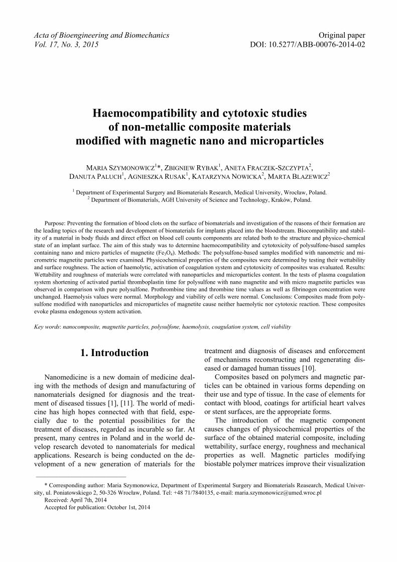
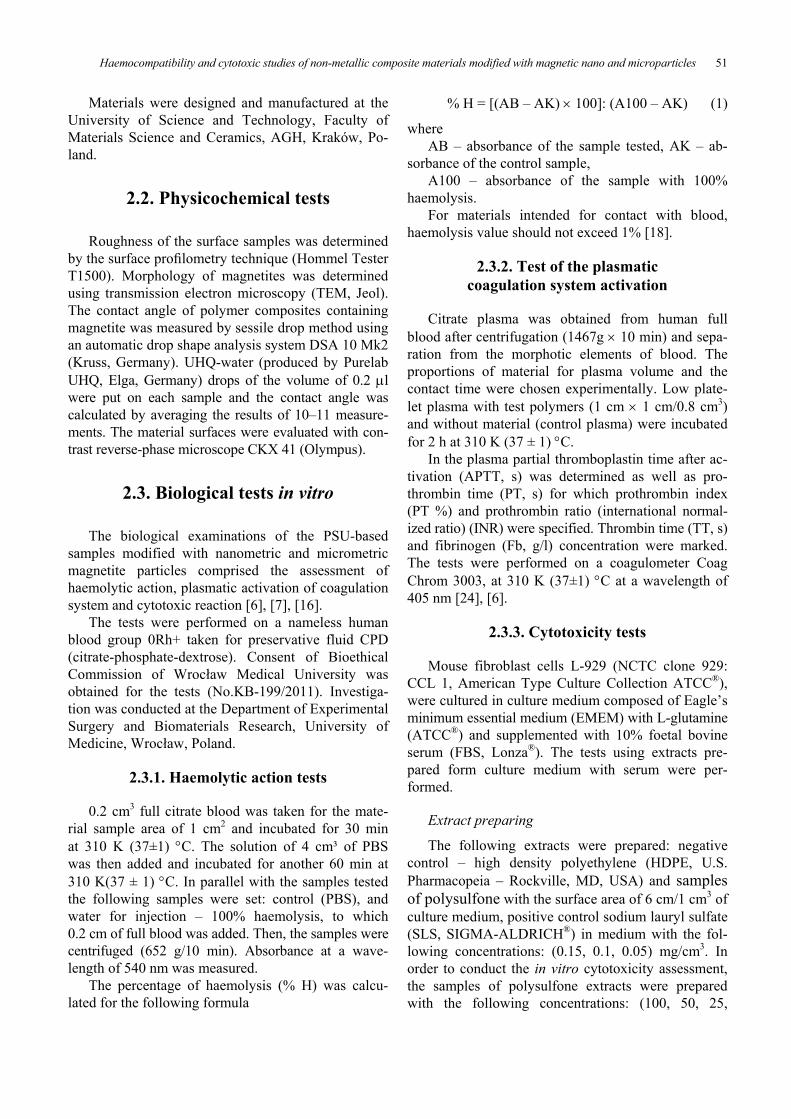
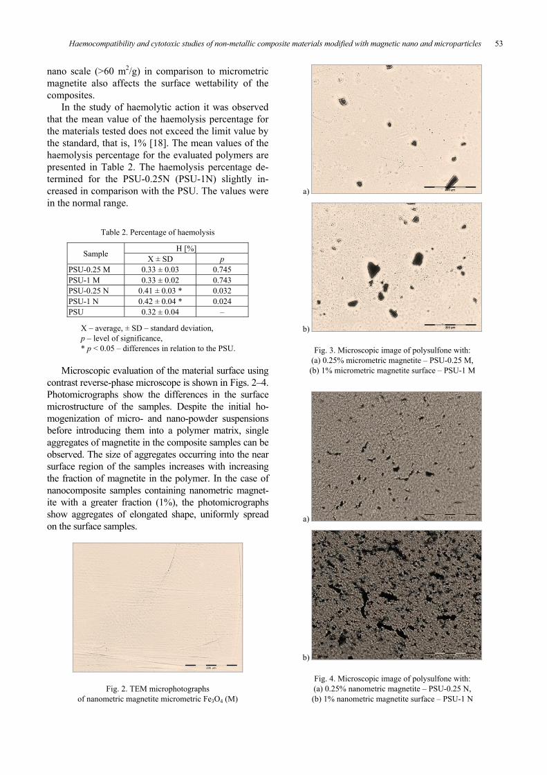
Microscopic evaluation of the material surface usingcontrast reverse-phase microscope is shown in Figs. 2–4.Photomicrographs show the differences in the surfacemicrostructure of the samples. Despite the initial ho-mogenization of micro- and nano-powder suspensionsbefore introducing them into a polymer matrix, singleaggregates of magnetite in the composite samples can beobserved. The size of aggregates occurring into the nearsurface region of the samples increases with increasingthe fraction of magnetite in the polymer. In the case ofnanocomposite samples containing nanometric magnet-ite with a greater fraction (1%), the photomicrographsshow aggregates of elongated shape, uniformly spreadon the surface samples.
Fig. 2. TEM microphotographsof nanometric magnetite micrometric Fe3O4 (M)
a)
b)
Fig. 3. Microscopic image of polysulfone with:(a) 0.25% micrometric magnetite – PSU-0.25 M,(b) 1% micrometric magnetite surface – PSU-1 M
a)
b)
Fig. 4. Microscopic image of polysulfone with:(a) 0.25% nanometric magnetite – PSU-0.25 N,(b) 1% nanometric magnetite surface – PSU-1 N

M. SZYMONOWICZ et al.54
The parameters of plasma coagulation system forthe control plasma, and after contact with the poly-mers are presented in Table 3. APTT, PT, TT and Fbvalues obtained for plasma after contact with themodified polysulfones were compared with the val-ues for plasma after contact with pure polysulfoneand with the value of the material-free plasma (con-trol).
Partial thromboplastin time after activation (APTT)of blood plasma after 120 min contact with PSU-1Mwas significantly ( p < 0.001) shortened, while forPSU-0.25M it was comparable to the APTT valueobserved for PSU. APTT value observed for PSU-1Nwas significantly ( p < 0.05) shortened and for thePSU-0.25N close to the value observed for the PSU.APTT values observed for the polymers under study
Table 3. Activated partial thromboplastin time (APTT) in the citrate blood plasma controland after contact with magnetite-modified PSU samples
APTT [s]Sample X ± SD p p1PSU-0,25 M 38.26 ± 0.25 ### 0.162 0.0008PSU-1 M 34.53 ± 0.38*** ### 0.0007 0.0008PSU-0,25 N 38.00 ± 0.11 ### 0.448 0.0007PSU-1 N 36.80 ± 0.40 * # 0.037 0.024PSU 37.80 ± 0.40 # – 0.003Control plasma 35.57 ± 0.45 –
Reference range APTT: 29.20 s– 36.50 s – 43.80 s,X – average, ± SD – standard deviation, p – level of significance,* p < 0.05, *** p < 0.001 – differences in relation to the PSU,# p < 0.05, ### p < 0.001 – differences in relation to the control plasma.
Table 4. Prothrombin time (PT) in the citrate blood plasma controland after contact with the magnetite-modified PSU samples
PTs INR PT, [%]Sample
X ± SD p p1 X ± SD p p1 X ± SD p p1PSU-0,25 M 12.20 ± 0.20 0.272 0.411 0.93 ± 0.02 0.411 0.411 107.33 ± 3.51 1.000 1.000PSU-1 M 15.06 ± 0.06 0.482 0.562 0.94 ± 0.01 1.000 1.000 108.33 ± 2.08 0.587 0.624PSU-0,25 N 12.40 ± 0.30 0.656 0.748 0.94 ± 0.02 0.829 0.829 107.00 ± 3.00 0.882 0.889PSU-1 N 12.13 ± 0.05 0.488 0.386 0.93 ± 0.02 0.411 0.411 109.66 ± 2.51 0.283 0.319PSU 12.30 ± 0.20 – 0.829 0.94 ± 0.02 – 1.000 107.33 ± 2.08 0.052 1.000Control, plasma 12.33 ± 0.15 – 0.94 ± 0.02 – 88.33 ± 0.57 –
Reference range: PT: 10.60 s – 13.20 s – 15.80 s; PT INR: 0.78 –0.99 – 1.20; PT%: 84.00 – 102.00 –125.00,INR – prothrombin ratio (international normalized ratio),PT% – prothrombin index,X – average, ± SD – standard deviation, p – level of significance.
Table 5. Thrombin time (TT) and fibrinogen concentration (Fb)in the citrate blood plasma control and after contact with magnetite-modified PSU samples
TT [s] Fb [g/l]Sample X ± SD p p1 X ± SD p p1PSU-0,25 M 11.03 ± 0.06 0.518 0.068 2.05 ± 0.06 0.169 0.278PSU-1 M 11.07 ± 0.06 0.101 0.102 2.02 ± 0.03 0.137 0,426PSU-0,25 N 11.17 ± 0.05 1.000 0.348 2.04 ± 0.03 0.405 0,313PSU-1 N 11.07 ± 0.12 1.000 0.144 2.01 ± 0.04 0.183 0,808PSU 11.07 ± 0.10 – 0.101 2.06 ± 0.04 – 0,557Control, plasma 11.26 ± 0.15 – 2.06 ± 0.04
Reference range TT: 10.30 s – 12.50 s – 15.50 s Fb: 2.55 g/L – 3.19 g/L – 3.82 g/l,X – average, ± SD – standard deviation, p – level of significance.

Haemocompatibility and cytotoxic studies of non-metallic composite materials modified with magnetic nano and microparticles 55
were significantly ( p < 0.05, p < 0.001) prolongedcompared to the control plasma APTT values. Theobserved changes in the values of the APPT do notexceed the reference values.
Prothrombin time (PT), prothrombin index PT%,prothrombin ratio (INR) thrombin time (TT) andfibrinogen plasma concentrations of blood plasmaafter contact with the test polymers were not sig-nificantly different from control values in plasma(Table 4).
PT values and plasma concentrations of fibrino-gen found for the polymers studied were not signifi-cantly different ( p > 0.05) from the values of theseparameters in plasma control. The values were com-parable to each other and close to the values obtainedfor PSU.
The results of cytotoxicity test are shown in Table 6.PSU sample and its composites with magnetite:PSU-0.25M; PSU-1M; PSU-0.25N; PSU-1N did notinduce morphological changes in cultured cells anddid not reduce cell viability above 30%.
a)
b)
Fig. 5. Cells culture after contact with:(a) negative control – HDPE, (b) positive control – SLS 0.15 mg/cm3
Table 6. Results of the in vitro cytotoxicity assessment of magnetite-modified PSU samples
Extract Cell viability[ % ] Description Grade
100% 94.2 Discrete intracytoplasmatic granules, no cell lysis (Fig. 2a) 050% 95.2 Discrete intracytoplasmatic granules, no cell lysis 025% 100.6 Discrete intracytoplasmatic granules, no cell lysis, density of culture
is comparable to density of negative control culture 0PSU
12.5% 100.3 Discrete intracytoplasmatic granules, no cell lysis, density of cultureis comparable to density of negative control culture 0
100% 95.2 Discrete intracytoplasmatic granules, no cell lysis (Fig. 2b) 0
50% 98.2 Discrete intracytoplasmatic granules, no cell lysis, density of cultureis comparable to density of negative control culture 0
25% 97.93 Discrete intracytoplasmatic granules, no cell lysis, density of cultureis comparable to density of negative control culture 0
PSU
-25
M
12.5% 99.0 Discrete intracytoplasmatic granules, no cell lysis, density of cultureis comparable to density of negative control culture 0
100% 95.0 Discrete intracytoplasmatic granules, no cell lysis (Fig. 2c) 050% 96.3 Discrete intracytoplasmatic granules, no cell lysis 025% 95.9 Discrete intracytoplasmatic granules, no cell lysis 0
PSU
- 1M
12.5% 99.3 Discrete intracytoplasmatic granules, no cell lysis, density of cultureis comparable to density of negative control culture 0
100% 87.9 Around 15% of the cells are round, loosely attached,Culture density lower than the negative control (Fig. 2d) 1
50% 91.2 Around 10% of the cells are round, loosely attached,Culture density lower than the negative control 1
25% 97.7 Discrete intracytoplasmatic granules, no cell lysis, density of cultureis comparable to density of negative control culture 0
PSU
-0.2
5 N
12.5% 99.3 Discrete intracytoplasmatic granules, no cell lysis, density of cultureis comparable to density of negative control culture 0
100% 77.7 Around 20% of the cells are round, loosely attached,Culture density lower than the negative control (Fig. 2e) 1
50% 77.9 Around 20% of the cells are round, loosely attached,Culture density lower than the negative control 1
25% 88.1 Around 10% of the cells are round, loosely attached,Culture density lower than the negative control 1PS
U-1
N
12.5% 88.7 Around 10% of the cells are round, loosely attached,Culture density lower than the negative control 1
M. SZYMONOWICZ et al.56
a)
b)
c)
d)
e)
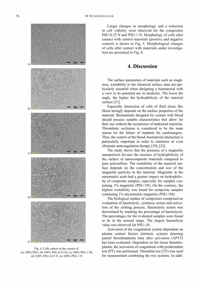
Fig. 6. Cells culture in the extract of:(a) 100% PSU, (b) 100% PSU-0.25 M, (c) 100% PSU-1 M,
(d) 100% PSU-0.25 N, (e) 100% PSU-1 N
Larger changes in morphology and a reductionin cell viability were observed for the compositesPSU-0.25 N and PSU-1 N. Morphology of cells aftercontact with control materials (positive and negativecontrol) is shown in Fig. 5. Morphological changesof cells after contact with materials under investiga-tion are presented in Fig. 6.
4. Discussion
The surface parameters of materials such as rough-ness, wettability or the chemical surface state are par-ticularly essential when designing a biomaterial witha view to its potential use in medicine. The lower theangle, the higher the hydrophilicity of the materialsurface [23].
Especially interaction of cells of fluid tissue likeblood strongly depends on the surface properties of thematerial. Biomaterials designed for contact with bloodshould possess suitable characteristics that allow fortheir use without the occurrence of undesired reactions.Thrombotic occlusion is considered to be the mainreason for the failure of implants for cardiosurgery.Thus, the control of the blood–biomaterial interaction isparticularly important in order to minimize or eveneliminate anticoagulation therapy [19], [22].
The study shows that the presence of a magnetitenanoparticle favours the increase of hydrophilicity ofthe surface of nanocomposite materials compared topure polysulfone. The wettability of the material sur-face depends on the concentration and size of themagnetite particles in the material. Magnetite in thenanometric scale had a greater impact on hydrophilic-ity of composite samples, especially for samples con-taining 1% magnetite (PSU-1N). On the contrary, thehighest wettability was found for composite samplescontaining 1% micrometric magnetite (PSU-1M).
The biological studies of composites comprised anevaluation of haemolytic, cytotoxic action and activa-tion of the clotting process. Haemolytic action wasdetermined by marking the percentage of haemolysis.The percentages for the evaluated samples were foundto be in the normal range. The largest haemolysisvalue was observed for PSU-1N.
Activation of the coagulation system dependent onplasma contact factors (intrinsic system) denotingpartial thromboplastin time after activation (APTT)has been evaluated. Dependent on the tissue thrombo-plastin, the activation of coagulation with prothrombintest (PT) was performed. Thrombin test (TT) was usedfor measurement combining the two systems. In addi-
Haemocompatibility and cytotoxic studies of non-metallic composite materials modified with magnetic nano and microparticles 57
tion, the concentration of fibrinogen was marked.Clotting activation of the composites was compared toAPTT pure polysulfone expressed by shortening thePSU-1M and PSU-1N, with no changes in the valuesof PT and fibrinogen concentration being observed.The highest shortening or stimulation of the extrinsiccoagulation factors was noted for PSU-1M. The ob-served changes may be attributed to higher surfaceroughness of this sample in comparison with othermaterials studied.
In assessing the morphological pictures and long-life cell culture, there were no significant changes.Only major changes in cell morphology and reducedcell viability were demonstrated for PSU-0.25 N andPSU-1N.
Blood tests of haemolytic activity, blood cell reac-tivity, coagulation activity and cell culture cytotoxicityas well as evaluation of topography surface of nano-and micro-engineered composite samples allowed ma-terials with optimal biological properties to be selected.In the evaluated materials the smallest changes wereobserved for PSU-0.25M. This will allow us to performfurther studies in vitro and in vivo. In the future, suchmaterials may be used to design and manufacture theimplants for bone tissue reconstruction and as biostabledevices in contact with blood.
Polysulfone has been used for years for mem-branes in dialyzers, and also as a material for the con-struction of elements and devices in contact withblood. Surface modification of this polymer with theuse of nanoparticles to alter its hydrophobicity andsurface roughness may contribute to extend its rangeof applications in cardiac surgery. Nano-engineeredsample surfaces can affect in an entirely different wayan answer of proteins responsible for thrombotic pro-cesses, and consequently the haemocompatibility ofthe whole material.
5. Conclusions
1. Composites made from polysulfone modified withnanoparticles and microparticles of magnetite donot cause haemolytic and cytotoxic reaction.
2. These composites cause plasma endogenous sys-tem activation without an influence on the exoge-nous system. These changes are within referentialvalues and thiss has a diagnostic significance.
3. Changes in coagulation activation and the mor-phology of the cell cultures are connected with thesurface properties of the composites tested, such aswettability and roughness.
Acknowledgements
The work was supported by the project no. ST-619 of theWrocław Medical University, Poland and the statute funds ofAGH – University of Science and Technology, Faculty of MaterialsScience and Ceramics, Poland project No. 11.11.160.256.
References
[1] CAO Q., HAN X., LI L., Enhancement of the efficiency ofmagnetic targeting for drug delivery: Development andevaluation of magnet system, J. Magn. Magn. Mater., 2011,Vol. 323, 1919–1924.
[2] CHŁOPEK J., Kompozyty w medycynie, Kompozyty (Compos-ites), 2001, Vol. 1, 50–54.
[3] DAHE G.J., KADAM S.S., SABALE S.S., KADAM D.P.,SARKATE L.B., BELLARE J.R., In Vivo Evaluation of thebiocompatibility of surface modified hemodialysis poly-sulfone hollow fibers in rat, PLoS ONE, 2011, Vol. 6(10),1–9.
[4] DAHE G.J., TEOTIA R.S., KADAM S.S., BELLARE J.R., Thebiocompatibility and separation performance of antioxidativepolysulfone/vitamin E TPGS composite hollow fiber mem-branes, Biomaterials, 2011, Vol. 32, 352–365.
[5] GUPTA A.K., GUPTA M., Synthesis and surface engineeringof iron oxide nanoparticles for biomedical applications,Biomaterials, 2005, Vol. 26(18), 3995–4021.
[6] ISO 10993-4: 2009, Biological evaluation of medical devices.Part 4: Selection of tests for interactions with blood.
[7] ISO 10993-5 2009, Tests for in vitro cytotoxicity.[8] LAURENT S., FORGE D., PORT M., ROCH A., ROBIC C.,
VANDER ELST L., Magnetic ironoxide nanoparticles: synthe-sis, stabilization, vectorization, physicochemical characteri-zations, and biological applications, Chem. Rev., 2008, Vol.108(6), 2064–2110.
[9] LINES S.W., CARTER A.M., DUNN E.J., LINDLEY E.J.,TATTERSALL J.E., WRIGHT M.J., A randomized controlledtrial evaluating the erythropoiesis stimulating agentsparing potential of a vitamin E-bonded polysulfone dialy-sis membrane, Nephrol. Dial. Transplant., 2014, Vol. 29,649–656.
[10] LIU H., WEBSTER T.J., Nanomedicine for implants: a reviewof studies and necessary experimental tools, Biomaterials,2007, Vol. 28, 354–369.
[11] MANGUAL J.O., LI S., PLOEHN H.J, EBNER A.D., RITTER J.A.,Biodegradable nanocomposite magnetite stent for implant-assisted magnetic drug targeting, J. Magn. Magn. Mater,2010, Vol. 322, 3094–3100.
[12] MAPHUTHA S., MOOTHI K., MEYYAPPAN M., IYUKE S.E.,A carbon nanotube-infused polysulfone membrane with poly-vinyl alcohol layer for treating oil-containing waste water,Scientific Reports, 2013, Vol. 3, 1–621.
[13] MCCARTHY J.R., WEISSLEDER R., Multifunctional magneticnanoparticles for targeted imaging and therapy, Adv. Drug.Deliv. Rev., 2008, Vol. 60(11), 1241–1251.
[14] MODRZEJEWSKA Z., PALUCH D., Investigation of the cytotoxicactivity of chitosan membranes to be used as dressing, Engi-neering of Biomaterials, 2011, Vol. 14 (103), 23–28.
[15] OH J.K., PARK J.M., Iron oxide-based superparamagneticpolymeric nanomaterials: Design, preparation, and biomedi-cal application, Progress in Polymer Science, 2011, Vol. 36,168–189.
M. SZYMONOWICZ et al.58
[16] SEYFERT U.T., BIEHL V., SCHENK J., In vitro hemocompati-bility testing of biomaterials according to the ISO 10993-4.Biomol. Eng., 2002, Vol. 19(2–6), 91–96.
[17] SHEN C., ZHANG G., MENG Q., Regulation of epithelial cellmorphology and functions approaching to more in vivo-likeby modifying polyethylene glycol on polysulfone membranes,PLoS ONE, 2012, Vol. 7(4), 1–10.
[18] SINGHAL J.P., RAY A., Synthesis of blood compatible polyamideblock copolymers, Biomaterials, 2002, Vol. 23, 1139–1245.
[19] SPIJKER H.T.R., GRAAFF P.W., BOONSTRA H.J., BUSSCHERW., VAN OEVEREN, On the influence of flow conditions andwettability on blood material interactions, Biomaterials,2003, Vol. 24, 4717–4727.
[20] STANKOVA L., FRACZEK-SZCZYPTA A., BLAZEWICZ M., FILOVAE., BLAZEWICZ S., LISA V., BACAKOVA L., Human osteoblast-like MG 63 cells on polysulfone modified with carbon nanotubesor carbon nanohorns, Carbon, 2014, Vol. 67, 578–591.
[21] SWIETEK M., TOKARZ W., FIGIEL H, TARASIUK J., WRONSKI S.,BLAZEWICZ M., Magnetic polymer sponge for medical appli-cations, Innovative technologies in biomedicine: the 1st In-
ternational Conference: October 15–16, 2013, Kraków,Poland, 52–53.
[22] SZELEST-LEWANDOWSKA A., MASIULANIS M., SZYMONOWICZM., PIELKA S., PALUCH D., Modified Poly (carbonateure-thane). Synthesis, properties and biological investigation invitro, Journal of Biomedical Materials Research part A,2007, Vol. 82(12), 509–520.
[23] SZYMONOWICZ M., FRACZEK-SZCZYPTA A., RYBAK Z.,BLAZEWICZ S., Comparative assesment of the effect ofcarbon-based material surfaces on blood clotting activa-tion and haemolysis, Diamond & Related Materials, 2013,Vol. 40, 89–95.
[24] SZYMONOWICZ M., PIELKA S., OWCZAREK A, HAZNAR D.,PLUTA J., Study on influence of gelatin-alginate matrixes onthe coagulation system and morphotic blood elements, Mac-romolecular Symposia, 2007, Vol. 253(1), 71–76.
[25] WOJCIK M., CHMIST J., PRZEWOZNIK J., FIGIEL H., BLAZEWICZ S.,Magnetic properties of PAN-based carbon fibres modifiedwith magnetite nanoparticles, Carbon, 2012, Vol. 50(4),1604–1613.