H-Ras and K-Ras oncoproteins induce different tumor spectra when ...
44
1 H-Ras and K-Ras oncoproteins induce different tumor spectra when driven by the same regulatory sequences by Matthias Drosten 1 , Lucía Simón-Carrasco 1 , Isabel Hernández-Porras 1 , Carmen G. Lechuga 1 , María T. Blasco 1 , Harrys K. C. Jacob 2 , Salvatore Fabbiano 2,4 , Nicoletta Potenza 3 , Xosé R. Bustelo 2 , Carmen Guerra 1 and Mariano Barbacid 1 1 Molecular Oncology Programme, Centro Nacional de Investigaciones Oncológicas (CNIO), Melchor Fernández Almagro 3, 28029 Madrid, Spain 2 Centro de Investigación del Cáncer and Instituto de Biología Molecular y Celular del Cáncer, CSIC-Universidad de Salamanca, Campus Miguel Unamuno s/n, 37007 Salamanca, Spain and Centro de Investigación Biomédica en Red-Oncología, Carlos III Health Institute, Spain 3 Department of Environmental, Biological and Pharmaceutical Sciences and Technologies (DiSTABiF), Second University of Naples, 81100 Caserta, Italy 4 Present address: Department of Cell Physiology and Metabolism, Centre Médical Universitaire (CMU) and Diabetes Centre, Faculty of Medicine, University of Geneva, 1206 Geneva, Switzerland Corresponding authors: Matthias Drosten, Molecular Oncology Programme, Centro Nacional de Investigaciones Oncológicas (CNIO), Melchor Fernández Almagro 3, 28029 Madrid, Spain, Phone: +34917328000, Fax: +34917328033, E-mail: [email protected] and Mariano Barbacid, Molecular Oncology Programme, Centro Nacional de Investigaciones Oncológicas (CNIO), Melchor Fernández Almagro 3, 28029 Madrid, Spain, Phone: +34917328000, Fax: +34917328033, E-mail: [email protected] Keywords: H-Ras / K-Ras / Ras oncoproteins / tumor development / oncogene-induced senescence DISCLOSURE OF POTENTIAL CONFLICTS OF INTERESTS None of the authors have any potential conflicts of interest. on April 14, 2018. © 2016 American Association for Cancer Research. cancerres.aacrjournals.org Downloaded from Author manuscripts have been peer reviewed and accepted for publication but have not yet been edited. Author Manuscript Published OnlineFirst on November 21, 2016; DOI: 10.1158/0008-5472.CAN-16-2925
-
Upload
nguyentruc -
Category
Documents
-
view
221 -
download
1
Transcript of H-Ras and K-Ras oncoproteins induce different tumor spectra when ...

1
H-Ras and K-Ras oncoproteins induce different tumor spectra when driven by the same regulatory sequences
by
Matthias Drosten1, Lucía Simón-Carrasco1, Isabel Hernández-Porras1, Carmen G.
Lechuga1, María T. Blasco1, Harrys K. C. Jacob2, Salvatore Fabbiano2,4, Nicoletta
Potenza3, Xosé R. Bustelo2, Carmen Guerra1 and Mariano Barbacid1
1Molecular Oncology Programme, Centro Nacional de Investigaciones Oncológicas
(CNIO), Melchor Fernández Almagro 3, 28029 Madrid, Spain 2Centro de Investigación del Cáncer and Instituto de Biología Molecular y Celular del
Cáncer, CSIC-Universidad de Salamanca, Campus Miguel Unamuno s/n, 37007
Salamanca, Spain and Centro de Investigación Biomédica en Red-Oncología, Carlos III
Health Institute, Spain 3 Department of Environmental, Biological and Pharmaceutical Sciences and
Technologies (DiSTABiF), Second University of Naples, 81100 Caserta, Italy 4Present address: Department of Cell Physiology and Metabolism, Centre Médical
Universitaire (CMU) and Diabetes Centre, Faculty of Medicine, University of Geneva,
1206 Geneva, Switzerland
Corresponding authors: Matthias Drosten, Molecular Oncology Programme, Centro
Nacional de Investigaciones Oncológicas (CNIO), Melchor Fernández Almagro 3,
28029 Madrid, Spain, Phone: +34917328000, Fax: +34917328033, E-mail:
[email protected] and Mariano Barbacid, Molecular Oncology Programme, Centro
Nacional de Investigaciones Oncológicas (CNIO), Melchor Fernández Almagro 3,
28029 Madrid, Spain, Phone: +34917328000, Fax: +34917328033, E-mail:
Keywords: H-Ras / K-Ras / Ras oncoproteins / tumor development / oncogene-induced
senescence
DISCLOSURE OF POTENTIAL CONFLICTS OF INTERESTS
None of the authors have any potential conflicts of interest.
on April 14, 2018. © 2016 American Association for Cancer Research. cancerres.aacrjournals.org Downloaded from
Author manuscripts have been peer reviewed and accepted for publication but have not yet been edited. Author Manuscript Published OnlineFirst on November 21, 2016; DOI: 10.1158/0008-5472.CAN-16-2925

2
ABSTRACT
Genetic studies in mice have provided evidence that H-Ras and K-Ras proteins are
bioequivalent. However, human tumors display marked differences in the association of
RAS oncogenes with tumor type. Thus, to further assess the bioequivalency of
oncogenic H-Ras and K-Ras, we replaced the coding region of the murine K-Ras locus
with H-RasG12V oncogene sequences. Germline expression of H-RasG12V or K-RasG12V
from the K-Ras locus resulted in embryonic lethality. However, expression of these
genes in adult mice led to different tumor phenotypes. Whereas H-RasG12V elicited
papillomas and hematopoietic tumors, K-RasG12V induced lung tumors and gastric
lesions. Pulmonary expression of H-RasG12V created a senescence-like state caused by
excessive MAP kinase signaling. Likewise, H-RasG12V but not K-RasG12V induced
senescence in mouse embryo fibroblasts. Label-free quantitative analysis revealed that
minor differences in H-RasG12V expression levels led to drastically different biological
outputs, suggesting that subtle differences in MAP kinase signaling confer non-
equivalent functions that influence tumor spectra induced by RAS oncoproteins.
on April 14, 2018. © 2016 American Association for Cancer Research. cancerres.aacrjournals.org Downloaded from
Author manuscripts have been peer reviewed and accepted for publication but have not yet been edited. Author Manuscript Published OnlineFirst on November 21, 2016; DOI: 10.1158/0008-5472.CAN-16-2925

3
Introduction
Mammals contain three Ras loci, H-Ras, K-Ras and N-Ras, that encode highly related
proteins (1-4). Ras proteins are small GTPases that function as mitogenic switches to
control the transmission of extracellular signals to the nucleus (1). They share extensive
homology at their N-terminal half, a region involved in nucleotide binding and
interaction with downstream effectors (2). Their unique features reside in their carboxy-
terminal half that includes the hypervariable region and a terminal domain known as the
CAAX box (2). These structural motifs have been implicated in differential transport,
post-translational processing and subcellular localization of the different Ras proteins
(3, 4).
Early knock-out data revealed significant functional differences for the three Ras loci.
Whereas H- and N-Ras were dispensable for embryonic development, K-Ras was
essential (5-7). These observations did not establish whether these differences were due
to the intrinsic properties of their cognate Ras proteins or to their patterns of expression.
This issue was solved, at least in part, when Potenza et al. replaced mouse K-Ras alleles
by chimeric K/H-Ras alleles encoding functional H-Ras proteins (8). These mice
developed to adulthood despite the absence of K-Ras, indicating that the requirement
for K-Ras alleles during embryonic development is primarily due to their pattern of
expression. Yet, these mice displayed cardiovascular defects, thus raising the possibility
that H-Ras and K-Ras proteins might have differential signaling properties, at least in
certain tissues (8).
RAS genes have also attracted interest due to their involvement in tumor development
(1, 2). The overall incidence of each RAS oncogene varies significantly among tumor
on April 14, 2018. © 2016 American Association for Cancer Research. cancerres.aacrjournals.org Downloaded from
Author manuscripts have been peer reviewed and accepted for publication but have not yet been edited. Author Manuscript Published OnlineFirst on November 21, 2016; DOI: 10.1158/0008-5472.CAN-16-2925

4
types (9). Whereas KRAS is frequently mutated in pancreatic, colorectal and lung
adenocarcinomas, HRAS oncogenes are found in a limited percentage of tumors from
the salivary gland, urinary track, cervix, or skin. On the other hand, NRAS oncogenes
are present in melanomas and hematopoietic tumors. As of to date, the molecular basis
for this incidence bias is still unresolved (9).
Mutant RAS genes also induce different phenotypes when expressed in the germline of
patients suffering from RASopathies, a series of developmental defects that result from
constitutive activation of RAS signaling pathways (10, 11). Oncogenic mutations in
HRAS lead to relatively mild developmental defects in Costello syndrome patients (12,
13). In contrast, those KRAS mutations present in human tumors have not been found in
RASopathy patients, suggesting that such mutations may cause embryonic lethality
(14).
Similar results have been observed in mouse strains carrying genetically engineered Ras
mutations. Whereas expression of resident K-Ras oncoproteins in the germline leads to
early embryonic death, expression of an endogenous H-Ras oncogene is well tolerated
and leads to developmental defects very similar to those observed in Costello patients
(15-18). Likewise, germline expression of the partially activated K-RasV14I mutant
isoform results in phenotypic defects that closely resemble those of Noonan patients
(19).
Here we provide genetic evidence that the wild-type H-Ras and K-Ras proteins are
bioequivalent in spite of their different structural and biological properties. We have
also compared the spectrum of tumors elicited upon expression of the H-RasG12V and K-
on April 14, 2018. © 2016 American Association for Cancer Research. cancerres.aacrjournals.org Downloaded from
Author manuscripts have been peer reviewed and accepted for publication but have not yet been edited. Author Manuscript Published OnlineFirst on November 21, 2016; DOI: 10.1158/0008-5472.CAN-16-2925

5
RasG12V oncoproteins from the same mouse K-Ras locus. These studies have revealed
that these oncoproteins induce a different spectrum of tumors primarily due to
differences in the intensity of MAP Kinase signaling that results from subtle differences
in their levels of expression.
on April 14, 2018. © 2016 American Association for Cancer Research. cancerres.aacrjournals.org Downloaded from
Author manuscripts have been peer reviewed and accepted for publication but have not yet been edited. Author Manuscript Published OnlineFirst on November 21, 2016; DOI: 10.1158/0008-5472.CAN-16-2925

6
MATERIALS AND METHODS
Mouse strains. Generation of K-Ras+/LSLH-RasG12V mice (KHRasV12) is described in
Supplementary information. KrasKI (8), HrasKI (8), H-Ras–/– (5), K-Ras+/LSLG12Vgeo
(KV12) (16) and p53lox/lox (20) genetically engineered strains have been described.
hUBC-CreERT221, Elas-tTA and tetO-Cre transgenic strains have also been reported
(21, 22). Activation of the inducible CreERT2 recombinase was achieved by feeding the
mice with a Tmx-containing diet (Harlan Laboratories). For activation of H-RasG12V and
K-RasG12V expression in lung tissue, mice were infected with Adeno-Cre particles as
described (23). Trametinib (Mekinist®) was purchased from Sellek Chemicals and was
administered orally daily (1 mg/kg) for 8 weeks. All mice were maintained in a mixed
129Sv/J x C57BL/6j background and housed in a barrier facility according to standards
established by the European Union. All animal experiments were approved by the
CNIO, the Carlos III Health Institute and the Comunidad de Madrid Ethical Committees
and performed in accordance with the ARRIVE (Animal Research: Reporting on In
Vivo Experiments) guidelines.
Histopathology, immunohistochemistry and SA β-Gal staining. Tissues were fixed
in 10% buffered formalin and embedded in paraffin. Hematoxilin & Eosin (H&E)
staining and immunohistochemistry (IHC) analyses were performed on 3 μm paraffin
sections. For IHC, the following antibodies were used: pErk1/2 (9101, Cell Signaling
Technology), Active Caspase-3 (MAB835, R&D Systems), SPC (AB3786, Abcam),
CC10 (T-18, Santa Cruz Biotechnology), CD3e (clone 145-2C11, Abcam). SA β-Gal
activity in cultured MEFs as well as in cryo-sections of lungs was detected by X-Gal
staining as described previously (24).
on April 14, 2018. © 2016 American Association for Cancer Research. cancerres.aacrjournals.org Downloaded from
Author manuscripts have been peer reviewed and accepted for publication but have not yet been edited. Author Manuscript Published OnlineFirst on November 21, 2016; DOI: 10.1158/0008-5472.CAN-16-2925

7
Southern and Western blot analysis. Southern blot analysis is described in
Supplementary information. Western blot analysis of protein extracts obtained from
total lung tissue or MEFs was performed as described (25). Primary antibodies used
included: Pan-Ras (OP40, Merck Millipore), H-Ras (clone 18, BD Transduction Labs),
pErk1/2 (9101), p53 (2524), pAkt (9271), Akt (9272) [all from Cell Signaling
Technology], p19Arf (clone 5-C3-1, Upstate), Erk1 (C16), p16INK4a (M-156) [all from
Santa Cruz Biotechnology], GAPDH (G8795, Sigma-Aldrich).
Statistical analysis. All statistical analyses were performed using an unpaired Student’s
t-test. P values < 0.05 were considered to be statistically significant (* P < 0.05, *** P
< 0.001).
on April 14, 2018. © 2016 American Association for Cancer Research. cancerres.aacrjournals.org Downloaded from
Author manuscripts have been peer reviewed and accepted for publication but have not yet been edited. Author Manuscript Published OnlineFirst on November 21, 2016; DOI: 10.1158/0008-5472.CAN-16-2925

8
RESULTS
H-Ras and K-Ras are bioequivalent proteins. Germline ablation of K-Ras results in
embryonic lethality (6, 7). Yet, expression of H-Ras proteins under the control of K-Ras
regulatory sequences results in viable mice, thus illustrating that H-Ras can replace K-
Ras isoforms, for most biological activities (8). Yet, these mice, designated as HrasKI,
displayed dilated cardiomyopathy and arterial hypertension when they reached
adulthood (8). These cardiovascular defects were initially attributed to the absence of K-
Ras proteins in heart tissue. Subsequent studies however, revealed that germline
expression of constitutively active H-RasG12V led to similar cardiovascular defects in a
mouse model of Costello syndrome (17). Thus, we reasoned that these cardiovascular
defects might be due to H-Ras overexpression due to the presence of four H-Ras coding
alleles (the knocked-in HrasKI alleles and the endogenous H-Ras alleles). To reduce the
load of H-Ras expression, we crossed HrasKI mice with H-Ras–/– animals. HrasKI;H-
Ras–/– mice displayed normal heart function and hypertension (Fig. 1A and B). In
addition, these mice displayed normal cardiomyocyte areas and did not accumulate
fibrosis in their heart (Fig. 1C and D). These results indicate that the cardiovascular
defects of HrasKI mice were due to H-Ras overexpression and not to differential
activities between H-Ras and K-Ras proteins.
Germline expression of the H-RasG12V oncoprotein from the K-Ras locus results in
embryonic lethality. Next, we interrogated whether their oncogenic isoforms, K-
RasG12V and H-RasG12V, also have similar properties. To this end, we knocked-in a
cDNA encoding an H-RasG12V oncogene within the first coding exon of the K-Ras locus
(Fig. 2A). We also knocked-in a lox-STOP-lox (LSL) cassette upstream of the initiator
codon to prevent expression of H-RasG12V (Supplementary Fig. S1A-C). Expression of
on April 14, 2018. © 2016 American Association for Cancer Research. cancerres.aacrjournals.org Downloaded from
Author manuscripts have been peer reviewed and accepted for publication but have not yet been edited. Author Manuscript Published OnlineFirst on November 21, 2016; DOI: 10.1158/0008-5472.CAN-16-2925

9
the H-RasG12V cDNA clone and a genomic DNA fragment containing the four H-Ras
coding exons resulted in similar expression levels indicating that the H-Ras intronic
sequences do not play a significant role in regulating H-Ras expression (Fig. 2B and C).
For simplicity, the wild-type K-Ras+/+ mice and the targeted K-Ras+/LSLG12Vgeo and K-
Ras+/LSLH-RasG12V strains will be referred to hereafter as K+, KV12 and KHRasV12,
respectively.
Previous studies have indicated that expression of a K-RasG12V oncogene in the mouse
germline results in embryonic lethality (16). In contrast, expression of the H-RasG12V
oncogene, even in homozygosity, is well tolerated during embryonic development (17,
18). To determine whether this differential effect is an intrinsic property of the K-
RasG12V and H-RasG12V oncoproteins, we crossed KHRasV12 mice to EIIA-Cre transgenics
(26) to express the H-RasG12V oncoprotein from the K-Ras locus in the germline. These
embryos were no longer viable and died right after implantation, a time similar to that
observed for embryos expressing an endogenous K-RasG12V oncoprotein (16). Hence,
the ability of mice, as well as of Costello syndrome patients, to tolerate expression of an
oncogenic H-RasG12V protein during embryonic development is dictated by the
expression pattern of the H-Ras locus.
Expression of H-RasG12V from the K-Ras locus in adult mice. Next, we explored the
oncogenic properties of the K-RasG12V and H-RasG12V isoforms expressed from the K-
Ras locus in adult mice. To this end, we inserted in the KV12 and KHRasV12 strains a
transgene encoding the inducible CreERT2 recombinase under the control of the human
ubiquitin C promoter (21). We exposed KV12;hUBC-CreERT2+/T and KHRasV12;hUBC-
CreERT2+/T mice to a continuous Tmx diet to ensure efficient recombination of the
on April 14, 2018. © 2016 American Association for Cancer Research. cancerres.aacrjournals.org Downloaded from
Author manuscripts have been peer reviewed and accepted for publication but have not yet been edited. Author Manuscript Published OnlineFirst on November 21, 2016; DOI: 10.1158/0008-5472.CAN-16-2925

10
targeted K-Ras alleles. Under these conditions, KV12;hUBC-CreERT2+/T mice had a
median survival of 24 weeks (Fig. 2D). In agreement with previous studies (27), these
mice displayed multiple lesions in their lungs as well as abundant gastric papillomas
(Fig. 2E). No other tissue displayed significant alterations at the histopathological level.
KHRasV12;hUBC-CreERT2+/T mice submitted to the same Tmx treatment died 4 to 5
weeks earlier (Fig. 2D). These mice did not develop detectable lesions in either lungs or
stomach (Fig. 2E) despite expression of the mutant H-RasG12V protein in these tissues
(Supplementary Fig. S1D). Instead, they displayed papillomas on their footpads after
approximately 2 months of treatment (Supplementary Fig. S1E). Moreover, they had
enlarged spleens and thymic tumors (Fig. 3). Tmx-treated control K+;hUBC-
CreERT2+/T mice did not display detectable lesions for up to one year of age. These
observations indicate that oncogenic signaling initiated by K-RasG12V and H-RasG12V
oncoproteins expressed under the same regulatory sequences has substantially different
consequences on tumor formation.
Expression of H-RasG12V from the K-Ras locus in adult mice induces hematopoietic
disorders. Post-mortem characterization of Tmx-treated KHRasV12;hUBC-CreERT2+/T
mice at humane endpoint revealed enlarged spleens infiltrated with myeloid cell
populations in 100% of the animals (Fig. 3A and B). This phenotype was not observed
in KV12;hUBC-CreERT2+/T or K+;hUBC-CreERT2+/T mice. Flow cytometry analyses of
these infiltrates revealed a dramatic expansion of Gr1+ and Gr1+/CD11b+ double-
positive cells indicative of myeloproliferative disease (MPD) (Fig. 3C and
Supplementary Fig. S2A). This increase in the myeloid cell compartment was
accompanied by a concomitant decrease in CD3+ T cells and CD19+ B cells
(Supplementary Fig. S2A). Analysis of committed progenitors in the bone marrow
on April 14, 2018. © 2016 American Association for Cancer Research. cancerres.aacrjournals.org Downloaded from
Author manuscripts have been peer reviewed and accepted for publication but have not yet been edited. Author Manuscript Published OnlineFirst on November 21, 2016; DOI: 10.1158/0008-5472.CAN-16-2925

11
(BM) of mice exposed to the Tmx diet for three months revealed a significant increase
in the common myeloid progenitors (CMPs) (Lin–/IL7Rα–/Sca-1–/c-
Kit+/FcγRlow/CD34+) as well as a slight expansion of granulocyte-macrophage
progenitor (GMP) population (Lin–/IL7Rα–/Sca-1–/c-Kit+/FcγRhigh/CD34+)
(Supplementary Fig. S2B). The common lymphoid progenitors (CLP) (Lin–
/IL7Rα+/Sca-1low/c-Kitlow), the megakaryocte-erythroid progenitors (MEP) (Lin–
/IL7Rα–/Sca-1–/c-Kit+/FcγRlow/CD34–) and LSK cells (Lin–/Sca-1+/c-Kit+), a population
enriched in hematopoietic stem cells, did not display significant changes. These
observations indicate that H-RasG12V but not K-RasG12V is capable of inducing MPD in
adult mice via expansion of a specific subset of committed progenitors in their BM.
We also detected large tumor masses in the thymus in over 85% of KHRasV12;hUBC-
CreERT2+/T animals, a pathology not observed in KV12;hUBC-CreERT2+/T or K+;hUBC-
CreERT2+/T mice (Fig. 3D). Histopathological characterization revealed the presence of
T cell lymphoblastic lymphoma (T-ALL), as determined by a uniform expansion of
CD3+ T lymphocytes. Moreover, tumors displayed an increase in double negative (DN)
CD4/CD8–, single positive CD4+ and single positive CD8+ cells (Fig. 3E). In particular,
when DN cells were further characterized, we detected elevated DN1 (CD44+/CD25–)
and DN2 (CD44+/CD25+) populations (Supplementary Fig. S3A). The characteristic
hyperactivation of the Notch pathway in T-ALL was also observed as demonstrated by
immunostaining of Hes1 (28) (Supplementary Fig. S3B). We also detected abundant
lymphocyte infiltrates in a variety of organs including lung, liver, kidney and eye
(Supplementary Fig. S3C). These data indicate that in addition to MPD, most mice
expressing the H-RasG12V oncoprotein from the K-Ras locus also developed T-ALL, a
tumor type not induced by the K-RasG12V isoform.
on April 14, 2018. © 2016 American Association for Cancer Research. cancerres.aacrjournals.org Downloaded from
Author manuscripts have been peer reviewed and accepted for publication but have not yet been edited. Author Manuscript Published OnlineFirst on November 21, 2016; DOI: 10.1158/0008-5472.CAN-16-2925

12
H-RasG12V driven from the K-Ras locus induces pancreatic tumors. Expression of a
resident K-RasG12V oncogene during late embryonic development in pancreatic acinar
cells results in pancreatic intraepithelial neoplasias (PanIN lesions) that occasionally
progress to pancreatic ductal adenocarcinomas (PDAC) (22). No such lesions were
observed in H-Ras+/G12V or H-RasG12V/G12V mice in which the H-RasG12V oncoprotein is
expressed from its own locus (17). To determine whether H-RasG12V could induce
pancreatic lesions when expressed from the K-Ras locus, we crossed KHRasV12 mice to
Elas-tTA/tetO-Cre transgenic animals known to selectively express Cre recombinase in
acinar cells from E16.5 onwards (22). Analysis of serial pancreatic tissue sections of
KHRasV12;Elas-tTA/tetO-Cre animals at one year of age revealed the appearance of focal
low and high-grade PanIN lesions indistinguishable from those present in KV12;Elas-
tTA/tetO-Cre mice (Supplementary Fig. S4A). However, the number of lesions was
significantly lower than in similar mice expressing the K-RasG12V oncoprotein (Fig.
4A).
Loss of the p53 tumor suppressor p53 accelerated tumor formation in KHRasV12;Elas-
tTA/tetO-Cre animals (22). These mice died of PDAC tumors although they survived
longer than those mice expressing the K-RasG12V oncoprotein (30 vs. 17 weeks average
survival, a 75% increase) (Fig. 4B and Supplementary Fig. S4A). Comparative analysis
of KHRasV12;p53lox/lox;Elas-tTA/tetO-Cre and KV12;p53lox/lox;Elas-tTA/tetO-Cre mice at
10 weeks of age revealed that the H-RasG12V expressing animals displayed significantly
fewer PanIN lesions and PDAC tumors than those expressing K-RasG12V (Fig. 4C).
These quantitative differences are likely to be due to a reduction in the number of
initiating events since the number of acinar-ductal metaplasias (ADM) was significantly
on April 14, 2018. © 2016 American Association for Cancer Research. cancerres.aacrjournals.org Downloaded from
Author manuscripts have been peer reviewed and accepted for publication but have not yet been edited. Author Manuscript Published OnlineFirst on November 21, 2016; DOI: 10.1158/0008-5472.CAN-16-2925

13
higher in acinar cell explants of KV12 mice than in those derived from KHRasV12 animals
(Fig. 4D and Supplementary Fig. S4B).
H-RasG12V failed to induce lung adenocarcinomas due to excessive MAPK
signaling. Systemic expression of H-RasG12V from the K-Ras locus did not yield lung
lesions including hyperplasias or benign adenomas. To rule out non-cell autonomous
effects, we infected KHRasV12 and KV12 mice with Adeno-Cre particles to selectively
induce oncogene expression in lung tissue. Six months after infection, none of the
KHRasV12 mice displayed detectable lesions in their lungs, whereas all KV12 animals had
developed multiple lesions including lung adenocarcinomas (Supplementary Fig. S4C
and S4D).
Tmx treatment of KV12;hUBC-CreERT2+/T and KHRasV12;hUBC-CreERT2+/T mice for
short periods of time allowed us to analyze the immediate events that followed
expression of the K-RasG12V and H-RasG12V oncoproteins in lung tissue. Four days after
Tmx treatment, H-RasG12V induced phosphorylation of the downstream Erk1/2 kinases
(pErk1/2) in over 20% of the cells, whereas expression of K-RasG12V only resulted in
pErk1/2 immunostaining in less than 5% of the cells (Fig. 5A). Similar results were
obtained seven days after treatment. These findings were not due to a differential
number of cells expressing the K-RasG12V and H-RasG12V oncoproteins since the levels
of Cre-mediated recombination in the lungs of KV12;hUBC-CreERT2+/T and
KHRasV12;hUBC-CreERT2+/T mice were similar (Supplementary Fig. S4E and S4F).
More importantly, when mice were analyzed two weeks after Tmx treatment, the
number of pErk1/2 expressing cells in the lungs of KHRasV12 mice decreased dramatically
whereas those present in KV12 lungs displayed a modest increase (Fig. 5A). These
on April 14, 2018. © 2016 American Association for Cancer Research. cancerres.aacrjournals.org Downloaded from
Author manuscripts have been peer reviewed and accepted for publication but have not yet been edited. Author Manuscript Published OnlineFirst on November 21, 2016; DOI: 10.1158/0008-5472.CAN-16-2925

14
results were further confirmed by Western blot analysis (Fig. 5B). These observations
were selective for lung cells since we did not observe significantly differential numbers
of pErk expressing cells in other tissues with the possible exception of cells in the basal
layer of the skin and in the white pulp of the spleen (Supplementary Fig. S5A). To
determine whether these results were due to differential expression levels of the two
mutant proteins, we compared their relative levels of expression in the lungs of
KV12;hUBC-CreERT2+/T and KHRasV12;hUBC-CreERT2+/T mice by a label-free
quantification approach (29). This technique allowed us to determine their relative
amounts based on the detection of the shared peptide (6-LVVVGAVGVGK-16) in
which the underlined residue corresponds to the activating valine (Supplementary Fig.
S5B). As illustrated in Fig. 5C, H-RasG12V is expressed at about 5-fold higher levels
than K-RasG12V. The higher levels of H-RasG12V expression also resulted in increased
levels of total GTP-bound active Ras complexes as determined by RBD pull down
assays (Fig. 5D).
Next, we examined whether the increased levels of GTP-bound H-RasG12V may have an
effect on the proliferation of H-RasG12V expressing cells that might prevent the
appearance of hyperplastic lesions. To this end, we used the recombinant K-RasH-RasG12V
allele as a molecular marker for the presence of H-RasG12V expressing lung cells.
Exposure of 4-week old KHRasV12;hUBC-CreERT2+/T mice to a Tmx diet for four weeks
resulted in the effective recombination of the K-RasLSLH-RasG12V allele, thus indicating
that a significant fraction of lung cells expressed the H-RasG12V oncoprotein (Fig. 6A).
However, when these KHRasV12;hUBC-CreERT2+/T mice were maintained in a diet
lacking Tmx for an additional 8 week period, those cells that contained the recombined
K-RasH-RasG12V allele diagnostic of H-RasG12V expression completely disappeared (Fig.
on April 14, 2018. © 2016 American Association for Cancer Research. cancerres.aacrjournals.org Downloaded from
Author manuscripts have been peer reviewed and accepted for publication but have not yet been edited. Author Manuscript Published OnlineFirst on November 21, 2016; DOI: 10.1158/0008-5472.CAN-16-2925

15
6A). These results were selective for lung cells since the K-RasH-RasG12V recombined
allele remains present in other tissues such as thymus, after the 8 week period in the
absence of Tmx (Fig. 6A). To determine whether the disappearance of the lung cells
was due to excessive H-RasG12V signaling, we treated KHRasV12;hUBC-CreERT2+/T mice
with a non-limiting dose (1 mg/kg) of Trametinib, a MEK inhibitor known to
effectively block MAPK signaling driven by Ras oncogenes (30). Trametinib was
provided during the 8 weeks in which mice were no longer exposed to Tmx. As
illustrated in Fig. 6A, these mice retained the K-RasH-RasG12V recombined allele
indicating that expression of the H-RasG12V oncoprotein was no longer deleterious for
lung cells in the presence of the MEK inhibitor.
H-RasG12V expression in lung cells induces a senescence-like arrest partially
mediated by p53. The disappearance of H-RasG12V expressing lung cells was not due to
apoptosis since we could not detect increased levels of Caspase 3 in the lungs of Tmx-
treated KHRasV12 mice (Supplementary Fig. S5C). Likewise, we did not detect
senescence-associated β-galactosidase (SA-βGal) expression, a standard marker for
oncogene-induced senescence (OIS). Yet, we observed the induction of other
senescence markers such as p16Ink4a and p19Arf (Fig. 5B and 6B). These results
suggest that H-RasG12V expression may induce some sort of non-canonical senescence-
like state. Finally, we interrogated whether this phenomenon could be mediated by p53.
To this end, we introduced p53lox alleles into KV12 and KHRasV12 mice. As illustrated in
Supplementary Fig. S6A and S6B, KHRasV12;p53lox/lox mice developed some hyperplasias
and occasional adenomas in 30% of the animals, all of which stained positive for type II
alveolar cell markers (Supplementary Fig. S6C). These results indicate that induction of
on April 14, 2018. © 2016 American Association for Cancer Research. cancerres.aacrjournals.org Downloaded from
Author manuscripts have been peer reviewed and accepted for publication but have not yet been edited. Author Manuscript Published OnlineFirst on November 21, 2016; DOI: 10.1158/0008-5472.CAN-16-2925
16
a senescence-like phenotype in lung tissue by expression of the H-RasG12V oncoprotein
from the K-Ras locus is only partially mediated by p53.
H-RasG12V expressed from the K-Ras locus induces OIS in mouse embryonic
fibroblasts. Finally, we decided to compare the effect of expressing the K-RasG12V and
H-RasG12V oncoproteins from the same K-Ras locus in mouse embryonic fibroblasts
(MEFs). Previous studies have shown that overexpression of these oncoproteins in
MEFs readily induced OIS (31). However, when they were expressed from their own
endogenous promoters, they failed to induce OIS (16, 17). We induced expression of
either K-RasG12V or H-RasG12V oncoproteins by infecting immortalized KV12 and
KHRasV12 MEFs with Adeno-Cre particles, respectively. As expected, K-RasG12V
expression did not cause significant changes in downstream signaling (Fig. 7A). In
contrast, expression of H-RasG12V resulted in a sustained increase in the phosphorylation
of the downstream substrates pErk and pAkt (Fig. 7A). Moreover, whereas K-RasG12V
expression had no detectable effects on proliferation, expression of H-RasG12V
effectively induced OIS leading to complete cessation of cell proliferation and SA-βGal
expression (Fig. 7B and C).
H-RasG12V expressing MEFs displayed increased levels of p53 (Fig. 7A). Yet, efficient
depletion of p53 by small-hairpin RNAs had no effect on cell cycle arrest or induction
of senescence, indicating that abrogation of p53 expression was not sufficient to
overcome H-RasG12V-induced OIS (Supplementary Fig. S7A-C). Likewise, we observed
no significant increase in the expression levels of p16INK4a. However, ectopic
expression adenoviral E1A, an oncoprotein known to target the p53 and Rb pathways
(32), we were able to bypass OIS and restored efficient proliferation of MEFs (Fig. 7B
on April 14, 2018. © 2016 American Association for Cancer Research. cancerres.aacrjournals.org Downloaded from
Author manuscripts have been peer reviewed and accepted for publication but have not yet been edited. Author Manuscript Published OnlineFirst on November 21, 2016; DOI: 10.1158/0008-5472.CAN-16-2925

17
and C and Supplementary Fig. S7D). Furthermore, E1A cooperated with H-RasG12V, but
not with K-RasG12V, to transform immortal MEFs (Figs. 7D and Supplementary Fig.
S7E). In an effort to provide a mechanistic explanation for the dramatic differential
effects induced by the K-RasG12V versus the H-RasG12V oncoproteins when expressed
from the same locus, we established their relative levels of expression using label-free
quantification techniques. As illustrated in Fig. 7E, H-RasG12V was more efficiently
expressed (about 2.5-fold) than K-RasG12V.
To determine whether these minor differences might be responsible for the drastically
differential outputs, we decided to explore how differential expression levels of the
same oncoprotein, H-RasG12V, affected the biological behavior of MEFs. Whereas H-
RasG12V expressed from its own promoter had no effect of MEF properties including
immortalization, proliferation or senescence (17), H-RasG12V expression from the K-Ras
promoter in KHRasV12 led to rapid OIS (Fig. 7B and C). Western blot analysis revealed
that the amount of H-Ras protein (both H-Ras and H-RasG12V) in KHRasV12 MEFs was 3
fold higher than in H-RasG12V/G12V MEFs (Fig. 7F). Since KHRasV12 MEFs also express a
wild-type H-Ras protein from its endogenous locus, the amount of H-RasG12V expressed
from the K-Ras locus in KHRasV12 MEFs compared to that expressed from its own locus
in H-RasG12V/G12V MEFs is a mere 2-fold higher (1.9±0.7) than that expressed from its
own locus. These observations establish that relatively subtle increases in the levels of
expression of the H-RasG12V oncoprotein can result in dramatically different biological
consequences.
on April 14, 2018. © 2016 American Association for Cancer Research. cancerres.aacrjournals.org Downloaded from
Author manuscripts have been peer reviewed and accepted for publication but have not yet been edited. Author Manuscript Published OnlineFirst on November 21, 2016; DOI: 10.1158/0008-5472.CAN-16-2925

18
DISCUSSION
Early genetic studies revealed that the three Ras loci have different biological
properties. Whereas K-Ras is essential for embryonic development, H-Ras and N-Ras
are dispensable (5-7). To determine whether these differences are due to the intrinsic
properties of the different Ras protein isoforms or to their pattern of expression, Potenza
et al. modified the endogenous K-Ras alleles so they direct the synthesis of H-Ras
instead of K-Ras proteins (8). These mice developed to adulthood indicating that H-Ras
could effectively compensate for the absence of the K-Ras proteins. Yet, these mice
displayed cardiovascular defects including dilated cardiomyopathy associated with
arterial hypertension, attributed to the lack of K-Ras proteins in heart tissue (8).
However, as illustrated in this study, these cardiovascular defects were a direct
consequence of the overexpression of H-Ras proteins, since these mice carry four H-Ras
expressing alleles, two endogenous H-Ras alleles and two HrasKI alleles. Indeed,
elimination of the endogenous H-Ras alleles completely prevented these cardiovascular
defects. Moreover, HrasKI;H-Ras–/– mice appear to be completely normal, indicating
that the H-Ras and K-Ras proteins are fully bioequivalent, at least under normal
homeostatic conditions. These observations are surprising considering the different
properties of the H-Ras and K-Ras proteins in subcellular transport, posttranslational
processing and localization within the plasma membrane (3, 4). Whether their
differential properties may play a role under stress conditions remains to be determined.
The intense focus on RAS biology is due, at least in part, to their involvement in human
cancer. Over the years, scientists have been baffled by the distinct incidence of the
various RAS oncogenes in tumors (9). In an attempt to shed some light on this issue, we
expanded the early work of Potenza et al. (8), by generating a new conditional mouse
on April 14, 2018. © 2016 American Association for Cancer Research. cancerres.aacrjournals.org Downloaded from
Author manuscripts have been peer reviewed and accepted for publication but have not yet been edited. Author Manuscript Published OnlineFirst on November 21, 2016; DOI: 10.1158/0008-5472.CAN-16-2925

19
strain, KHRasV12, that expresses the H-RasG12V oncoprotein from the endogenous K-Ras
locus. Germline expression of H-RasG12V from its own locus had no significant effect on
embryonic development. Yet, these animals displayed facial and cardiovascular defects
reminiscent of patients with Costello syndrome (17, 18). However, germline expression
of H-RasG12V from the endogenous K-Ras locus led to early embryonic death at a time
similar to that observed for embryos expressing an endogenous K-RasG12V oncoprotein
(16). Thus, expression of K-Ras and H-Ras oncoproteins from the K-Ras locus is
equally deleterious for embryonic development. These observations illustrate that the
viability of H-Ras+/G12V and H-RasG12V/G12V mice must be due to a more restricted
pattern of expression, lower expression levels or a combination of both (17, 18).
Expression of K-RasG12V in embryonic acinar cells resulted in the formation of low and
high-grade PanIN lesions and occasional PDAC tumors (24). Expression of the H-
RasG12V oncoprotein from the K-Ras locus also resulted in the formation of low and
high-grade PanIN lesions albeit at lower frequencies, with no detectable PDAC tumors,
at least by one year of age. Whether the high-grade PanINs will eventually progress to
yield PDAC tumors in older animals remains to be determined. Recent studies have
indicated that K-RasG12V but not H-RasG12V promotes tumor formation by suppressing
non-canonical Wnt signaling (33). This property was used to repress the transforming
activity of K-RasG12V in pancreatic xenograft tumor models with prostratin, a PKC
activator (33). It will be interesting to explore whether this natural product has
differential activity in pancreatic lesions driven by K-RasG12V and H-RasG12V
oncoproteins.
on April 14, 2018. © 2016 American Association for Cancer Research. cancerres.aacrjournals.org Downloaded from
Author manuscripts have been peer reviewed and accepted for publication but have not yet been edited. Author Manuscript Published OnlineFirst on November 21, 2016; DOI: 10.1158/0008-5472.CAN-16-2925

20
Ubiquitous expression of H-RasG12V in adult KHRasV12 mice led to a variety of tumors,
mainly papillomas and hematopoietic malignancies. More importantly, the pattern of
tumors observed in these mice was significantly different to that present in KV12
animals, thus indicating that H-RasG12V and K-RasG12V oncoproteins have different
transforming capabilities in different tissues. Tmx-treated adult KHRasV12;hUBC-
CreERT2+/T mice developed hematological malignancies not observed in KV12;hUBC-
CreERT2+/T animals, including MPD, a disease that resulted from expansion of the
CMP population, as well as T-ALL. Previous studies have reported that expression of a
resident K-RasG12D oncogene in the hematopoietic compartment caused fatal MPD (34,
35) as well as T-ALL upon BM transplantation (36). In these studies, however, K-
RasG12D expression was induced by an Mx1-Cre transgene known to express low levels
of Cre recombinase activity during embryonic development. Thus, it is possible that
these different results might be due to the differential susceptibility of embryonic
hematopoietic precursors to K-Ras oncoproteins.
On the contrary, some tissues are susceptible to K-RasG12V, but not to H-RasG12V-
induced neoplastic lesions such as the lung alveoli and the stomach epithelium. The lack
of lung tumors in KHRasV12 mice appears to be a consequence of overactivation of the
MAPK signaling cascade by the H-RasG12V oncoprotein. Indeed, H-RasG12V expression
in type II alveolar cells of KHRasV12 mice induced a significantly more robust
phosphorylation of the Erk kinases than that observed upon induction of the K-RasG12V
oncoprotein in KV12 animals. This increased signaling induced a non-canonical,
senescent-like state that prevented proliferation of the H-RasG12V-expressing lung
alveolar cells. Ablation of p53 in KHRasV12 mice resulted in limited induction of
hyperplastic lesions along with few adenomas, but not in overt lung tumor development.
on April 14, 2018. © 2016 American Association for Cancer Research. cancerres.aacrjournals.org Downloaded from
Author manuscripts have been peer reviewed and accepted for publication but have not yet been edited. Author Manuscript Published OnlineFirst on November 21, 2016; DOI: 10.1158/0008-5472.CAN-16-2925

21
Thus, indicating that the senescent-like state was only partially mediated by p53. These
senescent H-RasG12V expressing cells are short lived since they could no longer be
detected eight weeks after induction of H-RasG12V expression. Likewise, IHC
examination of lung tissue of KHRasV12 mice two weeks after induction of H-RasG12V
expression revealed a drastic reduction in the number of pErk-positive cells. The fate of
these cells remains unclear, although the absence of active Caspase 3 expression
suggests that they did not undergo apoptosis. Finally, exposure of KHRasV12 mice to the
MEK inhibitor Trametinib prevented elimination of the H-RasG12V expressing cells.
Thus providing further evidence that the senescent-like state of these H-RasG12V
expressing lung cells was due to excessive MAPK signaling.
It has been proposed that the abundance of Ras isoforms can be affected by differences
in protein translation efficiency (37). K-Ras mRNA is less efficiently translated than H-
Ras due to the preferential usage of rare codons (37). Germline replacement of rare
codons in the K-Ras locus resulted in mice that expressed higher levels of K-Ras
proteins (38). Interestingly, these mice displayed increased resistance to urethane-
mediated carcinogenesis, suggesting that increased levels of K-Ras oncoproteins had
adverse effects on lung tumorigenesis (38). Whether these observations were due to the
induction of a senescent-like state as described here for the H-RasG12V oncoprotein,
remains to be determined. Precise quantitative analysis of the relative amounts of H-
RasG12V and K-RasG12V proteins in lung tissue revealed that H-RasG12V was present at 5-
fold higher levels than K-RasG12V. Thus, it is possible that these oncoproteins may have
similar signaling properties and their differential effects in lung tissue could be
primarily due to quantitative differences in their levels of expression. Whether the
differential results obtained in hematopoietic cells in which only H-RasG12V was capable
on April 14, 2018. © 2016 American Association for Cancer Research. cancerres.aacrjournals.org Downloaded from
Author manuscripts have been peer reviewed and accepted for publication but have not yet been edited. Author Manuscript Published OnlineFirst on November 21, 2016; DOI: 10.1158/0008-5472.CAN-16-2925

22
of inducing tumors is also due to quantitative differences remains to be determined. For
instance, it is possible that the hematopoietic precursors responsible for initiating MPD
and T-ALL may require higher levels of MAP kinase signaling such as those provided
by H-RasG12V expression. Yet, at this time, we cannot rule out that the H-RasG12V and
K-RasG12V oncoproteins may utilize differential signaling pathways in these
hematopoietic cells.
Previous studies have illustrated the presence of H-Ras oncogenes in lung tumors of
HrasKI mice exposed to urethane (39). Indeed, these mice developed more lung tumors
than wild-type animals exposed to the same carcinogenic insult, a result attributed to the
frequent activation of H-Ras oncogenes (39). This apparent conundrum could be
explained by the presence of lower levels of H-Ras protein in HrasKI mice. Indeed,
HrasKI is a chimeric allele made of H-Ras and K-Ras sequences that contains K-Ras-
derived rare codons in two of the four coding exons. This fact may result in limited
translation efficiency (9, 36, 37). Thus, HrasKI mice may express lower levels of H-Ras
as compared to those present in KHRasV12 animals that exclusively use H-Ras-derived
codons. Alternatively, urethane may induce mutations and/or epigenetic alterations that
prevent the development of the senescent-like state induced by H-RasG12V expression in
KHRasV12 mice.
Finally, in vitro studies also provided strong support for a direct relationship between
H-RasG12V and K-RasG12V induced MAPK signaling and biological output. Expression
of a resident K-RasG12V oncoprotein in primary MEFs led to immediate immortalization
bypassing the replication-induced senescence characteristic of wild-type cells (16). In
contrast, of the H-RasG12V oncoprotein form the K-Ras locus resulted in canonical OIS
on April 14, 2018. © 2016 American Association for Cancer Research. cancerres.aacrjournals.org Downloaded from
Author manuscripts have been peer reviewed and accepted for publication but have not yet been edited. Author Manuscript Published OnlineFirst on November 21, 2016; DOI: 10.1158/0008-5472.CAN-16-2925

23
leading to complete cessation of cell proliferation and expression of SA-βGal. This
senescence phenotype was accompanied by a robust increase in the phosphorylation of
Erk1/2 and Akt. Quantitative analysis of the relative levels of expression of the H-
RasG12V and K-RasG12V oncoproteins in KHRasV12 and KV12 MEFs, respectively, revealed
2.5-fold higher levels of expression of H-RasG12V. Thus, raising the possibility that
differences other than levels of expression may account for the differential biological
outputs induced by these oncoproteins. However, comparative analysis of the levels of
expression of H-RasG12V in KHRasV12 MEFs in which induces irreversible OIS versus H-
RasG12V/G12V MEFs in which there are no significant biological changes revealed a
meager 2-fold difference in its levels of expression. Thus, minor changes in the levels of
expression of the H-RasG12V oncoprotein can induce significantly different outputs.
These results, taken together, underscore the need to better understand the molecular
mechanisms that regulate the intensity of MAPK signaling in order to control the
detrimental effects induced by Ras oncoproteins during neoplastic development.
It is difficult to reckon why the wild-type H-Ras and K-Ras isoforms are bioequivalent
in spite of their differential properties relating to cellular trafficking, post-translational
processing and subcellular localization, whereas their oncogenic counterparts display
differential transforming properties. It is conceivable that cells tolerate an ample range
of Ras signaling during normal homeostasis providing that the differential signaling
intensities between the different Ras isoforms stay below a critical threshold. In
contrast, elevated signaling induced by Ras oncoproteins may activate “emergency”
signals that either result in activation of negative feedback loops or in induction of OIS
ultimately leading to cell death. Activation of OIS and/or feedback loops might be cell
type dependent, leading to the manifestation of different phenotypes ranging from
on April 14, 2018. © 2016 American Association for Cancer Research. cancerres.aacrjournals.org Downloaded from
Author manuscripts have been peer reviewed and accepted for publication but have not yet been edited. Author Manuscript Published OnlineFirst on November 21, 2016; DOI: 10.1158/0008-5472.CAN-16-2925

24
senescence to malignant transformation. Finally, it should be noted that our
observations do not eliminate the possibility that different oncogenic Ras isoforms may
also engage differential signaling pathways. Understanding the differential outputs of
H-Ras and K-Ras oncogenes in different cell types will require a more precise
knowledge of the molecular mechanisms that control their key effector pathways.
on April 14, 2018. © 2016 American Association for Cancer Research. cancerres.aacrjournals.org Downloaded from
Author manuscripts have been peer reviewed and accepted for publication but have not yet been edited. Author Manuscript Published OnlineFirst on November 21, 2016; DOI: 10.1158/0008-5472.CAN-16-2925

25
Acknowledgements
We thank Alan Balmain (UCSF) for stimulating discussions and for critically reading
the manuscript. We also thank Marta San Roman, Raquel Villar, Beatriz Jiménez, Nuria
Cabrera, Patricia Villanueva and Jennifer Condo for excellent technical assistance. We
value the support of Juan Antonio Cámara and Francisca Mulero (Molecular Imaging
Core Unit, CNIO) for the echocardiographic analyses, Alba de Martino (Histopathology
Core Unit, CNIO) for her help with mouse histopathology as well as Eduardo Zarzuela
and Javier Muñoz (Proteomics Core Unit, CNIO) for mass spectrometric analyses.
Grant support
This work was supported by grants from the European Research Council (ERC-AG/
250297-RAS AHEAD), EU-Framework Programme (LSHG-CT-2007-
037665/CHEMORES, HEALTH-F2-2010-259770/LUNGTARGET and HEALTH-
2010-260791/EUROCANPLATFORM), Spanish Ministry of Economy and
Competitiveness (SAF2011-30173 and SAF2014-59864-R) and Autonomous
Community of Madrid (S2011/BDM-2470/ONCOCYCLE) to MB, Castilla-León
Autonomous Government (CSI101U13, BIO/SA01/15), the Spanish Ministry of
Economy and Competitiveness (SAF2012-31371, RD12/0036/0002), Worldwide
Cancer Research (14-1248), the Solórzano Foundation, and the Ramón Areces
Foundation to XRB. Spanish government-sponsored funding to XRB is partially
supported by the European Regional Development Fund. MB is the recipient of an
Endowed Chair from the AXA Research Fund.
on April 14, 2018. © 2016 American Association for Cancer Research. cancerres.aacrjournals.org Downloaded from
Author manuscripts have been peer reviewed and accepted for publication but have not yet been edited. Author Manuscript Published OnlineFirst on November 21, 2016; DOI: 10.1158/0008-5472.CAN-16-2925

26
REFERENCES
1. Malumbres M, Barbacid M. RAS oncogenes: the first 30 years. Nat Rev Cancer
2003;3:459-465.
2. Karnoub AE, Weinberg RA. Ras oncogenes: split personalities. Nat Rev Mol Cell
Biol 2008;9:517-531.
3. Cox AD, Der CJ, Philips MR. Targeting RAS membrane association: back to the
future for anti-RAS drug discovery? Clin Cancer Res 2015;21:1819-1827.
4. Prior IA, Hancock JF. Ras trafficking, localization and compartmentalized
signalling. Semin Cell Dev Biol 2012;23:145-153.
5. Esteban LM, Vicario-Abejón C, Fernández-Salguero P, Fernández-Medarde A,
Swaminthan N, Yienger K, et al. Targeted genomic disruption of H-ras and N-ras,
individually or in combination, reveals the dispensability of both loci for mouse
growth and development. Mol Cell Biol 2001;21:1444-1452.
6. Johnson L, Greenbaum D, Cichowski K, Mercer K, Murphy E, Schmitt E, et al. K-
ras is an essential gene in the mouse with partial functional overlap with N-ras.
Genes Dev 1997;11:2468-2481.
7. Koera K, Nakamura K, Nakao K, Miyoshi J, Toyoshima K, Hatta T, et al. K-ras is
essential for the development of the mouse embryo. Oncogene 1997;15:1151-1159.
8. Potenza N, Vecchione C, Notte A, De Rienzo A, Rosica A, Bauer L, et al.
Replacement of K-Ras with H-ras supports embryonic development despite
inducing cardiovascular pathology in adult mice. EMBO Rep 2005;6:432-437.
9. Prior IA, Lewis PD, Mattos CA. A comprehensive survey of Ras mutations in
cancer. Cancer Res 2012;72:2457-2467.
10. Tidyman WE, Rauen KA. The RASopathies: developmental syndromes of
Ras/MAPK pathway dysregulation. Curr Opin Genet Dev 2009;19(3):230-236.
on April 14, 2018. © 2016 American Association for Cancer Research. cancerres.aacrjournals.org Downloaded from
Author manuscripts have been peer reviewed and accepted for publication but have not yet been edited. Author Manuscript Published OnlineFirst on November 21, 2016; DOI: 10.1158/0008-5472.CAN-16-2925

27
11. Aoki Y, Niihori T, Inoue S, Matsubara Y. Recent advances in RASopathies. J Hum
Genet 2016;61:33-39.
12. Gripp KW, Lin AE. Costello syndrome: a Ras/mitogen activated protein kinase
pathway syndrome (rasopathy) resulting from HRAS germline mutations. Genet
Med 2012;14:285-292.
13. Aoki Y, Niihori T, Kawame H, Kurosawa K, Ohashi H, Tanaka Y, et al. Germline
mutations in HRAS proto-oncogene cause Costello syndrome. Nat Genet
2005;37:1038-1040.
14. Kratz CP, Schubbert S, Bollag G, Niemeyer CM, Shannon K, Zenker M. Germline
mutations in components of the Ras signaling pathway in Noonan syndrome and
related disorders. Cell Cycle 2006;5:1607-1611.
15. Tuveson DA, Shaw AT, Willis NA, Silver DP, Jackson EL, Chang S, et al.
Endogenous oncogenic K-ras(G12D) stimulates proliferation and widespread
neoplastic and developmental defects. Cancer Cell 2004;5:375-387.
16. Guerra C, Mijimolle N, Dhawahir A, Dubus P, Barradas M, Serrano M, et al. Tumor
induction by an endogenous K-ras oncogene is highly dependent on cellular context.
Cancer Cell 2003;4:111-120.
17. Schuhmacher A J, Guerra C, Sauzeau V, Cañamero M, Bustelo XR, Barbacid M. A
mouse model for Costello syndrome reveals an Ang II-mediated hypertensive
condition. J Clin Invest 2008;118:2169-2179.
18. Chen X, Mitsutake N, LaPerle K, Akeno N, Zanzonico P, Longo VA, et al.
Endogenous expression of HrasG12V induces developmental defects and neoplasms
with copy number imbalances of the oncogene. Proc Natl Acad Sci USA
2009;106:7979-7984.
on April 14, 2018. © 2016 American Association for Cancer Research. cancerres.aacrjournals.org Downloaded from
Author manuscripts have been peer reviewed and accepted for publication but have not yet been edited. Author Manuscript Published OnlineFirst on November 21, 2016; DOI: 10.1158/0008-5472.CAN-16-2925

28
19. Hernández-Porras I, Fabbiano S, Schuhmacher AJ, Aicher A, Cañamero M, Cämara
JA, et al. K-RasV14I recapitulates Noonan syndrome in mice. Proc Natl Acad Sci
USA 2014;111:16395-16400.
20. Jonkers J, Meuwissen R, Van der Gulden H, Peterse H, van der Valk M, Berns A.
Synergistic tumor suppressor activity of BRCA2 and p53 in a conditional mouse
model for breast cancer. Nat Genet 2001;29:418-425.
21. Ruzankina Y, Pinzon-Guzman C, Asare A, Ong T, Pontano L, Cotsarelis G, et al.
Deletion of the developmentally essential gene ATR in adult mice leads to age-
related phenotypes and stem cell loss. Cell Stem Cell 2007;1:113-126.
22. Guerra C, Schuhmacher AJ, Cañamero M, Grippo PJ, Verdaguer L, Pérez-Gallego
L, et al. Chronic Pancreatitis is essential for induction of pancreatic ductal
adenocarcinoma by K-Ras oncogenes in adult mice. Cancer Cell 2007;11:291-302.
23. Blasco RB, Francoz S, Santamaría D, Cañamero M, Dubus P, Charron J, et al. c-
Raf, but not B-Raf, is essential for development of K-Ras oncogene-driven non-
small cell lung carcinoma. Cancer Cell 2011;19:652-663.
24. Itahana K, Campisi J, Dimri GP. Methods to detect biomarkers of cellular
senescence: the senescence-associated β-Galactosidase assay. Methods Mol Biol
2007;371:21-31.
25. Drosten M, Sum EY, Lechuga CG, Simón-Carrasco L, Jacob HK, García-Medina R,
et al. Loss of p53 induces cell proliferation via Ras-independent activation of the
Raf/Mek/Erk signaling pathway. Proc Natl Acad Sci USA 2014;111:15155-15160.
26. Lakso M, Pichel JG, Gorman JR, Sauer B, Okamoto Y, Lee E, et al. Efficient in
vivo manipulation of mouse genomic sequences at the zygote stage. Proc Natl Acad
Sci USA 1996;93:5860-5865.
on April 14, 2018. © 2016 American Association for Cancer Research. cancerres.aacrjournals.org Downloaded from
Author manuscripts have been peer reviewed and accepted for publication but have not yet been edited. Author Manuscript Published OnlineFirst on November 21, 2016; DOI: 10.1158/0008-5472.CAN-16-2925

29
27. Mainardi S, Mijimolle N, Francoz S, Vicente-Dueñas C, Sánchez-García I, Barbacid
M. Identification of cancer initiating cells in K-ras driven lung adenocarcinoma.
Proc Natl Acad Sci USA 2014;111:255-260.
28. Wendorff AA, Koch U, Wunderlich FT, Wirth S, Dubey C, Brüning JC, et al. Hes1
is a critical but context-dependent mediator of canonical Notch signaling in
lymphocyte development and transformation. Immunity 2010;33:671-684.
29. Cox J, Hein MY, Luber CA, Paron I, Nagaraj N, Mann M. Accurate proteome-wide
label-free quantification by delayed normalization and maximal peptide ratio
extraction, termed MaxLFQ. Mol Cell Proteomics 2014;13:2513-2526.
30. Wright CJ, McCormack PL. Trametinib: first global approval. Drugs 2013;73:1245-
1254.
31. Serrano M, Lin AW, McCurrach ME, Beach D, Lowe SW. Oncogenic ras provokes
premature cell senescence associated with accumulation of p53 and p16INK4a. Cell
1997;88:593-602.
32. Frisch SM, Mymryk JS. Adenovirus-5 E1A: paradox or paradigm. Nat Rev Mol Cell
Biol 2002;3:441-452.
33. Wang MT, Holderfield M, Galeas J, Delrosario R, To MD, Balmain A, et al. K-Ras
promotes tumorigenicity through suppression of non-canonical Wnt signaling. Cell
2015;163:1237-1251.
34. Braun BS, Tuveson DA, Kong N, Le DT, Kogan SC, Rozmus J, et al. Somatic
activation of oncogenic Kras in hematopoietic cells initiates a rapidly fatal
myeloproliferative disorder. Proc Natl Acad Sci USA 2004;101:597-602.
35. Chan IT, Kutok JL, Williams IR, Cohen S, Kelly L, Shigematsu H, et al.
Conditional expression of oncogenic K-ras from its endogenous promoter induces a
myeloproliferative disease. J Clin Invest 2004;113:528-538.
on April 14, 2018. © 2016 American Association for Cancer Research. cancerres.aacrjournals.org Downloaded from
Author manuscripts have been peer reviewed and accepted for publication but have not yet been edited. Author Manuscript Published OnlineFirst on November 21, 2016; DOI: 10.1158/0008-5472.CAN-16-2925

30
36. Kindler T, Cornejo MG, Scholl C, Liu J, Leeman DS, Haydu JE, et al. K-RasG12D-
induced T-cell lymphoblastic lymphoma/leukemias harbor Notch1 mutations and
are sensitive to γ-secretase inhibitors. Blood 2008;112:3373-3382.
37. Lampson BJ, Pershing NL, Prinz JA, Lacsina JR, Marzluff WF, Nicchitta CV, et al.
Rare codons regulate KRas oncogenesis. Curr Biol 2013;23:70-75.
38. Pershing NL, Lampson BL, Belsky JA, Kaltenbrun E, MacAlpine DM, Counter
CM. Rare codons capacitate Kras-driven de novo tumorigenesis. J Clin Invest
2015;125:222-233.
39. To MD, Wong CE, Karnezis AN, Del Rosario R, Di Lauro R, Balmain A. Kras
regulatory elements and exon 4A determine mutation specificity in lung cancer. Nat
Genet 2008;40:1240-1244.
40. Gao S, Ho D, Vatner DE, Vatner SF. Echocardiography in mice. Curr Protoc
Mouse Biol 2011;1:71-83.
41. Navas C, Hernández-Porras I, Schuhmacher AJ, Sibilia M, Guerra C, Barbacid M.
EGR receptor signaling is essential for K-Ras oncogene-driven pancreatic ductal
adenocarcinoma. Cancer Cell 2012;22:318-330.
42. Shevchenko A, Tomas H, Havlis J, Olsen JV, Mann M. In-gel digestion for mass
spectrometric characterization of proteins and proteomes. Nat. Protoc. 2006;1:2856-
2860.
on April 14, 2018. © 2016 American Association for Cancer Research. cancerres.aacrjournals.org Downloaded from
Author manuscripts have been peer reviewed and accepted for publication but have not yet been edited. Author Manuscript Published OnlineFirst on November 21, 2016; DOI: 10.1158/0008-5472.CAN-16-2925

31
FIGURE LEGENDS
Figure 1. H-Ras and K-Ras proteins are functionally bioequivalent.
A, Heart function as determined by percentage of ejection fraction in the left ventricle
of KrasKI (n = 9, solid bar), HrasKI (n = 10, open bar) or HrasKI;H-Ras–/– (n = 9, grey
bar) mice. Data are represented as mean ± SD.
B, Mean arterial pressure of KrasKI (n = 6, solid bar), HrasKI (n = 7, open bar) or
HrasKI;H-Ras–/– (n = 7, grey bar) mice. Data are represented as mean ± SD. *** P <
0.001 (unpaired Student t test).
C, Cardiomyocyte area in the left ventricle of KrasKI (n = 4, solid bar), HrasKI (n = 4,
open bar) or HrasKI;H-Ras–/– (n = 5, grey bar) mice. Data are represented as mean ±
SD. *** P < 0.001 (unpaired Student t test).
D, Masson's trichrome staining for fibrosis in sections obtained from the left ventricle of
representative HrasKI and HrasKI;H-Ras–/– mice. (Scale bars, 200 μm.)
Figure 2. Phenotypic consequences of the expression of H-RasG12V and K-RasG12V
oncoproteins from the K-Ras locus in adult mice.
A, Schematic representation of K-Ras wild-type (K+), K-RasLSLG12Vgeo (KV12) and K-
RasLSLH-RasG12V (KHRasV12) alleles. The H-RasG12V cDNA (blue) was knocked-in into
exon 1 of the endogenous K-Ras locus (red) along with transcriptional termination
sequences (STOP) and a Hygromycin-expressing selection cassette (Hyg) flanked by
loxP sequences (closed triangles) upstream of exon 1, at the same location as in the KV12
allele. The exon 1 encoding the oncogenic G12V mutation is indicated by an asterisk.
The three alleles share the same ATG initiator codon sequences.
on April 14, 2018. © 2016 American Association for Cancer Research. cancerres.aacrjournals.org Downloaded from
Author manuscripts have been peer reviewed and accepted for publication but have not yet been edited. Author Manuscript Published OnlineFirst on November 21, 2016; DOI: 10.1158/0008-5472.CAN-16-2925
32
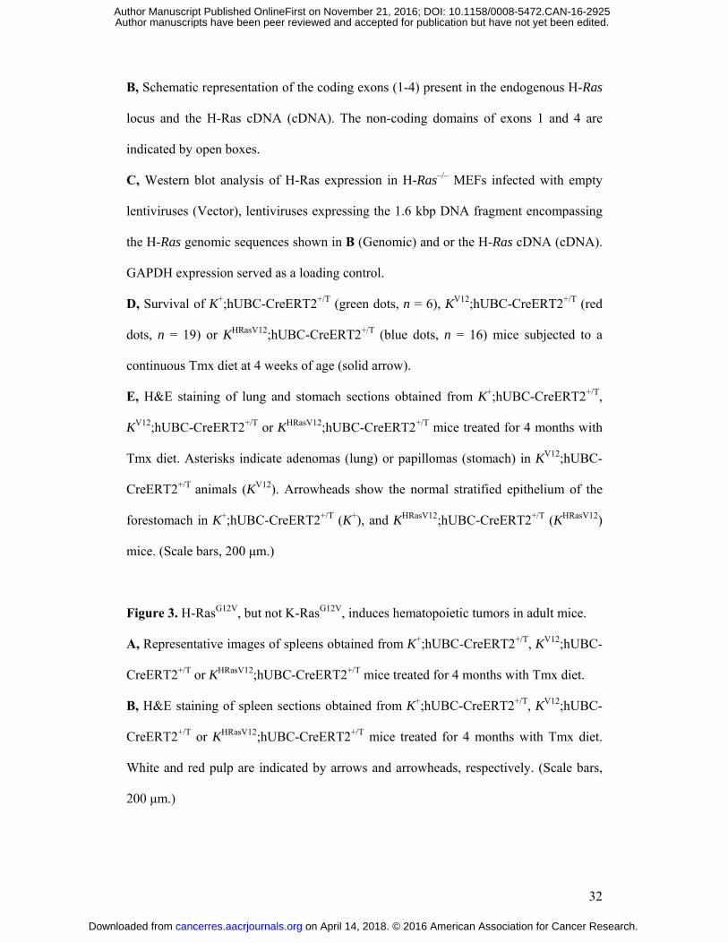
B, Schematic representation of the coding exons (1-4) present in the endogenous H-Ras
locus and the H-Ras cDNA (cDNA). The non-coding domains of exons 1 and 4 are
indicated by open boxes.
C, Western blot analysis of H-Ras expression in H-Ras–/– MEFs infected with empty
lentiviruses (Vector), lentiviruses expressing the 1.6 kbp DNA fragment encompassing
the H-Ras genomic sequences shown in B (Genomic) and or the H-Ras cDNA (cDNA).
GAPDH expression served as a loading control.
D, Survival of K+;hUBC-CreERT2+/T (green dots, n = 6), KV12;hUBC-CreERT2+/T (red
dots, n = 19) or KHRasV12;hUBC-CreERT2+/T (blue dots, n = 16) mice subjected to a
continuous Tmx diet at 4 weeks of age (solid arrow).
E, H&E staining of lung and stomach sections obtained from K+;hUBC-CreERT2+/T,
KV12;hUBC-CreERT2+/T or KHRasV12;hUBC-CreERT2+/T mice treated for 4 months with
Tmx diet. Asterisks indicate adenomas (lung) or papillomas (stomach) in KV12;hUBC-
CreERT2+/T animals (KV12). Arrowheads show the normal stratified epithelium of the
forestomach in K+;hUBC-CreERT2+/T (K+), and KHRasV12;hUBC-CreERT2+/T (KHRasV12)
mice. (Scale bars, 200 μm.)
Figure 3. H-RasG12V, but not K-RasG12V, induces hematopoietic tumors in adult mice.
A, Representative images of spleens obtained from K+;hUBC-CreERT2+/T, KV12;hUBC-
CreERT2+/T or KHRasV12;hUBC-CreERT2+/T mice treated for 4 months with Tmx diet.
B, H&E staining of spleen sections obtained from K+;hUBC-CreERT2+/T, KV12;hUBC-
CreERT2+/T or KHRasV12;hUBC-CreERT2+/T mice treated for 4 months with Tmx diet.
White and red pulp are indicated by arrows and arrowheads, respectively. (Scale bars,
200 μm.)
on April 14, 2018. © 2016 American Association for Cancer Research. cancerres.aacrjournals.org Downloaded from
Author manuscripts have been peer reviewed and accepted for publication but have not yet been edited. Author Manuscript Published OnlineFirst on November 21, 2016; DOI: 10.1158/0008-5472.CAN-16-2925

33
C, Flow cytometry analysis of Gr1+ and CD11b+ cells in spleens obtained from
K+;hUBC-CreERT2+/T, KV12;hUBC-CreERT2+/T or KHRasV12;hUBC-CreERT2+/T mice
treated for 4 months with Tmx diet.
D, H&E (above and middle) and CD3 IHC (bottom) staining in thymus sections
obtained from K+;hUBC-CreERT2+/T or KHRasV12;hUBC-CreERT2+/T mice treated for 4
months with Tmx diet. (Scale bars, 5 mm [above], 25 μm [middle, below].)
E, Flow cytometry analysis of CD4+ and CD8+ cells in thymuses obtained from
K+;hUBC-CreERT2+/T or KHRasV12;hUBC-CreERT2+/T mice treated for 4 months with
Tmx diet.
Figure 4. K-RasG12V and H-RasG12V oncoproteins expressed from the K-Ras locus
induce pancreatic lesions.
A, Number of low (P1) and high (P2/3) grade PanINs as well as PDACs (PDA) per
mouse in one year-old KV12;Elas-tTA/tetO-Cre (red circles, n = 13) or KHRasV12;Elas-
tTA/tetO-Cre (blue circles, n = 10) mice. Data are represented as mean (horizontal bars)
± SD.
B, Survival of KV12;p53lox/lox;Elas-tTA/tetO-Cre (KV12;p53lox/lox, red dots, n = 22) or
KHRasV12;p53lox/lox;Elas-tTA/tetO-Cre mice (KHRasV12;p53lox/lox, blue dots, n = 19).
C, Number of low (P1) and high (P2/3) grade PanINs as well as PDACs (PDA) per
mouse in 10 week-old KV12;p53lox/lox;Elas-tTA/tetO-Cre (red circles, n = 9) or
KHRasV12;p53lox/lox;Elas-tTA/tetO-Cre (blue circles, n = 8) mice. Data are represented as
mean (horizontal bars) ± SD.
D, Acinar cell explants isolated from pancreata of 6 to 8 week old KV12;Elas-tTA/tetO-
Cre (KV12, red bars, n = 6) and KHRasV12;Elas-tTA/tetO-Cre (KHRasV12, blue bars, n = 7)
mice were incubated in absence or presence of the indicated growth factors. The
on April 14, 2018. © 2016 American Association for Cancer Research. cancerres.aacrjournals.org Downloaded from
Author manuscripts have been peer reviewed and accepted for publication but have not yet been edited. Author Manuscript Published OnlineFirst on November 21, 2016; DOI: 10.1158/0008-5472.CAN-16-2925

34
number of metaplasias was determined after five days in culture. Data are represented
as mean ± SD. *** P < 0.001 (unpaired Student t test).
Figure 5. H-RasG12V expressed from the K-Ras locus induces robust downstream
signaling in lung tissue.
A, Top: IHC staining for pErk+ cells in lung sections obtained from KV12;hUBC-
CreERT2+/T or KHRasV12;hUBC-CreERT2+/T mice subjected to Tmx diet for the indicated
time. (Scale bars, 250 μm.) Bottom: Quantification of the percentage of pERK+ cells in
the lung sections shown above obtained from KV12;hUBC-CreERT2+/T (KV12, red bars, n
= 3) or KHRasV12;hUBC-CreERT2+/T (KHRasV12, blue bars, n = 3) mice. Data are
represented as mean ± SD. *** P < 0.001 (unpaired Student t test).
B, Western blot analysis of H-Ras, pErk1/2, Erk1/2, pAkt, Akt, p16Ink4a and p19Arf
expression in total lung extracts obtained from KV12;hUBC-CreERT2+/T or
KHRasV12;hUBC-CreERT2+/T mice exposed to Tmx diet for the indicated time. GAPDH
expression served as a loading control.
C, Relative quantification of the levels of expression of K-RasG12V and H-RasG12V
oncoproteins by label-free quantification (LFQ) in lungs obtained from K+;hUBC-
CreERT2+/T (n = 6), KV12;hUBC-CreERT2+/T (n = 6) or KHRasV12;hUBC-CreERT2+/T (n
= 6) mice treated for 2 months with Tmx diet. The tryptic peptide LVVVGAVGVGK
was used to detect expression of both K-RasG12V and H-RasG12V proteins. Data are
represented as mean ± SD.
D, Total Ras-GTP levels in lungs obtained from KV12;hUBC-CreERT2+/T or
KHRasV12;hUBC-CreERT2+/T mice treated with Tmx diet for the indicated time. GAPDH
was used as a loading control.
on April 14, 2018. © 2016 American Association for Cancer Research. cancerres.aacrjournals.org Downloaded from
Author manuscripts have been peer reviewed and accepted for publication but have not yet been edited. Author Manuscript Published OnlineFirst on November 21, 2016; DOI: 10.1158/0008-5472.CAN-16-2925

35
Figure 6. H-RasG12V expressed from the K-Ras locus induces a senescence-like arrest in
lung tissue that results in subsequent elimination of cells.
A, Southern blot analysis of DNA isolated from lung tissue obtained from 4 week old
KHRasV12;hUBC-CreERT2+/T mice exposed to a Tmx diet for 4 weeks either immediately
ending the treatment (left) or after mice were maintained for 8 additional weeks in a
regular diet (center and right). The latter mice were either left untreated during the
additional 8 week period (center) or treated daily with 1 mg/kg of Trametinib, a
selective MEK inhibitor (right). DNA isolated from thymus tissue was used as control.
The migration (open arrowheads) and sizes (solid arrowheads) of the diagnostic
SphI+KpnI DNA fragments for the recombined K-RasH-RasG12V allele that expresses the
H-RasG12V oncoprotein and the non-recombined K-RasLSLH-RasG12V allele that does not
allow H-RasG12V expression are indicated. Note the disappearance of the recombined K-
RasH-RasG12V allele in lung but not thymus tissue. Lung tissue from mice treated with
Trametinib also retains the recombined K-RasH-RasG12V allele.
B, Relative expression levels of p16Ink4a and p19Arf mRNAs in total lung extracts
from KV12;hUBC-CreERT2+/T (KV12, red bars, n = 3) or KHRasV12;hUBC-CreERT2+/T
(KHRasV12, blue bars, n = 3) mice exposed to Tmx diet for the indicated time, as
determined by qRT-PCR analysis. GAPDH expression levels were used for
normalization. Data are represented as mean ± SD. *** P < 0.001 (unpaired Student t
test).
Figure 7. H-RasG12V, but not K-RasG12V expressed from the K-Ras locus induces
senescence in MEFs.
on April 14, 2018. © 2016 American Association for Cancer Research. cancerres.aacrjournals.org Downloaded from
Author manuscripts have been peer reviewed and accepted for publication but have not yet been edited. Author Manuscript Published OnlineFirst on November 21, 2016; DOI: 10.1158/0008-5472.CAN-16-2925

36
A, Western blot analysis of the indicated proteins expressed in KV12 or KHRasV12 MEFs
infected with Adeno-Cre particles for the indicated time. GAPDH expression served as
a loading control.
B, Growth curve of KV12 or KHRasV12 MEFs infected with Adeno-GFP (open circles) or
Adeno-Cre particles (closed circles) for the indicated time after stable infection with
empty retroviruses (Vector) or retroviruses expressing the Ad5 E1A oncoprotein (E1A).
Data are represented as mean ± SD.
C, Percentage of senescence-associated β-Galactosidase (SA-βGal) positive cells in
KV12 or KHRasV12 MEFs stably infected with empty retroviruses (V) or retroviruses
expressing the Ad5 E1A oncoprotein (E1A) 4 days after infection with Adeno-Cre
particles. Data are represented as mean ± SD. *** P < 0.001 (unpaired Student t test).
D, Focus formation in KV12 or KHRasV12 MEFs stably infected with empty retroviruses
(V) or retroviruses expressing Ad5 E1A (E1A) 14 days after infection with Adeno-Cre
particles. Data are represented as mean ± SD. *** P < 0.001 (unpaired Student t test).
E, Relative quantification of the levels of expression of the K-RasG12V and H-RasG12V
oncoproteins by label-free quantification (LFQ) in KV12;hUBC-CreERT2+/T (red bars, n
= 3) or KHRasV12;hUBC-CreERT2+/T MEFs (blue bars, n = 3) 4 days after infection with
Adeno-Cre particles. The tryptic peptide LVVVGAVGVGK was used to detect
expression of K-RasG12V and H-RasG12V proteins. Data are represented as mean ± SD.
F, Western blot analysis of H-Ras protein expression (H-Ras and H-RasG12V) driven by
the K-Ras locus in KHRasV12 MEFs upon infection with Adeno-Cre particles for the
indicated time and by the H-Ras locus in H-RasG12V MEFs. Expression of Ras effector
proteins pErk1/2, Erk1/2, pAkt and Akt was also analyzed. GAPDH expression served
as loading control.
on April 14, 2018. © 2016 American Association for Cancer Research. cancerres.aacrjournals.org Downloaded from
Author manuscripts have been peer reviewed and accepted for publication but have not yet been edited. Author Manuscript Published OnlineFirst on November 21, 2016; DOI: 10.1158/0008-5472.CAN-16-2925

Fig. 1
10
0
20
30
40
50
Eje
ction fra
ction (
%)
A B
Mean a
rteri
al pre
ssure
(m
mH
g)
* * *
C
100
200
300
400
Card
iom
yocyte
are
a (
μm
2)
HrasKI;H-Ras–/– HrasKI D
* * * * * * * * *
100
80
60
110
90
70
on April 14, 2018. © 2016 American Association for Cancer Research. cancerres.aacrjournals.org Downloaded from
Author manuscripts have been peer reviewed and accepted for publication but have not yet been edited. Author Manuscript Published OnlineFirst on November 21, 2016; DOI: 10.1158/0008-5472.CAN-16-2925

D
C
Hyg 0 H-RasG12V K HRasV12
STOP Hyg 0 1*
G12V
K V12
ATG
ATG
STOP
0 1 K +
ATG
Fig. 2
A B
E
Lu
ng
S
tom
ach
K + K V12 K HRasV12
*
0
20
40
60
80
100
0 10 20 30 40 50
Age (weeks)
Surv
ival (%
)
H-Ras genomic DNA
H-Ras cDNA
ATG
ATG
TGA
TGA
1 2 3 4 H-Ras
GAPDH
H-Ras–/– cells
*
*
* *
*
K +
K V12
K HRasV12
on April 14, 2018. © 2016 American Association for Cancer Research. cancerres.aacrjournals.org Downloaded from
Author manuscripts have been peer reviewed and accepted for publication but have not yet been edited. Author Manuscript Published OnlineFirst on November 21, 2016; DOI: 10.1158/0008-5472.CAN-16-2925

Fig. 3
A B
C
D
K + K V12 K HRasV12 K + K V12 K HRasV12
Gr1
CD11b
6.10 2.48
1.57
4.87 6.85
2.35
7.23 51.9
4.43 102
103
104
105
102 103 104
K + K V12 K HRasV12
102 103 104 102 103 104
H&
E
CD
3 IH
C
H&
E
K + K HRasV12
E
CD
4
K HRasV12
K +
104
103
0
78.2
4.60 2.45
29.5 10.2
15.3 45.0
CD8
0
102
104
0 102 103 104
25.5
16.4 16.4
104
103
0
102
14.7
41.6
89.9 85.9 36.5
on April 14, 2018. © 2016 American Association for Cancer Research. cancerres.aacrjournals.org Downloaded from
Author manuscripts have been peer reviewed and accepted for publication but have not yet been edited. Author Manuscript Published OnlineFirst on November 21, 2016; DOI: 10.1158/0008-5472.CAN-16-2925

Fig. 4
C
A
D
B
0
10
100
1
Le
sio
ns p
er
mou
se
P1 P2/3 PDA P1 P2/3 PDA
K V12 K HRasV12
0
10
1
Le
sio
ns p
er
mou
se
K V12;p53 lox/lox K HRasV12;p53 lox/lox
P1 P2/3 PDA P1 P2/3 PDA
0
100
200
300
– EGF
Meta
pla
sia
s / w
ell
TGFα EGF+
TGFα
* * *
* * *
* * * K V12
K HRasV12 400
0
20
40
60
80
100
0 10 20 30 40 50
Age (weeks)
Surv
ival (%
)
K HRasV12;
p53 lox/lox
K V12;
p53 lox/lox
on April 14, 2018. © 2016 American Association for Cancer Research. cancerres.aacrjournals.org Downloaded from
Author manuscripts have been peer reviewed and accepted for publication but have not yet been edited. Author Manuscript Published OnlineFirst on November 21, 2016; DOI: 10.1158/0008-5472.CAN-16-2925

Fig. 5 A pERK IHC
K V
12
K
HR
asV
12
0
10
20
30
pE
RK
+ c
ells
(%
)
* * * * * *
Days in Tmx diet
day 0 day 4 day 7 day 14
K V12
K HRasV12
B
C D
pErk1/2
H-Ras
GAPDH
Erk1/2
p16Ink4a
Days in Tmx diet
p19Arf
K V12 K HRasV12
Akt
pAkt
K V12 K HRasV12
14
Days in Tmx diet
0 14 0
GAPDH
Ras-GTP
Ras
4 7 14 0 4 7 14 0
40
Norm
aliz
ed
in
ten
sity (
x10
7)
0
0.8
1.2
0.4
K V12 K HRasV12 K +
on April 14, 2018. © 2016 American Association for Cancer Research. cancerres.aacrjournals.org Downloaded from
Author manuscripts have been peer reviewed and accepted for publication but have not yet been edited. Author Manuscript Published OnlineFirst on November 21, 2016; DOI: 10.1158/0008-5472.CAN-16-2925

A
Fig. 6
B
Lung Thymus
Off Tmx: 8 wk
kbp
7.4
6.0 K-Ras LSLHRasV12
K-Ras HRasV12
Trametinib: – – + – – 8 wk 8 wk – –
Rela
tive m
RN
A
expre
ssio
n le
ve
ls
0
2
p19Arf p16Ink4a
1
3
4
* * * * * * K V12
K HRasV12
Tmx: 4 wk 4 wk 4 wk 4 wk 4 wk
on April 14, 2018. © 2016 American Association for Cancer Research. cancerres.aacrjournals.org Downloaded from
Author manuscripts have been peer reviewed and accepted for publication but have not yet been edited. Author Manuscript Published OnlineFirst on November 21, 2016; DOI: 10.1158/0008-5472.CAN-16-2925

p16Ink4a
p53
pErk1/2
H-Ras
0 2 4 6 8 10
Adeno-Cre (days)
Erk1/2
Akt
pAkt
GAPDH
0 2 4 6 8 10
K-Ras H/N-Ras
A E
1A
V
ecto
r
Days
Absorb
ance
0
8
16
20
0 0 2 4 6 8 10 0 2 4 6 8 10
40
K V12 K HRasV12 B
Foci /
pla
te
GAPDH
Akt
Erk1/2
pAkt
pErk1/2
H-Ras
Days
0 2 Adeno-Cre:
F
K V12 K HRasV12 C
0
10
20
30
0
25
50
75
SAβ
-Gal+
cells
(%
)
100
V E1A V E1A
K V12 K HRasV12 K V12
K HRasV12
V E1A E1A V
Norm
aliz
ed inte
nsity (
x10
7)
Fig. 7 D
E K V12 K HRasV12
0
3
1
2
on April 14, 2018. © 2016 American Association for Cancer Research. cancerres.aacrjournals.org Downloaded from
Author manuscripts have been peer reviewed and accepted for publication but have not yet been edited. Author Manuscript Published OnlineFirst on November 21, 2016; DOI: 10.1158/0008-5472.CAN-16-2925

Published OnlineFirst November 21, 2016.Cancer Res Matthias Drosten, Lucía Simón-Carrasco, Isabel Hernández-Porras, et al. when driven by the same regulatory sequencesH-Ras and K-Ras oncoproteins induce different tumor spectra
Updated version
10.1158/0008-5472.CAN-16-2925doi:
Access the most recent version of this article at:
Material
Supplementary
http://cancerres.aacrjournals.org/content/suppl/2016/11/19/0008-5472.CAN-16-2925.DC1
Access the most recent supplemental material at:
Manuscript
Authoredited. Author manuscripts have been peer reviewed and accepted for publication but have not yet been
E-mail alerts related to this article or journal.Sign up to receive free email-alerts
Subscriptions
Reprints and
To order reprints of this article or to subscribe to the journal, contact the AACR Publications
Permissions
Rightslink site. Click on "Request Permissions" which will take you to the Copyright Clearance Center's (CCC)
.http://cancerres.aacrjournals.org/content/early/2016/11/19/0008-5472.CAN-16-2925To request permission to re-use all or part of this article, use this link
on April 14, 2018. © 2016 American Association for Cancer Research. cancerres.aacrjournals.org Downloaded from
Author manuscripts have been peer reviewed and accepted for publication but have not yet been edited. Author Manuscript Published OnlineFirst on November 21, 2016; DOI: 10.1158/0008-5472.CAN-16-2925