Group I introns and Homing Endonucleases in T-even-like
73
Group I introns and Homing Endonucleases in T-even-like Bacteriophages Linus Sandegren Department of Molecular Biology and Functional Genomics Stockholm University Stockholm 2004
Transcript of Group I introns and Homing Endonucleases in T-even-like

Group I introns and Homing Endonucleases inT-even-like Bacteriophages
Linus Sandegren
Department of Molecular Biology and Functional GenomicsStockholm University
Stockholm 2004

2
Doctoral Thesis 2004Department of Molecular Biology and Functional GenomicsThe Arrhenius Laboratories for Natural SciencesStockholm UniversitySE-106 91 StockholmSweden
Previously published papers are reprinted with permission from thepublisher.
2004 Linus SandegrenISBN 91-7265-925-4

3
AbstractHoming endonucleases are rare-cutting enzymes that cleave DNA at asite near their own location, preferentially in alleles lacking the homingendonuclease gene (HEG). By cleaving HEG-less alleles the homingendonuclease can mediate the transfer of its own gene to the cleaved sitevia a process called homing, involving double strand break repair. Viahoming, HEGs are efficiently transferred into new genomes whenhorizontal exchange of DNA occurs between organisms.
Group I introns are intervening sequences that can catalyse their ownexcision from the unprocessed transcript without the need of any proteins.They are widespread, occurring both in eukaryotes and prokaryotes andin their viruses. Many group I introns encode a HEG within them thatconfers mobility also to the intron and mediates the combined transfer ofthe intron/HEG to intronless alleles via homing.
Bacteriophage T4 contains three such group I introns and at least 12freestanding HEGs in its genome. The majority of phages besides T4 donot contain any introns, and freestanding HEGs are also scarcelyrepresented among other phages.
In the first paper we looked into why group I introns are so rare inphages related to T4 in spite of the fact that they can spread betweenphages via homing. We have identified the first phage besides T4 thatcontains all three T-even introns and also shown that homing of at leastone of the introns has occurred recently between some of the phages inNature. We also show that intron homing can be highly efficient betweenrelated phages if two phages infect the same bacterium but that there alsoexists counteracting mechanisms that can restrict the spread of intronsbetween phages.
In the second paper we have looked at how the presence of intronscan affect gene expression in the phage. We find that the efficiency ofsplicing can be affected by variation of translation of the upstream exonfor all three introns in T4. Furthermore, we find that splicing is alsocompromised upon infection of stationary-phase bacteria. This is the firsttime that the efficiency of self-splicing of group I introns has beencoupled to environmental conditions and the potential effect of this onphage viability is discussed.
In the third paper we have characterised two novel freestandinghoming endonucleases that in some T-even-like phages replace two of theputative HEGs in T4. We also present a new theory on why it is aselective advantage for freestanding, phage homing endonucleases tocleave both HEG-containing and HEG-less genomes.

4

5
“Phages really are amazing creatures. You can learn all there is to knowabout their ways in a month, yet after a hundred years they can stillsurprise you at a pinch.”

6

7
Table of contents
ABSTRACT 3
TABLE OF CONTENTS 7
LIST OF PAPERS 8
ABBREVIATIONS 9
INTRODUCTION 10
CONCERNING PHAGES 10T-EVEN BACTERIOPHAGES 10BACTERIOPHAGE T4 12OVERVIEW OF T4 DEVELOPMENT 13TRANSCRIPTIONAL REGULATION 15NUCLEOTIDE METABOLISM 17DNA REPLICATION 19PHAGE ASSEMBLY AND LYSIS 19T-EVEN-LIKE PHAGE GENOMICS 21GROUP I INTRONS 23SPLICING MECHANISM 24STRUCTURE 25PROTEINS INVOLVED IN GROUP I INTRON SPLICING 28GROUP I INTRON DISTRIBUTION 29HOMING ENDONUCLEASES AND INTRON MOBILITY 29GROUP I INTRONS IN BACTERIOPHAGE T4 32T4 INTRON-ENCODED HOMING ENDONUCLEASES 34DISTRIBUTION OF INTRONS AMONG T-EVEN-LIKE PHAGES 35FREESTANDING HOMING ENDONUCLEASES IN T4 35INTRONS IN OTHER BACTERIOPHAGES 37
PRESENT STUDIES 41
PAPER I: DISTRIBUTION, SEQUENCE HOMOLOGY AND HOMING OF GROUP I INTRONS
AMONG T-EVEN-LIKE BACTERIOPHAGES. EVIDENCE FOR RECENT TRANSFER OF OLD
INTRONS. 41PAPER II: SELF-SPLICING OF THE BACTERIOPHAGE T4 GROUP I INTRONS IS AFFECTED
BY THE GROWTH OF THE INFECTED BACTERIUM AND REQUIRES EFFICIENT TRANSLATION
OF THE PRE-MRNA IN VIVO. 47PAPER III: TWO GENES ENCODING NOVEL HOMING ENDONUCLEASES REPLACE THE
PUTATIVE HOMING ENDONUCLEASE GENES MOBC AND MOBE IN SEVERAL T4-RELATED
PHAGES. 49FUTURE STUDIES 55
ACKNOWLEDGEMENTS 56
REFERENCES 58

8
List of papers
This thesis is based on the following original articles and manuscripts,which will be referred to by their roman numerals.
I Sandegren. L., Sjöberg. B-M.Distribution, Sequence Homology and Homing of GroupI Introns among T-even-like Bacteriophages. Evidencefor recent transfer of old introns.Journal of Biological Chemistry 279 (21) pp. 22218-22227 (2004)
II Sandegren. L., Sjöberg. B-M.Self-splicing of the Bacteriophage T4 Group I Introns isAffected by the Growth of the Infected Bacterium andRequires Efficient Translation of the Pre-mRNA In Vivo.Manuscript
III Sandegren. L., Nord. D., Sjöberg. B-M.Two Genes Encoding Novel Homing EndonucleasesReplace the Putative Homing Endonuclease Genes mobCand mobE in Several T4-related Phages.Manuscript

9
Abbreviations
DNA Deoxyribonucleic AcidRNA Ribonucleic AcidmRNA Messenger RNArRNA Ribosomal RNAtRNA Transfer RNAHEG Homing Endonuclease GeneORF Open Reading Frame
Nucleotides:ATP Adenosine TriphosphatedATP Deoxyadenosine TriphosphateCTP Cytidine TriphosphatedCTP Deoxycytidine TriphosphateGTP Guanosine TriphosphatedGTP Deoxyguanosine TriphosphateTTP Thymidine TriphosphatedTTP Deoxythymidine TriphosphateUTP Uridine Triphosphate
dNTP any Deoxyribonucleoside TriphosphatedNDP any Deoxyribonucleoside DiphosphatedNMP any Deoxyribonucleoside Monophosphate
Genes:nrdA Aerobic Ribonucleotide Reductase, Large SubunitnrdB Aerobic Ribonucleotide Reductase, Small SubunitnrdD Anaerobic Ribonucleotide ReductasenrdG Anaerobic Ribonucleotide Reductase, Activasetd Thymidylate Synthase
E. coli Escherichia coliB. subtilis Bacillus subtilis

10
Introduction
Concerning phages
This thesis is largely concerned with Phages, and from its pages a readermay discover much of their character and a little of their history.Bacteriophages, viruses that infect and kill bacteria (Greek, phage - "toeat"), have been studied and used as tools in molecular genetics andmicrobiology ever since they were discovered in the beginning of the1900:s. They have aided researchers in the elucidation of many of thefundamental molecular mechanisms of life, e.g. the confirmation thatDNA is the hereditary material (Hershey and Chase, 1952), the finding ofrestriction and modification of DNA (Luria and Human, 1952), thestructure of DNA (Watson and Crick, 1953) the definition of the genestructure (Benzer, 1955), the identification of mRNA as the messenger ofDNA (Brenner et al., 1961) and the definition of the genetic code (Crick etal., 1961). At the time of their discovery all phages were generally thoughtof as identical. Now we know that there exist a vast number of differentbacteriophages that are genetically and morphologically distinct.Traditionally, phages have been categorised according to theirmorphology and host range and it is not until now, with the increasingnumber of complete phage genomes sequenced, that comparisons can bemade with regards to the genetic history and relationships of phages.
T-even bacteriophages
Taxonomically the T-even bacteriophages belong to the familyMyoviridae (Phages with contractile tails) among dsDNA phages(International Committee on Taxonomy of Viruses,www.ncbi.nlm.nih.gov/ICTVdb/). Morphologically they are recognisedby their rather complex structure with an icosahedral head and a longcontractile tail ended with six tail fibres (Fig. 1). The exact origin of theT-even bacteriophages is somewhat uncertain, but they have been tracedback to "the Phage Group" in the lab of Max Delbrück in the 1940:s(Abedon, 2000). Delbrück and colleagues were concerned with the

11
problem that all people working with bacteriophages at that time had theirown, more or less isogenic, phage strains and it was very hard to compareresults. This problem originates in the early belief that all bacteriophageswere identical. Therefore the Phage group made a collection of sevenlytic phages that infected the routinely used intestinal bacteriumEscherichia coli B, and that they found were easy to work with at 37°C.These phages were called T1-T7 (T for type) (Demerec and Fano, 1945).Out of these, the T-even phages, T2, T4 and T6 were found to bemorphologically, antigenically and genetically very similar, and provedto be very useful for biochemical and genetic studies. T4 and T6 probablyoriginates from a mixture of phages supplied to the Phage Group by Dr.Tony L. Rakieten (Demerec and Fano, 1945), and were most likely isolatedfrom sewage (Abedon, 2000). T2 dates back to about 1927 and Dr.Jacques Bronfenbrennen. It was probably isolated from faeces and hasbeen known by a number of different names (P28, α, TI, γ, PC). Todaythere exists (at least) two commonly used “sub-strains” of T2, T2L andT2H, the former is likely an isolate of γ from Salvador Luria and thelatter an isolate by Hersey and coworkers (Abedon, 2000). Of the otherreference phages from Delbrück´s lab, T1 and T5 are related and belongto Siphoviridae (Phages with long non-contractile tails) and T3 and T7are related and belong to Podoviridae (Phages with short tails).
Figure 1. Bacteriophage T4. Figure adapted from (Eiserling and Black, 1994) andrepublished with permission of American Society for Microbiology; permissionconveyed through Copyright Clearance Center, Inc.

12
Today there is a vast number of isolates of bacteriophages withmorphological or genetic similarity to the T-even phages, most of themisolated from faeces from patients or from sewage. For clarity I willthroughout this thesis refer to T2, T4 and T6 as the T-even phages and tolater isolates as T-even-like phages. The practise of classifying phagesmorphologically has led to some confusion when genetic relationshipshave begun to be revealed. “T4-like phages” are classically phages thatmorphologically resemble T4. However, this class includes a very widerange of phages with different bacterial hosts and very little geneticsimilarity. With increasing amounts of sequencing data being collected,subgroups of the T4-like phages have been proposed. The phages closestrelated to T4, including T2 and T6, are frequently called T-even phages(although I prefer to separate the “original” T-evens and T-even-likephages). More distantly related phages have been named pseudo-T-evensand schizo-T-evens accordingly (Desplats et al., 2002; Monod et al., 1997;Tetart et al., 2001). Comparative studies of the additional genomesequences now being completed for phages related to T4 will likely shedmore light on the relationships and phylogenetic history of this group ofphages.
Bacteriophage T4
Most of what is known about the T-even, and T-even-like bacteriophageshas come from studies of T4. As mentioned earlier, the functions of manygenetic mechanisms were originally elucidated in T4 and several of itsproteins are used routinely in molecular biology today, due to the vastbiochemical knowledge we have about them. T4 has a genomecomprising 168903 base pairs (bp), one of the largest known forbacteriophages. It is now completely sequenced (Kutter et al., 1993) andcontains nearly 300 probable genes, 289 encoding proteins, 8 tRNAs andtwo other small RNA species of unknown function (Miller et al., 2003b).Half of the proposed genes still have no described function and nohomology to any genes in GenBank. Only 62 genes are essential understandard laboratory conditions while the rest can be deleted withoutabolishing phage growth (Miller et al., 2003b). However, these “non-essential” genes most likely provide important functions to the phage

13
during growth in more natural conditions or during transition betweendifferent growth conditions or different hosts.
The T4 DNA contains modified bases, probably to avoid degradation byhost restriction systems and to make a distinction between its own DNAand the host DNA that is rapidly degraded by phage endonucleases earlyin infection (reviewed in (Carlson et al., 1994)). Instead of cytosine, T4uses 5-hydroxymethylcytosine (HmC) that is further almost completelyglucosylated postreplicationally. T4 DNA is also highly enriched in A-Tbase pairs (65.5%) (Miller et al., 2003b) compared to E. coli (49%) (DeLey, 1970), and the phage genes contain a codon usage bias which iscomplemented for by the eight tRNAs encoded by the phage which arenormally only scarcely expressed by the host (Mosig, 1994). Themodification of cytosines has also been proposed to increase the doublestrand stability of the A-T rich genome (Miller et al., 2003b).
The majority of past and present work on T4 and the T-evenbacteriophages is done with E. coli as host, but if this is the preferredwild type host is not known. T4 has been shown to infect several differententeric bacteria such as Klebsiella, Shigella, Salmonella and Proteus(Ackermann and Krisch, 1997; Dawes, 1976) and it can replicate in anumber of different gram-negative bacteria even though infection isimpaired (Wais and Goldberg, 1969). It should be kept in mind that thereare a vast number of potential host bacteria in the mammalian gut thathave avoided characterisation due to problems of fulfilling their specificgrowth requirements when performing lab isolations from faeces.
Overview of T4 development
The lifecycle of bacteriophage T4 is outlined in figure 2. Infection by T4and its close relatives is very efficient approaching 100% platingefficiency under standard laboratory conditions (Goldberg et al., 1994).Adsorption to the bacterial cell wall is through binding to receptors on thecell. Initially the distal tips of the six tail fibers bind cooperatively butreversibly to the lipopolysaccharide (LPS) of the cell surface ((Stent andWollman, 1952) and reviewed in (Goldberg et al., 1994)). Reversiblebinding by the tail fibers allow the phage to “wander” over the cell

14
surface until it reaches a baseplate recognition site. A variety of majorouter membrane proteins of E. coli can work as receptors for permanentphage binding (Eddy, 1992; Schwarz et al., 1983). The hyper variable tipsof the tail fibers determine the host range of the T-even-like phages(Beckendorf, 1973; Beckendorf et al., 1973). After receptor recognition, thebase plate at the end of the phage tail makes contact with the outermembrane and the cell wall, the tail is contracted and gp5 (a lysosymelocated in the base plate) makes a hole through which the phage DNA isinjected (Kao and McClain, 1980).
Figure 2. Bacteriophage T4 infection cycle. Picture modified from (Carlson, 2000)and republished with permission from Magdalena Korotynska.
Once inside the cell, the phage DNA is recognised by the host RNApolymerase and early genes are transcribed (see T4 transcriptionalregulation). Among early genes are found genes for the phage take overof the cell machinery, genes for production of DNA precursors for phageDNA synthesis and genes for regulation of middle and late geneexpression. During early gene expression phage endonucleases areproduced that degrade the host chromosome, thereby completely stopping

15
bacterial gene expression (reviewed in (Carlson et al., 1994)). Early genesare followed by expression of middle genes that predominantly producesregulatory proteins, and then by late genes coding for structural phageproteins for building new phage particles and loading of the phagechromosome into the new heads. Assembly of the structural proteins intonew heads, tails and other structures occurs mainly by auto assemblyguided by chaperones such as GroEL from the host and gp31 from thephage (Keppel et al., 1990). The final stage is the packaging of the phagegenomes into the heads by terminase enzymes. This takes place by head-full packaging of slightly more than one genome equivalent generatingcircularly permuted chromosomes with terminal redundancy (reviewed in(Murialdo, 1991)).
Transcriptional regulation
Under laboratory conditions a T4 infection in logarithmically growing E.coli in rich medium at 37°C takes about 25 minutes from injection of thephage DNA to lysis of the host cell and release of typically 200-300progeny phage. This rapid production of new phage is dependent on atemporally regulated infection cycle (Koch and Hershey, 1959). T4 generegulation shows an ordered expression and repression of genes duringthe infection. As mentioned above, three different types of genes can bedistinguished, early, middle and late. Regulation of gene expression ismainly accomplished at the transcriptional level. All transcription ofphage genes is done by the host core RNA polymerase but with achanging set of modifications throughout the infection cycle (see below).
Early genes are transcribed immediately after the phage has injected itsDNA into the cell. The early promoters differ from E. coli σ70-promotersbut still contain a -35 box and a -10 box (Wilkens and Ruger, 1994) andcan compete with the host promoters for RNA polymerase binding. Aprotein called Alt is present in 30-50 copies in the phage head andinjected into the cell together with the phage DNA (Horvitz, 1974a;Horvitz, 1974b). It catalyses ADP-ribosylation of one of the subunits ofthe RNA polymerase (Rohrer et al., 1975). This modification seems tostrengthen the activation of transcription at T4 early promoters compared

16
to E. coli promoters (Koch et al., 1995; Wilkens and Ruger, 1996; Wilkens etal., 1997).
In contrast to early transcription, middle and late transcription aredependent upon synthesis of phage proteins. Among the early T4 genesproduced are four important transcriptional regulators, ModA, ModB,AsiA and MotA. ModA and B are two additional ADP-ribosylatingenzymes that further modify the subunits of the core RNA polymerase(Goff, 1974; Mosig et al., 1998). MotA is an activator of transcription frommiddle promoters (Guild et al., 1988). Middle promoters lack the –35 boxand instead have an "extended" -10 box (Hughes and Mathee, 1998; Stittand Hinton, 1994). A sequence in front of this extended -10 box, about 30base pairs upstream of the transcription initiation site, is recognised andbound by MotA. AsiA is an anti-σ70 factor that forms hetero-dimers withσ70, abolishing the -35-box recognition and thereby lowering theactivation of transcription at early promoters (reviewed in (Hughes andMathee, 1998)). At the same time the modified RNA polymerase caninteract with the MotA protein via contacts with AsiA, shiftingtranscription from early to middle promoters. In this way MotA, bound tothe upstream site, effectively replace the -35 region in recognition ofmiddle promoters (Hinton et al., 1996).
During middle transcription additional regulatory proteins for late geneexpression are made. The key player in late transcription is the phageencoded σ-factor gp55 that binds to the RNA polymerase core instead ofσ70. Normally, E. coli σ70 binds stronger to the RNA polymerase thangp55 and will thus outcompete it. AsiA however, acting as a true anti-σ70
factor, weakens the interaction between σ70 and the core subunits of RNApolymerase and shifts the preference to gp55 binding (Williams et al.,1994). Late promoters, like middle promoters, lack a recognisable -35 boxand instead only have a special T4 late -10 box (Christensen and Young,1982). Late transcription is therefore also dependent on additionalactivator proteins, especially RNA polymerase binding protein gp33 andDNA polymerisation sliding clamp protein gp45, to direct the RNApolymerase to the correct promoters. No late expression takes placewithout DNA replication and this is regulated mainly via the actions ofgp45 (Epstein et al., 1964). It is the active, ATP dependent, loading of thesliding clamp protein gp45 onto DNA that is thought to generate the

17
coupling of late transcription to DNA replication (Fu et al., 1996).Interaction between gp45 sliding along the DNA, and gp33 and gp55bound to the RNA polymerase directs transcriptional initiation to the latepromoters.
All T4 genes do not have a recognisable promoter and several aretranscribed as operons from a single promoter (Miller et al., 2003b). Thereare also cases where late proteins are transcribed on early and middletranscripts but not expressed until late in infection due to repression bymRNA processing and translational control. One characterisedmechanism to avoid translation of late genes from polycistronic earlytranscripts is the presence of an RNA hairpin in front of some late genes,that masks the translational initiation region at the start of these genes.The hairpin cannot form on transcripts from the correct, late promotersince the late promoter is located within the hairpin region (Macdonald etal., 1984; McPheeters et al., 1986). Such repression of translation of lategenes expressed on extended earlier transcripts has been recognised forthe homing endonuclease genes within the nrdB and td introns in T4 (Gottet al., 1988) see Paper I.
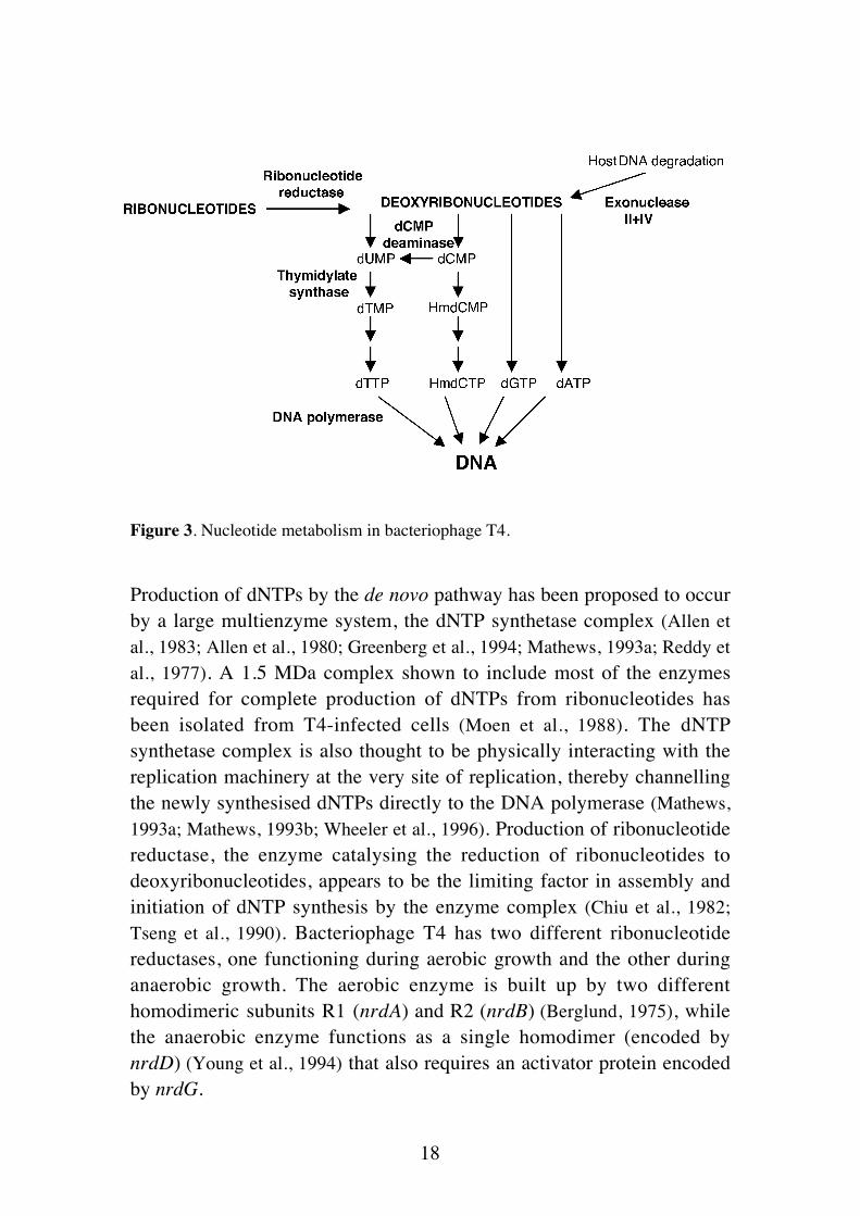
Nucleotide metabolism
Bacteriophage T4 devotes a large part of its genome to genes coding forenzymes used in nucleotide metabolism. All but one of the proteinsneeded for making the building blocks of phage DNA are encoded by thephage itself. The only host enzyme used is nucleoside diphosphate kinaseneeded for phosphorylation of dNDPs to dNTPs (Moen et al., 1988).Nucleotides for production of the phage genome can come from twodifferent sources, de novo synthesis of dNTPs from ribonucleotides orreutilization of dNMPs from the degraded host genome (the salvagepathway), as shown in figure 3. Most of the dNTPs needed for the 200-300 new T4 genomes produced under optimal conditions comes from denovo synthesis. The complete degradation of the E. coli genome canprovide nucleotides for less than 20 phage genomes. An exponentiallygrowing E. coli cell may contain 3-4 genome equivalents (Bremer andDennis, 1987) so the upper limit of phage genomes produced by thesalvage pathway would be 80.
18
Figure 3. Nucleotide metabolism in bacteriophage T4.
Production of dNTPs by the de novo pathway has been proposed to occurby a large multienzyme system, the dNTP synthetase complex (Allen etal., 1983; Allen et al., 1980; Greenberg et al., 1994; Mathews, 1993a; Reddy etal., 1977). A 1.5 MDa complex shown to include most of the enzymesrequired for complete production of dNTPs from ribonucleotides hasbeen isolated from T4-infected cells (Moen et al., 1988). The dNTPsynthetase complex is also thought to be physically interacting with thereplication machinery at the very site of replication, thereby channellingthe newly synthesised dNTPs directly to the DNA polymerase (Mathews,1993a; Mathews, 1993b; Wheeler et al., 1996). Production of ribonucleotidereductase, the enzyme catalysing the reduction of ribonucleotides todeoxyribonucleotides, appears to be the limiting factor in assembly andinitiation of dNTP synthesis by the enzyme complex (Chiu et al., 1982;Tseng et al., 1990). Bacteriophage T4 has two different ribonucleotidereductases, one functioning during aerobic growth and the other duringanaerobic growth. The aerobic enzyme is built up by two differenthomodimeric subunits R1 (nrdA) and R2 (nrdB) (Berglund, 1975), whilethe anaerobic enzyme functions as a single homodimer (encoded bynrdD) (Young et al., 1994) that also requires an activator protein encodedby nrdG.

19
DNA replication
T4 encodes all but one of the proteins needed for replicating its genome.The only host enzyme used in replication is the RNA polymerase in orderto synthesise RNA primers for initiation of leading strand replication(Luder and Mosig, 1982). DNA replication in T4 is initiated in twodifferent ways. The first replication fork is initiated at one of four majororigins of replication (ori) oriA, oriE, oriF and oriG (Kreuzer and Alberts,1985; Kreuzer and Morrical, 1994; Menkens and Kreuzer, 1988). After thefirst round of replication, initiation of new replication forks from the orisis repressed and instead occurs via recombination intermediates initiatedfrom the DNA ends of previously replicated chromosomes (Dannenbergand Mosig, 1983; Luder and Mosig, 1982). The major pathway for thisrecombination-dependent replication is through strand invasion by single-stranded, 3´ chromosomal ends that lead to new replication forks(reviewed in (Mosig, 1998)). This is a very efficient way of solving theproblem with replication of the ends of the linear chromosomes andresults in branched concatemers of T4 genomes that are later used forpackaging into the heads. The circularly permuted chromosomes of T4makes this recombination dependent replication very efficient and theamount of replication during infection is ten times higher than in theuninfected cell (Werner, 1968). Replication is terminated when the phageprotein gp2 is expressed which binds to free chromosome ends andrepresses new recombination initiation events (Lipinska et al., 1989).
Phage assembly and Lysis
The final stages of T4 infection are the assembly of new phage particlesand lysis of the host cell. Almost half of the genes in T4 codes for phagestructural proteins or proteins involved in the assembly of the structures(Miller et al., 2003b). There are 24 proteins involved in headmorphogenesis (reviewed in (Black et al., 1994)). The head is made up ofthe shell proteins gp23 and gp24 that are assembled on a scaffold made ofproteins gp21 and gp22. When the scaffold is covered gp21proteolytically cleaves the other proteins, degrading the scaffold thusforming a prohead that is ready for DNA packaging. DNA packaging isinitiated by the endonucleolytic generation of packable chromosome ends

20
by the terminase complex (gp16, 17, 17’, 17’’) (Franklin et al., 1998;Franklin and Mosig, 1996). The DNA is loaded into the head by a head fullmechanism that requires energy by ATP hydrolysis. Approximately 3%more than a complete genome is loaded generating the terminalredundancy of the chromosome ends (Streisinger et al., 1964). When thehead is full the terminase complex cleaves the genome and the headassembly is completed by addition of gp13, gp14 and Wac that make upthe “whiskers” and the attachment site for the tail.
The tail is made up of a baseplate and a two-layer cylinder. The innerlayer of the tail cylinder (the tail tube) is made up of gp19 while the outerlayer (the tail sheet) is made up of gp18 (Dickson, 1974; King andMykolajewycz, 1973). The inside of the tail contains a passage for theDNA upon injection (Smith and Aebi, 1976). Assembly of the head and thetail occurs independently and after completion they are joined together ina reaction that can occur spontaneously in vitro (Coombs and Arisaka,1994). The baseplate is a complex structure made up of 15 differentproteins (reviewed within (Miller et al., 2003b)) among others gp12making up the short tail fibers that anchor the phage irreversibly to thecell upon infection and gp5 that forms a needle structure that puncturesthrough the outer cell membrane upon tail contraction (Kanamaru et al.,2002) and that also contain a lysosyme-activity that degrades a hole in thecell wall for passage into the cell. The baseplate is also the attachmentsite for the long tail fibers that are the primary adsorption organelles forthe phage (Kellenberger et al., 1965).
The long tail fibers consist of proteins gp34, gp35, gp36 and gp37 withgp34 and gp37 forming the “legs” and gp35 and gp36 forming the”joints” (reviewed in (Wood et al., 1994)). The C-terminal part of gp37(forming the distal tips of the tail fibers) is hypervariable among T-even-like phages and determines the host range of the phage by receptorrecognition (Hashemolhosseini et al., 1994a; Hashemolhosseini et al., 1994b;Montag et al., 1990).
After the assembly of the phage particles the host cell is lysed via theaction of the T4 lysosyme gpe and the T4 holin gpt (Mukai et al., 1967;Streisinger et al., 1961). A pore is generated in the inner membrane by gptthrough which gpe can migrate and attack the cell wall from within and

21
the cell is disrupted, releasing the new phage particles. If additional phageattack an infected cell lysis is delayed (lysis inhibition) and infection canbe prolonged for up to several hours (reviewed in (Abedon, 1994)). It isnot known what signals this delay in gpt synthesis.
T-even-like phage genomics
As of the 29 of July 2004 there were 236 phage genome sequencesregistered in GenBank, 31 of which belong to the Myoviridae and 6 ofwhich are T4-like phages. In addition, the complete genome sequences ofPseudo-T-even phages RB43 and PHG31 have been determined by theTulane Phage Sequencing Group but not yet deposited in GenBank. Theemergence of whole genome sequences of T4-related phages opens up fora more extensive analysis of the phylogenetic history and evolution ofthese phages.
Table1. Genomic data of T4-related phagesPhage
(bacterial host)Class Genome size
(bp)tRNA HMC % G+C
T4 (E. coli) T-even 168903 8 + 35,3RB69 (E. coli) T-even-like 167903 2 ? 37,7RB43 (E. coli) Pseudo-T-even 180500 1 - 43,2RB49 (E. coli) Pseudo-T-even 164018 0 - 40,444RR (Aeromonas sp.) Pseudo-T-even 173591 16 ? 43,9PHG31 (Aeromonas sp.) Pseudo-T-even 172965 15 ? 43,9KVP40 (Vibrio sp.) Schizo-T-even 244835 29 - 42,6AehI (Aeromonas sp.) Schizo-T-even 233234 21 ? 42,8
Based on sequence similarity the T4-type phages can be divided intodifferent groups (Tetart et al., 2001). The original T-even phages T2, T4and T6 are very closely related at the primary sequence level as wasinitially determined by genetic mapping (Russell, 1967) and by DNA-duplex studies (Kim and Davidson, 1974). T-even-like phages typicallyshare 80-90% DNA sequence identity with T4 (Cowie et al., 1971; Loayzaet al., 1991; McPheeters et al., 1988; Sandegren and Sjöberg, 2004; Selick etal., 1993; Tetart et al., 2001). Pseudo-T-even phages have little DNAsequence similarity to T4 but they typically have 50-80% amino acidsequence identity in homologous proteins and the schizo-T-evens havearound 50% amino acid identity to T4 (Desplats et al., 2002; Monod et al.,

22
1997; Tetart et al., 2001). Exo-T-evens such as the cyanophage S-PM2only moderately resembles the T4-morphology (Desplats et al., 2002) andin those proteins where homology to T4 can be detected there is about30% identity at the protein level indicating that they are only distantlyrelated to the T-even phages (Hambly et al., 2001). Phages of all thesegroups appear to have similar genome organisation where groups ofhomologous genes are found in the same order on the chromosome(Hambly et al., 2001; Matsuzaki et al., 1999; Russell, 1967). However, evenamong closely related phages there are reorganisations where genes orblocks of genes have been inserted or deleted in the genomes in betweenotherwise conserved genes (Loayza et al., 1991; Miller and Jozwik, 1990;Repoila et al., 1994; Sandegren and Sjöberg, 2004; Selick et al., 1993). Theregion encoding the structural phage proteins of the head and tail appearsto be the most conserved among the T4-type phages (Hambly et al., 2001;Matsuzaki et al., 1999; Miller et al., 2003b; Monod et al., 1997; Tetart et al.,2001) with the order of genes 18-23 being conserved even between T4and the exo-T-even phage S-PM2 (Hambly et al., 2001). This has led to thesuggestion that the T4-type phages have a set of essential genes that havecoevolved together (Desplats et al., 2002) and that the optional sequencesin between such conserved regions have been included via gene exchangebetween distantly related T4-type phages (Repoila et al., 1994). This isfurther corroborated by the fact that a large percentage of the genes inevery new T4-type phage genome sequenced have no homologs to theother members of this group or to any other sequence in the databases(30% RB49 (Desplats et al., 2002), 65% KVP40 (Miller et al., 2003b).
The size of the phage head is what determines how much DNA it cancontain and in line with this the Schizo-T-even phages have largergenomes corresponding to their slightly elongated heads (Tetart et al.,2001). Another feature that vary among the T4-type phages is the use ofmodified bases. Several of the pseudo-T-even and schizo-T-even phageslack the genes for dCMP hydroxymethylation and DNA glucosylationand in line with this their DNA is also cleaved by restrictionendonucleases that are not able to cleave modified T4 DNA (Matsuzaki etal., 1992; Miller et al., 2003b; Monod et al., 1997). The number of phageencoded tRNAs also differ from none in RB49 to 29 in KVP40 (Table 1)probably reflecting different needs for complementing tRNAs fortranslation in different phages and bacterial hosts.

23
Another interesting fact that has emerged from whole genome sequenceanalysis is the differences in transcriptional regulation that apparentlyexists between T4, pseudo-T-even phage RB49 and schizo-T-even phageKVP40 (Desplats et al., 2002; Miller et al., 2003b). The two latter phagesappear to use early promoters very similar to E. coli-like σ70 promotersinstead of the extended early promoters used by T4. Furthermore, thereare no homologs of the T4 motA, asiA, modA or modB genes in RB49(Desplats et al., 2002) or KVP40 (Miller et al., 2003b) and no middlepromoters are found throughout their genomes indicating that this modeof transcriptional regulation is absent in these phages. In contrast,homologs of all proteins important for late transcription in T4 are presentin RB49 and KVP40. The variable occurrence in related phages ofgenetic systems central to T4 further strengthens the view that differencesbetween the T4-type phages can occur via exchange of whole geneticmodules (Botstein and Herskowitz, 1974; Repoila et al., 1994).
Group I introns
Splicing is the post-transcriptional (RNA splicing), and in some casespost-translational (protein splicing) removal of intervening, non-codingsequences from within a gene. Splicing at the RNA level can occur bydifferent mechanisms, Group I-, Group II-, Group III- and nuclear mRNAintrons all splice via two consecutive transesterification reactions closelyinvolving the intron RNA while splicing of nuclear tRNA introns andarchaeal introns occur via the action of endonuclease proteins followedby ligation. This division of introns into different groups is based both ontheir mechanism of splicing and on conserved intron motifs. Group I andgroup II introns are also called self-splicing introns because many ofthese introns can catalyse their own excision in vitro without any helpfrom proteins.
The first self-splicing introns found were the group I introns. These wereshown by Cech et al. to be able to splice in vitro in the total absence ofproteins (Cech et al., 1981). All group I introns share conserved structuralmotifs (Davies et al., 1982; Michel et al., 1982) and utilise the samecatalytic mechanism.

24
Splicing mechanism
A unifying feature of group I introns is the splicing reaction that takesplace through two consecutive transesterification reactions (Fig. 4). Thefirst step is catalysed by the binding of an exogenous guanosinenucleoside or nucleotide at a conserved guanosine-binding site in helixP7 in the intron core (Michel et al., 1989a) This free guanosine initiates thesplicing reaction by a nucleophilic attack of its 3’ hydroxyl group on thephosphorus atom at the 5’ splice site (Cech et al., 1981). Activation of thecleavage is dependent upon Mg2+ or Mn2+ ions that are positioned at thecatalytic site (Grosshans and Cech, 1989; Piccirilli et al., 1993; Steitz andSteitz, 1993; Weinstein et al., 1997). The guanosine forms a 3’, 5’phosphodiester bond to the 5’ end of the intron, and the 3’ hydroxyl ofthe last nucleotide in the upstream exon is free to make a nucleophilicattack on the 3’ splice site, ligating the two exons together and releasingthe intron (Price, 1987). This mechanism of two consecutivetransesterifications is similar in group II and nuclear mRNA splicing, butin those cases it is the 2’ hydroxyl group of an internal adenosine in theintron that initiates the first nucleophilic attack (reviewed in (Cech,1990)).
Figure 4. Splicing mechanism for group I introns. Straight lines are exons, wavy linesare introns. Drawn essentially as in (Cech, 1990). See text for details.

25
Structure
All group I introns share several regions of conserved nucleotides thatbuild up the core region of the intron shown in the secondary structurescheme (Fig. 5) (Michel et al., 1982). The secondary structure modelcontains nine paired regions (Davies et al., 1982; Michel et al., 1982) thatare, with the exception of P2, found in all group I introns so far.Additional sequences, making up extra, paired regions frequently existand group I introns are divided into subgroups according to the presenceof such additional regions (Michel and Westhof, 1990).
Although the secondary structures of large RNAs are fairlystraightforward to determine via free-energy minimizations andcovariation analysis, tertiary structures have been much harder toelucidate. This is primarily because of the difficulties of forming goodcrystals of the negatively charged RNA for X-ray crystallographicstudies. However, more and more knowledge has been gathered aboutlarge RNA structures with group I introns as general favourites. Micheland Westhof (1990) constructed a 3-dimensional group I intron modelbased on sequence covariations of the 87 group I introns known at thetime (Michel and Westhof, 1990). They predicted a tightly folded structurewith stacked helices held together by a number of tertiary interactions(see below).
In 1996 Cate et al. presented the structure of the P4-P6 domain of theTetrahymena rRNA intron, at 2.8 Å (Cate et al., 1996a; Cate et al., 1996b).The P4-P6 region has been shown to fold separately from the rest of theintron (Murphy and Cech, 1993). The crystal structure of this domainconfirmed many of the ideas earlier proposed by Michel and Westhof andalso revealed novel structural motifs for the folding of large catalyticRNAs. A crystal structure of almost the whole Tetrahymena intron at 5 Åresolution was later solved (Golden et al., 1998). Even though thisstructure is not at atomic resolution it shows the predicted tightly foldedglobular structure with two sets (P3-P9 and P4-P6) of coaxially stackedhelices packed against each other with P3-P9 forming a bentpseudocontinous helix wrapped around the P4-P6 domain (Golden et al.,1998). The P1 helix is missing in the structure but the positioning of P1with the 5’ splice site at the guanosine binding site within P7 as in the

26
Michel and Westhof model fits well with the crystallographic data anddoes not require any reorganisation of the domains (Golden et al., 1998).
The tight packing of the RNA helices is mediated both by binding ofpositively charged metal ions, mainly Mg2+, that compensates for thenegatively charged phosphate backbone of the RNA (Celander and Cech,1991; Christian and Yarus, 1993; Downs and Cech, 1996; Zarrinkar andWilliamson, 1996) and by a number of tertiary interactions betweendifferent regions of the intron, some of which are depicted in figure 5B. Acommon motif of large RNA structures is the GNRA tetraloop (Costa andMichel, 1995; Michel et al., 1989b; Michel and Westhof, 1990). Several suchmotifs are found in group I introns where the terminal bases of the loopmakes a tertiary contact with the minor groove of another helix such asinteractions P11, L5-P6 and L2-P8 (see Fig 5B) (Cate et al., 1996a; Costaand Michel, 1995; Jaeger et al., 1991; Michel and Westhof, 1990; Murphy andCech, 1994). Tetraloops are frequently found in optional regions of thedifferent subgroups of group I introns and several different folds appearto have evolved to stabilise the conserved core of the intron (Cate et al.,1996a; Michel and Westhof, 1990). Another important set of interactionsbetween the two intron domains are the triple helix regions that areformed at the junctions between P6-P7 (J6/7) and P3-P4 (J3/4) and thatare thought to help orient the P3-P9 and P4-P6 domains relative to eachother (Fig. 5B) (Downs and Cech, 1994; Michel et al., 1990; Zarrinkar andWilliamson, 1996).
The core of the intron is made up of the contact region between the twoseparately folding P4-P6 and P3-P9 domains with the guanosine bindingsite located in the P7 helix (Michel et al., 1989a) (see Fig. 5). Binding ofthe guanosine is through hydrogen bonding to an invariant guanineresidue and is facilitated via a special stacking interaction of an unpairedbase in the P7 helix (Ehrenman et al., 1989; Michel et al., 1989a). Alignmentof the 5’ and 3’ splice sites with the guanosine binding site for thecatalytic step is facilitated via base pairing of the internal guide sequence(IGS) that is made up by sequences in the P1 loop and sequences aroundthe 3’ splice site (Davies et al., 1982). The P9.0 interaction (Burke, 1989;Burke et al., 1990; Michel et al., 1989a; Michel and Westhof, 1990) bringstogether the 3’ splice site and the guanosine binding site, determining theposition of the 3’ splice site, and together with the P10 interaction (Michel

27
et al., 1989a) aligns the two splice sites in the correct orientation (Burke etal., 1990) (Fig. 5C). The P1 and P2 helices together with the 3’ end of theintron is positioned in a cleft created between the P4-P6 and P3-P9domains (Golden et al., 1998; Michel and Westhof, 1990; Wang et al., 1993).
Figure 5 A) The “old” secondary structural representation showing the paired regionsconserved among group I introns. Paired regions are numbered P1-P9 and the splice-sites are indicated with arrows. Exon sequences involved in structural contacts aroundthe splice-sites are drawn as dotted lines. The G-binding site in P7 is indicated. Drawnaccording to (Cech, 1990). B) The “new” secondary structure representation (Cech etal., 1994) with tertiary interactions depicted by coloured boxes and lines. C) Foldingof the T4 nrdB intron sequence at the catalytic site just after the firsttransesterification step (Drawn essentially as the Tetrahymena intron in (Burke et al.,1990)). P9.0 and P10 are boxed and the guanosine-binding site is shaded. Exonsequences and the bound guanosine are in upper case and intron sequences in lowercase.

28
Proteins involved in group I intron splicing
Although many group I introns are capable of folding and splicing invitro there are a number of proteins that have been shown to makesplicing more efficient in vivo, mainly by mediating correct and rapidfolding of the RNA. Large RNAs such as group I introns face two foldingproblems, they may get kinetically trapped in missfolded conformationsand not reach the correct catalytic fold or the correct fold may bethermodynamically unstable once it is reached (Herschlag, 1995).
Many group I and group II introns encode proteins on open readingframes within the intron (see below). Several such proteins have beenshown to possess maturase activity, stabilising the active fold of therespective intron (reviewed in (Lambowitz et al., 1999)). For group Iintrons, maturases have predominantly been found associated with yeastgroup I introns (Lambowitz et al., 1999; Solem et al., 2002). However,among the second type of catalytic introns, the group II introns,maturases encoded within the introns are much more common.Apart from the intron-associated proteins there are several nuclear or hostencoded proteins shown to enhance splicing by stabilising the active foldof the introns. Neurospora crassa mitochondrial tyrosyl-tRNA synthetaseCYT-18 (Collins and Lambowitz, 1985; Mannella et al., 1979; Wallweber etal., 1997) and the CBP2 protein of Saccharomyces yeasts (McGraw andTzagoloff, 1983) have both been shown to promote splicing of severalendogenous group I introns as well as group I introns from otherorganisms. CYT-18 can promote splicing of several group I introns (Guoand Lambowitz, 1992; Mohr et al., 1994; Mohr et al., 1992) by binding to theP4-P6 domain and provide a scaffold for stabilising the interactionsneeded for assembly of the P3-P9 domain (Caprara et al., 1996a; Caprara etal., 1996b; Caprara et al., 2001; Saldanha et al., 1996; Saldanha et al., 1995;Waldsich et al., 2002). CBP2 has instead been shown to bind on theopposite side of the intron, close to the P1 binding site, and to stabilisethe catalytically active conformation (Weeks and Cech, 1995a; Weeks andCech, 1995b; Weeks and Cech, 1996).
Other proteins enhance splicing of group I introns by resolvingmissfolded conformations thereby aiding the RNA in folding correctly.Such proteins that only help RNA folding and are not needed once theactive conformation is reached are called RNA chaperones (Herschlag,

29
1995). The best characterised RNA chaperones are E. coli proteins StpAand ribosomal protein S12. Both proteins bind RNA non-specifically andhave been shown to enhance splicing of the T4 td intron in vitro (Coetzeeet al., 1994; Zhang et al., 1995). In vivo studies confirm that StpA and S12facilitate splicing by resolving tertiary contacts thereby enabling theintron to refold into its active conformation (Clodi et al., 1999; Semrad andSchroeder, 1998; Waldsich et al., 2002). The list of proteins with RNAchaperone activity on group I introns is likely to become longer sinceassays for their identification both in vitro and in vivo now exist (Clodi etal., 1999; Herschlag et al., 1994; Zhang et al., 1995).
Group I intron distribution
Group I introns are widespread being found in both Eukaryotes andBacteria as well as in both eukaryotic and prokaryotic viruses while so farthere are no group I introns found in Archaea. They have been found inmitochondrial and chloroplast genomes and in nuclear genomes ofunicellular eukaryotes and in all three major RNA species: mRNA, rRNAand tRNA. Although widespread the distribution of group I introns isirregular, with differences in occurrence between closely related species.The fact that group I introns can be found in a specific gene of oneorganism and be absent from the same gene in closely related specieshave raised the question whether these differences have occurred throughdifferential loss or gain of introns. The finding that several of theseintrons are mobile (see below) lends strong support to the latter theory.
Homing endonucleases and intron mobility
A large number of group I introns have been shown to be mobile. Thiswas first described for the omega intron in the large-subunit rRNA geneof yeast mitochondria (Jacquier and Dujon, 1985). The omega intron wasfound to copy itself into intronless copies of the gene, converting them tointron-containing, and the mobility was shown to be fully dependent onthe expression of an endonuclease encoded in an open reading framewithin the intron (Jacquier and Dujon, 1985; Macreadie et al., 1985).Accordingly, introns lacking such a homing endonuclease gene (HEG) or
30
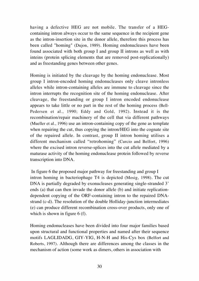
having a defective HEG are not mobile. The transfer of a HEG-containing intron always occur to the same sequence in the recipient geneas the intron-insertion site in the donor allele, therefore this process hasbeen called "homing" (Dujon, 1989). Homing endonucleases have beenfound associated with both group I and group II introns as well as withinteins (protein splicing elements that are removed post-replicationally)and as freestanding genes between other genes.
Homing is initiated by the cleavage by the homing endonuclease. Mostgroup I intron-encoded homing endonucleases only cleave intronlessalleles while intron-containing alleles are immune to cleavage since theintron interrupts the recognition site of the homing endonuclease. Aftercleavage, the freestanding or group I intron encoded endonucleaseappears to take little or no part in the rest of the homing process (Bell-Pedersen et al., 1990; Eddy and Gold, 1992). Instead it is therecombination/repair machinery of the cell that via different pathways(Mueller et al., 1996) use an intron-containing copy of the gene as templatewhen repairing the cut, thus copying the intron/HEG into the cognate siteof the repaired allele. In contrast, group II intron homing utilises adifferent mechanism called “retrohoming” (Curcio and Belfort, 1996)where the excised intron reverse-splices into the cut allele mediated by amaturase activity of the homing endonuclease protein followed by reversetranscription into DNA.
In figure 6 the proposed major pathway for freestanding and group Iintron homing in bacteriophage T4 is depicted (Mosig, 1998). The cutDNA is partially degraded by exonucleases generating single-stranded 3’ends (a) that can then invade the donor allele (b) and initiate replication-dependent copying of the ORF-containing intron to the repaired DNA-strand (c-d). The resolution of the double Holliday-junction intermediates(e) can produce different recombination cross-over products, only one ofwhich is shown in figure 6 (f).
Homing endonucleases have been divided into four major families basedupon structural and functional properties and named after their sequencemotifs LAGLIDADG, GIY-YIG, H-N-H and His-Cys box (Belfort andRoberts, 1997). Although there are differences among the classes in themechanism of action (some work as dimers, others in association with

31
Figure 6. The Double Strand Break Repair mechanism for intron homing drawnessentially as in (Mosig, 1998). Black lines indicate donor sequences, grey linesindicates recipient sequences. See text for mechanistic details.

32
additional proteins and even as RNA-protein complexes (Zimmerly et al.,1995)) the generation of double stranded cuts at, or near, the recognitionsequence is a unifying feature. The recognition sequences for cleavageare much longer than for type II restriction enzymes, spanning up to 40base pairs of DNA (Bryk et al., 1995). A specific nomenclature for naminghoming endonucleases has been adapted from the nomenclature forrestriction endonucleases. A prefix describing the locality of the HEG (I-for intron-encoded, PI- for intein-encoded, and F- for freestanding) isfollowed by a three-letter genus-species designation followed by aRoman numeral to distinguish enzymes from the same organism (e.g. theomega homing endonuclease is called I–SceI, the first intron-encodedHEG found in Saccharomyces cerevisiae, for latest updates ofnomenclature see (Roberts et al., 2003)).
Group I introns in bacteriophage T4
Bacteriophage T4 contains three group I introns, one in the aerobicribonucleotide reductase gene (nrdB) (Gott et al., 1986; Sjöberg et al.,1986), one in the anaerobic ribonucleotide reductase gene (nrdD)(formerly known as sunY) (Gott et al., 1986; Young et al., 1994), and one inthe gene coding for thymidylate synthase (td) (Chu et al., 1984). Thesequences of these three introns are remarkably similar compared to theoverall sequences of group I introns indicating that the T4 introns have acommon origin. Apart from the structural elements always present ingroup I introns, the T4 introns have additional regions and form their ownsubgroup (IA2) among the group I introns. The P7 helix is followed bytwo additional stem loops, P7.1 and P7.2, and helix P9 is made up ofthree stem loops P9, P9.1 and P9.2 instead of one.
Given the high degree of similarity between the three T4 introns aremarkable difference in the nrdD intron structure compared to the othertwo T4 introns is the lack of a P2 helix (Xu and Shub, 1989). This isespecially strange since this region is thought to form a tertiaryinteraction with P8, positioning the 5’ splice site at the G-binding site inthe catalytic centre (Michel and Westhof, 1990). The fact that this helix is

33
Figure 7. Secondary structure models for the T4 group I introns.

34
absent in some other group I introns implies that different folds haveevolved to accomplish the 5’ splice site positioning. In addition to theregions forming the catalytic structures of the intron, each of the T4introns contains a long open reading frame (ORF) within the intron.These ORFs are situated in the peripheral loop of P6a in the td and nrdBintrons and in the loop of P9.1 in the nrdD intron (Fig. 7). The ORFs are735 (td), 774 (nrdD) and 291 (nrdB) bases long and have for the td andnrdD introns been shown to encode homing endonucleases, while thenrdB intron ORF is a remnant of a homing endonuclease gene (seebelow).
T4 intron-encoded homing endonucleases
Although all three T4 introns contain homing endonuclease ORFs onlythe td and nrdD introns are mobile (Quirk et al., 1989a; Quirk et al., 1989b).The ORF products of the td and nrdD introns have been shown to behoming endonucleases (called I-TevI and I-TevII respectively) withrecognition sequences spanning the intron insertion sites (Bell-Pedersen etal., 1990; Bell-Pedersen et al., 1989) while the nrdB intron carries a non-functional version of such an endonuclease (Eddy and Gold, 1991). Thelack of mobility of the nrdB intron is due to a 491 base pairs deletionrepresenting almost two thirds of the original nrdB HEG. The closelyrelated phage RB3 has been found to contain an almost identical nrdBintron that contains an ORF of 807 base pairs (Eddy and Gold, 1991). ThisORF encodes a homing endonuclease (I-TevIII) with a recognitionsequence at the nrdB intron insertion site. Strangely though, this intronhas not been found to home in laboratory experiments (Eddy and Gold,1991). In contrast to the homology between the three introns in T4, theHEGs are highly divergent (Shub et al., 1988). I-TevI and I-TevII belongto the GIY-YIG family but I-TevIII, based on the RB3 sequence, belongsto the H-N-H family (Eddy and Gold, 1991). From this it seems that theHEGs have different origins and have invaded the introns at differenttimes. An indication that the T4 HEGs have resided in the phage genomefor a long time is that they display the same codon usage and high A-Tcontent as T4 and and also have highly T-even specific promoters and aprecise transcriptional regulation system (Edgell et al., 2000). To hinderthe HEGs from being translated from the unspliced pre-mRNA, which

35
would disturb intron splicing, a stem-loop structure is situated upstreamof the HEG, covering the translational initiation site (Gott et al., 1988;Shub et al., 1987). Instead, the HEGs are translated from transcriptsgenerated from their own T4 promoters. I-TevI and I-TevII have latepromoters and I-TevIII has both a middle and a late promoter (Guild et al.,1988; Kassavetis et al., 1986). Expression of the homing endonucleases latein infection is probably advantageous since multiple copies of the phagegenome, potential targets of homing, are present at that time.
Distribution of introns among T-even-like phages
As for many other group I introns, the distribution of the T4 introns isvery irregular (Eddy, 1992; Pedersen-Lane and Belfort, 1987; Quirk et al.,1989b; Sandegren and Sjöberg, 2004). The td intron is the one most widelypresent in the T-even phage family, being found in T4, T6 and the morerecently isolated phages RB3, LZ2, TuIa and U5 (Chu et al., 1984; Eddy,1992; Sandegren and Sjöberg, 2004). The nrdB intron has only been foundin T4, RB3 and U5 (Eddy and Gold, 1991; Sandegren and Sjöberg, 2004),and the nrdD intron only in T4 and U5 (Gott et al., 1986; Sandegren andSjöberg, 2004). The distribution of group I introns among the T-even-likebacteriophages is discussed more thoroughly in the section describingPaper I.
Freestanding homing endonucleases in T4
Apart from the intron-encoded homing endonucleases T4 also containsseveral freestanding homing endonuclease genes inserted in betweenother genes. Sharma et al. (1992) recognised that five previouslyuncharacterised T4 genes (segA-E, for similar to endonucleases of group Iintrons) share the GIY-YIG sequence motif with the I-TevI endonucleaseand intron encoded homing endonucleases of fungal mitochondria(Sharma et al., 1992) and they also showed that SegA has endonucleaseactivity in vitro with specificity to make a double strand cut within itsneighbour gene uvsX (Sharma et al., 1992; Sharma and Hinton, 1994). AlsoSegC and SegE proteins have been shown to have endonuclease activity

36
(Kadyrov et al., 1997; Shcherbakov et al., 2002) and reports of unpublisheddata (Kadyrov et al., 1997) states that SegB and SegD also show site-specific endonuclease activity. In addition, two more genes have beenadded to the seg-family in T4, segF (previously gene 69 (Belle et al.,2002)) and segG (previously gene 32.1 (Liu et al., 2003)) both of whichencode proteins that possess site-specific endonuclease activity. Of these,SegA, SegE and SegF cleaves the HEG-containing alleles in T4 althoughSegE cleaves RB30 DNA and SegF T2 DNA more efficiently (Belle et al.,2002; Kadyrov et al., 1997). SegC is the only one of these homingendonucleases reported not to cleave T4 DNA (Shcherbakov et al., 2002).In this respect the freestanding homing endonucleases seem to differ fromtheir intron-encoded counterparts where the intron-containing allele isimmune to cleavage due to the disruption of the endonuclease recognitionsites by the introns. The apparent preference of the seg-genes for cleavingthe genomes of related phages instead of the T4 genome has been shownto explain the concept of localized marker exclusion in mixed infectionsbetween T4 and related phages (Belle et al., 2002; Liu et al., 2003). Inmixed infections between T2 and T4, T2 genetic markers are generallypresent at only 10-20% frequency in the progeny and some T2 loci areeven less represented with <1% being transmitted to the offspring (Russelland Huskey, 1974). Belle et al. (2002) showed that the exclusion of T2gene 56 was attributable to the presence of segF next to the T4 gene 56and that the cleavage and following double strand break repair results inthe predominance of T4 gene 56 and segF in the progeny (Belle et al.,2002). If this is a general feature of the seg-genes, having multiplehoming endonuclease genes throughout the genome would give T4 aclear selective advantage in mixed infections with other phages by beingable to cleave their genomes.
A second group of genes in T4 that have homology to homingendonucleases are the mob-genes (mobA-E , for similarity to mobileendonucleases) that share the H-N-H motif with the I-TevIIIendonuclease and several mobile endonucleases of group I and group IIintrons (Kutter et al., 1995). None of the mob-genes have yet been reportedto have endonuclease or homing properties (Miller et al., 2003b). This willbe discussed further in the section describing Paper III.

37
Introns in other bacteriophages
The three introns represented in T4 are the only group I introns found inthe T-even-like bacteriophages or any other E. coli phages. However, agrowing number of group I introns are now being found in other phages,infecting gram-positive bacteria. Goodrich-Blair and co-workers haveshown that a group I intron in the gene coding for DNA polymerase isabundant among the Bacillus subtilis HMU phages (Goodrich-Blair et al.,1990; Goodrich-Blair and Shub, 1994). HMU phages SPO1, SP82, 2C andφe all contain a group I intron at the same position in their DNApolymerase gene while in an additional phage (SP8), also belonging tothis family, no intron was found (Goodrich-Blair et al., 1990). Although thesample size is small, this intron appears to be widespread in the HMUphage population in contrast to the scattered distribution of introns in theT-even-like phages. Interestingly, although the intron sequences of thesephages are highly similar their H-N-H endonuclease ORFs have divergedsubstantially having only 43-70% amino acid identity (Goodrich-Blair andShub, 1994). These homing endonucleases also have the unusual propertyof cleaving only one strand of their substrate and they cleave bothintronless and intron-containing alleles (Goodrich-Blair and Shub, 1996).They also appear to have evolved differences in their recognitionsequences so that SPO1 endonuclease cleaves SP82 intron-containingDNA and vice versa, with competition between phage sequences inmixed infections as a consequence (Goodrich-Blair and Shub, 1996;Landthaler et al., 2004). The unrelated Bacillus thuringiensis phageBastille contain a group I intron at the exact same site in the DNApolymerase gene as the HMU phages but the intron sequence differssubstantially and is instead more similar to the introns in theStaphylococcus phage Twort (see below) (Landthaler and Shub, 2003).However, the Bastille intron encodes a H-N-H homing endonuclease (I-BasI) homologous to the SPO1 and SP82 homing endonucleases I-HmuIand I-HmuII. I-BasI also nicks only one strand of the DNA target but itonly cleaves intronless alleles (Landthaler and Shub, 2003).
Two other B. subtilis phages have been shown to contain introns. Thevirulent β22 phage has an intron inserted in the thymidylate synthasegene (Bechhofer et al., 1994). The β22 intron is inserted 21 base pairsfurther downstream in the td gene compared to the insertion site of the T4td intron (Bechhofer et al., 1994). Like the T4 nrdB intron it has, what

38
appears to be, only a fragment of a homing endonuclease ORF but it isinserted in the P8 loop and not in the P6 loop as in the T4 td intron,indicating that these introns have been invaded by different HEGs onseparate occasions.
The temperate Bacillus subtilis prophage SPβ contains two group Iintrons, one in each of the genes coding for the two subunits (bnrdE andbnrdF) of a class Ib ribonucleotide reductase, and in addition also anintein (a protein splicing element that is removed post-translationally) inthe bnrdE gene (Lazarevic et al., 1998). In related prophages however,there are different intron/intein configurations (see Table 3) withvariation in the number and positions of intervening sequences (Lazarevic,2001). In total there are five different intron versions in the bnrdE andbnrdF genes among the seven prophage-containing Bacillus strainsscreened, with high degree of similarity (>98% identity) between intronsin the same position and with 60 to 70% identity between all introns.
Two group I introns have been reported in bacteriophages belonging tothe taxa Siphoviridae, infecting a completely different group of bacteria,the lactic acid bacteria. The lytic Lactobacillus delbrueckii bacteriophageLL-H was shown to have a group I intron in the gene terL (Mikkonen andAlatossava, 1995), coding for the large subunit of the terminase proteinresponsible for cutting the phage genome into genome size pieces duringpackaging into the phage capsids. The second intron was found in a geneof unknown function in the temperate lactococcal bacteriophage r1t (vanSinderen et al., 1996).
The phage containing the largest number of introns is the Staphylococcusaureus bacteriophage Twort that was shown to have at least five group Iintrons (Landthaler and Shub, 1999). Three group I introns (without HEGs)were shown to reside in the same gene (orf142), of unknown function(Landthaler and Shub, 1999). Two additional group I introns are inserted inthe phage nrdE gene (Landthaler et al., 2002). One of the nrdE introns hasan ORF inserted in the P6 loop that encodes a H-N-H homingendonuclease (I-TwoI) that nicks one strand of the intronless allele. Theother nrdE intron has a 106 nucleotides insertion in the P6 loop that maybe a remnant of a HEG.

39
A large screen for group I introns of Streptococcus thermophilusbacteriophages showed that half of the 62 phages screened contained agroup I intron in the lysin gene (Foley et al., 2000). All introns were foundto contain an ORF with the H-N-H motif and in five phages this ORF hassuffered deletions most likely rendering them non-functional. Sequencingof the introns from a subset of the phages showed that they are almostidentical even though the phages were isolated in different parts of theworld (Foley et al., 2000).
The latest additions to phage group I introns were found via the completegenome sequence of the Staphylococcal phage K (O'Flaherty et al., 2004).This phage contains three introns, one in the lysin gene (lys-I1) and theother two in the DNA polymerase gene (pol-I2 and pol-I3). All threeintrons contain a HEG belonging to the H-N-H family.
An interesting fact is that all group I introns found in phages belong tothe same structural subgroup (IA2) of the group I introns. Only threeother group I introns (two in Chlamydomonas eugametos and one inChlamydomonas reinhardtii) belong to this group, of the more than 2000group I introns characterised (For updated compilations of group I intronssee the RNA secondary s t ruc ture da tabase a t :http://www.rna.icmb.utexas.edu/). This indicates that all the phageintrons share a common ancestor. Furthermore, there is a clear biastowards genes coding for proteins involved in DNA synthesis beinginterrupted by introns in phages, with nucleotide synthesis genes(ribonucleotide reductases and thymidylate synthase) and DNApolymerases being clearly over-represented. This is discussed further inPaper II.

40
Table 2. Intron-containing genes in bacteriophages
Phage Gene Intronsubgroup
HEG Reference
T4 (E. coli) td IA2 (P6) I-TevI (GIY-YIG) (Chu et al., 1984)nrdB IA2 (P6) I-TevIII (H-N-H,
partial)(Gott et al., 1986; Sjöberg etal., 1986)
nrdD IA2 (no P2) (P9.1) I-TevII (GIY-YIG) (Gott et al., 1986; Young etal., 1994)
T6 (E. coli) td IA2 (P6) I-TevI (GIY-YIG)RB3 (E. coli) td IA2 (P6) I-TevI (GIY-YIG) (Eddy, 1992; Sandegren and
Sjöberg, 2004)nrdB IA2 (P6) I-TevIII (H-N-H) (Eddy and Gold, 1991)
LZ2 (E. coli) td IA2 (P6) I-TevI (GIY-YIG) (Eddy, 1992; Sandegren andSjöberg, 2004)
TuIa (E. coli) td IA2 (P6) I-TevI (GIY-YIG) (Eddy, 1992; Sandegren andSjöberg, 2004)
U5 (E. coli) td IA2 (P6) I-TevI (GIY-YIG) (Sandegren and Sjöberg,2004)
nrdB IA2 (P6) I-TevIII (H-N-H,partial)
(Sandegren and Sjöberg,2004)
nrdD IA2 (no P2) (P9.1) I-TevII (GIY-YIG,partial)
(Sandegren and Sjöberg,2004)
SPO1 (B. subtilis) DNA-pol IA2 (P8) I-HmuI (H-N-H) (Goodrich-Blair et al., 1990)SP82 (B. subtilis) DNA-pol IA2 (P8) I-HmuII (H-N-H) (Goodrich-Blair and Shub,
1994)2c (B. subtilis) DNA-pol IA2 (P8) I-HmuII (H-N-H) (Goodrich-Blair and Shub,
1994)φ (B. subtilis) DNA-pol IA2 (P8) (H-N-H) (Goodrich-Blair and Shub,
1994)
β22 (B. subtilis) td IA2 (P8) (GIY-YIG, partial) (Bechhofer et al., 1994)
Bastille (B. thuringiensis) DNA-pol IA2? (no P2) (P8) I-BasI (H-N-H) (Landthaler and Shub, 2003)
SPβ prophage (B. subtilis) bnrdE-I IA2 - (Lazarevic et al., 1998)bnrdF-I IA2 (P6) YosQ (H-N-H) (Lazarevic et al., 1998)
M1918 prophage (B. subtilis) bnrdE-I IA2 - (Lazarevic, 2001)bnrdF-I IA2 (P6) (GIY-YIG) (Lazarevic, 2001)
M1321 prophage (B. subtilis) bnrdE-I1 IA2 - (Lazarevic, 2001)bnrdE-I2 IA2 - (Lazarevic, 2001)
M135 prophage (B. subtilis) bnrdE-I IA2 - (Lazarevic, 2001)BSG40 prophage (B. subtilis) bnrdE-I1 IA2? (P6) ORF732 (GIY-YIG) (Lazarevic, 2001)
bnrdE-I2 IA2 (P6) (GIY-YIG) (Lazarevic, 2001)bnrdF-I IA2 (P6) (GIY-YIG) (Lazarevic, 2001)
Twort (S. aureus) Orf 142 I1 IA2 - (Landthaler and Shub, 1999)Orf 142 I2 IA2 - (Landthaler and Shub, 1999)Orf 142 I3 IA2 - (Landthaler and Shub, 1999)nrdE-I1 IA2 (P6) 106 nt remnant? (Landthaler et al., 2002)nrdE-I2 IA2 (P6) I-TwoI (H-N-H) (Landthaler et al., 2002)
LL-H (Lactobacillus) terL (Mikkonen and Alatossava,1995)
r1t (Lactococcus) orf40 (van Sinderen et al., 1996)
S3b+27 more (S. thermophilus) lysin IA2 (no P2) (P8) (H-N-H) (Foley et al., 2000)DT1+S92+S93 (S. thermophilus) lysin IA2 (no P2) (P8) (H-N-H, partial) (Foley et al., 2000)Sfi16A (S. thermophilus) lysin IA2 (no P2) (P8) (H-N-H, partial) (Foley et al., 2000)ST64 (S. thermophilus) lysin IA2 (no P2) (P8) (H-N-H, partial) (Foley et al., 2000)
Phage K (Staphylococcus sp.) lysin I1 I-KsaI (H-N-H) (O'Flaherty et al., 2004)DNApol I2 I-KsaII (H-N-H?) (O'Flaherty et al., 2004)DNApol I3 I-KsaII (H-N-H) (O'Flaherty et al., 2004)

41
Present studies
Paper I: Distribution, Sequence Homology and Homing of
Group I Introns among T-even-like Bacteriophages.
Evidence for recent transfer of old introns.
Before this study was initiated it was known that group I introns wereonly scarcely found among phages closely related to T4 (Eddy, 1992;Pedersen-Lane and Belfort, 1987; Quirk et al., 1989b). In 32 T-even-likephages examined, Sean Eddy found that besides T4 only T6, LZ2, RB3and TuIa contain the td intron and that RB3 also contains a nrdB intron(Eddy, 1992). This is somewhat unexpected since the td and nrdD intronsof T4 are mobile (Quirk et al., 1989a) and the I-TevIII homingendonuclease of the RB3 nrdB intron at least can initiate cleavage (Eddyand Gold, 1991). This spoke against a general selective advantage forintrons in phages, and a T2 construct with the td and nrdD introns did notdisplay any growth advantage over intronless T2 in one-step growthexperiments in rich medium (Quirk et al., 1989a). Eddy also attempted totest if the introns had any effect on the burst size of the phage bymeasuring the burst sizes of T4 with and without introns during growth indifferent media (Eddy, 1992). His conclusion from this was that any effect(positive or negative) of introns under the conditions tested was smallerthan could be detected in such experiments (the inherent variationbetween experiments was typically ± 20%).
Our aim in this paper was to investigate if the introns found in differentT-even-like phages indeed have been spread between the phages and tosee if the sequences of the introns and of the td, nrdB and nrdD genes ofphages with and without introns could give some indications to whyintrons are not more frequent among T-even-like phages. No intronsequences were available other than of the T4 introns and the RB3 nrdBintron and no complete sequences of any of the td, nrdB and nrdD genesfrom T-even-like phages other than T4 had been determined previously.During the course of this study the Tulane Phage Sequencing Group ledby Jim Karam sequenced several genomes of phages, more or less closelyrelated to T4. This gave us the opportunity to include complete sequences

42
of the three genes from phages outside the T-even-like group. No intronswere found in any of the genomes sequenced.
In our amplification of the three genes from phages with and withoutintrons we included the previously uncharacterised T-even-like phage U5.This phage was isolated at a microbiology course at Uppsala Universityand was kindly provided to us together with several of the RB-phages byKarin Carlson, Department of Cell and Molecular Biology, UppsalaUniversity. To our surprise, U5 contains all three introns and is the firstphage isolate besides T4 shown to do so. Like RB3, the U5 nrdB introncontains a much longer I-TevIII gene but the U5 I-TevIII has, like its T4counterpart, suffered a deletion although only of two base pairs. This ishowever enough to shift the reading frame and since it occurs early in thegene it most likely disrupts its function. In addition to the frame shiftdeletion there is also a 6 base pairs deletion exactly spanning the latepromoter of the I-TevIII gene (Fig. I1).
Figure I1. Deletions in U5 I-TevIII promoter region compared to T4 and RB3.

43
Interestingly, the I-TevII gene in the U5 nrdD intron has also suffered a 2base pairs deletion early in its reading frame. It is tempting to speculatethat expression of I-TevII and I-TevIII in some way is detrimental to thephage and that these deletions have enabled U5 to remain within thepopulation. If a homing endonuclease can cleave at additional sites in agenome, to which it cannot home, it is possible that this has a negativeeffect on phage viability. If this is a general trait of homingendonucleases it may be an explanation to their scattered appearanceamong phages. The toxicity of the T4 intron-encoded homingendonucleases to E. coli when cloned on other than low copy plasmids isgenerally thought to come from cleavage of the bacterial genome (Quirket al., 1989a). However, T4 is an example of a phage that containsmultiple HEG-containing introns and at least 12 freestanding genesrelated to homing endonucleases some of which have been shown topossess endonuclease activity and to induce cleavage at multiple sites inthe T4 genome (see paper III). This may reflect an increased tolerance todouble strand breaks in T4 (Belle et al., 2002).
When comparing the sequences of the intron-containing td, nrdB andnrdD genes there is a striking difference between the amount of sequencevariation in the exons and in the introns between all intron-containingphages and especially between American phage isolates T4, T6, RB3 andLZ2. Among the American phages there is only one nucleotide differencethroughout the td intron/HEG region while there is around 10% variationin td exon sequences between the phages. Although the catalytic parts ofthe introns may have a low mutation rate due to functional constraints thecoding regions of the HEGs should accumulate at least synonymouschanges over time. The lack of sequence variation between HEGs ofcognate introns together with the scattered distribution of the introns onthe gene-based phylogenies (Fig. 3 in paper I) strongly indicate that theintrons have been spread horizontally between the phages, and from thelack of variation between the td introns of American isolates theacquisition of the td introns appears to have been very recent among thesephages.
The positions of the nucleotide substitutions within catalytic regions ofthe nrdB and td introns between phages are shown in figure I2 (there areno differences in catalytic regions between the T4 and U5 nrdD introns).

44
None of the seven new introns sequenced in this study show anyvariations that can be predicted to negatively affect splicing. Differencesoccurring in nucleotides involved in base pairing interactions are eitheraccompanied by covariation at the corresponding site, keeping theinteraction intact, or introduce an accepted non Watson-Crick base pair(most often G-U).
Interestingly, a large part of the changes in the intron structural parts aresituated in regions P1, P9.0 and P10, known to be important in splice siteselection. The td sequence of TuIa differs from its T4 counterpart atseven positions in regions of intron secondary structure. Four of theseoccur in the internal guide sequence, made up of helix P1 and the P10interaction. P1 is the major determinant of the 5´ splice site while the P10interaction is responsible for keeping the region of 5´ cleavage in closeproximity to the 3´ splice site for ligation of the two exons (Burke, 1989;Davies et al., 1987). The P1-helix is maintained intact in spite of thesechanges, showing a high tolerance to variations given that base pairing ismaintained. A G→U change in the P1-loop of the td intron in TuIa, andan A→U change of the third base of td exon2 in U5, both facilitate thepossible formation of stronger P10 interactions in these phages. Togetherwith the variations seen in P1 this means that a large part of the variationsbetween the different phage strains occur in regions involved in the 5´splice site selection. These variations may be adaptations in order toavoid cryptic splicing due to variations in exon sequences between thephages.
Another interesting set of variations is seen in the P9.0 pairing of the tdand nrdB introns. In most group I introns the P9.0 interaction is made upof two base pairs between nucleotides preceding the P7 helix and thepenultimate nucleotides at the 3’ splice site (Burke et al., 1990; Michel andWesthof, 1990). In T4 only the nrdD intron contains a P9.0 interaction oftwo Watson-Crick base pairs while it is made up of only one base pair inboth td and nrdB introns (see Fig. I2). In U5, however, the nrdB intronhas an A→U substitution, compared to T4, in the J7/9 linking region andthus can form a two base pair P9.0. This feature is shared by the RB3nrdB intron (Eddy and Gold, 1991). In the td intron, the P9.0 of T4 is madeup of a single A-U base pair while in T6, U5, RB3 and LZ2 the U ispreceded by an A, instead of the C in T4 and TuIa. This A can then base

45
Figure I2. Sequence variations in catalytic parts among the td and nrdB introns.Changes are colour-coded for each phage. Changes in nrdB are identical for both RB3and U5.

46
pair with the penultimate U of the intron forming a P9.0 interaction oftwo base pairs. However, U5 has an A instead of a U preceding P9 andwill thus still only have a one base pair P9.0. Opposing A:s are also foundin both T4 nrdB and U5 td introns next to the single identifiable Watson-Crick base pair. Introns that entirely lack recognizable P9.0 pairings oftenhave opposing A:s on both strands in this region (Michel and Westhof,1990), indicating that non-Watson-Crick A-A base pairs may constitutethe P9.0 interaction. Thus, all T-even-like phage introns except the tdintron in T4 and TuIa probably form two base pair P9.0 interactions. Thisfurther corroborates the view that variations in the interactions importantfor splice-site selection are especially frequent among the phage introns.
Our screen of homing between phages in mixed infections showed, asexpected, that homing most easily occurs between closely related phages.The few sequence variations around the intron insertion sites in the nrdBand nrdD introns among the T-even-like phages indicate that it is not alack of homing sites that has limited the spread of these introns amongphages. However, the higher degree of variation between td allelestogether with the presence of an optional gene (td.1) flanking the td geneappear to lower the homing efficiency of the td intron between some ofthe phages. Furthermore, the process of general exclusion can reduce theefficiency of intron transfer but as was shown for the td intron of RB3 incrosses with T4∆IVS, homing can overcome this exclusion to someextent and from the few progeny that obtained the intron it can betransferred with increased efficiency in subsequent mixed infections.In conclusion we find that intron homing has occurred between T-even-like phages in Nature and can be very efficient between closely relatedphages but that sequence divergence of regions flanking the introninsertion sites, optional occurrence of flanking genes and the process ofexclusion can lower the efficiency of homing. This together with theapparent rapid degeneration of HEG-function that we find in some of theintrons may limit the spread of introns among the T-even-like phages.However, once an intron succeeds in transferring to a more distantlyrelated phage, subsequent homing to phages related to the new host willbe rapid.

47
Paper II: Self-splicing of the Bacteriophage T4 Group I
Introns is Affected by the Growth of the Infected Bacterium
and Requires Efficient Translation of the Pre-mRNA In
Vivo.
Ever since the T4 introns were found it has been speculated about howthey affect the phage viability. Initially it was argued that in order to bemaintained in the phage genome the introns must have a positive effect(probably through regulation of the intron containing genes) on phageviability (Eddy, 1992; Goodrich-Blair et al., 1990; Gott et al., 1986; Shub,1991; Shub et al., 1988; Shub et al., 1987) However, when it was found thatintrons are very rare among the T-even-like phages and that the intronsare/have been mobile the view that they are merely selfish geneticelements that confer no advantage to the phage prevailed. So far therehave been no reports of any effect, positive or negative, on phageviability that can be coupled to intron presence.
In 1998 Semrad and Schroeder published a paper showing that splicing ofthe T4 td intron was highly dependent upon translation of the upstreamexon (Semrad and Schroeder, 1998). The effect on td splicing was shown tobe due to the formation of an interfering tertiary interaction between aregion of the upstream exon and the 3´splice site of the intron. Duringnormal translation the movement of ribosomes along the pre-mRNAdisrupts this interaction but if translation is hindered by addition ofantibiotics or by introduction of stop codons upstream of the interactingregion, formation of the interaction will inhibit splicing. Since this couldmake the introns a regulatory “switch” for regulation of the intron-containing genes we decided to test if the dependence on translation ofthe upstream exon for efficient splicing was a common theme for the T4introns and if limited growth conditions could affect splicing by thismechanism. We found through a computer-based complementarity screenwith nrdB exon and intron sequences, that a highly similar interactingregion exists early in the nrdB exon (see Fig. 3A, Paper II). Several otherpotential interacting regions were also found. By measuring splicing inthe absence of translation by introduction of stop codons or addition ofchloramphenicol, that inhibits the elongation step of translation, we wereable to show that splicing of the nrdB intron also is dependent upon

48
translation of the upstream exon. This was however, not primarily due tothe interaction with similarity to the one in td but other regions, closer tothe intron, such as an alternative P1 stem (see Fig. 3B, Paper II) wereresponsible for most of the reduction. Deletion of the exon regionspotentially forming such interfering interactions restored the efficiency ofsplicing to close to wild type levels even in the absence of translation.
A number of potential interfering regions with complementarity to intronsequences can be predicted for all three T4 introns and we predict thatthis is a general feature of introns in translated genes. In line with this, wealso showed that reduction of splicing efficiency occurred for all three T4introns during phage infection of cells that had been treated withchloramphenicol and in which translation therefore was abolished. Asimilar effect of chloramphenicol on splicing was noted by Belfort andcoworkers already in 1985 but they ascribed the effect to the inhibition oftranslation of an unknown protein factor that was needed for splicing invivo (Belfort et al., 1985). So far no such protein factor essential for T4-intron splicing has been found, although several E. coli proteins thatenhance group I intron splicing have been characterised (see section onProteins involved in group I intron splicing in the introduction).
Since burst size experiments have large intrinsic variations (Eddy, 1992)we choose to measure the effect of growth condition on splicing directlyby limited primer extension. Using a plasmid-encoded T4 nrdB genecontaining the intron, we found a substantial reduction in splicingefficiency when the bacterial culture entered stationary phase and afterprolonged growth in stationary phase splicing efficiency was down to40% from the initial 90% during logarithmic growth. This effect onsplicing was also seen for all three T4 introns upon infection of bacteriain stationary phase. Although we have so far not definitely shown that itis the reduction of translation in stationary phase that reduce theefficiency of splicing, this is the first time that environmental effects suchas growth conditions have been shown to affect splicing of phage introns.The reduction of splicing when the infected cell is in a starving state wasthe favourite predicted scenario for a regulatory role of the introns (Eddy,1992; Goodrich-Blair et al., 1990; Gott et al., 1986; Shub, 1991; Shub et al.,1988; Shub et al., 1987). By lowering the de novo production ofribonucleotides for DNA synthesis, energy consumption could be reduced

49
and the survival of the infected bacterium prolonged. If too much energyis spent on nucleotide synthesis the bacterium might die before any viablephage has been produced. Prolonged infection might be a good way for ahighly infectious phage like T4 to stay dormant until there are new hostcells available to infect. Further experiments are needed in order toevaluate a possible effect of introns on the survival rate of infectedbacteria.
Paper III: Two Genes Encoding Novel Homing
Endonucleases Replace the Putative Homing Endonuclease
Genes mobC and mobE in Several T4-related Phages.
During the work on the distribution of group I introns among the T-even-like phages (Paper I) we noted that there were differences in the flankinggenes of all three intron-associated genes t d , nrdB and nrdD. Wetherefore performed an independent screen of these regions from 20 T-even-like phages that confirmed the presence of the mobE gene betweennrdA and nrdB in five other phages, the absence of mobC gene betweennrdD and nrdG in all phages except T4 and the presence of a previouslyunreported gene not present in T4 (we named it td.1 see Paper I)upstream of the td gene in many of the phages. The mobC and mobEgenes belong to a family of five T4 genes called the mob-genes (forsimilarity to mobile endonucleases (Kutter et al., 1995)) that sharehomology with the H-N-H homing endonucleases. The mob-genes areputative freestanding HEGs (i.e. not associated with introns). In additionto the five mob-genes, T4 also contains seven freestanding seg-genes (forsimilarity to endonucleases encoded by group I introns (Sharma et al.,1992)) containing the GIY-YIG motif, four of which (segA (Sharma et al.,1992; Sharma and Hinton, 1994), segE (Kadyrov et al., 1997), segF (Belle etal., 2002) and segG (Liu et al., 2003)) have been shown to encode proteinswith homing endonuclease activity. Table III1 shows a compilation of thedistribution of freestanding homing endonuclease genes among T4-likephages.
Our sequence of the T4 mobE gene differed from the previously reportedsequences of this region by two frame shifts that brought two proposed

50
open reading frames into one continuous open reading frame, with thestart codon overlapping the stop codon of nrdA and the stop codonoverlapping the start codon of nrdB (see Fig. III1). All five additionalmobE genes that we found in the screen (in T6, RB2, RB3, RB15 andLZ7 respectively) were inserted between nrdA and nrdB in exactly thesame manner. Since all our sequences were obtained by direct sequencingof PCR fragments from genomic DNA this strongly suggests that theoriginal sequence of mobE (obtained from a cloned genome fragment(Sjöberg et al., 1986; Tseng et al., 1988)) was of a non-functional gene andthat expression of full-length MobE protein is toxic to E. coli, a featureshared with several experimentally proven homing endonucleases(Kowalski and Derbyshire, 2002). In agreement with this we have beenunable to clone full-length mobE.
Even though most T-even-like phages lack mobC and mobE, severalphages have unrelated genes at the same sites. Phages T6, RB3 and LZ2all have a gene related to the seg-genes in place of mobC between nrdDand nrdG. We have named this gene segH as it is the eighth seg-genedescribed. In vitro expressed SegH protein displayed homingendonuclease-like activity by introducing site-specific, double-strand cutsin the end of the nrdD gene, a little more than eighty base pairs upstreamof the segH start codon. Similarly, phage U5 has an open reading frameof 544 codons between the nrdA and nrdB genes, in place of the T4 mobEgene. The product of this gene also showed homing endonuclease activityby introducing a double-strand cut in the downstream nrdB gene. The U5gene has no homology to any genes presently in GenBank and we havenamed it hef (for homing endonuclease-like function).
Although we have tested DNA targets from several different phages andtested several different buffer conditions we have still not been able todetect either double-, or single-strand cleavage in the regions of the mobCand mobE genes by either T4, T6 or RB3 MobE proteins or T4 MobC invitro. This could mean that they either are non-functional or that we havenot tested the correct conditions/target sequences for cleavage. It mayalso be that they only recognise and cleave the modified DNA(containing glucosylated hydroxymethyl-cytosine) that many T-even-likephages have.

51
Table III1. Freestanding homing endonucleases in T4-like bacteriophages
Phage mobA mobB mobC mobD mobE segA segB segC segD segE segF(gp69)
segG(gp32.1)
T2 -1 -1 -8 -2,3 -6
T4 + + + + + + + + + + + +T6 segH1 +1 -8 -2,3 -6
RB2 -1 +1
RB3 segH1 +1 -8 +6
RB6 +6
RB8 +6
RB9 +6
RB10 +6
RB14 -1 -1
RB15 -1 +1 -8 -2,3
RB18 -6
RB23 -1 -1
RB26 -1 -8
RB27 -1 -1 -6
RB30 -5
RB32 -1 -1 -6
RB49 -4 -4 -4 -4 -4 -4 -4 -4 -4 -4 -4 -4
RB51 -1 -5
RB61 -1
RB69 -4 -4 -4 -4 -4 -4,8 -4 -4 -4 -4 -4 -4
RB70 -6
LZ1 -1 -1
LZ2 segH1 -1
LZ5 -2,3
LZ7 -1 +1
LZ11 -8
U5 -1 hef1
TuIa -1 -8
TuIb -6
AehI -4 -4 -4 -4 “+4” -4 -4 -4 -4 -4 -4 -4
RR44 -4 -4 -4 -4 -4 -4
Mi +6
PST +6
FSα +6
SV76 -6
KVP40 -7 -7 -7 -7 -7 -7 -7 -7 “+7” -7 -7 -7
Refs. 1 – Our work; 2 – (Belle et al., 2002); 3 – (Mosig et al., 2001); 4 – genome sequence; 5 - (Kadyrov et al.,1997).; 6 – GenBank sequences/(Liu et al., 2003); 7 – (Miller et al., 2003a); 8 - (Sharma and Hinton, 1994)

52
To investigate if the novel homing endonucleases SegH and Hef canpromote homing of their genes, and to further investigate the reportedunidirectional inheritance of mobE, we performed mixed infections withphages with and without these genes. We were able to show that all threeversions of the segH gene (T6, RB3 and LZ2) are homing proficient incrosses with T2, but due to the general exclusion of other phages by U5we have still not been able to show if it is the activity of Hef thatpromotes the strong local exclusion of the nrdA-nrdB region of recipientphages in crosses with U5. By using T6 as donor phage for studies ofmobE homing we were able to avoid the problem of general exclusionthat we previously have experienced with T4 mobE. In a screen of theflanking nrdB region from T6/T2 progeny that had inherited the mobEgene we found that three of 13 phages had a recombination site in theregion of nrdB that we could screen. The sequence variation between T2and T6 in the first half of the nrdB gene would restrict homing-inducedrecombination events to the second half of the gene, which we have onlybeen able to screen a small part of. Nonetheless, the occurrence ofrecombination points in the nrdB gene of these progeny phages isencouraging and screening of an increased region of the nrdB gene is inprogress.
SegH and Hef were shown to cleave both HEG-containing and HEG-lessalleles. Cleavage of the own DNA may be lethal to the host phageperhaps explaining the limited occurrence of HEGs in phage genomes.However, limited cleavage activity at sites in the own genome may betolerated since it, at least in T4, induces recombination dependentreplication. We propose that it may also increase the propensity of theHEG to spread to new sites in a genome, as discussed in Paper III.Relocations have been proposed to be the only way for a HEG to avoiddegeneration and stay viable over evolutionary time (Goddard and Burt,1999) and the high frequency of deleterious deletions that we observe inintron-encoded HEGs (Paper I) suggests that the selective pressure toremove unnecessary sequences is high in phages.
Although insertion of freestanding HEGs between genes may be toleratedin many cases since the HEG does not interrupt the reading frames of thenearby genes, the insertion of mobE between the nrdA and nrdB genes,that code for subunits of the same protein, has affected the expression of

53
them. Tseng and coworkers (1990) showed that coordinated expression ofthe two proteins, R1 (nrdA) and R2 (nrdB), constituting the T4 aerobicribonucleotide reductase occur by a set of shifts in promoter usage andtranscriptional termination/anti-termination (Tseng et al., 1990). As shownin figure III1, expression of nrdA and nrdB occurs both from separatepromoters and from a common promoter as part of an operon togetherwith dihydrofolate reductase (frd), thymidylate synthase (td) and possiblyendonuclease II (denA) (Tseng et al., 1990).
Figure III1. Operon of nucleotide synthesis genes. Promoters (E = early, M = middle,L = late), and the transcriptional terminator (stem-loop) before nrdB are shown.Dotted lines indicate exons that are spliced together. Drawn essentially as in (Tseng etal., 1988).
Early in infection nrdA is expressed from the polycistronic transcriptgenerated from an early promoter located upstream of frd. Initially,transcripts from this promoter do not include the nrdB gene, downstreamof nrdA, due to a transcriptional termination site located 50 base pairsafter the nrdA stop codon. Instead, nrdB is transcribed from a MotAdependent, middle promoter just in front of the gene (Tseng et al., 1990).T4 anti-termination factors are produced at about 5 minutes afterinfection, and then transcription from the early, frd promoter reaches thenrdB gene. At approximately the same time transcription starts from alate promoter in front of nrdA, and this gives a coordinated expression ofthe ribonucleotide reductase genes during replication (Tseng et al., 1990).These alternations in the expression of nrdA and nrdB most likelyinfluence the production of dNTPs by the dNTP synthetase complex.
Intriguingly, both the transcriptional terminator downstream of nrdA andthe middle promoter in front of nrdB are situated within the mobE gene(Fig. III1). The complicated regulatory scheme of nrdA and nrdB

54
coexpression must therefore have been adopted upon insertion of mobEin the T4 genome. Phages lacking mobE only have a 50 base pairsintergenic region between n r d A and nrdB without obviousterminator/promoter sequences (our unpublished observation) andcoordinated expression of R1 and R2 proteins is likely to occur from thepromoters upstream of nrdA in these phages. Yet, how the extensivesequence variation in the N-terminal part of the RB3 mobE gene (whichlacks the nrdB middle promoter while having the upstream terminator)and the insertion of the much longer hef gene in U5 affect expression ofthe nrdA and nrdB genes remains to be investigated.

55
Future studies
Several interesting follow-up projects have emerged from the data thatthis thesis is based upon.
To determine if it is indeed the reduction of translation during stationarygrowth that affects the splicing efficiency of the T4 introns we aredeleting the major intron-interacting regions in the nrdB exon1 (∆45-129;∆Cry-P1). If splicing of such a construct is increased duringstationary growth in comparison to the wild type T4 nrdB transcript thiswould be a strong indication that the reduction of splicing that we haveseen is indeed coupled to the upstream exon of nrdB and not due to anyfactors acting on splicing in trans.
It would also be of interest to analyse if the reduction of splicing instationary phase bacteria will yield any difference in the survival rate ofbacteria infected with intron-containing phages compared to intronlessphages. An alternative way to detect an effect of introns upon phageproduction would be to measure the production of viable phage instationary phase without natural lysis by sampling an infected cultureover several hours/days, lyse the cells with chloroform and measure theamount of viable phage that was present inside the bacterium.
Further biochemical and bioinformatic studies on the Hef protein is underway to determine if it represents a new type of freestanding homingendonuclease or if it is a distant relative to one of the previouslycharacterised families. In vivo cleavage studies of both Hef and SegHcould shed more light on whether they have any preference for cleavageof HEG-less alleles. Homing and in vivo cleavage studies of mob-genesare also under way to determine if they really encode homingendonucleases. The mobE gene is so far the best candidate since itappears to promote homing.
We are also planning to do homing studies with the td.1 gene that issituated between the frd and td genes in most T-even-like phages that wehave screened but that is absent in T4. Preliminary studies have shownthat the sequence of td.1 is almost identical between all phages exceptRB23 that contain a completely unrelated gene at the same position. Nosequence homologies to any genes in the databases have been found forthese two genes so far.

56
Acknowledgements
I would like to thank the people without which help and support thisthesis would not have been possible:
My supervisor Britt-Marie Sjöberg for sharing her great knowledge ofscience and for always letting me try my own ideas.
My parallellslalompolare in the lab David Nord for all the help withphage crosses, massive PCR screenings, lunches, loftning, football-coaching, Zombie movies and for all the Great Wars in space andelsewhere.
The ”old” PhD students Jessica, Maja, Monica, Jasmine and Martin foralways helping out during the first years. Margareta Karlsson my firstlab-supervisor. Ant , Pernilla, Alex, Hanna, Tove , Anna , Patrick,Jossan, Annika and all the “new” PhD students and other personnel atthe Department of Molecular Biology & Functional Genomics for makingit a very nice place to work in.
All the nice “scientific” people I have had the opportunity to meet onconferences over the years (in order of appearance) Larry Gold who toldme to stay away from the phage introns because they were not worthworking with (didn’t follow that advise), Jack Szostak for nicediscussion and Lagavulin, Jennifer Doudna whose crystal structure ofP4-P6 got me interested in group I introns, John Roth who has told metwice to stay away from ppGpp strains (did follow that advise!), MattGoddard for all help with HEG phylogenetic theories, Sean Eddy for anice discussion about introns in phages, Betty Kutter for a very nice T4meeting 2003, Chris Mathews for advice on Paper I, Jim Karam forsharing ideas on phage genomes, Deborah Hinton, David Shub andDavid Edgell for nice discussions on T4 freestanding HEGs and SteveAbedon for being the MicroDude.
All friends that have made these years great: Andy, Dabuel, Milla, Amy,Elin. Oskar for fishing at the Big Essing. Richard and Bobby for theearly years, and indeed the late ones too. Dr. S for hosting all the crayfishparties. Q, Dr. T, Dr. Skåne, Dr. M. Dr. Martin for all the Whisky and

57
Ford Fairlane evenings. Stina the Pors provider. X-björn, Niklas“Typhoon” Holmström, Christian, Mikaela, Dr. Tina, Dan, Susanne,Love, Hanna. Kristina for nearly always booking the innebandy.Hubert, Stefan, Malin, Linda. Calle for all the battle-Sundays at KI.Dr. Martin S for fishing, walking and talking. Maria & Henrik for greatmidsummer-, and glögg-parties. And all the footballers, Loftare, NF andMoleQul people.
My Parents Gunnar and Britt-Mari for all their support when thebacteria/phages/genes and other lab-stuff don’t do as they are supposedto. My sister Ellinor, her husband Atte and little “Udo”. Minamorföräldrar Bertil och Irma (och Sussie). My aunt Britta, and Göran.Annas parents Åke and Birgitta for always being so nice and for greatdinners with a lot of great food. Astrid, Jonas, Maja and Lukas. TheItalian side Inga-Britt, Enzo, Ettore and Monica for a great time inRome after my licentiate defence.
Anna, for all her love and for being Världens bästa.
Several other persons/institutions have also made a difference during thiswork (in no particular order): AIK, J.R.R. Tolkien, Mark Knopfler, BKKanon, The Loft, The Macallan, Jorms fiskecamp, X-björns,Springbanks, Tullfritt, gamla Lantisgrillen, bastun under Arrhe, 106,7Rockklassiker, Gula Villan, Bono, BB King, Laphroaigh, Urga, NaturalSelection, and many many more…..
Inspiration and energy for the completion of this work has come frominnumerable cans of Coca Cola and on successful lab days from SingleMalt Whisky.

58
References
Abedon, S. T. (1994). Lysis and the Interaction between Free Phage and InfectedCells. In Molecular Biology of Bacteriophage T4., J. D. Karam, ed. (Washington, DC,American Society for Microbiology), pp. 397-405.
Abedon, S. T. (2000). The murky origin of Snow White and her T-even dwarfs.Genetics 155, 481-486.
Ackermann, H. W., and Krisch, H. M. (1997). A catalogue of T4-type bacteriophages.Arch Virol 142, 2329-2345.
Allen, J. R., Lasser, G. W., Goldman, D. A., Booth, J. W., and Mathews, C. K.(1983). T4 phage deoxyribonucleotide-synthesizing enzyme complex. Further studieson enzyme composition and regulation. J Biol Chem 258, 5746-5753.
Allen, J. R., Reddy, G. P., Lasser, G. W., and Mathews, C. K. (1980). T4ribonucleotide reductase. Physical and kinetic linkage to other enzymes ofdeoxyribonucleotide biosynthesis. J Biol Chem 255, 7583-7588.
Bechhofer, D. H., Hue, K. K., and Shub, D. A. (1994). An intron in the thymidylatesynthase gene of Bacillus bacteriophage beta 22: evidence for independent evolutionof a gene, its group I intron, and the intron open reading frame. Proc Natl Acad Sci US A 91, 11669-11673.
Beckendorf, S. K. (1973). Structure of the distal half of the bacteriophage T4 tailfiber. J Mol Biol 73, 37-53.
Beckendorf, S. K., Kim, J. S., and Lielausis, I. (1973). Structure of bacteriophage T4genes 37 and 38. J Mol Biol 73, 17-35.
Belfort, M., Pedersen-Lane, J., West, D., Ehrenman, K., Maley, G., Chu, F., andMaley, F. (1985). Processing of the intron-containing thymidylate synthase (td) geneof phage T4 is at the RNA level. Cell 41, 375-382.
Belfort, M., and Roberts, R. J. (1997). Homing endonucleases: keeping the house inorder. Nucleic Acids Res 25, 3379-3388.
Bell-Pedersen, D., Quirk, S., Clyman, J., and Belfort, M. (1990). Intron mobility inphage T4 is dependent upon a distinctive class of endonucleases and independent ofDNA sequences encoding the intron core: mechanistic and evolutionary implications.Nucleic Acids Res 18, 3763-3770.
Bell-Pedersen, D., Quirk, S. M., Aubrey, M., and Belfort, M. (1989). A site-specificendonuclease and co-conversion of flanking exons associated with the mobile tdintron of phage T4. Gene 82, 119-126.

59
Belle, A., Landthaler, M., and Shub, D. A. (2002). Intronless homing: site-specificendonuclease SegF of bacteriophage T4 mediates localized marker exclusionanalogous to homing endonucleases of group I introns. Genes Dev 16, 351-362.
Benzer, S. (1955). Fine structure of a genetic region in bacteriophage. Proc Natl AcadSci U S A 41, 344-354.
Berglund, O. (1975). Ribonucleoside diphosphate reductase induced by bacteriophageT4. III. Isolation and characterization of proteins B1 and B2. J Biol Chem 250, 7450-7455.
Black, L., Showe, M., and Steven, A. (1994). Morphogenesis of the T4 Head. InMolecular Biology of Bacteriophage T4., J. D. Karam, ed. (Washington, DC,American Society for Microbiology), pp. 218-258.
Botstein, D., and Herskowitz, I. (1974). Properties of hybrids between Salmonellaphage P22 and coliphage lambda. Nature 251, 584-589.
Bremer, H., and Dennis, P. P. (1987). Modulation of Chemical Composition andOther Parameters of the Cell by Growth Rate. In Escherichia coli and Salmonellatyphimurium: Cellular and Molecular Biology, F. C. Neidhardt, ed. (Washington DC,American Society for Microbiology), pp. 1527-1542.
Brenner, S., Jacob, F., and Meselson, M. (1961). Unstable intermediate carryinginformation from genes to ribosomes for protein synthesis. Nature 190, 41-46.
Bryk, M., Belisle, M., Mueller, J. E., and Belfort, M. (1995). Selection of a remotecleavage site by I-TevI, the td intron-encoded endonuclease. J Mol Biol 247, 197-210.
Burke, J. M. (1989). Selection of the 3'-splice site in group I introns. FEBS Lett 250,129-133.
Burke, J. M., Esherick, J. S., Burfeind, W. R., and King, J. L. (1990). A 3' splice site-binding sequence in the catalytic core of a group I intron. Nature 344, 80-82.
Caprara, M. G., Lehnert, V., Lambowitz, A. M., and Westhof, E. (1996a). A tyrosyl-tRNA synthetase recognizes a conserved tRNA-like structural motif in the group Iintron catalytic core. Cell 87, 1135-1145.
Caprara, M. G., Mohr, G., and Lambowitz, A. M. (1996b). A tyrosyl-tRNAsynthetase protein induces tertiary folding of the group I intron catalytic core. J MolBiol 257, 512-531.
Caprara, M. G., Myers, C. A., and Lambowitz, A. M. (2001). Interaction of theNeurospora crassa mitochondrial tyrosyl-tRNA synthetase (CYT-18 protein) with thegroup I intron P4-P6 domain. Thermodynamic analysis and the role of metal ions. JMol Biol 308, 165-190.
Carlson, K. (2000). Virus mot bakterier. Forskning och Framsteg, 8-13.

60
Carlson, K., Raleigh, E., and Hattman, S. (1994). Restriction and Modification. InMolecular Biology of Bacteriophage T4., J. D. Karam, ed. (Washington, DC,American Society for Microbiology), pp. 369-381.
Cate, J. H., Gooding, A. R., Podell, E., Zhou, K., Golden, B. L., Kundrot, C. E., Cech,T. R., and Doudna, J. A. (1996a). Crystal structure of a group I ribozyme domain:principles of RNA packing. Science 273, 1678-1685.
Cate, J. H., Gooding, A. R., Podell, E., Zhou, K., Golden, B. L., Szewczak, A. A.,Kundrot, C. E., Cech, T. R., and Doudna, J. A. (1996b). RNA tertiary structuremediation by adenosine platforms. Science 273, 1696-1699.
Cech, T. R. (1990). Self-splicing of group I introns. Annu Rev Biochem 59, 543-568.
Cech, T. R., Damberger, S. H., and Gutell, R. R. (1994). Representation of thesecondary and tertiary structure of group I introns. Nat Struct Biol 1, 273-280.
Cech, T. R., Zaug, A. J., and Grabowski, P. J. (1981). In vitro splicing of theribosomal RNA precursor of Tetrahymena: involvement of a guanosine nucleotide inthe excision of the intervening sequence. Cell 27, 487-496.
Celander, D. W., and Cech, T. R. (1991). Visualizing the higher order folding of acatalytic RNA molecule. Science 251, 401-407.
Chiu, C. S., Cook, K. S., and Greenberg, G. R. (1982). Characteristics of abacteriophage T4-induced complex synthesizing deoxyribonucleotides. J Biol Chem257, 15087-15097.
Christensen, A. C., and Young, E. T. (1982). T4 late transcripts are initiated near aconserved DNA sequence. Nature 299, 369-371.
Christian, E. L., and Yarus, M. (1993). Metal coordination sites that contribute tostructure and catalysis in the group I intron from Tetrahymena. Biochemistry 32,4475-4480.
Chu, F. K., Maley, G. F., Maley, F., and Belfort, M. (1984). Intervening sequence inthe thymidylate synthase gene of bacteriophage T4. Proc Natl Acad Sci U S A 81,3049-3053.
Clodi, E., Semrad, K., and Schroeder, R. (1999). Assaying RNA chaperone activity invivo using a novel RNA folding trap. Embo J 18, 3776-3782.
Coetzee, T., Herschlag, D., and Belfort, M. (1994). Escherichia coli proteins,including ribosomal protein S12, facilitate in vitro splicing of phage T4 introns byacting as RNA chaperones. Genes Dev 8, 1575-1588.
Collins, R. A., and Lambowitz, A. M. (1985). RNA splicing in Neurosporamitochondria. Defective splicing of mitochondrial mRNA precursors in the nuclearmutant cyt18-1. J Mol Biol 184, 413-428.

61
Coombs, D. C., and Arisaka, F. (1994). T4 Tail Structure and Function. In MolecularBiology of Bacteriophage T4., J. D. Karam, ed. (Washington, DC, American Societyfor Microbiology), pp. 259-281.
Costa, M., and Michel, F. (1995). Frequent use of the same tertiary motif by self-folding RNAs. EMBO J 14, 1276-1285.
Cowie, D. B., Avery, R. J., and Champe, S. P. (1971). DNA homology among the T-even bacteriophages. Virology 45, 30-37.
Crick, F. H. C., Barnett, L., Brenner, S., and Watts-Tobin, R. J. (1961). Generalnature of the genetic code for proteins. Nature 192, 1227-1232.
Curcio, M. J., and Belfort, M. (1996). Retrohoming: cDNA-mediated mobility ofgroup II introns requires a catalytic RNA. Cell 84, 9-12.
Dannenberg, R., and Mosig, G. (1983). Early intermediates in bacteriophage T4 DNAreplication and recombination. J Virol 45, 813-831.
Davies, R. W., Waring, R. B., Ray, J. A., Brown, T. A., and Scazzocchio, C. (1982).Making ends meet: a model for RNA splicing in fungal mitochondria. Nature 300,719-724.
Davies, R. W., Waring, R. B., and Towner, P. (1987). Internal guide sequence andreaction specificity of group I self-splicing introns. Cold Spring Harb Symp QuantBiol 52, 165-171.
Dawes, J. (1976). Bacterial characteristics determining the potential host range ofbacteriophage T4. J Bacteriol 127, 1024-1025.
De Ley, J. (1970). Reexamination of the association between melting point, buoyantdensity, and chemical base composition of deoxyribonucleic acid. J Bacteriol 101,738-754.
Demerec, M., and Fano, U. (1945). Bacteriophage-resistant mutants in Escherichiacoli. Genetics 30, 119-136.
Desplats, C., Dez, C., Tetart, F., Eleaume, H., and Krisch, H. M. (2002). Snapshot ofthe genome of the pseudo-T-even bacteriophage RB49. J Bacteriol 184, 2789-2804.
Dickson, R. C. (1974). Protein composition of the tail and contracted sheath ofbacteriophage T4. Virology 59, 123-138.
Downs, W. D., and Cech, T. R. (1994). A tertiary interaction in the Tetrahymenaintron contributes to selection of the 5' splice site. Genes Dev 8, 1198-1211.
Downs, W. D., and Cech, T. R. (1996). Kinetic pathway for folding of theTetrahymena ribozyme revealed by three UV-inducible crosslinks. RNA 2, 718-732.
Dujon, B. (1989). Group I introns as mobile genetic elements: facts and mechanisticspeculations-a review. Gene 82, 91-114.

62
Eddy, S. (1992) Introns in the T-even bacteriophages. Thesis, University of Colorado,Boulder.
Eddy, S. R., and Gold, L. (1991). The phage T4 nrdB intron: a deletion mutant of aversion found in the wild. Genes Dev 5, 1032-1041.
Eddy, S. R., and Gold, L. (1992). Artificial mobile DNA element constructed fromthe EcoRI endonuclease gene. Proc Natl Acad Sci U S A 89, 1544-1547.
Edgell, D. R., Belfort, M., and Shub, D. A. (2000). Barriers to intron promiscuity inbacteria. J Bacteriol 182, 5281-5289.
Ehrenman, K., Schroeder, R., Chandry, P. S., Hall, D. H., and Belfort, M. (1989).Sequence specificity of the P6 pairing for splicing of the group I td intron of phageT4. Nucleic Acids Res 17, 9147-9163.
Eiserling, A. F., and Black, W. L. (1994). Pathways in T4 morphology. In MolecularBiology of Bacteriophage T4., J. D. Karam, ed. (Washington, DC, American Societyfor Microbiology), pp. 209-212.
Epstein, R. H., Bolle, A., Steinberg, C., Kellenberger, E., Boy de la Tour, E.,Chevalley, R., Edgar, R., Susman, M., Denhardt, C., and Lielausis, I. (1964).Physiological studies of conditional lethal mutants of bacteriophage T4D. Cold SpringHarbor Symp Quant Biol 28, 375-392.
Foley, S., Bruttin, A., and Brussow, H. (2000). Widespread distribution of a group Iintron and its three deletion derivatives in the lysin gene of Streptococcusthermophilus bacteriophages. J Virol 74, 611-618.
Franklin, J. L., Haseltine, D., Davenport, L., and Mosig, G. (1998). The largest (70kDa) product of the bacteriophage T4 DNA terminase gene 17 binds to single-stranded DNA segments and digests them towards junctions with double-strandedDNA. J Mol Biol 277, 541-557.
Franklin, J. L., and Mosig, G. (1996). Expression of the bacteriophage T4 DNAterminase genes 16 and 17 yields multiple proteins. Gene 177, 179-189.
Fu, T. J., Sanders, G. M., O'Donnell, M., and Geiduschek, E. P. (1996). Dynamics ofDNA-tracking by two sliding-clamp proteins. EMBO J 15, 4414-4422.
Goddard, M. R., and Burt, A. (1999). Recurrent invasion and extinction of a selfishgene. Proc Natl Acad Sci U S A 96, 13880-13885.
Goff, C. G. (1974). Chemical structure of a modification of the Escherichia coliribonucleic acid polymerase alpha polypeptides induced by bacteriophage T4infection. J Biol Chem 249, 6181-6190.
Goldberg, E., Grinius, L., and Letellier, L. (1994). Recognition, Attachment, and,Injection. In Molecular Biology of Bacteriophage T4., J. D. Karam, ed. (Washington,DC, American Society for Microbiology), pp. 347-356.

63
Golden, B. L., Gooding, A. R., Podell, E. R., and Cech, T. R. (1998). A preorganizedactive site in the crystal structure of the Tetrahymena ribozyme. Science 282, 259-264.
Goodrich-Blair, H., Scarlato, V., Gott, J. M., Xu, M. Q., and Shub, D. A. (1990). Aself-splicing group I intron in the DNA polymerase gene of Bacillus subtilisbacteriophage SPO1. Cell 63, 417-424.
Goodrich-Blair, H., and Shub, D. A. (1994). The DNA polymerase genes of severalHMU-bacteriophages have similar group I introns with highly divergent open readingframes. Nucleic Acids Res 22, 3715-3721.
Goodrich-Blair, H., and Shub, D. A. (1996). Beyond homing: competition betweenintron endonucleases confers a selective advantage on flanking genetic markers. Cell84, 211-221.
Gott, J. M., Shub, D. A., and Belfort, M. (1986). Multiple self-splicing introns inbacteriophage T4: evidence from autocatalytic GTP labeling of RNA in vitro. Cell 47,81-87.
Gott, J. M., Zeeh, A., Bell-Pedersen, D., Ehrenman, K., Belfort, M., and Shub, D. A.(1988). Genes within genes: independent expression of phage T4 intron open readingframes and the genes in which they reside. Genes Dev 2, 1791-1799.
Greenberg, G. R., He, P., Hilfinger, J., and Tseng, M.-J. (1994). DeoxyribonucleosideTriphosphate Synthesis and Phage T4 DNA Replication. In Molecular Biology ofBacteriophage T4., J. D. Karam, ed. (Washington, DC, American Society forMicrobiology), pp. 14-27.
Grosshans, C. A., and Cech, T. R. (1989). Metal ion requirements for sequence-specific endoribonuclease activity of the Tetrahymena ribozyme. Biochemistry 28,6888-6894.
Guild, N., Gayle, M., Sweeney, R., Hollingsworth, T., Modeer, T., and Gold, L.(1988). Transcriptional activation of bacteriophage T4 middle promoters by the MotAprotein. J Mol Biol 199, 241-258.
Guo, Q., and Lambowitz, A. M. (1992). A tyrosyl-tRNA synthetase binds specificallyto the group I intron catalytic core. Genes Dev 6, 1357-1372.
Hambly, E., Tetart, F., Desplats, C., Wilson, W. H., Krisch, H. M., and Mann, N. H.(2001). A conserved genetic module that encodes the major virion components inboth the coliphage T4 and the marine cyanophage S-PM2. Proc Natl Acad Sci U S A98, 11411-11416.
Hashemolhosseini, S., Holmes, Z., Mutschler, B., and Henning, U. (1994a).Alterations of receptor specificities of coliphages of the T2 family. J Mol Biol 240,105-110.

64
Hashemolhosseini, S., Montag, D., Kramer, L., and Henning, U. (1994b).Determinants of receptor specificity of coliphages of the T4 family. A chaperonealters the host range. J Mol Biol 241, 524-533.
Herschlag, D. (1995). RNA chaperones and the RNA folding problem. J Biol Chem270, 20871-20874.
Herschlag, D., Khosla, M., Tsuchihashi, Z., and Karpel, R. L. (1994). An RNAchaperone activity of non-specific RNA binding proteins in hammerhead ribozymecatalysis. EMBO J 13, 2913-2924.
Hershey, A. D., and Chase, M. (1952). Independent functions of viral protein andnucleic acid in growth of bacteriophage. J Gen Physiol 36, 39-56.
Hinton, D. M., March-Amegadzie, R., Gerber, J. S., and Sharma, M. (1996).Characterization of pre-transcription complexes made at a bacteriophage T4 middlepromoter: involvement of the T4 MotA activator and the T4 AsiA protein, a sigma 70binding protein, in the formation of the open complex. J Mol Biol 256, 235-248.
Horvitz, H. R. (1974a). Bacteriophage T4 mutants deficient in alteration andmodification of the Escherichia coli RNA polymerase. J Mol Biol 90, 739-750.
Horvitz, H. R. (1974b). Control by bacteriophage T4 of two sequentialphosphorylations of the alpha subunit of Escherichia coli RNA polymerase. J MolBiol 90, 727-738.
Hughes, K. T., and Mathee, K. (1998). The anti-sigma factors. Annu Rev Microbiol52, 231-286.
Jacquier, A., and Dujon, B. (1985). An intron-encoded protein is active in a geneconversion process that spreads an intron into a mitochondrial gene. Cell 41, 383-394.
Jaeger, L., Westhof, E., and Michel, F. (1991). Function of P11, a tertiary base pairingin self-splicing introns of subgroup IA. J Mol Biol 221, 1153-1164.
Kadyrov, F. A., Shlyapnikov, M. G., and Kryukov, V. M. (1997). A phage T4 site-specific endonuclease, SegE, is responsible for a non-reciprocal genetic exchangebetween T-even-related phages. FEBS Lett 415, 75-80.
Kanamaru, S., Leiman, P. G., Kostyuchenko, V. A., Chipman, P. R., Mesyanzhinov,V. V., Arisaka, F., and Rossmann, M. G. (2002). Structure of the cell-puncturingdevice of bacteriophage T4. Nature 415, 553-557.
Kao, S. H., and McClain, W. H. (1980). Roles of bacteriophage T4 gene 5 and gene sproducts in cell lysis. J Virol 34, 104-107.
Kassavetis, G. A., Zentner, P. G., and Geiduschek, E. P. (1986). Transcription atbacteriophage T4 variant late promoters. An application of a newly devised promoter-mapping method involving RNA chain retraction. J Biol Chem 261, 14256-14265.

65
Kellenberger, E., Bolle, A., Boydelatour, E., Epstein, R. H., Franklin, N. C., Jerne, N.K., Reale-Scafati, A., and S'Echaud, J. (1965). Functions and Properties Related to theTail Fibers of Bacteriophage T4. Virology 26, 419-440.
Keppel, F., Lipinska, B., Ang, D., and Georgopoulos, C. (1990). Mutational analysisof the phage T4 morphogenetic 31 gene, whose product interacts with the Escherichiacoli GroEL protein. Gene 86, 19-25.
Kim, J. S., and Davidson, N. (1974). Electron microscope heteroduplex study ofsequence relations of T2, T4, and T6 bacteriophage DNAs. Virology 57, 93-111.
King, J., and Mykolajewycz, N. (1973). Bacteriophage T4 tail assembly: proteins ofthe sheath, core and baseplate. J Mol Biol 75, 339-358.
Koch, G., and Hershey, A. D. (1959). Synthesis of phage-precursor proteins inbacteria infected with T2. J Mol Biol 1, 260-276.
Koch, T., Raudonikiene, A., Wilkens, K., and Ruger, W. (1995). Overexpression,purification, and characterization of the ADP-ribosyltransferase (gpAlt) ofbacteriophage T4: ADP-ribosylation of E. coli RNA polymerase modulates T4 "early"transcription. Gene Expr 4, 253-264.
Kowalski, J. C., and Derbyshire, V. (2002). Characterization of homingendonucleases. Methods 28, 365-373.
Kreuzer, K. N., and Alberts, B. M. (1985). A defective phage system revealsbacteriophage T4 replication origins that coincide with recombination hot spots. ProcNatl Acad Sci U S A 82, 3345-3349.
Kreuzer, K. N., and Morrical, S. W. (1994). Initiation of DNA Replication. InMolecular Biology of Bacteriophage T4., J. D. Karam, ed. (Washington, DC,American Society for Microbiology), pp. 28-42.
Kutter, E., Gachechiladze, K., Poglazov, A., Marusich, E., Shneider, M., Aronsson,P., Napuli, A., Porter, D., and Mesyanzhinov, V. (1995). Evolution of T4-relatedphages. Virus Genes 11, 285-297.
Kutter, E., Guttman, B., Batts, D., Peterson, S., Djavakhishvili, T., Stidham, T.,Arisaka, F., Mesyanzhinov, V., Ruger, W., and Mosig, G. (1993). Genomic map ofbacteriophage T4. In Genetic maps., S. J. O´Brien, ed. (Cold Spring Harbor, NY,Cold Spring Harbor Laboratory Press).
Lambowitz, A. M., Caprara, M. G., Zimmerly, S., and Perlman, P. S. (1999). Group Iand Group II Ribozymes as RNPs: Clues to the Past and Guides to the Future. In TheRNA World, R. F. Gesteland, T. R. Cech, and J. F. Atkins, eds. (New York, ColdSpring Harbor Laboratory Press), pp. 451-485.
Landthaler, M., Begley, U., Lau, N. C., and Shub, D. A. (2002). Two self-splicinggroup I introns in the ribonucleotide reductase large subunit gene of Staphylococcusaureus phage Twort. Nucleic Acids Res 30, 1935-1943.

66
Landthaler, M., Lau, N. C., and Shub, D. A. (2004). Group I intron homing inBacillus phages SPO1 and SP82: a gene conversion event initiated by a nickinghoming endonuclease. J Bacteriol 186, 4307-4314.
Landthaler, M., and Shub, D. A. (1999). Unexpected abundance of self-splicingintrons in the genome of bacteriophage Twort: introns in multiple genes, a single genewith three introns, and exon skipping by group I ribozymes. Proc Natl Acad Sci U SA 96, 7005-7010.
Landthaler, M., and Shub, D. A. (2003). The nicking homing endonuclease I-BasI isencoded by a group I intron in the DNA polymerase gene of the Bacillus thuringiensisphage Bastille. Nucleic Acids Res 31, 3071-3077.
Lazarevic, V. (2001). Ribonucleotide reductase genes of Bacillus prophages: a refugeto introns and intein coding sequences. Nucleic Acids Res 29, 3212-3218.
Lazarevic, V., Soldo, B., Dusterhoft, A., Hilbert, H., Mauel, C., and Karamata, D.(1998). Introns and intein coding sequence in the ribonucleotide reductase genes ofBacillus subtilis temperate bacteriophage SPbeta. Proc Natl Acad Sci U S A 95(4),1692-1697.
Lipinska, B., Rao, A. S., Bolten, B. M., Balakrishnan, R., and Goldberg, E. B. (1989).Cloning and identification of bacteriophage T4 gene 2 product gp2 and action of gp2on infecting DNA in vivo. J Bacteriol 171, 488-497.
Liu, Q., Belle, A., Shub, D. A., Belfort, M., and Edgell, D. R. (2003). SegGendonuclease promotes marker exclusion and mediates co-conversion from a distantcleavage site. J Mol Biol 334, 13-23.
Loayza, D., Carpousis, A. J., and Krisch, H. M. (1991). Gene 32 transcription andmRNA processing in T4-related bacteriophages. Mol Microbiol 5, 715-725.
Luder, A., and Mosig, G. (1982). Two alternative mechanisms for initiation of DNAreplication forks in bacteriophage T4: priming by RNA polymerase and byrecombination. Proc Natl Acad Sci U S A 79, 1101-1105.
Luria, S. E., and Human, M. L. (1952). A nonhereditary, host-induced variation ofbacterial viruses. J Bacteriol 64, 557-569.
Macdonald, P. M., Kutter, E., and Mosig, G. (1984). Regulation of a bacteriophageT4 late gene, soc, which maps in an early region. Genetics 106, 17-27.
Macreadie, I. G., Scott, R. M., Zinn, A. R., and Butow, R. A. (1985). Transposition ofan intron in yeast mitochondria requires a protein encoded by that intron. Cell 41,395-402.
Mannella, C. A., Collins, R. A., Green, M. R., and Lambowitz, A. M. (1979).Defective splicing of mitochondrial rRNA in cytochrome-deficient nuclear mutants ofNeurospora crassa. Proc Natl Acad Sci U S A 76, 2635-2639.
Mathews, C. K. (1993a). The cell-bag of enzymes or network of channels? J Bacteriol175, 6377-6381.

67
Mathews, C. K. (1993b). Enzyme organization in DNA precursor biosynthesis. ProgNucleic Acid Res Mol Biol 44, 167-203.
Matsuzaki, S., Kuroda, M., Kimura, S., and Tanaka, S. (1999). Vibriophage KVP40and coliphage T4 genomes share a homologous 7-kbp region immediately upstreamof the gene encoding the major capsid protein. Arch Virol 144, 2007-2012.
Matsuzaki, S., Tanaka, S., Koga, T., and Kawata, T. (1992). A broad-host-rangevibriophage, KVP40, isolated from sea water. Microbiol Immunol 36, 93-97.
McGraw, P., and Tzagoloff, A. (1983). Assembly of the mitochondrial membranesystem. Characterization of a yeast nuclear gene involved in the processing of thecytochrome b pre-mRNA. J Biol Chem 258, 9459-9468.
McPheeters, D. S., Christensen, A., Young, E. T., Stormo, G., and Gold, L. (1986).Translational regulation of expression of the bacteriophage T4 lysozyme gene.Nucleic Acids Res 14, 5813-5826.
McPheeters, D. S., Gosch, G., and Gold, L. (1988). Nucleotide sequences of thebacteriophage T2 and T6 gene 32 mRNAs. Nucleic Acids Res 16, 9341.
Menkens, A. E., and Kreuzer, K. N. (1988). Deletion analysis of bacteriophage T4tertiary origins. A promoter sequence is required for a rifampicin-resistant replicationorigin. J Biol Chem 263, 11358-11365.
Michel, F., Ellington, A. D., Couture, S., and Szostak, J. W. (1990). Phylogenetic andgenetic evidence for base-triples in the catalytic domain of group I introns. Nature347, 578-580.
Michel, F., Hanna, M., Green, R., Bartel, D. P., and Szostak, J. W. (1989a). Theguanosine binding site of the Tetrahymena ribozyme. Nature 342, 391-395.
Michel, F., Jacquier, A., and Dujon, B. (1982). Comparison of fungal mitochondrialintrons reveals extensive homologies in RNA secondary structure. Biochimie 64, 867-881.
Michel, F., Umesono, K., and Ozeki, H. (1989b). Comparative and functionalanatomy of group II catalytic introns-a review. Gene 82, 5-30.
Michel, F., and Westhof, E. (1990). Modelling of the three-dimensional architectureof group I catalytic introns based on comparative sequence analysis. J Mol Biol 216,585-610.
Mikkonen, M., and Alatossava, T. (1995). A group I intron in the terminase gene ofLactobacillus delbrueckii subsp. lactis phage LL-H. Microbiology-Uk 141, 2183-2190.
Miller, E. S., Heidelberg, J. F., Eisen, J. A., Nelson, W. C., Durkin, A. S., Ciecko, A.,Feldblyum, T. V., White, O., Paulsen, I. T., Nierman, W. C., et al. (2003a). Completegenome sequence of the broad-host-range vibriophage KVP40: comparative genomicsof a T4-related bacteriophage. J Bacteriol 185, 5220-5233.

68
Miller, E. S., and Jozwik, C. E. (1990). Sequence analysis of conserved regA andvariable orf43.1 genes in T4-like bacteriophages. J Bacteriol 172, 5180-5186.
Miller, E. S., Kutter, E., Mosig, G., Arisaka, F., Kunisawa, T., and Ruger, W.(2003b). Bacteriophage T4 genome. Microbiol Mol Biol Rev 67, 86-156.
Moen, L. K., Howell, M. L., Lasser, G. W., and Mathews, C. K. (1988). T4 phagedeoxyribonucleoside triphosphate synthetase: purification of an enzyme complex andidentification of gene products required for integrity. J Mol Recognit 1, 48-57.
Mohr, G., Caprara, M. G., Guo, Q., and Lambowitz, A. M. (1994). A tyrosyl-tRNAsynthetase can function similarly to an RNA structure in the Tetrahymena ribozyme.Nature 370, 147-150.
Mohr, G., Zhang, A., Gianelos, J. A., Belfort, M., and Lambowitz, A. M. (1992). TheNeurospora CYT-18 protein suppresses defects in the phage T4 td intron bystabilizing the catalytically active structure of the intron core. Cell 69, 483-494.
Monod, C., Repoila, F., Kutateladze, M., Tetart, F., and Krisch, H. M. (1997). Thegenome of the pseudo T-even bacteriophages, a diverse group that resembles T4. JMol Biol 267, 237-249.
Montag, D., Hashemolhosseini, S., and Henning, U. (1990). Receptor-recognizingproteins of T-even type bacteriophages. The receptor-recognizing area of proteins 37of phages T4 TuIa and TuIb. J Mol Biol 216, 327-334.
Mosig, G. (1994). Synthesis and Maturation of T4-Encoded tRNAs. In MolecularBiology of Bacteriophage T4., J. D. Karam, ed. (Washington, DC, American Societyfor Microbiology), pp. 182-185.
Mosig, G. (1998). Recombination and recombination-dependent DNA replication inbacteriophage T4. Annu Rev Genet 32, 379-413.
Mosig, G., Colowick, N. E., and Pietz, B. C. (1998). Several new bacteriophage T4genes, mapped by sequencing deletion endpoints between genes 56 (dCTPase) anddda (a DNA-dependent ATPase-helicase) modulate transcription. Gene 223, 143-155.
Mosig, G., Gewin, J., Luder, A., Colowick, N., and Vo, D. (2001). Tworecombination-dependent DNA replication pathways of bacteriophage T4, and theirroles in mutagenesis and horizontal gene transfer. Proc Natl Acad Sci U S A 98,8306-8311.
Mueller, J. E., Clyman, J., Huang, Y. J., Parker, M. M., and Belfort, M. (1996). Intronmobility in phage T4 occurs in the context of recombination-dependent DNAreplication by way of multiple pathways. Genes Dev 10, 351-364.
Mukai, F., Streisinger, G., and Miller, B. (1967). The mechanism of lysis in phageT4-infected cells. Virology 33, 398-404.
Murialdo, H. (1991). Bacteriophage lambda DNA maturation and packaging. AnnuRev Biochem 60, 125-153.

69
Murphy, F. L., and Cech, T. R. (1993). An Independently Folding Domain of RNATertary Structure within the Tetrahymena Ribozyme. Biochemistry 32, 5291-5300.
Murphy, F. L., and Cech, T. R. (1994). GAAA tetraloop and conserved bulge stabilizetertiary structure of a group I intron domain. J Mol Biol 236, 49-63.
O'Flaherty, S., Coffey, A., Edwards, R., Meaney, W., Fitzgerald, G. F., and Ross, R.P. (2004). Genome of staphylococcal phage K: a new lineage of Myoviridae infectinggram-positive bacteria with a low G+C content. J Bacteriol 186, 2862-2871.
Pedersen-Lane, J., and Belfort, M. (1987). Variable occurrence of the nrdB intron inthe T-even phages suggests intron mobility. Science 237, 182-184.
Piccirilli, J. A., Vyle, J. S., Caruthers, M. H., and Cech, T. R. (1993). Metal ioncatalysis in the Tetrahymena ribozyme reaction. Nature 361, 85-88.
Price, J. V. (1987). Origin of the phosphate at the ligation junction produced by self-splicing of Tetrahymena thermophila pre-ribosomal RNA. J Mol Biol 196, 217-221.
Quirk, S. M., Bell-Pedersen, D., and Belfort, M. (1989a). Intron mobility in the T-even phages: high frequency inheritance of group I introns promoted by intron openreading frames. Cell 56, 455-465.
Quirk, S. M., Bell-Pedersen, D., Tomaschewski, J., Rüger, W., and Belfort, M.(1989b). The inconsistent distribution of introns in the T-even phages indicates recentgenetic exchanges. Nucleic Acids Res 17, 301-315.
Reddy, G. P., Singh, A., Stafford, M. E., and Mathews, C. K. (1977). Enzymeassociations in T4 phage DNA precursor synthesis. Proc Natl Acad Sci U S A 74,3152-3156.
Repoila, F., Tetart, F., Bouet, J. Y., and Krisch, H. M. (1994). Genomicpolymorphism in the T-even bacteriophages. EMBO J 13, 4181-4192.
Roberts, R. J., Belfort, M., Bestor, T., Bhagwat, A. S., Bickle, T. A., Bitinaite, J.,Blumenthal, R. M., Degtyarev, S., Dryden, D. T., Dybvig, K., et al. (2003). Anomenclature for restriction enzymes, DNA methyltransferases, homingendonucleases and their genes. Nucleic Acids Res 31, 1805-1812.
Rohrer, H., Zillig, W., and Mailhammer, R. (1975). ADP-ribosylation of DNA-dependent RNA polymerase of Escherichia coli by an NAD+: protein ADP-ribosyltransferase from bacteriophage T4. Eur J Biochem 60, 227-238.
Russell, R. L. (1967) Speciation among the T-even bacteriophages. Thesis, CaliforniaInstitute of Technology, Pasadena, California.
Russell, R. L., and Huskey, R. J. (1974). Partial exclusion between T-evenbacteriophages: an incipient genetic isolation mechanism. Genetics 78, 989-1014.
Saldanha, R., Ellington, A., and Lambowitz, A. M. (1996). Analysis of the CYT-18protein binding site at the junction of stacked helices in a group I intron RNA byquantitative binding assays and in vitro selection. J Mol Biol 261, 23-42.

70
Saldanha, R. J., Patel, S. S., Surendran, R., Lee, J. C., and Lambowitz, A. M. (1995).Involvement of Neurospora mitochondrial tyrosyl-tRNA synthetase in RNA splicing.A new method for purifying the protein and characterization of physical andenzymatic properties pertinent to splicing. Biochemistry 34, 1275-1287.
Sandegren, L., and Sjöberg, B. M. (2004). Distribution, sequence homology andhoming of group I introns among T-even like bacteriophages. Evidence for recenttransfer of old introns. J Biol Chem 279, 22218-22227.
Schwarz, H., Riede, I., Sonntag, I., and Henning, U. (1983). Degrees of relatedness ofT-even type E. coli phages using different or the same receptors and topology ofserologically cross-reacting sites. Embo J 2, 375-380.
Selick, H. E., Stormo, G. D., Dyson, R. L., and Alberts, B. M. (1993). Analysis offive presumptive protein-coding sequences clustered between the primosome genes,41 and 61, of bacteriophages T4, T2, and T6. J Virol 67, 2305-2316.
Semrad, K., and Schroeder, R. (1998). A ribosomal function is necessary for efficientsplicing of the T4 phage thymidylate synthase intron in vivo. Gene Develop 12, 1327-1337.
Sharma, M., Ellis, R. L., and Hinton, D. M. (1992). Identification of a family ofbacteriophage T4 genes encoding proteins similar to those present in group I intronsof fungi and phage. Proc Natl Acad Sci U S A 89(14), 6658-6662.
Sharma, M., and Hinton, D. M. (1994). Purification and characterization of the SegAprotein of bacteriophage T4, an endonuclease related to proteins encoded by group Iintrons. J Bacteriol 176, 6439-6448.
Shcherbakov, V., Granovsky, I., Plugina, L., Shcherbakova, T., Sizova, S., Pyatkov,K., Shlyapnikov, M., and Shubina, O. (2002). Focused genetic recombination ofbacteriophage T4 initiated by double-strand breaks. Genetics 162, 543-556.
Shub, D., A. (1991). The antiquity of group I introns. Curr Opin Genet Dev 1(4), 478-484.
Shub, D. A., Gott, J. M., Xu, M. Q., Lang, B. F., Michel, F., Tomaschewski, J.,Pedersen-Lane, J., and Belfort, M. (1988). Structural conservation among threehomologous introns of bacteriophage T4 and the group I introns of eukaryotes. ProcNatl Acad Sci U S A 85, 1151-1155.
Shub, D. A., Xu, M. Q., Gott, J. M., Zeeh, A., and Wilson, L. D. (1987). A family ofautocatalytic group I introns in bacteriophage T4. Cold Spring Harb Symp Quant Biol52, 193-200.
Sjöberg, B. M., Hahne, S., Mathews, C. Z., Mathews, C. K., Rand, K. N., and Gait,M. J. (1986). The bacteriophage T4 gene for the small subunit of ribonucleotidereductase contains an intron. EMBO J 5, 2031-2036.

71
Smith, P. R., and Aebi, U. (1976). Studies of the structure of the T4 bacteriophage tailsheath. I. The recovery of three-dimensional structural information from the extendedsheath. J Mol Biol 106, 243-271.
Solem, A., Chatterjee, P., and Caprara, M. G. (2002). A novel mechanism for protein-assisted group I intron splicing. RNA 8, 412-425.
Steitz, T. A., and Steitz, J. A. (1993). A general two-metal-ion mechanism forcatalytic RNA. Proc Natl Acad Sci U S A 90, 6498-6502.
Stent, G. S., and Wollman, E. L. (1952). On the two step nature of bacteriophageabsorption. Biochim Biophys Acta 8, 260-269.
Stitt, B., and Hinton, D. (1994). Regulation of middle mode transcription. InMolecular Biology of Bacteriophage T4., J. D. Karam, ed. (Washington, DC,American Society for Microbiology), pp. 142-160.
Streisinger, G., Edgar, R. S., and Denhardt, G. H. (1964). Chromosome Structure inPhage T4. I. Circularity of the Linkage Map. Proc Natl Acad Sci U S A 51, 775-779.
Streisinger, G., Mukai, F., Dreyer, W. J., Miller, B., and Horiuchi, S. (1961).Mutations affecting the lysozyme of phage T4. Cold Spring Harb Symp Quant Biol26, 25-30.
Tetart, F., Desplats, C., Kutateladze, M., Monod, C., Ackermann, H. W., and Krisch,H. M. (2001). Phylogeny of the major head and tail genes of the wide-ranging T4-type bacteriophages. J Bacteriol 183, 358-366.
Tseng, M. J., He, P., Hilfinger, J. M., and Greenberg, G. R. (1990). Bacteriophage T4nrdA and nrdB genes, encoding ribonucleotide reductase, are expressed bothseparately and coordinately: characterization of the nrdB promoter. J Bacteriol 172,6323-6332.
Tseng, M. J., Hilfinger, J. M., Walsh, A., and Greenberg, G. R. (1988). Totalsequence, flanking regions, and transcripts of bacteriophage T4 nrdA gene, coding foralpha chain of ribonucleoside diphosphate reductase. J Biol Chem 263, 16242-16251.
van Sinderen, D., Karsens, H., Kok, J., Terpstra, P., Ruiters, M. H., Venema, G., andNauta, A. (1996). Sequence analysis and molecular characterization of the temperateLactococcal bacteriophage r1t. Mol Microbiol 19, 1343-1355.
Wais, A. C., and Goldberg, E. B. (1969). Growth and transformation of phage T4 inEscherichia coli B-4, Salmonella, Aerobacter, Proteus, and Serratia. Virology 39,153-161.
Waldsich, C., Masquida, B., Westhof, E., and Schroeder, R. (2002). Monitoringintermediate folding states of the td group I intron in vivo. EMBO J 21, 5281-5291.
Wallweber, G. J., Mohr, S., Rennard, R., Caprara, M. G., and Lambowitz, A. M.(1997). Characterization of Neurospora mitochondrial group I introns revealsdifferent CYT-18 dependent and independent splicing strategies and an alternative 3'splice site for an intron ORF. RNA 3, 114-131.

72
Wang, J. F., Downs, W. D., and Cech, T. R. (1993). Movement of the guide sequenceduring RNA catalysis by a group I ribozyme. Science 260, 504-508.
Watson, J. D., and Crick, F. H. C. (1953). A structure for deoxyribose nucleic acid.Nature 171, 737.
Weeks, K. M., and Cech, T. R. (1995a). Efficient protein-facilitated splicing of theyeast mitochondrial bI5 intron. Biochemistry 34, 7728-7738.
Weeks, K. M., and Cech, T. R. (1995b). Protein facilitation of group I intron splicingby assembly of the catalytic core and the 5' splice site domain. Cell 82, 221-230.
Weeks, K. M., and Cech, T. R. (1996). Assembly of a ribonucleoprotein catalyst bytertiary structure capture. Science 271, 345-348.
Weinstein, L. B., Jones, B. C., Cosstick, R., and Cech, T. R. (1997). A secondcatalytic metal ion in group I ribozyme. Nature 388, 805-808.
Werner, R. (1968). Distribution of growing points in DNA of bacteriophage T4. J MolBiol 33, 679-692.
Wheeler, L. J., Ray, N. B., Ungermann, C., Hendricks, S. P., Bernard, M. A., Hanson,E. S., and Mathews, C. K. (1996). T4 phage gene 32 protein as a candidate organizingfactor for the deoxyribonucleoside triphosphate synthetase complex. J Biol Chem271, 11156-11162.
Wilkens, K., and Ruger, W. (1994). Transcription from early promoters. In MolecularBiology of Bacteriophage T4., J. D. Karam, ed. (Washington, DC, American Societyfor Microbiology), pp. 132-141.
Wilkens, K., and Ruger, W. (1996). Characterization of bacteriophage T4 earlypromoters in vivo with a new promoter probe vector. Plasmid 35, 108-120.
Wilkens, K., Tiemann, B., Bazan, F., and Ruger, W. (1997). ADP-ribosylation andearly transcription regulation by bacteriophage T4. Adv Exp Med Biol 419, 71-82.
Williams, P. K., Kassavetis, A. G., Herendeen, R. D., and Geiduschek, P. (1994).Regulation of late gene expression. In Molecular Biology of Bacteriophage T4., J. D.Karam, ed. (Washington, DC, American Society for Microbiology), pp. 161-175.
Wood, W., Eiserling, F., and Crowther, R. (1994). Long Tail Fibers:Genes, Proteins,Structure, and Assembly. In Molecular Biology of Bacteriophage T4., J. D. Karam,ed. (Washington, DC, American Society for Microbiology), pp. 282-290.
Xu, M. Q., and Shub, D. A. (1989). The catalytic core of the sunY intron ofbacteriophage T4. Gene 82, 77-82.
Young, P., Öhman, M., Xu, M. Q., Shub, D. A., and Sjöberg, B. M. (1994). Intron-containing T4 bacteriophage gene sunY encodes an anaerobic ribonucleotidereductase. J Biol Chem 269, 20229-20232.

73
Zarrinkar, P. P., and Williamson, J. R. (1996). The kinetic folding pathway of theTetrahymena ribozyme reveals possible similarities between RNA and proteinfolding. Nat Struct Biol 3, 432-438.
Zhang, A., Derbyshire, V., Salvo, J. L., and Belfort, M. (1995). Escherichia coliprotein StpA stimulates self-splicing by promoting RNA assembly in vitro. RNA 1,783-793.
Zimmerly, S., Guo, H., Eskes, R., Yang, J., Perlman, P. S., and Lambowitz, A. M.(1995). A group II intron RNA is a catalytic component of a DNA endonucleaseinvolved in intron mobility. Cell 83, 529-538.