Structure and function of type II restriction endonucleases
23
© 2001 Oxford University Press Nucleic Acids Research, 2001, Vol. 29, No. 18 3705–3727 SURVEY AND SUMMARY SURVEY AND SUMMARY Structure and function of type II restriction endonucleases Alfred Pingoud* and Albert Jeltsch Institut für Biochemie (FB 08), Justus-Liebig-Universität, Heinrich-Buff-Ring 58, D-35392 Giessen, Germany Received February 5, 2001; Revised March 23, 2001; Accepted June 7, 2001 ABSTRACT More than 3000 type II restriction endonucleases have been discovered. They recognize short, usually palindromic, sequences of 4–8 bp and, in the presence of Mg 2+ , cleave the DNA within or in close proximity to the recognition sequence. The orthodox type II enzymes are homodimers which recognize palin- dromic sites. Depending on particular features subtypes are classified. All structures of restriction enzymes show a common structural core comprising four β-strands and one α-helix. Furthermore, two families of enzymes can be distinguished which are structurally very similar (EcoRI-like enzymes and EcoRV-like enzymes). Like other DNA binding proteins, restriction enzymes are capable of non- specific DNA binding, which is the prerequisite for efficient target site location by facilitated diffusion. Non-specific binding usually does not involve inter- actions with the bases but only with the DNA backbone. In contrast, specific binding is characterized by an intimate interplay between direct (interaction with the bases) and indirect (interaction with the backbone) readout. Typically ∼15–20 hydrogen bonds are formed between a dimeric restriction enzyme and the bases of the recognition sequence, in addition to numerous van der Waals contacts to the bases and hydrogen bonds to the backbone, which may also be water mediated. The recognition process triggers large conformational changes of the enzyme and the DNA, which lead to the activation of the catalytic centers. In many restriction enzymes the catalytic centers, one in each subunit, are represented by the PD . . . D/EXK motif, in which the two carboxylates are responsible for Mg 2+ binding, the essential cofactor for the great majority of enzymes. The precise mech- anism of cleavage has not yet been established for any enzyme, the main uncertainty concerns the number of Mg 2+ ions directly involved in cleavage. Cleavage in the two strands usually occurs in a concerted fashion and leads to inversion of configuration at the phosphorus. The products of the reaction are DNA fragments with a 3′-OH and a 5′-phosphate. INTRODUCTION Restriction endonucleases occur ubiquitously among prokaryotic organisms (1,2). Their principal biological function is the protection of the host genome against foreign DNA, in particular bacteriophage DNA (3). Other functions are still being discussed, such as an involvement in recombination and transposition (4–7). In addition, there is evidence that the genes for restriction and modification enzymes may act together as selfish elements (8). By definition, restriction endonucleases are parts of restriction– modification (RM) systems, which comprise an endonuclease and a methyltransferase activity. Whereas the substrate of the restriction enzyme is foreign DNA, which is cleaved in response to defined recognition sites, that of the modification enzyme is the DNA of the host which is modified at the recog- nition sequence and, thereby, protected against attack by the restriction endonuclease. Three types of RM systems have been found and were classified according to their subunit composition, cofactor requirement and mode of action (9). The distinction between type I, II and III systems is still useful, but it is becoming apparent that there are intermediate cases (vide infra). The present review will deal with the type II restriction endo- nucleases, which, because of their extraordinary importance for gene analysis and cloning work, have been studied in great detail. Moreover, they have proven to be excellent model systems to study highly specific protein–nucleic acid inter- actions, to investigate structure–function relationships and, last but not least, to understand the mechanisms of evolution within a large family of functionally related enzymes. The last comprehensive reviews on the structure and function of type II restriction endonucleases appeared in 1993 (1) and 1997 (10). Since then about 1000 new type II restriction enzymes [compare entry numbers in (11) and (12)] were identified, eight more crystal structures determined (giving a total of 12 structures) and many biochemical studies published (http://rebase.neb.com). From these structural and functional studies it is clear that the family of type II restriction endonucleases is more hetero- geneous than originally thought. To point out the common *To whom correspondence should be addressed. Tel: +49 641 99 35 400; Fax: +49 641 99 35 409; Email: [email protected]
Transcript of Structure and function of type II restriction endonucleases

© 2001 Oxford University Press Nucleic Acids Research, 2001, Vol. 29, No. 18 3705–3727
SURVEY AND SUMMARYSURVEY AND SUMMARY
Structure and function of type II restrictionendonucleasesAlfred Pingoud* and Albert Jeltsch
Institut für Biochemie (FB 08), Justus-Liebig-Universität, Heinrich-Buff-Ring 58, D-35392 Giessen, Germany
Received February 5, 2001; Revised March 23, 2001; Accepted June 7, 2001
ABSTRACT
More than 3000 type II restriction endonucleaseshave been discovered. They recognize short, usuallypalindromic, sequences of 4–8 bp and, in the presenceof Mg2+, cleave the DNA within or in close proximityto the recognition sequence. The orthodox type IIenzymes are homodimers which recognize palin-dromic sites. Depending on particular featuressubtypes are classified. All structures of restrictionenzymes show a common structural core comprisingfour β-strands and one α-helix. Furthermore, twofamilies of enzymes can be distinguished which arestructurally very similar (EcoRI-like enzymes andEcoRV-like enzymes). Like other DNA bindingproteins, restriction enzymes are capable of non-specific DNA binding, which is the prerequisite forefficient target site location by facilitated diffusion.Non-specific binding usually does not involve inter-actions with the bases but only with the DNA backbone.In contrast, specific binding is characterized by anintimate interplay between direct (interaction with thebases) and indirect (interaction with the backbone)readout. Typically ∼15–20 hydrogen bonds areformed between a dimeric restriction enzyme and thebases of the recognition sequence, in addition tonumerous van der Waals contacts to the bases andhydrogen bonds to the backbone, which may also bewater mediated. The recognition process triggerslarge conformational changes of the enzyme and theDNA, which lead to the activation of the catalyticcenters. In many restriction enzymes the catalyticcenters, one in each subunit, are represented by thePD . . . D/EXK motif, in which the two carboxylates areresponsible for Mg2+ binding, the essential cofactorfor the great majority of enzymes. The precise mech-anism of cleavage has not yet been established forany enzyme, the main uncertainty concerns the numberof Mg2+ ions directly involved in cleavage. Cleavage inthe two strands usually occurs in a concertedfashion and leads to inversion of configuration at the
phosphorus. The products of the reaction are DNAfragments with a 3′-OH and a 5′-phosphate.
INTRODUCTION
Restriction endonucleases occur ubiquitously among prokaryoticorganisms (1,2). Their principal biological function is theprotection of the host genome against foreign DNA, inparticular bacteriophage DNA (3). Other functions are stillbeing discussed, such as an involvement in recombination andtransposition (4–7). In addition, there is evidence that thegenes for restriction and modification enzymes may acttogether as selfish elements (8).
By definition, restriction endonucleases are parts of restriction–modification (RM) systems, which comprise an endonucleaseand a methyltransferase activity. Whereas the substrate of therestriction enzyme is foreign DNA, which is cleaved inresponse to defined recognition sites, that of the modificationenzyme is the DNA of the host which is modified at the recog-nition sequence and, thereby, protected against attack by therestriction endonuclease. Three types of RM systems havebeen found and were classified according to their subunitcomposition, cofactor requirement and mode of action (9). Thedistinction between type I, II and III systems is still useful, butit is becoming apparent that there are intermediate cases(vide infra).
The present review will deal with the type II restriction endo-nucleases, which, because of their extraordinary importancefor gene analysis and cloning work, have been studied in greatdetail. Moreover, they have proven to be excellent modelsystems to study highly specific protein–nucleic acid inter-actions, to investigate structure–function relationships and, lastbut not least, to understand the mechanisms of evolution withina large family of functionally related enzymes.
The last comprehensive reviews on the structure and functionof type II restriction endonucleases appeared in 1993 (1) and1997 (10). Since then about 1000 new type II restrictionenzymes [compare entry numbers in (11) and (12)] wereidentified, eight more crystal structures determined (giving atotal of 12 structures) and many biochemical studies published(http://rebase.neb.com).
From these structural and functional studies it is clear thatthe family of type II restriction endonucleases is more hetero-geneous than originally thought. To point out the common
*To whom correspondence should be addressed. Tel: +49 641 99 35 400; Fax: +49 641 99 35 409; Email: [email protected]

3706 Nucleic Acids Research, 2001, Vol. 29, No. 18
features of these enzymes and the peculiarities of some of themwill be the main focus of this review.
THE DIVERSITY OF RESTRICTIONENDONUCLEASES
The main criterion for classifying a restriction endonuclease asa type II enzyme is that it cleaves specifically within or close toits recognition site and that it does not require ATP hydrolysisfor its nucleolytic activity.
The orthodox type II restriction endonuclease is ahomodimer of ∼2 × 30 kDa molecular mass, which recognizesa palindromic sequence 4–8 bp in length, and in the presence ofMg2+ cleaves the two strands of the DNA within or immediatelyadjacent to the recognition site to give a 5′-phosphate and a 3′-OHend. Typical representatives (Table 1) are EcoRI (whichproduces sticky ends with 5′-overhangs) (13), EcoRV (whichproduces blunt ends) (14) and BglI (which produces stickyends with 3′-overhangs) (15).
Many type II restriction endonucleases do not conform tothis narrow definition, making it necessary to define subdivisions.A new nomenclature for these heterodox type II restrictionendonucleases (Table 1) has recently been proposed(R.Roberts, personal communication).
Type IIS restriction endonucleases recognize asymmetricsequences and cleave these sequences at a defined distance(reviewed in 16), for example FokI. Until recently it wasbelieved that these enzymes function as monomers. However,it is now clear from studies on FokI that it dimerizes on theDNA and this may be a more general phenomenon (17).
Type IIE restriction endonucleases interact with two copiesof their recognition sequence, one being the target forcleavage, the other serving as allosteric effector (18), forexample NaeI.
Type IIF restriction endonucleases are similar to type IIEenzymes, in as much as they interact with two copies of theirrecognition sequence. They differ from the type IIE enzymesin that they cleave both sequences in a concerted reaction (19),for example NgoMIV.
Type IIT restriction endonucleases are composed of twodifferent subunits, for example Bpu10I and BslI. Bpu10I recog-nizes an asymmetric sequence and functions as a heterodimer(αβ) in which both subunits presumably have one active site(20). BslI recognizes a palindromic sequence and functions asa heterotetramer (α2β2) (21).
Type IIB restriction endonucleases cleave DNA at both sidesof the recognition sequence, for example BcgI which recog-nizes an asymmetric sequence, or BplI which recognizes a
Table 1. Nomenclature of type II restriction endonucleases
aRestriction endonucleases whose crystal structure is known are depicted in bold letters.bThe site of cleavage is indicated by ↓.
Subtype Characteristic feature Examplea Recognition sequenceb
Orthodox Palindromic recognition site, which is recognizedby a homodimeric enzyme, cleavage occurs withinor adjacent to the recognition site
EcoRI G↓C
AT
AT
TA
TA↑
CG
EcoRV GC
AT
T↓A↑
AT
TA
CG
BglI GC
CG
CG
NN↑
NN
NN
N↓N
NN
GC
GC
CG
Type IIS Asymmetric recognition site with cleavageoccuring at a defined distance
FokI GC
GC
AT
TA
GC
N9↓N9
NN
NN
NN
NN↑
Type IIE Two sites required for cleavage,one serving as allosteric effector
NaeI GC
CG
G↓C↑
CG
GC
CG
Type IIF Two sites required for cleavage, both sitesare cleaved in a concerted reaction by ahomotetrameric enzyme
NgoMIV G↓C
CG
CG
GC
GC↑
CG
Type IIT Different subunits with restriction and modificationactivity
Bpu10I CG
C↓G
TA
NN
AT↑
GC
CG
BslI CG
CG
NN
NN↑
NN
NN
N↓N
NN
NN
GC
GC
Type IIG One polypeptide chain with restriction andmodification activity
Eco57I CG
TA
GC
AT
AT
GC
N14N14↑
NN
N↓N
Type IIB Cleavage on both sides of the recognition site BcgI N↑N
N↓N
N10N10
CG
GC
AT
N6N6
TA
GC
CG
N10N10↑
NN
N↓N
BplI N↑N
N4↓N4
N8N8
GC
AT
GC
N5N5
CG
TA
CG
N8N8↑
N4N4
N↓N
Type IIM Methylated recognition site DpnI GC
mA↓T ↑
TµA
CG

Nucleic Acids Research, 2001, Vol. 29, No. 18 3707
symmetric sequence. These enzymes are composed ofdifferent subunits (BcgI, α2β; BplI, αβ) and have restrictionand modification activity. They require the presence ofAdoMet for restriction (22,23). For BcgI, it was shown that thecatalytic centers for restriction and modification are located inthe α-subunit, whereas the β-subunit harbors the targetrecognition domain (24).
Type IIG restriction endonucleases like IIB enzymes arestimulated by AdoMet but have both restriction and modifica-tion activity present in a single polypeptide chain (25), forexample Eco57I.
Type IIM restriction endonucleases recognize methylatedDNA (26), for example DpnI.
Restriction endonucleases, like McrBC, also require amethylated DNA substrate. They resemble type I and type IIIenzymes in as much as they are dependent on nucleosidetriphosphate hydrolysis (GTP in the case of McrBC) for DNAcleavage. Escherichia coli McrBC for cleavage requires twoC5- or N4-methylated (or C5-hydroxymethylated) PuC sites(Pu = A or G), carrying at least one methyl group per half-site,at a distance of 40 to ∼2000 bp (27). Cleavage occurs some-where between the two sites (28). Whereas the McrB subunit isresponsible for DNA recognition (29) and GTP cleavage (30),the McrC subunit harbors the catalytic center for phospho-diester bond hydrolysis (U.Pieper and A.Pingoud, submitted).The fact that McrBC requires GTP hydrolysis for cleavagewould also justify classifying it as a variant of the type IIIenzymes. These restriction endonucleases have not beenincluded in Table 1 because they are dependent on nucleosidetriphosphate hydrolysis.
It is clear that this nomenclature does not do justice toborderline cases. Consider for example FokI, the archetypalIIS enzyme, which according to recent investigations couldalso be considered a type IIE enzyme (192), as it requiresbinding to a second recognition sequence. The recently discov-ered restriction enzyme HaeIV like BcgI cleaves double-stranded DNA on both sides of its recognition sequence, whichmeans that it should be classified as a type IIB enzyme. On theother hand it harbors restriction and modification activity inone polypeptide chain, making it similar to type IIG enzymesbut, in contrast, is not stimulated by AdoMet (31). Type IITenzymes were originally classified as similar to type IISenzymes that recognize an asymmetric sequence, but consist oftwo different subunits. Only last year a type IIT enzyme wasdiscovered, BslI (21), that recognizes a palindromic sequence.Of course, restriction endonucleases that do not fit into any ofthese subdivisions will continue to be discovered. Eventuallythis will lead to new subdivisions.
THE SIMILARITY OF RESTRICTIONENDONUCLEASE STRUCTURES
With a few obvious exceptions of closely related isoschizomers,like EcoRI and RsrI (recognizing G↓AATTC), MthT1, FnuDIand NgoPII (recognizing GG↓CC), XmaI and Cfr9I (recognizingCCC↓GGG), BanI and HgiCI (recognizing G↓GYRCC),TaqI/TtHB8I (recognizing T↓CGA), BsoBI and AvaII (recog-nizing C↓YCRG), to name a few that share between 50 and80% identical amino acid residues, type II restriction enzymesdisplay little, if any, sequence homology, which had beeninterpreted to mean that these enzymes are evolutionarily
unrelated (4,32). This conviction began to lose credibility bythe observation that there is a statistically highly significantcorrelation between the genotype (amino acid sequence) andthe phenotype (recognition sequence, site of cleavage) ofrestriction enzymes (33).
With the determination of more crystal structures it becameclear that all restriction endonuclease structures so far known(Fig. 1) have a very similar core (34), including orthodoxrestriction enzymes producing sticky ends with a 5′-overhang(BamHI, BglII, EcoRI, MunI, BsoBI), sticky ends with a 3′-overhang(BglI) or blunt ends (EcoRV, PvuII), as well as members of thetype IIS (FokI), type IIE (NaeI) and type IIF (Cfr10I, NgoMIV)subdivisions (Fig. 1). This core consists of a five-strandedmixed β-sheet flanked by α-helices, as first recognized by acomparison of the structures of EcoRI and EcoRV (35).Intriguingly, this core is also present in four other proteins withan endonuclease function, namely λ-exonuclease (34,36),MutH (37) which is involved in methyl-directed mismatchrepair, Vsr endonuclease (38) which is involved in the repair ofTG mismatches, and TnsA (39), one of two subunits of the Tn7transposase. The conserved core harbors the catalytic center: itbrings into spatial proximity two carboxylates, typically oneaspartate and one glutamate or aspartate residue, and onelysine residue.
The structural similarity of the type II restriction endonucleasessuggests that they indeed have a common, although distant,ancestor. On the basis of a comparison of protein structures aphylogeny of the restriction endonuclease superfamily wasproposed (40), with two main branches, one comprising BglI,EcoRV and PvuII (as well as MutH and λ-exonuclease), theother BamHI, Cfr10I, EcoRI and FokI. The distinction betweenan EcoRI-like family and EcoRV-like family had been madebefore and not only associated with similarities in structure butalso with similarities in function: EcoRI, like BamHI, binds theDNA from the major groove side and produces sticky endswith 5′-overhangs, whereas EcoRV, like PvuII, approaches theDNA from the minor groove side and produces blunt ends.This has consequences for the positioning of the two activesites and, therefore, for the arrangement of the two subunits inthe homodimer. Thus, the nature of the cleavage pattern ratherthan the DNA sequence recognized, appears to be the mostimportant constraint on the mode of dimerization of restrictionendonucleases (41).
Within the common core, characteristic for type II restrictionendonucleases, only four β-strands are absolutely conserved,two of these strands (β2 and β3 in EcoRI; βd and βe in EcoRV)contain the amino acid residues directly involved in catalysis,the remaining ones may be critical for formation of the β-sheetand the hydrophobic core. The other secondary structureelements of the common core could have been altered duringdivergent evolution (42). In this context, it was also observedthat the EcoRI and EcoRV families differ in the orientation ofa β-strand (β5 in EcoRI; βh in EcoRV), as noted before basedon a smaller data set (43).
Several restriction enzymes function as homotetramers. Thecrystal structures of two of them, Cfr10I and NgoMIV, thelatter as an enzyme–product complex, were determined(44,45). As expected from their function as type IIF enzymes,they can be considered as dimers of dimers, with a back-to-back orientation, which puts the DNA binding sites of theprimary dimers at opposite ends of the tetramer. In the
3708 Nucleic Acids Research, 2001, Vol. 29, No. 18
NgoMIV tetramer, the dimers are rotated relative to each otherby ∼60° around their 2-fold axis, in Cfr10I the angle is more like90°. In both cases, dimer–dimer contacts are extensive, the totalcontact surface area between primary dimers being 3200 Å2
(NgoMIV) and 2300 Å2 (Cfr10I), respectively. As shown forCrf10I, tetramerization nevertheless can be easily disrupted bya single amino acid substitution at a strategic position in a loopat the tetramer interface (44). This argues for a continuoustransition between type IIF enzymes and orthodox type II
enzymes, some of which, e.g. EcoRI (46), also tend to behomotetramers at higher concentrations.
All restriction enzymes are composed of subdomains, one ofwhich constitutes the common core with the catalytic center.The other subdomains, which are in part responsible for DNAbinding and dimerization, are more diverse in structure thanthe catalytic core. Consider for example the related proteinsEcoRV (47) and PvuII (48). Both have an N-terminal dimerizationsubdomain which in EcoRV is formed by a short α-helix, a
Figure 1. Crystal structures of specific restriction endonuclease–DNA complexes. The two subunits of the homodimeric restriction endonucleases are shown inyellow and green (for the homotetrameric NgoMIV the individual subunits are colored yellow and green, purple and cyan), the DNA is shown in blue. In onesubunit the four strictly conserved β-strands (EcoRI: β1, β2, β3 and β4; EcoRV: βc, βd, βe and βg) and one α-helix (EcoRI: α2; EcoRV: αB) of the common core areshown in red, in the other subunit the Cα-positions of the three essential amino acid residues of the PD . . . D/EXK motif are depicted as black spheres. For PDBcodes see Table 2.
Nucleic Acids Research, 2001, Vol. 29, No. 18 3709
two-stranded antiparallel β-sheet, followed by a long α-helix,while in PvuII it consists of a long α-helix connected via a loopto a shorter α-helix. In spite of the difference in size of EcoRVand PvuII, the dimerization interface is of similar size (2300 Å2).BglI, which belongs to the same family as EcoRV and PvuII,but recognizes an interrupted sequence (GCCNNNN↓NGGC)and cleaves the DNA to produce sticky ends with 3′-overhangs,has an usually large dimer interface (3100 Å2) in which one‘side’ of each subunit is involved (49). In contrast, EcoRI (50)and BamHI (51) have a very similar dimerization module, twoα-helices which in the dimer form a four-helix bundle. EcoRIin addition has a small two-stranded antiparallel β-sheet, whichinteracts with the symmetry related β-sheet of the othersubunit. Altogether, BamHI has a considerably smaller subunitinterface than EcoRI (800 versus 2600 Å2). BsoBI, which isclosely related to BamHI and EcoRI (as well as the tetramericCrf10I) but with a molecular mass of 36.7 kDa per subunit thelargest one of the three, has a large all helical subdomain fusedto the cleavage domain. This subdomain is closely associatedwith the symmetry related other subdomain: between them3500 Å2 of surface area is buried, whereas between the pair ofcatalytic subdomains only 1000 Å2 is buried. With 4800 Å2
BsoBI has the largest subunit–subunit interface among thedimeric restriction enzymes whose structure is known so far(52). It must be emphasized that the principal functions ofrestriction enzymes, namely dimerization, DNA binding andDNA cleavage, are interwoven, which means that regionsinvolved in one function are often also of importance foranother function (see also Fig. 4).
The type IIS restriction endonuclease FokI has a two-domainstructure (53), a recognition domain comprising three smallersubdomains which are structurally related to the helix–turn–helixmotif containing DNA binding domain of the catabolite geneactivator protein, and a cleavage domain which is similar to aBamHI monomer. In the crystal, FokI is a dimer (54), in whichdimerization is mediated by the cleavage domain. The totalsurface area buried in the dimer interface is unusually small(800 Å2) which may explain why FokI is a monomer insolution. Dimerization is required for DNA cleavage: presumably,a FokI monomer binds DNA at its recognition site and thenrecruits a second FokI monomer bound to another recognitionsite to form a dimer which catalyzes cleavage at the first site(17,54).
Another restriction endonuclease with a two-domain structureis the homodimeric type IIE enzyme NaeI (42). One domain(‘Endo’ domain) is structurally very similar to other type IIrestriction endonucleases and is responsible for substratebinding and cleavage as well as for dimerization, the otherdomain (‘Topo’ domain) contains a helix–turn–helix motif,similar to the catabolite gene activator protein, and presumablyharbors the effector DNA binding site of NaeI (42). It is likelythat this domain is also responsible for the topoisomeraseactivity of the L43K variant of NaeI (55).
The unusually large amino acid sequence of some type IIrestriction endonucleases suggest that they are composed ofmore than one domain. EcoRII, for example, is a homodimerwith a subunit molecular mass of 45.6 kDa (56). Its enzymaticactivity depends on the simultaneous binding of two copies ofthe recognition sequence (57), which means that it must havetwo DNA binding sites: indeed, it was shown recently thatEcoRII like NaeI (58) induces loops in DNA containing two
recognition sites (59). This could be interpreted to mean thatEcoRII has a similar structural organization to NaeI, with oneactive site and one allosteric site (60) or, although less likely,two tightly coupled active sites as normally observed with typeIIF enzymes (61,62), which are homotetramers. Anotherexample of a large restriction endonuclease is Sau3AI, whichis a monomer with a molecular mass of 56.5 kDa (63).Biochemical experiments demonstrate that it dimerizes on theDNA and like a type IIE or F enzyme, requires two recognitionsites for efficient DNA cleavage (193). A remote sequence simi-larity between the N- and C-terminal halves of Sau3AI suggeststhat Sau3AI is a pseudodimer which dimerizes in the presenceof DNA and thus could be considered to be a pseudotetramerin its active form. The gene for HgiDII codes for a protein of68 kDa (64); inspection of the sequence revealed that itcontains in its N-terminal half all consensus elementstypically found in the GHKL family of ATPases, thesignificance of this observation being unclear (P.Friedhoff,personal communication).
THE INTERACTION OF RESTRICTIONENDONUCLEASES WITH DNA
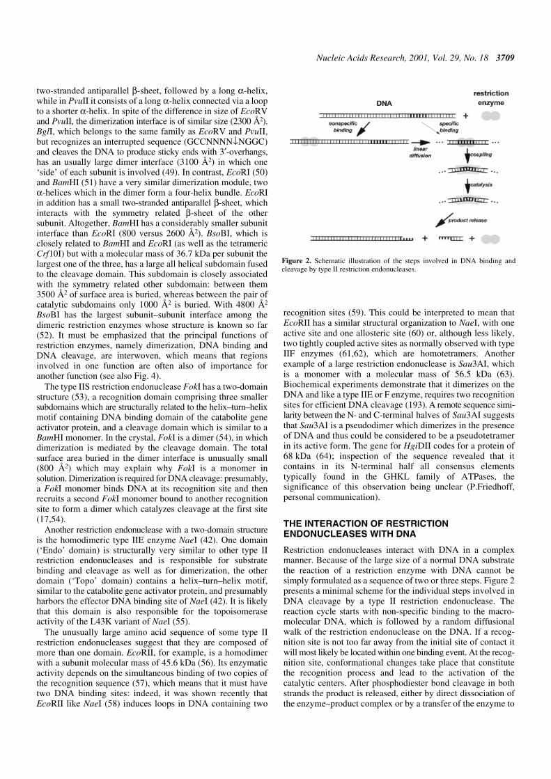
Restriction endonucleases interact with DNA in a complexmanner. Because of the large size of a normal DNA substratethe reaction of a restriction enzyme with DNA cannot besimply formulated as a sequence of two or three steps. Figure 2presents a minimal scheme for the individual steps involved inDNA cleavage by a type II restriction endonuclease. Thereaction cycle starts with non-specific binding to the macro-molecular DNA, which is followed by a random diffusionalwalk of the restriction endonuclease on the DNA. If a recog-nition site is not too far away from the initial site of contact itwill most likely be located within one binding event. At the recog-nition site, conformational changes take place that constitutethe recognition process and lead to the activation of thecatalytic centers. After phosphodiester bond cleavage in bothstrands the product is released, either by direct dissociation ofthe enzyme–product complex or by a transfer of the enzyme to
Figure 2. Schematic illustration of the steps involved in DNA binding andcleavage by type II restriction endonucleases.
3710 Nucleic Acids Research, 2001, Vol. 29, No. 18
non-specific sites on the same DNA molecule. Often this stepis rate limiting for DNA cleavage by restriction enzymes undermultiple turnover conditions. In the following sections we willdeal with the individual steps of this reaction cycle.
DNA BINDING AND TARGET SITE LOCATION
All restriction endonucleases bind DNA not only specificallybut also, with considerably weaker affinity, non-specifically,similar to other proteins that recognize a specific DNA sequence(65). Upon non-specific complex formation, counterions andwater molecules are released from the protein–DNA interface(66), which because of the associated favorable entropychanges balances the unfavorable loss of translational androtational entropies of the protein and DNA upon complexformation. Protein–phosphate contacts on the other hand willlead to positive enthalpy changes. For EcoRI (67) and EcoRV(68) it has been shown by analyzing osmotic pressure effectson DNA binding that non-specific complex formation isaccompanied by a release of 70–80 water molecules.
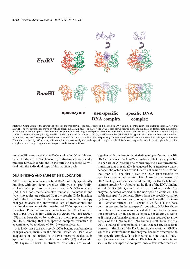
It is likely that upon non-specific DNA binding conformationalchanges occur, mainly in the protein, which will lead to anadaptation of the surface of the two macromolecules as isapparent from structural studies on EcoRV (47) and BamHI(69). Figure 3 shows the structures of EcoRV and BamHI
together with the structures of their non-specific and specificDNA complexes. For EcoRV it is obvious that the enzyme hasto open its DNA-binding site, which requires a conformationaltransition that presumably is triggered by a transient contactbetween the outer sides of the C-terminal arms of EcoRV andthe DNA (70) and that allows the DNA (non-specific orspecific) to enter the binding cleft. A similar mechanism ofDNA binding has been discovered recently for the T7 helicase–primase protein (71). A region at the floor of the DNA bindingsite of EcoRV (the Q-loop), which is disordered in the freeenzyme, becomes ordered in the non-specific complex. Thestable non-specific complex differs from the specific complexby being less compact and having a much smaller protein–DNA contact surface: 1370 versus 2173 Å (47). No basecontacts are seen in the non-specific complex; DNA backbonecontacts are fewer in numbers and differ substantially fromthose observed for the specific complex. For BamHI, it seemsas if major conformational transitions are not required to allowaccess of the DNA to the DNA binding cleft. Nevertheless,DNA binding is accompanied by an induced fit, as a largesegment at the floor of the DNA binding site (residues 79–92),which is disordered in the free enzyme, becomes ordered in thenon-specific complex. It is intriguing to note that no base-specific contacts and no direct DNA backbone contacts areseen in the non-specific complex, only a few water-mediated
Figure 3. Comparison of the crystal structures of the free enzyme, the non-specific and the specific DNA complex for the restriction endonucleases EcoRV andBamHI. The two subunits are shown in red and green, the DNA in blue. For EcoRV, the DNA is also shown viewed along the dyad axis to demonstrate the absenceof bending in the non-specific complex and the presence of bending in the specific complex. PDB code numbers are: EcoRV (1RVE), non-specific complex(2RVE), specific complex (4RVE); BamHI (1BAM), non-specific complex (1ESG), specific complex (1BHM). It is apparent that large conformational changestake place when the free enzymes bind to non-specific DNA and to specific DNA, respectively. In the case of EcoRV, these conformational changes include theDNA which is bent by 50° in the specific complex. It is noteworthy that in the specific complex the DNA is almost completely encircled which gives the specificcomplex a more compact appearance compared to the non-specific one.

Nucleic Acids Research, 2001, Vol. 29, No. 18 3711
contacts even though the non-specific DNA used in the co-crystallization experiment differed only in one base pair fromthe specific DNA sequence (69). As observed with EcoRV, thenon-specific BamHI–DNA complex is more open and lesscompact than the specific complex.
Non-specific DNA binding is the prerequisite for one-dimensional diffusion of proteins along DNA (72). Thestructures of the non-specific complexes of EcoRV and BamHI‘provide remarkable snapshots of enzymes poised for lineardiffusion (rather than cleavage)’ (69), the enzymes being onlyloosely bound to the DNA and their catalytic centers at a safedistance from the phosphodiester backbone. One-dimensionaldiffusion is defined as translocation along a DNA molecule,which does not involve a true free state of the protein: itincludes sliding (i.e. a helical movement due to tracking alonga groove of the DNA), hopping (i.e. a movement more or lessparallel to the DNA, during which the protein does not leavethe ‘DNA domain’) as well as intersegment transfer (whichrequires two DNA binding sites on the protein) (72–74). It hasbeen shown for EcoRI and EcoRV that sliding is the mostimportant process in target site location (75,76). Leaving thetarget site after DNA cleavage might involve either sliding orhopping (77,78). The biological significance of linear diffusionis obvious. It can accelerate target site location, as shown forEcoRI (75,79,80), BamHI (80,81), HindIII (80), EcoRV(76,82,83) and BssHII (84), it can increase processivity as forexample shown for EcoRI (85) or EcoRV (78) and it canaccelerate the dissociation from the specific site after cleavage,as is the case for EcoRI (79). Under optimum conditions,restriction endonucleases can scan ∼106 bp in one bindingevent. As this scan is a random walk, the effective slidingdistance is much shorter, ∼1000 bp, as shown for EcoRI andEcoRV (75,76,80). During linear diffusion, EcoRI follows thehelical pitch of the DNA, does not overlook any recognitionsite on its route and pauses at sites that resemble the recognitionsite; proteins firmly bound to DNA or unusual DNA structuresconstitute ‘road blocks’ (75). The ionic milieu, in particular theMg2+ concentration, has a strong influence on the effectivesliding distance, as shown for EcoRI (85) and EcoRV (76). Itmust be emphasized that linear diffusion is not just a test tubecuriosity but a process of importance in vivo (83,86), becausethe biological function of many enzymes acting on DNArequires fast target site location.
DNA RECOGNITION
Restriction endonucleases while linearly diffusing along theDNA must constantly scan the major groove, possibly alsothe minor groove, for recognition elements at the edges of thebases. Coming into contact with some idiosyncratic features ofthe DNA backbone and the bases, characteristic for the recog-nition sequence, triggers the highly cooperative conversion ofa non-specific to a specific complex, which requires majorconformational changes of both the protein and the DNA, aswell as the expulsion of solvent molecules from the interface toallow for more intimate contacts. For EcoRI it was shown thataltogether about 150 water molecules are released uponspecific DNA binding (67), much more than upon non-specificbinding (87). Interestingly, binding of BamHI to its cognatesequence is accompanied by the release of a somewhat smallernumber of solvent molecules (88). Whereas non-specific DNA
binding by EcoRI and BamHI has a ∆Cp° ≈ 0 and is enthalpydriven, specific DNA binding by these enzyme has a ∆Cp° < 0.Depending on temperature, specific binding is enthalpy orentropy driven (89).
The EcoRV system provides an excellent and so far uniqueexample among type II restriction endonuclease of a majorprotein-induced conformational change of the DNA. In thespecific complex the DNA is bent by ∼50° (compared withlittle if any bending in the non-specific complex), as determinedboth in the crystal (47) and in solution (90–92). This angle variessomewhat, depending on the crystal form, the particular oligo-deoxynucleotide and EcoRV variant used for the co-crystallization(93) (see also Fig. 6). Bending of the DNA is largest at thecentral TA step, which leads to an unstacking of the bases,widening of the minor groove with a concomitant compressionof the major groove, which most importantly brings the scissilephosphates deeper into the active site. It is interesting that theDNA bend is preserved in the product complex (94) as well asin a quasi-product complex in which the 5′-phosphate is missingat the site of cleavage (95), indicating that the continuity of thephosphodiester bond is not required for bending. In thiscontext it is worth mentioning that chemically modified oligo-deoxynucleotide substrates (in which G is replaced by inosineand C by 5-methyl cytosine) are bent to a similar extent as thecorresponding unmodified oligodeoxynucleotide (96) and thatin the crystal, bending is also observed in the absence ofdivalent cations (47). This means that bending is required butnot sufficient for DNA recognition.
Another example of DNA bending in the specific complex,although not as pronounced as with EcoRV, is provided by theEcoRI (97) and MunI systems (98). For both enzymes, whichrecognize the same AATT core sequence in their hexanucleotiderecognition sequence (G↓AATTC and C↓AATTG, respectively),a central kink is observed, accompanied by unwinding of theDNA. A similar but more localized unwinding and a similaroverall bending but without a central kink of the DNA has beenobserved in the specific BglII–DNA complex (99). In contrast,BamHI, which recognizes the same GATC core sequencewithin its hexanucleotide recognition sequence as BglII(G↓GATCT and A↓GATCT, respectively), does not bend,kink or unwind the DNA significantly (100). Whereas nomajor DNA distortion is observed for PvuII (43) which likeEcoRV is a blunt end cutter, BglI (49) which is a sticky endcutter leaving 3′-overhangs, bends the DNA by ∼20°, more orless smoothly without major kinks, the largest deviations fromB-form DNA being seen in the two recognition half-sites of theinterrupted GCCN4↓NGGC recognition sequence. Also, forthe most recently reported structure of a specific restrictionenzyme–DNA complex, BsoBI (52), no pronounced DNAbending is observed; however, slight deviations from canon-ical B-form exist: the DNA is extended and undertwisted,making the major and minor groove wider and more shallow.
Taken together, no generalization can be made for the kindand extent of distortion type II restriction enzymes induce intheir DNA substrate. In general, however, the local helicalparameters of the DNA in the specific complex differ fromideal B-DNA parameters (or where it is known from the helicalparameters of the specific oligodeoxynucleotide used in theco-crystallization). It is important to note that distortions arean intimate part of the recognition process. In a few instancesthis has been experimentally verified by facilitating such a

3712 Nucleic Acids Research, 2001, Vol. 29, No. 18
distortion using chemically modified substrates or substrateanalogs, for example EcoRV (101), and demonstrating thatthey are bound more firmly than the natural (undistorted in thefree state) substrate.
For EcoRV (47,93–95,102,103) and BamHI (69,100) thestructural changes occurring in the protein during the transitionfrom the non-specific complex to the specific complex areknown from detailed crystallographic analyses (Fig. 3). InEcoRV correlated movements of the protein occur in concertwith the binding and unwinding of the DNA (93). These move-ments are characterized by a translation and rotation of thelong B-helices as well as a rotation of the DNA bindingdomains by ∼25°; they lead to an induced fit of the proteinaround the DNA substrate. Of particular importance is theordering of the recognition loop, which appears to be largelyunstructured in the non-specific complex and becomes structuredonly in the specific complex. It is responsible for making allbase-specific contacts in the major groove and presumably ispart of a communication network between the two identicalsubunits which have to act in concert to achieve double-strandcleavage in one binding event (104). The specific complex ismore compact than the non-specific one, mainly because of therotation of the DNA binding domains which brings these twodomains closer together and allows them to encircle the DNAalmost completely (Fig. 3).
The conformational changes that BamHI undergoes in thetransition from non-specific to specific DNA binding are verydifferent from those observed for EcoRV, in spite of the factthat in both cases the binding cleft is wider in the non-specificcomplex than in the specific complex and that the specificcomplex is more compact than the non-specific complex. Themore compact structure of the specific BamHI–DNA complexis in part due to the fact that a segment of the protein is ‘pushedback’ into the protein core by the specific DNA, while thesame segment is located in the binding cleft in the non-specificDNA complex. Whereas the non-specific BamHI–DNAcomplex (69) preserves the 2-fold symmetry of the freeenzyme (51), the specific complex is characterized by apronounced asymmetry (100), produced by the unfolding ofthe C-terminal α-helix in both subunits and the insertion of theunfolded polypeptide segment in one subunit only in anextended conformation into the minor groove of the DNA,while in the other subunit the unfolded polypeptide makes a side-by-side contact with the phosphodiester backbone (Fig. 3).Furthermore, whereas in the non-specific complex the DNA isonly loosely bound within the cleft formed by the two subunitssuch that it protrudes out of the cleft (more so than withEcoRV), in the specific complex it is almost surrounded by theenzyme (like in EcoRV). Another remarkable differencebetween the specific and the non-specific complex concernsthe orientation of the DNA relative to the two subunits ofBamHI. In both complexes, the 2-fold axis of the dimericprotein coincides with the 2-fold axis of the DNA. However,compared with the non-specific complex, the enzyme is tiltedabout this axis by ∼20°, resulting in a different contact area atthe periphery of the DNA binding site in the non-specific andthe specific complex (69).
Given the fact that at present only in two systems, EcoRVand BamHI, can a comparison be made between the non-specific and the specific complex, only very general statementscan be made regarding the structural changes accompanying
the transition from non-specific to specific binding. Because ofthe similarities in function it is likely for all type II restrictionendonucleases that the specific complex will be more compactthan the non-specific one, in order to allow for a tighter contactbetween enzyme and substrate. Presumably, this will beachieved by a reorientation of the two subunits towards eachother and the DNA, which will lead to a compaction of theDNA binding site and a more or less complete encircling of theDNA. The re-orientation can be substantial, as is apparentfrom the comparison of the structures of BglII in the free (105)and bound state (98): to bind DNA, the enzyme has to open bya ‘scissor-like’ motion of the subunits parallel to the DNAhelix axis, which is accompanied by a complete rearrangementof the α-helices at the dimer interface. In contrast, in EcoRVand BamHI opening the binding cleft is achieved essentially bya motion of the subunits in a direction perpendicular to thehelix axis.
There is an interesting difference between EcoRV on oneside and BamHI as well as most of the other restriction endo-nucleases on the other side, including EcoRI. EcoRV requiresthe presence of Mg2+ (106) or Ca2+ (91) for specific binding. Inthe presence of EDTA, EcoRV in a gel electrophoreticmobility shift assay produces multiple bands, whose concen-tration-dependent distribution demonstrates that this enzymebinds all DNA sequences with similar affinity (82), a conclusionthat was challenged (107), and then confirmed by bindingstudies with oligodeoxynucleotides in solution (108). In amore recent study, the preference of EcoRV for its cognatesequence in the absence of divalent cations was shown to bewithin a factor of 10 at neutral pH (109), in agreement withresults obtained previously for wild-type EcoRV (110) and anEcoRV variant (111). Similar results were reported for PaeR7(112), TaqI (113), Cfr9I (114), BcgI (115), MunI (116), Cfr10I(117) and BglI (118), which also require Ca2+ (as a substitutefor Mg2+) for specific binding. For MunI it was shown that thisrequirement could be relaxed by protonation or substitution ofthe active site carboxylates (119), indicating that the divalentcation is required to decrease the electrostatic repulsionbetween the protein and the DNA at the active center. ForEcoRV, additional Mg2+ binding sites outside the catalyticcenter are required for specific binding, as the substitution ofthe active site carboxylates does not alleviate the Mg2+-dependence of specific binding (111). We suggest, therefore,that for some restriction endonucleases Mg2+ (or other divalentcations) is involved in the recognition process, not only in thetransition state, where its contribution is obvious, but also forpreferential and strong (i.e. specific) binding of the recognitionsequence. That restriction endonucleases have additionaldivalent metal ion binding sites already in the absence of DNAhas been shown by metal ion mapping experiments for TaqI(120) and by crystallography for PvuII near Tyr94 (M.Kokki-nidis, personal communication). This residue has beendiscussed previously as being involved in metal ion posi-tioning on the basis of a PvuII mutant–DNA co-crystal struc-ture (121). The Tyr94 site of PvuII is only seen with Mg2+
soaked into the crystals and not with Mn2+, which may explainwhy this site was not seen in the metal ion mapping experi-ments carried out with Fe2+ (122). In the presence of the DNAsubstrate more divalent metal ion binding sites may appear, ashas been shown by crystallography and biochemical studies forEcoRV at position His71, His193 and a phosphodiester group

Nucleic Acids Research, 2001, Vol. 29, No. 18 3713
within the recognition site (GpATATC), respectively (93,111)(F.Winkler, personal communication). Figure 6 gives a compi-lation of all metal ion binding sites observed in EcoRV so farillustrating that the interaction of a restriction enzyme withmetal ions must be considered a very complicated issue. It isinteresting to note that restriction enzymes that do not requiredivalent cations for specific DNA binding, like EcoRI, can bemade dependent on divalent cations by introducing amino acidsubstitutions in critical positions. The EcoRI K130A or E andR131E variants behave like EcoRV in requiring Ca2+ forspecific binding (123). This argues against a fundamentaldifference between enzymes that achieve specificity already atthe binding step (in the absence of Mg2+) or only in thecatalytic step (in the presence of Mg2+).
Since 1997, when we discussed the recognition process forEcoRI, EcoRV, BamHI and PvuII (for details see 10), six moreco-crystal structures of specific restriction endonuclease–DNAcomplexes were determined: FokI (53), BglI (49), MunI (98),BglII (99), NgoMIV (45) and BsoBI (52).
FokI
FokI, a type IIS enzyme recognizes the asymmetric sequenceGGATG and makes a staggered cut 9 and 13 nt, respectively,downstream of the recognition sequence, after dimerization onthe DNA via its cleavage domain (17,54). FokI approaches theDNA from the major groove side and appears to surround theDNA. The recognition domain consists of three subdomains(D1, D2 and D3), which all contain a helix–turn–helix motifand are similar to the DNA binding domain of the catabolitegene activator protein. DNA recognition is based on twomodules: subdomain D1, which covers the major groove at the3′-end of the recognition sequence (GGATG), and subdomainD2, which contacts the 5′-end of the recognition sequence(GGATC). Subdomain D3 is not involved in protein–DNA butrather in protein–protein interactions. FokI, like all otherrestriction endonucleases, makes extensive interactions to allbases of the recognition sequence: almost all hydrogen-bondacceptors and donors at the edges of the bases in the majorgroove are involved in direct contacts with the protein.
BglI
BglI, an orthodox type II enzyme, recognizes the sequenceGCCN5GGC and cleaves between the fourth and fifth unspecifiednucleotide to produce 3′-overhanging ends. BglI approachesthe DNA from the minor groove side (Fig. 1), similarly toEcoRV and PvuII with which it shares many structuralfeatures, in spite of the fact that the two subunits are arrangeddifferently than in these two proteins in order to accommodatethe unspecified sequence between the two recognition half-sites and to produce the different cleavage pattern (3′-over-hangs versus blunt ends). Due to the long distance betweenboth recognition half-sites, each subunit of BglI contacts onlyone half-site and cleaves close to it: there is no cross-overmode of recognition, as observed for most of the other type IIrestriction endonucleases and argued to be beneficial forconcerted double-strand cleavage (10). This might be achievedin this case solely by the extensive hydrogen-bonding networkthat connects the catalytic centers of the two subunits. BglImakes base contacts predominantly in the major groove. Theunspecified 5 bp between the two half-sites are contacted at thesugar–phosphate backbone. The base contacts in the major
groove involve amino acid residues located on or near to asmall three-stranded β-sheet (‘recognition sheet’), in a topo-logically similar location as observed for EcoRV and alsoPvuII (Fig. 4). Per recognition site there are 16 direct hydrogenbonds and two water-mediated ones, which saturates thehydrogen-bonding potential in the major groove. Moreover,there is one direct and several indirect, i.e. water-mediated,contacts to the bases from the minor groove side. In addition tothese base contacts (direct readout), there are numerous back-bone contacts (indirect readout); altogether 17 direct and 21water-mediated hydrogen bonds per subunit to the DNAphosphates.
MunI
MunI recognizes the sequence C↓AATTG. The core sequenceAATT as well as the cleavage pattern is the same as that forEcoRI. This and the identification of local sequence similarities,which concern structural elements of EcoRI involved in recog-nition and cleavage, led to the suggestion that MunI mightemploy a similar mechanism for DNA recognition andcleavage (124). The determination of the co-crystal structure ofthe specific MunI–DNA complex confirmed this proposition(Figs 1 and 4) and thereby provides the first example in whichtwo restriction enzymes contact common parts of their recog-nition sequence by homologous structural elements. MunI, likeEcoRI, approaches the DNA from the major groove side anddistorts the DNA in a similar manner as EcoRI. MunI makesbase contacts only in the major groove. There are altogether16 hydrogen bonds to the edges of the bases and six van derWaals contacts per hexanucleotide recognition site. The outerGC base pair is contacted by Arg115, which has no counterpartin EcoRI. The AATT core sequence is recognized by aminoacid residues located on one segment (Arg115 to Arg121),which in its topological location and function has a correlate inEcoRI, where it is responsible for the recognition of the samecore sequence (AATT). In addition to base-specific contacts,numerous contacts exist between the sugar–phosphate back-bone of the DNA and the protein, extending to two phosphateresidues outside of the recognition sequence. These contactscome from several regions of the protein, which in part are alsoinvolved in base contacts. Thus, direct and indirect readout areinterwoven. Some of these contacts are very similar to thoseobserved previously in the EcoRI–DNA complex and they areconsidered to stabilize the distorted DNA conformation(50,125). Thus, not only are there common features in baserecognition between EcoRI and MunI, but also in backbonerecognition (see also 126). Deibert et al. (98) suggest that thisfinding may eventually be extended to ApoI, which recognizesand cleaves the sequence Pu↓AATTPy.
BglII
BglII recognizes and cleaves the sequence A↓GATCT, whichclosely resembles the recognition sequence of BamHI(G↓GATCC). The determination of the co-crystal structure(Fig. 1) of a specific BglII–DNA complex (99) allowed for acomparison of the strategies employed for recognition (seealso 126). Although the enzymes have a similar core structure,there are remarkable differences in the way these two enzymesinteract with their substrate. The most obvious differenceregarding the mode of recognition is that in BglII the corestructure is augmented by a β-sandwich subdomain that fully

3714 Nucleic Acids Research, 2001, Vol. 29, No. 18
encircles the DNA and is responsible for the minor groovecontacts as well as some of the backbone contacts. Differentfrom the EcoRI/MunI systems, BamHI and BglII, which alsoshare a common tetranucleotide in their respective hexanucleo-tide recognition sequences, interact with this tetranucleotidesequence differently and—with one exception (Asn140 andSer141 in BglII correspond to Asp154 and Asp155 in BamHI,both recognizing the respective outer base pairs)—usedifferent structural elements for recognition (Fig. 4). AlthoughBglII (like BamHI) approaches the DNA from the majorgroove side, contacts are also made to the edges of the bases in
the minor groove. Three loops are responsible for all basecontacts: Asn140 and Ser141 (loop C) recognize via their sidechain functions the first TA base pair and the C of the secondCG base pair. The G of the second base pair is contacted bywater-mediated bidentate hydrogen bonds from Asn98 (loopB). The T of the third TA base pair is recognized by Tyr190 ofone subunit (loop D), and the A by Ser97 of the other subunit(loop B). There are in addition four more water-mediatedhydrogen bonds between the minor groove face of the basesand Tyr190 and Arg192. Altogether there are 14 hydrogenbonds to the major groove and five hydrogen bonds to the

Nucleic Acids Research, 2001, Vol. 29, No. 18 3715
minor groove. There is a pronounced intertwining of therecognition of the two strands/two halves of the recognitionsequence on one side and the two subunits on the other side.Numerous interactions exist between the protein and the DNA;they extend by two phosphate residues beyond the recognition
sequence. Altogether there are 28 backbone contacts, 20 ofthem are water-mediated.
BsoBI
BsoBI recognizes the degenerate sequence C↓PyCGPuG. Aremarkable feature of the co-crystal structure of the specificBsoBI–DNA complex is the complete encirclement of theDNA by the protein to form a 20 Å long ‘tunnel’ (52). Approxi-mately 3800 Å2 of the solvent accessible surface of the enzymeand the DNA are buried in the protein–DNA interface (Fig. 1and Table 2). As expected from its mode of cleavage, BsoBIapproaches the DNA from the major groove side. Each subunitinteracts with each recognition half-site and makes base-specific contacts in the major and minor grooves. The outerand inner CG base pairs are involved in several hydrogenbonds to the protein. Of particular interest is how this enzymemanages to accept a CG or TA and GC or AT base pair in thesecond and fourth position of the recognition sequence. This isnow understood in structural terms because this PyPu base pairis involved in only one direct hydrogen bond between Lys81and the N7 of the purine (A in the co-crystal structure), and inone water-mediated bidentate hydrogen bond between Asp246and N7 of the purine as well as the substituent in position 6 ofthe purine (N6 of A in the co-crystal structure). In addition tothese 22 hydrogen bonds to the bases, several van der Waalscontacts to the bases exist as well as 64 hydrogen bonds (24water-mediated) to the backbone per site. The backbonecontacts extend to two residues to the left and right of therecognition sequence.
NgoMIV
For NgoMIV only the structure of the enzyme–productcomplex has been determined (45). It is likely that many of thesequence-specific contacts required for the recognition of thesubstrate are preserved in the enzyme–product complex (as isthe case in the EcoRV and BamHI systems). NgoMIVapproaches the DNA from the major groove side and makesmost of the base-specific contacts in the major groove of thetarget sequence recognized (G↓CCGGC). One subunit formshydrogen bonds to the GCC half-site in the major groove,while the neighbor subunit forms a hydrogen bond to the C ofthe outer GC base pair in the minor groove. Base-specificcontacts come from three structural elements, namely loopspreceding α-helix 2, 7 and 8 (Fig. 4). It is noteworthy that threeneighboring amino acids (Arg191, Asp193, Arg194) make allpossible hydrogen bonds to the two adjacent GC base pairs inthe major groove. Altogether there are 18 direct and two water-mediated hydrogen bonds to the bases of the NgoMIV recogni-tion sequence. Interestingly, there is a hydrogen bond contactfrom Ser36 to the C on the 5′-side of the sequence, which mayexplain the flanking sequence preference of NgoMIV. Inaddition to the base-specific contacts, numerous contacts to thesugar–phosphate backbone exist, mainly from the othersubunit, such that direct readout for which one subunit isresponsible is interwoven with indirect readout for which theother subunit is responsible. Altogether, there are six direct andeight water-mediated contacts to the DNA–phosphates.
The region from Arg191–Arg194 (RSDR) has a structuralequivalent in Cfr10I (RPDR), which recognizes a similarrecognition sequence as NgoMIV (compare Pu↓CCGGPy withG↓CCGGC) and also has a Glu residue six residues away
Figure 4. (Opposite and above) The topologies of selected type II restriction endo-nucleases to illustrate similarities of architecture and to identify functionallyimportant regions. Shown are the secondary structure diagrams for those restrictionenzymes for which crystal structures of specific enzyme–DNA complexes have beendetermined (see Fig. 1). The elements comprising the common core are indi-cated in blue. Catalytically important amino acid residues are marked with across and those involved in contacts to the bases are marked with black filledcircles. Regions involved in dimerization contacts are colored red. The num-bering scheme is that of the respective crystallographers. The large helical N-terminal domain of BsoBI is not included in the diagram.

3716 Nucleic Acids Research, 2001, Vol. 29, No. 18
which is part of the catalytic center of NgoMIV (45). It is verylikely that the recognition of the adjacent GG sequence is doneby Crf10I using the equivalent residues as in NgoMIV. Onepossibly could extend this suggestion to other restrictionendonucleases that recognize adjacent GC base pairs (Table 3).In the lack of structural data or a detailed mutational analysisthis is speculative. For some of these enzymes, for exampleSsoII (V.Pingoud, personal communication), biochemicalevidence exists that the RXXR motif plays an important role inDNA binding.
The increasing numbers of co-crystal structures available forspecific restriction endonuclease–DNA complexes and comple-mentary biochemical studies allows us to make generalizationsregarding the mechanism of DNA recognition. (i) Enzymesthat produce blunt ends or sticky ends with 3′-overhangsapproach the DNA from the minor groove side, whereasenzymes that produce sticky ends with 5′-overhangs contactthe DNA from the major groove side. (ii) DNA binding isaccompanied by more or less pronounced distortions of theDNA and conformational adaptations of the enzyme, which inmany cases lead to a partial encircling of the DNA by theprotein. (iii) Specific DNA binding is accompanied by therelease of counter ions and partial dehydration of the enzyme
and the DNA at the protein–DNA interface. (iv) Enzymes thatproduce blunt ends or sticky ends with 3′-overhangs mainlyuse a β-strand and β-like turn for DNA recognition. In contrast,enzymes that produce sticky ends with 5′-overhangs mainlyuse an α-helix and a loop. (v) Recognition is achieved by directand indirect readout, i.e. base contacts, and backbone contacts,respectively. Contacts to the bases are predominantly in themajor groove and usually exhaust the hydrogen bondingpotential in the major groove. This means that a hexanucleo-tide sequence is recognized by ∼20 hydrogen bonds to thebases of the recognition sequence. Interactions with the back-bone are often water-mediated. (vi) Individual recognitionmodules (short sequence motifs) begin to show up that are usedby different restriction endonucleases to recognize commonparts in similar recognition sites.
COUPLING BETWEEN RECOGNITION ANDCATALYSIS
Specific DNA binding by restriction endonucleases is definedas strong and, more importantly, preferential binding to therecognition site. Its outcome is what we usually see in theco-crystal structures or what we measure in binding experiments(including footprinting and crosslinking experiments).Specific binding does not necessarily mean recognition that isdefined operationally, i.e. by the reaction that follows. By thisdefinition the co-crystal structures of the specific restrictionendonuclease–DNA complexes only mimic the recognitioncomplex. In a similar argument, the results of binding experimentsonly address the mechanism of specific binding and not in thestrictest sense the mechanism of recognition. Nevertheless,there is no doubt that the investigation of specific binding helpsto understand recognition, presumably because the enzyme–substrate complex (studied in the absence of Mg2+ or in thepresence of Ca2+) as well as the enzyme–product complex(studied in the presence of Mg2+, after turnover) is very similarto the ground state complex in the presence of Mg2+. In thiscontext it is important to note that the discrimination betweenspecific and non-specific sites requires multiple contacts to beformed between enzyme and substrate. In order to prevent
Table 2. Overview of co-crystal structures determined for enzyme–substrate complexes of orthodox type II restriction endonucleases
aWhere more than one co-crystal structure has been determined only the first high resolution structure is given.bDetermined by the Connolly method (188) using a probe with a radius of 1.4 Å. Note that this method yields different results from other methods used in the literature.The dimerization interface is defined by: 2 × surface monomer – surface dimer. The protein–DNA interface is defined by: surface dimer + surface DNA – surface enzyme–DNA complex.cTetramerization interface (A2). Tetramerization interface is defined by: 4 × surface monomer – surface tetramer.
EcoRI EcoRVa BamHI PvuIIa BglI MunI BglII NgoMIV BsoBI
Recognition sequence G↓AATTC GAT↓ATC G↓GATCC CAG↓CTG GCCN4↓NGGC C↓AATTG A↓GATCT G↓CCGGC C↓PyCGPuG
PBD code 1ERI 1RVB 1BHM 3PVI 1DMU 1DO2 1DFM 1FIU –
Length of DNA (bp) 13 11 12 13 17 10 16 11 12
R-factor (%) 17.0 16.5 18.9 20.5 17.7 18.0 19.2 17.4 19.0
Resolution (Å) 2.7 2.1 2.2 1.6 2.2 1.7 1.5 1.6 1.7
Protein–DNA interface (Å2)b 2200 2400 2000 1800 3300 1300 2800 2100 3800
Dimerization interface (Å2)b 2600 2300 800 2300 3800 2500 780 1000(10 800c)
2800
Table 3. Type II restriction endonucleases having recognition sequences withadjacent G residues and harboring a RXXR motif
Enzyme RXXR Recognition sequence
NgoMIV K187X3RSDRX6E G/CCGGC
Cfr10I K190X3RPDRX6E R/CCGGY
BsrFI K198X3RPDRX6E R/CCGGY
SgrAI K242X3RSDRX6E CR/CCGGYG
EcoRII K324X3K-DRX6E /CCWGG
PspGI K160X3R-ERX6E /CCWGG
SsoII K182X3R-ERX6E /CCNGG
AccI K196X3K-DRX6E T/CCGGA

Nucleic Acids Research, 2001, Vol. 29, No. 18 3717
these contacts, formed in the ground state complex, fromimpairing the catalytic efficiency (given by the difference inthe Gibb’s free energy of the transition state complex and theground state complex), it is necessary that these interactionsmust also stabilize the transition state (127). Therefore, theground state complex is likely to resemble the transition statecomplex very much, differences being localized to the site ofphosphodiester bond cleavage.
The coupling of specific binding, recognition and catalysis isill understood. There have been many attempts to understandhow the catalytic machinery is activated during the recognitionprocess. Ideally one would like to ‘see’ this by time-resolvedcrystallography or NMR. This, however, has not yet beenachieved. Instead, crystallographic studies of wild-type andmutant enzymes with canonical and chemically modifiedsubstrates, in the absence and presence of divalent metal ioncofactors, have been carried out and their results interpretedtogether with the results of single and multiple turnovercleavage studies. The best studied system in this respect is theEcoRV system, for which the structures of different enzyme–substrate complexes in different crystal lattices were determinedand for which detailed biochemical data were obtained. Recog-nition can be formally divided into direct and indirect readout. InEcoRV the recognition (R)-loop, comprising residues 182–187,whose importance for recognition has been confirmed by amutional analysis (128), makes all the base-specific contacts inthe major groove. A hydrogen bond network links the R-loopsto the scissile phosphates and the catalytic centers via Asn188and Lys92 (94). Base recognition in the minor groove isaccomplished by the glutamine (Q)-loop, comprising residues68–70. Gln69 is in close proximity to one catalytic center andvia Thr37 also to the other catalytic center (94); these two resi-dues are very important for catalysis (128–132). Thr37 is alsoone of the key amino residues involved in indirect readout(94,96,130,131,133). The results of these investigationsconcerning coupling of recognition and catalysis assign a crit-ical role to the symmetry related B-helices and Q-loops,which connect β-strands c and d. This region is located at thefloor of the DNA binding site, vis à vis the phosphodiesterbonds to be cleaved. It is known that this region adopts slightlydifferent conformations in various co-crystal structures ofEcoRV (93), which makes it likely that it has sufficient confor-mational freedom to be involved in activation of the catalyticcenters. Residues whose position is affected by these confor-mational changes include Asp36 and Lys38, which also havebeen shown by mutational analyses to be essential for EcoRV(134,135).
A major aspect of the mechanism of activation of the catalyticcenters of restriction enzymes concerns the positioning of thedivalent metal ion cofactors and the water molecules, one ofwhich in each catalytic center must take up a position in-linewith the phosphodiester bond to be cleaved. For EcoRV it hasbeen shown that Asp74 and Asp90, as well as the scissile phos-phate and its 3′-neighbor, which all cooperate in Mg2+ andwater binding at the catalytic centers, take up slightly differentpositions in different co-crystal structures of EcoRV (93). Thisis not unexpected for catalytically relevant residues(128,129,136) in a complex that is not active in the crystal. Ofcourse, one would like to know how all amino acid residues inspecific contact with the bases and the backbone communicatewith the catalytic centers. The fact that this communication
must be highly cooperative will make it very difficult toidentify an intramolecular signal transduction pathway. It mustbe admitted, therefore, that at present, it is at best partiallyunderstood how the catalytic centers of EcoRV or any otherrestriction endonuclease are activated during the recognitionprocess. Probably this is the main reason why all efforts tochange or expand the specificity of restriction endonucleasesby rational, i.e. structure-guided, design failed so far (137,138)or were not as successful as one had hoped (139).
Coupling of recognition to catalysis not only concernsintrasubunit but also intersubunit communication, as restrictionendonucleases in general catalyze a concerted double-strandcut. This means that the information of ‘recognition’ must bepassed on from one subunit to the other. As pointed out above,with few exceptions each subunit of a restriction enzymemakes contacts to both halves of the recognition sequence,which integrates the recognition process. This has beendemonstrated directly for EcoRV, using artificial heterodimers(104,131,140) and for PvuII using a single chain variant (194):substitution of a residue involved in base-specific contacts inonly one subunit affects cleavage in both strands, whereassubstitution of a catalytic residue in one active center does notaffect the other catalytic center and allows for cleavage in onestrand (nicking).
THE MECHANISM OF CATALYSIS OFPHOSPHODIESTER BOND CLEAVAGE BYRESTRICTION ENDONUCLEASES
The catalysis of phosphodiester bond cleavage by restrictionendonucleases can be considered as a phosphoryl transfer towater. For such a reaction two principal mechanisms may beoperative, an associative and a dissociative mechanism(reviewed in 141). Both result in an inversion of configurationat phosphorus, as shown experimentally for EcoRI (142),EcoRV (143), HpaIII and SfiI (144). The associative mechanism(Fig. 5, top) requires a general base to generate the activenucleophile, a hydroxide ion, which attacks the phosphorus ofthe scissile phosphodiester bond, a Lewis acid that stabilizesthe extra negative charge at the pentacoordinated phosphorusduring the transition state, and a general acid that stabilizes theleaving group. This can be a Brønsted acid that protonates theleaving group (the fragment with a 3′-O–) or a metal ion thatassociates with the alcoholate. It is this mechanism that usuallyis explicitly or implicitly assumed to be operative in DNAcleavage by restriction enzymes. The dissociative mechanism(Fig. 5, middle) is not so much dependent on a general base togenerate the active nucleophile, as there is only a small amountof bond formation to the incoming nucleophile, water, but alarge amount of bond cleavage to the outgoing leaving group,the fragment with a 3′-O–. Therefore, in this mechanismstabilization of the leaving group is the most important aspectof catalysis. In addition, for this mechanism a metaphosphate-like species has to be stabilized in the transition state. Thiscould be done by positively charged amino acid side chains,amino acid residues with hydrogen-bond donor functions(including protonated carboxylate side chains) or divalentmetal ions (Mg2+), which all must preferentially interact withthe trigonal bipyramidal transition state relative to the tetra-hedral ground state (145).

3718 Nucleic Acids Research, 2001, Vol. 29, No. 18
It must be emphasized that the associative and dissociativemechanisms represent two extremes of possible mechanisms.The actual mechanisms used by a restriction endonuclease islikely not to follow any of these extremes (in Fig. 5, bottom,which depicts the change in bond order for the bond beingmade and the bond being broken, the reaction pathway mayfollow any line between the red line characteristic for an asso-ciative mechanism and the blue line characteristic for thedissociative nechanism). For alkaline phosphatase, forexample, it has been shown that this enzyme can achievesubstantial catalysis via a transition state with dissociativecharacter (146).
All type II restriction endonucleases, whose crystal structureshave been determined, have a catalytic sequence motif incommon, the PD . . . D/EXK motif (41,128). As shown inTable 4 the consensus is somewhat relaxed which makes ithard in some cases to locate the catalytic sequence motif by aninspection of the sequence only. Nevertheless, it serves as aguideline to identify the presumptive active center of a type IIrestriction endonuclease, which then must be confirmed by amutional analysis. Examples of such an identification includeBsoBI (VD212 . . . E240LK) (147), Eco57I (PD78 . . . E92AK)(148), BcgI (PE53–E66DK) (24), EcoRII (PD77 . . . E96KR) (149)and HindIII (PE51 . . . D108AK) (150). It must be noted,however, that the identification of the catalytic sequence motifby a mutational analysis only is not fail-safe. An example isprovided by the homing endonuclease I-PpoI for which a
presumptive active site apparently was identified by a mutationalanalysis (PD109 . . . D140NK), which could not be confirmed bythe crystal structure analysis (151,152).
The PD . . . D/EXK motif has also been found in othernucleases as shown by crystal structure analyses, for exampleλ-exonuclease (PD119 . . . E129LK) (34), E.coli MutH (QD70 . . .E77LK) (37) and E.coli TnsA [D114 . . . (E149)Q130VK] (39) ortentatively assigned by mutational analyses, for exampleSulfolobus solfataricus (PD42 . . . E55MK) (153) and Pyrococcusfuriosus Holliday junction resolvase (VD36 . . . E46VK) (154),as well as E.coli McrBC (McrC: TD243 . . . D256AK; U.Pieperand A.Pingoud, submitted). In E.coli Vsr the PD . . . D/EXKmotif is only partially present, although the endonuclease foldis conserved (38)
The fact that so many restriction endonucleases have acommon catalytic sequence motif could suggest that theyfollow basically the same mechanism, with at least oneobvious exception: BfiI, a type IIS restriction enzyme, thatdoes not require a divalent metal ion cofactor, and in thisrespect differs from all type II restriction endonucleases known(155). This enzyme in its C-terminal part shows sequencehomology with the Salmonella typhimurium NucA, a memberof the HXKX4DX6GSXN superfamily of phosphodiesterases,whose crystal structure is known (156). It is likely that BfiI likeFokI has a two-domain structure, one domain being respon-sible for DNA recognition and the other for cleavage. As NucAis a homodimer with one catalytic center which is formed bythe two subunits, it might well be that BfiI has to form atetramer to afford double-strand cleavage. More of theseexceptions are likely to follow, as can be expected on the basisof recent sequence comparisons, which suggest that sometype II restriction enzymes belong to the HNH- and GIY-YIG-families of endonucleases (157,158).
For the great majority of type II restriction endonucleasesMg2+ is an essential cofactor which can be substituted withMn2+ (Fe2+, Co2+, Ni2+, Zn2+, Cd2+, depending on the enzyme)
Figure 5. Principle mechanisms of phosphoryl transfer reactions. On top themechanism of phosphodiester bond cleavage following an associative or adissociative route is shown. These mechanisms differ in the amount of bondformation and bond breakage in the transition state. This is illustrated in thegraph on the bottom in which the bond order of the bond to be cleaved is plottedagainst the bond order of the bond being formed. For an associative mechanism(red line) both bond orders change proportionally, whereas for a dissociativemechanism (blue line), the bond to the leaving group is largely cleaved whilethe bond to the attacking nucleophile is not yet formed. 1, Enzyme–substratecomplex; 2, enzyme–product complex; 3, transition state of the associativemechanism; 4, transition state of the dissociative mechanism.
Table 4. Catalytic sequence motifs in type II restriction endonucleases
aThe crystal structure analysis has shown that the second acidic residue of thePD . . . D/EXK motif is recruited for the catalytic center from a distal part ofthe structure.
Enzyme PD motif D/EXK motif Reference
EcoRI PD91 E111AK 189
EcoRV PD74 D90IK 47
BamHI ID74 E111FE 100
PvuII ND58 E68LK 43,48
Crf10Ia PD134 (E204) S188VK 190,191
FokI PD450 D467TK 53
BglI PD116 D142IK 49
MunI PD83 E98IK 98
NaeI TD86 D95CK 42
BglII ID84 E93VQ 99
NgoMIVa PD140 (E201) S185CK 45
BsoBI VD212 E240LK 52

Nucleic Acids Research, 2001, Vol. 29, No. 18 3719
but not, however, by Ca2+. There is no doubt that Mg2+ isintimately associated with the catalytic process, as in nearly allcases where divalent cations were soaked into crystals of typeII restriction endonucleases or into co-crystals of enzyme–DNA complexes, a divalent cation was found associated withthe carboxylates of the catalytic center, with interesting varia-tions: Mg2+, Mn2+, Ca2+, the divalent metal ions that are usuallyused in soaking experiments, do not necessarily occupy thesame site. For example, a soaking experiment with PvuII and50 mM Mg2+ had shown that Mg2+ is bound in one subunit toAsp58 and Glu68, i.e. at the catalytic center, while in the othersubunit Mg2+ is bound in the immediate vicinity of Tyr94. Thissite seems to be of physiological importance, because when itis destroyed by a Tyr→Phe exchange, the DNA cleavage isaffected and not concerted anymore (A.Spyridaki, C.Matzen,T.Lanio, A.Jeltsch, A.Simoncsits, A.Athanasiadis, E.Scheu-ring-Vanamee, M.Kokkinidis and A.Pingoud, submitted). If asoaking experiment is carried out with 50 mM Mn2+, a site closeto Leu69 and Ser71 is occupied. In contrast, when co-crystalsof a PvuII–DNA complex are grown at pH 4.5 in the presenceof 50 mM Ca2+, then crosslinked with glutaraldehyde and thenstep-wise transferred to pH 6.5 conditions, co-crystals areobtained that contain two Ca2+ ions in each subunit associatedwith Asp58 and Glu68 (159). Table 5 gives a compilation of allpublished data concerning divalent metal binding sites in
restriction endonuclease–DNA substrate complexes, whichdemonstrates the variety encountered among divalent metalions in binding sites of different restriction endonucleasesunder different conditions.
Table 5 contains 13 entries for EcoRV, which show thatdepending on the divalent metal ion, the substrate (or substrateanalog) and the variant, different binding sites can be occupied,most of them comprising carboxylates from the catalyticcenters. For EcoRV three sites are available for Ca2+, but onlytwo are occupied simultaneously: site I comprises Asp74(Asp90) and Ile91 (main chain carbonyl), site II Asp74 andAsp90, site III Glu45 and Asp74 (the roman numerals are usedto compare these sites with analogous divalent metal bindingsites in other restriction endonucleases, see also Fig. 7). It isnoteworthy that in several instances only one binding site persubunit is occupied and/or that only one subunit has divalent metalions bound, in spite of the fact that very high concentrations ofdivalent metal ions were used for the soaking, but also for theco-crystallization experiments. The fact that high concentrationsare being used for these experiments is a point of concern, asthey might lead to occupation of unphysiological binding sites(160,161). The occupation of such unphysiological sites maybe the reason why only in two cases was activity demonstratedin crystallo (Table 5), an argument that is supported bybiochemical data, which clearly demonstrate inhibition of
Table 5. Divalent metal ion binding sites at the catalytic centers of specific restriction enzyme–DNA complexes
aActive in crystallo.bI = inosin.cM = 5-methyl cytosine.dDeoxyribo-3′-S-phosphorothiolate at scissile phosphate.
Protein Metal ion pH Subunit 1 Subunit 2 Reference
EcoRIa 5 mM Mn2+ (soaking) 7.0 Asp90, Glu111 Asp90, Glu111 166
EcoRV 30 mM Mg2+ (soaking) 6.8 Asp74, Asp90, Glu45, Asp74 – 94
EcoRV 30 mM Mn2+ (soaking) 6.8 Asp74, Asp90 – 94
EcoRV 30 mM Co2+ (soaking) 6.8 Asp74, Asp90, Glu45, Asp74 Asp74, Asp90, Glu45, Asp74 94
EcoRV 30 mM Ca2+ (soaking) 6.8 Asp74, Asp90 Asp74, Asp90 94
EcoRV 15 mM Mg2+ + 15 mM Ca2+ (soaking) 6.8 Asp74, Asp90 Asp74, Asp90 94
EcoRV 1 mM Mn2+ + 20 mM Ca2+ (soaking) 6.8 Asp74, Asp90, Glu45, Asp74 Asp74, Asp90, Glu45, Asp74 94
EcoRV 40 mM Ca2+ (co-crystallizition) 6.8 Asp74, Asp90 Asp74, Asp90 102
EcoRV T93A 10 mM Ca2+ (co-crystallization) 7.5 Asp74, Ile91, Glu45, Asp74 – 103
EcoRVb (GACITC) 25 mM Mn2+ (co-crystallization) 7.5 – – 96
EcoRVb,c (GAMITC) 30 mM Mn2+ (co-crystallization) 7.5 Asp74, Asp90 Asp74, Asp90 96
EcoRVd (3′S) 50 mM Mg2+ 7.5 Asp74, Ile91, Glu45, Asp74 Asp74, Ile91 Glu45, Asp74 93
EcoRVd (3′S) 50 mM Mn2+ 7.5 Glu45, Asp74, Ile91, Glu45, Asp74,His71
His71, His193 93
EcoRVd (3′S) 50 mM Ca2+ 7.5 Asp74, Ile91 Glu45, Asp74 Asp74, Ile91, Glu45 93
BamHIa 10 mM Mn2+ (co-crystallization) 5.3 Asp94, Glu111, Phe112, Glu77, Asp94 – 178
BglI 125–150 mM Ca2+ (soaking) 6.5 Asp116, Asp142, Ile143, Asp116 Asp116, Asp142, Ile143 Asp116 49
MunI 50 mM Ca2+ 6.0 – – 98
BglII Unidentified metal ion (co-crystallization)
5.2 Asp84, Val94 Asp84, Val94 99
PvuII 50 mM Ca2+ (soaking) 4.5–6.5 Asp58, Glu68, Leu69, Asp58, Glu68 Asp58, Glu68, Leu69, Asp58, Glu68 159

3720 Nucleic Acids Research, 2001, Vol. 29, No. 18
DNA cleavage by EcoRV at higher concentrations of divalentcations (162). Mg2+ seems to have the same options as Ca2+ inoccupying sites associated with the catalytic centers of EcoRV.Mn2+, however, can occupy additional sites outside of thecatalytic centers (Fig. 6). Mn2+ binding to one of these sitesmodulates the catalytic efficiency and sequence selectivity ofthe enzyme (163). To further demonstrate the complexity ofthe situation, biochemical experiments suggest that there mustbe additional divalent metal ion binding sites in the EcoRV–DNAcomplex, not yet seen in the co-crystals, which are involved inrecognition (111). Figure 6 shows a superposition of thedivalent metal ion binding sites so far identified in EcoRV.
The situation seems to be not as complicated with the otherrestriction enzymes, possibly only because fewer structures areavailable. For two enzymes, EcoRI and BglII, only one metalion per subunit (Mn2+ in the case of EcoRI, an unidentifiedmetal ion or Ca2+, respectively, in the case of BglII) was seenin the co-crystal structures. It may be a coincidence, but thesesites were found occupied at 5 mM Mn2+ and without addeddivalent metal ion, respectively (Table 5). The Ca2+ bindingsites of BamHI, BglI and PvuII are homologous: in all threeenzymes two binding sites per subunit are present, correspondingto binding sites I and II of EcoRV (Table 5). Figure 7 shows acomparison of the Ca2+ (Mn2+ in the case of EcoRI) bindingsites observed in the co-crystals of BamHI, BglI, BglII, EcoRI,EcoRV and PvuII (based on ref. 159). This comparison revealssome intriguing similarities: all these restriction enzymes havea binding site I which is formed by the absolutely conserved firstand second acidic amino acid residues of the PD . . . D/EXK motifand by a main chain carbonyl of the non-polar amino acid
residue of this motif, PD . . . D/EXK. The divalent metal ionoccupying binding site I is coordinated to a water moleculewhich is directly or indirectly (via a second water molecule)associated with the proRp-oxygen of the phosphate 3′ to thescissile phosphodiester bond. This water molecule is in aposition for an in-line attack on the bond to be cleaved. It isheld there by the last residue of the PD . . . D/EXK motif,usually a Lys, but in BamHI a Glu and in BglII a Gln residue,which all (Glu should be protonated for that purpose) arecapable of forming a hydrogen bond to the attacking watermolecule. The divalent metal ion in binding site II which isformed by the first and in some cases also by the second acidicamino acid residue of the PD . . . D/EXK motif, could serve topolarize the P–O bond and thereby to make the phosphorusmore susceptible for a nucleophilic attack. It could also help tocoordinate a water molecule, which could be used to protonatethe leaving group, the 3′-O– or directly associate with thisgroup. For EcoRV, the direct association has been ruled out(164).
The elucidation of the mechanism (or mechanisms) of DNAcleavage by type II restriction enzymes is critically dependenton the knowledge of how many Mg2+ ions are directly involvedin catalysis. Crystal structure analyses are certainly veryhelpful to answer this question, but they can be also verymisleading, because they do not tell us whether the configurationseen in the structure is productive. For EcoRV at least threedifferent mechanisms were proposed, all based on structuralinformation but differing in the numbers and sites of Mg2+ ionsinvolved (‘one, two or three metals’) (36). The first mechanismwas based on the comparison of the co-crystal structures ofEcoRI and EcoRV as well as on molecular modeling(136,165). Based on the finding that a water molecule modeledinto the structures at the position of the attacking nucleophileforms a hydrogen bond to the phosphodiester group 3′ to thescissile phosphodiester bond, it has been suggested that thephosphate 3′ to the scissile phosphodiester bond serves as thegeneral base. Leaving group protonation would be afforded bya water molecule from the hydration sphere of a Mg2+ ionbound to Asp74 and Asp90 (site I/II). This aspect of themechanism has become commonly accepted because, in allstructures determined so far, a water in the hydration sphere ofa metal ion is hydrogen bonded to the leaving group making itan obvious candidate for leaving group protonation. Thecharacteristic feature of this mechanism is the substrateassisted catalysis, i.e. a direct role of the DNA substrate itselfin the catalytic mechanism. A variant of this mechanism hasbeen proposed for EcoRI recently (166). Although recruiting aphosphodiester group as the catalytic base might be consideredsurprising, this function is not uncommon. It has been proposedfor other enzymes involved in nucleotidyl- or phosphoryltransfer: p21ras and other G-proteins (167), aminoacyl-tRNAsynthetases (168,169) and acylphosphatase (170). Recently, ithas been shown that the catalytic mechanism of peptidyltransfer in the prokaryotic ribosome also might involve aphosphodiester group of the rRNA as final proton acceptor in arelay that starts at the N3 of an adenine residue and involvesalso the O6 and N2 of a guanine residue (171,172). An impor-tant point in favor of the 3′-phosphate being the general base isthe finding that in several high resolution structures of specificrestriction enzyme–DNA complex a water molecule is seen
Figure 6. Superposition of all solved structures of EcoRV and metal-ionbinding site in EcoRV. The figure shows how similar these structures arealthough they are determined from crystals in different crystal lattices, usingdifferent substrates and metal ions. In addition all divalent metal ion bindingsites as seen in different co-crystal structures and as deduced from biochemicalexperiments are indicated. The metal-ion binding sites near the active site areshown for both subunits, the additional sites (near His71, near His193 and nearthe GpATATC phosphate) are shown only for one subunit.

Nucleic Acids Research, 2001, Vol. 29, No. 18 3721
hydrogen bonded to the 3′-phosphate and in a position readyfor an in-line attack on the scissile phosphodiester bond.
Two alternative mechanisms for DNA cleavage by EcoRVwere based on the Kostrewa and Winkler structure (94). Theyconsider two Mg2+ ions as essential, but differ in an importantdetail, the identification of the general base. Kostrewa andWinkler (94) suggested that a water molecule in the hydrationsphere of Mg2+ ion bound to Asp74 and Asp90 (site II)provides the attacking nucleophile and a water molecule in thehydration sphere of Mg2+ ion bound to Glu45 and Asp74
(site III) serves to protonate the leaving group. Vipond et al.(173) in contrast proposed that Mg2+ in site III functions as thegeneral base. Later, Halford and coworkers (132,174) modifieddetails of the stereochemistry of their mechanism to take intoaccount the results of recent kinetic data for the Co2+ supportedDNA cleavage by EcoRV and the results of a moleculardynamics simulation. In an accompanying study (135) it wassuggested that the water molecule in the hydration sphere ofMg2+ in site III is deprotonated with the help of Glu45 (of onesubunit) and Asp36 (of the other subunit). The most recent
Figure 7. Comparison of the active sites of restriction enzymes as deduced from co-crystal structures of specific protein–DNA substrate complexes obtained in thepresence of divalent metal ions (based on 159). In EcoRV the sites formed by Asp90 and Ile91, Asp74 and Asp90 as well as Glu45 and Asp74 are numbered I, IIand III, respectively, in this review. The same numbering scheme is applied to all other enzymes.

3722 Nucleic Acids Research, 2001, Vol. 29, No. 18
addition to the list of mechanisms proposed for DNA cleavageby EcoRV has been provided by Perona and coworkers(93,103). They had, as mentioned above, identified a newdivalent metal binding site (site I) and intergrated this site intoa three-metal ion mechanism, which they called a metal ion-mediated substrate-assisted catalysis mechanism to account forthe fact that the metal ion in site I is coordinated to two watermolecules, one that bridges to the proSp-oxygen of the 3′-phosphate,and the other attacks the scissile phosphate. This metal ion isbound to Asp74, Asp90 and Ile91 [site I; note that Horton et al.(103) use a different numbering system]. A second metal ion isbound to Asp74 and Asp90 (site II) as well as to the scissilephosphate; in its coordination sphere there is a water moleculethat is close to the 3′-O of the leaving group. A third divalentmetal ion, bound by Glu45 and Asp74 (site III), is not seen inany structure together with the divalent metal ions occupyingsite I and II, but in principle could take up this position. It isproposed to have a structural role, which is in agreement withthe results of biochemical studies (162).
We have described the various proposals for the mechanismof DNA cleavage by EcoRV in detail, to make clear that evenwith detailed structural information it is not straightforward toderive the mechanism of an enzymatic reaction. A critical issuein the one-metal ion substrate-assisted catalysis mechanismand also, albeit to a lesser degree, in the two- and three-metalion-mechanisms, is the pKa of the general base. The pKa of aphosphodiester (pKa < 2) or of a Mg2+-aquo complex (pKa ≈ 11–12)clearly is far off to explain the pH-optimum of the EcoRV-catalyzedreaction (pH ≈ 8.5) (135,175). There are four possible ways outof this dilemma: (i) an efficient general base is not needed, ifthe transition state has dissociative character; (ii) the attackingnucleophile is not generated in situ but comes from bulksolvent and is only stabilized in the catalytic center, as alsosuggested for DNA polymerase I (176); (iii) amino acid residuesclose to the active center provide assistance in lowering the pKaof the Mg2+-aquo complex, like Asp36 (135) [for a differentview (134)]; and (iv) the pH profile of the cleavage reactionreflects a deprotonation of a residue (or of residues) which arenot part of the catalytic center. In this context it is worthwhileto note that restriction enzymes are among the best regulatedenzymes as they have to ensure that the catalytic centers areonly activated if the correct sequence is bound. Any residueimportant in DNA recognition and coupling of recognition andcatalysis could, therefore, dominate the apparent pH-dependence.As noted by Horton et al. (93), ‘establishing the structural basisfor DNA cleavage beyond reasonable doubt requires visualizationof essential catalytic elements within the confines of a singlehigh-resolution crystal structure of a ternary complex. Clearlythis elusive goal is yet to be attained’. Time-resolved spectroscopyusing NMR (177) and EPR should be particularly suited toclarify how many divalent metal ions are required for catalysisand to elucidate their particular roles.
For EcoRV (94), as for BamHI (178) and NgoMIV (45), thestructure of an enzyme–product complex in the presence ofMg2+ or Mn2+ were determined. In the EcoRV–productcomplex structure (obtained by co-crystallization) two Mg2+
ions were seen in both subunits, associated with the 5′-phosphateof one product as well as liganded to Glu45 and Asp74 (Mg2+
no. 1) and Gln69 (Mg2+ no. 2), respectively. In the BamHI–productcomplex structure (obtained by soaking), one Mn2+ ion is associatedwith the cleaved phosphate, Asp94 and Phe112, while the other is
coordinated to Asp94, but not anymore to Glu111. In the NgoMIV–product complex structure (obtained by co-crystallization) twoMg2+ ions are seen in each active site, both coordinated by thecleaved phosphate, Asp140 and an acetate ion, one Mg2+ ion inaddition by Cys186 (main chain carbonyl). The presence oftwo divalent metal ions at the catalytic centers of restrictionendonuclease–product complexes should not be taken asevidence for their participation in catalysis, but rather as areflection of the need to have a balance of charges. The acetateion seen in the NgoMIV–product structure associated withboth Mg2+ ions illustrates this requirement.
Two co-crystal structures of specific restriction enzyme–DNAcomplexes were determined without divalent metal ions: MunI(98) and BsoBI (52). The pronounced similarity between MunIand EcoRI suggests that MunI follows a similar mechanism asEcoRI. BsoBI, however, has particular features that merit aspecial discussion. This enzyme has a normal PD . . . D/EXKmotif (Asp212, Glu240, Lys242) which means that it couldfollow a similar mechanism as EcoRI. BsoBI shares with otherrestriction endonucleases the position of the attacking watermolecule which is located 3.7 Å away from the phosphorusatom of the scissile phosphodiester bond and in hydrogenbonding distance of the pro-Rp oxygen of the phosphate 3′ tothe scissile phosphodiester bond. In addition, this watermolecule is only 3.8 Å away from Lys243, which in turncontacts the pro-Sp oxygen of the phosphorus atom to beattacked. These features are similar in all other restrictionenzymes (see Fig. 7). BsoBI, on the other hand, is the onlyrestriction enzyme, which has an obvious general base close tothe active center, His253 (from the other subunit), which mayalso couple recognition to catalysis, as it is involved in ahydrogen bond to N7 of the inner G (C↓PyCGPuG). It is notclear, however, how Mg2+ binding will affect these relationships.A two-metal ion mechanism is discussed by van der Woerdet al. (52), which is different from the other two-metal ionmechanisms discussed above, as the role of His253 has to beconsidered. One Mg2+ is suggested to bind to Asp212 andGlu240 as well as the pro-Sp oxygen of the scissile phospho-diester bond, a second Mg2+ to the proRp-oxygen and Glu252of the other subunit.
The mechanism by which restriction endonucleases achieveDNA cleavage has not yet been proven for any restrictionenzyme. For the advocates of the two-metal ion mechanismoften the mechanism by which the 5′–3′ exonuclease domainof DNA polymerase I hydrolyzes DNA is taken as theparadigm (179), but in principle there the same uncertainty asto the participation of one- or two-metal ions in catalysis exists(160,161,180). A similar controversy as for EcoRV is going onfor other nucleases. For example, for RNaseH two alternativemechanisms have been proposed. One is a two-metal ionmechanism (181), where one of the two metal ions activatesthe attacking nucleophile, and the other is a one-metal ionmechanism (182), where an amino acid residue is consideredto be responsible for generating the hydroxide ion, faciliatedby a neighboring phospodiester group of the substrate (183).Another example is provided by the transposases andintegrases, which share a common catalytic triad, the DDEmotif. Most of the structures reported for these enzymes showthe binding of one Mn2+ ion at a site located between two of thethree carboxylates, for example in the Tn5 transposasesynaptic complex (184), but crystals for ASV integrase core

Nucleic Acids Research, 2001, Vol. 29, No. 18 3723
soaked with Zn2+ or Cd2+ exhibit metal ion binding at twoadjacent sites (185).
The BglII and BamHI restriction enzymes which cleave verysimilar sequences, A↓GATCT and G↓GATCC, have beendiscussed as representatives of type II restriction endonucleaseswhich follow a one- and two-metal ion mechanism, respectively,and which have diverged from a common ancestor (186). Theactive site of BglII, on the other hand, has a similar make-up tothe evolutionarily unrelated homing endonuclease I-CreI,which has one-metal ion bound per catalytic center (187). Thishas been considered as evidence for convergent evolution ofthese two enzymes. This conclusion certainly is validconcerning the amino acid residues involved in catalysis, butmust be considered as tentative only concerning the number ofmetal ions involved.
It must not only be emphasized that at present the mechanism ofDNA cleavage has not yet been elucidated for any restrictionendonuclease, but also that it is not even known whether alltype II restriction endonucleases with a bona fide PD . . . D/EXKmotif follow the same mechanism. We tend to believe that thisis the case, the argument being one of parsimony. Why elseshould this motif be conserved, in particular as it is possible toexplain the deviations. For example, the substitution of the D/E bya S in Cfr10I and NgoMIV (the crystal structure shows that theE is supplied from another structural element, but takes up therequired position in the three-dimensional structure). Like-wise, the K is substituted by an E and Q in BamHI and BglII,respectively, but the hydrogen-bond donor function ispreserved (when the E is protonated in BamHI). With theseconsiderations in mind, the following features appear asinvariant among all type II restriction endonucleases whoseco-crystal structure has been determined. (i) The essentialdivalent metal ion cofactor is bound to the two carboxylatesand the main chain carbonyl of the hydrophobic residue X ofthe PD . . . D/EXK motif as well as one of the non-bridgingoxygens of the phosphate to be attacked. Whether a second (orthird) divalent metal ion is required for cleavage is still amatter of debate. (ii) The phosphorus atom of the scissile phos-phodiester bond is attacked in-line with the bond to be cleavedby a water molecule, held in position by the essential divalentmetal ion, the K of the PD . . . D/EXK motif and one of thenon-bridging oxygens of the phosphate 3′-adjacent to thephosphate to be attacked. (iii) Whether cleavage of the phos-phodiester bond follows a more associative or a more dissoci-ative mechanism is not known. Accordingly, the structure ofthe transition state is a matter of speculation. For its stabilization,the divalent metal ion cofactor, positively charged amino acidresidues as well as amino acid residues with hydrogen bonddonor functions are required. (iv) The function of the generalbase that has to abstract a proton from the attacking water (notvery important if a dissociative mechanism is operative) couldbe taken over by the divalent metal ion, the K of the PD . . . D/EXKmotif and/or the 3′-adjacent phosphate. All these chemicalentities require a considerable shift in pKa (from their unper-turbed value in the free state) to function as the general base.Whether additional amino acid residues are recruited for thispurpose may differ from system to system. (v) Protonation ofthe leaving group or its transient stabilization, respectively,could be performed at the expense of a water molecule bound tothe divalent metal ion or the divalent metal ion itself, respectively.(vi) Whatever the precise mechanism of phosphodiester bond
cleavage by type II restriction endonucleases is, it involvesacid–base catalysis. For this type of catalysis a proton relay hasto be considered involving chemical entities not only at butalso close to the site of cleavage.
CODA
Up to 1% of the genome of prokaryotic organisms is taken upby the genes for RM systems. Although it is not clear whetherthe majority of RM systems are required for the maintenanceof the integrity of the genome or whether they are spreading asselfish genetic elements, they are key players in the ‘genomicmetabolism’ of prokaryotic organisms. As such they deservethe attention of biologists in general.
Restriction enzymes (as well as their companion modificationenzymes) constitute a huge family of enzymes with similarfunctions, the essential features of which are present also inother proteins interacting specifically with DNA, in particularproteins with nuclease function (recombinases, resolvases,transposases, integrases, repair enzymes). Elucidating themechanism of action of restriction enzymes, therefore, is ofinterest also to those interested in the mechanism of DNArecognition and cleavage in general.
Finally, restriction enzymes are the work horses of molecularbiology. Understanding their enzymology will be advantageous tothose who use these enzymes, and essential for those who aredevoted to the ambitious goal of changing the properties ofthese enzymes, and thereby make them even more useful.
ACKNOWLEDGEMENTS
We are obliged to all colleagues who have sent us reprints,preprints and manuscripts. Thanks are in particular due toDrs M. Deibert, V. Siksnys and A. Friedman for providingcoordinates before their official release. We thank Drs R. Roberts,V. Siksnys, F. Winkler and X. Cheng for comments on themanuscript. We would also like to express our appreciation toDrs R. Roberts and D. Macelis for making available REBASE,an invaluable tool for those who work with restrictionenzymes. We thank all our colleagues at Giessen, in particularDrs P. Friedhoff, T. Lanio, V. Pingoud and W. Wende, fornumerous discussions and useful information, as well asMrs K. Urbach for typing the manuscript. Work in the authors’laboratory has been supported by grants from the DeutscheForschungsgemeinschaft, the Bundesministerium für Bildung,Wissenschaft, Forschung und Technologie, and the EuropeanUnion.
REFERENCES
1. Roberts,R.J. and Halford,S.E. (1993) Type II restriction endonucleases. InLinn,S.M., Lloyd,R.S. and Roberts,R.J. (eds), Nucleases. Cold SpringHarbor Laboratory Press, Cold Spring Harbor, New York, NY, pp. 35–88.
2. Raleigh,E.A. and Brooks,J.E. (1998) In De Bruijn,F.J., Lupski,J.R. andWeinstock,G.M. (eds), Bacterial Genomes. Chapman & Hall, New York,pp. 78–92.
3. Arber,W. (1979) Promotion and limitation of genetic exchange. Science,205, 361–365.
4. Heitman,J. (1993) On the origins, structures and functions of restriction-modification enzymes. In Setlow,J.K. (ed.), Genetic Engineering. PlenumPress, New York, Vol. 15, pp. 57–108.
5. McKane,M. and Milkman,R. (1995) Transduction, restriction andrecombination patterns in Escherichia coli. Genetics, 139, 35–43.

3724 Nucleic Acids Research, 2001, Vol. 29, No. 18
6. Carlson,K. and Kosturko,L.D. (1998) Endonuclease II of coliphage T4: arecombinase disguised as a restriction endonuclease? Mol. Microbiol., 27,671–676.
7. Bilcock,D.T., Daniels,L.E., Bath,A.J. and Halford,S.E. (1999) Reactionsof type II restriction endonucleases with 8-base pair recognition sites.J. Biol. Chem., 274, 36379–36386.
8. Naito,T., Kusano,K. and Kobayashi,I. (1995) Selfish behavior ofrestriction-modification systems. Science, 267, 897–899.
9. Smith,H.O. and Nathans,D. (1973) A suggested nomenclature forbacterial host modification and restriction systems and their enzymes.J. Mol. Biol., 81, 419–423.
10. Pingoud,A. and Jeltsch,A. (1997) Recognition and cleavage of DNA bytype-II restriction endonucleases. Eur. J. Biochem., 246, 1–22.
11. Roberts,R.J. and Macelis,D. (1997) REBASE-restriction enzymes andmethylases. Nucleic Acids Res., 25, 248–262.
12. Roberts,R.J. and Macelis,D. (2000) REBASE – restriction enzymes andmethylases. Nucleic Acids Res., 28, 306–307.
13. Hedgpeth,J., Goodman,H.M. and Boyer,H.W. (1972) DNA nucleotidesequence restricted by the RI endonuclease. Proc. Natl Acad. Sci. USA,69, 3448–3452.
14. Schildkraut,I., Banner,C.D.B., Rhodes,C.S. and Parekh,S. (1984) Thecleavage site for the restriction endonuclease EcoRV is 5′-GAT↓ATC-3′.Gene, 27, 327–329.
15. Van Heuverswyn,H. and Fiers,W. (1980) Recognition sequence for therestriction endonuclease BglI from Bacillus globigii. Gene, 9, 195–203.
16. Szybalski,W., Kim,S.C., Hasan,N. and Podhajska,A.J. (1991) Class-IISrestriction enzymes – a review. Gene, 100, 13–26.
17. Bitinaite,J., Wah,D.A., Aggarwal,A.K. and Schildkraut,I. (1998) FokIdimerization is required for DNA cleavage. Proc. Natl Acad. Sci. USA, 95,10570–10575.
18. Krüger,D.H., Kupper,D., Meisel,A., Reuter,M. and Schroeder,C. (1995)The significance of distance and orientation of restriction endonucleaserecognition sites in viral DNA genomes. FEMS Microbiol. Rev., 17,177–184.
19. Halford,S.E., Bilcock,D.T., Stanford,N.P., Williams,S.A., Milsom,S.E.,Gormley,N.A., Watson,M.A., Bath,A.J., Embleton,M.L., Gowers,D.M.et al. (1999) Restriction endonuclease reactions requiring two recognitionsites. Biochem. Soc. Trans, 27, 696–699.
20. Stankevicius,K., Lubys,A., Timinskas,A., Vaitkevicius,D. andJanulaitis,A. (1998) Cloning and analysis of the four genes coding forBpu10I restriction-modification enzymes. Nucleic Acids Res., 26,1084–1091.
21. Hsieh,P.-C., Xiao,J.-P., O’Loane,D. and Xu,S.-Y. (2000) Cloning,expression and purification of a thermostable nonhomodimeric restrictionenzyme, BslI. J. Bacteriol., 182, 949–955.
22. Kong,H. and Smith,C.L. (1997) Substrate DNA and cofactor regulate theactivities of a multi-functional restriction-modification enzyme, BcgI.Nucleic Acids Res., 25, 3687–3692.
23. Vitkute,J., Maneliene,Z., Petrusyte,M. and Janulaitis,A. (1997) BplI, anew BcgI-like restriction endonuclease, which recognizes a symmetricsequence. Nucleic Acids Res., 25, 4444–4446.
24. Kong,H. (1998) Analyzing the functional organization of a novel restrictionmodification system, the BcgI system. J. Mol. Biol., 279, 823–832.
25. Janulaitis,A., Vaisvila,R., Timinskas,A., Klimasauskas,S. and Butkus,V.(1992) Cloning and sequence analysis of the genes coding for Eco57I typeIV restriction-modification enzymes. Nucleic Acids Res., 20, 6051–6056.
26. Lacks,S. and Greenberg,B. (1975) A deoxyribonuclease of Diplococcuspneumoniae specific for methylated DNA. J. Biol. Chem., 250, 4060–4066.
27. Stewart,F.J. and Raleigh,E.A. (1998) Dependence of McrBC cleavage ondistance between recognition elements. Biol. Chem., 379, 611–616.
28. Panne,D., Raleigh,E.A. and Bickle,T.A. (1999) The McrBC endonucleasetranslocates DNA in a reaction dependent on GTP hydrolysis.J. Mol. Biol., 290, 49–60.
29. Krüger,T., Wild,C. and Noyer-Weidner,M. (1995) McrB: a prokaryoticprotein specifically recognizing DNA containing modified cytosineresidues. EMBO J., 14, 2661–2669.
30. Pieper,U., Schweitzer,T., Groll,D.H. and Pingoud,A. (1999) Defining thelocation and function of domains of McrB by deletion mutagenesis.Biol. Chem., 380, 1225–1230.
31. Piekarowicz,A., Golaszewska,M., Sunday,A.O., Siwinska,M. andStein,D.C. (1999) The HaeIV restriction modification system ofHaemophilus aegyptius is encoded by a single polypeptide. J. Mol. Biol.,293, 1055–1065.
32. Wilson,G.G. and Murray,N.E. (1991) Restriction and modificationsystems. Annu. Rev. Genet., 25, 585–627.
33. Jeltsch,A., Kröger,M. and Pingoud,A. (1995) Evidence for anevolutionary relationship among type-II restriction endonucleases. Gene,160, 7–16.
34. Kovall,R.A. and Matthews,B.W. (1998) Structural, functional andevolutionary relationships between lambda-exonuclease and the type IIrestriction endonucleases. Proc. Natl Acad. Sci. USA, 95, 7893–7897.
35. Venclovas,C., Timinskas,A. and Siksnys,V. (1994) Five-stranded β-sheetsandwiched with two α-helices: a structural link between restrictionendonucleases EcoRI and EcoRV. Proteins, 20, 279–282.
36. Kovall,R.A. and Matthews,B.W. (1999) Type II restrictionendonucleases: structural, functional and evolutionary relationships.Curr. Opin. Chem. Biol., 3, 578–583.
37. Ban,C. and Yang,W. (1998) Structural basis for MutH activation in E.colimismatch repair and relationship of MutH to restriction endonucleases.EMBO J., 17, 1526–1534.
38. Tsutakawa,S.E., Jingami,H. and Morikawa,K. (1999) Recognition of aTG mismatch: the crystal structure of very short patch repair endonucleasein complex with a DNA duplex. Cell, 99, 615–623.
39. Hickman,A.B., Li,Y., Mathew,S.V., May,E.W., Craig,N.L. and Dyda,F.(2000) Unexpected structural diversity in DNA recombination: therestriction endonuclease connection. Mol. Cell, 5, 1025–1034.
40. Bujnicki,J.M. (2000) Phylogeny of the restriction endonuclease-likesuperfamily inferred from comparison of protein structures. J. Mol. Evol.,50, 39–44.
41. Anderson,J.E. (1993) Restriction endonucleases and modificationmethylases. Curr. Opin. Struct. Biol., 3, 24–30.
42. Huai,Q., Colandene,J.D., Chen,Y., Luo,F., Zhao,Y., Topal,M.D. andKe,H. (2000) Crystal structure of NaeI – an evolutionary bridge betweenDNA endonuclease and topoisomerase. EMBO J., 19, 3110–3118.
43. Cheng,X., Balendiran,K., Schildkraut,I. and Anderson,J.E. (1994) Structureof PvuII endonuclease with cognate DNA. EMBO J., 13, 3927–3935.
44. Siksnys,V., Skirgaila,R., Sasnauskas,G., Urbanke,C., Cherny,D.,Grazulis,S. and Huber,R. (1999) The Cfr10I restriction enzyme isfunctional as a tetramer. J. Mol. Biol., 291, 1105–1118.
45. Deibert,M., Grazulis,S., Sasnauskas,G., Siksnys,V. and Huber,R. (2000)Structure of the tetrameric restriction endonuclease NgoMIV in complexwith cleaved DNA. Nature Struct. Biol., 7, 792–799.
46. Modrich,P. and Zabel,D. (1976) EcoRI endonuclease. Physical andcatalytic properties of the homogeneous enzyme. J. Biol. Chem., 251,5866–5874.
47. Winkler,F.K., Banner,D.W., Oefner,C., Tsernoglou,D., Brown,R.S.,Heathman,S.P., Bryan,R.K., Martin,P.D., Petratos,K. and Wilson,K.S.(1993) The crystal structure of EcoRV endonuclease and of its complexeswith cognate and non-cognate DNA fragments. EMBO J., 12, 1781–1795.
48. Athanasiadis,A., Vlassi,M., Kotsifaki,D., Tucker,P.A., Wilson,K.S. andKokkinidis,M. (1994) Crystal structure of PvuII endonuclease revealsextensive structural homologies to EcoRV. Nature Struct. Biol., 1, 469–475.
49. Newman,M., Lunnen,K., Wilson,G., Greci,J., Schildkraut,I. andPhillips,S.E.V. (1998) Crystal structure of restriction endonuclease BglI boundto its interrupted DNA recognition sequence. EMBO J., 17, 5466–5476.
50. Kim,Y., Grable,J.C., Love,R., Greene,P.J. and Rosenberg,J.M. (1990)Refinement of EcoRI endonuclease crystal structure: A revised proteinchain tracing. Science, 249, 1307–1309.
51. Newman,M., Strzelecka,T., Dorner,L.F., Schildkraut,I. andAggarwal,A.K. (1994) Structure of restriction endonuclease BamHI andits relationship to EcoRI. Nature, 368, 660–664.
52. van der Woerd,M.J., Pelletier,J.J., Xu,S.-y. and Friedman,A.M. (2001)Restriction enzyme BsoBI-DNA complex: A tunnel for recognition ofdegenerate DNA sequences and potential histidine catalysis. Structure, 9,133–144.
53. Wah,D.A., Hirsch,J.A., Dorner,L.F., Schildkraut,I. and Aggarwal,A.K.(1997) Structure of the multimodular endonuclease FokI bound to DNA.Nature, 388, 97–100.
54. Wah,D.A., Bitinaite,J., Schildkraut,I. and Aggarwal,A.K. (1998)Structure of FokI has implications for DNA cleavage. Proc. Natl Acad.Sci. USA, 95, 10564–10569.
55. Jo,K. and Topal,M.D. (1995) DNA topoisomerase and recombinaseactivities in NaeI restriction endonuclease. Science, 267, 1817–1820.
56. Kossykh,V., Repyk,A., Kaliman,A. and Buryanov,Y. (1989) Nucleotidesequence of the EcoRII restriction endonuclease gene. Biochim. Biophys.Acta, 1009, 290–292.

Nucleic Acids Research, 2001, Vol. 29, No. 18 3725
57. Krüger,D.H., Barcak,G.J., Reuter,M. and Smith,H.O. (1988) EcoRII canbe activated to cleave refractory DNA recognition sites. Nucleic AcidsRes., 16, 3997–4008.
58. Topal,M.D., Thresher,R.J., Conrad,M. and Griffith,J. (1991) NaeIendonuclease binding to pBR322 DNA induces looping. Biochemistry,30, 2006–2010.
59. Mücke,M., Lurz,R., Mackeldanz,P., Behlke,J., Krüger,D.H. andReuter,M. (2000) Imaging DNA loops induced by restrictionendonuclease EcoRII. A single amino acid substitution uncouples targetrecognition from cooperative DNA interaction and cleavage. J. Biol.Chem., 275, 30631–30637.
60. Colandene,J.D. and Topal,M.D. (1998) The domain organization of NaeIendonuclease: Separation of binding and catalysis. Proc. Natl Acad. Sci.USA, 95, 3531–3536.
61. Wentzell,L.M., Nobbs,T.J. and Halford,S.E. (1995) The SfiI restrictionendonuclease makes a four-strand DNA break at two copies of itsrecognition sequence. J. Mol. Biol., 248, 581–595.
62. Nobbs,T.J., Szczelkun,M.D., Wentzell,L.M. and Halford,S.E. (1998) DNAexcision by the SfiI restriction endonuclease. J. Mol. Biol., 281, 419–432.
63. Seeber,S., Kessler,C. and Gotz,F. (1990) Cloning, expression andcharacterization of the Sau3AI restriction and modification genes inStaphylococcus carnosus TM300. Gene, 94, 37–43.
64. Düsterhoft,A. and Kroger,M. (1991) Cloning, sequence andcharacterization of m5C-methyltransferase-encoding gene, hgiDIIM(GTCGAC), from Herpetosiphon giganteus strain Hpa2. Gene, 106, 87–92.
65. von Hippel,P.H. (1994) Protein–DNA recognition: new perspectives andunderlying themes. Science, 263, 769–770.
66. Parsegian,V.A., Rand,R.P. and Rau,D.C. (1995) Macromolecules andwater: probing with osmotic stress. Methods Enzymol., 259, 43–94.
67. Robinson,C.R. and Sligar,S.G. (1998) Changes in solvation during DNAbinding and cleavage are critical to altered specificity of the EcoRIendonuclease. Proc. Natl Acad. Sci. USA, 95, 2186–2191.
68. Wenner,J.R. and Bloomfield,V.A. (1999) Buffer effects on EcoRVkinetics as measured by fluorescent staining and digital imaging ofplasmid cleavage. Anal. Biochem., 268, 201–212.
69. Viadiu,H. and Aggarwal,A.K. (2000) Structure of BamHI bound tononspecific DNA: A model for DNA sliding. Mol. Cell, 5, 889–895.
70. Schulze,C., Jeltsch,A., Franke,I., Urbanke,C. and Pingoud,A. (1998)Crosslinking the EcoRV restriction endonuclease across the DNA-bindingsite reveals transient intermediates and conformational changes of the enzymeduring DNA binding and catalytic turnover. EMBO J., 17, 6757–6766.
71. Ahnert,P., Picha,K.M. and Patel,S.S. (2000) A ring-opening mechanismfor DNA binding in the central channel of the T7 helicase-primase protein.EMBO J., 19, 3418–3427.
72. von Hippel,P.H. and Berg,O.G. (1989) Facilitated target location inbiological systems. J. Biol. Chem., 264, 675–678.
73. Berg,O.G., Winter,R.B. and von Hippel,P.H. (1981) Diffusion-drivenmechanisms of protein translocation on nucleic acids. 1. Models andtheory. Biochemistry, 20, 6929–6948.
74. Shimamoto,N. (1999) One-dimensional diffusion of proteins along DNA.Its biological and chemical significance revealed by single-moleculemeasurements. J. Biol. Chem., 274, 15293–15296.
75. Jeltsch,A., Alves,J., Wolfes,H., Maass,G. and Pingoud,A. (1994) Pausingof the restriction endonuclease EcoRI during linear diffusion on DNA.Biochemistry, 33, 10215–10219.
76. Jeltsch,A. and Pingoud,A. (1998) Kinetic characterization of lineardiffusion of the restriction endonuclease EcoRV on DNA. Biochemistry,37, 2160–2169.
77. Terry,B.J., Jack,W.E., Rubin,R.A. and Modrich,P. (1983) Thermodynamicparameters governing interaction of EcoRI endonuclease with specific andnonspecific DNA sequences. J. Biol. Chem., 258, 9820–9825.
78. Stanford,N.P., Szczelkun,M.D., Marko,J.F. and Halford,S.E. (2000) Oneand three dimensional pathways for proteins to reach specific DNA sites.EMBO J., 19, 6546–6557.
79. Jack,W.E., Terry,B.J. and Modrich,P. (1982) Involvement of outsideDNA sequences in the major kinetic path by which EcoRI endonucleaselocates and leaves its recognition sequence. Proc. Natl Acad. Sci. USA, 79,4010–4014.
80. Ehbrecht,H.-J., Pingoud,A., Urbanke,C., Maass,G. and Gualerzi,C. (1985)Linear diffusion of restriction endonucleases on DNA. J. Biol. Chem.,260, 6160–6166.
81. Nardone,G., George,J. and Chirikjian,J.G. (1986) Differences in thekinetic properties of BamHI endonuclease and methylase with linear DNAsubstrates. J. Biol. Chem., 261, 12128–12133.
82. Taylor,J.D., Badcoe,I.G., Clarke,A.R. and Halford,S.E. (1991) EcoRVrestriction endonuclease binds all DNA sequences with equal affinity.Biochemistry, 30, 8743–8753.
83. Jeltsch,A., Wenz,C., Stahl,F. and Pingoud,A. (1996) Linear diffusion ofthe restriction endonuclease EcoRV on DNA is essential for the in vivofunction of the enzyme. EMBO J., 15, 5104–5111.
84. Berkhout,B. and van Wamel,J. (1996) Accurate scanning of the BssHIIendonuclease in search for its DNA cleavage site. J. Biol. Chem., 271,1837–1840.
85. Terry,B.J., Jack,W.E. and Modrich,P. (1985) Facilitated diffusion duringcatalysis by EcoRI endonuclease: nonspecific interactions in EcoRIcatalysis. J. Biol. Chem., 260, 13130–13137.
86. Dowd,D.R. and Lloyd,R.S. (1990) Biological significance of facilitateddiffusion in protein–DNA interactions. Applications to T4 endonucleaseV-initiated DNA repair. J. Biol. Chem., 265, 3424–3431.
87. Sidorova,N.Y. and Rau,D.C. (1996) Differences in water release for thebinding of EcoRI to specific and nonspecific DNA sequences. Proc. NatlAcad. Sci. USA, 93, 12272–12277.
88. Lynch,T.W. and Sligar,S.G. (2000) Macromolecular hydration changesassociated with BamHI binding and catalysis. J. Biol. Chem., 275,30561–30565.
89. Jen-Jacobson,L., Engler,L.E. and Jacobson,L.A. (2000) Structural andthermodynamic strategies for site-specific DNA binding proteins.Struct. Fold. Des., 8, 1015–1023.
90. Stöver,T., Köhler,E., Fagin,U., Wende,W., Wolfes,H. and Pingoud,A. (1993)Determination of the DNA bend angle induced by the restriction endonucleaseEcoRV in the presence of Mg2+. J. Biol. Chem., 268, 8645–8650.
91. Vipond,I.B. and Halford,S.E. (1995) Specific DNA recognition byEcoRV restriction endonuclease induced by calcium ions. Biochemistry,34, 1113–1119.
92. Garcia,R.A., Bustamante,C.J. and Reich,N.O. (1996) Sequence-specificrecognition by cytosine C5 and adenine N6 DNA methyltransferases requiresdifferent deformations of DNA. Proc. Natl Acad. Sci. USA, 93, 7618–7622.
93. Horton,N.C. and Perona,J.J. (2000) Crystallographic snapshots along aprotein-induced DNA-bending pathway. Proc. Natl Acad. Sci. USA, 97,5729–5734.
94. Kostrewa,D. and Winkler,F.K. (1995) Mg2+ binding to the active site ofEcoRV endonuclease: a crystallographic study of complexes with substrateand product DNA at 2 Angstrom resolution. Biochemistry, 34, 683–696.
95. Horton,N.C. and Perona,J.J. (1998) Role of protein-induced bending inthe specificity of DNA recognition: crystal structure of EcoRVendonuclease complexes with d(AAAGAT) + d(ATCTT). J. Mol. Biol.,277, 779–787.
96. Martin,A.M., Sam,M.D., Reich,N.O. and Perona,J.J. (1999) Structuraland energetic origins of indirect readout in site-specific DNA cleavage bya restriction endonuclease. Nature Struct. Biol., 6, 269–277.
97. Kim,Y., Choi,J., Grable,J.C., Greene,P., Hager,P. and Rosenberg,J.M.(1994) In Sarma,R.H. and Sarma,M.H. (eds.), Structural Biology:The State of the Art. Adenine Press, New York, NY, pp. 225–246.
98. Deibert,M., Grazulis,S., Janulaitis,A., Siksnys,V. and Huber,R. (1999)Crystal structure of MunI restriction endonuclease in complex withcognate DNA at 1.7 Å resolution. EMBO J., 18, 5805–5816.
99. Lukacs,C.M., Kucera,R., Schildkraut,I. and Aggarwal,A.K. (2000)Understanding the immutability of restriction enzymes: crystal structureof BglII and its DNA substrate at 1.5 A resolution Nature Struct. Biol., 7,134–140.
100.Newman,M., Strzelecka,T., Dorner,L.F., Schildkraut,I. andAggarwal,A.K. (1995) Structure of BamHI endonuclease bound to DNA:partial folding and unfolding on DNA binding. Science, 269, 656–663.
101.Blättler,M.O., Wenz,C., Pingoud,A. and Benner,S.A. (1998) Distortingduplex DNA by dimethylenesulfone substitution: a new class of‘transition state analog’ inhibitors for restriction enzymes. J. Am. Chem.Soc., 120, 2674–2675.
102.Perona,J.J. and Martin,A.M. (1997) Conformational transitions andstructural deformability of EcoRV endonuclease revealed bycrystallographic analysis. J. Mol. Biol., 273, 207–225.
103.Horton,N.C., Newberry,K.J. and Perona,J.J. (1998) Metal ion-mediatedsubstrate-assisted catalysis in type II restriction endonucleases. Proc. NatlAcad. Sci. USA, 95, 13489–13494.
104.Stahl,F., Wende,W., Jeltsch,A. and Pingoud,A. (1996) Introduction ofasymmetry in the naturally symmetric restriction endonuclease EcoRV toinvestigate intersubunit communication in the homodimeric protein.Proc. Natl Acad. Sci. USA, 93, 6175–6180.

3726 Nucleic Acids Research, 2001, Vol. 29, No. 18
105.Lukacs,C.M., Kucera,R., Schildkraut,I. and Aggarwal,A.K. (2001)Structure of free BglII reveals an unprecedented scissor-like motion foropening an endonuclease. Nature Struct. Biol., 8, 126–130.
106.Thielking,V., Selent,U., Köhler,E., Landgraf,Z., Wolfes,H., Alves,J. andPingoud,A. (1992) Mg2+ confers DNA binding specificity to the EcoRVrestriction endonuclease. Biochemistry, 31, 3727–3732.
107.Engler,L.E., Welch,K.K. and Jen-Jacobson,L. (1997) Specific binding byEcoRV endonuclease to its DNA recognition site GATATC. J. Mol. Biol.,269, 82–101.
108.Erskine,S.G. and Halford,S.E. (1998) Reactions of the EcoRV restrictionendonuclease with fluorescent oligodeoxynucleotides: identicalequilibrium constants for binding to specific and non-specific DNA.J. Mol. Biol., 275, 759–772.
109.Reid,S.L., Parry,D., Liu,H.-H. and Connolly,B.A. (2001) Binding andrecognition of VATATC target sequences by the EcoRV restrictionendonuclease: a study using fluorescent oligonucleotides and fluorescencepolarisation. Biochemistry, 40, 2484–2494.
110.Alves,J., Selent,U. and Wolfes,H. (1995) Accuracy of the EcoRVrestriction endonuclease: binding and cleavage studies witholigodeoxynucleotide substrates containing degenerate recognitionsequences. Biochemistry, 34, 11191–11197.
111.Jeltsch,A., Maschke,H., Selent,U., Wenz,C., Köhler,E., Connolly,B.A.,Thorogood,H. and Pingoud,A. (1995) DNA binding specificity of theEcoRV restriction endonuclease is increased by Mg2+ binding to a metalion binding site distinct from the catalytic center of the enzyme.Biochemistry, 34, 6239–6246.
112.Ghosh,S.S., Obermiller,P.S., Kwoh,T.J. and Gingeras,T.R. (1990)Analysis of substrate specificity of the PaeR7I endonuclease: effect ofbase methylation on the kinetics of cleavage. Nucleic Acids Res., 18,5063–5068.
113.Zebala,J., Choi,J. and Barany,F. (1992) Characterization of steady state,single-turnover and binding kinetics of the TaqI restriction endonuclease.J. Biol. Chem., 267, 8097–8105.
114.Siksnys,V. and Pleckaityte,M. (1993) Catalytic and binding properties ofrestriction endonuclease Cfr9I. Eur. J. Biochem., 217, 411–419.
115.Kong,H., Roemer,S.E., Waite-Rees,P.A., Benner,J.S., Wilson,G.G. andNwankwo,D.O. (1994) Characterization of BcgI, a new kind ofrestriction-modification system. J. Biol. Chem., 269, 683–690.
116.Lagunavicius,A., Grazulis,S., Balciunaite,E., Vainius,D. and Siksnys,V.(1997) DNA binding specificity of MunI restriction endonuclease iscontrolled by pH and calcium ions: Involvement of active site carboxylateresidues. Biochemistry, 36, 11093–11099.
117.Skirgaila,R. and Siksnys,V. (1998) Ca2+-ions stimulate DNA bindingspecificity of Cfr10I restriction enzyme. Biol. Chem., 379, 595–598.
118.Gormley,N.A., Bath,A.J. and Halford,S.E. (2000) Reactions of BglI andother type II restriction endonucleases with discontinuous recognitionsites. J. Biol. Chem., 275, 6928–6936.
119.Lagunavicius,A. and Siksnys,V. (1997) Site-directed mutagenesis ofputative active site residues of MunI restriction endonuclease:replacement of catalytically essential carboxylate residues triggers DNAbinding specificity. Biochemistry, 36, 11086–11092.
120.Cao,W., Lu,J., Welch,S.G., Williams,R.A.D. and Barany,F. (1998) Cloningand thermostability of TaqI endonuclease isoschizomers from Thermusspecies SM32 and Thermus filiformis Tok6A1. Biochem. J., 333, 425–431.
121.Horton,J.R., Nastri,H.G., Riggs,P.D. and Cheng,X. (1998) Asp34 of PvuIIendonuclease is directly involved in DNA minor groove recognition andindirectly involved in catalysis. J. Mol. Biol., 284, 1491–1504.
122.Hlavaty,J.J., Benner,J.S., Hornstra,L.J. and Schildkraut,I. (2000)Identification of the metal-binding sites of restriction endonucleases byFe2+-mediated oxidative cleavage. Biochemistry, 39, 3097–3105.
123.Windolph,S. and Alves,J. (1997) Influence of divalent cations oninner-arm mutants of restriction endonuclease EcoRI. Eur. J. Biochem.,244, 134–139.
124.Siksnys,V., Zareckaja,N., Vaisvila,R., Timinskas,A., Stakenas,P.,Butkus,V. and Janulaitis,A. (1994) CAATTG-specific restriction-modification munI genes from Mycoplasma: sequence similaritiesbetween R.MunI and R.EcoRI. Gene, 142, 1–8.
125.Jen-Jacobson,L., Engler,L.E., Lesser,D.R., Kurpiewski,M.R., Yee,C. andMcVerry,B. (1996) Structural adaptations in the interaction of EcoRIendonuclease with methylated GAATTC sites. EMBO J., 15, 2870–2882.
126.Lukacs,C.M. and Aggarwal,A.K. (2001) BglII and MunI: what adifference a base makes. Curr. Opin. Struct. Biol., 11, 14–18.
127.Jen-Jacobson,L. (1997) Protein-DNA recognition complexes:conservation of structure and binding energy in the transition state.Biopolymers, 44, 153–180.
128.Thielking,V., Selent,U., Köhler,E., Wolfes,H., Pieper,U., Geiger,R.,Urbanke,C., Winkler,F.K. and Pingoud,A. (1991) Site-directedmutagenesis studies with EcoRV restriction endonuclease to identify regionsinvolved in recognition and catalysis. Biochemistry, 30, 6416–6422.
129.Selent,U., Rüter,T., Köhler,E., Liedtke,M., Thielking,V., Alves,J.,Oelgeschlager,T., Wolfes,H., Peters,F. and Pingoud,A. (1992)A site-directed mutagenesis study to identify amino acid residues involvedin the catalytic function of the restriction endonuclease EcoRV.Biochemistry, 31, 4808–4815.
130.Wenz,C., Jeltsch,A. and Pingoud,A. (1996) Probing the indirect readoutof the restriction enzyme EcoRV. J. Biol. Chem., 271, 5565–5573.
131.Stahl,F., Wende,W., Wenz,C., Jeltsch,A. and Pingoud,A. (1998) Intra- vsintersubunit communication in the homodimeric restriction enzymeEcoRV: Thr37 and Lys38 involved in indirect readout are only important forthe catalytic activity of their own subunit. Biochemistry, 37, 5682–5688.
132.Thomas,M.P., Brady,R.L., Halford,S.E., Sessions,R.B. and Baldwin,G.S.(1999) Structural analysis of a mutational hot-spot in the EcoRVrestriction endonuclease: a catalytic role for a main chain carbonyl group.Nucleic Acids Res., 27, 3438–3445.
133.Thorogood,H., Grasby,J.A. and Connolly,B.A. (1996) Influence of thephosphate backbone on the restriction and hydrolysis of DNA by theEcoRV restriction endonuclease. J. Biol. Chem., 271, 8855–8862.
134.Stahl,F., Wende,W., Jeltsch,A. and Pingoud,A. (1998) The mechanism ofDNA cleavage by the type II restriction enzyme EcoRV: Asp36 is notdirectly involved in DNA cleavage but serves to couple indirect readout tocatalysis. Biol. Chem., 379, 467–473.
135.Stanford,N.P., Halford,S.E. and Baldwin,G.S. (1999) DNA cleavage bythe EcoRV restriction endonuclease: pH dependence and proton transfersin catalysis. J. Mol. Biol., 288, 105–116.
136.Jeltsch,A., Alves,J., Wolfes,H., Maass,G. and Pingoud,A. (1993)Substrate-assisted catalysis in the cleavage of DNA by the EcoRI andEcoRV restriction enzymes. Proc. Natl Acad. Sci. USA, 90, 8499–8503.
137.Jeltsch,A., Wenz,C., Wende,W., Selent,U. and Pingoud,A. (1996)Engineering novel restriction endonucleases: principles and applications.Trends Biotechnol., 14, 235–238.
138.Lanio,T., Jeltsch,A. and Pingoud,A. (2000) On the possibilities andlimitations of rational protein design to expand the specificity ofrestriction enzymes: a case study employing EcoRV as the target.Protein Eng., 13, 275–281.
139.Schöttler,S., Wenz,C., Lanio,T., Jeltsch,A. and Pingoud,A. (1998) Proteinengineering of the restriction endonuclease EcoRV: structure-guideddesign of enzyme variants that recognize the base pairs flanking therecognition site. Eur. J. Biochem., 258, 184–191.
140.Wende,W., Stahl,F. and Pingoud,A. (1996) The production andcharacterization of artificial heterodimers of the restriction endonucleaseEcoRV. Biol. Chem. Hoppe Seyler, 377, 625–632.
141.Benkovic,S.J. and Schray,K.J. (1973) Chemical basis of biologicalphosphoryl transfer. In Boyer, P.D. (ed.), The Enzymes. Academic Press,New York, NY, Vol. VIII, pp. 201–238.
142.Connolly,B.A., Eckstein,F. and Pingoud,A. (1984) The stereochemicalcourse of the restriction endonuclease EcoRI-catalyzed reaction. J. Biol. Chem.,259, 10760–10763.
143.Grasby,J.A. and Connolly,B.A. (1992) Stereochemical outcome of thehydrolysis reaction catalyzed by the EcoRV restriction endonuclease.Biochemistry, 31, 7855–7861.
144.Mizuuchi,K., Nobbs,T.J., Halford,S.E., Adzuma,K. and Qin,J. (1999)A new method for determining the stereochemistry of DNA cleavagereactions: application to the SfiI and HpaII restriction endonucleases andto the MuA transposase. Biochemistry, 38, 4640–4648.
145.Admiraal,S.J. and Herschlag,D. (1995) Mapping the transition state for ATPhydrolysis: implications for enzymatic catalysis. Chem. Biol., 2, 729–739.
146.Hollfelder,F. and Herschlag,D. (1995) The nature of the transition state forenzyme-catalyzed phosphoryl transfer. Hydrolysis of O-arylphosphorothioates by alkaline phosphatase. Biochemistry, 34, 12255–12264.
147.Ruan,H., Lunnen,K.D., Pelletier,J.J. and Xu,S.-Y. (1997) Overexpressionof BsoBI restriction endonuclease in E.coli, purification of therecombinant BsoBI and identification of catalytic residues of BsoBI byrandom mutagenesis. Gene, 188, 35–39.
148.Rimseliene,R. and Timinskas,A. (1997) Site-directed mutagenesis of typeIV restriction endonuclease Eco57I. Biologija, 1, 31–33.

Nucleic Acids Research, 2001, Vol. 29, No. 18 3727
149.Reuter,M., Schneider-Mergener,J., Kupper,D., Meisel,A., Mackeldanz,P.,Krueger,D.H. and Schroeder,C. (1999) Regions of endonuclease EcoRIIinvolved in DNA target recognition identified by membrane-boundpeptide repertoires. J. Biol. Chem., 274, 5213–5221.
150.Dahai,T., Ando,S., Takasaki,Y. and Tadano,J. (1999) Site-directedmutagenesis of restriction endonuclease HindIII. Biosci. Biotechnol.Biochem., 63, 1703–1707.
151.Wittmayer,P.K. and Raines,R.T. (1996) Substrate binding and turnover bythe highly specific I-PpoI endonuclease. Biochemistry, 35, 1076–1083.
152.Galburt,E.A., Chevalier,B., Tang,W., Jurica,M.S., Flick,K.E.,Monnat,R.J.J. and Stoddard,B.L. (1999) A novel endonuclease mechanismdirectly visualized for I-PpoI. Nature Struct. Biol., 6, 1096–1099.
153.Wardleworth,B.N., Kvaratskhelia,M. and White,M.F. (2000)Site-directed mutagenesis of the yeast resolving enzyme Cce1 revealscatalytic residues and relationship with the intron-splicing factor Mrs1.J. Biol. Chem., 275, 23725–23728.
154.Komori,K., Sakae,S., Daiyasu,H., Toh,H., Morikawa,K., Shinagawa,H.and Ishino,Y. (2000) Mutational analysis of the Pyrococcus furiosusHolliday junction resolvase Hjc revealed functionally important residuesfor dimer formation, junction DNA binding and cleavage activities.J. Biol. Chem., 275, 40385–40391.
155.Sapranauskas,R., Sasnauskas,G., Lagunavicius,A., Vilkaitis,G., Lubys,A.and Siksnys,V. (2000) Novel subtype of type IIs restriction enzymes.J. Biol. Chem., 275, 30878–30885.
156.Stuckey,J.A. and Dixon,J.E. (1999) Crystal structure of a phospholipase Dfamily member. Nature Struct. Biol., 6, 278–284.
157.Aravind,L., Makarova,K.S. and Koonin,E.V. (2000) Survey andsummary: holliday junction resolvases and related nucleases:identification of new families, phyletic distribution and evolutionarytrajectories. Nucleic Acids Res., 28, 3417–3432.
158.Bujnicki,J.M., Rychlewski,L. and Radlinska,M. (2001) Polyphyleticevolution of type II restriction enzymes revisited: two independentsources of second-hand folds revealed. Trends Biochem. Sci., 26, 9–11.
159.Horton,J.R. and Cheng,X. (2000) PvuII endonuclease contains twocalcium ions in active sites. J. Mol. Biol., 300, 1049–1056.
160.Cowan,J.A. (1997) Metal mediated hydrolysis of biological phosphateesters: a critical analysis of the essential metal ion stoichiometry formagnesium dependent nuclease activation. J. Biol. Inorg. Chem., 2,168–176.
161.Cowan,J.A. (1998) Metal activation of enzymes in nucleic acidbiochemstry. Chem. Rev., 98, 1067–1087.
162.Groll,D.H., Jeltsch,A., Selent,U. and Pingoud,A. (1997) Does therestriction endonuclease EcoRV employ a two-metal ion mechanism forDNA cleavage? Biochemistry, 36, 11389–11401.
163.Sam,M.D., Horton,N.C., Nissan,T.A. and Perona,J.J. (2001) Catalyticefficiency and sequence selectivity of a restriction endonucleasemodulated by a distal manganese ion binding site. J. Mol. Biol., 306,851–861.
164.Vyle,J.S., Connolly,B.A., Kemp,D. and Cosstick,R. (1992) Sequence- andstrand-specific cleavage in oligodeoxyribonucleotides and DNAcontaining 3′-thiothymidine. Biochemistry, 31, 3012–3018.
165.Jeltsch,A., Alves,J., Maass,G. and Pingoud,A. (1992) On the catalyticmechanism of EcoRI and EcoRV. A detailed proposal based on biochemicalresults, structural data and molecular modelling. FEBS Lett., 304, 4–8.
166.Horvath,M., Choi,J., Kim,Y., Wilkosz,P.A., Chandrasekhar,K. andRosenberg,J.M. (2001) The integration of recognition and cleavage: X-raystructures of pre-transition state and post-reactive DNA EcoRIendonuclease complexes. Protein Sci., in press.
167.Schweins,T., Langen,R. and Warshel,A. (1994) Why have mutagenesisstudies not located the general base in ras p21. Nature Struct. Biol., 1,476–484.
168.Perona,J.J., Rould,M.A. and Steitz,T.A. (1993) Structural basis fortransfer RNA aminoacylation by Escherichia coli glutaminyl-tRNAsynthetase. Biochemistry, 32, 8758–8771.
169.Cavarelli,J., Eriani,G., Rees,B., Ruff,M., Boeglin,M., Mitschler,A.,Martin,F., Gangloff,J., Thierry,J.C. and Moras,D. (1994) The active siteof yeast aspartyl-tRNA synthetase: structural and functional aspects of theaminoacylation reaction. EMBO J., 13, 327–337.
170.Thunnissen,M.M., Taddei,N., Liguri,G., Ramponi,G. and Nordlund,P.(1997) Crystal structure of common type acylphosphatase from bovinetestis. Structure, 5, 69–79.
171.Ban,N., Nissen,P., Hansen,J., Moore,P.B. and Steitz,T.A. (2000) Thecomplete atomic structure of the large ribosomal subunit at 2.4 Åresolution. Science, 289, 905–920.
172.Nissen,P., Hansen,J., Ban,N., Moore,P.B. and Steitz,T.A. (2000) Thestructural basis of ribosome activity in peptide bond synthesis. Science,289, 920–930.
173.Vipond,I.B., Baldwin,G.S. and Halford,S.E. (1995) Divalent metal ions atthe active sites of the EcoRV and EcoRI restriction endonucleases.Biochemistry, 34, 697–704.
174.Baldwin,G.S., Sessions,R.B., Erskine,S.G. and Halford,S.E. (1999) DNAcleavage by the EcoRV restriction endonuclease: roles of divalent metalions in specificity and catalysis. J. Mol. Biol., 288, 87–103.
175.Sam,M.D. and Perona,J.J. (1999) Catalytic roles of divalent metal ions inphosphoryl transfer by EcoRV endonuclease. Biochemistry, 38, 6576–6586.
176.Fothergill,M., Goodman,M.F., Petruska,J. and Warshel,A. (1995)Structure-energy analysis of the role of metal ions in phosphodiester bondhydrolysis by DNA polymerase I. J. Am. Chem. Soc., 117, 11619–11627.
177.Dupureur,C.M. and Conlan,L.H. (2000) A catalytically deficient activesite variant of PvuII endonuclease binds Mg(II) ions. Biochemistry, 39,10921–10927.
178.Viadiu,H. and Aggarwal,A.K. (1998) The role of metals in catalysis by therestriction endonuclease BamHI. Nature Struct. Biol., 5, 910–916.
179.Beese,L.S. and Steitz,T.A. (1991) Structural basis for the 3′–5′ exonucleaseactivity of Escherichia coli DNA polymerase I: a two-metal ionmechanism. EMBO J., 10, 25–33.
180.Aaqvist,J. and Warshel,A.J. (1990) Free energy relationships inmetalloenzyme-catalyzed reactions. Calculations of the effects of metalion substitutions in Staphylococcal nuclease. J. Am. Chem. Soc., 112,2860–2868.
181.Yang,W., Hendrickson,W.A., Crouch,R.J. and Satow,Y. (1990) Structureof ribonuclease H phased at 2 Å resolution by MAD analysis of theselenomethionyl protein. Science, 249, 1398–1405.
182.Nakamura,H., Oda,Y., Iwai,S., Inoue,H., Ohtsuka,E., Kanaya,S.,Kimura,S., Katsuda,C., Katayanagi,K., Morikawa,K. et al. (1991) Howdoes RNase H recognize a DNA.RNA hybrid? Proc. Natl Acad. Sci. USA,88, 11535–11539.
183.Haruki,M., Tsunaka,Y., Morikawa,M., Iwai,S. and Kanaya,S. (2000)Catalysis by Escherichia coli ribonuclease HI is facilitated by a phosphategroup of the substrate. Biochemistry, 39, 13939–13944.
184.Davies,D.R., Goryshin,I.Y., Reznikoff,W.S. and Rayment,I. (2000)Three-dimensional structure of the Tn5 synaptic complex transpositionintermediate. Science, 289, 77–85.
185.Bujacz,G., Alexandratos,J., Wlodawer,A., Merkel,G., Andrake,M.,Katz,R.A. and Skalka,A.M. (1997) Binding of different divalent cations tothe active site of avian sarcoma virus integrase and their effects onenzymatic activity. J. Biol. Chem., 272, 18161–18168.
186.Galburt,E.A. and Stoddard,B.L. (2000) Restriction endonucleases: one ofthese things is not like the others. Nature Struct. Biol., 7, 89–91.
187.Jurica,M.S., Monnat,R.J.,Jr and Stoddard,B.L. (1998) DNA recognitionand cleavage by the LAGLIDADG homing endonuclease I-CreI.Mol. Cell, 2, 469–476.
188.Connolly,M.L. (1993) The molecular surface package. J. Mol. Graph., 11,139–141.
189.Rosenberg,J.M. (1991) Structure and function of restrictionendonucleases. Curr. Opin. Struct. Biol., 1, 104–113.
190.Bozic,D., Grazulis,S., Siksnys,V. and Huber,R. (1996) Crystal structureof Citrobacter freundii restriction endonuclease Cfr10I at 2.15 Åresolution. J. Mol. Biol., 255, 176–186.
191.Skirgaila,R., Grazulis,S., Bozic,D., Huber,R. and Siksnys,V. (1998)Structure-based redesign of the catalytic/metal binding site of Cfr10Irestriction endonuclease reveals importance of spatial rather thansequence conservation of active centre residues. J. Mol. Biol., 279,473–481.
192.Vanamee,E.S. Santagata,S., and Aggarwal,A.K. (2001) FokI requires twospecific DNA sites for cleavage. J. Mol. Biol., 309, 69–78.
193.Friedhoff,P., Lurz,R., Lüder,G. and Pingoud,A. (2001) Sau3AI, amonomeric type II restriction endonuclease that dimerizes on the DNAand thereby induces DNA loops. J. Biol. Chem., 276, 23581–23588.
194.Simoncsits,A.,Tjörnhammar,M.-L., Raskó,T., Kiss,A. and Pongor,S.(2001) Covalent joining of the subunits of a homodimeric type IIrestriction endonuclease: single-chain PvuII endonuclease. J. Mol. Biol.,309, 89–97.