Gross Abnormalities of the Umbilical Cord: Related...
6
Gross Abnormalities of the Umbilical Cord: Related Placental Histology and Clinical Significance P. Tantbirojn a, b , A. Saleemuddin a , K. Sirois a , C.P. Crum a , T.K. Boyd c , S. Tworoger d, e , M.M. Parast f, * a Division of Women’s and Perinatal Pathology, Department of Pathology, Brigham and Women’s Hospital and Harvard Medical School, Boston, MA, USA b Department of Obstetrics and Gynecology, Faculty of Medicine, Chulalongkorn University, Bangkok, Thailand c Department of Pathology, Children’s Hospital Boston and Harvard Medical School, Boston, MA, USA d Channing Laboratory, Department of Medicine, Brigham and Women’s Hospital and Harvard Medical School, Boston, MA, USA e Department of Epidemiology, Harvard School of Public Health, Boston, MA, USA f Department of Pathology, University of California San Diego, 9500 Gilman Drive, 0612, La Jolla, CA 92093, USA article info Article history: Accepted 17 September 2009 Keywords: Fetal thrombotic vasculopathy Intrauterine growth restriction Stillbirth Umbilical cord abnormalities abstract Objective: To evaluate umbilical cord abnormalities predisposing to mechanical cord compression and determine their relationship to adverse clinical outcomes and stasis-associated histologic changes in the placenta. Methods: Placental slides of 224 singleton pregnancies with gross cord abnormality (true knots, long cords, nuchal/body cords, abnormal cord insertion, hypercoiled cords, narrow cords with diminished Wharton’s jelly), delivered on or after 28 weeks gestational age, and 317 gestational age-matched controls, were reviewed and specifically evaluated for the following histologic changes: (1) fetal vascular ectasia, (2) fetal vascular thrombosis, (3) and fetal thrombotic vasculopathy/avascular villi. These changes were analyzed in relation to both clinical information and findings at gross pathologic examination. Results: Gross cord abnormalities were associated with stillbirth, intrauterine growth restriction, non-reassuring fetal tracing, meconium-stained amniotic fluid, and increased rate of emergency Cesarean section. At microscopic evaluation, cases with gross cord abnormalities showed a statistically significant association with both ectasia and thrombosis in the fetal vasculature, as well as changes of fetal thrombotic vasculopathy in the terminal villi. When considering individual gross cord abnormali- ties, long cord and nuchal cord had the highest rates of thrombosis-related histopathology. Finally, cases with both abnormal cords and histologic thrombosis had significantly higher rates of adverse outcomes, including IUGR and stillbirth. Conclusion: Gross cord abnormalities predispose the fetus to stasis-induced vascular ectasia and thrombosis, thus leading to vascular obstruction and adverse neonatal outcome, including IUGR and stillbirth. We recommend a thorough histopathologic evaluation of all placentas with gross cord abnormalities predisposing to cord compression. Ó 2009 Elsevier Ltd. All rights reserved. 1. Introduction Fatal compromise of umbilical circulation is suspected in at least 20% of stillbirths at autopsy [1]. Any type of force that compresses umbilical cords may lead to diminished blood flow in umbilical vessels and subsequent fetal hypoxia or circulatory compromise. Mechanical cord compression or ‘‘cord accident’’ can be caused by cord entanglements (nuchal/body cords) and cord prolapse; or it may arise from an abnormal configuration of the cord such as true knots, hypercoiling/twisting, abnormally long cords, abnormal cord insertions, or strictures [2]. These conditions may occur acutely or be present intermittently and cause chronic circulatory obstruction. Many intrapartum complications and adverse perinatal outcomes have been associated with such umbilical cords abnormalities, including stillbirth, intrauterine growth restriction (IUGR), non-reassuring fetal heart tracing (NRFHT), low Apgar scores, and meconium staining, and likely depend on duration and degree of occlusion [4–14]. In addition, late neonatal complications have also been associated with gross cord abnormalities, including pulmonary hypertension and neurologic impairment [15–17]. However, these same cord abnor- malities can also be found in unremarkable live births [2], and as such remain controversial as the mechanism behind the above- mentioned peri- and post-natal complications. * Corresponding author. E-mail address: [email protected] (M.M. Parast). Contents lists available at ScienceDirect Placenta journal homepage: www.elsevier.com/locate/placenta 0143-4004/$ – see front matter Ó 2009 Elsevier Ltd. All rights reserved. doi:10.1016/j.placenta.2009.09.005 Placenta 30 (2009) 1083–1088
Transcript of Gross Abnormalities of the Umbilical Cord: Related...
lable at ScienceDirect
Placenta 30 (2009) 1083–1088
Contents lists avai
Placenta
journal homepage: www.elsevier .com/locate/placenta
Gross Abnormalities of the Umbilical Cord: Related Placental Histology andClinical Significance
P. Tantbirojn a,b, A. Saleemuddin a, K. Sirois a, C.P. Crum a, T.K. Boyd c, S. Tworoger d,e, M.M. Parast f,*
a Division of Women’s and Perinatal Pathology, Department of Pathology, Brigham and Women’s Hospital and Harvard Medical School, Boston, MA, USAb Department of Obstetrics and Gynecology, Faculty of Medicine, Chulalongkorn University, Bangkok, Thailandc Department of Pathology, Children’s Hospital Boston and Harvard Medical School, Boston, MA, USAd Channing Laboratory, Department of Medicine, Brigham and Women’s Hospital and Harvard Medical School, Boston, MA, USAe Department of Epidemiology, Harvard School of Public Health, Boston, MA, USAf Department of Pathology, University of California San Diego, 9500 Gilman Drive, 0612, La Jolla, CA 92093, USA
a r t i c l e i n f o
Article history:Accepted 17 September 2009
Keywords:Fetal thrombotic vasculopathyIntrauterine growth restrictionStillbirthUmbilical cord abnormalities
* Corresponding author.E-mail address: [email protected] (M.M. Parast).
0143-4004/$ – see front matter � 2009 Elsevier Ltd.doi:10.1016/j.placenta.2009.09.005
a b s t r a c t
Objective: To evaluate umbilical cord abnormalities predisposing to mechanical cord compression anddetermine their relationship to adverse clinical outcomes and stasis-associated histologic changes in theplacenta.Methods: Placental slides of 224 singleton pregnancies with gross cord abnormality (true knots, longcords, nuchal/body cords, abnormal cord insertion, hypercoiled cords, narrow cords with diminishedWharton’s jelly), delivered on or after 28 weeks gestational age, and 317 gestational age-matchedcontrols, were reviewed and specifically evaluated for the following histologic changes: (1) fetal vascularectasia, (2) fetal vascular thrombosis, (3) and fetal thrombotic vasculopathy/avascular villi. These changeswere analyzed in relation to both clinical information and findings at gross pathologic examination.Results: Gross cord abnormalities were associated with stillbirth, intrauterine growth restriction,non-reassuring fetal tracing, meconium-stained amniotic fluid, and increased rate of emergencyCesarean section. At microscopic evaluation, cases with gross cord abnormalities showed a statisticallysignificant association with both ectasia and thrombosis in the fetal vasculature, as well as changes offetal thrombotic vasculopathy in the terminal villi. When considering individual gross cord abnormali-ties, long cord and nuchal cord had the highest rates of thrombosis-related histopathology. Finally, caseswith both abnormal cords and histologic thrombosis had significantly higher rates of adverse outcomes,including IUGR and stillbirth.Conclusion: Gross cord abnormalities predispose the fetus to stasis-induced vascular ectasia andthrombosis, thus leading to vascular obstruction and adverse neonatal outcome, including IUGR andstillbirth. We recommend a thorough histopathologic evaluation of all placentas with gross cordabnormalities predisposing to cord compression.
� 2009 Elsevier Ltd. All rights reserved.
1. Introduction
Fatal compromise of umbilical circulation is suspected in atleast 20% of stillbirths at autopsy [1]. Any type of force thatcompresses umbilical cords may lead to diminished blood flow inumbilical vessels and subsequent fetal hypoxia or circulatorycompromise. Mechanical cord compression or ‘‘cord accident’’ canbe caused by cord entanglements (nuchal/body cords) and cordprolapse; or it may arise from an abnormal configuration of thecord such as true knots, hypercoiling/twisting, abnormally long
All rights reserved.
cords, abnormal cord insertions, or strictures [2]. These conditionsmay occur acutely or be present intermittently and cause chroniccirculatory obstruction. Many intrapartum complications andadverse perinatal outcomes have been associated with suchumbilical cords abnormalities, including stillbirth, intrauterinegrowth restriction (IUGR), non-reassuring fetal heart tracing(NRFHT), low Apgar scores, and meconium staining, and likelydepend on duration and degree of occlusion [4–14]. In addition,late neonatal complications have also been associated with grosscord abnormalities, including pulmonary hypertension andneurologic impairment [15–17]. However, these same cord abnor-malities can also be found in unremarkable live births [2], and assuch remain controversial as the mechanism behind the above-mentioned peri- and post-natal complications.
P. Tantbirojn et al. / Placenta 30 (2009) 1083–10881084
We previously studied umbilical cord pathology and associatedhistopathologic changes in the fetoplacental circulation in a seriesof stillbirths, and established highly specific criteria for the histo-logic diagnosis of non-acute umbilical cord obstruction (‘‘cordaccident’’) [3]. These changes relate to vascular congestion andstasis, which occur following compression of the umbilical vein,and include ectasia and thrombosis of the large fetal vessels in theplacenta as well as avascularity or near-avascularity of terminalchorionic villi [3]. The latter findings, previously labeled as ‘‘fetalthrombotic vasculopathy’’ (FTV), have been found to be highlyassociated with poor perinatal outcome, including stillbirth andneurologic impairment [4]. While our study showed thrombosisand FTV to be highly specific for cord-related stillbirth, otherquestions remained: 1) whether these histologic changes occur inplacentas of live births with cord abnormalities; and more impor-tantly, 2) whether the presence of thrombosis and FTV couldpinpoint the subset of fetuses with umbilical cord abnormalities atrisk for adverse outcome. To this end, we set out to compare bothperinatal outcome and placental histologic features in all birthswith and without umbilical cord abnormalities.
2. Materials and methods
After approval by the Internal Review Board, the PathologyDepartment database at Brigham and Women’s Hospital wassearched for cases of singleton placentas with potentiallyobstructive gross umbilical cord abnormalities, delivered on orafter 28 weeks gestational age, received between 1987 and 2007.Cord abnormalities included true knots, excessively long cords,nuchal/body cords, hypercoiled cords (umbilical cord coilindex> 0.3 coils per cm) [4], narrow cords due to diminishedWharton’s jelly (maximum cord diameter< 0.8 cm) either focally(‘‘stricture’’) or involving a longer segment of cord [18], andabnormal cord insertion (marginal or velamentous). Long umbil-ical cord was defined as cord length greater than 68th percentile(one standard deviation above the mean) for gestational age [5].Additional gestational age-matched singleton placenta cases withno gross cord abnormality were selected randomly for the controlgroup. Due to the policy on placental referral for pathologicexamination, all placentas used in this study had an associatedabnormality either in the fetus (i.e. growth restriction), maternal/obstetric history (i.e. preeclampsia), or in the placenta itself (i.e.true knot in cord). Gross examination and sampling of placentaswas consistent over the study period, supervised by one of theauthors (K.S.), and included a minimum of one block of umbilicalcord and membrane roll and 2 blocks of placental parenchyma.Placental slides were reviewed jointly by two authors (M.M.P. andP.T.), blinded to all clinical and gross information contained in theSurgical Pathology reports. Cases with missing samples of umbil-ical cord, or containing less than two slides of chorionic plate,were deemed under-sampled for purposes of this study and werethus excluded.
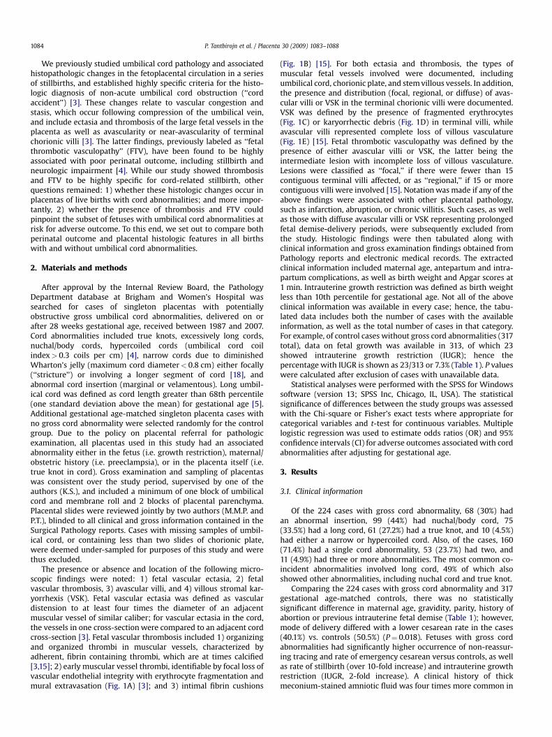
The presence or absence and location of the following micro-scopic findings were noted: 1) fetal vascular ectasia, 2) fetalvascular thrombosis, 3) avascular villi, and 4) villous stromal kar-yorrhexis (VSK). Fetal vascular ectasia was defined as vasculardistension to at least four times the diameter of an adjacentmuscular vessel of similar caliber; for vascular ectasia in the cord,the vessels in one cross-section were compared to an adjacent cordcross-section [3]. Fetal vascular thrombosis included 1) organizingand organized thrombi in muscular vessels, characterized byadherent, fibrin containing thrombi, which are at times calcified[3,15]; 2) early muscular vessel thrombi, identifiable by focal loss ofvascular endothelial integrity with erythrocyte fragmentation andmural extravasation (Fig. 1A) [3]; and 3) intimal fibrin cushions
(Fig. 1B) [15]. For both ectasia and thrombosis, the types ofmuscular fetal vessels involved were documented, includingumbilical cord, chorionic plate, and stem villous vessels. In addition,the presence and distribution (focal, regional, or diffuse) of avas-cular villi or VSK in the terminal chorionic villi were documented.VSK was defined by the presence of fragmented erythrocytes(Fig. 1C) or karyorrhectic debris (Fig. 1D) in terminal villi, whileavascular villi represented complete loss of villous vasculature(Fig. 1E) [15]. Fetal thrombotic vasculopathy was defined by thepresence of either avascular villi or VSK, the latter being theintermediate lesion with incomplete loss of villous vasculature.Lesions were classified as ‘‘focal,’’ if there were fewer than 15contiguous terminal villi affected, or as ‘‘regional,’’ if 15 or morecontiguous villi were involved [15]. Notation was made if any of theabove findings were associated with other placental pathology,such as infarction, abruption, or chronic villitis. Such cases, as wellas those with diffuse avascular villi or VSK representing prolongedfetal demise-delivery periods, were subsequently excluded fromthe study. Histologic findings were then tabulated along withclinical information and gross examination findings obtained fromPathology reports and electronic medical records. The extractedclinical information included maternal age, antepartum and intra-partum complications, as well as birth weight and Apgar scores at1 min. Intrauterine growth restriction was defined as birth weightless than 10th percentile for gestational age. Not all of the aboveclinical information was available in every case; hence, the tabu-lated data includes both the number of cases with the availableinformation, as well as the total number of cases in that category.For example, of control cases without gross cord abnormalities (317total), data on fetal growth was available in 313, of which 23showed intrauterine growth restriction (IUGR); hence thepercentage with IUGR is shown as 23/313 or 7.3% (Table 1). P valueswere calculated after exclusion of cases with unavailable data.
Statistical analyses were performed with the SPSS for Windowssoftware (version 13; SPSS Inc, Chicago, IL, USA). The statisticalsignificance of differences between the study groups was assessedwith the Chi-square or Fisher’s exact tests where appropriate forcategorical variables and t-test for continuous variables. Multiplelogistic regression was used to estimate odds ratios (OR) and 95%confidence intervals (CI) for adverse outcomes associated with cordabnormalities after adjusting for gestational age.
3. Results
3.1. Clinical information
Of the 224 cases with gross cord abnormality, 68 (30%) hadan abnormal insertion, 99 (44%) had nuchal/body cord, 75(33.5%) had a long cord, 61 (27.2%) had a true knot, and 10 (4.5%)had either a narrow or hypercoiled cord. Also, of the cases, 160(71.4%) had a single cord abnormality, 53 (23.7%) had two, and11 (4.9%) had three or more abnormalities. The most common co-incident abnormalities involved long cord, 49% of which alsoshowed other abnormalities, including nuchal cord and true knot.
Comparing the 224 cases with gross cord abnormality and 317gestational age-matched controls, there was no statisticallysignificant difference in maternal age, gravidity, parity, history ofabortion or previous intrauterine fetal demise (Table 1); however,mode of delivery differed with a lower cesarean rate in the cases(40.1%) vs. controls (50.5%) (P¼ 0.018). Fetuses with gross cordabnormalities had significantly higher occurrence of non-reassur-ing tracing and rate of emergency cesarean versus controls, as wellas rate of stillbirth (over 10-fold increase) and intrauterine growthrestriction (IUGR, 2-fold increase). A clinical history of thickmeconium-stained amniotic fluid was four times more common in
Fig. 1. Histologic changes associated with fetal thrombotic vasculopathy. A) Early organizing thrombus in chorionic plate vessel, identified as fibrin deposit on the vascular wall withfocal loss of endothelial lining (H&E, original magnification �200). B) Organized thrombus in stem villous vessel with intimal thickening on the right, compared to dilated, non-thrombosed vessel on the left (H&E, original magnification �40). C and D) Villous stromal karyorrhexis (VSK), defined by extravassated red blood cells (C) and karyorrhectic debris(D) in terminal villi (H&E, original magnification �200). E) Avascular villi with fibrotic stroma and loss of villous vasculature (H&E, original magnification �200).
P. Tantbirojn et al. / Placenta 30 (2009) 1083–1088 1085
cases than controls. Finally, although the delivered infants in thegross cord abnormality group had a higher proportion of low one-minute Apgar scores compared to controls, this difference was notsignificant.
3.2. Microscopic features
Fetal vascular ectasia, fetal vascular thrombosis, and fetalthrombotic vasculopathy (FTV, defined by avascular villi, villousstromal karyorrhexis or VSK, or a combination thereof) weresignificantly more common in cases with gross cord abnormalities(Table 2). Vascular ectasia and thrombosis were most common inthe chorionic plate and stem villous vessels, but were also morefrequently seen in the umbilical cord vessels. Combined histologicfindings of ectasia and thrombosis were found 3 times morefrequently in abnormal cord cases vs. controls (Table 2); combinedhistologic findings of ectasia, thrombosis and FTV were over 30times more frequent in abnormal cord cases (Table 2). In addition,fetal vascular thrombosis and FTV, as well as the combinedfeatures, were much more common in cord abnormality cases withadverse fetal outcome, when compared to normal cord controlswith similar adverse outcomes (Table 3).
Comparison between subtypes of gross cord abnormalities issummarized in Table 4. This data includes only cases with singlecord abnormalities. All categories, except for true knots, showed anincreased risk of stillbirth. Nuchal cord also showed an increasedrisk of IUGR, as well as the highest frequencies of non-reassuring
fetal tracing and low Apgar scores. On microscopic examination,fetal vascular ectasia was found at significantly higher frequenciesin all categories, except in membranous cord insertions. Throm-bosis and FTV were found at significantly higher frequencies in longcords and nuchal cords, although marginal cord insertions alsoshowed a significantly higher frequency of thrombosis in chorionicplate vessels (Table 4). Membranous cord insertions had lowprevalence in this study (only 12 cases) and showed only increasedfrequency of FTV (Table 4). Compared to single abnormalities, thepresence of multiple (two or more) gross abnormalities of theumbilical cord was associated with a higher rate of ectasia (2-foldhigher) and thrombosis (3–10 fold higher) in the cord and chorionicplate vessels, although the rate and extent of FTV was not furtherincreased. Interestingly, single and multiple cord abnormalitieswere both associated with a 9–11 fold increase in rate of stillbirth;however, the frequency of IUGR was increased significantly only inthe multiple cord abnormalities subgroup (single abnormalities,11.9% vs. 7.3%, P¼ 0.12; multiple abnormalities, 21.7% vs. 7.3%,P¼ 0.002).
In addition to gross cord abnormality, the additional presence ofthrombosis in the fetal vasculature was associated with both anincreased rate of stillbirth and IUGR (Table 5). The rate of stillbirthincreased from 1.4% in the control group to 11.7% in abnormal cordcases without thrombosis, but was highest (30.6%) in abnormal cordcases with thrombosis. IUGR was only slightly elevated in abnormalcord cases without thrombosis (10.7% vs. 7.5% in controls), but wassignificantly elevated in the abnormal cord cases with thrombosis
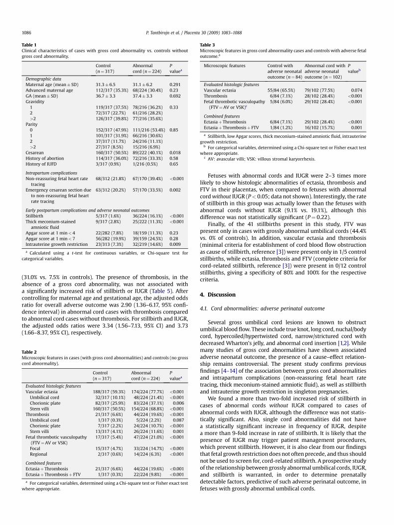
Table 1Clinical characteristics of cases with gross cord abnormality vs. controls withoutgross cord abnormality.
Control(n¼ 317)
Abnormalcord (n¼ 224)
Pvaluea
Demographic dataMaternal age (mean� SD) 31.3� 6.5 31.1� 6.2 0.291Advanced maternal age 112/317 (35.3%) 68/224 (30.4%) 0.23GA (mean� SD) 36.7� 3.3 37.4� 3.3 0.692Gravidity
1 119/317 (37.5%) 78/216 (36.2%) 0.332 72/317 (22.7%) 61/216 (28.2%)>2 126/317 (39.8%) 77/216 (35.6%)
Parity0 152/317 (47.9%) 111/216 (53.4%) 0.851 101/317 (31.9%) 66/216 (30.6%)2 37/317 (11.7%) 24/216 (11.1%)>2 27/317 (8.5%) 15/216 (6.9%)
Cesarean 160/317 (50.5%) 89/222 (40.1%) 0.018History of abortion 114/317 (36.0%) 72/216 (33.3%) 0.58History of IUFD 3/317 (0.9%) 1/216 (0.5%) 0.65
Intrapartum complicationsNon-reassuring fetal heart rate
tracing68/312 (21.8%) 67/170 (39.4%) <0.001
Emergency cesarean section dueto non-reassuring fetal heartrate tracing
63/312 (20.2%) 57/170 (33.5%) 0.002
Early postpartum complications and adverse neonatal outcomesStillbirth 5/317 (1.6%) 36/224 (16.1%) <0.001Thick meconium-stained
amniotic fluid9/317 (2.8%) 25/222 (11.3%) <0.001
Apgar score at 1 min< 4 22/282 (7.8%) 18/159 (11.3%) 0.23Apgar score at 1 min< 7 56/282 (19.9%) 39/159 (24.5%) 0.28Intrauterine growth restriction 23/313 (7.3%) 32/219 (14.6%) 0.009
a Calculated using a t-test for continuous variables, or Chi-square test forcategorical variables.
Table 3Microscopic features in gross cord abnormality cases and controls with adverse fetaloutcome.a
Microscopic features Control withadverse neonataloutcome (n¼ 84)
Abnormal cord withadverse neonataloutcome (n¼ 102)
Pvalueb
Evaluated histologic featuresVascular ectasia 55/84 (65.5%) 79/102 (77.5%) 0.074Thrombosis 6/84 (7.1%) 28/102 (28.4%) <0.001Fetal thrombotic vasculopathy
(FTV¼AV or VSK)c5/84 (6.0%) 29/102 (28.4%) <0.001
Combined featuresEctasiaþ Thrombosis 6/84 (7.1%) 29/102 (28.4%) <0.001Ectasiaþ Thrombosisþ FTV 1/84 (1.2%) 16/102 (15.7%) 0.001
a Stillbirth, low Apgar scores, thick meconium-stained amniotic fluid, intrauterinegrowth restriction.
b For categorical variables, determined using a Chi-square test or Fisher exact testwhere appropriate.
c AV: avascular villi; VSK: villous stromal karyorrhexis.
P. Tantbirojn et al. / Placenta 30 (2009) 1083–10881086
(31.0% vs. 7.5% in controls). The presence of thrombosis, in theabsence of a gross cord abnormality, was not associated witha significantly increased risk of stillbirth or IUGR (Table 5). Aftercontrolling for maternal age and gestational age, the adjusted oddsratio for overall adverse outcome was 2.90 (1.36–6.17, 95% confi-dence interval) in abnormal cord cases with thrombosis comparedto abnormal cord cases without thrombosis. For stillbirth and IUGR,the adjusted odds ratios were 3.34 (1.56–7.13, 95% CI) and 3.73(1.66–8.37, 95% CI), respectively.
Table 2Microscopic features in cases (with gross cord abnormalities) and controls (no grosscord abnormality).
Control(n¼ 317)
Abnormalcord (n¼ 224)
Pvaluea
Evaluated histologic featuresVascular ectasia 188/317 (59.3%) 174/224 (77.7%) <0.001
Umbilical cord 32/317 (10.1%) 48/224 (21.4%) <0.001Chorionic plate 82/317 (25.9%) 83/224 (37.1%) 0.006Stem villi 160/317 (50.5%) 154/224 (68.8%) <0.001
Thrombosis 21/317 (6.6%) 44/224 (19.6%) <0.001Umbilical cord 1/317 (0.3%) 5/224 (2.2%) 0.087Chorionic plate 7/317 (2.2%) 24/224 (10.7%) <0.001Stem villi 13/317 (4.1%) 26/224 (11.6%) 0.001
Fetal thrombotic vasculopathy(FTV¼AV or VSK)
17/317 (5.4%) 47/224 (21.0%) <0.001
Focal 15/317 (4.7%) 33/224 (14.7%) <0.001Regional 2/317 (0.6%) 14/224 (6.3%) <0.001
Combined featuresEctasiaþ Thrombosis 21/317 (6.6%) 44/224 (19.6%) <0.001Ectasiaþ Thrombosisþ FTV 1/317 (0.3%) 22/224 (9.8%) <0.001
a For categorical variables, determined using a Chi-square test or Fisher exact testwhere appropriate.
Fetuses with abnormal cords and IUGR were 2–3 times morelikely to show histologic abnormalities of ectasia, thrombosis andFTV in their placentas, when compared to fetuses with abnormalcord without IUGR (P< 0.05; data not shown). Interestingly, the rateof stillbirth in this group was actually lower than the fetuses withabnormal cords without IUGR (9.1% vs. 19.1%), although thisdifference was not statistically significant (P¼ 0.22).
Finally, of the 41 stillbirths present in this study, FTV waspresent only in cases with grossly abnormal umbilical cords (44.4%vs. 0% of controls). In addition, vascular ectasia and thrombosis(minimal criteria for establishment of cord blood flow obstructionas cause of stillbirth, reference [3]) were present only in 1/5 controlstillbirths, while ectasia, thrombosis and FTV (complete criteria forcord-related stillbirth, reference [3]) were present in 0/12 controlstillbirths, giving a specificity of 80% and 100% for the respectivecriteria.
4. Discussion
4.1. Cord abnormalities: adverse perinatal outcome
Several gross umbilical cord lesions are known to obstructumbilical blood flow. These include true knot, long cord, nuchal/bodycord, hypercoiled/hypertwisted cord, narrow/strictured cord withdecreased Wharton’s jelly, and abnormal cord insertion [1,2]. Whilemany studies of gross cord abnormalities have shown associatedadverse neonatal outcome, the presence of a cause–effect relation-ship remains controversial. The present study confirms previousfindings [4–14] of the association between gross cord abnormalitiesand intrapartum complications (non-reassuring fetal heart ratetracing, thick meconium-stained amniotic fluid), as well as stillbirthand intrauterine growth restriction in singleton pregnancies.
We found a more than two-fold increased risk of stillbirth incases of abnormal cords without IUGR compared to cases ofabnormal cords with IUGR, although the difference was not statis-tically significant. Also, single cord abnormalities did not havea statistically significant increase in frequency of IUGR, despitea more than 9-fold increase in rate of stillbirth. It is likely that thepresence of IUGR may trigger patient management procedures,which prevent stillbirth. However, it is also clear from our findingsthat fetal growth restriction does not often precede, and thus shouldnot be used to screen for, cord-related stillbirth. A prospective studyof the relationship between grossly abnormal umbilical cords, IUGR,and stillbirth is warranted, in order to determine prenatallydetectable factors, predictive of such adverse perinatal outcome, infetuses with grossly abnormal umbilical cords.
Table 4Clinical and microscopic features among different categories of single cord abnormalities, compared to controls without gross cord abnormalities.
Long cord(n¼ 31)
Nuchal cord(n¼ 59)
True knot(n¼ 33)
Abnormal insertion
Marginal insertion(n¼ 25)
Membranous insertion(n¼ 12)
ClinicalNon-reassuring fetal heart rate tracing 5/24 (20.8%) 32/47 (68.1%)a 9/25 (36.0%) 5/18 (27.8%) 1/9 (11.1%)Thick meconium-stained amniotic fluid 2/31 (6.5%) 10/59 (16.9%)a 4/32 (12.5%)a 0 1/12 (8.3%)Stillbirth 6/31 (19.4%)a 7/59 (11.9%)a 2/33 (6.1%) 7/25 (28.0%)a 3/12 (25.0%)a
Low Apgar scores 1/23 (4.3%) 7/42 (16.7%) 1/23 (4.3%) 1/18 (5.6%) 0Intrauterine growth restriction 0 13/59 (22.0%)a 3/31 (9.7%) 2/25 (8.0%) 1/12 (8.3%)
Microscopic featuresVascular ectasia 26/31 (83.7%)a 44/59 (74.6%)a 28/33 (84.8%)a 20/25 (80.0%)a 7/12 (58.3%)Thrombosis 6/31 (19.4%)a 12/59 (20.3%)a 5/33 (15.2%) 4/25 (16.0%) 0
Umbilical cord 0 0 0 1/25 (0.4%) 0Chorionic plate 2/31 (6.5%) 4/59 (6.8%) 2/33 (6.1%) 3/25 (12.0%)a 0Stem villi 4/31 (12.9%) 11/59 (18.6%)a 3/33 (9.1%) 2/25 (8.0%) 0
Fetal thrombotic vasculopathy (AV or VSK) 11/31 (35.5%)a 13/59 (22.0%)a 4/33 (12.1%) 4/25 (16.0%) 3/12 (25.0%)a
Focal 8/31 (25.8%)a 10/59 (16.9%)a 2/33 (6.1%) 3/25 (12.0%) 2/12 (16.7%)Regional 3/31 (9.7%)a 3/59 (5.1%)a 2/33 (6.1%)a 1/25 (4.0%) 1/12 (8.3%)
a P< 0.05.
P. Tantbirojn et al. / Placenta 30 (2009) 1083–1088 1087
4.2. Cord abnormalities: placental histologic findings
We evaluated placental histologic features in all cord abnor-mality cases and controls, focusing on fetal vascular changespreviously associated with restricted fetal blood flow [3,15]. Each ofthese parameters (ectasia, thrombosis, and FTV) was found morefrequently in cases vs. controls. Overall, similar to our stillbirthstudy [3], thrombosis and FTV were much more specific thanectasia for umbilical cord abnormalities (93.4% for thrombosis,94.6% for FTV, vs. 40.7% for ectasia).
We had previously defined minimal histologic criteria for cordcompression-related stillbirth as ectasia and thrombosis within theplacental fetal vascular tree [3]; complete criteria also included theadditional presence of FTV [3]. The current study, which includedboth stillbirths and live births, also showed high specificity forthese criteria. However, as expected, the proportion of cordabnormality cases with these criteria was low, although thesenumbers increased when measured in the subgroup of cordabnormalities with adverse perinatal outcome. One major limita-tion may have been inadequate sampling. Both chorionic plate andstem villus vessels need to be properly sampled in order to screenfor ectasia and thrombosis. In addition, FTV is by definition a focalor regional finding of avascular or near-avascular villi in theplacental parenchyma [15]. This lesion can rarely be seen grosslywhen it involves a large region of the placenta, in which case it willappear as a discrete, firm area of discoloration [19]. Due to thetemporally heterogeneous, and in most cases grossly undetectable,nature of this lesion, proper sampling of the placental parenchymais also needed. We therefore strongly recommend submission offour sections of placental parenchyma in all such cases; these
Table 5Clinical outcome of abnormal cord cases with thrombosis, vs. various control groups.
Controls withoutthrombosis (n¼ 296)
Controls wthrombosis
ClinicalNon-reassuring fetal heart rate tracing 64/292 (21.9%) 4/20 (20.0%Thick meconium-stained amniotic fluid 9/296 (3.0%) 0Stillbirth 4/296 (1.4%) 1/21 (4.8%)Apgar scores< 4 at 1 min 21/262 (8.0%) 1/20 (5.0%)Apgar scores< 7 at 1 min 52/262 (19.8%) 4/20 (20.0%Intrauterine growth restriction 22/292 (7.5%) 1/21 (4.8%)
a Global test: P value to test whether any difference exists across groups.
sections should include chorionic plate vessels near the cordinsertion, as we find this to be the most likely site of muscularvessel thrombosis.
Based on previous stillbirth studies, the presence of completelyavascular villi vs. near-avascular villi (or villous stromal karyor-rhexis/VSK) likely reflects the time elapsed since occurrence ofvascular obstruction, while the distribution (focal or regional) maycorrelate with severity and frequency of the obstruction[3,15,18,20,21]. Therefore, not only is identification of FTV importantfor specific confirmation of fetal blood flow obstruction, docu-mentation of the exact nature and distribution of the lesion is alsoimportant for establishing the timing and extent of fetal stressors. Inour study the majority of abnormal cord cases tended to have focalrather than regional FTV, with a similar overall frequency of avas-cular villi and VSK (data not shown). However, while long cord andnuchal cord cases showed a mixture of both avascular villi and VSK,true knot and abnormal cord insertion cases tended to only have anincreased frequency of VSK (data not shown), suggesting that theformer lesions may have caused obstruction over a more prolongedtime period. Coincidentally, these cases with long cord and nuchalcord also had a higher frequency of adverse clinical outcome. Futurestudy of large series of FTV cases is needed to better correlate thenature and extent of these histologic findings with cord-relatedfetal morbidity and mortality.
4.3. Summary of conclusions and limitations
While comprehensive, this study has several limitations. First, asa retrospective study, recording of clinical data was incomplete insome cases, and may have also been biased. Second, as clinical
ith(n¼ 21)
Abnormal cord withoutthrombosis (n¼ 180)
Abnormal cord withthrombosis (n¼ 44)
Pvaluea
) 53/143 (37.1%) 14/27 (51.9%) <0.00116/178 (9.0%) 9/44 (20.5%) <0.00121/180 (11.7%) 15/44 (34.1%) <0.00115/135 (11.1%) 3/24 (12.5%) 0.619
) 31/135 (23.0%) 8/24 (33.3%) 0.45619/177 (10.7%) 13/42 (31.0%) <0.001
P. Tantbirojn et al. / Placenta 30 (2009) 1083–10881088
follow-up was limited to NICU notes, only immediate neonatalcomplications could be recorded and analyzed. Third, clinicalconditions that may predispose to cord compression, without grosscord abnormalities, such as oligohydramnios, were not well docu-mented in the charts and therefore could not be analyzed. Finally,since microscopic review was based on limited placental sections(mostly two per case), histologic findings such as focal thrombosisor FTV may have been missed due to inadequate sampling.
In summary, gross cord abnormalities were associated withstillbirth, intrauterine growth restriction, and intrapartum orimmediate post-natal complications. Placental microscopic evalu-ation revealed that stasis-induced changes in the fetal vasculatureand chorionic villi were significantly increased in cases with grosscord abnormalities, particularly in those cases with adverse peri-natal outcome. We therefore recommend all placentas with grosscord abnormalities, particularly the ones predisposing to vascularobstruction, be sent for pathologic examination; this shouldinclude a thorough gross examination as well as adequate samplingof large fetal vessels and placental parenchyma in order to evaluatefor the presence of thrombosis and fetal thrombotic vasculopathy.
5. Disclosure/conflicts of interest
There are no conflicts of interest to report.
Acknowledgements
We would like to thank Dr. Kurt Benirschke for critical review ofthe manuscript. P.T. was supported by the Columbia Hospital forWomen’s Research Foundation.
References
[1] Baergen RN. Cord abnormalities, structural lesions, and cord ‘‘accidents’’.Semin Diagn Pathol 2007;24:23–32.
[2] Benirschke K. Obstetrically important lesions of the umbilical cord. J ReprodMed 1994;39(4):262–72.
[3] Parast MM, Crum CP, Boyd TK. Placental histologic criteria for umbilical bloodflow restriction in unexplained stillbirth. Hum Pathol 2008;39(6):948–53.
[4] Machin GA, Ackerman J, Gilbert-Barness E. Abnormal umbilical cord coiling isassociated with adverse perinatal outcomes. Pediatr Dev Pathol 2000;3:462–71.
[5] Naeye RL. Umbilical cord length: clinical significance. J Pediatr 1985;107:278–81.
[6] Peng HQ, Levitin-Smith M, Rochelson B, Kahn E. Umbilical cord stricture andovercoiling are common causes of fetal demise. Pediatr Dev Pathol 2006;9:14–9.
[7] Collins JH. Umbilical cord accidents: human studies. Semin Perinatol2002;26:79–82.
[8] Hasegawa J, Matsuoka R, Ichizuka K, Sekizawa A, Okai T. Velamentous cordinsertion: significance of prenatal detection to predict perinatal complications.Taiwan J Obstet Gynecol 2006;45:21–5.
[9] Baergen RN, Malicki D, Behling C, Benirschke K. Morbidity, mortality, andplacental pathology in excessively long umbilical cords: retrospective study.Pediatr Dev Pathol 2001;4:144–53.
[10] Spellacy WN, Gravem H, Fisch RO. The umbilical cord complications of trueknots, nuchal coils, and cords around the body. Am J Obstet Gynecol1966;94:1136–42.
[11] Rhoades DA, Latza U, Mueller BA. Risk factors and outcomes associated withnuchal cord. A population-based study. J Reprod Med 1999;44:39–45.
[12] Larson JD, Rayburn WF, Crosby S, Thurnau GR. Multiple nuchal cord entan-glements and intrapartum complications. Am J Obstet Gynecol1995;173:1228–31.
[13] Airas U, Heinonen S. Clinical significance of true umbilical knots: a population-based analysis. Am J Perinatol 2002;19:127–32.
[14] French AE, Gregg VH, Newberry Y, Parsons T. Umbilical cord stricture: a causeof recurrent fetal death. Obstet Gynecol 2005;105:1235–9.
[15] Redline RW. Clinical and pathological umbilical cord abnormalities in fetalthrombotic vasculopathy. Hum Pathol 2004;35:1494–8.
[16] Thureen PJ, Hall DM, Hoffenberg A, Tyson RW. Fatal meconium aspiration inspite of appropriate perinatal airway management: pulmonary and placentalevidence of prenatal disease. Am J Obstet Gynecol 1997;176:967–75.
[17] Collins JH, Collins CL. Umbilical cord compression, persistent pulmonaryhypertension, and placental villous hypervascularity. Am J Obstet Gynecol1997;177:1276–7.
[18] Redline RW, Ariel I, Baergen RN, Desa DJ, Kraus FT, Roberts DJ, et al. Fetalvascular obstructive lesions: nosology and reproducibility of placental reac-tion patterns. Pediatr Dev Pathol 2004;7(5):443–52.
[19] Kraus FT, Redline RW, Gersell DJ, Nelson DM, Dicke JM. Placental Pathology.Washington (DC): Armed Forces Institute of Pathology; 2004.
[20] Genest DR. Estimating the time of death in stillborn fetuses: II. Histologicevaluation of the placenta; a study of 71 stillborns. Obstet Gynecol1992;80(4):585–92.
[21] Redline RW. Cerebral palsy in term infants: a clinicopathologic analysis of 158medicolegal case reviews. Pediatr Dev Pathol 2008;11:456–64.


![Newborns ≥ 34 Weeks Gestation - umassmed.edu · Newborns ≥ 34 Weeks Gestation . ... fluid, placenta, umbilical cord and fetus) with normal ... abnormalities] Mukhopadhyay and](https://static.fdocuments.in/doc/165x107/5b0a2c3d7f8b9ac7678bf283/newborns-34-weeks-gestation-34-weeks-gestation-fluid-placenta.jpg)