Grattan-2015-60 YEARS OF NEUROENDOCRINOLOGY Trci.rutgers.edu/~advis/pdfs/00_Grattan-2015-60 YEARS...
22
60 YEARS OF NEUROENDOCRINOLOGY The hypothalamo-prolactin axis David R Grattan 1,2 1 Centre for Neuroendocrinology and Department of Anatomy, University of Otago, PO Box 913, Dunedin 9054, New Zealand 2 Maurice Wilkins Centre for Molecular Biodiscovery, Auckland, New Zealand Correspondence should be addressed to D R Grattan Email [email protected] Abstract The hypothalamic control of prolactin secretion is different from other anterior pituitary hormones, in that it is predominantly inhibitory, by means of dopamine from the tubero- infundibular dopamine neurons. In addition, prolactin does not have an endocrine target tissue, and therefore lacks the classical feedback pathway to regulate its secretion. Instead, it is regulated by short loop feedback, whereby prolactin itself acts in the brain to stimulate production of dopamine and thereby inhibit its own secretion. Finally, despite its relatively simple name, prolactin has a broad range offunctions in the body, in addition to its defining role in promoting lactation. As such, the hypothalamo-prolactin axis has many characteristics that are quite distinct from other hypothalamo-pituitary systems. This review will provide a brief overview of our current understanding of the neuroendocrine control of prolactin secretion, in particular focusing on the plasticity evident in this system, which keeps prolactin secretion at low levels most of the time, but enables extended periods of hyperprolactinemia when necessary for lactation. Key prolactin functions beyond milk production will be discussed, particularly focusing on the role of prolactin in inducing adaptive responses in multiple different systems to facilitate lactation, and the consequences if prolactin action is impaired. A feature of this pleiotropic activity is that functions that may be adaptive in the lactating state might be maladaptive if prolactin levels are elevated inappropriately. Overall, my goal is to give a flavour of both the history and current state of the field of prolactin neuroendocrinology, and identify some exciting new areas of research development. Key Words " prolactin " tuberoinfundibular dopamine neurons " pregnancy " lactation " prolactin-releasing factor Journal of Endocrinology (2015) 226, T101–T122 Introduction When Geoffrey Harris wrote his influential monograph on ‘Neural Control of the Pituitary Gland’, it was already apparent that prolactin, or ‘lactogenic hormone’ as he referred to it, might be controlled differently to the other adenohypophyseal hormones. Harris convincingly built the case that humeral factors from the hypothalamus control secretion of anterior pituitary hormones, correctly predict- ing the existence of hypothalamic ‘releasing factors’ that mediate the neural control of pituitary secretions (Harris 1948). Despite the clear evidence that neural stimuli (such as suckling) could stimulate prolactin secretion, however, Harris noted that cutting the pituitary stalk did not abolish lactation (Dempsey & Uotila 1940), leading to the inevitable conclusion that a hypothalamic ‘prolactin-releasing factor’ was not necessary to stimulate prolactin secretion. Indeed, it was subsequently shown in hypophysectomised rats that ectopic pituitary grafts were able to maintain corpus luteum function (Everett 1954) and lactation (Cowie et al. 1960), demonstrating hypothalamic-independent secretion of prolactin from the grafts. At first impression, this appeared to contradict Harris’ principle of neural control of the anterior pituitary. But further observations identified the Journal of Endocrinology Thematic Review Open Access D R Grattan The hypothalamo-prolactin axis 226 :2 T101–T122 http://joe.endocrinology-journals.org DOI: 10.1530/JOE-15-0213 Ñ 2015 Society for Endocrinology Published by Bioscientifica Ltd. Printed in Great Britain This work is licensed under a Creative Commons Attribution 3.0 Unported License.
Transcript of Grattan-2015-60 YEARS OF NEUROENDOCRINOLOGY Trci.rutgers.edu/~advis/pdfs/00_Grattan-2015-60 YEARS...
60 YEARS OF NEUROENDOCRINOLOGY
The hypothalamo-prolactin axis
David R Grattan1,2
1Centre for Neuroendocrinology and Department of Anatomy, University of Otago,
PO Box 913, Dunedin 9054, New Zealand2Maurice Wilkins Centre for Molecular Biodiscovery, Auckland, New Zealand
Correspondence
should be addressed
to D R Grattan
Abstract
The hypothalamic control of prolactin secretion is different from other anterior pituitary
hormones, in that it is predominantly inhibitory, by means of dopamine from the tubero-
infundibular dopamine neurons. In addition, prolactin does not have an endocrine target
tissue, and therefore lacks the classical feedback pathway to regulate its secretion. Instead,
it is regulated by short loop feedback, whereby prolactin itself acts in the brain to stimulate
production of dopamine and thereby inhibit its own secretion. Finally, despite its relatively
simplename,prolactin has a broadrange offunctions in the body, inadditionto its definingrole
in promoting lactation. As such, the hypothalamo-prolactin axis has many characteristics that
are quite distinct from other hypothalamo-pituitary systems. This review will provide a brief
overview of our current understanding of the neuroendocrine control of prolactin secretion,
in particular focusing on the plasticity evident in this system, which keeps prolactin secretion
at low levels most of the time, but enables extended periods of hyperprolactinemia when
necessary for lactation. Key prolactin functions beyond milk production will be discussed,
particularly focusing on the role of prolactin in inducing adaptive responses in multiple
different systems to facilitate lactation, and the consequences if prolactin action is impaired.
A feature of this pleiotropic activity is that functions that may be adaptive in the lactating state
might be maladaptive if prolactin levels are elevated inappropriately. Overall, my goal is to give
a flavour of both the history and current state of the field of prolactin neuroendocrinology, and
identify some exciting new areas of research development.
Key Words
" prolactin
" tuberoinfundibulardopamine neurons
" pregnancy
" lactation
" prolactin-releasing factor
Journal of Endocrinology
(2015) 226, T101–T122
Introduction
When Geoffrey Harris wrote his influential monograph on
‘Neural Control of the Pituitary Gland’, it was already
apparent that prolactin, or ‘lactogenic hormone’ as he
referred to it, might be controlled differently to the other
adenohypophyseal hormones. Harris convincingly built the
case that humeral factors from the hypothalamus control
secretion of anterior pituitary hormones, correctly predict-
ing the existence of hypothalamic ‘releasing factors’ that
mediate the neural control of pituitary secretions (Harris
1948). Despite the clear evidence that neural stimuli (such
as suckling) could stimulate prolactin secretion, however,
Harris noted that cutting the pituitary stalk did not abolish
lactation (Dempsey & Uotila 1940), leading to the inevitable
conclusion that a hypothalamic ‘prolactin-releasing factor’
was not necessary to stimulate prolactin secretion. Indeed,
it was subsequently shown in hypophysectomised rats that
ectopic pituitary grafts were able to maintain corpus luteum
function (Everett 1954) and lactation (Cowie et al. 1960),
demonstrating hypothalamic-independent secretion of
prolactin from the grafts. At first impression, this appeared
to contradict Harris’ principle of neural control of the
anterior pituitary. But further observations identified the
Jou
rnal
of
En
do
crin
olo
gy
Thematic Review
Open Access
D R Grattan The hypothalamo-prolactin axis 226 :2 T101–T122
http://joe.endocrinology-journals.orgDOI: 10.1530/JOE-15-0213
! 2015 Society for EndocrinologyPublished by Bioscientifica Ltd.
Printed in Great Britain
This work is licensed under a Creative CommonsAttribution 3.0 Unported License.
fact that hypothalamic regulation was indeed critical for
normal prolactin secretion, as Harris predicted, but that the
mode of control was different. Everett found that while
pituitaries located away from the hypothalamus would
induce pseudopregnancy, a marker of elevated prolactin
secretion in rodents, normal cycles (and therefore normal
low levels of prolactin) would resume if transplanted
pituitaries were re-vascularised by portal vessels under the
median eminence (Nikitovitch-Winer & Everett 1958). Two
groups independently demonstrated that extracts of the
median eminence/infundibular region could inhibit pro-
lactin secretion from anterior pituitary cells in vitro (Pasteels
1963, Talwalker et al. 1963). Subsequently, it was shown that
median eminence lesions (Arimura et al. 1972) or destruc-
tion of the pituitary stalk (Kanematsu & Sawyer 1973)
resulted in elevated prolactin secretion (accounting for the
pseudopregnancies described earlier by Everett). Thus, it was
proven that the hypothalamus was essential for the
regulation of prolactin secretion, but that it primarily
exerted an inhibitory influence.
This brief review will summarise our current under-
standing of the hypothalamic control of prolactin secretion,
and the neuroendocrine functions of prolactin, highlight-
ing (admittedly selected) areas of current research interest.
From the somewhat contrary beginnings previously intro-
duced, the neural control of prolactin secretion, and indeed
the whole hypothalamo-prolactin axis, continues to prove
itself a bit different from other hypothalamo-pituitary
systems. Not only is the hypothalamic regulation predomi-
nantly inhibitory, as opposed to stimulatory, it also involves
a catecholamine neurotransmitter, dopamine, rather than
the more typical peptide hypothalamic hormones involved
in regulating all other pituitary systems. Prolactin is also the
only anterior pituitary hormone that does not have an
endocrine target tissue, and therefore lacks a classical
hormonal feedback system. It is regulated, instead, by a
short loop feedback whereby prolactin itself stimulates the
secretion of the inhibitory factor, dopamine. Finally, despite
its rather simple and one-dimensional name, prolactin does
much more than simply PROmote LACTation. It is now
recognised as a pleiotrophic hormone with arguably the
widest range of physiological actions of any extracellular
signalling molecule in the body.
Neuroendocrine control of prolactin secretion
Dopamine as a prolactin-inhibitory hormone
Even after the clear demonstration of inhibitory regulation
of prolactin secretion in the 1950s, the search for the
inhibitory hormone mediating this action was controver-
sial. All hypothalamic hormones identified to date had been
peptides, and the expectation was that ‘prolactin-inhibitory
factor’ would also be a peptide. Initial clues that this might
not be the case came from observations that drugs such as
reserpine, which depletes endogenous catecholamines,
induced pseudopregnancy in rats (Barraclough & Sawyer
1959), indicative of elevated prolactin. It was assumed,
however, that the functional role of catecholamines were as
neurotransmitters acting in the hypothalamus to regulate
the release of a hypothalamic hormone (Kanematsu et al.
1963). Based on the evidence of dopaminergic nerve
terminals in the median eminence (Fuxe 1963), McLeod
proposed that dopamine may be released into the pituitary
portal system, and thereby acting as a hypothalamic
hormone (as distinct from its neurotransmitter role in
other systems) (MacLeod et al. 1970). He demonstrated
that dopaminergic agonists were effective at suppressing
prolactin secretion in vivo, and perhaps more importantly,
that dopamine could inhibit prolactin secretion from
isolated pituitary glands (MacLeod et al. 1970). Dopamine
was subsequently detected in the pituitary portal blood
(Kamberi et al. 1970), and Porter’s group (and others) found
that variations of levels of dopamine in the portal blood
accounted for changes in prolactin secretion in various
physiological conditions (Ben-Jonathan et al. 1977, 1980,
Gibbs & Neill 1978, De Greef & Neill 1979). Dopamine
receptors were identified on lactotroph cells in the anterior
pituitary (Mansour et al. 1990). The observation that mice
lacking the dopamine D2 receptor are hyperprolactinemic
(Kelly et al. 1997, Saiardi et al. 1997), clearly demonstrated
the critical role of dopamine in suppressing endogenous
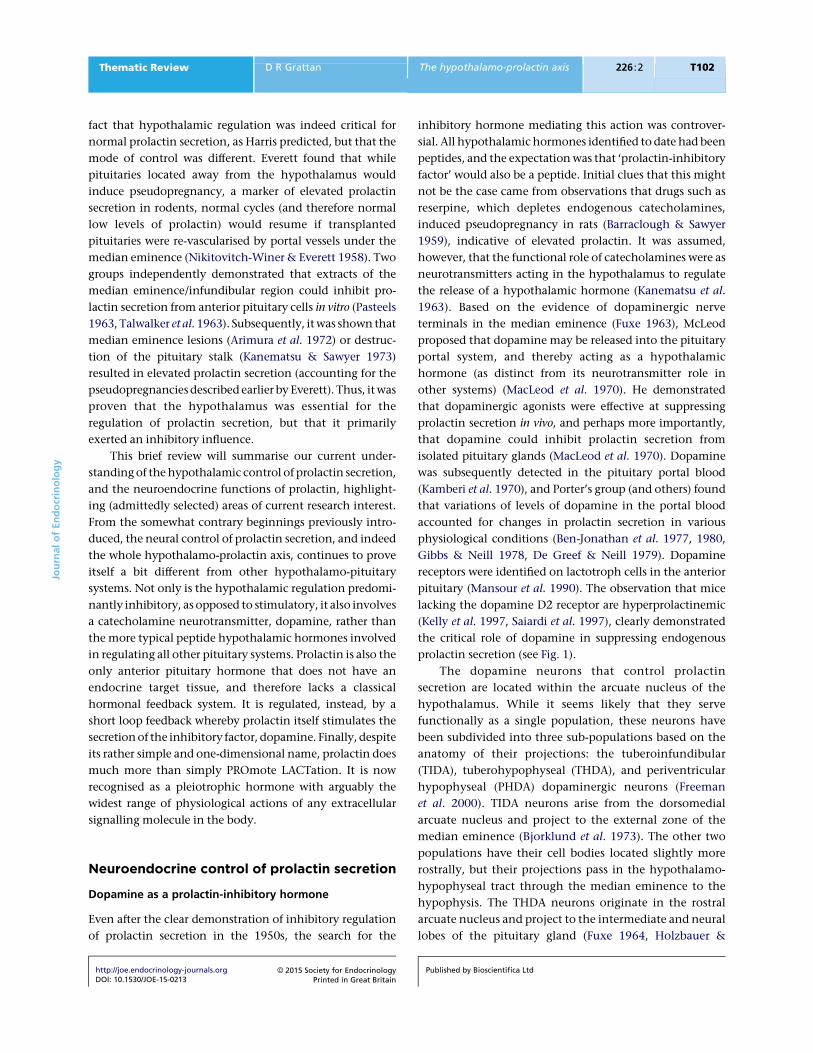
prolactin secretion (see Fig. 1).
The dopamine neurons that control prolactin
secretion are located within the arcuate nucleus of the
hypothalamus. While it seems likely that they serve
functionally as a single population, these neurons have
been subdivided into three sub-populations based on the
anatomy of their projections: the tuberoinfundibular
(TIDA), tuberohypophyseal (THDA), and periventricular
hypophyseal (PHDA) dopaminergic neurons (Freeman
et al. 2000). TIDA neurons arise from the dorsomedial
arcuate nucleus and project to the external zone of the
median eminence (Bjorklund et al. 1973). The other two
populations have their cell bodies located slightly more
rostrally, but their projections pass in the hypothalamo-
hypophyseal tract through the median eminence to the
hypophysis. The THDA neurons originate in the rostral
arcuate nucleus and project to the intermediate and neural
lobes of the pituitary gland (Fuxe 1964, Holzbauer &
Jou
rnal
of
En
do
crin
olo
gy
Thematic Review D R Grattan The hypothalamo-prolactin axis 226 :2 T102
http://joe.endocrinology-journals.orgDOI: 10.1530/JOE-15-0213
! 2015 Society for EndocrinologyPrinted in Great Britain
Published by Bioscientifica Ltd

Racke 1985), while the PHDA neurons originate in the
periventricular nucleus, with axons terminating in the
intermediate lobe (Goudreau et al. 1992). The TIDA
neurons produce the classical hypothalamic hormone
secretion into the pituitary portal blood vessels, while
THDA and PHDA neurons contribute to basal regulation of
prolactin secretion, after transport of dopamine to the
anterior pituitary gland through short portal vessels from
the neurohypophysis (Peters et al. 1981). While anatomi-
cally distinct, there is considerable overlap in their
dendritic fields (van den Pol et al. 1984), and all three
populations appear to be regulated similarly. For example,
all are stimulated by prolactin (DeMaria et al. 1999).
Hence, it is reasonable to consider them as a functional
unit of prolactin-inhibiting neurons.
Electrophysiological studies of hypothalamic dopa-
mine neurons in the rat have described TIDA neurons as
exhibiting a robust oscillation between hyperpolarized and
depolarized states, with periodicity of about 20 s, and a
spontaneous firing rate of w4 Hz during the depolarized
‘up-state’. Remarkably, the TIDA neurons were found to
show a synchronous pattern of firing suggestive of an
interconnected network, dependent on functional
expression of gap junctions (Lyons et al. 2010). Taking
advantage of transgenic technologies to label dopamin-
ergic neurons with fluorescent tags, studies of TIDA
electrical activity have also been completed in brain slices
from mice (Brown et al. 2012, Romano et al. 2013), showing
a similar pattern of firing to that seen in the rat, although
only a small proportion of TIDA neurons showed the phasic
oscillations in this model. Importantly, one of these latter
studies demonstrated that patterns of firing of an individ-
ual TIDA neuron were reflected in the pattern of dopamine
release from the population, as measured using in vivo
amperometry in the median eminence (Romano et al.
2013). These data support the concept postulated by Lyons
et al. (2010), that the neurons act as a synchronous network
to release dopamine in a pulsatile or phasic fashion.
Dopaminesecretion in
median eminence
Tuberoinfundibular dopamine (TIDA) neuronsin the hypothalamus
–
Short loopfeedback
Anteriorpituitary gland
Prolactin
1
2
1
2
+
Figure 1
Diagrammatic representation of the neuroendocrine regulation of
prolactin secretion. Anterior pituitary prolactin release is inhibited by
dopamine coming from the tuberoinfundibular dopamine neurons (shown
in the coronal section on the top left using immunohistochemistry against
tyrosine hydroxylase, brown) whose cell bodies are found in the arcuate
nucleus of the hypothalamus, with axons projecting to the external layer of
the median eminence. Images on the right show examples of both rapid
feedback (electrophysiological activation) and delayed feedback
(phosphorylation of STAT5, black nuclear staining) in TIDA neurons.
In each example, 1) illustrates prior to prolactin treatment, and 2) after
administration of prolactin (reproduced, with permission, from Brown RS,
Piet R, Herbison AE & Grattan DR (2012) Differential actions of prolactin on
electrical activity and intracellular signal transduction in hypothalamic
neurons. Endocrinology 153 2375–2384. Copyright 2012 The Endocrine
Society). Prolactin stimulates dopamine secretion, to inhibit its own
secretion by short loop feedback.
Jou
rnal
of
En
do
crin
olo
gy
Thematic Review D R Grattan The hypothalamo-prolactin axis 226 :2 T103
http://joe.endocrinology-journals.orgDOI: 10.1530/JOE-15-0213
! 2015 Society for EndocrinologyPrinted in Great Britain
Published by Bioscientifica Ltd
Of the five dopamine receptors, the two members of the
D2-like receptor family, D2 and D4 are found in the pituitary
gland (Valerio et al. 1994, Matsumoto et al. 1995) and it is
through these D2-like receptors that dopamine acts to
inhibit lactotroph cell function (Mansour et al. 1990,
Ben-Jonathan & Hnasko 2001). Uniquely among anterior
pituitary cells, lactotrophs display spontaneous electrical
activity in the absence of hypothalamic stimulation and
Ca2C influx through voltage-gated Ca2C channels (VGCC)
stimulates to prolactin secretion (Gregerson 2006). This
accounts for the high levels of basal prolactin secretion, and
is consistent with a regulatory mechanism primarily based
on inhibition. Dopamine inhibits calcium influx resulting
in membrane hyperpolarisation (Gregerson et al. 1994,
Gregerson2003)and reducedprolactin secretion (Lledo etal.
1990). In addition to its effect on secretion, dopamine-
induced suppression of adenylate cyclase leads to a
reduction in prolactin gene expression (Maurer 1982,
Elsholtz et al. 1991, Ishida et al. 2007). Dopamine also has
a significant role to regulate lactotroph proliferation, as
demonstrated in cultures of pituitary cells (Ishida et al.
2007), as well as in vivo by suppression of oestradiol-induced
proliferation (Borgundvaag et al. 1992). When dopamine
levels are increased, such as caused by the loss of the
dopamine transporter, there is a severe post-natal reduction
in lactotroph proliferation leading to a dramatic reduction
in the number of lactotrophs by 8 weeks of age (Bosse et al.
1997). In contrast, there is marked lactotroph hyperplaisia
following loss of the D2 receptor (Kelly et al. 1997, Saiardi
et al. 1997), leading to the formation of prolactinomas. This
is exacerbated by age, and more prevalent in females than
males (Saiardi et al. 1997, Asa et al. 1999). A bias towards
lactotroph hyperplasia and more rapid generation of
pituitary tumours in females may be expected from the
direct actions of estradiol to stimulate prolactin production
by lactotrophs, but this may not be the sole factor leading to
the increased female hyperplasia. Gonadectomy has been
shown to reduce lactotroph hyperplasia and tumour
formation in D2 knockout mice, but that this could not be
fully rescued by estradiol replacement, suggesting that
ovarian factors other than estradiol may contribute to the
proliferation of lactotrophs (Hentges & Low 2002).
Prolactin regulation of dopamine neurons: short loop
feedback
As previously mentioned, the hypothalamo-prolactin
system does not have a specific endocrine target, and
therefore lacks the classical hormone-mediate negative
feedback pathway described for all other anterior pituitary
hormones. Nevertheless, it is still regulated in a negative
feedback manner, with prolactin itself providing the
afferent signal in a process known as short-loop feedback.
The presence of prolactin receptors on dopamine neurons
(Lerant & Freeman 1998, Grattan 2001, Kokay & Grattan
2005) was predicted by early neurochemical studies that
showed that exogenous prolactin stimulated hypo-
thalamic dopamine synthesis (Hokfelt & Fuxe 1972) and
turnover (Eikenburg et al. 1977, Annunziato & Moore
1978), increased dopamine metabolism in the median
eminence (Lookingland et al. 1987) and promoted
dopamine secretion into the pituitary portal blood
(Gudelsky & Porter 1979). In contrast, hypoprolactinemia
induced by administration of dopamine agonists resulted
in suppression of dopamine secretion (Arbogast & Voogt
1991a), indicating that the basal activity of these neurons
is dependent on the endogenous levels of prolactin
present in the blood. Using these biochemical indices of
activity of TIDA neurons, the time course of prolactin
action was described as having a ‘rapid’ component of
increased activity observed 2–4 h after prolactin treatment
(Selmanoff 1985), and a delayed component seen w12 h
after prolactin treatment (Demarest et al. 1984a, 1986).
More recent electrophysiological data has demonstrated
even more rapid actions of prolactin on the electrical
activity of TIDA neurons in mice (Brown et al. 2012,
Romano et al. 2013) or rats (Lyons et al. 2012). These
studies show that prolactin induces a fourfold increase in
firing rate within seconds to minutes of application,
acutely changing the firing pattern from a basal phasic
pattern to a tonically active pattern. Hence, there seem to
be multiple mechanisms of prolactin regulation of these
dopamine neurons mediated over different time courses.
It was observed that prolactin feedback was markedly
impaired in mice lacking the transcription factor STAT5b
(Grattan et al. 2001), likely through impairment in the
long-term regulation of expression of the rate limiting
enzyme in dopamine synthesis, tyrosine hydroxylase
(Arbogast & Voogt 1991a, Ma et al. 2005). While such an
effect might account for the ‘delayed’ component of
prolactin feedback, which requires protein synthesis
(Johnston et al. 1980), it is unlikely to account for more
rapid components of short look feedback. The very rapid
action revealed by electrophysiology appears to involve
two components: a low voltage component from transient
receptor potential (TRP)-like current and high voltage
component from inhibition of a Ca2C-dependent BK-type
KC current, with the latter component being wortmannin
sensitive, suggesting an involvement of the PI3K pathway
(Lyons et al. 2012). The slower component, originally
Jou
rnal
of
En
do
crin
olo
gy
Thematic Review D R Grattan The hypothalamo-prolactin axis 226 :2 T104
http://joe.endocrinology-journals.orgDOI: 10.1530/JOE-15-0213
! 2015 Society for EndocrinologyPrinted in Great Britain
Published by Bioscientifica Ltd
described as ‘rapid’ in neurochemical experiments, with a
time course of minutes to hours, likely involves prolactin-
induced serine phosphorylation of tyrosine hydroxylase
(Ma et al. 2005), resulting in increased enzyme activity
(Arbogast & Voogt 1997). Together, these three layers of
prolactin regulation of hypothalamic dopamine neurons
provides a tight homeostatic control, with prolactin
rapidly increasing the firing rate of these neurons to
induce increased dopamine secretion into the portal blood
and rapid suppression of further prolactin secretion from
the lactotroph. At the same time, slower but more
prolonged changes in tyrosine hydroxylase phosphoryl-
ation and transcriptional events to maintain changes in
tyrosine hydroxylase gene transcription serve to regulate
neuronal function over a much longer time-course,
priming the neurons for continued responses to changes
in prolactin levels (Grattan & LeTissier 2015).
For endogenous prolactin to function in the short-loop
feedback manner, previously described, one important
consideration is how this relatively large (197–199 amino
acids; 21 kDa) polypeptide hormone crosses the blood
brain barrier to gain access to the dopamine neurons. While
it is possible that the arcuate nucleus/median eminence
region may have an incomplete blood-brain barrier such
that hormones can directly access to neurons in this area
(Schaeffer et al. 2013), it seems unlikely that this is the
major mechanism by which prolactin regulates the
hypothalamic dopamine neurons. Indeed, systemic
administration of prolactin has been shown to simul-
taneously activate neurons throughout the hypothalamus
(Brown et al. 2010, Sapsford et al. 2012), not simply in the
arcuate nucleus. There is clear evidence that systemic
prolactin crosses the blood brain barrier through a
saturable, carrier-mediated transport system (Walsh et al.
1987). As a result, prolactin levels in the cerebrospinal fluid
parallel changes in prolactin in the peripheral circulation
(Login & MacLeod 1977, Nicholson et al. 1980, Grattan &
Averill 1991). Because of the high levels of prolactin
receptor expression and prolactin binding seen in the
choroid plexus (Walsh et al. 1978, 1990, Pi & Grattan
1998a,b, Augustine et al. 2003), it has been widely assumed
that the prolactin receptor might mediate prolactin entry
into the cerebrospinal fluid. However, we have recently
shown that prolactin transport into the brain is indepen-
dent of the prolactin receptor, occurring just as well in
prolactin receptor knockout mice (Brown RSE, Wyatt AK,
Herbison RE, Knowles PJ, Ladyman SR, Binart N, Banks WA
& Grattan DR, unpublished observations). Hence, precise
mechanism that translocates prolactin from the blood into
the CSF remains to be determined.
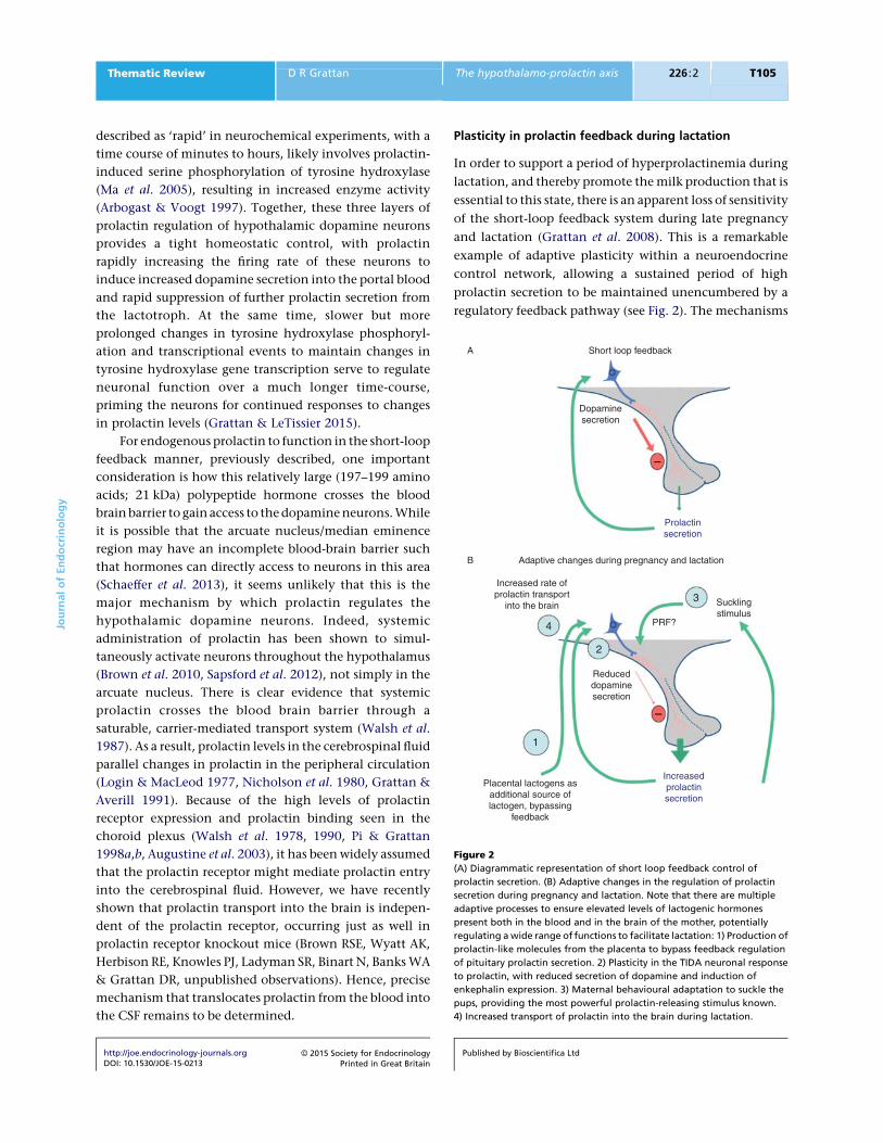
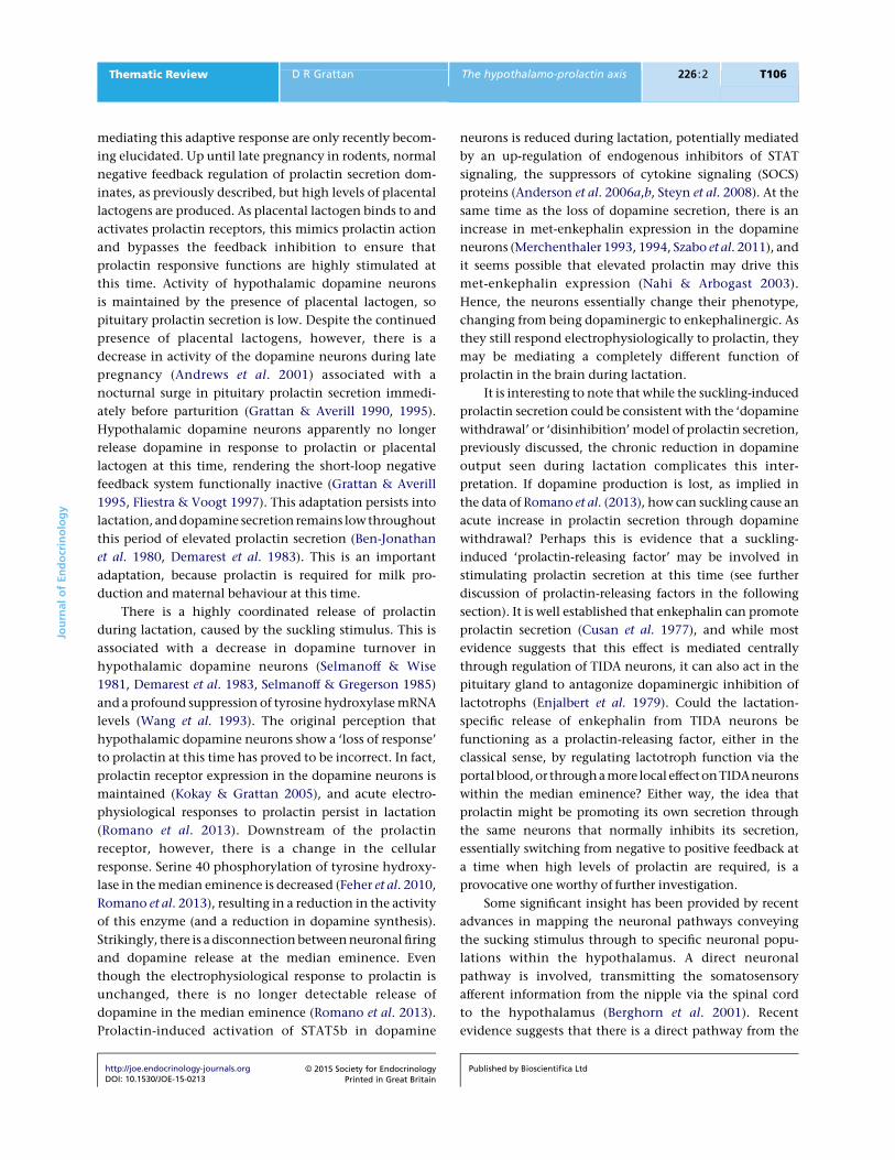
Plasticity in prolactin feedback during lactation
In order to support a period of hyperprolactinemia during
lactation, and thereby promote the milk production that is
essential to this state, there is an apparent loss of sensitivity
of the short-loop feedback system during late pregnancy
and lactation (Grattan et al. 2008). This is a remarkable
example of adaptive plasticity within a neuroendocrine
control network, allowing a sustained period of high
prolactin secretion to be maintained unencumbered by a
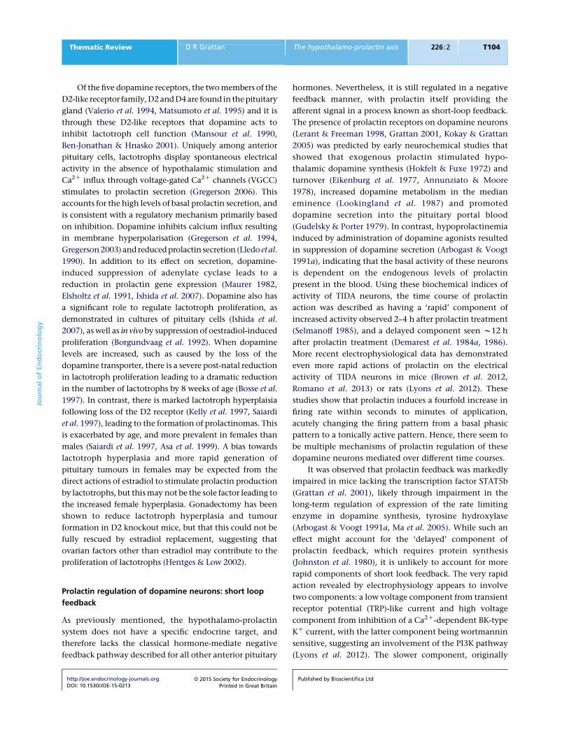
regulatory feedback pathway (see Fig. 2). The mechanisms
Short loop feedbackA
B Adaptive changes during pregnancy and lactation
Increased rate ofprolactin transport
into the brain Sucklingstimulus
PRF?
3
4
2
1
Reduceddopaminesecretion
Placental lactogens asadditional source oflactogen, bypassing
feedback
Dopaminesecretion
Prolactinsecretion
Increasedprolactinsecretion
–
–
Figure 2
(A) Diagrammatic representation of short loop feedback control of
prolactin secretion. (B) Adaptive changes in the regulation of prolactin
secretion during pregnancy and lactation. Note that there are multiple
adaptive processes to ensure elevated levels of lactogenic hormones
present both in the blood and in the brain of the mother, potentially
regulating a wide range of functions to facilitate lactation: 1) Production of
prolactin-like molecules from the placenta to bypass feedback regulation
of pituitary prolactin secretion. 2) Plasticity in the TIDA neuronal response
to prolactin, with reduced secretion of dopamine and induction of
enkephalin expression. 3) Maternal behavioural adaptation to suckle the
pups, providing the most powerful prolactin-releasing stimulus known.
4) Increased transport of prolactin into the brain during lactation.
Jou
rnal
of
En
do
crin
olo
gy
Thematic Review D R Grattan The hypothalamo-prolactin axis 226 :2 T105
http://joe.endocrinology-journals.orgDOI: 10.1530/JOE-15-0213
! 2015 Society for EndocrinologyPrinted in Great Britain
Published by Bioscientifica Ltd
mediating this adaptive response are only recently becom-
ing elucidated. Up until late pregnancy in rodents, normal
negative feedback regulation of prolactin secretion dom-
inates, as previously described, but high levels of placental
lactogens are produced. As placental lactogen binds to and
activates prolactin receptors, this mimics prolactin action
and bypasses the feedback inhibition to ensure that
prolactin responsive functions are highly stimulated at
this time. Activity of hypothalamic dopamine neurons
is maintained by the presence of placental lactogen, so
pituitary prolactin secretion is low. Despite the continued
presence of placental lactogens, however, there is a
decrease in activity of the dopamine neurons during late
pregnancy (Andrews et al. 2001) associated with a
nocturnal surge in pituitary prolactin secretion immedi-
ately before parturition (Grattan & Averill 1990, 1995).
Hypothalamic dopamine neurons apparently no longer
release dopamine in response to prolactin or placental
lactogen at this time, rendering the short-loop negative
feedback system functionally inactive (Grattan & Averill
1995, Fliestra & Voogt 1997). This adaptation persists into
lactation, and dopamine secretion remains low throughout
this period of elevated prolactin secretion (Ben-Jonathan
et al. 1980, Demarest et al. 1983). This is an important
adaptation, because prolactin is required for milk pro-
duction and maternal behaviour at this time.
There is a highly coordinated release of prolactin
during lactation, caused by the suckling stimulus. This is
associated with a decrease in dopamine turnover in
hypothalamic dopamine neurons (Selmanoff & Wise
1981, Demarest et al. 1983, Selmanoff & Gregerson 1985)
and a profound suppression of tyrosine hydroxylase mRNA
levels (Wang et al. 1993). The original perception that
hypothalamic dopamine neurons show a ‘loss of response’
to prolactin at this time has proved to be incorrect. In fact,
prolactin receptor expression in the dopamine neurons is
maintained (Kokay & Grattan 2005), and acute electro-
physiological responses to prolactin persist in lactation
(Romano et al. 2013). Downstream of the prolactin
receptor, however, there is a change in the cellular
response. Serine 40 phosphorylation of tyrosine hydroxy-
lase in the median eminence is decreased (Feher et al. 2010,
Romano et al. 2013), resulting in a reduction in the activity
of this enzyme (and a reduction in dopamine synthesis).
Strikingly, there is a disconnection between neuronal firing
and dopamine release at the median eminence. Even
though the electrophysiological response to prolactin is
unchanged, there is no longer detectable release of
dopamine in the median eminence (Romano et al. 2013).
Prolactin-induced activation of STAT5b in dopamine
neurons is reduced during lactation, potentially mediated
by an up-regulation of endogenous inhibitors of STAT
signaling, the suppressors of cytokine signaling (SOCS)
proteins (Anderson et al. 2006a,b, Steyn et al. 2008). At the
same time as the loss of dopamine secretion, there is an
increase in met-enkephalin expression in the dopamine
neurons (Merchenthaler 1993, 1994, Szabo et al. 2011), and
it seems possible that elevated prolactin may drive this
met-enkephalin expression (Nahi & Arbogast 2003).
Hence, the neurons essentially change their phenotype,
changing from being dopaminergic to enkephalinergic. As
they still respond electrophysiologically to prolactin, they
may be mediating a completely different function of
prolactin in the brain during lactation.
It is interesting to note that while the suckling-induced
prolactin secretion could be consistent with the ‘dopamine
withdrawal’ or ‘disinhibition’ model of prolactin secretion,
previously discussed, the chronic reduction in dopamine
output seen during lactation complicates this inter-
pretation. If dopamine production is lost, as implied in
the data of Romano et al. (2013), how can suckling cause an
acute increase in prolactin secretion through dopamine
withdrawal? Perhaps this is evidence that a suckling-
induced ‘prolactin-releasing factor’ may be involved in
stimulating prolactin secretion at this time (see further
discussion of prolactin-releasing factors in the following
section). It is well established that enkephalin can promote
prolactin secretion (Cusan et al. 1977), and while most
evidence suggests that this effect is mediated centrally
through regulation of TIDA neurons, it can also act in the
pituitary gland to antagonize dopaminergic inhibition of
lactotrophs (Enjalbert et al. 1979). Could the lactation-
specific release of enkephalin from TIDA neurons be
functioning as a prolactin-releasing factor, either in the
classical sense, by regulating lactotroph function via the
portalblood,or througha more local effectonTIDAneurons
within the median eminence? Either way, the idea that
prolactin might be promoting its own secretion through
the same neurons that normally inhibits its secretion,
essentially switching from negative to positive feedback at
a time when high levels of prolactin are required, is a
provocative one worthy of further investigation.
Some significant insight has been provided by recent
advances in mapping the neuronal pathways conveying
the sucking stimulus through to specific neuronal popu-
lations within the hypothalamus. A direct neuronal
pathway is involved, transmitting the somatosensory
afferent information from the nipple via the spinal cord
to the hypothalamus (Berghorn et al. 2001). Recent
evidence suggests that there is a direct pathway from the
Jou
rnal
of
En
do
crin
olo
gy
Thematic Review D R Grattan The hypothalamo-prolactin axis 226 :2 T106
http://joe.endocrinology-journals.orgDOI: 10.1530/JOE-15-0213
! 2015 Society for EndocrinologyPrinted in Great Britain
Published by Bioscientifica Ltd
subparafascicular nucleus and posterior thalamus to the
ventrolateral arcuate nucleus, possibly connecting with
the dynorphin neurons located in this region (Szabo et al.
2011). Neurons in this pathway express the peptide
tuberoinfundibular peptide of 39 residues (TIP39), and
this peptide may be a critical regulator of prolactin
secretion in response to suckling (Cservenak et al.
2010, Dobolyi 2011), acting through the parathyroid
hormone 2 receptor (Dobolyi et al. 2012) to suppress
activity of TIDA neurons.
Role of a ‘prolactin-releasing factor’?
Ever since Harris’ first proposal of the humeral control
of the anterior pituitary gland, researchers have searched
for a ‘prolactin-releasing factor’ to match that of other
pituitary hormones. There have been some notable
discoveries, but to date, convincing evidence for a
physiological prolactin-releasing factor has not been
forthcoming (for reviews, see Freeman et al. (2000),
Ben-Jonathan & Hnasko (2001) and Crowley (2015)).
Most of the factors that regulate prolactin secretion do
so by directly or indirectly influencing dopamine secretion
from the hypothalamic dopamine neurons. The best
example of this is prolactin itself, which stimulates
dopamine release to inhibit its own secretion (previously
discussed). Other examples are opioid peptides, which are
potent stimulators of prolactin secretion, and act pre-
dominantly through an inhibition of dopamine neurons.
Alternatively, factors may stimulate prolactin secretion
through an action on the pituitary gland. The ovarian
steroid, estradiol, is an excellent example, acting on
lactotrophs to increase prolactin gene expression and
increase levels of prolactin released in response to other
stimuli (Fink 1988). Neither of these actions would classify
as a hypophysiotrophic ‘prolactin releasing factor’, as
defined by Harris. For this, the factor would need to be
produced in the hypothalamus, be secreted into the portal
blood, and act in the pituitary gland to stimulate prolactin
secretion (and thereby oppose the actions of the hypo-
thalamic inhibitory hormone).
The levels of prolactin achieved by administration of
dopamine antagonists are as high as one would normally
associate with prolactin release stimulated under physio-
logical conditions, such as in response to the suckling
stimulus (Andrews & Grattan 2004). Thus, it would seem
possible to account for most stimulated prolactin
secretion simply by a process of disinhibition, removing
the normal dopaminergic inhibitory control. However,
whether such a total withdrawal of dopamine ever occurs
in vivo is unlikely (Martinez de la Escalera & Weiner 1992).
There is continued interest in the possibility that a
physiological ‘prolactin-releasing factor’ exists. Vasoactive
intestinal polypeptide (VIP) may be the ancestral regulator
of prolactin secretion, since it is the primary ‘prolactin-
releasing factor’ in non-mammalian vertebrates
(Horseman 1995), and it has stimulatory effects on
prolactin secretion in mammals (Murai et al. 1989).
However, while produced in both the hypothalamus and
in the pituitary gland, it is unlikely that VIP acts as a
hypophysiotrophic releasing factor, in the sense defined
by Harris. It does not appear to be secreted into the portal
system at levels higher than in the systemic circulation,
nor is it present at elevated levels in the blood at all times
that prolactin secretion is high. The same is probably true
for a large number of factors that have been investigated as
putative ‘prolactin releasing hormones’, including thyro-
tropin-releasing hormone, oxytocin, galanin, salsolinol,
prolactin-releasing peptide and others (reviewed in Free-
man et al. (2000), Crowley (2015) and Grattan & LeTissier
(2015)). Many of these factors may influence prolactin
secretion, either from effects on hypothalamic dopamine
neurons, or from effects on pituitary lactotrophs, but none
have proven to meet the criteria to be considered
hypothalamic hypophysiotrophic factors.
Freeman’s group tackled this question by investi-
gating whether changes in prolactin could be observed
independently of dopaminergic inhibition. They observed
that administration of the D2 antagonist domperidone
induced different levels of prolactin secretion at different
times of the day (Arey et al. 1989). Assuming that
antagonism of dopamine was complete at each time
point, they interpreted these data to demonstrate the
existence of an ‘endogenous stimulatory rhythm’, where
factors from the hypothalamus (including oxytocin and
VIP) were promoting prolactin secretion at specific times
in dependently of dopamine (Arey & Freeman 1990,
1992a,b), but this stimulation was usually masked by the
prevailing dopaminergic tone. Importantly, they also
showed that endogenous stimuli that reduce dopamine
input to the pituitary, such as suckling, could also reveal
this stimulatory rhythm, promoting different levels of
prolactin secretion at different times of the day (Arey et al.
1991). These data provide convincing evidence of
dopamine-independent regulation of prolactin secretion,
but cannot conclusively prove it is mediated by a
‘prolactin-releasing factor’. An alternative possibility is
that endogenous circadian regulators within the pituitary
gland might influence that amount of prolactin released
at different times of the day (Becquet et al. 2014).
Jou
rnal
of
En
do
crin
olo
gy
Thematic Review D R Grattan The hypothalamo-prolactin axis 226 :2 T107
http://joe.endocrinology-journals.orgDOI: 10.1530/JOE-15-0213
! 2015 Society for EndocrinologyPrinted in Great Britain
Published by Bioscientifica Ltd
Circadian regulation of prolactin secretion has also been
documented via melatonin actions in the pars tuberalis
(Lincoln et al. 2003).
If there is a physiological ‘prolactin-releasing factor’, it
remains elusive. One possible reason for this is that the
stimulatory control exerted from the hypothalamus may
not be a ‘classical’ system, as defined by Harris. In the late
1980s and early 1990s, there was a particularly well-
developed story regarding a putative prolactin-releasing
factor coming from a subpopulation of melanotrophs in the
intermediate lobe of the pituitary, as opposed to the median
eminence. This factor was originally discovered by Murai &
Ben-Jonathan (1987, 1990), who showed that surgical
removal of the posterior pituitary (including the inter-
mediate lobe) impaired the prolactin release in response to
estradiol administration or suckling. This was subsequently
supported by a number of other groups, with studies
demonstrating reduced or absent prolactin secretion after
removal of the neurointermediate lobe (Samson et al. 1990,
Averill et al. 1991, Andrews & Grattan 2004). Moreover,
mice with secretory tumours of the intermediate lobe
were hyperprolactinemic (Allen et al. 1995). Despite
extensive effort at characterisation, particularly from the
Ben-Jonathan group, the specific identity of this factor
(or factors) has not been identified, although a number of
known prolactin secretagogues (TRH, oxytocin, VIP) were
excluded (Allen et al. 1995, Hnasko et al. 1997). Never-
theless, this highlights the possibility that ‘non classical’
prolactin releasing systems may remain to be discovered.
Based on the magnitude of prolactin release in
response to dopamine inhibition (Andrews & Grattan
2004), I have previously held the view that most prolactin
release could be accounted for by a decrease in dopamine.
Our new data showing that even after marked reductions
in dopamine secretion during lactation in mice (Romano
et al. 2013), there is a sustained ability to regulate prolactin
secretion in response to suckling, however, has forced a
re-think of this view. It would seem that other regulatory
factors must be involved in the physiological regulation of
prolactin secretion, particularly during lactation when it is
most required. Whether one or more of these factors
becomes the long sought ‘prolactin-releasing factor’
predicted by Harris remains an exciting area needing
further investigation.
Regulation of prolactin secretion by estradiol
The ovarian steroid estradiol is arguably one of the most
important regulators of prolactin secretion in several
different physiological states. In the pituitary gland,
estradiol is a major stimulator of prolactin secretion,
although this is principally through a classical genomic
regulation of prolactin gene expression (Lieberman et al.
1981), by increasing the number of lactotrophs (Takahashi
et al. 1984, Scully et al. 1997, Kansra et al. 2005, 2010,
Nolan & Levy 2009) and by modifying lactotroph
responsiveness to other regulators (De Lean & Labrie
1977, Raymond et al. 1978, West & Dannies 1980),
although a rapid non-genomic actions of estradiol
stimulating prolactin secretion have been described
(Huerta-Ocampo et al. 2005). The TIDA neurons also
express receptors for both estradiol (estrogen receptor
alpha (ERa)) and progesterone (Sar 1984, 1988, Fox et al.
1990, Lonstein & Blaustein 2004, Steyn et al. 2007), and
gonadal steroids regulate prolactin secretion indirectly
through actions on these neurons (Jones & Naftolin 1990,
Arbogast & Voogt 1993, 1994, DeMaria et al. 2000). These
actions of estradiol are particularly important during the
reproductive cycle and during pregnancy. The predomi-
nant direct action of estradiol on TIDA neurons is one of
inhibition (Demarest et al. 1984b, Arita & Kimura 1987,
DeMaria et al. 2000, Morel et al. 2009), suppressing TH
expression (Blum et al. 1987, Morrell et al. 1989) and
activity (Pasqualini et al. 1993), and reducing secretion of
dopamine into the portal blood (Cramer et al. 1979),
thereby facilitating prolactin secretion. The estradiol-
induced proestrous prolactin surge is associated with a
steroid-dependent decline in TIDA activity (DeMaria et al.
1998, Yen & Pan 1998, Liu & Arbogast 2008, 2010), with
a prominent role for progesterone suppressing dopamine
release during the plateau phase of the surge (Arbogast &
Ben-Jonathan 1990, Arbogast & Voogt 1994). Similarly,
ovarian steroids play a critical role in controlling the
twice-daily prolactin surges required to sustain luteal
function pregnancy in rodents (Gunnet & Freeman
1983, Arbogast & Voogt 1991b). The rising levels of
estradiol during pregnancy are also critical to promoting
prolactin secretion, particularly during late pregnancy
(Grattan & Averill 1990, Andrews 2005), and to the
plasticity in the TIDA neurons as previously described
(Grattan et al. 2008). In addition to regulation of prolactin
secretion, estradiol may also regulate the cellular
responses to prolactin. In the brain, many of the neurons
expressing the prolactin receptor also express ERa (Furigo
et al. 2014). Estradiol may regulate prolactin receptor
expression on neurons (Lerant & Freeman 1998), and
several of the actions of prolactin are dependent on the
presence of estradiol (Anderson et al. 2008). Thus, estradiol
acts at multiple levels to both directly and indirectly
regulate prolactin synthesis, secretion and action.
Jou
rnal
of
En
do
crin
olo
gy
Thematic Review D R Grattan The hypothalamo-prolactin axis 226 :2 T108
http://joe.endocrinology-journals.orgDOI: 10.1530/JOE-15-0213
! 2015 Society for EndocrinologyPrinted in Great Britain
Published by Bioscientifica Ltd
Neuroendocrine functions of prolactin
Prolactin was identified and named for its critical role in
the physiology of lactation, and this remains its best-
characterised function. Prolactin is indispensible for
lactation. However, a vast array of additional functions
has also been characterised. These have been thoroughly
reviewed by Paul Kelly’s group (Bole-Feysot et al. 1998),
who classified the functions under six broad headings:
water and electrolyte balance, growth and development,
endocrinology and metabolism, brain and behaviour,
reproduction and immune regulation and protection.
The breadth of potential functions is astounding and
difficult to conceptualise into a theoretical framework.
Many of the reported actions of prolactin appear to be
redundant, as evidenced by the lack of a significant
phenotype in the prolactin or prolactin-receptor knockout
mice. This may simply be the evolutionary result of a
phylogenically old signalling molecule being used for
multiple adaptive roles in homeostasis. One can see
examples where prolactin has subserved similar functions
across many species. For example, in fish and amphibia, it
is involved in electrolyte balance, and movement of ions
and water across epithelial barriers (Bole-Feysot et al.
1998). Perhaps this is not so different from inducing
secretion of nutrients and electrolytes from an epithelial
gland, as in the crop milk of pigeons, and the breast milk
of mammals. Similarly, prolactin has been implicated in
parental behaviour ranging from nest fanning in fish,
through to incubation and brooding behaviour in birds, to
lactation and maternal behaviour in mammals. When
viewed from an evolutionary context, it seems logical that
the nurturing parental behaviour actions of prolactin
might have evolved in parallel with a nutrient synthesis
and secretion role, providing an adaptive advantage of
novel reproductive strategies.
There is insufficient space in this review to do justice to
the wide range of potential prolactin-sensitive functions.
Instead, I have taken the strategy of focusing on functions
that are specifically regulated when endogenous prolactin
levels are high. Apart from the estrogen-induced prolactin
surge during the female reproductive cycle (which may not
occur in all species (Ben-Jonathan et al. 2008), and the
response to stress, which is transient and of low magnitude
(Gala 1990), prolactin levels are typically maintained at low
levels as a consequence of the highly effective short loop
negative feedback. The exception to this is pregnancy and
lactation, where mammals exhibit at least three adaptations
to ensure high levels of lactogenic hormone activity
throughout these conditions (see Fig. 2). First, there is the
production of placental lactogen and/or decidual prolactin,
lactogens from reproductive tissues. These hormones act on
the prolactin receptor, and therefore bypass the short-loop
feedback regulation of the anterior pituitary gland to
provide constantly elevated levels of lactogenic hormones
throughout pregnancy. Secondly, there are the adaptive
changes in feedback occurring in the maternal hypo-
thalamic dopamine neurons, previously discussed, chan-
ging the manner in which they respond to prolactin,
enabling high secretion to prolactin to be maintained
from the maternal pituitary after the pregnancy-specific
placental lactogens are lost at parturition. Thirdly, there is
the hormone-dependent expression of maternal behavior,
with the consequent suckling stimulus from the pups
providing the most powerful stimulus to pituitary prolactin
secretion that is known in mammals. As previously
discussed, this might involve chronic and/or acute dopa-
mine withdrawal, as well as the additional stimulus of an as
yet unidentified ‘prolactin-releasing factor’ to maintain
elevated levels of prolactin. Finally, we have recent evidence
suggesting that transport of prolactin into the brain is
increased during lactation (Brown RSE, Wyatt AK, Herbison
RE, Knowles PJ, Ladyman SR, Binart N, Banks WA & Grattan
DR, unpublished observations), suggesting that many of the
CNS functions of prolactin might be further enhanced at
this time. These multiple adaptive changes make a
compelling argument to focus on pregnancy and lactation
as the most critical time for prolactin actions in the body.
It is absolutely clear that these elevated levels of
lactogenic hormones are required for development of the
mammary gland during pregnancy and for milk pro-
duction during lactation. The regulation of mammary
function by prolactin is extensively reviewed elsewhere
(Hovey et al. 2001, Trott et al. 2012). It is important to
recognize, however, that prolactin and placental lactogen
are also able to act in a wide variety of other tissues in the
body. The prolactin receptor is widely expressed in
numerous body systems, including bone, adipose tissue,
gut, reproductive tract, skin, immune system, pituitary
and brain (Bole-Feysot et al. 1998, Goffin et al. 2002), and
hence, when prolactin is elevated there is potential for a
wide variety of systems to be influenced. In recent reviews
(Grattan & Kokay 2008, Grattan & LeTissier 2015), we
have proposed the hypothesis that the wide range of
potential actions of prolactin in the body make some
collective sense if considered within the context of the
physiological hyperprolactinemic state of pregnancy and
lactation. These are complex and demanding processes for
a mother, requiring multiple diverse systems to undergo
adaptive changes to facilitate her successful transition
Jou
rnal
of
En
do
crin
olo
gy
Thematic Review D R Grattan The hypothalamo-prolactin axis 226 :2 T109
http://joe.endocrinology-journals.orgDOI: 10.1530/JOE-15-0213
! 2015 Society for EndocrinologyPrinted in Great Britain
Published by Bioscientifica Ltd
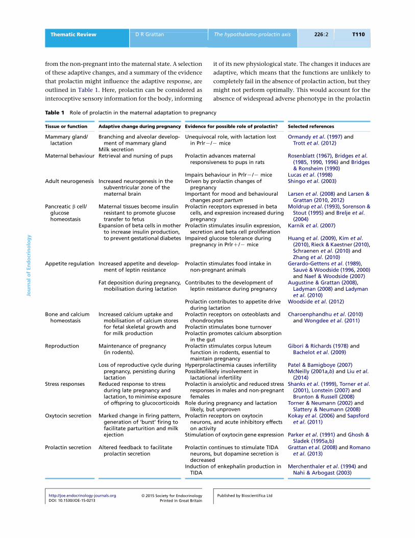
from the non-pregnant into the maternal state. A selection
of these adaptive changes, and a summary of the evidence
that prolactin might influence the adaptive response, are
outlined in Table 1. Here, prolactin can be considered as
interoceptive sensory information for the body, informing
it of its new physiological state. The changes it induces are
adaptive, which means that the functions are unlikely to
completely fail in the absence of prolactin action, but they
might not perform optimally. This would account for the
absence of widespread adverse phenotype in the prolactin
Table 1 Role of prolactin in the maternal adaptation to pregnancy
Tissue or function Adaptive change during pregnancy Evidence for possible role of prolactin? Selected references
Mammary gland/lactation
Branching and alveolar develop-ment of mammary gland
Unequivocal role, with lactation lostin PrlrK/K mice
Ormandy et al. (1997) andTrott et al. (2012)
Milk secretionMaternal behaviour Retrieval and nursing of pups Prolactin advances maternal
responsiveness to pups in ratsRosenblatt (1967), Bridges et al.
(1985, 1990, 1996) and Bridges& Ronsheim (1990)
Impairs behaviour in PrlrK/K mice Lucas et al. (1998)Adult neurogenesis Increased neurogenesis in the
subventricular zone of thematernal brain
Driven by prolactin changes ofpregnancy
Shingo et al. (2003)
Important for mood and behaviouralchanges post partum
Larsen et al. (2008) and Larsen &Grattan (2010, 2012)
Pancreatic b cell/glucosehomeostasis
Maternal tissues become insulinresistant to promote glucosetransfer to fetus
Prolactin receptors expressed in betacells, and expression increased duringpregnancy
Moldrup et al. (1993), Sorenson &Stout (1995) and Brelje et al.(2004)
Expansion of beta cells in motherto increase insulin production,to prevent gestational diabetes
Prolactin stimulates insulin expression,secretion and beta cell proliferation
Karnik et al. (2007)
Impaired glucose tolerance duringpregnancy in PrlrC/K mice
Huang et al. (2009), Kim et al.(2010), Rieck & Kaestner (2010),Schraenen et al. (2010) andZhang et al. (2010)
Appetite regulation Increased appetite and develop-ment of leptin resistance
Prolactin stimulates food intake innon-pregnant animals
Gerardo-Gettens et al. (1989),Sauve & Woodside (1996, 2000)and Naef & Woodside (2007)
Fat deposition during pregnancy,mobilisation during lactation
Contributes to the development ofleptin resistance during pregnancy
Augustine & Grattan (2008),Ladyman (2008) and Ladymanet al. (2010)
Prolactin contributes to appetite driveduring lactation
Woodside et al. (2012)
Bone and calciumhomeostasis
Increased calcium uptake andmobilisation of calcium storesfor fetal skeletal growth andfor milk production
Prolactin receptors on osteoblasts andchondrocytes
Charoenphandhu et al. (2010)and Wongdee et al. (2011)
Prolactin stimulates bone turnoverProlactin promotes calcium absorption
in the gutReproduction Maintenance of pregnancy
(in rodents).Prolactin stimulates corpus luteum
function in rodents, essential tomaintain pregnancy
Gibori & Richards (1978) andBachelot et al. (2009)
Loss of reproductive cycle duringpregnancy, persisting duringlactation
Hyperprolactinemia causes infertility Patel & Bamigboye (2007)Possible/likely involvement in
lactational infertilityMcNeilly (2001a,b) and Liu et al.
(2014)Stress responses Reduced response to stress
during late pregnancy andlactation, to minimise exposureof offspring to glucocorticoids
Prolactin is anxiolytic and reduced stressresponses in males and non-pregnantfemales
Shanks et al. (1999), Torner et al.(2001), Lonstein (2007) andBrunton & Russell (2008)
Role during pregnancy and lactationlikely, but unproven
Torner & Neumann (2002) andSlattery & Neumann (2008)
Oxytocin secretion Marked change in firing pattern,generation of ‘burst’ firing tofacilitate parturition and milkejection
Prolactin receptors on oxytocinneurons, and acute inhibitory effectson activity
Kokay et al. (2006) and Sapsfordet al. (2011)
Stimulation of oxytocin gene expression Parker et al. (1991) and Ghosh &Sladek (1995a,b)
Prolactin secretion Altered feedback to facilitateprolactin secretion
Prolactin continues to stimulate TIDAneurons, but dopamine secretion isdecreased
Grattan et al. (2008) and Romanoet al. (2013)
Induction of enkephalin production inTIDA
Merchenthaler et al. (1994) andNahi & Arbogast (2003)
Jou
rnal
of
En
do
crin
olo
gy
Thematic Review D R Grattan The hypothalamo-prolactin axis 226 :2 T110
http://joe.endocrinology-journals.orgDOI: 10.1530/JOE-15-0213
! 2015 Society for EndocrinologyPrinted in Great Britain
Published by Bioscientifica Ltd

receptor knockout mice. It is appropriate to point out that
many of the associations shown in Table 1 are, at this
stage, correlative only, and comprehensive investigation
proving that prolactin may be mediating a particular
adaptive change will require significant further work.
Nevertheless, we have found this to be a useful construct
for understanding why prolactin might be influencing
such a wide range of biological function. We used to be
concerned by the question of, ‘Why would there be over
300 physiological actions of prolactin?’ Now, we can
consider each of the different tissues that express the
prolactin receptor and ask the question, ‘Why might this
tissue need to change its function during lactation?’
Comprehensive reviews of the wide range of actions of
prolactin in the body are available elsewhere (Bole-Feysot
et al. 1998, Freeman et al. 2000), as is our hypothesis
regarding the role of prolactin in the physiological
adaptation to pregnancy (Grattan & Kokay 2008, Grattan
& LeTissier 2015), and hence, I will not go into detail here.
In the final section of this review, I would like to briefly
highlight three selected examples from recent research.
The first of these, the metabolic functions of prolactin,
nicely illustrates the context previously outlined that
prolactin is acting in a number of different tissues and cell
types in the mother to facilitate adaptation to pregnancy
or lactation. The second example, looking at prolactin
effects on fertility, highlights how functions of prolactin
that might be considered adaptive in a lactating female,
might be maladaptive should high prolactin occur at an
inappropriate time. The third example asks the question,
‘What is prolactin doing in the male?’ This example is used
to acknowledge the fact that some of the known functions
of prolactin do not comfortably fit into the conceptual
framework previously presented.
Metabolic function of prolactin
This is, perhaps, the best example of the pleiotropic role of
prolactin (defined to mean a single gene product, prolactin,
exerting multiple seemingly diverse actions). Prolactin
receptors are expressed on multiple tissues involved in
metabolic regulation, including adipose tissue, liver,
pancreas and the brain. It appears to play a broad role in
both pancreatic and adipose development. In adipose
tissue, prolactin is essential in adipogenesis and adipocyte
differentiation, as well as modulating lipid metabolism. It
also regulates the secretion is several adipokines, including
stimulation of leptin and inhibition of adiponectin
production (Ben-Jonathan et al. 2006, Carre & Binart
2014). In the pancreas, it promotes growth of islets during
development (Freemark et al. 2002), and increases insulin
expression and glucose-stimulated insulin secretion (Sinha
& Sorenson 1993, Brelje et al. 1994, 2004). It also increases
expression of glucose transporter 2 and promotes glucose
entry into the b-cells (Petryk et al. 2000), resulting in
enhanced activity of glucose-sensitive enzymes such as
glucokinase (Weinhaus et al. 2007). In both adipose tissue
and pancreas, these actions are likely to be profoundly
important during pregnancy and lactation. Lipid metab-
olism is altered, with lipid mobilisation from stores and
utilisation in mamary gland promoted by prolactin (Barber
et al. 1992). Adaptive changes in glucose homeostasis are
also important in pregnancy (Rieck & Kaestner 2010).
Maternal tissues develop insulin resistance to preferentially
direct glucose to the fetal/placental compartment (Herrera
2000), and to ensure the maternal tissues continue to
receive thenutrients required, there is increased demand for
maternal insulin secretion, and glucose-stimulated insulin
secretion increases. To adapt to this altered demand, there is
significant proliferation of b-cells in the islets (Parsons et al.
1992), enhanced insulin synthesis (Bone & Taylor 1976),
and decreased threshold for glucose-stimulated insulin
secretion (Sorenson & Parsons 1985), with prolactin
playing a critical adaptive role in promoting these changes
(Newbern & Freemark 2011). Failure of this adaptive res-
ponse results in gestational diabetes (Ramos-Roman 2011).
These peripheral actions of prolactin on metabolism
are complemented by CNS actions of prolactin to promote
appetite and potentially regulate glucose homeostasis.
Systemic prolactin administration increases food intake
in a variety of species (Moore et al. 1986, Gerardo-Gettens
et al. 1989, Noel & Woodside 1993, Buntin et al. 1999),
independent of potential effects on ovarian steroids (Noel
& Woodside 1993, 2007, Sauve & Woodside 1996). Thus,
the elevated prolactin secretion is likely to contribute to the
rapid increase in food intake during pregnancy (Shirley
1984, Ladyman & Grattan 2004, Ladyman et al. 2012) and
the extreme hyperphagia of lactation (Woodside 2007,
Woodside et al. 2012). Prolactin also induces functional
leptin resistance, which would contribute to increased food
intake (Naef & Woodside 2007, Augustine & Grattan 2008),
potentially mediating the well-established leptin resistance
of pregnancy (Grattan et al. 2007a, Augustine et al. 2008,
Ladyman 2008, Ladyman et al. 2010). Prolactin receptors
are found in many of the nuclei involved in the
homeostatic regulation of food intake, including the
arcuate, ventromedial and paraventricular nuclei
(Bakowska & Morrell 1997, Pi & Grattan 1998b, Brown
et al. 2010). However, prolactin receptors do not appear to
be expressed in the arcuate neuropeptide Y (NPY) and
Jou
rnal
of
En
do
crin
olo
gy
Thematic Review D R Grattan The hypothalamo-prolactin axis 226 :2 T111
http://joe.endocrinology-journals.orgDOI: 10.1530/JOE-15-0213
! 2015 Society for EndocrinologyPrinted in Great Britain
Published by Bioscientifica Ltd

proopiomelanocortin (POMC) neurons (Li et al. 1999,
Chen & Smith 2004, Kokay & Grattan 2005) that regulate
appetite. Hence, it seems likely that prolactin acts down-
stream of the arcuate neurons, such as at the paraventri-
cular nucleus. Consistent with this hypothesis, localised
injections of prolactin directly into the paraventricular
nucleus stimulate food intake in a dose-dependent manner
in female rats (Sauve & Woodside 2000).
Thus, seemingly diverse actions of prolactin in
multiple different cell types can be unified into a single
adaptive function, which is metabolic adaptation to
pregnancy, increasing energy availability to the mother
and offspring. This is an example of the conceptual
framework previously outlined, and it can be viewed as a
positive, adaptive mechanism. Should hyperprolactine-
mia occur at an inappropriate time, however, then one
might predict this could contribute to metabolic disorders.
There is some evidence for this. While it is not universally
observed, patients with hyperprolactinemia are prone to
excessive weight gain (Creemers et al. 1991, Delgrange
et al. 1999, Doknic et al. 2002, Baptista et al. 2004), and
normalisation of prolactin levels using dopamine agonists
is associated with weight loss (Greenman et al. 1998,
Doknic et al. 2002, Galluzzi et al. 2005). Interestingly,
genome-wide association studies have revealed that a
common variant adjacent to the prolactin gene is
associated with obesity (Meyre et al. 2009, Nilsson et al.
2011) suggesting that abnormalities in prolactin or
prolactin signalling may contribute to human obesity.
Hyperprolactinemia and infertility
Hyperprolactinemia causes infertility in both males and
females (Patel & Bamigboye 2007), and this provides an
even more clear-cut example of a potentially adaptive
function under certain conditions becoming clearly mala-
daptive in another situation. The mechanism by which
prolactin inhibits the reproductive axis is not clear, but
evidence suggests that prolactin impacts fertility through
actions on GnRH neurons. In humans, hyperprolactinae-
mia is associated with a marked reduction in both the
frequency and amplitude of LH pulses (Bohnet et al. 1976,
Matsuzaki et al. 1994) indicative of a change in GnRH
pulses, and the suppression of LH pulsatility can be reversed
by reducing serum prolactin concentrations to normal
(Moult et al. 1982). While prolactin could exert effects in
either the pituitary or gonad, pulsatile GnRH replacement
can reverse the infertility induced by hyperprolactinaemia
(Polson et al. 1986, Matsuzaki et al. 1994, Lecomte et al.
1997), suggesting a prolactin-induced suppression of GnRH
release is the proximal cause of infertility. Similarly,
prolactin suppresses both the frequency and amplitude of
LH pulses in male and female rats (Cohen-Becker et al. 1986,
Fox et al. 1987, Park & Selmanoff 1991, Park et al. 1993) and
measurements of GnRH secretion into the portal blood
have revealed prolactin-induced suppression of GnRH
release (Weber et al. 1983, Koike et al. 1984, 1991, Sarkar
et al. 1992). Furthermore, hyperprolactinaemia has been
shown to prevent the castration-induced increase in GnRH
mRNA expression in rats (Selmanoff et al. 1991). Thus,
although there is ample evidence that prolactin can act in
the pituitary gland to suppress LH secretion (Smith 1978,
1982, Cheung 1983, Morel et al. 1994, Tortonese et al.
1998), in animal models, as in clinical studies, the primary
cause of infertility appears to be the suppression of the
activity of GnRH neurons. This effect of prolactin is unlikely
to be mediated directly by an action on GnRH neurons, as
the majority of these neurons do not express the prolactin
receptor (Grattan et al. 2007b, Kokay et al. 2011). Thus,
prolactin-induced inhibition of GnRH neurons must
involve prolactin-sensitive afferents to these cells. Interest-
ingly, most prolactin responsive neurons also express ERa
(Furigo et al. 2014), so prolactin may share a common
mechanism of regulating GnRH with the negative feedback
pathway mediated by estradiol. As such, kisspeptin neurons
have emerged as the most likely intermediate regulators.
Since first being identified as essential for puberty
in humans (de Roux et al. 2003, Seminara et al. 2003),
kisspeptin neurons are now recognised as critical parts of
the circuit regulating activity of the GnRH neurons that
control fertility (Pinilla et al. 2012). Kisspeptin neurons
may have an important role in mediating the suppressive
effect of prolactin on fertility. Kisspeptin is the most
potent stimulator of GnRH neuronal activity yet identified
(Han et al. 2005, Liu et al. 2008). In most mammalian
species, there are two populations of kisspeptin neurons,
with kisspeptin neurons in the rostral periventricular area
of the third ventricle (RP3V) playing an essential role
in enabling ovulation in rodents by activating GnRH
neurons (Herbison 2008, Clarkson & Herbison 2009,
Oakley et al. 2009), while kisspeptin neurons in the
arcuate nucleus are thought to be involved in the
regulation of the basal pulsatile secretion of GnRH
(Li et al. 2009, Roseweir et al. 2009, Lehman et al. 2010,
Navarro et al. 2011). Prolactin receptors are expressed
in the majority of kisspeptin neurons in both populations
(Kokay et al. 2011, Li et al. 2011), and prolactin has
recently been shown to induce the phosphorylation of
signal transducer and activator of transcription 5 (pSTAT5)
in arcuate nucleus kisspeptin neurons in the rat
Jou
rnal
of
En
do
crin
olo
gy
Thematic Review D R Grattan The hypothalamo-prolactin axis 226 :2 T112
http://joe.endocrinology-journals.orgDOI: 10.1530/JOE-15-0213
! 2015 Society for EndocrinologyPrinted in Great Britain
Published by Bioscientifica Ltd

(Araujo-Lopes et al. 2014) and rostral hypothalamic
kisspeptin neurons in the mouse (Brown et al. 2014).
Chronic infusion of prolactin in female mice abolished
estrous cyclicity and suppressed global Gnrh and Kiss1
mRNA expression in the hypothalamus, while kisspeptin
therapy restored estrous cycles in hyperprolactinemic
mice (Sonigo et al. 2012), consistent with the hypothesis
that prolactin-induced suppression of GnRH secretion is
mediated by an inhibition of kisspeptin neurons.
Clearly, under most conditions, hyperprolactinemia
represents a pathological condition with adverse con-
sequences. During pregnancy and lactation, however,
hyperprolactinemia is physiologically appropriate. When
viewed from this context, an inhibitory action of prolactin
on fertility during pregnancy and lactation would be
highly adaptive, allowing the mother to focus energy on
feeding her offspring, before investing resources in a
further pregnancy (Valeggia & Ellison 2009). Lactation is
associated with a period of infertility in most mammalian
females, including women (McNeilly 2001a). In humans,
this function serves as a critical regulator of population
growth, spacing the timing of births to allow the mother
to ration her metabolic investment across sequential
pregnancies (Short 1976). Despite the extensive impact
on mammalian reproductive physiology, our understand-
ing of the mechanisms mediating lactational infertility
remains incomplete (McNeilly 1994, 2001a,b). It is clear
that suckling is the critical inhibitory signal (Tsukamura &
Maeda 2001), and the principle cause of infertility is an
almost-complete suppression of the pulsatile secretion of
GnRH from the hypothalamus, the consequent loss of
pituitary gonadotropin secretion and ovulation failure
(Fox & Smith 1984). The pathways linking the suckling
stimulus to the suppression of ovulation, however, are
unclear. Kiss1 mRNA and protein levels are reduced in the
arcuate nucleus of lactating rats associated with the
suppression of pulsatile GnRH secretion during lactation
(Yamada et al. 2007, 2012, Smith et al. 2010, True et al.
2011, Araujo-Lopes et al. 2014, Ladyman & Woodside
2014), and in both populations in the mouse (Brown et al.
2014). The RP3V kisspeptin population is harder to study
in rats (Yamada et al. 2007, Desroziers et al. 2010), with
reports of both Kiss1 mRNA remaining unchanged during
lactation (Yamada et al. 2007) and of kisspeptin protein
increasing while Kiss1 mRNA labeling decreased during
lactation (Smith et al. 2010). More importantly, we have
also shown that the reduction in kisspeptin expression
results in complete loss of capacity for these neurons to
activate GnRH neurons, even if they are stimulated
exogenously (Liu et al. 2014). Given the similarity between
the effect of suckling and the effect of prolactin, and the
knowledge that suckling stimulates prolactin secretion, it
seems likely that elevated prolactin during pregnancy and
lactation contributes to the infertility of lactation, but this
remains to be proven, and the relative roles of prolactin
and/or suckling may be different in different species.
What does prolactin do in the male?
The hypothesis that most prolactin actions in the body can
be tied to the adaptation to pregnancy and lactation clearly
does not explain effects of prolactin in the male. Up to 40%
of the male pituitary gland is dedicated to lactotrophs,
suggesting that some function is retained in males, but
knockout studies havenot identified anessential functionof
prolactin. While there is no male equivalent of lactation,
many of the other functions of prolactin in females can also
be observed in males. For example, as in females, prolactin
seems to be involved in parental behaviour in males,
although the relative role that the mammalian father plays
in parental care of offspring varies amongst species. In
species where the male plays some role in rearing of the
offspring, including humans (Gordon et al. 2010, Gettler
et al. 2012), studies have found a association between
prolactin and paternal care (Schradin & Anzenberger
1999), although the overall picture is unclear and con-
troversial (Schradin 2007, Wynne-Edwards & Timonin
2007). Nevertheless, paternal recognition of offspring is
consistent amongst most species. Pup-contact by male rats
can lead to some forms of parental care behaviour and this is
associated with an increase in serum prolactin, as well as
increased expression of the long-form of the prolactin
receptor in the brain (Sakaguchi et al. 1996). Further
evidence for a direct role for prolactin in paternal recog-
nition of offspring has been shown by studies of PrlrK/K
fathers, who fail to distinguish adult offspring from non-
offspring, possibly as a result of failure of prolactin-induced
neurogensis in the sub-ventricular zone and the dentate
gyrus (Mak & Weiss 2010).
As in females, pathological hyperprolactinemia causes
infertility in males, but it is not clear that there is an adaptive
role for prolactin in male reproduction. At lower levels,
prolactin contributes a range of functions in the male
reproductive tract, revealed by subtle reproductivedeficits in
the prolactin receptor deficient mice (Grattan & LeTissier
2015). In addition, many of the metabolic and immune
functions ofprolactin can be observed inmales, but whether
prolactin levels are ever sufficient for these effects to be of
physiological significance is uncertain. Perhaps the most
consistent stimulus for prolactin secretion in males is stress,
Jou
rnal
of
En
do
crin
olo
gy
Thematic Review D R Grattan The hypothalamo-prolactin axis 226 :2 T113
http://joe.endocrinology-journals.orgDOI: 10.1530/JOE-15-0213
! 2015 Society for EndocrinologyPrinted in Great Britain
Published by Bioscientifica Ltd
but the functional consequences of this response are not
well-understood (Gala 1990).
Conclusion
While Harris was correct in proposing that the brain
controls prolactin secretion, the hypothalamo-prolactin
axis proved itself to be quite different from all other
pituitary systems. It remains the most complex and
versatile of all of the hypothalamo-pituitary axes. Even if
we just consider the relatively simple task of controlling
milk production during lactation, there is much that
remains to be understood, such as the possible role of one
or more prolactin-releasing factors during the suckling
stimulus, and the mechanism controlling the loss of
dopamine production in the TIDA neurons and the
changes in prolactin negative feedback. If we include
the wide range of additional functions of prolactin, then
the complexity becomes overwhelming. I have presented
here a context to attempt to understand the pleiotropic
roles of prolactin, arguing that many of the functions of
prolactin can be unified into the overall task of maternal
adaptation to pregnancy and lactation. Within this
context, prolactin function promotes adaptive changes
in a variety of body systems, but such actions can also be
maladaptive, in a different context, if hyperprolactinemia
occurs at an inappropriate time. This theoretical construct
presents many new opportunities for generating testable
hypotheses about prolactin function. But there are also
many functions that do not fit easily into this construct,
providing further opportunities for expanding our under-
standing. I anticipate that the coming availability of novel
tools for investigating prolactin function, including gene-
targeting approaches that allow conditional regulation of
prolactin responsive cells, will provide the impetus for a
new wave of research to enhance our understanding of
this fascinating system. Sixty years on from Geoffrey
Harris’ prescient predictions, we still have a lot of work to
do to understand the hypothalamo-prolactin system.
Footnote
This paper is part of a thematic review section on 60 years of
neuroendocrinology. The Guest Editors for this section were Ashley
Grossman and Clive Coen.
Declaration of interest
The author declares that there is no conflict of interest that could be
perceived as prejudicing the impartiality of this review.
Funding
Prof. D R Grattan is supported by research grants from the New Zealand
Health Research Council and the Marsden Fund of the Royal Society of
New Zealand.
ReferencesAllen DL, Low MJ, Allen RG & Ben-Jonathan N 1995 Identification of two
classes of prolactin-releasing factors in intermediate lobe tumors from
transgenic mice. Endocrinology 136 3093–3099. (doi:10.1210/endo.136.
7.7789336)
Anderson GM, Beijer P, Bang AS, Fenwick MA, Bunn SJ & Grattan DR 2006a
Suppression of prolactin-induced signal transducer and activator of
transcription 5b signaling and induction of suppressors of cytokine
signaling messenger ribonucleic acid in the hypothalamic arcuate
nucleus of the rat during late pregnancy and lactation. Endocrinology
147 4996–5005. (doi:10.1210/en.2005-0755)
Anderson ST, Barclay JL, Fanning KJ, Kusters DH, Waters MJ & Curlewis JD
2006b Mechanisms underlying the diminished sensitivity to prolactin
negative feedback during lactation: reduced STAT5 signalling and
upregulation of cytokine-inducible SH2-domain-containing protein
(CIS) expression in tuberoinfundibular dopaminergic neurons.
Endocrinology 147 1195–1202. (doi:10.1210/en.2005-0905)
Anderson GM, Kieser DC, Steyn FJ & Grattan DR 2008 Hypothalamic
prolactin receptor messenger ribonucleic acid levels, prolactin
signaling, and hyperprolactinemic inhibition of pulsatile luteinizing
hormone secretion are dependent on estradiol. Endocrinology 149
1562–1570. (doi:10.1210/en.2007-0867)
Andrews ZB 2005 Neuroendocrine regulation of prolactin secretion during
late pregnancy: easing the transition into lactation. Journal of Neuro-
endocrinology 17 466–473. (doi:10.1111/j.1365-2826.2005.01327.x)
Andrews ZB & Grattan DR 2004 The roles of dopamine and the
neurointermediate lobe of the pituitary in the regulation of prolactin
secretion during late pregnancy in rats. Journal of Neuroendocrinology 16
859–865. (doi:10.1111/j.1365-2826.2004.01241.x)
Andrews ZB, Kokay IC & Grattan DR 2001 Dissociation of prolactin
secretion from tuberoinfundibular dopamine activity in late pregnant
rats. Endocrinology 142 2719–2724. (doi:10.1210/endo.142.6.8196)
Annunziato L & Moore KE 1978 Prolactin in CSF selectively increases
dopamine turnover in the median eminence. Life Sciences 22
2037–2042. (doi:10.1016/0024-3205(78)90551-9)
Araujo-Lopes R, Crampton JR, Aquino NS, Miranda RM, Kokay IC, Reis AM,
Franci CR, Grattan DR & Szawka RE 2014 Prolactin regulates kisspeptin
neurons in the arcuate nucleus to suppress LH secretion in female rats.
Endocrinology 155 1010–1020. (doi:10.1210/en.2013-1889)
Arbogast LA & Ben-Jonathan N 1990 The preovulatory prolactin surge is
prolonged by a progesterone-dependent dopaminergic mechanism.
Endocrinology 126 246–252. (doi:10.1210/endo-126-1-246)
Arbogast LA & Voogt JL 1991a Hyperprolactinemia increases and
hypoprolactinemia decreases tyrosine hydroxylase messenger
ribonucleic acid levels in the arcuate nuclei, but not the substantia
nigra or zona incerta. Endocrinology 128 997–1005. (doi:10.1210/
endo-128-2-997)
Arbogast LA & Voogt JL 1991b Mechanisms of tyrosine hydroxylase
regulation during pregnancy: evidence for protein dephosphorylation
during the prolactin surges. Endocrinology 129 2575–2582.
(doi:10.1210/endo-129-5-2575)
Arbogast LA & Voogt JL 1993 Progesterone reverses the estradiol-induced
decrease in tyrosine hydroxylase mRNA levels in the arcuate nucleus.
Neuroendocrinology 58 501–510. (doi:10.1159/000126583)
Arbogast LA & Voogt JL 1994 Progesterone suppresses tyrosine hydroxylase
messenger ribonucleic acid levels in the arcuate nucleus on proestrus.
Endocrinology 135 343–350. (doi:10.1210/endo.135.1.7912184)
Jou
rnal
of
En
do
crin
olo
gy
Thematic Review D R Grattan The hypothalamo-prolactin axis 226 :2 T114
http://joe.endocrinology-journals.orgDOI: 10.1530/JOE-15-0213
! 2015 Society for EndocrinologyPrinted in Great Britain
Published by Bioscientifica Ltd
Arbogast LA & Voogt JL 1997 Prolactin (PRL) receptors are colocalized in
dopaminergic neurons in fetal hypothalamic cell cultures: effect of
PRL on tyrosine hydroxylase activity. Endocrinology 138 3016–3023.
(doi:10.1210/endo.138.7.5227)
Arey BJ & Freeman ME 1990 Oxytocin, vasoactive-intestinal peptide, and
serotonin regulate the mating-induced surges of prolactin secretion in
the rat. Endocrinology 126 279–284. (doi:10.1210/endo-126-1-279)
Arey BJ & Freeman ME 1992a Activity of oxytocinergic neurons in the
paraventricular nucleus mirrors the periodicity of the endogenous
stimulatory rhythm regulating prolactin secretion. Endocrinology 130
126–132. (doi:10.1210/endo.130.1.1727695)
Arey BJ & Freeman ME 1992b Activity of vasoactive intestinal peptide and
serotonin in the paraventricular nucleus reflects the periodicity of the
endogenous stimulatory rhythm regulating prolactin secretion.
Endocrinology 131 736–742. (doi:10.1210/endo.131.2.1639019)
Arey BJ, Averill RL & Freeman ME 1989 A sex-specific endogenous
stimulatory rhythm regulating prolactin secretion. Endocrinology 124
119–123. (doi:10.1210/endo-124-1-119)
Arey BJ, Kanyicska B & Freeman ME 1991 The endogenous stimulatory
rhythm regulating prolactin secretion is present in the lactating rat.
Neuroendocrinology 53 35–40. (doi:10.1159/000125694)
Arimura A, Dunn JD & Schally AV 1972 Effect of infusion of hypothalamic
extracts on serum prolactin levels in rats treated with nembutal, CNS
depressants or bearing hypothalamic lesions. Endocrinology 90 378–383.
(doi:10.1210/endo-90-2-378)
Arita J & Kimura F 1987 Direct inhibitory effect of long term estradiol
treatment on dopamine synthesis in tuberoinfundibular dopaminergic
neurons: in vitro studies using hypothalamic slices. Endocrinology 121
692–698. (doi:10.1210/endo-121-2-692)
Asa SL, Kelly MA, Grandy DK & Low MJ 1999 Pituitary lactotroph
adenomas develop after prolonged lactotroph hyperplasia in dopamine
D2 receptor-deficient mice. Endocrinology 140 5348–5355.
(doi:10.1210/endo.140.11.7118)
Augustine RA & Grattan DR 2008 Induction of central leptin resistance in
hyperphagic pseudopregnant rats by chronic prolactin infusion.
Endocrinology 149 1049–1055. (doi:10.1210/en.2007-1018)
Augustine RA, Kokay IC, Andrews ZB, Ladyman SR & Grattan DR 2003
Quantitation of prolactin receptor mRNA in the maternal rat brain
during pregnancy and lactation. Journal of Molecular Endocrinology 31
221–232. (doi:10.1677/jme.0.0310221)
Augustine RA, Ladyman SR & Grattan DR 2008 From feeding one to feeding
many: hormone-induced changes in bodyweight homeostasis during
pregnancy. Journal of Physiology 586 387–397. (doi:10.1113/jphysiol.
2007.146316)
Averill RL, Grattan DR & Norris SK 1991 Posterior pituitary
lobectomy chronically attenuates the nocturnal surge of prolactin
in early pregnancy. Endocrinology 128 705–709. (doi:10.1210/endo-
128-2-705)
Bachelot A, Beaufaron J, Servel N, Kedzia C, Monget P, Kelly PA, Gibori G &
Binart N 2009 Prolactin independent rescue of mouse corpus luteum
life span: identification of prolactin and luteinizing hormone target
genes. American Journal of Physiology. Endocrinology and Metabolism 297
E676–E684.
Bakowska JC & Morrell JI 1997 Atlas of the neurons that express mRNA for
the long form of the prolactin receptor in the forebrain of the female
rat. Journal of Comparative Neurology 386 161–177. (doi:10.1002/
(SICI)1096-9861(19970922)386:2!161::AID-CNE1O3.0.CO;2-)
Baptista T, de Baptista EA, Lalonde J, Plamondon J, Kin NM, Beaulieu S,
Joober R & Richard D 2004 Comparative effects of the antipsychotics
sulpiride and risperidone in female rats on energy balance, body
composition, fat morphology and macronutrient selection. Progress
in Neuro-Psychopharmacology & Biological Psychiatry 28 1305–1311.
(doi:10.1016/j.pnpbp.2004.08.001)
Barber MC, Clegg RA, Finley E, Vernon RG & Flint DJ 1992 The role of
growth hormone, prolactin and insulin-like growth factors in the
regulation of rat mammary gland and adipose tissue metabolism during
lactation. Journal of Endocrinology 135 195–202. (doi:10.1677/joe.0.
1350195)
Barraclough CA & Sawyer CH 1959 Induction of pseudopregnancy in the
rat by reserpine and chlorpromazine. Endocrinology 65 563–571.
(doi:10.1210/endo-65-4-563)
Becquet D, Boyer B, Rasolonjanahary R, Brue T, Guillen S, Moreno M,
Franc JL & Francois-Bellan AM 2014 Evidence for an internal and
functional circadian clock in rat pituitary cells. Molecular and Cellular
Endocrinology 382 888–898. (doi:10.1016/j.mce.2013.11.004)
Ben-Jonathan N & Hnasko R 2001 Dopamine as a prolactin (PRL) inhibitor.
Endocrine Reviews 22 724–763. (doi:10.1210/edrv.22.6.0451)
Ben-Jonathan N, Oliver C, Weiner HJ, Mical RS & Porter JC 1977 Dopamine
in hypophysial portal plasma of the rat during the estrous cycle and
throughout pregnancy. Endocrinology 100 452–458. (doi:10.1210/
endo-100-2-452)
Ben-Jonathan N, Neill MA, Arbogast LA, Peters LL & Hoefer MT 1980
Dopamine in hypophysial portal blood: relationship to circulating
prolactin in pregnant and lactating rats. Endocrinology 106 690–696.
(doi:10.1210/endo-106-3-690)
Ben-Jonathan N, Hugo ER, Brandebourg TD & LaPensee CR 2006 Focus on
prolactin as a metabolic hormone. Trends in Endocrinology and
Metabolism 17 110–116. (doi:10.1016/j.tem.2006.02.005)
Ben-Jonathan N, Lapensee CR & Lapensee EW 2008 What can we learn
from rodents about prolactin in humans? Endocrine Reviews 29 1–41.
(doi:10.1210/er.2007-0017)
Berghorn KA, Le WW, Sherman TG & Hoffman GE 2001 Suckling stimulus
suppresses messenger RNA for tyrosine hydroxylase in arcuate neurons
during lactation. Journal of Comparative Neurology 438 423–432.
(doi:10.1002/cne.1325)
Bjorklund A, Moore RY, Nobin A & Stenevi U 1973 The organization of
tubero-hypophyseal and reticulo-infundibular catecholamine neuron
systems in the rat brain. Brain Research 51 171–191. (doi:10.1016/
0006-8993(73)90371-5)
Blum M, McEwen BS & Roberts JL 1987 Transcriptional analysis of tyrosine
hydroxylase gene expression in the tuberoinfundibular dopaminergic
neurons of the rat arcuate nucleus after estrogen treatment. Journal of
Biological Chemistry 262 817–821.
Bohnet HG, Dahlen HG, Wuttke W & Schneider HP 1976 Hyperprolacti-
nemic anovulatory syndrome. Journal of Clinical Endocrinology and
Metabolism 42 132–143. (doi:10.1210/jcem-42-1-132)
Bole-Feysot C, Goffin V, Edery M, Binart N & Kelly PA 1998 Prolactin (PRL)
and its receptor: actions, signal transduction pathways and phenotypes
observed in PRL receptor knockout mice. Endocrine Reviews 19 225–268.
(doi:10.1210/edrv.19.3.0334)
Bone AJ & Taylor KW 1976 Metabolic adaptation to pregnancy shown by
increased biosynthesis of insulin in islets of Langerhans isolated from
pregnant rat. Nature 262 501–502. (doi:10.1038/262501a0)
Borgundvaag B, Kudlow JE, Mueller SG & George SR 1992 Dopamine
receptor activation inhibits estrogen-stimulated transforming growth
factor-a gene expression and growth in anterior pituitary, but not in
uterus. Endocrinology 130 3453–3458. (doi:10.1210/endo.130.6.
1534540)
Bosse R, Fumagalli F, Jaber M, Giros B, Gainetdinov RR, Wetsel WC,
Missale C & Caron MG 1997 Anterior pituitary hypoplasia and
dwarfism in mice lacking the dopamine transporter. Neuron 19
127–138. (doi:10.1016/S0896-6273(00)80353-0)
Brelje TC, Parsons JA & Sorenson RL 1994 Regulation of islet b-cell
proliferation by prolactin in rat islets. Diabetes 43 263–273.
(doi:10.2337/diab.43.2.263)
Brelje TC, Stout LE, Bhagroo NV & Sorenson RL 2004 Distinctive roles for
prolactin and growth hormone in the activation of signal transducer
and activator of transcription 5 in pancreatic islets of Langerhans.
Endocrinology 145 4162–4175. (doi:10.1210/en.2004-0201)
Bridges RS & Ronsheim PM 1990 Prolactin (PRL) regulation of maternal
behavior in rats: bromocriptine treatment delays and PRL promotes the
Jou
rnal
of
En
do
crin
olo
gy
Thematic Review D R Grattan The hypothalamo-prolactin axis 226 :2 T115
http://joe.endocrinology-journals.orgDOI: 10.1530/JOE-15-0213
! 2015 Society for EndocrinologyPrinted in Great Britain
Published by Bioscientifica Ltd
rapid onset of behavior. Endocrinology 126 837–848. (doi:10.1210/
endo-126-2-837)
Bridges RS, DiBiase R, Loundes DD & Doherty PC 1985 Prolactin
stimulation of maternal behavior in female rats. Science 227 782–784.
(doi:10.1126/science.3969568)
Bridges RS, Numan M, Ronsheim PM, Mann PE & Lupini CE 1990
Central prolactin infusions stimulate maternal behavior in
steroid-treated, nulliparous female rats. PNAS 87 8003–8007.
(doi:10.1073/pnas.87.20.8003)
Bridges RS, Robertson MC, Shiu RPC, Friesen HG, Stuer AM & Mann PE
1996 Endocrine communication between conceptus and mother:
placental lactogen stimulation of maternal behavior.
Neuroendocrinology 64 57–64. (doi:10.1159/000127098)
Brown RS, Kokay IC, Herbison AE & Grattan DR 2010 Distribution of
prolactin-responsive neurons in the mouse forebrain. Journal of
Comparative Neurology 518 92–102. (doi:10.1002/cne.22208)
Brown RS, Piet R, Herbison AE & Grattan DR 2012 Differential actions of
prolactin on electrical activity and intracellular signal transduction in
hypothalamic neurons. Endocrinology 153 2375–2384. (doi:10.1210/
en.2011-2005)
Brown RS, Herbison AE & Grattan DR 2014 Prolactin regulation of
kisspeptin neurones in the mouse brain and its role in the
lactation-induced suppression of kisspeptin expression. Journal of
Neuroendocrinology 26 898–908. (doi:10.1111/jne.12223)
Brunton PJ & Russell JA 2008 The expectant brain: adapting for
motherhood. Nature Reviews. Neuroscience 9 11–25.
Buntin JD, Hnasko RM & Zuzick PH 1999 Role of the ventromedial
hypothalamus in prolactin-induced hyperphagia in ring doves. Physi-
ology & Behavior 66 255–261. (doi:10.1016/S0031-9384(98)00288-1)
Carre N & Binart N 2014 Prolactin and adipose tissue. Biochimie 97 16–21.
(doi:10.1016/j.biochi.2013.09.023)
Charoenphandhu N, Wongdee K & Krishnamra N 2010 Is prolactin the
cardinal calciotropic maternal hormone? Trends in Endocrinology and
Metabolism 21 395–401. (doi:10.1016/j.tem.2010.02.002)
Chen P & Smith MS 2004 Regulation of hypothalamic neuropeptide Y
messenger ribonucleic acid expression during lactation: role of
prolactin. Endocrinology 145 823–829. (doi:10.1210/en.2003-1255)
Cheung CY 1983 Prolactin suppresses luteinizing hormone secretion and
pituitary responsiveness to luteinizing hormone-releasing hormone by
a direct action at the anterior pituitary. Endocrinology 113 632–638.
(doi:10.1210/endo-113-2-632)
Clarkson J & Herbison AE 2009 Oestrogen, kisspeptin, GPR54 and the
pre-ovulatory luteinising hormone surge. Journal of Neuroendocrinology
21 305–311. (doi:10.1111/j.1365-2826.2009.01835.x)
Cohen-Becker IR, Selmanoff M & Wise PM 1986 Hyperprolactinemia alters
the frequency and amplitude of pulsatile luteinizing hormone
secretion in the ovariectomized rat. Neuroendocrinology 42 328–333.
(doi:10.1159/000124459)
Cowie AT, Tindal JS & Benson GK 1960 Pituitary grafts and milk secretion
in hypophysectomized rats. Journal of Endocrinology 21 115–123.
(doi:10.1677/joe.0.0210115)
Cramer OM, Parker CR Jr & Porter JC 1979 Estrogen inhibition of dopamine
release into hypophysial portal blood. Endocrinology 104 419–422.
(doi:10.1210/endo-104-2-419)
Creemers LB, Zelissen PM, van ’t Verlaat JW & Koppeschaar HP 1991
Prolactinoma and body weight: a retrospective study. Acta
Endocrinologica 125 392–396. (doi:10.1530/acta.0.1250392)
Crowley WR 2015 Neuroendocrine regulation of lactation and milk
production. Comprehensive Physiology 5 255–291. (doi:10.1002/cphy.
c140029)
Cservenak M, Bodnar I, Usdin TB, Palkovits M, Nagy GM & Dobolyi A 2010
Tuberoinfundibular peptide of 39 residues is activated during lactation
and participates in the suckling-induced prolactin release in rat.
Endocrinology 151 5830–5840. (doi:10.1210/en.2010-0767)
Cusan L, Dupont A, Kledzik GS, Labrie F, Coy DH & Schally AV 1977 Potent
prolactin and growth hormone releasing activity of more analogues of
Met-enkephalin. Nature 268 544–547. (doi:10.1038/268544a0)
De Greef WJ & Neill JD 1979 Dopamine levels in hypophysial stalk
plasma of the rat during surges of prolactin secretion induced by
cervical stimulation. Endocrinology 105 1093–1099. (doi:10.1210/
endo-105-5-1093)
De Lean A & Labrie F 1977 Sensitizing effect of treatment with estrogens on
TSH response to TRH in male rats. American Journal of Physiology 233
E235–E239.
Delgrange E, Donckier J & Maiter D 1999 Hyperprolactinaemia as a
reversible cause of weight gain in male patients? Clinical Endocrinology
50 271. (doi:10.1046/j.1365-2265.1999.00700.x)
Demarest KT, McKay DW, Riegle GD & Moore KE 1983 Biochemical indices
of tuberoinfundibular dopaminergic neuronal activity during lacta-
tion: a lack of response to prolactin. Neuroendocrinology 36 130–137.
(doi:10.1159/000123449)
Demarest KT, Riegle GD & Moore KE 1984a Prolactin-induced activation of
tuberoinfundibular dopaminergic neurons: evidence for both a rapid
‘tonic’ and a delayed ‘induction’ component. Neuroendocrinology 38
467–475. (doi:10.1159/000123935)
Demarest KT, Riegle GD & Moore KE 1984b Long-term treatment with
estradiol induces reversible alterations in tuberoinfundibular
dopaminergic neurons: a decreased responsiveness to prolactin.
Neuroendocrinology 39 193–200. (doi:10.1159/000123979)
Demarest KT, Riegle GD & Moore KE 1986 The rapid ‘tonic’ and the delayed
‘induction’ components of the prolactin-induced activation of
tuberoinfundibular dopaminergic neurons following the systemic
administration of prolactin. Neuroendocrinology 43 291–299.
(doi:10.1159/000124543)
DeMaria JE, Livingstone JD & Freeman ME 1998 Characterization of
the dopaminergic input to the pituitary gland throughout the
estrous cycle of the rat. Neuroendocrinology 67 377–383. (doi:10.1159/
000054336)
DeMaria JE, Lerant AA & Freeman ME 1999 Prolactin activates all three
populations of hypothalamic neuroendocrine dopaminergic neurons
in ovariectomized rats. Brain Research 837 236–241. (doi:10.1016/
S0006-8993(99)01667-4)
DeMaria JE, Livingstone JD & Freeman ME 2000 Ovarian steroids influence
the activity of neuroendocrine dopaminergic neurons. Brain Research
879 139–147. (doi:10.1016/S0006-8993(00)02763-3)
Dempsey EW & Uotila UU 1940 The effect of pituitary stalk section upon
reproductive phenomena in the female rat. Endocrinology 27 573–579.
(doi:10.1210/endo-27-4-573)
Desroziers E, Mikkelsen J, Simonneaux V, Keller M, Tillet Y, Caraty A &
Franceschini I 2010 Mapping of kisspeptin fibres in the brain of the
pro-oestrous rat. Journal of Neuroendocrinology 22 1101–1112.
(doi:10.1111/j.1365-2826.2010.02053.x)
Dobolyi A 2011 Novel potential regulators of maternal adaptations during
lactation: tuberoinfundibular peptide 39 and amylin. Journal of
Neuroendocrinology 23 1002–1008. (doi:10.1111/j.1365-2826.2011.
02127.x)
Dobolyi A, Dimitrov E, Palkovits M & Usdin TB 2012 The neuroendocrine
functions of the parathyroid hormone 2 receptor. Frontiers in
Endocrinology 3 121. (doi:10.3389/fendo.2012.00121)
Doknic M, Pekic S, Zarkovic M, Medic-Stojanoska M, Dieguez C,
Casanueva F & Popovic V 2002 Dopaminergic tone and obesity:
an insight from prolactinomas treated with bromocriptine. European
Journal of Endocrinology/European Federation of Endocrine Societies 147
77–84. (doi:10.1530/eje.0.1470077)
Eikenburg DC, Ravitz AJ, Gudelsky GA & Moore KE 1977 Effects of estrogen
on prolactin and tuberoinfundibular dopaminergic neurons. Journal of
Neural Transmission 40 235–244. (doi:10.1007/BF01257017)
Elsholtz HP, Lew AM, Albert PR & Sundmark VC 1991 Inhibitory control of
prolactin and Pit-1 gene promoters by dopamine. Dual signaling
Jou
rnal
of
En
do
crin
olo
gy
Thematic Review D R Grattan The hypothalamo-prolactin axis 226 :2 T116
http://joe.endocrinology-journals.orgDOI: 10.1530/JOE-15-0213
! 2015 Society for EndocrinologyPrinted in Great Britain
Published by Bioscientifica Ltd
pathways required for D2 receptor-regulated expression of the prolactin
gene. Journal of Biological Chemistry 266 22919–22925.
Enjalbert A, Ruberg M, Arancibia S, Priam M & Kordon C 1979 Endogenous
opiates block dopamine inhibition of prolactin secretion in vitro.
Nature 280 595–597. (doi:10.1038/280595a0)
Everett JW 1954 Luteotrophic function of autographs of the rat
hypophysis. Endocrinology 54 685–690. (doi:10.1210/endo-54-6-685)
Feher P, Olah M, Bodnar I, Hechtl D, Bacskay I, Juhasz B, Nagy GM &
Vecsernyes M 2010 Dephosphorylation/inactivation of tyrosine
hydroxylase at the median eminence of the hypothalamus is required
for suckling-induced prolactin and adrenocorticotrop hormone
responses. Brain Research Bulletin 82 141–145. (doi:10.1016/j.brain-
resbull.2010.02.006)
Fink G 1988 Oestrogen and progesterone interactions in the control of
gonadotrophin and prolactin secretion. Journal of Steroid Biochemistry
30 169–178. (doi:10.1016/0022-4731(88)90090-8)
Fliestra RJ & Voogt JL 1997 Lactogenic hormones of the placenta and
pituitary inhibit suckling-induced prolactin (PRL) release but not the
ante-partum PRL surge. Proceedings of the Society for Experimental Biology
and Medicine 214 258–264. (doi:10.3181/00379727-214-44094)
Fox SR & Smith MS 1984 The suppression of pulsatile luteinizing hormone
secretion during lactation in the rat. Endocrinology 115 2045–2051.
(doi:10.1210/endo-115-6-2045)
Fox SR, Hoefer MT, Bartke A & Smith MS 1987 Suppression of pulsatile LH
secretion, pituitary GnRH receptor content and pituitary responsive-
ness to GnRH by hyperprolactinemia in the male rat. Neuroendo-
crinology 46 350–359. (doi:10.1159/000124844)
Fox SR, Harlan RE, Shivers BD & Pfaff DW 1990 Chemical characterization
of neuroendocrine targets for progesterone in the female rat brain and
pituitary. Neuroendocrinology 51 276–283. (doi:10.1159/000125350)
Freeman ME, Kanyicska B, Lerant A & Nagy G 2000 Prolactin: structure,
function, and regulation of secretion. Physiological Reviews 80
1523–1631.
Freemark M, Avril I, Fleenor D, Driscoll P, Petro A, Opara E, Kendall W,
Oden J, Bridges S, Binart N et al. 2002 Targeted deletion of the PRL
receptor: effects on islet development, insulin production, and
glucose tolerance. Endocrinology 143 1378–1385. (doi:10.1210/endo.
143.4.8722)
Furigo IC, Kim KW, Nagaishi VS, Ramos-Lobo AM, de Alencar A, Pedroso JA,
Metzger M & Donato J Jr 2014 Prolactin-sensitive neurons express
estrogen receptor-a and depend on sex hormones for normal respon-
siveness to prolactin. Brain Research 1566 47–59. (doi:10.1016/
j.brainres.2014.04.018)
Fuxe K 1963 Cellular localisations of monoamines in the median eminence
and in the infundibular stem of some mammals. Acta Physiologica
Scandinavica 58 383–384. (doi:10.1111/j.1748-1716.1963.tb02662.x)
Fuxe K 1964 Cellular localization of monoamines in the median eminence
and the infundibular stem of some mammals. Zeitschrift fur Zell-
forschung und Mikroskopische Anatomie. Abteilung Histochemie 61
710–724.
Gala RR 1990 The physiology and mechanisms of the stress-induced
changes in prolactin secretion in the rat. Life Sciences 46 1407–1420.
(doi:10.1016/0024-3205(90)90456-2)
Galluzzi F, Salti R, Stagi S, La Cauza F & Chiarelli F 2005 Reversible weight
gain and prolactin levels – long-term follow-up in childhood. Journal of
Pediatric Endocrinology & Metabolism 18 921–924. (doi:10.1515/JPEM.
2005.18.9.921)
Gerardo-Gettens T, Moore BJ, Stern JS & Horwitz BA 1989 Prolactin
stimulates food intake in a dose-dependent manner. American Journal of
Physiology 256 R276–R280.
Gettler LT, McDade TW, Feranil AB & Kuzawa CW 2012 Prolactin,
fatherhood, and reproductive behavior in human males. American
Journal of Physical Anthropology 148 362–370. (doi:10.1002/ajpa.22058)
Ghosh R & Sladek CD 1995a Prolactin modulates oxytocin mRNA during
lactation by its action on the hypothalamo-neurohypophyseal axis.
Brain Research 672 24–28.
Ghosh R & Sladek CD 1995b Role of prolactin and gonadal steroids in
regulation of oxytocin mRNA during lactation. American Journal of
Physiology 269 E76–E84.
Gibbs DM & Neill JD 1978 Dopamine levels in hypophysial stalk blood in
the rat are sufficient to inhibit prolactin secretion in vivo. Endocrinology
102 1895–1900. (doi:10.1210/endo-102-6-1895)
Gibori G & Richards JS 1978 Dissociation of two distinct luteotropic effects
of prolactin: regulation of luteinizing hormone-receptor content and
progesterone secretion during pregnancy. Endocrinology 102 767–774.
Goffin V, Binart N, Touraine P & Kelly PA 2002 Prolactin: the new biology
of an old hormone. Annual Review of Physiology 64 47–67. (doi:10.1146/
annurev.physiol.64.081501.131049)
Gordon I, Zagoory-Sharon O, Leckman JF & Feldman R 2010 Prolactin,
oxytocin, and the development of paternal behavior across the first six
months of fatherhood. Hormones and Behavior 58 513–518.
(doi:10.1016/j.yhbeh.2010.04.007)
Goudreau JL, Lindley SE, Lookingland KJ & Moore KE 1992 Evidence that
hypothalamic periventricular dopamine neurons innervate the inter-
mediate lobe of the rat pituitary. Neuroendocrinology 56 100–105.
(doi:10.1159/000126214)
Grattan DR 2001 The actions of prolactin in the brain during pregnancy
and lactation. Progress in Brain Research 133 153–171. (doi:10.1016/
S0079-6123(01)33012-1)
Grattan DR & Averill RL 1990 Effect of ovarian steroids on a nocturnal surge
of prolactin secretion that precedes parturition in the rat. Endocrinology
126 1199–1205. (doi:10.1210/endo-126-2-1199)
Grattan DR & Averill RL 1991 Intrahypothalamic pituitary grafts elevate
prolactin in the cerebrospinal fluid and attenuate prolactin release
following ether stress. Proceedings of the Society for Experimental Biology
and Medicine 196 42–46. (doi:10.3181/00379727-196-43161)
Grattan DR & Averill RL 1995 Absence of short-loop autoregulation of
prolactin during late pregnancy in the rat. Brain Research Bulletin 36
413–416. (doi:10.1016/0361-9230(94)00216-N)
Grattan DR & Kokay IC 2008 Prolactin: a pleiotropic neuroendocrine
hormone. Journal of Neuroendocrinology 20 752–763. (doi:10.1111/
j.1365-2826.2008.01736.x)
Grattan DR & LeTissier P 2015 Hypothalamic control of prolactin secretion,
and the multiple reproductive functions of prolactin. In Knobil and
Neill’s Physiology of Reproduction, 4th edn, pp 469–526. Eds TM Plant &
AJ Zelesnik. Amsterdam: Elsevier.
Grattan DR, Xu J, McLachlan MJ, Kokay IC, Bunn SJ, Hovey RC & Davey
HW 2001 Feedback regulation of PRL secretion is mediated by the
transcription factor, signal transducer, and activator of transcription
5b. Endocrinology 142 3935–3940. (doi:10.1210/endo.142.9.8385)
Grattan DR, Ladyman SR & Augustine RA 2007a Hormonal induction of
leptin resistance during pregnancy. Physiology & Behavior 91 366–374.
(doi:10.1016/j.physbeh.2007.04.005)
Grattan DR, Jasoni CL, Liu X, Anderson GM & Herbison AE 2007b Prolactin
regulation of gonadotropin-releasing hormone neurons to suppress
luteinizing hormone secretion in mice. Endocrinology 148 4344–4351.
(doi:10.1210/en.2007-0403)
Grattan DR, Steyn FJ, Kokay IC, Anderson GM & Bunn SJ 2008 Pregnancy-
induced adaptation in the neuroendocrine control of prolactin
secretion. Journal of Neuroendocrinology 20 497–507. (doi:10.1111/j.
1365-2826.2008.01661.x)
Greenman Y, Tordjman K & Stern N 1998 Increased body weight associated
with prolactin secreting pituitary adenomas: weight loss with
normalization of prolactin levels. Clinical Endocrinology 48 547–553.
(doi:10.1046/j.1365-2265.1998.00403.x)
Gregerson KA 2003 Functional expression of the dopamine-activated K(C)
current in lactotrophs during the estrous cycle in female rats:
correlation with prolactin secretory responses. Endocrine 20 67–74.
(doi:10.1385/ENDO:20:1-2:67)
Gregerson KA 2006 Prolactin: structure, function, and regulation of
secretion. In Knobil and Neill’s Physiology of Reproduction, pp 1703–1726.
Ed. JD Neill. Amsterdam: Elsevier.
Jou
rnal
of
En
do
crin
olo
gy
Thematic Review D R Grattan The hypothalamo-prolactin axis 226 :2 T117
http://joe.endocrinology-journals.orgDOI: 10.1530/JOE-15-0213
! 2015 Society for EndocrinologyPrinted in Great Britain
Published by Bioscientifica Ltd
Gregerson KA, Golesorkhi N & Chuknyiska R 1994 Stimulation of prolactin
release by dopamine withdrawal: role of membrane hyperpolarization.
American Journal of Physiology 267 E781–E788.
Gudelsky GA & Porter JC 1979 Release of newly synthesized dopamine
into the hypophysial portal vasculature of the rat. Endocrinology 104
583–587. (doi:10.1210/endo-104-3-583)
Gunnet JW & Freeman ME 1983 The mating-induced release of prolactin:
a unique neuroendocrine response. Endocrine Reviews 4 44–61.
(doi:10.1210/edrv-4-1-44)
Han SK, Gottsch ML, Lee KJ, Popa SM, Smith JT, Jakawich SK, Clifton DK,
Steiner RA & Herbison AE 2005 Activation of gonadotropin-releasing
hormone neurons by kisspeptin as a neuroendocrine switch for the
onset of puberty. Journal of Neuroscience 25 11349–11356. (doi:10.1523/
JNEUROSCI.3328-05.2005)
Harris GW 1948 Neural control of the pituitary gland. Physiological Reviews
28 139–179.
Hentges ST & Low MJ 2002 Ovarian dependence for pituitary tumori-
genesis in D2 dopamine receptor-deficient mice. Endocrinology 143
4536–4543. (doi:10.1210/en.2002-220421)
Herbison AE 2008 Estrogen positive feedback to gonadotropin-releasing
hormone (GnRH) neurons in the rodent: the case for the rostral
periventricular area of the third ventricle (RP3V). Brain Research Reviews
57 277–287. (doi:10.1016/j.brainresrev.2007.05.006)
Herrera E 2000 Metabolic adaptations in pregnancy and their implications
for the availability of substrates to the fetus. European Journal of Clinical
Nutrition 54(Suppl 1) S47–S51. (doi:10.1038/sj.ejcn.1600984)
Hnasko R, Khurana S, Shackleford N, Steinmetz R, Low MJ & Ben-Jonathan
N 1997 Two distinct pituitary cell lines from mouse intermediate lobe
tumors: a cell that produces prolactin-regulating factor and a
melanotroph. Endocrinology 138 5589–5596. (doi:10.1210/endo.138.
12.5656)
Hokfelt T & Fuxe K 1972 Effects of prolactin and ergot alkaloids on the
tubero-infundibular dopamine (DA) neurons. Neuroendocrinology 9
100–122. (doi:10.1159/000122042)
Holzbauer M & Racke K 1985 The dopaminergic innervation of the
intermediate lobe and of the neural lobe of the pituitary gland. Medical
Biology 63 97–116.
Horseman ND 1995 Prolactin, proliferation, and protooncogenes.
Endocrinology 136 5249–5251. (doi:10.1210/endo.136.12.7588267)
Hovey RC, Trott JF, Ginsburg E, Goldhar A, Sasaki MM, Fountain SJ,
Sundararajan K & Vonderhaar BK 2001 Transcriptional and spatio-
temporal regulation of prolactin receptor mRNA and cooperativity with
progesterone receptor function during ductal branch growth in the
mammary gland. Developmental Dynamics 222 192–205. (doi:10.1002/
dvdy.1179)
Huang C, Snider F & Cross JC 2009 Prolactin receptor is required for normal
glucose homeostasis and modulation of b-cell mass during pregnancy.
Endocrinology 150 1618–1626. (doi:10.1210/en.2008-1003)
Huerta-Ocampo I, Christian HC, Thompson NM, El-Kasti MM & Wells T
2005 The Intermediate lactotroph: a morphologically distinct, ghrelin-
responsive pituitary cell in the dwarf (dw/dw) rat. Endocrinology 146
5012–5023. (doi:10.1210/en.2005-0335)
Ishida M, Mitsui T, Yamakawa K, Sugiyama N, Takahashi W, Shimura H,
Endo T, Kobayashi T & Arita J 2007 Involvement of cAMP response
element-binding protein in the regulation of cell proliferation and the
prolactin promoter of lactotrophs in primary culture. American
Journal of Physiology. Endocrinology and Metabolism 293 E1529–E1537.
(doi:10.1152/ajpendo.00028.2007)
Johnston CA, Demarest KT & Moore KE 1980 Cycloheximide disrupts the
prolactin-mediated stimulation of dopamine synthesis in tuberoin-
fundibular neurons. Brain Research 195 236–240. (doi:10.1016/0006-
8993(80)90883-5)
Jones EE & Naftolin F 1990 Estrogen effects on the tuberoinfundibular
dopaminergic system in the female rat brain. Brain Research 510 84–91.
(doi:10.1016/0006-8993(90)90730-Y)
Kamberi IA, Mical RS & Porter JC 1970 Prolactin-inhibiting activity in
hypophysial stalk blood and elevation by dopamine. Experientia 26
1150–1151. (doi:10.1007/BF02112730)
Kanematsu S & Sawyer CH 1973 Elevation of plasma prolactin after
hypophysial stalk section in the rat. Endocrinology 93 238–241.
(doi:10.1210/endo-93-1-238)
Kanematsu S, Hilliard J & Sawyer CH 1963 Effect of reserpine on pituitary
prolactin content and its hypothalamic site of action in the rabbit.
Acta Endocrinologica 44 467–474. (doi:10.1530/acta.0.0440467)
Kansra S, Yamagata S, Sneade L, Foster L & Ben-Jonathan N 2005
Differential effects of estrogen receptor antagonists on pituitary
lactotroph proliferation and prolactin release. Molecular and Cellular
Endocrinology 239 27–36. (doi:10.1016/j.mce.2005.04.008)
Kansra S, Chen S, Bangaru ML, Sneade L, Dunckley JA & Ben-Jonathan N
2010 Selective estrogen receptor down-regulator and selective estrogen
receptor modulators differentially regulate lactotroph proliferation.
PLoS ONE 5 e10060. (doi:10.1371/journal.pone.0010060)
Karnik SK, Chen H, McLean GW, Heit JJ, Gu X, Zhang AY, Fontaine M,
Yen MH & Kim SK 2007 Menin controls growth of pancreatic b-cells in
pregnant mice and promotes gestational diabetes mellitus. Science 318
806–809. (doi:10.1126/science.1146812)
Kelly MA, Rubinstein M, Asa SL, Zhang G, Saez C, Bunzow JR, Allen RG,
Hnasko R, Ben-Jonathan N, Grandy DK et al. 1997 Pituitary
lactotroph hyperplasia and chronic hyperprolactinemia in dopamine
D2 receptor-deficient mice. Neuron 19 103–113. (doi:10.1016/
S0896-6273(00)80351-7)
Kim H, Toyofuku Y, Lynn FC, Chak E, Uchida T, Mizukami H, Fujitani Y,
Kawamori R, Miyatsuka T, Kosaka Y et al. 2010 Serotonin regulates
pancreatic b cell mass during pregnancy. Nature Medicine 16 804–808.
(doi:10.1038/nm.2173)
Koike K, Aono T, Miyake A, Tasaka K, Chatani F & Kurachi K 1984 Effect of
pituitary transplants on the LH-RH concentrations in the medial basal
hypothalamus and hypophyseal portal blood. Brain Research 301
253–258. (doi:10.1016/0006-8993(84)91093-X)
Koike K, Miyake A, Aono T, Sakumoto T, Ohmichi M, Yamaguchi M &
Tanizawa O 1991 Effect of prolactin on the secretion of hypothalamic
GnRH and pituitary gonadotropins. Hormone Research 35 5–12.
(doi:10.1159/000181921)
Kokay IC & Grattan DR 2005 Expression of mRNA for prolactin
receptor (long form) in dopamine and pro-opiomelanocortin
neurones in the arcuate nucleus of non-pregnant and lactating rats.
Journal of Neuroendocrinology 17 827–835. (doi:10.1111/j.1365-2826.
2005.01374.x)
Kokay IC, Bull PM, Davis RL, Ludwig M & Grattan DR 2006 Expression of
the long form of the prolactin receptor in magnocellular oxytocin
neurons is associated with specific prolactin regulation of oxytocin
neurons. American Journal of Physiology: Regulatory, Integrative and
Comparative Physiology 290 R1216–R1225.
Kokay IC, Petersen SL & Grattan DR 2011 Identification of prolactin-
sensitive GABA and kisspeptin neurons in regions of the rat
hypothalamus involved in the control of fertility. Endocrinology 152
526–535. (doi:10.1210/en.2010-0668)
Ladyman SR 2008 Leptin resistance during pregnancy in the rat.
Journal of Neuroendocrinology 20 269–277. (doi:10.1111/j.1365-2826.
2007.01628.x)
Ladyman SR & Grattan DR 2004 Region-specific reduction in leptin-
induced phosphorylation of signal transducer and activator of
transcription-3 (STAT3) in the rat hypothalamus is associated with
leptin resistance during pregnancy. Endocrinology 145 3704–3711.
(doi:10.1210/en.2004-0338)
Ladyman SR & Woodside B 2014 Food restriction during lactation
suppresses Kiss1 mRNA expression and kisspeptin-stimulated LH
release in rats. Reproduction 147 743–751. (doi:10.1530/REP-13-0426)
Ladyman SR, Augustine RA & Grattan DR 2010 Hormone interactions
regulating energy balance during pregnancy. Journal of Neuro-
endocrinology 22 805–817. (doi:10.1111/j.1365-2826.2010.02017.x)
Jou
rnal
of
En
do
crin
olo
gy
Thematic Review D R Grattan The hypothalamo-prolactin axis 226 :2 T118
http://joe.endocrinology-journals.orgDOI: 10.1530/JOE-15-0213
! 2015 Society for EndocrinologyPrinted in Great Britain
Published by Bioscientifica Ltd
Ladyman SR, Fieldwick DM & Grattan DR 2012 Suppression of leptin-
induced hypothalamic JAK/STAT signalling and feeding response
during pregnancy in the mouse. Reproduction 144 83–90. (doi:10.1530/
REP-12-0112)
Larsen CM & Grattan DR 2010 Prolactin-induced mitogenesis in the
subventricular zone of the maternal brain during early pregnancy is
essential for normal postpartum behavioral responses in the mother.
Endocrinology 151 3805–3814. (doi:10.1210/en.2009-1385)
Larsen CM & Grattan DR 2012 Prolactin, neurogenesis, and maternal
behaviors. Brain, Behavior, and Immunity 26 201–209. (doi:10.1016/
j.bbi.2011.07.233)
Larsen CM, Kokay IC & Grattan DR 2008 Male pheromones initiate
prolactin-induced neurogenesis and advance maternal behavior in
female mice. Hormones and Behavior 53 509–517. (doi:10.1016/j.yhbeh.
2007.11.020)
Lecomte P, Lecomte C, Lansac J, Gallier J, Sonier CB & Simonetta C 1997
Pregnancy after intravenous pulsatile gonadotropin-releasing hormone
in a hyperprolactinaemic woman resistant to treatment with dopamine
agonists. European Journal of Obstetrics, Gynecology, and Reproductive
Biology 74 219–221. (doi:10.1016/S0301-2115(97)00091-2)
Lehman MN, Coolen LM & Goodman RL 2010 Minireview: Kisspeptin/
neurokinin B/dynorphin (KNDy) cells of the arcuate nucleus: a central
node in the control of gonadotropin-releasing hormone secretion.
Endocrinology 151 3479–3489. (doi:10.1210/en.2010-0022)
Lerant A & Freeman ME 1998 Ovarian steroids differentially regulate the
expression of prolactin receptors in neuroendocrine dopaminergic
neuron populations – a double-label confocal microscopic study.
Brain Research 802 141–154. (doi:10.1016/S0006-8993(98)00583-6)
Li C, Chen P & Smith MS 1999 Neuropeptide Y and tuberoinfundibular
dopamine activities are altered during lactation: role of prolactin.
Endocrinology 140 118–123. (doi:10.1210/endo.140.1.6437)
Li XF, Kinsey-Jones JS, Cheng Y, Knox AM, Lin Y, Petrou NA, Roseweir A,
Lightman SL, Milligan SR, Millar RP et al. 2009 Kisspeptin signalling in
the hypothalamic arcuate nucleus regulates GnRH pulse generator
frequency in the rat. PLoS ONE 4 e8334. (doi:10.1371/journal.pone.
0008334)
Li Q, Rao A, Pereira A, Clarke IJ & Smith JT 2011 Kisspeptin cells in the
ovine arcuate nucleus express prolactin receptor but not melatonin
receptor. Journal of Neuroendocrinology 23 871–882. (doi:10.1111/
j.1365-2826.2011.02195.x)
Lieberman ME, Maurer RA, Claude P, Wiklund J, Wertz N & Gorski J 1981
Regulation of pituitary growth and prolactin gene expression by
estrogen. Advances in Experimental Medicine and Biology 138 151–163.
Lincoln GA, Andersson H & Hazlerigg D 2003 Clock genes and the long-
term regulation of prolactin secretion: evidence for a photoperiod/
circannual timer in the pars tuberalis. Journal of Neuroendocrinology 15
390–397. (doi:10.1046/j.1365-2826.2003.00990.x)
Liu B & Arbogast LA 2008 Phosphorylation state of tyrosine hydroxylase in
the stalk-median eminence is decreased by progesterone in cycling
female rats. Endocrinology 149 1462–1469. (doi:10.1210/en.2007-1345)
Liu B & Arbogast LA 2010 Progesterone decreases tyrosine hydroxylase
phosphorylation state and increases protein phosphatase 2A activity
in the stalk-median eminence on proestrous afternoon. Journal of
Endocrinology 204 209–219. (doi:10.1677/JOE-09-0335)
Liu X, Lee K & Herbison AE 2008 Kisspeptin excites gonadotropin-releasing
hormone neurons through a phospholipase C/calcium-dependent
pathway regulating multiple ion channels. Endocrinology 149
4605–4614. (doi:10.1210/en.2008-0321)
Liu X, Brown RS, Herbison AE & Grattan DR 2014 Lactational anovulation
in mice results from a selective loss of kisspeptin input to GnRH
neurons. Endocrinology 155 193–203. (doi:10.1210/en.2013-1621)
Lledo PM, Legendre P, Israel JM & Vincent JD 1990 Dopamine inhibits
two characterized voltage-dependent calcium currents in identified
rat lactotroph cells. Endocrinology 127 990–1001. (doi:10.1210/
endo-127-3-990)
Login IS & MacLeod RM 1977 Prolactin in human and rat serum and
cerebrospinal fluid. Brain Research 132 477–483. (doi:10.1016/
0006-8993(77)90196-2)
Lonstein JS & Blaustein JD 2004 Immunocytochemical investigation of
nuclear progestin receptor expression within dopaminergic neurones
of the female rat brain. Journal of Neuroendocrinology 16 534–543.
(doi:10.1111/j.1365-2826.2004.01198.x)
Lonstein JS 2007 Regulation of anxiety during the postpartum period.
Frontiers in Neuroendocrinology 28 115–141.
Lookingland KJ, Jarry HD & Moore KE 1987 The metabolism of dopamine
in the median eminence reflects the activity of tuberoinfundibular
neurons. Brain Research 419 303–310. (doi:10.1016/0006-
8993(87)90597-X)
Lucas BK, Ormandy CJ, Binart N, Bridges RS & Kelly PA 1998 Null mutation
of the prolactin receptor gene produces a defect in maternal behavior.
Endocrinology 139 4102–4107. (doi:10.1210/endo.139.10.6243)
Lyons DJ, Horjales-Araujo E & Broberger C 2010 Synchronized network
oscillations in rat tuberoinfundibular dopamine neurons: switch to
tonic discharge by thyrotropin-releasing hormone. Neuron 65 217–229.
(doi:10.1016/j.neuron.2009.12.024)
Lyons DJ, Hellysaz A & Broberger C 2012 Prolactin regulates tuberoinfun-
dibular dopamine neuron discharge pattern: novel feedback control
mechanisms in the lactotrophic axis. Journal of Neuroscience 32
8074–8083. (doi:10.1523/JNEUROSCI.0129-12.2012)
Ma FY, Grattan DR, Goffin V & Bunn SJ 2005 Prolactin-regulated tyrosine
hydroxylase activity and messenger ribonucleic acid expression in
mediobasal hypothalamic cultures: the differential role of specific
protein kinases. Endocrinology 146 93–102. (doi:10.1210/en.2004-0800)
MacLeod RM, Fontham EH & Lehmeyer JE 1970 Prolactin and growth
hormone production as influenced by catecholamines and agents that
affect brain catecholamines. Neuroendocrinology 6 283–294.
(doi:10.1159/000121933)
Mak GK & Weiss S 2010 Paternal recognition of adult offspring mediated by
newly generated CNS neurons. Nature Neuroscience 13 753–758.
(doi:10.1038/nn.2550)
Mansour A, Meador-Woodruff JH, Bunzow JR, Civelli O, Akil H & Watson SJ
1990 Localization of dopamine D2 receptor mRNA and D1 and D2
receptor binding in the rat brain and pituitary: an in situ hybridization-
receptor autoradiographic analysis. Journal of Neuroscience 10 2587–2600.
Martinez de la Escalera G & Weiner RI 1992 Dissociation of dopamine from
its receptor as a signal in the pleiotropic hypothalamic regulation of
prolactin secretion. Endocrine Reviews 13 241–255. (doi:10.1210/edrv-
13-2-241)
Matsumoto M, Hidaka K, Tada S, Tasaki Y & Yamaguchi T 1995 Full-length
cDNA cloning and distribution of human dopamine D4 receptor.
Brain Research. Molecular Brain Research 29 157–162. (doi:10.1016/0169-
328X(94)00245-A)
Matsuzaki T, Azuma K, Irahara M, Yasui T & Aono T 1994 Mechanism of
anovulation in hyperprolactinemic amenorrhea determined by pulsa-
tile gonadotropin-releasing hormone injection combined with human
chorionic gonadotropin. Fertility and Sterility 62 1143–1149.
Maurer RA 1982 Estradiol regulates the transcription of the prolactin gene.
Journal of Biological Chemistry 257 2133–2136.
McNeilly AS 1994 Suckling and the control of gonadotropin secretion. In
The Physiology of Reproduction, 2nd edn, pp 1179–1212. Eds E Knobil &
JD Neill. New York: Raven Press.
McNeilly AS 2001a Lactational control of reproduction. Reproduction,
Fertility, and Development 13 583–590. (doi:10.1071/RD01056)
McNeilly AS 2001b Neuroendocrine changes and fertility in breast-feeding
women. Progress in Brain Research 133 207–214.
Merchenthaler I 1993 Induction of enkephalin in tuberoinfundibular
dopaminergic neurons during lactation. Endocrinology 133 2645–2651.
(doi:10.1210/endo.133.6.7694844)
Merchenthaler I 1994 Induction of enkephalin in tuberoinfundibular
dopaminergic neurons of pregnant, pseudopregnant, lactating and aged
female rats. Neuroendocrinology 60 185–193. (doi:10.1159/000126750)
Jou
rnal
of
En
do
crin
olo
gy
Thematic Review D R Grattan The hypothalamo-prolactin axis 226 :2 T119
http://joe.endocrinology-journals.orgDOI: 10.1530/JOE-15-0213
! 2015 Society for EndocrinologyPrinted in Great Britain
Published by Bioscientifica Ltd
Meyre D, Delplanque J, Chevre JC, Lecoeur C, Lobbens S, Gallina S,
Durand E, Vatin V, Degraeve F, Proenca C et al. 2009 Genome-wide
association study for early-onset and morbid adult obesity identifies
three new risk loci in European populations. Nature Genetics 41
157–159. (doi:10.1038/ng.301)
Moldrup A, Petersen ED & Nielsen JH 1993 Effects of sex and pregnancy
hormones on growth hormone and prolactin receptor gene expression
in insulin-producing cells. Endocrinology 133 1165–1172. (doi:10.1210/
endo.133.3.8365359)
Moore BJ, Gerardo-Gettens T, Horwitz BA & Stern JS 1986 Hyperprolacti-
nemia stimulates food intake in the female rat. Brain Research Bulletin
17 563–569. (doi:10.1016/0361-9230(86)90226-1)
Morel G, Ouhtit A & Kelly PA 1994 Prolactin receptor immunoreactivity in
rat anterior pituitary. Neuroendocrinology 59 78–84. (doi:10.1159/
000126641)
Morel GR, Caron RW, Console GM, Soaje M, Sosa YE, Rodriguez SS, Jahn
GA & Goya RG 2009 Estrogen inhibits tuberoinfundibular dopamin-
ergic neurons but does not cause irreversible damage. Brain Research
Bulletin 80 347–352. (doi:10.1016/j.brainresbull.2009.08.026)
Morrell JI, Rosenthal MF, McCabe JT, Harrington CA, Chikaraishi DM &
Pfaff DW 1989 Tyrosine hydroxylase mRNA in the neurons of the
tuberoinfundibular region and zona incerta examined after gonadal
steroid hormone treatment. Molecular Endocrinology 3 1426–1433.
(doi:10.1210/mend-3-9-1426)
Moult PJ, Rees LH & Besser GM 1982 Pulsatile gonadotrophin secretion in
hyperprolactinaemic amenorrhoea an the response to bromocriptine
therapy. Clinical Endocrinology 16 153–162. (doi:10.1111/j.1365-2265.
1982.tb03159.x)
Murai I & Ben-Jonathan N 1987 Posterior pituitary lobectomy abolishes the
suckling-induced rise in prolactin (PRL): evidence for a PRL-releasing
factor in the posterior pituitary. Endocrinology 121 205–211.
(doi:10.1210/endo-121-1-205)
Murai I & Ben-Jonathan N 1990 Acute stimulation of prolactin release by
estradiol: mediation by the posterior pituitary. Endocrinology 126
3179–3184. (doi:10.1210/endo-126-6-3179)
Murai I, Reichlin S & Ben-Jonathan N 1989 The peak phase of the
proestrous prolactin surge is blocked by either pituitary lobectomy or
antisera to vasoactive intestinal peptide. Endocrinology 124 1050–1055.
(doi:10.1210/endo-124-2-1050)
Naef L & Woodside B 2007 Prolactin/leptin interactions in the control of
food intake in rats. Endocrinology 148 5977–5983. (doi:10.1210/en.
2007-0442)
Nahi F & Arbogast LA 2003 Prolactin modulates hypothalamic prepro-
enkephalin, but not proopiomelanocortin, gene expression during
lactation. Endocrine 20 115–122. (doi:10.1385/ENDO:20:1-2:115)
Navarro VM, Castellano JM, McConkey SM, Pineda R, Ruiz-Pino F, Pinilla L,
Clifton DK, Tena-Sempere M & Steiner RA 2011 Interactions between
kisspeptin and neurokinin B in the control of GnRH secretion in the
female rat. American Journal of Physiology. Endocrinology and Metabolism
300 E202–E210. (doi:10.1152/ajpendo.00517.2010)
Newbern D & Freemark M 2011 Placental hormones and the control of
maternal metabolism and fetal growth. Current Opinion in Endocrinology,
Diabetes, and Obesity 18 409–416. (doi:10.1097/MED.
0b013e32834c800d)
Nicholson G, Greeley GH Jr, Humm J, Youngblood WW & Kizer JS 1980
Prolactin in cerebrospinal fluid: a probable site of prolactin auto-
regulation. Brain Research 190 447–457. (doi:10.1016/0006-
8993(80)90287-5)
Nikitovitch-Winer M & Everett JW 1958 Functional restitution of pituitary
grafts re-transplanted from kidney to median eminence. Endocrinology
63 916–930. (doi:10.1210/endo-63-6-916)
Nilsson L, Olsson AH, Isomaa B, Groop L, Billig H & Ling C 2011 A common
variant near the PRL gene is associated with increased adiposity in
males. Molecular Genetics and Metabolism 102 78–81. (doi:10.1016/
j.ymgme.2010.08.017)
Noel MB & Woodside B 1993 Effects of systemic and central prolactin
injections on food intake, weight gain, and estrous cyclicity
in female rats. Physiology & Behavior 54 151–154. (doi:10.1016/0031-
9384(93)90057-M)
Nolan LA & Levy A 2009 The trophic effects of oestrogen on male rat
anterior pituitary lactotrophs. Journal of Neuroendocrinology 21 457–464.
(doi:10.1111/j.1365-2826.2009.01864.x)
Oakley AE, Clifton DK & Steiner RA 2009 Kisspeptin signaling in the brain.
Endocrine Reviews 30 713–743. (doi:10.1210/er.2009-0005)
Ormandy CJ, Binart N & Kelly PA 1997 Mammary gland development in
prolactin receptor knockout mice. Journal of Mammary Gland Biology
and Neoplasia 2 355–364. (doi:10.1023/A:1026395229025)
Park SK & Selmanoff M 1991 Dose-dependent suppression of postcastration
luteinizing hormone secretion exerted by exogenous prolactin
administration in male rats: a model for studying hyperprolactinemic
hypogonadism. Neuroendocrinology 53 404–410. (doi:10.1159/
000125748)
Park SK, Keenan MW & Selmanoff M 1993 Graded hyperprolactinemia first
suppresses LH pulse frequency and then pulse amplitude in castrated
male rats. Neuroendocrinology 58 448–453. (doi:10.1159/000126575)
Parker SL, Armstrong WE, Sladek CD, Grosvenor CE & Crowley WR 1991
Prolactin stimulates the release of oxytocin in lactating rats: evidence
for a physiological role via an action at the neural lobe. Neuroendo-
crinology 53 503–510.
Parsons JA, Brelje TC & Sorenson RL 1992 Adaptation of islets of
Langerhans to pregnancy: increased islet cell proliferation and insulin
secretion correlates with the onset of placental lactogen secretion.
Endocrinology 130 1459–1466. (doi:10.1210/endo.130.3.1537300)
Pasqualini C, Guibert B & Leviel V 1993 Short-term inhibitory effect of
estradiol on tyrosine hydroxylase activity in tuberoinfundibular
dopaminergic neurons in vitro. Journal of Neurochemistry 60 1707–1713.
(doi:10.1111/j.1471-4159.1993.tb13394.x)
Pasteels JL 1963 Morphological and experimental research on prolactin
secretion. Archives de Biologie 74 439–553.
Patel SS & Bamigboye V 2007 Hyperprolactinaemia. Journal of Obstetrics and
Gynaecology 27 455–459. (doi:10.1080/01443610701406125)
Peters LL, Hoefer MT & Ben-Jonathan N 1981 The posterior pituitary:
regulation of anterior pituitary prolactin secretion. Science 213
659–661. (doi:10.1126/science.7256264)
Petryk A, Fleenor D, Driscoll P & Freemark M 2000 Prolactin induction of
insulin gene expression: the roles of glucose and glucose transporter-2.
Journal of Endocrinology 164 277–286. (doi:10.1677/joe.0.1640277)
Pi XJ & Grattan DR 1998a Differential expression of the two forms of
prolactin receptor mRNA within microdissected hypothalamic nuclei
of the rat. Brain Research. Molecular Brain Research 59 1–12.
(doi:10.1016/S0169-328X(98)00109-0)
Pi XJ & Grattan DR 1998b Distribution of prolactin receptor immuno-
reactivity in the brain of estrogen-treated, ovariectomized rats.
Journal of Comparative Neurology 394 462–474. (doi:10.1002/(SICI)
1096-9861(19980518)394:4!462::AID-CNE5O3.0.CO;2-)
Pinilla L, Aguilar E, Dieguez C, Millar RP & Tena-Sempere M 2012
Kisspeptins and reproduction: physiological roles and regulatory
mechanisms. Physiological Reviews 92 1235–1316. (doi:10.1152/
physrev.00037.2010)
van den Pol AN, Herbst RS & Powell JF 1984 Tyrosine hydroxylase-
immunoreactive neurons of the hypothalamus: a light and electron
microscopic study. Neuroscience 13 1117–1156. (doi:10.1016/
0306-4522(84)90292-6)
Polson DW, Sagle M, Mason HD, Adams J, Jacobs HS & Franks S 1986
Ovulation and normal luteal function during LHRH treatment of
women with hyperprolactinaemic amenorrhoea. Clinical Endocrinology
24 531–537. (doi:10.1111/j.1365-2265.1986.tb03282.x)
Ramos-Roman MA 2011 Prolactin and lactation as modifiers of diabetes
risk in gestational diabetes. Hormone and Metabolic Research 43 593–600.
(doi:10.1055/s-0031-1284353)
Jou
rnal
of
En
do
crin
olo
gy
Thematic Review D R Grattan The hypothalamo-prolactin axis 226 :2 T120
http://joe.endocrinology-journals.orgDOI: 10.1530/JOE-15-0213
! 2015 Society for EndocrinologyPrinted in Great Britain
Published by Bioscientifica Ltd
Raymond V, Beaulieu M, Labrie F & Boissier J 1978 Potent antidopami-
nergic activity of estradiol at the pituitary level on prolactin release.
Science 200 1173–1175. (doi:10.1126/science.418505)
Rieck S & Kaestner KH 2010 Expansion of b-cell mass in response to
pregnancy. Trends in Endocrinology and Metabolism 21 151–158.
(doi:10.1016/j.tem.2009.11.001)
Romano N, Yip SH, Hodson DJ, Guillou A, Parnaudeau S, Kirk S, Tronche F,
Bonnefont X, Le Tissier P, Bunn SJ et al. 2013 Plasticity of hypothalamic
dopamine neurons during lactation results in dissociation of electrical
activity and release. Journal of Neuroscience 33 4424–4433. (doi:10.1523/
JNEUROSCI.4415-12.2013)
Rosenblatt JS 1967 Nonhormonal basis of maternal behavior in the rat.
Science 156 1512–1514. (doi:10.1126/science.156.3781.1512)
Roseweir AK, Kauffman AS, Smith JT, Guerriero KA, Morgan K, Pielecka-
Fortuna J, Pineda R, Gottsch ML, Tena-Sempere M, Moenter SM et al.
2009 Discovery of potent kisspeptin antagonists delineate physiologi-
cal mechanisms of gonadotropin regulation. Journal of Neuroscience 29
3920–3929. (doi:10.1523/JNEUROSCI.5740-08.2009)
de Roux N, Genin E, Carel JC, Matsuda F, Chaussain JL & Milgrom E 2003
Hypogonadotropic hypogonadism due to loss of function of the
KiSS1-derived peptide receptor GPR54. PNAS 100 10972–10976.
(doi:10.1073/pnas.1834399100)
Saiardi A, Bozzi Y, Baik JH & Borrelli E 1997 Antiproliferative role of
dopamine: loss of D2 receptors causes hormonal dysfunction and
pituitary hyperplasia. Neuron 19 115–126. (doi:10.1016/
S0896-6273(00)80352-9)
Sakaguchi K, Tanaka M, Ohkubo T, Dohura K, Fujikawa T, Sudo S &
Nakashima K 1996 Induction of brain prolactin receptor long-form
mRNA expression and maternal behavior in pup-contacted male rats:
promotion by prolactin administration and suppression by female
contact. Neuroendocrinology 63 559–568. (doi:10.1159/000127085)
Samson WK, Martin L, Mogg RJ & Fulton RJ 1990 A nonoxytocinergic
prolactin releasing factor and a nondopaminergic prolactin inhibiting
factor in bovine neurointermediate lobe extracts: in vitro and in vivo
studies. Endocrinology 126 1610–1617. (doi:10.1210/endo-126-3-1610)
Sapsford TJ, Kokay IC, Ostberg L, Bridges RS & Grattan DR 2011 Differential
sensitivity of specific neuronal populations of the rat hypothalamus to
prolactin action. Journal of Comparative Neurology 520 1062–1077
(doi:10.1002/cne.22775).
Sapsford TJ, Kokay IC, Ostberg L, Bridges RS & Grattan DR 2012 Differential
sensitivity of specific neuronal populations of the rat hypothalamus to
prolactin action. Journal of Comparative Neurology 520 1062–1077.
(doi:10.1002/cne.22775)
Sar M 1984 Estradiol is concentrated in tyrosine hydroxylase-containing
neurons of the hypothalamus. Science 223 938–940. (doi:10.1126/
science.6141639)
Sar M 1988 Distribution of progestin-concentrating cells in rat brain:
colocalization of [3H]ORG.2058, a synthetic progestin, and antibodies
to tyrosine hydroxylase in hypothalamus by combined autoradio-
graphy and immunocytochemistry. Endocrinology 123 1110–1118.
(doi:10.1210/endo-123-2-1110)
Sarkar DK, Frautschy SA & Mitsugi N 1992 Pituitary portal plasma levels of
oxytocin during the estrous cycle, lactation, and hyperprolactinemia.
Annals of the New York Academy of Sciences 652 397–410. (doi:10.1111/
j.1749-6632.1992.tb34370.x)
Sauve D & Woodside B 1996 The effect of central administration of
prolactin on food intake in virgin female rats is dose-dependent, occurs
in the absence of ovarian hormones and the latency to onset varies with
feeding regimen. Brain Research 729 75–81. (doi:10.1016/0006-
8993(96)00227-2)
Sauve D & Woodside B 2000 Neuroanatomical specificity of prolactin-
induced hyperphagia in virgin female rats. Brain Research 868 306–314.
(doi:10.1016/S0006-8993(00)02344-1)
Schaeffer M, Langlet F, Lafont C, Molino F, Hodson DJ, Roux T, Lamarque L,
Verdie P, Bourrier E, Dehouck B et al. 2013 Rapid sensing of circulating
ghrelin by hypothalamic appetite-modifying neurons. PNAS 110
1512–1517. (doi:10.1073/pnas.1212137110)
Schradin C 2007 Comments to K.E. Wynne-Edwards and M.E. Timonin
2007. Paternal care in rodents: weakening support of hormonal
regulation of the transition to behavioral fatherhood in rodent animal
models of biparental care, Horm & Behav 52: 114-121. Hormones and
Behavior 52 557–559; reply 560. (doi:10.1016/j.yhbeh.2007.08.008)
Schradin C & Anzenberger G 1999 Prolactin, the hormone of paternity.
News in Physiological Sciences 14 223–231.
Schraenen A, Lemaire K, de Faudeur G, Hendrickx N, Granvik M, Van
Lommel L, Mallet J, Vodjdani G, Gilon P, Binart N et al. 2010 Placental
lactogens induce serotonin biosynthesis in a subset of mouse b cells
during pregnancy. Diabetologia 53 2589–2599. (doi:10.1007/s00125-
010-1913-7)
Scully KM, Gleiberman AS, Lindzey J, Lubahn DB, Korach KS &
Rosenfeld MG 1997 Role of estrogen receptor-a in the anterior
pituitary gland. Molecular Endocrinology 11 674–681. (doi:10.1210/
mend.11.6.0019)
Selmanoff M 1985 Rapid effects of hyperprolactinemia on basal prolactin
secretion and dopamine turnover in the medial and lateral median
eminence. Endocrinology 116 1943–1952. (doi:10.1210/endo-116-5-
1943)
Selmanoff M & Gregerson KA 1985 Suckling decreases dopamine turnover
in both medial and lateral aspects of the median eminence in the rat.
Neuroscience Letters 57 25–30. (doi:10.1016/0304-3940(85)90035-7)
Selmanoff M & Wise PM 1981 Decreased dopamine turnover in the median
eminence in response to suckling in the lactating rat. Brain Research
212 101–115. (doi:10.1016/0006-8993(81)90036-6)
Selmanoff M, Shu C, Petersen SL, Barraclough CA & Zoeller RT 1991 Single
cell levels of hypothalamic messenger ribonucleic acid encoding
luteinizing hormone-releasing hormone in intact, castrated, and
hyperprolactinemic male rats. Endocrinology 128 459–466.
(doi:10.1210/endo-128-1-459)
Seminara SB, Messager S, Chatzidaki EE, Thresher RR, Acierno JS Jr,
Shagoury JK, Bo-Abbas Y, Kuohung W, Schwinof KM, Hendrick AG et al.
2003 The GPR54 gene as a regulator of puberty. New England Journal of
Medicine 349 1614–1627. (doi:10.1056/NEJMoa035322)
Shanks N, Windle RJ, Perks P, Wood S, Ingram CD & Lightman SL 1999
The hypothalamic–pituitary–adrenal axis response to endotoxin is
attenuated during lactation. Journal of Neuroendocrinology 11 857–865;
erratum 12 471.
Shingo T, Gregg C, Enwere E, Fujikawa H, Hassam R, Geary C, Cross JC &
Weiss S 2003 Pregnancy-stimulated neurogenesis in the adult female
forebrain mediated by prolactin. Science 299 117–120. (doi:10.1126/
science.1076647)
Shirley B 1984 The food intake of rats during pregnancy and lactation.
Laboratory Animal Science 34 169–172.
Short RV 1976 Lactation – the central control of reproduction. Ciba
Foundation Symposium 45 73–86.
Sinha YN & Sorenson RL 1993 Differential effects of glycosylated and
nonglycosylated prolactin on islet cell division and insulin secretion.
Proceedings of the Society for Experimental Biology and Medicine 203
123–126. (doi:10.3181/00379727-203-43582aa)
Slattery DA & Neumann ID 2008 No stress please! Mechanisms of stress
hyporesponsiveness of the maternal brain. Journal of Physiology 586
377–385.
Smith MS 1978 A comparison of pituitary responsiveness to luteinizing
hormone-releasing hormone during lactation and the estrous cycle of
the rat. Endocrinology 102 114–120. (doi:10.1210/endo-102-1-114)
Smith MS 1982 Effect of pulsatile gonadotropin-releasing hormone on the
release of luteinizing hormone and follicle-stimulating hormone
in vitro by anterior pituitaries from lactating and cycling rats.
Endocrinology 110 882–891. (doi:10.1210/endo-110-3-882)
Smith MS, True C & Grove KL 2010 The neuroendocrine basis of lactation-
induced suppression of GnRH: role of kisspeptin and leptin. Brain
Research 1364 139–152. (doi:10.1016/j.brainres.2010.08.038)
Jou
rnal
of
En
do
crin
olo
gy
Thematic Review D R Grattan The hypothalamo-prolactin axis 226 :2 T121
http://joe.endocrinology-journals.orgDOI: 10.1530/JOE-15-0213
! 2015 Society for EndocrinologyPrinted in Great Britain
Published by Bioscientifica Ltd

Sonigo C, Bouilly J, Carre N, Tolle V, Caraty A, Tello J, Simony-Conesa FJ,
Millar R, Young J & Binart N 2012 Hyperprolactinemia-induced ovarian
acyclicity is reversed by kisspeptin administration. Journal of Clinical
Investigation 122 3791–3795. (doi:10.1172/JCI63937)
Sorenson RL & Parsons JA 1985 Insulin secretion in mammosomatotropic
tumor-bearing and pregnant rats. A role for lactogens. Diabetes 34
337–341. (doi:10.2337/diab.34.4.337)
Sorenson RL & Stout LE 1995 Prolactin receptors and JAK2 in islets of
Langerhans: an immunohistochemical analysis. Endocrinology 136
4092–4098. (doi:10.1210/endo.136.9.7649117)
Steyn FJ, Anderson GM & Grattan DR 2007 Expression of ovarian steroid
hormone receptors in tuberoinfundibular dopaminergic neurones
during pregnancy and lactation. Journal of Neuroendocrinology 19
788–793. (doi:10.1111/j.1365-2826.2007.01590.x)
Steyn FJ, Anderson GM & Grattan DR 2008 Hormonal regulation of
suppressors of cytokine signaling (SOCS) messenger ribonucleic acid in
the arcuate nucleus during late pregnancy. Endocrinology 149
3206–3214. (doi:10.1210/en.2007-1623)
Szabo FK, Le WW, Snyder NS & Hoffman GE 2011 Comparison of the
temporal programs regulating tyrosine hydroxylase and enkephalin
expressions in TIDA neurons of lactating rats following pup removal
and then pup return. Journal of Molecular Neuroscience 45 110–118.
(doi:10.1007/s12031-010-9466-2)
Takahashi S, Okazaki K & Kawashima S 1984 Mitotic activity of prolactin
cells in the pituitary glands of male and female rats of different ages.
Cell and Tissue Research 235 497–502. (doi:10.1007/BF00226945)
Talwalker PK, Ratner A & Meites J 1963 In vitro inhibition of pituitary
prolactin synthesis and release by hypothalamic extract. American
Journal of Physiology 205 213–218.
Torner L & Neumann ID 2002 The brain prolactin system: involvement in
stress response adaptations in lactation. Stress 5 249–257.
Torner L, Toschi N, Pohlinger A, Landgraf R & Neumann ID 2001
Anxiolytic and anti-stress effects of brain prolactin: improved efficacy
of antisense targeting of the prolactin receptor by molecular modeling.
Journal of Neuroscience 21 3207–3214.
Tortonese DJ, Brooks J, Ingleton PM & McNeilly AS 1998 Detection of
prolactin receptor gene expression in the sheep pituitary gland and
visualization of the specific translation of the signal in gonadotrophs.
Endocrinology 139 5215–5223. (doi:10.1210/endo.139.12.6365)
Trott JF, Schennink A, Petrie WK, Manjarin R, VanKlompenberg MK &
Hovey RC 2012 Triennial Lactation Symposium: Prolactin: the multi-
faceted potentiator of mammary growth and function. Journal of
Animal Science 90 1674–1686. (doi:10.2527/jas.2011-4682)
True C, Kirigiti M, Ciofi P, Grove KL & Smith MS 2011 Characterisation of
arcuate nucleus kisspeptin/neurokinin B neuronal projections and
regulation during lactation in the rat. Journal of Neuroendocrinology 23
52–64. (doi:10.1111/j.1365-2826.2010.02076.x)
Tsukamura H & Maeda K 2001 Non-metabolic and metabolic factors
causing lactational anestrus: rat models uncovering the neuroendo-
crine mechanism underlying the suckling-induced changes in the
mother. Progress in Brain Research 133 187–205.
Valeggia C & Ellison PT 2009 Interactions between metabolic and
reproductive functions in the resumption of postpartum fecundity.
American Journal of Human Biology 21 559–566. (doi:10.1002/ajhb.
20907)
Valerio A, Alberici A, Tinti C, Spano P & Memo M 1994 Antisense strategy
unravels a dopamine receptor distinct from the D2 subtype, uncoupled
with adenylyl cyclase, inhibiting prolactin release from rat pituitary
cells. Journal of Neurochemistry 62 1260–1266. (doi:10.1046/
j.1471-4159.1994.62041260.x)
Walsh RJ, Posner BI, Kopriwa BM & Brawer JR 1978 Prolactin binding sites
in the rat brain. Science 201 1041–1043. (doi:10.1126/science.684427)
Walsh RJ, Slaby FJ & Posner BI 1987 A receptor-mediated mechanism
for the transport of prolactin from blood to cerebrospinal fluid.
Endocrinology 120 1846–1850. (doi:10.1210/endo-120-5-1846)
Walsh RJ, Mangurian LP & Posner BI 1990 Prolactin receptors in the
primate choroid plexus. Journal of Anatomy 168 137–141.
Wang HJ, Hoffman GE & Smith MS 1993 Suppressed tyrosine hydroxylase
gene expression in the tuberoinfundibular dopaminergic system during
lactation. Endocrinology 133 1657–1663. (doi:10.1210/endo.133.4.
8104777)
Weber RF, de Greef WJ, de Koning J & Vreeburg JT 1983 LH-RH and
dopamine levels in hypophysial stalk plasma and their relationship
to plasma gonadotrophins and prolactin levels in male rats bearing
a prolactin- and adrenocorticotrophin-secreting pituitary tumor.
Neuroendocrinology 36 205–210. (doi:10.1159/000123457)
Weinhaus AJ, Stout LE, Bhagroo NV, Brelje TC & Sorenson RL 2007
Regulation of glucokinase in pancreatic islets by prolactin: a
mechanism for increasing glucose-stimulated insulin secretion during
pregnancy. Journal of Endocrinology 193 367–381. (doi:10.1677/JOE-07-
0043)
West B & Dannies PS 1980 Effects of estradiol on prolactin production and
dihydroergocryptine-induced inhibition of prolactin production in
primary cultures of rat pituitary cells. Endocrinology 106 1108–1113.
(doi:10.1210/endo-106-4-1108)
Wongdee K, Tulalamba W, Thongbunchoo J, Krishnamra N & Charoen-
phandhu N 2011 Prolactin alters the mRNA expression of osteoblast-
derived osteoclastogenic factors in osteoblast-like UMR106 cells.
Molecular and Cellular Biochemistry 349 195–204. (doi:10.1007/s11010-
010-0674-4)
Woodside B 2007 Prolactin and the hyperphagia of lactation. Physiology &
Behavior 91 375–382. (doi:10.1016/j.physbeh.2007.04.015)
Woodside B, Budin R, Wellman MK & Abizaid A 2012 Many mouths to
feed: the control of food intake during lactation. Frontiers in
Neuroendocrinology 33 301–314. (doi:10.1016/j.yfrne.2012.09.002)
Wynne-Edwards KE & Timonin ME 2007 Paternal care in rodents:
weakening support for hormonal regulation of the transition to
behavioral fatherhood in rodent animal models of biparental care.
Hormones and Behavior 52 114–121. (doi:10.1016/j.yhbeh.2007.03.018)
Yamada S, Uenoyama Y, Kinoshita M, Iwata K, Takase K, Matsui H, Adachi S,
Inoue K, Maeda KI & Tsukamura H 2007 Inhibition of metastin
(kisspeptin-54)-GPR54 signaling in the arcuate nucleus-median emi-
nence region during lactation in rats. Endocrinology 148 2226–2232.
(doi:10.1210/en.2006-1529)
Yamada S, Uenoyama Y, Deura C, Minabe S, Naniwa Y, Iwata K, Kawata M,
Maeda KI & Tsukamura H 2012 Oestrogen-dependent suppression of
pulsatile luteinising hormone secretion and kiss1 mRNA expression in
the arcuate nucleus during late lactation in rats. Journal of Neuroendo-
crinology 24 1234–1242. (doi:10.1111/j.1365-2826.2012.02330.x)
Yen SH & Pan JT 1998 Progesterone advances the diurnal rhythm of
tuberoinfundibular dopaminergic neuronal activity and the prolactin
surge in ovariectomized, estrogen-primed rats and in intact proestrous
rats. Endocrinology 139 1602–1609. (doi:10.1210/endo.139.4.5901)
Zhang H, Zhang J, Pope CF, Crawford LA, Vasavada RC, Jagasia SM & Gannon
M 2010 Gestational diabetes mellitus resulting from impaired b-cell
compensation in the absence of FoxM1, a novel downstream effector of
placental lactogen. Diabetes 59 143–152. (doi:10.2337/db09-0050)
Received in final form 18 June 2015Accepted 22 June 2015Accepted Preprint published online 22 June 2015
Jou
rnal
of
En
do
crin
olo
gy
Thematic Review D R Grattan The hypothalamo-prolactin axis 226 :2 T122
http://joe.endocrinology-journals.orgDOI: 10.1530/JOE-15-0213
! 2015 Society for EndocrinologyPrinted in Great Britain
Published by Bioscientifica Ltd