GOUT:DiagnosisandManagement - Seminars, Surgery … · ketosis, sarcoidosis, berylliosis, lead...
6
ETIOLOGY Gout is a disease that results from the deposition of monosodium urate crystals in synovial fluid and other tissues, as well as formation of uric acid stones in the kidney. Gout can cause renal disease. When gout affects joints, it can involve the subchondral bone and lead to destructive and painful changes. Primary gout is a metabolic disease, having its basis in an inborn error of metabolism in purine metabolism. Hyperuricemia can occur with decreased renal function and in genetic disorders that increase the production or limit the excretion of uric acid. Several medications can increase serum uric acid concentration by modifying the filtered load of uric acid or its tubular transport process. Aspirin, ingested above 2 grams per day is uricosuric. Aspirin, at a dose of 81 mg per day, thought to be cardioprotective, does not interfere with renal filtration of uric acid. Other doses of aspirin do cause underexcretion of uric acid. There are many etiologies of gout, and the secondary causes are numerous (Table 1). Serum uric acid concentrations are elevated when there is rapid cell proliferation and turnover; where there is an excess of nucleoproteins. Conditions that result in elevated serum uric acid concentrations include psoriasis (severe), Paget’s disease, rhabdomyloysis, exercise, alcohol intake, obesity, a purine-rich diet, Down’s Syndrome, starvation ketosis, sarcoidosis, berylliosis, lead intoxication, hyperparathyroidism, hypothyroidism, pregnancy toxemia, Bartter’s Syndrome, nicotinic acid (niacin or nicolar) and cyclosporine (sandimmune). A combined mechanism – specifically glucose-6-phosphate dehydrogenase deficiency, fructose-1-phosphate aldolase deficiency, alcohol and shock can also lead to elevated concentrations. The disease is often, but not always, associated with increased serum uric acid levels. Elevated levels are above 6 mg/dl in women and 7 mg/dl in men. Gout typically occurs in men over the age of thirty years. When it occurs in women, they are postmenopausal. Uric acid is the end product of purine metabolism and has no physiologic role. Animals posses the enzyme uricase, however man does not, leaving uric acid, if deposited in tissues, to create an inflammatory reaction, a chronic granulomatous synovitis. The disease is a result of either overproduction of uric acid or under-excretion of uric acid. In 90% of patients, gout is caused by under-excretion. Hyperuricemia is a risk factor for the disease, however acute gouty arthritis can occur in the presence of normal uric acid levels. Conversely, many patients with high uric acid levels may never experience acute gouty attacks. GOUT: Diagnosis and Management Raymond G. Cavaliere, DPM CHAPTER 40 Table 1 ETIOLOGIES OF GOUT Overproduction of urate Decreased excretion of uric acid • Primary idiopathic hyperuricemia • Primary idiopathic hyperuricemia • Hypoxanthine-guanine phosphoribosyl • Renal insufficiency transferase deficiency • Polycystic kidney disease • Phosphoribosylpyrophosphate • Diabetes insipidus synthetase over-activity • Hypertension • Hemolytic processes • Acidosis (lactic, diabetic ketoacidosis) • Lymphoproliferative disease • Drug ingestion • Myeloproliferative disease salicylates (less than 2 g per day) • Polycythemia vera diuretics alcohol levodopa-carbidopa (Sinemet) Ethambutol, myambutol Pyrazinamide
Transcript of GOUT:DiagnosisandManagement - Seminars, Surgery … · ketosis, sarcoidosis, berylliosis, lead...

ETIOLOGY
Gout is a disease that results from the deposition ofmonosodium urate crystals in synovial fluid and othertissues, as well as formation of uric acid stones in thekidney. Gout can cause renal disease. When gout affectsjoints, it can involve the subchondral bone and lead todestructive and painful changes. Primary gout is ametabolic disease, having its basis in an inborn error ofmetabolism in purine metabolism. Hyperuricemia canoccur with decreased renal function and in geneticdisorders that increase the production or limit theexcretion of uric acid. Several medications can increaseserum uric acid concentration by modifying the filteredload of uric acid or its tubular transport process. Aspirin,ingested above 2 grams per day is uricosuric. Aspirin, at adose of 81 mg per day, thought to be cardioprotective,does not interfere with renal filtration of uric acid. Otherdoses of aspirin do cause underexcretion of uric acid.There are many etiologies of gout, and the secondarycauses are numerous (Table 1).
Serum uric acid concentrations are elevated when thereis rapid cell proliferation and turnover; where there is anexcess of nucleoproteins. Conditions that result in elevatedserum uric acid concentrations include psoriasis (severe),
Paget’s disease, rhabdomyloysis, exercise, alcohol intake,obesity, a purine-rich diet, Down’s Syndrome, starvationketosis, sarcoidosis, berylliosis, lead intoxication,hyperparathyroidism, hypothyroidism, pregnancy toxemia,Bartter’s Syndrome, nicotinic acid (niacin or nicolar) andcyclosporine (sandimmune). A combined mechanism –specifically glucose-6-phosphate dehydrogenase deficiency,fructose-1-phosphate aldolase deficiency, alcohol and shockcan also lead to elevated concentrations.
The disease is often, but not always, associated withincreased serum uric acid levels. Elevated levels are above6 mg/dl in women and 7 mg/dl in men. Gout typicallyoccurs in men over the age of thirty years. When it occursin women, they are postmenopausal. Uric acid is the endproduct of purine metabolism and has no physiologic role.Animals posses the enzyme uricase, however man doesnot, leaving uric acid, if deposited in tissues, to create aninflammatory reaction, a chronic granulomatoussynovitis. The disease is a result of either overproductionof uric acid or under-excretion of uric acid. In 90% ofpatients, gout is caused by under-excretion.
Hyperuricemia is a risk factor for the disease, howeveracute gouty arthritis can occur in the presence of normaluric acid levels. Conversely, many patients with high uricacid levels may never experience acute gouty attacks.
GOUT: Diagnosis and Management
Raymond G. Cavaliere, DPM
C H A P T E R 40
Table 1
ETIOLOGIES OF GOUT
Overproduction of urate Decreased excretion of uric acid• Primary idiopathic hyperuricemia • Primary idiopathic hyperuricemia• Hypoxanthine-guanine phosphoribosyl • Renal insufficiency
transferase deficiency • Polycystic kidney disease• Phosphoribosylpyrophosphate • Diabetes insipidus
synthetase over-activity • Hypertension• Hemolytic processes • Acidosis (lactic, diabetic ketoacidosis)• Lymphoproliferative disease • Drug ingestion• Myeloproliferative disease salicylates (less than 2 g per day)• Polycythemia vera diuretics
alcohollevodopa-carbidopa (Sinemet)Ethambutol, myambutolPyrazinamide
CHAPTER 40 209
Up to 75% of uric acid is excreted by the kidneys. Uricacid is first filtered by the glomeruli and then reabsorbedby the proximal renal tubules. Whatever is not reabsorbed,is excreted. A total of 98% of patients with hyperuricemiahave defective renal management of uric acid.
PREVELANCE
Although hyperuricemia is common, most patients neverdevelop gout. With serum uric acid levels above 10 mg/dl,for 5 years, the incidence of gout is 30%. With serum uricacid concentrations of less than 7 mg/dl, the incidence is0.6 %. Gout affects 6/1000 population for men and1/1000 population for women. The incidence of gout,worldwide is increasing.
COMORBIDITIES
Hyperuricemia has been associated with hypertriglyc-eridemia and diabetes mellitus. It may be a risk factor inthe development of coronary artery disease (25%) andvascular disease in general. Specifically, the risk withcomorbidities is as follows: type II diabetes mellitus (15%),insulin resistance (63%), hyperglycemia, dyslipidemia,hypertriglyceridemia, low HDL level, hypertension(>50%), obesity (>71%), renal insufficiency (creatininelevel >1.5, 5%), and kidney stones (14%). Gout has beenassociated with increased risk of myocardial infarction.
Risk factors for gout include heredity, obesity, dietaryexcess, and renal insufficiency.
Certain medications decrease uric acid excretion(Table 2). The elderly are therefore at increased riskof acute gouty attacks. Attacks can also be precipitatedby dehydration.
CLINICAL PRESENTATION
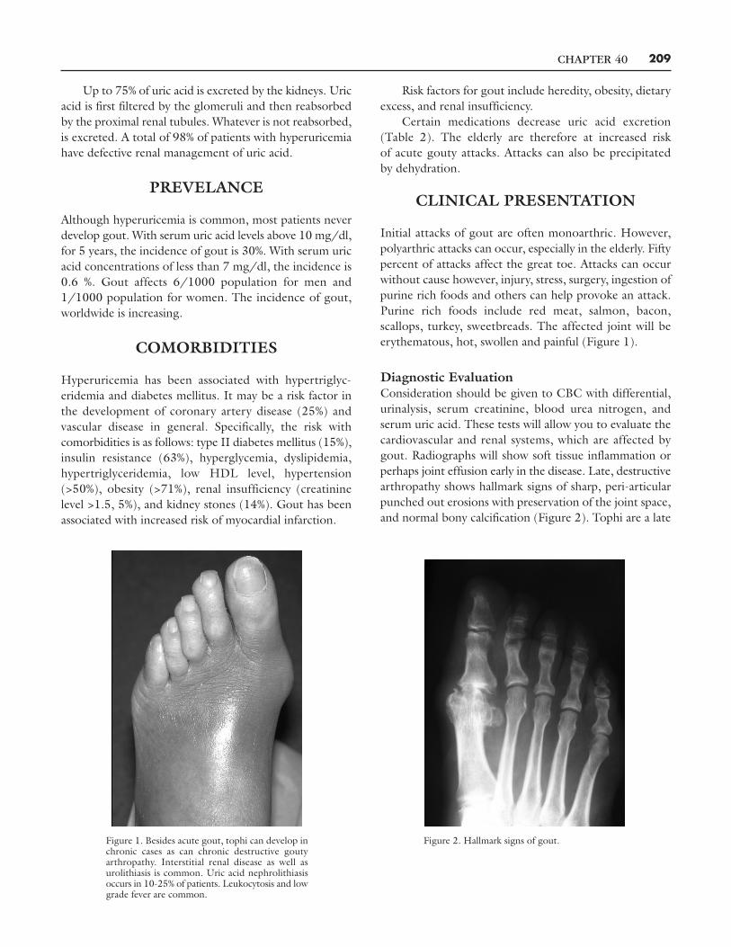
Initial attacks of gout are often monoarthric. However,polyarthric attacks can occur, especially in the elderly. Fiftypercent of attacks affect the great toe. Attacks can occurwithout cause however, injury, stress, surgery, ingestion ofpurine rich foods and others can help provoke an attack.Purine rich foods include red meat, salmon, bacon,scallops, turkey, sweetbreads. The affected joint will beerythematous, hot, swollen and painful (Figure 1).
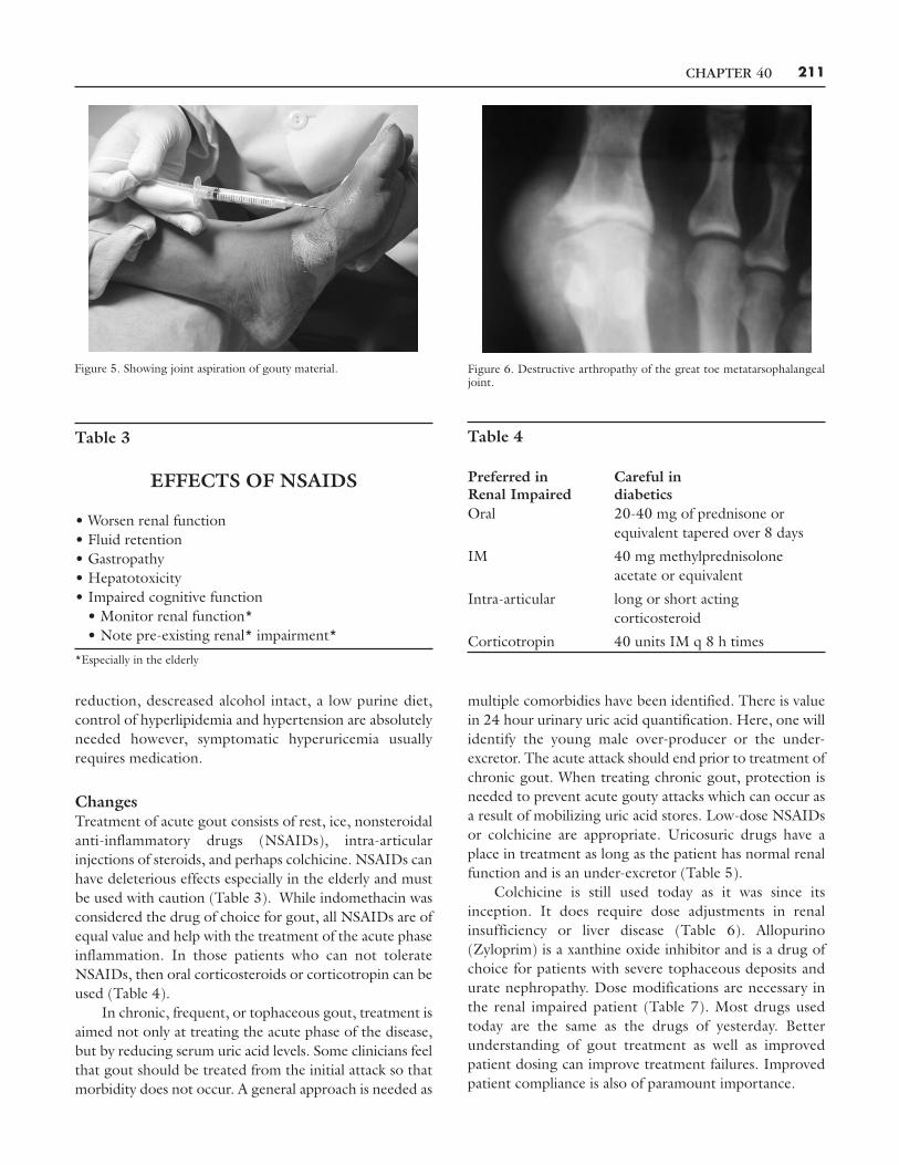
Diagnostic EvaluationConsideration should be given to CBC with differential,urinalysis, serum creatinine, blood urea nitrogen, andserum uric acid. These tests will allow you to evaluate thecardiovascular and renal systems, which are affected bygout. Radiographs will show soft tissue inflammation orperhaps joint effusion early in the disease. Late, destructivearthropathy shows hallmark signs of sharp, peri-articularpunched out erosions with preservation of the joint space,and normal bony calcification (Figure 2). Tophi are a late
Figure 1. Besides acute gout, tophi can develop inchronic cases as can chronic destructive goutyarthropathy. Interstitial renal disease as well asurolithiasis is common. Uric acid nephrolithiasisoccurs in 10-25% of patients. Leukocytosis and lowgrade fever are common.
Figure 2. Hallmark signs of gout.

CHAPTER 40210
development of the disease and often affect the elbows,knees, wrists, hands, great toe, Achilles tendon, and pinaof the ear (Figure 3). Tophi can invade the subcondralbone and cause painful destruction. Classically gout wasdiagnosed by a clinical triad of inflammation, elevatedserum uric acid, and clinical response to colchicine. Goutresponds to colchicine quite predictably.
Differential DiagnosisDifferential diagnosis includes cellulitis, septic arthritis,tumor, fracture, and pseudogout (Figure 4).
Limitations in DiagnosisSerum uric adic levels are normal in 40% of acute goutyattacks. Chronic polyarticular gout occurs with increasingage. Psuedogout (calcium pyrophosphate crystal depositiondisease [CCPD]) can mimic gout. CPPD does respond tocolchicine as well. Chondrocalcinosis may be seen onradiographs, which can help in confirming the diagnosis.Joint aspiration, looking for negatively birefringent sharpneedle-shaped crystals is necessary for appropriate diagnosisof the disease (Figure 5). The aspirate should be kept in asimple syringe and sent for microscopic review. Properobserver accuracy is often a problem. If infection is in thedifferential diagnosis, then gram stain as well as culture andsensitivity are needed.
TREATMENT GOALS
Termination of acute attacks, prevention of recurrentattacks and prevention of complications associated withurate deposition in tissues are all treatment concerns.The general medical health of the patient cannot beoverlooked. Dietary and lifestyle changes such as weight
Table 2
THE EFFECT OF MEDICATIONS ON URIC ACID EXCRETION
Medications that reduce Medication that increaseuric acid excretion uric acid excretion (Uricosuric)• Thiazide diuretics • High dose aspirin
• Loop diuretics• Low-dose aspirin
• Cyclosporine• Niacin
• Ethambutol• Pyrasinamide• Didanosine
Figure 3. It takes approximately 11 years for gouty tophi to develop.
Figure 4. Osteomyelitis masquerading as gout.
CHAPTER 40 211
reduction, descreased alcohol intact, a low purine diet,control of hyperlipidemia and hypertension are absolutelyneeded however, symptomatic hyperuricemia usuallyrequires medication.
ChangesTreatment of acute gout consists of rest, ice, nonsteroidalanti-inflammatory drugs (NSAIDs), intra-articularinjections of steroids, and perhaps colchicine. NSAIDs canhave deleterious effects especially in the elderly and mustbe used with caution (Table 3). While indomethacin wasconsidered the drug of choice for gout, all NSAIDs are ofequal value and help with the treatment of the acute phaseinflammation. In those patients who can not tolerateNSAIDs, then oral corticosteroids or corticotropin can beused (Table 4).
In chronic, frequent, or tophaceous gout, treatment isaimed not only at treating the acute phase of the disease,but by reducing serum uric acid levels. Some clinicians feelthat gout should be treated from the initial attack so thatmorbidity does not occur. A general approach is needed as
multiple comorbidies have been identified. There is valuein 24 hour urinary uric acid quantification. Here, one willidentify the young male over-producer or the under-excretor. The acute attack should end prior to treatment ofchronic gout. When treating chronic gout, protection isneeded to prevent acute gouty attacks which can occur asa result of mobilizing uric acid stores. Low-dose NSAIDsor colchicine are appropriate. Uricosuric drugs have aplace in treatment as long as the patient has normal renalfunction and is an under-excretor (Table 5).
Colchicine is still used today as it was since itsinception. It does require dose adjustments in renalinsufficiency or liver disease (Table 6). Allopurino(Zyloprim) is a xanthine oxide inhibitor and is a drug ofchoice for patients with severe tophaceous deposits andurate nephropathy. Dose modifications are necessary inthe renal impaired patient (Table 7). Most drugs usedtoday are the same as the drugs of yesterday. Betterunderstanding of gout treatment as well as improvedpatient dosing can improve treatment failures. Improvedpatient compliance is also of paramount importance.
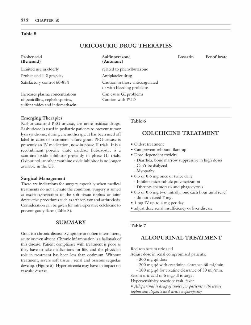
Figure 5. Showing joint aspiration of gouty material. Figure 6. Destructive arthropathy of the great toe metatarsophalangealjoint.
Table 3
EFFECTS OF NSAIDS
• Worsen renal function• Fluid retention• Gastropathy• Hepatotoxicity• Impaired cognitive function
• Monitor renal function*• Note pre-existing renal* impairment*
*Especially in the elderly
Table 4
Preferred in Careful inRenal Impaired diabeticsOral 20-40 mg of prednisone or
equivalent tapered over 8 days
IM 40 mg methylprednisoloneacetate or equivalent
Intra-articular long or short actingcorticosteroid
Corticotropin 40 units IM q 8 h times
CHAPTER 40212
Emerging TherapiesRasburicase and PEG-uricase, are urate oxidase drugs.Rasburicase is used in pediatric patients to prevent tumorlysis syndrome, during chemotherapy. It has been used offlabel in cases of treatment failure gout. PEG-uricase ispresently an IV medication, now in phase II trials. It is arecombinant porcine urate oxidase. Fuboxostat is axanthine oxide inhibitor presently in phase III trials.Oxipurinol, another xanthine oxide inhibitor is no longeravailable in the US.
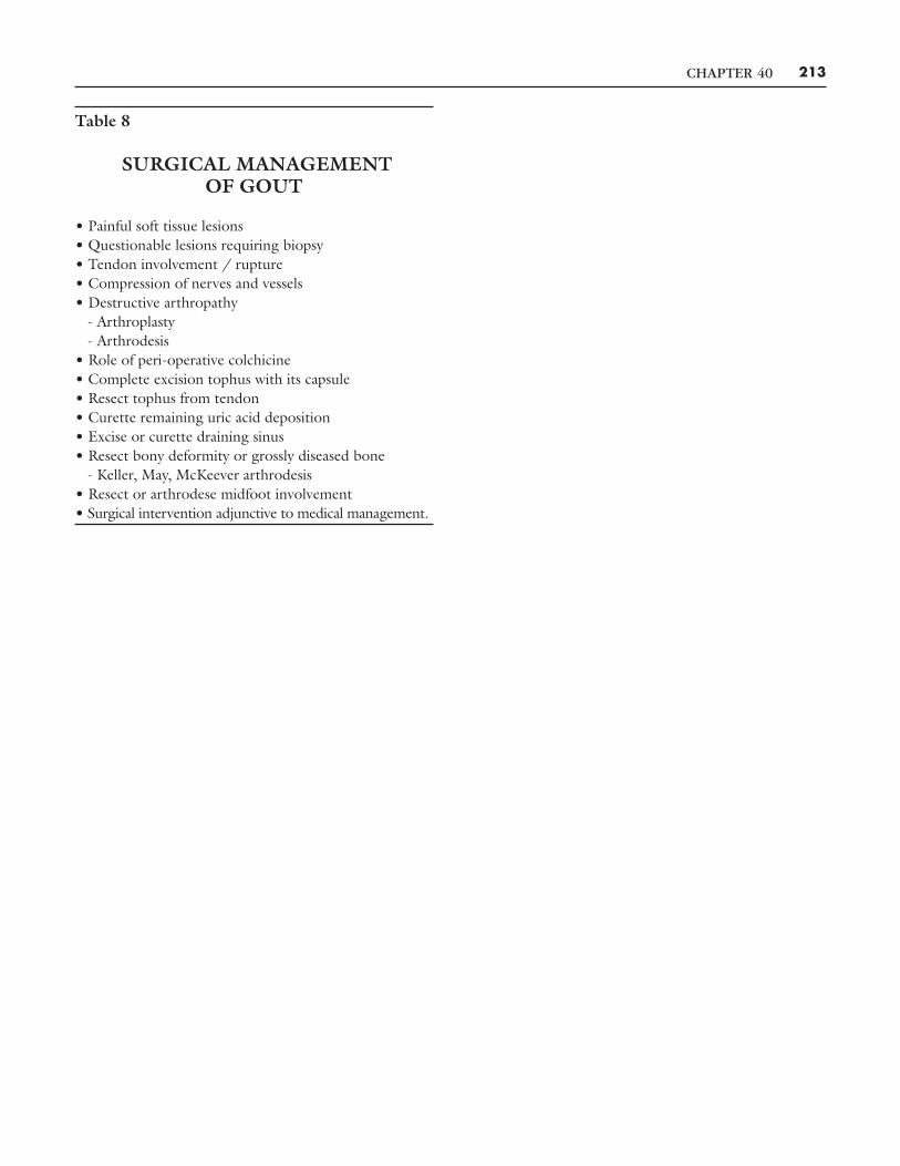
Surgical ManagementThere are indications for surgery especially when medicaltreatments do not alleviate the condition. Surgery is aimedat excision/resection of the soft tissue tophus or jointdestructive procedures such as arthroplasty and arthrodesis.Consideration can be given for intra-operative colchicine toprevent gouty flares (Table 8).
SUMMARY
Gout is a chronic disease. Symptoms are often intermittent,acute or even absent. Chronic inflammation is a hallmark ofthis disease. Patient compliance with treatment is poor asthey have to take medications for life, and the physicianrole in treatment has been less than optimum. Withouttreatment, severe soft tissue , renal and osseous sequelaedevelop. (Figure 6). Hyperuricemia may have an impact onvascular disease.
Table 5
URICOSURIC DRUG THERAPIES
Probenecid Sulfinpyrazone Losartin Fenofibrate(Benemid) (Anturane)
Limited use in elderly related to phenylbutazone
Probenecid 1-2 gm/day Antiplatelet drug
Satisfactory control 60-85% Caution in those anticoagulatedor with bleeding problems
Increases plasma concentrations Can cause GI problemsof penicillins, cephalosporins, Caution with PUDsulfonamides and indomethacin.
Table 6
COLCHICINE TREATMENT
• Oldest treatment• Can prevent rebound flare-up• Dose-dependent toxicity
- Diarrhea, bone marrow suppressive in high doses- Can’t be dialyzed- Myopathy
• 0.5 or 0.6 mg once or twice daily- Inhibits microtubule polymerization- Disrupts chemotaxis and phagocytosis
• 0.5 or 0.6 mg two initially; one each hour until relief- do not exceed 7 mg.
• 1 mg IV up to 4 mg per day• adjust dose renal insufficiency or liver disease
Table 7
ALLOPURINAL TREATMENT
Reduces serum uric acidAdjust dose in renal compromised patients:
- 300 mg qd dose- 200 mg qd with creatinine clearance 60 ml/min.- 100 mg qd for creatine clearance of 30 ml/min.
Serum uric acid of 6 mg/dl is targetHypersensitivity reaction: rash, fever• Allopurinal is drug of choice for patients with severetophaceous deposits and urate nephropathy
CHAPTER 40 213
Table 8
SURGICAL MANAGEMENTOF GOUT
• Painful soft tissue lesions• Questionable lesions requiring biopsy• Tendon involvement / rupture• Compression of nerves and vessels• Destructive arthropathy
- Arthroplasty- Arthrodesis
• Role of peri-operative colchicine• Complete excision tophus with its capsule• Resect tophus from tendon• Curette remaining uric acid deposition• Excise or curette draining sinus• Resect bony deformity or grossly diseased bone
- Keller, May, McKeever arthrodesis• Resect or arthrodese midfoot involvement• Surgical intervention adjunctive to medical management.