GOLD-COATED MAGNETIC POLYMER MICROSPHERE …synthesis of several forms of MPMs. ... properties,...
35
GOLD-COATED MAGNETIC POLYMER MICROSPHERE SYNTHESIS AND CHARACTERIZATION: APPLICATIONS IN BIOLOGICAL CONTEXTS By Phoebe Penamon Thesis Submitted to the Faculty of the Graduate School of Vanderbilt University in partial fulfillment of the requirements for the degree of MASTER OF SCIENCE in Chemistry December, 2012 Nashville, Tennessee Approved: Professor David W. Wright Professor Fredrick R. Haselton
Transcript of GOLD-COATED MAGNETIC POLYMER MICROSPHERE …synthesis of several forms of MPMs. ... properties,...

GOLD-COATED MAGNETIC POLYMER MICROSPHERE SYNTHESIS AND
CHARACTERIZATION: APPLICATIONS IN BIOLOGICAL CONTEXTS
By
Phoebe Penamon
Thesis
Submitted to the Faculty of the
Graduate School of Vanderbilt University
in partial fulfillment of the requirements for
the degree of
MASTER OF SCIENCE
in
Chemistry
December, 2012
Nashville, Tennessee
Approved:
Professor David W. Wright
Professor Fredrick R. Haselton
ii
TABLE OF CONTENTS
Page
LIST OF FIGURES ....................................................................................................... iii
Chapter
I. FUNCTIONALIZATION AND APPLICATION OF GOLD-COATED
MAGNETIC POLYMER MICROSPHERES ....................................................... 1
Introduction ......................................................................................................... 1
Magnetism on the Nanoscale ................................................................................ 3
Magnetic Particle Coatings................................................................................... 6
Magnetic Polymer Microsphere Functionalization .............................................. 10
Electroless Plating of Magnetic Polymer Microspheres ...................................... 14
Aims .................................................................................................................. 18
II. GOLD-COATED MAGNETIC POLYMER MICROSPHERES ........................ 19
Introduction ....................................................................................................... 19
Experimental ...................................................................................................... 19
Results and Discussion ....................................................................................... 21
Conclusions ....................................................................................................... 24
III. APPLICATION OF GOLD-COATED MAGNETIC POLYMER
MICROSPHERES ............................................................................................. 25
Introduction ....................................................................................................... 25
Experimental ...................................................................................................... 26
Results and Discussion ....................................................................................... 28
Conclusions ....................................................................................................... 30
REFERENCES .............................................................................................................. 31

iii
LIST OF FIGURES
Figure Page
1. Morphs of Magnetic Polymer Microspheres ......................................................... 1
2. Types of Magnetic Behavior ................................................................................ 3
3. Mechanism of SMCC coupling .......................................................................... 12
4. Mechanism of EDC coupling ............................................................................. 12
5. Plating Schematic............................................................................................... 14
6. Carryover Schematic .......................................................................................... 16
7. SEM of Gold-Coated Magnetic Polymer Microspheres ...................................... 21
8. TEM of Gold-Coated Magnetic Polymer Microspheres ...................................... 21
9. Iron and Gold Monitoring using EDX ................................................................ 22
10. SEM of Heat Degraded Particles ........................................................................ 22
11. Zeta Potentials of Functionalized Particles ......................................................... 28
12. Pull Through Measurements ............................................................................... 28

1
CHAPTER I
FUNCTIONALIZATION AND APPLICTATION OF GOLD-COATED, MAGNETIC
POLYMER MICROSPHERES
Introduction
Magnetic polymer microspheres (MPMs) are a multicomponent inorganic and
organic material that can be easily manipulated with a magnetic field. It was realized in
the 1950’s that MPMs could be coated with antigens and used to agglutinate in the
presence of the corresponding antibody, which has lead to successful application in
numerous biological assays.1 The success of MPMs in biological assays has led to the
synthesis of several forms of MPMs.
A MPM is composed of a magnetic and polymer component that can form one of
the following morphs shown in Fig. 1: (a) a single magnetic particle surrounded by a
polymer matrix, (b) numerous magnetic
particles homogeneously distributed
throughout a polymer matrix, (c) a
polymer matrix coated with
nanoparticles, or (d) a magnetic particle with polymeric ligands extending from the
surface. The most commercially available MPMs exhibit either a or b type morphs.
Each type has its advantages and disadvantages and no one preparation method can
satisfy all of the following criteria: functionalized, monodispersed, spherical particles
completely encapsulating magnetic cores while controlling size and biocompatibility.2
Figure 1.: Morphs of magnetic polymer microspheres.

2
The polymer matrix serves as a protective shell against the degradation of the magnetic
particles and presents a robust surface that can be further functionalized with ligands that
enhance the utility of MPMs.
With the advent of more powerful microscopic instruments, research into
nanoscale materials has grown significantly in the last few decades. As materials are
formed at the nanoscale, optical, electronic, and magnetic properties deviate from those
observed in the corresponding bulk materials. Along with those interesting intrinsic
properties, nanomaterials can encompass a range of forms and sizes, and they also have
the composition of single or multicomponent structures. Multicomponent structures can
be composed of a single element or multiple elements and compounds. This allows for
greater customization of the nanoscale properties. The unique properties and structural
diversity of the nanomaterials make them excellent candidates for a variety of
applications in diagnostics, therapeutics, environmental testing.
This work will highlight properties of magnetism on the nano- and microscale and
common issues associated with the use of magnetic particles. The issue of magnetic
degradation can be overcome by careful consideration of magnetic nanoparticle synthetic
strategy and method of surface passivation. The choice of polymer matrix can
circumvent these issues related to magnetic degradation, but there is a trade off between
the passivation and protection of the magnetic particles with ease of conjugation that
must be considered. Common methods of microsphere synthesis will be reviewed
culminating with a facile method of coalescence of gold to the MPM surface that
alleviates ligand coupling issues that arise with organic coupling strategies.
3
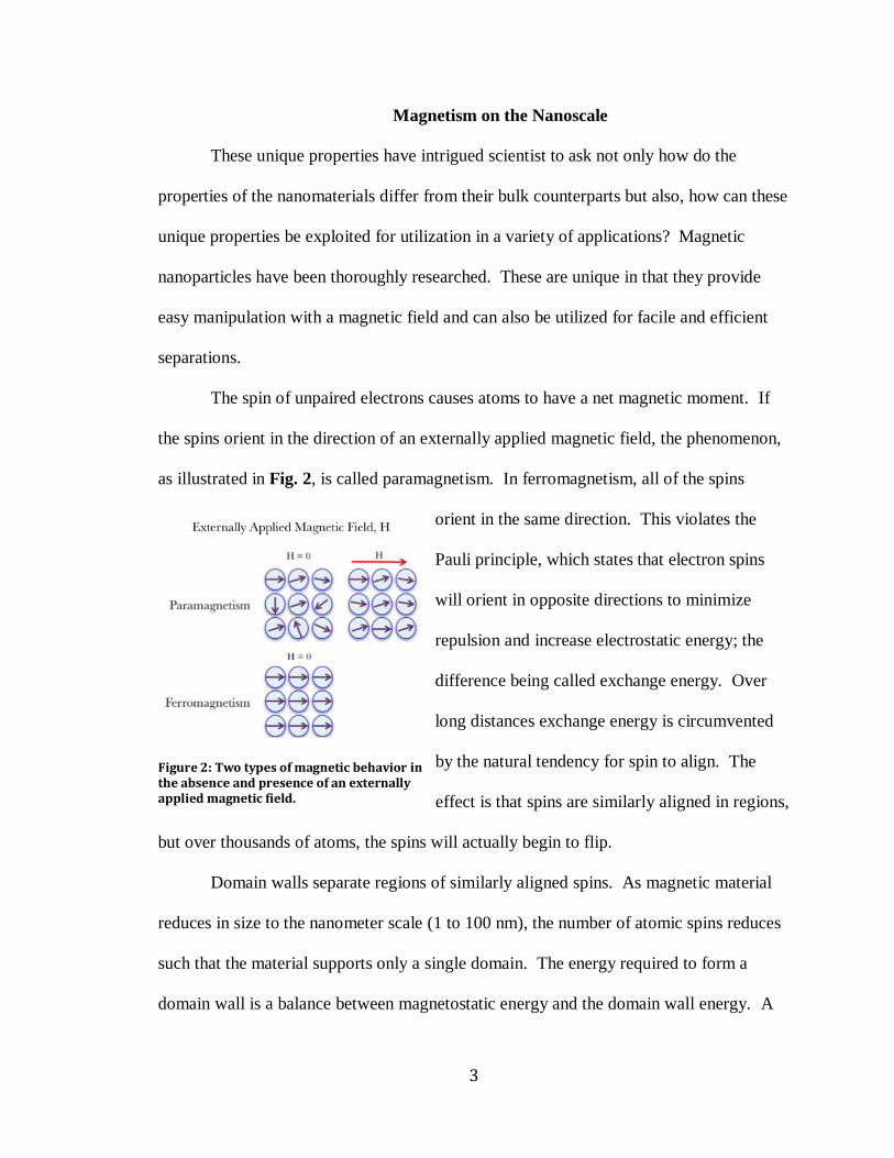
Magnetism on the Nanoscale
These unique properties have intrigued scientist to ask not only how do the
properties of the nanomaterials differ from their bulk counterparts but also, how can these
unique properties be exploited for utilization in a variety of applications? Magnetic
nanoparticles have been thoroughly researched. These are unique in that they provide
easy manipulation with a magnetic field and can also be utilized for facile and efficient
separations.
The spin of unpaired electrons causes atoms to have a net magnetic moment. If
the spins orient in the direction of an externally applied magnetic field, the phenomenon,
as illustrated in Fig. 2, is called paramagnetism. In ferromagnetism, all of the spins
orient in the same direction. This violates the
Pauli principle, which states that electron spins
will orient in opposite directions to minimize
repulsion and increase electrostatic energy; the
difference being called exchange energy. Over
long distances exchange energy is circumvented
by the natural tendency for spin to align. The
effect is that spins are similarly aligned in regions,
but over thousands of atoms, the spins will actually begin to flip.
Domain walls separate regions of similarly aligned spins. As magnetic material
reduces in size to the nanometer scale (1 to 100 nm), the number of atomic spins reduces
such that the material supports only a single domain. The energy required to form a
domain wall is a balance between magnetostatic energy and the domain wall energy. A
Figure 2: Two types of magnetic behavior in the absence and presence of an externally applied magnetic field.

4
collection of these single-domain particles in the presence of a magnetic field can align
with the field. This phenomenon is called superparamagnetism. Superparamagnetic
particles are ferromagnetic in nature, because all of the spins are aligned in each
individual nanoparticle. They have the added benefit that when the field is removed; the
nanoparticles exhibit no remanence, which allows for facile resuspension throughout a
medium. There exist a critical volume below which it cost more energy to create domain
walls than support the external magnetostatic energy of the single-domain state.
Magnetic Nanoparticle Synthesis
Magnetic nanoparticles can be composed of iron oxides (Fe3O4, α-Fe2O3, or γ-
Fe2O3), pure metals (Fe or Co), ceramic spinel type oxides (MgFe2O4, MnFe2O3, or
CoFe2O3), or alloys (CoPt, FePt). Magnetic nanoparticles can be synthesized by a top
down or bottom up approach that offer differing degrees of morphology, stability, and
dispersity control. Co-precipitation, thermal decomposition, and mechanochemical
synthetic strategies are three techniques that are efficient and reproducible for scale up to
larger systems. Although only a few techniques will be explored in depth, a host of other
techniques are available to synthesize magnetic nanoparticles including: laser pyrolysis,
hydrothermal synthesis, and microemulsions.
Co-precipitation is an in situ precipitation technique for iron oxides, Fe3O4, α-
Fe2O3. The precipitation reactions are usually undertaken under inert atmospheres, basic
conditions and ambient or elevated temperatures. In general, the dispersity can be
controlled by the reaction temperature, pH, ionic strength of the solution, types of iron
salts used, and the ratio of Fe2+
to Fe3+
. Without the use of stabilizing agents, the
5
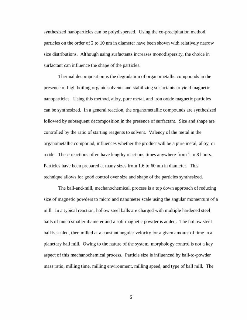
synthesized nanoparticles can be polydispersed. Using the co-precipitation method,
particles on the order of 2 to 10 nm in diameter have been shown with relatively narrow
size distributions. Although using surfactants increases monodispersity, the choice in
surfactant can influence the shape of the particles.
Thermal decomposition is the degradation of organometallic compounds in the
presence of high boiling organic solvents and stabilizing surfactants to yield magnetic
nanoparticles. Using this method, alloy, pure metal, and iron oxide magnetic particles
can be synthesized. In a general reaction, the organometallic compounds are synthesized
followed by subsequent decomposition in the presence of surfactant. Size and shape are
controlled by the ratio of starting reagents to solvent. Valency of the metal in the
organometallic compound, influences whether the product will be a pure metal, alloy, or
oxide. These reactions often have lengthy reactions times anywhere from 1 to 8 hours.
Particles have been prepared at many sizes from 1.6 to 60 nm in diameter. This
technique allows for good control over size and shape of the particles synthesized.
The ball-and-mill, mechanochemical, process is a top down approach of reducing
size of magnetic powders to micro and nanometer scale using the angular momentum of a
mill. In a typical reaction, hollow steel balls are charged with multiple hardened steel
balls of much smaller diameter and a soft magnetic powder is added. The hollow steel
ball is sealed, then milled at a constant angular velocity for a given amount of time in a
planetary ball mill. Owing to the nature of the system, morphology control is not a key
aspect of this mechanochemical process. Particle size is influenced by ball-to-powder
mass ratio, milling time, milling environment, milling speed, and type of ball mill. The

6
ball and mill process is the most industrial technique that can be quickly and reproducibly
extended for mass production of magnetic nanoparticles.
Owing of the high percentage of atoms at the surface of nanoparticles surface,
electrostatic energy and interface reactions become increasingly appreciable. Many
routes have been taken to coat magnetic nanoparticles for passivation or protection of the
surface to decrease surface effects that may lead to loss of magnetization.3 Coating
strategies that have been utilized for magnetic particles include, but is not limited to:
surfactant and polymer coating,4 precious metal coating,
5 silica coating,
6 carbon coating,
7
and matrix dispersion.7 Because the vast majority of research has been focused on
polymer and silica coatings, this work will focus only on surfactant/polymer and silica
coating techniques in broad detail.
Magnetic Particle Coatings
Applications of Magnetic Polymer Microspheres
As mentioned previously, one of the first major applications of MPMs was in the
area of biological separations. MPMs can be utilized to capture a host of biological
materials including: DNA, proteins, enzymes, cells, and biomolecules. This leads to
applications in cell separating, counting, identification, imagining, and immunoassays.1, 2
If those applications are viewed in a more general sense, the process involves positive or
negative separation. Positive separations require the use of functionalized MPMs to
capture and transport the target of interest away from a sample. Conversely, in negative
separations, the unwanted material is removed to purify the sample. Examples of
positive separations include: metal recovery quantification, nucleotide capturing,

7
detection, and gene separation. Examples of negative separations include: signal
amplification. The general view is that the particles capture and transport with ease of
manipulation of a magnetic field. This has inspired research in catalysis,5 surface
enhanced Raman spectrometry (SERS),8 ligand blotting,
9 molecular profiling,
spectrofluorometric assays, electrochemiluminescence, optical encoding, metal
chelation,10
diagnostics, and therapeutics. MPMs can provide binding sites for chemical
or physical adsorption. The choice of polymer matrix has great implications on the
applications available to the MPM.
Silica Coating
Stability in aqueous conditions, well-understood silica chemistry, and easy to
control inter-particle interactions in solution and within structures make silica fitting for
magnetic nanoparticle coating. Homola and colleagues have shown that a silica coating
is used to tune the magnetic properties of nanoparticles, since the extent of dipolar
coupling is related to the distance between particles and this in turn depends on the
thickness of the inert silica shell.3 Iron oxide, one of the most used magnetic materials,
has a strong affinity to silica. Most syntheses begin with the Stöber method to generate
silica nanoparticles.6 The surface can then be modified to contain different
functionalities by reacting with silanated ligands. For example, 3-
aminopropyltrimethoxysilane (APTES) contains amine and silane terminal ends, which
allow silane to react with the hydroxyl group of the silanated nanoparticle. Once the
surface is amine-terminated, the particles can undergo organic coupling strategies for
further functionalization.

8
There are several difficulties associated with silica coatings. For example,
nanometer scale thickness with uniform shell thickness is difficult to obtain. Deposition
of pure metals is difficult due to lack to hydroxyl groups on the surface, and in some
cases, surface grafting of the nanoparticle is required. Silica based coatings are instable
under basic conditions and may contain pores that allow the diffusion of oxygen and
other unwanted species, which can reduce the effectiveness of the magnetic core.
Surfactant and Polymer Coatings
Surfactant and polymer coatings use electrostatic and steric interactions to coat
magnetic nanoparticles and passivate their surfaces and keep them in a stable colloidal
state. These coatings can either be chemically or physically adsorbed to the magnetic
nanoparticle to form a single layer or multiple layers of oppositely charged electrostatic
interactions. These coatings can provide a range of functional groups at the surface
depending upon the monomer reactants chosen. This allows for various functionalization
strategies to be attempted based upon the exposed surface molecules. Although the there
are many advantages, thin polymer coatings do not provided enough barrier to highly
reactive metal particles and are not suitable for very reactive magnetic nanoparticles.
Furthermore, at high temperature, polymer coatings exhibit lower stability. Their
stability in a range of pH values and the ability to grow thick polymer shells with a
lighter density than silica makes polymer coatings ideal for a variety of applications.

9
Preparation of Magnetic Polymer Microspheres
Two major routes exist for the preparation of MPMs. One is the mixing of
separately prepared magnetic cores and polymers. Strategies under this heading include:
phase separation, solvent evaporation, layer-by-layer processes, and sol-gel transitions.
These strategies have been reviewed elsewhere.2 The most frequently used techniques
involve heterogeneous polymerization in the presence of magnetic nanoparticles. For the
purpose of this work the more common techniques under this heading will be reviewed.
They including: suspension polymerizations, microemulsion polymerizations,
miniemulsion polymerizations, and dispersion polymerizations.
In all of these techniques, monomer subunits are polymerized in the presence of
magnetic particles to produce magnetic polymer microspheres. Most routes are an
extension of the bottom up approaches to magnetic nanoparticle synthesis outlined
previously. The different techniques yield microspheres of diameters ranging from tens
of nanometers to millimeters. The magnetic material should have the greatest attraction
to the monomers and little affinity to the other components of the system. In all cases,
two immiscible liquid phases are mixed and the MPMs form in the droplets of the
emulsion. This can generate hydrophilic or hydrophobic surface charges depending upon
the characteristics of the droplets. Microemulsions produce very tiny particles due to the
nature of microemulsions; two immiscible solutions mix to yield stable isotropic and
optically transparent solutions.
10
Magnetic Polymer Microsphere Functionalization
The previously mentioned applications would not be possible without the
functionalization of the MPM surface. In this work, conjugation is defined as the process
of adsorbing an arrangement of molecules or atoms onto a solid support. The definition
highlights three aspects for consideration: (1) the type of solid support, (2) the properties
of the molecules or atoms (ligand), and (3) the actual process of conjugation. Chapter II
chronicles the formation of a gold-coated MPM solid support that was originally realized
for application involving electromagnetic radiation then later used in SERS. Chapter III
is dedicated to the properties of the ligand and the process of conjugation to MPMs.
Any ligand utilized for conjugation to the MPM, will be composed of (1) a head
group, (2) the backbone (chain), and (3) a terminal group. The head group provides
linkage to the MPM. For uniform and expedient processes, the head group conjugation
should be facile. The chain controls the sterics and thereby density of the molecules on
the solid support, allows for distance from the solid support, and stabilization of the
molecule. The terminal group imparts the surface functionality of the particle. Each of
these parts contributes not only the structural and physical properties of the conjugated
particles, but also, the ease of synthesizing or the cost of the ligands.
Coupling reactions are essential reactions to organic chemistry that combine two
hydrocarbons via reactions involving metal catalyst, organic catalyst, or activating
compounds. The coupling techniques and reactions available for MPMs are dominated
by two factors: (1) the surface functionalities available for purchase from the MPM
manufacturer and (2) ligands with the available functionality amenable to the specific
cross coupling. A host of surface functionalities are available from manufacturers.
11
Amine, carboxylic, epoxy, tosylactivated, and silanated are a few common types.
Because of the carboxylic and amine terminated offered opportunities for well known
coupling strategies, they will be explored in more detail.
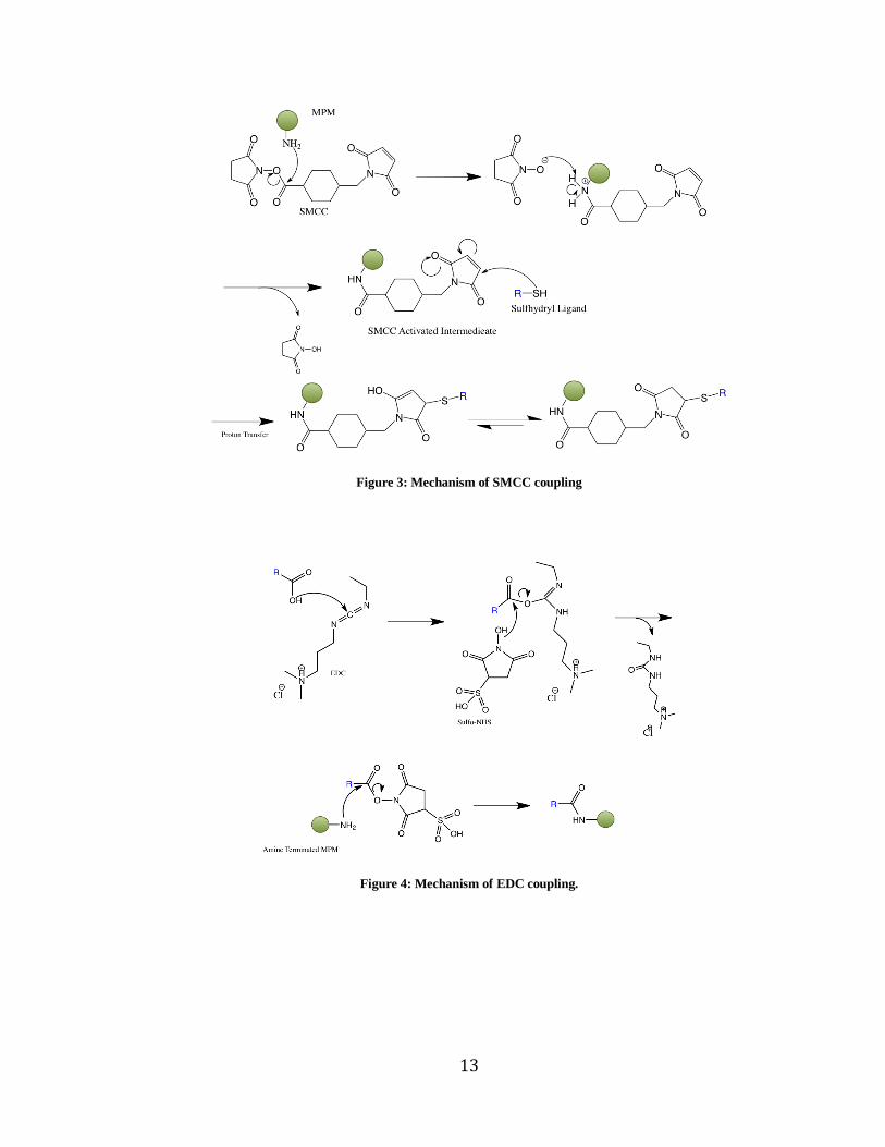
Coupling of thiolated ligands via succinimidyl-4-(N-
maleimidomethyl)cyclohexane-1-carboxylate (SMCC) is available. Fig 3. outlines the
reaction of an amine-terminated particle with a thiolated ligand. The amine of the MPM
attacks the ester of the SMCC to yield an activated SMCC intermediate and N-
hydroxysulfosuccinimide (NHS) as a byproduct. A sulfhydryl containing compound can
then react with the succinimide moiety followed by a proton transfer to yield a linked
MPM and ligand of interest. Carboxyl and amine-terminated MPMs have access to N,N’-
Dicyclohexylcarbodiimide (DCC)11
and 1-Ethyl-3-(3-dimethylaminopropyl)carbodiimide
(EDC) coupling.12
Both EDC and DCC are reagents used to couple amino acids during
peptides synthesis reactions. EDC requires an acidic pH and is used as a carboxyl-
activating agent for the coupling of primary amines to synthesize amide bonds. The
synthetic strategy for EDC coupling with sulfo-NHS is shown in Fig. 4. Sulfo-NHS is
used to stabilize the intermediate otherwise the EDC activated compound would be
vulnerable to hydrolysis. The activated carboxylic intermediate is attacked by the
terminal amines of the MPM. After a proton transfer and cyclization of sulfo-NHS, the
amine is now coupled to the carboxy terminated ligand.
Bartczak and Kanaras recently showed the demanding task of optimization for a
preparing peptide functionalized gold nanoparticles using EDC. In a typical reaction, 10
µL of peptide was added to the oligoethylene glycol nanoparticles (OEG-NPs), 15 µL of
EDC and sulfo-NHS added simultaneously, the reaction was stirred for 24 hours then
12
purified by centrifugation. The concentration of EDC/Sulfo-NHS, peptide, and buffer
was varied in the study, but in each case the volume added was kept constant. For the
addition of EDC/Sulfo-NHS, the ratio was kept at 1:2 for EDC/Sulfo-NHS. Reaction
time and morphology were two more parameters that were varied during optimization.
After thirty reactions, the optimal conditions were determined for this microscale
reaction. If the reactions were scaled up, the results may not be the same. Therefore,
coupling of ligands to the surface of particles may not be the most efficient route.
Each organic coupling technique requires optimization of several different
parameters. Although the SMCC and EDC coupling reaction have well known
chemistries, the optimization and application of these reactions in a heterogeneous
reaction systems is not as standard as expected. The success of these reactions requires
optimization of pH, temperature, and reagent concentration and is not easily manipulated
on a heterogeneous reaction system. Therefore, a more facile method of conjugation
utilizing the efficient gold-thiol bond has been researched.
13
Figure 3: Mechanism of SMCC coupling
Figure 4: Mechanism of EDC coupling.
14
Electroless Plating of Magnetic Polymer Microspheres
Properties of Nanoscale Gold
A multitude of nanoscale research pertains to biological systems, because
nanoscale objects have an increased ability to be taken up by cells and offer interaction
with nano- and micro-size objects within the cell, such as DNA. Gold is a noble metal
that is often employed in medical and biological applications not only for its intrinsically
interesting nanoscale properties, but also the significant degree of biocompatibility of the
material.13
Gold nanoparticles are so small that electrons are not free to move about as in
bulk gold which gives rise to the phenomena of surface plasmon resonance. This
restricted movement causes the nanoparticles to react differently with light changing
causing a red shift in the absorbance. This allows for applications in imaging, and
detection that are based on electromagnetic properties.14
Electroless Plating
A nanoshell is an electronically conducting material encapsulating a dielectric
particle core that is coated with a functionalizable layer.15
Halas and colleagues set out to
create a material capable of tailorable interactions with electromagnetic radiation to
controllably tune optical characteristics over a wide wavelength range. The method was
first realized with AuS particles with a conducting gold shell surrounding a dielectric
sulfide core. They later moved to synthesizing particles with larger dielectric cores from
silica that spanned the range of hundreds of nanometers. The silica particles were made
via the Stöber method then functionalized as mentioned previously to present an amine-
terminated surface capable of undergoing electroless plating.

15
Figure 5. Seed and Growth (Plating) Schematic
Electroless plating (Fig. 5) utilizes molecular self-assembly and colloid reduction
chemistry. In a typical electroless plating (seed and growth) scheme for MPMs, the
reaction begins with an amine-terminated particle. Often the dielectric core is either
silica or polystyrene and the coating layer is gold. The amine sites serve as electrostatic
attachment points for negatively charged small colloidal gold particles. The subsequent
growth of the thin layer comes from the coalescence of the colloidal gold at these
nucleation sites. Using this method then is limited only by having an amine-terminated
surface.
The colloidal particles for seeding can be synthesized in a variety of methods.
One such strategy is the Turkevich method, which involves the reduction of chloroauric
acid (HAuCl4) with sodium citrate. Another method involves reduction of chloroauric
acid with tetrakis(hydroxymethyl)phosphonium chloride (THPC). The distinction
between the two methods is the size of the gold nanoparticles generated. The Turkevich
and THPC methods yield on average 12 nm and 2 nm particles, respectively. It has also
been shown that the reduction with THPC yields more uniformly spherical particles over
the Turkevich method.15
The THPC method yields a more uniform coating in a fewer
plating steps. The gold coatings provide a functionalizable surface with interesting
optical properties that have access to efficient gold-thiol chemistry
16
Self-Assembled Monolayers
The discovery of self-assembled monolayers (SAMs), propelled the field of gold-
thiol chemistry. Consequently, the general aspects of thiol SAMs on gold, the self
assembly process, and the structure of the gold-thiol surface have been extensively
researched and thoroughly reviewed.16
The gold-thiol bond strength is 44 kcal/mol.
Although the gold-thiol bond is on the order of covalent strength, the aspects of the gold-
thiol bond degradation must be considered. The gold-thiol bond has been shown to
degrade by oxidation and UV exposure to disulfides and sulfonates via the following
reactions:
2 RS-Au RSSR + 2 Au (1)
RS-Au + H2O + O3 RSO3H + HO-Au (2)
Degradation is heavily dependent upon size and structure of gold support. Two week
stability in ambient conditions has been shown for gold-thiol substrates.17,18
Thermal
degradation is influenced by chain length and surface roughness among other things. It
has been shown that the gold-thiol bond is stable up to 100 °C. Longer chain lengths
tend to be stable at increased temperatures.
Carryover
The overarching goal is to build an easily reproducible system that has plug and
play functionality and can be utilized in a variety of assays. A tubular extraction cassette
has been introduced by Bordelon and colleagues that takes advantage of the capture and
transport abilities of MPMs.19
In the cassette, a polymer tube is filled such that liquid

17
Figure 6: A) tubular extraction cassette. B) Schematic of fluid carryover after MPMs have traversed a valve.
solutions (chambers) are separated by either pockets of air or mineral oil. The separator
either air or oil is referred to as a valve. The blue (exiting) chamber contains the target of
interest and is the initial chamber in which the MPMs enter. After the MPMs and have
been incubated to allow for maximal capture, the MPMs are magnetically pulled through
the valve (Fig 6, B) into the next, receiving, chamber. Small amounts of liquid from the
capture chamber can become trapped in the interstices of the magnet packed MPMs as
the beads traverse the valve. The process of liquid being carried over from one chamber
to the other across the mineral oil or air valve in the tubular extraction cassette is called
Carryover.
Carryover can affect the purity of the receiving chamber. The volume of each
chamber is usually on the order of hundreds µL. Therefore, if even microliters volumes
of liquid are being carried over to the receiving chamber, the receiving volume is being
altered by a significant percent. This can lead to inconsistencies in results and ambiguity
about the actual concentration of the target being captured. The functionalized AuMPMs
can be tailored to coordinate to a range of ligands. Provided the solutions are aqueous,
the gold-coated magnetic particles can be tuned to have a more hydrophobic surface that
in theory would have less carryover and circumvent issues of purity in the tubular
extraction system. Therefore, AuMPMs were functionalized with the three ligands

18
representing a positive, neutral, and negative charges and their efficiency in the magnetic
was tested. Changing the hydrophobicity of the surface structure could have an impact
on the amount of force required for the beads to traverse the valves.
Aims
The aim of this research is investigate the fundamental aspects of the production of gold-
coated MPMs. The ideal MPM for use in the extraction and purification cassette would
be evenly coated with gold, spherical monodispersed, and easily synthesized. This work
probes the uniformity and dispersity throughout the process of gold-coating and
subsequent functionalization. The ease and conditions necessary for conjugation will be
investigated. Finally, using gold-coated and functionalized MPMs, they will be applied
to a tubular purification system to determine the effects of different charged surfaces on
the ability to overcome electrostatic interactions at the surface of the liquid-air and liquid-
mineral oil interfaces. Ultimately, a platform technology is being developed that can
easily coordinate and bind different types of ligands and can be utilized in a variety of
biological, environmental, and analytical contexts. The works provided in this document
provide the foundation for more thorough investigations into tuning the gold surface for
thickness and smoothing, the impact of the gold-thiol bond on magnetic properties of the
MPMs, electronics associated with the gold-thiol bond, and chemical and thermal
stability of the conjugated MMP.
19
CHAPTER II
GOLD-COATED MAGNETIC POLYMER MICROSPHERES
Introduction
As noted in Chapter I, MPMs can serve as an enabling technology in a number of
important applications. The key to successful application of the MPMs lies in their
ability to be functionalized with a variety of ligands specific to the application. MPMs
can be purchased with different surface functionalities. Some of which represent specific
interactions i.e., streptavidin coated MPMs utilized for streptavidin-biotin complexation.
Other functionalities present opportunities for conjugation and cross coupling techniques.
As mentioned in Chapter I, there are many difficulties associated with performing
organic coupling reactions with the particles. The use of well-known gold-thiol
chemistry might provide a useful approach to circumvent these problems.
The following experiments chronicle the synthetic strategy towards gold-coated
MPMs. The synthesized particles are characterized by TEM and SEM to probe dispersity
and homogeneity of gold coating. Experiments to examine the thermal stability of the
gold-coated MPMs is presented.
Experimental
Materials
Amine-terminated MPMS were purchased from Invitrogen Life Sciences at a stock
concentration of 2 x 10⁹ MPMs per mL. Sodium hydroxide was obtained from Sigma-

20
Aldrich. Tetrakis(hydroxymethyl)phosphonium chloride (THPC) and chloroauric acid
were obtained from Aldrich. All chemicals were used as received unless otherwise
noted.
Preparation of Gold Nanoparticle (AuNP) Seeding Solution
To 45 ml of H2O, 500 ml of 1 M NaOH and 1 ml of THPC solution (12 µL in 1 ml H2O)
were added. The solution was stirred for 5 min. Next, 2 ml of 1% HAuCl4 added quickly
resulting in a brownish color. The solution was stirred for 5 min then refrigerated at 4 °C
when not used immediately.
Preparation of AuNP Plating Solution
A 1.8 mM solution of K2CO3 was made with one liter H2O. After the solution had stirred
for 10 min, 15.5 ml of a 1% v/v HAuCl4 solution was added. The solution stirred three
min then was left quiescent for 30 min. The solution was stored at room temperature and
shielded from light when not in use.
Seeding of Amine-Terminated Magnetic Polymer Microspheres
One hundred µL of stock MPMs were washed 3 times with H2O. The MPMs were
magnetically separated between washes. After washing, the MPMs were resuspended in
100 µL of H2O. To the dispersed MPMs, 1 mL AuNP seeding solution was added. The
solution was stirred for 5 min, then allowed to sit quiescently and unexposed to light.
After 2 hr, the solution was magnetically separated and washed with H2O three times.
The particles were resuspended in 1 mL H2O.
21
Plating of Seeded Magnetic Polymer Microspheres
To 100 µL of seeded particles, 1 ml of plating solution was added. The particles reacted
in solution for 3 min. After reaction, the microspheres were was magnetically separated
and washed with H2O 2 times. The microspheres were resuspended in 100 µL H2O. This
procedure was repeated to achieve successive platings. After the particles were
resuspended in 1 ml, 14 µL aliquots were removed for transmission electron microscopy
(TEM) and scanning electron microscopy (SEM).
Thermostability Test
Two mg of gold-coated microspheres and amine-terminated MPMs at an equivalent
concentration were heated separately to 800 °C in a thermogravimetic analyzer.
Results and discussion
Characterization
The seed and growth method was utilized with amine-terminated MPMs from Invitrogen
Life Sciences. The growth (plating) step was repeated to provide a gold-coated MPM
(AuMPM). Ten plating steps (Au(10)MPM) were sufficient to fully coat the surface of
the particles. With more coatings, agglutination became apparent with the visible eye,
and the particle density increased causing the particles to rapidly settle out of solution.
Philips CM20 Transmission Electron Microscope and Hitachi S-4200 Scanning Electron
Microscope were utilized to probe the physical aspects of the gold-coated and uncoated
MPMs. In Fig. 7, the MPM (A) and seed (B) are marked by large pores. As the particles

22
are successively plated from A to F, the surface becomes less porous and smoother. The
particle diameter remains and monodispersity remain constant through successive
platings. In Fig. 8, the TEM micrograph shows the decreased transmission of electron
through the material equating to the attachment of gold. Branches appear at the surface
of the particle, which also signifies the coalescence of gold at the MPM surface.
Figure 7: Plating monitoring via SEM. A) amine-terminated microsphere B) seeded amine-terminated polymer
microsphere C) 3x plated microsphere D) 5x plated microsphere E) 7x plated microsphere and F) 10x plated
microsphere. Scale bar 1 µm.
Figure 8: TEM images of A) uncoated MPM and B) Au(10)-MPM. Scale bar 500 nm.
Iron-Gold Monitoring
TEM and SEM provide evidence of gold coalescence, but the quantity of gold has yet to
be measured. Using energy dispersive x-ray (EDX) capabilities of the SEM, the atomic

23
percentage of elements in the sample was monitored. In a typical EDX experiment, the
sample was irradiated with X-rays at 20 µA. The instrument measures the incident x-rays
from the sample and compares them against a standard to determine atomic composition.
Assuming a fairly constant percentage of iron per microsphere, increased gold attachment
to the particles would cause a decrease in the iron:gold ratio. In the EDX experiment, a
196 µm2 area of planar particles was scanned for 2, 4, 6, 8, and 10 platings (Fig. 9). As,
expected, as the number of plating steps increased, the iron:gold ratio decreased.
Figure 9: Iron and gold monitoring of plated MPMs using EDX. A) Iron to gold ratio and B) size of scan the
yielded the results. Scale bar 7 µm.
Thermostability
In an effort to probe the robustness of the Au(10)MPMs, the beads were heated to 800 °C
under a nitrogen atmosphere. SEM images for the manufacturers amine-terminated
MPMs and Au(10)MPMs can be seen in Fig. 10. In both cases the MPMs retain
monodispersity and spherical shape. The particles have undergone a reduction in size
from the initial diameter of 2.8 microns to approximately 1.0 micron. This is attributable
to the loss of the polymer matrix. Gold on the surface of the MPMs has the elemental
state and remains attached to the particles.

24
Figure 10: After heating to 800 °C, SEM images of A) Uncoated MPMs and B) Au(10)-MPMs. Scale bar 1 µm.
Conclusions
The MPMs have been successfully coated with gold using a facile seed and growth
method. The MPMs remained monodispersed throughout both seeding and plating
reactions. As the number of plating steps increase, the amount of gold is also increasing,
leading to a thicker shell of gold. Ten platings were sufficient enough to have a fully
coated gold surface. Furthermore, the microspheres are robust can be heated to extreme
temperatures and retain dispersity and shape.

25
CHAPTER III
APPLICATION OF MAGNETIC POLYMER MICROSPHERES
Introduction
The force required to move the group of MPMs through the interfacial or surface
tension valve in the tubing, where the movement is constrained to the x direction only
(Fm,x), was approximated using the following equation (Gijs 2004)20
:
(3)
where V is the bulk volume occupied by the MPMs, χv is the volume susceptibility, μ0 is
the permeability of free space (4π × 10-7
), and B is the magnetic field along the axis of the
tube through which we are pulling the MPMs (x axis). To measure the force required to
pull the MPMs through the surface tension valve, an apparatus was developed to measure
x, y, and z coordinates of the magnetic field (B) of a 2.54 cm cube permanent magnet
along the axis of the tube using a F.W. Bell series 9900 gaussmeter. The values for the x,
y, and z coordinates were plotted as a function of distance from the edge of the magnet.
The gradient of the magnetic field for the x, y, and z coordinates was
roughly approximated for by using the slope of the lines between two measurements from
the curves. Because the gradient of the magnetic field in the y and z coordinates was
approximately zero, the and terms of the magnetic force equation were set
to zero.
26
To obtain the χv an Alfa Aesar Magnetic Susceptibility Balance Mark 1 was used.
The balance provides reading of mass susceptibility, χg, which can be expressed as
follows:
(4)
Where CBal is a constant proportionality, R is sample reading, R0 is the blank reading, l is
the sample length (cm), and m is the sample mass. Once χg has been resolved, the
measurement can be can be converted to χv for use in equation 3 by the following
equation:
(5)
Where d is the density under a magnetic field. The force will be used to determine how
neutrally, positively, and negatively charged MPMs traverse the valves of the tubular
extraction cassette.
The following experiments probe the dispersity, morphology, and surface charge
of the gold-coated MPMs that have been functionalized with thiolated ligands. The effect
of surface change on transit through a valve was examined by measuring a parameter
called pull through.
Experimental
Materials
From Sigma-Aldrich, the following chemicals were purchased: 6-amino-1-hexanethiol
hydrochloride, 6-mercaptohexanoic acid, and 6-mercapto-1-hexanol. Ethanol purchased
from Pharco-aaper. Silica gel purchased from Dynamic Adsorbents. All chemicals used
27
without further purification. Cole-Parmer vinyl tubing (tygon) with and inner diameter
1/16 in and outer diameter 1/8 was utilized.
Ligand Conjugation
Each ligand was prepared at a concentration of 1 mM by dissolving in ethanol. One
hundred microliters of gold-coated, MPMs at 3.32 x 10⁻¹³
M were added to a conical
tube. To this tube, 900 µL of each stock ligand solution was added. The solution
incubated while stirring for 8 hours. The solution was magnetically separated and
washed with H2O 3 times. After washing, the MPMs were resuspended at a
concentration 3.32 x 10⁻¹³
M.
Zeta Potentials
To the 100 µL sample of AuMPMs, 650 µL of H2O was added for a concentration of
6.65 x 10⁻¹⁴
M. This was found to be the optimal concentration to obtain the most
reliable data. The zeta potentials were measured on a Malvern Zetasizer. All
measurements were taken in water at a pH of 5.6.
Pull Through Measurement
To measure the force required to pull the MPMs through a surface tension valve,
a preloaded tube containing 20 μL of a 3.32 x 10⁻¹³
M MPMs suspension. The tube
slowly moved toward the 2.54 cm cube magnet along the x coordinate of the measured
magnetic field until the point where the MPMs pulled through the valve. The distance at
28
which the MPMs pulled through was recorded and used to approximate the magnetic
field strength (Bx) and the magnetic field gradient ( ) at that distance.
Magnetic Susceptibility
Volume (V) was measured as the bulk volume that the particles occupied under the
influence of a magnetic field. The values were calculated by measuring the cylindrical
volume that a known mass of MPMs occupied in a short length of 1.6 mm inner diameter
tubing. One mg of Invitrogen Life Sciences MPMs was diluted into 114 mg silica gel.
To a small glass vial, 114 mg of silica gel was weighed. To this, 1 mg of dried gold-
coated magnetic microspheres was mixed thoroughly. A blank sample of only 115 mg
silica gel was also prepared. The magnetic susceptibility was measured using an Alfa
Aesar magnetic susceptibility balance. The balance was zeroed and the calibration
constant was calculated using the manganese chloride standard supplied by the
manufacturer. The blank was made using 114 mg silica gel without MPMs added. The
tube was rinsed with water between each sample, dried at 100 °C for 10 min, and
measured empty to verify that residual magnetic MPMs had been removed after each
wash. Each sample was measured three times, removing and repacking the MPMs
between each measurement.
Results and discussion
Zeta Potentials
Confirmation of conjugation was obtained using zeta potentials. The uncoated magnetic
polymer microsphere in Fig. 11 shows a highly positive charge as is to be expected of

29
amine-terminated microspheres from the factory. Interestingly the Au(10) microspheres
showed a highly negative charge that is consistent with the K2CO3 stabilizer in the plating
solution that is apparently acting as a capping agent to the microspheres. The carboxyl
microspheres fall within the range of the Au(10)MPMs considering that they are both
capped with the same terminal functionality. More neutral measurements were expected
for the hydroxyl terminated Au(10)MPMs. The hydroxyl ligand may not have
sufficiently displaced the K2CO3 stabilizing agent. Also, there is no significant difference
between the amine functionalized MPM and the MPM from the manufacturer.
Figure 11: Zeta Potentials of functionalize Au(10)-MPMs.
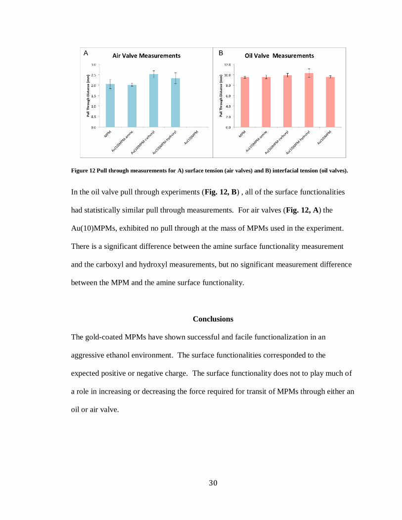
Magnetic Pull Through Measurements
The pull through for surface and interfacial tension valves were measured.
30
Figure 12 Pull through measurements for A) surface tension (air valves) and B) interfacial tension (oil valves).
In the oil valve pull through experiments (Fig. 12, B) , all of the surface functionalities
had statistically similar pull through measurements. For air valves (Fig. 12, A) the
Au(10)MPMs, exhibited no pull through at the mass of MPMs used in the experiment.
There is a significant difference between the amine surface functionality measurement
and the carboxyl and hydroxyl measurements, but no significant measurement difference
between the MPM and the amine surface functionality.
Conclusions
The gold-coated MPMs have shown successful and facile functionalization in an
aggressive ethanol environment. The surface functionalities corresponded to the
expected positive or negative charge. The surface functionality does not to play much of
a role in increasing or decreasing the force required for transit of MPMs through either an
oil or air valve.

31
REFERENCES
1. Martin, C. R.; Mitchell, D. T., Nanomaterials in analytical chemistry. Analytical Chemistry 1998, 70 (9), 322A-327A. 2. Horak, D.; Babic, M.; Mackova, H.; Benes, M. J., Preparation and properties of magnetic nano- and microsized particles for biological and environmental separations. Journal of Separation Science 2007, 30 (11), 1751-1772. 3. Lu, A.-H.; Salabas, E. L.; Schueth, F., Magnetic nanoparticles: Synthesis, protection, functionalization, and application. Angewandte Chemie-International Edition 2007, 46 (8), 1222-1244. 4. Butterworth, M. D.; Bell, S. A.; Armes, S. P.; Simpson, A. W., Synthesis and characterization of polypyrrole-magnetite-silica particles. Journal of Colloid and Interface Science 1996, 183 (1), 91-99; Tartaj, P.; Morales, M. P.; Gonzalez-Carreno, T.; Veintemillas-Verdaguer, S.; Serna, C. J., Advances in magnetic nanoparticles for biotechnology applications. Journal of Magnetism and Magnetic Materials 2005, 290, 28-34. 5. Park, J. I.; Cheon, J., Synthesis of "solid solution" and "core-shell" type cobalt-platinum magnetic nanoparticles via transmetalation reactions. Journal of the American Chemical Society 2001, 123 (24), 5743-5746. 6. Stober, W.; Fink, A.; Bohn, E., CONTROLLED GROWTH OF MONODISPERSE SILICA SPHERES IN MICRON SIZE RANGE. Journal of Colloid and Interface Science 1968, 26 (1), 62-&. 7. Ang, K. H.; Alexandrou, I.; Mathur, N. D.; Amaratunga, G. A. J.; Haq, S., The effect of carbon encapsulation on the magnetic properties of Ni nanoparticles produced by arc discharge in de-ionized water. Nanotechnology 2004, 15 (5), 520-524. 8. Piao, L.; Park, S.; Lee, H. B.; Kim, K.; Kim, J.; Chung, T. D., Single Gold Microshell Tailored to Sensitive Surface Enhanced Raman Scattering Probe. Analytical Chemistry 2010, 82 (1), 447-451. 9. Dekki, N.; Refai, E.; Holmberg, R.; Kohler, M.; Jornvall, H.; Berggren, P.-O.; Juntti-Berggren, L., Transthyretin binds to glucose-regulated proteins and is subjected to endocytosis by the pancreatic beta-cell. Cellular and Molecular Life Sciences 2012, 69 (10), 1733-1743. 10. Ji, Z. S.; Pinon, D. I.; Miller, L. J., Development of magnetic beads for rapid and efficient metal-chelate affinity purifications. Analytical Biochemistry 1996, 240 (2), 197-201. 11. Jaszay, Z. M.; Petnehazy, I.; Toke, L.; Szajani, B., PREPARATION OF CARBODIIMIDES USING PHASE-TRANSFER CATALYSIS. Synthesis-Stuttgart 1987, (5), 520-523. 12. Nakajima, N.; Ikada, Y., MECHANISM OF AMIDE FORMATION BY CARBODIIMIDE FOR BIOCONJUGATION IN AQUEOUS-MEDIA. Bioconjugate Chemistry 1995, 6 (1), 123-130. 13. Shukla, R.; Bansal, V.; Chaudhary, M.; Basu, A.; Bhonde, R. R.; Sastry, M., Biocompatibility of gold nanoparticles and their endocytotic fate inside the cellular compartment: A microscopic overview. Langmuir 2005, 21 (23).

32
14. Adams, N. M.; Jackson, S. R.; Haselton, F. R.; Wright, D. W., Design, Synthesis, and Characterization of Nucleic-Acid-Functionalized Gold Surfaces for Biomarker Detection. Langmuir 2012, 28 (2), 1068-1082. 15. Shi, W. L.; Sahoo, Y.; Swihart, M. T.; Prasad, P. N., Gold nanoshells on polystyrene cores for control of surface plasmon resonance. Langmuir 2005, 21 (4), 1610-1617. 16. Vericat, C.; Vela, M. E.; Benitez, G.; Carro, P.; Salvarezza, R. C., Self-assembled monolayers of thiols and dithiols on gold: new challenges for a well-known system. Chemical Society Reviews 2010, 39 (5), 1805-1834. 17. Cortes, E.; Rubert, A. A.; Benitez, G.; Carro, P.; Vela, M. E.; Salvarezza, R. C., Enhanced Stability of Thiolate Self-Assembled Monolayers (SAMs) on Nanostructured Gold Substrates. Langmuir 2009, 25 (10), 5661-5666. 18. Vericat, C.; Benitez, G. A.; Grumelli, D. E.; Vela, M. E.; Salvarezza, R. C., Thiol-capped gold: from planar to irregular surfaces. Journal of Physics-Condensed Matter 2008, 20 (18). 19. Bordelon, H.; Adams, N. M.; Klemm, A. S.; Russ, P. K.; Williams, J. V.; Talbot, H. K.; Wright, D. W.; Haselton, F. R., Development of a Low-Resource RNA Extraction Cassette Based on Surface Tension Valves. Acs Applied Materials & Interfaces 2011, 3 (6), 2161-2168. 20. Gijs, M. A. M., Magnetic bead handling on-chip: new opportunities for analytical applications. Microfluidics and Nanofluidics 2004, 1 (1), 22-40.