GLANDULAR CELL ABNORMALITIES - bosnianpathology.org glandular1.pdf · •Endometrial ve...
10
16.06.2016 1 GLANDULAR CELL ABNORMALITIES Aysun UĞUZ Plan • General info • Bethesda System- 2001 /glandular cells • Atypical glandular cells – Atypical Endocervical Cells: NOS – Atypical Endocervical Cells: Favor neoplasia – Diff diagnosis • Endocervical Adenocarcinoma İn Situ – Diff diagnosis • Endometrial Adenocarcinoma – Diff diag • Extrauterine Adenocarcinoma • Cervical cytology (CC) is primarily a screening test for squamous intraepithelial lesions and squamous cell carcinoma. • Sensitivity for glandular lesions is limited and by problems with both sampling and interpretation. • 2001 Bethesda System terminology was aimed to better reflect current knowledge and understanding of glandular neoplasia in cervical cytology, improve communication among laboratories and clinicians. • Thereby, facilitate to patient management. • The term “atypical glandular cells of undetermined significance (AGC-US or AGUS)” has been eliminated from Bethesda System to avoid confusion with the terminology for squamous cell abnormalities. • AGC (atypical glandular cell) terminology is used for either endocervival or endometrial cell abnormalities. • Whenever possible, it should be categorized as to cell type of origins, as the clinical workup and management for patients with glandular abnormalities may vary significantly depending upon the cell type. • Cytopatologists should be determined to origin of the glandular cells – endocervical or endometrial cells? – adenocarcinoma in situ? – adenocarcinoma? • endocervical? • endometrial? • extrauterin glandular cell? • not-otherwised specified (NOS) • Endocervical and endometrial glandular cells may show a variety of cellular changes associated with various benign process in the endocervical canal and endometrium. • Many of these reactive changes are not specific for any particular disease, but are of significance as mimimics of glandular neoplasia in CC. • Follow up of AGC cytologic diagnosis shows that high-grade lesions are identified in 10%- 40% of cases.
Transcript of GLANDULAR CELL ABNORMALITIES - bosnianpathology.org glandular1.pdf · •Endometrial ve...
16.06.2016
1
GLANDULAR CELL ABNORMALITIES
Aysun UĞUZ
Plan
• General info
• Bethesda System- 2001 /glandular cells
• Atypical glandular cells
– Atypical Endocervical Cells: NOS
– Atypical Endocervical Cells: Favor neoplasia
– Diff diagnosis
• Endocervical Adenocarcinoma İn Situ
– Diff diagnosis
• Endometrial Adenocarcinoma
– Diff diag
• Extrauterine Adenocarcinoma
• Cervical cytology (CC) is primarily a
screening test for squamous intraepithelial
lesions and squamous cell carcinoma.
• Sensitivity for glandular lesions is limited and
by problems with both sampling and
interpretation.
• 2001 Bethesda System terminology was aimed to better reflect current knowledge and understanding of glandular neoplasia in cervical cytology, improve communication among laboratories and clinicians.
• Thereby, facilitate to patient management.
• The term “atypical glandular cells of undetermined significance (AGC-US or AGUS)” has been eliminated from Bethesda System to avoid confusion with the terminology for squamous cell abnormalities.
• AGC (atypical glandular cell) terminology is used for either endocervival or endometrial cell abnormalities.
• Whenever possible, it should be categorized as to cell type of origins, as the clinical workup and management for patients with glandular abnormalities may vary significantly depending upon the cell type.
• Cytopatologists should be determined to origin of the glandular cells
– endocervical or endometrial cells?
– adenocarcinoma in situ?
– adenocarcinoma?
• endocervical?
• endometrial?
• extrauterin glandular cell?
• not-otherwised specified (NOS)
• Endocervical and endometrial glandular cells may show a variety of cellular changes associated with various benign process in the endocervical canal and endometrium.
• Many of these reactive changes are not specific for any particular disease, but are of significance as mimimics of glandular
neoplasia in CC.
• Follow up of AGC cytologic diagnosis shows that high-grade lesions are identified in 10%-
40% of cases.
16.06.2016
2
• AGC term is thought to not sufficient for discriminating between neoplastic and reactive lesions
• Then "AGUS / favors reactive changes” is eliminated from Bethesda system.
Bethesda System- 2015
• GLANDULAR CELLS
* Atypical
- endocervical cells (NOS or specify in comments)
- endometrial cells (NOS or specify in comments),
• - glandular cells (NOS or specify in comments),
• * Atypical
- endocervical cells, favor neoplastic
- glandular cells, favor neoplastic
* Endocervical adenocarcinoma in situ (AIS)
* Adenocarcinoma:
- endocervical
- endometrial
- extrauterin
• - NOS
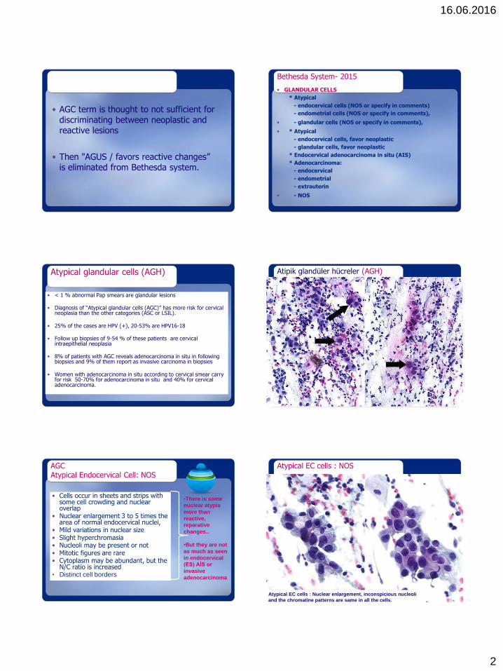
• < 1 % abnormal Pap smears are glandular lesions • Diagnosis of “Atypical glandular cells (AGC)” has more risk for cervical
neoplasia than the other categories (ASC or LSIL). • 25% of the cases are HPV (+), 20-53% are HPV16-18 • Follow up biopsies of 9-54 % of these patients are cervical
intraepithelial neoplasia • 8% of patients with AGC reveals adenocarcinoma in situ in following
biopsies and 9% of them report as invasive carcinoma in biopsies • Women with adenocarcinoma in situ according to cervical smear carry
for risk 50-70% for adenocarcinoma in situ and 40% for cervical adenocarcinoma.
Atypical glandular cells (AGH) Atipik glandüler hücreler (AGH)
AGC Atypical Endocervical Cell: NOS
• Cells occur in sheets and strips with some cell crowding and nuclear overlap
• Nuclear enlargement 3 to 5 times the area of normal endocervical nuclei,
• Mild variations in nuclear size • Slight hyperchromasia • Nucleoli may be present or not • Mitotic figures are rare • Cytoplasm may be abundant, but the
N/C ratio is increased • Distinct cell borders
•There is some
nuclear atypia
more than
reactive,
reparative
changes..
•But they are not
as much as seen
in endocervical
(ES) AİS or
invasive
adenocarcinoma
Atypical EC cells : NOS
Atypical EC cells : Nuclear enlargement, inconspicious nucleoli
and the chromatine patterns are same in all the cells.
16.06.2016
3
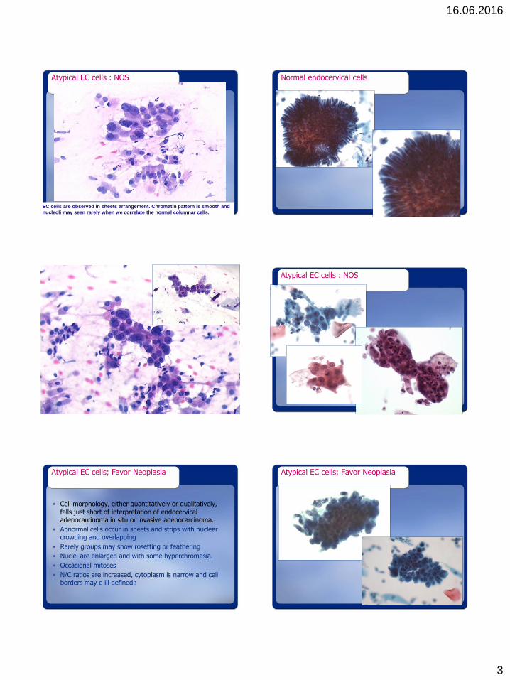
Atypical EC cells : NOS
EC cells are observed in sheets arrangement. Chromatin pattern is smooth and
nucleoli may seen rarely when we correlate the normal columnar cells.
Normal endocervical cells
Atypical EC cells : NOS
Atypical EC cells; Favor Neoplasia
• Cell morphology, either quantitatively or qualitatively, falls just short of interpretation of endocervical adenocarcinoma in situ or invasive adenocarcinoma..
• Abnormal cells occur in sheets and strips with nuclear crowding and overlapping
• Rarely groups may show rosetting or feathering
• Nuclei are enlarged and with some hyperchromasia.
• Occasional mitoses
• N/C ratios are increased, cytoplasm is narrow and cell borders may e ill defined.
Atypical EC cells; Favor Neoplasia
16.06.2016
4
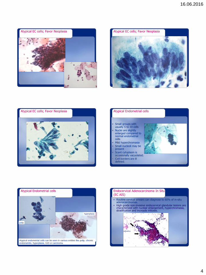
Atypical EC cells; Favor Neoplasia Atypical EC cells; Favor Neoplasia
Atypical EC cells; Favor Neoplasia Atypical Endometrial cells
• Small groups with usually 5 to 10 cells
• Nuclei are slightly enlarged compared to normal endometrial cells
• Mild hyperchromasia
• Small nucleoli may be present
• Scant cytoplasm is occasionally vacuolated.
• Cell borders are ill defined.
Atypical Endometrial cells
Atypical endometrial cells can be seen in various entities like polip, chronic
endometritis, hyperplasia, IUD or carcinoma.
hyperplasia
polip
Endocervical Adenocarcinoma In Situ (EC AIS)
• Routine cervical smears can diagnose to 60% of in-situ adenocarcinomas.
• High grade non-invasive endocervical glandular lesions are characterized with nuclear enlargement, hyperchromasia, stratification and increade mitoses.
palisading
16.06.2016
5
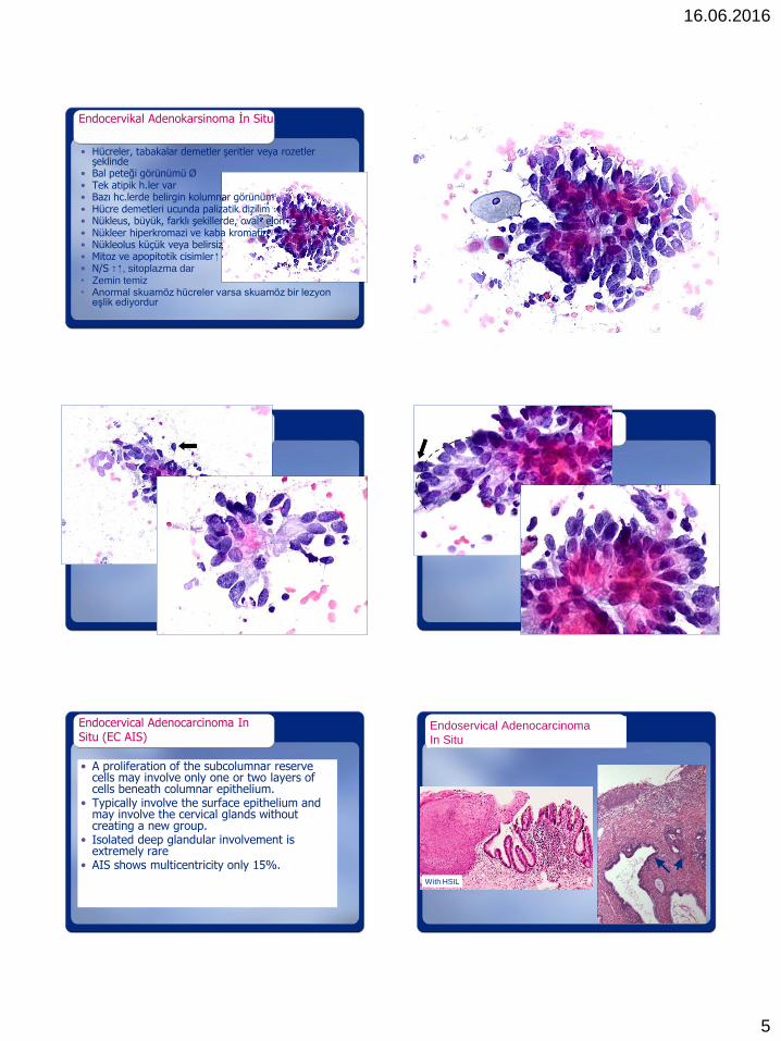
Endocervikal Adenokarsinoma İn Situ
• Hücreler, tabakalar demetler şeritler veya rozetler şeklinde
• Bal peteği görünümü Ø • Tek atipik h.ler var • Bazı hc.lerde belirgin kolumnar görünüm • Hücre demetleri ucunda palizatik dizilim • Nükleus, büyük, farklı şekillerde, oval- elonge • Nükleer hiperkromazi ve kaba kromatin • Nükleolus küçük veya belirsiz • Mitoz ve apopitotik cisimler • N/S ↑ , sitoplazma dar • Zemin temiz • Anormal skuamöz hücreler varsa skuamöz bir lezyon
eşlik ediyordur
Endocervical Adenocarcinoma In Situ (EC AIS)
• A proliferation of the subcolumnar reserve cells may involve only one or two layers of cells beneath columnar epithelium.
• Typically involve the surface epithelium and may involve the cervical glands without creating a new group.
• Isolated deep glandular involvement is extremely rare
• AIS shows multicentricity only 15%.
Endoservical Adenocarcinoma
In Situ
With HSIL
16.06.2016
6
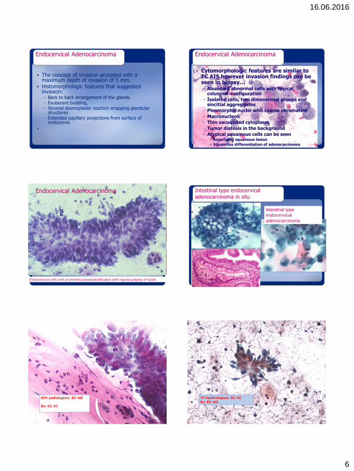
Endocervical Adenocarcinoma
• The concept of invasion accepted with a maximum depth of invasion of 5 mm.
• Histomorphologic features that suggested invasion: – Back to back arrangement of the glands,
– Exuberant budding, – Stromal desmoplastic reaction wrapping glandular
structures – Extended papillary projections from surface of
endocervix
•
• Cytomorphologic features are similar to EC AIS however invasion findings can be seen in biopsy… – Abundant abnormal cells with typical
columnar configuration
– İsolated cells, two dimensional groups and sincitial aggreggates
– Pleomorphic nuclei with coarse chromatine
– Macronucleoli
– Thin vacuolated cytoplasm
– Tumor diatesis in the background
– Atypical squamous cells can be seen • Associated squamous lesion
• Squamous differentiation of adenocarcinoma
Endocervical Adenocarcinoma
Endocervical cells with prominent pseudostratification with regular polarity of nuclei
Endocervical Adenocarcinoma Intestinal type endocervical adenocarcinoma in situ
Intestinal type
endocervical
adenocarcinoma
82% pathologists: EC AIS
Bx: EC AC
70 %pathologists: EC AC
Bx: EC AIS
16.06.2016
7
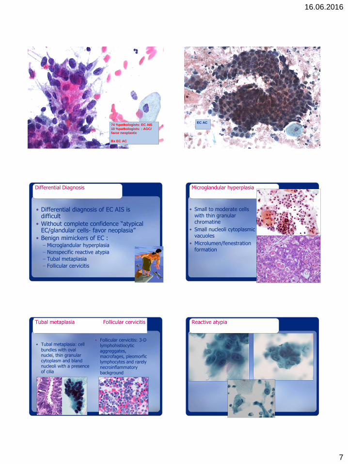
74 %pathologists: EC AIS
10 %pathologists: : AGC/
favor neoplastic
Bx EC AC
EC AC
Differential Diagnosis
• Differential diagnosis of EC AIS is difficult
• Without complete confidence “atypical EC/glandular cells- favor neoplasia”
• Benign mimickers of EC : – Microglandular hyperplasia
– Nonspecific reactive atypia
– Tubal metaplasia
– Follicular cervicitis
Microglandular hyperplasia
• Small to moderate cells with thin granular chromatine
• Small nucleoli cytoplasmic vacuoles
• Microlumen/fenestration formation
Tubal metaplasia Follicular cervicitis
• Tubal metaplasia: cell bundles with oval nuclei, thin granular cytoplasm and bland nucleoli with a presence of cilia
• Follicular cervicitis: 3-D lymphohistiocytic aggreggates, macrofages, pleomorfic lymphocytes and rarely necroinflammatory background
Reactive atypia
16.06.2016
8
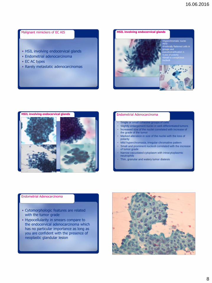
Malignant mimickers of EC AIS
• HSIL involving endocervical glands
• Endometrial adenocarcinoma
• EC AC types
• Rarely metastatic adenocarcinomas
HSIL involving endocervical glands
•Hyperchromatic nuclei
•N/S
•Preferally flattened cells in
groups and
pseudostratification n
•Loss of polarity
•Small to conspicious
nucleoli
HSIL involving endocervical glands Endometrial Adenocarcinoma
• Single or small cohesive groups of cells
• Slightly enlargement nuclei in well-differentiated tumors
• İncreased size of the nuclei correlated with increase of the grade of the tumor
• Marked alteration in size of the nuclei with the loss of polarity
• Mild hyperchromasia, irregular chromatine pattern
• Small and prominent nucleoli correlated with the increase of tumor grade
• Narrow vacuolated cytoplasm with intracytoplasmic neutrophils
• Thin, granular and watery tumor diatesis
Endometrial Adenocarcinoma
• Cytomorphologic features are related with the tumor grade
• Hypocellularity in smears compare to the endocervical adenocarcinoma which has no particular importance as long as you are confident with the presence of neoplastic glandular lesion
16.06.2016
9
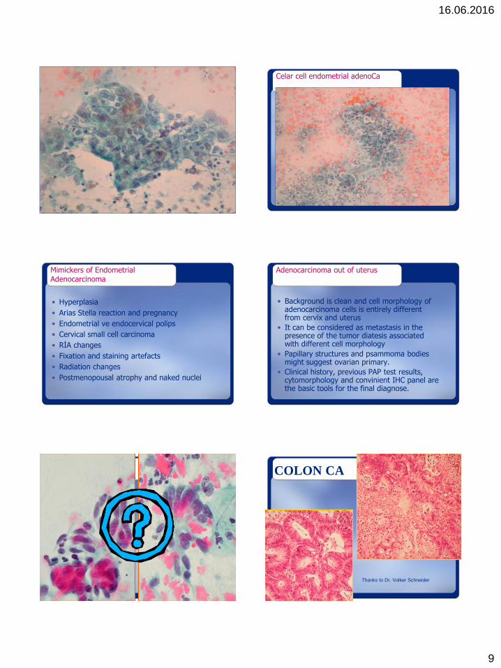
Celar cell endometrial adenoCa
Mimickers of Endometrial Adenocarcinoma
• Hyperplasia
• Arias Stella reaction and pregnancy
• Endometrial ve endocervical polips
• Cervical small cell carcinoma
• RİA changes
• Fixation and staining artefacts
• Radiation changes
• Postmenopousal atrophy and naked nuclei
Adenocarcinoma out of uterus
• Background is clean and cell morphology of adenocarcinoma cells is entirely different from cervix and uterus
• It can be considered as metastasis in the presence of the tumor diatesis associated with different cell morphology
• Papillary structures and psammoma bodies might suggest ovarian primary.
• Clinical history, previous PAP test results, cytomorphology and convinient IHC panel are the basic tools for the final diagnose.
COLON CA
Thanks to Dr. Volker Schneider
16.06.2016
10
• AIS progresses to invasive adenocarcinoma in 5 to 14 years with the lack of any therapeutic management
• Conisation must be performed in cylindrical shape with at least 25 mm length of cervix as involving entire transformation zone which decreases the residuel disease risk from 75% to 33%
• Basic hysterectomy might be an alternative without any desire of fertility
• There is need of further works to clarify the need of histerectomy with the absence of permanent disease
Diagnose and Treatment
Thank you for your attention... [email protected]
http://screening.iarc.fr/atlascyto_