Giant Cell tumor of Fibula Head Web Site - Tumor Surgery Studies/Giant Cell tumor of...Giant Cell...
33
Giant Cell Tumor of Giant Cell Tumor of the Proximal Fibula the Proximal Fibula James C. Wittig, MD James C. Wittig, MD Associate Professor of Orthopedic Surgery Associate Professor of Orthopedic Surgery Chief, Orthopedic Oncology Chief, Orthopedic Oncology Mount Mount Sinai Medical Center Sinai Medical Center
Transcript of Giant Cell tumor of Fibula Head Web Site - Tumor Surgery Studies/Giant Cell tumor of...Giant Cell...

Giant Cell Tumor of Giant Cell Tumor of the Proximal Fibulathe Proximal Fibula
James C. Wittig, MDJames C. Wittig, MD
Associate Professor of Orthopedic SurgeryAssociate Professor of Orthopedic SurgeryChief, Orthopedic Oncology Chief, Orthopedic Oncology MountMount Sinai Medical CenterSinai Medical Center
Clinical HistoryClinical History
19 year old female with a mildly painful enlargement 19 year old female with a mildly painful enlargement on the outside of her left knee for several months.on the outside of her left knee for several months.The patient gave a history of a twisting injury to the The patient gave a history of a twisting injury to the knee several months prior to the onset of pain.knee several months prior to the onset of pain.The patient was otherwise healthy. The patient was otherwise healthy. She was born in the U.S. and gave no history of travel.She was born in the U.S. and gave no history of travel.There were no fevers, night sweats or weight loss.There were no fevers, night sweats or weight loss.Blood tests were normalBlood tests were normal
XX--raysrays
XX--rays demonstrated a geographic, expansile lesion of rays demonstrated a geographic, expansile lesion of the head of the fibula. There was a surrounding the head of the fibula. There was a surrounding ““egg egg shellshell”” rim of calcification indicating the periosteum was rim of calcification indicating the periosteum was intact.intact.There were internal There were internal trabeculationstrabeculations within the within the lesion/tumorlesion/tumorThe lesion was expansile and displaced the The lesion was expansile and displaced the peronealperonealnerve and nerve and poplitealpopliteal blood vessels.blood vessels.The entire head of the fibula was destroyed by the The entire head of the fibula was destroyed by the neoplasmneoplasm

Tumor
Geographic and Expansile
Sharp Zone of Transition between Tumor and Normal Bone/Fibula

CT ScanCT Scan
CT scan shows a thin CT scan shows a thin cortical shell around cortical shell around the tumor indicating the tumor indicating the periosteum is intact the periosteum is intact and the tumor is likely and the tumor is likely benignbenignThere was no There was no ossification or ossification or calcification within the calcification within the tumor indicating that tumor indicating that the tumor was probably the tumor was probably not a bone or cartilage not a bone or cartilage producing tumorproducing tumor

CT Scan Axial SectionCT Scan Axial Section
Tumor

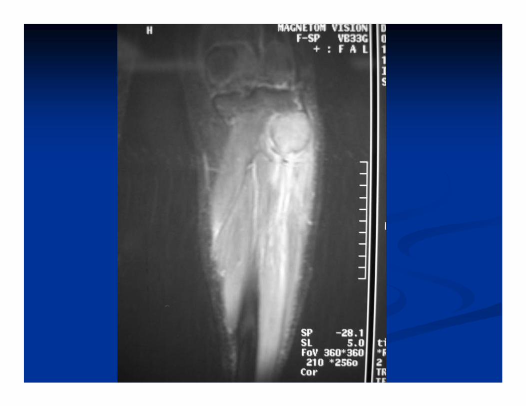
MRIMRI
The MRI findings were not specific for a The MRI findings were not specific for a particular type of neoplasm or infectionparticular type of neoplasm or infectionThe lesion was low to intermediate signal on T1 The lesion was low to intermediate signal on T1 and intermediate to high signal on T2 weighted and intermediate to high signal on T2 weighted images. The tumor diffusely enhanced with images. The tumor diffusely enhanced with contrast. There were no contrast. There were no ““fluidfluid--fluidfluid”” levels that levels that would indicate cystic changes.would indicate cystic changes.The MRI nicely demonstrated the tumorThe MRI nicely demonstrated the tumor’’s local s local extent and proximity to the vascular structures.extent and proximity to the vascular structures.

MRIMRI

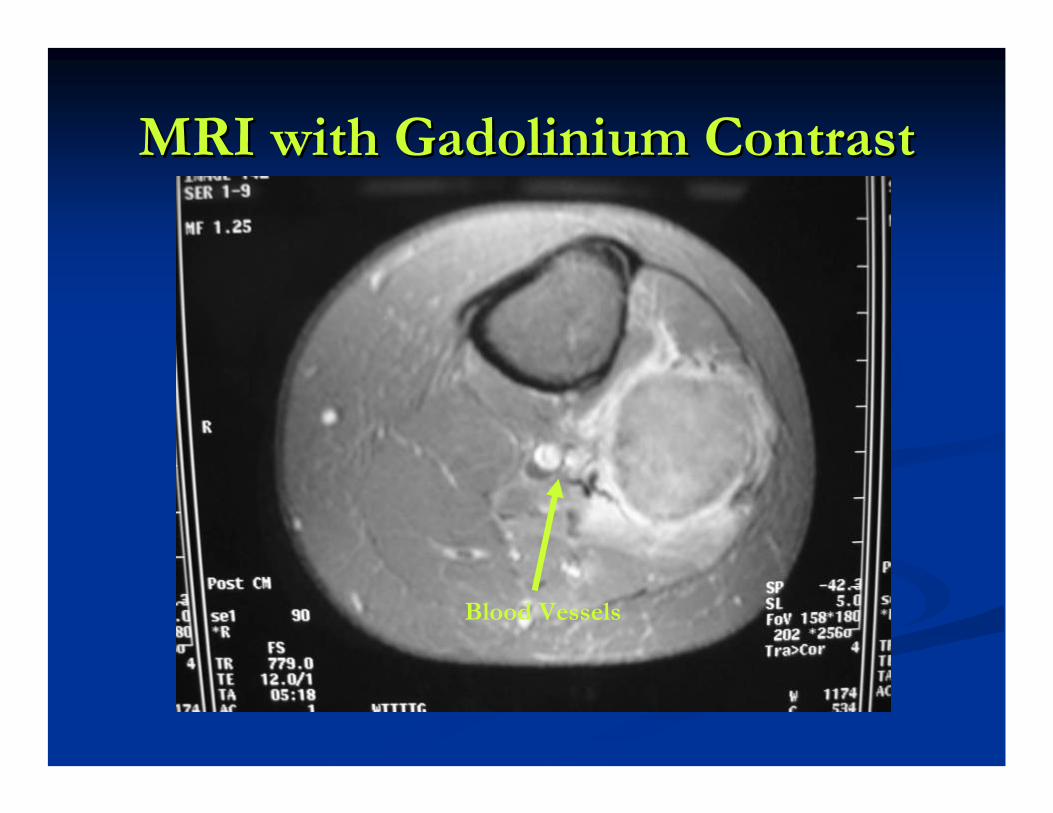
MRI with Gadolinium ContrastMRI with Gadolinium Contrast
Blood Vessels

T1 Weighted Axial MRIT1 Weighted Axial MRI

Bone Scan Demonstrates Increased Bone Scan Demonstrates Increased Activity in NeoplasmActivity in Neoplasm
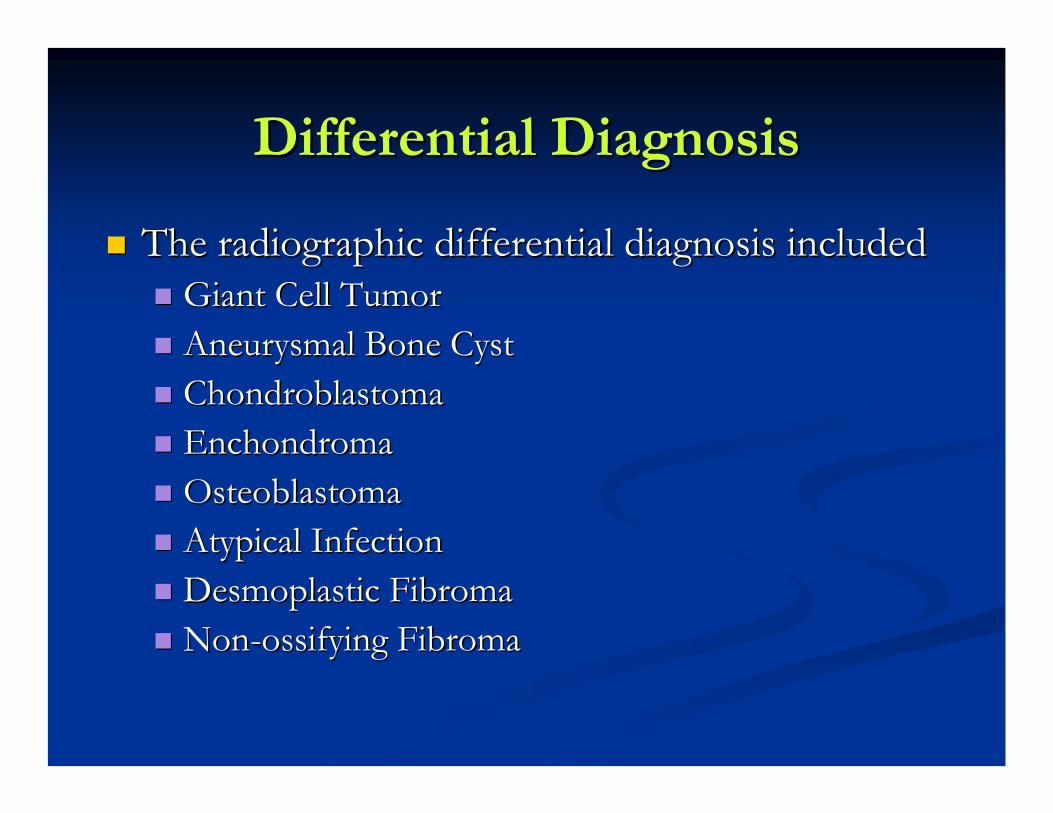
Differential DiagnosisDifferential Diagnosis
The radiographic differential diagnosis includedThe radiographic differential diagnosis includedGiant Cell TumorGiant Cell TumorAneurysmalAneurysmal Bone CystBone CystChondroblastomaChondroblastomaEnchondromaEnchondromaOsteoblastomaOsteoblastomaAtypical InfectionAtypical InfectionDesmoplasticDesmoplastic FibromaFibromaNonNon--ossifying ossifying FibromaFibroma

Differential DiagnosisDifferential Diagnosis
The radiographic studies support the diagnosis of a The radiographic studies support the diagnosis of a benign aggressive neoplasm. The lesion expands the benign aggressive neoplasm. The lesion expands the bone and the periosteum appears to be intact and to bone and the periosteum appears to be intact and to contain the lesion. There is a sharp zone of transition contain the lesion. There is a sharp zone of transition between the tumor and normal bone (geographic between the tumor and normal bone (geographic pattern of bone destruction). Given the age, benign pattern of bone destruction). Given the age, benign aggressive appearance, origin in the aggressive appearance, origin in the metaphysismetaphysis and and involvement of the epiphysis and lack of involvement of the epiphysis and lack of mineralization, the most likely diagnosis is a Giant Cell mineralization, the most likely diagnosis is a Giant Cell Tumor of Bone.Tumor of Bone.

Differential DiagnosisDifferential Diagnosis
The lack of mineralization argues against a The lack of mineralization argues against a chondroblastomachondroblastoma, , enchondromaenchondroma and and osteoblastoma although these lesions do not osteoblastoma although these lesions do not always demonstrate mineralization. The always demonstrate mineralization. The epiphysealepiphyseal involvement suggests a involvement suggests a chondroblastomachondroblastoma however this would be a very however this would be a very rare site for a rare site for a chondroblastomachondroblastoma and and chondroblastomaschondroblastomas usually do not show internal usually do not show internal trabeculationstrabeculations..

Differential DiagnosisDifferential DiagnosisThe differential diagnosis of internal The differential diagnosis of internal trabeculationstrabeculationsincludes includes desmoplasticdesmoplastic fibromafibroma, , chondromyxofibromachondromyxofibroma, , hemangiomahemangioma, , aneurysmalaneurysmal bone cyst, bone cyst, nonossifyingnonossifying fibromafibromaand giant cell tumor. and giant cell tumor. DesmoplasticDesmoplastic fibromafibroma is extremely is extremely rare and this would be an unusual age and location for a rare and this would be an unusual age and location for a desmoplasticdesmoplastic fibromafibroma. This would also be an . This would also be an extrmelyextrmely rare rare site for a site for a chondromyxofibromachondromyxofibroma. . ChondromyxofibromasChondromyxofibromasalso usually arise eccentrically from the bone and have a also usually arise eccentrically from the bone and have a border that is very expansile and another border with an border that is very expansile and another border with an indolent appearance. indolent appearance. NonossifyingNonossifying fibromasfibromas are usually are usually sharply circumscribed, arise eccentrically from the bone sharply circumscribed, arise eccentrically from the bone and do not expand and destroy the bone. This is also an and do not expand and destroy the bone. This is also an unusual site for a unusual site for a nonossifyingnonossifying fibromafibroma..

Differential DiagnosisDifferential Diagnosis
AneurysmalAneurysmal Bone Cyst: ABCs arise in this age Bone Cyst: ABCs arise in this age group. This would be an unusual site and there group. This would be an unusual site and there were no were no ““fluidfluid--fluidfluid”” levels detected on the MRI levels detected on the MRI which would be consistent with a primary or which would be consistent with a primary or secondary ABC.secondary ABC.

Differential DiagnosisDifferential Diagnosis
Infections can be considered within the Infections can be considered within the differential. TB and Fungal infections can differential. TB and Fungal infections can present in an unusual manner such as this. present in an unusual manner such as this. However, the patient gave no history of travel, However, the patient gave no history of travel, exposure to tuberculosis and was born in the exposure to tuberculosis and was born in the U.S. She had no fevers, night sweats and all U.S. She had no fevers, night sweats and all blood tests were normal.blood tests were normal.The key to an accurate diagnosis lies in the The key to an accurate diagnosis lies in the biopsy of the tumor/lesion.biopsy of the tumor/lesion.

BiopsyBiopsy
A CT guided core needle biopsy was performedA CT guided core needle biopsy was performedThe pathology demonstrated many giant cells The pathology demonstrated many giant cells dispersed amongst a sea of uniform mononuclear cellsdispersed amongst a sea of uniform mononuclear cellsThe nuclei of the mononuclear cells resembled the The nuclei of the mononuclear cells resembled the nuclei in the giant cellsnuclei in the giant cellsThere was no evidence of ossification or calcificationThere was no evidence of ossification or calcificationThere was no matrix productionThere was no matrix productionThere were no There were no granulomasgranulomasCultures were negativeCultures were negative

Giant Cells
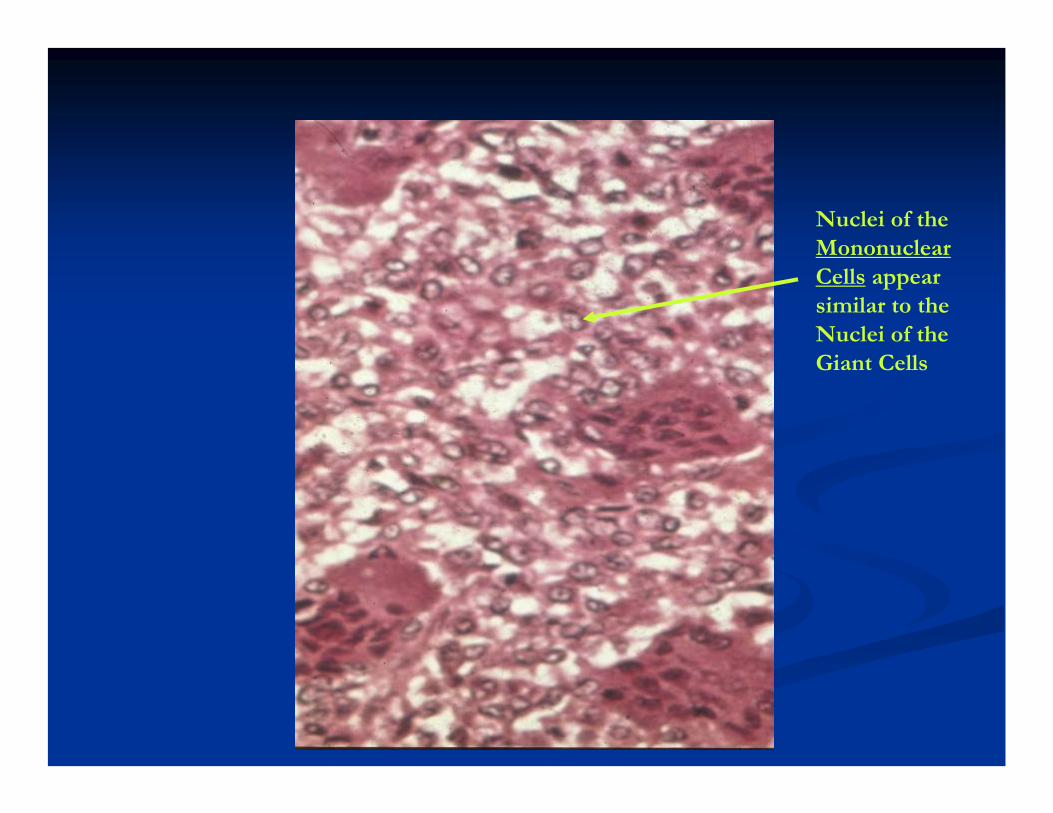
Nuclei of the Mononuclear Cells appear similar to the Nuclei of the Giant Cells

Giant Cell
Mononuclear Cells

DiagnosisDiagnosis
The diagnosis is Giant Cell TumorThe diagnosis is Giant Cell TumorGiant Cells can be seen in many different tumors. The Giant Cells can be seen in many different tumors. The key is that the cells surrounding the giant cells are all key is that the cells surrounding the giant cells are all mononuclear cells and their nuclei are very similar to mononuclear cells and their nuclei are very similar to the nuclei within the giant cells. These mononuclear the nuclei within the giant cells. These mononuclear cells coalesce to form the giant cells. Notice that the cells coalesce to form the giant cells. Notice that the nuclei are all clumped within the center of the giant cell. nuclei are all clumped within the center of the giant cell. Giant cells are also present in TB and Fungal infections, Giant cells are also present in TB and Fungal infections, these types of giant cells are called these types of giant cells are called LangerhanLangerhan’’ss Giant Giant Cells. The nuclei of these giant cells are arranged Cells. The nuclei of these giant cells are arranged around the periphery of the giant cell.around the periphery of the giant cell.

SurgerySurgery
The surgery consists of a wide/radical resection The surgery consists of a wide/radical resection of the tumor/proximal fibula.of the tumor/proximal fibula.

SurgerySurgery
The The peronealperoneal nerve and all its branches to the nerve and all its branches to the peronealperoneal muscles, anterior muscles, anterior tibialistibialis muscle, muscle, extensor extensor digitorumdigitorum longus and extensor longus and extensor hallucishallucisloguslogus (all the muscles that lift the foot off the (all the muscles that lift the foot off the ground/ground/dorsiflexdorsiflex the ankle and toes) is dissected the ankle and toes) is dissected and separated from the neoplasm. The nerve and separated from the neoplasm. The nerve and all its branches are protected while the and all its branches are protected while the fibula is cut at a distance from the tumor in fibula is cut at a distance from the tumor in order to remove the tumor with an adequate order to remove the tumor with an adequate margin.margin.

SurgerySurgery
The biceps The biceps femorisfemoris muscle and lateral collateral muscle and lateral collateral ligament are released from the insertion on the ligament are released from the insertion on the tumor/head of fibula. They are later repaired with tumor/head of fibula. They are later repaired with suture anchors to the tibia.suture anchors to the tibia.The remaining muscles are subsequently rotated and The remaining muscles are subsequently rotated and closed to each other to cover the defect.closed to each other to cover the defect.After physical therapy, most patients have a normal After physical therapy, most patients have a normal functioning, stable knee. The gait is normal and the leg functioning, stable knee. The gait is normal and the leg is virtually normal for almost all patients.is virtually normal for almost all patients.Possible complications include foot drop, tumor Possible complications include foot drop, tumor recurrence, infection, knee pain recurrence, infection, knee pain anndannd instability and instability and neurovascular injury.neurovascular injury.The fibula is considered an expendable bone and can be The fibula is considered an expendable bone and can be sacrificed with very little compromise in functionsacrificed with very little compromise in function


Peroneal Nerve
Tumor
Peroneal Muscles
Soleus Muscle

Biceps Femoris Muscle and Lateral Collateral LigamentDetached from Head of FibulaPreserved for Later Repair

SpecimenSpecimen

SpecimenSpecimen

DefectDefect
Tibia Portion of Tib-Fib Joint
Normal Remaining Fibula
Biceps Femoris/Lateral Collateral Ligament