Genomic Characterization of the Lysophosphatidic Acid ... Genomics 2000.pdf · Genomic...
15
Genomic Characterization of the Lysophosphatidic Acid Receptor Gene, lp A2 /Edg4, and Identification of a Frameshift Mutation in a Previously Characterized cDNA James J. A. Contos* and Jerold Chun* , ² ,1 *Neurosciences Graduate Program and ²Biomedical Sciences Graduate Program, Department of Pharmacology, School of Medicine, University of California, San Diego, La Jolla, California 92093-0636 Received October 26, 1999; accepted January 6, 2000 To understand the regulation, evolution, and genet- ics of lp A2 /Edg4, a second lysophosphatidic acid recep- tor gene, we characterized its complete cDNA se- quence, genomic structure, and chromosomal loca- tion. The full-length mouse transcript sequence was determined using rapid amplification of cDNA ends. Southern blot and restriction fragment length poly- morphism segregation analyses revealed that the mouse gene was present as a single copy and located at the middle of Chromosome 8 near the mutations for myodystrophy (myd) and “kidney–anemia–testes” (kat). This region is syntenic with human chromosome 19p12, where the human genomic clone containing the lp A2 gene (EDG4) was mapped. Sequence analysis of genomic clones demonstrated that both mouse and human transcripts were encoded by three exons, with an intron separating the coding region for transmem- brane domain VI. Reverse transcriptase-PCR demon- strated that the three exons were spliced in all mouse tissues shown to express the transcript. Finally, in a comparison of all human lp A2 sequences present in the database, we identified several sequence variants in multiple tumors. One such variant (a G deletion) in the initially characterized Edg4 cDNA clone (derived from an ovarian tumor) results in a frameshift mutation near the 3* end of the coding region. In addition to increasing our understanding of the mechanisms un- derlying lysophosphatidic acid signaling and lyso- phospholipid receptor gene evolution, these results have important implications regarding the genomic targeting and oncogenic potential of lp A2 . © 2000 Academic Press INTRODUCTION Lysophospholipids such as lysophosphatidic acid (LPA) and sphingosine-1-phosphate (S1P) are extracel- lular signaling molecules produced by various cell types, including activated platelets (reviewed in Durieux, 1995; Moolenaar et al., 1997). LPA and S1P activate specific G-protein-coupled receptors (GPCRs), thereby causing a wide array of cellular effects, such as increased proliferation, Rho-dependent morphological changes, and increased ion conductances in mem- branes (e.g., van Corven et al., 1989; Ridley and Hall, 1992; Fernhout et al., 1992). The first identified lyso- phospholipid receptor gene, lp A1 (also called vzg-1/ Edg2/mrec1.3/Gpcr26), specifically interacts with LPA (Hecht et al., 1996; Fukushima et al., 1998; Chun et al., 1999). Upon initial searches of GenBank, two groups of similar receptor sequences were identified: those en- coded by the Edg1, H218/Agr16, and Edg3 orphan receptor genes, with 32–36% amino acid sequence iden- tity to lp A1 , and those encoded by the Cnr1 (CB1) and Cnr2 (CB2) cannabinoid receptor genes, with approxi- mately 28% amino acid identity to lp A1 (Contos and Chun, 1998) The endogenous cannabinoid receptor li- gands (anandamide and 2-arachidonylglycerol) are lipid molecules with structures similar to those of ly- sophospholipids (Felder et al., 1993; Stella et al., 1997). We and others determined that the Edg1, H218/Agr16, and Edg3 genes encode high-affinity receptors for S1P (An et al., 1997; Lee et al., 1998; Zondag et al., 1998; Zhang et al., 1999). We thus refer to our characterized mouse clones for these genes as lp B1 , lp B2 , and lp B3 , respectively (Zhang et al., 1999; Contos and Chun, 1998). In the course of genomic characterization of the mouse lp A1 gene, another GenBank search revealed a novel human genomic sequence that encoded a recep- tor with 55% identity to lp A1 (Contos and Chun, 1998). Such high identity, of magnitude similar to that within the lp B subfamily or within other GPCR subfamilies that bind the same ligand, suggested that this receptor was a high-affinity LPA receptor and should be clus- tered in the same subfamily as lp A1 . We thus termed this gene lp A2 (Contos and Chun, 1998). Concurrently with our studies, it was shown that overexpression of Sequence data from this article have been deposited with the EMBL/GenBank Data Libraries under Accession No. AF218844. 1 To whom correspondence should be addressed at Department of Pharmacology, School of Medicine, University of California, San Diego, 9500 Gilman Drive, La Jolla, CA 92093-0636. Telephone: (619) 534-2659. Fax: (619) 822-0041. E-mail: [email protected]. Genomics 64, 155–169 (2000) doi:10.1006/geno.2000.6122, available online at http://www.idealibrary.com on 155 0888-7543/00 $35.00 Copyright © 2000 by Academic Press All rights of reproduction in any form reserved.
Transcript of Genomic Characterization of the Lysophosphatidic Acid ... Genomics 2000.pdf · Genomic...
Genomics 64, 155–169 (2000)doi:10.1006/geno.2000.6122, available online at http://www.idealibrary.com on
Genomic Characterization of the Lysophosphatidic Acid ReceptorGene, lpA2/Edg4, and Identification of a Frameshift Mutation
in a Previously Characterized cDNA
James J. A. Contos* and Jerold Chun* ,† ,1
*Neurosciences Graduate Program and †Biomedical Sciences Graduate Program, Department of Pharmacology,School of Medicine, University of California, San Diego, La Jolla, California 92093-0636
Received October 26, 1999; accepted January 6, 2000
aticb1p
Cm
Z
To understand the regulation, evolution, and genet-ics of lpA2/Edg4, a second lysophosphatidic acid recep-tor gene, we characterized its complete cDNA se-quence, genomic structure, and chromosomal loca-tion. The full-length mouse transcript sequence wasdetermined using rapid amplification of cDNA ends.Southern blot and restriction fragment length poly-morphism segregation analyses revealed that themouse gene was present as a single copy and locatedat the middle of Chromosome 8 near the mutationsfor myodystrophy (myd) and “kidney–anemia–testes”(kat). This region is syntenic with human chromosome19p12, where the human genomic clone containing thelpA2 gene (EDG4) was mapped. Sequence analysis ofgenomic clones demonstrated that both mouse andhuman transcripts were encoded by three exons, withan intron separating the coding region for transmem-brane domain VI. Reverse transcriptase-PCR demon-strated that the three exons were spliced in all mousetissues shown to express the transcript. Finally, in acomparison of all human lpA2 sequences present in thedatabase, we identified several sequence variants inmultiple tumors. One such variant (a G deletion) in theinitially characterized Edg4 cDNA clone (derived froman ovarian tumor) results in a frameshift mutationnear the 3* end of the coding region. In addition toincreasing our understanding of the mechanisms un-derlying lysophosphatidic acid signaling and lyso-phospholipid receptor gene evolution, these resultshave important implications regarding the genomictargeting and oncogenic potential of lpA2. © 2000 Academic
Press
INTRODUCTION
Lysophospholipids such as lysophosphatidic acid(LPA) and sphingosine-1-phosphate (S1P) are extracel-
Sequence data from this article have been deposited with theEMBL/GenBank Data Libraries under Accession No. AF218844.
1 To whom correspondence should be addressed at Department ofPharmacology, School of Medicine, University of California, SanDiego, 9500 Gilman Drive, La Jolla, CA 92093-0636. Telephone:(619) 534-2659. Fax: (619) 822-0041. E-mail: [email protected].
155
lular signaling molecules produced by various celltypes, including activated platelets (reviewed inDurieux, 1995; Moolenaar et al., 1997). LPA and S1Pctivate specific G-protein-coupled receptors (GPCRs),hereby causing a wide array of cellular effects, such asncreased proliferation, Rho-dependent morphologicalhanges, and increased ion conductances in mem-ranes (e.g., van Corven et al., 1989; Ridley and Hall,992; Fernhout et al., 1992). The first identified lyso-hospholipid receptor gene, lpA1 (also called vzg-1/
Edg2/mrec1.3/Gpcr26), specifically interacts with LPA(Hecht et al., 1996; Fukushima et al., 1998; Chun et al.,1999). Upon initial searches of GenBank, two groups ofsimilar receptor sequences were identified: those en-coded by the Edg1, H218/Agr16, and Edg3 orphanreceptor genes, with 32–36% amino acid sequence iden-tity to lpA1, and those encoded by the Cnr1 (CB1) and
nr2 (CB2) cannabinoid receptor genes, with approxi-ately 28% amino acid identity to lpA1 (Contos and
Chun, 1998) The endogenous cannabinoid receptor li-gands (anandamide and 2-arachidonylglycerol) arelipid molecules with structures similar to those of ly-sophospholipids (Felder et al., 1993; Stella et al., 1997).We and others determined that the Edg1, H218/Agr16,and Edg3 genes encode high-affinity receptors for S1P(An et al., 1997; Lee et al., 1998; Zondag et al., 1998;
hang et al., 1999). We thus refer to our characterizedmouse clones for these genes as lpB1, lpB2, and lpB3,respectively (Zhang et al., 1999; Contos and Chun,1998).
In the course of genomic characterization of themouse lpA1 gene, another GenBank search revealed anovel human genomic sequence that encoded a recep-tor with 55% identity to lpA1 (Contos and Chun, 1998).Such high identity, of magnitude similar to that withinthe lpB subfamily or within other GPCR subfamiliesthat bind the same ligand, suggested that this receptorwas a high-affinity LPA receptor and should be clus-tered in the same subfamily as lpA1. We thus termedthis gene lpA2 (Contos and Chun, 1998). Concurrentlywith our studies, it was shown that overexpression of
0888-7543/00 $35.00Copyright © 2000 by Academic Press
All rights of reproduction in any form reserved.
2
tgo
nimco
ts
els
a
eeeeeeeeeeeeeeeeeeeeeeeeeAA
ll
156 CONTOS AND CHUN
the human lpA2 gene (called EDG4) could potentiateLPA responsivity, but not S1P responsivity, on a cellline, supporting the hypothesis that this was a secondLPA receptor gene (An et al., 1998).
A characterization was undertaken to understandseveral aspects of the lpA2 gene more clearly: (1) itscomplete genomic structure, (2) cis-elements control-ling its expression, (3) its chromosomal location, and(4) its evolutionary relationship to other lp receptorgenes. Furthermore, mouse lpA2 genomic characteriza-ion is requisite for the generation of mice with a tar-eted deletion of the gene, which would allow analysisf lpA2 biological function in the whole animal. Previous
characterization of the mouse lpA1 gene provided abasis for the analysis of lpA2 (Contos and Chun, 1998).The coding region of lpA1 is spread over two or threeexons, depending on which of the multiple 59 exons isfound in the transcript. One intron is located withinthe coding region for transmembrane domain VI, aninsertion site not found in other GPCR gene subfami-lies, including the lpB and Cnr genes, which are intron-less in their coding region (Liu and Hla, 1997; Abood etal., 1997; Zhang et al., 1999). While the entire lpA1 geneis located on mouse Chromosome 4, the last exon isduplicated on Chromosome 6 in some mouse strains.With this background in mind, we isolated and char-acterized mouse lpA2 genomic clones. We found thatalthough lpA2 has a conserved intron similar to that inlpA1, there is only one primary exon. The two codingexons are present as single copies in Mus musculus andare located at the central part of mouse Chromosome 8.In addition, we determined that the previously charac-terized Edg4 cDNA clone (An et al., 1998) has a gua-
ine nucleotide deletion that causes a frameshift nearts C-terminal coding region. This likely reflects a so-
atic mutation in the ovary tumor cells from which theDNA was isolated and may have altered the functionf the encoded receptor.
MATERIALS AND METHODS
59 and 39 rapid amplification of cDNA ends (RACE). For both 59and 39 RACE, we utilized Marathon cDNA templates from differentmouse tissues (Clontech). After a primary PCR, products were di-luted 1:20, and 1 mL was used as template in a secondary PCR withnested primers on either end. PCRs of 50 mL consisted of 13 PCRbuffer B [75 mM Tris, pH 9.0, 15 mM (NH4)2SO4],3 0.25 mM eachdNTP, 0.5 mM each primer, 1 mL of template, and 0.5 U Pfu (addedafter the reaction mix was overlaid with mineral oil and heated to90°C). Reaction mixes were cycled 353 (95°C for 30 s, 60°C for 30 s,and 72°C for 3 min). Of the many lpA2 gene primer combinationsried, the following combinations gave clear products that could beubcloned: 59 RACE, edg6t9/AP1 and then edg6e/AP2; 39 RACE,
2 The approved symbol for the lpA2/Edg4 gene is Edg4 for mousend EDG4 for human.
3 All chemical reagents used were purchased from Sigma, with theexceptions of radionucleotides (Dupont), random hexamers/T3 poly-merase/T7 polymerase/RNasin (Boehringer Mannheim), sequencingreagents (USB), and others where noted. All restriction and modify-ing enzymes used were purchased from from New England Biolabs,with the exception of Taq and Superscript (Gibco), Pfu polymerase(Stratagene), and Sequenase (Amersham).
dg6a/AP1 and then edg6o/AP24 (oligonucleotide sequences areisted in Table 1). The 59 RACE products were subcloned into pBlue-cript SK(1) (Stratagene) using two enzymes whose restriction sites
were at the ends of the final PCR product: SacI (in edg6e) and XmaI(by AP2). Colonies were grown with blue/white selection and whitecolonies screened using PCR (edg6e/T7). 39 RACE products weresubcloned using blunt/sticky (product digested with NotI and 4-cut-ters listed below; pBS digested with EcoRV/NotI) or blunt/bluntligations (product digested separately with AluI, HaeIII, BstUI, orRsaI; pBS digested with EcoRV). This “shotgun” technique allowedrapid sequencing of the 1.8-kb 39 RACE product.
Genomic Southern blot analysis. Genomic DNA (20 mg) from a M.musculus mixed background strain (C57BL/6J 3 Balb/C) was 10-foldoverdigested with the restriction enzymes indicated in Fig. 2. Con-ditions for making and probing the Southern blots are detailedelsewhere (Contos and Chun, 1998), with the exceptions listed below.Probe fragments were purified with the Qiaquick gel extraction kit(Qiagen) after PCR amplification from genomic clone templates.Labeled probes (edg6a/edg6b and edg6e3b/edg6m9) were hybridizedat a concentration of 1.5 3 106 dpm/mL instead of 1.0 3 106 dpm/mL.After being air-dried, the blot was exposed at 280°C to Kodak MS filmin a cassette containing two regular intensifying screens and the specialHE (high-energy) screen (Kodak). The more sensitive film and intensi-fying screen allowed exposure times to be reduced approximately 5-fold(Southen blots shown in Fig. 2 were exposed for 40 h).
4 As expected, using edg6f/AP2 as the secondary reaction for 39RACE yielded a robust product only ;30 bp smaller than 6o/AP2.However, this reaction also yielded a product of equal intensity thatwas approximately 200 bp smaller (this smaller product was notcharacterized). Considering that edg6f and edg6o lie adjacent to oneanother in the cDNA clone but are in exons 2 and 3, respectively, thismay indicate a distinct third exon.
TABLE 1
Oligonucleotide Sequences
edg6a 59-CGAGACCATCGGTTTCTTCTATA-39dg6a9 59-CGAGACCATCGGCTTTTTCTATA-39dg6b 59-CCCAGGATGATGACAACAGTCTT-39dg6b9 59-CCCAGAATGATGACAACCGTCTT-39dg6c 59-CTCATGTTCCACACTGGTCC-39dg6d 59-AAGCCCTGCCGCAGGAACCA-39dg6e 59-CTGAGCTCCAAGCCGCTGTT-39dg6f 59-GACCACACTCAGCCTAGTCA-39dg6h 59-TCCTCAGCCCTGGTTGGTTT-39dg6i4 59-GGCCAGCCTGGTCTACCAA-39dg6i5 59-GCAGAGGCAGGCAGAACTTTT-39dg6Te159 59-GCCATGGGTCTCAGTCCTGCTTCAA-39dg6m9 59-GAAGAGACCTGAGAGTTTG-39dg6o 59-TTTGTGGTGTGCTGGACACCG-39dg6t9 59-TGTGCAGGTAGCAACCCCAGA-39dg6v 59-GTGACTTGGACAGTTGCTCACGCAT-39dg6s 59-AGCCCTGATCTTCCTATTCC-39dg6x 59-GCTGGGCATGGGATTTCA-39dg6e3a 59-AGTCTAGAGGCTCTGCAAGTGACCT-39dg6e3b 59-CATCTCAGGTTTTAGGGTTT-39dg6e3c 59-ACATCTAGAGATAACACAGTAA-39dg6KO2 59-AGACTTCGGGAAGCAAGGTAGT-39dg6P1 59-CCACTCGTGCCGCACTACCTT-39dg6P2 59-GTTAAAGACGCTGCTCTTACTG-39dg6P4 59-TAGTGCCACACACCTCTACTTG-39dg6P59 59-TCTTGCACATTTGTCTTTGTGCT-39P1 59-CCATCCTAATACGACTCACTATAGGGC-39P2 59-ACTCACTATAGGGCTCGAGCGGC-39
b-actin a 59-ACAGCTTCTTTGCAGCTCC-39b-actin b 59-GGATCTTCATGAGGTAGTCTGTC-39pA2e2mh1 59-CCTACCTCTTCCTCATGTTC-39pA2e3mh1 59-TAAAGGGTGGAGTCCATCAG-39

attt
l
sguCscTtovpwpd
atdnptN
Hddiwhn
sR
Dcmp
a
tawts
ee
e
157GENOMIC CHARACTERIZATION OF lpA2
Genomic clone isolation and restriction mapping. A PCR strategy(detailed in Contos and Chun, 1998) was used to isolate l clones from
129/SvJ genomic library in Lambda FIX II (Stratagene). PCR withhe initially designed primers (edg6a/6b) gave a specific product ofhe expected size (;714 bp) using mouse 129/SvJ genomic DNA asemplate. This product was confirmed to be lpA2 by T/A subcloning
(see below) and sequencing. Using the edg6a/6b PCR, we screened atotal of approximately 460,000 clones (46 wells 3 10,000 clones/well)and isolated three positives after tertiary screens by conventionalplaque hybridization.5 The three pure phage clones were grown byiquid lysate, and DNA was isolated with the Wizard l Prep Kit
(Promega). All three clones were characterized by restriction map-ping, making use of sites that flank each side of the insert (XbaI,SacI, SalI, and NotI). Since no NotI or SalI sites were found in anyof the inserts, we double-digested with one of these enzymes (NotI) tomap EcoRI, HindIII, XhoI, or BamHI sites (in addition to singledigestions with each). Digested clones were electrophoresed, South-ern blotted (as above), and then probed with the edg6a/6b probe.PCRs using the primers edg6a, edg6b, edg6c, edg6d, edg6e, or edg6hwith T7 or T3 (both T3 and T7 flank the inserts) allowed determi-nation of orientations and more precise relative locations of the exonamplified by edg6a/6b. Restriction maps were constructed utilizingall these data, as well as the determined sequence.
Subcloning and sequencing. Inserts from the purified l cloneDNA were subcloned for ease of larger preparations, finer restrictionmapping, and the obtainment of templates that could be sequencedmanually. Table 2 lists the fragments subcloned and names of theplasmids containing them. In addition to l genomic clones, otherubcloned fragments (from PCR and RACE) are listed. Subcloning ofenomic fragments and 59 RACE products was usually performedsing an in-gel ligation protocol, described elsewhere (Contos andhun, 1998). Digested 39 RACE products were subcloned with theame protocol, except inserts were simply heat-treated, ethanol pre-ipitated, and resuspended (in place of the insert being gel-purified).he edg6a/edg6b PCR product (amplified with regular Taq, whichends to add an A at the 39 ends) was T/A subcloned by adding a Tnto the ends of EcoRV-digested pBS (Marchuk et al., 1991). Cloningector (pBS) was almost always treated with calf intestinal alkalinehosphatase briefly after restriction digestion. Subcloned insertsere sequenced using the dideoxy method and T3, T7, or reverserimers that flank the multiple cloning site or with oligonucleotidesesigned to insert sequence (Sanger et al., 1977).
DNA sequence analysis. Raw sequence data were read into filesnd assembled into contigs using the DNasis software program (Hi-achi). The human genomic cosmid and Edg4 cDNA sequences wereownloaded from GenBank at the NCBI Web site (http://www.ncbi.lm.nih.gov). Searches (with parts of these sequences) were thenerformed using the BLAST algorithm (Altschul et al., 1990) in bothhe nonredundant (nr) and the EST (dbEST) databases, also at theCBI Web site. Alignments of all ESTs were made with DNasis.
5 It is likely that there were at least double this number of clonespresent in the initial population. However, because adequate num-bers of clones were being isolated on secondary and tertiary screens,all initial positives were not pursued to this level of purity.
TAB
Subcloned Fragmen
Donor DNA Fragment subcloned
clone 1F 6.0-kb XhoI/NotIclone 1F 6.0-kb XhoI/SalIclone 3F 12-kb XhoI/NotIPCR product edg6a/6bXN5.0 4.0-kb ApaI/SalIXN5.0 1.0-kb ApaI/ApaIXN5.0 2.4-kb XhoI/NheIXN5.0 0.73-kb SacI/SacI
owever, where substantial parts of the sequences became veryivergent from the consensus, they were separated and aligned in-ependently. Repetitive elements (Smit, 1996) were determined us-ng RepeatMasker (A. F. A. Smit and P. Green; http://ftp.genome.ashington.edu/RM/RepeatMasker.html). All sequences determinedere were deposited with GenBank. Human sequence variations nototed in Fig. 8 included EST AA298791 (CTG CTT GTT), which
differed from the genomic and Edg4 cDNA clone sequences (CTGCTT GTC), but did not change the encoded amino acid. In additon,EST AA312795 (AAT GTT GCT) differed from three other sequences(AAT GCT GCT), though the amino acid change was conservative(valine to alanine). The one mouse sequence variation found (in39UTR) was beween AA118482 (TCATAGT) and the three followingsequences: our genomic clone, our 39 RACE product, and ESTAI550109 (TCACAGT).
Reverse transcriptase-PCR (RT-PCR). To generate cDNA tem-plate, total RNA from various cell lines or mouse Balb/C tissues wasisolated using either a guanidinium isothiocyanate (Ausubel et al.,1994) or a Trizol reagent protocol (Gibco) and quantitated spectro-photometrically. A 40-mL reaction consisting of 13 Superscript first-trand synthesis buffer, 20 U RNasin, 0.5 mM each dNTP, 10 mgNA, 2 mL (200 pmol) diluted random hexamers, and 200 U Super-
script was incubated at 23°C for 10 min, at 42°C for 60 min, at 95°Cfor 5 min, cooled on ice, diluted to 100 mL with H2O, and stored at220°C. PCR was performed under reaction conditions similar tothose used in the RACE experiments, except that the annealingtemperature was 56°C, Taq was used instead of Pfu, and 1 mL ofdiluted cDNA was used as template in 20-mL reactions. Genomic
NA (gDNA; 100 ng) was used as a control template. The number ofycles for all PCRs shown was adjusted so product was not maxi-ized, compared to quantities obtained with excess product as tem-
late (35 cycles for the lpA2 reactions and 23 cycles for the b-actinreactions). Primer combinations (sequences listed in Table 2), rela-tive locations, and expected product sizes shown in Fig. 7 were:
edg6Te159/edg6t9 exons 1 and 2 569 bplpA2e2mh1/lpA2e3mh16 exons 2 and 3 798 bpctin-a/actin-b b-actin exons 630 bp
The b-actin primers were designed to mouse sequence correspondingo the rat sequences previously used (Raff et al., 1997) and do notmplify any product from pseudogenes in gDNA (a common problemith a large proportion of normalization studies performed). Addi-
ional primer combinations utilized to confirm and/or check expres-ion were as follows:
dg6f/edg6e3c exons 2 and 3 517 bpdg6Te159/edg6i5 exon 1 and intron 2 1252 bp spliced;
2089 bp unspliceddg6Te159/edg6i4 exon 1 and intron 2 1627 bp spliced;
2464 bp unspliced
6 The lpA2e2mh1 and lpA2e3mh1 primers are complementary tohuman sequence as well as mouse sequence. Thus these primers canbe used in RT-PCR from both species.
2
and Plasmid Names
Name Relative location of subclone
N5.0 Promoter, exons 1–3S5.0 Promoter, exons 1–3N12 Promoter, exons 1–3, 39 sequence
Sedg6a/6b Exon 2S4.0 Exon 2–exon 3A0.65 Intron 1, exon 2Nhe2.4 Promoter, exons 1–2
0.73 Intron 1
LE
ts
XXXpBAAXSS
ctcttsedf
N(wcnc
(
158 CONTOS AND CHUN
Restriction fragment length polymorphism detection and chromo-somal mapping. To find restriction fragment length polymorphisms(RFLPs), primers were used to amplify products from both M. mus-culus (C57BL6/JEi) and M. spretus (SPRET/Ei) genomic DNA (pur-hased from The Jackson Laboratory). These two strains are referredo as B and S samples, respectively. We found that the primerombination edg6e/6KO2 yielded robust product from B but not Semplate, while edg6t9/6KO2 yielded robust product from S but not Bemplate. Expected product sizes of approximately the same inten-ity from both species were obtained with edg6f/6m9, edg6a/6b, anddg6s/edg6x. By analyzing fragment sizes produced after indepen-ent digestion of these products with 4–14 restriction enzymes, weound the following RFLPs: edg6f/6m9 (DpnII, HinfI, NlaIII, PstI)
and edg6s/edg6x (AluI, PstI, Hinf I). The HinfI RFLP in the edg6s/edg6x PCR product was selected to screen 94 individuals from eachbackcross panel. PCR conditions were the same as outlined above forRACE, except the final volume was 20 mL, the annealing tempera-ture was 54°C, Taq was used instead of Pfu, and 1 mL of dilutedgDNA (50 ng) was used as template. Reaction products were digestedby adding 10 mL of a mixture consisting of 7.5 mL H2O, 2 mL of 103
EB2 (New England Biolabs restriction digest buffer 2), and 0.5 mL5 U) HinfI. Tubes were incubated at 37°C for 2 h, 63 loading bufferas added, and 20 mL was electrophoresed on a 1.4% agarose gel
ontaining ethidium bromide (Ausubel et al., 1994). The formalames of the crosses are The Jackson Laboratory interspecific back-ross panels (C57BL/6J 3 M. spretus)F1 3 C57BL/6J, called Jackson
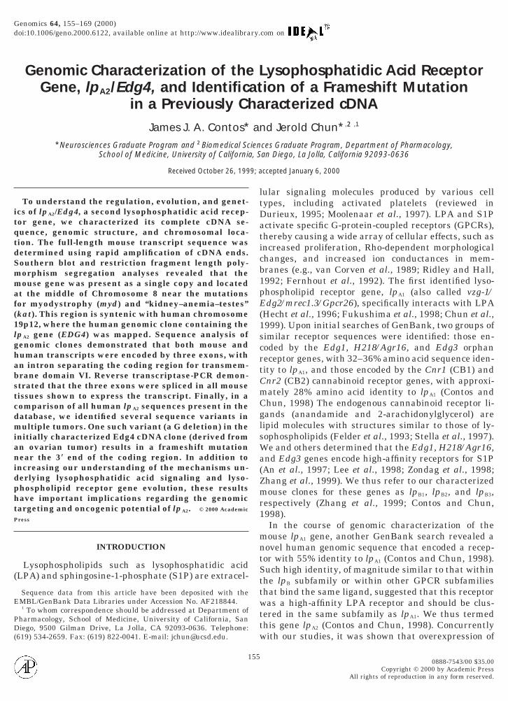
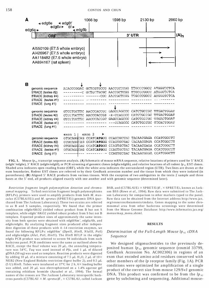
FIG. 1. Mouse lpA2 transcript sequence analysis. (A) Schematic ofedg6t9/edg6e), 39 RACE (edg6v/edg6f), or PCR screening of genomic c
Shaded area indicates open reading frame (ORF), while the white arexon boundaries. Rodent EST clones are referred to by their GenBaparentheses). (B) Aligned 59 RACE products from various tissues. Wbases at the 59 end, all products aligned exactly with one another a
BSB, and (C57BL/6JEi 3 SPRET/Ei)F1 3 SPRET/Ei, known as Jack-son BSS (Rowe et al., 1994). Raw data were submitted to The Jack-son Laboratory for comparison to other markers typed to the panel.Raw data can be obtained from the Internet address http://www.jax.org/resources/documents/cmdata. Genes mapping to the same chro-mosomal area from other backcross screenings were determinedfrom the Mouse Genome Database: http://www.informatics.jax.org/menus/map_menu.shtml.
RESULTS
Determination of the Full-Length Mouse lpA2 cDNASequence
We designed oligonucleotides to the previously de-posited human lpA2 genomic sequence (cosmid 33799,GenBank Accession No. AC002306) in regions of anexon that encoded amino acid residues conserved withother members of the lp receptor family (Fig. 1A). PCRconditions were optimized for amplification of a singleproduct of the correct size from mouse 129/SvJ genomicDNA. This product was confirmed to be from the lpA2
gene by subcloning and sequencing. Additional mouse-
use mRNA sequence, relative locations of primers used for 59 RACEes (edg6a/edg6b), and relative locations of all rodent lpA2 EST clones.ndicates the untranslated region (UTR). Two lines are drawn at theaccession number and the tissue from which they were isolated (in
the exception of two ambiguities in the testis 2 sample and threewith genomic sequence (determined later).
molonea inkith
nd
(bemtttCi
pfrgi
g
RcP
159GENOMIC CHARACTERIZATION OF lpA2
specific primers were then designed for use in both 59and 39 RACE experiments. Because Northern blotsindicated that the lpA2 transcript was most abundantin testis, kidney, and lung (J. J. A. Contos and J. Chun,unpublished observation), we used cDNA from thesetissues as templates in the amplifications. Unlike thelpA1 gene, which has multiple, divergent 59 sequences,we found that for lpA2, all four subcloned 59 RACEproducts from each tissue had the same sequence, al-though starting at slightly different points (Fig. 1B). In39 RACE experiments we obtained one product, whichcorresponded to three mouse expressed sequence tag(EST) clones deposited with GenBank, all from E7.5whole embryos (Fig. 1A). In addition to likely primersequence at the end of one EST, there was only oneother sequence variation between the cDNA and theESTs (noted under Materials and Methods). Polyade-nylation sites were identical in both the 39 RACE prod-ucts and the one EST clone that contained a poly(A)tail (AI426967). In addition, a rat EST clone(AA894167) appeared to add a poly(A) tail at this point.The total transcript length was 2647 bp, which com-bined with a 250-nt poly(A) tail (Wahle, 1995) corre-sponds to the approximate size of the major hybridiz-ing RNA species observed by Northern blot (J. J. A.Contos and J. Chun, unpublished observation). Anopen reading frame (ORF) of 1044 bp is present in thecDNA and encodes a putative protein with 347 aminoacids and 55% identity to mouse LPA1. The AUG startcodon (at nt 96) for this ORF lies within a Kozakconsensus sequence (Fig. 1B). Unlike lpA1, there are nomRNA destabilization AUUUA consensus sequencesin the lpA2 transcript.
The lpA2 Gene is Present as a Single Copyin M. musculus
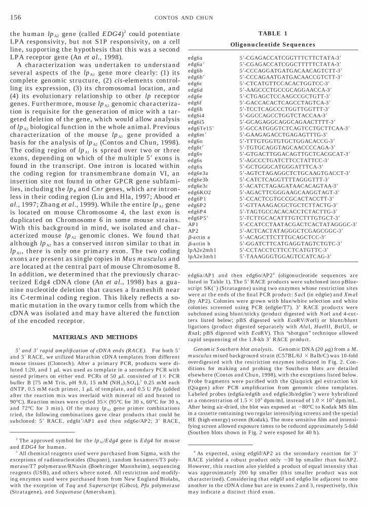
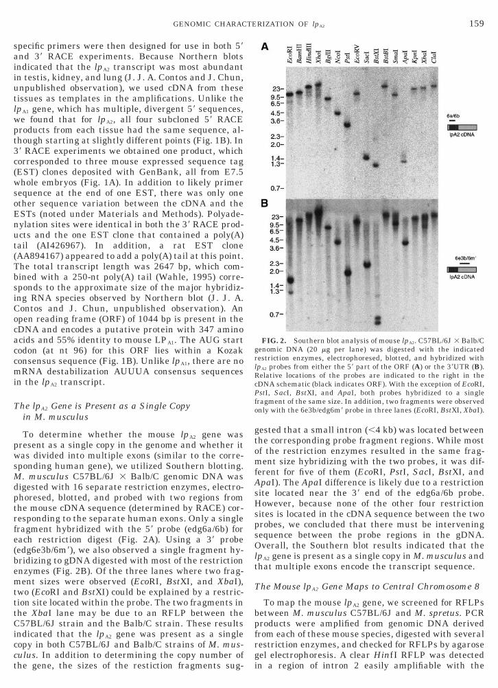
To determine whether the mouse lpA2 gene waspresent as a single copy in the genome and whether itwas divided into multiple exons (similar to the corre-sponding human gene), we utilized Southern blotting.M. musculus C57BL/6J 3 Balb/C genomic DNA wasdigested with 16 separate restriction enzymes, electro-phoresed, blotted, and probed with two regions fromthe mouse cDNA sequence (determined by RACE) cor-responding to the separate human exons. Only a singlefragment hybridized with the 59 probe (edg6a/6b) foreach restriction digest (Fig. 2A). Using a 39 probeedg6e3b/6m9), we also observed a single fragment hy-ridizing to gDNA digested with most of the restrictionnzymes (Fig. 2B). Of the three lanes where two frag-ent sizes were observed (EcoRI, BstXI, and XbaI),
wo (EcoRI and BstXI) could be explained by a restric-ion site located within the probe. The two fragments inhe XbaI lane may be due to an RFLP between the57BL/6J strain and the Balb/C strain. These results
ndicated that the lpA2 gene was present as a singlecopy in both C57BL/6J and Balb/C strains of M. mus-culus. In addition to determining the copy number ofthe gene, the sizes of the restiction fragments sug-
gested that a small intron (,4 kb) was located betweenthe corresponding probe fragment regions. While mostof the restriction enzymes resulted in the same frag-ment size hybridizing with the two probes, it was dif-ferent for five of them (EcoRI, PstI, SacI, BstXI, andApaI). The ApaI difference is likely due to a restrictionsite located near the 39 end of the edg6a/6b probe.However, because none of the other four restrictionsites is located in the cDNA sequence between the twoprobes, we concluded that there must be interveningsequence between the probe regions in the gDNA.Overall, the Southern blot results indicated that thelpA2 gene is present as a single copy in M. musculus andthat multiple exons encode the transcript sequence.
The Mouse lpA2 Gene Maps to Central Chromosome 8
To map the mouse lpA2 gene, we screened for RFLPsbetween M. musculus C57BL/6J and M. spretus. PCR
roducts were amplified from genomic DNA derivedrom each of these mouse species, digested with severalestriction enzymes, and checked for RFLPs by agaroseel electrophoresis. A clear HinfI RFLP was detectedn a region of intron 2 easily amplifiable with the
FIG. 2. Southern blot analysis of mouse lpA2. C57BL/6J 3 Balb/Cenomic DNA (20 mg per lane) was digested with the indicated
restriction enzymes, electrophoresed, blotted, and hybridized withlpA2 probes from either the 59 part of the ORF (A) or the 39UTR (B).
elative locations of the probes are indicated to the right in theDNA schematic (black indicates ORF). With the exception of EcoRI,stI, SacI, BstXI, and ApaI, both probes hybridized to a single
fragment of the same size. In addition, two fragments were observedonly with the 6e3b/edg6m9 probe in three lanes (EcoRI, BstXI, XbaI).
160 CONTOS AND CHUN
primers edg6s/edg6x. This was used to screen two F2
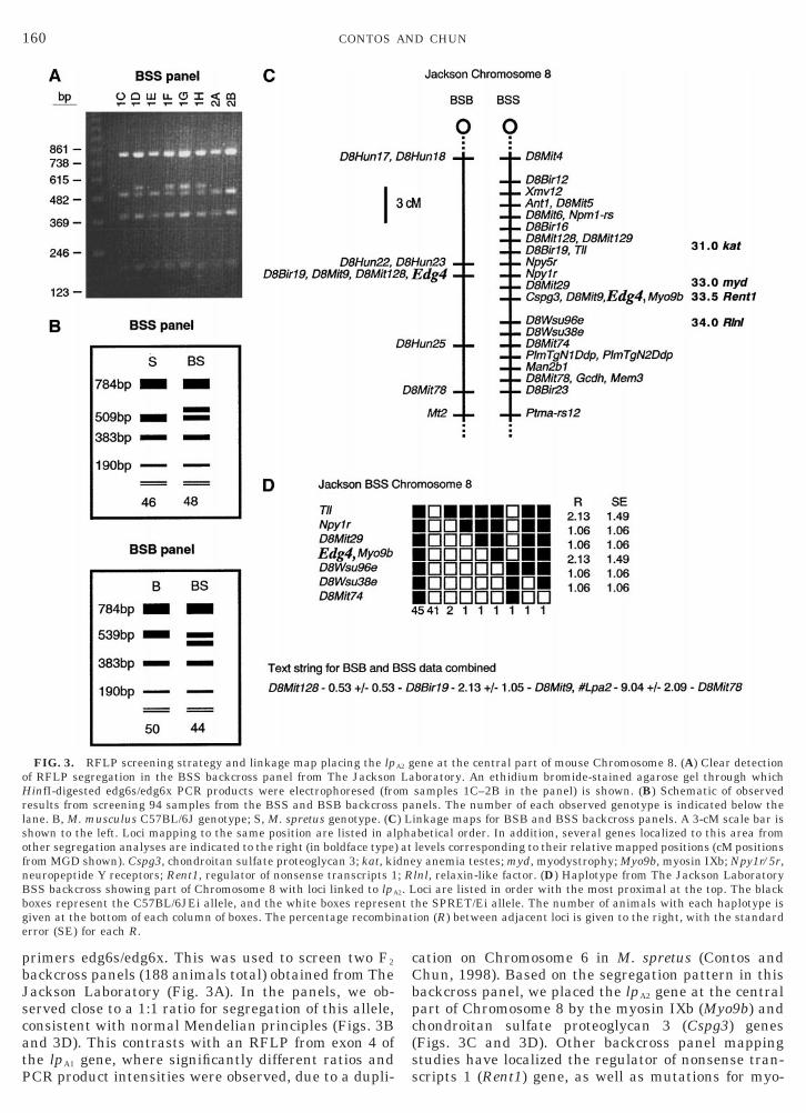
backcross panels (188 animals total) obtained from TheJackson Laboratory (Fig. 3A). In the panels, we ob-served close to a 1:1 ratio for segregation of this allele,consistent with normal Mendelian principles (Figs. 3Band 3D). This contrasts with an RFLP from exon 4 ofthe lpA1 gene, where significantly different ratios andPCR product intensities were observed, due to a dupli-
FIG. 3. RFLP screening strategy and linkage map placing the lpof RFLP segregation in the BSS backcross panel from The JacksonHinfI-digested edg6s/edg6x PCR products were electrophoresed (froresults from screening 94 samples from the BSS and BSB backcrosslane. B, M. musculus C57BL/6J genotype; S, M. spretus genotype. (Cshown to the left. Loci mapping to the same position are listed in alother segregation analyses are indicated to the right (in boldface typefrom MGD shown). Cspg3, chondroitan sulfate proteoglycan 3; kat, kineuropeptide Y receptors; Rent1, regulator of nonsense transcripts 1BSS backcross showing part of Chromosome 8 with loci linked to lpA
boxes represent the C57BL/6JEi allele, and the white boxes represegiven at the bottom of each column of boxes. The percentage recombierror (SE) for each R.
cation on Chromosome 6 in M. spretus (Contos andChun, 1998). Based on the segregation pattern in thisbackcross panel, we placed the lpA2 gene at the centralpart of Chromosome 8 by the myosin IXb (Myo9b) andchondroitan sulfate proteoglycan 3 (Cspg3) genes(Figs. 3C and 3D). Other backcross panel mappingstudies have localized the regulator of nonsense tran-scripts 1 (Rent1) gene, as well as mutations for myo-
ene at the central part of mouse Chromosome 8. (A) Clear detectionboratory. An ethidium bromide-stained agarose gel through whichsamples 1C–2B in the panel) is shown. (B) Schematic of observednels. The number of each observed genotype is indicated below theinkage maps for BSB and BSS backcross panels. A 3-cM scale bar isbetical order. In addition, several genes localized to this area fromlevels corresponding to their relative mapped positions (cM positionsy anemia testes; myd, myodystrophy; Myo9b, myosin IXb; Npy1r/5r,lnl, relaxin-like factor. (D) Haplotype from The Jackson Laboratoryoci are listed in order with the most proximal at the top. The blackhe SPRET/Ei allele. The number of animals with each haplotype ision (R) between adjacent loci is given to the right, with the standard
A2 gLampa
) Lpha) atdne; R2. Lnt tnat
g
aab
laros
161GENOMIC CHARACTERIZATION OF lpA2
dystrophy (myd) and kidney–anemia–testes (kat), veryclose to this locus as well (Fig. 3C).
The Mouse and Human lpA2 Transcript SequencesAre Encoded by Three Exons
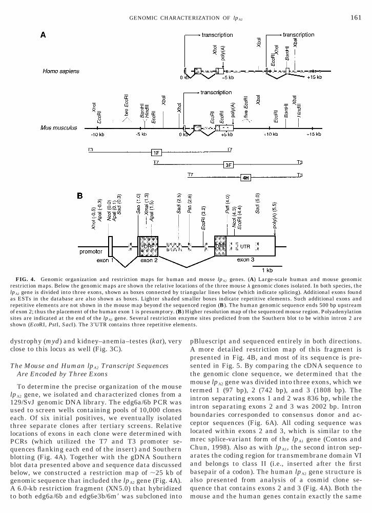
To determine the precise organization of the mouselpA2 gene, we isolated and characterized clones from a129/SvJ genomic DNA library. The edg6a/6b PCR wasused to screen wells containing pools of 10,000 cloneseach. Of six initial positives, we eventually isolatedthree separate clones after tertiary screens. Relativelocations of exons in each clone were determined withPCRs (which utilized the T7 and T3 promoter se-quences flanking each end of the insert) and Southernblotting (Fig. 4A). Together with the gDNA Southernblot data presented above and sequence data discussedbelow, we constructed a restriction map of ;25 kb ofenomic sequence that included the lpA2 gene (Fig. 4A).
A 6.0-kb restriction fragment (XN5.0) that hybridizedto both edg6a/6b and edg6e3b/6m9 was subcloned into
FIG. 4. Genomic organization and restriction maps for humanrestriction maps. Below the genomic maps are shown the relative locpA2 gene is divided into three exons, shown as boxes connected by ts ESTs in the database are also shown as boxes. Lighter shaded sepetitive elements are not shown in the mouse map beyond the sequf exon 2; thus the placement of the human exon 1 is presumptory. (Bites are indicated at the end of the lpA2 gene. Several restriction en
shown (EcoRI, PstI, SacI). The 39UTR contains three repetitive elem
pBluescript and sequenced entirely in both directions.A more detailed restriction map of this fragment ispresented in Fig. 4B, and most of its sequence is pre-sented in Fig. 5. By comparing the cDNA sequence tothe genomic clone sequence, we determined that themouse lpA2 gene was divided into three exons, which wetermed 1 (97 bp), 2 (742 bp), and 3 (1808 bp). Theintron separating exons 1 and 2 was 836 bp, while theintron separating exons 2 and 3 was 2002 bp. Intronboundaries corresponded to consensus donor and ac-ceptor sequences (Fig. 6A). All coding sequence waslocated within exons 2 and 3, which is similar to themrec splice-variant form of the lpA1 gene (Contos andChun, 1998). Also as with lpA1, the second intron sep-rates the coding region for transmembrane domain VInd belongs to class II (i.e., inserted after the firstasepair of a codon). The human lpA2 gene structure is
also presented from analysis of a cosmid clone se-quence that contains exons 2 and 3 (Fig. 4A). Both themouse and the human genes contain exactly the same
d mouse lpA2 genes. (A) Large-scale human and mouse genomicns of the three mouse l genomic clones isolated. In both species, thegular lines below (which indicate splicing). Additional exons foundller boxes indicate repetitive elements. Such additional exons andced region (B). The human genomic sequence ends 500 bp upstreamgher resolution map of the sequenced mouse region. Polyadenylation
e sites predicted from the Southern blot to be within intron 2 arets.
anatiorianmaen
) Hizymen

Aft(ttspmbs
A
162 CONTOS AND CHUN
size of exon 2 and a very similarly sized intron 2 (1973bp in human). However, polyadenylation occurs in thehuman clone approximately 1 kb upstream of the anal-ogous position in mice, which explains the shorter hu-man transcript size observed by Northern blot (An etal., 1998). The mouse polyadenylation sequence,
UUAAA, like the AGUAAA in human, differs slightlyrom the stronger consensus AAUAAA (Fig. 6B), al-hough both have been found to be used in other genesBirnstiel et al., 1985). Some GU clusters are found inhe appropriate location relative to the polyadenyla-ion consensus sequence (Figs. 6B and 8C). If theseignal sequences are not utilized, transcription mightroceed to produce the larger transcripts (;7 kb inouse and ;8 kb in human) observed in some tissues
y Northern blot (in addition to the more prevalentmaller transcript).
nalysis of lpA2 Exon Splicing Patterns by RT-PCR
To determine whether the proposed splicing indi-cated in Fig. 4 occurs in most tissues that express the
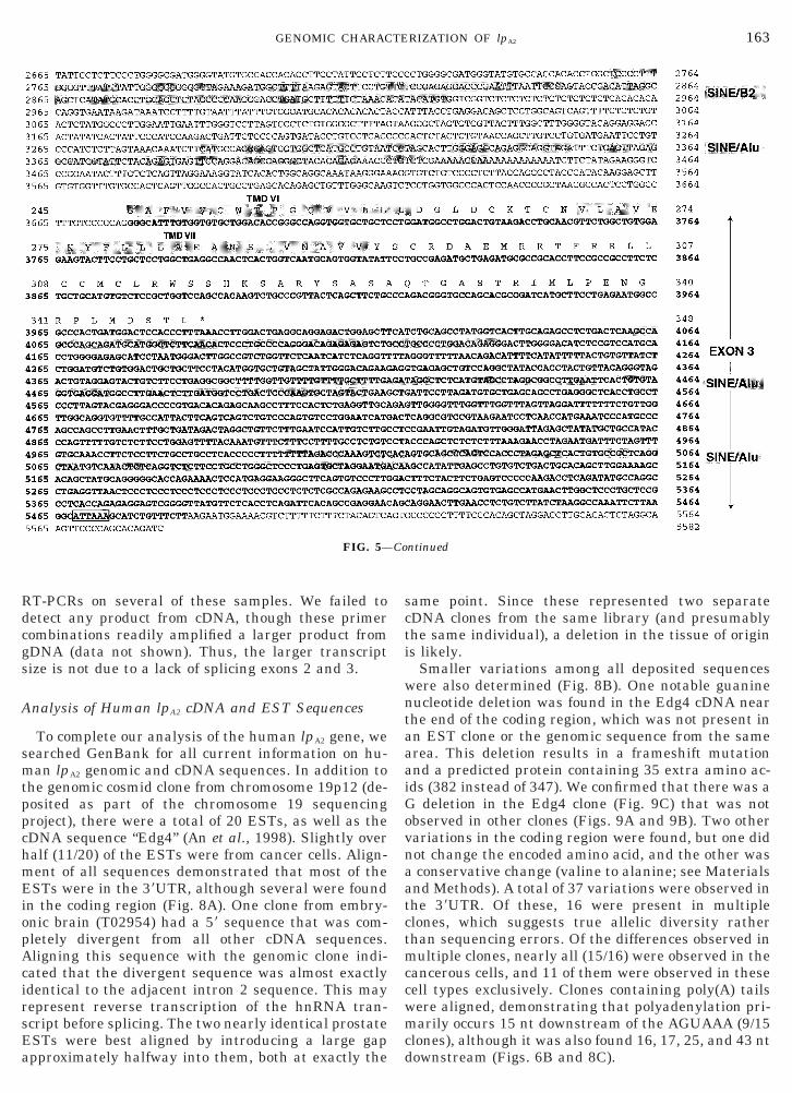
FIG. 5. Sequence of the mouse lpA2 gene. A total of 5588 bp of 12as determined by 59 RACE. The lpA2 gene exons are in boldface type anPutative transmembrane domains in the translation product are sharepetitive elements are shaded and indicated to the right.
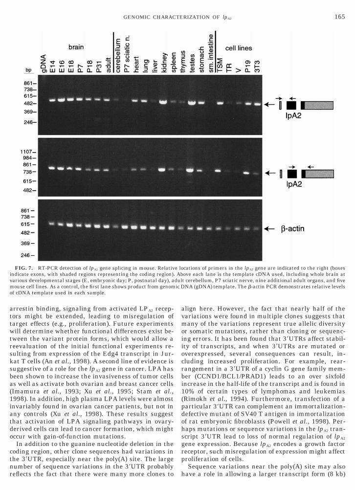
lpA2 gene, we used RT-PCR to detect spliced tran-scripts. Figure 7 shows that in all mouse tissues exam-ined that splice exons 2 and 3 together, exons 1 and 2are also spliced together. Furthermore, in this semi-quantitative assay, the relative levels of each of thesePCR products were similar between tissues, demon-strating that the RNA transcript consisting of splicedexons 1, 2, and 3 is the predominant form in most celltypes. Together with the 59 RACE results, this sug-gests that alternate primary exons are not utilized intranscription of the mouse lpA2 gene, which contrastswith the the lpA1 gene, where multiple primary exonsare found (Contos and Chun, 1998).
There is a larger mouse transcript observed byNorthern blot (of approximately 7 kb), which is mostvisible in the embryonic brain samples, but also detect-able in other adult tissues expressing large amounts ofthe smaller 2.8-kb transcript (J. J. A. Contos andJ. Chun, unpublished observation). To determinewhether this larger transcript contains intron 2 se-quence, we used primers from exon 1 and intron 2 in
vJ genomic sequence is shown, with 11 being the most 59 start sitendicated to the right of the sequence, with the ORF translated above.. In addition, the polyadenylation consensus sequence is boxed, and
9/Sd ided
tppchmEiopAcirsEa
scti
wntaaaiGovnaatctmccwmcd
163GENOMIC CHARACTERIZATION OF lpA2
RT-PCRs on several of these samples. We failed todetect any product from cDNA, though these primercombinations readily amplified a larger product fromgDNA (data not shown). Thus, the larger transcriptsize is not due to a lack of splicing exons 2 and 3.
Analysis of Human lpA2 cDNA and EST Sequences
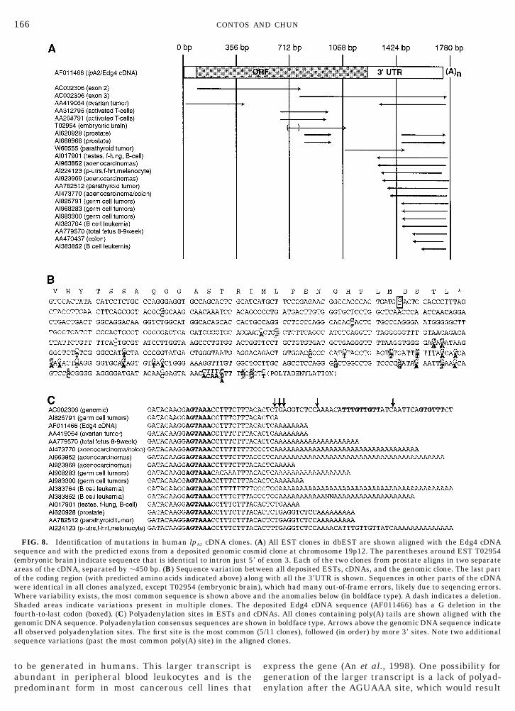
To complete our analysis of the human lpA2 gene, wesearched GenBank for all current information on hu-man lpA2 genomic and cDNA sequences. In addition tohe genomic cosmid clone from chromosome 19p12 (de-osited as part of the chromosome 19 sequencingroject), there were a total of 20 ESTs, as well as theDNA sequence “Edg4” (An et al., 1998). Slightly overalf (11/20) of the ESTs were from cancer cells. Align-ent of all sequences demonstrated that most of theSTs were in the 39UTR, although several were found
n the coding region (Fig. 8A). One clone from embry-nic brain (T02954) had a 59 sequence that was com-letely divergent from all other cDNA sequences.ligning this sequence with the genomic clone indi-
ated that the divergent sequence was almost exactlydentical to the adjacent intron 2 sequence. This mayepresent reverse transcription of the hnRNA tran-cript before splicing. The two nearly identical prostateSTs were best aligned by introducing a large gappproximately halfway into them, both at exactly the
FIG. 5—
ame point. Since these represented two separateDNA clones from the same library (and presumablyhe same individual), a deletion in the tissue of origins likely.
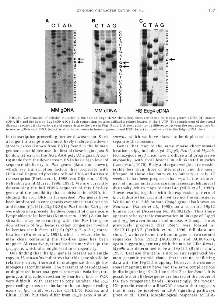
Smaller variations among all deposited sequencesere also determined (Fig. 8B). One notable guanineucleotide deletion was found in the Edg4 cDNA nearhe end of the coding region, which was not present inn EST clone or the genomic sequence from the samerea. This deletion results in a frameshift mutationnd a predicted protein containing 35 extra amino ac-ds (382 instead of 347). We confirmed that there was a
deletion in the Edg4 clone (Fig. 9C) that was notbserved in other clones (Figs. 9A and 9B). Two otherariations in the coding region were found, but one didot change the encoded amino acid, and the other wasconservative change (valine to alanine; see Materialsnd Methods). A total of 37 variations were observed inhe 39UTR. Of these, 16 were present in multiplelones, which suggests true allelic diversity ratherhan sequencing errors. Of the differences observed inultiple clones, nearly all (15/16) were observed in the
ancerous cells, and 11 of them were observed in theseell types exclusively. Clones containing poly(A) tailsere aligned, demonstrating that polyadenylation pri-arily occurs 15 nt downstream of the AGUAAA (9/15
lones), although it was also found 16, 17, 25, and 43 ntownstream (Figs. 6B and 8C).
ntinued
Co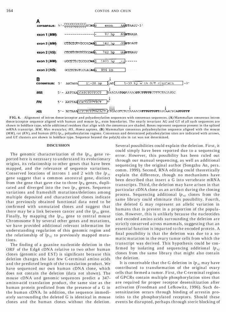
dsm(a
164 CONTOS AND CHUN
DISCUSSION
The genomic characterization of the lpA2 gene re-ported here is necessary to understand its evolutionaryorigins, its relationship to other genes that have beenmapped, and the relevance of sequence variations.Conserved locations of introns 1 and 2 with the lpA1
gene suggest that a common ancestral gene, distinctfrom the gene that gave rise to three lpB genes, dupli-cated and diverged into the two lpA genes. Sequencevariations and frameshift mutations/deletions amongmultiple deposited and characterized clones indicatethat previously obtained functional data need to beconfirmed with unmutated clones and suggest thatthere may be a link between cancer and the lpA2 gene.Finally, by mapping the lpA2 gene to central mouseChromosome 8 by several other genes and mutations,we have provided additional relevant information forunderstanding regulation of this genomic region andthe relationship of lpA2 to previously mapped muta-tions.
The finding of a guanine nucleotide deletion in theORF of the Edg4 cDNA relative to two other humanclones (genomic and EST) is significant because thisdeletion changes the last few C-terminal amino acidsand the predicted length of the translation product. Wehave sequenced our own human cDNA clone, whichdoes not contain the deletion (data not shown). Themouse cDNA and genomic sequences predict a 347-amino-acid translation product, the same size as thehuman protein predicted from the presence of a G inthe human cDNA. In addition, the sequence immedi-ately surrounding the deleted G is identical in mouseclones and the human clones without the deletion.
FIG. 6. Alignment of intron donor/acceptor and polyadenylationonor/acceptor sequence aligned with human and mouse lpA2 exon bhown in boldface type, while additional residues that align with theRNA transcript. MM, Mus musculus; HS, Homo sapiens. (B) Mam
MM), rat (RN), and human (HS) lpA2 polyadenylation regions. Consnd GT clusters are shown in boldface type. Sequence beyond the p
Several possibilities could explain the deletion. First, itcould simply have been reported due to a sequencingerror. However, this possibility has been ruled outthrough our manual sequencing, as well as additionalsequencing by the original author (Songzhu An, pers.comm. 1999). Second, RNA editing could theoreticallyexplain the difference, though no mechanisms havebeen described that insert a G into vertebrate mRNAtranscripts. Third, the deletion may have arisen in thatparticular cDNA clone as an artifact during the cloningprocess. Sequencing additional lpA2 clones from thesame library could eliminate this possibility. Fourth,the deleted G may represent an allelic variation inhumans that is present in a proportion of the popula-tion. However, this is unlikely because the nucleotidesand encoded amino acids surrounding the deletion arestrictly conserved across mammals, suggesting that anessential function is imparted to the encoded protein. Afinal possibility is that the deletion was due to a so-matic mutation in the ovary tumor cells from which thetranscript was derived. This hypothesis could be con-firmed by isolating and sequencing additional lpA2
clones from the same library that might also containthe deletion.
It is conceivable that the G deletion in lpA2 may havecontributed to transformation of the original ovarycells that formed a tumor. First, the C-terminal regionsof GPCRs contain multiple phosphorylation sites thatare required for proper receptor desensitization afteractivation (Freedman and Lefkowitz, 1996). Such de-sensitization occurs through binding of arrestin pro-teins to the phosphorylated receptors. Should theseevents be disrupted, perhaps through steric blocking of
uences with consensus sequences. (A) Mammalian consensus introndaries. The nearly invariant AG and GT of all such sequences aresensus are shaded. Boxes represent sequence present in the splicedalian consensus polyadenylation sequence aligned with the mouseus and determined polyadenylation sites are indicated with arrows,A) site in rat was not determined.
seqounconm
ensoly(
ttwtrsks
1iatdo
ctnr
avmoiiocr
pdoh
ivmo
165GENOMIC CHARACTERIZATION OF lpA2
arrestin binding, signaling from activated LPA2 recep-ors might be extended, leading to misregulation ofarget effects (e.g., proliferation). Future experimentsill determine whether functional differences exist be-
ween the variant protein forms, which would allow aeevaluation of the initial functional experiments re-ulting from expression of the Edg4 transcript in Jur-at T cells (An et al., 1998). A second line of evidence isuggestive of a role for the lpA2 gene in cancer. LPA has
been shown to increase the invasiveness of tumor cellsas well as activate both ovarian and breast cancer cells(Imamura et al., 1993; Xu et al., 1995; Stam et al.,998). In addition, high plasma LPA levels were almostnvariably found in ovarian cancer patients, but not inny controls (Xu et al., 1998). These results suggesthat activation of LPA signaling pathways in ovary-erived cells can lead to cancer formation, which mightccur with gain-of-function mutations.In addition to the guanine nucleotide deletion in the
oding region, other clone sequences had variations inhe 39UTR, especially near the poly(A) site. The largeumber of sequence variations in the 39UTR probablyeflects the fact that there were many more clones to
FIG. 7. RT-PCR detection of lpA2 gene splicing in mouse. Relativndicate exons, with shaded regions representing the coding region).arious developmental stages (E, embryonic day; P, postnatal day), aouse cell lines. As a control, the first lane shows product from genom
f cDNA template used in each sample.
lign here. However, the fact that nearly half of theariations were found in multiple clones suggests thatany of the variations represent true allelic diversity
r somatic mutations, rather than cloning or sequenc-ng errors. It has been found that 39UTRs affect stabil-ty of transcripts, and when 39UTRs are mutated orverexpressed, several consequences can result, in-luding increased proliferation. For example, rear-angement in a 39UTR of a cyclin G gene family mem-
ber (CCND1/BCL1/PRAD1) leads to an over sixfoldincrease in the half-life of the transcript and is found in10% of certain types of lymphomas and leukemias(Rimokh et al., 1994). Furthermore, transfection of a
articular 39UTR can complement an immortalization-efective mutant of SV40 T antigen in immortalizationf rat embryonic fibroblasts (Powell et al., 1998). Per-aps mutations or sequence variations in the lpA2 tran-
script 39UTR lead to loss of normal regulation of lpA2
gene expression. Because lpA2 encodes a growth factorreceptor, such misregulation of expression might affectproliferation of cells.
Sequence variations near the poly(A) site may alsohave a role in allowing a larger transcript form (8 kb)
cations of primers in the lpA2 gene are indicated to the right (boxesove each lane is the template cDNA used, including whole brain att cerebellum, P7 sciatic nerve, nine additional adult organs, and fiveNA (gDNA) template. The b-actin PCR demonstrates relative levels
e loAb
dulic D
ge
166 CONTOS AND CHUN
to be generated in humans. This larger transcript isabundant in peripheral blood leukocytes and is thepredominant form in most cancerous cell lines that
FIG. 8. Identification of mutations in human lpA2 cDNA clones.sequence and with the predicted exons from a deposited genomic cos(embryonic brain) indicate sequence that is identical to intron just 5areas of the cDNA, separated by ;450 bp. (B) Sequence variation beof the coding region (with predicted amino acids indicated above) alowere identical in all clones analyzed, except T02954 (embryonic braiWhere variability exists, the most common sequence is shown aboveShaded areas indicate variations present in multiple clones. Thefourth-to-last codon (boxed). (C) Polyadenylation sites in ESTs andgenomic DNA sequence. Polyadenylation consensus sequences are shall observed polyadenylation sites. The first site is the most commonsequence variations (past the most common poly(A) site) in the alig
express the gene (An et al., 1998). One possibility foreneration of the larger transcript is a lack of polyad-nylation after the AGUAAA site, which would result
All EST clones in dbEST are shown aligned with the Edg4 cDNAclone at chromosome 19p12. The parentheses around EST T02954
exon 3. Each of the two clones from prostate aligns in two separateen all deposited ESTs, cDNAs, and the genomic clone. The last partwith all the 39UTR is shown. Sequences in other parts of the cDNAwhich had many out-of-frame errors, likely due to seqencing errors.the anomalies below (in boldface type). A dash indicates a deletion.
osited Edg4 cDNA sequence (AF011466) has a G deletion in theAs. All clones containing poly(A) tails are shown aligned with thein boldface type. Arrows above the genomic DNA sequence indicate
/11 clones), followed (in order) by more 39 sites. Note two additionalclones.
(A)mid9 oftwengn),anddepcDNown(5
ned
rmogv
elwpdTlWNhaa
c
167GENOMIC CHARACTERIZATION OF lpA2
in transcription proceeding further downstream. Sucha longer transcript would most likely include the down-stream exons (known from ESTs) found in the humangenomic cosmid because the first of these begins just 5kb downstream of the AGUAAA poly(A) signal. A con-tig made from the downstream ESTs has a high level ofsequence similarity to Pbx genes (data not shown),which are transcription factors that cooperate withHOX and Engrailed proteins to bind DNA and activatetranscription (Phelan et al., 1995; van Dijk et al., 1995;Peltenburg and Murre, 1996, 1997). We are currentlydetermining the full cDNA sequence of this Pbx-likegene and the possibility that a bicistronic mRNA, in-cluding the lpA2 ORF, is transcribed. Pbx genes havebeen implicated in oncogenesis ever since translocationand fusion [t(1:19) (q23;p13.3)] of Pbx1 to the E2A genewere shown to precede the development of many acutelymphoblastic leukemias (Kamps et al., 1990) A similarsituation may be occurring with the Pbx-like genedownstream of lpA2, because other congenital myeloidleukemias result from t(11;19) (q23;p12–p13.1) trans-locations (Huret et al., 1993), which is where the hu-man clone containing the Pbx-like gene has beenmapped. Alternatively, translocations may disrupt thelpA2 gene, which also might lead to oncogenicity.
Our finding that the lpA2 gene is present as a singlecopy in M. musculus indicates that this gene should beelatively straightforward to mutagenize through ho-ologous recombination. The presence of pseudogenes
r duplicated functional genes can make isolation, tar-eting, and specific detection by Southern blot or PCRery difficult. With respect to copy number, the lpA2
gene coding exons are similar to the analogous codingexons of lpA1 in M. musculus C57BL/6J (Contos andChun, 1998), but they differ from lpA1’s exon 4 in M.
FIG. 9. Confirmation of deletion mutation in the human Edg4 cDNA (B), and the human Edg4 cDNA (C). Each sequencing reaction
dideoxy reactions is shown for ease of comparison to the data in Figsin mouse gDNA and cDNA (which is also the sequence in human ge
spretus, which we have shown to be duplicated on aseparate chromosome.
Genes that map to the same mouse chromosomallocation as lpA2 include myd, Cspg3, Rent1, and Myo9b.Homozygous myd mice have a diffuse and progressivemyopathy, with focal lesions in all skeletal muscles(Lane et al., 1976). Body and organ weights are consid-rably less than those of littermates, and the meanifespan of those that survive to puberty is only 17eeks. It has been proposed that myd is the counter-art of human mutations causing facioscapulohumeralystrophy, which maps to distal 4q (Mills et al., 1995).hese results, together with the expression pattern of
pA2, indicate that lpA2 and myd are not the same gene.e found the 12-kb human Cspg3 gene, also known aseurocan (Rauch et al., 1995), on a separate 19p12uman cosmid (Accession No. AC002126). Thus thereppears to be syntenic conservation in linkage of Cspg3nd lpA2 between human and mouse. Although it was
previously proposed that Rent1 was located at19p13.11–p13.2 (Perlick et al., 1996; full data notshown), we have found the human gene on two cosmidsequences from 19p12 (AC003972 and AC004517),again suggesting synteny with the mouse. Like Rent1,Myo9b was determined to be at 19p13.1 (Bahler et al.,1997). Because this gene is not on any sequenced hu-man genomic cosmid clone, there are no conflictingdata with the 19p13.1 mapping. However, the chromo-somal mapping may have had the same complicationsin distinguishing 19p13.1 and 19p12 as for Rent1. It ispossible that all these genes are located at the border ofthe two cytogenetic bands. Interestingly, the myosinIXb protein contains a RhoGAP domain that suggeststhat it may be involved in LPA signaling pathways(Post et al., 1998). Morphological responses to LPA-
A clone. Sequences are shown for mouse genomic DNA (A), mouseilized a primer located in the 39UTR. The complement of the actual
and 8. Arrows point to the difference between the sequences: two Gsic and EST clones) and only one G in the Edg4 cDNA clone.
DNut
. 5nom
pbA
W
t
ft
A
A
vation of myosin by Rho-associated kinase (Rho-kinase). J. Biol.
H
I
J
K
L
L
168 CONTOS AND CHUN
receptor stimulation rely on Rho activation (Moolenaaret al., 1997; Fukushima et al., 1998), which in otherexperiments has been shown to cause phosphorylationof myosins through activation of a Rho kinase (Amanoet al., 1996). Perhaps there is a genomic regulatory linkbetween lpA2 and this RhoGAP.
Another gene that maps fairly close to lpA2 is kat. Thegross phenotype of homozygous kat mutant mice (on anRBF background), as well as homozygous kat2J allelicmutant mice, includes polycystic kidney disease, ane-mia, and male sterility (Janaswami et al., 1997). How-ever, the homozygous kat phenotype is dependent onbackground strain (e.g., on a C57BL/6J background,the only gross phenotypes observed were a domedskull, dwarfing, and a mortality of ;50% before wean-ing). Interestingly, the more severe phenotypes are inorgans where the lpA2 gene is most abundantly ex-
ressed. The kat gene was mapped to a small intervaletween the D8Mit128 and the D8Mit129 marker loci.lthough we found no crossovers between lpA2 and
D8Mit128 in the BSB panel, there were four crossoverswith both D8Mit128 and D8Mit129 in the BSS panel.
e also excluded mutations in lpA2 exons as the causeof the kat phenotype through sequencing amplified ex-ons from both kat and RBF strains (data not shown).Thus, mutations in the lpA2 gene are likely not respon-sible for the kat phenotypes.
Our genomic characterization and sequence compar-ison results provide a large number of hypotheses thatwill be interesting to test in future studies. These in-volve the role of 39UTR or coding mutations in theoncogenicity of the lpA2 gene, the possibility that mu-ations in lpA2 are common in certain types of cancer,
and the relation of lpA2 and LPA signaling to neighbor-ing transcription units in the genome. Furthermore,we provide the necessary information and reagents tocreate lpA2 targeted mutations. The genomic character-ization of the lpA2 gene presented thus provides a basisor future experiments that will definitively determinehe role of lpA2 in both development and disease.
ACKNOWLEDGMENTS
We thank Lucy Rowe and Mary Barter for alignment of the seg-regation mapping results and providing the linkage maps; SongzhuAn for providing the Edg4 cDNA clone; Carol Akita for sequencingone of the Edg4 clones; and K. C. Cox for copyediting the manuscript.This work was supported by the National Institute of Mental Healthand by a grant from Allelix Biopharmaceuticals.
REFERENCES
Abood, M. E., Ditto, K. E., Noel, M. A., Showalter, V. M., and Tao, Q.(1997). Isolation and expression of a mouse CB1 cannabinoid re-ceptor gene. Comparison of binding properties with those of nativeCB1 receptors in mouse brain and N18TG2 neuroblastoma cells.Biochem. Pharmacol. 53: 207–214.
ltschul, S. F., Gish, W., Miller, W., Myers, E. W., and Lipman, D. J.(1990). Basic local alignment search tool. J. Mol. Biol. 215: 403–410.mano, M., Ito, M., Kimura, K., Fukata, Y., Chihara, K., Nakano, T.,Matsuura, Y., and Kaibuchi, K. (1996). Phosphorylation and acti-
Chem. 271: 20246–20249.An, S., Bleu, T., Hallmark, O. G., and Goetzl, E. J. (1998). Charac-
terization of a novel subtype of human G protein-coupled receptorfor lysophosphatidic acid. J. Biol. Chem. 273: 7906–7910.
An, S., Bleu, T., Huang, W., Hallmark, O. G., Coughlin, S. R., andGoetzl, E. J. (1997). Identification of cDNAs encoding two G pro-tein-coupled receptors for lysosphingolipids. FEBS Lett. 417: 279–282.
Ausubel, F. M., Brent, R., Kingston, R. E., Moore, D. D., Seidman,J. G., Smith, J. A., and Struhl, K. (1994). “Current Protocols inMolecular Biology,” Wiley, New York.
Bahler, M., Kehrer, I., Gordon, L., Stoffler, H. E., and Olsen, A. S.(1997). Physical mapping of human myosin-IXB (MYO9B), thehuman orthologue of the rat myosin myr 5, to chromosome19p13.1. Genomics 43: 107–109.
Birnstiel, M. L., Busslinger, M., and Strub, K. (1985). Transcriptiontermination and 39 processing: The end is in site! Cell 41: 349–359.
Chun, J., Contos, J., and Munroe, D. (1999). A growing family ofreceptor genes for lysophosphatidic acid (LPA) and other lysophos-pholipids (LPs). Cell. Biochem. Biophys. 30: 213–242.
Contos, J. J., and Chun, J. (1998). Complete cDNA sequence,genomic structure, and chromosomal localization of the LPA re-ceptor gene, lpA1/vzg-1/Gpcr26. Genomics 51: 364–378.
Durieux, M. E. (1995). “Lysophosphatidate Signaling: Cellular Ef-fects and Molecular Mechanisms,” Landes, Austin, TX.
Felder, C. C., Briley, E. M., Axelrod, J., Simpson, J. T., Mackie, K.,and Devane, W. A. (1993). Anandamide, an endogenous canna-bimimetic eicosanoid, binds to the cloned human cannabinoid re-ceptor and stimulates receptor-mediated signal transduction.Proc. Natl. Acad. Sci. USA 90: 7656–7660.
Fernhout, B. J., Dijcks, F. A., Moolenaar, W. H., and Ruigt, G. S.(1992). Lysophosphatidic acid induces inward currents in Xenopuslaevis oocytes: Evidence for an extracellular site of action. Eur.J. Pharmacol. 213: 313–315.
Freedman, N. J., and Lefkowitz, R. J. (1996). Desensitization of Gprotein-coupled receptors. Rec. Prog. Horm. Res. 51: 319–353.
Fukushima, N., Kimura, Y., and Chun, J. (1998). A single receptorencoded by vzg-1/lpA1/edg-2 couples to G-proteins and mediatesmultiple cellular responses to lysophosphatidic acid (LPA). Proc.Natl. Acad. Sci. USA 95: 6151–6156.
Hecht, J. H., Weiner, J. A., Post, S. R., and Chun, J. (1996). Ventric-ular zone gene-1 (vzg-1) encodes a lysophosphatidic acid receptorexpressed in neurogenic regions of the developing cerebral cortex.J. Cell. Biol. 135: 1071–1083.uret, J. L., Brizard, A., Slater, R., Charrin, C., Bertheas, M. F.,Guilhot, F., Hahlen, K., Kroes, W., van Leeuwen, E., Schoot, E. V.,Beishuizen, A., Tanzer, J., and Hagemeijer, A. (1993). Cytogeneticheterogeneity in t(11;19) acute leukemia: Clinical, hematologicaland cytogenetic analyses of 48 patients—Updated published casesand 16 new observations. Leukemia 7: 152–160.
mamura, F., Horai, T., Mukai, M., Shinkai, M., Sawada, M., andAdkedo, H. (1993) Induction of in vitro tumor cell invasion ofcellular monolayers by lysophosphatidic acid or phospholipase D.Biochem. Biophys. Res. Commun. 193: 497–503.
anaswami, P. M., Birkenmeier, E. H., Cook, S. A., Rowe, L. B.,Bronson, R. T., and Davisson, M. T. (1997). Identification andgenetic mapping of a new polycystic kidney disease on mousechromosome 8. Genomics 40: 101–107.amps, M. P., Murre, C., Sun, X. H., and Baltimore, D. (1990). A newhomeobox gene contributes the DNA binding domain of the t(1;19)translocation protein in pre-B ALL. Cell 60: 547–555.
ane, P. W., Beamer, T. C., and Myers, D. D. (1976). Myodystrophy,a new myopathy on chromosome 8 of the mouse. J. Hered. 67:135–138.
ee, M. J., Van Brocklyn, J. R., Thangada, S., Liu, C. H., Hand, A. R.,Menzeleev, R., Spiegel, S., and Hla, T. (1998). Sphingosine-1-
phosphate as a ligand for the G protein-coupled receptor EDG-1.
L
M
M
M
P
P
P
P
P
P
R
R
R
Rimokh, R., Berger, F., Bastard, C., Klein, B., French, M., Archim-
R
S
S
S
S
v
v
W
X
X
Z
Z
169GENOMIC CHARACTERIZATION OF lpA2
Science 279: 1552–1555.iu, C. H., and Hla, T. (1997). The mouse gene for the inducibleG-protein-coupled receptor edg-1. Genomics 43: 15–24.archuk, D., Drumm, M., Saulino, A., and Collins, F. S. (1991).Construction of T-vectors, a rapid and general system for directcloning of unmodified PCR products. Nucleic Acids Res. 19: 1154.ills, K. A., Mathews, K. D., Scherpbier-Heddema, T., Schelper,R. L., Schmalzel, R., Bailey, H. L., Nadeau, J. H., Buetow, K. H.,and Murray, J. C. (1995). Genetic mapping near the myd locus onmouse chromosome 8. Mamm. Genome 6: 278–280.oolenaar, W. H., Kranenburg, O., Postma, F. R., and Zondag, G. C.(1997). Lysophosphatidic acid: G-protein signalling and cellularresponses. Curr. Opin. Cell Biol. 9: 168–173.
eltenburg, L. T., and Murre, C. (1996). Engrailed and Hox home-odomain proteins contain a related Pbx interaction motif thatrecognizes a common structure present in Pbx. EMBO J. 15: 3385–3393.
eltenburg, L. T., and Murre, C. (1997). Specific residues in the Pbxhomeodomain differentially modulate the DNA-binding activity ofHox and Engrailed proteins. Development 124: 1089–1098.
erlick, H. A., Medghalchi, S. M., Spencer, F. A., Kendzior, R. J., Jr.,and Dietz, H. C. (1996). Mammalian orthologues of a yeast regu-lator of nonsense transcript stability. Proc. Natl. Acad. Sci. USA93: 10928–10932.
helan, M. L., Rambaldi, I., and Featherstone, M. S. (1995). Coop-erative interactions between HOX and PBX proteins mediated bya conserved peptide motif. Mol. Cell. Biol. 15: 3989–3997.
ost, P. L., Bokoch, G. M., and Mooseker, M. S. (1998). Humanmyosin-IXb is a mechanochemically active motor and a GAP forrho. J. Cell. Sci. 111: 941–950.
owell, A. J., Gates, P. B., Wylie, D., Velloso, C. P., Brockes, J. P.,and Jat, P. S. (1998). Immortalization of rat embryo fibroblasts bya 39-untranslated region. Exp. Cell Res. 240: 252–262.
aff, T., van der Giet, M., Endemann, D., Wiederholt, T., and Paul,M. (1997). Design and testing of beta-actin primers for RT-PCRthat do not co-amplify processed pseudogenes. BioTechniques 23:456–460.auch, U., Grimpe, B., Kulbe, G., Arnold-Ammer, I., Beier, D. R., andFassler, R. (1995). Structure and chromosomal localization of themouse neurocan gene. Genomics 28: 405–410.idley, A. J., and Hall, A. (1992). The small GTP-binding protein rhoregulates the assembly of focal adhesions and actin stress fibers inresponse to growth factors. Cell 70: 389–399.
baud, E., Rouault, J. P., Santa Lucia, B., Duret, L., Vuillaume, M.,et al. (1994). Rearrangement of CCND1 (BCL1/PRAD1) 39 un-translated region in mantle-cell lymphomas and t(11q13)-associ-ated leukemias. Blood 83: 3689–3696.owe, L. B., Nadeau, J. H., Turner, R., Frankel, W. N., Letts, V. A.,Eppig, J. T., Ko, M. S., Thurston, S. J., and Birkenmeier, E. H.(1994). Maps from two interspecific backcross DNA panels avail-able as a community genetic mapping resource. Mamm. Genome 5:253–274.
anger, F., Nicklen, S., and Coulson, A. R. (1977). DNA sequencingwith chain-terminating inhibitors. Proc. Natl. Acad. Sci. USA 74:5463–5467.
mit, A. F. (1996). The origin of interspersed repeats in the humangenome. Curr. Opin. Genet. Dev. 6: 743–748.
tam, J. C., Michiels, F., van der Kammen, R. A., Moolenaar, W. H.,and Collard, J. G. (1998). Invasion of T-lymphoma cells: Coopera-tion between Rho family GTPases and lysophospholipid receptorsignaling. EMBO J. 17: 4066–4074.
tella, N., Schweitzer, P., and Piomelli, D. (1997). A second endoge-nous cannabinoid that modulates long-term potentiation. Nature388: 773–778.
an Corven, E. J., Groenink, A., Jalink, K., Eichholtz, T., andMoolenaar, W. H. (1989). Lysophosphatidate-induced cell prolifer-ation: Identification and dissection of signaling pathways medi-ated by G proteins. Cell 59: 45–54.
an Dijk, M. A., Peltenburg, L. T., and Murre, C. (1995). Hox geneproducts modulate the DNA binding activity of Pbx1 and Pbx2.Mech. Dev. 52: 99–108.ahle, E. (1995). Poly(A) tail length control is caused by terminationof processive synthesis. J. Biol. Chem. 270: 2800–2808.
u, Y, Fang, X. J., Casey, G., and Mills, G. B. (1995). Lysophospho-lipids activate ovarian and breast cancer cells. Biochem. J. 309:933–940.
u, Y, Shen, Z., Wiper, D. W., Wu, M., Morton, R. E., Elson, P.,Kennedy, A. W., Belinson, J., Markman, M., and Casey, G. (1998).Lysophosphatidic acid as a potential biomarker for ovarian andother gynecologic cancers. J. Am. Med. Assoc. 280: 719–723.
hang, G., Contos, J. J., Weiner, J. A., Fukushima, N., and Chun, J.(1999). Comparative analysis of three murine G-protein coupledreceptors activated by sphingosine-1-phosphate. Gene 227: 89–99.
ondag, G. C., Postma, F. R., Etten, I. V., Verlaan, I., and Moolenaar,W. H. (1998). Sphingosine 1-phosphate signalling through theG-protein-coupled receptor Edg-1. Biochem. J. 330: 605–609.