Genes, Chromosomes,and Disease - the-eye.eu Chromosomes and Disease... · Chapter 4,...
353
Transcript of Genes, Chromosomes,and Disease - the-eye.eu Chromosomes and Disease... · Chapter 4,...
ptg

ptg
Genes, Chromosomes,and Disease

ptg
This page intentionally left blank

ptg
Genes, Chromosomes,and Disease
From Simple Traits, to Complex Traits,to Personalized Medicine
Nicholas Wright Gillham

ptg
Vice President, Publisher: Tim MooreAssociate Publisher and Director of Marketing: Amy NeidlingerAcquisitions Editor: Kirk JensenEditorial Assistant: Pamela BolandOperations Manager: Gina KanouseSenior Marketing Manager: Julie PhiferPublicity Manager: Laura CzajaAssistant Marketing Manager: Megan ColvinCover Designer: Chuti PrasertsithCover Illustration: Digital Art/CorbisManaging Editor: Kristy HartProject Editor: Betsy HarrisCopy Editor: Karen AnnettProofreader: Kathy RuizIndexer: Rebecca SalernoSenior Compositor: Gloria SchurickManufacturing Buyer: Dan Uhrig
© 2011 by Pearson Education, Inc.Publishing as FT Press ScienceUpper Saddle River, New Jersey 07458
FT Press offers excellent discounts on this book when ordered in quantity for bulk purchasesor special sales. For more information, please contact U.S. Corporate and Government Sales,1-800-382-3419, [email protected]. For sales outside the U.S., please contactInternational Sales at [email protected].
Company and product names mentioned herein are the trademarks or registered trademarksof their respective owners.
All rights reserved. No part of this book may be reproduced, in any form or by any means,without permission in writing from the publisher.
Printed in the United States of AmericaFirst Printing June 2011
ISBN-10: 0-13-707544-8ISBN-13: 978-0-13-707544-7
Pearson Education LTD.Pearson Education Australia PTY, Limited.Pearson Education Singapore, Pte. Ltd.Pearson Education Asia, Ltd.Pearson Education Canada, Ltd.Pearson Educación de Mexico, S.A. de C.V.Pearson Education—JapanPearson Education Malaysia, Pte. Ltd.
Library of Congress Cataloging-in-Publication DataGillham, Nicholas W. (Nicholas Wright), 1932- author.Genes, chromosomes, and disease : from simple traits, to complex traits, to personalized
medicine/ Nicholas Gillham.p. ; cm.
Includes bibliographical references.ISBN-13: 978-0-13-707544-7 (hardback : alk. paper)ISBN-10: 0-13-707544-8 (hardback : alk. paper)1. Genetic disorders—Susceptibility. 2. Human chromosomes. I. Title. [DNLM: 1. Genetic Disease, Inborn—genetics. 2. Chromosomes, Human. 3. Genetic
Disease, Inborn—therapy. 4. Genetic Predisposition to Disease—genetics. 5. GeneticTechniques. QZ 50]RB155.5.G55 2011616'.042—dc22
2010051297
Wow! eBook <WoweBook.Com>

ptg
In memory of my brother Oliver
Wow! eBook <WoweBook.Com>

ptg
This page intentionally left blank
Wow! eBook <WoweBook.Com>

ptg
Contents
Preface . . . . . . . . . . . . . . . . . . . . . . . . . . . . . ix
Chapter 1 Hunting for disease genes . . . . . . . . . . . . . . . 1
Chapter 2 How genetic diseases arise . . . . . . . . . . . . . 25
Chapter 3 Ethnicity and genetic disease . . . . . . . . . . . . 55
Chapter 4 Susceptibility genes and risk factors . . . . . . . 81
Chapter 5 Genes and cancer . . . . . . . . . . . . . . . . . . . 103
Chapter 6 Genes and behavior . . . . . . . . . . . . . . . . . . 129
Chapter 7 Genes and IQ: an unfinished story . . . . . . . 151
Chapter 8 Preventing genetic disease . . . . . . . . . . . . . 175
Chapter 9 Treating genetic disease . . . . . . . . . . . . . . . 199
Chapter 10 The dawn of personalized medicine . . . . . . . 235
Postscript: a cautionary note . . . . . . . . . . . 249
References and notes . . . . . . . . . . . . . . . . 253
Glossary . . . . . . . . . . . . . . . . . . . . . . . . . . 293
Some useful human genetics Web sites . . . 307
Acknowledgments . . . . . . . . . . . . . . . . . . . 309
About the author . . . . . . . . . . . . . . . . . . . . 311
Index . . . . . . . . . . . . . . . . . . . . . . . . . . . . . 313

ptg
This page intentionally left blank
Wow! eBook <WoweBook.Com>

ptg
Preface
The science of genetics began in 1900 with the independent rediscoveryof Mendel’s 1866 paper by Carl Correns and Hugo de Vries. Until themiddle of the nineteenth century, blending theories of inheritance pre-vailed, but it became clear to Charles Darwin and his cousin Francis Gal-ton that the hereditary elements must be particulate to provide the kindof variation upon which natural selection could work. Each of them pro-posed a particulate theory of inheritance, but the particles had to behypothetical as the architecture of the cell and its different componentswere only beginning to reveal themselves to the curious eye. By 1900, agreat deal was known about cell structure. In particular, chromosomeshad been identified and Walther Flemming, a German scientist, hadcharacterized their behavior in cell division (mitosis). Another Germanscientist, Theodor Boveri, provided evidence that chromosomes of thegerm cell lineage provided continuity between generations. And in 1902,an American graduate student, Walter Sutton, connected chromosomeswith genes, in a classic paper. Thomas Hunt Morgan and his associatesobtained experimental proof of the chromosome theory using the fruitfly Drosophila as a model. Working with Drosophila in his Fly Room atColumbia University, Morgan and his colleagues would elucidate manyof the most important principles of Mendelian genetics.
In England, William Bateson became Mendel’s great advocate. Onewould have thought such advocacy unnecessary except that, just aboutthe time of Mendel’s rediscovery, Francis Galton had come up with amodel of inheritance, which he called his Ancestral Theory. Particularlyin Great Britain, there was much controversy in the first decade of thetwentieth century between Galton’s supporters and Bateson. TheMendelians finally won out. In the course of these heated exchanges,Bateson became aware of the work of an English doctor, Archibald Gar-rod. Garrod was studying a disease called alkaptoneuria that caused theurine to blacken. His results suggested to Bateson that a recessive genemutation might be involved. Bateson entered into a correspondencewith Garrod, who in 1902 published a paper titled “The Incidence ofAlkaptoneuria: A Study in Chemical Individuality.” And with that paper,Garrod made the first connection between a human disease and a gene.

ptg
The aim of this book is to provide an overview of the relationshipbetween genes and disease, what can be done about these diseases, andthe prospects for the future as we enter the era of personalized medi-cine. The first three chapters deal with diseases that are simple in thesense that they result because of single gene mutations. Chapter 1,“Hunting for disease genes,” considers the pedigree and its use in deci-phering human genetic diseases and, at the end, the question of howmany genetic diseases there are in the context of the structure of thehuman genome and the genes it contains. Chapter 2, “How genetic dis-eases arise,” is about how the process of mutation gives rise to geneticdefects, but also about how this same process has produced millions oftiny genomic changes called single nucleotide polymorphisms (SNPs).Most SNPs have little or no effect on the individual, but they are ofmajor importance to those who desire to investigate genetic diseases,particularly complex ones. People with and without a genetic disease canbe compared to see if any of these SNPs can be associated with specificdiseases. The chapter also considers what happens when mistakes occurin partitioning chromosomes properly to sperm and eggs. Chapter 3,“Ethnicity and genetic disease,” examines the reasons why some diseasesare more prevalent in some races and ethnic groups than others andexplains why this has nothing to do with race or ethnicity per se.
The second group of three chapters considers genetically complexdiseases. Chapter 4, “Susceptibility genes and risk factors,” is aboutgenetic risk factors and diseases like type 2 diabetes, coronary disease,and asthma, where the environment also plays an important role. In eachcase, there are single gene mutations that can cause the disease. Thesedisease mutations are considered in some detail as they show how certainsingle gene changes can lead to complex diseases. However, people withthese single gene changes only represent a small fraction of those suffer-ing from the disease. In most people who suffer from asthma, have type2 diabetes, or are susceptible to coronary disease, there is a complexinterplay between a variety of genetic risk factors and the environment.Unraveling these interactions is a work in progress.
Chapter 5, “Genes and cancer,” discusses cancer, a large collectionof different genetic diseases. What they all have in common is thepropensity for uncontrolled growth. It has only been possible to work outthe many different genetic pathways that lead to cancer because of basicresearch in cell biology. This has provided the necessary background
x genes, chromosomes, and disease
Wow! eBook <WoweBook.Com>

ptg
information on how the normal pathways themselves are organized. Thetopic of cancer genetics is so vast that select examples have been chosento illustrate several different points concerning the disease. For example,cervical cancer shows how viruses sometimes act as causative agents ofcancer. The greatly increased frequency of lung cancer in recent yearsillustrates that decades can elapse between the exposure of a tissue ororgan to carcinogens, in this case those present in cigarette smoke, andthe appearance of the disease.
Like type 2 diabetes or coronary disease, schizophrenia and bipolardisease are genetically complex, as discussed in Chapter 6, “Genes andbehavior.” There have been many false alarms in identifying susceptibil-ity genes for these and other behavioral conditions—the gay gene con-troversy comes to mind. But there have also been some notablesuccesses. The chapter begins by recounting the history of the “warriorgene.” This odd gene has been implicated in a wide variety of bad or risk-taking behaviors.
Chapter 7, “Genes and IQ: an unfinished story,” deals with a subjectwhose relevance may not seem apparent initially. The reader may rightlyask what on earth this topic has to do with disease. The answer is that notonly do quite a number of genetic diseases affect IQ, but in the first halfof the last century, the presumption that “feeblemindedness” was inher-ited was the basis for involuntary sterilizations, particularly of women, inmany states in the United States, Scandinavia, and Nazi Germany. Tothis day, there are those who argue that IQ differences between racesand classes are largely genetic in nature and, therefore, explain certainalleged inferiorities.
For better or worse, it seems likely that IQ and related tests will beused to measure intelligence for a long time because they yield numbersand numbers are easier for most people to deal with than descriptions.Take wine, for instance. All that business about tasting like black cherrieswith a hint of cinnamon loses out to Robert Parker’s numbering system.However, his scale is so compressed, between the high 80s and 100, thata Bordeaux wine that rates 96 can command a far greater price than onethat Parker grades as 90. IQ scores, in contrast, are not compressed andfollow the pleasing shape of the bell curve. Furthermore, IQ does meas-ure something that relates to what we would call intelligence. Mostwould agree that the cognitive powers of children with Down syndromeare qualitatively different from those of ordinary children. This differ-
preface xi
Wow! eBook <WoweBook.Com>

ptg
ence is captured in IQ distributions for children suffering from Downsyndrome and children without this affliction. In both cases, IQs are nor-mally distributed, but the upper end of the Down distribution overlapswith the lower end of the distribution for children who do not have thedisease. However, the data on the heritability of IQ rest on shaky under-pinnings. They largely depend on comparing the IQs of less than 200pairs of identical twins reared apart and the assumption that the environ-ments in which these twins were reared are not correlated.
Having dealt at length with genetic diseases, the next question iswhat to do about them. Chapter 8, “Preventing genetic disease,” dis-cusses prevention as the most desirable outcome, particularly for themost severe genetic diseases, but how do we accomplish this? Suppose aman and woman in their late thirties get married and want to have a childwhile it is still possible. They have a relatively high risk of giving birth toa child with Down syndrome. What should they do? A good place tobegin is to initiate a discussion with a genetic counselor. Should amnio-centesis or chorionic villus sampling predict the birth of a Down child,the counselor can be helpful in explaining in a nondirective way theoptions open to the couple. They themselves will have to decide whetherthe pregnancy should continue or whether to terminate it. Or supposeanother couple knows that they may give birth to a Tay-Sachs child. Thecouple has the choice of initiating the pregnancy and aborting the fetus ifit has Tay-Sachs or planning to have a healthy baby following in vitro fer-tilization and preimplantation genetic diagnosis. This permits the doctorto implant embryos that will not develop into Tay-Sachs babies althoughsome of them may be carriers of the mutant gene. The procedure is notfail-safe, however, and multiple rounds of in vitro fertilization may berequired. Moreover, these procedures are costly and the couple mayhave ethical or religious reasons for not opting either for abortion or invitro fertilization.
Specific treatments need to be devised for each genetic ailment andmany such diseases are not treatable, as explained in Chapter 9, “Treat-ing genetic disease.” The first line of defense for diseases like phenylke-toneuria is newborn screening. If left untreated, the disease causes arapid loss of cognition and a precipitous drop in IQ. Fortunately, if aphenylketoneuric infant is given a special diet shortly after birth, thesecognitive declines can be avoided. All the states have mandatory new-born screening for this disease and many others where early intervention
xii genes, chromosomes, and disease
Wow! eBook <WoweBook.Com>

ptg
can make all the difference. Treatment of some genetic diseases involvesadministering an enzyme that is missing because of the genetic defect.This sort of therapy is often very expensive and it must be continued forlife.
Then there is gene therapy. After 20 years of trying, it is fair to saythat, despite all the hype that accompanied gene therapy, particularly inthe beginning, gene therapy has delivered very little except in the case ofa couple of diseases where the immune system has been rendered non-functional. In these cases, insertion of a copy of the normal gene intocertain bone marrow stem cells has proven effective. We hope that thisheralds the beginning of a new era for gene therapy, possibly in combina-tion with stem cells, a topic that is hardly discussed in this book. Themain reason that this book has practically nothing to say about embry-onic or adult stem cells is that, despite very encouraging results withmouse models, we have no idea how this technology is going to play outin humans. In fact, the first approved clinical trial got under way late in2010. We hope that the disappointments that have plagued gene therapywill not also arise in the case of stem cells, but only time will tell.
Today, drugs are being developed to target specific mutationaldefects for cystic fibrosis and other genetic diseases, as described inChapter 10, “The dawn of personalized medicine.” It has also becomeclear that certain drugs are effective with people with one genetic back-ground, but not another. Gene testing companies are measuring geneticrisk for complex diseases like type 2 diabetes, and genome sequencingwill soon cost around $1,000, making it affordable for a lot of people.With regard to their own genomes, the problem for most people will bean overload of information. What are they to do with it? How are they toweigh it? How much do they really want to know? We have entered theera of personalized medicine, an era in which most of us are going toneed some guidance. Before proceeding to discuss the array of topicsthat are the subject of this book, a word about the diverse ways in whichhuman genetic diseases are named is in order.
Genetic diseases are named in various ways. Most commonly, theybear the names of their discoverers. Down syndrome, for instance, isnamed for its discoverer, John Langdon Down, a nineteenth-centuryBritish physician. Sometimes the name is descriptive—sickle cell anemiacomes to mind. The red blood cells of people with this disease do sickle.Sometimes the names are misleading or hard to understand. Why would
preface xiii
Wow! eBook <WoweBook.Com>

ptg
anyone name a disease that can cause profuse bleeding hemophilia?Only Count Dracula would appreciate that. Or thalassemia. What’s thatabout? It’s a disease like sickle cell disease, but its name refers to the seain Greek. The reason for this odd name is that this disease was onceprevalent around the rim of the Mediterranean. Sometimes diseases arenamed quite specifically for the function they perturb. G6PD refers to acommon alteration that results in a deficiency of the enzyme glucose-6-phosphate dehydrogenase.
xiv genes, chromosomes, and disease
Wow! eBook <WoweBook.Com>
ptg
Hunting for disease genes
Leopold George Duncan Albert, Duke of Albany, eighth child andyoungest son of Queen Victoria, was buried on Saturday, April 12,1884, in the Albert Memorial Chapel, Windsor Castle.1 He was only31. Leopold’s pregnant wife Princess Helene, the daughter of GeorgeVictor, reigning Prince of Waldeck-Pyrmont, arrived by carriage toview her husband’s remains and to shed some tears over them. Next,the Seaforth Highlanders, in which Leopold was an honorary colonel,arrived. They were wearing their medals and sidearms. The Cold-stream Guards followed the Seaforths led by their band. The servantsof the late Prince Albert, the servants of the Queen, and then the gen-tlemen of Leopold’s household followed them. The coffin was borneby eight Seaforth Highlanders and followed by the Prince of Wales inthe uniform of a field marshal.
Also marching in the funeral procession was a French generalwho had accompanied Leopold’s remains from Cannes, where he haddied. On March 27, Leopold had slipped on a tiled floor in the YachtClub and injured his knee. Although it has been claimed that PrinceLeopold died from the effects of the morphine he had been given toease the pain on top of the claret he had consumed with his dinner, itseems more likely that he died of a cerebral hemorrhage.2 Leopoldwas the first victim of what has been called the “Royal Disease” orhemophilia.
Hemophilia A and B, recessive, sex-linked diseases, are normallyexpressed only in males because a male has a single X chromosome,whereas a female has two, one usually having the normal gene. Thatis, women are carriers who do not show any symptoms of hemophilia.Hemophilia spread from the British royal line into the Russian,
1
1
ptg
2 genes, chromosomes, and disease
Prussian, and Spanish royal lines through intermarriage. Its sourcewas Queen Victoria. She had two daughters who were carriers inaddition to Leopold, but her other five offspring did not express ortransmit the defective gene to their progeny.
Although it is remotely possible that a hemophilia mutationoccurred in Queen Victoria very early in egg formation, it is muchmore likely that Queen Victoria was a carrier of the hemophilia muta-tion because three of her children had the hemophilia gene. If theQueen was a carrier, the egg from which she arose would either havehad to be fertilized by a mutant sperm from her father Edward, Dukeof Kent, or else her father was not the duke. After spending manyyears in Europe in the company of various mistresses, notably Ade-laide Dubus and Julie St. Laurent, Edward married Victoire (or Vic-toria) of Saxe-Coburg-Saalfeld, the widow of the Prince of Leningen,in 1818. Victoria was born the next year and Edward died in 1820.
Perhaps the sperm that fertilized the egg that produced QueenVictoria possessed the hemophilia mutation. If so, the mutationwould have arisen during spermatogenesis in the duke as there is noprior evidence of hemophilia in the royal line. In his book The Victo-rians, A. N. Wilson proposes a different theory. Another man mayhave fathered Victoria. Wilson supposes that man may have been hermother’s secretary Sir John Conroy, a man Queen Victoria detested.Conroy and Victoire were widely suspected of being lovers, but thereis no evidence he had hemophilia. Even if Conroy was not QueenVictoria’s father, Wilson writes, “it seems overwhelmingly probablethat Victoire, uncertain of her husband’s potency or fertility, took alover to determine that the Coburg dynasty would eventually takeover the throne of England.”3 If so, the presumptive interloper wouldhave needed to work quickly. After all, Victoria of Saxe-Coburg-Saalfeld and Edward, Duke of Kent, were married on May 29, 1818,and Queen Victoria was born just a year later.
It has long been assumed that hemophilia A rather than hemo-philia B was the disease transmitted by Queen Victoria becausehemophilia A accounts for 85% of all cases and hemophilia B forabout 14% with various other clotting defects accounting for theremaining 1%.4 However, we now know that Queen Victoria carried ahemophilia B mutation. This finding emerges from some remarkable
Wow! eBook <WoweBook.Com>

ptg
1 • hunting for disease genes 3
detective work involving the remains of the murdered family ofNicholas II, the last Russian czar.
On July 16, 1918, the czar, his family, the royal physician, and threeservants were herded into the cellar of Ipatiev House in Yekaterinburgwhere they were held prisoner and shot by a firing squad.5 The bodieswere to be thrown down a mine shaft, but the truck that carried thembegan to have engine problems so the murderers dug a shallow pit as agrave, poured sulfuric acid on the bodies to impede their identification,covered the bodies, and drove the truck back and forth over the gravesite to flatten it. Half a year later, a Russian investigator, NicholasSokolov, retrieved some valuable objects from the likely tomb, butreported no evidence of skeletal remains. He concluded that the bodieshad been destroyed, but in April 1989, a filmmaker named Geli Ryabovclaimed that the bodies had not been destroyed, but that they werelocated five miles from the site discovered by Sokolov. Ryabov and ageologist colleague had worked out the actual burial place from photo-graphs and the original report written by the head executioner.
DNA analysis confirmed the presence of the skeletal remains ofnine people. They included the czar, the czarina, three of their fivechildren, the royal physician, and three servants. However, two of thechildren were missing. This was in accord with the executioner’sreport that he had burned two of the bodies, one of which belonged tothe czar’s only son Alexei, a hemophiliac. Burned bone fragmentsfrom two skeletons were found in 2007 in another grave at the site ofa bonfire in the same area.6 The fragments proved to be what was leftof Alexei and his sister Alexandra.
The hemophilia A and B genes are called F8 and F9 because theyencode clotting factors 8 and 9, respectively. DNA analysis of the F8and F9 genes recovered from the remains revealed that only the lattergene was altered and that Alexandra was a carrier, whereas Alexei’s sin-gle X chromosome had, of course, the hemophilia mutation.7
The pedigree of the “Royal Disease” illustrates how useful a goodlineage is in attributing a specific disease to a defective gene. This chap-ter considers two different approaches to identifying disease and sus-ceptibility genes. The first is to target a specific gene. The examplegiven here is the discovery of the gene whose alteration results in Hunt-ington’s chorea. The pedigree that provided the answer was found on
Wow! eBook <WoweBook.Com>

ptg
4 genes, chromosomes, and disease
the shores of Lake Maracaibo in Venezuela. Once an approximate chro-mosomal location had been established for the gene, the investigators,led by James Gusella at Harvard and Nancy Wexler at Columbia, had toinch along the chromosome to the actual gene using various moleculartechniques, a method referred to as “positional cloning” (see Glossary).
The second approach is to search for a variety of deleteriousgenes in a specific sect or group that exhibits characteristics such asoriginating from a small founding group, inbreeding, or a high inci-dence of several different disease genes. The Amish, Ashkenazi Jews,and French Canadians are examples. This is one approach favored bymany gene-hunting companies. Once again, pedigree analysis andpositional cloning play key roles.
Until the last ten years or so, these were the two majorapproaches to gene identification, but with the discovery that thehuman genome is riddled with small genetic differences called singlenucleotide polymorphisms or SNPs (see Glossary and Chapter 2,“How genetic diseases arise”) coupled with the publication of thehuman genome sequence, two other approaches became popular thatdo not require information from pedigrees. In the first, called thecandidate gene method, the investigator makes an educated guess ata gene or genes mutation of which might lead to a specific geneticdisability. The gene and surrounding DNA are compared betweenpeople with and without the condition to see whether there are anyalterations specific to people having the disease. The second methodis completely unbiased and involves comparing entire genomesbetween the two groups for differences in SNPs. These genome-wideassociation studies (GWAS) have the potential for discovering differ-ences related to genes that might not normally have been suspectedof causing the disease. These methods, especially the latter, are par-ticularly well adapted to finding genetic factors underlying complexgenetic diseases like type 2 diabetes (see Chapter 4, “Susceptibilitygenes and risk factors,” for a fuller discussion). However, as the priceof whole genome sequencing continues to drop rapidly, wholegenome sequencing comparisons will probably replace the candidategene and GWAS approaches.
Wow! eBook <WoweBook.Com>

ptg
1 • hunting for disease genes 5
Venezuelan adventures: the isolation of theHuntington’s geneOne day in 1858, George Huntington, a boy of eight, was riding withhis father George Lee Huntington, a physician. His father was mak-ing his medical rounds on a wooded road between the towns of Ama-gansett and Easthampton on the South Fork of Long Island when“we suddenly came upon a mother and a daughter, both bowing,twisting, grimacing. I stared in wonderment, almost in fear. Whatcould it mean?”8 Thus was George Huntington introduced to the dis-ease that would later bear his name, Huntington’s chorea. Hunting-ton’s grandfather, a physician like his father, migrated to the easternend of Long Island from Connecticut in 1797. Both his grandfatherand father had observed the “slow onset and gradual development” ofthis hereditary disease and how some of its victims “worked on theirtrades long after the choreic movements had developed, but gradu-ally succumbed to the inevitable, becoming more and more helplessas time advanced, and often mind and body failed at an even pace.”
Like his father and grandfather before him, George Huntingtonbecame a doctor after obtaining his medical degree at Columbia Uni-versity in 1871. That same year, he moved to Pomeroy, Ohio, to set upa family practice. On February 15, 1872, he traveled five miles acrossthe icy landscape to Middleport, Ohio, to deliver a paper to the Meigsand Mason Academy of Medicine. The academy’s membership wasmade up of physicians from two sparsely populated counties of thesame name. In his report titled “On Chorea,” Huntington began witha general review pointing out that “chorea” was a disease of the nerv-ous system whose name derived from “the dancing propensities ofthose who are affected by it.” He noted that chorea was principally adisease of childhood. In contrast, “hereditary chorea” as he called itwas confined to the few families he had observed in Easthampton as“an heirloom from generations away back in the dim past” and it didnot manifest itself until “adult or middle life.”
Huntington’s presentation was well received, so he submitted themanuscript to the editors of the Medical and Surgical Reporter ofPhiladelphia, where it was published on April 13, 1872.9 Huntington’spaper describing what he called “hereditary chorea” was short, clear,and concise and was widely discussed, abstracted for international
Wow! eBook <WoweBook.Com>

ptg
6 genes, chromosomes, and disease
yearbooks, and published in its entirety in various texts. In 1915,Charles Benedict Davenport, Director of the Eugenics RecordsOffice at Cold Spring Harbor, New York, and a member of theNational Academy of Sciences, published a paper on Huntington’schorea in the first volume of its Proceedings.10 Pedigree data fromfour families suggested strongly that a dominant gene mutation wasresponsible for the disease, a hypothesis that has proved to be correct.
The discovery of the defective gene that causes Huntington’schorea really begins with the folk singer and songwriter WoodyGuthrie.11 In 1956, he was arrested in New Jersey for “wanderingaimlessly,” a charge often brought against the mentally ill or con-fused. He was committed to the Greystone Park Psychiatric Hospitalin Morris Plains, New Jersey, a sprawling complex of 43 buildings thatopened in 1876. He remained there until 1961 by which time his con-dition had worsened and he was transferred to the Creedmore facilityon Long Island, where he died in 1967. Following Guthrie’s death,his widow formed the Committee to Combat Huntington’s Disease.Milton Wexler, a doctor, joined Guthrie in her quest. His wife andthree brothers-in-law were suffering from the disease.12 Wexler’sdaughter Nancy was in graduate school when her mother was diag-nosed with Huntington’s disease. She realized she had a 50% chanceof having the Huntington’s mutation herself. In her PhD dissertationat the University of Michigan in clinical psychology, she explored thecognitive and emotional consequences of being at risk for Hunting-ton’s disease. She has never revealed publicly whether she has beentested for the gene. However, it seems unlikely that she will become adisease victim because she is now over 60 and has not expressed itssymptoms.
Nancy Wexler was determined to try to identify the Huntington’sgene. Her big break came in 1972 when Dr. Americo Negrette, aVenezuelan physician, presented a paper at a conference in theUnited States.13 Dr. Negrette had set up his practice in 1952 in aremote community near the great saltwater gulf called Lake Mara-caibo. He soon noticed that certain individuals were stumbling, weav-ing, and falling down, and concluded they were probably drunk. Helearned from the residents, however, that they were not drunk, but
Wow! eBook <WoweBook.Com>

ptg
1 • hunting for disease genes 7
suffered from a disease that was locally called El Mal. He soon real-ized that they were expressing the symptoms of Huntington’s diseaseand published a book on the subject in 1955.
Dr. Negrette had already begun to construct a pedigree for Hunt-ington’s disease in the Lake Maracaibo population when NancyWexler and her team joined him 1979. Members of the relevant fam-ilies live in three villages on the shores of the lake. The scientists suc-ceeded in tracing the pedigree back to a woman named MariaConcepcion who lived in the early 1800s and had ten children. Shemay not have had the disease herself. It is likely that the children ofhers who suffered from the disease may have inherited it from theirfather, possibly a sailor from Europe. By 2004, this pedigree num-bered 18,149 of whom 15,409 were still living.14
The blood samples from the pedigree were shipped to JamesGusella’s laboratory at Massachusetts General Hospital. In only fouryears, by mid-1983, Gusella had located a region near the end of theshort arm of chromosome 4 that was close to the gene,15 but, given thetechnological limitations at the time, it took another ten years to findand sequence the gene itself.16 Some idea of the enormous amount ofwork that went into locating and characterizing the Huntington’sgene is apparent from the authorship of the paper, which is given sim-ply as “The Huntington’s Disease Collaborative Research Group.” Itturns out this is the collective title for six groups located at differentinstitutions. There are multiple named authors from each institution.
The nature of the defect in Huntington’s chorea was unexpected.In the middle of the gene is the sequence CAG. The four bases inDNA are cytosine (C), guanine (G), adenine (A), and thymine (T). Inthe genetic code, they are read in groups of three. Each base isattached via a sugar molecule (deoxyribose) to a phosphorous groupthat hooks the whole structure into the DNA backbone (see Figure1–1). This structure is referred to as a nucleotide with a group ofthree nucleotides being a trinucleotide. The CAG sequence specifiesthe amino acid glutamine in the middle of a nerve cell protein thatwas named huntingtin and was encoded by the Huntington’s gene.The CAG sequence in the gene and the corresponding glutaminesequence in the protein are repeated a number of times. In Hunting-ton’s disease, there are more CAG repeats than normal. The longer
Wow! eBook <WoweBook.Com>

ptg
8 genes, chromosomes, and disease
the CAG stretch, the earlier the onset of the disease. This type of trinucleotide repeat mutation is not unique to Huntington’s disease,but is characteristic of certain other genetic diseases as well (seeTable 1–1).
A
T
A
S
S
S
S
S
S
P
P
P
P
P
PS
S
S
S
S
S
P
P
P
P
P
P
A
T
G C
C G
G C
T
Hydrogenbonds
Base pairsSugar-
phosphatebackbone
Sugar-phosphatebackbone
Base pair
Nucleotide
Deoxyribonucleic Acid (DNA) Nitrogenous Bases
H
H
H
C
NH2
CC
N
N
CC H
H
H
H H
H
CN
CN
N
O
CytosineG Guanine
CC
N
N
CC
H
H
CH
NC
H
N
A Adenine
CC
H
C H
H
NC
O
N
NH2
CC
H3C
C H
H
H
NC
O
N
O
CC
H
C H
H
H
NC
O
N
O
T Thymine
U Uracil
Replaces Thymine in RNA
hydrogen bonds
hydrogen bonds
Figure 1–1 Left. A short sequence from the DNA double helix showing thefour bases, adenine (A), thymine (T), guanine (G), and cytosine (C). Each baseis bonded to a sugar molecule (deoxyribose) which is linked in turn to a phos-phate atom to form a nucleotide. The nucleotides are linked to each other viastrong, covalent bonds to form the sugar-phosphate backbone of each strandof the helix. The two strands of the helix are held together by hydrogen bondsbetween the bases with A pairing with T and G with C. Right. G-C and A-T basepairs in more detail. Note that hydrogen bonds are much weaker than covalentbonds and that uracil (U) replaces thymine in RNA.
Courtesy: National Human Genome Research Institute.
Wow! eBook <WoweBook.Com>

ptg
1 • hunting for disease genes 9
Table 1–1 Examples of trinucleotide repeat diseases
Number of repeats
Disease Trinucleotide Normal Disease
Huntington’s disease
CAG 10–35 40–121
Fragile X syndrome
CGG 5–54 >200–2,000
Friedreich’s ataxia
GAA 7–34 200–1,700
Myotonicdystrophy
CTG 5–37 50–11,000
Ethnicity, religion, and the gene-hunting companiesAs Nancy Wexler’s Venezuelan pedigree for Huntington’s choreashows, certain populations are particularly suitable candidates in thesearch for disease genes. For example, Mormons are a favorable pop-ulation for the discovery of new disease genes. Mark Skolnick, a Uni-versity of Utah scientist, realized this many years ago when hebecame interested in the genetics of breast cancer. In 1991, he wasone of the founders of Myriad Genetics of Salt Lake City, Utah, and iscurrently the chief scientific officer of the company.17
Utah’s Mormon population was established in the 1840s. Itsfounders were often polygamous, had large families, and seldommoved. Mormons also marry young so the time span between gener-ations is relatively short. Furthermore, they keep meticulousgenealogical records and Myriad Genetics has the rights to theserecords. As a result, the company has been involved in the identifica-tion of the two genes involved in 80% of hereditary breast cancer
*Huntington’s disease is one of eight “polyglutamine diseases” where the sequence CAG isamplified. Each of these sequences occurs in a specific gene encoding a different protein andthe disease is neurological in every case. Although the other three diseases shown eachinvolves a specific trinucleotide, these are not within coding regions of the respective genes.Hence, they do not specify different amino acids in the protein products of these genes,although they would do so if they fell within the coding regions. For each disease, there is anumerical gap between the number of repeats found in a normal individual and the numberrequired to cause the disease. This reflects existing uncertainty as to the number of repeatsrequired for the disease to express itself.
Wow! eBook <WoweBook.Com>
ptg
10 genes, chromosomes, and disease
cases (BRCA1, BRCA2) as well as genes important in prostate cancer,colon cancer, melanoma, and also genes disposing individuals to risksother than cancer. The company has developed predictive tests forthese genes. Their gene-searching technology is especially useful forsuch cancer genes because, in addition to having available a comput-erized genealogy of Mormon pioneers and their descendants, thecompany can access the Utah Tumor Registry. The registry hasrequired that a record be kept of every cancer occurring in the statesince 1973. Using these tools in combination has proved a powerfulway of finding tumor genes.
The molecular diagnostic products marketed by Myriad Geneticsare “designed to analyze genes and their mutations to assess an indi-vidual’s risk for developing disease later in life or a patient’s likelihoodof responding to a particular drug, assess a patient’s risk of diseaseprogression and disease recurrence, and measure a patient’s exposureto drug therapy to ensure optimal dosing and reduced drug toxicity.”18
Myriad’s molecular diagnostic revenues in 2009 were $326.5 million, a47% increase over the previous year. One reason for this profitabilityis that Myriad is currently the exclusive provider of tests for theBRCA1 and 2 genes, which are protected by patents. These patents,which are under legal challenge (Chapter 5, “Genes and cancer”),prevent gene-testing companies like 23andme or deCODE geneticsfrom offering tests for these genes, although they do offer tests forother potential genetic risk factors for breast cancer. This is a poten-tial source of confusion for the uninitiated because these companiesassess some, but not all, of the potential breast cancer genetic risk.This is why it is of critical importance for people sending off a saliva orcheek swab sample to a gene-testing company to consult with profes-sionals who can inform them as to exactly what the tests will and willnot tell them.
One advantage the Mormon population lacks for gene hunters isthat, unlike the Amish, it is not inbred.19 Although Brigham Youngfounded Salt Lake City in July 1847 with around 2,000 followers, col-onization proceeded rapidly so that by 1890, when most immigrationinto Utah had ended, there was a total population of 205,889, of whomabout 70% were Mormons. They included a great many Mormon con-verts who frequently came from Great Britain and Scandinavia.
Wow! eBook <WoweBook.Com>

ptg
1 • hunting for disease genes 11
Certain populations benefit the gene hunter by originating fromsmall founding populations. Just by chance, this sometimes meansthat a deleterious gene may be amplified in the founding populationas compared with the population from which it was derived. Forexample, Tay-Sachs is a common genetic disease among AshkenaziJews. Suppose you have a settlement containing 100 couples (exclud-ing children for the purpose of the example) giving a population of200. Suppose that 10 individuals are carriers of the gene. This yields acarrier frequency of 5%. If 10 couples from the original populationdecide to form a new settlement and, by chance, they include the 10carriers, the frequency of carriers rises to 50%. This is the foundereffect and accounts for the high frequency of some genetic diseasesamong certain populations (see Chapter 3, “Ethnicity and geneticdisease”).
When such a population can be identified and has remained rela-tively homogenous, it becomes an attractive target for a gene-huntingcompany. At the Genome 2001 Tri-conference held in San Franciscoin March 2001, Phillipe Douville, Vice President and Chief BusinessOfficer of Galileo Genomics Inc. of Montreal, remarked that the“main recognized founder populations in the world are those of Que-bec, Finland, Sardinia, Iceland, Costa Rica, the northern Nether-lands, Newfoundland, and several discrete ethnic groups includingthe Ashkenazi Jews.”20
In fact, it was the population of Quebec that Galileo, now calledGenizon BioSciences, planned to focus on. About 15,000 French set-tlers arrived in eastern Canada in the course of the seventeenth cen-tury.21 Around 2,600 of these hardy souls made their way to Quebec.This population has expanded 800 times over the ten generationssince, with intermarriage within the group predominating. Thus, theQuebec founder population is relatively homogenous. Genizon Bio-Sciences claims to have over 47,000 subjects in its biobank, 95% ofwhom have authorized the company to contact them again.22 Genizonhas research teams investigating eight complex conditions, includingAlzheimer’s disease, obesity, and schizophrenia by means of genomewide association studies. The company hopes to identify specific pat-terns of genetic variation that correlate with these conditions and,ultimately, to tie each condition to specific genetic markers (seeChapters 3 and 4 for a fuller discussion).
Wow! eBook <WoweBook.Com>

ptg
12 genes, chromosomes, and disease
Gene hunting can be an expensive and unprofitable business. It isusually supported for a while by grants, contracts, venture capital,and deep-pocketed drug companies, but, eventually, it must be prof-itable. The fate of IDgene Pharmaceuticals Ltd., a Jerusalemgenomics start-up founded in 1999, shows what can happen when thepath to profitability is not achieved soon enough.
The founder effect, coupled with homogeneity and inbreeding,means that the Ashkenazi Jews are favorable material for the discov-ery of new disease and susceptibility genes. Hence, IDgenePharmaceuticals’ goal was to search for disease genes among theAshkenazim.23 Suitable patients with major chronic ailments that hadfour Ashkenazi grandparents were asked to donate a single blood sam-ple for genetic testing. Written consent was required and the resultswere kept anonymous. Israeli Ashkenazi Jews suffering from asthma,type 2 diabetes, schizophrenia, Parkinson’s disease, Alzheimer’s dis-ease, breast cancer, and colon cancer were studied. Using thismethod, the company’s president, Dr. Ariel Darvasi, and colleaguesreported strong genetic evidence supporting the hypothesis that agene called COMT encoding an enzyme involved in the breakdown ofcertain neurotransmitters is involved in schizophrenia (see Chapter 6,“Genes and behavior”).24 But, subsequently, IDgene failed to raisesufficient capital to continue in operation and closed down in 2004.25
The biggest pedigree of all: deCODE genetics and theIcelandic populationA company called deCODE genetics initiated the biggest gene-hunt-ing project of all time. The company proposed to use the entire popu-lation of Iceland as a genetic resource because Iceland was founded bya small group of Scandinavian settlers centuries ago. The population ishomogenous, and has undergone many population constrictions.
Irish monks, the first inhabitants of Iceland, arrived in the eighthcentury, but did not become established permanently.26 A small bandof Norsemen who settled Iceland between AD 870 and AD 930 fol-lowed them. In the latter year, an annual parliament, the Althing, wasestablished to make laws and solve disputes, making the Althing theoldest parliament in the world. In 1000, Iceland adopted Christianityas its official religion. Iceland’s rule over the intervening centuries has
Wow! eBook <WoweBook.Com>

ptg
1 • hunting for disease genes 13
been complex, beginning with its recognition of the King of Norwayas its monarch in 1262–1264 and ending with a complete dissolutionof Iceland’s ties with Denmark in a 1944 referendum.
Viking traders brought the black plague to Iceland. The diseasekilled as many as 40,000 inhabitants or more than half the populationbetween 1402 and 1404. The plague returned in 1494–95 with a sim-ilarly devastating effect. Around 15,000 people, one-third of the pop-ulation, died during the smallpox epidemic of 1707–09 just as theIcelandic population was recovering from the depredations of theplague and farming was beginning to flourish. In 1783, the Lakigigareruption resulted in one of the world’s worst volcanic disasters. Theeruption lasted for eight months. Gases from the eruption reachedaltitudes of greater than 9,000 feet. The aerosols formed by thesegases cooled the Northern Hemisphere by as much as 1 degree centi-grade. The haze that formed caused the loss of most of Iceland’s live-stock from eating fluorine-contaminated grass. Crop failure from acidrain also occurred resulting in the death of 9,000 people, about one-quarter of the population, from the resulting famine.
The small founding population of Iceland coupled with the popu-lation bottlenecks just described, plus the relative isolation of the Ice-landic population from immigration, rendered it a natural laboratoryfor human genetic research. In 1996, Kari Stefansson, a native Ice-lander and Chief of Neuropathology at Boston’s Beth Israel Dea-coness Hospital, left his comfortable academic perch to founddeCODE genetics, a company whose goal was nothing less than touse the enormous human genetic database of Iceland to identifygenetic factors involved in common ailments.27 His certainty thatmultiple sclerosis involved such factors and his frustration in trying toidentify them was one of the underlying reasons for this move.
Genealogy is a passion in Iceland and local newspaper obituariesgive detailed family trees that can extend back a hundred years ormore. Furthermore, comprehensive clinical records of Iceland’s pub-lic health service go back as far as 1915. Stefansson recognized that acomputerized database of this information for the entire Icelandicpopulation would be an invaluable tool for tracking down genetic dis-eases. Even more important, Stefansson knew that an exclusiveagreement between his company and the government of Iceland
Wow! eBook <WoweBook.Com>

ptg
14 genes, chromosomes, and disease
would be an integral part of any business plan. This would givedeCODE a major advantage over potential competitors.
In February 1998, deCODE signed an agreement with Hoffman-La Roche stipulating that Hoffman-La Roche would pay deCODEmore than $200 million in “benchmark” payments over five years if thecompany succeeded in identifying genes associated with commondebilitating and often lethal syndromes like stroke, heart disease,Alzheimer’s disease, and emphysema.28 However, these “benchmark”payments required that deCODE achieve specific goals within a givenamount of time. In an ominous portent of things to come, deCODEfailed to achieve the expected goals and received only around $74.3million of the original total.
The company initially began its work with DNA donated bysmall groups of Icelanders.29 This approach was followed up by apublicity campaign designed to attract donors in larger numbers. Butthe great coup was the Althing’s passage of the Health Sector Data-base Act in December 1998 by a majority of 37 to 20 with 6 absten-tions and with the strong support of the Prime Minister DavidOddsson.30 The database act authorized the development of a HealthSector Database for the collection of genetic and medical informa-tion already stored in various places around Iceland as part of thecountry’s national health system.
The government had several altruistic reasons for wanting to formthe database.31 First, the act stated that the comprehensive medicalrecords held by the national health system were a national resource thatshould be kept intact and utilized in the best way possible. Because gov-ernment funds were used to support construction of the database, thegovernment rejected the notion that any records submitted to the data-base could be of a proprietary nature. Neither legal entities nor individ-uals could be granted ownership of specific medical data. Hence, thedatabase would provide the nation with the opportunity to make use ofits information to improve medical services for the people of Iceland.
Second, in 1997, the Ministry of Health and Social Security madepublic a policy statement regarding its plans for utilizing informationtechnology within the national health system. The idea was to create anumber of dispersed personal databases that could be linked. Thislinked database would include medical records and summarizeresearch in fields of possible relevance to Icelandic health, including
Wow! eBook <WoweBook.Com>

ptg
1 • hunting for disease genes 15
epidemics, demographics, and genetic diseases. The cost of construct-ing such a database was beyond the capacity of the national govern-ment, but deCODE’s participation would make the effort possible.
Third, the government hoped the database might reverse the Ice-landic brain drain by enticing Icelandic scientists interested in humangenetics to return to their country. Fourth, the government expectedthat the database would provide economic benefits to Iceland.
Further actions favorable to deCODE genetics followed.32 In Jan-uary 2000, the minister of health granted a 12-year license to thecompany to operate the database. In 2002, the Althing passed a billpermitting the government to issue state bonds as security for a $200million loan to deCODE to show its support for the company and tohelp in financing construction of the database.
Initially, the idea of establishing such a database met withstrong support as the results obtained held the potential of bringingto Iceland enormous sums of money from pharmaceutical compa-nies. Several Icelandic politicians expressed the hope that thedeCODE database might be as significant for the country as thediscovery of North Sea oil was for Norway.33 Opposition to the proj-ect soon emerged, however, as it became evident that Icelandwould be the only country in the world to have passed a law author-izing a private company to collect, store, and analyze the geneticheritage of an entire population for commercial purposes.
Some of the concerns were as follows: First, if an individual’s per-sonal health information was accessed from the database by an unau-thorized person or company, that individual’s privacy would beviolated or worse.34 deCODE countered that a person’s informationwould be encrypted. Second, the database act assumed all Icelandershad given their consent to have their personal statistics entered.Although an individual could opt out of the database at any time, dataalready recorded on that person remained in the database. Further-more, Icelanders had only six months from the time that the databasewas constructed to request that their data not be included in the data-base. This provision was only added to the act because an earlier ver-sion had assumed “presumed consent” rather than informed consent.Additionally, data relating to deceased family members would beincluded automatically without regard to the possible privacy inter-ests of living relatives.
Wow! eBook <WoweBook.Com>
ptg
16 genes, chromosomes, and disease
Third, there was danger of genetic stereotyping. One of the dis-eases studied for which Hoffman-La Roche provided financing wasschizophrenia. If a certain fraction of the population proved to haveor be susceptible to this disease, then this might suggest to healthinsurers that anybody of Icelandic heritage any place on earth mightbe at risk of becoming schizophrenic. Fourth, as the sole licensee,deCODE had monopoly control of the data, although the databaseitself was the property of the national health system and was managedby the government. Furthermore, deCODE was to be permitted touse the data for commercial purposes for 12 years and access to thedata by others was denied if it threatened the financial interest of thecompany. Fifth, deCODE would make its data available to pharma-ceutical and insurance companies for a price. Furthermore, thearrangement with Hoffman-La Roche, according to which deCODEwould exclusively investigate 12 different diseases, prevented othersfrom studying these diseases in Iceland.
Pétur Hauksson, a psychiatrist, founded Mannvernd (an Ice-landic word meaning human protection), a nonprofit human rightsgroup. Its goal soon became to overturn the Health Sector DatabaseAct. One of Mannvernd’s most important complaints was that the actwas based on the presumed consent of Icelandic citizens. In addi-tion, citizens who agreed to give blood for one of deCODE’s geneticdisease investigations had to consent to have the samples used forother genetic studies without knowledge of what they might be.Because of Mannvernd’s efforts, Icelanders were now able to refuseto have their information entered in the database by submitting anappropriate form. By June 2001, 20,000 Icelanders, about 7% of thepopulation, had opted out of the Health Sector Database. The Ice-landic Medical Association also voiced its opposition to the databaseact. Many doctors refused to turn over patients’ records without theirconsent. In April 1999, the Icelandic Medical Association broughtthe Health Sector Database Act before the World Medical Associa-tion. The latter body stated full support for the position taken by itsIcelandic member in opposition to the database act. Other interna-tional criticism was also on the rise. For example, Harvard’s RichardLewontin, a distinguished population geneticist, published an op-edpiece in the New York Times on January 23, 1999, titled “People AreNot Commodities,” which argued that the database act had
Wow! eBook <WoweBook.Com>
ptg
1 • hunting for disease genes 17
transformed the “entire population of Iceland into a captive biomed-ical community.”35
A major concern of the Icelandic Medical Association was theprotection of personal data under the database act. Were the encryp-tion technologies sufficient to prevent some unauthorized individualfrom linking medical data with a specific individual? The associationhired Ross Anderson, a Lecturer in the University of CambridgeComputer Laboratory, in fall of 1998 to look into this question.Anderson concluded that deCODE and the Icelandic Data Protec-tion Commission would have to use coded identifiers that would per-mit linkage of personal data to specific individuals. Because theencryption system would be broken sooner or later, it seemed toAnderson that informed consent standards would have to apply.
Meanwhile, deCODE had begun to achieve scientific successwith the more traditional approach by making use of family pedigreeswith their informed consent. Hence, the company’s obligations underthe database act became more of a burden than an opportunity, espe-cially because deCODE was unable to bring the Icelandic MedicalAssociation and the Data Protection Commission on board. The finalblow to construction of the database came on November 27, 2003,the day that the Icelandic Supreme Court rendered its verdict in thecase of Gudmundsdóttir v. Iceland.
The case was prompted by a young woman who wrote to the Ice-landic Ministry of Health in February 2000 requesting that any infor-mation in her father’s medical records and any genealogical or geneticdata concerning him not be transferred to the database. The medicaldirector of health denied her request after he had obtained a legalopinion. The Icelandic District Court upheld the director’s decisionarguing that the medical information available in the database couldnot be connected to a specific person. But the Supreme Courtreversed the lower court decision stating that Gudmundsdóttir had apersonal privacy interest in her father’s medical data. However, theCourt broadened its ruling pointing out that, because by Icelandic lawindividual medical records were required to contain detailed informa-tion on people’s health, employment, lifestyles, social circumstances,and so on, a guarantee had to be applied to ensure the individual’sfreedom from interference with privacy, home, and family life.
Wow! eBook <WoweBook.Com>

ptg
18 genes, chromosomes, and disease
Although the database act was dead, deCODE was making goodscientific progress in gene discovery. On its Web site, the companyclaimed to have “discovered risk factors for dozens of common dis-eases ranging from cardiovascular disease to cancer.”36 deCODE alsointroduced a new program called deCODEme, which offered cus-tomers complete scans that would allow them to discover their“genetic risk for 46 diseases and traits ranging from heart attack anddiabetes to alcohol flush reaction and testicular cancer.”37 The com-pany also offered a cardiovascular risk scan, a similar scan for sevencommon cancers, and a scan of a person’s DNA to discover theirgenetic roots. The problem was that deCODE had never made aprofit, was losing money, and was becoming increasingly indebted toits creditors. On November 17, 2009, deCODE filed for bankruptcyunder Chapter 11 of the United States Bankruptcy Code.38 At thesame time, it entered into an agreement with Saga Investments LLCto purchase its Iceland-based subsidiary Islensk Erfdagreining and itsdrug discovery and development programs. Following the sale ofthese assets, deCODE genetics would be liquidated.
In reporting the bankruptcy of deCODE genetics, the Times ofLondon said that it had been assured by Kari Stefansson “that owner-ship of genetic data remained with the company’s customers and thatSaga would be bound by a privacy policy that prevents disclosure ofdata to third parties such as insurers, employers or doctors.”39 But DanVorhaus, a lawyer with the American firm of Robinson, Bradshaw, andHinson, which specializes in genomics, was not convinced. He notedthe agreements that deCODE had made with its customers were“often unclear and contradictory.”40
“The ownership is going to change, and the people making deci-sions about how to run the company are going to change,” Vorhaussaid. “This information was held by deCODE, a scientific researchorganisation. What you have now is Saga, an investment company witha different agenda, very much focused on the bottom line.
Within the range of allowable uses, deCODE’s new ownershipmay choose to use that information in a different way, and possibly toa greater extent, than was previously the case.”41
So the question of genetic privacy, that became such an issueafter the passage of the database act, arises once more with the
Wow! eBook <WoweBook.Com>
ptg
1 • hunting for disease genes 19
bankruptcy of deCODE genetics. It will become an issue againshould other gene-hunting companies declare bankruptcy or enterinto mergers or takeovers such as the one between deCODE andSaga. In January 2010, deCODE emerged from bankruptcy underthe ownership of Saga Investments.42 Its new CEO was a lawyernamed Earl Collier with its founder and former CEO Kari Stefanssonnow head of research.
How many disease genes are there?In 1957, Victor McKusick was appointed director of the new MooreClinic for Chronic Diseases at Johns Hopkins University and head ofthe newly established Division of Medical Genetics at its medicalschool.43 He had come into human genetics via his research on disor-ders affecting connective tissue, including Marfan’s syndrome. Mar-fan’s sufferers typically have long slender limbs and are often tallerthan normal. The most serious conditions associated with the diseaseprimarily involve the cardiovascular system, as there may be leakagethrough the mitral or aortic valves that control blood flow through theheart. McKusick noticed that Marfan’s syndrome exhibited a familialpattern of occurrence and, indeed, we know today that a dominantgenetic mutation is involved. The Marfan’s pedigree sparked McKu-sick’s interest and he began to specialize in human clinical genetics.
In 1966, he published his first catalog of all known genes andgenetic disorders, Mendelian Inheritance in Man (MIM). The 12th
edition of his catalog was published in 1998. Meanwhile, a free onlineversion (OMIM) first became available in 1987. It is continuouslyupdated. The database is linked with the National Center forBiotechnology Information and the National Library of Medicine fordistribution. In the 1980s, only a few genes were being found eachyear. By 2000, the number of genes discovered each year wasapproaching 175. More than 6,000 single gene disorders are currentlyknown,44 meaning that mutations in somewhere around 24% of theapproximately 25,000 human genes found so far can cause geneticdisease. Because of the broad interest in disease genes as well as theavailability of increasingly sophisticated technical and statistical tools,the rate of disease gene discovery has expanded rapidly. Whether ornot it plateaus at some point remains to be seen.
Wow! eBook <WoweBook.Com>

ptg
Table 1–2 Genome sizes and gene density in humans as compared withother organisms frequently used in genetic research
From Human Genome Project Information: Functional and Comparative Genomics FactSheet. www.ornl.gov/sci/techresources/Human_Genome/faq/compgen.shtml
20 genes, chromosomes, and disease
It was originally thought that the human genome might contain asmany as 100,000 genes. Once the Human Genome Project was com-pleted in 2003 and a few further revisions were made, this numberdropped to around 25,000, roughly the same range as the mouse (seeTable 1–2). But the surprising thing is that these protein-encodinggenes represent less than 2% of the 3.2 billion base pairs in thehuman genome.45 Unlike the even spacing of a string of pearls, ourgenes often cluster in gene-rich regions separated by gene-poordeserts.
Table 1–2 Genome sizes and gene density in humans as compared withother organisms frequently used in genetic research
OrganismEstimatedsize (basepairs)
Estimatedgenenumber
Average genedensity
Chromosomenumber
Homo sapiens(human)
3.2 billion ~25,000 1 gene per100,000 bases
46
Mus musculus(mouse)
2.6 billion ~25,000 1 gene per100,000 bases
40
Drosophilamelanogaster(fruit fly)
137 million 13,000 1 gene per9,000 bases
8
Arabidopsisthaliana (plant)
100 million 25,000 1 gene per4,000 bases
10
Caenorhabditiselegans(roundworm)
97 million 19,000 1 gene per5,000 bases
12
Saccharomycescerevisiae(yeast)
12.1 million 6,000 1 gene per2,000 bases
32
Escherichia coli(bacteria)
4.6 million 3,200 1 gene per1,400 bases
1
H. influenzae(bacteria)
1.8 million 1,700 1 gene per1,000 bases
1
Wow! eBook <WoweBook.Com>

ptg
1 • hunting for disease genes 21
The human genome is distributed between 23 chromosomes.These are found singly in sperm and eggs (haploid), but in pairs in allof the rest of our cells (diploid). This halving in chromosomes numberin eggs and sperm is achieved during the two cell divisions of meiosis.During the first division, homologous paternal and maternal chromo-somes pair respectively with paternal and maternal chromosomesassorting independently of each other. During the pairing, chromo-some segments are exchanged between homologs, a process calledgenetic recombination (see Glossary for a brief introduction toMendelian genetics). Although not generating new genetic alter-ations, the processes of independent assortment and recombinationprovide the opportunity to assort existing parental genes in a varietyof new combinations. Creation of all of this new genetic variability onwhich natural selection can act is a major reason why sexual reproduc-tion predominates in animals and plants.
Like the genes of other higher organisms, human genes them-selves are not single blocks of DNA that encode specific proteins.Instead, they are broken up into coding sequences (exons) and non-coding sequences (introns). Following the process of transcription,when the information in a gene is copied into a messenger RNA mol-ecule, the intron sequences are spliced out of the message so only thecoding sequences in the messenger RNA can be translated into pro-tein sequence.
What is all that other DNA doing that has no obvious geneticfunction? We know that at least 50% of the genome, perhaps more,is made up of repeated sequences that do not encode human pro-teins and often no proteins at all. These repeats are of several kinds,but the most abundant are “mobile” genetic elements that make uproughly 43% of the mammalian genome.46 They either are or at onetime were capable of movement from one site in the genome toanother.
Transposons are the first group of mobile elements. They com-prise around 3% of the genome. The name transposon evokes theword transposition and, indeed, these elements are capable of movingfrom one to another place in the genome. The easiest way to thinkabout transposition is as a “cut-and-paste” process. One cuts out aword, or a group of words, in a text and then pastes those words into aspecific place elsewhere in the text. The important difference
Wow! eBook <WoweBook.Com>

ptg
22 genes, chromosomes, and disease
between transposition and cutting and pasting is that, although trans-position will take place only into its target DNA sequence, the elementcan be pasted into that sequence anywhere in the genome. An enzymecalled a transposase encoded by the element catalyzes the transposi-tion process. Hence, transposons are sometimes called jumping genes.
The second group includes several sets of elements of whichthree are the most abundant. The first are endogenous retroviruses.These are viruses whose genetic material is RNA. An enzyme called areverse transcriptase encoded by the virus catalyzes synthesis of DNAcopies of the viral RNA. These DNA copies are then inserted into thegenome. The AIDS virus is the best-known retrovirus, but unlikeAIDS, the retroviral fragments that inhabit our genomes today are,for the most part, the remains of ancient retroviruses that have losttheir ability to become independent of the genome.
LINES (long interspersed nuclear elements) comprise the secondgroup (see Glossary for a more complete discussion of LINES andSINES). They are retrotransposons. One way to think about a retro-transposon is as an odd sort of printing press. An RNA copy is tran-scribed from the retrotransposon DNA. In the case of LINES,translation of the RNA copy results in the production of two proteins.One of these proteins is essential for the transposition process. The sec-ond catalyzes synthesis of a DNA copy of the RNA and then makes acut in a specific DNA sequence (e.g., TTTTAA/AAAATT for L1) in thegenome where the newly made retrotransposon can insert. Thismethod of reproduction has the potential for enormously amplifyingthe number of retrotransposons in the genome that can then home intotheir target sequences wherever they are in the genome.
There are several different kinds of LINE elements, but L1,which predominates in the human genome, has evolved along withthe mammals over the past 160 million years or more. Expansion inthe number of L1s in the genome was rapid, but appears to haveslowed down about 25 million years ago. The 500,000 or so copies ofL1 present today in the human genome amount to around 18% of itscontent. The intact L1 element is about 6,000 base pairs in length,but truncated versions are common. L1s are the only active trans-posons in the human genome today.
Wow! eBook <WoweBook.Com>
ptg
1 • hunting for disease genes 23
SINES (short interspersed nuclear DNA elements) are shortDNA sequences of less than 500 base pairs. SINES do not encodeany proteins and are not autonomous. They can only transpose withthe aid of the two proteins made by active LINE elements. The mostimportant SINES are the Alu elements.47 More than a million copiesof these short DNA sequences are found in the human genome. Theyrepresent around 13% of the total DNA. Alu elements originated andcoordinated their amplification with the radiation of the primatesabout 65 million years ago.
Because nobody is exactly sure why human and other animal andplant genomes contain so many repeated elements, they have some-times been treated as irritants with regard to the real genes, gainingthem epithets such as “junk DNA” and “selfish DNA.” In a recentreview, Goodier and Kazazian point out that a more sophisticatedname “dark matter” is coming into vogue for these repeated ele-ments, acknowledging the fact that we don’t really understandwhether they have an as-yet-to-be-discovered function. Goodier andKazazian prefer to think of mobile elements as “dark energy.”48 Theyare “a dynamic force that not only accelerates expansion but alsohelps set the warp and weft of genomes for better and for worse.Transposable elements arose as intracellular parasites that becamedomesticated.”
Well not entirely. Transposition of these elements can disruptgene function. In a 1998 paper in Nature, Kazazian and his colleaguesreported two unrelated cases of hemophilia A for which there was nofamily history, suggesting that the mutations had arisen de novo.49
Each of them involved the insertion of L1 sequences into the F8gene. So we end this chapter where we began it—with hemophilia.Transposon insertions have also been implicated in a wide spectrumof genetic diseases other than hemophilia.50
Wow! eBook <WoweBook.Com>

ptg
This page intentionally left blank
Wow! eBook <WoweBook.Com>

ptg
How genetic diseases arise
In 1902, Archibald Garrod (later Sir Archibald Garrod), a casualtyphysician at St. Bartholomew’s Hospital in London, published a paperof remarkable importance. It was titled “The Incidence of Alkap-toneuria: A Study in Chemical Individuality.”1 Garrod had long beeninterested in compounds that were excreted in the urine and espe-cially in diseases that caused it to change color. Garrod had also struckup a friendship with the distinguished physiological chemist Freder-ick Gowland Hopkins at nearby Guy’s Hospital. They collaborated inthe 1890s on several papers describing compounds present in urine.
Garrod first encountered patients with alkaptoneuria in 1897.The urine produced by persons having this disease turns black uponexposure to air. Garrod knew the disease was not serious. However,he believed that alkaptoneuria might be inborn because individualshad the disease for life. The compound that turned black in alkap-toneurics’ urine had been shown earlier to be homogentisic acid.
If alkaptoneuria was inherited, one might expect to see the dis-ease in more than one family member. In a 1901 article, “About Alka-ptoneuria,” Garrod reported the key observation. The mother of oneof his alkaptoneuric patients had her fifth child, a baby boy, on May 1,1901. Garrod instructed the attending nurses to examine the baby’sdiapers carefully for the telltale staining that would indicate the pres-ence of homogentisic acid in the baby’s urine. No staining was evident15 hours after the boy’s birth, but by 52 hours after the infant’s birth,deep staining was evident. Thus, alkaptoneuria appeared to be aninherited disorder. Garrod’s finding that first-cousin marriagestended to yield higher frequencies of alkaptoneurics also supportedthis conclusion.
2
25

ptg
26 genes, chromosomes, and disease
Garrod did not know about Mendel’s paper. It had gathered dustsince it was published in 1866, but was rediscovered independently in1900 by Hugo de Vries in Holland and Karl Correns in Germany.2
However, William Bateson, a fellow at St. John’s College, Cambridge,had learned about Mendel’s paper from de Vries. Bateson had beenstudying discontinuously varying characters (e.g., white versus blackcoat color in mammals) for years and had laid out his findings in detailin his book Materials for the Study of Evolution (1894). But untilMendel’s paper came along, Bateson had no way of relating his find-ings to a model of inheritance. Mendel’s paper provided that modeland Bateson became a major defender of Mendel. Defender is theright word because in 1897, prior to the rediscovery of Mendel’s principles, Francis Galton had proposed an alternative model ofinheritance.
Galton’s Ancestral Law of Heredity seemed to work well for con-tinuously varying characters such as height because it posited, reason-ably enough, that each parent contributes one-half of their hereditarymaterial to each child, each grandparent contributes one-quarter, andso forth.3 But it was hard to see how it could be made to work for dis-crete characters like the ones studied first by Mendel and later byBateson. A great war of words ensued in the scientific periodicalsbetween Bateson, Mendel’s champion in Britain, and Galton’s disci-ples Karl Pearson, a superb statistician, and Walter Weldon, a finezoologist. Mendelism eventually triumphed, but it got so bad thatBateson behaved very rudely on July 1, 1908, at the Darwin-Wallacecelebration of the Linnean Society of London.4 Some “wag on theLinnean” executive sat Pearson and Bateson next to each other. Pear-son planned to greet Bateson politely, if not warmly. But Bateson satdown in his chair sideways and remained with his back to Pearsonduring the whole of the ceremony.
A few months after Bateson had learned about Mendel’s paper, hebecame aware of Garrod’s work. Bateson realized that alkaptoneuriamight be caused by a recessive gene mutation, and, in a paper pre-sented to the Evolution Committee of the Royal Society on December17, 1901, he elaborated on this hypothesis.5 Bateson and Garrod nowentered into a correspondence that led to Garrod’s 1902 paper on alka-ptoneuria and its probable inheritance. Garrod went further by point-ing out that albinism and cystineuria might also be genetic diseases.
Wow! eBook <WoweBook.Com>

ptg
2 • how genetic diseases arise 27
In 1908, Garrod delivered the Croonian Lectures before the RoyalCollege of Physicians.6 He titled his topic “inborn errors of metabo-lism.” The lectures were published as a book of the same title a yearlater. A second edition appeared in 1923. In the later edition, Garrodexplained that he thought alkaptoneurics accumulated homogentisicacid because of an enzymatic defect that prevented them from break-ing the substance down. As we now know, Garrod was correct. Becauseof an enzymatic defect, alkaptoneuria blocks a step in the pathway thatleads to the degradation of the amino acid phenylalanine. Othergenetic diseases interfere with different steps in the pathway. The bestknown of these diseases is phenylketoneuria. This genetic diseasecauses mental retardation unless the phenylketoneuric child is rearedon a diet low in the amino acid phenylalanine.
The significance of Garrod’s work was only slowly recognized bygeneticists. They were mostly engaged in deciphering the laws ofheredity with the aid of model systems, most notably fruit flies andmaize. But these systems were not very suitable for studying the rela-tionship between genes and biochemical pathways so George Beadleand Edward Tatum used a different model system, the red breadmold, Neurospora crassa. Its simple requirements allowed them toisolate mutations blocking biochemical pathways to the formation ofcompounds like amino acids and vitamins. Thus, it became apparentto them that each gene probably specified a single protein. This led tothe “one gene one enzyme” hypothesis, an expression coined by Nor-man Horowitz, also working with Neurospora. We now know thatsome enzymes and other proteins are composed of polypeptidechains encoded by different genes. Hemoglobin is a good example.Adult hemoglobin (there are other kinds) is composed of two alphaand two beta polypeptide chains encoded by different genes. So thehypothesis has now been condensed to the one gene-one polypeptidehypothesis.
In 1958, Beadle and Tatum were awarded the Nobel Prize inPhysiology or Medicine for their discovery that genes specify proteins,but Beadle was characteristically modest in his acceptance speech.
“Regardless of when it was first written down on paper, or in whatform, I myself am convinced that the one gene-one enzyme conceptwas the product of gradual evolution beginning with Garrod and contributed to by many others including Moore, Goldschmidt,
Wow! eBook <WoweBook.Com>
ptg
28 genes, chromosomes, and disease
Troland, Haldane, Wright, Grüneberg, and many others. Horowitzand his co-workers have given it...its clearest and most explicit formulation.”7
In 1996, a group of Spanish investigators reported the isolation ofthe gene defects in which cause alkaptoneuria. The HGD gene (alsocalled HGO) encodes the enzyme (homogentisate 1,2 dioxygenase).This enzyme converts homogentisic acid to maleylacetoacetic acid,the next compound in the degradation pathway of phenylalanine.8
They also characterized some of the mutations blocking the functionof this enzyme. Thus, Garrod’s original hypothesis, first published in1902, that alkaptoneuria was the result of some kind of genetic defect,was finally proved unequivocally only in 1996.
Having established that genes encode proteins and that muta-tions affecting these proteins can cause genetic diseases, the rest ofthis chapter is devoted to the subject of how genetic diseases arise. Aswe shall see, many of these diseases result from mutation, whereasothers arise from imbalances in chromosome number and otheralterations.
Most genetic diseases are caused by mutationsSo what were those mutations in the HGD gene? Most of themwere missense mutations.9 What are they? To explain missense andother mutations, a short detour into the properties of the geneticcode is in order.
As soon as it became apparent that DNA was the genetic mate-rial, the question became how the four bases in DNA—adenine (A),thymine (T), guanine (G), and cytosine (C)—could specify the 20 dif-ferent amino acids commonly found in proteins. Obviously, they hadto be used in combination. But all combinations of two yield only 16possibilities, too few, while all combinations of three yield 64 differ-ent arrangements, too many. It turned out that the latter solution wascorrect because the genetic code is degenerate (see Table 2–1). Thatis, all the amino acids except for two are specified by more than onesequence of three bases (codon). A missense mutation results from abase pair substitution in the DNA that causes a codon change suchthat the original amino acid in the protein coded by the gene isreplaced by a different amino acid. This often causes the protein to
Wow! eBook <WoweBook.Com>

ptg
2 • how genetic diseases arise 29
become nonfunctional or poorly functional. Thus, one of the mis-sense mutations in the HGD gene causes an AUG codon specifyingmethionine to change to a GUG codon that will be read as valine (seeTable 2–1). Note that uracil (U) replaces thymine (T) in RNA, but thetwo bases have very similar properties. Hence, codons are normallygiven in terms of their RNA bases. That is, GUG rather than GTG.
But how is the genetic code punctuated? It must have some sortof punctuation because the protein synthesizing machinery has toknow where to begin to translate the information contained in themessenger RNA into its protein product and when to stop. Maybe the
UUUPhe
UUCU
U C
Second Position
Th
ird P
ositio
nFir
st P
osi
tio
n
A G
U
C
A
G
UUA
UUGLeu
UAUTyr
UAC
UAA
UAG
Stop
Stop
UGUCys
UGC
UGA
UGG
Stop
Trp
UCU
UCCSer
UCA
UCG
Leu
CUU
CUCC
U
C
A
G
CUA
CUG
CAUHis
CAC
CAA
CAGGln
CGU
CGCArg
CGA
CGG
CCU
CCCPro
CCA
CCG
AUU
lle
Met
AUCA
U
C
A
G
AUA
AUG
AAUAsn
AAC
AAA
AAGLys
AGUSer
AGC
AGA
AGGArg
ACU
ACCThr
ACA
ACG
Val
GUU
GUCG
U
C
A
G
GUA
GUG
GAUAsp
GAC
GAA
GAGGlu
GGU
GGC
GGA
GGG
Gly
GCU
GCCAla
GCA
GCG
Table 2–1 The genetic code showing the RNA codons
Amino acid abbreviations: Ala = alanine; Arg = arginine; Asn = asparagine; Asp = aspartic acid;Cys = cysteine; Gln = glutamine; Glu = glutamic acid; Gly = glycine; His = histidine; Ile =isoleucine; Leu = leucine; Lys = lysine; Met = methionine; Phe = phenylalanine; Pro = proline;Ser = serine; Thr = threonine; Trp – trlyptophan Tyr = tyrosine; Val = valine
Source: http://tigger.uic.edu/classes/phys/phys461/phys450/ANJUM02/codon_table.jpg
Wow! eBook <WoweBook.Com>

ptg
code is punctuated like an English sentence with external punctua-tion: the capital letter at the beginning of the sentence, a period at theend, and internal punctuation as well, commas, semicolons, and soforth. In fact, the genetic code has no internal punctuation. A sen-tence in the genetic code begins with AUG, which specifies theamino acid methionine, and ends with one of three codons (UAA,UGA, UAG). They are called stop or termination codons. These arethe periods at the ends of genetic sentences.
The existence of stop codons, and the lack of internal punctuationin the genetic code, opens the door for two other kinds of mutations towreak havoc with the protein product of the gene. The simplest ofthese mutations to understand is the nonsense mutation. Suppose,somewhere in the middle of the mRNA sequence encoding a protein,there is a CAG sequence encoding the amino acid glutamine. If thissequence is mutated to UAG, the sentence ends prematurely at thisgenetic period, so synthesis of the protein chain terminates at thatpoint, resulting in an incomplete protein. Chain-termination mutantis a useful descriptive name, but to show the reader how weird geneti-cists can sometimes be, it is interesting to note in passing that UAG,UAA, and UGA mutants are called amber, ochre, and opal mutations.Why? A bacterial virus (bacteriophage) T4 that infects E. coli used tobe a favorite genetic tool. A couple of young scientists, Dick Epsteinand Charley Steinberg, at California Institute of Technology, whowere studying T4 genetics, convinced a graduate student at that insti-tution, Harris Bernstein, to look for a specific kind of T4 mutant theywere interested in.10 Bernstein’s reward was to have the mutant namedafter him. Bernstein found the mutant. Because Bernstein would be arather awkward name for a mutant, the three decided to call it anamber mutation, the English translation for Bernstein. Bernstein thenacquired the nickname “Immer Wieder Bernstein” (i.e., foreveramber). The ochre and opal mutations were named to conform to thenonexistent color code established for the amber mutation.
Missense and nonsense mutations are also known as point muta-tions because they exchange one nucleotide pair (e.g., A-T) foranother (e.g., G-C). Silent mutations and neutral mutations are theother two kinds of point mutations. They have no measurable delete-rious effects. Silent mutations do not result in an amino acid change.For example, a change from AUU to AUC causes no amino acid
30 genes, chromosomes, and disease
Wow! eBook <WoweBook.Com>

ptg
2 • how genetic diseases arise 31
change because both codons specify isoleucine. A neutral mutationdoes result in an amino acid change, but one that does not affect thefunction of the protein encoded by the gene. The new amino acid willusually have chemical properties that are similar to those of theamino acid specified by the original amino acid.
The absence of internal punctuation in the genetic code meansthat the insertion or deletion of a base pair within the codingsequence of a gene can cause trouble. In the example given in Table2–2, the addition of a G-C base pair in the DNA adds a G to the sec-ond codon in the mRNA, which is now read as GCA rather thanCAU. As a result, the histidine in the second position is changed toalanine and the proline and arginine residues in the third and fourthpositions in the normal protein are replaced by two serine residuesfollowed by a stop codon, where synthesis of the protein chain willnow terminate. Deletion of a G-C base pair in the third codon causesa C to be deleted from the mRNA so the proline, arginine, arginine,glycine, alanine sequence in the normal protein is now replaced withleucine, valine, glycine, and glutamic acid. Deletion and additionmutations not only involve single base pairs, but often involve lots ofthem, sometimes extending to whole chromosome regions.
Table 2–2 Consequences of frameshift mutations*
Normal amino acid sequence in protein
His His Pro Arg Arg Gly Ala
Normal codon sequence in mRNA
CAU CAU CCU CGU AGG GGA GCC
Amino acid sequence in protein His Ala Ser Ser
Add one base to mRNA sequence CAU GCA UCC UCG UAG GGG AGC
(+) G
Amino acid sequence in protein His His Leu Val Gly Glu
Delete one base from mRNA sequence
CAU CAU CUC GUA GGG GAG CC
*Note that the base changes in mRNA are, of course, the result of a base pair addition or dele-tion in the corresponding DNA sequence. The abbreviations for the amino acids are as fol-lows: alanine (Ala), arginine (Arg), glutaminc acid (Glu), glycine (Gly), histidine (His), leucine(Leu), proline (Pro), serine (Ser), and valine (Val).
(-)C
Wow! eBook <WoweBook.Com>

ptg
The previously described mutations are all what are referred to assingle nucleotide polymorphisms or SNPs. The point mutationsexchange one base pair for another. The frameshift mutations orindels insert or delete a base pair. Recall that only about 2% of thehuman genome is actually composed of protein-coding genes. Thereare literally millions of SNPs in our genomes that have no effect onour genes. However, SNPs may account for over 80% of the geneticvariation between individuals.11
SNPs, the HapMap, and genome-wide associationstudiesThe implication of the discovery of SNPs for genetic medicine canhardly be overstated. They, together with rapid and cheap genomicsequencing, are allowing scientists and doctors to march toward theera of personalized medicine (see Chapter 10, “The dawn of person-alized medicine”). Personalized medicine will permit people toobtain profiles for a vast number of gene variants that increase therisk that an individual will be subject to genetic diseases, especiallythose that have a complex basis like heart disease or schizophrenia(see Chapters 4 and 6, “Susceptibility genes and risk factors” and“Genes and behavior,” respectively). These complex diseases ofteninvolve several or more genetic risk factors whose ability to cause dis-ease is frequently subject to a very large environmental component.
However, SNPs by themselves are not of great use without ameans of classifying them. This is the goal of the InternationalHapMap Project. It is classifying the millions of existing SNPs intogroups that can be used to map risk-factor genes rapidly. HAP derivesfrom the term haplotype. Haplotype is a contraction of haploid geno-type. As mentioned in Chapter 1, “Hunting for disease genes,” spermand eggs have one set of chromosomes each and are called haploid.Somatic cells have two sets of chromosomes and are termed diploid.Haplotype refers to a set of genetic markers and molecular markerssuch as SNPS that are on the same chromosome and usually segre-gate together.12
On July 18–19, 2001, the National Institutes of Health (NIH)sponsored a conference whose title was “Developing a Haplotype Mapof the Human Genome for Finding Genes Related to Health and
32 genes, chromosomes, and disease
Wow! eBook <WoweBook.Com>

ptg
2 • how genetic diseases arise 33
Disease.”13 By that time, about 2.4 million SNPs had been identified. Itwas estimated that a comparison of the DNA from two people chosenat random would show that they differed at about 1 in every 1,000DNA sites. Hence, when these two genomes were compared overtheir entire extent, there should be about 3 million differences. Even-tually, the total number of SNPs will probably total between 10 and 30million. Of these, perhaps 4 million are relatively common with fre-quencies over 20%.
In theory, there could be many different haplotypes in a chromo-some region, but so far, only a few common haplotypes have beenfound. To find genes that increase the risk of a particular disease, theSNP profiles of people with and without the disease are compared tosee whether one of the profiles correlates with the disease. Occasion-ally, one of the SNPs will mark the gene itself, but more frequently,they do not. Because the chromosomal locations of many SNPs areknown, this makes it possible to map the genetic risk factor to a spe-cific chromosomal location. Once the variant risk-factor SNP or therisk-factor gene is identified, determining whether a person has it ornot is very easy.
The International HapMap Project was officially launched at ameeting held from October 27–29, 2002, with a budget of $120 mil-lion.14,15 Funding and workload are divided among the different partic-ipating groups of scientists in several countries. For example, in Japan,scientists at the University of Tokyo and the University of Hokkaidocollaborate. The first group’s assignment is to map SNPs in about 25%of the human genome and, specifically, chromosomes 5, 11, 14, 15, 16,17, and 19. The second group at Hokkaido provides samples and con-sultation with the public. The Japanese MEXT (Ministry of Education,Culture, Sports, Science, and Technology) funded this part of the proj-ect. In the United States, the NIH is providing funding for six partici-pating groups. Between them, they have divided up chromosomesconstituting around 31% of the genome. Assignments were made toparticipating groups in other countries in a similar fashion.
The participants were to examine a total of 270 samples. Somecame from a U.S. Utah population with Northern and Western Euro-pean ancestry, whereas other samples came from the Yoruba peopleof Northern Nigeria, Japanese from Tokyo, and Han Chinese fromBeijing. Although this might seem to imply that the consortium
Wow! eBook <WoweBook.Com>
ptg
expected differences that were correlated with race, it makes greatscientific sense because these populations have been separated fromone another for millennia. Hence, examining them should give a widerange of SNP markers, some of which ought to be more frequent inone population than another.
The consortium published a second-generation HapMap in2007.16 Over 3.1 million SNPs had now been mapped. The authorspointed out that the Phase II HapMap “provided the most completeavailable resource for selecting tag SNPs genome-wide.” Despite thesuccess of the HapMap Project, the consortium also acknowledgesthat genome sequencing is getting ever faster and cheaper. “Finally,in the future, whole-genome sequencing will provide a natural con-vergence of technologies to type both SNP and structural variation.Nevertheless, until that point, and even after, the HapMap Projectdata will provide an invaluable resource for understanding the struc-ture of human genetic variation and its link to phenotype.”
The HapMap together with commercially available DNA arrayson microchips or beads has made genome-wide association studies(GWAS) easier to conduct. This has greatly simplified attempts todefine the genetic basis of complex diseases. A review published in2008 listed over 50 disease susceptibility loci. For diseases includingtypes 1 and 2 diabetes, inflammatory bowel disease, prostate cancer,and breast cancer, “there has been a rapid expansion in the numbersof loci implied in predisposition.”17
As a preview of the personalized genetics of the future, let us con-sider J. Craig Venter’s genome. In 1998, Venter, an exuberant biologistand entrepreneur, founded Celera Genomics, whose Latin root meansspeed. Its goal was to sequence an entire human genome in threeyears.18 He was in competition with a government-funded consortiumthat had split the task up among several different academic centers.James Watson led the consortium first, but was later supplanted byFrancis Collins, discoverer of the cystic fibrosis gene. The consortiumhad begun the project in 1990, but Venter had developed a much fastermethod for genome sequencing. Venter’s group had already completeda number of genomic sequences, principally of the small genomes ofbacteria. Thus, Venter and the government were in a race—withVenter, the late starter, progressing rapidly. By June 2000, the competi-tors were neck and neck preparing draft sequences of the human
34 genes, chromosomes, and disease
Wow! eBook <WoweBook.Com>
ptg
2 • how genetic diseases arise 35
genome. The consortium and Venter published draft versions of thecomplete human DNA sequence in 2001. But in 2002, Celera abruptlyfired Venter, so the consortium won when, in 2003, it announced that ithad completed the first complete human DNA sequence.
By 2006, the irrepressible Venter was up and running again creat-ing the J. Craig Venter Institute (JCVI) by fusing several of the com-panies he had created earlier. They included The Institute forGenomic Research (TIGR) and The Center for the Advancement ofGenomics (TCAG). The alert reader will notice that the acronymTIGR suggests ambition, whereas acronym TCAG corresponds to thefour letters in the genetic code.
In 2007, Venter and his colleagues published Venter’s own DNAsequence.19 Some of Venter’s DNA sequences were associated withincreased risk of antisocial behavior, Alzheimer’s, cardiovascular dis-ease, and, somewhat bizarrely, wet earwax. At the time, HuntingtonWillard, Director of Duke University’s Institute of Genomic Researchand Policy, called Venter’s sequence “the gold standard right now.”20
This was partly because Venter’s sequence is the most complete todate, and partly because it was obtained from a single individualrather than from a mixture of donors as was true in the case of theconsortium. The public availability of Venter’s genomic sequencemight not have bothered him, but it could have concerned anotherperson. Some might like to know whether they are at risk for heartdisease or Alzheimer’s, especially if preventative treatments are avail-able. But would you really want to know if you have antisocial tenden-cies and do you care if you have wet earwax?
What causes mutations?In 1868, Charles Darwin published a new work titled Variation andEvolution in Plants.21 The first volume was replete with examples ofthe results of artificial selection in producing new animals and plants.In that volume, Darwin was extending and solidifying his theory ofevolution beyond all of the examples he had already provided in 1859in On the Origin of Species. But the second volume was a differentstory. The first three chapters dealt with inheritance, the fourth withthe laws of variation, but in the final chapter, Darwin set forth his“Provisional Theory of Pangenesis.” Darwin knew that for natural
Wow! eBook <WoweBook.Com>

ptg
selection to work, existing “paint pot” or blending theories of inheri-tance would not suffice because the discrete variants upon which nat-ural selection was supposed to act would be swept away in the mixingprocess. Just think of a few drops of black paint added to a can ofwhite paint. No, Darwin reasoned, the units of heredity must be par-ticulate and favorable units would be selected. Once the notion ofparticles was accepted, the next question was how they were transmit-ted and the final question was how variants of these particles arose.
Darwin assumed that his particles, which he called gemmules,were distributed throughout the organism. They were subsequentlygathered together from all over the organism to constitute the sexualelements that would lead to the development of the next generation.To create the variation, Darwin needed to have a means of alteringgemmules. He proposed two mechanisms. First, he imagined that, ifthe reproductive organs were injured in some way, gemmules mightfail to aggregate properly. There might be too many of some gemmulesand too few of other gemmules, resulting in modification and varia-tion. Darwin’s second mechanism was Lamarckian. He imagined thatgemmules in a particular part of the body could be modified directly inresponse to changed conditions. But for Darwin, the important thingwas that the two mechanisms he envisioned for altering gemmuleswould create the variation on which natural selection could act.
Herman J. Muller, writing 80 years later, well after the rediscov-ery of Mendel’s principles, knew a lot more about genetics and aboutthe process of mutation. He thought correctly that most mutationswere deleterious. “Our Load of Mutations” was the title of the presi-dential address that he gave at the second annual meeting of theAmerican Society of Human Genetics in 1949.22 Muller had been agraduate student in Thomas Hunt Morgan’s Fly Room at ColumbiaUniversity, where many of the basic principles of genetics were firstworked out using the fruit fly, Drosophila melanogaster. In 1926, heproved that X-rays could induce mutations and chromosomerearrangements in fruit flies. For this discovery, he was awarded theNobel Prize in Physiology or Medicine in 1946.
Muller’s assumption was that, with regard to fundamentalprocesses like mutation, the human genome was much like that of afruit fly. If that were the case, it should constantly be changing due tothe process of mutation. A few of these mutations would be beneficial,
36 genes, chromosomes, and disease
Wow! eBook <WoweBook.Com>
ptg
2 • how genetic diseases arise 37
but most would be deleterious. They might arise “spontaneously,”meaning that the mechanism that caused the mutation to occur wasunknown, or mutagenic agents such as X-rays might induce them.Over time, these mutations, most of which would be recessive, wouldaccumulate in the population, constituting what Muller called theirgenetic load. In prehistoric times, a rough equilibrium would havebeen established in which people made less fit by their genetic loadwould have been eliminated from the population. But because ofadvances in such things as sanitation, housing, nutrition, and medicine,Muller speculated that the genetic load in mankind was increasing.Muller then made a distinctly eugenic plea. Those people with highgenetic loads, presumably identifiable by genetic disorders, shouldrefrain from reproduction and make way for those “more fortunatelyendowed to reproduce to more than the average extent.”23
A number of years after Muller’s discovery that X-rays weremutagenic, the first chemical mutagens were found. Charlotte Auer-bach was working at the Institute of Animal Genetics at the Univer-sity of Edinburgh when Muller arrived there in 1938 fresh from theSoviet Union from which he had made a hasty exit.24 Like many youngAmerican intellectuals in the 1920s and ‘30s, Muller had beenenticed by the utopian mirage of a classless society that seemed toshimmer far across the sea following the Bolshevik revolution. He leftfor the Soviet Union in 1933. In 1935, he published a book titled Outof the Night. In his book, Muller advocated the use of artificial insem-ination as a means for improving the human stock. Muller sent a copyof Out of the Night to Stalin. The book seemed to have infuriatedStalin who by then was in the thrall of Trofim Lysenko. Lysenko’sessentially neo-Lamarckian ideas, roughly speaking that the properenvironment could improve the stock, were much more in keepingwith the notion of a classless society than Muller’s elitist eugenicnotions. Life became unpleasant for Muller in the Soviet Union. Sohe spent a few weeks in the laboratory of the noted geneticist BorisEphrussi in Paris before accepting an offer from F. A. E. Crew, Auer-bach’s boss, to go to Edinburgh.
Muller inspired Auerbach, who now desired to work on mutation.The Edinburgh pharmacologist, A. J. Clark, had noted similarities inthe lesions caused by X-rays and mustard gas. Using a clever geneticsystem devised by Muller for detecting sex-linked lethal mutations in
Wow! eBook <WoweBook.Com>
ptg
fruit flies, Auerbach found that mustard gas was a mutagen. However,because World War II had started and the British feared the Nazismight use mustard gas against them, Auerbach’s paper could not bepublished until 1946. Thereafter, a host of new chemical mutagenswere discovered using various models.
Many different testing systems have been developed for detect-ing mutagens, but probably the best known of these is the Ames test.25
This test, developed by Bruce Ames at the University of California,Berkeley, employs mutant strains of the bacterium Salmonella enter-ica sv typhimurium. This organism, a close relative of E. coli, is bestknown for causing food poisoning. But S. enterica can grow on simplemedia because it is able to manufacture its own amino acids and vita-mins. One of these amino acids is histidine. Ames used several testerstrains unable to synthesize histidine that were well characterized asto the mutational defects they carried (e.g., missense mutations,frameshift mutations). Tester strains were then spread over Petridishes containing media with enough histidine to permit them togrow a little and chemical mutagens were spotted on the dishes inseveral different places. If the mutagen could revert the originalmutant to the wild type or normal condition, a mass of coloniesappeared at that point. Some chemicals, called pro-mutagens, are notactive in the Salmonella tests, but may be active in mammals. Ameswas able to test for this possibility by adding a rat liver extract thatcontained the enzymes required to convert the promutagen to itsactive form. Of particular significance was the relationship Ames dis-covered between mutagenic compounds and carcinogens.
In 1775, Sir Percivall Pott, a distinguished surgeon at St.Bartholomew’s Hospital in London, made the first connectionbetween chemicals and cancer.26 Pott observed that chimney sweepshad a high incidence of scrotal cancer, later shown to be squamouscell carcinoma. This resulted in the Chimney Sweeper’s Act of 1788.It may be the first piece of environmental legislation ever passed.How effective it was is difficult to assess as its major recommendationwas to admonish the boys to go to church on Sunday, but the act alsolimited a sweeper to six apprentices who must be at least eight yearsold.27 This act was not generally enforced, but a much more seriouspiece of legislation was passed in 1875, which required chimney
38 genes, chromosomes, and disease
Wow! eBook <WoweBook.Com>

ptg
2 • how genetic diseases arise 39
sweepers to be authorized by the police to carry on their business in adistrict, thus providing enforcement.
Although Pott’s observation made the connection between chem-icals and cancer, the link between chemicals, mutation, and cancerwas made in 1975 in an important paper from Ames’s group titled“Carcinogens are Mutagens: Analysis of 300 chemicals.”28 In additionto specific chemical carcinogens, cigarette smoke condensates andsoot were found to be mutagenic. Since Ames reported his results, farmore detailed studies have been published in which specific chemi-cals present in cigarette smoke, soot, diesel exhaust fumes, and soforth have been tested to see whether they were mutagenic in theAmes test. For example, a Swedish study published in 1980 tested239 of the more abundant chemicals present in tobacco and tobaccosmoke for their ability to induce mutations in the Ames test.29 Only 9were found to be mutagenic, including a known mutagenbenzo(a)pyrene that is found in coal tar, in automobile exhaust fumes(especially from Diesel engines), burnt toast, and cooked meats. Infact, benzo(a)pyrene was very likely one of the culprits, if not the cul-prit, that caused Pott’s chimney sweeps to develop scrotal cancer.
In 1976, Ames and his colleagues published a paper claiming thathair dyes contained a variety of mutagens.30 There are three kinds ofhair-coloring products. Temporary and semipermanent coloring dyesdo not penetrate the hair shaft. Permanent dyes do penetrate the hairshaft and last until new growth makes a new application of dye neces-sary. The dyes studied by Ames were of the latter type. The carcino-genic potential of permanent hair dyes has been the subject of manysubsequent studies. It is true that permanent dyes do contain certaincompounds that are carcinogenic in high enough concentrations.However, authors of recent reviews have concluded that there is nostrong evidence that hair dyes increase the risk of cancer in peoplewho use them, although hairdressers may face an increased risk ofdeveloping bladder cancer. It is fortunate that the risk of hair dyesinducing cancer is minimal because the population using these dyesincludes about one-third of women in America and Europe andaround 10% of men over 40. Furthermore, permanent dyes represent70% of the market.
Wow! eBook <WoweBook.Com>

ptg
In the 1970s, guidelines were being formulated in the UnitedStates for testing environmental chemicals.31 In the 1980s, refinementof the guidelines continued with food additives being included in1982. It had also become apparent that the Ames test was not all-inclusive. In particular, additional tests were added to check for chro-mosomal changes. These, of course, could not be carried out inbacteria. The FDA issued further refinements of the testing require-ments in 1993. In the late 1990s, the International Conferences onHarmonization of the Toxicological Requirements for Registration ofPharmaceuticals for Human Use achieved consensus. The testsrequired for pharmaceutical registration would include the Ames testand tests for chromosome damage.
We are, of course, exposed to a potent mutagen every day that isnot a chemical: ultraviolet light. Most weather reports today, espe-cially in the summer, come complete with a UV Index, developed bythe National Weather Service and the Environmental ProtectionAgency.32 The strength of UV radiation is given on a scale of 1 (low) to11+ (extremely high). The National Weather Service calculates thepredicted UV Index for the next day in each area of the United States.UV light is part of the electromagnetic spectrum. The visible spec-trum extends from 700 to 400 nm (nanometers or billionths of ameter) followed in decreasing wavelength by UV. UV light is arbitrar-ily divided into three sets of wavelengths. UVA (400–320 nm), UVB(320–290 nm), and UVC (280–100 nm).33 UVC is absorbed by theozone layer, but UVA and UVB penetrate the atmosphere and yourskin.
UV radiation is considered to be the main cause of nonmelanomaskin cancers, including basal cell carcinoma and squamous cell carci-noma.34 Basal cell carcinomas are estimated to strike over a millionpeople in the United States every year, whereas squamous cell carci-nomas affect around 250,000. UV also often plays a key role inmelanoma formation, the most lethal form of skin cancer. UVA pene-trates deeper into the skin than UVB and is largely responsible forskin aging and wrinkling. UVA also plays a major role in tanning andso the light in tanning booths is principally UVA. The high-pressuresunlamps used in tanning salons emit 12 times the UVA of sunlight so,unsurprisingly, people who make use of tanning salons are 2.5 timesmore likely to develop squamous cell carcinoma and 1.5 times more
40 genes, chromosomes, and disease
Wow! eBook <WoweBook.Com>

ptg
2 • how genetic diseases arise 41
likely to elaborate basal cell carcinomas. UVB is the chief culprit inproducing the reddening of the skin in sunburn.
The FDA currently approves 17 different compounds for filter-ing UVA and UVB light. With a few exceptions, they either filter oneset of frequencies or the other. Because UVB produces sunburn, sun-screens are currently classified according to their ability to protectagainst UVB. Thus, a sun protection factor (SPF) of 15 means that itwill take 15 times as long for the skin to achieve the level of redness itwould without the sunscreen. An SPF15 sunscreen blocks out 93% ofthe UVB rays, SPF 30 protects against 97%, and SPF 50 does the jobfor 98%.
The trouble with the SPF system is that it does not take UVA intoaccount. In addition, although UVA tans, it also causes skin cancers.Within the next few years, the FDA will probably come up with guide-lines for manufacturers to follow that indicate the degree of protectionof a given sunscreen against UVA as well as UVB. Obviously, if you areinterested in getting a nice tan, you will probably choose a sunscreenthat protects well against UVB, but not against UVA. It’s a Catch-22.
Because mutations are usually detrimental, organisms from sin-gle-celled bacteria to humans have developed repair systems forDNA that recognize and correct mutational lesions. DNA damage byultraviolet light is repaired in part by an enzyme called a photolyasethat requires visible light to work. But photoreactivation is just one ofa whole collection of processes by which cellular DNA damage isidentified and corrected (see Repair in Glossary for more details).
Some mutations are worse than others: variation in thecystic fibrosis geneIt has been known for centuries that the infant who tastes salty whenkissed is marked for death. Such infants were thought to bebewitched.35 A salty taste is the hallmark of cystic fibrosis. Cystic fibro-sis is a relatively common ailment. About 8 million Americans possessa copy of the defective gene, which is recessive, whereas about 30,000children and young adults have the disease.36 Around 1,000 babies areborn each year in the United States with cystic fibrosis. Cystic fibrosisis not randomly distributed among different racial or ethnic groups,
Wow! eBook <WoweBook.Com>

ptg
but is instead found mostly within the Caucasian population (seeChapter 3, “Ethnicity and genetic disease”).
A single gene defect characteristically signals its presence by caus-ing a variety of symptoms.37 Depending on the nature of the mutation,people with cystic fibrosis either have a defective or missing proteincalled CFTR. This protein channels chloride ions out of mucus-pro-ducing epithelial cells that line the surfaces of parts of the body like thelungs and the intestines as well as tissues that produce sweat, saliva, anddigestive enzymes. Water normally follows the chloride ions out ofthese cells by the process of osmosis. In cystic fibrosis, chloride trans-port is blocked so water does not pass out of the cells. The mucusbecomes much more viscous than normal because there is insufficientwater available to dilute it.
The external surfaces of many of the cells, such as those that linethe lungs, are covered with beating cilia that propel the mucus, andany bacteria that may be contained in it, into the throat and out of thebody. In cystic fibrosis, the viscous mucus that accumulates preventsthe cilia from beating. Clearing the lungs daily of mucus is essentialand requires another person to clap the sufferer’s back vigorously.Because the accumulating mucus prevents the cilia from clearing outbacteria from the lungs, cystic fibrosis victims are also susceptible tobacterial infections by organisms like Pseudomonas aeroginosa andBurkholderia cepacia. These are known as opportunistic pathogensbecause they do not normally cause disease in healthy people or tis-sues. Despite these manifest complications, survival rates for cysticfibrosis sufferers have improved dramatically. As a careful study car-ried out in northern Italy reports, death from cystic fibrosis wasexpected to occur in the first decade of life 40 years ago, but today themedian survival rate is 37.7 years among the patients studied.38 Thissurvival rate is somewhat higher than in the United States.
Since 1989, when Francis Collins and his colleagues isolated andsequenced the cystic fibrosis (CF) gene, more than 1,500 mutationshave been identified in the gene that can cause cystic fibrosis. A muta-tion called phe508del or δ508 causes a severe form of the disease, andaccounts for 70% to 90% of the total cases in most Caucasian popula-tions.39 This mutation results in omission of phenylalanine at position508 in the CFTR protein chain because of the deletion of the threebase pairs encoding this amino acid at the corresponding position in
42 genes, chromosomes, and disease
Wow! eBook <WoweBook.Com>
ptg
2 • how genetic diseases arise 43
the CF gene. The mutant protein does not fold properly and isdestroyed. This mutation has been around for at least two millenniahaving been identified in the molars of 3 of 32 Iron Age Austrians whowere found buried in cemeteries near Vienna.40
Individuals with mutations like phe508del that result in completeloss of CFTR function have the most severe symptoms of cystic fibro-sis. Others have mutations that allow partial functioning of the pro-tein and their symptoms are less severe. In fact, the followingfunctional classification has been developed for cystic fibrosis muta-tions: Class I, defective production of the CFTR protein; Class II,defective protein processing; Class III, defective protein regulation;Class IV, defective conductance of chloride ions by the CFTRprotein; and Class V, reduced amounts of functional CFTR protein.Classes I–III are associated with the severe forms of the disease,whereas individuals in Classes IV and V have less-severe cases of cys-tic fibrosis.41 So, as the case of cystic fibrosis shows, the type of muta-tion determines the nature of the defect and the severity of thedisease. The same is true of other disease genes as well (e.g., thoseencoding hemoglobin) where more than one mutation is known.
When chromosomes foul upJohn Langdon Down, a London doctor, was appointed resident physi-cian at the Earlswood Asylum for Idiots in Surrey in 1858.42 He wassubsequently made medical superintendent of the institution. Duringhis years at the asylum, he set up a system that became a model forcare of the mentally ill in the United Kingdom. In 1866, Down pub-lished a paper titled “Observations on an Ethnic Classification ofIdiots.”43 His purpose was to devise a system for categorizing the “fee-ble-minded, by arranging them around various ethnic standards.”Down’s intent was simply to make it easier to classify and treat differ-ent kinds of mental disease based on the physical appearance of theindividual. He based his system on observations made both atEarlswood and at the London Hospital. He noted that there weremany examples of “the great Caucasian family,” but also some of the“Ethiopian variety.” These people did not have dark skin pigmenta-tion, but had woolly hair and puffy lips. Others “arrange themselvesaround the Malay variety.”
Wow! eBook <WoweBook.Com>

ptg
But the main focus of Down’s paper was on what he called “thegreat Mongolian family.” He reported that the “Mongolian type ofidiocy occurs in more than ten percent of the cases which are presentedto me.” Down then went on to describe the characteristics of peoplewith the syndrome that today bears his name. And at the very end of hispaper, Down used the examples he had presented to argue against thenotion that the “great racial divisions are fixed and definite.” If theywere, “how comes it that disease is able to break down the barrier, andto simulate so closely the features of the members of another division. Icannot but think that the observations which I have recorded, are indi-cations that the differences in the races are not specific but variable.”
Terms like “Mongolian Idiocy” and “Mongolism” continued to beused well into the twentieth century. However, Dr. P.W. Hunter atthe Royal Albert Asylum in Lancaster took Down’s hypothesis a stepfurther. He suggested that Down syndrome did not result from rever-sion to a more primitive race, but approached more closely primaterelatives of humans such as the gorilla or chimpanzee.44 FrancisCrookshank, a London physician, embraced and modified this theme.He hypothesized that children whose features resembled those ofmore “primitive” races had never undergone complete developmentin the womb. These children were more like our primate ancestorsthan even the “inferior” Mongol race. In short, the Mongolian idiotwas a sort of missing link. Crookshank elaborated these ideas in apopular book titled The Mongol in Our Midst that was first publishedin 1924 and came out in two subsequent editions in the early 1930s.This, of course, was a time when the eugenics movement was reach-ing its zenith. The idea of linking “feeblemindedness” with the idea ofan “inferior race” and even with our primate ancestors probably hadconsiderable appeal.
In the first part of the twentieth century, “feeblemindedness” wasthe catchall label for mental disease. In the early 1930s, Lionel Penrose,an English physician, began to make sense out of the many forms ofmental disease.45 As the result of a survey conducted by Edmund O.Lewis, a British physician and expert on mental health, for the govern-ment’s Joint Committee on Mental Deficiency, it had become clear thatthe various grades of mental defectives needed to be sorted out andproperly categorized. The Royal Eastern Institution at Colchester
44 genes, chromosomes, and disease
Wow! eBook <WoweBook.Com>
ptg
2 • how genetic diseases arise 45
seemed the ideal place for this work. There were more than a thousandpatients there and it was in a part of England where Lewis’s survey haddetected the greatest number of mentally deficient children.
Funds were found to support the project and Penrose wasappointed as the investigator in October 1930. He soon realized thatthere were many different types of mental retardation and he beganto sort them out. By the time the third edition of Crookshank’s bookcame out in 1931, Penrose had a group of 42 “Mongolian imbeciles”at Colchester and was searching for others in the nearby London hos-pitals. He quickly concluded that Mongolian “imbeciles are no moreracially Mongolian than any other imbeciles.” Penrose appreciatedthem for their gentle childlike quality and began to refer to them asDown syndrome patients. Although it had been noted early in thetwentieth century that the frequency of Down syndrome birthsincreased with maternal age, it was Penrose who established this rela-tionship convincingly. Older mothers were much more likely to haveDown children than younger mothers.
But what was the basis of this strange disease whose incidenceincreased with maternal age? We know today that the great majority ofDown syndrome cases arise in people who have a third copy of chro-mosome 21 (trisomy 21), but at the time Penrose was conducting theColchester survey, knowledge of human chromosome number wasprimitive. The problem was that there were a lot of chromosomes andthey were hard to see.46 Early attempts to count human chromosomesgave an estimate of around 24. But these estimates made use of mate-rial from corpses, often of executed criminals, and the chromosomestended to clump together soon after death. The Belgian cytologistHans von Winiwarter was familiar with the clumping problem and in1912 came up with an estimate of 48 using fresh tissue from femaleoogonia and 47 chromosomes from male spermatogonia. He missedthe Y chromosome in the male tissue, concluding that males were ofthe XO chromosome type as were some species of insects.
Despite von Winiwarter’s work, most investigators continued toassume that the correct human chromosome number was 24. That allchanged in the early 1920s—thanks to the work of TheophilusPainter, a gifted cytogeneticist at the University of Texas. One ofPainter’s former students, practicing medicine at a state mental
Wow! eBook <WoweBook.Com>
ptg
institution in Austin, provided Painter with the testes of one whiteand two black men. Inmates of the institution were occasionally cas-trated for “therapeutic reasons” because “excessive abuse coupledwith certain phases of insanity made the removal of the sex glandsdesirable.” Whether or not this rational made sense, Painter had freshmaterial and immediately went to work. He confirmed von Wini-warter’s number and added the Y chromosome, the smallest humanchromosome, which von Winiwarter had missed.
Painter’s count of 48 chromosomes stood until 1955 when Joe HinTjio, working in the laboratory of the Swedish geneticist Albert Levanat the University of Lund, counted only 46 chromosomes while exam-ining human lung cells in tissue culture. Tjio and Levan published thisnumber, which is correct, in 1956. They were able to visualize thechromosomes much better than their predecessors due to severalimportant technical advances in human chromosome cytology. Overthe years, methods for banding chromosomes into light and darkregions have also been developed.47 Quinacrine mustard was the firstdye used for this purpose (Q-banding). This is a fluorescent dye thatcauses a chromosome to appear as a mosaic of bright and dull bands.The bright bands are rich in adenine and thymine while in the dullbands guanine and cytosine predominate. Giemsa stain is the mostcommonly used banding dye (G-banding). Some pretreatment, forexample with a protein-digesting enzyme is required so that the chro-mosomal DNA becomes accessible. Like quinacrine, this dye alsostains adenine-thymine rich regions dark and regions where guanineand cytosine predominate lightly.
The man who discovered the chromosomal basis of Down syn-drome was a French pediatrician and geneticist named Jérôme Leje-une.48 In 1959, Lejeune, who was working in Raymond Turpin’slaboratory at the Hospital St. Louis in Paris, and his colleague MartheGautier reported that Down syndrome patients had an extra copy ofchromosome 21. Lejeune’s discovery of the extra chromosome 21 inDown syndrome was ironic for him. Lejeune was a pro-life Catholicwho specialized in the treatment of children with Down syndrome.Most women who find that their fetus has an extra chromosome 21choose to abort it.
46 genes, chromosomes, and disease
Wow! eBook <WoweBook.Com>

ptg
2 • how genetic diseases arise 47
How does the fetus acquire an extra chromosome? To answer thisquestion, a short detour into the physiology of human egg formationis in order. By the time the fetus is 20 weeks old, diploid oogonia havegiven rise to all of the primary oocytes a female fetus will ever possess. These oocytes are arrested in the first meiotic division. Nofurther change occurs until many years later when a girl’s menstrualcycle begins. Usually, once a month some of the primary oocytesenlarge and complete the first meiotic division. Each primary oocyteproduces a secondary oocyte and a polar body that is discarded.
Following fertilization, the secondary oocyte undergoes the sec-ond meiotic division to yield the fertilized egg and a second polarbody that is discarded.
If an error occurs at the first meiotic division so that both copiesof chromosome 21 go into the secondary oocyte instead of one goinginto the first polar body and the second meiotic division occurs nor-mally, the secondary oocyte will end up with two copies of chromo-some 21 (primary nondisjunction). An error can also occur at thesecond meiotic division so that the egg ends up with two copies ofchromosome 21 (secondary nondisjunction). In either case, the fertil-ized egg will contain three copies of chromosome 21. If the resultingembryo is not aborted, a Down baby will result. The probability of anondisjunctional event resulting in a Down infant increases dramati-cally with maternal age (see Table 2–3). The presence of an extracopy of chromosome 21 accounts for about 92% of all Down syn-drome births, but in 3% to 5%, the extra copy is missing. In thesecases, a piece of chromosome 21 becomes attached to chromosome14. Unlike the majority of Down syndrome cases, this type of translo-cational Down syndrome can be inherited, but hereditary transmis-sion occurs in only one out of three cases.49
Techniques like chorionic villus sampling, amniocentesis, andsampling of blood from the umbilical cord of the fetus (see Glossary)can be used to determine whether an expectant mother will give birthto a Down syndrome baby. A study published in 1999 that summa-rized studies of termination rates for several diseases including Downsyndrome reported that in the United States anywhere from 87% to98% of Down syndrome pregnancies are terminated.50 Similar rates oftermination were observed in other countries. Termination rates so
Wow! eBook <WoweBook.Com>

ptg
48 genes, chromosomes, and disease
Will’s son Jon evidently copes pretty well. This illustrates the factthat mental impairment can be quite mild (IQ: 50–70), but it can alsobe moderate (IQ: 35–50) and is occasionally severe (IQ: 20–35).52
Unfortunately, there is no way to predict the degree to which a childwith Down syndrome will be mentally impaired before birth. Therecan be other complications as well. For instance, about 75% of Downchildren show some degree of hearing loss, and obstructive sleepapnea affects 50% to 75% of individuals with this syndrome. Theseconsiderations, especially the unpredictability of the degree of mentalimpairment, probably explain why such a high proportion of prospec-tive parents opt for termination of pregnancy when told they aregoing to have a baby with Down syndrome. The Committee onGenetics of the American Academy of Pediatrics has issued specificguidelines for the care of children with Down syndrome.53
Table 2–3 Relationship of Down syndrome to maternal age
Maternal age Incidence of Down syndrome/1,000 births
20 0.579
25 0.799
30 0.795
32 1.044
34 2.098
36 2.582
38 5.113
40 12.188
42 17.751
Modified from Hook, Ernest B. and Agneta Linsjö. Down Syndrome in LiveBirths by Single Year Maternal Age Interval in a Swedish Study: Comparison withResults from a New York State Study. American Journal of Human Genetics30(1978): 19–27.
high understandably shocked Washington Post columnist GeorgeWill whose son Jon has Down syndrome. In a 2005 opinion piecetitled, “Eugenics By Abortion,”51 Will pointed out that his son readilynavigated the Washington subway system to attend Wizards basket-ball games and soon would be attending Washington Nationals base-ball games.
Wow! eBook <WoweBook.Com>

ptg
2 • how genetic diseases arise 49
Remarkably few children with chromosome anomalies are bornbecause a highly effective natural system exists for their detection.54
The vast majority (90%) of chromosomally abnormal pregnanciesmiscarry in the first trimester of pregnancy, whereas 93% of thosewith normal chromosome complements continue. Recurrent miscar-riage, defined as three consecutive pregnancy losses before week 24of pregnancy, occurs in 0.5% to 1.0% in all fertile couples of repro-ductive age. According to one hypothesis, recurrent miscarriageresults from the failure of nature’s control system, allowing “poorquality” embryos to implant and then miscarry.
Chromosome 21 is the smallest chromosome other than the Ychromosome. About 1 in 29,000 babies are born with trisomy 13(Patau syndrome) and 1 in 7,000 with trisomy 18 (Edwards syn-drome).55 Most affected babies die before there first birthday.Around 50% of spontaneous abortions before 15 weeks of gestationresult from chromosomal imbalances with trisomies accounting for50% of these events. The incidence of trisomy has been studied insome detail for several other chromosomes. As is true for chromo-some 21, the vast majority of nondisjunction events leading to tri-somy are maternal in origin for chromosomes 13, 15, 16, and 18.Trisomy 16 is the most common human trisomy and its frequencyclearly increases with maternal age. However, nondisjunction eventsappear to be independent of maternal age in the case of several otherchromosomes.56
Tortoiseshell cats and sex chromosomesTortoiseshell cats are rather odd. The males are either black ororange, but the females have patches of both black and orange fur.Why is this? The sex chromosomes of cats are like ours. Females havetwo X chromosomes and males have an X and a Y. The primary coatcolor gene for cats is located on the X chromosome. It comes in twoforms. One codes for black color and the other codes for orange color.Ergo, males are either black or orange. But why do the females havepatches of orange and black? The explanation is a phenomenon calledX-chromosome inactivation, first postulated by Mary Lyon in 1961.57
Early in the development of the female cat, one X chromosome or theother is turned off. In a cell line where the X chromosome having the
Wow! eBook <WoweBook.Com>

ptg
50 genes, chromosomes, and disease
orange form of the gene is turned off, a patch of black fur will appear.Conversely, in a cell line where the X chromosome having the blackform of the gene is turned off, a patch of orange fur will result.
The phenomenon of X-chromosome inactivation also occurs inwomen. In 1949, Murray Barr, a Canadian physician and cytologist,discovered a small structure in the nuclei of female cells that staineddeeply.58 Male cells lacked this structure that was subsequently namedthe “Barr body.” Meanwhile, Paul Polani, a doctor at Guy’s Hospital inLondon, who was investigating congenital heart disease, came acrossthree women with a peculiar condition found exclusively in womencalled Turner’s syndrome. These women had an aortal defect, usuallyfound in men, and a webbed neck. They did have female genitalia, butvery poorly developed ovaries. Polani wondered whether the Turner’swomen were really women after all. Examination of their cells for thepresence of the Barr body revealed that it was missing, so genetically,these women were behaving like males, but they lacked a Y chromo-some. That is, they were XO.
The most characteristic feature of men with Klinefelter’s syn-drome is that they have very small testes and are infertile.59 Klinefel-ter’s men have two X chromosomes plus a Y chromosome (XXY) 80%of the time and exhibit a Barr body. The other 20% of Klinefelter’sindividuals have additional X chromosomes (e.g., XXXY, XXXXY) andsometimes an extra Y chromosome (e.g., XXYY) as well. The numberof Barr bodies is always one less than the number of X chromosomes.That is, all but one of the X chromosomes is inactive.
In the summer of 2009, the rare birth of a male tortoiseshell catin England made it into the national press.60 The eight-week-old catwas brought to Mrs. Karen Horne, a veterinarian, in Harpenden,Hertfordshire, who adopted it. She had never seen such an animalnor had her colleagues at the veterinary clinic with a collective 30years of experience. Horne named the cat Eddie after Eddie Izzard,the stand-up British comedian who frequently cross-dresses becauseEddie the cat “is essentially a boy dressed in girls’ clothing.” Eddie isa Klinefelter’s cat. The fact that he is a tortoiseshell shows that he is amosaic like a female tortoiseshell cat with one X chromosome expressedin some tissues and the second X chromosome in other tissues.
Wow! eBook <WoweBook.Com>
ptg
2 • how genetic diseases arise 51
X chromosome inactivation functions as a dosage-compensationsystem. It ensures that the dosage of genes on the X chromosome isthe same in both sexes. But because both X chromosomes areexpressed separately in different tissues of the female, disease-caus-ing mutations on the X chromosome (e.g., those causing hemophiliaA and B) are masked in women. Dosage compensation is achieved bythe X inactivation center (Xic) located on the X chromosome.61 Xic issomehow able to count the number of X chromosomes. Xic contains agene called Xist whose product is an RNA molecule whose sole role isto silence the X chromosome to be inactivated (Xi), coating it withthis RNA. On the active X chromosome (Xa), this process does notoccur. Exactly how chromosome counting is done, how one X chro-mosome or the other is chosen for inactivation, and how the inactivestate is maintained are questions for the future.
X inactivation is a form of what is called epigenetic gene silencing,but this phenomenon is not restricted to the X chromosome. ConsiderAngelman syndrome, for example. In 1965, Harry Angelman, an Eng-lish pediatrician, published a paper titled “‘Puppet’ Children. A reportof three cases.”62 All three children walked stiffly with a jerky gait, didnot speak, laughed frequently, and were subject to seizures. At first,Angelman did not know how to describe the children, but, while vaca-tioning in Italy, he visited the Castelvecchio Museum in Verona. Therehe saw a painting by Giovanni Francesco Caroto that was titled at thetime “Boy with a Puppet,” but is now called “Boy with a Drawing.”
“The boy’s laughing face and the fact that my patients exhibitedjerky movements gave me the idea of writing an article about thethree children with a title of Puppet Children. It was not a name thatpleased all parents but it served as a means of combining the three lit-tle patients into a single group. Later the name was changed to Angel-man syndrome.”63
Angelman syndrome most commonly arises as the result of thedeletion of part of the maternal copy of chromosome 15, containing agene called UBE3A. This gene is normally present in both the paternaland maternal copies of chromosome 15. Both copies are active in mosttissues.64 However, in certain areas of the brain, notably the hippocam-pus and the cerebellum, only the maternal copy is functional becausethe paternal copy has been imprinted (silenced) through the addition
Wow! eBook <WoweBook.Com>

ptg
52 genes, chromosomes, and disease
of specific chemical (in this case, methyl) groups. The absence ofexpression of the UBE3A gene in these regions of the brain appears tobe responsible for causing the various symptoms of the disease.
What happens if there is a similar deletion in the paternal copy ofchromosome 15? A different genetic disease results called Prader-Willisyndrome. People with this disease are short, have small hands and feet,and have mild mental retardation.65 They may also be subject to tempertantrums, stubbornness, and compulsive behavior. A major problemthat accompanies Prader-Willi syndrome is an insatiable appetite sosomeone with this disease will rapidly become obese unless his or herdiet is strictly controlled. The paternal region of chromosome 15 con-tains several genes that are expressed exclusively from this chromo-some with the same genes in the maternal region being inactivated.66
However, as is true in a normal individual, the maternal UBE3A gene isunaffected and functional in a person with Prader-Willi syndrome.
People with either of these syndromes often have unusually paleskins and fair hair. The reason is that the deletions in both cases ofteninclude the OCA2 gene. OCA2 encodes the P protein.67 The P pro-tein is found in the cells (melanocytes) that specialize in producingthe dark pigment melanin. When only one copy of OCA2 is present,less P protein is made. Hence, the amount of melanin is reducedcausing the skin and hair to lighten.
The kind of epigenetic silencing seen for the X chromosome andin the case of Prader-Willi syndrome is part of the permanent humanepigenetic regulatory system. Epigenomics, a new and important areaof research, focuses on epigenetic alterations across the entiregenome. Some of these epigenetic modifications are part of the nor-mal regulatory apparatus, but others are not, although they may per-sist for many generations. These latter sorts of alterations are oftentriggered by some environmental cue and may involve addition ofmethyl groups to specific genes that usually silences them.
On January 6, 2010, Time magazine ran a lead article by John Cloudtitled “Why Your DNA Isn’t Your Destiny.”68 Its subject was the emerg-ing science of epigenomics and it reviewed in some detail the work ofDr. Lars Olov Bygren, a preventive-health specialist at the KarolinskaInstitute in Stockholm. Dr. Bygren’s research focused on the populationof Sweden’s most northern county, Norrbotten. During the nineteenth
Wow! eBook <WoweBook.Com>

ptg
2 • how genetic diseases arise 53
century, the harvests there were very unpredictable. Some years, 1800or 1836 for instance, saw total crop failure while in other years, 1801and 1844 for example, crops were abundant so people who had gonehungry the year before were able to eat as much as they wanted.
Bygren zeroed in on a random sample of 99 individuals born in theOverkalix parish of Norbotten in 1905. He then used historical recordsto follow their parents and grandparents back to their births. By analyz-ing the detailed agricultural records that were available, Bygren andtwo colleagues could calculate how much food had been available tothe parents and grandparents when they were young. Their astonishingfinding was that girls and boys who were fortunate enough to experi-ence winters when food was abundant produced children and grand-children with strikingly shorter life spans than the sons and grandsonsof those who had endured winters marked by poor harvests. As Bygrenrecognized, the shortened life span of the children and grandchildrenof those who had eaten well must have been the consequence of somesort of epigenetic modification whose basis has yet to be determined.How long these variations in life span will persist only time can tell.
Wow! eBook <WoweBook.Com>

ptg
This page intentionally left blank
Wow! eBook <WoweBook.Com>

ptg
Ethnicity and genetic disease
Some ethnic groups have higher frequencies of certain genetic dis-eases than others. There are two main reasons. The first involvesselection. Sometimes a disease gene can actually confer a selectiveadvantage on its carrier. The classic case involves sickle cell anemia.Carriers of the mutant gene are more resistant to the ravages ofmalaria than people who lack the gene. The fact that African Ameri-cans are far more likely to have sickle cell anemia than other ethnicgroups reflects the fact that their ancestors came from parts of Africawhere malaria is rampant.
The second reason why disease genes are at a higher frequency insome populations than others has to do with chance alone (seeChapter 1, “Hunting for disease genes”). The so-called “Jewishgenetic diseases,” including Tay-Sachs disease, may well fall into thiscategory, but a stronger case can be made for the two founding popu-lations of French Canada, one in Quebec and the other in Acadia.
The potential protective role that disease genes might haveagainst threats like malaria was first suggested by John BurdonSanderson Haldane. “J. B. S” Haldane, as he preferred to be called,was a big man with a walrus mustache.1 He was the son of an Oxfordprofessor of physiology and descended from an ancient line of Scot-tish aristocrats. Following Eton, which he hated, and New College,Oxford, Haldane volunteered for the Scottish Black Watch in WorldWar I. He was immediately sent to the front, serving initially as thebombing officer for the third battalion before becoming trenchmortar officer for the first. Much to his dismay, he found that heactually enjoyed killing Germans. He was wounded twice and per-sonally delivered bombs and conducted sabotage behind enemy
3
55

ptg
56 genes, chromosomes, and disease
lines. His commander called him “the bravest and dirtiest soldier inmy army.” Fortunately, Haldane survived the war to become one ofthe greatest population geneticists of all time, spending most of hiscareer at University College in London. In the 1920s, he becameinterested in studying the mechanisms involved in the process ofnatural selection. In his 1932 book The Causes of Evolution, hepointed out that one “of the principal characters possessing survivalvalue is immunity to disease.” This idea was initially ignored untilmounting evidence began to indicate that genes conferring diseaseresistance did in fact exist.
In 1949, Haldane participated in a symposium on ecological andgenetic factors in animal speciation held at the Istituto SieroterapicoMilanese in Milan.2 In his paper, he emphasized once again the deci-sive role that disease and genetic resistance to disease could play inevolution. After Haldane’s presentation, the Italian geneticistGiuseppe Montalenti mentioned a conversation he had previouslyhad with Haldane in which Haldane suggested that carriers of thethalassemia gene might be more resistant to malaria than normalindividuals. This meant that carriers of a serious genetic diseasewould be at a selective advantage to normal individuals in parts of theworld where malaria is endemic even though one-quarter of the chil-dren produced by two carriers would become ill with thalassemia.
Malaria and sickle cell diseaseThe Ga, Fante, and Ewe people live mostly in the Republic of Ghana.3
There are many different languages in Ghana and each of these tribalgroupings speaks a different tongue. In fact, the Ga form a tribalgroup with the Adangbe. Today, the Adangbe are found to the eastand the Ga groups to the west. Their languages are now mutuallyunintelligible despite deriving from a common proto-Ga-Adangbeancestral language. These and other West African tribal groups haveknown about sickle cell disease for hundreds of years and each lan-guage has its own word for the syndrome. It is, for example,chwechweechwe in Ga, ahotutuo in Twi, a language belonging to thelarger Akan language group and widely spoken in Ghana, nuidudui inEwe, and nwiiwii in Fante. All these names contain repeating sylla-
Wow! eBook <WoweBook.Com>

ptg
3 • ethnicity and genetic disease 57
bles possibly symbolizing the repeating painful episodes caused by thedisease or perhaps in imitation of the cries and moans of those suffer-ing from the disease.
Most of these tribes were probably in place when the Portuguesearrived in 1471.4 Because Ghana was known to be the source of goldthat reached Muslim North Africa via Saharan trade routes, the areabecame known as the Gold Coast. As European plantations began tosprout up in the Americas in the 1500s, slaves soon dwarfed gold as theprincipal export of the region. The slave trade reached its peak in theeighteenth century. It has been estimated that 6.3 million slaves wereshipped from West Africa to North and South America and the WestIndies between 1500 and the end of the slave trade. About 4.5 millionindividuals were sent abroad between 1701 and 1810 alone. Not onlydid African slaves travel west to the New World, but sickle cell anemiaaccompanied them. Today, about 1 in 500 African Americans developsickle cell anemia as compared with 1 in 100 in Africa. The differencein frequency is most easily explained by the fact that malaria is nolonger acting as a selective agent in the United States.
The reason why sickle cell anemia is so prevalent in African pop-ulations started to emerge in 1946 when E. A. Beet, a doctor in north-ern Rhodesia (now Zambia), observed that among a population ofpatients in his hospital, 15.3% whose blood was normal had malaria,but only 9.8% of those who were carriers of the genetic mutation thatcauses sickle cell anemia (sickle cell trait) were suffering from the dis-ease.5 Anthony Allison, who had taken Oxford degrees in biochem-istry and genetics just after World War II, began a carefulexamination of the situation in Africa in the early 1950s. In 1954, hepublished a hypothesis as to why sickle cell anemia is so common inmuch of Africa.6 He had found that in some tribes, up to 40% of thepeople had sickle cell trait. This is a much higher frequency than onewould expect for such a deleterious mutation. Allison suggested thatindividuals with sickle cell trait must be more resistant to malariathan normal individuals. This would explain why the mutant gene wasin an abnormally high frequency in the population even though theconsequences of sickle cell anemia itself were so severe.
Allison also noted that the African regions in which the fre-quency of sickle cell trait and sickle cell anemia were the highest
Wow! eBook <WoweBook.Com>
ptg
58 genes, chromosomes, and disease
corresponded in geography to the distribution of the most deadlyform of the malaria parasite (Plasmodium falciparum). Allison rec-ognized that the sickle cell gene was being maintained as a “bal-anced polymorphism.” That is, carriers with one copy of the normalgene and one copy of the mutant gene (heterozygotes) had a selec-tive advantage over individuals with two copies of the normal geneor two copies of the mutant gene (homozygotes).
Following publication of his paper, Allison was invited to give apresentation of his work at University College London. Afterward,there was a friendly discussion during which Haldane said that he hadearlier realized that carriers of the thalassemia gene might be at aselective advantage with regard to malaria, a point that had come outin his 1949 symposium paper. However, Haldane graciously addedthat speculation was one thing, but that Allison had produced the firstreal evidence that natural selection operates in humans.7
The reason why individuals with sickle cell trait are more resist-ant to infection by the malaria parasite became apparent with thecharacterization of the disease. In 1910, Dr. James Herrick, a Chicagophysician, described a young black man from Grenada who had a caseof severe anemia as having sickle-shaped red blood corpuscles.8 Oncesickle cell trait had been clearly distinguished from sickle cell anemia,it was found that the blood corpuscles of individuals with sickle celltrait did not sickle to any great degree except at very low oxygen ten-sions. Exactly how sickle cell trait confers malaria resistance is still notfully understood, but it undoubtedly involves an enhanced reaction ofthe immune system to the parasites as well as destruction of theinfected cells by phagocytes.9
Malaria is estimated to cause around one million deaths per yearwith most of these deaths occurring in African children.10 Theimmune system helps a person to resist attacks by P. falciparum andmaternal antibodies transmitted to newborns prior to birth providesome immunity to malaria for the first few months. Thereafter, thechild’s immune system starts to function. The protective effect of pos-sessing sickle cell trait appears to be the bridge between these twoprocesses, a time at which the child is vulnerable to the most seriousconsequences of malaria.
Wow! eBook <WoweBook.Com>

ptg
3 • ethnicity and genetic disease 59
Sickle cell anemia was the first genetic disease whose molecularbasis was fully understood.11 Because the disease alters the propertiesof adult hemoglobin (there are embryonic and fetal hemoglobins too),it is important to understand a bit about the structure of the molecule.An adult hemoglobin molecule is composed of four polypeptide chainscalled globins. There are two identical alpha (α) globin chains and twoidentical beta (β) globin chains. The structure of adult hemoglobin is,therefore, (α2β2). Each of the four globin molecules has an iron-con-taining heme molecule tightly associated with it.
It is the iron atom that carries oxygen to the tissues. The α globinchains are encoded by the HBA gene of which there are two copieslocated near the tip of the short arm of chromosome 16 while the βglobin chains are encoded by the HBB gene of which there is a singlecopy that is found on chromosome 11.
Using a technique called electrophoresis, Linus Pauling reportedin 1949 that normal hemoglobin (HbA) moved at a different positionin an electrical field than sickle cell hemoglobin (HbS). In the mid1950s, Vernon Ingram was able to show that the reason for this wasthat the normal beta chain of hemoglobin contained a water-soluble,acidic amino acid (glutamic acid) at position six near the beginning ofthe protein chain (amino terminus), whereas HbS possessed a water-insoluble neutral amino acid at this position (valine).
This seemingly minor change has profound effects in individualswith sickle cell disease.12 Not only do their red blood cells sickleseverely, but numerous secondary complications result. Among these,two dominate. One is the anemia itself. This results because the cellsachieve a normal configuration upon oxygenation following whichthey sickle as they become deoxygenated. Repeated cycles of reoxy-genation and deoxygenation result in permanent damage to the cellsand their subsequent destruction.
The other complication arises because the sickle-shaped erythro-cytes tend to accumulate within blood vessels, resulting in oxygendeprivation and damage to surrounding tissues. This results in theperiodic onset of “painful episodes.” Most of these are of mild tomoderate severity and can be treated at home. The anemia itself, ofcourse, contributes to overall tissue deprivation for oxygen. Mortalitystatistics indicate that the first five years of life are the most critical.
Wow! eBook <WoweBook.Com>

ptg
60 genes, chromosomes, and disease
Today, over 85% of individuals with sickle cell anemia survive beyondthe age of 20, but most will die in their mid-40s.13 Penicillin treatmentof children prevents 80% of life-threatening pneumonia episodes.14
The only approved medication for sickle cell anemia is hydrox-yurea. This compound works by increasing the proportion of fetalhemoglobin in adults. In fetal hemoglobin, the two β polypeptides arereplaced by two gamma (γ) polypeptides so the structure of thishemoglobin is α2γ2. The two gene copies encoding the γ polypeptidesare found on chromosome 11 and are distinct from the HBB gene.Hence, fetal hemoglobin is normal in a fetus with sickle cell anemia.However, after birth, adult hemoglobin gradually replaces fetalhemoglobin. Hydroxyurea boosts the proportion of fetal hemoglobinin the blood in children from 5%–10% to 15%–20%.15 Clinical evi-dence shows that hydroxyurea effects a marked reduction in thenumber of painful episodes caused by sickle cell anemia and, in onestudy, reduced mortality by 40%.16 Exactly how hydroxyurea causesan increase in the proportion of fetal hemoglobin in the blood is notentirely understood, but the drug appears to reduce the productionof red blood cells containing sickle cell hemoglobin while favoring theproduction of cells having a high proportion of fetal hemoglobin.17
A 1970 paper in the New England Journal of Medicine reportedthe sudden deaths of four African American army recruits undergoingbasic combat training at an army post whose altitude was given as4,060 feet.18 It turned out that each of the recruits had sickle cell traitand that the post was Fort Bliss in El Paso, Texas. The resulting fearthat individuals with sickle cell trait might be unusually susceptible tothe effects of high altitudes led the Air Force Academy to prohibittheir entry.19 In addition, several major airlines mandated thatemployees with sickle cell trait would be restricted to ground jobs.Furthermore, insurance companies often began to charge higher pre-miums to people with sickle cell trait. Because African Americanswere the predominant group affected by sickle cell disease, these var-ious decisions seemed to smack of discrimination. Consequently, in1981, the Air Force Academy ended its prohibition against admittingstudents with sickle cell trait.
In 1972, the preamble to the National Sickle Cell Anemia Con-trol Act erroneously stated that two million Americans suffered from
Wow! eBook <WoweBook.Com>

ptg
3 • ethnicity and genetic disease 61
sickle cell “disease.” Instead, the preamble should have said that thisnumber of Americans had sickle cell trait because fewer than onehundred thousand had the disease.20 Despite this error in its pream-ble, the National Sickle Cell Anemia Control Act has had salutaryconsequences. The law provided for the establishment of voluntarysickle cell anemia screening and counseling programs; informationand education programs for health professionals and the public; andresearch and training in the diagnosis, treatment, and control of sicklecell anemia. However, early screening programs were fraught withproblems, including making the distinction between sickle cell ane-mia and sickle cell trait. Marked improvement has occurred sincethen. Most states require newborn screening for sickle cell diseaseand sickle cell trait as well as a group of other inherited diseases (for alist by state, see reference 21). By its 30th anniversary in 2002, theNational Sickle Cell Anemia Control Act had dispensed over $923million for the various activities mandated by the act via the NationalHeart, Lung, and Blood Institute of the National Institutes ofHealth.22
African American players dominate college and professional sportslike basketball, football, and track. In 1973, a star quarterback for theCoral Gables High School named Polie Poitier was recruited to playfootball at the University of Colorado at Boulder by its great coachEddie Crowder.23 That was Crowder’s last year as coach so the newcoach of the football team, Bill Mallory, inherited Poitier whom he feltwould fit right into the team’s lineup. In August 1974, just before thefootball season began, Poitier went out on a training run. During therun, he collapsed and died. As it turned out, Poitier had also collapsedon the first day of practice in 1973. Poitier’s death was the firstrecorded by the NCAA caused by complications related to sickle celltrait. It also fit a pattern that has become familiar. Poitier’s deathoccurred during conditioning rather than practicing for or playing agame.
The problem of accidental death of individuals with sickle celltrait during training became serious enough for the National AthleticTrainers’ Association to issue a consensus statement on the subject in2007.24 They note that rapid breakdown of muscular tissue starved ofblood (acute rhabdomyolysis) tied to sickle cell trait is the third most
Wow! eBook <WoweBook.Com>
ptg
62 genes, chromosomes, and disease
important killer of high school and college athletes after cardiovascu-lar conditions and heatstroke. A 2006 survey of NCAA Division I-Aschools found that, among schools that responded, 64% screened forsickle cell trait. The consensus statement recommends that no athletewith sickle cell trait should be disqualified from playing his or hersport, but lists a series of precautions that should be observed. TheTrainers’ Association also encourages all athletes potentially at risk tohave a simple and inexpensive blood test to see whether they are car-riers of the sickle cell gene.
Sickle cell trait has posed a problem in professional sports too.Just before the Pittsburgh Steelers were scheduled to play the Den-ver Broncos in a showdown game at Mile High Stadium on Novem-ber 9, 2009, there was speculation that free safety Ryan Clark mightnot be in the game.25 Clark has sickle cell trait. Soon after a gameplayed in Denver on October 21, 2007, Clark became very ill. Someof his blood vessels burst and Clark’s spleen was removed as well ashis gall bladder. Shortly before the game against the Broncos onNovember 9, Coach Mike Tomlin deactivated Clark because,despite the importance of the game, he recognized the potential riskto Clark’s life.26
Tomlin did not deactivate the Steelers wide receiver SantonioHolmes who also has sickle cell trait. Holmes had also played in theOctober 21, 2007, game at Mile High Stadium.27 He did not becomeill, although, by his own admission, he had real trouble breathing andhad an oxygen mask and inhaler right next to him for use when thePittsburgh defense was on the field. One thing that the Fort Bliss sol-diers, Polie Poitier, and Ryan Clark all had in common was that theyexercised strenuously at high altitude where oxygen is less abundant.Only Clark survived.
The natural distribution of sickle cell disease generally coincideswith the range of malaria caused by Plasmodium falciparum. Hence,it is found in Africa; in parts of Greece, Turkey, and the ArabianPeninsula; and in India. Because these different populations havebeen isolated for a long time, the cause seems likely to have beenrecurrent mutation in different places. There is evidence that inAfrica alone the sickle cell mutation has arisen and been selectedindependently on four different occasions.28 A fifth independent
Wow! eBook <WoweBook.Com>

ptg
3 • ethnicity and genetic disease 63
mutation has been identified in the Eastern Province of Saudi Arabiathat is also found in India.
Malaria and thalassemiaMalaria was once common around the Mediterranean. Herodotus(484–425 BC) and Hippocrates (460–370 BC) both refer to a feverrecognizable as malaria.29 Some Roman writers attributed malaria tofoul-smelling fumes given off by the swamps around Rome. In fact,the name malaria derives from the Italian mal’aria and may first havebeen used in print by Leonardi Bruni in 1476. However, after WorldWar II, DDT spraying programs directed at Anopheles mosquitoessuccessfully eradicated malaria from countries like Cyprus, Greece,Italy, and Spain.
Although the sickle cell gene still occurs at high frequencies inparts of Sicily and southern Italy, and in northern Greece, of muchgreater concern around the rim of the Mediterranean is anotherhemoglobin disease called beta thalassemia. This disease wasdescribed in 1925 by a tall, aristocratic Detroit pediatrician of austeredemeanor and perfect manners named Thomas Benton Cooley andhis associate Pearl Lee.30 Their paper, published in the Transactionsof the American Pediatric Society, described a disease they hadobserved in several children of Italian descent as a severe anemiawith associated spleen and liver enlargement, discoloration of theskin, and bony changes. Initially, the disease became known as “Coo-ley’s anemia,” a name that Cooley frowned upon. Seven years later,George Hoyt Whipple, Dean of Medicine and Dentistry at the Uni-versity of Rochester, in collaboration with his colleague WilliamBradford, Professor of Pediatrics, described the pathology of “Coo-ley’s anemia” and gave it a new name “thalassemia.”31 The namederives from “thalassa,” Greek for sea and it is clear from the title of a1936 paper “Mediterranean disease-thalassemia” that they werethinking of the Mediterranean Sea.
Once again, the HBB gene is involved as it is in sickle cell disease.It seems likely that the defective HBB gene was selected in Mediter-ranean populations because carriers exhibit at most a mild anemia(thalassemia minor) and are more resistant to malaria than normal
Wow! eBook <WoweBook.Com>
ptg
64 genes, chromosomes, and disease
individuals. This is borne out by studies carried out in New Guineawhere carriers are far more frequent in coastal areas where the incidence of malaria is high, whereas they are very low in the malaria-free highlands.32 In contrast to the sickle cell gene whose product is adefective β globin molecule caused by a single amino acid change,over 200 mutations are known in the HBB gene that can cause β thalassemia.33
As is true of sickle cell trait, thalassemia minor usually poses noproblem unless a person is subjected to great physical stress. In earlySeptember 1996, Pete Sampras, one of the greatest tennis stars of alltime, played Alex Corretja of Spain in a grueling quarterfinal match atthe U.S. Open. During the fifth set tiebreaker, which Sampras even-tually won, he began to wobble unsteadily, gasped for air, and threwup. Toronto Globe and Mail reporter Tom Tebbutt guessed correctlythat Sampras has thalassemia minor.34 In fact, Sampras knew he hadthalassemia minor, but had said never said anything about it. In hisautobiography, he wrote that he would have denied it had Tebbuttinterviewed him because he did not want to give his opponents anedge.35
Thalassemia major has much more serious consequences thansickle cell anemia because a person with this disease is unable to pro-duce β globin and, therefore, hemoglobin. This is not apparent atbirth because, as mentioned earlier, fetal γ globin substitutes for βglobin prior to birth, but in the newborn infant, expression of fetalhemoglobin is shut off and β globin begins to replace γ globin. Ane-mia begins to develop within the first months after birth and a childwith this disease becomes dependent on blood transfusions. Thesemust often be given every two to four weeks. However, the repeatedtransfusions result in an iron overload. A drug called deferoxamine(Desferal) that removes iron from the blood was the standard treat-ment for many years.36 Unfortunately, the drug has to be given underthe skin using a portable pump hooked up overnight. It is also expen-sive. The annual cost for an adult in the United States in 2006 was$16,270. In November 2005, a pill, Deferasirox, was approved for usein the United States that serves the same purpose. Sadly, the pill ismore than twice as expensive as desferoxamine.
Wow! eBook <WoweBook.Com>

ptg
3 • ethnicity and genetic disease 65
In the Americas, from 0% to 3% of the population are carriers of adefective thalassemia beta globin gene. This increases to 2%–18% inthe Eastern Mediterranean.37 The Island of Cyprus is at the high end asover 14% of the population carry a thalassemia gene.38 When the islandwas divided between Greeks and Turks in 1974, Cyprus lacked a bloodbank. This meant that parents who had a child with β thalassemia hadto find a blood donor who would accompany the child to the hospitalonce a month. By 1982, a voluntary screening program in the Greekpart of the island had cut the number of thalassemic babies born to 8from 51 in 1974. In 1983, the Greek physicians running the programpersuaded the Orthodox Church, which controls weddings on theGreek part of the island, to require a premarital certificate stating thatcouples had been tested for the thalassemia gene. No thalassemicbabies were born in 1986. Only 10 have been born since. One was theresult of a misread test and most of the rest were born to parents whodeclined to have an abortion.
Like the Greek side of the divided island, the Turkish-governedpart of the island also introduced mandatory screening programs for βthalassemia. After screening, people on either side can make theirown decisions about marriage. If two carriers marry and desire tohave children, the woman can voluntarily undergo prenatal screeningand request an abortion if the fetus is fated to have the disease. Inboth the Turkish and Greek regions, the government will pay for theabortion. This policy has the support of both constituencies becauseof the enormous toll the disease was taking on the population. Duringthe 1970s, both governments agreed to pay the cost of Desferal treat-ment, but this is no longer true as it would be prohibitively expensiveat $1,800 to $5,000 per child per year and more than $7,000 for anadult.
The α thalassemias are a collection of different mutations thatresult in deficiencies of α globin. As mentioned earlier, there are twogenetic loci coding for α globin called HBA1 and HBA2. Thus, anyone individual has four copies of these genes: Two are maternallyderived and two are paternally derived. Alpha thalassemia usuallyresults from deletion of one or more of these genes. The α+ tha-lassemias are quite common, but only produce mild forms of anemiabecause some α chains are present. In α0 thalassemia, in contrast, no
Wow! eBook <WoweBook.Com>

ptg
66 genes, chromosomes, and disease
α polypeptides are produced and fetuses with this condition are still-born. Like thalassemia minor, the protective effect of α+ thalassemiaagainst malaria has been demonstrated in both Africa39 and PapuaNew Guinea.40
Malaria and hemoglobins C and EHemoglobins C and E also appear to protect against malaria. Likesickle cell anemia, hemoglobin C results from an amino acid change inposition 6 of the β chain of hemoglobin.41 But in this case, the acidicamino acid (glutamic acid) is replaced by a basic amino acid (lysine)rather than a neutral amino acid (valine) as is true of sickle cell ane-mia. Hemoglobin C carriers show no anemia and people with twocopies of the mutant gene have only a mild anemia that may not bediscovered until adulthood. This hemoglobin is restricted to parts ofWest and North Africa.
Hemoglobin C protects against malaria both in carriers and evenmore so in individuals with two copies of the gene. However, protec-tion against malaria is achieved by a mechanism completely differentfrom that found with sickle cell anemia. It turns out that normal redblood cells infected with the malaria parasite display a cell surfaceadhesion protein, PfEMP-1, that is organized in finely distributedknobs. This protein enables infected cells to adhere to the endothelialcells that line the tiny blood vessels of the host circulatory systemwhere the parasite-infected cells are sequestered. PfEMP-1 alsocauses infected host cells to stick to uninfected red blood cells in aprocess called rosetting. This produces clumps of cells that canimpair blood flow in critical places such as the brain. In infected redblood cells containing hemoglobin C, the knobs are abnormally dis-tributed and less PfEMP-1 protein is displayed. Consequently, thesecells adhere poorly to endothelial cells and uninfected red bloodcells, meaning they are far less of an impediment to circulation.
Like hemoglobins C and S, hemoglobin E involves a single aminoacid change in the β chain of hemoglobin, but this time at position 26where an acidic amino acid (glutamic acid) replaces a basic aminoacid (lysine).42 Hemoglobin E is abundant in populations rangingfrom the eastern half of the Indian subcontinent throughout South-east Asia with carriers sometimes exceeding 60% of the population.
Wow! eBook <WoweBook.Com>

ptg
3 • ethnicity and genetic disease 67
It is also asymptomatic. Published data suggest that the P. falciparummay be less likely to invade red blood cells from hemoglobin E carri-ers than red blood cells from either normal individuals or with twocopies of the hemoglobin E gene, but the explanation for this protec-tive effect remains to be determined.
Malaria and glucose-6-phosphate dehydrogenasedeficiencyDuring World War II, primaquine was widely used by soldiers servingin areas where malaria was prevalent.43 Some soldiers, almost all ofthem black, could not tolerate the drug. They developed a conditioncalled acute hemolytic anemia, where red blood cells are destroyedfaster than they can be replaced. Hemolytic anemia can be triggered byviral or bacterial infections and also by reactive oxygen species. It waslater discovered that people who manifested this anemia were deficientin a metabolic enzyme called glucose-6-phosphate dehydrogenase(G6PD) encoded by a gene located on the X chromosome.44 One of thefunctions of G6PD is to protect red blood cells against reactive oxygenspecies. Although the mechanism of action of primaquine is not wellunderstood, it may produce reactive oxygen radicals. This wouldexplain why the drug adversely affects individuals with G6PDdeficiency.
G6PD deficiency in humans is the most common genetic condi-tion in the world, affecting an estimated 400 million people. Some400 variants are known. In 1989, the World Health Organization clas-sified these variants into five groups.45 Class I variants have severeG6PD deficiency and chronic hemolytic anemia, whereas Class Vvariants overproduce G6PD. Different populations have distinctmutations, but for our purposes, only two widespread variants needto be considered.46 G6PD A (-) is a Class II variant that reaches fre-quencies of 15% in sub-Saharan Africa and produces 10%–20% ofnormal enzyme levels. There is convincing evidence that this variantis associated with 46%–58% reduction in risk of severe malaria forboth sexes. G6PD Med (Mediterranean) is also a Class II variant, butwith less than 10% of normal G6PD activity. As its name implies, it iscommon around the Mediterranean, presumably because it also
Wow! eBook <WoweBook.Com>
ptg
68 genes, chromosomes, and disease
alleviated malarial infections before malaria was eliminated in theregion after World War II.
Both of these variants appear to be of relatively recent origin. TheA- mutation is estimated to have arisen 3–12 thousand years agowhile the Med mutation probably arose 1.6–7 thousand years ago.These numbers suggest that they were selected at around the timethat Plasmodium falciparum, the most deadly malarial parasite, isestimated to have evolved (see the following section).
The older name for G6PD deficiency is favism.47 This namecomes from the fava or broad bean. These beans, cultivated sinceancient times in the Old World, produce pods much like peas or limabeans and, as is true of these vegetables, the seeds rather than thepods are eaten. But some people become sick from eating fava beansand even from the pollen of these plants. The reason is that favabeans can induce hemolytic anemia in people with G6PD deficiencybecause the beans contain the powerful oxidants vicine andcovicine.48
The recent evolution of the deadliest malariaPlasmodium falciparum is by far the most deadly of the four speciesof Plasmodium that cause human malaria. It is estimated that P. falci-parum is responsible for over 500 million cases of malaria each yearand as many as one million deaths, mostly of children.49 A remarkablescientist named Francisco Ayala and his colleagues have now tracedthe origin of this dread organism. Ayala, a University Professor at theUniversity of California, Irvine, is a former Dominican priest whowon the National Medal of Science in 2001.50
Ayala and his colleagues have found that P. falciparum arose fromthe chimpanzee species P. reichenowi.51 This event probablyoccurred about 10,000 years ago at about the time our Neolithicancestors were leaving hunting and gathering behind in favor of anagricultural existence. This mutational event in the parasite enabledP. falciparum merozoites (the infective form of the parasite), in con-trast to those of P. reichenowi, to infect human erythrocytes. Chim-panzee erythrocytes have two proteins (let’s call them A and B, seereference 51 for precise details) that can potentially act as receptors
Wow! eBook <WoweBook.Com>

ptg
3 • ethnicity and genetic disease 69
for a surface protein on the P. reichenowi merozoites (let’s call it R).However, R strongly prefers to bind to A. Human erythrocytes lack Adue to a gene mutation, but have B. Hence, for 2 or 3 million yearsour hominid ancestors may have been protected from P. reichenowi.But then in one strain of P. reichenowi (let’s call it F), a preferencefor B was selected. This was the strain that gave rise to P. falciparum.The time frame for the emergence of P. falciparum is also consistentwith the appearance of human genetic mechanisms to resist P. falci-parum malaria such as G6PD deficiency.
HIV resistance: the smallpox connectionWe learn from malaria that increasing the frequency of a gene variantthat protects against a disease requires a long period of severe andcontinuous selection. By the same token, once selection is relaxed, ittakes a long time for a variant to dilute out of a population even whenit is deleterious. So why is it that around 10% of Europeans are resist-ant to the AIDS virus, which first emerged in Africa a few decadesago?
The explanation seems to relate to smallpox.52 Europeanpopulations were subjected to the ravages of smallpox for centuries.The disease had a fatality rate of around 30%. The majority of its vic-tims were children. Because of the persistence of smallpox, onewould expect that resistance mutations would have arisen and havebeen selected for just as they have been in the case of malaria. In con-trast, the bubonic plague ravaged Europe a number of times in thepast, but it was a sporadic threat that never lasted long enough toselect human resistance mutations.
A receptor protein called CCR5 seems to be the key that linksHIV-1 and smallpox. This protein is required early in infection for theHIV-1 virus to gain entry into T cells. CCR5-Δ32, a deletion muta-tion, blocks viral infection. This mutation is present at a frequency ofaround 10% in European populations, but is virtually absent inAfrica, Asia, and Middle East, despite the fact that AIDS is rampantin Africa and parts of Asia. Population genetic modeling suggests thatthe CCR5-Δ32 mutation arose around 700 years ago and conferred adominant resistance against smallpox. This link is further strength-ened by the fact that the pox viruses gain entry to white blood cells via
Wow! eBook <WoweBook.Com>

ptg
70 genes, chromosomes, and disease
proteins like CCR5. In short, AIDS resistance seems to be the fortu-itous consequence of a residual resistance to smallpox.
Cystic fibrosis (CF) in Caucasian populationsAs the great cities of Europe grew up in the eighteenth and nine-teenth centuries, the rivers running through many of them servedboth as sources of drinking water and vehicles for sewage disposal.Sewage was a particular problem for London because the Thames is atidal river all the way to the British capital, meaning that sewagebacked up. The summer of 1858 was the hottest, and probably thesmelliest, on record at the time.53 MPs avoided Parliament becausethe river stink was so bad. The Prime Minister, Benjamin Disraeli,fled Parliament with a handkerchief over his nose complaining aboutthe “Stygian pool” that the Thames had become.
The presence of raw sewage in rivers like the Thames meant thatwaterborne bacterial diseases like diarrhea, cholera, and typhoidbecame scourges. In his book The Victorians, A. N. Wilson remarks“the simplest and most life-threatening of all the hazards facing theurban Victorians was the sheer squalor resultant from their failure tounderstand that cholera, typhoid and typhus fever were water-borne.”54 Later, Wilson mused on the likelihood that Prince Albert,Queen Victoria’s consort, succumbed to typhoid fever.55 London alsoendured four cholera epidemics between 1831 and 1867.56
These considerations caused researchers seeking to determinewhether CF confers some beneficial effect on carriers to focus onwaterborne bacterial diseases. At first, it appeared that CF carriersmight be resistant to cholera toxin using a mouse model in which onecopy of the CF gene had been inactivated.57 It was claimed that carri-ers had 50% of the normal amount of the CFTR protein and secretedhalf the amount of fluid and chloride ions in response to choleratoxin. Because diarrhea and dehydration are major consequences ofcholera, often leading to death, this finding would clearly indicate animportant selective advantage for CF carriers. However, a subse-quent study, also using the mouse model, reported normal secretionof fluid and chloride ions in carriers in response to cholera toxin.58
Furthermore, another study comparing human carriers with normal
Wow! eBook <WoweBook.Com>

ptg
3 • ethnicity and genetic disease 71
people found that both groups secreted chloride at the same rate.59
Hence, resistance cholera toxin appears not to explain why CF carri-ers might have a selective advantage.
The second waterborne candidate to be nominated for selectiveagent was Salmonella typhi, the bacterium that causes typhoid.60 Dr.Gerald Pier and a group he led at Harvard Medical School comparedthe ability of typhoid bacteria to enter untreated cells in tissue culturefrom a patient with the Δ508 mutation that causes severe cystic fibro-sis with identical cells into which the normal CF gene had been intro-duced. They found that the latter cells took up two to ten times moretyphoid bacteria than the cells with the deletion mutation.
Pier and his group then compared normal mice with mice thatwere carriers of the Δ508 mutation and mice in which both genes weremutant. The carriers took up 86% less typhoid bacteria than the normalmice, whereas the cystic fibrosis mice took up no bacteria at all. Theseresults seem to show rather conclusively that the CFTR protein isrequired for uptake of typhoid bacteria by epithelial cells and that thisprocess is greatly inhibited in carriers. The results of Pier and his col-leagues are supported by another study showing that CFTR is the bind-ing site for typhoid bacteria.61
The most recent entry into the field of conjecture concerning theelevated frequency of cystic fibrosis in Caucasian populations focuseson a tuberculosis epidemic that began in sixteenth-century Europe.62
The lead investigator, Alison Galvani at Yale, was involved in the HIV-smallpox study. Cholera and typhoid were dismissed as failing to exertsufficiently strong selective pressure in favor of tuberculosis. Theproblem with this argument is that there is no direct scientific evi-dence that cystic fibrosis carriers tend to be resistant to tuberculosis.There is simply the very tenuous speculation that a deficiency in anenzyme called arylsulfatase B that accompanies cystic fibrosis couldbe responsible.63
So, at this point in time, it is not clear why cystic fibrosis is foundat elevated frequencies in Caucasian populations, although it seemshighly likely that some sort of selection is involved. The case fortyphoid being the selective agent for cystic fibrosis seems thestrongest.
Wow! eBook <WoweBook.Com>
ptg
72 genes, chromosomes, and disease
French Canadians and the founder effectIn the middle part of the last century, there was a major reawakeningof evolutionary thought. It was captured particularly well in fourbooks, Evolution: The Modern Synthesis (1942) by Julian Huxley plusanother three, all published by Columbia University Press. The firstwas Genetics and the Origin of the Species by Theodosius Dobzhan-sky (1937); the second was Systematics and the Origin of Species(1942) by Ernst Mayr; and the third was Tempo and Mode in Evolu-tion (1944) by George Gaylord Simpson. These books synthesizedvast amounts of information on the evolutionary process from thefields of genetics, taxonomy, and paleontology.
Mayr’s book emphasized the importance of geographic isolationin the origin of species. Mayr was fascinated by ornithology and hisearly ideas about speciation evolved during collecting expeditions toNew Guinea and other parts of Melanesia in the late 1920s and early1930s. Mayr was also familiar with the work of mathematical popula-tion geneticist Sewall Wright, although, not being mathematicallyinclined, he had not actually read Wright’s papers, relying instead onhis friend Dobzhansky for a simplified explanation of what Wrightwas up to.64
Wright had proposed that changes in gene frequencies could occurin populations, particular small ones, by a random process he calledgenetic drift. In his 1942 book, Mayr began to develop a special casegenetic drift that he called the founder effect. His point was that thefounders of a new population would have only a fraction of the geneticvariation of the original population. Furthermore, by chance alone,gene frequencies in a founding population might diverge from those inthe parent population (see Chapter 1).
An excellent example of the genetic consequences of the foundereffect in human populations can be seen among the French-Cana-dian inhabitants of the Province of Quebec.65 The ca. 6 millionFrench-Canadian residents of this province are the descendants ofabout 8,500 hundred French settlers who arrived in Nouvelle Francebetween 1608 and 1759. The natural increase in this population inrelative isolation over four centuries has led to the clustering of atleast 22 hereditary diseases in Quebec. The founder effect probably
Wow! eBook <WoweBook.Com>

ptg
3 • ethnicity and genetic disease 73
accounts for the high frequency of a number of these genetic dis-eases, including Tay-Sachs, β-thalassemia, and cystic fibrosis.
Some of these diseases can be followed back to their roots inFrance. For example, familial hypercholesterolemia is characterizedby very high blood cholesterol levels.66 People with this conditionhave a high risk of developing coronary artery disease. Mutations inany of four different genes can result in hypercholesterolemia, butmutations in the LDLR gene are by far the most frequent. This is alsotrue in French-Canadian populations where a large (15,000 base pair)deletion accounts for 56% of the mutant genes. This deletion hasbeen traced back to the Saumur region in the Loire Valley of France.
The most severe form of hereditary tyrosinemia is prevalent inparts of northeastern Quebec. The symptoms of this lethal recessivedisease vary, but they are usually manifested in infancy and caninclude liver failure and hepatic cirrhosis. This disease resultsbecause a gene (FAH) encoding one of the enzymes in the pathwayleading to the degradation of the amino acid tyrosine is renderednonfunctional. The disease in Quebec has been traced to a singlefounder couple.
One normally thinks of Tay-Sachs disease in relation to theAshkenazi Jewish population, but the frequency of carriers amongFrench Canadians is 5%–7%. The most common form of this terribledisease makes itself evident in infants between three and six months.67
These children then lose motor skills, vision, and hearing and liveonly for a few years. The enzyme affected in Tay-Sachs disease is β-hexoseaminidase A. This enzyme breaks down a fatty substance calledGM2 ganglioside that belongs to a family of compounds that play arole in cell recognition and communication. It is composed of two dis-similar subunits that are encoded by the HEXA and HEXB genes.
Tay-Sachs disease arises as the result of mutations in the HEXAgene. Although the HEXA mutations found in Quebec’s Jewish popu-lation are characteristic of those found in European populations, thepredominant HEXA mutation in French Canadians is a 7,500 basepair deletion that may have arisen in a person born in New France.
Although Canada provides universal health insurance, the provin-cial authorities control health care and the distribution of services.Because inherited genetic diseases are relatively common in Quebec,
Wow! eBook <WoweBook.Com>

ptg
74 genes, chromosomes, and disease
the government of the province created “The Quebec Network ofGenetic Medicine” in 1971 to provide genetic counseling and screen-ing services.
The AcadiansQuebec’s French Canadians are only half of the story. Another groupof French migrants formed an independent colony in the MaritimeProvinces in 1604.68 These Acadian French had a thriving colony untilBritish colonists took it over in the course of King William’s War(1690–1697). As part of the peace settlement, Acadia was returned toFrance, but during Queen Anne’s War (1702–1713), the Britishregained control of Acadia when it was ceded to Great Britain in theTreaty of Utrecht (1713). Hostilities continued intermittently untilthe French and Indian War erupted in 1754.
The British burned Acadian houses, accused the Acadians ofguerilla action, and demanded that they swear fealty to the Britishcrown or suffer the consequences. Between 6,000 and 7,000 Acadianswere expelled from Nova Scotia. Many fled to Louisiana, believing itstill to be a French possession. But in 1764, Louisiana was ceded toSpain as part of the settlement at the end of the Seven Years War(1756–1763).
The Acadians or Cajuns, as they now became, brought their owncollection of genetic disabilities with them. They included diseaseslike Friedreich’s ataxia, Charcot-Marie-Tooth disease, and, onceagain, Tay-Sachs.69 Two different Tay-Sachs mutations have beenidentified in the Cajun population.70 The most abundant of these inCajun populations is identical with the one found in 70% of all Ashke-nazi carriers. This mutation has traced back to an ancestral Frenchcouple not known to be Jewish who came to Louisiana from Francein the early 1700s. The second mutation was brought from Europeabout 100 years ago.
Tay-Sachs and other “Jewish” genetic diseasesTay-Sachs disease is 100 times more frequent in infants of AshkenaziJewish ancestry than in non-Jewish infants. Estimates indicate that 1
Wow! eBook <WoweBook.Com>

ptg
3 • ethnicity and genetic disease 75
in 25 Ashkenazi Jews is a carrier and that one in 2,500 infants is bornwith the disease. The Center for Jewish Genetic Diseases at MountSinai Hospital in New York City was founded in 1982.71 Its mission is(1) to improve the diagnosis, treatment, and counseling of patientsand their families having genetic diseases with a high incidence inJewish populations and (2) to carry out research on these diseases todiscover ways of combating them. The Center established the firstscreening program for couples that may be unsuspecting carriers ofTay-Sachs and several other genetic diseases that are prevalent in theAshkenazi Jewish population. The Center also provides prenatal diag-nosis for at-risk couples. Cities like Chicago72 and Phoenix73 also havecenters that screen for Jewish genetic diseases.
In 1983, Rabbi Joseph Eckstein, who had the appalling bad luckto have four infants who were born with Tay-Sachs disease, foundedan international genetic testing organization called Dor Yeshorim(upright generation in Hebrew) or the Committee for the Preventionof Jewish Genetic Diseases.74 Its mission is to prevent these diseasesfrom occurring in the Jewish community worldwide.
In its first year, Dor Yeshorim screened 45 people in New Yorkfor Tay-Sachs disease. As of 2009, the organization was screening21,000 people around the world for Tay-Sachs and other genetic dis-eases that are prevalent in the Ashkenazi community. Individuals arescreened at Yeshivas, Seminaries, Bais Yaakovs (elementary and sec-ondary schools for Jewish girls from religious families), and at privatescreening locations. The screening is anonymous with each individualand his or her results being assigned a number. The participant pro-vides Dor Yeshorim with their date of birth and telephone number. Ifa couple planning to marry carries the same genetic disease, thematch is discouraged. Most carriers never learn their genetic statusunless they have a bad match.
Reform and Conservative Jews account for 35% and 26% of thetotal, respectively.75 Both forms of Judaism are tolerant of using meth-ods such as abortion following prenatal diagnosis to prevent birth ofbabies having a genetic disease like Tay-Sachs, and Reform and Con-servative Jews choose their marriage partners freely. For OrthodoxJews, who make up around 10% of the American Jewish population,screening is particularly important. This is because of shidduch, the
Wow! eBook <WoweBook.Com>

ptg
76 genes, chromosomes, and disease
system of matchmaking in which single Jews are introduced to eachother in Orthodox communities strictly for the purpose of marriage.Also, Orthodox Judaism generally opposes abortion unless themother’s life is at risk.
An example of where matchmaking can lead is given in a paperwritten in 2002 for a seminar on Race and Ethnicity by a ColumbiaUniversity student, Alex Shimo-Barry.76 She quotes the experience ofa man named Israel who spotted a woman across a Brooklyn coatstore who looked like an acceptable wife. “She had clean clothes andwas very well put together,” said Israel. “She wasn’t a mess. I thoughtshe was very nice, very appropriate.”
Israel had found his potential bride through a matchmaker whotold him to find out whether she appeared to be a promising wife byvisiting her at her place of work. Israel liked what he saw and told hismatchmaker so. Then Israel discovered through a blood test adminis-tered by Dor Yeshorim that they were both carriers of the same debil-itating genetic disease. He decided he must find a new wife. “I hadfound out she’s very good, very nice mother’s girl, everything, all thedetails. I heard very nice information about her. I was very upset.”
Rabbi Eckstein, Dor Yeshorim’s founder, is an outspoken defenderof the organization’s approach.77 He wrote that by “keeping the resultsa secret, the testing program avoids the cost of counseling every carrierof a genetic disease. You don’t need the counseling, we do the job foryou.” Dr. Fred Rosner, a professor at Mt. Sinai School of Medicine andan expert on Jewish medical ethics, agrees, “Dor Yeshorim performs atremendous service.” Rosner believes that the aversion of the Ortho-dox community to abortion makes screening of parents a moral obliga-tion.
But Rabbi Moshe David Tendler, a professor of medical ethics atYeshiva University, is concerned. “The question arises, when do youstop?” he asks. “There are close to 90 different genes you wouldn’twant to have. Will this lead to people showing each other computerprintouts of their genetic conditions? We’ll never get married.”
Tendler is no crank. He is a great advocate of medical progressand an outspoken supporter of embryonic stem cell research andtherapeutic (as opposed to reproductive) cloning for experimental
Wow! eBook <WoweBook.Com>

ptg
3 • ethnicity and genetic disease 77
purposes. He also has a PhD in biology from Columbia Universityand teaches Talmudic Law.
One of Rabbi Tendler’s major concerns is that a couple whosematch is denied will be stigmatized because each of them will be rec-ognized as a carrier of some genetic disease. The rabbi’s concerns areborne out by Brooklyn oncologist Mark Levin. “Our personal experi-ence as well as those working within the Orthodox Jewish communi-ties is that (stigma) is a real concern and that stigmatization of carriersfor purposes of marriage has occurred.” Levin observed that with“high school students, nearly half of carriers felt ‘worried ordepressed.’” As Christine Rosen has written in the article from whichthese excerpts were taken, “Stigmatization of Jews based on theirgenes inevitably raises the specter of eugenics.”
Despite these concerns, it is important to point out that Tay-Sachs screening and testing programs have been extremely success-ful, reducing the incidence of Tay-Sachs disease by 90% whereverthey are carried out.78
There are at least 18 different genetic diseases that are found atelevated frequencies in Ashkenazi Jewish populations.79 Although theconsensus seems to be that repeated population bottlenecks and ran-dom genetic drift accounts for this, one group of four genetic diseasesincluding Tay-Sachs has received a lot of attention because they allresult in enzymatic defects in cellular organelles called lysosomes.These spherical structures perform the cellular equivalents of collect-ing, recycling, and then disposing of cellular garbage far more effi-ciently than we humans do in our euphemistically named “landfills.”
In 1987, Pulitzer Prize winning author and scientist Jared Dia-mond and Jerome Rotter speculated in a Nature article that Tay-Sachs carriers were being selected for tuberculosis resistance undercrowded ghetto conditions.80 Although this hypothesis has since beendiscarded,81 Diamond had recognized that there was something oddabout the fact that not just Tay-Sachs, but three other genetic dis-eases of lysosomal enzymes had been selected in Ashkenazi Jews. In1994, he published another paper in Nature titled “Jewish lyso-somes.” In that paper, he speculated that their might be “selection inJews for intelligence putatively required to survive recurrent persecu-tion, and also to make a living by commerce, because Jews were
Wow! eBook <WoweBook.Com>

ptg
78 genes, chromosomes, and disease
barred from the agricultural jobs available to the non-Jewish popula-tion.”82 However, Diamond came up with no mechanistic model toexplain why carriers of these lysosomal diseases might possess a selec-tive advantage under the right circumstances.
In 2006, Gregory Cochran, Jason Hardy, and Henry Harpendingpublished a paper titled “Natural History of Ashkenazi Intelligence.”83
Harpending is an anthropologist at the University of Utah and a mem-ber of the National Academy of Sciences; Cochran is an independentinvestigator and iconoclast. Cochran et al. formulated a detailedmodel of improved neuronal growth in carriers of the four AshkenaziJewish lysosomal diseases. They argued that additional neuronalgrowth should foster the high intelligence that characterizes Ashke-nazi Jews, providing them with a selective advantage. Cochran andcolleagues also extended their model to carriers of four diseasesrelated to DNA repair that are also common in Ashkenazi Jews. Inter-estingly, the periodical in which they published their paper, theJournal of Biosocial Science, had been known as the Eugenics Reviewuntil 1968.
Naturally, the Cochran paper received quite a bit of press (e.g.,The New York Times, New York magazine, and the Economist) imply-ing as it did a kind of Faustian bargain in which Ashkenazi Jews paidfor their brains with the possibility that they might have geneticallydefective children. Nicholas Wade of the New York Times recordedthe opinions of two distinguished population geneticists, both beingstrong proponents of genetic drift.84 One of them, Neil Risch of theUniversity of California at San Francisco, had published an importantpaper a couple of years before the Cochran paper came out arguingthat the higher frequencies of the lysosomal genetic diseases inAshkenazi Jews were the result of genetic drift. He had found thatwhen the lysosomal and nonlysosomal genetic diseases were com-pared using criteria like the number and frequency of disease muta-tions and the geographic distribution of these mutations, there was noevidence that special selective forces had acted on the lysosomalgenetic disease carriers.85 Dr. Risch was circumspect saying he wasnot persuaded by the Cochran paper. In an interview with JenniferSenior of New York Magazine, Risch was blunter. “I see no positive
Wow! eBook <WoweBook.Com>

ptg
3 • ethnicity and genetic disease 79
impact from this. When the guys at the University of Utah said they’ddiscovered cold fusion, did that have a positive impact?”86
In a 2004 paper, Montgomery Slatkin of the University of Califor-nia at Berkeley published a statistical test for the founder hypothesisand examined its implications for Ashkenazi Jews.87 Slatkin came tothe same conclusion as Risch: namely that the high frequency of thefour lysosomal genetic diseases in Ashkenazi Jews could be accountedfor by founder effects. Like Risch, Slatkin was cautious in his inter-view with Wade, but he was much franker when interviewed byNathaniel Popper of the Jewish Daily Forward. “The only contro-versy among geneticists is how polite to be about this study. I don’tknow anyone who thinks it’s true.”88
But could it be that there is more to the story? Suppose there is aselective agent, but we don’t know what it is. By analogy with the var-ious mutational changes that have occurred in different populationsin response to Plasmodium falciparum malaria, we might expect thatthe four lysosomal storage diseases would be found at elevated fre-quencies in non-Jewish populations living in the same general geo-graphic area where the Ashkenazi Jewish population originated,namely the Middle East.
As it happens, lysosomal storage diseases are also common inSaudi populations except that Tay-Sachs is replaced by Sandhoff’s dis-ease (see Table 3–1). Hexoseaminidase A activity is affected in Tay-Sachs disease because the disease inactivates the α subunit of atwo-subunit enzyme. Sandhoff’s disease inactivates the β subunit ofthe same enzyme, but, because this also leads to the loss of the α sub-unit, both Hexosaminidase A and B activities are absent in Sandhoff’sdisease. Furthermore, these are not the only genetic diseases sharedby Ashkenazi Jewish and Saudi Arabian populations. Canavan’s dis-ease, familial cholesterolemia, and glycogen storage disease Type Iare among other genetic diseases relatively common in both popula-tions (see references in Table 3–1). So, could it be that there is or wasmillennia ago a subtle form of selection at work? At a minimum, itwill be important to compare the so-called Ashkenazi Jewish diseaseswith those of other peoples inhabiting the Middle East in more detail.
Wow! eBook <WoweBook.Com>
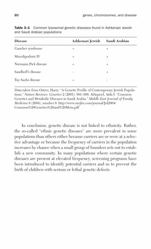
ptg
80 genes, chromosomes, and disease
In conclusion, genetic disease is not linked to ethnicity. Rather,the so-called “ethnic genetic diseases” are more prevalent in somepopulations than others either because carriers are or were at a selec-tive advantage or because the frequency of carriers in the populationincreases by chance when a small group of founders sets out to estab-lish a new community. In many populations where certain geneticdiseases are present at elevated frequency, screening programs havebeen introduced to identify potential carriers and so to prevent thebirth of children with serious or lethal genetic defects.
Table 3–1 Common lysosomal genetic diseases found in Ashkenazi Jewishand Saudi Arabian populations
Disease Ashkenazi Jewish Saudi Arabian
Gaucher syndrome + +
Mucolipodosis IV + +
Niemann-Pick disease + +
Sandhoff’s disease - +
Tay-Sachs disease + -
Data taken from Ostrer, Harry. “A Genetic Profile of Contemporary Jewish Popula-tions.” Nature Reviews: Genetics 2 (2001): 891–898. AlAqueel, Aida I. “CommonGenetics and Metabolic Diseases in Saudi Arabia.” Middle East Journal of FamilyMedicine 6 (2004), number 6. http://www.mejfm.com/journal/Jul2004/Common%20Genetics%20and%20Meta.pdf
Wow! eBook <WoweBook.Com>

ptg
Susceptibility genes and risk factors
Lipoprotein particles transport water-insoluble cholesterol and fatsthrough the bloodstream. The particles formed vary in density withthe most familiar being high-density lipoprotein (HDL) and low-den-sity lipoprotein (LDL). HDL, also known as “good cholesterol,” car-ries cholesterol to the liver for processing, thus protecting againstheart attack. LDL, or “bad cholesterol,” circulates in the bloodstreamand builds up into plaques in the inner walls of arteries that feed thebrain and the heart. These particles are made up of several differentlipoproteins called apolipoproteins A through E. A differentapolipoprotein (APO) gene encodes each of these proteins.
For years, most research on the apolipoproteins and the genesencoding them focused on their role in fat and cholesterol transport.Then, a startling discovery, made by Allen Roses and his group atDuke University, was announced to the world. The enthusiastic titleof Natalie Angier’s article on August 13, 1993, in the New York Timeswas “Scientists Detect a Genetic Key to Alzheimer’s.”1 Roses and hiscolleagues had been studying 234 people from 42 families and foundthat those with two copies the APOE4 variant had eight times the riskof late-onset Alzheimer’s than those who had the other two variants ofthis gene, E2 and E3, or a combination of these two. Even having onecopy of the E4 variant was sufficient to double or triple the risk oflate-onset Alzheimer’s. Hence, the APOE4 variant of the gene was avery strong predictor of late-onset Alzheimer’s disease.
APOE4 is a relatively straightforward example of a susceptibilitygene for a specific disease, but, as we shall see later in this chapter,there are many susceptibility genes for complex conditions likeasthma, diabetes, and heart disease. The mechanism by which alter-
4
81
ptg
82 genes, chromosomes, and disease
ations in a few of these genes cause disease is understood, but for themajority, this is still a black box. Nevertheless, enough has beenlearned in the last few years about disease genes that it is now possi-ble to classify these genes in a new way. Geneticists have used thisinformation to construct a map of what they have called the “disea-some,” the spectrum of diseases and the genes associated with them.We begin this chapter with the story of a susceptibility gene that pre-vents adults of many races from digesting milk or milk-related prod-ucts such as cheese properly, but that has been selected against innorthern European populations.
Selection against a susceptibility gene: cow’s milk andlactose intoleranceJacqueline Jannota was never fond of milk.2 When she went off to col-lege in Chicago, she recalls that there “was pizza everywhere, andevery time I ate it, I’d be doubled over in pain.” Jacqueline’s father, aphysician, suspected that she was unable to tolerate milk sugar (lac-tose) because she lacked the lactase enzyme required to process lac-tose. He therefore suggested that Jacqueline take lactase pills. Theyworked. She could now eat pizza without ill effects. Thus, JacquelineJannota is susceptible to lactose because the gene that encodes lac-tase has been turned off. As long as she does not eat dairy products ortakes lactase pills, this poses no problem.
An estimated 75% of people worldwide are intolerant of lactoseas adults. This is because lactase declines rapidly after weaning.3
However, in northern Europe, the frequency of lactose tolerance isvery high. Thus, in Sweden and Denmark, lactose-tolerant individu-als make up over 90% of the population. The frequency of lactose tol-erance declines from northwest to southeast across Europe. Around75% of the Caucasian population of the United States is lactose toler-ant, reflecting its mixed European origins. Among nonpastoralistssuch as the Chinese, only around 1% of the population tolerates lac-tose as adults.
Lactose tolerance results from dominant mutations that preventlactase production from being turned off.4 The predominant muta-tion in European populations seems to be absent in ancient DNAfrom early Neolithic central Europeans.5 But with the advent of dairy
Wow! eBook <WoweBook.Com>

ptg
4 • susceptibility genes and risk factors 83
farming about 7,500 years ago, lactose tolerance began to emerge.Lactase persistence is not restricted to Europeans, but also occurs incertain African pastoralist groups.6 However, in these cases, the muta-tional variants are distinct from those seen in Caucasians. Thus, selec-tion for lactose tolerance seems to have evolved independently onseveral occasions in response to the domestication of cattle and theintroduction of cow’s milk as a human food.
Is smoking in the genes?Popular articles on genes and smoking often start out with MarkTwain’s famous observation. He said that quitting smoking is easy. Hehad done it hundreds of times himself. But you might also want toparaphrase St. Augustine’s famous supplication to God about chastity.“Lord, make me stop smoking, but not yet.”
Smoking is a susceptibility trait for which there is no obviousselective advantage or disadvantage. Because cigarette smokingaccounts for at least 30% of all cancer deaths and more than three-quarters (87%) of lung cancer deaths in developed countries, youmight question the second part of the previous sentence. However,lung cancer usually appears late enough that most sufferers will havepassed through their reproductive years before contracting the dis-ease. This makes the smoking trait selectively neutral in contrast tolactose tolerance, where there is a clear selective advantage in popu-lations that have relied heavily on dairy products for food over thecenturies.
Nicotine is both a stimulant and a relaxant.7 The nicotine fromcigarette smoke travels rapidly from the lungs to the brain, spurringthe release of the numerous neurotransmitters and hormones that areresponsible for its varied effects. Nicotine achieves this stimulus inpart by binding to receptors for the important neurotransmitteracetylcholine. So far, 17 different protein subunits have been identi-fied that belong to these receptors. There are two families of subunitsdesignated α and β. They are assembled in different combinations.The CHRNA gene family specifies the different α subunits, while thefamily encoding the β subunits is called CHRNB. Obviously, variantsin these genes affecting the binding of nicotine either positively or
Wow! eBook <WoweBook.Com>

ptg
84 genes, chromosomes, and disease
negatively should influence the degree to which nicotine acts as astimulant and, hence, the extent to which it becomes addictive.
The body also has a system to detoxify nicotine. It is part of thecytochrome P450 (CYP) system.8 Estimates suggest that around halfof all drugs are detoxified by enzymes belonging to this system, mostof which are located in the liver. These enzymes also metabolize toxiccompounds, including pollutants, solvents, and recreational drugs.The downside to the CYP system is that it converts certain com-pounds into carcinogens.
The CYP system includes many polymorphic enzymes, meaningthey exist in more than one form. One of these enzymes, CYP2A6,converts nicotine to a metabolically inactive compound called coti-nine. The rate at which this conversion occurs depends on the partic-ular CYP2A6 polymorphism a person possesses and, hence, the genevariant that person happens to have. Evidently, any gene variant thataffects the rate at which nicotine is converted to cotinine is likely toaffect smoking behavior.
As discussed earlier (Chapter 2, “How genetic diseases arise”),publication of the sequence of the human genome coupled with theHapMap gave geneticists two powerful new tools beyond the pedi-gree to hunt for disease genes: the candidate gene approach andgenome-wide association studies. In the former approach, the astutegeneticist flips through the genomic library to focus on genes likely tobe involved in a specific disease or condition. The idea is to comparethis region in people with and without the disease or condition to seeif there are certain single nucleotide polymorphisms (SNPs) thatassociate specifically with that disease or condition. The hope is thatthey will mark genes of interest.
The second approach is totally unbiased and does not focus on theusual suspects. Instead, whole genomes are screened from lots of peo-ple with or without the disease or condition in the same way as in thecandidate gene approach. Because they are unbiased, genome-wideassociation studies appeared to hold the promise of turning up diseasegenes that are completely novel and that would not be found using thecandidate gene approach. Both candidate gene studies and, especially,genome-wide association studies are enormously labor intensive. Youneed only count the number of authors on papers using these methods.
Wow! eBook <WoweBook.Com>

ptg
4 • susceptibility genes and risk factors 85
So, what are the genes involved in nicotine susceptibility?9 Let’sbegin with the CHRN genes and focus an imaginary microscope onthe human genome. As we ratchet up the magnification, our attentionis attracted to a region on the long (q) arm of chromosome 15. It goesby the slightly awkward name of 15q24.1/15q25.1. We discover thatthis region contains three CHRN genes. They are called CHRNA3,CHRNA5, and CHRNB4. Comparisons between smokers and non-smokers in candidate gene studies and genome-wide associationstudies of this region have identified several SNPs as being related tonicotine dependence and/or lung cancer (see Table 4–1).10–12
Table 4–1 Single nucleotide polymorphisms (SNPs) associated with nicotinedependence, lung cancer, or both in the CHRNA5-CHRNA3-CHRNB4 region ofchromosome 15
SNP Nicotinedependence
Lung cancer* Gene Reference**
rs8034191 Not shown Yes AGPHD1 10
rs16969968 Yes Not shown CHRNA5 11
rs1051730 Yes Yes CHRNA3 10,12
An SNP in the CHRNA5 gene (rs16969968) results in the substi-tution of the amino acid aspartic acid for asparagine at position 398 inthe CHRNA5 protein. This amino acid substitution increases the riskof nicotine dependence. Another SNP in the CHRNA3 gene(rs1051730) was found to be closely associated with smoking quantityand lung cancer. A variant (rs8034191) in the aminoglycoside phos-photransferase gene (AGPHD1), located close to CHRNA5, alsoappears to increase the risk of lung cancer, but so far has not beenshown to increase the risk of nicotine dependence.
Although carriers of the SNPs in the CHRNA3 and CHRNA5genes seem to be more susceptible to nicotine dependence thanthose who lack them, it is important to note that plenty of people who
*Unlike the other two SNPs, rs16969968 has not been associated directly with lung cancer.**See commentary in references concerning which studies demonstrate a linkage with nico-
tine dependence or lung cancer.
Wow! eBook <WoweBook.Com>

ptg
86 genes, chromosomes, and disease
smoke lack the variants. For instance, in one study comparing theassociation of rs16969968 in European-American and African-Amer-ican smokers, the frequency of the variant in the former group was39% and 6% in the latter group.13 That means that most smokers inboth groups, particularly African Americans, lacked the variant.
So, what can we conclude from all of this? It seems that the twovariants in the CHRNA3 and CHRNA5 genes increase susceptibilityto nicotine dependence, but the connection with lung cancer maywell be indirect because analysis of four studies of people who havenever smoked and have lung cancer revealed no statistically signifi-cant association between the 15q24,25 region and lung cancer risk.14
What about smoking behavior and the polymorphic CYP2A6 sus-ceptibility gene? Adolescents with CYP2A6 variants that slow themetabolism of nicotine become tobacco dependent at relatively lowconsumption levels.15 They also smoke less than adolescents whoseCYP2A6 enzyme metabolizes nicotine more rapidly. The same is trueof adults who metabolize nicotine slowly. Deletion of the CYP2A6gene is fairly common in Asia. A Japanese study revealed that peoplewith this deletion smoke less than those with an intact gene.16 This fitswith expectation as, in the absence of the CYP2A6 gene, they shouldmetabolize nicotine slowly. The corollary is that people with the dele-tion are also less likely to suffer from lung cancer than those with anintact gene.
Candidate genes for nicotine susceptibility have also been soughtelsewhere, for example, in the dopamine and serotonin neurotrans-mitter pathways.17 These candidates include the DRD2 gene thatencodes a dopamine receptor. Dopamine receptors are implicated inneurological processes like motivation, pleasure, and cognition,whereas serotonin affects things like mood, sleep, and appetite. ForDRD2, the results have been equivocal with some studies suggestingan association with smoking and others not.18 Furthermore, agenome-wide association study found that the region on chromosome11q23 that includes DRD2 increased the chance of nicotine depend-ence, but that the evidence that DRD2 itself was involved was weak.Instead, a strong association was found with two other genes in thesame region that are not well characterized.19 This shows howgenome-wide association studies can often generate surprises thatrequire further explanation.
Wow! eBook <WoweBook.Com>

ptg
4 • susceptibility genes and risk factors 87
In view of the ambiguous status of the DRD2 variant vis-à-vissmoking, it seems that G-Nostics, based in Oxford, UK, may havejumped the gun in testing for smoking-associated variants in theDRD2 as well as the CYP2A6 gene. G-Nostics offers a geneticanalysis that targets these two genes. It is called NicoTest.20 How-ever, as the Times of London reported, characterization of the uni-verse of gene variants that may be involved in nicotine dependenceis still at a fairly early stage. Dr. Saskia Sanderson, a research fellowin health psychology and genetics at University College London,worries about this. “It is almost certain that in 20 years’ time we willhave tests for 50–100 genes that help to explain why one personsmokes and gets hooked and another does not.”21 Dr. Robert West,a professor of tobacco studies at University College, remarks thatNicoTest “has been marketed without adequate supportive evi-dence. It is all very interesting, but not close to being something toconsider as a reliable technology.”22 Dr. Mark Tucker, the CEO ofG-Nostics, begs to differ. “I find it incredible because nothing is toosoon for someone who is trying to quit smoking. Anything that helpsthem to find a path to quitting can’t come soon enough.”23 To befair, articles cited on the company’s Web site from papers like theMail on Sunday and the Daily Telegraph were quite laudatoryabout NicoTest.24
According to the G-Nostics Web site, the company has gottensmokers to quit at a rate of about 65% compared with the 35% for thesmoking service of the National Health Service.25 So what accountsfor the company’s success? It seems likely that the comprehensiveonline support offered to would-be quitters plays an important role.
AsthmaAt the end of November 2005, the death of a 15-year-old girl namedChristina Desforges, who lived in Saguenay, Quebec, was widelyreported because she was said to have died from her boyfriend’s kiss.The supposedly deadly kiss was at first thought to have triggeredChristina’s peanut allergy because her boyfriend had eaten a peanutbutter sandwich hours earlier. Christina’s tragic kiss provided a fieldday for the media. “Allergy Teen’s Fatal Kiss” blared the New YorkPost.26 The Times of London’s title was bit more staid, “The Kiss of
Wow! eBook <WoweBook.Com>

ptg
88 genes, chromosomes, and disease
Death for Girl with Nut Allergy.”27 ABC News headline read, “‘FatalKiss’ Puts Spotlight on Food Allergies.”28
As it turned out, it was not Christina’s peanut allergy that causedher death, but an asthma attack.29 In March 2006, the coroner, MichelMiron, went public. He said that peanut allergy was not the cause ofChristina’s death, but he was not yet prepared to give a fuller explana-tion of why she had died. He told the Associated Press that he felt itnecessary to make the statement because the “Canadian Associationof Food Allergies intended to use the Desforges case to launch aneducation campaign. I had to tell them the cause of death was differ-ent than first believed.” The coroner’s report, released in May 2006,concluded that Christina Desforges had suffered a fatal asthma attackthat had nothing to do with the supposedly lethal kiss. Furthermore,traces of the active ingredient in marijuana were found in the girl’sbody, suggesting she may have been smoking pot before she died.
With asthma and allergies, we enter the realm of complex dis-eases where a variety of genetic risk factors are involved whose con-tributions are just beginning to be understood. Furthermore,environmental factors also make a decisive contribution. So naturemust be assessed in terms of nurture, a phrase coined by the Victo-rian scientist Francis Galton, who probably lifted it from Shake-speare’s play The Tempest. In Act IV, scene I, Prospero, the Duke ofMilan, is speaking to the spirit Ariel about the man-beast Caliban. “Adevil, a born devil, on whose nature nurture can never stick.” Fortu-nately, for reader and writer, if not for the victim, there are usuallyone or more clear-cut genetic mutations that can cause a complex dis-ease whose mechanism of action can be explained in addition to themany other genetic factors that make contributions. It is these singlegene alterations that are stressed in the discussion that follows.
Let’s return to asthma.30 Asthma is a chronic disease that mostoften starts in childhood. It inflames and narrows the airways in yourlungs. Asthma causes shortness of breath, coughing, wheezing, andtightness in the chest. In the United States, over 22 million peoplesuffer from asthma. Symptoms may be mild and disappear on theirown or they may intensify so much in an asthma attack that they arelethal as in the case of Christina Desforges. As was true of this youngwoman, most people with asthma also have allergies.
Wow! eBook <WoweBook.Com>

ptg
4 • susceptibility genes and risk factors 89
Factors that determine a person’s susceptibility to asthma includegenetics, certain childhood respiratory infections, and contact withallergens like those produced by tobacco smoke, dander from cats anddogs, and cockroaches. But dust mites are the main culprits becausethey possess at least two proteins that can cause allergy and asthma.31
About 20 million people in the United States are allergic to dust mites.These mites are worldwide in distribution. The American house dustmite (Dermatophagoides farinae) is only about 0.5 mm long and, likeother dust mites, feeds on flakes of human skin. We produce a lot ofdust mite food because the average individual sheds 0.5 to 1 gram ofskin per day.
There has been a steady rise in allergic and autoimmune diseasesin developed countries over the past three decades.32 The same is trueof asthma. Furthermore, asthma prevalence increases when peoplemove from a rural to an urban environment.33 Observations like thesehave been explained by the hygiene hypothesis. This hypothesis pro-poses that increases in autoimmune diseases and asthma haveoccurred because we are too clean for our own good. We don’t havesufficient exposure to infectious agents, parasites, or just plain dirtany more. If we did, we would be less likely to suffer from allergiesand asthma because our immune system would have been kept busydealing with these external threats.
As science writer Matt Ridley has observed, asthma “is the tip ofthe iceberg of ‘atopy.’”34 By this, he means that asthma is just one mem-ber of the triad of hyperallergic responses that normally begins witheczema and includes hay fever. The allergic response begins whenallergen molecules such as dust mite proteins are recognized as foreigninvaders by the body.35 An immune cascade initiates when allergenmolecules attach themselves to the mucous membranes of the nose,throat, and lungs. Cells in these membranes “present” the allergenmolecules to immune system cells called T cells. The T cells elaboratesignaling molecules called cytokines that induce other types of immunesystem cells, the B cells or B-lymphocytes. These cells then produceimmunoglobulin E (IgE), one of the five classes of immunoglobulinsfound in placental mammals. Within a few weeks after allergen expo-sure, IgE antibodies specific to the allergen are produced. These IgEantibodies circulate in the bloodstream and some of them attach tocells involved in the inflammatory response called mast cells. When an
Wow! eBook <WoweBook.Com>
ptg
90 genes, chromosomes, and disease
allergic individual is exposed a second time to the allergen, the IgEbound to the mast cells undergoes a process called cross-linking thatcauses these cells to release inflammatory chemical mediators like his-tamine, prostaglandins, and leukotrienes. The inflammatory responseensues and the symptoms of asthma become apparent.
The large number of actors involved in eliciting the immuneresponse suggests that variations in many different genes might affectthe process. In fact, a total of 120 different genes have been identi-fied in at least one study as being associated in some way withasthma.36 Let’s consider just three of these as they illustrate howgenes having very different functions may be involved in causingasthma. The mechanism of action of the first gene is very well charac-terized, but less so for the other two reflecting the fact that the prod-ucts of these genes play many roles within the cell.
Ichthyosis vulgaris is a common inherited, semidominant skindisorder that affects around one in 250 people.37 People with this dis-ease possess a dry and scaly skin. The latter characteristic accountsfor the name because ichthys is Greek for fish. People with this dis-ease frequently suffer from eczema, allergies, and asthma. The rea-son is that they have loss of function mutations in a gene called FLGthat encodes an important skin protein, filaggrin.
Human skin consists of two major layers called the dermis andthe epidermis. The epidermal outer layer serves as a protective bar-rier against viruses, bacteria, and fungi, as well as allergenic particles.The epidermis is composed of several layers of cells. At the base arecells called keratinocytes whose major gene products are keratin pro-teins. The dividing keratinocytes progress through two layers of cellsabove them and finally reach the surface of the epidermis, where theylose their nuclei and become terminally differentiated corneocytes.These cells pack tightly to form part of the impermeable stratumcorneum. In the final stages of differentiation, a structure called thecornified envelope forms around each corneocyte. This is integral tobarrier function and one of its essential protein components is filag-grin. Filaggrin assists in the aggregation of keratin filaments. Thisexplains the protein’s odd name. It is shorthand for filamentaggregation protein. When filaggrin is missing, the keratin filamentsdo not aggregate properly, the protective barrier becomes defective,
Wow! eBook <WoweBook.Com>

ptg
4 • susceptibility genes and risk factors 91
and the protective barrier is breached by external allergens that trig-ger asthma in people with ichthyosis vulgaris.
The FLG mutations show how variants in a specific gene can leadto asthma. Two other genes in which there has been recent interestare the vitamin D receptor gene and the GPR154 gene that encodes aG protein (GPRA).38 The vitamin D receptor gene (VDR) specifies aprotein that binds vitamin D. This complex modulates many cellularprocesses, including important immune system functions. Certainreceptor gene variants in the French-Canadian founder populationare associated with asthma, but what their role is in causing the afflic-tion is a question for the future.
G protein is short for guanine-nucleotide binding protein. G pro-teins act like molecular switches. The off switch is a compound calledguanosine diphosphate that has two phosphate groups. The additionof a third phosphate group to form guanosine triphosphate turns theswitch to the on position. G proteins relay signals from each of morethan 1,000 receptors.39 Mutations in G protein-encoding genes cause anumber of inherited disorders. Why the GPR154 variants are involvedin asthma is not known. Working out the precise role of genes likeVDR and GPR154, whose protein products have multiple cellularfunctions, illustrates the challenges faced by investigators who want tounderstand the role such genes play in contributing to asthma.
Coronary artery diseaseThe Mona Lisa by Leonardo da Vinci may well be the world’s best-known painting. Madonna Lisa Maria de Gherardini, her real name,died in 1516 at the age of 37.40 The cause of her death was unknownat the time. Leonardo was not only an artist, but was also an inventorand a scientist. His scientific works include a clear-cut description ofatherosclerosis. A careful examination of the Mona Lisa reveals a yel-low spot at the inner end of the left upper eyelid. It is irregular inshape and leatherlike in texture. There is also a soft and bumpy, butwell-defined swelling on the upper side of Mona Lisa’s right handbeneath her index finger.
Yellowish patches around the eyelids as well as lumps in the ten-dons of the hands, elbows, knees, and feet are characteristic of an
Wow! eBook <WoweBook.Com>

ptg
92 genes, chromosomes, and disease
inherited disease called familial hypercholesterolemia (FH).41 Peoplewith this condition develop coronary artery disease much earlier thanis true of the general population. Consequently, they may suffer frompremature heart attacks. Perhaps Mona Lisa had FH too. If so, shecould have died of a heart attack, although death at an early age froma variety of causes was common in her day.
In the 1970s and ’80s, the genetic basis underlying FH wasworked out by Joseph Goldstein and Michael Brown, who becamefriends while medical interns at Massachusetts General Hospital inBoston in 1966.42 They also developed a consuming interest in FH.Why did people with the disease have such high levels of cholesterolin their blood? No wonder they were likely to suffer from heartattacks. Also, FH appeared to be dominant. People with just one copyof the mutant gene suffered from the disease. Children who had twocopies of the mutant gene were particularly sad. They had blood cho-lesterol levels six to eight times higher than normal, almost all ofwhich was in the LDL form. These children often had heart attacksbefore they reached puberty and generally died of coronary diseasebefore reaching the age of 20.
After being separated for a few years of further training in genet-ics, Brown and Goldstein both ended up at the University of TexasHealth Center in Dallas in 1972. They continued their experimentson FH using cultured cells from normal individuals and a young girlwho carried two copies of the defective gene. Brown and Goldsteindiscovered that both kinds of cells could make cholesterol if bathed inplasma that was low in cholesterol, but, when the surrounding mediacontained sufficient cholesterol, the normal cells stopped making thecompound while the FH cells continued to do so.
Their results led Brown and Goldstein to hypothesize that nor-mal liver cells possess a receptor that recognizes LDL cholesteroland turns off its synthesis when there is sufficient cholesterol around,but this receptor was missing or nonfunctional in FH cells, so theycontinued to churn out LDL cholesterol. With the help of volunteers,Brown and Goldstein were able to prove their hypothesis for whichthey were awarded the 1985 Nobel Prize in Physiology or Medicine.Since then, it has been shown that FH usually arises because of
Wow! eBook <WoweBook.Com>

ptg
4 • susceptibility genes and risk factors 93
mutations in the LDLR gene, whose product is the LDL receptorprotein. These mutations render the receptor protein incapable ofremoving LDL proteins from the blood and, thus, of regulating cho-lesterol levels.
Mutations in several other genes can also result in FH.43 One ofthese genes is the APOB gene that we met very briefly at the begin-ning of this chapter. The APOB gene encodes both short and longversions of apolipoprotein B known as B-48 (short) and B-100(long).44 It is the B-100 form of apolipoprotein B that is of concern tous here. This form of the protein is found in LDLs, as well as in otherlipoprotein particles that are involved in cholesterol and lipid trans-port. At least five mutations are known that affect the B-100 form ofapolipoprotein B in such a way that binding of LDLs to the LDLreceptor is blocked. As a consequence, cholesterol accumulates tohigh levels in the bloodstream and coronary artery disease results.
FH affects about 1 in 500 people,45 but coronary artery disease isthe most common form of heart disease and the leading cause of deathfor men and women in the United States.46 Hence, despite the unques-tioned importance of environmental factors such as diet, it seems likelythat a spectrum of genetic variants also affects coronary artery disease,some of which are rare, whereas others are common.47 There is a coro-nary-associated disease region on chromosome 9 (9p21), which alsoshows an association with type 2 diabetes. Hence, it seems likely thatthere are risk factors common to both diseases. Connections like theseform the basis for the diseasome discussed at the end of this chapter.
Diabetes mellitusType 1 and type 2 diabetes are the most common forms of this dis-ease.48 As of the year 2000, a minimum of 171 million people aroundthe world were known to have diabetes. In the United States, type 2diabetes is by far the most common form of the ailment—accountingfor between 90% and 95% of cases.
Type 1 diabetes is an autoimmune disease that results in thedestruction of the insulin-producing β-cells found in the islet ofLangerhan’s in the pancreas. This form of diabetes can affect children
Wow! eBook <WoweBook.Com>

ptg
94 genes, chromosomes, and disease
or adults, but has been called “juvenile diabetes” because type 1 dia-betes accounts for the majority of cases of the disease found in children.
Just under half of the risk for contracting type 1 diabetes can betraced to the most gene-dense region of the mammalian genome, themajor histocompatibility complex (MHC) on the short arm of chro-mosome 6.49,50 Roughly half of the 149 genes in this region haveknown functions in the immune system. Almost all autoimmune dis-eases can be traced to the products of genes encoded in the MHCregion. This includes type 1 diabetes and specifically the products ofthe HLA (for human leukocyte antigen) genes of which there are twogroups, HLA I and HLA II. These are highly variable genes encodingproteins of HLA classes I and II, respectively.
Comparison of the HLA genes in diabetic and nondiabetic indi-viduals reveals that both HLA I and II genetic variants are involved intype 1 diabetes. These variants may contribute to the autoimmunedestruction of the pancreatic β cells. Molecules of classes I and IIpresent antigen fragments to different subsets of immune systemcells. These are respectively called cytotoxic T and T helper cells. Ifthe genetic variants in question recognize β-cell fragments, theimmune system will be stimulated to destroy β-cells and type 1 dia-betes will ensue. HLA genes are not the only type 1 diabetes suscep-tibility genes. Variants in at least 15 other genes may also contributeto the disease.51
Type 2 diabetes is associated with obesity.52 The disease developswhen chronic overconsumption acts in concert with the genetic back-ground to impair the insulin-signaling mechanism.53 This condition isreferred to as insulin resistance. Type 2 diabetes is also accompaniedby a deficiency in insulin secretion. This disease is not an autoim-mune disease, but it is genetically complex.54 Gene variants that areinvolved in type 2 diabetes belong to four different classes. However,the two major classes of variants affect insulin secretion or insulinsensitivity. At least 18 susceptibility genes for type 2 diabetes havebeen identified, but they account for only 6% of the heritability ofthis disease (see Table 4–2). Environmental factors also play animportant role in type 2 diabetes. Thus, having elevated blood sugarlevels, being overweight, and lack of exercise greatly increase a per-son’s risk of developing this disease.
Wow! eBook <WoweBook.Com>

ptg
4 • susceptibility genes and risk factors 95
Table 4–2 Estimates of heritability and number of loci for several complextraits
Disease Number ofloci
Proportion of heritabilityexplained
Age-related macular degeneration
5 50%
Crohn’s disease 32 20%
Systemic lupus erythematosis 6 15%
Type 2 diabetes 18 6%
HDL cholesterol 7 5%
Height 40 5%
Early onset myocardial infarction
9 3%
Fasting glucose 4 1.5%
Modified from Table 1 in Manolio, Teri A. et al. “Finding the Missing Heritability ofComplex Diseases.” Nature 461 (2009): 747–753.
Type 2 diabetes is an evermore common ailment in the UnitedStates.55 The increase in frequency of the disease is strongly corre-lated with the increasing proportion of Americans who are obese.Today, there are around 23.6 million Americans who have type 2 dia-betes or about 7.8% of the population. Perhaps even more alarming isthe fact that 57 million Americans are “prediabetic.”
As is true of other complex diseases, unraveling the interplaybetween the panoply of susceptibility genes associated with type 2diabetes and the environmental factors that contribute to this diseaseis going to take time. However, it seems likely that proper attention toenvironmental factors like obesity can override the risks posed by atleast some susceptibility genes for type 2 diabetes.
For every complex disease, there seems to be a spectrum of sus-ceptibility genes, but, as we have seen, there are usually some clear-cut single gene defects that cause the same symptoms, although thesegenerally affect a minority of individuals. One such disease goes bythe awkward-sounding name of maturity-onset diabetes of the youngor MODY.56 MODY shows the clear-cut familial pattern of inheri-tance characteristic of a dominant gene. Symptoms of the disease
Wow! eBook <WoweBook.Com>

ptg
96 genes, chromosomes, and disease
usually appear during adolescence or in young adults. Mutations insix different genes have been identified that cause MODY. All ofthem limit the ability of the pancreas to produce insulin.
Are genetic disease variants common or rare?Two major hypotheses have been proposed to predict the degree towhich genes play a role in complex diseases like coronary artery dis-ease or type 2 diabetes.
The first is the Common Disease/Common Variant hypothesis.57
According to this hypothesis, at each of the major susceptibility loci,there will only be one or a few alleles that predispose an individual toa specific disease. These will be present in high frequency as long asthey have no debilitating effect until after people are no longer repro-ductively active. In fact, such variants may be selectively advanta-geous during the first few decades of life.
One example of a common variant associated with a diseaseexpressed late in life is the APOE4 gene discussed at the beginning ofthis chapter. This genetic variant occurs in about 40% of people whodevelop late-onset Alzheimer’s and is present in about 25% to 30% ofthe population.58
The alternative hypothesis proposes that disease susceptibilityresults from multiple rare variants in many genes.59 This is called thegenetic heterogeneity or multiple rare variant model. A rather extremeexample of a rare variant involves a disease called progeria.60 Progeriais a genetic disease that is not transmitted, but results from a dominant,spontaneous mutation. A baby born with progeria seems to undergo agreatly accelerated aging process, becoming older looking and morewizened as childhood progresses until death occurs, usually from heartattack, stroke, or atherosclerosis by the age of 13. Hence, death hap-pens before an individual with progeria reaches reproductive maturity.
Most cells in the human body, with the notable exception of redblood cells, possess nuclei. An envelope surrounds the nucleus. Prog-eria is caused by a dominant mutation in the LMNA gene thatencodes a protein called lamin that is integral to the nuclear enve-lope. The mutation creates an unusable form of lamin, meaning thatall nuclear envelopes in the body are defective. Precisely how thisdefect leads to progeria remains to be established.
Wow! eBook <WoweBook.Com>
ptg
4 • susceptibility genes and risk factors 97
It seems reasonable to suppose that these two hypotheses neednot be mutually exclusive. The frequency of the variant in the popula-tion is going to depend on the degree to which it reduces the potentialto produce offspring versus the selective advantage it confers eitherwhen homozygous or heterozygous. Sickle cell anemia comes to mindonce again. Although sickle cell disease very likely reduces reproduc-tive potential, the selective advantage conferred on carriers in malar-ial regions overcomes this disadvantage. There are undoubtedly farmore subtle genetic balancing acts that contribute to the repertoire ofsusceptibility genes.
Will the real risk factors please stand up?At the beginning of the twenty-first century, it looked as thoughgenome-wide association studies were going to be the gold standardfor the identification of genetic risk factors associated with complexdiseases. By identifying different genetic risk factors and attaching ameasure of probable risk to each one, a person’s genetic risk profilefor a complex disease like type 2 diabetes could be spelled out, tak-ing, of course, environmental considerations into account as well. Atthe end of the decade, after great expense both in terms of moneyand person power, it seems fair to conclude that this shimmering grailis still out of reach. As Nicholas Wade put it in the New York Times inJanuary 2010, although some 2,000 disease-associated SNPs havebeen identified, “this mountainous labor produced something of amouse. In each disease, with few exceptions, the SNPs accounted fora small percentage of the risk. A second puzzling feature was thatmany of the disease-linked SNPs did not occur in the DNA thatcodes for genes, but rather in the so-called junk regions of thegenome. Biologists speculated that these SNPs must play an as-yet-undefined role in deranging regulation of nearby genes.”61 As Wadepointed out, the Common Disease/Common Variant hypothesisassumed that for diseases that usually strike or cause death late in life,such as coronary artery disease or type 2 diabetes, selection does noteliminate “bad genes” because the child-bearing years are over.Hence, to find disease genes, you need only look at the few siteswhere genetic variations are common, defined as a frequency of atleast 1% in the population.
Wow! eBook <WoweBook.Com>

ptg
But David Goldstein, of the Duke University Institute for GenomicScience and Policy, and his colleagues argue that natural selection hasactually done a good job of getting rid of common disease-producingvariants.62 They hypothesize that the most important disease-producingvariants are, in fact, rare, although that cannot always be true as shownby the association of APOE4 with late-onset Alzheimer’s disease.
Goldstein and his colleagues also have done a very important kindof control experiment. They looked for SNPs associated with sicklecell anemia where the mutation responsible for the disease had beenidentified long ago as falling in the HBB gene (refer to Chapter 3,“Ethnicity and genetic disease”). They found that 179 SNPs showedsignificant association with the disease. These SNPs were spread overa span of 2.5 million base pairs on chromosome 11 in a region thatcontains “dozens of genes.” Furthermore, within this region there areblocks of sequence in which genetic markers remain associated in anonrandom fashion (see Linkage Disequilibrium in the glossary formore details). Goldstein and colleagues concluded that, “highly sig-nificant association signals can travel...to distant genomic regions.”This means that genome-wide association studies probably capture alot of genetic “noise” and that it can be difficult to separate the “sig-nal” from this noise.
A different way of evaluating the results of genome-wide associa-tion studies is to ask how much of the heritability of a complex traitcan be explained by the genetic markers so far discovered (see Table4–2). With the exception of age-related macular degeneration wherehalf of the heritability of this common disease is explained by a smallnumber of common variants with large effects, little of the heritabilityis explained by the loci discovered so far. As we have seen already, inthe case of type 2 diabetes, some 18 genetic loci are listed as con-tributing to the disease, but they account for only 6% of the heritabil-ity, implying there is a very long way to go in characterizing thegenetic architecture underlying this disease.
On October 9–10, 2010, Matt Ridley titled his Wall Street Journalscience column “The Failed Promise of Genomics.”63 He wrote that it istrue that genomics has made a big difference for rare inherited dis-eases. “But not for the common ailments we all get. Nor has it explainedthe diversity of the human condition in things like height, intelligence,and extroversion. If you sign up to have your genes tested today, you will
98 genes, chromosomes, and disease
Wow! eBook <WoweBook.Com>

ptg
4 • susceptibility genes and risk factors 99
get back a genetic horoscope couched in caveats that would embarrassa fairground fortune teller. ‘Slightly above average probability of osteo-porosis, etc.’” This a particularly important point for a person to keep inmind who may be contemplating making use of a gene-testing companyto find out about their susceptibility to different genetic diseases (seealso Chapter 10, “The dawn of personalized medicine”). Unfortunately,as Ridley points out, genome-wide association studies have turned uplots of genes, “but they still explain embarrassingly little.”
“I think most people now view genome wide association studiesas something we absolutely had to do and have now been done,”Goldstein said. “It’s fair to say that for many common diseases noth-ing of very great importance was discovered, but these studies havetold us what to do next.”64 That, of course, will be to sequence entiregenomes and identify likely mutations in the risk-factor genes them-selves or else within regulatory regions outside of these genes. Thisshould soon be possible as the cost of whole genomic sequencing isdropping rapidly (see Chapter 10). However, be cautioned. Althoughcomplete sequences will reveal all of the differences between the dis-ease and control populations, the important ones will still have to besorted out from those that are trivial. In this case, familial studies mayhelp as they should identify alterations common to individuals in thefamily who have a particular complex disease.
The DiseasomeNosology, from the Greek word for disease, is the medical disciplinethat deals with disease classification. Serious attempts to classify dis-eases began in the eighteenth century with the Nosologia methodicaof François Bossier de Lacroix.65 Linnaeus had a go at the problem inhis Genera morborum as did Erasmus Darwin, the grandfather ofCharles Darwin, in his Zoonomia. Both of these scientists appliedmodified versions of Linnaeus’s binomial system used for the classifi-cation of plants and animals to classify diseases.
Although these classification systems have not withstood the testof time, a workable classification system is essential if one is to makesense of mortality statistics. The statistical study of disease actuallybegan in the seventeenth century with a man named John Grauntwho was working on the London Bills of Mortality. These bills were
Wow! eBook <WoweBook.Com>

ptg
originally drawn up as a means of warning of plague epidemics. WhatGraunt did was to classify the proportion of children under six whohad died by disease. In the nineteenth century, William Farr, a Britishmedical statistician, improved and standardized the disease classifica-tion system and promoted its international use. The importance ofsuch a system became clear at the first International Statistical Con-gress held in Brussels in 1853.
In 1891, the International Statistics Institute, which succeededthe International Statistical Congress, met in Vienna. Its participantsappointed a committee, chaired by Jacques Bertillon, Chief of Statis-tical Services of the City of Paris, to develop a system for classifyingthe causes of death. Bertillon’s system went into general use inCanada, Mexico, and the United States following a meeting of theAmerican Public Health Association in 1898. Subsequently, in August1900, delegates from 26 countries arrived in Paris for an internationalconference whose purpose was to revise the Bertillon system.
So far, the classification system had been directed strictly at mor-tality, but earlier on William Farr had recognized the desirability ofextending the statistical classification system to diseases that, thoughnot fatal, caused disability. This would greatly broaden the classifica-tion system to include the kinds of diseases that spread in places likearmies, hospitals, and public institutions. A classification system formental diseases affecting the inmates of lunatic asylums also seemeda good idea. At the 1900 International Conference, the need for auniform system for classifying diseases as well as mortality was firstrecognized.
Over the years, the classification systems for diseases and mortal-ity have been revised further. In 1946, the World Health Organiza-tion was charged with the responsibility of setting up a worldwidesystem of classifying the causes of disease and mortality. The resultwas the International Classification of Diseases, Injuries, and Causesof Death. Since 1946, this classification has undergone ten revisionswith the last being in 1990.
But as our knowledge of the effects of human molecular geneticshas increased by leaps and bounds, an entirely new way of looking atdiseases has evolved. This has crystallized into the human disease net-work or diseasome. The idea is that one can connect different humandiseases into a network by the genes that affect them.66
100 genes, chromosomes, and disease
Wow! eBook <WoweBook.Com>

ptg
The diseasome map can be explored online courtesy of the NewYork Times at www.nytimes.com/interactive/2008/05/05/science/20080506_DISEASE.html.
The diseasome opens a new way of thinking about genetic dis-ease, particularly complex diseases, some of which overlap eachother, such as type 2 diabetes and coronary disease. There is no doubtthat we are going to learn a lot more about the interactions of genesand disease as the diseasome becomes refined in evermore increasingdetail. Exactly how the diseasome will be reconciled with the currentsystem for classifying diseases remains to be seen, but it could givethe system an entirely new dimension.
4 • susceptibility genes and risk factors 101
Wow! eBook <WoweBook.Com>

ptg
This page intentionally left blank
Wow! eBook <WoweBook.Com>

ptg
Genes and cancer
In his State of the Union address in January 1971, President RichardNixon announced his intention to request an appropriation of $100million “to launch an intensive campaign to find a cure for cancer,and I will ask later for whatever additional funds can effectively beused. The time has come in America when the same kind of concen-trated effort that split the atom and took man to the moon should beturned toward conquering this dread disease. Let us make a totalnational commitment to achieve this goal.”1
On December 23, 1971, President Nixon signed the NationalCancer Act into law, declaring, “I hope in the years ahead we will lookback on this action today as the most significant action taken duringmy administration.” The National Cancer Act, “The War on Cancer,”gave the National Cancer Institute a unique autonomy within theNational Institutes of Health to deal with the many manifestations ofthe disease that it has managed with skill and great success.
Usually when we think of war, we think of a defined enemy, forexample, the Axis nations in World War II. But the War on Cancer ismore like the War on Terror. Both phrases conjure up dread images.On the one hand, we think of turbans, beards, and car bombs, but onthe other hand, we think of uncontrolled cell growth and metastasis.But both these phrases oversimplify enormously complex problems.We are only beginning to understand the array of tribal societies thatinhabit places like Afghanistan, Pakistan, Somalia, and Yemen withtheir strange mores, odd customs, and difficult languages. Similarly,cancer is a large collection of different genetic diseases, each of whichhas its own unique characteristics. Nevertheless, we are graduallycoming to grips with the many forms of this most protean of diseases.
5
103

ptg
104 genes, chromosomes, and disease
This has only been possible because our basic understanding of themolecular and genetic basis of cell function, interaction, and the con-trol of cell proliferation has advanced so much in the past few decades.
This chapter begins with a discussion of skin cancer. Skin cancerwill be familiar to many readers of this book who, like the author,must make regular trips to their watchful dermatologist. The geneticsof cancer is far too massive a subject to be covered in any detail in abook of this kind so the rest of this chapter is devoted to several dif-ferent types of cancer. They are chosen, with one exception, to illus-trate different aspects of the biology of this complex disease.
Cancer and the skinSkin is an epithelial tissue. Epithelial tissues are the protective layersof cells that line your inside (e.g., the intestine) and outside (e.g.,skin). Depending on their function, epithelial tissues can derive fromany one of the three main primitive cell layers of the embryo. Thus,skin arises from the outermost layer, the ectoderm; the ovaries derivefrom the middle layer, the mesoderm; and the epithelial layers ofinternal organs like the lungs, liver, pancreas, and gall bladder as wellas the lining of the stomach and intestines derive from the innermostcell layer, the endoderm.
Actinic keratosis is by far the most common lesion with the poten-tial to become malignant.2 An actinic keratosis is usually a small (2–6mm), reddish, rough or scaly spot of thickened growth (keratosis)caused by significant exposure to sunlight (actinic). Actinic keratosescan either regress or remain the same, but occasionally develop intosquamous cell carcinomas. Dermatologists frequently prescribeFudex to treat actinic keratoses. This is actually a compound called 5-fluorouracil. It interferes with the action of an enzyme calledthymidylate synthetase that is required for the synthesis of thymine-containing nucleotides and, hence, for the replication of DNA. As aconsequence, the dividing cells in the actinic keratosis are killedbecause they make defective DNA molecules.
Squamous cell carcinomas are the second most common form ofskin cancer. They normally do not cause problems if caught andtreated early. Squamous cell carcinomas can also arise in other kinds
Wow! eBook <WoweBook.Com>

ptg
5 • genes and cancer 105
of epithelial cells in addition to skin cells. For example, most cases ofhead and neck cancer result from squamous cell carcinomas that ini-tiate in the moist, mucosal cells that cover the surfaces of the mouth,nose, throat, and esophagus.3 Head and neck cancers account foraround 3% to 5% of all cancers in the United States. Adenocarcino-mas are the other big class of carcinomas. They arise in specializedcells that have a secretory function that line internal organs. Forexample, adenocarcinomas arise in the ducts and lobules of thebreast. They are glandular tissues that make breast milk. In fact, car-cinomas are the most common of all human cancers, accounting for80% of cancer deaths in the West.4
Basal cell carcinoma is the most common type of skin cancer.5
Unlike squamous cell carcinomas for which actinic keratoses are theprecursors, there are no clear precursors for basal cell carcinomas.UV radiation is the ultimate culprit for all three of these conditions.Most of these cancers are spontaneous (sporadic) in nature, but someare hereditary. Over 90% of squamous cell carcinomas and 50% ofbasal cell carcinomas have mutations in the TP53 tumor suppressorgene that encodes the p53 protein.
Tumor suppressors are one of three classes of genes involved incancer. The other two classes are called oncogenes and repair genes.Oncogenes are derived by mutation from proto-oncogenes. These arenormal genes that help regulate cell growth and differentiation.Oncogenes have been compared with stuck gas pedals in that theirprotein products accelerate cell division and growth in an uncontrol-lable fashion. Tumor suppressor proteins act like car brakes. Theykeep cell division under control and slow it down in response to per-ceived defects arising in DNA during replication or in the apparatusthat ensures proper chromosome distribution during mitosis. Repairgenes are like mechanics. The proteins encoded by them fix defectsthat arise in the cellular DNA, in the apparatus required to maintainaccurate distribution of the chromosomes during mitosis, and so on.
The p53 protein has been portrayed as the guardian of thegenome. Many forms of cellular stress can activate the protein. Forinstance, if damage to the DNA has occurred and been recognized,p53 can arrest cell growth at the point in the cell cycle prior to DNAsynthesis in preparation for cell division. The p53 protein can also
Wow! eBook <WoweBook.Com>

ptg
106 genes, chromosomes, and disease
activate DNA repair proteins to fix the damage. And it can initiate theprocess of apoptosis or programmed cell death if the damage cannotbe fixed.
Once the guardian is inactivated, as the result of a TP53 mutationin a basal cell or squamous cell carcinoma, there is no way to close thegate (i.e., arrest the cell cycle). This permits chromosomal damage tobe passed on to daughter cells as long as it is not lethal to them. This isthe first step in genomic destabilization. It is followed by chromoso-mal rearrangements and changes in chromosome number. New muta-tions that may affect oncogenes and other tumor suppressors also gouncorrected. For example, mutations in another tumor suppressorgene called patched (PTCH) play a major role in basal cell carcinomaformation.6 The PTCH gene product is a receptor protein that spansthe cell membrane (see reference 6 for a more detailed description ofwhat happens when PTCH mutates).
Although the great majority of cancers are sporadic, a familialcancer can usually be identified that relates to the broken gene. Forexample, loss of p53 function is one characteristic of over 50% of allcancers.7 The familial cancer associated with this loss of function iscalled Li-Fraumeni syndrome.8 The disease shows a dominant pat-tern of inheritance and greatly increases the risk of developing severaltypes of cancer, particularly in children and young adults. These com-monly include breast cancer, osteosarcoma, a form of bone cancer,and cancers of soft tissues like muscle. Cancers of the latter type arereferred to as sarcomas as opposed to the carcinomas so far under dis-cussion. Sarcomas amount to only about 1% of all cancers and share acommon origin in the mesoderm.9 Sarcomas affect tendons, muscle,bone, and other connective tissues.
Let’s consider for a moment what dominance means in the caseof Li-Fraumeni syndrome. Dominance is pretty clear-cut in the casesof diseases like cystic fibrosis or Tay-Sachs. The normal form of thegene is dominant to the disease gene. But dominance in the case offamilial cancers usually means something else. It means that a personis carrying a damaged form of the cancer gene, in this case TP53, sothere is only one instead of two good copies of the gene. Suppose theprobability of a mutation in the TP53 gene is 1 in a million (1 in 106)per cell generation. The probability of a cell containing two mutant
Wow! eBook <WoweBook.Com>

ptg
5 • genes and cancer 107
TP53 gene mutations is the product of the individual probabilities orone in a trillion (1 in 1012). On the other hand, if one copy of the geneis already mutated, the probability of a mutation inactivating the goodgene is one in a million. That might seem like a big number, but it isnot if you consider the large number of epithelial cells in the bodyand the fact that over a lifetime they divide a great many times.
The familial disease associated with PTCH mutations is calledGorlin syndrome.10 Compared with the number of people whodevelop sporadic basal cell carcinomas, only 1% suffer from Gorlinsyndrome. Individuals with this disease begin to develop basal cellcarcinomas during adolescence or as young adults. The number ofbasal cell carcinomas varies among affected individuals. Generallyspeaking, people with lighter skin develop more basal cell carcinomasthan those who are darker in color. People with Gorlin syndromeoften develop benign tumors of the jaw and are also at higher risk ofdeveloping other tumors than the general population. The high inci-dence of basal cell carcinomas in Gorlin syndrome is exactly what onewould expect based on the observation that PTCH is often mutated insporadic basal cell carcinomas.
In the early 1970s, Alfred Knudson, a pediatrician who was inter-ested in childhood cancers, first made the distinction between spo-radic and familial cancers.11 Knudson and his colleague Louise Strongwere studying a childhood eye cancer called retinoblastoma. Prior to1866, this disease was lethal as the cancer spread to the brain and mostchildren with the disease died before the age of ten. However, with theinvention of the opthalmascope in that year it became possible for doc-tors to peer into the eye and detect the incipient mass of cancer cells atthe back of the eyeball. The disease could now be prevented fromspreading, but the price was very high as it involved surgical removalof the eyes. If these blinded children went on to have families of theirown, it turned out that some of their children also had retinoblastoma.
By the time Knudson started his own work on retinoblastoma, oph-thalmologists were able to kill the cancerous cells with radiation if thetumor was caught early enough. And now, Knudson came up with amodel for what was happening. It was called the “two-hit” hypothesis.The first “hit” was the mutant gene that was familial in transmission,but a tumor did not begin to develop until a retinal cell had received a
Wow! eBook <WoweBook.Com>

ptg
108 genes, chromosomes, and disease
second mutational “hit” that disrupted the normal copy of the gene. Inconfirmation of this model, Knudson and Strong found that, in malig-nant retinal cells from patients with the disease, both copies of chromo-some 13 were lacking the same piece of DNA. Today we know that thekey gene that is missing is the RB gene, another tumor suppressor.
Let’s return to squamous cell carcinomas one last time. Amplifi-cation of a gene called MYC has been noted in some actinic keratoses,but more frequently in squamous cell carcinomas (for more explana-tion on MYC’s oncogenic properties, see reference 12). When MYC iseither deregulated or amplified, it behaves like an oncogene. Thegene is frequently amplified in squamous cell carcinomas, but it isderegulated in 15% to 30% of all other human cancers.13
The most serious form of skin cancer is malignant melanoma.Most Australians (Aborigines excluded) arrived in Australia fromEngland in the eighteenth and nineteenth centuries having been“transported” as criminals or other sorts of riffraff. These pale-skinned people and their descendants were very sensitive to theintense Australian sun and, hence, very susceptible to skin cancer.Because moles and melanomas can be confused, the AustralasianCollege of Dermatologists has formulated easy-to-follow, but detailedguidelines for moles and melanoma.14 The College points out thatmany Australians have up to 100 moles that are 2 mm or more in sizeby the age of 15 years. People with greater than 100 moles are atincreased risk of developing melanoma. This does not mean thatmelanomas develop from the moles themselves, although they some-times do. Many melanomas arise as new spots on the skin.
Approximately 90% of melanomas are sporadic,15 but 10% ofmelanoma cases are inherited. At least 25% of hereditary melanomascan be attributed to mutations in a tumor suppressor gene calledCDKN2A. The CDKN2A gene can yield two different protein prod-ucts depending upon how it is “read.” One of these, called p16,inhibits enzymes called cyclin-dependent kinases that are involved incell cycle regulation. The second protein product of CDKN2A iscalled p14ARF. This protein acts to stabilize p53 by preventing degra-dation of this protein by a protein called MDM2.16 Hence, a mutationthat inactivates both CDKN2A functions will affect regulation of the cell cycle while, at the same time, hastening p53 degradation
Wow! eBook <WoweBook.Com>
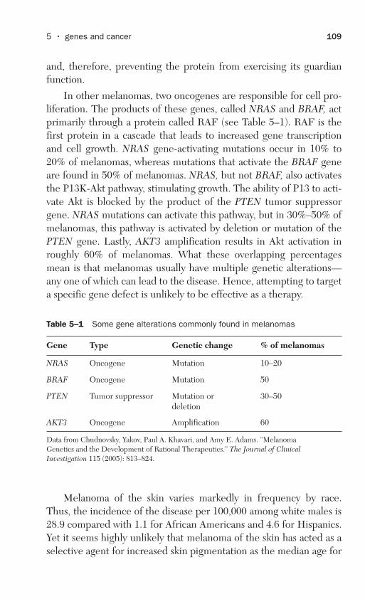
ptg
5 • genes and cancer 109
and, therefore, preventing the protein from exercising its guardianfunction.
In other melanomas, two oncogenes are responsible for cell pro-liferation. The products of these genes, called NRAS and BRAF, actprimarily through a protein called RAF (see Table 5–1). RAF is thefirst protein in a cascade that leads to increased gene transcriptionand cell growth. NRAS gene-activating mutations occur in 10% to20% of melanomas, whereas mutations that activate the BRAF geneare found in 50% of melanomas. NRAS, but not BRAF, also activatesthe P13K-Akt pathway, stimulating growth. The ability of P13 to acti-vate Akt is blocked by the product of the PTEN tumor suppressorgene. NRAS mutations can activate this pathway, but in 30%–50% ofmelanomas, this pathway is activated by deletion or mutation of thePTEN gene. Lastly, AKT3 amplification results in Akt activation inroughly 60% of melanomas. What these overlapping percentagesmean is that melanomas usually have multiple genetic alterations—any one of which can lead to the disease. Hence, attempting to targeta specific gene defect is unlikely to be effective as a therapy.
Melanoma of the skin varies markedly in frequency by race.Thus, the incidence of the disease per 100,000 among white males is28.9 compared with 1.1 for African Americans and 4.6 for Hispanics.Yet it seems highly unlikely that melanoma of the skin has acted as aselective agent for increased skin pigmentation as the median age for
Table 5–1 Some gene alterations commonly found in melanomas
Gene Type Genetic change % of melanomas
NRAS Oncogene Mutation 10–20
BRAF Oncogene Mutation 50
PTEN Tumor suppressor Mutation or deletion
30–50
AKT3 Oncogene Amplification 60
Data from Chudnovsky, Yakov, Paul A. Khavari, and Amy E. Adams. “MelanomaGenetics and the Development of Rational Therapeutics.” The Journal of ClinicalInvestigation 115 (2005): 813–824.
Wow! eBook <WoweBook.Com>

ptg
diagnosis is 59 years of age, well past the years of maximum reproduc-tion.17 So why is dark skin favored in regions of the world where thesun is at its brightest?
It seems likely that our early hominid ancestors had black fur andlight skin like the chimpanzee.18 As our African ancestors evolved fur-ther, they became bipedal and hairless and acquired a modern mech-anism for sweating. At the same time, their skin acquired more andmore UV-absorbing melanin pigment. The effects of UVB radiationare especially harmful (refer to Chapter 2, “How genetic diseasesarise”). Not only does irradiation in this wavelength range cause sun-burn, but UVB-induced damage to sweat glands disrupts sweatingand, thereby, thermoregulation. Yet even this might not be enough toexplain the selection of dark pigmentation in our ancestors.
Another explanation involves the essential nutrient folic acid thatis converted to folate in the body. Folate is essential for nucleotidesynthesis and, therefore, the replication and repair of DNA. Folatedeficiency has been linked to fetal malformation in nonhuman mam-mals and to neural tube defects in humans. These can lead to anen-cephaly, a lethal condition by the time of birth, craniorachischisis, acondition causing embryonic development to fail early on, and spinabifida. A folic acid supplement prevents 70% of these neural tubedefects. Folate has also been shown to be critical for spermatogene-sis in mice and rats. Because folate is exquisitely sensitive to UVlight, it may be that dark skin pigmentation evolved to prevent folatedegradation in response to reduced rates of embryonic survival andspermatogenesis.
In contrast, exposure of the skin to the UV rays in sunlight causesthe conversion of 7-dehydrocholesterol to previtamin D and subse-quently to vitamin D. Vitamin D deficiency is the primary cause ofrickets, a very common childhood disease in developing countries.Lighter skin pigmentation may have been selected at higher latitudeswhere light intensities are lower in response to vitamin D deficiency.
Six or more genes are involved in controlling skin color. Geneticinvestigations suggest that light skin color evolved independently inEuropean and East Asian populations.19 This finding implies that,even if the vitamin D hypothesis is wrong, there is selection for lightskin color in the northern regions of the earth.
110 genes, chromosomes, and disease
Wow! eBook <WoweBook.Com>

ptg
5 • genes and cancer 111
Finally, there is no better way to illustrate the role of repairenzymes in preventing cancer than to consider a group of rare autoso-mal recessive diseases referred to collectively as xeroderma pigmen-tosum.20 Individuals with xeroderma pigmentosum are at athousandfold greater risk of UV-induced skin cancer before the age of20 than normal individuals. People with this disease have to stay outof sunlight. Mutations that cause xeroderma pigmentosum do notaffect the gene encoding the photolyase enzyme. This enzymerequires visible light to repair damage to DNA (see Chapter 2).Instead, they principally affect repair genes in one of the “darkrepair” pathways (i.e., light is not required for repair) called thenucleotide excision repair pathway. The enzymes coded by thesegenes remove damaged nucleotides and facilitate insertion of undam-aged nucleotides. People with xeroderma pigmentosum lack a func-tioning excision repair pathway, so they are unable to repairUV-damaged nucleotides; mutations can, therefore, occur in onco-genes and tumor suppressors, posing a serious risk of skin cancer.
The virus connection: cervical cancerAll women are at risk for cervical cancer, but the disease is uncommonin women under 30.21 In the United States, the median age at diagnosisis 47. In 2006, 11,982 women in this country were diagnosed with cer-vical cancer and 3,976 died from the disease. The cervical cancer ratevaries somewhat with race and ethnicity with the incidence for all racesbeing 8.0 per 100,000. Worldwide, 500,000 women contract cervicalcancer each year with 80% of cases being reported in the developingworld.22
Cervical cancer is caused by human papillomaviruses (HPV).There are more than 100 kinds of HPV.23 Certain types of HPV causewarts, or papillomas, hence the name. These are benign tumors.Genital HPV infections are sexually transmitted and quite common.Over 30 types of HPV are transmitted sexually. Two of these, HPV-6and HPV-11, are frequently associated with genital warts. Withrespect to cervical cancer, HPV strains separate into “low-risk” and“high-risk” groups. That latter group includes at least 15 differenttypes, of which HPV-16 and HPV-18 are responsible for about 70% ofcervical cancers.
Wow! eBook <WoweBook.Com>

ptg
In 1928, a Greek pathologist named George Papanicolaou reportedthat he could tell the difference between normal cells and cancerousuterine cells by means of a simple, vaginal smear.24 Papanicolaou hademigrated from Greece to the United States in 1913, accepting a posi-tion in the Department of Anatomy in Cornell Medical School in NewYork City. In 1943, Papanicolaou published a monograph titled “Diag-nosis of Uterine Cancer by the Vaginal Smear” in collaboration with Dr.Herbert Traut of the Department of Obstetrics and Gynecology at Cor-nell Medical School. This was the paper that led the medical commu-nity to recognize the importance of the discovery that Papanicolaouhad made much earlier. Papanicolaou’s diagnostic procedure, sincenamed the Pap test or Pap smear, has been the standard procedure ingynecological examinations for detection of cervical cancer for manyyears now. If caught early, cervical cancer is readily treated surgically.
The realization that there is a relationship between viruses andcancer dates back to 1909 when Peyton Rous, working at the Rocke-feller Institute in New York City, began the research that led to thediscovery of the first tumor virus.25 Rous was studying a sarcoma thathad appeared in the breast muscle of chicken. He found that if heground up the cancerous tissue, filtered it, and injected it into youngchickens, they developed tumors too. Because the infective agentcould be passed through a fine filter, it could be no bigger than avirus. Rous published his results in 1911, but had to wait 55 years forthe ultimate recognition of the importance of his discovery when hereceived the Nobel Prize in Physiology or Medicine in 1966. Thevirus was by now known as the Rous sarcoma virus (RSV).
In the early 1950s, Harry Rubin in Renato Dulbecco’s laboratoryat the California Institute of Technology discovered that, when cul-tured embryonic fibroblast cells from chicken were infected withRSV particles, they did not kill their host cells like most viruses, butcontinued to produce virus particles over a period of days, weeks, andeven months. Unlike normal cells in tissue culture that cease to growafter they form a confluent layer of cells over the surface on whichthey were inoculated (contact inhibition), these infected cellscontinued to grow and pile up on one another (transformation).
A graduate student of Dulbecco’s, Howard Temin, continued towork on RSV together with Rubin. Their results showed that thetransformed cells retained the virus, but this did not necessarily mean
112 genes, chromosomes, and disease
Wow! eBook <WoweBook.Com>
ptg
5 • genes and cancer 113
that the virus had transformed the cells it had infected. An experi-ment done at Berkeley involving a mutant of RSV that was sensitiveto a temperature of 41°C, but grew normally at 37°C settled thequestion. As long as the cells were grown at 37°C, they retained theirtransformed character, but when they were switched to 41°C, theyreverted to the shape and growth pattern they had possessed beforebeing infected with RSV.
RSV particles were known to contain single-stranded RNA,rather than double-stranded DNA, as their genetic material. Howwas the virus maintained in the transformed cells in this state? In1960, Temin began to formulate the provirus hypothesis, which heformally proposed in 1963.26 Temin’s hypothesis seemed like heresybecause he had postulated that the RNA of RSV was copied into aDNA provirus, which could in turn produce RNA copies. At the time,the central dogma of molecular biology held that the information inDNA was transcribed into messenger RNA whose information wasthen translated into protein. An exception had to be made for RNAviruses. It was assumed that these viruses transferred their informa-tion from RNA to RNA and from RNA to protein, but a reversal ofinformation transfer from RNA to DNA was a bridge too far. Temin’shypothesis was ignored for over six years. Then in 1970, both he andDavid Baltimore showed independently that RSV RNA encoded anenzyme called reverse transcriptase that could make a DNA copyfrom the viral RNA. Baltimore, Temin, and Dulbecco shared theNobel Prize in Physiology or Medicine in 1975.
Within a year of the discovery of RSV reverse transcriptase, provi-ral DNA was detected within the chromosomal DNA of RSV-infectedcells. But a piece of the puzzle was still missing. Why did the presenceof the RSV genome turn normal cells into cancer cells? J. MichaelBishop and Harold Varmus solved this problem in their joint labora-tory at the University of California, San Francisco. One of the viralgenes must encode a protein that transformed normal cells to cancer-ous cells. The hypothetical gene was named src, short for sarcoma. Acomparison of the RSV genome with other retroviral genomes thatdid not cause cellular transformation revealed that there was only alittle information left in the RSV genome to encode the src gene, asmost of the genome was devoted to making necessary viral proteins.
Wow! eBook <WoweBook.Com>

ptg
Bishop and Varmus made a DNA probe for this small region and,much to their surprise, they found this DNA sequence not only in cellsinfected by the virus, but in uninfected cells too. In other words, thevirus had hijacked a cellular gene. It turned out that the cellular gene(c-src) was part of the normally functioning genome of the cell, a proto-oncogene, but when captured by the virus (v-src), it became an onco-gene. The product of this gene was sufficient to transform a normal cellinto a cancer cell. In 1989, Harold Varmus and J. Michael Bishopreceived the Nobel Prize in Physiology or Medicine for this work.
It is time now to return to cervical cancer. A great deal of work wasalso being done on DNA viruses suspected of producing cancer. Theseincluded a virus that caused papillomas (warts) on the skins of rabbitsdiscovered by Richard Shope and named for him, a mouse virus calledpolyoma, and a monkey virus with the name of SV (for simian virus)40. Although these viruses usually followed a normal life cycle inwhich they reproduced in an infected cell and ultimately lysed the cell,there were certain kinds of cells on which they could not grow. How-ever, occasionally, one of these cells out of thousands would be trans-formed in the same way that RSV transformed cells. Like the DNAcopies of RSV, the DNA of these viruses also became associated withthe chromosomal DNA of the cell. This group of tumor viruses or fam-ily originally received the name papovavirus for papiloma, polyoma,and the vacuoles caused by SV40 virus when it went through its pro-ductive cycle that resulted in cell lysis. Today, these viruses are sepa-rated into the viral families Papillomaviridae and Polyomaviridae.27
Human papillomaviruses (HPV) are small, circular DNAviruses.28 The HPV genome consists of nine overlapping genes and acontrol region. The control region and the seven genes (E1–E7)expressed early on following viral infection (early region) are involvedin the regulation of the viral life cycle and in the transcription of viralDNA. The other two genes (L1 and L2) are expressed subsequently(late region). These genes specify the major and minor proteins of thecoat (capsid) of the virus that encloses the viral DNA.
HPV virus particles (virions) initially infect the basal cells of theepithelium. The viral DNA is maintained independently of the hostgenome in these infected cells at a copy number of about 50–100viral genomes per cell. As infected daughter cells migrate to theupper layers of the epithelium, the late genes are expressed, new
114 genes, chromosomes, and disease
Wow! eBook <WoweBook.Com>

ptg
5 • genes and cancer 115
virus particles are formed, following which the progeny virus arereleased and able to infect new cells. The role of the E6 and E7 pro-teins is essentially to prevent the immune system from eliminatingthe virus by blocking interferon regulatory proteins from activatinginterferon proteins and, thus, stimulating the immune response.
Although a woman’s lifetime risk of cervical HPV infection is80%, most of these infections are resolved spontaneously by theimmune system. Integration of the viral genome into the host genomeis a rare event, but once it happens, transformation of the infected tis-sue into cervical cancer proceeds rapidly. In this case, the role of theE6 and E7 proteins is very different. E6 promotes the degradation ofp53, whereas E7 disrupts the function of the retinoblastoma proteinso the two viral proteins contribute to cancer formation by interferingwith cell cycle control. Thus, cervical cancer is caused by two proteinsencoded by the viral genome in contrast to RSV, which induces can-cer via a hijacked host protein.
If a woman’s pap smear indicates cervical cancer, a number oftreatment options are available. They include surgery, liquid nitrogenfreezing of the tumor, and carbon dioxide laser destruction (ablation).The most promising preventive method at present involves vaccina-tion against HPV types that are highly likely to cause cervical cancer.
In 2010, freelance science writer Rebecca Skloot published abook titled The Immortal Life of Henrietta Lacks.29 The subject of herbook was a young black woman named Henrietta Lacks who had diedfrom cervical cancer. While Lacks was being treated at Johns HopkinsUniversity hospital, a young doctor, George Gey, snipped samplesfrom her cancer without telling her. Gey, who was head of tissue cul-ture research at Hopkins, had been trying for years to culture animmortal line of human cells. Gey found that these cancer cells fromHenrietta Lacks, which he called HeLa cells, could be culturedindefinitely. Later, Henrietta’s cancer cells would play a crucial role inthe development of a polio vaccine; they traveled on the first spacemission to see what would happen to human cells exposed to zerogravity; and they would find many more important uses as well. Ofcourse, poor Henrietta Lacks never knew what uses her “immortal”cells had been put to and neither did her family. But the family finallydid learn when Rebecca Skloot told the story of Henrietta’s cells toher family while she was in the process of interviewing them.
Wow! eBook <WoweBook.Com>
ptg
Breast and ovarian cancer and the problem of genepatentsIn 2009, the American Cancer Society estimated that 192,370 newcases of invasive breast cancer would be diagnosed in the UnitedStates and that an additional 62,280 breast cancer cases would bedetected before the disease spread.30 In that same year, 40,170women were expected to die from breast cancer with only lung can-cer killing more women.
Although a number of genes have been implicated in causingbreast cancer, two in particular stand out as villains: BRCA1 andBRCA2. An estimated 5%–10% of breast cancers are hereditary withthe rest being sporadic, with BRCA1 and BRCA2 being the genesmost often implicated in hereditary breast cancer.31 These genes areusually referred to as being dominant for breast cancer, but in factthey are dominant only in the sense we used the word earlier in thischapter that a person with a breast cancer mutation in one copy of,say, the BRCA1 gene is quite likely to suffer from breast cancerbecause a second mutation strikes the good copy of the gene. Theprotein products of both the BRCA1 and BRCA2 genes are involvedin repairing double-stranded breaks in DNA, although BRCA1 isinvolved in a number of other important cellular activities as well. Theprobability that a woman will suffer from breast cancer if she carries amutation in either of these genes increases dramatically with age (seeTable 5–2).
Ethnicity plays a role in hereditary breast cancer. Thus, 1% ofAshkenazi Jews have a specific deletion mutation in the BRCA1 gene.Ashkenazi Jews also have deletion mutation in the BRCA2 gene at afrequency of 1%. Icelandic women have a different BRCA2 deletionmutation at the rather high frequency of 7.7%.
The fact that BRCA1 and BRCA2 mutations play such an impor-tant role in hereditary breast cancer means that testing for mutationsin these genes assumes great importance, particularly in at-riskgroups. When Genae Girard was diagnosed with breast cancer in2006, she decided to take a genetic test to see whether she was also atrisk of ovarian cancer.32 She tested positive. Because the positive testmeant that she would have to have her ovaries removed, she asked for
116 genes, chromosomes, and disease
Wow! eBook <WoweBook.Com>

ptg
5 • genes and cancer 117
Table 5–2 Cumulative risk of breast cancer by age in women from fami-lies with predisposing mutations in either BRCA1 or BRCA2 genes
Cumulative risk (%)
Age (years) BRCA1 BRCA2
30 3.2 4.6
40 19.1 12.0
50 50.8 46.0
60 54.2 61.0
70 85.0 86.0
Modified after Petrucelli, Nancie et al. “BRCA1 and BRCA2 Hereditary Breast/OvarianCancer.” Gene Reviews. http://www.ncbi.nlm.nih.gov/bookshelf/br.fcgi?book=gene&part=brca1
Genae Girard’s inability to get that second opinion formed thebasis for a lawsuit filed in federal court in New York on May 12, 2009,by the American Civil Liberties Union and the Public Patent Founda-tion.34 The defendants named in the lawsuit were the U.S. Patent andTrademark Office, Myriad Genetics, and the University of UtahResearch Foundation. The suit was joined by several national organiza-tions, including the American College of Medical Genetics and BreastCancer Action, prominent human geneticists, and several women atrisk of breast cancer. In total, around 150,000 researchers, pathologists,and laboratory professionals are represented in the suit. Organizationslike the American Medical Association and the March of Dimes have
a second opinion. She was told this was not possible because MyriadGenetics (refer to Chapter 1, “Hunting for disease genes”) owned thepatents on both genes and on the tests for measuring the risks ofbreast and ovarian cancer. This does not mean that the tests devisedby Myriad Genetics are not widely available. They are. But the patentprevents others from attempting to develop new tests for these genes,although in point of fact, new tests would be unlikely to provide a sec-ond opinion different from the first opinion. However, MyriadGenetics reportedly charges up to $3,000 per test and competitivenew tests might bring the price down.33
Wow! eBook <WoweBook.Com>

ptg
also filed friend-of-court briefs in support of the lawsuit. The first hear-ing was held in federal court on February 2, 2010. On March 29, 2010,U.S. District Court Judge Robert W. Sweet issued a 152-page decisioninvalidating seven of Myriad’s BRCA 1 and 2 patents on the groundsthat the patents had been “improperly granted.” The judge argued thatthe notion that isolating a gene made it patentable was “a ‘lawyer’s trick’that circumvents the prohibition on the direct patenting of the DNA inour bodies but which, in practice, reaches the same result.”
Christopher A. Hansen, Senior National Staff Counsel for theAmerican Civil Liberties Union (ACLU), said that Myriad Geneticswas not at fault for asking that the patent be granted, but the patentoffice was at fault for granting it. When Hansen found that the patentoffice had authorized that a gene be patented, he couldn’t believe itwas true. “Patenting human genes is like patenting e=mc2, blood orair,” Hansen remarked.35 A recent paper published by a group atDuke University titled, “Metastasizing Patent Claims on BRCA1,”argues that the patent is far too broad.36 For example, it includes over300,000 short DNA sequences (oligonucleotides) that are found onchromosome 1. This chromosome does not even contain the BRCA1gene. It is located on chromosome 17.
Because there are between 3,000 and 5,000 patents on humangenes, the outcome of this lawsuit is of great importance to thebiotechnology and pharmaceutical industries. If Judge Sweet’s deci-sion is upheld on appeal, it will have far-reaching consequencesbecause these patents will become subject to challenge.
Men can develop breast cancer too, but it is rare and usually affectsmen between the ages of 60 and 70. A much more serious problem formen is prostate cancer. This disease will affect one in six American menover their lifetime.37 Although there are many prostate cancer suscepti-bility genes scattered about on different chromosomes, includingBRCA2, there is no one gene mutation that confers the risks thatBRCA1 and 2 mutations do for breast cancer.38 Furthermore, this is anongoing area of research with one group alone recently reporting thatthey have identified ten new susceptibility loci.39
According to the National Cancer Institute, genetic testing forprostate cancer risk is currently not available except as part of aresearch study.40 However, deCODE Genetics markets a test for eight
118 genes, chromosomes, and disease
Wow! eBook <WoweBook.Com>
ptg
5 • genes and cancer 119
susceptibility genes.41 The company claims that these eight variantscombined seem to account for about half the cases of prostate cancer.As we shall see later (see Chapter 10, “The dawn of personalizedmedicine”), deCODE’s prostate cancer test may have saved the com-pany’s cofounder from having to grapple with the disease. Neverthe-less, the number of prostate cancer susceptibility loci seems to beincreasing rapidly, so perhaps deCODE’s test will soon contain addi-tional susceptibility loci and capture even more of the risk of a mandeveloping prostate cancer. One must hope so in view of the fact thatthe utility of the PSA (prostate specific antigen) test for detectionprostate cancer has now been seriously questioned.
A malign progression: colorectal cancerEvery year, 160,000 people in the United States are diagnosed withcolon cancer and 57,000 of them will die of the disease.42 Colon can-cer is initiated when a benign polyp becomes cancerous (adenoma-tous). The resulting adenoma then invades the wall of the colon. Upto this point (stages I and II), the cancer can be removed surgically.But in stage III, it metastasizes to the regional lymph nodes. The dis-ease is still curable at this stage with surgery and chemotherapy, butwhen the cancer reaches distant lymph nodes (stage IV), it is nor-mally incurable.
During the 1980s, Bert Vogelstein at Johns Hopkins School ofMedicine began to work out the genetic changes that accompany thephysical changes involved in the conversion of a benign polyp to aninvasive cancer.43 Voglestein found that one change involved TP53,but he and his colleagues discovered another important gene that wasdeleted from chromosome 18. This gene was a tumor suppressor thatthey named deleted in colon cancer (DCC). Thus, Vogelstein and col-leagues had begun to work out the genetic pathway to colon cancer.
Today, we have a clearer picture of the progressive geneticchanges that accompany colon cancer.44 Genomic instability appears tobe a major feature of colon cancer and the disease progresses viaeither of two pathways. Chromosomal instability (CIN), meaningchanges in chromosome number, occurs in around 85% of all coloncancer tumors (see Table 5–3). The APC gene plays a role early on in
Wow! eBook <WoweBook.Com>
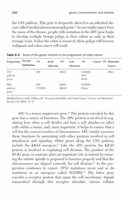
ptg
the CIN pathway. This gene is frequently altered in an inherited dis-ease called familial adenomatous polyposis.45 As you might expect fromthe name of the disease, people with mutations in the APC gene beginto develop multiple benign polyps in their colons as early as theirteenage years. Unless the colon is removed, these polyps will becomemalignant and colon cancer will result.
120 genes, chromosomes, and disease
APC is a tumor suppressor gene.46 The protein encoded by thisgene has a variety of functions. The APC protein is involved in reg-ulating how often a cell divides and how a cell attaches to othercells within a tissue, and, most important, it helps to ensure that acell has the correct number of chromosomes. APC mainly exercisesthese functions by associating with other proteins involved in cellattachment and signaling. Other genes along the CIN pathwayinclude the KRAS oncogene.47 Like the APC protein, the KRASprotein is involved in regulating cell division. The product of theBUB1 gene, in contrast, plays an important role in mitosis by ensur-ing the mitotic spindle is prepared to function properly and that thechromosomes are aligned correctly for cell division.48 As the pro-gression continues to cancer, TP53 mutations occur and so domutations in an oncogene called TGFBR2.49 The latter geneencodes a receptor protein that spans the cell membrane. Signalstransmitted through this receptor stimulate various cellular
Table 5–3 Some of the genes involved in the progression of colon cancer
Progression Normal
Epithelium⇒ Early
Adenoma
⇒ Late
Adenoma
⇒ Cancer ⇒ Metastatic
Cancer
CINpathwaygenes
APC KRAS TGFBR2TP53BUB1
PRL3
MSIpathwaygenes
APCCTNNB1
KRASBRAF1
TGFBR2Others
Modified from Grady, William M. “Genomic Instability and Colon Cancer.” Cancer and MetastasisReviews 23 (2004): 11–27.
Wow! eBook <WoweBook.Com>

ptg
5 • genes and cancer 121
responses, but, most important, one of those signals tells the cell tostop growing and dividing. In short, progression along the CINpathway involves a series of genetic changes that encourages chro-mosome instability while promoting uncontrolled cell growth withthe endpoint of the pathway being metastasis. When the PRL3 geneat the end of the genetic progression is overexpressed, it promotescancer metastasis.
About 15% of colon cancers initiate in a different way, but theprogression bears many similarities to the CIN pathway. In this case,the mutations involved affect a set of genes that fix specific mistakesoccurring during DNA replication. This repair system is called themismatch repair system. Examples of the kinds of mismatches thissystem repairs are when A mistakenly pairs with C rather than T orwhen G gets it wrong with T rather than its correct mate C. Muta-tions blocking the function of mismatch repair genes probably leadinto the pathway to colon cancer by permitting mutations to occur inAPC. Except for a few genes, this pathway is quite similar to the CINpathway.
If the marker for the CIN pathway is chromosomal instability, theone that characterizes the mismatch repair pathway is somethingcalled microsatellite instability (MSI). Microsatellites are short,tandemly repeated sequences of two to four nucleotides scatteredthroughout the genome. For example, (CA)n is a common repeatsequence. Clusters of these repeats may be amplified from 10 to 100times. When these microsatellites are extracted from the DNA, theyform a characteristic fingerprint that reflects their length and num-ber. When the mismatch repair system is not working, this pattern isaltered, often reflecting a change in the number of repeating units.
Smoking, carcinogens, and lung cancerThe explosion of the Unit 4 nuclear reactor at Chernobyl, Ukraine, onApril 26, 1986, released massive amounts of radioactive materials intothe atmosphere, causing consternation around the world, but espe-cially in Europe, the Baltic countries, and Belarus as the prevailingwinds drove the radioactive plume westward. Heavy rains shortlyafterward brought much of this material to earth where it entered the
Wow! eBook <WoweBook.Com>

ptg
122 genes, chromosomes, and disease
ground water. There have been a multitude of studies directed atdetermining whether this massive radioactive fallout increased theincidence of cancer in nearby countries. An analysis of these resultssuggests that only childhood thyroid cancer showed unambiguousincreases in frequency.50 Radioactive iodine (131I), generated by theexplosion and required for the thyroid hormones thyroxine and tri-iodothyronine to function, is presumed to be responsible.
Just as Chernobyl shows how radioactive fallout from a nuclearexplosion can cause a specific type of cancer, namely thyroid cancer, thecarcinogenic compounds present in cigarette smoke cause lung cancer.As Robert Proctor wrote in reviewing the history of tobacco and lungcancer, “tobacco is the world’s single most avoidable cause of death.”51
Tobacco is a New World plant. While Christopher Columbus wasin the Bahamas, the local Indians offered him a gift of dried tobaccoleaves. They used these leaves in several rituals. Soon thereafter, someof Columbus’s men began to smoke the leaves and the habit laterbecame widespread in Europe and elsewhere although usage wassporadic. Cigarettes became popular in the nineteenth century, butthe laborious process of rolling them by hand meant that the womenwho did the rolling were unable to produce more than 200 each day.
Cigarette manufacture received a big boost late in the nineteenthcentury when automated rolling equipment became available. Cleveradvertising and promotions exacerbated the habit. Early on, forexample, cigarette companies included small cards in each box as areward.52 The cards, collectibles that could be traded, depictedfamous movie stars, baseball players, and Native American chiefs.The practice was stopped during World War II to save paper, but peo-ple still collect the cards and rare cards, like one depicting formerPittsburgh Pirates great Honus Wagner, can garner more than $2 mil-lion today. Later, there were posters and magazine advertisementsposing attractive men and women with cigarettes. There were alsothe catchy jingles. “Winston tastes good like a cigarette should,” or“Light up a Lucky.” If this were not enough, cigarette usage receiveda big boost in the two world wars when they were dispensed as part ofthe soldiers’ rations. Consequently, after each conflict, an army offighting men went home as an army of cigarette addicts.
Wow! eBook <WoweBook.Com>

ptg
5 • genes and cancer 123
John Hill, an Englishman, first mooted the possible connectionbetween tobacco and cancer in 1761.53 He observed that excessive useof snuff sometimes caused cancer of the nose. In 1795, Samuel vonSoemerring in Germany reported that pipe smokers were prone tocancer of the lip. The connection between pipe smoking and cancer ofthe lip and mouth was bolstered further in the nineteenth century, butlung cancer was extremely rare before the twentieth century. Duringthe second decade of the twentieth century, physicians started tonotice increasing frequencies of lung cancer. In 1912, Isaac Adler sug-gested a possible connection between lung cancer and smoking, butthis connection seemed tenuous at first because the time lag betweenfirst exposure and the development of lung cancer is on the order of20 years. It was only after World War II that lung cancer and smokingwere definitively linked. Establishing this link took a long time withthe tobacco companies fighting it tooth and nail all the way. Thelegacy of this battle for the year 2009 was 219,440 new cases of lungcancer in the United States and 159,390 deaths from this disease.54
There are at least 55 carcinogens in tobacco smoke of which 20have been shown to cause lung cancer in at least one animal species.55
These compounds initiate lung cancer by causing mutations in tumorsuppressors like TP53 and RB and also in oncogenes like KRAS. Fur-thermore, there are over 50 cancer-causing compounds in secondhandsmoke.56 According to the American Cancer Society, there were 46 mil-lion American smokers in 2001 (the most recent year for which num-bers were available).57 Unfortunately, almost 27% of these smokerswere aged 18–24, so the habit seems not to be declining among theyoung. Cigarette smoking accounts for at least 30% of all cancer deaths.These cigarette-caused deaths are not just from lung cancer, but alsofrom cancer of the larynx, oral cavity, throat, and esophagus. But can-cers only account for around half of all smoking-related deaths. Heartdisease, emphysema, and stroke are among the life-threatening dis-eases exacerbated by smoking. Overall, male smokers lose an average of13.2 years of life and female smokers 14.5 years of life because of theirhabit. Perhaps the most important lesson lung cancer teaches is thatthere is usually a very long lag between the time a person begins tosmoke and the appearance of the disease. Smoking may seem harmlessenough at first, but its consequences can be lethal later on.
Wow! eBook <WoweBook.Com>

ptg
124 genes, chromosomes, and disease
Fixing bad blood: lymphoma and leukemiaLymphoma and leukemia are both cancers of the blood. Lymphomasoriginate in the lymph system of the body. Lymphocytes are whiteblood cells that, upon conversion to lymphoma cells, begin to accu-mulate in masses around the lymph nodes and in other parts of thelymph system. There are three types of lymphocytes. Natural killer(NK) cells are large, granular lymphocytes, whereas B and T cells aresmall lymphocytes.
Thomas Hodgkin, an English physician, provided the firstdescription of a lymphoma.58 In 1826, Hodgkin, with a medicaldegree from Edinburgh in hand, was appointed the first lecturer inmorbid anatomy and museum curator at the new Guy’s Hospital inLondon. He performed several hundred autopsies and logged in over3,000 specimens in the Hospital’s Green Book. In 1832, Hodgkinpublished a paper titled “On Some Morbid Appearances of theAbsorbent Glands and Spleen.” In his paper, Hodgkin described hispostmortem discovery of enlarged lymph nodes or the spleen in thebodies of seven former patients. In 1865, another well-known physi-cian at Guy’s Hospital named Samuel Wilks published a paper on thesame disease and christened it Hodgkin’s disease. Later on, it was dis-covered that Hodgkin lymphoma is characterized by the presence ofmultinucleated cells called Reed-Sternberg cells.
Today, lymphomas are classified as either Hodgkin or non-Hodgkin. In 2009, around 601,180 people in the United States wereeither living with lymphoma or were in remission.59 They included148,460 people with Hodgkin lymphoma and 452,720 people havingnon-Hodgkin lymphoma.
There are many kinds of non-Hodgkin lymphomas with about 85%affecting B cells and the rest T cells or NK cells. Some of these lym-phomas are classified as slow growing and others as fast growing. Thediscussion that follows will focus on the most common fast-growinglymphoma. It is called a diffuse large B-cell lymphoma (DLBCL). Themost frequently detected genetic alteration associated with DLBCL isa rather curious one involving a gene called BCL6.60 This gene is notmutated, but instead a piece of a different chromosome becomesattached upstream of the gene (translocation). The translocation
Wow! eBook <WoweBook.Com>
ptg
5 • genes and cancer 125
attaches regulatory sequences to the BCL6 gene that perturb itsexpression.
BCL6 encodes a transcription factor that has many functions. Butat least two of these seem to be closely related to its role in lymphomaformation.61 First, the BCL6 transcription factor is crucial for rapid Bcell proliferation. This occurs in sites located within the lymph nodescalled germinal centers. Second, this transcription factor appears toinhibit programmed cell death (apoptosis). If these two functions areperturbed, as they are in DLBCL, B cells should proliferate rapidly,but they will not be destroyed by apoptosis. Instead, they will accu-mulate in masses, a distinctive feature of the disease.
The Leukemia & Lymphoma Society Web site (www.lls.org/hm_lls) has lots of useful information about the respective diseases,including a summary of the various drug therapies currently in use.The one used to treat DLBCL is called R-CHOP and often involvessix cycles of treatment. What is R-CHOP? R refers to rituxan, a mon-oclonal antibody, which binds to CD20, a surface protein on bothnormal and malignant B cells.62 Rituxan binding marks these cells fordestruction. Stem cells in the bone marrow lack CD20, allowing new,healthy B cells to be regenerated. Over time, these new and normal Bcells accumulate while the sheets of cancerous B cells, having beenmarked for elimination by rituxan, are destroyed.
CHOP refers to four different compounds that interfere with thegrowth of both normal and malignant cells.63 Cyclophosphamide (C)and doxorubicin (H) interfere with DNA replication, but in differentways. Vincristine (O) blocks the formation of the microtubule struc-tures required for the assembly of the mitotic spindle essential forcell division, whereas prednisone (P) is an immune suppressant usedto slow down the rapid growth of cancerous white blood cells. CHOPis the most common chemotherapy for lymphoma, although there aremany other regimens as well. It has been used very effectively fordecades to combat highly aggressive lymphomas like DLBCL. Topromote white cell regeneration, nulasta is given after eachchemotherapy treatment. Nulasta is a genetically engineered humanprotein called G-CSF that promotes white cell growth.64 In addition,radiation therapy is used to target those places where cancerousmasses have been detected.
Wow! eBook <WoweBook.Com>
ptg
126 genes, chromosomes, and disease
In 1960, two cytologists working at different institutions inPhiladelphia noted that patients with chronic myelogenous leukemia(CML) had an unusually small copy of chromosome 22.65 The factthat both investigators hailed from Philadelphia led to the naming ofthis peculiar chromosome in dubious honor of that city. Later on, itwas discovered that the Philadelphia chromosome was one of the twoproducts of a reciprocal translocation that had taken place betweenchromosomes 9 and 22. This resulted in the fusion of two genescalled ABL1 and BCR. ABL1 is a proto-oncogene. It encodes a tyro-sine kinase. Kinases transfer phosphate groups from high-energydonors to various substrates. These kinase-catalyzed phosphate trans-fers are important cellular-signaling mechanisms. For example, theABL1 kinase has been implicated in processes of cell differentiation,cell division, cell adhesion, and stress response. When the fusion genewas discovered, the function of the second gene was unknown.Hence, the BCR gene was simply named (B)reakpoint (C)luster(R)egion to indicate that the gene fusions could occur at differentpoints within this gene. It now appears that this gene, of which thereare several versions depending on the position of the breakpoint, alsoencodes a kinase. The resulting gene fusion produces an activatedkinase that promotes unregulated cell division.
It turned out that the tyrosine kinase domain derived from theABL1 gene proved to be the key element in converting the fused geneinto an oncogene. If a drug could be developed that blocked thekinase function, it might also do away with uncontrolled proliferationof cancerous white blood cells. In 2001, the FDA approved Gleevecfor just this purpose. Although Gleevec also targets 4 of the 90 or soother human tyrosine kinases, this does not seem to cause seriousproblems. Gleevec was the first anticancer drug to be synthesizedwith a specific target in mind. Its success has spurred the develop-ment of other drugs designed specifically to inhibit the function ofother oncoproteins.
As its name implies, CML is a chronic disease.66 Gleevec treat-ment cannot cure it, but with treatment most patients having the dis-ease remain symptom free for long periods of time. For treatment ofpatients who cannot tolerate Gleevec or whose disease is resistant tothe drug, the FDA has approved two new drugs, Sprycel and Tarceva.
Wow! eBook <WoweBook.Com>

ptg
5 • genes and cancer 127
The big pictureIn a book of this kind, it is only possible to touch briefly on a few of themany ways in which gene alterations lead to the initiation and spreadof cancer. In 2004, Bert Vogelstein and his colleague Kenneth W. Kin-zler published a paper titled “Cancer Genes and the Pathways TheyControl.”67 It is a short, but highly informative overview of the subject.
According to Vogelstein and Kinzler, there are many fewer path-ways to cancer formation than there are genes involved in the process.Because virtually all cancer-causing DNA tumor viruses encode pro-teins that inactivate both the Rb and p53 proteins, Vogelstein andKinzler speculate that it may be impossible for a tumor of epithelialorigin (carcinoma) to form unless the tumor suppressor pathways con-trolled by the genes encoding these proteins have been inactivated.
Vogelstein and Kinzler distinguish between solid and liquidtumors, defining the latter group as including lymphomas andleukemias. They note that at least three mutations seem to berequired to develop a malignant solid tumor in adults. In contrast,“perhaps liquid tumors don’t require as many pathways to be inacti-vated because their precursor cells are already mobile and invasive,key characteristics that solid tumor cells must develop to becomemalignant.” Interestingly, the events that seem commonly to initiateliquid tumors are chromosome translocations whose fusion leads toconversion of a perfectly normal gene into a cancerous one.
One topic Vogelstein and Kinzler discuss that is not even touchedon in this chapter is tumor angiogenesis. This is the method by whichtumors attract a blood supply, a critical step in tumor development.
Vogelstein and Kinzler point out that between 1994 and the pub-lication of their article in 2004, “significant gains have been made incancer therapeutics.” They identified three major challenges in thedecade ahead: discovery of new genes that play a role in cancer, delin-eation of the pathway that these genes act upon, and “development ofnew ways to exploit this knowledge for the benefit of patients.” In theend, cancer takes a long time to develop. “It takes 30–40 years for atypical epithelial cell to accumulate the multiple genetic alterationsrequired to progress to metastatic disease.” This means that manytumors ought to be detectable when they are still curable by conven-tional surgery. “Though less dramatic than cures, prevention and
Wow! eBook <WoweBook.Com>

ptg
128 genes, chromosomes, and disease
early detection are perhaps the most promising and feasible means toreduce cancer deaths within the next decade.”
Given that the human body is made up of between 10 and 100 tril-lion cells (estimates vary) most of which divide, some very rapidly, thesurprising thing is not that we get cancer, but rather that it is not evenmore frequent given that, in a dividing cell population so enormous,mistakes are bound to happen. What is truly remarkable is how effi-cient cellular repair systems are and how successful cell division controlmechanisms are in stopping progression of the cell cycle to allow timefor these repair systems to weed out unwanted and noxious mutations.We needlessly put pressure on this remarkable protective system whenwe smoke, create polluted cities, and spend too much time in the sun.
Wow! eBook <WoweBook.Com>

ptg
Genes and behavior
In 1978, a woman stopped by the office of Han Brunner, who wasstudying for his MD at the University of Groningen in the Nether-lands, to discuss a problem that was concerning her.1 It seemed thatmany of the men in her family were prone to violent outbursts. Brun-ner wondered why, but before he investigated further, he had toobtain his medical degree. Following that, Brunner obtained a PhDat Nijmegen University, where he found and characterized the genefor myotonic dystrophy for his dissertation research. But he also con-tinued his research on the violent relatives of the woman he had metin 1978.
Finally, in 1993, Brunner and his colleagues reported their find-ings on the Dutch family.2 Fortunately for them, it so happened thatthe woman’s maternal great uncle, who was not violent, had kept arecord that revealed that nine male relatives exhibited aberrantbehavior. Since then, five more men in the family had displayed thedisorder. Those studied by Brunner and his colleagues included aman who raped his sister and later, having been confined to a mentalinstitution, chased the warden with a pitchfork. Another brandished aknife in front of his sisters and forced them to disrobe, while a thirdhad attempted to run over his boss after being told that his work wasnot satisfactory. Two others were arsonists. They all exhibited slightmental retardation, with IQ scores of around 85. The women in thefamily were neither violent nor mentally deficient.
These findings suggested that a gene on the X chromosome wasprobably involved. Brunner and his colleagues showed that thisindeed was the case and that the gene in question encoded anenzyme called monoamine oxidase A (MAOA). Affected males had a
6
129

ptg
130 genes, chromosomes, and disease
point mutation in the gene that caused a total loss of activity of thisimportant enzyme that plays a central role in the inactivation of fourmonoamine neurotransmitters found in the brain (serotonin, norepi-nephrine, melatonin, and dopamine). Although these genetic find-ings, coupled with their neurological implications, strongly suggestedthat the absence of MAOA was linked to aggressive behavior, theauthors stressed that aggressive behavior varied markedly in severityover time within the pedigree. Naturally, the Dutch findings pro-voked much interest. Natalie Angier, writing in the New York Timesunder the headline “Gene Tie to Male Violence Is Studied,” made acorrect prediction when she prophesied that the discovery of thisgenetic defect was “likely to rekindle the harsh debate over the causesof criminal and abnormal behavior.”3
The gene defect unique to the Dutch pedigree proved extremelyrare, but later on, a polymorphism was found upstream of the MAOAgene that has a profound effect on its expression.4 This was a repeatedsequence of 30 base pairs. Although there were several variants, threeand four repeats were by far the most common. Expression of theMAOA gene was considerably lower for the “short” (three repeats)version of the gene than the “long” (four repeats) version.
This discovery has been the basis for a whole series of investiga-tions on antisocial behavior and MAOA levels. A key contribution wasmade by Avshalom Caspi and Terri Moffit, clinical psychologists atthe University of Wisconsin and King’s College London, and others in2002.5 These investigators decided to examine whether MAOA geneexpression could be correlated with aggression among children whohad experienced maltreatment. The children in question had grownup by the time Caspi and his colleagues conducted their study, butthey had been assessed repeatedly over the intervening years sincethey were part of a cohort of 1,037 babies who had been born inDunedin, New Zealand, between April 1, 1972, and March 31, 1973.During the “assessment phase,” all members of the Dunedin Studyare brought back to the University of Otago where the study islocated and their physical and mental health is examined.6 Longinterviews are part of the process.
Caspi and colleagues focused on male children because they havea single X chromosome and, hence, but one copy and one form of the
Wow! eBook <WoweBook.Com>
ptg
6 • genes and behavior 131
MAOA gene. They determined whether each individual had the longor short form of the gene, using this as a proxy for MAOA activity. Theyfound that the interaction between maltreatment and antisocial behav-ior, using four recognized measures of the latter, was significantlyweaker among males with high MAOA activity than it was in maleswith low MAOA activity. Furthermore, men with low MAOA activitywho had been maltreated were more likely to be convicted of a violentcrime than men with the same low activity who had been brought upnormally. In contrast, there was no significant increase in risk of violentcrime among maltreated men with high MAOA activity. One impor-tant difference between the Caspi study and Brunner’s results was thatthe men profiled in the Caspi report had normal IQs, so lower IQscould not be invoked as a reason for frustration, leading ultimately toviolence. Additional support for Caspi’s conclusion was soon forthcom-ing when a group in Virginia confirmed their results using a sample ofboys from the Virginia Twin Study for Adolescent Behavioral Develop-ment and somewhat different criteria for measuring conduct disorder.7
In 2004, journalist Ann Gibbons wrote up the highlights of theannual meeting of the American Association of Physical Anthropolo-gists held in Tampa, Florida, for Science magazine.8 Gibbons reportedthat a team of geneticists led by Tim Newman at the National Insti-tute of Alcohol Abuse and Alcoholism had found the short variant ofthe MAOA gene in chimpanzees, gorillas, and many other primates.However, Newman also discovered that the short variant was foundonly in Old World (Asian and African) monkeys, but not in NewWorld (South American) species. Hence, the short variant appearedto have arisen after Old and New World monkeys separated some 25million years ago. Newman suggested that there must be a reasonwhy the short variant was present at such a high frequency in humanand Old World primate populations. “Bold, aggressive males mighthave been quicker to catch prey or detect threats.”
Gibbons titled her article “Tracking the Evolutionary History of a‘Warrior’ Gene.” And, of course, the name caught on for the shortversion of the MAOA gene.
But later, Newman and colleagues proceeded to perform the rhe-sus monkey equivalent of the Caspi study, but with somewhat surpris-ing results.9 Monkeys reared without mothers were supposed to be the
Wow! eBook <WoweBook.Com>

ptg
132 genes, chromosomes, and disease
equivalent of badly treated human children, but the monkeys with theleast amount of MAOA activity also showed the lowest levels of aggres-sion. When their mothers reared these monkeys, they exhibited thehighest levels of aggression of any group. Conversely, the monkeys withhigh levels of MAOA activity showed the lowest levels of aggressionwhen reared by their mothers, but aggression increased markedlywhen they were reared without parents. As Newman and colleaguespoint out, the situations in the Caspi and their studies are somewhatdifferent. First, distinct species were involved. Second, mothers versusno mothers were compared in the Newman experiments, whereas inCaspi, all the males in question had parents, some of whom were bad.Nevertheless, the differences in results between the two studies areimportant because they illustrate why one must be careful in extrapo-lating the results of animal studies, even when primates are involved, topredict the possible outcomes of human studies conducted in a similarfashion.
Warrior gene studies turned sinister when a group led by Kevin M.Beaver of Florida State University’s College of Criminology and Crim-inal Justice linked the low activity form to gang membership.10 Thiscaught the attention of the national media. Reporter Kathleen Kings-bury titled an article in TIME magazine “Which Kids Join Gangs? AGenetic Explanation.”11 But of even greater consequence was the useof the warrior gene argument to convince a jury that a convicted mur-derer did not deserve the death penalty.
On the evening of October 16, 2006, Bradley Waldroup was drink-ing in his trailer home in the mountains of Tennessee while awaiting thearrival of his estranged wife and their four children for the weekend.12
An obviously concerned Penny Waldroup arrived with her children andher friend Leslie Susan Bradshaw having left word with another friendto call the police if they did not return by a specified time.
When Penny Waldroup told her husband that she and her friendwere leaving, an argument ensued. In a rage, Brad Waldroup shotBradshaw eight times and then cut her head open with a sharp object.Next, he chased his wife with a machete, chopping off a finger andcutting her again and again. When the police arrived, Bradshaw wasdead, Penny Waldroup was badly injured, and there was blood allover the trailer, including the walls, the carpet, and even on the bible
Wow! eBook <WoweBook.Com>
ptg
6 • genes and behavior 133
that Brad Waldroup had been reading while awaiting the arrival of hiswife and children.
Prosecutors charged Waldroup with the felony murder of Brad-shaw, a death penalty offense, and with the attempted first-degreemurder of his wife. Prosecutor Cynthia Lecroy-Schemel said thatthere “were numerous things he did around the crime scene thatwere conscious choices. One of them was [that] he told his children to‘come and tell your mama goodbye,’ because he was going to kill her.And he had the gun, and he had the machete.” The case was prettycut and dried as even Waldroup admitted. He claimed to have“snapped” and said, “I’m not proud of none of it.”
Because the defense could not dismantle the evidence, they hadto take a different tack. As attorney Wylie Richardson put it, “It was-n’t a who done it? It was a why done it?” To try to get an answer on“why,” Richardson asked forensic psychiatrist William Bernet at Van-derbilt University to give Waldroup a psychiatric examination. Bernetdid so, but he also took a blood sample as one of his interests was therelationship between MAOA and criminal behavior and he was quitefamiliar with the work of Caspi and colleagues.13 In fact, by the timeof the Waldroup trial, Bernet and his colleagues had tested some 30criminal defendants, the majority having been charged with murder.14
The prosecution objected strongly to Bernet’s being allowed totestify, but the judge allowed his testimony. Bernet and his colleaguesfound that Waldroup indeed had the warrior gene. Because he hadalso been abused as a child, Bernet said that Waldroup’s “geneticmakeup, combined with his history of child abuse, together created avulnerability that he would be a violent adult.” Bernet was careful topoint out that he hadn’t said, “these things made him become violent,but they certainly constituted a risk factor of vulnerability.”
Prosecutor Drew Robinson believed the genetic evidence was“smoke and mirrors” designed to confuse the jury. He called in a psy-chiatrist named Terry Holmes, the Clinical Director of MoccasinBend Mental Health Institute in Chattanooga, Tennessee. Holmesbelittled Bernet’s interpretation saying it was too early to try andmake such a connection in a court of law. Holmes believed thatbecause Waldroup was “intoxicated and mad” he “was gonna hurtsomebody. It had little to nothing to do with his genetic makeup.”
Wow! eBook <WoweBook.Com>

ptg
134 genes, chromosomes, and disease
After deliberating on the Waldroup case for 11 hours, the juryconvicted Waldroup of voluntary manslaughter and attempted sec-ond-degree murder. Drew Robinson, the prosecutor, couldn’t believehis ears. “I was just flabbergasted. I did not know how to react to [theverdict].” In explanation, juror Sheri Lard said the warrior gene wasjust one piece of evidence. Some attached importance to it, but forothers it was less significant. Where it was decisive for the jury was indeciding whether to find Waldroup guilty of murder and subject to thedeath penalty. For juror Debbie Beaty, the genetic evidence was per-suasive. “A diagnosis is a diagnosis, it’s there,” she said. “A bad gene isa bad gene.”
Beaty’s statement explains why the prosecution was so anxious toprevent Bernet’s testimony. Bernet was an expert where the warriorgene was concerned, but Holmes clearly was not. This fact was notlost on the jury who, given the nature of the evidence, concluded thatWaldroup’s actions might have been beyond his control.
On a lighter note, perhaps, political scientists and economists gotinto the warrior gene act using game playing as a measure of aggres-sion.15 In this case, they measured the willingness of male subjects topay to administer hot sauce to someone who had taken money fromthem. It turned out there was little difference between low and highMAOA activity groups when only 20% of their money was taken, butthe warrior gene participants administered hot sauce more aggres-sively than their calmer colleagues after losing 80% of their money.
And now, we have the MAOA gene predicting credit card debtaccording to Jan Emmanuel Neve, a graduate student at the LondonSchool of Economics, and James H. Fowler, a political scientist at theUniversity of California, San Diego.16 They report that having one copyof the low-efficiency variant raises the average likelihood of havingcredit card debt 7.8%, whereas having two copies increases this numberto 15.8%. Their numbers also suggest there may be a sex bias becausemen have only one copy of the MAOA gene, whereas women have two.This is the kind of observation that titillates the media and was pickedup by Newsweek and the Wall Street Journal’s Smart Money.17
Race also briefly raised its ugly head with regard to the warriorgene. In August 2006, Dr. Rod Lea, a genetic epidemiologist at the
Wow! eBook <WoweBook.Com>

ptg
6 • genes and behavior 135
ESR Kanepuru Science Centre in Porirua, New Zealand, caused afuror, particularly Down Under, when he presented a report titled“Tracking the Evolutionary History of the Warrior Gene in the SouthPacific” at the International Congress of Human Genetics held inBrisbane, Australia. As the Sydney Morning Herald reported onAugust 9, Lea told Congress participants that the Maori had a “strik-ing over-representation” of the warrior gene and that this went “a longway to explaining some of the problems Maoris have.”18 “Obviously,this means they are going to be more aggressive and violent and morelikely to get involved in risk-taking behaviour like gambling.” Leaadded the caveat that he thought nongenetic factors might also play arole, but then he went on to link the gene to smoking and drinkingamong the Maori.
Lea’s paper provoked an eruption that he hastened to quell in aneditorial in the New Zealand Medical Journal where he attempted toput a positive spin on his findings. Lea argued that his main point wasthat the warrior gene had been selected in the Maori in connectionwith their breathtakingly risky canoe migrations over vast stretches ofocean not knowing whether they would find land or not. Lea, and hiscolleague Geoffrey Chambers, pleaded that they were ethicalresearchers who had received far too much negative media hype.
The most thoughtful contribution to the whole debate over thewarrior gene and Maori aggression came from a distinguished Maoriscientist named Gary Raumati Hook.19 He aimed to assess the “truth”of the warrior gene hypothesis and what it might mean for the Maori.Hook summarized the many negative behaviors that have been linkedto the warrior gene (see Table 6–1). But as he pointed out, “posses-sion of these traits would pose severe disadvantages especially in theancient world of the voyaging and war-like Polynesians.” Further-more, “in the case of Maori domestic violence blaming nature asopposed to nurture simplifies the problem because it implies thatMaori has no one to blame but himself for his own condition. Contri-butions to racial stereotyping by trained scientists are unethical andscandalous.”
Wow! eBook <WoweBook.Com>

ptg
136 genes, chromosomes, and disease
Table 6–1 Behavioral disorders reported to be associated with low MAOAactivity*
Disorder Reference**
Personality disorders 19
Antisocial behavior 5,7
Violence and risk taking 19
Impulsive aggression 19
Mental disorders 19
Credit card debt 16,17
Obesity 19
Propensity to join gangs 10
Behavioral aggression in game playing 15
In fact, the problem with the whole warrior gene hypothesis isthat the low-activity variant is common in most populations (see Table6–2), yet the majority of people with this variant do not exhibit badbehavior. Because it is so abundant, it probably does have some selec-tive advantage. The variety of anomalies that have been associatedwith the warrior gene variant (refer to Table 6–1) show just how diffi-cult it is to sort out behavioral and more complex conditions involvingmany genes. On top of that, there are environmental conditions thatplay an important role. Studies like those of Caspi et al.20 illustrate theimportance of combining a specific environmental trigger (childabuse) with the warrior gene in causing bad behavior.
*Modified from Hook, G. Raumati. “‘Warrior genes’ and the disease of being Maori.” MAIReview 2 (2009). Target article.**See chapter references at end of book.
Wow! eBook <WoweBook.Com>
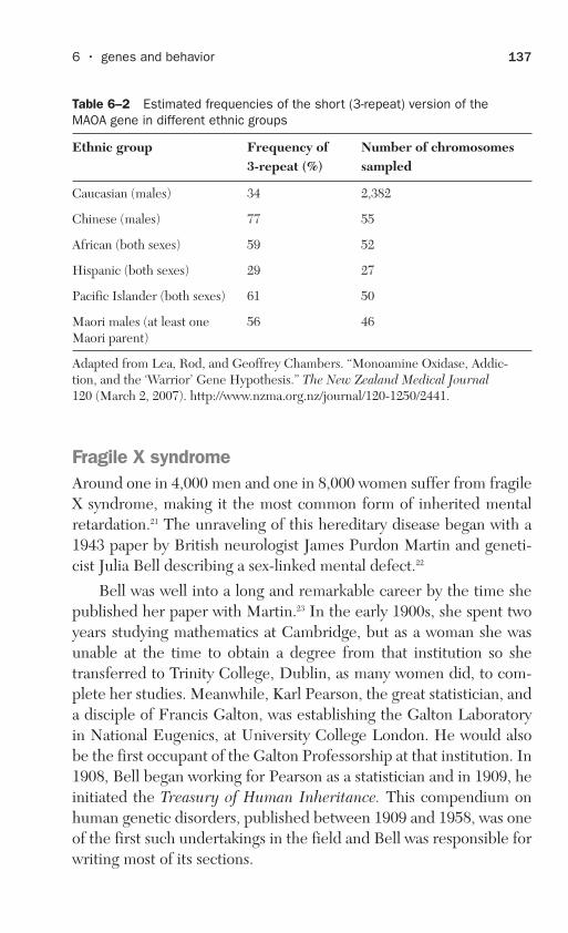
ptg
6 • genes and behavior 137
Table 6–2 Estimated frequencies of the short (3-repeat) version of theMAOA gene in different ethnic groups
Ethnic group Frequency of 3-repeat (%)
Number of chromosomessampled
Caucasian (males) 34 2,382
Chinese (males) 77 55
African (both sexes) 59 52
Hispanic (both sexes) 29 27
Pacific Islander (both sexes) 61 50
Maori males (at least oneMaori parent)
56 46
Adapted from Lea, Rod, and Geoffrey Chambers. “Monoamine Oxidase, Addic-tion, and the ‘Warrior’ Gene Hypothesis.” The New Zealand Medical Journal120 (March 2, 2007). http://www.nzma.org.nz/journal/120-1250/2441.
Fragile X syndromeAround one in 4,000 men and one in 8,000 women suffer from fragileX syndrome, making it the most common form of inherited mentalretardation.21 The unraveling of this hereditary disease began with a1943 paper by British neurologist James Purdon Martin and geneti-cist Julia Bell describing a sex-linked mental defect.22
Bell was well into a long and remarkable career by the time shepublished her paper with Martin.23 In the early 1900s, she spent twoyears studying mathematics at Cambridge, but as a woman she wasunable at the time to obtain a degree from that institution so shetransferred to Trinity College, Dublin, as many women did, to com-plete her studies. Meanwhile, Karl Pearson, the great statistician, anda disciple of Francis Galton, was establishing the Galton Laboratoryin National Eugenics, at University College London. He would alsobe the first occupant of the Galton Professorship at that institution. In1908, Bell began working for Pearson as a statistician and in 1909, heinitiated the Treasury of Human Inheritance. This compendium onhuman genetic disorders, published between 1909 and 1958, was oneof the first such undertakings in the field and Bell was responsible forwriting most of its sections.
Wow! eBook <WoweBook.Com>
ptg
138 genes, chromosomes, and disease
Martin and Bell had established that the fragile X condition, alsoknown as Martin-Bell syndrome, was sex-linked, but it was HerbertLubs who observed, in another pedigree of sex-linked mental retar-dation, that there was a constriction near the end of the long arm ofthe X chromosome in cultured lymphocytes.24 Lubs’s results provedirreproducible initially because the constriction, or fragile site, wasnot evident using cell culture media different from those used byhim. But once this had been realized, the constriction that Lubs haddescribed became evident once more.
The gene responsible for fragile X syndrome was isolated in 1991and named FMR-1 for fragile X mental retardation-1.25 The diseaseproved to be caused by overamplification of a CGG repeat sequence.That is, it was a trinucleotide repeat disease like Huntington’s chorea(see Chapter 1, “Hunting for disease genes,” and Table 1–1). Normalindividuals have 5–54 repeats of this arginine-specifying codon. Peo-ple with 55–200 repeats are referred to as having a premutation,whereas those with more than 200 repeats have the full mutationwith the gene becoming completely inactive due to epigeneticmethylation of cytosine residues in the repeat sequence.26 The inacti-vation of FMR-1 means that its protein FMRP is not made. Impair-ment of brain function then occurs, probably in part becausesynaptic connections between at least some neurons do not formproperly.
Fragile X syndrome is dominant, meaning that the disease isexpressed in women as well as in men. People with the premutationare usually unaffected.27 However, in women with the premutation,the repeat sequence in the affected X chromosome may amplify dur-ing oogenesis so that some of their children may express the full-blown disease. Babies with fragile X syndrome seem normal duringthe first year, but they experience delays in speaking and languagedevelopment with lapses in short-term memory and problem-solvingability appearing later on. Autism is often associated with fragile Xsyndrome, although individuals with fragile X syndrome onlyaccount for 2%–6% of autism cases.28 IQ tests reveal mild to severemental retardation in men (IQs of 20 to 70), but women and less-affected men may have IQs approaching 80. Characteristic physicalchanges also occur in people with fragile X disease, so they tend, for
Wow! eBook <WoweBook.Com>

ptg
6 • genes and behavior 139
example, to have long, thin faces and prominent ears. Lifeexpectancy is normal.
The gay gene controversyIn 1991, J. Michael Bailey and Richard C. Pillard reported the resultsof a twin study designed to examine the heritability of homosexual-ity.29 They found 52% of identical twins in their sample were bothhomosexual, but this dropped to 22% among nonidentical twins, and11% among adoptive brothers. Because identical (monozygotic) twinspossess identical genomes, whereas nonidentical (dizygotic) twins areno more alike genetically than brothers and sisters, the results Baileyand Pillard reported strongly suggested that homosexuality was atleast partially genetic in nature.
Meanwhile, Simon LeVay published an anatomical study in Sciencemagazine that same year in which he reported that the anterior hypo-thalamus in men was twice as large as the same organ in women andhomosexuals.30 This region is believed to control sexual activity. He hadmade this comparison because an earlier study had demonstrated thatthis portion of the hypothalamus was larger in males than in females.
Based on these studies, Newsweek ran a 1992 article on homosex-uality titled “Born or Bred?”31 The article was a well-balanced reviewof these studies, the reasoning of their critics, and opinions of expertsin the area and of several gays. At the end of their article, the authorturned to Evelyn Hooker for an answer. In the 1950s, Hooker showedthat it was impossible to distinguish between heterosexuals andhomosexuals on the basis of psychological testing. Hooker asked,“Why do we want to know the cause? It’s a mistake to hope that wewill be able to modify or change homosexuality...If we understand itsnature and accept it as a given, then we come much closer to the kindof attitudes which will make it possible for homosexuals to lead adecent life in society.” Hooker was 84 when she was interviewed, butas the authors of the Newsweek article said, “The rest of us could doworse than listen to her now.”
Despite Hooker’s sensible advice, the question of whether beinggay was genetically determined was in the news in a big way the fol-lowing year. On July 19, 1993, President Bill Clinton announced a
Wow! eBook <WoweBook.Com>

ptg
new policy regarding gays in the military.32 Recruits would not beasked about their sexual orientation and, if they were homosexuals,would be obliged not to reveal this fact. Three days earlier, a paperhad appeared in the journal Science claiming that a region on the longarm of the X chromosome called Xq28 contained a genetic variantthat predisposed men to be homosexuals.33 This report naturallyreceived wide coverage in the press. The senior author was DeanHamer, an investigator at the National Institutes of Health. Hamerdid not claim to have identified a gay gene per se, but only a chromo-some region that appeared to contain such a gene.
John Maddox, editor of Nature, discussed the reaction of theBritish press to Hamer’s paper in an editorial titled “Wilful publicmisunderstanding of genetics.”34 Maddox worried about “the ten-dency of even sober-sided newspapers to overdramatise discoveriesonly, afterwards, to complain that they have been misled. Even acasual reading of the original article will reveal a commendable list ofcaveats.” In contrast, the American newspapers generally exhibitedremarkable restraint. The titles of most American newspaper articleson Hamer’s report tended to be variants on that in the New YorkTimes: “Report Suggests Homosexuality Is Linked to Genes.”35
Although the Philadelphia Daily News picked up an AP report titled“Scientists ID Gene Common to Gays,”36 most papers pointed outthat Hamer only claimed to have found a region on the X chromo-some that was linked to homosexuality. This was also true of a July 26article “Born Gay” in TIME magazine. That article got to the crux ofthe problem by pointing out that “if homosexuals are deemed to havea foreordained nature, many of the arguments now used to blockequal rights would lose force.”37
Several articles quoted Elliot Gershon, Chief of the Clinical Neuro-genetics branch at the National Institute of Mental Health, who calledHamer’s report “a major breakthrough in behavioral genetics,” but cau-tioned that the study must be replicated for credibility.38 But RichardKnox, writing in the Boston Globe, realized that, if Hamer was right,there could be serious consequences.39 He pointed out that “a prenataltest could conceivably tell parents if a fetus carried a gene that mightpredispose the child to homosexuality so they could choose abortion.”
140 genes, chromosomes, and disease
Wow! eBook <WoweBook.Com>
ptg
6 • genes and behavior 141
Nevertheless, there were skeptical scientists. Evan Balaban, thenat Harvard University, and Brown University professor Anne Fausto-Sterling raised several questions, including “the lack of an adequatecontrol group.”40 Fausto-Sterling and Balaban argued that Hamershould have looked for Xq28 DNA markers in the nonhomosexualbrothers of the gay men analyzed in the study because “chromosomeswithout the markers should show up in non-homosexual brothers.”Hamer and colleagues responded that they did have a proper controlgroup of “142 randomly selected brothers” and that “because homo-sexuality is a stigmatized trait, individuals who identify themselves asgay are expected to give more reliable information about any homo-sexual behavior than those who identify themselves as straight.”41 Butas Fausto-Sterling and Balaban had pointed out, if the mother hadbeen a carrier of the Xq28 markers, then the straight brothers shouldhave lacked them. Then statistical geneticist Neil Risch, who hadoriginated one of the statistical methods used by Hamer, raised tech-nical questions about Hamer’s analysis.42
In 1994, Hamer publicized his findings in a popular book calledThe Science of Desire in which he detailed his hunt for the gay gene.43
To add credibility to his case, in 1995, Hamer’s group reported a fol-low-up study with a different group of subjects that again indicatedlinkage of homosexuality to Xq28 in men, but not in women.44
But the doubts persisted. On June 25, 1995, John Crewdson ofthe Chicago Tribune broke a story titled “Study on ‘Gay Gene’ Chal-lenged Author Defends Findings Against Allegations.”45 Apparently, apostdoctoral fellow who was an author on the study raised questionsabout the work and was ordered to leave Hamer’s laboratory shortlythereafter. Crewdson wrote that the Office of Research Integrity waslooking into the accusations. In the same article, Crewdson reportedthat Dr. George Ebers, a neurogeneticist from the University of West-ern Ontario, had searched unsuccessfully “for a Hamer-style geneticlink to homosexuality in more than 50 pairs of gay Canadian brothers.”
On February 7, 1999, the Boston Globe ran an article by MatthewBrelis titled “The Fading Gay Gene.”46 Brelis cited the still unpub-lished Canadian study, but he also quoted Dr. Richard Pillard, whowas involved in the study of twins and sexuality, as pointing out thatwhile a gay man’s fraternal twin has a 22% chance of being gay, the
Wow! eBook <WoweBook.Com>

ptg
frequency rises to only 50% among identical twins. Concordanceshould be 100% if genes were everything. As Brelis wrote, “interestin—and enthusiasm for—the ‘gay gene’ research has waned amongactivists and scientists alike.” On April 23, 1999, the Canadian studyappeared in Science.47 It showed no linkage of homosexuality to DNAmarkers at Xq28; Neil Risch was one of the authors. Hamer, ofcourse, disagreed, citing possible problems with the way in which theOntario group selected its subjects. The New York Times reported thenew finding the same day as did the Washington Post. “I think thejury is still out,” the Post quoted Elliot Gershon, now Chairman ofPsychiatry at the University of Chicago, as saying. “All of theses stud-ies may be essentially accurate, and until we have hundreds of gaymen enrolled in studies we are not going to get a firm answer.” Mean-while, Hamer seems quietly to have put his X-linked gay gene aside.In 2005, he and his colleagues reported on a genome-wide scan forgenes related to male homosexuality.48 This time, they found sugges-tive evidence for genes that might be involved in chromosomes 7, 8,and 10, but no linkage to Xq28 in the full sample.
Bipolar disease and the biological clockThe prevalence of bipolar disorder or manic depression was previ-ously thought to be around 1% in the population, but more recentdiagnoses indicate that it could be as high as 5%.49 Family, twin, andadoption studies all suggest that bipolar disorder has an importantgenetic component. Furthermore, a host of studies has pointed fin-gers at a great many different genes. Neil Risch and David Botsteinaddressed this point in a 1996 paper titled “A Manic Depressive His-tory.” As they wrote, “the recent history of genetic linkage studies forthis disease is rivaled only by the course of the disease itself. Theeuphoria of linkage findings being replaced by the dysphoria of non-replication has become a regular pattern, creating a roller coaster-type existence for many psychiatric genetics practitioners as well astheir interested observers.”50 Risch and Botstein went on to make spe-cific suggestions for tightening up the criteria for establishing geneticlinkage. Yet a 2006 review of linkage and genome-wide associationstudies implicated sites on 19 chromosomes as being associated with
142 genes, chromosomes, and disease
Wow! eBook <WoweBook.Com>
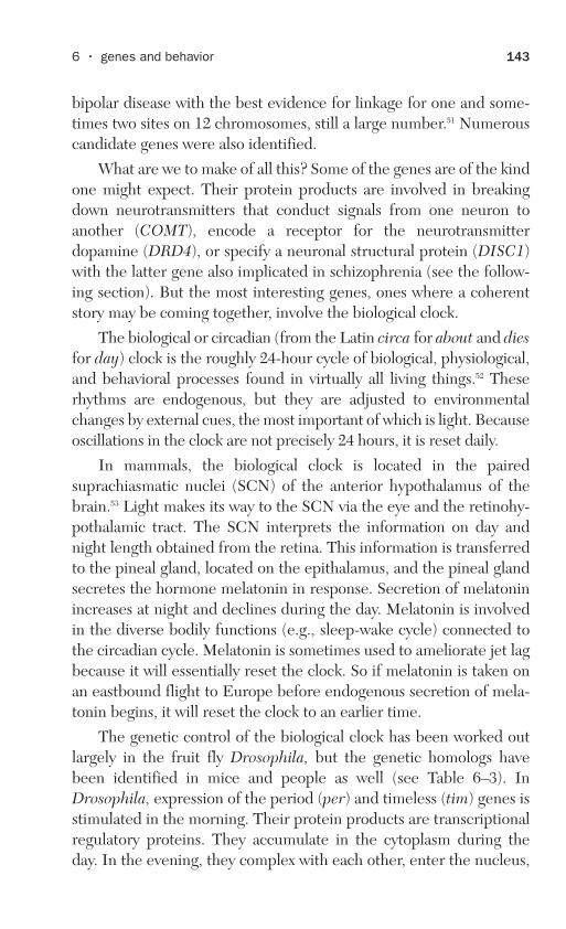
ptg
6 • genes and behavior 143
bipolar disease with the best evidence for linkage for one and some-times two sites on 12 chromosomes, still a large number.51 Numerouscandidate genes were also identified.
What are we to make of all this? Some of the genes are of the kindone might expect. Their protein products are involved in breakingdown neurotransmitters that conduct signals from one neuron toanother (COMT), encode a receptor for the neurotransmitterdopamine (DRD4), or specify a neuronal structural protein (DISC1)with the latter gene also implicated in schizophrenia (see the follow-ing section). But the most interesting genes, ones where a coherentstory may be coming together, involve the biological clock.
The biological or circadian (from the Latin circa for about and diesfor day) clock is the roughly 24-hour cycle of biological, physiological,and behavioral processes found in virtually all living things.52 Theserhythms are endogenous, but they are adjusted to environmentalchanges by external cues, the most important of which is light. Becauseoscillations in the clock are not precisely 24 hours, it is reset daily.
In mammals, the biological clock is located in the pairedsuprachiasmatic nuclei (SCN) of the anterior hypothalamus of thebrain.53 Light makes its way to the SCN via the eye and the retinohy-pothalamic tract. The SCN interprets the information on day andnight length obtained from the retina. This information is transferredto the pineal gland, located on the epithalamus, and the pineal glandsecretes the hormone melatonin in response. Secretion of melatoninincreases at night and declines during the day. Melatonin is involvedin the diverse bodily functions (e.g., sleep-wake cycle) connected tothe circadian cycle. Melatonin is sometimes used to ameliorate jet lagbecause it will essentially reset the clock. So if melatonin is taken onan eastbound flight to Europe before endogenous secretion of mela-tonin begins, it will reset the clock to an earlier time.
The genetic control of the biological clock has been worked outlargely in the fruit fly Drosophila, but the genetic homologs havebeen identified in mice and people as well (see Table 6–3). InDrosophila, expression of the period (per) and timeless (tim) genes isstimulated in the morning. Their protein products are transcriptionalregulatory proteins. They accumulate in the cytoplasm during theday. In the evening, they complex with each other, enter the nucleus,
Wow! eBook <WoweBook.Com>

ptg
In mice and people, the expression of the three per genes and thegenes encoding the blue light photoreceptors cryptochromes 1 and 2(cry 1 and 2), which also play a role in the circadian cycle, is driven bytranscriptional activating factors encoded by the clk and bmal1 genes.The latter is the homolog of the cyc gene of Drosophila. The productsof the clk and bmal1 genes form an active complex in the same way astheir Drosophila homologs.
With this background on the circadian cycle and its genetic con-trol, let us turn to its possible relationship to bipolar disease. ColleenMcClung, at the University of Texas Southwestern Medical Center,and her colleagues have characterized the behavior of mice with amutation in the clk gene that prevents the CLOCK protein fromstimulating transcription of the per and cry genes.54 They find that the
144 genes, chromosomes, and disease
Table 6–3 Homologous genes in Drosophila and mice that play a role incircadian clock regulation
Drosophila Mouse
Period (per) mPeriod1
mPeriod2
mPeriod3
Timeless (tim) None
Time-out mTimeless
Cryptochromes (Cry) mCry1
mCry2
Clock (clk) mClock
Cycle (cyc) mBma1 (MOP3)
mBmal2 (MOP9)
Double-time casein kinases 1 epsilon (TAU)
Adapted from Rivkees, Scott A. “Circadian Rhythms—Genetic Regulation and ClinicalDisorders.” Growth: Genetics & Hormones 18 (2002): 1–6.
and turn off their own expression. The protein products of the cycle(cyc) and clock (clk) genes also play a role. When they complex toform dimers, they stimulate per and tim expression.
Wow! eBook <WoweBook.Com>

ptg
6 • genes and behavior 145
behavioral profile of the mutant mice partially mimics bipolar dis-ease. For example, during the manic phase of the disease, affectedpeople generally experience an increase in energy and a decreasedneed for sleep. They can also be more irritable and often abuse sub-stances like alcohol or cocaine. In contrast, the depressive phase ofthe disease is often characterized by persistent feelings of sadness,anxiety, guilt, anger, isolation, or hopelessness among other things.
Mice with the clk mutation are hyperactive, sleep less, and exhibitan increased response to cocaine. Furthermore, lithium treatment alle-viates the symptoms just as it does in the case of human bipolar disease.However, the mouse model only replicates the behavior characteristicof mania; it does not replicate the mood oscillations characteristic ofbipolar disease. Even though the results obtained by McClung and hercolleagues incompletely model bipolar disease, they suggest that focus-ing on genes affecting the behavior of the circadian clock will probablybe a profitable avenue for future research on bipolar disease. This isalready happening. One study, for example, reported that SNPs associ-ated with three genes including clk were significantly correlated withbipolar disease55 while another paper documented an association of thetim gene with depression and sleep disturbance.
DISC1 and schizophreniaSchizophrenia is fairly common, with a median lifetime prevalence of0.7%–0.8% with the malady usually appearing in adolescence or inearly adulthood.56 Like bipolar disease, schizophrenia involves impor-tant environmental as well as genetic factors. One of the genes thatclearly plays a role in schizophrenia when broken is called DISC1.
In July 1990, a group of scientists based in Edinburgh reported ona large Scottish pedigree in which many members suffered from schiz-ophrenia or other mental disorders.57 These diseases were associatedwith a translocation between chromosomes 1 and 11. Subsequently,the translocation was found to have disrupted two genes on chromo-some 1. They were named DISC1 and DISC2 for disrupted in schizo-phrenia 1 and 2.58 DISC1 encodes a protein while DISC 2 specifies anoncoding RNA molecule (antisense) that could regulate expression ofDISC1 by binding to DISC1 messenger RNA and preventing its trans-lation.59 So far, attention has been focused almost entirely on DISC1.
Wow! eBook <WoweBook.Com>

ptg
Individuals with the translocation in the original Scottish pedi-gree showed a variety of symptoms ranging from schizophrenia tonormality (see Table 6–4). In fact, over one-quarter of individualshaving the translocation seemed to be normal. In this regard, it isimportant to note that individuals with the translocation do not lackthe DISC1 protein completely because a good copy of the undis-rupted gene is also present on the unaffected copy of chromosome 1.In fact, the DISC1 protein is present in about half the normal level intissue culture cell lines containing the translocation.60 So, it appearsthat other environmental factors as well as differences in geneticbackgrounds between individuals probably determine the behavioraldifferences between the various individuals in the pedigree.
146 genes, chromosomes, and disease
Although the translocation seen in the Scottish pedigree has notbeen seen in other families with a history of schizophrenia, othermutations have been found in DISC1 that affect behavior.61 Forexample, a deletion mutation in the DISC1 gene has been character-ized in one pedigree.62 The father is a carrier, but he is asymptomatic,whereas three of his children, who are also carriers, suffer from schiz-ophrenia or schizoaffective disorder. What is surprising is that themutation is absent in three other siblings, two of whom suffer frommajor depression and one who has schizotypal disorder, a malady thatmimics some of the symptoms of schizophrenia.
Table 6–4 Psychiatric diagnoses of individuals carrying the translocationdisrupted DISC1 and DISC2 genes
Diagnosis Number affected
Schizophrenia 7
Bipolar affective disorder 1
Recurrent major depression 10
Adolescent conduct disorder 2
Anxiety, alcoholism, minor depression 1
Unaffected 8
From Millar, J. Kirsty et al. “DISC1 and DISC2: Discovering and DissectingMolecular Mechanisms Underlying Psychiatric Illness.” Annals of Medicine 36(2004): 367–378.
Wow! eBook <WoweBook.Com>
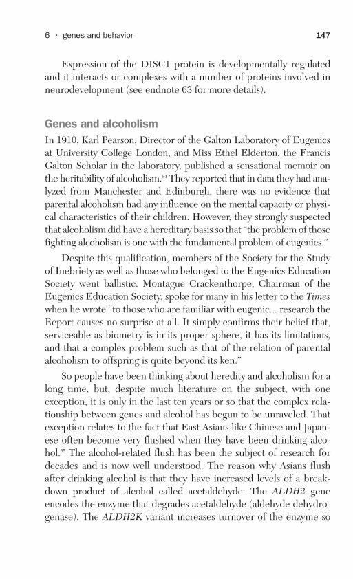
ptg
6 • genes and behavior 147
Expression of the DISC1 protein is developmentally regulatedand it interacts or complexes with a number of proteins involved inneurodevelopment (see endnote 63 for more details).
Genes and alcoholismIn 1910, Karl Pearson, Director of the Galton Laboratory of Eugenicsat University College London, and Miss Ethel Elderton, the FrancisGalton Scholar in the laboratory, published a sensational memoir onthe heritability of alcoholism.64 They reported that in data they had ana-lyzed from Manchester and Edinburgh, there was no evidence thatparental alcoholism had any influence on the mental capacity or physi-cal characteristics of their children. However, they strongly suspectedthat alcoholism did have a hereditary basis so that “the problem of thosefighting alcoholism is one with the fundamental problem of eugenics.”
Despite this qualification, members of the Society for the Studyof Inebriety as well as those who belonged to the Eugenics EducationSociety went ballistic. Montague Crackenthorpe, Chairman of theEugenics Education Society, spoke for many in his letter to the Timeswhen he wrote “to those who are familiar with eugenic... research theReport causes no surprise at all. It simply confirms their belief that,serviceable as biometry is in its proper sphere, it has its limitations,and that a complex problem such as that of the relation of parentalalcoholism to offspring is quite beyond its ken.”
So people have been thinking about heredity and alcoholism for along time, but, despite much literature on the subject, with oneexception, it is only in the last ten years or so that the complex rela-tionship between genes and alcohol has begun to be unraveled. Thatexception relates to the fact that East Asians like Chinese and Japan-ese often become very flushed when they have been drinking alco-hol.65 The alcohol-related flush has been the subject of research fordecades and is now well understood. The reason why Asians flushafter drinking alcohol is that they have increased levels of a break-down product of alcohol called acetaldehyde. The ALDH2 geneencodes the enzyme that degrades acetaldehyde (aldehyde dehydro-genase). The ALDH2K variant increases turnover of the enzyme so
Wow! eBook <WoweBook.Com>

ptg
148 genes, chromosomes, and disease
there is less of it around and, hence, the degradation of acetaldehydeproceeds more slowly, allowing the compound to increase in thebloodstream.66 This variant is found at frequencies of up to 50% inAsian populations. It is much less common among Caucasians.
Twin studies have indicated repeatedly that the heritability ofalcohol dependence is in the range of 50% to 70%.67 Studies of Amer-ican Indians in the Southwest and also with Caucasian populationshave led to the conclusion that GABRA2 is a susceptibility gene foralcohol dependence. This gene encodes a protein subunit of thereceptor for a neurotransmitter called GABA (gamma-aminobutyricacid). GABA, the main inhibitory neurotransmitter in the brain, helpsto induce relaxation and sleep. However, the association with alcoholdependence appears to be modest and not well understood. This isconsistent “with a model whereby multiple genes of small effect con-tribute to vulnerability to alcohol dependence.”68
Once again, sorting out the roles of nature and nurture for thiscomplex trait is an ongoing challenge. Members of the CollaborativeStudy on the Genetics of Alcoholism (COGA) consortium have car-ried out much of the important work linking genetic variants to alco-hol dependence.69 COGA investigators initially carried out geneticlinkage studies to identify risk-factor genes for alcoholism. In theprocess, they assembled a collection of over 300 extended familiestotaling more than 3,000 individuals in which alcoholism is a seriousproblem. COGA investigators have profiled these families in detailfor clinical, neuropsychological, electrophysiological, biochemical,and genetic data.
The World Health Organization estimates that there are 2 billionalcohol users, 1.3 billion tobacco users, and 185 million illicit drugusers worldwide.70 One of the most interesting developments inrecent years is the discovery that genes, variants of which may beinvolved in alcohol dependence, may also play a role in addiction toother substances, including nicotine and other drugs (see Table 6–5).For instance, CYP2A6, a known risk-factor gene for nicotine depend-ence (refer to Chapter 4, “Susceptibility genes and risk factors”), alsoappears to be a risk-factor gene for alcohol dependence. These find-ings are consistent with the interpretation that different substances
Wow! eBook <WoweBook.Com>
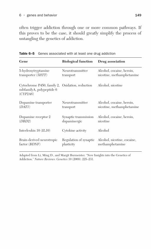
ptg
6 • genes and behavior 149
Table 6–5 Genes associated with at least one drug addiction
Gene Biological function Drug association
5-hydroxytryptaminetransporter (5HTT)
Neurotransmittertransport
Alcohol, cocaine, heroin,nicotine, methamphetamine
Cytochrome P450, family 2,subfamilyA, polypeptide 6(CYP2A6)
Oxidation, reduction Alcohol, nicotine
Dopamine transporter(DAT1)
Neurotransmittertransport
Alcohol, cocaine, heroin,nicotine, methamphetamine
Dopamine receptor 2(DRD2)
Synaptic transmissiondopaminergic
Alcohol, cocaine, heroin,nicotine
Interleukin 10 (IL10) Cytokine activity Alcohol
Brain-derived neurotropicfactor (BDNF)
Regulation of synapticplasticity
Alcohol, nicotine, cocaine,methamphetamine
Adapted from Li, Ming D., and Margit Burmeister. “New Insights into the Genetics ofAddiction.” Nature Reviews: Genetics 10 (2009): 225–231.
often trigger addiction through one or more common pathways. Ifthis proves to be the case, it should greatly simplify the process ofuntangling the genetics of addiction.
Wow! eBook <WoweBook.Com>

ptg
This page intentionally left blank
Wow! eBook <WoweBook.Com>

ptg
Genes and IQ: an unfinished story
Why would IQ be included in a book on genes and disease? The rea-son is that low IQ has often been equated with poor heredity andtreated as if it were a disease. From early in the last century untiltoday, those scientists with a hereditarian (as opposed to an environ-mental) bent have argued that cognitive deprivation is largely theresult of a less-than-satisfactory genetic background. This chapterbriefly outlines the fraught history relating genes and environment tointelligence. In the first part of the last century, the presumed geneticbasis of “feeblemindedness,” as it was called, served as the “scientific”basis for the involuntary sterilization statutes passed by many states inAmerica, at the provincial level in Alberta and British Columbia inCanada, and at the federal level in the Scandinavian countries andNazi Germany. The notion that levels of intelligence differedbetween races also played a role in the passage of the ImmigrationAct of 1924, which severely limited immigration into the UnitedStates from southern and eastern Europe until the mid-1960s. And intheir 1994 book The Bell Curve, Richard Herrnstein and CharlesMurray worried that society was becoming stratified by intelligenceinto a “cognitive elite” at the top and a growing underclass of the“custodial state.”
Martin Kallikak’s ChildrenIn 1865, Francis Galton published a remarkable two-part article inMacmillan’s Magazine, a popular Victorian periodical, titled “Heredi-tary Talent and Character.”1 His idea was to identify men of talent usingappropriate compendia following which he would try to establish theirpedigrees. Galton believed that if talent and character were inherited,
7
151

ptg
152 genes, chromosomes, and disease
then eminent male relatives should appear in these pedigrees in higherfrequency than they did in the population as a whole. The results heobtained led him to argue that his thesis was supported even though hehad to acknowledge that influence, for example of an eminent father inobtaining a desirable position for his son, might also play a role. In1869, Galton expanded this approach greatly in his book HereditaryGenius where he obtained pedigrees of famous judges, statesmen, mil-itary commanders, and so forth.2 He argued that if talent and characterwere inherited, then the closest male relatives to the eminent man(e.g., fathers and sons) were more likely to be eminent than more dis-tant relatives (e.g., grandfathers and grandsons). He concluded that hisresults supported this expectation.
Galton had also become intrigued in the application of the normaldistribution, particularly whether it might be somehow applied todata for mental ability. As a proxy, he used examination marks foradmission to the Royal Military College at Sandhurst. The data clearlyfit a normal distribution at the upper tail and in the center, but therewere no data for those who got low scores. Galton also constructed ahypothetical normal distribution to classify British men “according totheir natural gifts” while estimating the population of the UnitedKingdom to be about 15 million. The latter curve was eerily reminis-cent of normal distributions of IQ that would be published much lateron.
Although Galton collected and analyzed anthropometric data,investigated mental imagery, and used composite photography to seeif criminals, soldiers, and so on shared unique facial features, henever discovered a method for measuring mental ability.3 However,shortly before Galton’s death, Alfred Binet did invent such a test.4
Binet, a psychologist, had become interested in mentally handi-capped children. Under new French laws mandating universal educa-tion, such children were required to attend school, whereas earlierthey would have dropped out or never have attended school at all.Because they could not keep up with normal curricular requirements,they needed special attention. In 1904, Binet joined a governmentcommission investigating the status of the mentally handicapped inFrance. The commission concluded that accurate diagnosis posed thebiggest problem.
Wow! eBook <WoweBook.Com>

ptg
7 • genes and IQ: an unfinished story 153
In 1905, Binet, together with Theodore Simon, a young physicianwho was studying psychology with him, began to try to devise a testthat would unequivocally differentiate between normal and subnor-mal children. They avoided tests that relied heavily on reading andwriting so as not to confuse intelligence with lack of schooling. Manyof the items they chose assumed a familiarity with French life andwere based on work that Binet had done earlier using his own twodaughters as well as other children as experimental material. Aftersome initial frustration, Binet and Simon realized that age had to betaken into account in differentiating between normal and subnormalchildren because both groups could learn to pass the same tests, butnormal children did so at a younger age. The original tests hadfocused mainly on the most retarded and youngest children, so Binetand Simon set out to revise their intelligence tests in 1908 and againin 1911 because some of the most difficult educational decisionsinvolved older children who were closer to being normal. Accord-ingly, Binet and Simon were eventually able to arrive at an age-stan-dardized system that could compute each child’s intellectual level.
Henry Herbert Goddard brought Binet’s tests to the UnitedStates.5 Goddard received his PhD in Psychology from Clark Univer-sity and subsequently began to study children at the Vineland Train-ing School for Feeble-Minded Girls and Boys in New Jersey, wherehe became director of the Psychological Research Laboratory, newlyestablished in 1906 by its superintendent, Edward Johnstone.6 In1908, Goddard traveled to Europe seeking new techniques for deter-mining mental ability. He had become disenchanted with the psy-chophysical measurements devised by James Cattell, based on theanthropometric methods of Francis Galton, as being of any help inclassifying mental deficiency.7
While in Europe, Goddard learned about the Binet-Simon testswhose significance he soon appreciated. Upon his return to theUnited States, he began to apply the tests to children in the TrainingSchool.8 Unlike Cattell’s psychophysical measurements, the Binet-Simon tests did seem to classify children in a manner that was consis-tent with the observations made by his staff. The test results revealedthe existence of wide variations in the degree of “feeblemindedness”and Goddard used the numbers he obtained to erect specific cate-gories. Those children with a mental age of one or two were classified
Wow! eBook <WoweBook.Com>

ptg
154 genes, chromosomes, and disease
as “idiots,” those with a mental age of three to seven were “imbeciles,”whereas those whose mental ages were between eight and twelvewere dubbed “morons,” a term Goddard himself took from the Greekword for dull or stupid.
Up to this point, Goddard’s work had no hereditarianimplications, but in 1912, he published The Kallikak Family: A Studyin the Heredity of Feeblemindedness. The name Kallikak was a pseu-donym constructed from the Greek words kalós (good) and kakós(bad). Goddard had long suspected that feeblemindedness mighthave a heritable component as his earlier field studies revealed thatsome families seemed to have a high incidence of mental disease. Thebook followed the lives of a young Vineland woman, pseudonymouslynamed Deborah Kallikak, and her relatives. The evidence, obtainedby Goddard’s fieldworker Elizabeth Kite, who had employed inter-views rather than Binet-Simon tests, nevertheless seemed convincingto Goddard.
Deborah’s great-great grandfather, Martin Kallikak Sr., a Revolu-tionary War soldier, had an affair with a feebleminded woman whenhe was a youth. She gave birth to Martin Kallikak Jr. and among 480descendants, 143 were feebleminded with only 46 known to be nor-mal, with the mental abilities of the rest unknown.
On leaving the Revolutionary Army, Martin Sr. “married arespectable girl of good family” and their 496 descendants were allnormal. What could be clearer? Feeblemindedness was obviouslyinherited and probably Mendelian in nature. In fact, Goddard’sresults were bolstered by the many pedigrees presented principallyby American and British workers at the First International Congressof Eugenics held in London in 1912. They appeared to show that theMendelian inheritance of feeblemindedness and other undesirabletraits (e.g., tuberculosis, “criminalistic tendencies,” alcoholism) inindividual pedigrees. Also, research conducted by fieldworkers of theEugenics Record Office at Cold Spring Harbor, New York, directedby Charles Davenport, poured out a steady stream of supportingresults. So, two powerful ideas now interacted to bolster the case forsterilization or segregation of the feebleminded and those withrelated disabilities. Numerous pedigrees alleged that these traitswere inherited as Mendelian characters, whereas IQ tests could be
Wow! eBook <WoweBook.Com>

ptg
7 • genes and IQ: an unfinished story 155
used to identify individuals of subnormal intelligence irrespective ofheredity.
Goddard’s work encouraged others to experiment with methodsdesigned to assess the quantitative properties of mental ability. Oneof these was Lewis Terman, a Stanford University psychologist, whopublished a revision of the Binet-Simon test that thereafter becameknown as the Stanford-Binet test.9 Terman, a hereditarian like God-dard, introduced the term IQ. This, of course, stood for intelligencequotient, a number invented by the German psychologist WilliamStern in 1912 in which he multiplied the ratio of a child’s mental ageto his actual age by 100. Hence, an average child would have a ratioof 1 or an IQ of 100.
Meanwhile, Charles Spearman, a British psychologist and a finestatistician, invented a totally new concept of general intelligence thathe represented by the symbol g.10 What Spearman found was that if twomental tests were given to a large number of individuals, the correla-tion coefficient between them was nearly always positive. Spearmanrealized that one explanation of his results was that these positive corre-lations might be reduced to a single underlying factor, g. To distinguishwhether a single factor or multiple factors explained the positive corre-lations, Spearman invented a new statistical technique, factor analysis.He concluded that a single underlying factor was indeed involved andpublished his theory in 1904. Spearman’s concept of g seems to havewithstood the test of time pretty well.
As it became increasingly clear that the United States was aboutto enter World War I, the National Academy of Sciences establishedthe National Research Council to mobilize scientists for defense.11 InMay 1917, a group of psychologists was organized with the blessing ofthe council to design a testing system that would aid the army in plac-ing its men in proper categories with respect to military occupation.Goddard and Terman were included, but the chief tester was RobertYerkes, a young Harvard faculty member, who together with JamesBridges, a Harvard graduate student in psychology, developed theYerkes-Bridges scale, a rival to Terman’s Stanford-Binet test. Thesepsychologists proceeded to improvise two intelligence tests called theArmy Alpha and the Army Beta. The Army Alpha consisted of prob-lems and questions that have become familiar to many generations of
Wow! eBook <WoweBook.Com>

ptg
156 genes, chromosomes, and disease
students: word analogies, sequences of numbers, arithmetic prob-lems, synonym-antonym puzzles, and queries that required common-sense answers. The Army Beta tests were pictorial and given toilliterates and those who failed the Army Alpha tests. Eventually, 1.75million men were tested and Yerkes wrote a huge monograph titledPsychological Examining in the United States Army (1921).
The major conclusions were that the average mental age of whiteAmerican men was just above that of a moron; that European immi-grants could be graded by country of origin with southern and easternEuropeans being less intelligent than those from northern and west-ern Europe; and blacks were at the bottom. So, the tests convenientlyreinforced existing prejudices except for the embarrassment concern-ing the average white male. In 1923, another of the army testers, CarlBrigham, an enthusiastic Yerkes disciple, published A Study of Amer-ican Intelligence.12 He reported that the army tests revealed the firstsignificant differences in mental abilities between different races,claiming that the average intelligence of black Americans was lowcompared with white Americans, thus opening a debate that contin-ues to this day. He also claimed that the Alpine and Mediterranean“races” were “intellectually inferior to the Nordic race” and that theaverage intelligence of immigrants was declining. This kind of “evi-dence” would be used in support of the Immigration Act of 1924,which restricted the influx of immigrants from southern and easternEurope.
Six years later, Brigham recanted having decided that the armytests failed to measure innate intelligence accurately.13 Goddard alsorealized that these tests must be flawed as it made little sense that theaverage mental age of white recruits was just above that of a moron,the category he had created for the feebleminded who performedbest on IQ tests. In 1928, he published an article titled “Feeblemind-edness: A Question of Definition” in which he concluded that feeble-mindedness was not incurable and that the “feeble-minded do notgenerally need to be segregated in institutions.”14 In Goddard’s ownwords, he had “gone over to the enemy.” The last of the hereditarianpsychologists to admit defeat did so in a much more subtle way. In1937, Terman revised his 1916 book on the measurement of intelli-gence.15 He no longer argued that differences between groups in IQ
Wow! eBook <WoweBook.Com>

ptg
7 • genes and IQ: an unfinished story 157
could be explained by heredity, but now framed them in environmen-tal terms.
At the same time, it was becoming clear from the investigations ofLionel Penrose at the Royal Eastern Counties’ Institution in Colch-ester, England (refer to Chapter 2, “How genetic diseases arise”), that“feeblemindedness” was actually a composite description thatincluded, but was not circumscribed, by discreet genetic diseasessuch as phenylketonuria and Down syndrome. A vocal critic of thehereditarian interpretation of intelligence throughout was the youngjournalist Walter Lippman. He was adept at identifying the flaws inthe hereditarian arguments, pointing out that intelligence “is not anabstraction like length and weight; it is an exceedingly complicatednotion which nobody has as yet succeeded in defining.”16
But before we move on, let us return to Carl Brigham for amoment. After assisting Robert Yerkes in designing the Army Alphatests, Brigham joined the faculty of Princeton University. In 1925, theCollege Board asked Brigham to try to design a test for college admis-sion that could be applied universally.17 To meet this requirement,Brigham tinkered with the Army Alpha, principally by making itharder. In 1926, he administered his test experimentally to a few thou-sand college applicants. By 1933, he had concluded that his scholasticaptitude test (SAT) reliably predicted the likelihood of academic suc-cess.
Meanwhile in 1933, James Bryant Conant was appointed Presi-dent of Harvard University. He wanted to increase the breadth of Har-vard’s student body beyond the eastern preparatory schools thatsupplied it. His solution was to initiate a scholarship program for giftedboys from elsewhere. But how were applicants to the new programgoing to be evaluated? Conant delegated the problem of finding asolution to Henry Chauncey, an assistant dean. Chauncey metBrigham and, impressed with his test, recommended that Harvardadopt the test.
But then Brigham began to have doubts about his test, a develop-ment that Conant, and especially Chauncey, found irritating for by1938, Chauncey had convinced all the member schools of the CollegeBoard that they should use the test, but only for scholarship appli-cants. Brigham’s opposition prevented the test from being generally
Wow! eBook <WoweBook.Com>

ptg
158 genes, chromosomes, and disease
adopted, but his premature death at the age of 52 in 1943 cleared theway for acceptance of the SAT. It became the standard test for allapplicants in 1944. In that year, under contract to the army and thenavy, Chauncey administered the SAT to 300,000 officer candidatesfrom all over the United States in a single day. In 1947, the Educa-tional Testing Service was chartered as a nonprofit organization withChauncey as its first president. Today, millions of college applicantstake the SAT every year. Over the years, the test has changed in somerespects, but not completely.
The rise of behaviorismDuring the very period when the hereditary determinist view of intel-ligence was at its peak, a new theory that was its antithesis, behavior-ism, was emerging. The main proponent of what came to be calledclassic behaviorism was John Broadus Watson.18 Watson, a professorat Johns Hopkins University, coined the term in a 1913 article inPsychological Review titled “Psychology as the Behaviorist ViewsIt.”19 In that paper, Watson stated that psychology “as the behavioristviews it is a purely objective branch of experimental science. Intro-spection forms no essential part of its methods, nor is the scientificvalue of its data dependent upon the readiness with which they lendthemselves to interpretation in terms of consciousness.”
Watson here was stating his belief that psychology, as he envi-sioned it, was a hard science in which inferences about one’s own con-sciousness or the inferred consciousness of anyone else had no place.Watson went on to say that there was “no dividing line between manand brute.” He eschewed the anthropocentric approach be it in termsof consciousness or as in Darwin’s time when “the whole Darwinianmovement was judged by the bearing it had upon the origin anddevelopment of the human race.” But the “moment zoology under-took the experimental study of evolution and descent, the situationimmediately changed. Man ceased to be the center of reference.”
Later, Watson went on to define more precisely what he meantwhen he said that the sort of psychology that he wanted “to build up would take as a starting point, first, the observable fact that organisms, man and animal alike, do adjust themselves to their envi-ronment by means of hereditary and habit equipments.” But despite
Wow! eBook <WoweBook.Com>

ptg
7 • genes and IQ: an unfinished story 159
his acknowledgment of the importance of “hereditary equipment” indetermining behavior, it was clear that Watson’s goal was to controlbehavior in a predictable way so that “given the response the stimulican be predicted; given the stimuli the response can be predicted.”One could achieve this end by placing an “animal in a situation wherehe will respond (or learn to respond)” to a specific stimulus.
The key phrase here of course is “learn to respond” and in 1920,Watson published a classic paper in the Journal of Experimental Psy-chology with his graduate student Rosalie Rayner titled “ConditionedEmotional Reactions.”20 The subject of this paper was an infantnamed Albert B who had been reared in a hospital where his motherwas a wet nurse. He was stolid, unemotional, and rarely cried. Watsonand Rayner decided to see whether a fear reaction could be inducedin “Little Albert,” although with “considerable hesitation” as “a cer-tain responsibility attaches to such a procedure.” When Albert wasalmost 9 months old, Watson and Rayner attempted to induce thefear response by hitting a steel bar with a hammer behind the child’sback. The child “started violently” the first two times the bar was hitand the third time began to cry. At a little over 11 months, Albert waspresented with a white rat that he reached for. “Just as his handtouched the animal the bar was struck immediately behind his head.”Albert “jumped violently and fell forward.” The experiments contin-ued in this vein and were expanded to include a rabbit, a dog, and asealskin coat. The upshot was that Albert became afraid of furrythings. Although there was “a certain loss of intensity with time,” theinvestigators concluded these “conditioned emotional responses”probably “persist and modify personality throughout life.”
It seems that Watson and Rayner, his graduate student, also had aconditioned emotional response to each other. Watson’s marriage toMary Ickes, the sister of Harold Ickes, later Roosevelt’s Secretary ofthe Interior, had been under strain for many years. Nevertheless,Watson’s liaison with Rosalie Rayner and the subsequent divorce pro-ceedings caused a national sensation. As a consequence of his adul-tery with his student, Watson was fired from his professorial post atJohns Hopkins in 1920. Through a friend of his, Watson landed a jobat J. Walter Thompson, the advertising agency, where he appliedbehavioral psychology promoting Pond’s Facial Cream, for example,with considerable success.
Wow! eBook <WoweBook.Com>

ptg
160 genes, chromosomes, and disease
Watson continued to publish actively and in his 1924 bookBehaviorism, coined one of the best-known statements in psychology.
Give me a dozen healthy infants, well-formed and my ownspecified world to bring them up in and I’ll guarantee to takeany one at random and train him to become any type of spe-cialist I might select—doctor, lawyer, artist, merchant-chief,and, yes, even beggarman and thief, regardless of his talents,penchants, tendencies, abilities, vocations, and race of hisancestors. I am going beyond my facts and I admit it, but sohave the advocates of the contrary and they have been doingit for many thousands of years.21
Watson’s behaviorism served as the impetus that propelled Bur-rhus Frederic Skinner into the field.22 Following his graduation fromHamilton College, Skinner returned to his parents’ home in Scran-ton, Pennsylvania, built a study in their attic, and attempted, withoutnotable success, to write fiction. But Skinner read widely and one ofhis favorite authors was Bertrand Russell. In one of his books, Russellpresented a critique of Watson’s Behaviorism. Later, Skinner read anarticle by H. G. Wells about Ivan Pavlov and George Bernard Shaw.Shaw detested Pavlov for he felt he was a vivisectionist harming dogsthrough his operations just to see what might happen.
But Skinner was intrigued by Pavlov’s experiments as he had beenby Watson’s behaviorism and decided to make his career in behavioralpsychology. He was accepted as a graduate student at Harvard pro-gressing through a postdoctoral fellowship to be invited to become ajunior fellow in Harvard’s distinguished Society of Fellows. During hiseight years at Harvard, he had much opportunity to experiment and theculmination of these experiments was the Skinner box. When a whiterat was introduced into this box, it discovered, after suitable explo-ration, that by pressing a bar it could release a food pellet into a tray.The rat soon caught on as shown by the cumulative record Skinner wasable to keep. Skinner called the provisioning of food in response topressing the bar “reinforcement” and named this kind of learnedbehavior “operant conditioning.” Skinner had faculty positions at theUniversity of Minnesota and Indiana University before returning toHarvard in 1948.
Wow! eBook <WoweBook.Com>

ptg
7 • genes and IQ: an unfinished story 161
Meanwhile, he continued to improve and refine the notion ofreinforcement. Reinforcers, like the provision of food when the ratpressed on a bar, were positive in nature, but there was negative rein-forcement too. This means strengthening a particular behavior byavoiding or stopping a condition perceived as being negative. Drivingin heavy traffic is unpleasant for most of us. Suppose you go to workearlier than you planned and you encounter less traffic. If you decidethat you will continue to drive to your place of employment early toavoid heavy traffic, you are avoiding a condition you find unpleasant(negative reinforcement).
Meanwhile, Skinner was collecting students and postdoctoral fel-lows and becoming famous. In 1948, he published Walden Two, autopian novel describing an ideal society in which negative reinforce-ment was nonexistent and children sought only positive reinforce-ment.23 Consequently, they grew up to be happy, sociable, andcivilized. Frazier, the hero of the novel, is accused by a skeptical visi-tor named Castle of being a despot because Frazier believes thatthrough the use of positive reinforcement, a planned society will becreated. Frazier remarks, “it’s a limited sort of despotism” that shouldnot worry anyone because the “despot must wield his power for thegood of others.” If he “reduces the sum total of human happiness hispower is reduced by a like amount. What better check against amalevolent despotism could you ask for?”
As might be expected, Walden Two proved controversial. Criticsfelt Skinner’s happy, but controlled society was in fact totalitarian,whereas others responded positively. In 1971, Skinner produced abest-selling book titled Beyond Freedom and Dignity in which heargued that the notion of “autonomous man,” which is the foundationof many of the institutions of Western society, is misguided.24
Although we usually “credit” people with doing good deeds “of theirown free will” rather than because they are required to, in reality wesimply do not know the contingencies that resulted in the behavior inthe first instance whereas we do in the second. In the end, behavioralfreedom is an illusory concept and the reality is that behavior is condi-tioned most effectively over the long term by positive as opposed tonegative reinforcement. Once this principle is accepted, it becomesincumbent for societies to become like the one described in Walden
Wow! eBook <WoweBook.Com>
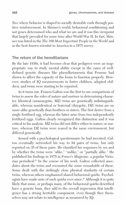
ptg
162 genes, chromosomes, and disease
Two where behavior is shaped to socially desirable ends through pos-itive reinforcement. In Skinner’s world, behavioral conditioning andnot genes determined who and what we are and it was this viewpointthat largely prevailed for some time after World War II. In fact, Skin-ner was listed in the The 100 Most Important People in the World andas the best-known scientist in America in a 1975 survey.
The return of the hereditariansBy the late 1930s, it had become clear that pedigrees were an inap-propriate way to study mental ability except in the cases of well-defined genetic diseases like phenylketoneuria that Penrose hadshown to affect the capacity of the brain to function properly. How-ever, studies of IQ measurements in foster children, adopted chil-dren, and twins were starting to be reported.
As it turns out, Francis Galton was the first to use comparisons oftwins to assess the roles of nature and nurture in determining charac-ter. Identical (monozygotic, MZ) twins are genetically indistinguish-able, whereas nonidentical or fraternal (dizygotic, DZ) twins are nomore alike genetically than brothers or sisters. The first derive from asingle fertilized egg, whereas the latter arise from two independentlyfertilized eggs. Galton clearly recognized this distinction and it wascritical to his analysis. MZ twins did not differ either in nature or nur-ture, whereas DZ twins were reared in the same environment, butdiffered genetically.
Armed with a psychological questionnaire he had invented, Gal-ton eventually networked his way to 94 pairs of twins, but onlyreported on 35 of these pairs. He classified his responses by sex andby whether the twins were “alike,” “unlike,” or “partially alike.” Hepublished his findings in 1875 in Fraser’s Magazine, a popular Victo-rian periodical.25 In the course of his work, Galton collected anec-dotes about the twins and recounted the more remarkable of these.Some dealt with the strikingly close physical similarity of certaintwins, whereas others emphasized shared behavioral quirks. Psychol-ogists have made note of such quirks ever since.26 Although it is quitelikely that some, or perhaps many, of the behavioral quirks describedhave a genetic basis, they add to the overall impression that intelli-gence has a strong heritable component, even though they them-selves may not relate to intelligence as measured by IQ.
Wow! eBook <WoweBook.Com>

ptg
7 • genes and IQ: an unfinished story 163
Frank N. Freeman and Karl J. Holzinger, two University ofChicago psychologists, in collaboration with Horatio H. Newman, abiologist, carried out the most important early study of intelligence inidentical twins.27 In 1937, they reported on the IQs of 19 pairs of twinsreared apart compared with a similar number reared together. Thecorrelation of 0.67 for the twins reared apart implied a strong heredi-tary component. However, they also noted that the greatest IQ differ-ences were found for those twins reared in the most strikinglydifferent environments. The three scientists were cautious in theirconclusions saying that both nature and nurture seemed to playimportant roles in producing the correlation. This study, supported byother findings, led to a consensus that both heredity and environmentwere important and interacting contributors to human intelligence.This comfortable situation remained until the famous British psychol-ogist Sir Cyril Burt reported intelligence test correlations of approxi-mately .771 for ever-increasing numbers of pairs of identical twinsreared apart in three papers published in 1943, 1955, and 1966, withthe latter paper including 53 pairs.28 The correlations had a suspiciousconsistency.
Meanwhile, the Berkeley psychologist Arthur Jensen had beeninvited to write a major article on the question “How much can weboost IQ and scholastic achievement?” by the student editors ofHarvard Educational Review.29 His conclusion, stated at the begin-ning of the article, was that compensatory education had failed andthat large programs designed to improve the scholastic aptitudes ofculturally deprived children either were unsuccessful or resulted inminor gains at best. Jensen went on to state that nature was moreimportant than nurture in determining intelligence and cited a num-ber of studies supporting this conclusion, but most notably those ofBurt. He concluded that IQ heritability ranged from .70 to .90 cen-tering near .80.
Jensen’s article was long and scholarly, but he got into deep trou-ble in a short section of the paper that compared the IQs of blacksand whites. There, Jensen noted that there was a vast amount of liter-ature suggesting that on average the IQ of blacks was 15 points belowthe IQ of whites. When gross socioeconomic differences wereaccounted for, this number reduced to 11 points that was “about thesame spread as the average difference between siblings in the same
Wow! eBook <WoweBook.Com>

ptg
164 genes, chromosomes, and disease
family.” But a little later, Jensen wrote that it seemed “not unreason-able, in view of the fact that intelligence variation has a large geneticcomponent, to hypothesize that genetic factors might play a part inthis picture.” He pointed out that the notion that black/white IQ dif-ferences might in part be hereditary in origin had been stronglydenounced, but “it has been neither contradicted nor discredited bythe evidence.” And then he took a stand. “The preponderance of theevidence is, in my opinion, less consistent with a strictly environmen-tal hypothesis than with a genetic hypothesis, which, of course, doesnot exclude the influence of environment or its interaction withgenetic factors.” With those words, Jensen had let an evil genie out ofits bottle.
The student editors of the Harvard Educational Review were sotaken aback with the negative response Jensen’s article had receivedthat they denied having ever solicited it even though he sent them acopy of his original letter of invitation.30 Feature articles in TIME,Newsweek, LIFE, U.S. News & World Report, the New York TimesMagazine, and many other newspapers and magazines, as well asradio and TV programs jumped on Jensen’s claims. Jensen became apariah on his own campus and subject to much abuse. Despite all ofthis, Jensen was not deterred, but has continued to publish a streamof books and articles on mental tests and what they mean. In 1982, healso proclaimed the demise of Watson’s behaviorism, arguing that IQis much less plastic than the behaviorists supposed as genetic variance“in IQ and information-processing capacity are strongly influenced byhereditary factors, with genetic variance constituting about 70% ofthe total population variance in IQ.”31 That would leave the behavior-ists with only 30% of the variance to work with.
In 1971, Harvard psychology professor Richard Herrnstein gothimself into hot water in a popular article about IQ testing in theAtlantic that he hoped would promote debate.32 After reviewing thehistory of IQ testing, Herrnstein discussed the advantages of having ahigh IQ, one of which was economic. Salaries of those with high IQswere greater than those with low IQs. Herrnstein then averred thatIQ appeared to have a large heritable component, echoing Jensen’scontention that it was not possible to boost IQ and scholastic achieve-ment very much. Herrnstein considered IQ differences betweenblacks and whites, pointing out that Jensen’s data concerning the
Wow! eBook <WoweBook.Com>

ptg
7 • genes and IQ: an unfinished story 165
heritability of IQ had been based almost entirely on whites, a pointJensen himself acknowledged. Herrnstein continued that although“there are scraps of evidence for a genetic component in theblack/white difference, the overwhelming case is for believing thatAmerican blacks have been at an environmental disadvantage.” But,he continued, setting “aside the racial issue, the conclusion aboutintelligence is that, like other important though not necessarily vitaltraits, it is highly heritable.”
Toward the end of his article, Herrnstein imagined a world inwhich “the social classes not only continue but become ever moresolidly built on inborn differences.” He envisioned that as “the wealthand complexity of human society” grew “there will be precipitatedout of the mass of humanity a low-capacity (intellectual and other-wise) residue that may be unable to master the common occupations,cannot compete for success and achievement, and are most likely tobe born to parents who have similarly failed.” Herrnstein, in short,was beginning to develop themes that would emerge over twodecades later in The Bell Curve. Fellow scientists, pro and con, weregenerally constrained in their responses, but student radicals pilloriedHerrnstein much as they had Jensen.
The scientific counterattack on the hereditarian view of IQ, nowdefined in the dictionary as “Jensenism,” began with a discoverymade by Princeton psychologist Leon Kamin. After reading Herrn-stein’s article, Kamin realized that Burt’s twin studies were the foun-dation upon which the hereditarian thesis rested. So Kamin set towork to examine Burt’s results and made a surprising discovery.33 Inwhat Burt described as the “Group Test,” Burt’s correlations wereconstant at .771 in his 1955 paper on 21 twin pairs reared apart, in“over thirty” pairs reported in 1958, and in the 53 pairs discussed inhis 1966 paper. Subsequently, Kamin found other instances of invari-ant correlations, leading him to believe that Burt’s twin studies hadbeen faked.
Kamin then examined other twin studies and found these correla-tions variable. Moreover, he showed that twins not genuinely sepa-rated in these studies were more alike in IQ than those who reallywere reared separately where bigger differences emerged. Kaminwas similarly critical of Jensen’s adoption studies. Meanwhile, thescandal around Burt, who had died in 1971, mushroomed and has
Wow! eBook <WoweBook.Com>

ptg
166 genes, chromosomes, and disease
seesawed back and forth ever since, although it now seems prettyclear that the twins data were fraudulent.34 Soon thereafter, otheropponents weighed in on the controversy, notably Harvard popula-tion geneticist Richard Lewontin and his colleague, paleontologistStephen Jay Gould whose popular book The Mismeasure of Man(1981) was aimed at debunking the hereditarian theory of intelli-gence.35 Several years later, Lewontin, in collaboration with LeonKamin and Steven Rose, published a book titled Not In Our Genes(1984).36 In that book, they not only questioned what IQ was reallymeasuring, but also raised serious concerns once again about adop-tion studies and those of identical twins reared apart.
For a while, the hereditarian thesis seemed to go into decline, butit reemerged with a vengeance with the publication of The Bell Curvein 1994 by Richard Herrnstein and Charles Murray37 together with acompanion article in the New Republic titled “Race, Genes, andI.Q.—An Apologia.”38 The book, divided into four parts, made thecase that intelligence (as measured by IQ) rather than socioeconomicstatus is the major determinant of success in life; that twin studiesshowed that IQ has a major heritable component; and that there wasa 15-point average IQ difference between blacks and whites. This lat-ter point was brought out most starkly in the New Republic article.Because of all of this, Herrnstein and Murray argued that a “cognitiveelite” was evolving and that society was becoming stratified becauseof an emerging economic system that rewards intelligence above allelse.
The last section of the book dealt with policy matters. There,Herrnstein and Murray asked the question, “Can people becomesmarter if they are given the right kind of help?” They answered neg-atively, writing that cognitive gains achieved by children in projectslike Head Start vanished by the end of primary school. Herrnsteinand Murray envisioned a future America dividing into an affluentaristocracy of the “cognitive elite” while at the bottom would be thegrowing underclass of the “custodial state.” The masses in the lattercategory would be sternly controlled by the welfare state “a high-techand more lavish version of the Indian reservation.” To avoid this out-come, Herrnstein and Murray argued that current efforts to achieve“equality of outcomes” should be abandoned. People should be
Wow! eBook <WoweBook.Com>

ptg
7 • genes and IQ: an unfinished story 167
allowed to find their own “valued places in society” according to theirability. In a “simpler America,” even people of lower intelligencewere valued and nurtured in the local community. Federal centraliza-tion has “drained” life out the community and more social functionsshould be returned to the neighborhood. Also, regulations relating tocriminal justice, small business, and so forth needed to be simplifiedso the less bright could understand them.
The Bell Curve, a hardcover book of over 800 pages, sold over500,000 copies and was reviewed on countless occasions in the publicand academic press. Much of the commentary was negative, ridicul-ing the hereditarian bias that the authors introduced into their argu-ments, especially where race was concerned. The Bell Curve alsobecame the focus of entire books bent on rebuttal. Claude S. Fischerand colleagues of his in the Department of Sociology at the Univer-sity of California, Berkeley, made a particularly perceptive analysis ofthe The Bell Curve.39 Not only did they uncover basic technical errorsin the book, but they found numerous cases where Herrnstein andMurray had presented data seeming to document that intelligencewas of greater importance than socioeconomic status in determiningsuccess where the reverse was, in fact, true. For example, Herrnsteinand Murray had claimed that the probability of being poor was morea function of intelligence level than socioeconomic status, but Fischeret al. found that socioeconomic status was actually more important.
They also demonstrated that the questions on the Armed ForcesQualifying Test (AFQT) used by Herrnstein and Murray as theirmeasure of IQ were geared to the school achievement level of theindividuals being examined. Thus, a student who had done well inschool was likely to score higher on the AFQT than a poor studentwho might have dropped out of high school along the way. They alsoargued that the reason blacks and Latinos average lower than whiteson tests like the AFQT had to do more with socioeconomic depriva-tion, including poor schools, than it did with heredity. Perhaps mostimportant of all, Fischer and colleagues seriously questioned whetherpsychometric methods using IQ tests as tools were really useful inmeasuring intelligence at all. They pointed to Arthur Jensen’s quipthat “intelligence is what intelligence tests measure.”
Wow! eBook <WoweBook.Com>
ptg
168 genes, chromosomes, and disease
Balancing nature with nurtureIn The Bell Curve, Herrnstein and Murray discussed a phenomenonthat they called “The Flynn Effect” as a possible environmentalexplanation for the 15-point mean difference between the IQs ofwhites and blacks.40 They picked this name because James R. Flynn, aprofessor of political science at the University of Otago in Dunedin,New Zealand, made the remarkable discovery that during the twenti-eth century, there were massive gains in IQ from generation to gener-ation.41 Furthermore, data obtained from a Dutch colleague showedthat in young Dutch males, these gains could occur within a singlegeneration. The gains exhibited linearity over time and were greaterin certain subtests (similarities) and much less dramatic in others(information & arithmetic & vocabulary). Because Flynn found strik-ing IQ gains not only between generations, but also within a genera-tion, these gains could not be explained on the basis of heredity.Furthermore, the differences in IQ increases between different sub-tests showed that the overall test scores reflected an internal hetero-geneity.
Then, Nicholas J. Mackintosh, a distinguished animal-learningtheorist at the University of Cambridge, decided to wade into thecontroversy over nature, nurture, and intelligence.42 He came intothis fraught field without any particular prejudices and proceeded tosort through much of the relevant literature coming up with a bal-anced view on the subject. Mackintosh summarized the data on MZtwins reared apart. By now, this included the work done by ThomasBouchard, the Director of the Minnesota Center for Twin and Adop-tion Research at the University of Minnesota, and his colleagues.Bouchard’s studies were the most recent and complete studiesreported to date. Macintosh also included the four other similar stud-ies published earlier, omitting Burt’s papers because, if not fraudu-lent, they were laced with errors.
Macintosh observed that these reports gave a mean correlation inIQ between MZ twins reared by different parents that range from0.69 to 0.78. By contrast, the correlation between the IQ scores ofadopted children and those of their adoptive parents was usually lessthan between parents and their biological children. Furthermore, theIQs of these adopted children generally resembled those of their bio-logical mothers more closely than they did their adoptive mothers.
Wow! eBook <WoweBook.Com>
ptg
7 • genes and IQ: an unfinished story 169
At first sight, these findings seemed to indicate that IQ was ahighly heritable trait, but closer scrutiny suggested that it was danger-ous to take these numbers too literally because the sum total of MZtwins examined in these five studies was only 162. Furthermore, itbecame apparent to Macintosh that the home environments in whichthe twins were brought up were not wholly uncorrelated. For example,there were significant correlations between the father’s occupation,the mother’s education, material possessions at home, as well as familybackground. However, some of these variables (e.g., father’s occupa-tion and material possessions at home) were themselves correlated sothey could not independently contribute to the IQ similaritiesbetween the separated twins.
Mackintosh concluded that some of the assumptions of what hecalled the “simple model” for interpreting the results of twin andadoption studies were false. For example, the model assumed that allmembers of a family share a uniform environment. “But MZ twinsmay well share more experiences in common than DZ twins, who inturn may share more common experiences than siblings of differentages.” With regard to adoption studies, no provision was made for thelikelihood that adoptive families were not a representative cross-sec-tion of the population, or for selective placement of adoptive chil-dren. Mackintosh prudently concluded that “the broad heritability ofIQ in modern industrialized societies is probably somewherebetween 0.30 and 0.75, and that neither the data nor the models jus-tify much greater precision.”
Mackintosh also addressed the much-reported average IQ differ-ence between blacks and whites of 15 points. As Herrnstein and Mur-ray took pains to point out in their 1994 article, “Race, Genes andI.Q.—An Apologia,” this difference, which amounts to 1.2 standarddeviations, “means considerable overlap in the cognitive ability distri-bution for blacks and whites.”43 Mackintosh analyzed the relevantdata in detail concluding that, “there is remarkably little evidence thatthe difference is genetic in origin.” But if “the effect is an environ-mental one, it remains to be shown what the proximal environmentalcauses are.” The same problems arose in trying to sort out why Chinese and Japanese seem to score higher than whites on spatial IQ tests and possibly on other nonverbal tests. Then, there was thequestion of whether IQ tests were in some way biased against certain
Wow! eBook <WoweBook.Com>

ptg
170 genes, chromosomes, and disease
ethnic groups. Mackintosh argued that the only way to tell whetheran IQ test is biased is to compare the results with “an independent,better measure of ‘intelligence.’” But there was no such test.
Richard Nisbett has echoed many of the concerns raised by Mac-intosh.44 Nisbett is a social psychologist and distinguished universityprofessor at the University of Michigan and the first psychologist in ageneration to be elected to the National Academy of Sciences.45 Nis-bett pointed out that individuals like Arthur Jensen, whom hereferred to as “strong hereditarians,” believe that the heritability ofIQ is 75% to 85%, whereas “the environmentalist camp estimatesheritability to be 50% or less.” He proceeded to cast serious doubt onthe hereditarian position, pointing out, much as Macintosh hadbefore him, that for MZ twins reared apart “a tacit assumption that issurely false” is that “the twins were placed into environments at ran-dom.” Nisbett mentioned, for example, the work of Urie Bronfen-brenner who showed that, when twins reared apart were brought upin very similar environments, the correlation between their IQ scoreswas .83 to .91. However, when environments were not alike, the cor-relations ranged from .26 to .67.
Eric Turkheimer at the University of Virginia took a differentapproach. Turkheimer studied over 600 pairs of twins reared togethermost of whom were in families below the poverty level.46 He “foundthat for the poorest twins, IQ seemed to be determined almost exclu-sively by their socioeconomic status, which is to say their impover-ished environment. Yet, for the best-off families, genes were the mostimportant factor to determining IQ, with environment playing amuch less important role.” Turkheimer’s findings suggested thatupper-middle-class families were providing a much better environ-ment for the development of intelligence and they may not differ agreat deal between themselves in this regard. Because studies oftwins reared apart tend to be conducted using middle-class andupper-middle-class families who are relatively easy to contact, thiswill bias estimates of heritability toward the high end.
Nisbett also weighed in on the factors responsible for the 15-point average differential in IQ scores between blacks and whitesdebunking heredity as being responsible. For one thing, an interest-ing natural experiment occurred after World War II when American
Wow! eBook <WoweBook.Com>

ptg
7 • genes and IQ: an unfinished story 171
GIs fathered children by German women. Some of these soldierswere black and some were white. German children of white fathersaveraged IQs of 97.0, whereas those of black fathers averaged 96.5.Nisbett was careful to point out that American blacks have an averageof 20% European genes, so they ranged in genetic composition fromalmost purely African to mostly European.
Then there was the work of psychologists Joseph Fagan and Cyn-thia Holland. They tested black and white community college stu-dents on their ability to learn and reason using words and concepts.Although whites had a broader knowledge of words and concepts, thetwo sets of participants had an equal ability to learn new words, forexample, from a dictionary. Whites also comprehended sayings betterthan blacks, could recognize similarities better, and had greater facil-ity with analogies whose solutions depended upon a knowledge ofwords and concepts that were more likely to be more familiar towhites. But when this sort of reasoning was restricted to words andconcepts equally familiar to blacks and whites, no differences wereapparent.
Psychologists like Mackintosh and Nisbett have seriously chal-lenged the hereditarian thesis. They argue that intelligence is mal-leable rather than fixed. You can improve on your intelligencethrough learning. Certainly, most would-be college or professionalschool students would agree. They usually take advantage of the testpreparation services offered by Kaplan, Inc., founded in 1938 byStanley Kaplan.47 The College Entrance Examination Board, whichoversaw the SAT believed it was uncoachable.48 Mr. Kaplan thoughtotherwise. He regarded himself at the nemesis of the EducationalTesting Service that develops the SAT and other tests. Kaplanclaimed he could improve SAT scores by 100 points, but a FederalTrade Commission investigation of the coaching business revealedthat Kaplan’s program helped participants raise their scores by only50 points. That is no mean achievement and it has nothing to do withgenes. It would be interesting to know what Carl Brigham, whodeveloped the original SAT, would have thought of all this.
Wow! eBook <WoweBook.Com>

ptg
172 genes, chromosomes, and disease
Stalking intelligence genesAs this brief history shows, question of the genetic contribution to IQand, hence, to intelligence has been the subject of debate for over acentury. Opinion has seesawed back and forth from the notion thatgenes are the principal determinants of IQ to the belief that the envi-ronment mainly determines IQ and back again. The principal prob-lem is that the available experimental approaches have evolved verylittle over time. Since 1937, when Freeman and their colleagues pub-lished their original study, we have been largely dependent on studiesof MZ twins reared apart for estimates of the heritability of IQ. Amore precise understanding of the genetic contribution to intelli-gence is going to require a better idea of the spectrum of genesinvolved and their roles and acquiring this knowledge will take time.
One of the reasons that twins have been relied on so heavily isthat IQ is a complex trait. Oddly enough, it is similar to height in sev-eral respects. Like height, IQ is a metric trait so the distribution of IQscores is easily plotted and, like height again, a normal distributionresults. Traits like these are referred to as being polygenic becausemany genes, usually having small effects individually, are involved indetermining the distribution. Sorting these out is going to take timeespecially because the environment can also affect the distribution.For example, poor nutrition results in shorter height and lower IQ onaverage. Despite these drawbacks, characterization of genes affectingIQ should eventually provide a far more precise idea of the role thatnature plays in determining intelligence.
So what do we know about genes affecting intelligence at themoment? One way of approaching this question is to ask how manygenes possess mutations that cause mental retardation and what dothe normal, nonmutated forms of these genes do? Obviously, some ofthese genes will affect neurological processes we associate with intel-ligence, but in other cases the effect will be indirect. For example,mental retardation in the case of phenylketoneuria results becausethe amino acid phenylalanine accumulates in the blood and in thebrain. As of September 2003, 282 genes had been identified withmutations which caused mental retardation.49 The majority of thegenes identified could be classified as being involved in signaling
Wow! eBook <WoweBook.Com>

ptg
7 • genes and IQ: an unfinished story 173
pathways, metabolic pathways, and transcription, but numerousgenes involved in neuronal or glial (nonneuronal cells that supportneuronal activities) function were identified too.
Candidate gene and genome-wide association studies of intelli-gence are also under way.50 More than 20 candidate genes have so farbeen identified, but many apparently significant associations havefailed to replicate. Genome-wide association studies have identifiedsingle nucleotide polymorphisms (SNPs) and individual genes thatseem to relate to intelligence, but none of these studies has beenreplicated well.
Future progress in understanding how genes contribute to intel-ligence is likely to come from collaborative investigations like thosebeing carried out by the Genes to Cognition Project (G2C).51 Investi-gators involved in this project are focusing on a complex structurecalled the NMDA receptor that seems to be involved in memoryfunction and contains more than 100 proteins. NMDA receptors sitat the junctions (synapses) between nerve cells and modulate thestrength of signals passing between neurons. Systematic investigationof the functions of the different proteins in this complex can beaccomplished in mice where the genes encoding proteins of the com-plex can be knocked out one by one and the effect on learningassessed. For example, mice lacking a protein called PSD-95 thatbinds to the NMDA receptor have severe learning difficulties.
Genomic sequencing is becoming ever faster and cheaper.George Church at Harvard Medical School runs the PersonalGenome Project that now has 16,000 volunteers.52 Its goal is tosequence all of their genomes and eventually to expand the numberto 100,000 genomes. So far, the genomes of 12 people have beensequenced. The sequences have been made publicly available. If Dr.Church, and others like him, succeed in sequencing so manygenomes, it is fair to say that we will know a lot more about the genesinvolved in intelligence before too long. Understanding how theirproducts interact in the brain and nervous system and how they inter-act with each other and with the environment may take much longer.
Wow! eBook <WoweBook.Com>

ptg
This page intentionally left blank
Wow! eBook <WoweBook.Com>

ptg
Preventing genetic disease
On June 26, 1974, just prior to her second birthday, Melisa Howarddied of Tay-Sachs disease.1 Her mother, Laura Howard, had been apatient of Dr. B. Douglass Lecher, a specialist in the field of obstet-rics and gynecology. He had cared for Mrs. Howard during her previ-ous pregnancy, performed regular gynecological examinations onMrs. Howard prior to Melisa’s birth, and was present when the babywas born.
Following Melisa’s death, Mr. and Mrs. Howard, who were Jew-ish, sued Dr. Lecher claiming that his negligence had prevented themfrom terminating the pregnancy by abortion. They alleged that Dr.Lecher should have known that, because of their genetic background,the plaintiffs were “potential carriers of Tay-Sachs disease,” and thattests were available that would have permitted diagnosis of Tay-Sachsdisease in the fetus so it could have been aborted.
The Howards complaint sought damages for “severe mental dis-tress and emotional disturbances resulting from the illness, and phys-ical deterioration and death of their infant daughter and from theknowledge that no treatment or cure was available and that [she]would soon die.” The Howards also sought recovery of “medical, hos-pital, nursing and related care and funeral expenses.”
The trial court ruled that the Howards were entitled to seek dam-ages for mental and emotional distress and that they were entitled torecover all their medical and related expenses. However, theirrequest to be compensated for mental anguish was denied by theNew York Court of Appeals. Four reasons were given for this deci-sion: Lecher’s failure to recommend the genetic test did not directlyaffect the parents themselves; the damages were impossible to calcu-late; recognition of the claim would be “unwarranted and dangerous
8
175

ptg
176 genes, chromosomes, and disease
extension of malpractice liability”; and were the claim sustained itwould “either open the way for fraudulent claims or enter a field thathas no sensible stopping point.”
Two years after the Howard’s filed their complaint, on December27, 1978, the same New York Court of Appeals consolidated two casesin which the plaintiffs had also sued their doctors after they had givenbirth to genetically defective babies.2 The first case involved DoloresBecker. In 1974, she became pregnant at the age of 37 and subse-quently gave birth to a child with Down syndrome. Mrs. Becker statedthat, had she realized that the genetic defect could have been detectedin the embryo following amniocentesis, she would have opted for theprocedure. The plaintiffs in the second case were Hetty and StevenPark. In 1969, she had a baby with polycystic kidney disease, a rarerecessive genetic condition in children. The baby died within fivehours. The Parks consulted their obstetricians about having anotherchild and were incorrectly informed that the disease was not hereditary.Their second child was also born with polycystic kidney disease anddied at the age of two and a half. The Court of Appeals consolidated thetwo cases and upheld judgments for the parents’ claims in both cases.However, the court denied the child’s claim of wrongful birth in bothinstances, hence establishing that under New York law a child cannotsue for being born with genetic defects.
But there was a quite different outcome in a California case.3
Hyam and Phillis Curlender knew they were at risk of giving birth toa Tay-Sachs baby, so in 1977 they retained Bio-Sciences Laboratoriesand Automated Laboratory Services to perform blood tests on themto see whether they were likely to transmit Tay-Sachs genes to theiroffspring. DNA tests were not available at the time, but carriers couldbe distinguished from those lacking the mutant gene by assaying theenzyme hexosaminidase A. You will recall (refer to Chapter 3, “Eth-nicity and genetic disease”) that this enzyme consists of two dissimilarsubunits and it is the α subunit that is rendered nonfunctional in Tay-Sachs disease. Therefore, carriers exhibit lower activity of thisenzyme than those without the mutation.
Because the tests were reported to be negative, the Curlendersdecided to have a baby, Mrs. Curlender conceived, but their daugh-ter, Shauna, was diagnosed with Tay-Sachs disease. The Curlenderssued the testing laboratories, but in this case, the plaintiff was their
Wow! eBook <WoweBook.Com>

ptg
8 • preventing genetic disease 177
stricken daughter Shauna. Their complaint sought costs of the plain-tiff’s care and also damages for emotional distress and deprivation of“72.6 years of her life.” In addition, punitive damages of $3 millionwere requested because the plaintiff claimed that the defendant’sscientific procedures were defective. Although the court denied theclaim concerning deprivation of normal life expectancy, it ruled thatShauna had the right “to recover damages for pain and sufferingduring the limited lifespan available...and any pecuniary loss result-ing from the impaired condition.”
In each of these cases except the last, the physician in charge waseither misinformed or failed to recommend a procedure that couldhave avoided a tragic outcome. In the last case, the testing laboratorieswere at fault. But these failures occurred at a time when the impor-tance of genetic testing and genetic counseling was just beginning tobe appreciated. It was also a time when the range of genetic diseasesthat could be detected in carriers was still quite limited. Today, theexplosion in DNA technology and disease gene identification permitsdiagnosis of a wide range of genetic diseases both in newborns andparents contemplating having children. As discussed in Chapter 3, dif-ferent ethnic groups recommend, and sometimes mandate, testing forgenetic diseases that are prevalent among them.
This chapter begins with a discussion of the role of genetic coun-seling in preventing genetic disease. Had counseling been generallyavailable at the time of these “wrongful life cases” as it is today, theymight never have gone to litigation. The chapter then turns to thegenesis of in vitro fertilization and how it made possible the selectionof embryos lacking disease-producing mutations, thereby preventingthe birth of children with genetic defects.
The role of genetic counselingThe Heredity Clinic at the University of Michigan, established in1940 by Lee R. Dice, a geneticist and mammalogist at the university,was the first center to offer any sort of genetic counseling in theUnited States.4 In 1948, James V. Neel, one of the founders of mod-ern human genetics, became the physician in charge of the clinic. Hewas soon receiving letters from across the country requesting advice,but the clinic could only undertake the study of families who could
Wow! eBook <WoweBook.Com>

ptg
178 genes, chromosomes, and disease
actually make the trip to Ann Arbor. It shied away from giving adviceby mail as this was akin to making a medical diagnosis by mail andclearly subject to error. The clinic was subsequently subsumed in theDepartment of Human Genetics, with the new department beingchaired by Neel.
The Dight Institute at the University of Minnesota was the sec-ond counseling establishment to open.5 The money to found the insti-tute had been in the bequest of a physician named Charles FremontDight, a rabidly enthusiastic eugenicist and fan of involuntary sterili-zation of the “feebleminded.” Between 1921 and 1935, the Min-neapolis newspapers published some 300 letters written by Dight onvarious aspects of eugenics.
Dight was a rather odd chap. He lived for a time in an ingeniouslydesigned tree house suitable for a single person. Most residents ofMinneapolis probably remembered him more for his tree house thanfor his strident views on eugenics as it was the subject of innumerablenewspaper articles and appears not to have been an entirely satisfac-tory way of living. Outside his tree house, Dight hung a sign that read,“Truth shall triumph. Justice shall be law.” Dight managed to leave asizable legacy to establish the Dight Institute by being very frugal,investing smartly, and failing to pay his income taxes. Dight wantedhis eponymous institute “to promote biological race betterment—betterment in human brain structure and mental endowment andtherefore behavior.”
Dight died in 1937 and the Dight Institute for Human Geneticswas founded on July 1, 1941.6 In his will, Dight had stipulated that theinstitute that would bear his name and should provide courses andpublic lectures on human genetics, initiate research studies, and pro-vide consultation on questions related to human genetics. Dispensa-tion of eugenic advice and consultation was also encouraged. Its firstdirector, Clarence P. Oliver, adhered to the advice given in Dight’s will,but in 1946, he left to take a position at the University of Texas inAustin. Sheldon Reed succeeded him. It is to Reed we owe the term“genetic counseling” for Reed felt that expressions like “genetichygiene” connoted things like toothpaste and deodorant, thusbesmirching the serious nature of the endeavor. He also found thatfamilies he met with had little interest in eugenics and the term genetic
Wow! eBook <WoweBook.Com>

ptg
8 • preventing genetic disease 179
counseling occurred to him “as an appropriate description of theprocess which I thought of as a kind of genetic social work” withouteugenic connotations.
The Dight Institute Advisory Committee was not overly enam-ored of Reed’s literary invention, but it has stood the test of time.Although Reed was not trained as a psychologist, he came to realizethe importance of emotions like shame, guilt, blame, and resentmentin the counseling process. Reed was instrumental in endowinggenetic counseling with the ethos of respect and caring that charac-terizes the relationship between counselors and those they counseltoday. In his classic text Counseling in Medical Genetics (1955), Reedwrote that “the second requirement” for a counselor “or perhaps thefirst—is that he have deep respect for the sensitivities, attitudes andreactions of the client.”
According to the 1951 Dight Institute Bulletin, there were tengenetic-counseling centers in the United States at that time.7 How-ever, there were no educational programs for training genetic coun-selors. Today, the term describes somebody with a masters-leveldegree who has in-depth exposure to human genetics and counsel-ing skills. The first program to meet this requirement was initiated atSarah Lawrence College. It graduated its initial group of “geneticassociates” in 1971. Melissa Richter, a biologist at the college whowas also attracted to psychology, founded the program in 1969. Sherealized that counseling might provide an attractive career choice forwomen. Existing nursing and social work programs did not includegenetic associate training within their domains, so Dr. Richter andher colleagues were able to convince medical professionals thatcounselors could serve a useful purpose, especially because humangenetics was evolving rapidly. They proceeded to construct a well-designed program in genetics and counseling following consultationwith geneticists.
In 1972, Dr. Richter went on a sabbatical to Radcliffe College, butshe would never return to the program she had initiated for she wassoon to die of breast cancer. Two codirectors succeeded her and one ofthem, Joan Marks, would lead the program over the next 26 years. TheSarah Lawrence program was directed at students who already hadtheir bachelor’s degree. They were taught the scientific principles of
Wow! eBook <WoweBook.Com>
ptg
180 genes, chromosomes, and disease
human and medical genetics as well as the fundamentals of counseling,including its important psychological dimensions. The first students toenroll in the program were older women who had raised their familiesand wanted to enter a new career.
Marks succeeded in attracting faculty members with expertise inareas such as human genetics, cytogenetics, biochemistry, and embry-ology to teach courses to her students. They were then introduced tofamilies and patients with different genetic diseases at the severalgenetics clinics that existed in the greater New York area. Fundingwas always a problem because Sarah Lawrence College was notknown for its scientific or medical orientation. Nevertheless, the pro-gram flourished and by 2004, when Marks received the AmericanSociety of Human Genetics Award for Excellence in Human Genet-ics Education, there were approximately 600 graduates of the pro-gram. They accounted for about one-third of the genetic counselorsin the United States at the time. Practically all programs that havebeen initiated since were built along the lines of the Sarah Lawrencemodel and in 2004, one-half of these were directed by SarahLawrence graduates. Today, there are 29 accredited graduate pro-grams in the United States and 3 in Canada.
It was only in 1977, when there were already several centers inthe United States offering special training programs in genetic coun-seling, that the American Society of Human Genetics set up theAmerican Board of Medical Genetics.8 The Board was charged withaccrediting training programs in genetic counseling. It was alsotasked with creating a mechanism whereby professional credentialsin medical genetics could be awarded to MDs and PhDs entering thefield and also to genetic counselors with a master’s certificate ingenetic counseling.
In 1992, the American Board of Medical Genetics sought tobecome a member of the American Medical Association’s Board ofMedical Specialties. This meant that the American Board of MedicalGenetics would have to exclude nondoctoral level candidates such asgenetic counselors from the certification process. This resulted in theformation of the American Board of Genetic Counseling.9 This Boardcurrently has the responsibility for accrediting training programs forgenetic counselors and also for certifying nascent genetic counselors.
Wow! eBook <WoweBook.Com>
ptg
8 • preventing genetic disease 181
Certification of a new counselor by the Board requires that theperson has graduated from a Board-accredited training program; thathe or she has acted as principal counselor for 50 supervised clinicalcases during training; and that the counselor-to-be has passed twoexaminations. One of these stresses the general principles of medicalgenetics while the second focuses on counseling. The newly mintedcounselor will probably choose to join the National Society of GeneticCounselors that was formed in 1978 and publishes the Journal ofGenetic Counseling.
Genetic counseling relates to all stages of the life cycle beginningwith prospective parents who are concerned for some reason thattheir baby may be born with a genetic defect.10 This could be becausethe prospective father or mother knows that he or she has a record ofan inherited genetic disease in the family, cystic fibrosis for example;thinks that some congenital anomaly is present in the family, but doesnot know what it is; or the prospective parents are older and perhapsconcerned about the likelihood that they may give birth to a babywith Down syndrome.
Counseling may also involve prenatal diagnosis. For example,heart defects are a leading cause of death in infants during the firstyear of life. If stillborn babies are included, around 30 per 1,000babies have congenital heart defects, most of which are sporadic inorigin.11 If only live births are counted, the frequency of these sorts ofdefects are still quite high at 8 per 1,000. The fetal heart begins toform by day 18 and starts to beat by day 22. Performance of fetalechocardiography, an ultrasound technique, during the first trimesteris used to detect irregular behavior of the heart. If it appears the fetusdoes have congenital heart disease, the counselor helps the family tounderstand the condition and to prepare for the birth of a baby with adiseased heart.
Down syndrome presents a different problem.12 The chances ofgiving birth to a baby with Down syndrome rise dramatically withmaternal age (refer to Table 2–3). Thus, about 30% of all Downbabies are born to mothers 35 years of age or older. For expectingmothers younger than 35, the counselor will probably recommendmaternal serum screening. This is a noninvasive procedure that usually involves testing for one protein (alpha-fetoprotein) and two
Wow! eBook <WoweBook.Com>

ptg
182 genes, chromosomes, and disease
hormones (unconjugated estriol and human chorionic gonadotropin).With Down syndrome, alpha-fetoprotein and unconjugated estriolare about 25% lower than in a normal pregnancy, whereasgonadotropin is about twice the normal amount. This test is usuallyperformed in the second trimester of pregnancy between 15 and 18weeks of gestation. The “triple test” will detect about 60% of Downpregnancies with a false positive rate of 9%. During the first or sec-ond trimester, ultrasound screening will detect a condition callednuchal translucency that results from the accumulation of fluid in thenecks of Down syndrome fetuses. Combining ultrasound screeningwith the triple test probably increases the detection rate for Downsyndrome fetuses to between 74% and 80%.
Should the triple test prove positive, counselors reassure theexpecting couple that the test is not infallible and that a more invasivetest such as chorionic villus sampling or amniocentesis may be inorder. During the second trimester, fetal tissue can be obtained eitherby chorionic villus sampling (11–14 weeks), early amniocentesis(12–15 weeks), or second-trimester amniocentesis (15–20 weeks) fordirect confirmation of Down syndrome by chromosome analysis.Chorionic villus sampling or amniocentesis is recommended forwomen 35 or older on their due date, although the counselor maysuggest taking the triple test before reaching a decision about theother two options.
If it turns out that the couple is going to have a Down syndromebaby, the counselor provides them with up-to-date information aboutthe condition and assists them in reaching a decision. They maychoose to continue the pregnancy with the option of either raising theDown baby or putting it up for adoption or the couple may decide toterminate the pregnancy. As we have seen, the vast majority of cou-ples choose to terminate the pregnancy (refer to Chapter 2, “Howgenetic diseases arise”).
The decision to end a pregnancy means the couple has to find adoctor willing to perform abortions. But abortion is a very sensitivesubject in the United States and abortion doctors risk their own livesin certain parts of this country, especially if they perform late-term or“partial birth” abortions. One such physician was Dr. George Tillerwhose clinic was in Wichita, Kansas. Tiller became the preeminentabortion practitioner in America, advertising widely and drawing in
Wow! eBook <WoweBook.Com>
ptg
8 • preventing genetic disease 183
women from all over the country.13 He also drew frequent referralsfrom genetic counselors of expecting parents desiring to terminateDown syndrome pregnancies.
On Sunday, May 31, 2009, Reformation Lutheran Church wascelebrating the Festival of the Pentecost with a special musical prel-ude. Most members, including Dr. Tiller’s wife, were seated and theservice was about to begin, but Dr. Tiller, an usher that day, was greet-ing stragglers in the foyer. Pastor Lowell Michelson was beating agoblet shaped drum known as a darbuka, most commonly found inthe Middle East and parts of Africa, while singing an African songcalled “Celebrate the Journey!” All of a sudden, the pastor heard asharp crack and was summoned to the foyer by another usher. GeorgeTiller had been shot by an antiabortionist named Scott Roeder anddied in the foyer.
Tiller’s shooting was the climax of a long campaign of criminalharassment of Tiller by antiabortionists. For over 30 years, the antiabor-tion movement had been trying to drive him out of business thinking itwould greatly cripple the “abortion industry.” Women entering hisclinic would often encounter protesters. They would see a “TruthTruck” whose side panels were adorned with large color photographs ofdismembered fetuses. Children chanted “Tiller, Tiller, the baby killer,”an expression picked up by Bill O’Reilly, the widely viewed Fox Newshost, who regularly attacked Dr. Tiller beginning in 2005.
On June 10, 2009, shortly after Tiller’s death, Amy Goodman ofDemocracy Now interviewed a genetic counselor who wanted toremain anonymous. The counselor confirmed that the National Soci-ety of Genetic Counselors probably made “the vast majority of refer-rals” to Tiller’s clinic. The counselor was distressed that the societyhad failed to make a public statement concerning Tiller’s death. Sheinquired as to why this was the case and was informed “that therewere some genetic counselors who specifically requested that theynot make a public statement.” She speculated that fear was probablythe reason. But on June 14, the National Society of Genetic Coun-selors did issue a short, official statement condemning the murder.On July 1, 2010, Tiller’s murderer, Scott Roeder, was sentenced to lifein prison with no possibility of parole. Roeder had argued that he hadchosen to obey “God’s law” to save babies. As he said during his trial,
Wow! eBook <WoweBook.Com>

ptg
184 genes, chromosomes, and disease
“I did kill him. It was not a murder. If you were to obey the higherpower of God himself, you would acquit me.”
Genetic counseling not only involves working with couples whoare either thinking about having children or are already prospectiveparents, but also the diagnosis of genetic disorders in newborns, chil-dren, adult-onset disorders, and diseases such as Alzheimer’s thatusually affect the elderly.14 A key process in genetic counseling is theestablishment of a pedigree based on medical family history. Thepedigree not only establishes the pattern of inheritance of a disease orcondition, but it also helps to determine what genetic testing strate-gies to take. Construction of the pedigree is also an important tool forbuilding a working relationship with the person or persons seekingadvice. Depending on the nature of the condition or disease, the per-son being counseled may exhibit a range of emotions, includinganger, anxiety, disbelief, denial, helplessness, and many otherresponses that the counselor will need to be able to cope with.
Suppose, for example, that a woman whose mother died frombreast cancer consults a genetic counselor about her risk of contract-ing the disease. Although the vast majority of cases are sporadic,around 5%–10% are hereditary with the frequency of hereditarybreast and ovarian cancer increasing in certain populations (refer toChapter 3). The counselor will want to establish whether any of thewoman’s other female relations have had breast or ovarian cancer. Ifso, the counselor will probably suggest that the woman be checkedfor mutations in the BRCA1 or BRCA2 genes that can result in breastand ovarian cancer. Women with a BRCA1 mutation have a65%–85% lifetime risk of breast cancer and a 39%–60% lifetime riskof ovarian cancer. For a BRCA2 mutation, the comparable risks are45%–85% for breast cancer and 11%–25% for ovarian cancer.
Women at risk are advised to perform a monthly breast examinationbeginning at age 18 if a mutation has been detected that early in theirlives. Beginning at age 25, monthly clinical breast examination and anannual mammogram are recommended. Transvaginal ultrasound and aspecific antigen can be used to detect the appearance of ovarian cancer,although the efficacy of these methods is uncertain. Mastectomy andovarian removal significantly reduce the risk of breast/ovarian cancer,
Wow! eBook <WoweBook.Com>

ptg
8 • preventing genetic disease 185
but do not eliminate it. If a woman diagnosed with a BRCA1 mutationwants to have children, she will have to risk contracting ovarian canceruntil her children are born even if she has undergone mastectomy toreduce her chances of getting breast cancer. These are all topics she willwant to discuss with her genetic counselor. The counselor can give herprecise information on the risks she faces of contracting cancer andhelp her to reach a decision.
Now consider a different kind of problem. Suppose you have a cou-ple that very much wants to have children, but they both have familymembers who suffer from cystic fibrosis. The counselor will probablysuggest that they should be tested to see if they are carriers. If they are,there is a one in four chance that they will have a baby with the disease.To avoid this outcome, the counselor may suggest that the couple con-sider in vitro fertilization (IVF) followed by preimplantation geneticdiagnosis (PGD). This will permit the couple to select for implantationonly those embryos that lack the cystic fibrosis mutation or are carriersof the mutant gene. In this way, they can avoid giving birth to a childsuffering from cystic fibrosis.
IVF presents a problem for certain religious groups. In a ple-nary address to the Congregation for the Doctrine of Faith on Jan-uary 31, 2008, Pope Benedict XVI condemned the use of IVFsaying that it had given rise to “new problems” such as “freezing ofhuman embryos, embryonal reduction, pre-implantation diagnosis,stem cell research and attempts at human cloning.” All these, saidthe pope, “clearly show how, with artificial insemination outside thebody, the barrier to human dignity had been broken.”15
Judie Brown, President of American Life League, the largestgrassroots Catholic pro-life organization in the United States, was“elated” by the pope’s condemnation of IVF, but was “saddened bythe realization that the American Catholic bishops refuse to even takeup an explanation of what the Church teaches let alone condemn theevil practice of in vitro fertilization.” Consequently, most NorthAmerican Catholics do not realize that IVF is immoral according tothe teachings of the church.
Wow! eBook <WoweBook.Com>
ptg
186 genes, chromosomes, and disease
In vitro fertilization (IVF)On July 10, 2006, the Daily Mail reported that a young womannamed Louise Brown was pregnant.16 Ms. Brown was 27 and lived inBristol, England, with her husband of two years Wesley Mullinder, asecurity officer. Ms. Brown and Mr. Mullinder made an unremark-able, middle-class English couple—except for one thing. LouiseBrown was the world’s first “test tube baby.” Louise Brown’s sonCameron was born on December 20, but, while the overjoyed mothersaid, “He’s tiny, just under six pounds, but he’s perfect,” it was a bit-tersweet occasion as Louise Brown’s father had died just two weeksearlier. So Louise Brown, conceived in a “test tube,” had becomepregnant the normal way and given birth to a little boy.
The story of in vitro fertilization really begins with Walter Heape,a physician and professor, at the University of Cambridge in Eng-land.17 Heape was interested in the reproduction of different animalspecies. On April 27, 1890, he succeeded in transferring embryosfrom oviducts of an Angora rabbit female to the uterus of a recentlymated Belgian hare. She produced four Belgians and two Angoras. SoHeape had proved that it was possible to transfer preimplantationembryos from one pregnant female rabbit to another and have thedonor embryos implant successfully in the uterus of the recipient.
Gregory Pincus, a Harvard endocrinologist remembered by mostas one of the developers of “the pill,” made the next major advance inthe field when he and a colleague reported in 1939 that rabbit andhuman oocytes released from the follicles that contained them wouldundergo meiosis to yield eggs in vitro within 12 hours.18 John Rock, oneof America’s best-known fertility specialists, followed Pincus. Rock, acommitted Roman Catholic and a professor at Harvard MedicalSchool, was highly skilled at tubal surgery. Yet tubal surgery onlyworked 7% of the time and was of no use to women who lacked Fallop-ian tubes or whose tubes were irreversibly damaged.
Rock came to believe that in vitro fertilization was the only hopein such cases. To accomplish this, he would have to extract an oocytefrom a woman who was otherwise healthy, cause it to undergo matu-ration, fertilize it in vitro, and reimplant the resulting embryo in herwomb. Rock retrieved more than 800 primary oocytes from womenwho had undergone hysterectomies. The state of maturation of the
Wow! eBook <WoweBook.Com>

ptg
8 • preventing genetic disease 187
oocytes was crudely estimated and they were quite heterogeneous.Hoping that at least some of the oocytes had yielded eggs, Rockattempted to fertilize 138 of them in vitro. He thought he hadobserved embryonic cleavage in three cases suggesting that he hadsuccessfully fertilized these eggs. However, it is likely that Rock hadnot achieved fertilization and cell division at all because fragmenta-tion of oocytes is fairly common in this work. He, of course, did notrealize this. In 1944, he announced that he and his research assistanthad managed to fertilize three human eggs in vitro and published hisfindings several years later.
The popular press learned of Rock’s results and raised the hopesof infertile women that a solution to their problem was at hand. A1947 article in Parents magazine reported optimistically, “nowadaysspecialists are effecting gratifying ‘cures’ of the apparently infertile.”It was a typical case of the press getting ahead of the technology.Rock was besieged with requests from infertile women wishing thathe could come to their rescue, not realizing that Rock was not evenclose to achieving IVF let alone embryo transfer. Some also offeredtheir eggs and ovaries for additional experimentation.
In 1958, a young Edinburgh-educated embryologist, RobertEdwards, joined the National Institute of Medical Research. He wasintrigued by Pincus’s experiments and attempted to repeat them. Loand behold, he found that oocytes liberated from mice, rats, and ham-sters did mature to yield eggs within 12 hours, but those from humans,cows, sheep, rhesus monkeys, and baboons did not. Edwards spent twofrustrating years varying conditions in order to get the latter oocytes tomature within 12 hours without success.
Molly Rose, a gynecologist who had delivered two of Edwards’daughters, provided Edwards human ovarian material from which hecould extract oocytes, but she could only do so occasionally. Edwardswas still unable to replicate Pincus’s results and began to suspect thathis timing was wrong for human oocytes. Perhaps they took longer.He waited for 18 hours for the oocytes to mature without success, butat 25 hours he had his eureka moment when the oocytes matured.
The next step was to achieve in vitro fertilization of human eggs,but for this Edwards needed a partner who could aspirate oocytesfrom their follicles in the ovary. Edwards knew that Patrick Steptoe, an
Wow! eBook <WoweBook.Com>

ptg
188 genes, chromosomes, and disease
obstetrician in the old textile-manufacturing town of Oldham nearManchester, had made use of a new fiber-optic device called a laparo-scope to perform minimally invasive abdominal surgery. In 1968,Edwards phoned Steptoe and suggested that they collaborate on invitro fertilization because he knew Steptoe could easily aspirateoocytes from their follicles. Edwards and Steptoe mulled over thesafety and ethics of what they were about to do and agreed to partici-pate as equals in their undertaking. They would stop if it appeared thattheir patients or children might be endangered, but not “for vaguereligious or political reasons.” Edwards and Steptoe were partners for20 years until Steptoe died in 1988.
Blocked Fallopian tubes were a major cause of female infertilityand Steptoe realized that if he could aspirate eggs directly from infer-tile women, they could be fertilized in vitro and transferred into theuterus, thus avoiding the blocked Fallopian tubes. For about adecade, Steptoe and Edwards performed one in vitro experimentafter another. Often, they used gonadotropin hormones to stimulatefollicle formation and induce ovulation. They achieved some veryshort-lived pregnancies and one clinical pregnancy. But the latter wasin the wrong place (ectopic) and had to be removed at 11 weeks.Ironically, the experiments carried out by Steptoe and Edwards weresupported in part by money earned by Steptoe for performing legalabortions.
So far, they had waited four or more days before implantation bywhich time the embryo had undergone about 100 cell divisions. Butafter repeated failures, Steptoe and Edwards decided on a radicaldeparture. They would introduce the embryo after just two and halfdays when it was at the eight-cell stage. Lesley Brown had blocked Fal-lopian tubes. She was only the second natural cycle patient that Steptoeand Edwards had worked with. All the others had been treated withgonadotropin to encourage follicle formation. Steptoe aspirated hersingle oocyte and quickly inseminated the resulting egg. The embryowas introduced into Lesley Brown’s uterus at the eight-cell stage andshe became pregnant. Nine months later, Dr. Steptoe delivered LouiseBrown by Caesarean section on July 26, 1978. Her delighted fatherrecalled, “It was like a dream. I couldn’t believe it.”19
Wow! eBook <WoweBook.Com>
ptg
8 • preventing genetic disease 189
Others were of a different opinion. University of Chicago biolo-gist/ethicist Leon Kass was appalled and invoked a doomsday sce-nario worrying that “this blind assertion of will against our bodilynature—in contradiction of the meaning of human generation itseeks to control—can only lead to self-degradation and dehumaniza-tion.” Paul Ramsay, a well-known Methodist ethicist, also shook hishead and admonished the likes of Steptoe and Edwards. “Men oughtnot to play God before they learn to be men, and after they havelearned to be men they will not play God.” Predictably, the RomanCatholic Church was dismayed. “From a moral point of view procre-ation is deprived of its proper perfection when it is not desired as thefruit of the conjugal act, that is to say of the specific act of the spouses’union.” Similar comments would accompany the embryonic stem celldebate at the beginning of the twenty-first century not to mentionvery recently with the successful implantation of a synthetic, but min-imal bacterial genome.
The fact that IVF was likely to become popular with coupleswhere one member of the pair was effectively infertile spurred gov-ernments into action.20 In the United States, the 1973 Roe v. Wadedecision was still quite recent as well as being extremely unpopular inthe large part of the population that is opposed to abortion and fetalresearch. During the Carter administration, Health, Education, andWelfare Secretary Joseph Califano tasked a federal commission tomake recommendations on reproductive services such as IVF. Manyexpected the commission to extend a moratorium on federal fundingfor this research that had been put in place late in the Nixon adminis-tration, but the commission instead reported positively. Despite thecommission’s recommendation, Califano and the National Institutesof Health were reluctant to provide funding for IVF research becauseof the extreme sensitivity of the topic.
In the private sector, however, there was great interest in IVFamong infertile couples despite the expense, roughly $5,000 percycle, and low probability of success, around 10% to 15% in 1987.However, success rates improved rapidly and private clinics multi-plied. By 2006, there were 430 clinics in the United States and in thatyear, IVF was responsible for 43,412 births or around 1% of all babiesborn in this country. These clinics often provide donor sperm or eggs
Wow! eBook <WoweBook.Com>

ptg
190 genes, chromosomes, and disease
that can be used for IVF. They can even separate X chromosome- andY chromosome-bearing sperm for sex selection.
The Centers for Disease Control monitors IVF and other assistedreproductive technologies (ART) under the 1992 Fertility SuccessRate Act.21 The act obliges the agency to provide annual reports onthe topic, but does not allow the agency to exercise any sort of federalcontrol over the practices of fertility clinics. The detailed annualreports contain a wealth of information. For example, the statisticsshow the overall success rate of IVF improved from 37% to 46% forwomen under 35 from 1998 to 2007, but drops markedly as maternalage increases (see Table 8–1). However, successful IVF frequentlyrequires several attempts, each of which necessitates the administra-tion of follicle-stimulating hormones. This causes the ovary todevelop multiple follicles, each containing an egg. Normally, awoman produces a single egg per menstrual cycle, but many eggs arerequired to increase the probability of success.
Table 8–1 Percent success of IVF as a function of maternal age
% success of IVF*
Maternal age 1998 2007
Under 35 37 46
35–37 32 37
38–40 24 27
41–42 14 16
*These figures are for fresh, nondonor eggs.
Adapted from 2007 ART Report Section 5-ART Trends 1998–2007(see Reference 21).
The British government has taken a different approach. In 1990,Parliament authorized the formation of the Human Fertilisation andAuthority (HFEA), amending and updating the act in 2008.22 Amongother things, HFEA licenses and monitors fertility clinics; watchesover establishments carrying out embryo research; maintains a register of licenses held by clinics, research laboratories, and storage
Wow! eBook <WoweBook.Com>

ptg
8 • preventing genetic disease 191
centers; and regulates the storage of eggs, sperm, and embryos. Thisagency also deals with highly sensitive topics like embryonic stem cellresearch and therapeutic (as opposed to reproductive) cloning. How-ever, in the United Kingdom, unlike in the United States, spermselection for “family balancing” is banned.
A big problem concerns what to do with leftover stored embryos.Typically, multiple embryos are created for IVF and the best of theseare used. This means that there are a lot of frozen embryos that willnever be implanted. Over half of the embryos created in the UKbetween 1991 and 2005 (1.2 million) were never used.23 Unusedembryos in the UK may be discarded, frozen, donated for research,or donated to other infertile couples. Frozen embryos retained forfuture use must be destroyed after ten years.
In contrast, the United States lacks a federal authority regulat-ing IVF and related technology. Instead, the Society for AssistedReproductive Technology, which represents over 85% of the fertilityclinics in the United States, works closely with the Centers for Dis-ease Control to provide data on the outcomes of the procedures theseclinics conduct.24 However, this does not apply to precise statistics onstored embryos. A RAND Corporation study published in 2003attempted an estimate of the number of stored embryos in the UnitedStates by surveying all 430 assisted reproduction clinics that existed inthis country at the time.25 Of these clinics, 340 reported having396,526 embryos in storage as of April 11, 2002.
Similarly, there are no precise guidelines about how long storedembryos can be kept. A survey of embryo disposal practices elicitedresponses from 64% of 341 clinics contacted.26 Virtually all were will-ing to create spare embryos and 175 of these practiced disposalwhereas 33 did not. Of the clinics that did practice disposal, 78%required permission of both members of a couple. All clinics offeredcontinued cryopreservation, although the majority (96%) charged afee. Most clinics also offered to donate embryos for use by other clin-ics (76%) and for research (60%). Once a couple has successfully pro-duced one or more children following IVF, the majority (72%) showlittle interest in the frozen embryos left behind.27 But sooner or later,most of them have to confront the embryo storage problem anddecide what to do because they receive a storage bill or a reminderfrom the fertility clinic.
Wow! eBook <WoweBook.Com>
ptg
192 genes, chromosomes, and disease
The overall impression is that the British system of federal regu-lation is far more efficient than the scattershot approach taken in theUnited States. HFEA imposes standardized policies on IVF practicesand clinics that are lacking in the United States. This applies not onlyto IVF, but also to embryonic stem cell research. HFEA defines thetime at which human life commences as 14 days after fertilizationwhen primitive streak formation begins. This is the point at whichstructural differentiation of the embryo commences. Embryonicstem cells are obtained from 3- to 5-day-old embryos at the blastocyststage. In 2001, the Human Fertilisation and Embryology (ResearchPurposes) Regulations were enacted. These regulations effectivelyincreased the scope of HFEA by extending the reasons for which anembryo could be created to include therapeutic cloning. There is nosimilar law in the United States. Reproductive cloning is banned inthe UK as it is in the United States.
A good part of the reason that embryonic stem cell research failsto raise much concern in the UK is that the country is far more secu-lar than the United States. On July 17, 2001, President George W.Bush, a practicing Methodist, restricted federal funding for stem cellresearch to 64 stem cell lines that existed at the time and specificallybanned funding for research that required destruction of existingembryos to create stem cell lines or proposed the creation of newembryos for this purpose. In August 2002, the list was expanded to 78lines that fitted the president’s criteria. Although this might seem likea lot of stem cell lines, many were of doubtful quality or availability.The result was that privately funded initiatives such as the HarvardStem Cell Institute came into existence or statewide initiatives suchas California’s, approved by voters in proposition 71, were enacted toprovide state funding for embryonic stem cell research. On March 9,2009, President Barack Obama signed an executive order lifting theban on the use of federal dollars for embryonic stem cell researchimposed by President Bush.
On October 4, 2010, it was announced that Robert G. Edwards,the man who started it all, had won the Nobel Prize in Physiology orMedicine. That same day, the International Federation of CatholicMedical Associations voiced its disagreement with the award.28 TheInternational Federation deplored the fact that IVF had led to
Wow! eBook <WoweBook.Com>

ptg
8 • preventing genetic disease 193
creation of “millions of embryos” that would be discarded followingtheir creation “as experimental animals destined for destruction.” Thestatement went on to say that IVF “has led to a culture where[embryos] are regarded as commodities rather than the precious indi-viduals which they are.” And that is the central dilemma for many.
Preimplantation genetic diagnosis (PGD)In 1967, Edwards made the first contribution to PGD when he showedthat he could identify male and female rabbit embryos only five daysafter conception by using fluorescence microscopy to identify sex chro-matin, the inactive X chromosome present in female cells, but not malecells (refer to Chapter 2).29 But it required the arrival of another tech-nique, the polymerase chain reaction (PCR) for PGD to really get offthe ground. PCR permits the identification and amplification of spe-cific DNA sequences starting with very small amounts of DNA (seeGlossary).
In 1989, Alan H. Handyside, Robert Winston, and their col-leagues at the Institute of Obstetrics and Gynaecology at Hammer-smith Hospital in London reported they could extract single cellsfrom embryos at the six- to ten-cell stage following which they usedPCR to amplify a repeated sequence specific to the Y chromosome.30
This allowed them to determine which of the embryos were male.The rest that lacked the sequence were assumed to be female.Handyside, Winston et al. pointed out that their approach “may bevaluable for couples at risk of transmitting X-linked disease.”
By the following year, 1990, they had demonstrated that this wasindeed the case.
Five couples who were at risk of transmitting recessive X chromo-some linked genes including X-linked mental retardation,adrenoleukodystrophy, Lesch-Nyhan syndrome, and Duchenne mus-cular dystrophy were informed that the sex of their embryos could bedetermined following IVF. The idea would be to discard the maleembryos because they might be diseased and implant the femaleembryos. They might be disease carriers, but would themselves be dis-ease free. At the eight-cell stage, two cells from each embryo wereremoved for duplicate PCR amplification using probes that would
Wow! eBook <WoweBook.Com>
ptg
amplify a short repeat sequence present in the Y chromosome. Follow-ing transfer of the female embryos, pregnancies were confirmed in twoand possibly three of the five women who had undergone treatment.
In 1992, Handyside and his colleagues extended their PGD tech-nology to include the Δ508- deletion mutation that is responsible forsome 70% of cystic fibrosis cases (Chapter 3).31 Three women wereinvolved this time. In the case of one poor woman, DNA analysis ofone of the two embryos chosen for implantation failed, whereas thesecond proved to be homozygous for the deletion mutation so neitherembryo was implanted. The fertilized eggs of the other two womenformed carrier, noncarrier, and affected embryos. Both couples choseto have a noncarrier and a carrier embryo implanted. One womanbecame pregnant and gave birth to a girl free of the deletion mutationin both chromosomes.
The pioneering experiments of Handyside and Winston naturallyattracted attention on the other side of the Atlantic. Yury Verlinskywas one of the first scientists in the United States to make use ofPGD.32 Verlinsky had been born in Ishim, Siberia, in the formerSoviet Union. He received his PhD in embryology and cytogeneticsfrom Kharkov University in the Ukraine, but he found it very diffi-cult to obtain funding for his research in the Soviet Union. Verlinsky,his wife, and nine-year-old son immigrated to the United States in1979. He soon obtained a research position at the Michael ReeseHospital in Chicago where he was put in charge of the cytogeneticslaboratory because as he explained “of my experience and because achromosome in any language is a chromosome and a microscope is amicroscope.”
In 1982, Verlinsky introduced chorionic villus sampling to theUnited States. While in the Soviet Union, he had worked on develop-ing this method of fetal testing with a colleague, Anver Kuliev, withwhom he had subsequently lost contact because interaction betweenémigré scientists and those remaining in the USSR was forbidden.Verlinsky later learned at a scientific meeting in Britain that Kulievhad had a brilliant career as a Soviet bureaucratic scientist and wasnow head of genetics at the World Health Organization. Verlinskycontacted Kuliev and together they began to contemplate the possi-bility of PGD, but it would be some years before the two scientistswould be able to work together.
194 genes, chromosomes, and disease
Wow! eBook <WoweBook.Com>
ptg
8 • preventing genetic disease 195
In the 1980s, Verlinsky also pioneered a technique that involveddetermining if, during egg formation, the egg had received a “good”or a “bad” chromosome with respect to a specific genetic disease.Human egg formation begins with the primary oocyte that is arrestedin the first meiotic division (refer to Chapter 2). The primary oocytecompletes the first meiotic division to form a secondary oocyte and apolar body. The secondary oocyte then enters the second meioticdivision, but is arrested there until fertilized, whereupon it producesthe fertilized egg and another polar body. Suppose you have a carrierof the Δ508 cystic fibrosis deletion. If the polar bodies contain thedeletion, then the egg should not and ought to be suitable for IVF. Bythe early 1990s, Verlinsky was also using the methods pioneered byHandyside and Wilson that involved removing a cell from the embryoat the six- or eight-cell stage to see whether it contained the diseasegene and if so whether it was a carrier.
By this time, Verlinsky had left the Michael Reese Hospital andstarted his own Reproductive Genetics Institute. Meanwhile, theSoviet Union collapsed and his old friend Kuliev joined him. TheReproductive Genetics Institute, housed in a two-story brick buildingin Chicago’s Boystown, one of the largest gay and lesbian communi-ties in the nation, was the first medical institute in the United Statesand one of the first in the world to offer chorionic villus sampling andPGD for genetic diseases. Verlinsky and his colleagues pioneereddiagnostic tests for many genetic diseases so that today over 200 dif-ferent genetic diseases and disorders can be detected using thesemethods.
Yury Verlinsky was diagnosed with colon cancer in 2007 and suc-cumbed to the disease in July 2009. In Verlinsky’s obituary in the NewYork Times, Dr. Andrew La Barbera, the scientific director for theAmerican Society of Reproductive Medicine, was quoted as sayingthat Verlinsky “was a giant in the field because he transformed P.G.D.into a routine procedure that has enabled innumerable couples toconceive children free of genetic disease. He made it available to clin-ics around the world.” Dr. Jamie Grifo, Program Director at the NewYork University Fertility Center, could only agree. “If it wasn’t forYury, who knows how far this field would have come?”
Wow! eBook <WoweBook.Com>
ptg
Today, Verlinsky’s Reproductive Genetics Institute is a full-fledged fertility clinic. It is also one of the few that offers PGD for agreat array of different genetic diseases.33 It is the only one that usesVerlinsky’s polar body method in addition to embryo screening.
Genesis Genetics Institute of Detroit, Michigan, also offers awide variety of genetic tests.34 It was founded by Mark Hughes,another of the pioneers in PGD. Today, Genesis Genetics is the PGDlaboratory of choice for many fertility clinics worldwide.
There is another use for IVF combined with PGD. Suppose a cou-ple has a child with a nasty genetic disease like Fanconi’s anemia. It is adisease that can be alleviated with a bone marrow transplant from ahealthy donor or by using umbilical cord blood from a newborn, but itis important that the donor is well matched immunologically to therecipient. In the absence of a suitable brother or sister, the parents ofthe afflicted child may choose to make use of IVF and PGD to select anembryo for implantation into the child’s mother that is disease free.Both Verlinsky and Hughes have made use of these methods, as weshall see in the next chapter, to try to help families with Fanconi chil-dren to obtain suitable matches.
IVF can be expensive and prices vary (see Table 8–2). Further-more, a single cycle of IVF is often not sufficient to produce a preg-nancy. Thus, the Genetics & IVF Institute of Fairfax, Virginia, offersa single cycle of IVF for $8,900.35 However, their Delivery Promiseprogram, which offers four cycles of IVF plus a refund if unsuccess-ful, costs $19,500 for women under 36 and $24,500 for women 36 to38 years old. PGD for a limited number of genetic diseases thatincludes cystic fibrosis and sickle cell anemia costs an additional$5,000. Furthermore, insurance coverage of IVF, except in states likeMassachusetts, Connecticut, and Rhode Island, which have passedlaws mandating that insurers help pay the cost of in vitro fertilization,is highly variable and might not be offered at all.36
196 genes, chromosomes, and disease
Wow! eBook <WoweBook.Com>

ptg
8 • preventing genetic disease 197
Table 8–2 The cost of a single cycle of in vitro fertilization*
Clinic Location Cost of a single cycle
Genetics & IVF Institute Virginia $8,900
NYU Fertility Center New York 10,565
Center for ReproductiveMedicine & AdvancedReproductive Technologies
Minnesota 12,442
The Fertility Center Michigan 5,542
North Houston Center forReproductive Medicine
Texas 9,878
*Costs listed on the Web sites of each clinic as of November 2010. These clinics and a host ofothers listed alphabetically for each state can be found on IHR.com Infertility Resources forConsumers. http://www.ihr.com/infertility/provider/.
Wow! eBook <WoweBook.Com>

ptg
This page intentionally left blank
Wow! eBook <WoweBook.Com>
ptg
Treating genetic disease
Robert Guthrie was born in the Ozarks in 1916.1 He received both anMD and a PhD from the University of Minnesota and eventuallyjoined the Department of Pediatrics where he was appointed Profes-sor of Pediatrics and Microbiology. Guthrie mainly credited his niece,Margaret Doll, for inspiring him to campaign for newborn genetictesting and, perhaps even more significantly, for developing a methodfor detecting phenylketoneuria in infants. His niece, who was pro-foundly mentally retarded, was diagnosed as having phenylketoneuriaat the age of 15 months, too late to prevent brain damage.
Guthrie succeeded in developing a simple test that would makescreening of newborns for phenylketoneuria rapid and accurate.2 Hetook spores of a bacterium called Bacillus subtilis and mixed them ina medium containing thienylalanine, an analog of phenylalanine, thatwould prevent their growth. Once the agar-containing medium hadhardened, he could add filters impregnated with newborn blood tothe medium. If the blood contained phenylalanine, the effect of theanalog was reversed and the cells grew. Overnight incubation was suf-ficient to obtain results. Guthrie’s test was soon widely adopted, thefirst such test for a genetic disease in newborns.
After the development of the Guthrie test, a number of states andmunicipalities initiated testing programs for phenylketoneuria.3 InNew York City, 51 newborns with phenylketoneuria were identifiedbetween 1966 and 1974. The price of testing infants came to less thana dollar each, adding up to no more than a million dollars, whereas theexpense of keeping these children with phenylketoneuria institutional-ized would probably have been in the neighborhood of 13 milliondollars.
9
199

ptg
200 genes, chromosomes, and disease
The obvious cost effectiveness of the New York and otherphenylketoneuria screening programs suggested that the scope ofnewborn screening should be increased to include other genetic dis-eases. By 2002, almost every baby born in an American hospital,birthing center, or at home with a midwife was contributing a fewdrops of blood to be screened for phenylketoneuria and two or moreother serious hereditary disorders.4 Although every state now had anewborn-screening program, the number of disorders screened var-ied between states. In 2002, Wisconsin boasted the most advancednewborn-screening program, testing for 21 inherited diseases, fol-lowed by Massachusetts, which tested for 11, but dropping to just 3for babies born in West Virginia.
Dr. Kenneth A. Pass, who directed New York’s Newborn Screen-ing Program in Albany, reported that 1 baby in 350 was positive forone of the eight diseases tested for in that state. Dr. Pass pointed outin a seminar sponsored by the March of Dimes, whose mission is toimprove the health of babies by preventing birth defects, prematurebirth, and infant mortality, that “Although the conditions, taken indi-vidually, are rare, collectively they are not so rare.” Then Dr. Passmade a telling point saying that as the technology had improved “theeconomics of screening have increased since more information canbe gleaned from the same five drops of blood at no additional cost.”
In the spring of 2005, a federally appointed advisory group issueda report recommending that a uniform method of screening of new-borns for 29 different genetic diseases be adopted across the UnitedStates.5 For five or six of these conditions, it is unclear whether thetreatments alleviate the problem or the frequency with which a babymay exhibit a false positive result. Furthermore, the tests uninten-tionally identify about 25 other conditions besides the 29 they wereintended to target. How significant these other conditions are orwhether they are associated with specific genetic diseases is or was atthe time unknown. For these reasons, reactions to the report variedgreatly, particularly with respect to how the information obtainedshould be disseminated and used.
“Giving parents the result, saying, ‘Here’s the mutation; we arenot sure what the outcome will be,’ is better than not telling,” accord-ing to Sharon Terry, President and Chief Executive Officer of the
Wow! eBook <WoweBook.Com>
ptg
9 • treating genetic disease 201
Genetic Alliance, a group that promotes optimum health care forpeople with genetic disorders. Ms. Terry felt it was paternalistic fordoctors to assume that it was better for parents not to know.
Dr. R. Rodney Howell, a pediatrics professor at the Leonard M.Miller School of Medicine at the University of Miami, who chairedthe committee that wrote the report as well as the federal advisorygroup, agreed.
“Do I feel it will be difficult for physicians and caretakers to dealwith this?” Dr. Howell remarked. “The answer is yes. But I just don’tthink it is proper for us to have information about an anomaly withoutconveying it.”
But Dr. Lainie Friedman Ross, a University of Chicago pediatri-cian and ethicist, worried: “We don’t know if they are medical condi-tions. We don’t know what to do with the information. Reporting testdata for which there are no systems in place for follow-up testing andtreatment is not rejecting paternalism, but it is patient abandon-ment.”
The disparate nature of these responses is understandable. If anewborn has a genetic disease, it is important that parents beinformed immediately of the nature of the disease, its consequences,and its treatment. But if it has 1 of 25 ill-defined conditions thatmight or might not be important, why worry the parents about itunless there is something that can be done to alleviate the condition?
Whatever the ethical reservations people might have, Dr. Howellnoted that the states were expanding testing programs rapidly: “It’snot really a question of, ‘Should we expand newborn screening?’ It’shappening. It’s going like a house on fire.”
This is certainly the case. By March 2010, all 29 conditions werebeing screened for in all 50 states with the exception of 1 or 2 conditionsin 1 or 2 states.6 Furthermore, the report on newborn screening listed25 other genetic conditions that were of secondary concern. Many ofthese are also being tested for in most states.
In December 2008, President George W. Bush’s Council onBioethics delivered the president a lengthy white paper on newbornscreening.7 In his cover letter, Chairman Edmund D. Pellegrinoconveyed the council’s recommendation “that the states mandate
Wow! eBook <WoweBook.Com>

ptg
202 genes, chromosomes, and disease
newborn screening only for diseases that meet traditional criteria,including the availability of an effective treatment.” This would notpreclude the participation of infants in pilot studies for “conditionsthat do not meet the traditional criteria,” but this should only be withthe “informed consent of the infant’s parents.” The council felt “thatthe potential benefits of mandatory, population-wide newborn screen-ing for diseases for which there is no current treatment are out-weighed by the potential harms.”
In October 2009, Francis Collins, Director of the National Insti-tutes of Health, said, at a meeting organized by the American Associ-ation for the Advancement of Science, that “whether you like it ornot, a complete sequencing of newborns is not far away.”8 Dr. Collinsmay well be right and all sorts of risk-factor genes are likely to beidentified in addition to the occasional disease that needs to betreated soon after birth. But what are parents, or for that matter thechild after it grows up, going to with this information? It will simplyworry them needlessly unless the meaning of these risk factors isclearly explained by a specialist in medical genetics such as a coun-selor or doctor with the appropriate training.
Newborn genetic testing also yields leftover samples that aresometimes employed for research without parental consent or knowl-edge.9 These samples are not only used to improve screening methods,but also to do research on bigger problems such as the nature ofgenetic alterations that cause childhood cancer. Texas has been forcedto dispose of samples from more than 5 million babies to settle a law-suit brought by parents who objected to secret DNA warehousing. Thejudge dismissed a similar suit brought in Minnesota. Michigan incor-porated 4 million leftover blood spots into a new “BioTrust for Health.”Public health officials plan a campaign to educate parents on thevirtues of allowing retention of the samples for research purposes, butalso to explain to families how they can opt out. Federal governmentadvisers are working on national recommendations to ensure that allnewborns still get tested, but allowing parents to have more influenceon what happens to the samples following testing.
Wow! eBook <WoweBook.Com>

ptg
9 • treating genetic disease 203
Similar concerns have been raised in the United Kingdom wheresome facilities have kept samples for as long as 20 years despite thefact that the government advises that the samples be destroyed after5 years.10 Even though the DNA of each newborn is stored anony-mously, the UK Newborn Screening Programme Centre, which over-sees the use of these samples, points out that they could be linked tohospital admissions and a child could be identified in that way. ShamiChakrabarti, the director of Liberty, an organization devoted to pro-tecting civil liberties and promoting human rights, said that as “some-one who gave consent for my own baby to be tested, I’m horrifiedthat anyone would breach my trust, keep my child’s sample for yearson end and use it for all sorts of extraneous purposes.” She wrote thehealth secretary on behalf of Liberty asking for an immediate investi-gation.
There is no doubt that newborn screening serves a very importantpurpose, but it is equally true that parents should be informed andtheir permission requested if their baby’s blood samples are to be keptfor use in research. In this regard, the Michigan BioTrust for Healthoffers an appropriate model.11 The Web site poses a series of ques-tions and answers that explain why there are leftover blood spots, howthey are stored, and how they are used in research. The Web site alsoprovides instructions for parents who do not want their child’s bloodspots used in research. “If you or your child were born between July1984 and 2010, there are two options. You may fill out a form to: (1)request that the sample be saved but not used for research, or (2)request that the sample be destroyed after newborn screening is com-pleted.”
Newborn screening is the first line of defense in combatinggenetic disease and, as we shall see in the next section, the vast major-ity of the genetic defects screened for in the panel of 29 primary dis-ease targets are ones in which dietary modifications can make a hugedifference (see Table 9–1). However, speed is of the essence.
Wow! eBook <WoweBook.Com>
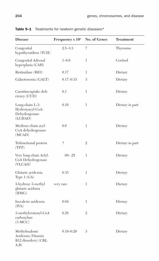
ptg
204 genes, chromosomes, and disease
Table 9–1 Treatments for newborn genetic diseases*
Disease Frequency x 104 No. of Genes Treatment
Congenitalhypothyroidism (TCH)
2.5–3.3 7 Thyroxine
Congenital Adrenalhyperplasia (CAH)
1–0.6 1 Cortisol
Biotinidase (BIO) 0.17 1 Dietary
Galactosemia (GALT) 0.17–0.33 3 Dietary
Carnitineuptake defi-ciency (CUD)
0.1 1 Dietary
Long-chain L-3-Hydroxyacyl-CoA-Dehydrogenase(LCHAD)
0.16 1 Dietary in part
Medium-chain acyl-CoA dehydrogenase(MCAD)
0.6 1 Dietary
Trifunctional protein(TFP)
? 2 Dietary in part
Very long-chain Aclyl-CoA Dehydrogenase(VLCAD)
.08–.25 1 Dietary
Glutaric acidemiaType 1 (GA)
0.33 1 Dietary
3-hydroxy 3-methylglutaric aciduria(HMG)
very rare 1 Dietary
Isovaleric acidemia(IVA)
0.04 1 Dietary
3-methylcrotonyl-CoAcarboxylase(3-MCC)
0.28 2 Dietary
MethylmalonicAcidemia (VitaminB12 disorders) (CBLA,B)
0.10–0.20 3 Dietary
Wow! eBook <WoweBook.Com>

ptg
9 • treating genetic disease 205
Disease Frequency x 104 No. of Genes Treatment
MethylmalonicAcidemia (MUT)
0.1–0.2 3 Dietary
Beta ketothiolase(BKT)
0.01 1 Dietary
Propionic academia(PROP)
0.10 2 Dietary
Multiple carboxylase(MCD)
0.11 1 Dietary
ArgininosuccinateAciduria (ASA)
0.14 1 Dietary
Citrullinemia Type 1(CIT1)
0.175 1 Dietary
Homocystinueria(HCY)
0.03–0.05 4 Vitamin B6
Maple syrup urine dis-ease (MSUD)
0.05 4 Dietary
Phenylketoneuria(PKU)
0.67–1.0 1 Dietary
Tyrosenimia Type 1(TYR-1)
0.25 1 Dietary
Omitted are tests for hearing, cystic fibrosis, sickle cell anemia and trait, and severe com-bined immunodeficiency. Genetic disease abbreviations are standard.
*Most of the information in this table is derived from Genetics Home Reference (http://ghr.nlm.nih.gov/) and references, particularly MedLine Plus contained therein. The table includes24 of the 29 gene defects screened for in newborns.
Wow! eBook <WoweBook.Com>

ptg
The importance of dietThe 24 genetic diseases listed in Table 9–1 are among the 29 primarytarget diseases that are screened for in newborns. Like PKU, the vastmajority are metabolic diseases that respond at least in part to dietarytherapy.
One of these diseases, called maple syrup urine disease (MSUD),has a rather long and interesting history that involves Robert Guthrieonce more.12 In 1951, Dr. John Menkes, an intern at Boston Chil-dren’s Hospital who was planning a career in pediatric neurology,encountered a newborn infant who was drowsy and who also refusedformula. The baby had been diagnosed as having kernicterus, a kindof brain damage that results from jaundice.
Menkes discussed the baby’s condition with its mother andlearned she had earlier had two other children whose urine had anunusual odor and had died, whereas a daughter whose urine lackedthe odor had lived. By the third day, the urine and perspiration of theinfant that had been brought to Menkes’ attention also had a strangeodor.
“I am sure that I must have asked nearly everyone at the hospitalbut could get no better answer than that it smelled like maple syrup,”Dr. Menkes recalled later on. “After all, we were in New England.”
Why was it that the urine of infants with MSUD smelled like maplesyrup? The answer to this question came in the late 1950s fromMenkes, and from a collaborative venture between Joseph Dancis atNew York University-Bellevue Medical Center and Roland Westall atUniversity College Hospital Medical School in London.13 They inde-pendently discovered that there were increased levels of three aminoacids, leucine, isoleucine, and valine, in the baby’s urine and plasma.This suggested that there was a block in the pathway leading to thedegradation of these three amino acids. This caused the accumulationof breakdown products called keto acids.
Like phenylalanine, all three amino acids are essential. That is,humans cannot synthesize them. This meant that dietary controlmight work for MSUD as it did for PKU. Roland Westfall and his col-league C. E. Dent at University College announced progress on thisfront in 1961. They attempted dietary modification on a girl withMSUD who had been transferred to University College Hospital at
206 genes, chromosomes, and disease
Wow! eBook <WoweBook.Com>

ptg
9 • treating genetic disease 207
the age of eight months. She was already severely mentally retardedand lay on her back with only occasional attempts to raise her head.Westfall and Dent were able to devise a diet that resulted in normalplasma levels of isoleucine, leucine, and valine and also caused theurine to lose the odor of maple syrup. Furthermore, the baby gainedweight, but her clinical condition did not change. Other dietaryimprovements followed.
Meanwhile, Robert Guthrie and his colleague Edwin Naylor hadcome up with a bacterial screening test for MSUD similar to the onethey had devised for PKU.14 They began a field trial of the assay in July1964 together with five laboratories that were already collaboratingwith them on PKU screening. These laboratories, all in the UnitedStates except for one in New Zealand, reported their results monthly toNaylor and Guthrie. They were also getting annual updates from labo-ratories around the world. By 1978, more than 9 1/2 million newbornshad been screened for MSUD worldwide.
Today, we know that mutations in any one of four genes can leadto MSUD.15 Each gene encodes a protein, forming part of an enzymecomplex that is responsible for processing the keto acid breakdownproducts of leucine, isoleucine, and valine to the next step in thedegradation process. The diagnostic process involves recognition ofthe characteristic clinical features of MSUD, but the Guthrie test isno longer used to assay for amino acid accumulation. Instead, theactivity of the enzyme complex is measured directly or else moleculargenetic testing of all four genes may be done. MSUD is managed byusing high-calorie formulas free of leucine, isoleucine, and valine.Because all three amino acids are required for protein synthesis, theymust be supplied, but in carefully monitored amounts.
The 29 primary genetic diseases targeted for newborn testinginclude several other amino acid disorders, organic acid disorders,fatty acid disorders, plus several other conditions (refer to Table 9–1).Treatment of these diseases includes dietary modification, but this isoften not sufficient by itself. Consider tyrosinemia type I (TYR-1). Asis true of PKU and MSUD, this disease involves a defect in the degra-dation of an amino acid, in this case tyrosine.16 The genetic defect inthis disease causes the terminal enzyme in the tyrosine degradationpathway to lose function. This means that fumarylacetoacetic acid
Wow! eBook <WoweBook.Com>

ptg
208 genes, chromosomes, and disease
(FAA), the precursor with which this enzyme reacts, begins to accu-mulate. FAA and other compounds derived from it ultimately causesevere liver damage. If left untreated, this means the child will usu-ally die before the age of ten.
As soon as TYR-1 is diagnosed, a compound called nitisinone isgiven. Nitisinone blocks the tyrosine degradation pathway at an ear-lier step. This prevents FAA accumulation and subsequent liver dam-age. However, nitisinone treatment causes tyrosine to accumulate inthe blood. This means that the patient must be kept on a low tyrosinediet to prevent the accumulation of tyrosine crystals in the cornea.
Hormones and proteinsCongenital hypothyroidism (TCH) and congenital adrenal hyperplasia(CAH) are among the panel of genetic diseases tested for in newborns(refer to Table 9–1).17 Both diseases result in hormone deficienciesthat can be corrected with administration of the appropriate hor-mones. TCH arises when the thyroid gland fails to develop or functionproperly. Even though mutations in seven different genes have beenfound to cause TCH, the vast majority of cases are sporadic and,therefore, not predicted on a familial basis. Diagnosis of TCH involvesmeasuring two thyroid hormones, the thyroid stimulating hormoneand thyroxine. The test is best carried out on blood droplets collectedwhen the baby is between two and six days old. Rapid diagnosis isimportant because, in the absence of these hormones, IQ begins todecline noticeably and mental retardation results.
Congenital renal hyperplasia usually arises as a consequence ofmutations in the CYP21A gene.18 This gene encodes an enzyme called21-hydroxylase. The enzyme is found in the adrenal gland where it isinvolved in producing the hormones cortisol and aldosterone. Corti-sol plays an important role in maintaining blood sugar levels, protect-ing against stress, and suppressing inflammation. Aldosteroneregulates salt retention by the kidneys.
A deficiency of 21-hydroxylase leads to the accumulation of com-pounds that are normally converted to cortisol and aldosterone.These substances then build up in the adrenal glands and are con-verted into male-determining hormones or androgens. Conse-quently, females with this disease usually have external genitalia that
Wow! eBook <WoweBook.Com>

ptg
9 • treating genetic disease 209
look neither distinctively male nor female. They may also exhibit thegrowth of excess body hair, male pattern baldness, and unpredictablemenstruation. About 75% of affected individuals have the mostsevere form of the disease that is known as the salt-wasting type. Thiscauses the loss of large amounts of sodium in the urine. Infants withthis form of the disease feed poorly, lose weight, and are subject todehydration and vomiting. The disease is life threatening if nottreated. Fortunately, by supplying a form of cortisol, aldosterone, orboth, the symptoms are readily reversed. The hormone(s) must besupplied for life, but the prognosis is good.
Hemophilia is probably the most familiar genetic disease treat-able by a protein. Mutations in the sex-linked F8 and F9 genes,respectively, cause hemophilia A and B. F8 encodes clotting factorVIII, whereas F9 specifies clotting factor IX (also refer to Chapter 1,“Hunting for disease genes,” with reference to hemophilia in QueenVictoria’s progeny).19 By treating hemophiliacs at regular intervalswith the appropriate clotting factor purified from blood plasma,bleeding can be controlled. Current guidelines recommend the infu-sion of concentrated factor VIII three times a week for hemophilia Aand of factor IX twice a week for hemophilia B. The concentrates canbe administered at home.
But there is (or was) a potential problem. To purify factors VIIIand IX blood, concentrates are pooled from many donors. If theblood of any one of these donors is infected with a virus, that virus canbe transmitted to the hemophilic recipient. The reason why hemo-philia is probably familiar to many is because many hemophiliacsbecame HIV positive in the 1970s and 1980s following treatmentbecause the concentrates used were contaminated with the AIDSvirus.20
After the discovery of the AIDS virus in 1981, it became evidentthat homosexual men and intravenous drug users were its main vic-tims, but in 1982 the Centers for Disease Control reported that threehemophiliacs had become ill from the AIDS virus. Because HIV wasprevalent among intravenous drug users and homosexuals, some ofwhom were blood donors, in March 1983, the agency warned that thevirus might be contaminating blood products, including the concen-trates used for hemophilia prevention. However, there was no way of
Wow! eBook <WoweBook.Com>
ptg
210 genes, chromosomes, and disease
telling which donors were HIV positive or which plasma samples contained the virus because a test for its presence had yet to be devel-oped.
Cutter laboratories, a division of Bayer, was a producer of con-centrates for hemophiliacs and in January 1983, its manager forplasma procurement acknowledged that there was good reason tobelieve that AIDS was being transmitted to other people through theuse of plasma products.21 Then, Cutter learned that a rival was heat-ing the concentrate to inactivate the virus. Cutter’s sales were slip-ping, but, according to an internal memo, rather than admitting thatthey had not so far supplied heated concentrate, Cutter conceived amarketing plan that was designed to give the impression that theywere continuing to improve their product without acknowledgingthat they soon expected to have heated concentrate available.
In June 1983, the company tried to minimize the danger hemo-philiacs faced from using their unheated concentrate. In a letter todistributors in 21 countries, Cutter wrote, “AIDS has become thecenter of irrational response in many countries. This is of particularconcern to us because of unsubstantiated speculation that this syn-drome may be transmitted by certain blood products.”
The consequences were particularly egregious in France, wherenearly half of the country’s 3,000 hemophiliacs contracted AIDS.22 Ina series of articles published in L’Evenement du Jeudi in the spring of1990, physician-journalist Ann-Marie Casteret revealed that the rea-son so many French hemophiliacs had contracted AIDS could betraced to a decision made in 1985 by officials at the French NationalCenter for Blood (CNTS).
In October 1984, Michel Garetta became director of the CNTS.The organization had been operating at a loss and was dependent onthe Ministry of Health to make up the difference, so anything Garettacould do to make the organization more profitable would be helpful.Furthermore, an important goal of the CNTS was to make Franceself-sufficient in blood products. So Garetta stepped up the produc-tion of factor VIII without taking the time to develop a heatingmethod, even though American laboratories like Travenol had beganadvising as early as 1983 that it was best to use heated concentrate forhemophiliacs. It was a terrible error.
Wow! eBook <WoweBook.Com>

ptg
9 • treating genetic disease 211
Garetta’s decision became even more inexcusable in view of aninternal memorandum sent to the health ministry by Dr. JeanFrançois Pinon in January 1985. Pinon was chief of hematology at theCochin Hospital in Paris. An immunologist, Jacques Leibovitch, hadconvinced him it might be a good idea to screen the hospital’s stocksof blood. On December 12, Leibovitch reported that 10 out of the2,000 samples he had tested were positive for HIV. The implicationsfor Garetta were devastating. Because CNTS produced its concen-trates for factor VIII production from up to 5,000 pooled blood dona-tions, all were almost certainly contaminated.
In February 1985, Luc Montagnier, discoverer of the AIDSvirus, reported in the Lancet that heat-treating blood destroyed thevirus and that heating the concentrate prevented infection. Mean-while, in the same month, both Pasteur Diagnostics in France andAbbott Laboratories in the United States filed applications in Francefor AIDS virus tests that they had developed independently. TheAbbott test was approved for use in the United States in March andby April Robert Netter, Director of the French National Health Lab-oratory, was urging approval of the Pasteur test because he did notsee how approval of the Abbott test could be delayed much longerwithout an appeal by Abbott to the Council of State charging abuseof power.
In a French cabinet meeting held on May 9, 1985, chaired byFrançois Gros, Health Adviser to Prime Minister Laurent Fabius and aformer Director of the Pasteur Institute, the decision was made to con-tinue to hold off on the testing of blood donors for AIDS using eitherthe Abbott or Pasteur tests on the basis that it would cost too much.Furthermore, recommendations to heat-treat both the French andimported bloodstocks were ignored. Finally, in July 1985, both theAbbott and Pasteur tests were approved.
It was also on May 9, 1985, months after Pinon’s warning, whenGaretta finally contacted the Ministry of Social Affairs saying thatpurging the French blood supply of HIV contamination was essential.However, Garetta continued, a compromise was necessary betweenthe interests of public health and economic realities. From that time,each delay of three months in testing donors and preparing heated fac-tor VIII would result in the deaths of five to ten hemophiliacs unableto obtain factor VIII to correct bleeding episodes. Furthermore, the
Wow! eBook <WoweBook.Com>

ptg
212 genes, chromosomes, and disease
economic consequences of having to import heated factor VIII anddestroy contaminated stocks coupled with the 20% production lossthat occurred because of heating would cost millions of francs (in dol-lar terms, well over $5 million). Those costs would have to be coveredby the Ministries of Health and Social Services.
To minimize all of these problems, Garetta recommended thatthe CNTS continue to provide unheated stocks to its clients untilmid-July for what were mainly economic reasons. The Association ofFrench Hemophiliacs went along with Garetta’s recommendation,but urged that the ministry ban unheated products after October 1,1985. Why would they have done so? Because Garetta had told theassociation that technical problems had held up the production ofheated product by the CNTS while omitting to mention the fact thatfactor VIII stocks were contaminated.
Casteret’s 1990 articles triggered a government investigation.Michel Lucas, the Inspector General of Social Affairs, spent threemonth locating and studying official documents on the contaminatedblood scandal. He constructed a precise chronology of the events thatled to the conclusion that the decision to continue using the suspectblood for six months without an AIDS test was monetary. In 1991,Garetta and three other health officials were put on trial before apanel of judges at the Palais de Justice in Paris. Garetta was sen-tenced to four years in prison with a substantial fine. He was releaseda year early for good behavior.
But the investigation did not end there. On February 10, 1999,former Prime Minister Laurent Fabius and two other ministers in thegovernment he headed from 1984 to 1986 were put on trial in con-nection with the contaminated blood scandal that had occurred dur-ing their administration. On March 9, Fabius and his Social AffairsMinister, Georgina Dufoix, were acquitted of manslaughter. The for-mer Health Minister, Edmond Herve, was convicted of two cases ofnegligence, but was not sentenced. The court ruled that Herveshould have ordered the destruction of the untreated blood samplesonce it was known that they might contain the AIDS virus. He faceda possible five-year sentence, but the court found that the scandalhad gone on for so many years that Herve had been denied the rightof presumption of innocence. The French Transfusion Association,
Wow! eBook <WoweBook.Com>

ptg
9 • treating genetic disease 213
an advocacy group for transfusion victims, called the acquittals “dis-graceful.”
Meanwhile back in the United States, Cutter got approval to sellheated concentrate on February 29, 1984, the last of the four majorcompanies that distributed blood products to do so.23 But Cutter con-tinued to make the older unheated product for five more months inorder to profit from several large contracts where the price hadalready been fixed, and also because some customers wanted theunheated concentrate believing that heating might cause the concen-trate to deteriorate.
In a meeting held on November 15, 1984, Cutter noted that ithad an excess of unheated concentrate. Because Cutter had incurredconsiderable costs in obtaining, preparing, and storing the concen-trate, the company wanted to recoup its expenses somehow. At thesame meeting, company executives decided to see whether the addi-tional product could be sold abroad. Cutter successfully sold morethan 100,000 vials of the unheated concentrate worth over $4 million,particularly in Asia. Doctors in Taiwan, Singapore, Malaysia, andIndonesia were primarily dispensing the unheated concentrate.
At Hong Kong’s Queen Mary Hospital, Dr. Chan Tai-kwonglamented that he and other doctors at the hospital were unable toobtain the heated concentrate from Cutter. Dr. Chan found that 40%of his patients were HIV positive. Dr. Patrick Yuen, who was associ-ated with a different hospital, reported similar results. The story wasrepeated in Taiwan. A scandal like the one in France also erupted inJapan.
AIDS virus is not the only virus that may contaminate concentratesof factor VIII.24 Since the 1970s, when plasma concentrates of coagula-tion factors first became widely available, hepatitis B and C viruseshave occasionally been unwelcome contaminants. Today’s concentratesprepared from plasma are much safer. In addition to heating, a solvent-detergent mixture is now used to prevent viral infection. The lattermixture inactivates viruses with a lipid envelope, but does not work forthe hepatitis A virus that lacks such an envelope.
The early 1990s also saw the licensing of the first two recombinantclotting factors. These factors are generally produced in hamster cellsin tissue culture. Currently, four preparations are available.25 Bayer
Wow! eBook <WoweBook.Com>

ptg
214 genes, chromosomes, and disease
makes two of these, Helixate FS and Kogenate FS. Advate is a Baxterproduct and Wyeth produces Xyntha.
Treating hemophilia is expensive because factor VIII concen-trates, whether obtained from pooled blood samples or producedusing recombinant DNA, cost a lot. In one recent study of boys withsevere hemophilia treated with Kogenate, the estimated cost perchild per year was in the neighborhood of $300,000.26 The NationalHemophilia Association estimates the cost of treatment per personper annum at $60,000 to $150,000.27 The association seemingly throwsup its hands where insurance is concerned. “The state of insurancereimbursement today is constantly changing to meet this competitiveenvironment. Patients and medical professionals alike are finding thesystem difficult to manage, complex and often confusing. Whileorganizations and treatment professionals can educate people to bet-ter understand insurance, it ultimately falls on individuals to managetheir own healthcare reimbursement.”
Enzyme replacement therapy is another method for alleviatingcertain genetic diseases, particularly the lysosomal storage diseases.28
These tend to be at higher frequency in Ashkenazi Jewish popula-tions. The lysosome, a membrane-bound organelle, is tasked withdegrading various cellular waste products or digesting substancesbrought in from outside the cell. Loss of lysosomal enzyme functionpromotes the accumulation of substrates for these enzymes. This, inturn, results in cellular death, and tissue damage or organ failure.
Gaucher disease is the one of the most common lysosomal stor-age diseases. It results from a deficiency in an enzyme called acidbeta-glucosidase. As a consequence, glucocerebroside accumulates inthe lysosomes of cells called macrophages. Macrophages are pro-duced by differentiation of certain kinds of white blood cells calledmonocytes. They are capable of amoeboid movement and ingest cel-lular debris as well as pathogens. Digestion of a pathogen involvesformation of a structure called a phagosome by the macrophage.Fusion of lysosomes with the phagosome creates a phagolysosome inwhich the pathogen is broken down by lysosomal enzymes. InGaucher disease, these cells become engorged because they cannoteliminate glucocerebroside and assume a characteristic crumpled tis-sue paper appearance that has led to them being called Gaucher cells.
Wow! eBook <WoweBook.Com>

ptg
9 • treating genetic disease 215
Although there are three types of Gaucher disease, Type 1accounts for 80% to 90% of all cases. In the majority of individualswith this disease, its symptoms are not expressed until adulthood.Type 1 Gaucher disease is currently a target for enzyme replacementtherapy. It turns out that, during the normal cellular processing of alysosomal enzyme, a tag is attached to the enzyme molecule that ismade up of a sugar molecule (mannose) with a phosphate residue(mannose-6-phosphate). A mannose-6-phosphate receptor moleculethen sorts the enzyme molecule into an intracellular transport vesiclethat is destined for a lysosome.
The whole cellular processing sequence that the enzyme moleculeundergoes before it enters a lysosome cannot be mimicked by simplyinjecting a Gaucher patient with enzyme. Instead, enzyme therapytakes advantage of the fact that macrophages have mannose-6-phos-phate receptors. A recombinant enzyme is used that has the mannose-6-phosphate tag. This results in its uptake by macrophages and itstransfer to lysosomes within the macrophage. The enzyme can then cat-alyze the breakdown of the accumulated glucocerebroside. The FDAhas approved this general enzyme replacement strategy for Gaucherdiseases and for several other lysosomal disorders.
The main problem with treating Gaucher disease using enzymereplacement therapy is the expense.29 Treatment was estimated to cost$145,000 to $290,000 per year in 2005. Estimates for drugs used totreat three other lysosomal storage diseases that were approved at thattime ranged from $156,000 per year to $377,000 per year. The reasonis that treatment for Gaucher disease and related lysosomal storagediseases is covered by the Orphan Drug Act of 1983.30 Its specific pur-pose was to encourage pharmaceutical companies to develop drugsthat alleviated rare diseases. One of the rewards for a company thatagreed to do this was a seven-year exclusivity clause upon FDAapproval. Once a company was approved to create a certain drug, itwas unlikely that other pharmaceutical companies were going todevelop competing drugs anyway in view of the small number ofpatients involved and the fact that they were not eligible for the bene-fits of the Orphan Drug Act.
The story of cerezyme, the drug used to treat Gaucher disease,illustrates how the Orphan Drug Act has led to an effective treatment
Wow! eBook <WoweBook.Com>
ptg
216 genes, chromosomes, and disease
for this disease, but at an exorbitant price.31 The tale begins with a dis-tinguished physician named Roscoe Brady at the National Institutesof Health. Dr. Brady became interested in metabolic storage diseasesand the possibility of correcting them with enzyme therapy. By theearly 1970s, his associate, Dr. Peter Pentschev, using human placen-tas, had managed to isolate tiny amounts of glucocerebrosidase, thedefective enzyme in Gaucher disease. Two patients received theenzyme and the initial results looked promising.
A large-scale purification method had been developed by 1977,but the resulting enzyme preparation gave inconsistent results. Aswe have seen, to be effective, the sugar chains of the enzyme had toend with mannose in order to interact with the mannose receptorson the macrophages. The problem was that the mannose residues onthe sugar side chains of glucocerebrosidase were internal in posi-tion, so Brady’s team succeeded in developing a method for strip-ping off the blocking sugar molecules to expose the mannoseresidues. In the first clinical trial involving eight people withGaucher disease, only the smallest one, a child, showed beneficialeffects. The other seven people in the trial were adults and theenzyme replacement therapy did not help them. In the second clin-ical trial involving 12 individuals, the enzyme dosage was increasedand all responded positively. By the end of 1975, the NIHresearchers had patented their method for isolating the enzyme.
To carry out the clinical trials necessary to gain FDA approval ofenzyme therapy for Gaucher disease, Brady’s team at NIH needed aregular supply of enzyme. Hence, the agency contracted with theNew England Enzyme Center at Tufts University Medical School tosupply enzyme prepared according to NIH procedures. The directorof the center was Henry Blair and the NIH contracts over a period ofseveral years amounted to nearly $1 million dollars. In 1981, the cen-ter closed and a biotechnology entrepreneur named Sheridan Snyderfounded Genzyme together with Henry Blair.
For the next 11 years, the NIH held contracts with Genzyme thatamounted to nearly $9 million under which Genzyme provided theNIH with enzyme for clinical trials. Because the NIH had patentedits procedure for enzyme isolation and investigators there had pub-lished papers on the subject, Genzyme could not obtain a patent for
Wow! eBook <WoweBook.Com>

ptg
9 • treating genetic disease 217
the drug it was now providing to NIH, but in 1985, Genzyme’sAlglucerase, as it was then called, received official designation as anorphan drug. This meant that the company would have exclusivemarketing rights for Alglucerase for seven years and, hence,enhanced profits. Furthermore, it was unlikely that a competitiveproduct would appear on the market because of the costs of develop-ing another drug for such a small market. Hence, Genzyme wouldprobably be able to sell Alglucerase for a high price without fear ofcompetition after the seven-year period of protection had elapsed.There was a hitch, however. Genzyme could not market its enzymepreparation without FDA approval and this would not be forthcom-ing until April 5, 1991.
Despite the steady stream of NIH contracts and the money thecompany made from selling certain chemicals to bigger companies,Genzyme was in trouble within two years of its formation. Head-hunters sought out a new CEO and made a pitch to Henri Termeerwho had previously been employed by Baxter. Because the Gaucherdrug, now renamed Cerezyme, would be the only FDA-approveddrug available to treat Gaucher disease, Termeer realized that hecould sell Cerezyme to patients for a very high price. Upon FDAapproval in 1991, Genzyme brought Cerezyme on the market for anaverage of $200,000 per patient per year.
Genzyme explained that the price of Cerezyme was so highbecause it originally took 22,000 placentas to produce enough of thedrug to treat a patient for a year. In 1994, Genzyme developed amethod for producing Cerezyme from genetically modified cells inculture, but the $200,000 price remained the same. In 2004, aboutone-third of Genzyme’s $2.2 billion in revenue came from the sale ofCerezyme. In 2007, Cerezyme sales amounted to $1.1 billion.
The price of Cerezyme placed it beyond the pocketbooks of mostpatients, so it was essential that insurance companies pick up at leastpart of the tab. The problem for Ed and Peggy DeGranier, whose sonBrian had Gaucher disease, was that Ed worked for a small employer.Getting the insurer to pay for Brian was going to make all of the com-pany’s insurance premiums unaffordable. So Ed DeGranier quit hisjob and went to work selling cable services door-to-door for Comcast,
Wow! eBook <WoweBook.Com>

ptg
218 genes, chromosomes, and disease
reasoning that the expense of the insurance payments for Brian’streatment would now be spread among thousands of employees.
Henri Termeer was becoming rich and famous. MassachusettsGovernor Deval Patrick appointed Termeer to his council of eco-nomic advisers and hosted the Termeers for dinner in the spring of2008. Termeer himself had earned more than $50 million in totalcompensation and, in the spring of 2008, was worth around $260 mil-lion because of the performance of Genzyme’s stock. But then disas-ter struck. The company experienced serious production problems,not only for Cerezyme, but also for Fabrazyme, a product designed tocombat Fabry’s disease, another lysosomal storage disease. In June of2009, Genzyme had to shut its main factory in Boston temporarilydue to viral contamination. The company first predicted that produc-tion should return to normal in six to eight weeks, but there wererepeated setbacks. The value of Genzyme stock began to decline andthe production problems opened the company to a proxy challengeby investor Carl Icahn.
In November 2009, Adam Feuerstein, writing for the Street,picked Termeer for the 2009 Worst Biotech CEO of the Year Award.32
Feuerstein blamed Termeer for “fostering an arrogant, irresponsiblebusiness culture in which employees are rewarded more for beingloyal to the CEO than they are for being competent at their jobs.”Feuerstein accused the company of “sloppy manufacturing methodsand a dirty, run-down manufacturing plant in Boston that has becomean ongoing target for FDA inspectors.”
The shortage of Cerezyme allowed two other companies to seekapproval of their own Gaucher drugs.33 Both companies received helpfrom the FDA because the agency had good reason to feel that thesole supplier model no longer worked. In February 2010, one ofthese companies, Shire, received FDA approval of its Gaucher drug.In September, the EU approved Shire’s new drug. Shire planned tosell its drug at a 15% discount to Cerezyme, still expensive, but at lastthere is price competition for Gaucher drugs.
Big pharmaceutical firms often like to acquire smaller companieslike Genzyme that have one or a few highly profitable products. InAugust 2010, the Sanofi-Aventis offered to buy Genzyme for $69 ashare.34 Genzyme’s Board of Directors rejected the offer so the
Wow! eBook <WoweBook.Com>
ptg
9 • treating genetic disease 219
French pharmaceutical firm launched a hostile takeover in Octoberof that year.
There are several lessons to be learned from the Genzyme story.First, it was because of basic research done by Roscoe Brady and hisgroup at NIH that treatment of lysosomal storage diseases likeGaucher became possible through enzyme therapy. To scale up theprocess for clinical trials, Brady and his colleagues turned to the NewEngland Enzyme Center at Tufts University run by Henry Blair. Blairand a colleague subsequently founded Genzyme, whose principalmoney earner became the Gaucher drug Cerezyme. In short, no orig-inal research by Genzyme was involved, although they later devel-oped a method for obtaining Cerezyme from tissue culture cellsrather than placentas. Because the company benefited from theOrphan Drug Act, Genzyme could charge anything it wanted for thedrug once Cerezyme had been approved by the FDA because a com-petitive drug could not be brought to market for 7 years. In fact,because the number of Gaucher patients was small, Genzymeenjoyed a monopoly on enzyme therapy for Gaucher disease foralmost 20 years. The downside of this for patients was that Genzymecharged an outrageous price for Cerezyme and, when the companyexperienced manufacturing problems in 2009, there was no otherplace for them to turn to for the drug.
A quite different approach to reducing the amounts of toxic sub-stances that accumulate in lysosomes in storage diseases is called sub-strate reduction therapy.35 The idea here is to make use of a smallmolecule that inhibits or limits synthesis of a precursor to the sub-strate of the defective enzyme. This treatment has been approved forpatients with mild to moderate Type 1 Gaucher’s disease.
One final example of enzyme replacement therapy involves a dis-ease called Severe Combined Immunodeficiency or SCID. There areseveral forms of this disease caused by mutations in different genes,but they all result, as the name suggests, in a highly defective immunesystem. This means that babies born with the disease are very suscep-tible to viral, bacterial, and fungal infections and to “opportunistic”organisms that do not normally cause people with functioningimmune systems to get sick. Infants with SCID usually develop pneu-monia and diarrhea. They do not survive for more than a year or two.
Wow! eBook <WoweBook.Com>
ptg
220 genes, chromosomes, and disease
The form of the disease treatable by enzyme replacement resultsfrom mutations in the ADA gene on chromosome 20. This geneencodes an enzyme called adenosine deaminase.36 ADA-SCID is veryrare, occurring in around 1 in 200,000 to 1,000,000 infants world-wide. Adenosine deaminase is found throughout the body, butassumes critical importance in the immune system. The immune sys-tem includes white blood cells called B-cell lymphocytes and T-celllymphocytes. B cells produce antibodies to invading bacteria andviruses that mark them for destruction. T cells do not produce anti-bodies, but regulate B-cell activity. Helper T cells assist in the matu-ration of B cells while regulatory (suppressor) T cells shut down Tcell-mediated immunity and, therefore, antibody production at theend of an immune reaction. Cytotoxic T cells destroy virus-infectedcells and cancerous cells.
Adenosine deaminase converts a compound that is toxic to T cells(deoxyadenosine) to a harmless compound (deoxyinosine). Mutationsin the ADA gene that inactivate the enzyme result in a buildup ofdeoxyadenosine in T cells that result in their death. Obviously, thisresults in loss of immune system control and is lethal before long.
The therapy of choice for any kind of SCID for over 20 years hasbeen bone marrow/stem cell transplantation from a donor, usually asibling, who is immunologically compatible (HLA identical). However,enzyme replacement therapy has also been available for ADA-SCIDfor over 15 years. Treatment of this disease, like the lysosomal storagediseases, is very expensive. Biweekly injections of a modified form ofthe enzyme are required and the cost may exceed $200,000–$300,000per patient annually, but the treatment works over the long term.37
Even though lymphocyte counts are below normal in enzyme-treatedpatients, their immune systems function well enough to ward off majorinfections and to foster good health.
Gene therapy: mirage or grail?Rare genetic diseases come to the attention of the public only occa-sionally, usually when a newspaper article is published on an other-wise obscure ailment and often in connection with some impendingform of therapy. One disease that gained a lot of attention in the
Wow! eBook <WoweBook.Com>

ptg
9 • treating genetic disease 221
1970s and early 1980s was SCID-X1.38 This disease is caused by muta-tions in a gene found on the X chromosome and is almost exclusivelyfound in males. The reason for all the attention was that David Vetter,a little boy with this disease, was encased in a germ-free plastic bubble for ten years to prevent him from contracting an infectiousdisease.
David’s parents, David Joseph Vetter Jr. and Carol Ann Vetter,had a daughter named Katharine. They also had a son named Davidwho had died seven months after his birth from SCID. Three physi-cians from Baylor College of Medicine, John Montgomery, Mary AnnSouth, and Raphael Wilson, told the Vetters that another son had a50% chance of having SCID because Carol Ann carried an X chromo-some with the mutant form of the gene. However, they assured theVetters that, if they had a son with SCID-X1, the baby could be put ina sterile chamber until a bone marrow transplant could be per-formed. Their assumption was that sister Katharine would be a suit-able donor.
In due course, the Vetters had a second son, whom they alsonamed David, and he had SCID-X1. The baby was placed in his ster-ile incubator pending the bone marrow transplant from his sister.However, his sister was not a good match and a good match was neverfound. So little David lived in plastic isolation for ten years on thethird floor of the West Tower of Texas Children’s Hospital in Houstonwhere all he could see was Fannin Street from the room’s window.Occasionally, David would be transferred to a bubble at his home fora few weeks. A psychologist named Mary Murphy befriended David,spending a lot time with him. She said that David grew angry. He didn’t see the point of learning to read or of schooling. “What goodwill it do?” he said, “I won’t ever be able to do anything anyway.”
Over the years, David’s three original doctors moved on to posi-tions elsewhere. Two new doctors, Ralph Feigin and William Shearer,took over. Despite exhaustive searching, no suitable bone marrowdonor had appeared. The new doctors tried to convince the Vetters totake David home and to try to keep him healthy on a regime ofgamma globulin and antibiotics. Perhaps his immune system hadimproved. The Vetters rejected this solution after having consultedthe original trio of doctors.
Wow! eBook <WoweBook.Com>
ptg
222 genes, chromosomes, and disease
Another four years passed and by now doctors in Boston had hadsome success in transplanting unmatched bone marrow. At this point,all of the doctors concurred that using unmatched bone marrow was arisk worth taking. David’s sister Katharine donated the bone marrow. Itwas treated in Boston and flown back to Houston. The transplant wasperformed on October 21, 1983.
For a few months, everything seemed to go well and the doctorsbegan to hope that David would be able to leave his bubble. But inearly February 1984, David became violently ill and he died on Feb-ruary 22. An autopsy revealed that David’s body was full of tumors.He had died of a cancer called Burkitt’s lymphoma. It turned out thatthe screening of Katharine’s marrow had missed the presence ofEpstein-Barr virus, an important contributor to this cancer.
Considering that immunologically matched bone marrow trans-plants are regarded as the most satisfactory treatments for the differentforms of SCID, bone marrow cells of a person with the disease wouldseem ideal targets for gene therapy. Assuming that a good gene couldbe introduced into the appropriate cell type and functioned there, thewhole immunological matching problem would be obviated. It shouldbe noted that this sort of gene therapy is referred to as somatic genetherapy. The defective gene is repaired only in the targeted somaticcells. This is the only kind of gene therapy possible in humans at themoment. Reproductive gene therapy, where the defect is corrected ineggs and sperm, is not even on the horizon.
W. French Anderson and colleagues conducted the first seriousattempt at gene therapy and it involved the ADA gene. Gettingapproval for a human gene therapy trial was a drawn-out process.Anderson’s quest for approval began in June 1988.39 The NIH and theFDA subjected Anderson’s clinical protocols to detailed technicalreview. In early 1990, Anderson cleared the final hurdle and receivedapproval for his gene therapy test from the Recombinant DNA Advi-sory Committee of the NIH.
And Anderson had a suitable patient. She was an ADA-deficientlittle girl named Ashanti DeSilva. She was only four and was receivingenzyme replacement therapy. To deliver the gene, Anderson neededan appropriate vector. These vectors are usually disabled viruses. Thevector Anderson chose to use to insert a good copy of the ADA gene
Wow! eBook <WoweBook.Com>
ptg
9 • treating genetic disease 223
into Ashanti’s T cells was a modified retrovirus. These are RNAviruses that copy themselves into double-stranded DNA and in thisform insert themselves into the genome (refer to Chapter 1).
There are lots of retroviral remains littering the human genome.The basic structure of a retrovirus is quite simple. There are threegenes called gag, pol, and env flanked on either end by long terminalrepeats (LTR). Creation of a retroviral vector usually involves remov-ing the three viral genes from a DNA copy of the virus. The therapeu-tic gene is then inserted in place of them. To prepare virus particles toinfect the target cell, the modified retroviral DNA is transferred to apackaging cell line. This cell line has been modified to synthesize theviral proteins required to make virus particles. These particles arethen used to deliver the human gene to the target cells that are main-tained in culture.
On September 14, 1990, Ashanti deSilva was treated for the firsttime with an infusion of her own T cells into which a normal ADAgene had been inserted. The retroviral vector carrying the normalADA gene had been mixed with T cells that had been extracted fromAshanti’s blood and subsequently grown in culture. When these cellswere returned by injection to the little girl, they began to produce theADA enzyme. Because Ashanti seemed to benefit and there were noobvious harmful effects, Anderson and his team repeated the sameprotocol with nine-year-old Cynthia Cutshall on January 30, 1991.Both girls’ immune systems now began to function properly. To be onthe safe side, the two girls also continued on enzyme replacementtherapy.
Because T cells turn over, injection of genetically modified T cellscontinued with each girl receiving approximately ten infusions over 2years. When the girls were monitored after 4 and then 12 years, ADAexpression in T cells could still be detected in Ashanti’s case.40 Around20% of her T cells still carried and expressed the introduced ADAgene. However, in Cynthia Cutter’s case, the persistence of cells withthe ADA gene was very low (<0.1%) and no expression of the genewas detectable.
Anderson’s success with ADA-SCID stimulated a number of pro-posals for gene therapy trials. By 2000, investigators had initiatedover 400 of them. But in 1999, tragedy struck. Dr. James M. Wilson,
Wow! eBook <WoweBook.Com>

ptg
224 genes, chromosomes, and disease
who had been recruited by the University of Pennsylvania in 1993 toestablish an Institute for Human Gene Therapy, was in the process ofconducting a liver gene therapy trial. One of his subjects was a youngman of 18 from Tucson, Arizona, named Jesse Gelsinger.41
Shortly after Wilson arrived at Penn, he had met Dr. Mark Bat-shaw, an expert on metabolic diseases. Batshaw was particularly inter-ested in a disease that results from a deficiency in an enzyme calledornithine transcarbamylase (OTCD). This enzyme is involved in thesynthesis of the amino acid arginine. It is also one of the urea cycleenzymes that convert ammonia to urea. If the cycle is disrupted, as itis in OTCD, ammonia accumulates in the liver, causing liver failureand death.
The OTC gene encoding ornithine transcarbamylase is on the Xchromosome, meaning that OTCD usually affects males. Newbornbaby boys that completely lack the enzyme enter a coma within threedays and die if the disease is not treated. Even with current treatmentmethods, most surviving children have severe cognitive defects.
A crucial step in preparation for the OTCD trial was the devel-opment of a vector that would deliver the functional gene to liverhepatocytes. These are the major functional cells of the liver, makingup 70%–80% of the cytoplasmic mass. They are also fairly stablewith a life span of around five months. The vector chosen was a dis-abled or attenuated version of an adenovirus that could express thenormal OTC gene. Adenoviruses are DNA viruses that commonlycause respiratory infections or colds. Adenoviral vectors have theadvantage that they infect cells very readily and can accommodate asubstantial foreign DNA insert. However, they do not integrate intothe genome nor do they or the genes they contain replicate. Thismeant that, if the procedure worked, it would have to be repeatedfrom time to time as new hepatocytes replaced the older ones.
Dr. Wilson and his colleagues submitted a grant proposal to theNIH on March 23, 1994, and they soon obtained promising data.They found, using a mouse model, that the OTCD metabolic defectcould be eliminated by their modified adenovirus for several weeks toa month. High doses of virus were administered both to mice andrhesus monkeys to assess the potential for toxic effects. At the very
Wow! eBook <WoweBook.Com>

ptg
9 • treating genetic disease 225
highest dose, the monkeys developed severe liver disease and a clot-ting disorder that would have been lethal had the monkeys not beeneuthanized.
The investigators received permission to enroll subjects in a clini-cal trial on October 21, 1996, by which time the adenovirus vector theywere using had undergone significant improvements. They decidedthat adults and not infants would be admitted to the trial because onlyadults were capable of informed consent. Eighteen subjects wereenrolled in the clinical trial. Most of the subjects were female carriersof a defective OTC gene, but four, including Jesse Gelsinger, weremales who had functional and nonfunctional versions of the genedespite the fact that it was located on the X chromosome. The clinicaltrial was initiated with its first subject on April 7, 1997.
The first 17 subjects enrolled in the trial exhibited a number ofsurprising side effects and immunological events, some being unpre-dicted by the preclinical trials. Furthermore, there was no evidencethat the introduced OTC gene was expressed. Subject 18 was JesseGelsinger. He had a partial deficiency in the OTC enzyme that hadbeen diagnosed at 30 months of age following an episode of vomiting,seizures, and coma associated with elevated ammonia levels. Despitedietary and other treatments, he continued to display episodes of dis-tress associated with high levels of ammonia from time to time.
When Gelsinger signed up for the clinical trial, he realized it wasto test the efficacy of gene therapy for infants and that it probablywould not benefit him. Nevertheless, if it did, he might be releasedfrom his restrictive diet where half a hot dog was a treat. As he said toa friend shortly before he left for Penn Hospital in Philadelphia,“What’s the worst that can happen to me? I die, and it’s for thebabies.”42 And that, sad to say, is precisely what happened. WhenGelsinger underwent treatment with the adenovirus vector carryingthe OTC gene, he developed a massive systemic inflammatoryresponse, multiple organ failure, and died 98 hours after beingtreated.
Reactions to Gelsinger’s death were not long in coming.43 Officialsat the FDA said that Jesse Gelsinger should have been judged ineligi-ble for the trial because his liver was not functioning well enough
Wow! eBook <WoweBook.Com>

ptg
226 genes, chromosomes, and disease
before he had been infused with the vector. The agency temporarilyshut down gene therapy experiments at the University of Pennsylva-nia. The Senate got involved complaining that gene therapy trialsneeded proper oversight. A six-member panel led by Dr. William H.Danforth, Chancellor Emeritus of Washington University in St.Louis, recommended, but did not insist, that clinical trials at theInstitute for Human Gene Therapy be discontinued. The universityimplemented this suggestion.
One year and a day after Jesse Gelsinger died, a wrongful deathsuit was filed in state court in Philadelphia. It accused Dr. Wilson andcolleagues of failing to follow federal rules properly and failing toinform Jesse Gelsinger of the risks he faced. Not only were Dr. Wil-son and two of his colleagues named in the suit, but Arthur Caplan,Director of the University’s Center of Bioethics, was also named as adefendant. In early November 2000, the University of Pennsylvaniasettled the suit out of court.
Then, just as gene therapy seemed to have entered the slough ofdespond, an exciting report was published by a French team led byDr. Alain Fischer, an immunologist and gene therapist, at the NeckerHospital in Paris.44 They had used retroviral gene therapy to treatSCID-X1 disease in two little boys aged 11 and 8 months at the timeof the trial.
SCID-X1 is a very different disease than ADA-SCID. It is themost common form and results from mutations in the IL2RG genelocated on the X chromosome. The protein encoded by this gene isrequired for normal immune system function. Without it, T and nat-ural killer (NK) cells are absent and B cells are nonfunctional. Aretroviral vector with the functional IL2RG gene was used to treatspecific bone marrow cells called CD34+. CD34 is a protein markerfor stem cells of the blood-forming (hematopoietic) system. By treat-ing these cells, the French investigators were aiming for a long-termsolution because these cells give rise to the differentiated cells of theimmune system (e.g., B and T lymphocytes).
Then in the spring of 2002, a cute little red-haired boy 18 monthsin age named Rhys Evans from South Wales was reported to havebeen cured of SCID-X1 using gene therapy.45 Dr. Adrian Thrasher atLondon’s Great Ormond Street Hospital for Children led the team
Wow! eBook <WoweBook.Com>

ptg
9 • treating genetic disease 227
that cured Rhys. By Christmas, Rhys’ condition had vastly improved,so much so that his parents made a video of Rhys playing under theChristmas tree to send to his doctors as a thank you.
More children were treated, but then the storm clouds gatheredagain.46 Four of the ten children treated by the Paris team developeda leukemialike disease between three and six years of age. In the firsttwo Parisian cases, the vector had integrated near a proto-oncogenecalled LMO2 and activated it causing cell proliferation. In the thirdcase, there was also vector integration next to LMO2 and also nearanother proto-oncogene called BM11, but once again it was the eventinvolving LMO2 that led to leukemia. In the final case, a third proto-oncogene CCND2 was involved. In three of the four patients,chemotherapy was successful in restoring normal T cell populations,but failed in the fourth patient who died. Then in 2007, the eighth lit-tle boy treated by the Thrasher team in London also developedleukemia. Once again, integration leading to the expression of LMO2seemed to be the root cause.
A potentially fatal problem now manifested itself.47 Was the ther-apeutic gene itself causing cancer? A group led by Inder Verma, anexpert on the development of viral vectors for gene therapy, pub-lished a paper in Nature titled “Therapeutic Gene Causing Lym-phoma.” Using a mouse model of SCID-X1, they introduced theIL2RG gene into the experimental mice and followed them for up toa year and a half. One-third of the mice that underwent gene therapydeveloped T cell lymphomas. Verma and colleagues concluded thatIL2RG was itself the culprit. They recommended that preclinicaltreatments with transgenes should include a long-term follow-upprior to clinical trials.
The teams in Paris and London argued that, for several reasons,the murine results did not apply to the human trials. For one thing,their own experiments with the murine model resulted in only threecases of lymphoma in one trial where 68 mice (26 SCID and 42 nor-mal) had been tested. There were none in two other transgenic linesin which 54 normal mice were tested.
In 2002 in the United States, the FDA put three clinical trials ofSCID therapy on hold following a French announcement that one ofthe boys in their trial had developed leukemia.48 The trials started
Wow! eBook <WoweBook.Com>
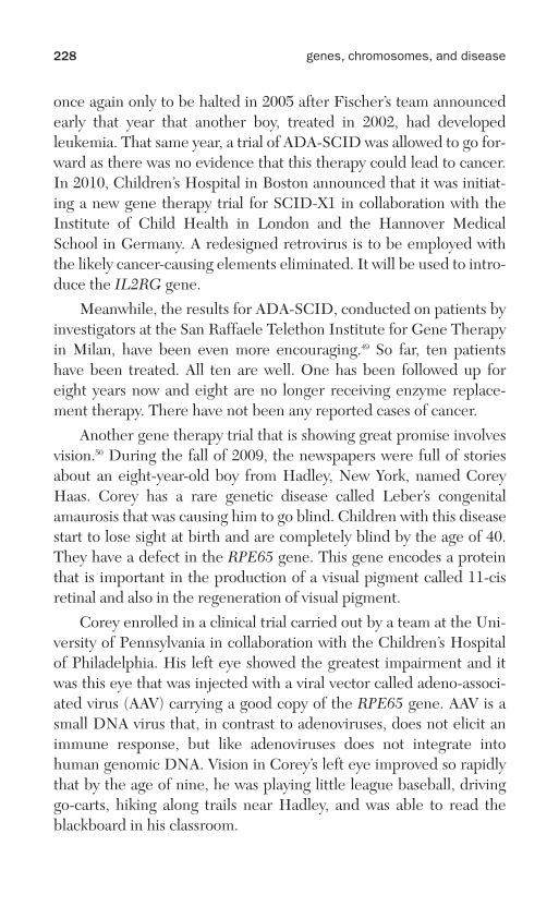
ptg
228 genes, chromosomes, and disease
once again only to be halted in 2005 after Fischer’s team announcedearly that year that another boy, treated in 2002, had developedleukemia. That same year, a trial of ADA-SCID was allowed to go for-ward as there was no evidence that this therapy could lead to cancer.In 2010, Children’s Hospital in Boston announced that it was initiat-ing a new gene therapy trial for SCID-X1 in collaboration with theInstitute of Child Health in London and the Hannover MedicalSchool in Germany. A redesigned retrovirus is to be employed withthe likely cancer-causing elements eliminated. It will be used to intro-duce the IL2RG gene.
Meanwhile, the results for ADA-SCID, conducted on patients byinvestigators at the San Raffaele Telethon Institute for Gene Therapyin Milan, have been even more encouraging.49 So far, ten patientshave been treated. All ten are well. One has been followed up foreight years now and eight are no longer receiving enzyme replace-ment therapy. There have not been any reported cases of cancer.
Another gene therapy trial that is showing great promise involvesvision.50 During the fall of 2009, the newspapers were full of storiesabout an eight-year-old boy from Hadley, New York, named CoreyHaas. Corey has a rare genetic disease called Leber’s congenitalamaurosis that was causing him to go blind. Children with this diseasestart to lose sight at birth and are completely blind by the age of 40.They have a defect in the RPE65 gene. This gene encodes a proteinthat is important in the production of a visual pigment called 11-cisretinal and also in the regeneration of visual pigment.
Corey enrolled in a clinical trial carried out by a team at the Uni-versity of Pennsylvania in collaboration with the Children’s Hospitalof Philadelphia. His left eye showed the greatest impairment and itwas this eye that was injected with a viral vector called adeno-associ-ated virus (AAV) carrying a good copy of the RPE65 gene. AAV is asmall DNA virus that, in contrast to adenoviruses, does not elicit animmune response, but like adenoviruses does not integrate intohuman genomic DNA. Vision in Corey’s left eye improved so rapidlythat by the age of nine, he was playing little league baseball, drivinggo-carts, hiking along trails near Hadley, and was able to read theblackboard in his classroom.
Wow! eBook <WoweBook.Com>

ptg
9 • treating genetic disease 229
At eight years old, Corey was the youngest of 12 patients in hisclinical trial with the oldest being 44. Although all 12 patients showedvisual improvement, it was clear that the degree of improvement wasage dependent with the youngest patients faring best. The resultssupported the investigators’ hypothesis that the success of gene ther-apy depends on the extent of retinal degeneration and that increaseswith age.
As Carolyn Johnson of the Boston Globe wrote on February 28,2009, “Gene therapy started the year with a bang.”51 But it has taken along time. Between the first proposals for clinical trials over 20 yearsago and 2008, over 1,300 such trials have been initiated in 28 coun-tries and the success rate has been very low.52 Perhaps that willchange now with better gene delivery methods, not all of whichinvolve viral vectors, and a better understanding of risk factors. Cer-tainly in the case of a disease like SCID-X1, where enzyme therapy isnot an option, the choice is between bone marrow transplant, ideallywith a well-matched donor, and gene therapy. And now gene therapyseems to be working well over the long term for ADA-SCID therapyand for Leber’s congenital amaurosis.
The great new hope is for stem cell therapy. Not only has Presi-dent Obama lifted the ban on federally funded embryonic stem cellresearch, but great progress has been made in creating stem cellsfrom adult cells that gain the potential of being able to differentiateinto virtually any cell type. So far, however, only stem cells fromhematopoietic tissue have been used successfully in gene therapy inthe case of SCID. But myoblasts appear to be good candidates forgene therapy because these muscle stem cells fuse with nearby mus-cle tissue and become an integral part of muscle.
But James Wilson, in whose clinical trial Jesse Gelsinger died, hasreflected on lessons taught for stem cell therapy from gene therapy’srocky road.53 In the 1990s, the sky seemed to be the limit for genetherapy. It was hyped as the potential cure for a great array of ail-ments. But in a review written in 2000, the FDA concluded that “thehyperbole has exceeded the results” and “little has worked.” Wilsonobserves, “many of the social and economic forces that drove genetherapy’s burst of clinical activity also exist today in the stem cellarena.” He points out that the rapid acceleration of gene therapy
Wow! eBook <WoweBook.Com>

ptg
230 genes, chromosomes, and disease
experiments from bench and model to clinical trials was driven bymany factors, including the following: (1) a “simplistic, theoreticalmodel that the approach ‘ought to’ work”; (2) “a large population ofpatients with disabling or lethal diseases and their affiliated founda-tions harboring fervent hopes that this novel therapy could helpthem”; (3) “unbridled enthusiasm of some scientists in the field,fueled by uncritical media coverage”; and (4) “commercial develop-ment by the biotechnology industry during an era in which value andliquidity could be achieved almost entirely on promise, irrespective ofactual results.”
It would seem wise for stem cell researchers to keep these fourpoints in mind as they forge ahead into this promising new field with itsgreat potential. If gene therapy is any guide, there may be unexpectedand, possibly, serious disappointments ahead.
We should soon know more about the potential and pitfalls ofstem cell therapy. In January 2009, the FDA approved plans by acompany called Geron to conduct the first clinical trial involvingembryonic stem cells.54 These stem cells were manipulated to act asprecursors of certain kinds of nerve cells to try to repair recentlyincurred spinal cord injuries. The first trial is really designed to assessthe safety of the procedure. Its first patient was treated in the fall of2010 at the Shepherd Center in Atlanta, Georgia, a facility that spe-cializes in brain and spinal injuries.
Creating a life to save a lifeFanconi’s anemia is a collection of recessive genetic defects affectingany one of 13 different genes.55 The products of these genes areinvolved in repairing damage to DNA. In fact, the protein products ofeight of these genes form a single repair complex. The major cause ofdeath from Fanconi’s anemia is bone marrow failure that usuallyoccurs in childhood.
In 1994, Mark Hughes (refer to Chapter 8, “Preventing geneticdisease”) got a phone call from John Wagner, an expert transplant sur-geon at the University of Minnesota.56 Wagner often dealt with Fan-coni patients, meaning a bone marrow transplant was in order. If aclosely matched immune system could be found, for example from a
Wow! eBook <WoweBook.Com>

ptg
9 • treating genetic disease 231
sibling, there was an 85% chance that the patient would survive, but ifthe match was unrelated, the success rate dropped to 30%. Wagnerwondered whether a better solution might be in vitro fertilization(IVF) coupled with preimplantation genetic diagnosis (PGD). Sup-pose a couple had a child with Fanconi’s anemia. If the parents choseto couple IVF with PGD to have another child, that child might pro-vide the right immunological match for the Fanconi child. The ethicalproblem, of course, was that the couple had to want to have anotherbaby not just to save the Fanconi child, but because they genuinelydesired a new baby.
Hughes was concerned for political, but not for technical reasonsfor he was then at Georgetown, a Catholic school that opposed any sortof fetal research. Eventually, Hughes and Wagner reached a compro-mise. Hughes would screen embryos of parents who had a child withFanconi’s anemia, but only if the parents were young, carried the mostcommon Fanconi mutation, and had planned to have additional chil-dren anyway. The mutation in question is an A-T to T-A change in theFANCC gene in the fourth intron. This mutation, particularly commonin Ashkenazi Jewish populations, causes a severe form of the diseasethat takes hold early in childhood. Furthermore, the chances of survivalfollowing an unrelated bone marrow transplant for this particular man-ifestation of the disease are not 30%, but close to zero.
Using these criteria, Hughes and Wagner contacted Arleen Auer-bach at Rockefeller University who maintains the national Fanconiregistry. They identified two suitable couples for IVF and PGD. Theywere Lisa and John Nash whose 20-month-old daughter Molly hadFanconi’s anemia and Laurie Strongin and Allen Goldberg whose 5-month-old son Henry had the disease.
Auerbach had already told the Nashes and the Strongin-Gold-bergs the bad news about the type of Fanconi’s anemia their two chil-dren had. But she continued that the one thing they had going intheir favor was that Henry and Molly were still very young. Afterdeciding with Wagner and Hughes that these two children would besuitable for IVF and PGD, Auerbach called the Nashes in Denverand then the Strongin-Goldbergs in Washington to ask them whetherthey were willing to make use of these procedures to produce ahealthy baby that might be a good immunological match for theirFanconi children.
Wow! eBook <WoweBook.Com>

ptg
232 genes, chromosomes, and disease
The Nashes immediately said yes, but Laurie Strongin had justtaken a home pregnancy test and found she was expecting. Of course,if the baby to come was healthy and a good match for Henry therewould be no problem. However, when Jack Strongin-Goldberg wasborn in December 1995, he proved to be a poor immunologicalmatch, even though he was free of Fanconi’s anemia. So, the Stron-gin-Goldbergs were going to have to have yet another baby if theywere to save Henry.
Just about the time that Jack was being born, Lisa Nash was get-ting shots to help her produce eggs for in vitro fertilization, but fol-lowing the procedure, she failed to get pregnant. At the same time,Mark Hughes got into trouble with NIH and his wife became seri-ously ill with breast cancer. He was accused of using federal funds forembryo research, a violation of the Congressional ban. Hughes hadscrupulously kept his federally funded research completely separatefrom his attempts to aid the two Fanconi families. Furthermore, heargued that he was not working with embryos at all. He was only char-acterizing the DNA from a biopsied embryonic cell that had beensent to him for this purpose. His arguments were rejected. In theend, Mark Hughes gave up his grant and left Georgetown Universityfor a privately funded institute associated with Wayne State Univer-sity near Detroit where neither federal nor ecclesiastical meddlingwould obstruct his work.
The Strongin-Goldbergs and the Nashes finally met in the springof 1997. Laurie and Lisa soon became good friends, telephoning fre-quently to compare notes on their ailing offspring. In January 1998,Hughes was finally ready to go forward again in his Wayne State labo-ratory. Laurie took the train to New York and underwent in vitro fer-tilization at Weill Cornell Medical Center under the direction of Dr.Zev Rosenwaks, an expert in the field. Her husband flew the 16 fertil-ized embryos to Detroit where Hughes picked them up. Two wereimmunologically perfect matches for Henry, but both had Fanconi’sanemia.
In the end, Laurie Strongin and Allen Goldberg would undergonine unsuccessful attempts to create a sibling to save Henry. Duringher ordeal, Laurie was injected 353 times and produced 198 eggs.
Wow! eBook <WoweBook.Com>

ptg
9 • treating genetic disease 233
Meanwhile, Henry’s blood counts were dropping. On June 6, 2000,Henry received a bone marrow transplant from an anonymous donor.It was not a success. Henry got worse and died in early 2003.
The Nash family was more fortunate. Molly’s blood counts keptdropping and they kept sending Mark Hughes frantic e-mail mes-sages. In the summer of 1998, he informed them that he could do nomore for them. Hughes told them that he was unable to access criti-cal records in the possession of his former lab at Georgetown becausethe university administration refused to get involved in any way. Thenthey learned, almost by accident, about Yuri Verlinsky’s private clinicin Chicago (refer to Chapter 8). There they met Charles Strom, whowas then head of the Reproductive Genetics Institute’s genetics lab.He agreed to try to help the Nashes. For much of the time the storyprogressed as it had for the Strongin-Goldbergs. There werematches, miscarriages, and failed pregnancies. But then the Nashesapproached Dr. William Schoolcraft, a fertility doctor in their nativeColorado. Lisa had been producing relatively few eggs per cycle soSchoolcraft insisted on a procedural change to Lisa’s hormone ther-apy to boost her egg production.
In December 1999, Strom retrieved 24 eggs from Lisa and, afterIVF, reported that there was a single match. Lisa became pregnant,but it was a very precarious pregnancy punctuated by frighteningbleeding episodes. In August 1999, Lisa gave birth to a little boy,Adam. His cord blood was kept and it saved Molly Nash’s life. InMarch 2010, Laurie Strongin published Saving Henry: A Mother’sJourney. It was a difficult journey indeed and one rarely taken.
Concluding thoughtsAs Dr. Kenneth Pass noted when discussing New York’s newborn-screening program, although genetic diseases individually are rare,they are not so rare collectively. Genetic diseases, unlike most otherdiseases, are forever and they often get worse with time. Each onedemands a unique kind of treatment that can often be very costly. Fortales like the one told by Laurie Strongin about her son Henry, thereare thousands of other equally heart-rending stories that never gettold at all. Pedigrees can sometimes help to alert prospective parents
Wow! eBook <WoweBook.Com>

ptg
234 genes, chromosomes, and disease
to a potential problem, but there are lots of instances where the dis-ease is rare enough that carriers who marry and have children areoften unaware that one or more of their children may be victims.
As the last chapter of this book unfolds, the scene is changing.Genomic sequencing is getting cheaper and faster. Before long, hid-den disease genes will be revealed to prospective parents who want toknow their genetic bills of health before they have children. Andoften enough, a couple will find they do carry mutant genes for someobscure genetic disease. They will then have to decide how to pro-ceed. Should they take the one in four odds for a genetically recessivemutation, for instance, that they will have a defective child and go for-ward the natural way? Maybe the prospective mother can have chori-onic villus sampling or amniocentesis to detect the defect in the fetus.But should the fetus be genetically diseased, the couple will have todecide whether to have an abortion and start all over again.
Alternatively, the couple may choose to have PGD and have onlyembryos lacking the defect, or perhaps carrier embryos, implanted.This itself is not necessarily a fail-safe procedure and it becomes lessso as the prospective mother ages. Furthermore, it is costly and sev-eral cycles of IVF may be required and success is not necessarilyguaranteed.
These are the kinds of decisions couples will face in the future.They are decisions that will depend both on religious preference andalso on how one defines a defect. And with the latter question, wehave found our way to the slippery slope. At what point along thespectrum of disease does a couple decide what constitutes an imper-fection serious enough to warrant avoidance?
Wow! eBook <WoweBook.Com>

ptg
The dawn of personalized medicine
Deciphering of the human genome coupled with the HapMap andthe ever-decreasing cost of genomic sequencing has brought us to anew era where remedies can be targeted evermore precisely againstdiseases and, perhaps more disturbingly, the onset of specific diseasescan be predicted with more certainty.
None of the genetic disease treatments described in Chapter 9,“Treating genetic disease,” is targeted at specific mutations in thedefective gene. Instead, they are meant to alleviate the effects of thedisease for any mutation in that gene. Dietary prescriptions, forexample for phenylketoneuria, apply to anyone with the disease, irre-spective of which of the 500 different identified mutations they carryin this gene. The same is true for enzyme replacement therapy andfor gene therapy. But in the case of cystic fibrosis, none of theseapproaches has succeeded. So, a company named Vertex has beenworking with the Cystic Fibrosis Foundation for a decade to developproducts that target the defective CFTR protein produced by specificmutants to try to restore partial function.
One of the products that has promise is named VX-770.1 Thisproduct has undergone clinical trials with patients having a mutationcalled G551D. This mutation is responsible for about 4% of all cysticfibrosis cases. VX-770 is known as a potentiator, meaning that it opensthe closed channel in the CFTR protein made by the mutant. In2008, Chrissy Falletti, a young Ohio woman, was enrolled in a clinicaltrial of VX-770.2 Since childhood, she had faced a daily regime thatincluded 15 different medicines and treatments. Despite this, Falletticompeted in state and national competitions in gymnastics when shewas only 16. She married an optician and became a first-grade school
10
235

ptg
236 genes, chromosomes, and disease
teacher. By 2007, her lung function was only 50% of normal and shewas approaching the age when cystic fibrosis is often lethal.
The clinical trial in which Chrissy Falletti participated was a sec-ond, or phase II trial. Normally, a drug is administered to an ever-expanding population of patients in three separate clinical trials.Assuming all three clinical trials show that the drug is efficacious, thedrug maker is now in a position to propose to the FDA that it belicensed. Chrissy Falletti took VX-770 orally. It was designed to makethe abnormal protein functional so that it could properly facilitatechloride movement. It seemed to work as Falletti said, “On the Ver-tex study, I just felt completely different. My sister said she used toknow when I entered church because she could hear my cough.Then, when I was on the study, my nephew told me, ‘I can’t tellwhen you come in anymore.’”
After a little less than a month, Falletti’s lung function hadimproved by 18%. But then the trial ended and Falletti’s lung func-tion began to decline once again. She keeps calling the Cystic Fibro-sis Center in hopes that VX-770 will be approved for use for hervariety of cystic fibrosis before much longer. The expected comple-tion date for the VX-770 clinical trials is 2011.
Another Vertex compound undergoing clinical trials is VX-809.3
This is called a “corrector” molecule. Its target is the mutant CFTRprotein produced in people with the ΔF508 mutation. This is, ofcourse, by far, the most common cause of cystic fibrosis. The deletionof the amino acid phenylalanine at position 508 in the CFTR proteinprevents the mutant protein from folding properly and impedes itsmovement to the membrane surface where it would normally func-tion. The hope is that VX-809 can overcome these problems. So far,the results from clinical trials are encouraging. The next step will beto test VX-809 in combination with VX-770. This will combine a cor-rector molecule with a potentiator molecule. Several other promising“corrector” molecules have been discovered, but they have not yetbeen tested in clinical trials.4
PTC Therapeutics has taken a different approach that may havewider application.5 Nonsense mutations within a gene cause prema-ture polypeptide chain termination (refer to Chapter 2, “How geneticdiseases arise”). They do this because they are read as the stop codons
Wow! eBook <WoweBook.Com>

ptg
10 • the dawn of personalized medicine 237
(UGA, UAA, UAG) that are normally found at the end of the geneticsentence specifying a protein. PTC Therapeutics sought a compoundthat would permit an internal nonsense codon to be ignored whileallowing stop codon recognition at the end of the genetic sentence atthe same time. UGA was the codon of choice and PTC124 emergedas the compound of choice after around 800,000 low molecularweight chemicals had been tested.
Nonsense mutations account for 10% of all CF cases, but in Israel,these mutations are responsible for most CF cases. PTC124, nowcalled ataluren, is proving highly effective in clinical trials not only forCF UGA nonsense mutations, but also for similar mutations in theDMD gene encoding the muscle protein dystrophin. These mutationsresult in Duchenne/Becker muscular dystrophy.
The DMD gene is on the X chromosome so males are the pre-dominant victims of Duchenne muscular dystrophy. As we have seenearlier, this is also true in the case of hemophilias A and B that arecaused by mutations in the F8 and F9 genes, respectively. Males withnonsense mutations in these genes are also participating in clinicaltrials of ataluren. So this compound is in one sense a broad-spectrumdrug in that it may be able to override mutations in many differentgenes, but ataluren is, at the same time, highly specific in that it isonly effective against individuals having UGA nonsense mutations.Like the CF drugs made by Vertex, ataluren does not actually repairthe genetic defect that causes the disease, so it must be taken orallyon a regular basis.
Pharmacogenomics: tailoring drugs to peopleMaking drugs that attempt to render mutant proteins functional isjust one aspect of personalized medicine. It is equally important todiscern whether an existing drug actually benefits or perhaps evenharms a person. This can depend on genotype. Unanticipated drugresponses are estimated to result in 2 million hospitalizations and100,000 deaths in the United States each year. By being able to pre-dict such adverse reactions in advance, pharmacogenetics couldreduce their number substantially.6
About 10% of the labels for drugs approved by the FDA containpharmacogenomic information.7 The FDA maintains an extensive list
Wow! eBook <WoweBook.Com>

ptg
238 genes, chromosomes, and disease
of genetic “biomarkers” for specific drugs that it updates periodically.The list also indicates whether pharmacogenomic testing is required,recommended, or for information only.
For example, testing is required before administering Selzentry(Pfizer). It is prescribed for treating a specific kind of HIV infection.8
The AIDS virus enters T cells, macrophages, and so on through a cellsurface protein complex that includes CD4 and one of two corecep-tor proteins, CCR5 or CXCR4. Most primary HIV strains use theCD4-CCR5 complex, and Selzentry blocks entry of the virus throughthis receptor complex. However, this is not the case for the CD4-CXCR4 complex, so before Selzentry is prescribed, a test is requiredto determine which entry pathway the virus is using.
Warfarin, perhaps better known by the brand name Coumadin, isa widely used anticoagulant that was initially employed as a rat poi-son.9 It has a narrow therapeutic index, meaning that use of too littleof the compound can result in stroke or other complications, whereasexcess warfarin usage can cause bleeding and hemorrhage. During2006, more than 36,000 patients with warfarin-related dosage prob-lems were admitted to emergency rooms in the United States.
Blood coagulation involves a cascade of secondary factors thatlead ultimately to the formation of fibrin from fibrinogen and toblood clot formation. These, of course, include clotting factors VIIIand IX encoded by the F8 and F9 genes mutations, which are respon-sible for hemophilias A and B, respectively (refer to Chapters 1 and 9,“Hunting for disease genes” and “Treating genetic disease,” respec-tively). The activation of several coagulation factors so that they cando their jobs requires vitamin K in the reduced form. That is, it hasgained electrons from hydrogen. Warfarin blocks the reduction ofvitamin K from its oxidized form, meaning that the clotting factorsrequiring reduced vitamin K cannot be activated. This blocks clot for-mation and promotes bleeding.
Warfarin prevents the reduction of vitamin K by interfering withthe action of the enzyme that catalyzes this process, vitamin K epox-ide reductase (VKOR). The VKORC1 gene encodes this enzyme andvariations within this gene affecting the activity of the enzyme alsodetermine the optimal warfarin dosage. One polymorphism upstreamof the VKORC1 gene in the promoter region results in decreased
Wow! eBook <WoweBook.Com>
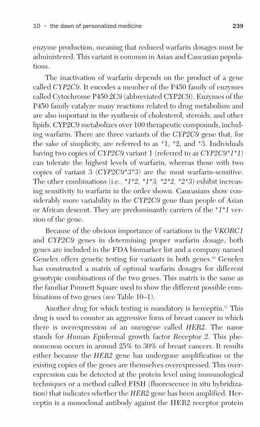
ptg
10 • the dawn of personalized medicine 239
enzyme production, meaning that reduced warfarin dosages must beadministered. This variant is common in Asian and Caucasian popula-tions.
The inactivation of warfarin depends on the product of a genecalled CYP2C9. It encodes a member of the P450 family of enzymescalled Cytochrome P450 2C9 (abbreviated CYP2C9). Enzymes of theP450 family catalyze many reactions related to drug metabolism andare also important in the synthesis of cholesterol, steroids, and otherlipids. CYP2C9 metabolizes over 100 therapeutic compounds, includ-ing warfarin. There are three variants of the CYP2C9 gene that, forthe sake of simplicity, are referred to as *1, *2, and *3. Individualshaving two copies of CYP2C9 variant 1 (referred to as CYP2C9*1*1)can tolerate the highest levels of warfarin, whereas those with twocopies of variant 3 (CYP2C9*3*3) are the most warfarin-sensitive.The other combinations (i.e., *1*2, *1*3, *2*2, *2*3) exhibit increas-ing sensitivity to warfarin in the order shown. Caucasians show con-siderably more variability in the CYP2C9 gene than people of Asianor African descent. They are predominantly carriers of the *1*1 ver-sion of the gene.
Because of the obvious importance of variations in the VKORC1and CYP2C9 genes in determining proper warfarin dosage, bothgenes are included in the FDA biomarker list and a company namedGenelex offers genetic testing for variants in both genes.10 Genelexhas constructed a matrix of optimal warfarin dosages for differentgenotypic combinations of the two genes. This matrix is the same asthe familiar Punnett Square used to show the different possible com-binations of two genes (see Table 10–1).
Another drug for which testing is mandatory is herceptin.11 Thisdrug is used to counter an aggressive form of breast cancer in whichthere is overexpression of an oncogene called HER2. The namestands for Human Epidermal growth factor Receptor 2. This phe-nomenon occurs in around 25% to 30% of breast cancers. It resultseither because the HER2 gene has undergone amplification or theexisting copies of the genes are themselves overexpressed. This over-expression can be detected at the protein level using immunologicaltechniques or a method called FISH (fluorescence in situ hybridiza-tion) that indicates whether the HER2 gene has been amplified. Her-ceptin is a monoclonal antibody against the HER2 receptor protein
Wow! eBook <WoweBook.Com>
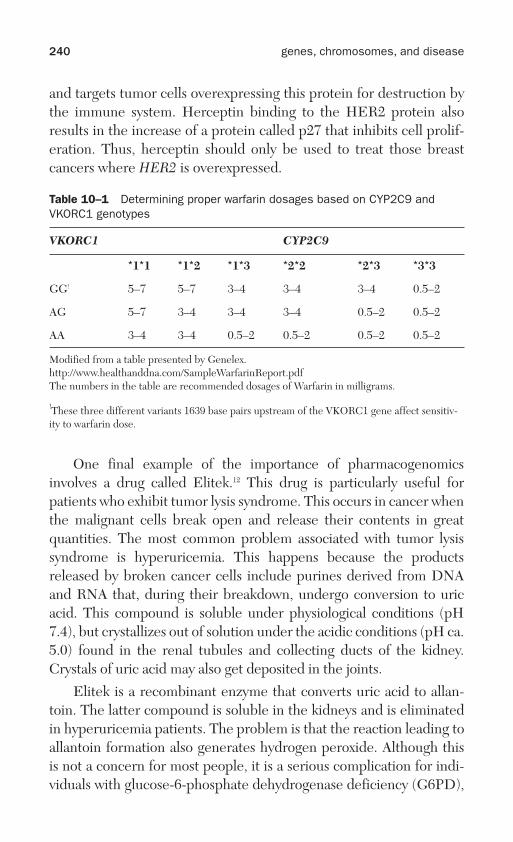
ptg
240 genes, chromosomes, and disease
and targets tumor cells overexpressing this protein for destruction bythe immune system. Herceptin binding to the HER2 protein alsoresults in the increase of a protein called p27 that inhibits cell prolif-eration. Thus, herceptin should only be used to treat those breastcancers where HER2 is overexpressed.
Table 10–1 Determining proper warfarin dosages based on CYP2C9 andVKORC1 genotypes
VKORC1 CYP2C9
*1*1 *1*2 *1*3 *2*2 *2*3 *3*3
GG! 5–7 5–7 3–4 3–4 3–4 0.5–2
AG 5–7 3–4 3–4 3–4 0.5–2 0.5–2
AA 3–4 3–4 0.5–2 0.5–2 0.5–2 0.5–2
Modified from a table presented by Genelex.http://www.healthanddna.com/SampleWarfarinReport.pdfThe numbers in the table are recommended dosages of Warfarin in milligrams.
!These three different variants 1639 base pairs upstream of the VKORC1 gene affect sensitiv-ity to warfarin dose.
One final example of the importance of pharmacogenomicsinvolves a drug called Elitek.12 This drug is particularly useful forpatients who exhibit tumor lysis syndrome. This occurs in cancer whenthe malignant cells break open and release their contents in greatquantities. The most common problem associated with tumor lysissyndrome is hyperuricemia. This happens because the productsreleased by broken cancer cells include purines derived from DNAand RNA that, during their breakdown, undergo conversion to uricacid. This compound is soluble under physiological conditions (pH7.4), but crystallizes out of solution under the acidic conditions (pH ca.5.0) found in the renal tubules and collecting ducts of the kidney.Crystals of uric acid may also get deposited in the joints.
Elitek is a recombinant enzyme that converts uric acid to allan-toin. The latter compound is soluble in the kidneys and is eliminatedin hyperuricemia patients. The problem is that the reaction leading toallantoin formation also generates hydrogen peroxide. Although thisis not a concern for most people, it is a serious complication for indi-viduals with glucose-6-phosphate dehydrogenase deficiency (G6PD),
Wow! eBook <WoweBook.Com>
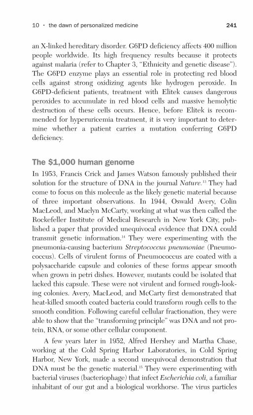
ptg
10 • the dawn of personalized medicine 241
an X-linked hereditary disorder. G6PD deficiency affects 400 millionpeople worldwide. Its high frequency results because it protectsagainst malaria (refer to Chapter 3, “Ethnicity and genetic disease”).The G6PD enzyme plays an essential role in protecting red bloodcells against strong oxidizing agents like hydrogen peroxide. InG6PD-deficient patients, treatment with Elitek causes dangerousperoxides to accumulate in red blood cells and massive hemolyticdestruction of these cells occurs. Hence, before Elitek is recom-mended for hyperuricemia treatment, it is very important to deter-mine whether a patient carries a mutation conferring G6PDdeficiency.
The $1,000 human genomeIn 1953, Francis Crick and James Watson famously published theirsolution for the structure of DNA in the journal Nature.13 They hadcome to focus on this molecule as the likely genetic material becauseof three important observations. In 1944, Oswald Avery, ColinMacLeod, and Maclyn McCarty, working at what was then called theRockefeller Institute of Medical Research in New York City, pub-lished a paper that provided unequivocal evidence that DNA couldtransmit genetic information.14 They were experimenting with thepneumonia-causing bacterium Streptococcus pneumoniae (Pneumo-coccus). Cells of virulent forms of Pneumococcus are coated with apolysaccharide capsule and colonies of these forms appear smoothwhen grown in petri dishes. However, mutants could be isolated thatlacked this capsule. These were not virulent and formed rough-look-ing colonies. Avery, MacLeod, and McCarty first demonstrated thatheat-killed smooth coated bacteria could transform rough cells to thesmooth condition. Following careful cellular fractionation, they wereable to show that the “transforming principle” was DNA and not pro-tein, RNA, or some other cellular component.
A few years later in 1952, Alfred Hershey and Martha Chase,working at the Cold Spring Harbor Laboratories, in Cold SpringHarbor, New York, made a second unequivocal demonstration thatDNA must be the genetic material.15 They were experimenting withbacterial viruses (bacteriophage) that infect Escherichia coli, a familiarinhabitant of our gut and a biological workhorse. The virus particles
Wow! eBook <WoweBook.Com>
ptg
242 genes, chromosomes, and disease
contained DNA wrapped up in a protein coat. Using radioactive label-ing, Hershey and Chase demonstrated that only the bacteriophageDNA entered infected cells where it replicated and produced newprogeny viruses. And also, in the early 1950s, a biochemist namedErwin Chargaff at Columbia University demonstrated that the fourbases in DNA (A, C, G, and T) were present in very specific ratios suchthat A=T and G=C.16
So Watson and Crick knew that DNA must be the genetic mate-rial and their model had to account for the equivalence of A with Tand G with C. There were other crucial pieces of information. Wat-son and Crick were working in the Cavendish Laboratories at theUniversity of Cambridge, but they were also in close contact withMaurice Wilkins at King’s College in London. Rosalind Franklin, acolleague of Wilkins working with Raymond Gosling, had producedX-ray diffraction images of DNA. These not only provided evidencefor the probable helical structure of DNA, but also that the phos-phate backbones of the two helices were on the outside of the mole-cule, meaning that the paired bases must be on the inside. It wasFranklin who told Watson and Crick that the backbones had to be onthe outside.
In their paper on the structure of DNA, Watson and Crick usedsome of Franklin’s unpublished data. Consequently, many feel thatshe should have been an author on that paper. In fact, she, herself,was close to solving the helix. In any event, she died of cancer in 1958,four years before Watson, Crick, and Wilkins were awarded theNobel Prize in Physiology or Medicine.
Publication of the structure of DNA in 1953 initiated an enor-mously productive era in molecular biology. Soon, the mechanism bywhich information encoded in DNA was converted to the amino acidsequence of a protein was being unraveled while the genetic codeitself was being worked out. Gene regulation became an area ofintense interest and by the early 1970s Walter Gilbert and AllanMaxam at Harvard University and Frederick Sanger at the Universityof Cambridge had developed methods for sequencing DNA.17 In sub-sequent years, Sanger’s method has prevailed.
By 1988, gene sequencing had progressed to the point that theNational Institutes of Health approved the human genome-sequencing
Wow! eBook <WoweBook.Com>

ptg
10 • the dawn of personalized medicine 243
project under the direction of James Watson. In 1991, PresidentGeorge H. W. Bush appointed Dr. Bernadine Healy as the Director ofNIH. Healy was in favor of patenting genes despite the objections ofWatson, so she approved patents on 347 human genes believing thatthis would promote rather than hinder access to information aboutthem. Watson disagreed and resigned in 1992 to return to Cold SpringHarbor Laboratories as president while Francis Collins took over ashead of the Human Genome Project.
A working draft sequence of the human genome was publishedby the genome consortium in 2000 and a complete sequence in 2003,just 50 years after the publication of the original paper describing thestructure of DNA by Watson and Crick. Craig Venter was in hot pur-suit (refer to Chapter 2). The public project had cost an estimated$2.7 billion, taken 13 years, and involved over 2,800 scientists at insti-tutions in six different countries (see Table 10–2).18 Furthermore, theconsortium sequence did not represent a single individual, but was acomposite sequence that made use of DNA from volunteers whorepresented diverse populations.
Table 10–2 The first three human genomes*
Genome sequenced HGP (2003) Venter (2007) Watson (2008)
Time taken 13 years 4 years 2 months
Cost $2.7 billion $100 million <$1.5 million
Institutes involved 16 5 2
Countries involved 6 3 1
In 2007, Venter’s faster method produced the sequence of hisown genome at a cost of around $100 million (see Table 10–2). Itemployed just 31 scientists in three countries. In 2008, a Branford,Connecticut, company called 454 Life Sciences, acquired by Roche,produced the complete sequence of Jim Watson’s genome for lessthan $1.5 million in a couple of months with a team of 27 scientists inone country, the United States. At the inaugural Consumer Genetics
*Adapted from Wadman, Meredith. “James Watson’s Genome Sequenced at High Speed.”Nature 452 (2008): 788. Note that the Human Genome Project (HGP) sequence is a compos-ite from different individuals.
Wow! eBook <WoweBook.Com>

ptg
244 genes, chromosomes, and disease
Show in Boston in June 2009, Jay Flatley, the CEO of SanDiego–based Illumina, announced the launch of a personal genome-sequencing service priced at $48,000.19 A year later, Illumina’s pricehad dropped to under $20,000. Flatley disclosed that the companyhad so far sequenced the genomes of at least 14 different individuals,including Flatley himself, Henry “Skip” Gates, the Harvard professor,and his father as well as Glenn Close.
Illumina was in close competition with Applied Biosystems andthe price kept dropping. As 2010 wore on, Illumina claimed it couldsequence a genome for $10,000 and Applied Biosystems counteredwith a price of $6,000 that then dropped to $3,000.20
Another company competing in the cheap genome-sequencingcontest is Complete Genomics.21 The company reported sequencingthree human genomes for an average cost of $4,400. It’s importantto note that these cost estimates are somewhat deceptive. They areusually based on the reported costs of consumable supplies used insequencing. They do not include the initial investment in sequenc-ing equipment and labor. For example, on the Science paper thatresulted from Complete Genomics sequencing of three humangenomes at an average cost of $4,400, there were 65 authors.22 Thissuggests that labor expenses must have been considerable. How-ever, this is of no concern to the consumer as long as the companycan afford it. The bottom line is that at $1,000 per genome, a lot ofpeople are likely to have their genomes sequenced.
The technique used by 454 Life Sciences, Illumina, and othercompanies is called “massively parallel sequencing.”23 Different com-panies use somewhat distinct technical approaches, but they all involvereading relatively short DNA sequences, the use of specific algorithmsto align the sequences, and powerful computers. Faster and bettersequencing machines continue to emerge. At the Advances in GenomeBiology and Technology meeting held on Marco Island in Florida inFebruary 2010, Hugh Martin, CEO of Pacific Biosciences, describedwhat he calls the first third-generation sequencing machine.24 It’s big,weighs 1,900 pounds, and costs $695,000, but with machines like this,the speed and cost of genomic sequencing will continue to drop.
The X Prize Foundation will award the Archon X Genomics XPrize for the first team to successfully sequence 100 human genomes in
Wow! eBook <WoweBook.Com>

ptg
10 • the dawn of personalized medicine 245
10 days.25 A plausible contender for this prize is George Church of Har-vard Medical School. Church, an established leader in the develop-ment of DNA sequencing technology, started up a not-for-profit calledPersonal Genome Project in 2006.26 He laid out his goal in a ScientificAmerican article titled “Genomes for All.” He plus nine volunteerswould have their genomes sequenced following which all of that infor-mation would be made public. Church’s eventual aim would be tosequence the genomes of 100,000 individuals. This would contributegreatly in our understanding of human genetic variation and its rela-tionship to disease. Church started very cleverly by recruiting well-known people like Harvard psychologist and author Steven Pinker,prominent health-care investor Esther Dyson, and Helicos Biosciencesfounder Stanley Lapidus as guinea pigs. Church’s Personal GenomeProject has already enrolled 16,000 volunteers.
Measuring genetic riskIn November 2007, deCODE genetics became the first company tooffer a direct-to-consumer (DTC) genetic-testing service.27 The com-pany named it deCODEme. Its purpose was to identify genetic riskfactors for certain diseases, including type 2 diabetes and certain can-cers. Jeffrey Gulcher, the cofounder of deCODE, submitted a sampleof his DNA for analysis. Because he was already getting bald, Gulcherwas not surprised that analysis of his genome suggested a propensitytoward baldness. Neither was he concerned that he had average risksfor a dozen other medical conditions. However, he had twice theaverage risk of becoming a type 2 diabetic and a one in three chanceof developing prostate cancer. Although Gulcher could counter thepotential of contracting diabetes with diet and exercise, it was notclear what he should do about his increased chances of suffering fromprostate cancer.
Although Gulcher’s prostate specific antigen (PSA) tested only a lit-tle above normal, his doctor suggested that he consult with Ian Cat-alona, a highly regarded urologist and prostate cancer specialist, atNorthwestern University. Catalona carried out an ultrasound biopsy onGulcher’s prostate that indicated he had a Gleason grade of 6. Gleasongrades run from 1 to 5 with 1 being almost normal and 5 being veryabnormal. Because two potentially cancerous areas of the prostate are
Wow! eBook <WoweBook.Com>
ptg
246 genes, chromosomes, and disease
scored to obtain the Gleason score, a very moderate cancer would be a2(1+1), whereas a very serious cancer would be a 10(5+5), so a score of6(3+3) meant Gulcher had a mildly aggressive form of prostate cancer.Nevertheless, Catalona removed Gulcher’s tumor. It had been detectedearly and there were no signs of metastasis. So, the deCODE test mayhave saved Gulcher’s life; Kari Steffanson, deCODE’s then CEO, cer-tainly thought so.
deCODE is just one of three major DNA testing companies. Theother two are Navigenics, founded by David Agus and DietrichStephan, and 23andme, whose founders are Linda Avey and AnnWojcicki, the wife of Google cofounder Sergei Brin. All three compa-nies offer online services that allow customers to order tests for a vari-ety of different genetic traits and conditions with a credit card.Navigenics also offers genetic counseling to its customers to helpthem interpret the results they receive.
A major concern is how the public will make use of such informa-tion in the absence of the opinion of a genetic specialist such as a coun-selor or doctor. For example, 23andme offers tests for several geneticvariants that increase the risk of breast cancer somewhat. However,the company does not test for BRCA1 and BRCA2 mutations,although they greatly increase the risk of breast cancer, because Myr-iad Genetics has the patents for these genes (refer to Chapter 5,“Genes and cancer”). 23andme does point out that if a woman has afamilial risk of breast cancer, she should probably consult a doctor“who may suggest getting a clinical genetic test. The effects of rare butserious mutations in genes such as BRCA1 and BRCA2 far outweighthose of the SNPs reported here.”28
A different problem concerns the weighting of genetic risk. Thisis going to change for a given disease as new molecular markers arediscovered. Eventually it seems likely that this problem will be obvi-ated when genetic-testing companies move to whole genomesequencing, as opposed to providing tests for very small sections ofthe human genome. A company called Knome, cofounded by GeorgeChurch in April 2009, points to the direction that testing will proba-bly take in the future.29 The company “provides whole genomesequencing services and interpretation solutions to biomedicalresearchers and physician-directed families seeking to understandthe genetic underpinnings of human disease.” Ultimately, it can only
Wow! eBook <WoweBook.Com>
ptg
10 • the dawn of personalized medicine 247
be through whole genome sequencing that total genetic risk can beestimated.
So how do you estimate genetic risk anyway? There are two com-ponents: relative risk and absolute risk.30 The average for relative riskis 1.0. So, if you have a risk of 0.5, that means your relative risk is halfthe normal average, but if it is 1.5, your risk of having the disease orcondition is 50% greater than normal. That might seem like a lot, butnow let’s consider absolute risk. For type 2 diabetes, the absolute riskof contracting the disease before age 60 is 23%. This means that, ifyour own relative risk is 1.5, the chance that you will become a type 2diabetic by age 60 is 1.5 x 23 or 34.5%. Conversely, if your relative riskis 0.5, your risk drops to 0.5 x 23 or 11.5%. Obviously, there is a largeenvironmental component involved with this disease as well thatneeds to be factored into consideration.
At the moment, state regulation of DTC genetic testing is highlyvariable. As of 2007, 25 states and the District of Columbia allowedunrestricted DTC testing, 13 states prohibited the practice, and 12permitted testing for specified categories of conditions, but thesetended to exclude genetic tests.31 Both the American College of Med-ical Genetics and the American Society of Human Genetics haveissued cautionary statements about DTC testing.32 The recommenda-tions of the American College of Medical Genetics stress that, “aknowledgeable professional should be involved in the process ofordering and interpreting a genetic test;” that “the consumer shouldbe fully informed regarding what the test can and cannot say abouthis or her health;” “that the scientific evidence on which a test isbased should be clearly stated;” and that “the clinical testing labora-tory must be accredited by the CLIA, the State and/or other applica-ble accrediting agencies.”
The CLIA refers to the Clinical Laboratory ImprovementAmendments. These are federal regulations governing proficiencytesting of clinical laboratories and the like. By 2011, the NIH expectsto have a Genetic Testing Registry in place that will “contain informa-tion voluntarily submitted by genetic test providers.”33 At present, it isunclear whether this registry anticipates future federal regulation ofDTC testing.
Wow! eBook <WoweBook.Com>

ptg
248 genes, chromosomes, and disease
An obvious concern for people contemplating genetic testing,DTC or otherwise, is whether they will be covered by their healthinsurance or discriminated against in the workplace if a deleterioustrait is discovered. The answer is probably not. On May 21, 2008,President George W. Bush signed the Genetic Information Nondis-crimination Act (GINA) into law.34 The act is designed to prohibit dis-crimination against individuals by insurance companies andemployers based on their genetic information. The purpose of thislaw is to allow people “to take full advantage of the promise of per-sonalized medicine without fear of discrimination.” There are alreadymany state statutes in place that are designed to limit genetic discrim-ination, but they are highly variable. Some are more protective thanGINA and some less so. At a minimum, all state provisions must nowcomply with GINA.
So from both the scientific and legal point of view, United Statescitizens will be increasingly able to access their genetic informationwithout fear of discrimination. Most people will be at a loss when try-ing to weigh the significance of what they learn, partly because theyface information overload and partly because they won’t be able tounderstand the meaning of various risk factors or susceptibilities.Some will find they are faced with an uncertain future. Perhaps theyare slated to suffer from Parkinson’s disease or early-onsetAlzheimer’s. Hence, it is of paramount importance that people whogain information about what is in their genomes also be counseled bygenetic specialists, either counselors or doctors, on what it all means.
Wow! eBook <WoweBook.Com>

ptg
Postscript: a cautionary note
In the year 1902, Archibald Garrod made the first connectionbetween a gene and a disease, alkaptoneuria. Today, over 6,000 singlegene disorders are known and many genetic risk factors for complexdiseases like type 2 diabetes and coronary disease and behavioral con-ditions like bipolar disease and schizophrenia have been identified.Direct-to-consumer gene-testing companies already provide infor-mation to potential disease gene carriers while also offering to assessgenetic risk that a person will be susceptible to a complex conditionlike coronary disease. Even more personal genetic information willsoon be on the way as genomic sequencing becomes so cheap thatmany people will decide to have their sequences read. Francis Collinseven predicts that newborn screening for genetic diseases will expandto genomic sequencing of newborns. What this means is that a delugeof personal genetic information is soon going to be spilling out ofcomputers. This raises two important questions. First, what safe-guards will there be to make sure this genetic information is kept con-fidential? Second, how will the average person make sense of thesequence of his or her genome?
Safeguarding of genetic information is going to be of paramountimportance particularly if genomic sequencing becomes routine fornewborns. We are already familiar with the problems that arise whenan insurer or a bank loses a laptop with a raft of personal information.What measures will be taken to ensure this does not happen?
Where genomic sequencing is likely to be particularly important isin the identification of unsuspecting carriers of rare genetic diseaseswho intend to have children, but for whom there is no familial recordof the disease. Suppose that genomic sequencing reveals that a couple
249

ptg
250 genes, chromosomes, and disease
intending to have children are both carriers of Type 1 Gaucher disease.Without sequencing, they could have gone ahead and had a baby onlyto discover later on that their child has Type 1 Gaucher disease. Cur-rently, treating the disease would cost them around $200,000 per yearfor enzyme therapy with no end in sight. But if genomic sequencingreveals they both carry the Gaucher mutation, they have a couple ofother options open to them. The couple may choose to avoid having aGaucher baby by making use of in vitro fertilization coupled withpreimplantation genetic diagnosis. This might cost on average$15,000–$20,000 for a single cycle of in vitro fertilization, includingpreimplantation genetic diagnosis. This is a lot less costly than enzymetherapy, but success is not guaranteed. In fact, for women under theage of 35, the success rate of in vitro fertilization is 46% and drops withincreasing maternal age. Hence, more than one cycle may be requiredfor pregnancy to occur. Then, there is the option of taking yourchances, checking the fetus for Type 1 Gaucher disease using chori-onic villus sampling or amniocentesis and aborting the fetus if it hasType 1 Gaucher disease. This might cost $1,500, including the physi-cian’s fee. A first trimester abortion might cost between $350 and$550. So this is probably the least expensive option, but the couple willhave to do it all over again if they abort a Gaucher fetus with no guar-antee that the next pregnancy will not yield another fetus withGaucher. Whether either of these avenues is open to the couple willdepend upon how they regard abortion and embryo creation anddestruction.
Genomic analysis will undoubtedly turn up all sorts of geneticrisk factors and people will need expert advice, either from a geneticcounselor or a physician trained in genetics, in order to make sense ofthis information, some of which they might prefer not to know. It iseasy to envision a time not too far away when a large expansion in thenumber of genetic counselors is going to be needed, especially onesthat are familiar with the relationship between genetic risk factorsand complex diseases or conditions.
We are all aware of the potential that embryonic and now adultstem cells may have for alleviating specific diseases. For a person witha debilitating genetic disease, the idea of being treated with stem cells
Wow! eBook <WoweBook.Com>

ptg
postscript • a cautionary note 251
from normal embryos produced by couples with no sign of the dis-ease may be very appealing. These cells are pluripotent, meaningthey can differentiate into any cell type in the body. Imagine, forinstance, that a boy with Duchenne/Becker muscular dystrophy hasthe opportunity of being treated with embryonic cells that have thepotential of integrating into his withering muscles to provide normalmuscle function. It’s a captivating notion. But wait a minute. We don’tknow if immune system rejection will be a problem. We don’t yethave the clinical trial data that will inform the medical establishmentabout whether such a treatment is going to work. We have the hypejust as we did 20 years ago with gene therapy. Let’s just hope the outcome this time is more promising and does not deliver so manydisappointments.
Suppose immune rejection is a problem. There are always adultstem cells from the patient that can be used. This should obviate therejection problem, but at the same time a new problem arises. Thesemutant cells must be treated with copies of the normal gene. In otherwords, they must undergo gene therapy just like the stem cells of thehematopoetic system in the case of severe combined immune defi-ciency disease. The experience with this type of gene therapy ispromising so we can only hope that it will work with adult stem cells.Once again, only time will tell what the outcome will be.
In the next few years, there is bound to be lots of news on thecancer front too. Excitement will wax and then wane concerning thepromise of some new drugs, but others will prove their worth. Itseems very likely that targeted therapies for a much wider range ofcancers are going to emerge within the next decade, but it also seemsequally unlikely that chemotherapy, radiation, and surgery will besupplanted anytime soon for most cancers.
So the reader is urged to be cautious when reading of excitingnew breakthroughs in genetic medicine. They will certainly come,but there will be disappointments too. Although we all hope for agreen light, it is best to pay attention when the amber light is blinking.
Wow! eBook <WoweBook.Com>

ptg
This page intentionally left blank
Wow! eBook <WoweBook.Com>
ptg
References and notes
Chapter 1: Hunting for disease genes1 Wikipedia. “Prince Leopold, Duke of Albany.” http://en.wikipedia.org/wiki/
Prince_Leopold,_Duke_of_Albany. “Death of Prince Leopold.” New York Times,March 29, 1884. McGonagall, William Topaz. Poemhunter.com. “The Death ofPrince Leopold.” http://www.poemhunter.com/poem/the-death-of-prince-leopold/#at.
2 “The Death of Prince Leopold.” Medical Record 25 (1884): 444.3 Wilson, A. N. The Victorians. London: Arrow, 2002, p. 25.4 Sawaf, Haidi, Adonis Lorenzana, and Lawrence F. Jardine. “Hemophilia A and B.”
emedicine, http://emedicine.medscape.com/article955590-print.5 Gill, Peter et. al. “Identification of the Remains of the Romanov Family by DNA
Analysis.” Nature Genetics 6 (1994): 130–135. Ivanov, Pavel et. al. “MitochondrialDNA Sequence Heteroplasmy in Grand Duke of Russia Georgij RomanovEstablishes the Authenticity of the Remains of Tsar Nicholas II.” Nature Genetics12 (1996): 417–420.
6 Rogaev, Evgeny et. al. “Genomic Identification in the Historical Case of theNicholas II Royal Family.” Proceedings of the National Academy of Sciences 106(2009): 5258–5263.
7 Rogaev, Evgeny et. al. “Genotype Analysis Identifies the Cause of the RoyalDisease.” Science 326 (2009): 817.
8 Lanska, Douglas J. “George Huntington (1850–1916) and Hereditary Chorea.”Journal of the History of the Neurosciences 9 (2000): 76–89.
9 Huntington, George. “On Chorea.” The Medical and Surgical Reporter 26 (1872):317–321.
253

ptg
254 genes, chromosomes, and disease
10 Davenport, C. B. “Huntington’s Chorea in Relation to Heredity and Eugenics.”Proceedings of the National Academy of Sciences of the United States 1 (1915):283–285.
11 Bischoff, Dan. “Of Jersey Ruins and Woody Guthrie.” The Newark Star Ledger,January 17, 2003.
12 For an excellent, if older, account of the search for the Huntington’s gene, seeChapters 1 and 7 in Bishop, Jerry E., and Michael Waldholz. Genome: The Story ofOur Astonishing Attempt to Map All the Genes in the Human Body. New York:Simon & Schuster, 1991.
13 Okun, Michael S., and Nia Thommi. “Americo Negrette (1924–2003): DiagnosingHuntington Disease in Venezuela.” Neurology 63 (2004): 340–343.
14 The U.S.-Venezuela Collaborative Research Project and Nancy S. Wexler.“Venezuelan Kindreds Reveal That Genetic and Environmental Factors ModulateHuntington’s Disease Age of Onset.” Proceedings of the National Academy ofSciences 101 (2004): 3498-3503.
15 Gusella, James F., and Nancy S. Wexler et. al. “A Polymorphic DNA MarkerGenetically Linked to Huntington’s Disease.” Nature 306 (1983): 234–238.
16 The Huntington’s Disease Collaborative Research Group. “A Novel GeneContaining a Trinucleotide Repeat That Is Expanded and Unstable onHuntington’s Disease Chromosomes.” Cell 72 (1993): 971–983.
17 Myriad Genetics. http://www.myriad.com.18 Myriad Genetics, Inc. 2009 Annual Report. http://investor.myriad.com/annuals.
cfm.19 McLellan, Tracy, Lynn B. Jorde, and Mark H. Skolnick. “Genetic Distances
Between the Utah Mormons and Related Populations.” American Journal ofHuman Genetics 36 (1984): 836–857.
20 Lewis, Ricki. “Founder Populations Fuel Gene Discovery.” The Scientist 15 (2001):8.
21 Singer, Emily. “Gene Hunting in Canada.” Technology Review (April 10, 2006).http://www.technologyreview.com/printer_friendly_article.sspx?id=16680&channel=biomedicine§ion=.
22 Genizon BioSciences. http://ww.genizon.com/.23 Siegel, Judy. “Dawning of the Age of Genetic Healing.” The Internet Jerusalem
Post, October 21, 2001. http://cgis.jpost.com/cgi-bin/General/printarticle.cgi?article=/Editions/2001/10/21/Health/Health.36631.html.
24 Shifman, Sagiv et al. “A Highly Significant Association Between a COMTHaplotype and Schizophrenia.” American Journal of Human Genetics 71 (2002):1296–1302.
25 Fishman, Racheller H. B. “Lack of Funding, Failed Contract Forcing IDgene toClose Doors.” BioWorld International, March 17, 2004. http://www.accessmylibrary.com/article/print/iGi-114423393.
Wow! eBook <WoweBook.Com>
ptg
references and notes 255
26 History of Iceland. http://www.iceland.freewebspace.com/ Timeline: Iceland, BBCNews. http://newsvote.bbc.co.uk/mpapps/pagetools/print/news.bbc.co.uk/1/hi/world/europe/countly_profiles/1025288.stm.
27 Unless otherwise noted, the primary source for much of the discussion in this sec-tion concerning Iceland’s Health Sector Database and the Health Sector DatabaseAct is the excellent article by David E. Winickoff. “Genome and Nation: Iceland’sHealth Sector Database and Its Legacy.” Innovations: Technology, Governance,Globalization 1 (2006): 80–105. There is also an entire book on the deCODE storyby Michael Fortun. Promising Genomics: Iceland and deCODE Genetics in aWorld of Speculation. Berkeley, CA: University of California Press, 2008.
28 “Random Samples.” Science 279 (1998): 991.29 Hlodan, Oksana. “For Sale: Iceland’s Genetic History.” Actionbioscience, June
2000. http://www.actionbioscience.org/genomic/hlodan.html.30 GENETarchive. http://www.gene.ch/genet/1999/Mar/msg00032.html.31 Jónatnsson, Hróbjartur. “Iceland’s Health Sector Database: A Significant Head
Start in the Search for the Biological Grail or an Irreversible Error?” AmericanJournal of Law and Medicine 26 (2000): 31–67.
32 Republic of Iceland. “Privacy and Human Rights 2003: Iceland.” http://www.privacyinternational.org/survey/phr2003/countries/iceland.htm.
33 Specter, Michael. “Decoding Iceland.” The New Yorker, January 18, 1999, pp.40–51.
34 Hlodan, Oksana, op. cit., 29.35 Lewontin, Richard. “Op-ed/People Are Not Commodities.” The New York Times,
January 23, 1999, A19.36 About deCODE genetics. http://www.decode.com/Company/index.php.37 deCODEme. http://www.decodeme.com/.38 deCODE genetics, Inc. “Files Voluntary Chapter 11 Petition to Facilitate Sale of
Assets.” http://www.decode.wom/News/news.php?s=32.39 Henderson, Mark. “Privacy Fears as DNA Testing Firm deCODE Genetics Goes
Bust.” TIMESONLINE, November 18, 2009. http://www.timesonline.co.uk/tol/news/science/genetics/article6920653.ece?print=yes&randnum=1258549974971.
40 Ibid.41 Ibid.42 Wade, Nicholas. “Out of Bankruptcy, Genetics Company Drops Drug Efforts.” The
New York Times, January 21, 2010, page B2.43 Kevles, Daniel J. In the Name of Eugenics. Cambridge: Harvard University Press,
1985, 1995, pp.232–233.44 “Genetic Disease Information—pronto!” Human Genome Project Information,
last modified July 21, 2008. http://www.ornl.gov/sci/techsources/Human_Genome/medicine/assist.shmtl.
Wow! eBook <WoweBook.Com>

ptg
256 genes, chromosomes, and disease
45 “The Science Behind the Human Genome Project: Basic Genetics, Genome DraftSequence, and Post-Genome Science.” Human Genome Project Information, lastmodified on March 26, 2008. http://www.ornl.gov/sci/techresources/Human_Genome/project/info.shtml.
46 Goodier, John L., and Haig H. Kazazian. “Retrotransposons Revisted: TheRestraint and Rehabilitation of Parasites.” Cell 135 (2008): 23–35.
47 Batzer, Mark A., and Prescott L. Deininger. “Alu Repeats and Human GenomicDiversity.” Nature Reviews Genetics 3 (2002): 370–379.
Alu1 stands for an enzyme from the bacterium A(rthrobacter) lu(teus) that makes acut in double-stranded DNA at the sequence AGTC/TCGA. This enzyme is calleda restriction enzyme. There are many different bacterial restriction enzymes andthey cut at different sequences in DNA, usually four to six base pairs. They are veryimportant tools in molecular biology. Like Alu, each of the first letters in the desig-nation is the first letter of the generic name and the second two are the first two let-ters of the specific name. Bacteria make these enzymes to defeat incoming virusDNA. They modify the restriction sites in their own DNA so they are not suscepti-ble to attack. Hence, these bacterial defense systems are called restriction-modifi-cation systems.
48 Goodier, John L., and Haig H. Kazazian, op. cit., 46.49 Kazazian, Haig H. et al. “Hemophilia A Resulting from de Novo Insertion of L1
Sequences Represents a Novel Mechanism for Mutation in Man.” Nature 232(1988): 164–166.
50 Balancio, Victoria P., Prescott L. Deininger, and Astrid M. Roy-Engel. “LineDancing in the Human Genome: Transposable Elements and Disease.” GenomeMedicine 1 (2009): 97.1–97.8.
Chapter 2: How genetic diseases arise1 Garrod, Archibald E. “The Incidence of Alkaptonuria: A Study in Chemical
Individuality.” Lancet 2 (1902): 1616–1620. Garrod’s important contributions tobiochemical genetics are reviewed by Alexander G. Bearn and Elizabeth Miller.“Archibald Garrod and the Development of the Concept of Inborn Errors ofMetabolism.” Bulletin of the History of Medicine 53 (1979): 315–328.
2 Erich von Tschermak, a 26 year-old graduate student in Ghent, Belgium, is oftenlisted as the third person to rediscover Mendel’s paper, but Robin Marantz Henigin The Monk in the Garden (Boston: Houghton Mifflin, 2001), points out that hispart in the rediscovery is usually dismissed today.
3 See Chapter 21 of Nicholas W. Gillham’s A Life of Sir Francis Galton: From AfricanExploration to the Birth of Eugenics (New York: Oxford University Press, 2001) fora detailed discussion of the battle between Mendel’s proponents and those ofGalton’s Ancestral Law.
4 Ibid., p. 338.5 Bearn and Miller, op. cit., 1, pp. 323–24.
Wow! eBook <WoweBook.Com>

ptg
references and notes 257
6 Ibid., p. 326–27.7 Nobelprize.org. “The Nobel Prize in Physiology or Medicine 1958: George Beadle,
Edward Tatum, Joshua Lederberg.” http://nobelprize.org/nobel_prizes/medicine/laureates/1958/beadle-lecture.html.
8 Fernández-Cañón, Jóse M. et al. “The Molecular Basis of Alkaptonuria.” NatureGenetics 14 (1996): 19–24.
9 Beltrán-Valero de Bernabé, D. et. al. “Analysis of Alkaptoneuria (AKU) Mutationsand Polymorphisms Reveals That the CCC Sequence Motif Is a Mutational HotSpot in the Homogenisate 1,2 Dioxygenase Gene (HGO).” American Journal ofHuman Genetics 64 (1999): 1316–1322.
10 Stahl, Franklin W. “The Amber Mutants of Phage T4.” Genetics 141 (1995):439–442.
11 Carlson, Bruce. “SNPS—A Shortcut to Personalized Medicine.” GEN: GeneticEngineering & Biotechnology News 28 (2008), no. 12.
12 Note that many genetic-testing companies use haplotype to refer to an individualcollection of short tandem repeats (STRs) while using haplogroup for SNPs.“Haplotype.” http://en.wikipedia.org/wiki/haplotype.
13 National Human Genome Research Institute. http://www.genome.gov/10001665.14National Institutes of Health. “International HapMap Consortium Publishes
Scientific Strategy.” NIH News Advisory, December 18, 2003. http://www.genome.gov/11509579.
15 Gibbs, R.A.. “The International HapMap Project.” Nature 426 (2003): 789–796.16 Frazer, K.A. “A Second Generation Human Haplotype Map of Over 3.1 Million
SNPs.” Nature 449 (2007): 851–862.17 McCarthy, Mark I. et al. “Genome-Wide Association Studies for Complex Traits:
Consensus, Uncertainty and Challenges.” Nature Reviews: Genetics 9 (2008):356–369.
18 Wade, Nicholas. “In the Genome Race, the Sequel Is Personal.” The New YorkTimes, September 4, 2007, page B1.
19 Levy S. et al. “The Diploid Genome Sequence of an Individual Human.” PloSBiology 5 (2007): 2113–2133; Venter, J. Craig. A Life Decoded. New York: VikingPress, 2007.
20 Wade, op. cit., 18.21 See Gillham, N.W. op. cit., 3, Chapter 13 for a fuller discussion of pangenesis.22 Muller, H. J. “Our Load of Mutations.” American Journal of Human Genetics 2
(1950): 111–176.23 See discussion of genetic load in Kevles, Daniel L. In the Name of Eugenics.
Cambridge: Harvard University Press, 1995, pp. 259–262.24 Kilbey, Brian J. “Charlotte Auerbach (1899–1994).” Genetics 141 (1995): 1–5.
Wow! eBook <WoweBook.Com>

ptg
258 genes, chromosomes, and disease
25 Ames, Bruce N., Frank D. Lee, and William E. Durston. “An Improved BacterialTest System for the Detection and Classification of Mutagens and Carcinogens.”Proceedings of the National Academy of Sciences USA 70 (1973): 782–786.
26 Percivall Pott. http://en.wikipedia.org/wiki/Percivall_Pott.27 Apple Valley Chimney Sweeps. “About Us and the History of Chimney Sweeps.”
https://applevalleychimneysweep.com/About_Us.html.28 McCann, Joyce, and Bruce N. Ames. “Detection of Carcinogens as Mutagens in the
Salmonella/Microsome Test: Assay of 300 Chemicals: Discussion.” Proceedings ofthe National Academy of Sciences USA 73 (1976): 950–954.
29 Florin, Inger et al. “Screening of Tobacco Smoke Constituents for MutagenicityUsing the Ames’ Test.” Toxicology 18 (1980): 219–232.
30 Ames, Bruce N., H. O, Kammen, and Edith Yamasaki. “Hair Dyes Are Mutagenic:Identification of a Variety of Mutagenic Ingredients.” Proceedings of the NationalAcademy of Sciences of the United States 72 (1975): 2423–2427. Takkouche, Bahi,Mahyar Etminan, and Agustin Montes-Martinez. “Personal Use of Hair Dyes andRisk of Cancer: A Meta-Analysis.” Journal of the American Medical Association 293(2005): 2516–2525. Bolt Hermann M., and Klaus Golka. “The Debate onCarcinogenicity of Permanent Hair Dyes: New Insights.” Current Reviews inToxicology 37 (2007): 521–536.
31 MacGregor, James T., Danial Casciano, and Lutz Müller. “Strategies and TestingMethods for Identifying Mutagenic Risks.” Mutation Research 455 (2000): 3–20.
32 UV Index: SunWise Program. http://nsdi.epa.gov/sunwise/uvindex.html.33 The Skin Cancer Foundation. “Understanding UVA and UVB.” http://www.
skincancer.org/Understanding-UVA-and-UVB.html.34 Ibid.35 Quinton, Paul M. “Physiological Basis of Cystic Fibrosis: A Historical Perspective.”
Physiological Reviews 79, Supplement 1 (1999): S3–S22.36 Cystic Fibrosis Research Directions: NIDDK. http://www.wrongdiagnosis.com/
artic/cystic_fibrosis_research_directions_niddk.htm.37 O’Sullivan, Brian P., and Steven Freedman. “Cystic Fibrosis.” The Lancet 373
(2009): 1891–1904.38 Assael, Baroukh M. et al. “Epiedemiology and Survival Analysis of Cystic Fibrosis
in an Area of Intense Screening Over 30 Years.” American Journal of Epidemiology66 (2002): 397–401.
39 Bobadilla, Joseph L. et al. “Cystic Fibrosis: A Worldwide Analysis of CFTRMutations—Correlation with Incidence Data and Application to Screening.”Human Mutation 19 (2002): 575–606.
40 Ferrell, Philip et al. “Discovery of the Principal Cystic Fibrosis Mutation (F508del)in Ancient DNA from Iron Age Europeans.” Nature Precedings:hdl:10110/npre.2007.1276.1: Posted 29 October 2007 http://precedings.nature.com/users/1e96a0049683c41e24fc029ad9fa9dad.
41 Quinton, op. cit., 35.
Wow! eBook <WoweBook.Com>

ptg
references and notes 259
42 Who Named It? “John Langdon Haydon Down.” http://www.whonamedit.com/doctor.cfm/335.html.
43 Down, J. Langdon H. “Observations on an Ethnic Classification of Idiots.” LondonHospital Reports 3 (1866): 259–262.
44 Wright, David. Mental Disability in Victorian England: The Earlswood Asylum1847–1901. Oxford: Oxford University Press, 2001, pp.172–174.
45 For an excellent summary of the work done by Penrose, see Chapter 10 in Kevles,Daniel. Op. cit., 23.
46 For an excellent summary of the history of human chromosome counting, seeKottler, Malcolm Jay. “From 48 to 46: Cytological Technique, Preconception, andthe Counting of Human Chromosomes.” Bulletin of the History of Medicine 48(1974): 465–502.
47 Answers.com. “Chromosomal Banding.” http://www.answers.com/topic/chromosomal-banding.
48 Wikipedia. “Jérôme Lejeune.” http://en.wikipedia.org/wiki/ Jérôme_Lejeune.49 “What Is Down syndrome?” http://www.downsyn.com/whatisds.php.50 Mansfield, Caroline et al. “Termination Rates After Prenatal Diagnosis of Down
Syndrome, Spina Bifida, Anencephaly, and Turner and Klinefelter Syndromes: ASystematic Literature Review.” Prenatal Diagnosis 19 (1999): 808–812.
51 Will, George F. “Eugenics By Abortion.” Washington Post, April 14, 2005, pageA27.
52 Cuniff, Christopher et al. “Health Supervision for Children With DownSyndrome.” Pediatrics 107 (2001): 442–449.
53 Ibid.54 Quenby, Siobhan et al. “Recurrent Miscarriage: A Defect in Nature’s Quality
Control?” Human Reproduction 17 (2002): 1959–1963.55 Nicolaidis, Peter, and Michael B. Petersen. “Origin and Mechanisms of Non-
Disjunction in Human Autosomal Trisomies.” Human Reproduction 13 (1998):313–319.
56 Morton, N. E. et al. “Maternal Age in Trisomy.” Annals of Human Genetics 52(1988): 227–236.
57 Lyon, Mary. “Gene Action in the X-chromosome of the Mouse (Mus musculus L.).”Nature 190 (1961): 372–373.
58 Kevles, op. cit., 23, pp. 242–245.59 Lanfranco, Fabio et al. “Klinefelter’s Syndrome.” The Lancet 364 (2004): 273–283.60 “Male Tortoiseshell Cat ‘Genetically Impossible.” Daily Telegraph, August 26,
2009.61 Wutz, Anton. “Xist Function: Bridging Chromatin and Stem Cells.” Trends in
Genetics 23 (2007): 457–464. Wutz, Anton, and Joost Gribnau. “X InactivationXplained.” Current Opinion in Genetics and Development 17 (2007): 387–393.
Wow! eBook <WoweBook.Com>

ptg
260 genes, chromosomes, and disease
62 Angelman Syndrome Foundation, Inc. “Harry Angelman and the History of AS.”http://www.angelman.org/stay-informed/facts-about-angelman-syndrome---7th-edition/harry-angelman-and-the-history-of-as/.
63 Ibid.64 Genetics Home Reference: Your Guide to Understanding Genetic Conditions.
“Angelman syndrome.” http://ghr.nlm.nih.gov/condition=angelmansyndrome.65 Genetics Home Reference: Your Guide to Understanding Genetic Conditions.
“Prader-Willi syndrome.” http://ghr.nlm.nih.gov/condition=praderwillisyndrome.66 Horsthemke, Bernhard, and Joseph Wagstaff. “Mechanisms of Imprinting of the
Prader-Willi/Angelman Regon.” American Journal of Medical Genetics Part A 146A(2008): 2041–2052. Note that the paternal chromosome 15 region deleted inPrader-Willi syndrome also contains around 70 genes encoding C/D box snoRNAs.These small RNAs guide methylation of other RNAs including ribosomal RNA andtransfer RNAs.
67 Genetics Home Reference. “OCA2.” http://ghr.nlm.nih.gov/gene=oca2.68 Cloud, John. “Why Your DNA Isn’t Your Destiny.” Time, January 6, 2010.
Chapter 3: Ethnicity and genetic disease1 Wikipedia. “J. B. S. Haldane.” http://en.wikipedia.org/wiki/J._B._S._Haldane. “The
Unofficial Stephen Jay Gould Archive: People: J.B.S. Haldane.” http://www.stephenjaygould.org/people/john_haldane.html.
2 Lederberg, Joshua. “J. B. S. Haldane (1949) on Infectious Disease and Evolution.”Genetics 153 (1999): 1–3. Also see Haldane, J. B. S. “The Rate of Mutation ofHuman Genes.” Hereditas 35 (1949): 267–273.
3 “A Brief History of Sickle Cell Disease.” http://sickle.bwh.harvard.edu/scd_history.html. Innvista. “Sickle Cell History.” http://www.innvista.com/health/ailments/anemias/sickhist.htm. The Africa Guide. “Ghana.” http://www.africaguide.com/country/ghana/. Ethnologue, Languages of the World “Languages of Ghana.”http://www.ethnologue.com/show_country.asp?name=ghana.
4 GhanaWeb. “Early European Contact and the Slave Trade.” http://www.ghanaweb.com/GhanaHomePage/history/slave-trade.php.
5 Washington University Biology Department. “Biogeography and Ecology of SickleCell Anemia.” http://www.nslc.wustl.edu/sicklecell/part3/biogeography.html.
6 Allison, A. C. “Protection Afforded by Sickle-Cell Trait Against Subtertian MalarialInfection.” British Medical Journal 1 (1954): 290–294.
7 Lederberg, op. cit., 2.8 Encyclopedia Brittanica. “James Bryan Herrick.” http://www.britannica.com/
EBchecked/topic/263733/James-Bryan-Herrick. Wikipedia. “James B. Herrick.”http://en.wikipedia.org/wiki/James_B._Herrick.
9 Williams, T. N. et al. “An Immune Basis for Malaria Protection by the Sickle CellTrait.” PloS Medicine 2 (2005): 0442–0445.
Wow! eBook <WoweBook.Com>

ptg
references and notes 261
10 Greenwood, Brian M. et al. “Malaria.” The Lancet 365 (2005): 1487–1498.11 There is a nice account of the unraveling of the molecular basis of sickle cell anemia
in Chapter 15 of Daniel Kevles’s book In the Name of Eugenics. Cambridge:Harvard University Press, 1995.
12 There are all sorts of sites dealing with sickle cell disease and its symptoms on theInternet. A good succinct description of this genetic disease and many others is tobe found in Genetics Home Reference. http://ghr.nlm.nih.gov/. For a far moredetailed description, see Bender, M. A., and W. Hobbs. National Center forBiotechnology Information, U.S. National Library of Medicine. Gene Reviews.“Sickle Cell Disease.” http://www.ncbi.nlm.nih.gov/bookshelf/br.fcgi?book=gene&part=sickle.
13 Platt, O. S. et al. “Mortality in Sickle Cell Disease—Life Expectancy and RiskFactors for Early Death.” New England Journal of Medicine 330 (1994):1639–1644. Quinn, Charles T., Zora R. Rogers, and George R. Buchanan. “Survivalof Children with Sickle Cell Disease.” Blood 103 (2004): 4023–4027.
14 Bender and Hobbs, op. cit., 12.15 Strouse, John J. et al. “Hyroxyurea for Sickle Cell Disease: A Systematic Review for
Efficacy and Toxicity in Children.” Pediatrics 122 (2008): 1332–1342.16 Platt, Orah S. “Hydroxyurea for the Treatment of Sickle Cell Anemia.” The New
England Journal of Medicine 358 (2008): 1362–1369.17 Ibid.18 Jones, Stephen R., Richard A. Binder, and Everett M. Donowho, Jr. “Sudden
Death in Sickle Cell Trait.” New England Journal of Medicine 282 (1970): 323–325.19 Kevles, op. cit., 11, pp. 278, 300.20 Ibid. 256, 278.21 Weiss, Robin Elise. About.com: Pregnancy & Childbirth. “Newborn Screening
Recommendations by State.” http://pregnancy.about.com/od/newbornbabies/a/newbornstate.htm.
22 National Heart, Lung, and Blood Institute, National Institutes of Health. “SickleCell Research for Treatment and Cure.” http://www.nhlbi.nih.gov/resources/docs/scd30/scd30.pdf.
23 Dodd, Dennis. CBS Sports.com. “Fatalities Continue—and So Does Ignorance ofSickle Cell Trait.” http://www.cbssports.com/collegefootball/story/11742560.
24 National Athletic Trainers’ Association. 2007. “Consensus Statement: Sickle CellTrait and the Athlete.” http://www.nata.org/statements/consensus/sicklecell.pdf.
25 Bouchette, Ed. “On the Steelers: Holmes’ Sickle Cell Not Affected in Denver.”Pittsburgh Post-Gazette, November 3, 2009.
26 NFL Fanhouse. “No Ryan Clark, but Same Steel Curtain.” http://nfl.fanhouse.com/2009/11/10/no-ryan-clark-but-same-steel-curtain/.
27 Bouchette, op. cit., 25.
Wow! eBook <WoweBook.Com>

ptg
262 genes, chromosomes, and disease
28 Alabdulaali, M. K. “Sickle Cell Disease Patients in Eastern Province of SaudiArabia Suffer Less Severe Acute Chest Syndrome Than Patients with AfricanHaplotypes.” Annals of Thoracic Medicine 2 (2007): 158–162.
29 Wikipedia. “History of Malaria.” http://en.wikipedia.org/wiki/History_of_malaria.30 Who Named It? “Thomas Benton Cooley.” http://www.whonamedit.com/doctor.
cfm/1931.html Who Named It? “Cooley’s Anaemia.” http://www.whonamedit.com/doctor.cfm/1931.html.
31 Nobelprize.org. “George H. Whipple: The Nobel Prize in Physiology or Medicine1934.” http://nobelprize.org/nobel_prizes/medicine/laureates/1934/whipple-bio.html.
32 Hill, A. V. S. et al. “β Thalassemia in Melanesia: Association With Malaria andCharacterization of a Common Variant (IVS-1 at G->C).” Blood 72 (1988): 9–14.Genetics Home Reference. “Beta Thalassemia.” http://ghr.nlm.nih.gov/condition=betathalassemia
33 Weatherall, David et al. “Inherited Disorders of Hemoglobin.” Chap. 34 in DiseaseControl Priorities in Developing Countries. 2nd ed. Oxford University Press andthe World Bank, 2006. http://files.dcp2.org/pdf/DCP/DCP34.pdf.
34 Tebbutt, Tom. “Sampras Battles Inherited Anemia.” Toronto Globe and Mail,September 24, 1996.
35 Sampras, Pete with Peter Bodo. A Champion’s Mind: Lessons from a Life in Tennis.New York: Random House, 2008, p. 181.
36 Neufeld, Ellis J. “Oral Chelators Deferasirox and Deferiprone for TransfusionalIron Overload in Thalassemia Major: New Data, New Questions.” Blood 107(2006): 3436–3441.
37 Weatherall, David, op. cit., 33, Table 34.1.38 Guterman, Lila, and Francis X. Rocca. “Choosing Eugenics: How Far Will Nations
Go to Eliminate Genetic Disease?” The Chronicle of Higher Education 49 (2003):A22–24.
39 Mockenhaupt, Frank P. et al. “α+-thalassemia Protects from Severe Malaria inAfrican Children.” Blood 104 (2004): 2003–2006. Williams, Thomas N. et al. “BothHeterozygous and Homozygous α+ thalassemias Protect Against Severe and FatalPlasmodium falciparum Malaria on the Coast of Kenya.” Blood 106 (2005):368–371.
40 Fowkes, Freya J. I. et al. “Increased Microerythrocyte Count in Homozygous α+-Thalassemia Contributes to Protection Against Severe Malarial Anaemia.” PloSMedicine 5 (2008): 0494–0501.
41 Fairhurst, R. M. et al. “Abnormal Display of PfEMP-1 on Erythrocytes CarryingHaemoglobin C May Protect Against Malaria.” Nature 435 (2005): 1117–1121.
42 Chotivanich, K. et al. “Hemoglobin E: A Balanced Polymorphism ProtectiveAgainst High Parasitemias and thus Severe P. falciparum Malaria.” Blood 100(2002): 1172–1176.
43 Kalow, Werner. “Pharmacogenetics: Its Place in Medicine and Biology.” Journal ofPharmacy Practice 5 (1992): 312–316.
Wow! eBook <WoweBook.Com>

ptg
references and notes 263
44 Genetics Home Reference. “Glucose-6-Phosphate Dehydrogenase Deficiency.”http://ghr.nlm.nih.gov/condition=glucose6phosphatedehydrogenasedeficiency/show/print Mason, Philip J., José M. Bautista, and Florinda Gilsanz. “G6PDDeficiency: The Genotype-Phenotype Association.” Blood Reviews 21 (2007):267–283.
45 WHO Working Group. “Glucose-6-Phosphate Dehydrogenase Deficiency.”Bulletin of the World Health Organization 67 (1989): 801–811.
46 Tishkoff, Sarah A. et al. “Haplotype Diversity and Linkage Disequilibrium atHuman G6PD: Recent Origin of Alleles That Confer Malarial Resistance.” Science293 (2001): 455–462.
47 Belsey, Mark A. “The Epidemiology of Favism.” Bulletin of the World HealthOrganization 48 (1973): 1–13.
48 Cornell University Department of Animal Science. “Vicine and Covicine: PlantsPoisonous to Livestock.” http://www.ansci.cornell.edu/plants/toxicagents/vicine.html.
49 Snow, Robert W. et al. “The Global Distribution of Clinical Episodes ofPlasmodium falciparum Malaria.” Nature 434 (2005): 214–217.
50 Wikipedia. “Francisco J. Ayala.” http://en.wikipedia.org/wiki/Francisco_J._Ayala.51 Rich, Stephen M. et al. “The Origin of Malignant Malaria.” Proceedings of the
National Academy of Sciences. 106 (2009): 14902–14907. The erythrocyte-bindingantigen on the surface of the plasmodium merozoite is called EBA175. EBA175recognizes sialic acids (Sias) presented on clusters of O-linked glycans attached toerythrocyte surface glycophorin A. Thus, the Sia-capped glycoprotein GYPA is theprimary binding target for EBA175. Mammalian erythrocytes commonly have twoSias N-glycolylneuraminic acid (Neu5Gc) and N-acetylneuraminic acid (Neu5Ac).Neu5Gc is synthesized from Neu5Ac. Plasmodium reichenowi EBA175 binds pref-erentially to Neu5Gc, but a mutation in the human CMAH gene blocks synthesis ofNeu5Gc from Neu5Ac protecting humans against P. reichenowi. However, P. falci-parum EBA175 does bind to Neu5Ac and so its merozoites easily infect human ery-throcytes.
52 Galvani, Alison P., and Montgomery Slatkin. “Evaluating Plague and Smallpox asHistorical Selective Pressures for the CCR5-Δ32 HIV-Resistance Allele.”Proceedings of the National Academy of Sciences 100 (2003): 15276–15279.
53 Halliday, Stephen. The Great Stink of London: Sir Joseph Bazalgette and theCleansing of the Victorian Metropolis. London: Sutton Publishing Ltd., 1999, p. ix.
54 Wilson, A. N. The Victorians. London: Arrow Books, 2003, p. 155. Note thattyphoid and waterborne typhus are the same disease.
55 Ibid. p. 243.56 Halliday, op. cit., 53, p. xii–xiii.57 Gabriel, Sherif E. et al. “Cystic Fibrosis Heterozygote Resistance to Cholera Toxin
in the Cystic Fibrosis Mouse Model.” Science 266 (1994): 107–109.58 Cuthbert, A. W. et al. “The Genetic Advantage Hypothesis in Cystic Fibrosis
Heterozygotes: A Murine Study.” Journal of Physiology 482.2 (1995): 449–464.
Wow! eBook <WoweBook.Com>

ptg
264 genes, chromosomes, and disease
59 Högenauer, Christoph. “Active Intestinal Chloride Secretion in Human Carriers ofCystic Fibrosis Mutations: An Evaluation of the Hypothesis That HeterozygotesHave Subnormal Active Intestinal Chloride Secretion.” American Journal ofHuman Genetics 67 (2000): 1422–1427.
60 Pier, Gerald B. et al. “Salmonella Typhi Uses CFTR to Enter Intestinal EpithelialCells.” Nature 393 (1998): 79–82.
61 Tsui, I. S. M. et al. “The Type IVB Pili of Salmonella Enterica Serovar Typhi Bindto the Cystic Fibrosis Transmembrane Conductance Regulator.” InfectiousImmunology 71 (2003): 6049–6050.
62 Poolman, Eric M., and Alison P. Galvani. “Evaluating Candidate Agents of SelectivePressure for Cystic Fibrosis.” Journal of the Royal Society Interface 4 (2007): 91–98.
63 Tobacman, Joanne K. “Does Deficiency of Arylsulfatase B Have a Role in CysticFibrosis?” Chest 123 (2003): 2130–2139.
64 Provine, William B. “Ernst Mayr: Genetics and Speciation.” Genetics 167 (2004):1041–46.
65 Scriver, Charles R. “Human Genetics: Lessons from Quebec Populations.” AnnualReview of Genomics and Human Genetics 2 (2001): 69–101. Unless otherwisenoted by another citation, the discussion of hereditary diseases in Quebec isderived from this reference.
66 Genetics Home Reference. “Hypercholesterolemia.” http://ghr.nlm.nih.gov/condition=hypercholesterolemia.
67 Genetics Home Reference. “Tay-Sachs Disease.” http://ghr.nlm.nih.gov/condition=taysachsdisease.
68 Scriver, op. cit., 65.69 Lacassie, Yves, and Luisa Flórez. LSU Health Center. Genetics and Louisiana
Families. “Some Genetic Disorders Among Acadian People.” http://www.med-school.lsuhsc.edu/genetics_center/louisiana/article_geneticdisorders.htm.
70 McDowell, Geraldine A. et al. “The Presence of Two Different Infantile Tay-SachsDisease Mutations in a Cajun Population.” Journal of Human Genetics 51 (1992):1071–1077.
71 Center for Jewish Genetic Diseases, Mount Sinai Hospital. http://www.mssm.edu/research/programs/jewish-genetics-disease-center.
72 Chicago Center for Jewish Genetic Disorders. http://www.jewishgenetics.org.73 Jewish Genetic Diseases Center of Greater Phoenix. http://www.jewishgeneticsphx.
org.74 Klein, Aaron. “Genetic Screening Causes Controversy.” Yeshiva University
Commentator 62, issue 10. http://commie.droryikra.com/archives/v62iA/news/doryesharim.html. “Dor Yeshorim.” The Jewish Press, November 25, 2009. http://www.jewishpress.com/pageroute.do/41623.
75 The Jewish Federations of North America. Jewish Life. “NJPS 2000-01 Report.”http://www.jewishfederations.org/page.aspx?id=45894.
Wow! eBook <WoweBook.Com>
ptg
references and notes 265
76 Shimo-Barry, Alex. “Modern Matchmaker in Race and Ethnicity in the New UrbanAmerica.” http://web.jrn.columbia.edu/studentwork/race/2002/gene-shimo.shtml.
77 Rosen, Christine. “Eugenics—Sacred and Profane.” The New Atlantis, Summer2003, 79–89.
78 Kaplan, Feige. “Tay-Sachs Disease Carrier Screening: A Model for Prevention ofGenetic Disease.” Genetic Testing 2 (1998): 271–292.
79 Oster, Harry. “A Genetic Profile of Contemporary Jewish Populations.” NatureReviews Genetics 2 (2001): 891–898.
80 Rotter, Jerome J., and Jared M. Diamond. “What Maintains the Frequencies ofHuman Genetic Diseases?” Nature 329 (1987): 289–290.
81 Spyropoulos B. et al. “Heterozygous Advantage in Tay-Sachs Carriers? AmericanJournal of Human Genetics 33 (1981): 375–380.
82 Diamond, Jared. “Jewish Lysosomes.” Nature 368 (1994): 291–292.83 Cochran, Gregory, Jason Hardy, and Henry Harpending. “Natural History of
Ashkenazi Intelligence.” Journal of Biosocial Science 38 (2006): 659–693.84 Wade, Nicholas. “Researchers Say intelligence and Diseases May Be Linked in
Ashkenazic Genes.” The New York Times, June 3, 2005, A21.85 Risch, Neil et al. “Geographic Distribution of Disease Mutations in the Ashkenazi
Jewish Population Supports Genetic Drift over Selection.” American Journal ofHuman Genetics 72 (2003): 812–822.
86 Senior, Jennifer. “Are Jews Smarter?” New York Magazine. October 16, 2005. http://nymag.com/nymetro/news/culture/features/1478/.
87 Slatkin, Montgomery. “A Population-Genetic Test of Founder Effects andImplications for Ashkenazi Jewish Diseases.” American Journal of HumanGenetics. 75 (2004): 282–293.
88 Popper, Nathaniel. “Study’s Claim On Intelligence of Ashkenazim Spurs a Debate.”Jewish Daily Forward, June 10, 2005. http://www.forward.com/articles/3623/.
Chapter 4: Susceptibility genes and risk factors1 Angier, Natalie. “Scientists Detect a Genetic Key to Alzheimer’s.” The New York
Times, August 13, 1993. For a review of the early findings, see Strittmatter, WarrenJ., and Allen D. Roses. “Apolipoprotein E and Alzheimer’s Disease.” AnnualReview of Genetics 19 (1996): 53–77. Wikipedia. “Apolipoprotein E.” http://en.wikipedia.org/wiki/Apolipoprotein_E.
2 WebMD. Mojica, Ray Camille “Tolerate Lactose Intolerance.” www.webmd.com/diet/features/tolerate-lactose-intolerance.
3 Swallow, Dallas M. “Genetics of Lactase Persistence and Lactose Intolerance.”Annual Review of Genetics 37 (2003): 197–219.
Wow! eBook <WoweBook.Com>

ptg
266 genes, chromosomes, and disease
4 These mutations do not arise in the gene encoding lactase (LCT), but are upstreamin introns of a gene called MCM6. The variant that prevails in Caucasian popula-tions is called C/T(-13910) located in intron 13 of the MCM6 gene. This base pairchange converts the sequence into an enhancer that binds the OCT1 transcriptionfactor. This probably stimulates lactase mRNA transcription. OMIM. “MIM ID*601806 Minichromosome Maintenance, S. pombe, Homolog of, 6: MCM6.”http://www.ncbi.nlm.nih.gov/entrez/dispomim.cgi?id=601806&a=601806_AllelicVariant0001.
5 Itan, Yuval et al. “The Origins of Lactase Persistence in Europe.” PloSComputational Biology 5 (2009) 8: e1000491. doi:10.1371.pcbi.1000491. http://www.ploscompbiol.org/article/info:doi%2F10.1371%2Fjournal.pcbi.1000491.
6 Tishkoff, Sarah A. et al. “Convergent Adaptation of Lactase Persistence in Africaand Europe.” Nature Genetics 39 (2007): 31–40. Note that other mutations inintrons of the MCM6 enhance lactase production in certain African pastoralist pop-ulations.
7 Wikipedia. “Nicotine.” http://en.wikipedia.org/wiki/Nicotine.8 Coon, Minor J. “Cytochrome P450: Nature’s Most Versatile Biological Catalyst.”
Annual Review of Pharmacology and Toxicology 45 (2005): 1–25. Rautio, A.“Polymorphic CYP2A6 and Its Clinical and Toxicological Significance.” ThePharmacogenomics Journal 3 (2003): 6–7.
9 Munafò, Marcus, and Elaine C. Johnstone. “Genes and Cigarette Smoking.”Addiction 103 (2008): 893–904.
10 These papers identify rs8034191 and rs1051730 as increasing the risk of lung can-cer. Amos, Christopher I. et al. “Genome-Wide Association Scan of Tag SNPsIdentifies a Susceptibility Locus for Lung Cancer at 15q25.1.” Nature Genetics 40(2008): 616–622. Hung, Rayjean J. et al. “A Susceptibility Locus for Lung CancerMaps to Nicotinic Acetylcholine Receptor Subunit Genes on 15q25.” Nature 452(2008): 633–637.
11 The following papers all document the relationship of rs16969968 to nicotineaddiction. Saccone, Scott et al. “Cholinergic Nicotinic Receptor Genes Implicatedin a Nicotine Dependence Association Study Targeting 348 Candidate Genes.”Human Molecular Genetics 16 (2007): 36–49. Weiss, Robert et al. “A CandidateGene Approach Identifies the CHRNA5-A3-B4 Region as a Risk Factor for Age-Dependent Nicotine Addiction.” PloS Genetics 4 (2008): e1000125.doi:10.1371/journal.pgen.1000125. Saccone, Nancy L. et al. “The CHRNA5-CHRNA3-CHRNB4 Nicotinic Receptor Subunit Gene Cluster Affects Risk forNicotine Dependence in African Americans and in European Americans.” CancerResearch 69 (2009): 6848–6856.
12 The following paper documents the relationship rs1051730 to nicotine addiction.Thorgeirsson, Thorgeir E. et al. “A Variant Associated with Nicotine Dependence,Lung Cancer, and Peripheral Arterial Disease.” Nature 452 (2008): 638–641.
13 Saccone, Nancy L. et al. “The CHRNA5-CHRNA3-CHRNB4 Nicotinic ReceptorSubunit Gene Cluster Affects Risk for Nicotine Dependence in African Americansand in European Americans.” Cancer Research 69 (2009): 6848–6856.
Wow! eBook <WoweBook.Com>
ptg
references and notes 267
14 Galvan, Antonella, and Tommaso A. Dragani. “Nicotine Dependence May Linkthe 15q25 Locus to Lung Cancer Risk.” Carcinogenesis Advance Access,November 12, 2009. http://carcin.oxfordjournals.org/cgi/reprint/bgp282v1.pdf.
15 O’Laughlin, J. et al. “Genetically Decreased CYP2A6 and the Risk of TobaccoDependence: A Prospective Study of Novice Smokers.” Tobacco Control 13 (2004):422–428.
16 Ariyoshi, Noritaka et al. “Genetic Polymorphism of CKYP2A6 Gene and Tobacco-Induced Lung Cancer Risk in Male Smokers.” Cancer Epidemiology, Biomarkers& Prevention 11 (2002): 890–894.
17 Munafò and Johnstone, op. cit., 9.18 Ibid.19 Gelernter, Joel et al. “Haplotype Spanning TTC12 and ANKK1, Flanked By the
DRD2 and NCAM1 Loci, Is Strongly Associated to Nicotine Dependence in TwoDistinct American Populations.” Human Molecular Genetics 15 (2006): 3498–3507.
20 Newsom, Ainsley. “Tracking the Smoking Gene.” The Times, November 19, 2005.21 Ibid.22 Ibid.23 Ibid.24 g-Nostics. http://www.g-nostics.com/.25 Ibid.26 Profita, Hillary. “And In The End, Peanut Butter Was Not To Blame.” Public Eye
CBS News, May 12, 2006. http://www.cbsnews.com/sections/publiceye/main500486.shtml?keyword=kiss+of+death.
27 Bhat, Devika. “The Kiss of Death for Girl with Nut Allergy.” The Times, November29, 2005. http://www.timesonline.co.uk/tol/news/world/article597704.ece.
28 “‘Fatal Kiss’ Puts Spotlight on Food Allergies.” ABC News, November 30, 2005.http://abcnews.go.com/WNT/Health/story?id=1360248Parke.
29 “Quebec Teen Died of Asthma, Not Nutty Kiss.” CBC News, May 11, 2006. http://www.cbc.ca/canada/story/2006/05/11/asthma-death.html.
30 National Heart Lung and Blood Institute. “Diseases and Conditions Index:Asthma.” http://www.nhlbi.nih.gov/health/dci/Diseases/Asthma/Asthma_WhatIs.html.
31 Dust Mites, Asthma and Allergy Foundation of America. http://www.aafa.org/print.cfm?id=9&sub=22&cont=315#
32 Bach, Jean-Francois. “The Effect of Infections on Susceptibility to Autoimmuneand Allergic Diseases.” The New England Journal of Medicine 347 (2002): 911–920.
33 Beasley, Richard. “The Burden of Asthma with Specific Reference to the UnitedStates.” Journal of Allergy and Clinical Immunology 109 (2002): S482–S489.
34 Ridley, Matt. Genome: The Autobiography of a Species in 23 Chapters. New York:Harper Collins, 1999, p. 67.
Wow! eBook <WoweBook.Com>

ptg
268 genes, chromosomes, and disease
35 For a simple summary, see Genentech. “IgE and Its Role in Asthma.” http://www.gene.com/gene/products/education/immunology/ige.html.
36 The discussion that follows is based largely on the following review. Bossé, Yohan,and Thomas J. Hudson. “Toward a Comprehensive Set of Asthma SusceptibilityGenes.” Annual Review of Medicine 58 (2007): 171–184.
37 McGrath, John A., and Jouni Uitto. “The Filaggrin Story: Novel Insights into Skin-Barrier Function and Disease.” Trends in Molecular Medicine 14 (2007): 20–27.
38 Bossé and Hudson, op. cit., 36 for a discussion.39 Farfel, Zvi, Henry R. Bourne, and Taroh Liri. “The Expanding Spectrum of G
Protein Diseases.” The New England Journal of Medicine 340 (1999): 1012–1020.40 Ose, Leiv. “The Real Code of Leonardo da Vinci.” Current Cardiology Reviews 4
(2008): 60–62.41 Wikipedia. “Familial Hypercholesterolemia.” http://en.wikipedia.org/wiki/
Familial_hypercholesterolemia.42 There is a nice account of their discovery in Bishop, Jerry E., and Michael
Waldholz. Genome. New York: Simon and Schuster, 1990, Chapter 9.43 Genetics Home Reference. “Hypercholesterolemia.” http://ghr.nlm.nih.gov/
condition=hypercholesterolemia.44 Genetics Home Reference. “APOB.” http://ghr.nlm.nih.gov/gene=apob/show/
print.45 Genetics Home Reference, op. cit., 43.46 Medline Plus. “Coronary Artery Disease.” http://www.nlm.nih.gov/medlineplus/
coronaryarterydisease.html.47 Hamsten, A., and P. Eriksson. “Identifying the Susceptibility Genes for Coronary
Artery Disease: From Hyperbole Through Doubt to Cautious Optimism. Journal ofInternal Medicine 263 (2008): 538–552.
48 Wikipedia. “Diabetes Mellitus.” http://en.wikipedia.org/wiki/Diabetes_mellitus.49 Grant, Struan F. A., and H. Hakonarson. “Genome-Wide Association Studies in
Type 1 Diabetes.” Current Diabetes Reports 9 (2009): 157–163.50 Roep, B. O. “Missing Links.” Nature 450 (2007): 799–800. Nejentsev, Sergey et al.
“Localization of Type 1 Diabetes Susceptibility to the MHC Class 1 Genes HLA-Band HLA-A.” Nature 450 (2007): 887–892.
51 Grant and Hakonarson, op. cit., 49.52 National Institutes of Health. Fact Sheet. “Type 2 Diabetes.” http://www.nih.gov/
about/researchresultsforthepublic/Type2Diabetes.pdf.53 Muolo, Deborah M., and Christopher B. Newgard. “Molecular and Metabolic
Mechanisms of Insulin Resistance and β-cell Failure in Type 2 Diabetes.” NatureReviews: Molecular Cell Biology 9 (2008): 193–205.
54 Staiger, Harald et al. “Pathomechanisms of Type 2 Diabetes Genes.” EndocrineReviews 30 (2009): 557–585.
55 Op. cit., 52.
Wow! eBook <WoweBook.Com>
ptg
references and notes 269
56 National Institute of Diabetes and Digestive and Kidney Diseases, NationalInstitutes of Health. “Monogenic Forms of Diabetes: Neonatal Diabetes Mellitusand Maturity-Onset Diabetes of the Young.” http://www.diabetes.niddk.nih.gov/dm/pubs/mody/#9.
57 Reich, David E., and Eric S. Lander. “On the Allelic Spectrum of Human Disease.”Trends in Genetics 17 (2001): 502–510.
58 National Institute on Aging. National Institutes of Health. “Alzheimer’s DiseaseGenetics Fact Sheet.” http://www.nia.nih.gov/NR/rdonlyres/3C4B634E-A2D8-4415-927F-4B79BEC47EA6/11207/84206ADEARFactsheetGeneticsFINAL08DEC23.pdf
59 Bodmer, Walter, and Carolina Bonilla. “Common and Rare Variants inMultifactorial Susceptibility to Common Diseases.” Nature Genetics 40 (2008):695–701.
60 Wikipedia. “Progeria.” http://en.wikipedia.org/wiki/Progeria.61 Wade, Nicholas. “A New Way to Look for Disease’ Genetic Roots.” New York
Times, January 26, 2010.62 Dickson, Samuel P. et al. “Rare Variants Create Synthetic Genome-Wide
Associations.” PloS Biology 8 (2010): 1–12.63 Ridley, Matt. “The Failed Promise of Genomics.” The Wall Street Journal. October
9–10, 2010, C4.64 Wade, op. cit., 61.65 International Classification of Diseases (ICD) WHO: History. “History of the
Development of the ICD.” http://www.who.int/classifications/icd/en/HistoryOfICD.pdf.
66 Goh, Kwang-Il et al. “The Human Disease Network.” Proceedings of the NationalAcademy of Sciences of the United States 104 (2007): 8685–8690.
Chapter 5: Genes and cancer1 National Cancer Institute. “Milestone (1971) National Cancer Act of 1971.” http:
//dtp.nci.nih.gov/timeline/noflash/milestones/M4_Nixon.htm.2 Rockoff Alan, and William C. Shiel Jr. MedicineNet.com. “Actinic Keratosis (Solar
Keratosis).” http://www.medicinenet.com/script/main/art.asp?articlekey=18893&pf=3&page=1. Spencer, James M., and Amy Lynn Basile. Medscape: CDCCommentary Series. “Actinic Keratosis.” http://emedicine.medscape.com/article/1099775-overview.
3 National Cancer Institute. “Head and Neck Cancer: Questions and Answers.”http://www.cancer.gov/cancertopics/factsheet/Sites-Types/head-and-neck/print?page=&keyword=.
4 Weinberg, Robert A. The Biology of Cancer. New York: Garland Science, Taylor &Francis Group, LLC, 2007, pp. 28–33.
Wow! eBook <WoweBook.Com>

ptg
270 genes, chromosomes, and disease
5 Lacour, J. P. “Carcinogenesis of Basal Cell Carcinomas: Genetics and MolecularMechanisms.” British Journal of Dermatology 146 (2002), Suppl. 61: 17–19.
6 Ibid. Also see Weinberg, op. cit., 4, p.779. A protein called hedgehog binds to thepatched receptor. In the absence of hedgehog, patched inhibits another membraneprotein called smoothened. This prevents smoothened from protecting a proteincalled gli from cleavage in the cytoplasm. One of the cleavage products of gli movesto the nucleus, where it acts as a transcriptional repressor. When hedgehog binds topatched, smoothened is not inhibited and protects gli from cleavage. The intact gliprotein then moves to the nucleus, where it stimulates transcription and expressionof certain genes resulting in stimulation of cell division. Thus, acting throughpatched, hedgehog can modulate this process. Patched mutants cannot preventsmoothened from protecting gli, meaning that the gli protein continues to stimulatetranscription and cell growth when it should not. Similarly, certain smoothenedmutants fail to respond to patched, so the gli protein is protected and growth stim-ulation continues when it is supposed to have stopped.
7 Sherr, Charles J. “Principles of Tumor Suppression.” Cell 116 (2004): 235–246.8 Genetics Home Reference. “Li-Fraumeni Syndrome.” http://ghr.nlm.nih.gov/
condition=lifraumenisyndrome.9 Weinberg, op. cit., 4, p. 34.10 Genetics Home Reference. “Gorlin Syndrome.” http://ghr.nlm.nih.gov/
condition=gorlinsyndrome.11 Bishop, Jerry E., and Michael Waldholz. Genome. New York: Touchstone, Simon &
Schuster, 1990, pp. 135–145.12 Toll, A. et al. “MYC Numerical Aberrations in Actinic Keratosis and Cutaneous
Squamous Cell Carcinoma.” British Journal of Dermatology 161 (2009):1112–1118. MYC encodes a protein known as a transcription factor. When theMYC protein forms a complex with another protein called MAX, the MYC-MAXprotein complex promotes transcription and cell division. As cell differentiationproceeds, a new complex is formed between a protein called MAD and MAX thatinhibits cell division. Hence, when the MYC gene is either deregulated or ampli-fied, it behaves as an oncogene because excess MYC accumulates and encouragescell division by continuing to complex with MAX when it is not supposed to.
13 Weinberg, op. cit., 4, pp. 284–288.14 Australasian College of Dermatology. “A–Z of Skin: Moles & Melanoma.” http:
//www.dermcoll.asn.au/public/a-z_of_skin-moles_melanoma.asp.15 Cancer. Net. American Society of Clinical Oncology. “The Genetics of Melanoma.”
http://www.cancer.net/patient/All+About+Cancer/Genetics/The+Genetics+of+Melanoma.
16 Chudnovsky, Yakov et al. “Melanoma Genetics and the Development of RationalTherapeutics.” Journal of Clinical Investigation 115 (2005): 813–824.
17 National Cancer Institute. Surveillance Epidemiology and End Results. “SEERStat Fact Sheets: Melanoma of the Skin.” http://seer.cancer.gov/statfacts/html/melan.html.
Wow! eBook <WoweBook.Com>

ptg
references and notes 271
18 Jablonski, Nina G., and George Chaplin. “The Evolution of Human Skin Color.”Journal of Human Evolution 39 (2000): 57–106.
19 Norton, Heather L. et al. “Genetic Evidence for the Convergent Evolution of LightSkin in Europeans and East Asians.” Molecular Biology and Evolution 24 (207):710–722.
20 Kraemer, Kenneth H. “Xeroderma Pigmentosum.” Gene Reviews (2008).http://www.ncbi.nlm.nih.gov/bookshelf/br.fcgi?book=gene&part=xp.
21 Centers for Disease Control and Prevention. CDC Features. “Cervical CancerRates by Race and Ethnicity.” http://www.cdc.gov/features/dscervicalcancer/.
22 Cutts, F. T. et al. “Human Papillomavirus and HPV Vaccines: A Review.” Bulletin ofthe World Health Organization 85 (2007): 719–726.
23 Brentjens, Mathijs H. et al. “Human Papillomavirus: A Review.” DermatologicClinics 20 (2002): 315–331. National Cancer Institute. U.S. National Institutes ofHealth. “Human Papillomaviruses and Cancer: Questions and Answers.” http://www.cancer.gov/cancertopics/factsheet/Risk/HPV.
24 Pravda online. “Georgios Papanikolaou the Inventor of the Pap Test.” March 2005.http://engforum.pravda.ru/showthread.php?t=225632.
25 See Weinberg, op. cit., 4, Chapter 3, pp. 57–90 for an excellent account of thetumor virus history.
26 Temin, Howard M. “The DNA Provirus Hypothesis: The Establishment andImplications of RNA-Directed DNA Synthesis.” Science 192 (1976): 1075–1080.
27 Wikipedia. “Virus Classification.” http://en.wikipedia.org/wiki/Virus_classification.28 Brentjens et al., op. cit., 23. Narisawa-Saito, Mako, and Tohru Kiyono. “Basic
Mechanisms of High-Risk Human Papillomavirus-Induced Carcinogenesis: Rolesof E6 and E7 Proteins.” Cancer Science 98 (2007): 1505–1511.
29 Zielinski, Sarah. Smithsonian.com. “Henrietta Lacks’ ‘Immortal’ Cells.” http://www.smithsonianmag.com/science-nature/Henrietta-Lacks-Immortal-Cells.html.
30 American Cancer Society Statistics. “Breast Cancer.” http://www.cancer.org/downloads/STT/F861009_final%209-08-09.pdf.
31 Petrucelli, Nancie. “BRCA1 and BRCA2 Hereditary Breast/Ovarian Cancer.” GeneReviews. http://www.ncbi.nlm.nih.gov/bookshelf/br.fcgi?book=gene&part=brca1.
32 Schwartz, John. “Cancer Patients Challenge the Patenting of a Gene.” The NewYork Times, May 13, 2009.
33 “Key Phase of Gene-Patent Lawsuit in Judge’s Hands.” The Salt Lake Tribune,February 2, 2010.
34 ACLU. “ACLU and PUBPAT Argue Today That Patents on Breast Cancer GenesAre Unconstitutional and Invalid.” February 10, 2010. http://www.aclu.org/free-speech-womens-rights/aclu-and-pubpat-argue-today-patents-breast-cancer-genes-are-unconstitution. Schwartz, John, and Andrew Pollack. “Judge InvalidatesHuman Gene Patent.” The New York Times, March 29, 2010.
35 Schwartz, op. cit., 32. ACLU, op. cit., 34.
Wow! eBook <WoweBook.Com>
ptg
272 genes, chromosomes, and disease
36 Kepler, Thomas B., Colin Crossman, and Robert Cook-Deegan. “Metastasizing PatentClaims on BRCA1.” Genomics March 2010 in press. http://www.henrikbranden. se/filer/Material/BRCA-i%20patentartikel.pdf.
37 National Cancer Institute. “Genetics of Prostate Cancer.” http://www.cancer.gov/cancertopics/pdq/genetics/prostate/HealthProfessional/page6.
38 Ostrander, Elaine A., Kyriacos Markianos, and Janet L. Stanford. “Finding ProstateCancer Susceptibility Genes.” Annual Review of Human Genetics 5 (2004):151–175.
39 Eeles, Rosalind A. et al. “Multiple Newly Identified Loci Associated with ProstateCancer Susceptibility.” Nature Genetics 40 (2008): 316–321. Eeles, Rosalind A. etal. “Identification of Seven New Prostate Cancer Susceptibility Loci Through aGenome-Wide Association Study.” Nature Genetics 41 (2009): 1116–1121.
40 National Cancer Institute, op. cit., 37.41 “deCODE Prostate Cancer.” http://www.decodehealth.com/prostate_cancer.php42 Markowitz, Sanford D., and Monica M. Bertagnolli. “Molecular Basis of Colorectal
Cancer.” New England Journal of Medicine. 361 (2009): 2449–2660.43 See Bishop, Jerry E., and Michael Waldholz. Genome. New York: Simon and
Schuster, 1990, pp. 171–178.44 Grady, William M. “Genomic Instability and Colon Cancer.” Cancer and Metastasis
Reviews 23 (2004): 11–27.45 Genetics Home Reference. “Familial Adenomatous Polyposis.” http://ghr.nlm.nih.
gov/condition=familialadenomatouspolyposis.46 Genetics Home Reference. “APC.” http://ghr.nlm.nih.gov/gene=apc.47 Genetics Home Reference. “KRAS.” http://ghr.nlm.nih.gov/gene=kras.48 Genetics Home Reference. “BUB1.” http://ghr.nlm.nih.gov/gene=bub1.49 Genetics Home Reference. “TGFBR2.” http://ghr.nlm.nih.gov/gene=tgfbr2.50 Moysich, Kirsten B., Ravi J. Menezes, and Arthur M. Michalek. “Chernobyl-
Related Ionizing Radiation Exposure and Cancer Risk: An EpidemiologicalReview.” The Lancet Oncology 3 (2002): 269–279.
51 Proctor, Robert N. “Tobacco and the Global Lung Cancer Epidemic.” NatureReviews Cancer 1 (2001): 82–86.
52 Olstad, Scott. “Cigarette Advertising.” Time, June 15, 2009.53 Proctor, op. cit., 51.54 National Cancer Institute. “Lung cancer.” http://www.cancer.gov/cancertopics/types/
lung.55 Hecht, Stephen S. “Tobacco Smoke Carcinogens and Lung Cancer.” Journal of the
National Cancer Institute 91 (1999): 1195–1210.56 Office of the Surgeon General. “Secondhand Smoke Is Toxic and Poisonous.” http:
//www.surgeongeneral.gov/library/secondhandsmoke/factsheets/factsheet9.html.57 American Cancer Society. “Cigarette Smoking.” http://www.cancer.org/docroot/
PED/content/PED_10_2X_Cigarette_Smoking_and_Cancer.asp.
Wow! eBook <WoweBook.Com>

ptg
references and notes 273
58 Stone, Marvin J. “Thomas Hodgkin: Medical Immortal and UncompromisingIdealist.” BUMC Proceedings 18 (2005): 368–375.
59 The Leukemia & Lymphoma Society. “Lymphoma.” http://www.leukemia-lymphoma.org/all_page.adp?item_id=7030.
60 Hartmann, Elena M., German Ott, and Andreas Rosenwald. “Molecular Biologyand Genetics of Lymphomas.” Hematology and Oncology Clinics of North America22 (2008): 807–823.
61 Klein, Ulf, and Riccardo Dalla-Favera. “Germinal Centres: Role in B-cellPhysiology and Malignancy.” Nature Reviews Immunology 8 (2008): 22–33.
62 Genentech. “Rituxan.” http://www.gene.com/gene/products/information/oncology/rituxan/.
63 Non-Hodgkin’s Lymphoma Cyberfamily. “Chemotherapy.” http://www.nhlcyberfamily.org/treatments/chemotherapy.htm.
64 HealthInForum. “Pegfilgrastim (Neulasta) from Amgen.” http://www.healthinforum.org/Pegfilgrastim-Neulasta-from-Amgen-info-26087.html.
65 Weinberg, op. cit., 4, pp. 757–765.66 The Leukemia & Lymphoma Society. “Chronic Myelogenous Leukemia.”
http://www.leukemia-lymphoma.org/all_page.adp?item_id=8501.67 Vogelstein, Bert, and Kenneth W. Kinzler. “Cancer Genes and the Pathways They
Control.” Nature Medicine 10 (2004): 789–799.
Chapter 6: Genes and behavior1 Morrell, Virginia. “Evidence Found for a Possible ‘Aggression Gene.’” Science 260
(1993): 1722–1723. Han Brunner. ICHG Scientific Program Committee Members.(October 11–15, 2011). http://www.ifhgs.org/pages/ichg_comm_members.shtml.
2 Brunner, H. G. et al. “X-Linked Borderline Mental Retardation with ProminentBehavioral Disturbance: Phenotype, Genetic Localization, and Evidence forDisturbed Monoamine Metabolism.” American Journal of Human Genetics 52(1993): 1032–1039. Brunner H. G. et al. “Abnormal Behavior Associated with aPoint Mutation in the Structural Gene for Monoamine Oxidase A.” Science 262(1993): 578–580.
3 Angier, Natalie. “Gene Tie to Male Violence.” The New York Times, October 22,1993.
4 Sabol, Sue Z., Stella Hu, and Dean Hamer. “A Functional Polymorphism in theMonoamine Oxidase A Gene Promoter.” Human Genetics 103 (1998): 273–279.Deckert, Jürgen et al. “Excess of High Activity Monoamine Oxidase A GenePromoter Alleles in Female Patients with Panic Disorder.” Human MolecularGenetics 8 (199): 621–624.
5 Caspi, Avshalom et al. “Role of Genotype in the Cycle of Violence of MaltreatedChildren.” Science 297 (2002): 851–854.
Wow! eBook <WoweBook.Com>

ptg
274 genes, chromosomes, and disease
6 Dunedin Multidisciplinary Health and Development Research Unit. “History ofthe Study.” http://dunedinstudy.otago.ac.nz/about-us/how-we-began/history-of-the-study.
7 Foley, Debra L. et al. “Childhood Adversity, Monoamine Oxidase A Genotype, andRisk for Conduct Disorder.” Archives of General Psychiatry 61 (2004): 738–744.
8 Gibbons, Ann. “Tracking the Evolutionary History of the ‘Warrior’ Gene.” Science304 (2004): 818–819.
9 Timothy K. Newman et al. “Monoamine Oxidase A Gene Promoter Variation andRearing Experience Influences Aggressive Behavior in Rhesus Monkeys.”Biological Psychiatry 57 (2005): 167–172.
10 Beaver, Kevin et al. “Monoamine Oxidase A Genotype Is Associated with GangMembership and Weapon Use. Comprehensive Psychiatry 51 (2010): 130–134.
11 Kingsbury, Kathleen. “Which Kids Join Gangs? A Genetic Explanation.” Time, June10, 2009.
12 Hagerty, Barbara Bradley. “Can Your Genes Make You Murder?” National PublicRadio. http://www.npr.org/templates/story/story.php?storyId=128043329.
13 Bernet, William et al. “Bad Nature, Bad Nurture, and Testimony Regarding MAOAand SLC6A4 Genotyping at Murder Trials.” Journal of Forensic Sciences 52 (2007):1362–1371.
14 Hagerty, op. cit., 12.15 McDermott, Rose et al. “Monoamine Oxidase A Gene (MAOA) Predicts
Behavioral Aggression Following Provocation.” Proceedings of the NationalAcademy of Sciences 106 (2009): 2118–2123.
16 De Neve, Jan Emmanuel, and James H. Fowler. “The MAOA Gene Predicts CreditCard Debt.” SSRN: Social Science Research Network, abstract, January 27, 2010.http://papers.ssrn.com/sol3/papers.cfm?abstract_id=1457224. The entire paper isalso posted on the Web. http://jhfowler.ucsd.edu/maoa_and_credit_card_debt.pdf.
17 Begley, Sharon. “Millions of Useless Purchases Explained at Last!” Newsweek,November 11, 2009. Sager, Ryan. “Overspending: Blame It on Your Debt Gene?”Smart Money, November 6, 2009.
18 “Once Were Warriors: Gene Linked to Maori Violence.” Sydney Morning Herald,August 9, 2006. Lea, Rod, and Geoffrey Chambers. “Monoamine Oxidase,Addiction, and the ‘Warrior’ Gene Hypothesis.” The New Zealand Medical Journal120 (2007), 1250.
19 Hook, G. Raumati. “‘Warrior genes’ and the Disease of Being Maori.” MAI Review2 (2009), Target Article.
20 Caspi, op. cit., 5.21 March of Dimes. “Fragile X Syndrome.” http://www.marchofdimes.com/
professionals/14332_9266.asp.22 Martin, J. P., and J. Bell. “A Pedigree of a Mental Defect Showing Sex-Linkage.”
Journal of Neurology, Neurosurgery, and Psychiatry 6 (1943): 154–157.
Wow! eBook <WoweBook.Com>
ptg
references and notes 275
23 Harper, Peter S. “Julia Bell and the Treasury of Human Inheritance.” HumanGenetics 116 (2005): 422–432.
24 Moore, Byron C. “Fragile X-Linked Mental Retardation and Macro-orchidism.”Western Journal of Medicine 137 (1982): 278–281.
25 Verkerk, Annemieke J. M. H. et al. “Identification of a Gene (FMR-1) Containing aCGG Repeat Coincident with a Breakpoint Cluster Region Exhibiting LengthVariation in Fragile X Syndrome.” Cell 65 (1991): 905–914.
26 Jewell, Jennifer A. emedicine. “Fragile X Syndrome.” http://emedicine.medscape.com/article/943776-overview.
27 Stöger Reinhard et al. “Epigenetic Variation Illustrated by DNA MethylationPatterns of the Fragile-X Gene FMR1.” Human Molecular Genetics 6 (1997):1791–1801.
28 Jewell op. cit., 26 The National Fragile X Foundation. “Austism and the Fragile XSyndrome.” http://www.fragilex.org/html/autism_and_fragile_x_syndrome.htm.The National Fragile X Foundation. “Characteristics of Cognitive Development.”http://www.fragilex.org/html/cognitive.htm.
29 Bailey, J. Michael, and Richard C. Pillard. “A Genetic Study of Male SexualOrientation.” Archives of General Psychiatry 48 (1991): 1089–1096.
30 LeVay, Simon. “A Difference in Hypothalamic Structure Between Heterosexualand Homosexual Men.” Science 253 (1991): 1034–1037.
31 Gelman, David. “Born or Bred?” Newsweek 119 (1992) issue 8, p. 46.32 President’s News Conference, in Public Papers of the Presidents of the United
States, William J. Clinton, 1993, Book 1, July 19, 1993: published 1994: 1111.33 Hamer, Dean H. et al. “A Linkage Between DNA Markers on the X Chromosome
and Male Sexual Orientation.” Science 261 (1993): 321–327.34 Maddox, John. “Wilful Public Misunderstanding of Genetics.” Nature 364 (1993):
281.35 Angier, Natalie. “Report Suggests Homosexuality Is Linked to Genes.” New York
Times, July 16, 1993.36 Associated Press. “Scientists ID Gene Common to Gays.” Philadelphia Daily News,
July 16, 1993.37 “Born Gay?” Time, July 26, 1993.38 Maugh, Thomas H. II. “Study Strongly Links Genetics, Homosexuality.” Los
Angeles Times, July 16, 1993.39 Knox, Richard. “Discovery Prompts Fears of Sexual Manipulation.” Boston Globe,
July 16, 1993.40 Fausto-Sterling, Anne, and Evan Balaban. “Genetics and Male Sexual Orientation.”
Science 261 (1993): 1257.41 Hamer, Dean et al. “Response.” Science 261 (1993): 1259.42 Risch, Neil, Elizabeth Squires-Wheeler, and Bronya J. B. Keats. “Male Sexual
Orientation and Genetic Evidence.” Science 262 (1993): 2063–2064.
Wow! eBook <WoweBook.Com>

ptg
276 genes, chromosomes, and disease
43 Hamer, Dean, and Peter Copeland. The Science of Desire. New York: Simon &Schuster, 1994.
44 Hu, Stella et al. “Linkage Between Sexual Orientation and Chromosome Xq28 inMales But Not in Females.” Nature Genetics 11 (1995): 248–256.
45 Crewdson, John. “Study on ‘Gay Gene’ Challenged Author Defends AgainstAllegations.” Chicago Tribune, June 25, 1995.
46 Brelis, Matthew. “The Fading Gay Gene.” Boston Globe, February 17, 1999.47 Rice, George et al. “Male Homosexuality: Absence of Linkage to Microsatellite
Markers at Xq28.” Science 284 (1999): 665–667. Wickelgren, Ingrid. “Discovery of‘Gay Gene’ Questioned.” Science 284 (1999): 571.
48 Mustanski, Brian S. et al. “A Genomewide Scan of Male Sexual Orientation.”Human Genetics 116 (2005): 272–278.
49 Martinowich, Keri, Robert J. Schloesser, and Husseini K. Manji. “Bipolar Disorder:From Genes to Behavior Pathways.” Journal of Clinical Investigation 119 (2009):726–736.
50 Risch, Neil, and David Botstein. “A Manic Depressive History.” Nature Genetics 12(1996): 351–353.
51 Hayden, E. P., and J. I. Nurnberger Jr. “Molecular Genetics of Bipolar Disorder.”Genes, Brain and Behavior 5 (2006): 85–95.
52 Wikipedia. “Circadian Rhythm.” http://en.wikipedia.org/wiki/Circadian_rhythm.53 Rivkees, Scott A. “Circadian Rhythms—Genetic Regulation and Clinical
Disorders.” Growth, Genetics and Hormones 18 (2002): 1–6.54 Roybal, Kole et al. “Mania-Like Behavior Induced by Disruption of CLOCK.”
Proceedings of the National Academy of Sciences 104 (2007): 6406–6411. Coyle,Joseph T. “What Can a Clock Mutation in Mice Tell Us About Bipolar Disorder?”Proceedings of the National Academy of Sciences 104 (2007): 6097–6098.
55 Shi, J. et al. “Clock Genes May Influence Bipolar Disorder Susceptibility andDysfunctional Circadian Rhythm. American Journal of Medical Genetics. B.Neuropsychiatric Genetics 147B (2008): 1047–1055.
56 Sullivan, Patrick F. “The Genetics of Schizophrenia.” PloS Medicine 2 (2005):0614–0618.
57 St. Clair, David et al. “Association Within a Family of a Balanced Translocation withMajor Mental Illness.” The Lancet 336 (1990): 13–16.
58 Millar, J. K. et al. “Disruption of Two Novel Genes by a TranslocationCosegregating with Schizophrenia.” Human Molecular Genetics 9 (2000):1415–1423.
59 Millar, J. Kirsty et al. “DISC1 and DISC2: Discovering and Dissecting MolecularMechanisms Underlying Psychiatric Illness.” Annals of Medicine 36 (2004):367–378.
60 Millar, J. Kirsty et al. “DISC1 and PDE4B Are Interacting Genetic Factors inSchizophrenia That Regulate cAMP Signaling.” Science 310 (2005): 1187–1191.
Wow! eBook <WoweBook.Com>

ptg
references and notes 277
61 Mackie, Shaun, J. Kirsty Millar, and David J. Porteous. “Role of DISC1 in NeuralDevelopment and Schizophrenia.” Current Opinion in Neurobiology. 17 (2007):95–102.
62 Sachs, N. A. et al. “A Frameshift Mutation in Disrupted in Schizophrenia 1 in anAmerican Family with Schizophrenia and Schizoaffective Disorder.” MolecularPsychiatry 10 (2005): 758–764.
63 Mackie, Millar, and Porteous, op. cit., 61. For example, DISC1 interacts with theNDEL1 and FEZ1 proteins that are involved in development of the nervous sys-tem and also with PDE4 that is part of a neurosignaling pathway. The expression ofthis gene, the interaction of the DISC1 protein with other proteins, and its role inneurogenesis are currently the subjects of intense study in humans and in themouse model system.
64 Gillham, Nicholas W. A Life of Sir Francis Galton: From African Exploration to theBirth of Eugenics. New York: Oxford University Press, 2001, pp. 340–341.
65 Nurnberger, John L., Jr., and Laura Jean Eierut. “Seeking the Connections:Alcoholism and Our Genes.” Scientific American April (2007): 46–53.
66 Xiao, Q., H. Weiner, and D. W. Crabb. “The Mutation in the MitochondrialAldehyde Dehydrogenase (ALDH2) Gene Responsible for Alcohol-InducedFlushing Increases Turnover of the Enzyme Tetramers in a Dominant Fashion.”Journal of Clinical Investigation 98 (1996): 2027–2032.
67 Li, Ming D., and Margit Burmeister. “New Insights into the Genetics ofAddiction.” Nature Reviews Genetics 10 (2009): 225–231.
68 Bierut, Laura J. “A Genome-Wide Association Study of Alcohol Dependence.”Proceedings of the National Academy of Sciences 107 (2010): 5082–5087.
69 National Institutes of Health. National Institute on Alcohol Abuse and Alcoholism.“Collaborative Studies on Genetics of Alcoholism (COGA).” http://www.niaaa.nih.gov/ResearchInformation/ExtramuralResearch/SharedResources/projcoga.htm.
70 Li and Burmeister, op. cit., 67.
Chapter 7: Genes and IQ: an unfinished story1 Galton, Francis. “Hereditary Talent and Character.” MacMillan’s Magazine 12
(1865): 157166, 318–327.2 Galton, Francis. Hereditary Genius: An Inquiry into Its Laws and Consequences.
London: Macmillan, 1869.3 Gillham, Nicholas Wright. Sir Francis Galton: From African Exploration to the
Birth of Eugenics. New York: Oxford University Press, 2001.4 Human Intelligence. “Alfred Binet: French Psychologist.” http://www.indiana.edu/
~intell/binet.shtml.5 Human Intelligence. “Henry Herbert Goddard: American Psychologist.” http:
//www.indiana.edu/~intell/goddard.shtml.
Wow! eBook <WoweBook.Com>

ptg
278 genes, chromosomes, and disease
6 Vineland Training School History. http://www.vineland.org/history/trainingschool/history/history.html.
7 For a brief discussion of Cattell’s relationship with Galton as well as his own work,see Gillham, op. cit., 3, pp. 227–228.
8 See the discussion in Kevles, Daniel J. In the Name of Eugenics. Cambridge:Harvard University Press, 1995. pp. 77–79.
9 Human Intelligence. “Lewis Madison Terman: Cognitive Psychologist.” http://www.indiana.edu/~intell/terman.shtml. See Kevles, op. cit., 8, pp. 79–81.
10 Human Intelligence. “Charles Spearman: English Psychologist.” http://www.indiana.edu/~intell/spearman.shtml. Williams, Richard H. et al. “Charles Spearman:British Behavioral Scientist.” The Human Nature Review 3 (2003): 114–118.
11 Kevles, op. cit., 8, pp. 79–82.12 Ibid. pp. 82–83.13 Ibid. p. 130.14 Goddard, Henry H. “Feeblemindedness: A Question of Definition.” Journal of
Psycho Asthenics 33 (1928): 219–227.15 Terman, Lewis. M., and Maud A. Merrill. Measuring Intelligence: A Guide to the
Administration of the New Revised Stanford-Binet Tests of Intelligence. Boston:Houghton Mifflin, 1937.
16 Kevles, op. cit., 8, pp. 129–130.17 A Brief History of the SAT: Secrets of the SAT. http://www.pbs.org/wgbh/pages/
frontline/shows/sats/where/history.html. Piacenza, Matt. “Flawed from the Start:The History of the SAT.” http://journalism.nyu.edu/pubzone/race_class/edu-matt3.htm. Blackwell, Jon. “1947: America’s Tester-in-Chief.” The Trentonian. http://www.capitalcentury.com/1947.html.
18 Fancher, Raymond. Pioneers of Psychology, 3rd ed. New York: W. W. Norton &Company, 1996. See esp. chap. 9, “Psychology as the Science of Behavior: IvanPavlov, John Watson, and B. F. Skinner.” Todd, James T., and Edward K. Morris,ed. Modern Perspectives on John B. Watson and Classical Behaviorism. Westport,CT: Greenwood Press, 1994.
19 Watson, John B. “Psychology as a Behaviorist Views It.” Psychological Review 20(1913): 158–177.
20 Watson, John B., and Rosalie Rayner. “Conditioned Emotional Reactions.” Journalof Experimental Psychology 3 (1920): 1–14.
21 Watson, John B. Behaviorism, revised edition. Chicago: University of ChicagoPress, 1930, p. 82.
22 Fancher, op. cit., 18 O’Donohue, William, and Kyle E. Ferguson. The Psychologyof B. F. Skinner. Thousand Oaks, CA: Sage, 2001.
23 Skinner, B. F. Walden Two. New York: Macmillan, 1948.24 Skinner, B. F. Beyond Freedom and Dignity. New York: Knopf, 1971.25 Galton, Francis. “The History of Twins, as a Criterion for the Relative Powers of
Nature and Nurture.” Fraser’s Magazine 12 (1875): 566–576.
Wow! eBook <WoweBook.Com>

ptg
references and notes 279
26 For example, see Lawrence Wright. “Double Mystery.” The New Yorker, August1995, pp. 45–62.
27 Kevles, op. cit., 8, 140–141.28 Human Intelligence. “The Cyril Burt Affair.” http://www.indiana.edu/~intell/
burtaffair.shtml.29 Jensen, Arthur R. “How Much Can We Boost IQ and Scholastic Achievement?”
Harvard Educational Review 39 (1969): 1–123.30 Wikipedia. “How Much Can We Boost IQ and Scholastic Achievement?” http://en.
wikipedia.org/wiki/User:David.Kane/How_Much_Can_We_Boost_IQ_and_Scholastic_Achievement%3F.
31 Jensen, Arthur R. “The Limited Plasticity of Human Intelligence.” Originally pub-lished in The Eugenics Bulletin, Fall 1982. http://forums.skadi.net/archive/index.php/index.php/t-40779.html.
32 Herrnstein, Richard. “I.Q.” The Atlantic 228 (1971): 44–64.33 Kamin, Leon. The Science and Politics of IQ. Potomac, MD: Lawrence Earlbaum
Associates, Inc., 1974.34 William H. Tucker has written two definitive articles on the controversy. “Re-
reconsidering Burt Beyond a Reasonable Doubt.” Journal of the History of theBehavioral Sciences 33 (1997): 145–162. “Burt’s Separated Twins: The LargerPicture.” Journal of the History of the Behavioral Sciences 43 (2007): 81–86.
35 Gould, Stephen Jay. The Mismeasure of Man. New York: W. W. Norton &Company, 1981.
36 Lewontin, R. C., Steven Rose, and Leon J. Kamin. Not In Our Genes: Biology,Ideology, and Human Nature. New York: Pantheon Books, 1984.
37 Herrnstein, Richard J., and Charles Murray. The Bell Curve: Intelligence and ClassStructure in American Life. New York: The Free Press, 1994.
38 Murray, Charles, and Richard J. Herrnstein. “Race, Genes, and I.Q.—AnApologia.” The New Republic, October 31, 1994, pp. 27–37.
39 Fischer, Claude S. et al. Inequality by Design: Cracking the Bell Curve Myth.Princeton: Princeton University Press, 1996.
40 Murray and Herrnstein, op. cit., 38, pp. 307–309.41 Flynn, James R. What Is Intelligence? New York: Cambridge University Press,
2007.42 Mackintosh, N. J. IQ and Human Intelligence. New York: Oxford University Press,
1998.43 Murray and Herrnstein, op. cit., 38.44 Nisbett, Richard E. Intelligence and How to Get It. New York: W. W. Norton &
Company, 2009.45 Leigh Bureau. “Richard Nisbett.” http://www.leighbureau.com/speakers/rnisbett/
nisbett.pdf.
Wow! eBook <WoweBook.Com>

ptg
280 genes, chromosomes, and disease
46 Parker Martin, Christine. Oscar. “Nature or Nurture: An IQ Study Shows That IfYou’re Poor, It’s Not Easy to Bloom Where You Are Planted.” http://oscar.virginia.edu/x5701.xml.
47 Wikipedia. “Kaplan, Inc.” http://en.wikipedia.org/wiki/Kaplan,_Inc. Kaplan TestPrep and Admissions http://www.kaptest.com/
48 Winstein, Keith J. “Kaplan’s Tutoring Business Made the Grade.” The Wall StreetJournal, August 25, 2009.
49 Inlow, Jennifer K., and Linda L. Restifo. “Molecular and Comparative Genetics ofMental Retardation.” Genetics 166 (2004): 835–881.
50 Deary, Ian J., W. Johnson, and L.M. Houlihan. “Genetic Foundations of HumanIntelligence.” Human Genetics 126 (2009): 215–232.
51 Newton, Giles. The Human Genome. “Genes and Cognition.” http://genome.wellcome.ac.uk/doc_WTD020801.html.
52 Duncan, David Ewing. “On a Mission to Sequence the Genomes of 100,000People.” The New York Times, Tuesday, June 8, 2010, D3.
Chapter 8: Preventing genetic disease1 Katz, Linda G. “Howard v. Lecher: An Unreasonable Limitation on a Physician’s
Liability in a Wrongful Life Suit.” New England Law Review 12 (1977): 819–840.Pelias, Mary Z. “Torts of Wrongful Birth and Wrongful Life: A Review.” AmericanJournal of Medical Genetics 25 (1986): 71–80.
2 Annas, George. “Medical Paternity and Wrongful Life.” The Hastings CenterReport 9 (1979): 15–17. Pelias, op. cit., 1 Smith, Don C., and Joseph D. McInerney.“Human Genetics and the Law: Biological Progress and Challenges to TraditionalValues.” The American Biology Teacher 47 (1985): 396–401. Culliton, Barbara.“Briefing: Physicians Sued for Failing to Give Genetic Counseling.” Science 203(1979): 251.
3 Nelson, Merrill F. “Wrongful Life—Impaired Infant’s Cause of Action Recognized:Curlender v. Bio-Science Laboratories.” lawreview.byu.edu/archives/1980/3/nel.pdf. Kevles, Daniel. In the Name of Eugenics. Cambridge: Harvard UniversityPress, 1985, p. 293. Curlender v. Bio-Science Laboratories (1980) 106 Cal.App.3d911, 165 Cal.Rptr. 477 http://www.lawlink.com/research/CaseLevel3/56757.
4 Kevles, Daniel J. In the Name of Eugenics. Cambridge: Harvard University Press,1995. p. 253. Gorski, Jermoe L. “James V. Neel, 1915–2000: In Memorium.”American Journal of Medical Genetics 95 (2000): 1–3. Dice, Lee R. “HeredityClinics: Their Value for Public Service and for Research.” The American Journal ofHuman Genetics 4 (1952): 1–13.
5 Kevles, op. cit., 4, p. 253. The Dight Institute of the University of Minnesota. TheJournal of Heredity 35 (1944): 32. Phelps, Gary. “The Eugenics Crusade of CharlesFremont Dight.” Minnesota History Fall 1984, pp. 99–108.
Wow! eBook <WoweBook.Com>

ptg
references and notes 281
6 Kevles, Ibid. Resta, R. G. “The Historical Perspective: Sheldon Reed and 50 Yearsof Genetic Counseling.” Journal of Genetic Counseling 6 (1997): 375–377.Anderson, V. Elving. “Obituary: Sheldon C. Reed, Ph.D. (November 7,1910–February 1, 2003): Genetic Counseling, Behavioral Genetics.” AmericanJournal of Human Genetics 73 (2003): 1–4.
7 Begleiter, Michael L. “Training for Genetic Counselors.” Nature Reviews Genetics3 (2002): 557–561. Bennett, Robin L. et al. “Genetic Counselors: TranslatingGenomic Science into Practice.” The Journal of Clinical Investigation. 112 (2003):1274–1279. Sarah Lawrence College. “Human Genetics Program History,1972–1980.” http://www.slc.edu/graduate/programs/human-genetics/character/history/1972-1980.html Motulsky, Arno G. “2003 ASHG Award for Excellence inHuman Genetics Education: Introductory Speech for Joan Marks.” AmericanJournal of Human Genetics 74 (2004): 393–394. Kenen, Regina H. “GeneticCounseling: The Development of a New Interdisciplinary Occupational Field.”Social Science and Medicine 18 (1984): 541–549.
8 Begleiter, op. cit., 7.9 American Board of Genetic Counseling: General Information.
http://www.abgc.net/english/View.asp?x=1465.10 Bennett, Robin L. et al. op. cit.,7. Ciarleglio, Leslie J. et al. “Genetic Counseling
Throughout the Life Cycle.” The Journal of Clinical Investigation 112 (2003):1280–1285.
11 A search of Genetics Home Reference yielded 172 entries.12 Leshin, Len. “Prenatal Screening for Down Syndrome.” http://www.ds-health.com/
prenatal.htm. Newberger, David S. “Down Syndrome: Prenatal Risk Assessmentand Diagnosis.” American Family Physician, August 15, 2000. http://www.aafp.org/afp/20000815/825.html
13 Barstow, David. “An Abortion Battle, Fought to the Death.” The New York Times,July 26, 2009. “Genetic Counselor Blasts National Society of Genetic CounselorsFor Not Putting Out a Public Statement Condemning Dr. Tiller’s Murder.”Democracy Now, June 10, 2009. “National Society of Genetic Counselors ReleasesStatement on Murder of Dr. George Tiller.” http://www.newsguide.us/health-med-ical/general/National-Society-of-Genetic-Counselors-Releases-Statement-on-Murder-of-Dr-George-Tiller/?date=2009-06-14. Hegeman, Roxana. “Scott RoederSentenced to Life in Prison: George Tiller’s Murderer Describes Abortion InCourt.” The Huffington Post, July 6, 2010. http://www.huffingtonpost.com/2010/04/01/scott-roeder-sentenced-to_n_522654.html?view=print. Stumpf, Joe,and Monica Davey. “Abortion Doctor Shot to Death in Kansas Church.” The NewYork Times, June 1, 2009.
14 Bennett, Robin L. et al., op. cit., 7. Ciarleglio, Leslie J. et al., op. cit., 10.15 Westen, John-Henry. “Pope Condemns In Vitro Fertilization: ‘Barrier protecting
human dignity has been broken.’” LifeSiteNews.com, January 31 2008.http://www.lifesitenews.com/ldn/printerfriendly.html?articleid=08013110.
Wow! eBook <WoweBook.Com>
ptg
282 genes, chromosomes, and disease
16 “World’s First Test-Tube Baby Is Pregnant.” Mail Online, 10 July 2006.http://www.dailymail.co.uk/news/article-394894/Worlds-test-tube-baby-pregnant.html?printingPage=true. BBC News. “Baby Son Joy for Test-TubeMother.” http://news.bbc.co.uk/2/hi/uk_news/6260171.stm.
17 Biggers, J. D. “Walter Heape, FRS: A Pioneer in Reproductive Biology. Centenaryof His Embryo Transfer Experiments.” Journal of Reproduction and Fertility 93(1991): 173–186. Morris, Randy S. IVF1. “IVF—In Vitro Fertilization.” http://www.ivf1.com/ivf/. World In Vitro Fertilization Units. “The History of IVF—MajorMilestones of the Process.” http://www.ivf-programm.de/IVF-History.pdf. Kevles,Daniel., op. cit., 4, p. 189.
18 Edwards, Robert G. “The Bumpy Road to Human In Vitro Fertilization.” NatureMedicine 7 (2001): 1091–1094. Golden, Frederic. “Patrick Steptoe and RobertEdwards: Brave New Baby Doctors.” Time, Monday March 29, 1999. Spar, DeboraL. The Baby Business. Cambridge: Harvard Business School Press, 2006, pp.21–26. Cohen, Jean C. et al. “The Early Days of IVF Outside the UK.” HumanReproduction Update 11 (2005): 439–459.
19 This quotation and the ones in the following paragraph are taken from Spar Ibid. p.26. Spar’s book gives an excellent account of the economics of what she calls “TheBaby Business.”
20 Ibid. pp. 26–30.21 Centers for Disease Control and Prevention. “Assisted Reproductive Technology
(ART).” http://www.cdc.gov/art/. Centers for Disease Control and Prevention. “2007ART Report Section 1-Overview CDC.” http://www.cdc.gov/art/ART2007/section1.htm#f3. Centers for Disease Control and Prevention. “2007 ART Report Section 5-ART Trends 1998–2007.” http://www.cdc.gov/art/ART2007/section5.htm.
22 Ibid. Human Fertilsation and Embryology Authority. http://www.hfea.gov.uk/135.html.
23 Roberts, MacKenna. IVF.net. “UK Parliament Alarmed by 1.2 Million LeftoverEmbryos.” January 9, 2008. http://www.ivf.net/ivf/uk_parliament_alarmed_by_1_2_million_leftover_ivf_embryos-o3162.html.
24 “What Is SART?” http://www.sart.org/WhatIsSART.html. Hoffman, David I. et al.“Cryopreserved Embryos in the United States and Their Availability for Research.”Fertility and Sterility 79 (2003): 1063–1069.
25 Hoffman, David I. et al. “Cryopreserved Embryos in the United States and TheirAvailability for Research.” Fertility and Sterility 79 (2003): 1063–1069.
26 Gurmankin, Andrea D., Sisti, Dominic, and Arthur L. Caplan. “Embryo DisposalPractices in IVF Clinics in the United States.” Politics and the Life Sciences 22 (2004):4–8.
27 Nachtigall, Robert D. et al. “Parents’ Conceptualization of Their Frozen EmbryosComplicates the Disposition Decision.” Fertility and Sterility 84 (2005): 431–434.
28 MercoPress. “Catholic Church Insists in Questioning Nobel Prize to In Vitro fertil-ization.” http://en.mercopress.com/2010/10/05/catholic-church-insists-in-question-ing-nobel-prize-to-in-vitro-fertilization.
Wow! eBook <WoweBook.Com>
http://www.dailymail.co.uk/news/article-394894/Worlds-test-tube-baby-pregnant.html?printingPage=true

ptg
references and notes 283
29 Edwards, R. G., and R. L. Gardner. “Sexing of Live Rabbit Blastocysts.” Nature 214(1967): 576–577.
30 Handyside, A. H. et al. “Biopsy of Human Preimplantation Embryos and Sexing byDNA Amplification.” The Lancet 333 (1989): 347–349. Handyside, A. H. et al.“Pregnancies from Biopsied Human Preimplantation Embryos Sexed by Y-specificDNA Amplification.” Nature 344 (1990): 768–770.
31 Handyside, A. H. et al. “Birth of a Normal Girl After In Vitro Fertilization andPreimplantation Diagnostic Testing for Cystic Fibrosis.” New England Journal ofMedicine 327 (1992): 951–953.
32 Gessen, Masha. Blood Matters: From Inherited Illness to Designer Babies How theWorld and I Found Ourselves in the Future of the Gene. Orlando: HoughtonMifflin Harcourt Publishers, 2008, Chapter 12. Hevesi, Dennis. “Yury Verlinsky,Expert in Embryonic Screening, Is Dead at 65.” The New York Times, July 23,2009. Maugh, Thomas H. “Yury Verlinsky; Russian émigré was Pioneer in PrenatalTesting.” Los Angeles Times, July 23, 2009. Morris, Randy S. IVF1 “RememberingYury Verlinsky, PhD.” http://www.ivf1.com/yury-verlinsky/. Wikipedia. “YuryVerlinski.” http://en.wikipedia.org/wiki/Yury_Verlinsky.
33 Reproductive Genetics Institute. http://www.reproductivegenetics.com/.34 Spar, op. cit., 18, pp. 117–118. Genesis Genetics Institute. http://www.
genesisgenetics.org/.35 Genetics & IVF Institute. http://www.givf.com/.36 Reproductive Science Center. “Insurance.” http://www.rscnewengland.com/
infertility-insurance.html.
Chapter 9: Treating genetic disease1 Who Named It? “Robert Guthrie.” http://www.whonamedit.com/doctor.cfm/3435.
html. Bjorhus, Jennifer. “Dr. Robert Guthrie, Developed Pku Test.” The SeattleTimes, June 28, 1995. http://community.seattletimes.nwsource.com/archive/?date=19950628&slug=2128681. “Obituaries: Robert Guthrie, Professor;Developer of PKU Test.” Reporter, August 31, 1995. http://www.buffalo.edu/ubre-porter/archives/vol27/vol27n01/n14.html.
2 Guthrie, Robert, and Ada Susi. “A Simple Phenylalanine Method for DetectingPhenylketoneuria in Large Populations of Newborn Infants.” Pediatrics 32 (1963):338–343.
3 Kevles, Daniel. In the Name of Eugenics. Cambridge: Harvard University Press,1985, p. 255.
4 Brody, Jane E. “PERSONAL HEALTH; Pounds of Prevention in Drops of BabyBlood.” The New York Times, January 29, 2002.
5 “Newborn Screening: Toward a Uniform Screening Panel and System.” FederalRegister 70, no. 44, March 8, 2005. Kolata, Gina. “Panel to Advise Testing Babiesfor 29 Diseases.” The New York Times, February 21, 2005.
Wow! eBook <WoweBook.Com>
ptg
284 genes, chromosomes, and disease
6 “National Newborn Screening Status Report.” Updated 03/01/10. http://genes-r-us.uthscsa.edu/nbsdisorders.pdf.
7 “The Changing Moral Focus of Newborn Screening: An Ethical Analysis by thePresident’s Council on Bioethics.” Washington, DC, December 2008. http://www.bioethics.gov.
8 McElroy, Molly. AAAS News: News Archives. “Researchers and PolicymakersPoint to Success and Challenges in Personalized Medicine.” http://www.aaas.org/news/releases/2009/1214personalized_medicine.shtml.
9 “Blood from Newborn Tests Stir Ethics Debate.” Daily Herald, February 15, 2010.http://www.dailyherald.com/story/?id=358327.
10 Driver, Carol. “DNA from Millions of Newborn Babies Is Secretly Stored on NHSDatabase.” Mail Online. http://www.dailymail.co.uk/news/article-1280891/NHS-creates-secret-database-babies-blood-samples-parental-consent.html?printingPage=true.
11 Michigan Bio Trust For Health: Questions and Answers. http://www.michigan.gov/mdch/0,1607,7-132-2942_4911_4916_53246-232933—,00.html#How_is_privacy_protected?
12 “In Memoriam: Dr. Menkes Was Expert on Inherited Metabolic Disease.” AAPNews. http://aapnews.aappublications.org/cgi/reprint/30/2/25. Maugh, ThomasH. II. “John Menkes, 79; Neurologist Identified Congenital Disorders.” http://www.boston.com/bostonglobe/obituaries/articles/2008/12/05/john_menkes_79_neurologist_identified_congenital_disorders/. Menkes John H. et al. “A NewSyndrome: Progressive Familial Infantile Cerebral Dysfunction Associated withan Unusual Urinary Substance. Pediatrics 14 (1954): 462–467.
13 Dent, C. E., and R. G. Westall. “Studies in Maple Syrup Urine Disease.” Archivesof Disease in Childhood 36 (1961): 259–268.
14 Naylor, Edwin W., and Robert Guthrie. “Newborn Screening for Maple SyrupUrine Disease (Branched-Chain Ketoaciduria).” Pediatrics 61 (1978): 262–266.
15 Strauss, Kevin A., Erik G. Puffenberger, and D. Holmes Morton. “Maple SyrupUrine Disease.” Last Update: December 15, 2009. Gene Reviews.http://www.ncbi.nlm.nih.gov/bookshelf/br.fcgi?book=gene&part=msud.
16 King, Lisa Sniderman, Cristine Trahms, and C. Ronald Scott. “Tyrosinemia Type I.”Gene Reviews. http://www.ncbi.nlm.nih.gov/bookshelf/br.fcgi?book=gene&part=tyrosinemia#tyrosinemia.Management.
17 Genetics Home Reference. “Congenital Hypothyroidism.” http://ghr.nlm.nih.gov/condition/congenital-hypothyroidism. Rastogi, Maynika V., and Stephen H.LaFranchi. “Congenital Hypothyroidism.” Orphanet Journal of Rare Diseases 5(2010): 17–39. http://www.ojrd.com/content/5/1/17.
18 Genetics Home Reference. “21-hydroxylase Deficiency.” http://ghr.nlm.nih.gov/condition/21-hydroxylase-deficiency/show/print. Google Health. “CongenitalAdrenal Hyperplasia.” https://health.google.com/health/ref/Congenital+adrenal+hyperplasia. Wilson, Thomas A. emedicine. “Congenital Adrenal Hyperplasia.”http://emedicine.medscape.com/article/919218-print.
Wow! eBook <WoweBook.Com>

ptg
references and notes 285
19 World Federation of Hemophilia. “Guidelines for the Management of Hemophilia.”http://www.ehc.eu/fileadmin/dokumente/Gudelines_Mng_Hemophilia.pdf. Schwartz,Robert A., Elzbieta Klujszo, and Rajalaxmi McKenna. emedicine. “Factor VIII.”Updated 8/13/09. Genetics Home Reference. “Hemophilia.” http://ghr.nlm.nih.gov/condition/hemophilia.
20 Bogdanich, Walt, and Eric Koli. “2 Paths of Bayer Drug in 80’s: Riskier OneSteered Overseas.” The New York Times, May 22, 2003. http://www.nytimes.com/ 1996/06/11/business/blood-money-aids-hemophiliacs-are-split-liability-cases-bogged-down-disputes.html?pagewanted=print. http://emedicine.medscape.com/article/201319-overview. Contaminated haemophilia blood products. Wikipedia.http://emedicine.medscape.com/article/201319-overview. HIV/AIDS. NationalHemophilia Foundation. http://www.hemophilia.org/ NHFWeb/MainPgs/MainNHF.aspx?menuid=43&contentid=39&rptname=bloodsafety. Meier, Barry.“Blood, Money and AIDS: Hemophiliacs Are Split: Liability Cases Bogged Downin Disputes.” The New York Times, June 11, 1996. http://partners.nytimes.com/library/national/science/aids/061196sci-aids.html.
21 Bogdanich and Koli, op. cit., 20.22 “World: Europe, Blood Scandal Ministers Walk Free.” BBC News, March 9, 1999.
http://news.bbc.co.uk/2/hi/europe/293367.stm. Breo, Dennis L. “Blood, Money,and Hemophiliacs—The Fatal Story of France’s ‘AIDSgate.’” Journal of theAmerican Medical Association 266 (1991): 3477–3482. Dorfman, Andrea. “BadBlood in France.” Time, July 8, 1991. Hunter, Mark. “Blood Money.” Discover,August 1, 1993. Ingram, Mike. “Court Acquits Former Prime Minister.” WorldSocialist Web Site, March 12, 1999. http://www.wsws.org/articles/1999/mar1999/hiv-m12.shtml. Wikipedia. “Infected Blood Scandal (France).”http://en.wikipedia.org/wiki/Infected_blood_scandal_(France).
23 Bogdanich and Koli, op. cit., 20.24 Mannucci, Pier M., and Edward G. D. Tuddenham. “The Hemophilias—From
Royal Genes to Gene Therapy.” The New England Journal of Medicine 344 (2001):1773–1779.
25 Recombinant Factor VIII Comparison Chart. http://www.hemophilia.ca/files/rFVIII%20Comparison%20Chart%20-%2001-03-2010.pdf.
26 Manco-Johnson, Marilyn J. et al. “Prophylaxis Versus Episodic Treatment toPrevent Joint Disease in Boys with Severe Hemophilia.” New England Journal ofMedicine 357 (2007): 535–544. Roosendaal, Goris, and Floris Lafeber.“Prophylactic Treatment for Prevention of Joint Disease in Hemophilia—CostVersus Benefit.” New England Journal of Medicine 357 (2007): 603–605.
27 National Hemophilia Foundation. “Financial and Insurance Issues.”http://www.hemophilia.org/NHFWeb/MainPgs/MainNHF.aspx?menuid=34&contentid=24.
28 Grabowski, Gregory A., and Robert J. Hopkin. “Enzyme Therapy for LysosomalStorage Disease: Principles, Practice, and Prospects.” Annual Review of Genomicsand Human Genetics 4 (2003): 403–436. Lim-Melia, Elizabeth R., and David F.Kronn. “Current Enzyme Replacement Therapy for the Treatment of LysosomalStorage Diseases.” Pediatric Annals 38 (2009): 448–455. Wikipedia. “Macrophage.”
Wow! eBook <WoweBook.Com>
ptg
286 genes, chromosomes, and disease
http://en.wikipedia.org/wiki/Macrophage. Wikipedia. “Mannose Receptor.” http://en.wikipedia.org/wiki/Mannose_receptor.
29 Beutler, Ernest. “Lysosomal Storage Diseases: Natural History and Ethical andEconomic Aspects.” Molecular Genetics and Metabolism 88 (2006): 208–215.
30 Office of Inspector General: Health and Human Services. “The Orphan Drug Act:Implementation and Impact,” May 2001. http://oig.hhs.gov/oei/reports/oei-09-00-00380.pdf.
31 Anand, Geeta. “How Drugs for Rare Diseases Became Lifeline for Companies.”The Wall Street Journal, November 15, 2005. Roscoe Brady M.D. ChildrensGaucher Research Fund. http://www.childrensgaucher.org/about-2/advi. “Dr.Roscoe Brady & Gaucher Disease.” http://history.nih.gov/exhibits/gaucher/docs/page_02.html. Federal and Private Roles in the Development of AlgluceraseTherapy for Gaucher Disease. OTA-BP-H-104 October 1992. Gagnon, Geoffrey.“So, This is What a Biotech Tycoon Looks Like.” Boston Magazine, May 19, 2008.
32 Feuerstein, Adam. “Genzyme’s Termeer: Worst Biotech CEO of ’09.” The Street.http://www.thestreet.com/story/10627877/genzymes-termeer-worst-biotech-ceo-of-09.html.
33 “Shire Announces FDA Approval of VPRIVTM (Velaglucerase Alfa for Injection) forthe Treatment of Type 1 Gaucher Disease.” Shire, February 26, 2010.http://www.shire.com/shireplc/en/investors/.../irshirenews?id=331. “UPDATE2-U.S. Approves Shire’s Drug for Gaucher Disease.” Reuters, February 26, 2010.http://www.reuters.com/article/idUSN2620418520100226. “EU Approves Shire’sVpriv for Gaucher Disease.” EuroPharma Today, September 7, 2010. http://www.europharmatoday.com/2010/09/eu-approves-shires-vpriv.html.
34 McBride, Ryan. “Sanofi-Aventis Launches a Hostile Takeover Bid for Genzyme.”Xconomy: Boston. October 4, 2010. http://www.xconomy.com/boston/2010/10/04/sanofi-aventis-launches-hostile-takeover-bid-for-genzyme/.
35 Lim-Melia and Kronn, op. cit., 28. Pastores, Gregory M. “Miglustat: SubstrateReduction Therapy for Lysosomal Storage Disorders Associated with PrimaryNervous System Involvement.” Recent Patents on CNS Drug Discovery 1 (2006):77–82.
36 Genetics Home Reference. “Adenosine Deaminase Deficiency.” http://ghr.nlm.nih.gov/condition/adenosine-deaminase-deficiency. Hershfield, Michael. “AdenosineDeaminase Deficiency.” Gene Reviews. Last revision: July 14, 2009.http://www.ncbi.nlm.nih.gov/bookshelf/br.fcgi?book=gene&part=ada#ada.Management.
37 Chan, Belinda et al. “Long-Term Efficacy of Enzyme Replacement Therapy forAdenosine Daminase (ADA)-Deficient Severe Combined Immunodeficiency(SCID). Clinical Immunology 117 (2005): 133–143.
38 McVicker, Steve. “Bursting the Bubble.” Houston Press, April 10, 1997. http://www.houstonpress.com/1997-04-10/news/bursting-the-bubble/. Wikipedia. “DavidVetter.” http://en.wikipedia.org/wiki/David_Vetter.
39 Naam, Ramez. More Than Human. New York: Broadway Books, 2005, Chapter 1.“ADA (Adenosine Deaminase) Deficiency—Treatments for ADA Deficiency.”http://science.jrank.org/pages/66/ADA-Adenosine-Deaminase-Deficiency.html.
Wow! eBook <WoweBook.Com>

ptg
references and notes 287
40 Blaese, R. Michael et al. “T Lymphocyte-Directed Gene Therapy for ADA-SCID:Initial Trial Results After 4 Years.” Science 270 (1995): 475–480. Muul, LindaMesler et al. “Persistence and Expression of the Adenosine Deaminase Gene for 12Years and Immune Reaction to Gene Transfer Components: Long-Term Results ofthe First Clinical Gene Therapy Trial.” Blood 101 (2003): 2563–2569.
41 Raper, Steven E. et al. “A Pilot Study of In Vivo Liver-Directed Gene Transfer withan Adenoviral Vector in Partial Ornithine Transcarbamylase Deficiency.” HumanGene Therapy 13 (2002): 163–175. Raper, Steven E. et al. “Fatal SystemicInflammatory Response Syndrome in a Ornithine Transcarbamylase DeficientPatient Following Adenoviral Gene Transfer.” Molecular Genetics and Metabolism80 (2003): 148–158. Wilson, James M. “Lessons Learned from the Gene TherapyTrial for Ornithine Transcarbamylase Deficiency.” Molecular Genetics andMetabolism 96 (2009): 151–157.
42 Stolberg, Sheryl Gay. “The Biotech Death of Jesse Gelsinger.” The New York Times,November 28, 1999.
43 Stolberg, Sheryl Gay. “F.D.A. Officials Fault Penn Team in Gene Therapy Death.”The New York Times, December 9, 1999. Stolberg, Sheryl Gay. “Gene TherapyOrdered Halted at University.” The New York Times, January 22, 2000. Stolberg,Sheryl Gay. “Senators Press for Answers on Gene Trials.” The New York Times,February 3, 2000. National News Briefs. “Suit Filed Over Death in Gene TherapyTest.” The New York Times, September 19, 2000. National News Briefs. “PennSettles Suit on Genetic Test.” The New York Times, November 4, 2000.
44 Cavazzana-Calvo, Marina et al. “Gene Therapy of Human Severe CombinedImmunodeficiency (SCID)-X1 Disease.” Science 288 (2000): 669–672. Cavazzana-Calvo, Marina et al. “Gene Therapy for Severe Combined Immunodeficiency.”Annual Review of Medicine 56 (2005): 585–602.
45 Connor, Steve. “Doctors Claim a First Genetic ‘Cure’ for Rhys, the Boy in theBubble.” The Independent, April 4, 2002. “Gene Therapy Provides Cure for‘Bubble Baby’ Rhys.” Mail Online. http://www.dailymail.co.uk/news. “GeneTherapy Saves Life of Boy in Bubble.” The Daily Telegraph, April 4, 2002.
46 Hacein-Bey-Abina, Salima et al. “Insertional Oncogenesis in 4 Patients AfterRetrovirus-Mediated Gene Therapy of SCID-X1.” The Journal of ClinicalInvestigation 118 (2008): 3122–3142. Henderson, Mark. “Junk Medicine: PioneersCannot Put Safety First.” Times Online, October 11, 2003. http://www.timesonline.co.uk/tol/life_and_style/health/article1032989.ece. Highfield, Roger. “GeneTransplant ‘Cancer Risk.’” Telegraph, April 27, 2006. http://www.telegraph.co.uk/news/worldnews/europe/france/1516758/Gene-transplant-cancer-risk.html. “WhyGene Therapy Caused Leukemia In Some ‘Boy in the Bubble Syndrome’Patients.” Science Daily, August 10, 2008. http://www.sciencedaily.com/releases/2008/08/080807175438.htm.
47 Pike-Overzet, Karin et al. “Is IL2RG Oncogenic in T-Cell Development?” Nature443 (2006): E5. Thrasher, Adrian J. et al. “X-SCID Transgene Leukaemogenicity.”Nature 443 (2006): E5–E6. Woods, Niels-Bjarne et al. “Therapeutic Gene CausingLymphoma.” Nature 440 (2006): 1123. Woods et al. “Reply.” Nature 443 (2006):E6–E7.
Wow! eBook <WoweBook.Com>

ptg
288 genes, chromosomes, and disease
48 Check, Erika. “Gene Therapy Put on Hold as Third Child Develops Cancer.”Nature 433 (2005): 561. Check, Erika. “Gene-Therapy Trials to Restart FollowingCancer Risk Review.” Nature 434 (2005): 127. Marcus, Amy Dockser. “StudyShows Hope for Gene Therapy.” The Wall Street Journal, July 10, 2010.“International Gene Therapy Trial Launched at Children’s Hospital Boston for‘Bubble Boy’ Syndrome.” Children’s Hospital Boston. July 10, 2010. http://www.childrenshospital.org/newsroom/Site1339/mainpageS1339P1sublevel644.html.Stolberg, Sheryl Gay. “Trials Are Halted on Gene Therapy.” The New York Times,October 4, 2002.
49 Aliuti, Alessandro et al. “Correction of ADA-SCID by Stem Cell Gene TherapyCombined with Nonmyeloablative Conditioning.” Science 296 (2002): 2410–2413.Aiuti, Alessandro, and Maria Grazia Roncarlo. “Ten Years of Gene Therapy forPrimary Immune Deficiencies.” Hematology (2009): 682–689. Aiuti, Alessandro etal. “Gene Therapy for Immunodeficiency Due to Adenosine DeaminaseDeficiency.” The New England Journal of Medicine 360 (2009): 447–458.
50 Belluck, Pam. “Giving Sight By Therapy With Genes.” The New York Times,November 3, 2009. Johnson, Carolyn Y. “Genetic Therapy Gets Closer to a ‘Cure.’”The Boston Globe, February 29, 2009. Kaiser, Jocelyn. “Gene Therapy Helps BlindChildren See.” Science Now, October 24, 2009. http://news.sciencemag.org/sciencenow/2009/10/24-01.html. Maguire, Albert et al. “Age-Dependent Effects ofRPE65 Gene Therapy for Leber’s Congenital Amaurosis: A Phase 1 Dose-Escalation Trial.” The Lancet 374 (2009): 1597–1605. RPE65 Genetics HomeReference. http://ghr.nlm.nih.gov/gene/RPE65.
51 Carolyn Johnson, op. cit., 50.52 Fischer, Alain, and Marina Cavazzana-Calvo. “Gene Therapy of Inherited
Diseases.” The Lancet 371 (2008): 2044–2047. O’Connor, Timothy P., and RonaldG. Crystal. “Genetic Medicines: Treatment Strategies for Hereditary Disorders.”Nature Reviews Genetics 7 (2006): 261–276. Kimmelman, Jonathan. “The Ethics ofHuman Gene Transfer.” Nature Reviews Genetics 9 (2008): 239–244.
53 Wilson, James M. “A History Lesson for Stem Cells.” Science 324 (2009): 727–728.54 Fox, Maggie. “First Patient Treated in Geron Stem Cell Trials.” Reuters. http:
//www.reuters.com/article/idUSTRE69A27F20101011. Pollack, Andrew. “F.D.A.Approves a Stem Cell Trial.” The New York Times, January 23, 2009.
55 Alter, Blanche P. eMedicine Pediatrics: General Medicine. “Anemia, Fanconi.”http://emedicine.medscape.com/article/960401-overview. Taniguchi, Toshiyasu.“Fanconi Anemia.” Gene Reviews. http://www.ncbi.nlm.nih.gov/bookshelf/br.fcgi?book=gene&part=fa.
56 Belkin, Lisa. “The Made-to-Order Savior.” The New York Times, July 1, 2001.Mundy, Liza. “Life in Henry’s Wake: Laurie Strongin Won’t Let Her Son’s DeathBe a Total Loss.” Washington Post, March 25, 2010. Verlinsky, L. Yuri et al.“Preimplantation Diagnosis for Fanconi Anemia Combined with HLA Matching.”JAMA 285 (2001): 3130–3133.
Wow! eBook <WoweBook.Com>
ptg
references and notes 289
Chapter 10: The dawn of personalized medicine1 Cystic Fibrosis Foundation. “FAQs About VX-770.” http://www.cff.org/research/
ClinicalResearch/FAQs/VX-770/. Finder, Jonathan. “In the Pipeline: NewTreatments for Cystic Fibrosis.” RT Magazine. http://www.rtmagazine.com/issues/articles/2010-02_04.asp. Van Goor, Frederick et al. “Rescue of CF AirwayEpithelial Cell Function In Vitro by a CFTR Potentiator, VX-770.” Proceedings ofthe National Academy of Sciences of the United States. 106 (2009): 18825–18830.VX-770 Vertex. http://www.vrtx.com/current-projects/drug-candidates/vx-770.html.“Vertex Reviews 2010 Business Priorities to Support Goal of Becoming Fully-Capable Biopharmaceutical Company.” http://files.shareholder.com/downloads/VRTX/0x0x343162/2120270d-e491-4470-b3d2-c040e7b2532d/VRTX_News_2010_1_10_Webcasts.pdf.
2 Groopman, Jerome. “Open Channels; Annals of Medicine.” The New Yorker 85(2009): 30–36.
3 Cystic Fibrosis Foundation. “CF Investigational Drug VX-809 Shows EncouragingResults in Phase 2a Trial.” http://www.cff.org/aboutCFFoundation/NewsEvents/02-03-VX-809-Shows-Encouraging-Results-in-Phase2a.cfm. VX-809 Vertex. http://www.vrtx.com/current-projects/drug-candidates/VX-809.html. “Vertex AnnouncesResults from Phase 2a Trial of VX-809 Targeting the Defective Protein Responsiblefor Cystic Fibrosis.” Vertex. http://investor.shareholder.com/vrtx/releasedetail.cfm?ReleaseID=442429. “Vertex Reviews 2010 Business Priorities,” see op. cit., 1.
4 Chiaw, Patrick Kim et al. “A Chemical Corrector Modifies the Channel Function ofF508del-CFTR.” Molecular Pharmacology 78 (2010): 411–418. Robert, Renaud etal. “Correction of the ΔPhe 508 Cystic Fibrosis Transmembrane ConductanceRegulator Trafficking Defect by the Bioavailable Compound Glafenine.” MolecularPharmacology 77 (2010): 922–930. Loo, Tip W., M. Claire Bartlett, and David M.Clarke. “Correctors Promote Folding of the CFTR in the EndoplasmicReticulum.” Biochemical Journal 413 (2008): 29–36.
5 PTC Therapeutics. “Ataluren for Genetic Disorders.” http://www.ptcbio.com/3.1.1_genetic_disorders.aspx. Karem, Eitan et al. “Effectiveness of PTC124 Treatmentof Cystic Fibrosis Caused by Nonsense Mutations: A Prospective Phase II Trial.”The Lancet 372 (2008): 719–727. Welch, Ellen M. et al. “PTC124 Targets GeneticDisorders Caused by Nonsense Mutations.” Nature 447 (2007): 87–91.
6 Lazarou, J., B. H. Pomeranz, and P. N. Corey. “Incidence of Adverse DrugReactions in Hospitalized Patients: A Meta-Analysis of Prospective Studies.” JAMA279 (1998): 1200–1205. Pharmacogenomics. http://www.ornl.gov/sci/techresources/Human_Genome/medicine/pharma.shtml.
7 Beals, Jacquelyn K. “FDA Releases List of Genomic Biomarkers Predictive ofDrug Interactions.” Medscape Medical News, August 1, 2008. http://www.medscape.com/viewarticle/578464. “Table of Valid Genomic Biomarkers in theContext of Approved Drug Labels.” http://www.fda.gov/Drugs/ScienceResearch/ResearchAreas/Pharmacogenetics/ucm083378.htm.
8 The Body: The Complete HIV/AIDS Resource. “Co-Receptors: CCR5—Understanding HIV.” http://www.thebody.com/content/art4978.html?ts=pf.“SELZENTRY (maraviroc) tablets.” http://www.selzentry.com/.
Wow! eBook <WoweBook.Com>

ptg
290 genes, chromosomes, and disease
9 Moyer, Thomas P. et al. “Warfarin Sensitivity Genotyping: A Review of theLiterature and Summary of Patient Experience.” Mayo Clinic Proceedings 84(2009): 1079–1094.
10 Genelex DNA Drug Sensitivity Test (DST) Results. Cytochrome P450CYP2C9 andVKORC1 (Warfarin, Coumadin) Test. http://www.healthanddna.com/SampleWarfarinReport.pdf.
11 FAQs: Questions and Answers onHER2 Testing in Clinical Practice. Herceptintrastuzumab. http://www.herceptin.com/hcp/HER2-testing/faqs.jsp. Wikipedia.“HER2/neu.” http://en.wikipedia.org/wiki/HER2/neu. Le, Xiao-Feng, Pruefer, andRobert C. Bast, Jr. “HER2-Targeting Antibodies Modulate the Cyclin-DependentKinase Inhibitor p27Kip1 via Multiple Signaling Pathways.” Cell Cycle 4 (2005):87–95.
12 Carter, Suzanne M., and Susan J. Gross. emedicine. “Glucose-6-PhosphateDehydrogenase Deficiency.” http://emedicine.medscape.com/article/200390-overview. “ELITEK (rasburicase) Mechanism of Action.” http://www.elitekinfo.com/elitek_efficacy/MOA.aspx. “Hyperuricemia.” http://www.elitekinfo.com/hyperuricemia/default.aspx. “Tumor Lysis Syndrome (TLS).” http://www.elitekinfo.com/tumor_lysis_syndrome/default.aspx.
13 Watson, J. D., and F. H. C. Crick. “A Structure for Deoxyribose Nucleic Acid.”Nature 171 (1953): 737–738.
14 Avery, Oswald T., Colin M. MacLeod, and Maclyn McCarty. “Studies of theChemical Nature of the Substance Inducing Transformation of PneumococcalTypes: Induction of Transformation by a Desoxyribonucleic Acid Fraction Isolatedfrom Pneumococcus Type III.” Journal of Experimental Medicine 79 (1944):137–158.
15 Hershey, A. D., and M. Chase. “Independent Functions of Viral Protein andNucleic Acid in Growth of Bacteriophage.” Journal of General Physiology 36(1952): 39–56.
16 Chargaff, E. “Chemical Specificity of Nucleic Acids and Mechanism of TheirEnzymatic Degradation.” Experientia 6 (1950): 201–209.
17 For an excellent summary of the history of the Human Genome Project from thestart of DNA sequencing through 2001, see Roberts, Leslie. “Controversial Fromthe Start.” Science 291 (2001): 1182–1188. And an overlapping article that carriesthe genome through its completion is Collins, Francis S., Michael Morgan, andAristides Patrinos. “The Human Genome Project: Lessons from Large-ScaleBiology.” Science 300 (2003): 286–290.
18 Wadham, Meredith. “James Watson’s Genome Sequenced at High Speed.” Nature452 (2008): 788. Wheeler, David A. et al. “The Complete Genome of an Individualby Massively Parallel DNA Sequencing.” Nature 452 (2008): 872–876.
19 Davies, Kevin. “Illumina to Offer Personal Genome Sequencing Service.” June 11,2009. Bio.ITWorld.com. http://www.bio-itworld.com/BioIT_Article.aspx?id=92480&terms=June+11. Davies, Kevin. “Illumina Drops Personal Genome SequencingPrice to Below $20,000.” Bio.ITWorld.com. http://www.bio-itworld.com/news/06/03/10/Illumina-personal-genome-sequencing-price-drop.html.
Wow! eBook <WoweBook.Com>

ptg
references and notes 291
20 Davies, Kevin, Maark Gabrenya, and Allison Proffit. “The Road to the $1,000Genome.” September 28, 2010. Bio-IT World.com. http://www.bio-itworld.com/2010/09/28/1Kgenome.html.
21 Vorhaus, Dan. “Another Step on the Road to the $1,000 Genome.” Genomics LawReport, January 12, 2010. http://www.genomicslawreport.com/index.php/2010/01/12/another-stop-on-the-road-to-the-1000-genome/.
22 Drmanac, Radoje et al. “Human Genome Sequencing Using Unchained BaseReads on Self-Assembling DNA Nanoarrays.” Science 327 (2010): 78–81.
23 Tucker, Tracy, Marco Marra, and Jan M. Friedman. “Massively ParallelSequencing: The Next Big Thing in Genetic Medicine.” The American Journal ofHuman Genetics 85 (2009): 142–154.
24 Davies, Kevin. “Splash Down: Pacific Biosciences Unveils Third-GenerationSequencing Machine.” Bio.ITWorld.com. http://www.bio-itworld.com/2010/02/26/pacbio.html.
25 Archon Genomics X Prize. http://genomics.xprize.org/archon-x-prize-for-genomics/prize-overview. Duncan, David Ewing. “On a Mission to Sequence the Genomes of100,000 People.” The New York Times, June 7, 2010.
26 Davies, Kevin. The $1000 Genome: The Revolution in DNA Sequencing and theNew Era of Personalized Medicine. New York: Free Press, 2010, pp. 174–179.
27 Ibid. pp. 1–2.28 23andme. “Breast Cancer. Sample Report.” https://www.23andme.com/health/
Breast-Cancer/.29 Davies, op. cit., 26, pp. 206–211. Knome, homepage. http://www.knome.com/
index.html.30 See the excellent discussion in Collins, Francis. The Language of Life: DNA and
the Revolution in Personalized Medicine. New York: Harper-Collins Publishers,2010, pp. 79–81.
31 Publication Announcement: Comparison of State Laws for Direct-to-ConsumerTesting. Genetics & Public Policy Center. http://www.dnapolicy.org/news.release.php?action=detail&pressrelease_id=81.
32 American College of Medical Genetics. “ACMG Statement on Direct-to-Consumer Genetic Testing.” http://www.acmg.net/StaticContent/StaticPages/DTC_Statement.pdf. “ASHG Statement on Direct-to-Consumer Genetic Testingin the United States.” The American Journal of Human Genetics 81 (2007):635–637.
33 “Evaluating the NIH’s New Genetic Testing Registry.” http://www.genomicslawreport.com/index.php/2010/03/18/evaluating-the-nihs-new-genetic-testing-registry/.
34 National Human Genome Research Institute. “Genetic InformationNondiscrimination Act (GINA) of 2008.” http://www.genome.gov/24519851.“GINA” The Genetic Information Nondiscrimination Act of 2008; Information forResearchers and Health Care Professionals. April 6, 2009. http://www.genome.gov/Pages/PolicyEthics/GeneticDiscrimination/GINAInfoDoc.pdf.
Wow! eBook <WoweBook.Com>

ptg
This page intentionally left blank
Wow! eBook <WoweBook.Com>
ptg
Glossary
alleleAn alternative form of a gene (one member of a pair) that is located ata specific position on a specific chromosome. For example, the colorof the pea seeds Mendel worked with was yellow or green. Hence,yellow and green are alleles at the same genetic locus. Sometimesthere are more than two alleles at a given locus (multiple alleles).
amniocentesisA prenatal test used to determine whether a fetus has a genetic defect.Following ultrasound to determine a safe place to insert a needle intothe amniotic sac to withdraw amniotic fluid, a sample of amniotic fluidis collected. The whole procedure takes about 45 minutes. The amni-otic fluid, containing fetal cells, is then subjected to laboratory analysisfor chromosomal or genetic defects. Amniocentesis is usually per-formed between 14 and 20 weeks.
antisenseAntisense RNA is complementary to sense or messenger RNA. Bybinding to this RNA, antisense RNA renders it nonfunctional. In anti-sense therapy, the whole idea is to block the expression of a specificmRNA or mRNAs that encode proteins whose synthesis is deleterious.
candidate geneAny gene thought to cause a disease. In the candidate gene method,people with and without the disease are compared to see whetherthere are any single nucleotide polymorphisms (see single nucleotidepolymorphism) within the gene or in surrounding regions that differbetween the two groups that might be related to the disease.
carcinomaAn invasive malignant tumor derived from epithelial cells.
293

ptg
294 genes, chromosomes, and disease
chorionic villus samplingA prenatal genetic test in which a sample of chorionic villi is removedfrom the placenta for testing. During pregnancy, the placenta pro-vides oxygen and nutrients to the fetus and removes waste productsfrom its blood. The chorionic villi are wispy projections that make upmost of the placenta and include fetal tissue. Chorionic villus sam-pling usually occurs 10 to 13 weeks after the last menstrual period.
chromosomeDNA molecules in the nucleus of each cell are organized in thread-like structures called chromosomes. Each chromosome consists of aDNA molecule tightly coiled many times around proteins called his-tones that organize its structure. Chromosomes cannot be visualizedin the nuclei of undividing cells, but they become visible under themicroscope at the time of cell division because they become muchmore tightly packed. Specific stains are usually employed that makethe chromosomes easier to see.
Each chromosome has a constriction point called the centromere,which divides the chromosome into two parts, or “arms.” The shortarm of the chromosome is labeled the “p arm.” The long arm of the chromosome is labeled the “q arm.” Chromosomes can be distinguished on the basis of size and the relative positions of theircentromeres.
codeThe genetic code consists of codons of three letters each. There are64 codons in the genetic code, but they specify only 20 amino acids.As a consequence, the code is said to be degenerate, meaning thatmore than one codon usually specifies a given amino acid. For exam-ple, the amino acid serine is specified by the codons UCU, UCC,UCA, UCG, AGU, and AGC, whereas proline has the codons CCU,CCC, CCA, and CCG. Note that codons are usually specified by theirRNA letters, so U substitutes for the letter T in DNA. There is nointernal punctuation in the genetic code. However, the codon at thebeginning of the genetic sentence is usually AUG, which specifiesmethionine, whereas the three termination or stop codons at the endof the sentence are UAA, UAG, and UGA.
Wow! eBook <WoweBook.Com>

ptg
glossary 295
diploidDiploid refers to the fact that somatic cells in animals and plants usu-ally have two sets of chromosomes (2n) as opposed to haploid eggsand sperm that are haploid (n), having one set of chromosomes.
diseasomeA network of disorders and disease genes linked by known disorder-gene associations.
dominantA dominant gene is the one whose expression one sees even if itsrecessive allele is present on the homologous chromosome. Forexample, yellow seed color in Mendel’s peas (Y) is dominant overgreen (y), which is recessive. So when plants that form yellow seeds(YY) are crossed with plants whose seeds are green (yy), the progenyare all yellow even though they are genotypically (Yy).
dosage compensationIn humans and other mammals, dosage compensation usually refersto the fact that, although the female sex usually has two sex (XX) chro-mosomes and the male has just one plus a Y chromosome (XY), onlyone X chromosome in the female is active, while its homolog is inacti-vated. However, a female is a mosaic as the paternal X chromosome isactive in some tissues while the maternal X chromosome is active inother tissues.
eggThe term egg tends to be used rather loosely by fertility clinics, mostof which have egg donation programs and offer eggs for fertilizationto infertile couples. In fact, what they are offering are secondaryoocytes that have not completed the second division of meiosis. Uponfertilization, these secondary oocytes complete the second division ofmeiosis yielding a fertilized egg. So humans do not produce unfertil-ized eggs. See oocyte and oogenesis for a more complete description.
epigeneticAn inherited alteration in gene function that occurs without alteringthe DNA sequence. For example, methylation of a gene sequencemay alter or block the gene from functioning. Another example is theinactivation of one X chromosome out of the two possessed by afemale. See dosage compensation.
Wow! eBook <WoweBook.Com>

ptg
296 genes, chromosomes, and disease
epigenomeThe heritable factors such as methylation of nucleotides that modifygenomic expression without altering DNA sequence.
exonThe genes of humans and other higher organisms are split into codingsequences called exons and noncoding sequences called introns. Theintrons are spliced out of the messenger RNA following transcriptionso that the exons form a continuous sequence for translation.
FISHFluorescence in situ hybridization is a technique that is used todetect and localize the presence or absence of specific chromosomalDNA sequences. FISH uses fluorescent probes that bind to onlythose parts of the chromosome with which they possess a high degreeof sequence similarity.
geneThe unit of heredity. Genes are DNA sequences that usually encodespecific proteins or polypeptides. Most genes of higher organisms,including humans, are split into coding sequences (exons) and non-coding sequences (introns). See exon.
genomeThe sum total of an organism’s genetic information.
genome-wide association study (GWAS)GWAS compares the genomes of people with the disease or conditionof interest to those who lack it to search for specific genetic markersthat are associated with the disease or condition. See indel, singlenucleotide polymorphism.
genotypeThe genetic constitution of an organism as compared with the phe-notype or appearance of the organism. For example, a pea plant thathas yellow seeds (phenotype) may have the genotype YY, meaning itis pure (homozygous) for the Y gene, or it may have the genotype(Yy), meaning it is a carrier (heterozygous) for the green gene.
haploidSee diploid.
Wow! eBook <WoweBook.Com>

ptg
glossary 297
haplotypeA contraction of haploid genotype. A haplotype is a set of tightlylinked genetic markers present on a chromosome that are usuallyinherited as a group and not separated by recombination.
HapMapA haplotype map of the human genome. See haplotype.
heterozygoteSee genotype.
homozygoteSee genotype.
Human Genome ProjectThe international research effort to sequence and map all of thehuman genes, collectively known as the genome. The project attainedits goal in 2003 with the first detailed map of the human genome.
immune systemA system within an organism that protects the organism by identify-ing and destroying pathogens and tumor cells. The immune systemdetects a wide variety of undesirable agents, from viruses to parasiticworms. To function properly, the immune system has to distinguishthese foreign agents (nonself) from the organism’s own healthy cellsand tissues (self). The “adaptive immune system” is responsible for astrong immune response to a foreign agent. This system also func-tions to maintain immunological memory in which each pathogen is“remembered” by a signature antigen. The principal cellular work-horses of this system are B and T lymphocytes.
B and T cells possess receptor molecules that recognize specific tar-gets. T cells recognize a “nonself” target like a pathogen, only afterfragments of the pathogen (antigens) have been processed and pre-sented in combination with a “self” receptor called a major histocom-patibility complex (MHC) molecule (see MHC locus). There are twomajor varieties of T cells: the killer T cell and the helper T cell. KillerT cells only recognize antigens coupled to Class I MHC molecules,whereas helper T cells only recognize antigens coupled to Class IIMHC molecules. A B cell recognizes foreign antigens such as thoseproduced by pathogens when the antigen binds to an antibody on itscell surface. The antigenic molecules are then displayed on MHCClass II molecules on the surface of the B cell. The MHC-antigen
Wow! eBook <WoweBook.Com>

ptg
298 genes, chromosomes, and disease
combination then attracts a matching helper T cell. The T cell releaseslymphokines. These activate the B cell, causing it to divide with thedaughter cells secreting millions of copies of the antibody molecule.These antibodies then circulate in the blood stream, following whichthey bind to the pathogens marking them for destruction.
immunoglobulinsProteins synthesized by plasma cells and lymphocytes that play a cru-cial role in the immune system. These proteins attach to foreign sub-stances, such as bacteria, and help in their destruction. The classes ofimmunoglobulins are named immunoglobulin A (IgA), immunoglob-ulin G (IgG), immunoglobulin M (IgM), immunoglobulin D (IgD),and immunoglobulin E (IgE).
indelInsertion or deletion mutations.
intronSee exon.
in vitro fertilization (IVF)A method that permits sperm to fertilize eggs outside of the body.
LINEs and SINEsLong and short interspersed nuclear elements, respectively. Theyinvade new genomic sites using RNA intermediates. These elementsare found in almost all higher organisms and together account for atleast 34% of the human genome. LINEs move from one location toanother by a copy-and-paste mechanism. Copying of LINE DNAinto RNA is accomplished by the cellular RNA polymerase II thatalso transcribes genes into mRNA. A reverse transcriptase encodedby the LINE then copies this RNA into DNA, following which is itinserted into a specific target sequence in the DNA following a cutmade at that site by the endonuclease encoded by the LINE. Thenoncoding SINEs depend on reverse transcriptase and endonucleasefunctions encoded by partner LINEs.
linkageThe tendency of certain genes to stay together. Thus, genes that arephysically close together on a chromosome tend to stay together mostof the time, whereas those that are further apart tend to recombinewith each other. Genetic maps can be constructed for linked geneticmarkers where the measure of distance between any two genes is
Wow! eBook <WoweBook.Com>

ptg
glossary 299
represented by the frequency with which they recombine. See alsomeiosis.
linkage disequilibriumThe occurrence in a population of certain combinations of linkedalleles in greater proportion than expected from the allele frequen-cies at the loci.
lymphocyteSee immune system.
meiosisThe cellular division process that leads to the production of spermand eggs while reducing the chromosome number from the diploid(2n) to the haploid (n) level. It consists of two divisions preceded by around of DNA replication. Pairing of homologous paternal andmaternal chromosomes occurs at Metaphase of meiosis I.
Let’s consider an organism with two pairs of chromosomes. The firstpair is marked by gene A on the paternal chromosome and its allele aon the maternal chromosome. The second pair is marked gene B onthe paternal chromosome and its allele b on the maternal chromo-some. These chromosome pairs will assort independently at the firstmeiotic division and then segregate into sperm and eggs at the secondmeiotic division yielding AB, Ab, aB, and ab sperm or eggs in equalfrequency. Note that, if the first chromosome pair has an additionalpair of genetic alleles present—let’s call them C on the paternal chro-mosome and c on the maternal chromosome—they can also undergorecombination. So, looking just at the A and C genes, we will haveAC, Ac, aC, and ac sperm and eggs, but they will not be in equal fre-quency unless the two genes are far apart on the chromosome. If theyare very close together, parental AC and ac sperm and eggs will be ata much higher frequency than recombinant Ac and aC sperm andeggs. The frequency of the recombinants can be used to obtain agenetic map distance between the two genes. So, if 10% of the spermor eggs are recombinant, we say they are 10 map units apart.
Mendelian geneticsMendel’s first law relates to the segregation of characters. Supposeyou cross pea plants that produce yellow seeds (YY) with plants whoseseeds are green (yy). The progeny will be yellow phenotypically, butgenotypically they will be Yy. If we now cross these F1 progeny to
Wow! eBook <WoweBook.Com>
ptg
300 genes, chromosomes, and disease
each other, the genotypic ratio of the progeny in the next generation(F2) will be 1 YY: 2YY: 1yy, but because yellow is dominant to green,the phenotypic ratio will be 3 yellow plants: 1 green plant.
Mendel’s second law relates to the independent assortment of differ-ent characters. Now let’s bring in a second character affecting seeds.Sometimes they are smooth (SS) and sometimes wrinkled (ss). If wecross plants that have smooth seeds with wrinkled-seeded plants, weget plants with smooth seeds, so smooth must be dominant to wrin-kled. If we now cross yellow and smooth-seeded plants (YYSS) withgreen and wrinkle-seeded plants (yyss), we get plants that produceyellow, smooth seeds, but these F1 plants are genotypically YySs. Ifwe cross these F1 plants with each other in the next generation (F2),we will get a 9 yellow smooth (Y_S_): to 3 yellow wrinkled (Y_ss): 3green smooth (yyS_): 1 green wrinkled (yyss). In other words, the twogenes assort independently. Note that a _ means the remaining alleleis either Y or y or S or s. You can verify the 9:3:3:1 ratio for yourself byconstructing a Punnett square.
MHC locusThe major histocompatibility complex (MHC) is the most gene-denseregion of the mammalian genome and includes the many genes thatencode MHC molecules. These molecules play an important role inthe immune system. When the MHC-protein complex, displayed onthe surface of a cell, is presented to an immune system cell such as aT cell or natural killer (NK) cell, the immune cell can kill the infectedcell, and other infected cells displaying the same proteins if they arerecognized as nonself. Class I MHC molecules are found on mostcells and present foreign proteins to killer T cells. Class II MHC molecules are found on certain immune cells themselves, mainlymacrophages and B-lymphocytes, also known as antigen-presentingcells. These antigen-presenting cells ingest microbes, destroy them,and digest them into fragments. The Class II MHC molecules on theantigen-presenting cells present the fragments to helper T cells thatstimulate other cells to elicit an immune reaction.
mitosisThe process by which the chromosomes are separated into two iden-tical packages at the time of cell division.
monoclonal antibodyA pure antibody directed against a specific target.
Wow! eBook <WoweBook.Com>

ptg
glossary 301
mutation typesA missense mutation in a gene replaces one base pair (e.g., A-T) witha second (e.g., G-C). The resulting change causes an amino acid sub-stitution in the protein encoded by the gene, rendering it nonfunc-tional or poorly functional. A neutral mutation is like a missensemutation except that the amino acid change does not affect the func-tion of the protein. A silent mutation is like a neutral mutation exceptthe codon change does not result in an amino acid change. Nonsensemutations replace a codon specifying an amino acid in protein withUAG, UGA, or UAA. Each of these codons signals the protein syn-thesizing apparatus to stop at that point. This results in polypeptidechain termination and an incomplete protein. Frameshift mutationsinsert or remove a base pair. This causes a change in the readingframe of the messenger RNA so that amino acids not originally speci-fied to be included in the protein are added to the protein.Frequently, a newly created premature stop codon is encountered,resulting in the abrupt termination of the incomplete polypeptidechain.
nucleotideA nucleotide is composed of a nitrogenous base usually one of twopurines, adenine (A) or guanine, or one of two pyrimidines, cytosine(C) and either thymine (T) in DNA or its equivalent uracil (U) inRNA, a five-carbon sugar, either ribose in RNA or 2’-deoxyribose inDNA, and one to three phosphate groups.
oncogeneA gene that when mutated or expressed at high levels when it shouldnot be is instrumental in turning a normal cell into a cancer cell.
oocyte and oogenesisThe ovary contains many follicles composed of a developing egg sur-rounded by an outer layer of follicle cells. Each egg begins oogenesisas a primary oocyte. At birth, each female carries a lifetime supply ofprimary oocytes, each of which is arrested early in the first meioticdivision (Prophase I). Following completion of the first meiotic divi-sion, a secondary oocyte is released each month from puberty untilmenopause, a total of 400–500. If a secondary oocyte encounters asperm, it completes the second meiotic division to yield a fertilizedegg. At each meiotic division, a polar body cell is also formed thateventually disintegrates.
Wow! eBook <WoweBook.Com>

ptg
302 genes, chromosomes, and disease
phenotypeAny observable characteristic or trait of an organism. The phenotypedoes not necessarily reflect the genotype. For example, pea plantswith yellow seeds may either be homozygous (YY) or heterozygousfor the green allele that is recessive (Yy) to the dominant yellowallele.
polymerase chain reaction (PCR)PCR permits amplification of single or a few copies of a piece ofDNA to yield millions of copies of that DNA sequence. The methodrelies on repeated heating and cooling cycles. The heating cycle meltsthe hydrogen bonds of the DNA helix, yielding single strands ofDNA. Short DNA sequences (primers) complementary to bits of thetarget region are added to the mix. This permits a heat-stable DNApolymerase from a bacterium called Thermus aquaticus (Taq) to rec-ognize the primers and replicate the DNA of the target sequence ateach cycle. As PCR progresses, the DNA generated at each cycleserves as a template for replication, setting in motion the exponentialamplification of the target DNA.
polymorphismRefers to the presence of one of two or more variants of a particularDNA sequence in a population. Single base pair polymorphisms arethe most common, but polymorphisms are sometimes much larger insize.
polypeptideA peptide is made up of at least two amino acids. Longer chains ofamino acids are referred to as polypeptides. Many proteins are madeup of a single type of polypeptide, but others—hemoglobin is anexample—possess more than one distinct polypeptide chain.
positional cloningA method employed to locate a disease-associated gene on its chro-mosome. This method works even if there is a paucity of informationabout the molecular basis of the disease. Genetic linkage analysis isused to identify molecular markers close to the gene. Once theapproximate location of the gene has been determined, partially over-lapping DNA segments are isolated that progress along the chromo-some until they include the candidate gene. This is how FrancisCollins and colleagues isolated the cystic fibrosis gene.
Wow! eBook <WoweBook.Com>
ptg
glossary 303
preimplantation genetic diagnosis (PGD)A method used to determine whether embryos created by in vitro fer-tilization have genetic defects before the embryos are implanted. Forexample, if a couple wants to have children and both are carriers of acystic fibrosis gene mutation, one in four of the embryos would beexpected to be homozygous for the mutation, meaning they will havethe disease. PGD would be used to identify these embryos so theywould not be implanted.
recessiveSee dominant.
recombinant gene or proteinA gene or genes that have been spliced into the DNA of anotherspecies. The introduced gene becomes part of the genetic apparatusof the host and programs the synthesis of a recombinant protein. Theproteins produced are often ones that have important therapeuticuses.
recombinationGenetic recombination is a process by which a molecule of DNA, butoccasionally RNA, is broken and then joined to a different one.Recombination can occur between similar molecules of DNA(homologous recombination) or dissimilar molecules (nonhomolo-gous recombination). In higher organisms, recombination, or cross-ing over, between homologous chromosomes occurs during the firstmeiotic division. The crossing over process recombines genes onhomologous chromosomes in different combinations. See alsomeiosis.
repairDNA repair includes several different processes by which defects inthe DNA, often induced by UV radiation or chemical mutagens, areidentified and corrected. UV light induces the formation of thyminedimers, which are corrected by the photolyase enzyme that requiresvisible light to function. There are three other main “dark repair”(i.e., light is not required for function) repair systems. One of theseexcises damaged bases, the second removes damaged nucleotides,and the third corrects mismatches in DNA (e.g., A-C or G-T).
retrotransposonSee LINEs and SINEs.
Wow! eBook <WoweBook.Com>
ptg
304 genes, chromosomes, and disease
retrovirusAn RNA virus that encodes a protein called reverse transcriptase.This enzyme catalyzes the synthesis of a DNA copy from RNAgenome whose incorporation into the genome of the host cell is facil-itated by an integrase enzyme also encoded in the viral genome.
sarcomaA cancer that arises from connective tissue cells. These cells origi-nate from embryonic mesoderm, or the middle layer that forms thebone, cartilage, and fat tissues.
sex-linkedGenes on either the X or Y chromosome are said to be sex-linked.
single nucleotide polymorphism (SNP)DNA sequence variations that occur when a single nucleotide (A, T,C, or G) in the genome sequence is altered. For example, an SNPmight change the DNA sequence AAGGCTAA to ATGGCTAA. Fora variation to be classified as an SNP, it must be present in at least 1%of the population. SNPs comprise about 90% of all human geneticvariation. SNPs are sometimes found in genes, but are often found innoncoding sequences of the human genome. Most SNPs probablyhave little effect, but some may predispose a person to a certaingenetic disease.
transcriptionThe first step in gene expression. It involves making an RNA copy ofone strand of the DNA under the aegis of the enzyme RNA poly-merase. This genetic message (mRNA) can then be translated intoprotein once the introns have been spliced out.
transgeneA gene that has been transferred from one organism to another eithernaturally or through the use of genetic engineering.
translationThe process by which the information contained in a messenger RNAmolecule is converted into the amino acid sequence of the polypep-tide encoded by that molecule.
translocationA chromosome alteration in which a whole chromosome or segmentof a chromosome becomes attached to or interchanged with a nonho-mologous chromosome or chromosome segment.
Wow! eBook <WoweBook.Com>
ptg
glossary 305
transposonDNA transposons in contrast to retrotransposons (see LINEs andSINEs) do not make use of an RNA intermediate in the transpositionprocess. A transposase enzyme catalyzes the transposition process.Some transposases can bind nonspecifically to any target site,whereas others bind to specific sequence targets.
tumor suppressorA gene that reduces the probability that a normal cell will be con-verted to a cancer cell. For example, the products of certain tumorsuppressor genes spot mutational mistakes that occur during DNAreplication and slow down the process so that they can be repaired.When this mechanism fails, some of those mutations will cause thecell to become cancerous, for example, when an oncogene is acti-vated as the result of mutation.
wild typeThe term geneticists use to describe the typical form of a species as itoccurs in nature. For example, wild type fruit flies have red eyes, butthere is a host of mutants that affect eye color.
Wow! eBook <WoweBook.Com>

ptg
This page intentionally left blank
Wow! eBook <WoweBook.Com>
ptg
Some useful human genetics Web sites
All about the Human Genome Project (HGP): http://www.genome.gov/10001772
GeneReviews: http://www.ncbi.nlm.nih.gov/sites/GeneTests/?db=GeneTests
Genetics Home Reference: http://ghr.nlm.nih.gov/
Human Genome Resources: http://www.ncbi.nlm.nih.gov/genome/guide/human/
Online Mendelian Inheritance in Man (OMIM): http://www.ncbi.nlm.nih.gov/omim
307

ptg
This page intentionally left blank
Wow! eBook <WoweBook.Com>

ptg
Acknowledgments
I want to acknowledge with pleasure my friends Ned Arnett, JoeBlum, Clark Havighurst, Sy Mauskopf, and Bob Wilkins who readand commented on some bits and pieces of this book at a very earlystage in its writing. My longtime editor and friend Kirk Jensen hasprovided invaluable guidance as always. And my wife Carol has putup with my efforts that quite often got in the way of other things I wassupposed to do with such good humor and understanding. I, ofcourse, take full responsibility for any errors that have crept into thebook.
309

ptg
This page intentionally left blank
Wow! eBook <WoweBook.Com>
ptg
About the author
Nicholas Wright Gillham is James B. Duke Professor of Biol-ogy Emeritus. His research interests involved the genetics andmolecular biology of cellular organelles called chloroplasts and mito-chondria. For more than a decade he taught a course entitled “TheSocial Implications of Genetics.” This course fostered his interest ineugenics, human genetics, and their history. He has authored twobooks on chloroplasts and mitochondria plus a biography of the Victo-rian scientist Francis Galton entitled, A Life of Sir Francis Galton:From African Exploration to the Birth of Eugenics (Oxford UniversityPress, 2001).
311

ptg
This page intentionally left blank
Wow! eBook <WoweBook.Com>
ptg
Index
Numbers3-hydroxy 3-methyl glutaric
aciduria, 2043-methylcrotonyl-CoA
carboxylase, 2045HTI gene, 14923andme gene-testing company,
10, 246454 Life Sciences, 243-244
AAAV (adeno-associated virus), 228Abbott Laboratories, 211ABL1 gene, 126abortion, 182-183“About Alkaptoneuria”
(Garrod), 25Acadian French, 74acetaldehyde (aldehyde
dehydrogenase), 147ACLU (American Civil Liberties
Union), 117-118actinic keratosis, 104acute rhabdomyolysis, 61ADA gene, 220, 222-223ADA-SCID, 228Adangbe people, 56addiction, 149
addition mutations, 31adeno-associated virus (AAV), 228adenocarcinomas, 105adenosine deaminase, 220Adler, Isaac, 123adrenoleukodystrophy, 193adult stem cells, 250-251Advate, 214AFQT (Armed Forces Qualifying
Test), 167African Americans and sickle cell
anemia, 55, 57AGPHD1 gene, 85Agus, David, 246AIDS resistance, 69-70AIDS virus, 22, 209-213, 238AlAqueel, Aida I., 80albinism, 26alcohol-related flush, 147alcoholism, 147-149ALDH2 gene, 147ALDH2K gene, 147aldosterone, 208Alglucerase, 217alkaptoneuria, 25-28, 249All about the Human Genome
Project (HGP) Web site, 307
allantoin, 240
313
ptg
alleles, 293allergic response, 89allergies
dust mites, 89genetic risk factors, 88ichthyosis vulgaris, 90IgE antibodies, 89peanut allergy, 87rise of, 89
Allison, Anthony, 57-58alpha-fetoprotein, 181Alu elements, 23, 256Alzheimer’s disease, 81, 98amber mutation, 30American Association for the
Advancement of Science, 202American Association of Physical
Anthropologists, 131American Board of Genetic
Counseling, 180-181American Board of Medical
Genetics, 180American Cancer Society,
116, 123American Civil Liberties Union
(ACLU), 117-118American College of Medical
Genetics, 117, 247American Life League, 185American Medical
Association, 117American Public Health
Association, 100American Society of Human
Genetics, 180, 247Ames test, 38-40Ames, Bruce, 38-40amino acid disorders, 207amino acids, 206-207, 224amniocentesis, 47, 182, 293Ancestral Law of Heredity, 26Anderson, W. French, 222-223anencephaly, 110Angelman syndrome, 51
Angelman, Harry, 51Angier, Natalie, 81, 130anticoagulants, 238antisense RNA, 293APC gene, 120-121APC protein, 120APOB gene, 93APOE4 gene, 81, 96, 98apolipoprotein (APO) gene, 81apolipoprotein B, 93apolipoproteins, 81Applied Biosystems, 244Arabidopsis thaliana (plant)
genome, 20arginine, 224argininosuccinate aciduria
(ASA), 205Armed Forces Qualifying Test
(AFQT), 167Army Alpha intelligence test, 155Army Beta intelligence test, 155ASA (argininosuccinate
aciduria), 205Ashkenazi Jews, 11-12, 74-80asthma, 88-91ataluren, 237atherosclerosis, 91Auerbach, Arleen, 231Auerbach, Charlotte, 37Australasian College of
Dermatologists, 108autism, 138autoimmune diseases, 89Automated Laboratory
Services, 176Avery, Oswald, 241Avey, Linda, 246Ayala, Francisco, 68
Bbacterium Streptococcus
pneumoniae (Pneumococcus),241
Bailey, J. Michael, 139
314 index
Wow! eBook <WoweBook.Com>
ptg
Balaban, Evan, 141Baltimore, David, 113banding chromosomes, 46Barr body, 50Barr, Murray, 50basal cell carcinoma, 40, 105-107Bateson, William, 26Batshaw, Mark, 224Baxter, 214Bayer, 210, 213BCL6 gene, 124-125BCR gene, 126BDNF gene, 149Beadle, George, 27Beaty, Debbie, 134Beaver, Kevin M., 132Becker, Dolores, 176Beet, E. A., 57behavior
addiction, 149alcoholism, 147-149bipolar disorder, 142-144homosexuality, 139-142schizophrenia, 145-147violent behavior, 129-137
behavioral psychology, 160behaviorism, 158-160, 162Behaviorism (Watson), 160The Bell Curve (Herrnstein and
Murray), 151, 165-168Bell, Julia, 137-138benzo(a)pyrene, 39Bernet, William, 133-134Bernstein, Harris, 30Bertillon, Jacques, 100Bertillon system, 100beta ketothiolase (BKT), 205Beyond Freedom and Dignity
(Skinner), 161Binet, Alfred, 152-153Bio-Sciences Laboratories, 176biological clock, 143-145biomarkers, 238
biotinidase, 204bipolar disorder, 142-145Bishop, J. Michael, 113-114BKT (beta ketothiolase), 205Blair, Henry, 216, 219blindness, 228-229blood clots, 238blood coagulation, 238bmal1 gene, 144Bossier de Lacroix, François, 99Botstein, David, 142Bouchard, Thomas, 168Bradford, William, 63Bradshaw, Leslie, 132-133Brady, Roscoe, 216, 219BRAF gene, 109BRCA1 gene, 10, 116-118, 246BRCA2 gene, 10, 116-118, 246breast cancer, 10, 116-118,
184-185, 239, 246Breast Cancer Action, 117Brelis, Matthew, 141Brigham, Carl, 156-157Brown, Judie, 185Brown, Lesley, 188Brown, Louise, 186, 188Brown, Michael, 92Bruni, Leonardi, 63Brunner, Hans, 129, 131BUB1 gene, 120Burkitt’s lymphoma, 222Burt, Sir Cyril, 163, 165Bush, George H. W., 243Bush, George W., 192, 201, 248Bygren, Lars Olov, 52-53
CCaenorhabditis elegans
(roundworm) genome, 20CAH (congenital adrenal
hyperplasia), 204, 208Cajun people, 74
index 315
Wow! eBook <WoweBook.Com>

ptg
Califano, Joseph, 189California Institute of
Technology, 112Canadian Association of Food
Allergies, 88Canavan’s disease, 79cancer
breakthroughs in treatments, 251
breast cancer, 10, 116-118, 184-185, 239, 246
cervical cancer, 111-115chemicals, 37-40cigarette smoking, 83colon cancer, 10, 119-121familial cancers, 107leukemia, 124, 126lung cancer, 83, 122-123lymphoma, 124-125melanoma, 10mutagens, 37-40National Cancer Act, 103ovarian cancer, 116prostate cancer, 10, 118-119,
245-246skin cancer, 104-111sporadic cancers, 107thyroid cancer, 122tumor angiogenesis, 127tumor lysis syndrome, 240tumor suppressors, 105viruses, 112-114
“Cancer Genes and the PathwaysThey Control’” (Vogelstein andKinzler), 127-128
candidate gene, 4, 293Caplan, Arthur, 226“Carcinogens are Mutagens:
Analysis of 300 chemicals”(Ames group), 39
carcinoma, 293carnitineuptake deficiency
(CUD), 204
Caspi, Avshalom, 130-131Casteret, Ann-Marie, 210, 212Catalona, Ian, 245Cattell, James, 153Caucasians and cystic fibrosis
(CF), 70-71The Causes of Evolution
(Haldane), 56Cavendish Laboratories, 242CCR5 protein, 69CDKN2A gene, 108Celera Genomics, 34-35The Center for Jewish Genetic
Diseases at Mount SinaiHospital, 75
The Center for the Advancementof Genomics (TCAG), 35
Centers for Disease Control,190-191
centromere, 294cerebral hemorrhage, 1Cerezyme, 215, 217-219cervical cancer, 111-112, 114-115CFTR protein, 42-43, 71,
235-236CH (congenital hypothyroidism),
204Chakrabarti, Shami, 203Chambers, Geoffrey, 135Charcot-Marie-Tooth disease, 74Chargaff, Erwin, 242Chase, Martha, 241Chauncey, Henry, 157chemical mutagens, 37-40Chernobyl explosion, 121-122child abuse, 133Chimney Sweeper’s Act
of 1788, 38cholesterol, 81cholesterolemia, 79chorea, 5chorionic villus sampling, 47,
182, 194, 294
316 index
Wow! eBook <WoweBook.Com>

ptg
CHRN genes, 85-86CHRNA gene, 83, 85CHRNB gene, 83, 85chromosomal instability
(CIN), 119chromosomal locations, 4chromosome 22, 126chromosomes
banding, 46centromere, 294clumping, 45defined, 294diploid, 295number of, 45-46p arm, 294q arm, 294X-chromosome inactivation,
49-51chronic myelogenous leukemia
(CML), 126Church, George, 173, 246cigarette smoking, 83-87,
122-123CIN (chromosomal
instability), 119CIN pathway, 120-121circadian clock, 143-144Citrullinemia Type 1 (CIT 1), 205Clark, A. J., 37Clark, Ryan, 62classification systems, 99, 101Clinical Laboratory
Improvement Amendments(CLIA), 247
Clinton, President Bill, 139clk gene, 144CLOCK protein, 144cloning, 192Close, Glenn, 244clotting factors, 3, 209, 238Cloud, John, 52clumping of chromosomes, 45CML (chronic myelogenous
leukemia), 126
CNTS (French National Centerfor Blood), 210, 212
coagulation factors, 238Cochran, Gregory, 78code, defined, 294coding sequences (exons), 21codons
defined, 28stop (or termination) codons, 30
Cold Spring HarborLaboratories, 241, 243
Collaborative Study on theGenetics of Alcoholism(COGA), 148
College Board, 157, 171Collier, Earl, 19Collins, Francis, 42, 202,
243, 249colon cancer, 10, 119-121Committee on Genetics of the
American Academy ofPediatrics, 48
Committee to CombatHuntington’s Disease, 6
Common Disease/CommonVariant hypothesis, 96
Complete Genomics, 244compounds in urine, 25Conant, James Bryant, 157Concepcion, Maria, 7“Conditioned Emotional
Reactions” (Watson andRayner), 159
congenital adrenal hyperplasia(CAH), 204, 208
congenital heart defects, 181congenital hypothyroidism (CH),
204, 208congenital renal hyperplasia,
208-209Conroy, Sir John, 2Consumer Genetics Show, 244Cooley, Thomas Benton, 63Cooley’s anemia, 63
index 317
Wow! eBook <WoweBook.Com>

ptg
coronary artery disease, 93corrector molecule, 236Correns, Karl, 26Corretja, Alex, 64cortisol, 208cotinine, 84Coumadin, 238Council on Bioethics, 201Counseling in Medical Genetics
(Reed), 179covicine, 68Crackenthorpe, Montague, 147craniorachischisis, 110credit card debt, 134Crew, F. A. E., 37Crewdson, John, 141Crick, Francis, 241-242Crookshank, Francis, 44Crowder, Eddie, 61cry gene, 144CUD (carnitineuptake
deficiency), 204Curlender, Hyam, 176Curlender, Phillis, 176Cutshall, Cynthia, 223Cutter laboratories, 210, 213CYP system, 84CYP21A gene, 208CYP2A6 gene, 86, 148-149CYP2C9 gene, 239-240cystic fibrosis, 41-43, 70-71, 185,
194, 235-236Cystic Fibrosis Center, 236Cystic Fibrosis Foundation,
235cystineuria, 26cytochrome P450 (CYP)
system, 84
DDancis, Joseph, 206Danforth, William H., 226dark energy, 23
dark matter, 23Darwin, Charles, 35-36Darwin, Erasmus, 99DATI gene, 149de novo, 23de Vries, Hugo, 26deCODE genetics, 10, 12-19,
118, 245-246deCODEme genetic testing
service, 245Deferasirox, 64deferoxamine (Desferal), 64DeGranier, Brian, 217DeGranier, Ed, 217DeGranier, Peggy, 217deleterious genes, 4deletion mutations, 31Delivery Promise program
(Genetics & IVF Institute), 196
Democracy Now, 183Dent, C.E., 206Desforges, Christina, 87-88DeSilva, Ashanti, 222-223diabetes mellitus, 93-95Diamond, Jared, 77Dice, Lee R., 177dietary prescriptions, 235dietary therapy, 206-208diffuse large B-cell lymphoma
(DLBCL), 124-125Dight Institute at the University
of Minnesota, 178-179Dight, Charles Fremont, 178diploid, 295direct-to-consumer gene-testing
companies, 245-249DISC1 gene, 145-146DISC1 protein, 147DISC2 gene, 145-146discrimination, 248diseasome, 82, 99-101, 295DLBCL (diffuse large B-cell
lymphoma), 124-125
318 index
Wow! eBook <WoweBook.Com>

ptg
DMD gene, 237DNA repair, 303DNA structure, 28-31, 241-242DNA transposons, 305Dobzhansky, Theodosius, 72Doll, Margaret, 199dominant gene, 295Dor Yeshorim, 75-76dosage compensation, 295Douville, Phillipe, 11Down syndrome, 44-48, 157,
176, 181-183Down, John Langdon, 43-45DRD2 gene, 86-87, 149Drosophila melanogaster (fruit
fly) genome, 20drugs
biomarkers, 238pharmacogenomic
information, 237Duchenne/Becker muscular
dystrophy, 193, 237, 251Dufoix, Georgina, 212Duke University Institute for
Genomic Science and Policy, 98Dulbecco, Renato, 112Dunedin Study, 130dust mites, 89
EE6 protein, 115E7 protein, 115Ebers, George, 141Eckstein, Joseph, 75-76eczema, 89-90Educational Testing Service,
158, 171Edwards syndrome, 49Edwards, Robert, 187-189,
192-193egg, 295El Mal, 7Elderton, Ethel, 147
electrophoresis, 59Elitek, 240-241embryonic stem cells, 250endogenous retroviruses, 22env gene, 223environmental chemicals, 40Environmental Protection
Agency’s UV Index, 40enzyme replacement therapy,
214-215, 219-220, 235Ephrussi, Boris, 37epigenetic, 295epigenetic gene silencing, 51-52epigenome, 296epigenomics, 52epithelial tissues, 104Epstein, Dick, 30Epstein-Barr virus, 222Escherichia coli (bacteria)
genome, 20, 241ethnicity, as a factor in genetic
diseases, 55eugenics, 178“Eugenics By Abortion”
(Will), 48Eugenics Education Society, 147European populations, 69Evans, Rhys, 226Evolution: The Modern Synthesis
(Huxley), 72Ewe people, 56exons, 21, 296
FF8 gene, 3, 23, 209, 237F9 gene, 3, 209, 237FAA (fumarylacetoacetic
acid), 208Fabius, Laurent, 212Fabrazyme, 218Fabry’s disease, 218Fagan, Joseph, 171Falletti, Chrissy, 235-236
index 319
Wow! eBook <WoweBook.Com>

ptg
familial cancers, 107familial cholesterolemia, 79familial hypercholesterolemia,
73, 92-93FANCC gene, 231Fanconi’s anemia, 196, 230-233Fante people, 56Farr, William, 100fatty acid disorders, 207Fausto-Sterling, Anne, 141fava beans, 68favism, 68FDA (Food and Drug
Administration), 41feeblemindedness, 151-157Feigin, Ralph, 221Feuerstein, Adam, 218filaggrin, 90Fischer, Alain, 226Fischer, Claude S., 167FISH (fluorescence in situ
hybridization), 296Flatley, Jay, 244FLG gene, 90-91fluorescence in situ hybridization
(FISH), 296Flynn Effect, 168Flynn, James R., 168FMR-1 gene, 138folate, 110folic acid, 110food additives, 40Food and Drug Administration
(FDA), 41founder effect, 72-74Fowler, James H., 134Fox News, 183Fragile X syndrome, 9, 137-138frameshift mutations, 32Franklin, Rosalind, 242Freeman, Frank N., 163French Canadians
founder effect, 72-74Tay-Sachs disease, 73
French National Center forBlood (CNTS), 210, 212
French Transfusion Association, 212
Friedreich’s ataxia, 9, 74fruit flies (as model system), 27fruit fly genome, 20Fudex, 104fumarylacetoacetic acid
(FAA), 208
GG-banding, 46G-Nostics, 87G protein (GPRA), 91Ga people, 56GABA (gamma-aminobutyric
acid), 148GABRA2 gene, 148gag gene, 223galactosemia, 204Galileo Genomics Inc. of
Montreal, 11Galton Laboratory of
Eugenics at University College London, 147
Galton, Francis, 26, 88, 151-152, 162
Galvani, Alison, 71gambling, 135gamma-aminobutyric acid
(GABA), 148gang membership, 132Garetta, Michel, 210-212Garrod, Archibald, 25-28, 249Gates, Henry “Skip,” 244Gaucher disease, 214-219, 250Gaucher syndrome, 80gay gene, 139-142Gelsinger, Jesse, 224-226, 229gene density, 20gene hunting, 9, 11-19gene identification, 4
320 index
Wow! eBook <WoweBook.Com>

ptg
gene regulation, 242gene sequencing, 242, 244-245gene testing, 10gene therapy, 222-230, 235gene-testing companies, 249Genelex, 239Genera morborum, 99GeneReviews Web site, 307genes
5HTI gene, 149ABL1 gene, 126ADA gene, 220, 222-223ALDH2 gene, 147ALDH2K gene, 147alleles, 293APC gene, 120-121APOB gene, 93APOE4 gene, 81, 96, 98apolipoprotein (APO) gene, 81BCL6 gene, 124-125BCR gene, 126BDNF gene, 149bmal1 gene, 144BRAF gene, 109BRCA1 gene, 10, 116-118,
246BRCA2 gene, 10, 116-118, 246BUB1 gene, 120candidate gene, 293CDKN2A gene, 108CHRN genes, 85-86CHRNA gene, 83, 85CHRNB gene, 83, 85clk gene, 144coding sequences (exons), 21cry gene, 144CYP21A gene, 208CYP2A6 gene, 86, 148-149CYP2C9 gene, 239-240DATI gene, 149defined, 296deleterious genes, 4DISC1 gene, 145-146DISC2 gene, 145-146
DMD gene, 237dominant gene, 295DRD2 gene, 86-87, 149endogenous retroviruses, 22env gene, 223exons, 296F8 gene, 3, 23, 209, 237F9 gene, 3, 209, 237FANCC gene, 231FLG gene, 90-91FMR-1 gene, 138GABRA2 gene, 148gag gene, 223gay gene, 139-142genetic code, 28-31GPR154 gene, 91HBA gene, 59HBB gene, 59-60, 63, 98hemophilia, 2-3HER2 gene, 239HEXA gene, 73HGD gene, 28-29HLA (human leukocyte
antigen) genes, 94human genes, 21IL10 gene, 149IL2RG gene, 226-228intelligence genes, 172-173LDLR gene, 73, 93LINES (long interspersed
nucleotide elements), 22linkage, 298LMNA gene, 96MAOA gene, 130-137mobile genetic elements, 21mutations
causes, 36-38chemical mutagens, 37-38cystic fibrosis, 41-43defined, 28-32repair systems, 41types, 301X-rays, 36
MYC gene, 108
index 321
Wow! eBook <WoweBook.Com>

ptg
noncoding sequences (introns), 21
OCA2 gene, 52oncogene, 105, 301OTC gene, 224-225patenting, 243patents, 117-118per gene, 144pol gene, 223PRL3 gene, 121protective role of disease genes,
55-56protein-coding genes, 32PTCH gene, 106-107RB gene, 108recombinant gene, 303repair genes, 105retrotransposons, 22RPE65 gene, 228sex-linked, 304SINES (short interspersed
nuclear DNA elements), 23susceptibility genes, 81, 119TGFBR2 gene, 120thalassemia gene, 56, 58tim gene, 145TP53 gene, 106transgene, 304transposons, 21tumor genes, 10tumor suppressor, 305UBE3A gene, 51-52vitamin D receptor gene
(VDR), 91VKORC1 gene, 238-240warrior gene, 131-137
Genes to Cognition Project(G2C), 173
Genesis Genetics Institute, 196
Genetic Alliance, 201genetic associates, 179genetic biomarkers, 238
genetic code, 294genetic counseling, 177-185genetic diseases
ethnicity, 55lawsuits, 175-177prenatal diagnosis, 181-182protective role of disease genes,
55-56screening programs, 80selection, 55
genetic drift, 72genetic heterogeneity model, 96Genetic Information
Nondiscrimination Act (GINA), 248
genetic recombination, 21, 303genetic risk, 246-248genetic risk factors, 88genetic stereotyping, 16genetic testing
amniocentesis, 182breast cancer, 184-185chorionic villus sampling,
182, 194maternal serum screening, 181phenylketoneuria, 199-200
Genetic Testing Registry, 247Genetics & IVF Institute, 196Genetics Home Reference Web
site, 307Genetics and the Origin of the
Species (Dobzhansky), 72Genizon BioSciences, 11genome
defined, 296HapMap, 297Human Genome Project, 297
genome sizes, 20genome-wide association study
(GWAS), 4, 97-99, 296genomic sequencing, 234genomic sequencing of
newborns, 249-250
322 index
Wow! eBook <WoweBook.Com>

ptg
genotype, 296Genzyme, 216-219Geron, 230Gershon, Elliot, 140, 142Gey, George, 115Ghana, 56-57Gibbons, Ann, 131Giemsa stain, 46Gilbert, Walter, 242GINA (Genetic Information
Nondiscrimination Act), 248Girard, Genae, 116-117Gleevec, 126glutaric acidemia Type 1, 204glycogen storage disease
Type I, 79Goddard, Henry Herbert,
153-156Goldberg, Allen, 231-232Goldstein, David, 98-99Goldstein, Joseph, 92gonadotropin, 182, 188Goodier, 23Goodman, Amy, 183Gorlin syndrome, 107Gosling, Raymond, 242Gould, Stephen Jay, 166GPR154 gene, 91Graunt, John, 99Greece
sickle cell trait, 62thalassemia, 63, 65
Gros, François, 211G6PD deficiency, 67-68, 241G2C (Genes to Cognition
Project), 173Gulcher, Jeffrey, 245-246Gusella, James, 4, 7Guthrie, Robert, 199, 206-207Guthrie, Woody, 6GWAS (genome-wide association
study), 4, 97-99, 296
HH. influenzae (bacteria)
genome, 20Haas, Corey, 228-229hair dyes, 39Haldane, John Burdon
Sanderson, 55-56, 58Hamer, Dean, 140-142Hammersmith Hospital, 193Handyside, Alan H., 193-194Hansen, Christopher A., 118haploid genotype, 32haplotypes, 32-34, 297HapMap, 297Hardy, Jason, 78Harpending, Henry, 78Harvard Stem Cell Institute, 192Harvard University, 242Hauksson, Pétur (Mannvernd), 16hay fever, 89HBA gene, 59HBB gene, 59-60, 63, 98HCY (homocystinueria), 205HDL (high-density
lipoprotein), 81Health Sector Database Act
(Iceland), 14-16Healy, Bernadine, 243Heape, Walter, 186heart defects, 181Helixate FS, 214hemoglobin, 27hemoglobin C, 66hemoglobin E, 66-67hemolytic anemia, 67hemophilia, 1-3, 23, 209-214,
237-238hepatitis A, 213hepatitis B, 213hepatitis C, 213HER2 (Human Epidermal
growth factor Receptor 2), 239
index 323
Wow! eBook <WoweBook.Com>

ptg
HER2 gene, 239herceptin, 239Hereditary Genius (Galton), 152“Hereditary Talent and
Character” (Galton), 151Heredity Clinic at the University
of Michigan, 177Herodotus, 63Herrick, James, 58Herrnstein, Richard, 151,
164-168Hershey, Alfred, 241Herve, Edmond, 212HEXA gene, 73hexosaminidase A enzyme, 176HFEA (Human Fertilisation and
Authority), 190, 192HGD gene, 28-29high-density lipoprotein
(HDL), 81Hill, John, 123Hippocrates, 63histamine, 90HIV, 209, 238HIV resistance, 69-70HLA (human leukocyte antigen)
genes, 94Hodgkin lymphoma, 124Hodgkin, Thomas, 124Hoffman-La Roche, 14Holland, Cynthia, 171Holmes, Santonio, 62Holmes, Terry, 133Holzinger, Karl J., 163homocystinueria (HCY), 205homogentisic acid, 25homosexuality, 139-142Hook, Gary Raumati, 135Hooker, Evelyn, 139Hopkins, Frederick Gowland, 25hormone deficiencies, 208hormones
aldosterone, 208cortisol, 208
gonadotropin, 188human chorionic
gonadotropin, 182unconjugated estriol, 182
Horne, Karen, 50Horowitz, Norman, 27Howard, Laura, 175Howard, Melisa, 175Howell, R. Rodney, 201HPV (human papillomaviruses),
111, 114-115Hughes, Mark, 196, 230-233human chorionic
gonadotropin, 182Human Epidermal growth factor
Receptor 2 (HER2), 239Human Fertilisation and
Authority (HFEA), 190, 192Human Fertilisation and
Embryology (ResearchPurposes) Regulations, 192
human genes. See geneshuman genome, 243-245Human Genome Project,
20-21, 297Human Genome Resources Web
site, 307human genome-sequencing
project, 243human leukocyte antigen (HLA)
genes, 94human papillomaviruses (HPV),
111, 114-115Hunter, Dr. P.W.,44Huntington’s chorea, 3, 5-6, 9Huntington’s disease, 7-9Huntington, George, 5Huxley, Julian, 72hydroxyurea, 60hygiene hypothesis, 89hypercholesterolemia, 73, 92-93hyperuricemia, 240-241
324 index
Wow! eBook <WoweBook.Com>

ptg
IIcahn, Carl, 218Icelandic Medical Association,
16-17Icelandic population, 12-19ichthyosis vulgaris, 90-91IDgene Pharmaceuticals Ltd.,
12IgE antibodies, 89IL10 gene, 149IL2RG gene, 226-228Illumina, 244Immigration Act of 1924,
151, 156The Immortal Life of Henrietta
Lacks (Skloot), 115immune system, 297-298immunoglobulin E (IgE), 89immunoglobulins, 89, 298in vitro fertilization (IVF),
185-193, 196-197, 298“The Incidence of
Alkaptoneuria: A Study inChemical Individuality”(Garrod), 25
indels, 32, 298India, 62infants, screening, 199-205Ingram, Vernon, 59inheritance, 26-27The Institute for Genomic
Research (TIGR), 35Institute for Human Gene
Therapy, 224Institute of Obstetrics and
Gynaecology at HammersmithHospital, 193
insulin resistance, 94intelligence
heredity, 151-157, 162-168intelligence genes, 172-173nature versus nurture, 168-171
race, 151, 156testing, 153-158
International Classification ofDiseases, Injuries, and Causesof Death, 100
International Conferences onHarmonization of theToxicological Requirements forRegistration of Pharmaceuticalsfor Human Use, 40
International Federation of Catholic MedicalAssociations, 192
International HapMap Project,32-34
International StatisticalCongress, 100
International Statistics Institute, 100
introns (noncoding sequences), 21
involuntary sterilization statutes, 151
IQheredity, 151-157, 162-168intelligence genes, 172-173nature versus nurture, 168-171race, 151, 156testing, 153-158
isoleucine, 206-207isovaleric acidemia (IVA), 204IVA (isovaleric acidemia), 204IVF (in vitro fertilization),
185-193, 196-197, 298
JJ. Craig Venter Institute
(JCVI), 35Jannota, Jacqueline, 82Japanese MEXT (Ministry of
Education, Culture, Sports,Science, and Technology), 33
index 325
Wow! eBook <WoweBook.Com>

ptg
Jensen, Arthur, 163-164, 167Jewish people, 55, 74-78Johns Hopkins University, 19Johnson, Carolyn, 229Johnstone, Edward, 153Journal of Genetic
Counseling, 181jumping genes, 22junk DNA, 23juvenile diabetes, 94
KThe Kallikak Family: A
Study in the Heredity ofFeeblemindedness(Goddard), 154
Kamin, Leon, 165-166Kaplan, Inc., 171Kaplan, Stanley, 171Kass, Leon, 189Kazazian, 23kernicterus, 206keto acids, 206King’s College, 242Kingsbury, Kathleen, 132Kinzler, Kenneth W., 127-128Klinefelter’s syndrome, 50Knome, 246Knox, Richard, 140Knudson, Alfred, 107-108Kogenate FS, 214KRAS protein, 120Kuliev, Anver, 194
LLa Barbera, Andrew, 195Lacks, Henrietta, 115lactase enzyme, 82lactase pills, 82lactose intolerance, 82lactose tolerance, 82-83
lamin protein, 96Lard, Sheri, 134lawsuits, 175-177LCHAD (Long-chain
L-3-Hydroxyacyl-CoA-Dehydrogenase), 204
LDL (low-density lipoprotein), 81
LDLR gene, 73, 93Lea, Rod, 134-135Leber’s congenital amaurosis,
228-229Lecher, B. Douglass, 175Lecroy-Schemel, Cynthia, 133Lee, Pearl, 63Leibovitch, Jacques, 211Lejeune, Jérôme, 46Leopold George Duncan Albert
(Duke of Albany), 1Lesch-Nyhan syndrome, 193leucine, 206-207leukemia, 124, 126Leukemia & Lymphoma Society
Web site, 125leukotrienes, 90Levan, Albert, 46LeVay, Simon, 139Lewis, Edmund O., 44Lewontin, Richard, 16, 166Li-Fraumeni syndrome, 106Liberty civil liberties and human
rights organization, 203life span, 53LINEs (long interspersed
nuclear elements), 22, 298linkage, 298linkage disequilibrium, 299Linnaeus, 99lipoprotein particles, 81Lippman, Walter, 157liquid tumors, 127LMNA gene, 96
326 index
Wow! eBook <WoweBook.Com>

ptg
London Bills of Mortality, 99long interspersed nuclear
elements (LINEs), 22, 298Long-chain L-3-Hydroxyacyl-
CoA-Dehydrogenase(LCHAD), 204
low-density lipoprotein (LDL), 81
Lubs, Herbert, 138Lucas, Michel, 212lung cancer, 83, 122-123lymphoma, 124-125Lyon, Mary, 49Lysenko, Trofim, 37lysosomal diseases, 77-80lysosomal storage diseases,
214, 219
MMackintosh, Nicholas J.,
168-169, 171MacLeod, Colin, 241Macmillan’s Magazine, 151Maddox, John, 140maize (as model system), 27major histocompatibility complex
(MHC), 94, 300malaria, 55, 58-59, 63, 66-69malignant melanoma, 108-110Mallory, Bill, 61manic depression, 142-145“A Manic Depressive History”
(Risch and Botstein), 142Mannvernd nonprofit human
rights group, 16MAOA (monoamine oxidase A),
129-132, 134, 136MAOA gene, 130-137Maori people, 135maple syrup urine disease
(MSUD), 205-207March of Dimes, 117, 200
Marfan’s syndrome, 19Marks, Joan, 179-180Martin, James Purdon, 137-138Martin-Bell syndrome, 138Massachusetts General
Hospital, 92massively parallel
sequencing, 244maternal serum screening, 181maturity-onset diabetes of the
young (MODY), 95Maxam, Allan, 242Mayr, Ernst, 72MCAD (Medium-chain acyl-CoA
dehydrogenase), 204McCarty, Maclyn, 241McClung, Colleen, 144-145MCD (multiple carboxylase), 205McKusick, Victor (Moore Clinic
for Chronic Diseases at JohnsHopkins University), 19
MDM2 protein, 108Medium-chain acyl-CoA
dehydrogenase (MCAD), 204meiosis, 299melanoma, 10, 40, 108-110Mendelian genetics, 299-300Mendelian Inheritance in Man
(MIM), 19Mendelism, 26Menkes, John, 206mental retardation, 27,
137-138, 193methylmalonic acidemia (MUT),
204-205MHC (major histocompatibility
complex), 94, 300MHC locus, 300Michigan BioTrust for
Health, 203microsatellite instability
(MSI), 121Miron, Michel, 88
index 327
Wow! eBook <WoweBook.Com>

ptg
The Mismeasure of Man(Gould), 166
missense mutations, 28-30mitosis, 300mobile genetic elements, 21Moccasin Bend Mental Health
Institute, 133model systems, 27MODY (maturity-onset diabetes
of the young), 95Moffit, Terri, 130molecular diagnostic
products, 10Mona Lisa, 91The Mongol in Our Midst
(Crookshank), 44“Mongolian Idiocy,” 44“Mongolism,” 44monoamine oxidase A (MAOA),
129-132, 134, 136monoclonal antibody, 300Montagnier, Luc, 211Montalenti, Giuseppe, 56Montgomery, John, 221Moore Clinic for Chronic
Diseases at Johns HopkinsUniversity, 19
Morgan, Thomas Hunt, 36Mormons, 9-10mouse genome, 20MSI (microsatellite
instability), 121MSUD (maple syrup urine
disease), 205-207Mucolipodosis IV, 80Muller, Herman J., 36-37Mullinder, Wesley, 186multiple carboxylase
(MCD), 205multiple rare variant model, 96Murray, Charles, 151, 166-168Mus musculus (mouse)
genome, 20
muscular dystrophy, 193, 237, 251
mustard gas, 37-38MUT (methylmalonic
acidemia), 205mutagens, 37-40mutation types, 301mutations in genes
causes, 36-38chemical mutagens, 37-38cystic fibrosis, 41-43defined, 28-32repair systems, 41X-rays, 36
MYC gene, 108, 270Myotonic dystrophy, 9, 129Myriad Genetics, 9-10,
117-118
NNash, John, 231Nash, Lisa, 231-232Nash, Molly, 231, 233National Academy of
Sciences, 155National Athletic Trainers’
Association, 61National Cancer Act, 103National Cancer Institute, 118National Center for
Biotechnology Information, 19National Institute of Mental
Health, 140National Institutes of Health,
202, 242National Institutes of Health
(NIH), 32-33, 103, 247National Library of Medicine,
19National Research Council, 155National Sickle Cell Anemia
Control Act, 60
328 index
Wow! eBook <WoweBook.Com>
ptg
National Society of GeneticCounselors, 183
National Weather Service, 40natural killer (NK) cells, 124nature versus nurture, 168-171Navigenics, 246Naylor, Edwin, 207Neel, James V., 177-178Negrette, Americo, 6-7Netter, Robert, 211neural tube defects, 110Neurospora crassa (red bread
mold), 27neutral mutation, 31Neve, Jan Emmanuel, 134New England Enzyme Center at
Tufts University MedicalSchool, 216
New Zealand Medical Journal, 135
newbornsgenomic sequencing of,
249-250screening, 199-205
Newman, Horatio H., 163Newman, Tim, 131-132Nicolas II (Russian czar), 3NicoTest, 87nicotine, 83-87Niemann-Pick disease, 80NIH (National Institutes of
Health), 32-33, 103, 247Nisbett, Richard, 170-171nitisinone, 208Nixon, President Richard, 103NK (natural killer) cells, 124NMDA receptor, 173non-Hodgkin lymphoma,
124-125noncoding sequences
(introns), 21nonsense mutations, 30normal distribution, 152
Nosologia methodica, 99nuchal translucency, 182nucleotide, 301nurture versus nature, 168-171
OObama, Barack, 192, 229obesity, 94-95“Observations on an Ethnic
Classification of Idiots”(Down), 43-44
OCA2 gene, 52ochre mutation, 30Oliver, Clarence P., 178On the Origin of Species
(Darwin), 35oncogene, 105, 301Online Mendelian Inheritance
in Man (OMIM) Web site, 307
oocyte, 301oogenesis, 301opal mutation, 30operant conditioning, 160O’Reilly, Bill, 183organic acid disorders, 207ornithine transcarbamylase
(OTCD), 224Orphan Drug Act, 215, 219Ostrer, Harry, 80OTC gene, 224-225OTCD (ornithine
transcarbamylase), 224Out of the Night (Muller), 37ovarian cancer, 116
Pp arm (chromosomes), 294P protein, 52Painter, Theophilus, 45Pap test, 112, 115Papanicolaou, George, 112
index 329
Wow! eBook <WoweBook.Com>
ptg
Park, Hetty, 176Park, Steven, 176Pass, Kenneth, 200, 233Pasteur Diagnostics, 211Patau syndrome, 49patenting genes, 117-118, 243Patrick, Deval, 218Pauling, Linus, 59Pavlov, Ivan, 160PCR (polymerase chain
reaction), 302peanut allergy, 87Pearson, Karl, 26, 137, 147pedigree analysis, 3-4Pellegrino, Edmund D., 201Penrose, Lionel, 44-45, 157Pentschev, Peter, 216per gene, 144personal genetic information,
249-250Personal Genome Project, 173personalized medicine, 235-241,
245-248personalized medicine: single
nucleotide polymorphisms(SNPs), 32
p53 protein, 105-106, 108p14ARF protein, 108PGD (preimplanation genetic
diagnosis), 185,193-196, 303pharmacogenomic
information, 237pharmacogenomic testing, 238pharmacogenomics, 237-241phenotype, 302phenylalanine, 27phenylketoneuria, 27, 157,
199-200, 205, 235Philadelphia chromosome,
126photolyases, 41photoreactivation, 41Pier, Gerald, 71
Pillard, Richard, 139, 141Pincus, Gregory, 186Pinon, Jean François, 211PKU, 27, 157, 199-200, 205,
235plant genome, 20Plasmodium falciparum, 68-69pluripotent cells, 251Pneumococcus, 241point mutations, 30Poitier, Polie, 61-62pol gene, 223Polani, Paul, 50polio vaccine, 115polycystic kidney disease, 176polyglutamine diseases, 9polymerase chain reaction
(PCR), 302polymorphism, 302polypeptide, 302Pope Benedict XVI, 185Popper, Nathaniel, 79positional cloning, 4, 302potentiator, 235potentiator molecule, 236Pott, Percivall, 38-39Prader-Willi syndrome, 52prediabetic, 95preimplanation genetic diagnosis
(PGD), 185, 193-196, 303prenatal testing and diagnosis,
181-182, 293-294primaquine, 67PRL3 gene, 121Proctor, Robert, 122progeria, 96propionic academia, 205prostaglandins, 90prostate cancer, 10, 118-119,
245-246protective role of disease genes,
55-56protein-coding genes, 32
330 index
Wow! eBook <WoweBook.Com>

ptg
proteinsalpha-fetoprotein, 181APC protein, 120apolipoprotein B, 93CFTR protein, 235-236CLOCK protein, 144DISC1 protein, 147E6 protein, 115E7 protein, 115G protein (GPRA), 91KRAS protein, 120lamin protein, 96MDM2 protein, 108p53 protein, 105-106, 108p14ARF protein, 108p16 protein, 108PSD-95 protein, 173RAF protein, 109recombinant protein, 303trifunctional protein (TFP), 204
“Provisional Theory ofPangenesis,” 35
PSA (prostate specific antigen)test, 119
PSD-95 protein, 173p16 protein, 108Psychological Examining in
the United States Army(Yerkes), 156
“Psychology as the BehavioristViews It” (Watson), 158
PTC Therapeutics, 236PTC124, 237PTCH gene, 106-107Public Patent Foundation, 117“‘Puppet’ Children. A report of
three cases.” (Angelman), 51Punnett Square, 239
Q–Rq arm (chromosomes), 294Q-banding, 46
Quebec Network of GeneticMedicine, 74
quinacrine mustard, 46
R-CHOP, 125race and intelligence, 151, 156RAF protein, 109Ramsay, Paul, 189RAND Corporation, 191Rayner, Rosalie, 159RB gene, 108recombinant gene, 303recombinant protein, 303recombination, 303red bread mold (as model
system), 27Reed, Sheldon, 178-179Reed-Sternberg cells, 124repair genes, 105, 303repair systems for mutational
lesions, 41reproductive cloning, 192reproductive gene therapy,
222Reproductive Genetics Institute,
195-196Republic of Ghana, 56-57retinoblastoma, 107retrotransposons, 22, 305retrovirus, 22, 304rhesus monkey, 131Richardson, Wylie, 133Richter, Melissa, 179rickets, 110Ridley, Matt, 89, 98-99Risch, Neil, 78, 141-142rituxan, 125rituxan binding, 125Robinson, Bradshaw, and Hinson
law firm, 18Robinson, Drew, 133-134Roche, 243Rock, John, 186-187
index 331
Wow! eBook <WoweBook.Com>

ptg
Rockefeller Institute of MedicalResearch, 112, 241
Roeder, Scott, 183Rose, Molly, 187Rosenwaks, Zev, 232Roses, Allen, 81Ross, Lainie Friedman, 201Rotter, Jerome, 77roundworm genome, 20Rous sarcoma virus (RSV),
112-113Rous, Peyton, 112Royal Disease, 1, 3RPE65 gene, 228RSV (Rous sarcoma virus),
112-113Rubin, Harry, 112Ryabov, Geli, 3
SSaccharomyces cerevisiae (yeast)
genome, 20safeguarding of genetic
information, 249Saga Investments, 19Salmonella typhi, 71Sampras, Pete, 64San Raffaele Telethon Institute
for Gene Therapy, 228Sanderson, Saskia, 87Sandhoff’s disease, 79-80Sanger, Frederick, 242Sarah Lawrence College,
179-180sarcoma, 106, 304SAT (scholastic aptitude test),
157-158, 171Saudi Arabian populations,
79-80Saving Henry: A Mother’s
Journey (Strongin), 233schizophrenia, 16, 145-147scholastic aptitude test (SAT),
157-158, 171
Schoolcraft, William, 233SCID (Severe Combined
Immunodeficiency), 219-222SCID-X1, 221-222, 226-228screening newborns, 199-205screening programs, 80, 199-200scrotal cancer, 38selection, 55selfish DNA, 23Selzentry (Pfizer), 238Senior, Jennifer, 78sequencing genes, 234, 244-245Severe Combined
Immunodeficiency (SCID),219-222
sex-linked genes, 304Shaw, George Bernard, 160Shearer, William, 221Shimo-Barry, Alex, 76Shire, 218Shope, Richard, 114short interspersed nuclear DNA
elements (SINEs), 23, 298sickle cell anemia, 55-60, 97-98sickle cell trait, 60-63silent mutations, 30Simon, Theodore, 153Simpson, George Gaylord, 72SINEs (short interspersed
nuclear DNA elements), 23, 298
single nucleotide polymorphisms(SNPs), 4, 32-33, 304
skin cancer, 40, 104-111skin color, 110Skinner box, 160Skinner, Burrhus Frederic,
160-162Skloot, Rebecca, 115Skolnick, Mark, 9Slatkin, Montgomery, 79smallpox, 69-70smoking, 83-87, 122-123SNPs (single nucleotide
polymorphisms), 4, 32-33, 304
332 index
Wow! eBook <WoweBook.Com>

ptg
snuff, 123Snyder, Sheridan, 216Society for Assisted
Reproductive Technology, 191Society for the Study of
Inebriety, 147Sokolov, Nicholas, 3solid tumors, 127South, Mary Ann, 221Spearman, Charles, 155SPF (sun protection factor), 41spina bifida, 110sporadic cancers, 107Sprycel, 126squamous cell carcinoma, 40,
104, 106, 108Stalin, Joseph, 37Stanford-Binet test, 155Stefansson, Kari, 13, 18-19, 246Steinberg, Charley, 30stem cells, 192, 230, 250-251Stephan, Dietrich, 246Steptoe, Patrick, 187-189sterilization statutes, 151Stern, William, 155stop codons, 30Strong, Louise, 107-108Strongin, Laurie, 231-233Strongin-Goldberg, Henry,
231, 233Strongin-Goldberg, Jack, 232structure of DNA, 241-242A Study of American Intelligence
(Brigham), 156substrate reduction therapy, 219sun protection factor (SPF), 41susceptibility genes, 81, 119susceptibility traits, 83, 85-87Sweet, U.S. District Court Judge
Robert W., 118Systematics and the Origin of
Species (Mayr), 72
TT cells, 89Tai-kwong, Chan, 213tanning booths, 40Tarceva, 126Tatum, Edward, 27Tay-Sachs disease, 11, 55, 73-78,
80, 175-176TCH (congenital
hypothyroidism), 208Tebbutt, Tom, 64Temin, Howard, 112-113Tempo and Mode in Evolution
(Simpson), 72Tendler, Moshe David, 76Terman, Lewis, 155-156Termeer, Henri, 218termination codons, 30Terry, Sharon, 200testing
amniocentesis, 182, 293breast cancer, 184-185chorionic villus sampling,
182, 194, 294direct-to-consumer (DTC)
genetic-testing service, 245-248
gene-testing companies, 249intelligence, 153-158maternal serum screening, 181pharmacogenomic, 238phenylketoneuria, 199-200PSA (prostate specific antigen)
test, 119TFP (trifunctional protein),
204TGFBR2 gene, 120thalassemia, 56, 58, 63-66Thrasher, Adrian, 2263-hydroxy 3-methyl glutaric
aciduria, 2043-methylcrotonyl-CoA
carboxylase, 204
index 333
Wow! eBook <WoweBook.Com>
ptg
thyroid cancer, 122Tiller, George, 182-183tim gene, 145Tjio, Joe Hin, 46tobacco, 122-123Tomlin, Mike, 62tortoiseshell cat, 49-50TP53 gene, 106“Tracking the Evolutionary
History of a ‘Warrior’ Gene”(Gibbons), 131
“Tracking the EvolutionaryHistory of the Warrior Gene inthe South Pacific” (Lea), 135
transcription, 304transgene, 304translocation, 304translocational Down
syndrome, 47transposon insertions, 23transposons, 21, 305Traut, Herbert, 112Travenol, 210treatments
AIDS, 238breast cancer, 239-240cancer, 251cystic fibrosis, 235-236dietary therapy, 206-208enzyme replacement therapy,
214, 219-220, 235future, 233-234Gaucher disease, 215-219gene therapy, 222-230, 235hyperuricemia, 240-241Severe Combined
Immunodeficiency (SCID),219-220
substrate reduction therapy, 219trifunctional protein (TFP), 204trinucleotide repeat diseases, 9trisomy 18 (Edwards
syndrome), 49trisomy 13 (Patau syndrome), 49
trisomy 21 (Down syndrome), 45tuberculosis, 71Tucker, Mark, 87Tufts University Medical
School, 216tumor angiogenesis, 127tumor genes, 10tumor lysis syndrome, 240tumor suppressors, 105, 305Turkey, 62, 65Turkheimer, Eric, 170Turner’s syndrome, 50Turpin, Raymond, 4623andme gene-testing company,
10, 246“two-hit” hypothesis, 107type 1 diabetes, 93-94type 1 Gaucher disease, 250type 2 diabetes, 93-95typhoid, 71TYR-1 (tyrosinemia type I), 205,
207-208tyrosine, 207tyrosinemia type I (TYR-1), 205,
207-208
UUBE3A gene, 51-52UK Newborn Screening
Programme Centre, 203ultraviolet light, 40-41unconjugated estriol, 182University of Cambridge, 242University of Hokkaido, 33University of Michigan Heredity
Clinic, 177University of Minnesota Dight
Institute, 178-179University of Texas Health
Center, 92University of Tokyo, 33University of Utah Research
Foundation, 117
334 index
Wow! eBook <WoweBook.Com>

ptg
uric acid, 240urine, compounds in, 25U.S. Patent and Trademark
Office, 117Utah Tumor Registry, 10UV Index, 40-41UVB radiation, 110
Vvaline, 206-207Variation and Evolution in Plants
(Darwin), 35variations of life span, 53Varmus, Harold, 113-114VDR (vitamin D receptor)
gene, 91Venter, J. Craig, 34-35, 243Verlinsky, Yury, 194-196Verma, Inder, 227Vertex, 235-236very long-chain aclyl-CoA
dehydrogenase (VLCAD), 204Vetter, Carol Ann, 221Vetter, David, 221-222Vetter, David Joseph Jr., 221vicine, 68Victoria (queen of England), 1-2The Victorians (A.N. Wilson), 2violent behavior, 129-137Virginia Twin Study for
Adolescent BehavioralDevelopment, 131
viruses, and cancer, 112-114vision, 228-229Vitamin B12 disorders, 204vitamin D, 110vitamin D receptor gene
(VDR), 91vitamin K, 238vitamin K epoxide reductase
(VKOR), 238VKOR (vitamin K epoxide
reductase), 238
VKORC1 gene, 238-240VLCAD (very long-chain aclyl-
CoA dehydrogenase), 204Vogelstein, Bert, 119, 127-128von Soemerring, Samuel, 123von Winiwarter, Hans, 45-46Vorhaus, Dan, 18VX-809, 236VX-770, 235-236
WWade, Nicholas, 78, 97Wagner, Honus, 122Wagner, John, 230Walden Two (Skinner), 161Waldroup, Bradley, 132-134Waldroup, Penny, 132-133War on Cancer (National Cancer
Act), 103warfarin, 238-240warrior gene, 131-137Watson, James, 34, 241-243Watson, John Broadus, 158-160Web sites
All about the Human GenomeProject (HGP), 307
GeneReviews, 307Genetics Home Reference, 307Human Genome
Resources, 307Leukemia & Lymphoma
Society Web site, 125Online Mendelian Inheritance
in Man (OMIM), 307Weldon, Walter, 26Wells, H. G., 160West, Robert, 87Westall, Roland, 206Wexler, Milton, 6Wexler, Nancy, 4, 6-7, 9Whipple, George Hoyt, 63“Why Your DNA Isn’t Your
Destiny” (Cloud), 52
index 335
Wow! eBook <WoweBook.Com>

ptg
wild type, 305Wilkins, Maurice, 242Will, George, 48Willard, Huntington, 35Wilson, A.N., 2Wilson, James, 223-224, 229Wilson, Raphael, 221Winston, Robert, 193-194Wojcicki, Ann, 246World Health Organization,
100, 148Wright, Sewall, 72Wyeth, 214
X–Y–ZX-chromosome inactivation,
49-51X-linked mental retardation, 193X-rays, and gene mutations,
36-37xeroderma pigmentosum, 111Xyntha, 214
yeast genome, 20Yerkes, Robert, 155, 157
Zoonomia, 99
336 index
Wow! eBook <WoweBook.Com>

ptg
This page intentionally left blank
Wow! eBook <WoweBook.Com>

ptg
The life sciences revolution is transforming our world as profoundly as the industrial
and information revolutions did in the last two centuries.
FT Press Science will capture the excitement and promise of the new life sciences, bringing breakthroughknowledge to every professional and interested citizen.
We will publish tomorrow’s indispensable work in genetics, evolution, neuroscience, medicine,
biotech, environmental science, and whatever new fields emerge next.
We hope to help you make sense of the future, so you can live it, profit from it, and lead it.
Wow! eBook <WoweBook.Com>