14 Chromosomes: how many? Chromosomes: genes …€¦ · ˜ distinguish between autosomes and sex...
26
KEY KNOWLEDGE This chapter is designed to enable students to: ■ develop knowledge and understanding that chromosomes are packages of DNA containing the genetic material of organisms ■ distinguish between autosomes and sex chomosomes ■ recognise that chromosomes occur as homologous pairs that, in the case of the autosomes, carry the same gene loci ■ identify the abnormalities that underpin human chromosomal disorders including Down syndrome ■ gain understanding that the chromosomes of an organism can be shown as various presentations. FIGURE 14.1 A scanning electron micrograph of some double-stranded chromosomes (dyads). The DNA in these chromosomes has been replicated; this means that the chromosomes are composed of two sister chromatids. The inset shows TH Morgan, an American geneticist, whose experiments in 1910 with the fruit fly (Drosophila melanogaster) revealed that chromosomes are the carriers of the genes. 14 Chromosomes: carriers of genes CHAPTER PROOFS FS omes are packa omes are packa somes somes logous pairs tha logous pairs tha n human chromo n human chromo PR mosomes of an mosomes of an P
Transcript of 14 Chromosomes: how many? Chromosomes: genes …€¦ · ˜ distinguish between autosomes and sex...
KEY KNOWLEDGE
This chapter is designed to enable students to: ■ develop knowledge and understanding that chromosomes are packages of DNA containing the genetic material of organisms
■ distinguish between autosomes and sex chomosomes ■ recognise that chromosomes occur as homologous pairs that, in the case of the autosomes, carry the same gene loci
■ identify the abnormalities that underpin human chromosomal disorders including Down syndrome
■ gain understanding that the chromosomes of an organism can be shown as various presentations.
FIGURE 14.1 A scanning electron micrograph of some double-stranded chromosomes (dyads). The DNA in these chromosomes has been replicated; this means that the chromosomes are composed of two sister chromatids. The inset shows TH Morgan, an American geneticist, whose experiments in 1910 with the fruit � y (Drosophila melanogaster) revealed that chromosomes are the carriers of the genes.
14 Chromosomes: carriers of genes
CHAPTER
CHAPTER14
Chromosomes: carriers of genesChromosomes: how many?Chromosomes: genes carriersBiologist at workBiochallengeChapter review
ONLINE P
AGE PROOFS
PROOFSdevelop knowledge and understanding that chromosomes are packages of
PROOFSdevelop knowledge and understanding that chromosomes are packages of
distinguish between autosomes and sex chomosomes
PROOFSdistinguish between autosomes and sex chomosomesrecognise that chromosomes occur as homologous pairs that, in the case of
PROOFSrecognise that chromosomes occur as homologous pairs that, in the case of
identify the abnormalities that underpin human chromosomal disorders
PROOFSidentify the abnormalities that underpin human chromosomal disorders
PROOFS
PROOFS
gain understanding that the chromosomes of an organism can be shown as
PROOFS
gain understanding that the chromosomes of an organism can be shown as
PROOFS
NATURE OF BIOLOGY 1522
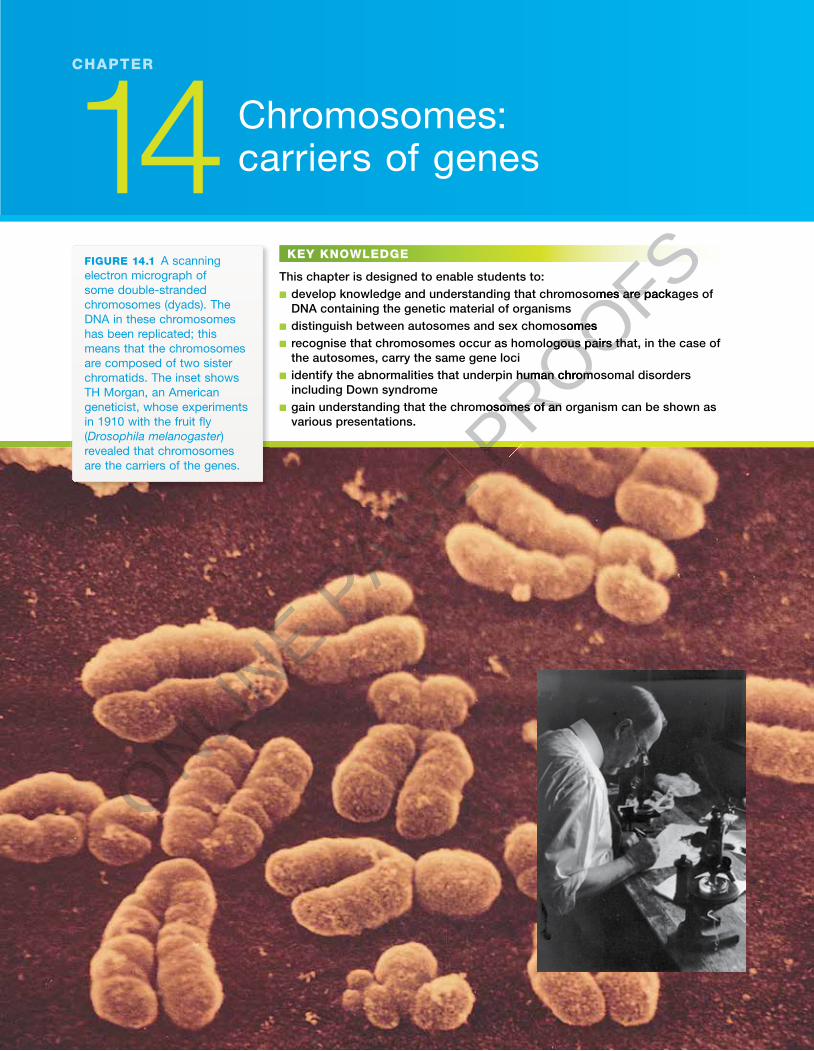
Chromosomes: how many?A small plant (Colchicum autumnale) that grows across southern Europe has the common names meadow sa� ron, autumn crocus and naked lady. � e name ‘naked lady’ is due to the fact that after the leaves of the plant appear in spring they die o� , and the � owers appear in autumn on their own (see � gure 14.2).
� is simple but beautiful plant is poisonous. Deaths have occurred, often after a person has mistaken the plant for wild garlic and eaten its bulb-like corm. � e poison in the autumn crocus is an alkaloid, known as colchicine. � is poison was to play an important role in establishing the correct count of the human chromosomes in somatic cells, that is, discovering that the diploid number (2n) of chromosomes is 46.
Treatment of plant and animal cells with colchicine stops mitosis. Colchicine acts by interfering with spindle formation by binding to and disrupting the micro-tubules that form the structural elements of the mitotic spindle. If the spindle is faulty, the migration of chromosomes at anaphase cannot occur. Instead, the chromosomes are left at metaphase of mitosis. So, dividing cells treated with colchicine will stop their progress through the cell cycle at metaphase.
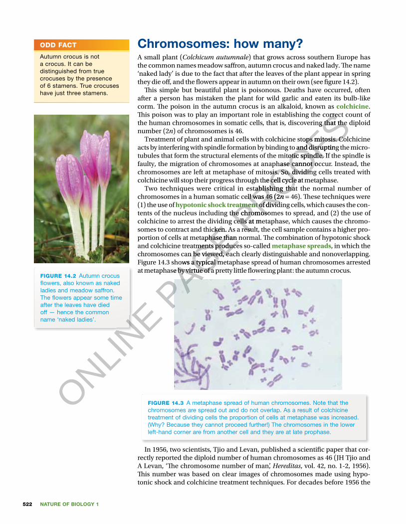
Two techniques were critical in establishing that the normal number of chromosomes in a human somatic cell was 46 (2n = 46). � ese techniques were (1) the use of hypotonic shock treatment of dividing cells, which causes the con-tents of the nucleus including the chromosomes to spread, and (2) the use of colchicine to arrest the dividing cells at metaphase, which causes the chromo-somes to contract and thicken. As a result, the cell sample contains a higher pro-portion of cells at metaphase than normal. � e combination of hypotonic shock and colchicine treatments produces so-called metaphase spreads, in which the chromosomes can be viewed, each clearly distinguishable and nonoverlapping. Figure 14.3 shows a typical metaphase spread of human chromosomes arrested at metaphase by virtue of a pretty little � owering plant: the autumn crocus.
FIGURE 14.3 A metaphase spread of human chromosomes. Note that the chromosomes are spread out and do not overlap. As a result of colchicine treatment of dividing cells the proportion of cells at metaphase was increased. (Why? Because they cannot proceed further!) The chromosomes in the lower left-hand corner are from another cell and they are at late prophase.
In 1956, two scientists, Tjio and Levan, published a scienti� c paper that cor-rectly reported the diploid number of human chromosomes as 46 (JH Tjio and A Levan, ‘� e chromosome number of man’, Hereditas, vol. 42, no. 1-2, 1956). � is number was based on clear images of chromosomes made using hypo-tonic shock and colchicine treatment techniques. For decades before 1956 the
ODD FACT
Autumn crocus is not a crocus. It can be distinguished from true crocuses by the presence of 6 stamens. True crocuses have just three stamens.
FIGURE 14.2 Autumn crocus � owers, also known as naked ladies and meadow saffron. The � owers appear some time after the leaves have died off — hence the common name ‘naked ladies’.
ONLINE
ONLINE
ONLINE
ONLINE P
AGE colchicine to arrest the dividing cells at metaphase, which causes the chromo-
PAGE colchicine to arrest the dividing cells at metaphase, which causes the chromo-
PAGE somes to contract and thicken. As a result, the cell sample contains a higher pro-
PAGE somes to contract and thicken. As a result, the cell sample contains a higher pro-portion of cells at metaphase than normal. � e combination of hypotonic shock
PAGE portion of cells at metaphase than normal. � e combination of hypotonic shock and colchicine treatments produces so-called
PAGE and colchicine treatments produces so-called chromosomes can be viewed, each clearly distinguishable and nonoverlapping.
PAGE chromosomes can be viewed, each clearly distinguishable and nonoverlapping. Figure 14.3 shows a typical metaphase spread of human chromosomes arrested
PAGE Figure 14.3 shows a typical metaphase spread of human chromosomes arrested at metaphase by virtue of a pretty little � owering plant: the autumn crocus.
PAGE at metaphase by virtue of a pretty little � owering plant: the autumn crocus.
PAGE PROOFS
� is poison was to play an important role in establishing the correct count of
PROOFS� is poison was to play an important role in establishing the correct count of the human chromosomes in somatic cells, that is, discovering that the diploid
PROOFSthe human chromosomes in somatic cells, that is, discovering that the diploid
Treatment of plant and animal cells with colchicine stops mitosis. Colchicine
PROOFSTreatment of plant and animal cells with colchicine stops mitosis. Colchicine acts by interfering with spindle formation by binding to and disrupting the micro-
PROOFSacts by interfering with spindle formation by binding to and disrupting the micro-tubules that form the structural elements of the mitotic spindle. If the spindle is
PROOFStubules that form the structural elements of the mitotic spindle. If the spindle is faulty, the migration of chromosomes at anaphase cannot occur. Instead, the
PROOFSfaulty, the migration of chromosomes at anaphase cannot occur. Instead, the chromosomes are left at metaphase of mitosis. So, dividing cells treated with
PROOFSchromosomes are left at metaphase of mitosis. So, dividing cells treated with colchicine will stop their progress through the cell cycle at metaphase.
PROOFScolchicine will stop their progress through the cell cycle at metaphase.
Two techniques were critical in establishing that the normal number of
PROOFSTwo techniques were critical in establishing that the normal number of
chromosomes in a human somatic cell was 46 (2
PROOFSchromosomes in a human somatic cell was 46 (2n
PROOFSn =
PROOFS=
hypotonic shock treatment
PROOFS
hypotonic shock treatment of dividing cells, which causes the con-
PROOFS
of dividing cells, which causes the con-tents of the nucleus including the chromosomes to spread, and (2) the use of PROOFS
tents of the nucleus including the chromosomes to spread, and (2) the use of colchicine to arrest the dividing cells at metaphase, which causes the chromo-PROOFS
colchicine to arrest the dividing cells at metaphase, which causes the chromo-somes to contract and thicken. As a result, the cell sample contains a higher pro-PROOFS
somes to contract and thicken. As a result, the cell sample contains a higher pro-
523CHAPTER 14 Chromosomes: carriers of genes
accepted diploid number of human chromosomes was 48, based on images in a scienti� c paper published in 1921.
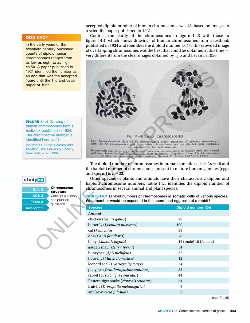
Contrast the clarity of the chromosomes in � gure 14.3 with those in � gure 14.4, which shows drawings of human chromosomes from a textbook published in 1934 and identi� es the diploid number as 48. � is crowded image of overlapping chromosomes was the best that could be obtained at this time — very di� erent from the clear images obtained by Tjio and Levan in 1956.
FIGURE 14.4 Drawing of human chromosomes from a textbook published in 1934. The chromosome number is identi� ed here as 48. (Source: LC Dunn, Heredity and Variation, The University Society, New York, p. 46, 1934.)
� e diploid number of chromosomes in human somatic cells is 2n = 46 and the haploid number of chromosomes present in mature human gametes (eggs and sperm) is n = 23.
Other species of plants and animals have their characteristic diploid and haploid chromosome numbers. Table 14.1 identi� es the diploid number of chromosomes in several animal and plant species.
TABLE 14.1 Diploid numbers of chromosomes in somatic cells of various species. What number would be expected in the sperm and egg cells of a rabbit?
Species Diploid number (2n)
Animal
chicken (Gallus gallus) 78
butter� y (Lysandra nivescens) 190
cat (Felis catus) 38
dog (Canis familiaris) 78
bilby (Macrotis lagotis) 19 (male) 18 (female)
garden snail (Helix aspersa) 54
honeybee (Apis mellifera) 32
house� y (Musca domestica) 12
leopard seal (Hydrurga leptonyx) 34
platypus (Ornithorhynchus anatinus) 52
rabbit (Oryctolagus cuniculus) 44
Eastern tiger snake (Notechis scutatus) 34
fruit � y (Drosophila melanogaster) 8
ant (Myrmecia pilosula) 2(continued)
ODD FACT
In the early years of the twentieth century published counts of diploid human chromosomes ranged from as low as eight to as high as 50. A paper published in 1921 identi� ed the number as 48 and that was the accepted � gure until the Tjio and Levan paper of 1956.
Unit 2 Chromosome structureConcept summary and practice questions
AOS 2
Topic 2
Concept 1
ONLINE haploid chromosome numbers. Table 14.1 identi� es the diploid number of
ONLINE haploid chromosome numbers. Table 14.1 identi� es the diploid number of
chromosomes in several animal and plant species.
ONLINE chromosomes in several animal and plant species.
TABLE 14.1
ONLINE TABLE 14.1
What number would be expected in the sperm and egg cells of a rabbit?
ONLINE
What number would be expected in the sperm and egg cells of a rabbit?
ONLINE
ONLINE
Species
ONLINE
Species
ONLINE
ONLINE Concept summary
ONLINE Concept summary
PAGE
PAGE � e diploid number of chromosomes in human somatic cells is 2
PAGE � e diploid number of chromosomes in human somatic cells is 2
the haploid number of chromosomes present in mature human gametes (eggs
PAGE the haploid number of chromosomes present in mature human gametes (eggs and sperm) is
PAGE and sperm) is n
PAGE n =
PAGE = 23.
PAGE 23.
Other species of plants and animals have their characteristic diploid and PAGE Other species of plants and animals have their characteristic diploid and
haploid chromosome numbers. Table 14.1 identi� es the diploid number of PAGE haploid chromosome numbers. Table 14.1 identi� es the diploid number of chromosomes in several animal and plant species.PAGE
chromosomes in several animal and plant species.
PROOFS
NATURE OF BIOLOGY 1524
TABLE 14.1 (continued)
Species Diploid number (2n)
Plant
crimson bottlebrush (Callistemon citrinus) 22
drooping she oak (Casuarina stricta) 26
edible pea (Pisum sativum) 14
ginkgo (Ginkgo biloba) 24
Iceland poppy (Papaver nudicaule) 14
kangaroo paw (Anigozanthos � avidus) 12
Ovens wattle (Acacia pravissima) 26
pineapple (Ananas comosus) 50
river red gum (Eucalyptus cameldulensis) 22
silky oak (Grevillea robusta) 20
silver wattle (Acacia dealbata) 26
Sydney blue gum (Eucalyptus saligna) 22
tomato (Lycopersicon esculentum) 24
corn (Zea mays) 20
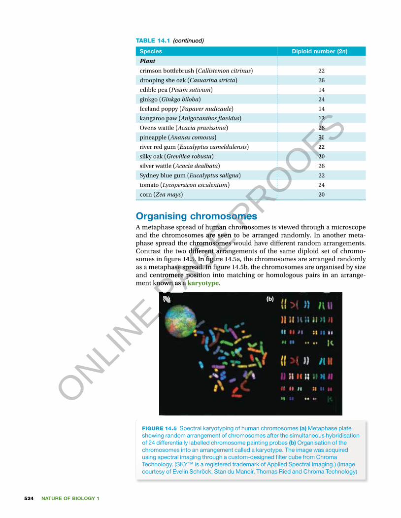
Organising chromosomesA metaphase spread of human chromosomes is viewed through a microscope and the chromosomes are seen to be arranged randomly. In another meta-phase spread the chromosomes would have di� erent random arrangements. Contrast the two di� erent arrangements of the same diploid set of chromo-somes in � gure 14.5. In � gure 14.5a, the chromosomes are arranged randomly as a metaphase spread. In � gure 14.5b, the chromosomes are organised by size and centromere position into matching or homologous pairs in an arrange-ment known as a karyotype.
FIGURE 14.5 Spectral karyotyping of human chromosomes (a) Metaphase plate showing random arrangement of chromosomes after the simultaneous hybridisation of 24 differentially labelled chromosome painting probes (b) Organisation of the chromosomes into an arrangement called a karyotype. The image was acquired using spectral imaging through a custom-designed � lter cube from Chroma Technology. (SKY™ is a registered trademark of Applied Spectral Imaging.) (Image courtesy of Evelin Schröck, Stan du Manoir, Thomas Ried and Chroma Technology)
(a) (b)
ONLINE
ONLINE P
AGE Organising chromosomes
PAGE Organising chromosomesA metaphase spread of human chromosomes is viewed through a microscope
PAGE A metaphase spread of human chromosomes is viewed through a microscope and the chromosomes are seen to be arranged randomly. In another meta-
PAGE and the chromosomes are seen to be arranged randomly. In another meta-phase spread the chromosomes would have di� erent random arrangements.
PAGE phase spread the chromosomes would have di� erent random arrangements. Contrast the two di� erent arrangements of the same diploid set of chromo-
PAGE Contrast the two di� erent arrangements of the same diploid set of chromo-somes in � gure 14.5. In � gure 14.5a, the chromosomes are arranged randomly
PAGE somes in � gure 14.5. In � gure 14.5a, the chromosomes are arranged randomly as a metaphase spread. In � gure 14.5b, the chromosomes are organised by size
PAGE as a metaphase spread. In � gure 14.5b, the chromosomes are organised by size and centromere position into matching or homologous pairs in an arrange-
PAGE and centromere position into matching or homologous pairs in an arrange-ment known as a PAGE ment known as a karyotypePAGE
karyotypePAGE
(a)PAGE
(a)
PROOFS
PROOFS
PROOFS
PROOFS
PROOFS
PROOFS
PROOFS
PROOFS
PROOFS
PROOFS
PROOFS
PROOFS
PROOFS12
PROOFS12
26
PROOFS26
50
PROOFS50
22
PROOFS22
20
PROOFS20
Organising chromosomesPROOFS
Organising chromosomesA metaphase spread of human chromosomes is viewed through a microscope PROOFS
A metaphase spread of human chromosomes is viewed through a microscope
525CHAPTER 14 Chromosomes: carriers of genes
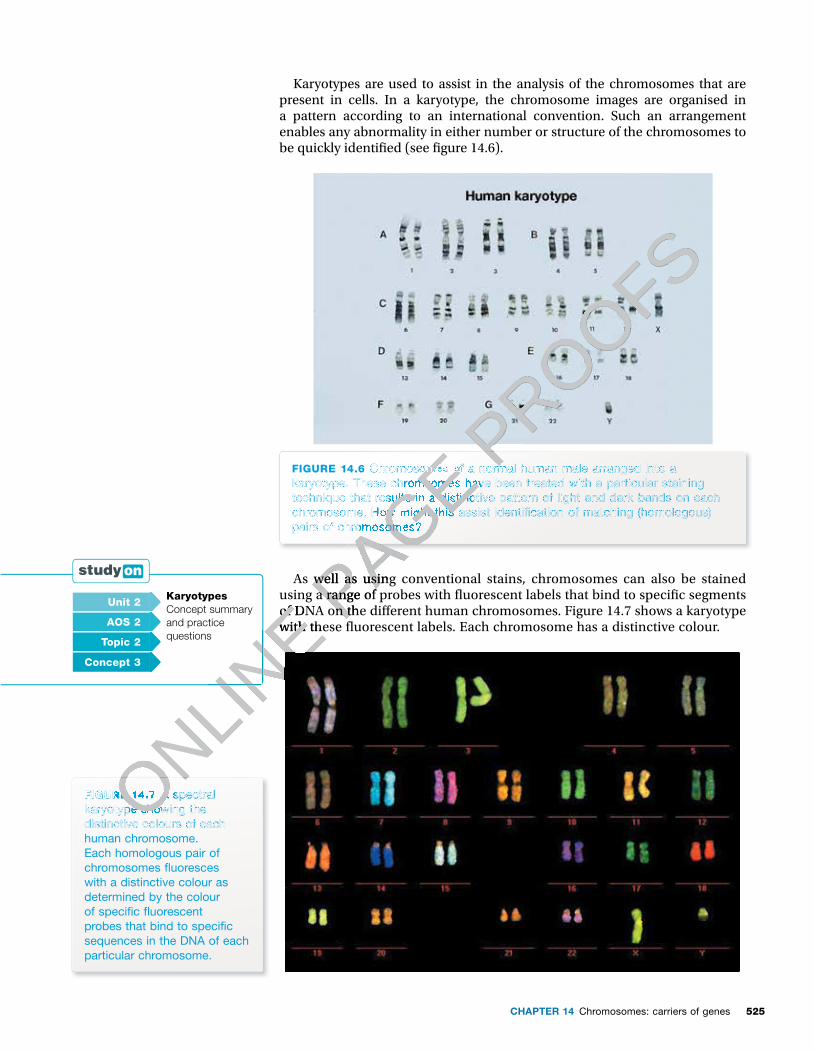
Karyotypes are used to assist in the analysis of the chromosomes that are present in cells. In a karyotype, the chromosome images are organised in a pattern according to an international convention. Such an arrangement enables any abnormality in either number or structure of the chromosomes to be quickly identi� ed (see � gure 14.6).
FIGURE 14.6 Chromosomes of a normal human male arranged into a karyotype. These chromsomes have been treated with a particular staining technique that results in a distinctive pattern of light and dark bands on each chromosome. How might this assist identi� cation of matching (homologous) pairs of chromosomes?
As well as using conventional stains, chromosomes can also be stained using a range of probes with � uorescent labels that bind to speci� c segments of DNA on the di� erent human chromosomes. Figure 14.7 shows a karyotype with these � uorescent labels. Each chromosome has a distinctive colour.
FIGURE 14.7 A spectral karyotype showing the distinctive colours of each human chromosome. Each homologous pair of chromosomes � uoresces with a distinctive colour as determined by the colour of speci� c � uorescent probes that bind to speci� c sequences in the DNA of each particular chromosome.
Unit 2 KaryotypesConcept summary and practice questions
AOS 2
Topic 2
Concept 3
ONLINE using a range of probes with � uorescent labels that bind to speci� c segments
ONLINE using a range of probes with � uorescent labels that bind to speci� c segments
of DNA on the di� erent human chromosomes. Figure 14.7 shows a karyotype
ONLINE of DNA on the di� erent human chromosomes. Figure 14.7 shows a karyotype
with these � uorescent labels. Each chromosome has a distinctive colour.
ONLINE with these � uorescent labels. Each chromosome has a distinctive colour.
ONLINE
ONLINE
FIGURE 14.7 ONLINE
FIGURE 14.7 A spectral ONLINE
A spectral karyotype showing the ONLIN
E
karyotype showing the distinctive colours of each ONLIN
E
distinctive colours of each ONLINE
ONLINE
ONLINE
ONLINE P
AGE
PAGE
PAGE Chromosomes of a normal human male arranged into a
PAGE Chromosomes of a normal human male arranged into a karyotype. These chromsomes have been treated with a particular staining
PAGE karyotype. These chromsomes have been treated with a particular staining technique that results in a distinctive pattern of light and dark bands on each
PAGE technique that results in a distinctive pattern of light and dark bands on each chromosome. How might this assist identi� cation of matching (homologous)
PAGE chromosome. How might this assist identi� cation of matching (homologous) pairs of chromosomes?
PAGE pairs of chromosomes?
PAGE As well as using conventional stains, chromosomes can also be stained PAGE As well as using conventional stains, chromosomes can also be stained
using a range of probes with � uorescent labels that bind to speci� c segments PAGE using a range of probes with � uorescent labels that bind to speci� c segments of DNA on the di� erent human chromosomes. Figure 14.7 shows a karyotype PAGE
of DNA on the di� erent human chromosomes. Figure 14.7 shows a karyotype
PROOFS
PROOFS
PROOFS
PROOFS
PROOFS
Chromosomes of a normal human male arranged into a PROOFS
Chromosomes of a normal human male arranged into a PROOFS
NATURE OF BIOLOGY 1526
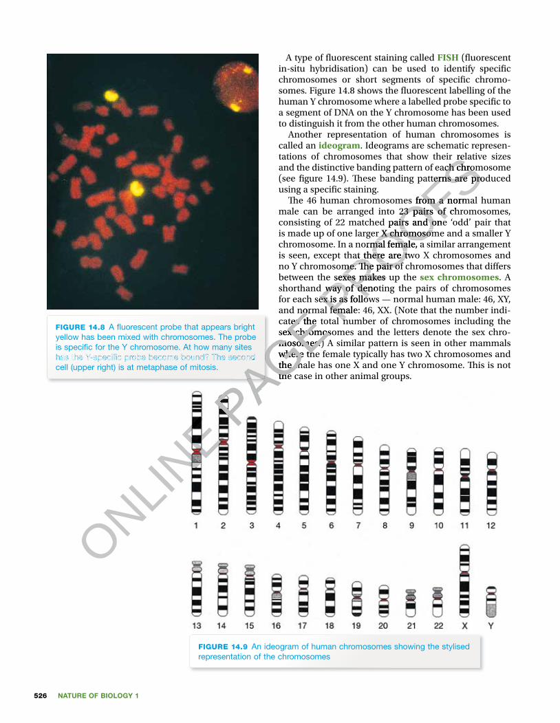
A type of � uorescent staining called FISH (� uorescent in-situ hybridisation) can be used to identify speci� c chromosomes or short segments of speci� c chromo-somes. Figure 14.8 shows the � uorescent labelling of the human Y chromosome where a labelled probe speci� c to a segment of DNA on the Y chromosome has been used to distinguish it from the other human chromosomes.
Another representation of human chromosomes is called an ideogram. Ideograms are schematic represen-tations of chromosomes that show their relative sizes and the distinctive banding pattern of each chromosome (see � gure 14.9). � ese banding patterns are produced using a speci� c staining.
� e 46 human chromosomes from a normal human male can be arranged into 23 pairs of chromosomes, consisting of 22 matched pairs and one ‘odd’ pair that is made up of one larger X chromosome and a smaller Y chromosome. In a normal female, a similar arrangement is seen, except that there are two X chromo somes and no Y chromosome. � e pair of chromo somes that di� ers between the sexes makes up the sex chromosomes. A shorthand way of denoting the pairs of chromosomes for each sex is as follows — normal human male: 46, XY, and normal female: 46, XX. (Note that the number indi-cates the total number of chromosomes including the sex chromosomes and the letters denote the sex chro-mosomes.) A similar pattern is seen in other mammals where the female typically has two X chromosomes and the male has one X and one Y chromosome. � is is not the case in other animal groups.
FIGURE 14.9 An ideogram of human chromosomes showing the stylised representation of the chromosomes
FIGURE 14.8 A � uorescent probe that appears bright yellow has been mixed with chromosomes. The probe is speci� c for the Y chromosome. At how many sites has the Y-speci� c probe become bound? The second cell (upper right) is at metaphase of mitosis.
ONLINE
ONLINE P
AGE and normal female: 46, XX. (Note that the number indi-
PAGE and normal female: 46, XX. (Note that the number indi-cates the total number of chromosomes including the
PAGE cates the total number of chromosomes including the sex chromosomes and the letters denote the sex chro-
PAGE sex chromosomes and the letters denote the sex chro-mosomes.) A similar pattern is seen in other mammals
PAGE mosomes.) A similar pattern is seen in other mammals where the female typically has two X chromosomes and
PAGE where the female typically has two X chromosomes and the male has one X and one Y chromosome. � is is not
PAGE the male has one X and one Y chromosome. � is is not the case in other animal groups.
PAGE the case in other animal groups.
PAGE
PAGE
PAGE has the Y-speci� c probe become bound? The second
PAGE has the Y-speci� c probe become bound? The second
PAGE PROOFS
tations of chromosomes that show their relative sizes
PROOFStations of chromosomes that show their relative sizes and the distinctive banding pattern of each chromosome
PROOFSand the distinctive banding pattern of each chromosome (see � gure 14.9). � ese banding patterns are produced
PROOFS(see � gure 14.9). � ese banding patterns are produced
� e 46 human chromosomes from a normal human
PROOFS� e 46 human chromosomes from a normal human male can be arranged into 23 pairs of chromosomes,
PROOFSmale can be arranged into 23 pairs of chromosomes, consisting of 22 matched pairs and one ‘odd’ pair that
PROOFSconsisting of 22 matched pairs and one ‘odd’ pair that is made up of one larger X chromosome and a smaller Y
PROOFSis made up of one larger X chromosome and a smaller Y chromosome. In a normal female, a similar arrangement
PROOFSchromosome. In a normal female, a similar arrangement is seen, except that there are two X chromo somes and
PROOFSis seen, except that there are two X chromo somes and no Y chromosome. � e pair of chromo somes that di� ers
PROOFSno Y chromosome. � e pair of chromo somes that di� ers between the sexes makes up the
PROOFSbetween the sexes makes up the shorthand way of denoting the pairs of chromosomes PROOFS
shorthand way of denoting the pairs of chromosomes for each sex is as follows — normal human male: 46, XY, PROOFS
for each sex is as follows — normal human male: 46, XY, and normal female: 46, XX. (Note that the number indi-PROOFS
and normal female: 46, XX. (Note that the number indi-cates the total number of chromosomes including the PROOFS
cates the total number of chromosomes including the
527CHAPTER 14 Chromosomes: carriers of genes
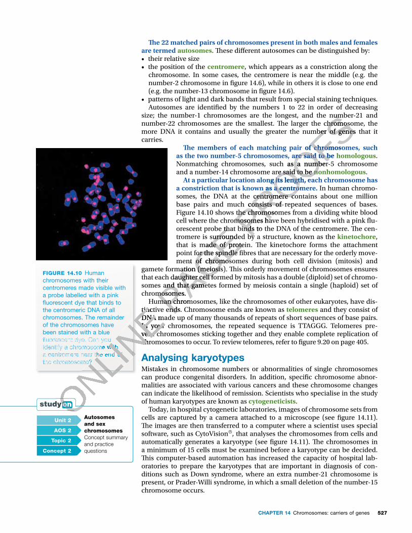
� e 22 matched pairs of chromosomes present in both males and females are termed autosomes. � ese di� erent autosomes can be distinguished by: • their relative size • the position of the centromere, which appears as a constriction along the
chromosome. In some cases, the centromere is near the middle (e.g. the number-2 chromosome in � gure 14.6), while in others it is close to one end (e.g. the number-13 chromosome in � gure 14.6).
• patterns of light and dark bands that result from special staining techniques. Autosomes are identi� ed by the numbers 1 to 22 in order of decreasing
size; the number-1 chromosomes are the longest, and the number-21 and number-22 chromosomes are the smallest. � e larger the chromosome, the more DNA it contains and usually the greater the number of genes that it carries.
� e members of each matching pair of chromosomes, such as the two number-5 chromosomes, are said to be homologous. Nonmatching chromosomes, such as a number-5 chromosome and a number-14 chromosome are said to be nonhomologous.
At a particular location along its length, each chromosome has a constriction that is known as a centromere. In human chromo-somes, the DNA at the centromere contains about one million base pairs and much consists of repeated sequences of bases. Figure 14.10 shows the chromosomes from a dividing white blood cell where the chromosomes have been hybridised with a pink � u-orescent probe that binds to the DNA of the centromere. � e cen-tromere is surrounded by a structure, known as the kinetochore, that is made of protein. � e kinetochore forms the attachment point for the spindle � bres that are necessary for the orderly move-ment of chromosomes during both cell division (mitosis) and
gamete formation (meiosis). � is orderly movement of chromosomes ensures that each daughter cell formed by mitosis has a double (diploid) set of chromo-somes and that gametes formed by meiosis contain a single (haploid) set of chromosomes.
Human chromosomes, like the chromosomes of other eukaryotes, have dis-tinctive ends. Chromosome ends are known as telomeres and they consist of DNA made up of many thousands of repeats of short sequences of base pairs. In your chromosomes, the repeated sequence is TTAGGG. Telomeres pre-vent chromosomes sticking together and they enable complete replication of chromosomes to occur. To review telomeres, refer to � gure 9.20 on page 405.
Analysing karyotypes Mistakes in chromosome numbers or abnormalities of single chromosomes can produce congenital disorders. In addition, speci� c chromosome abnor-malities are associated with various cancers and these chromosome changes can indicate the likelihood of remission. Scientists who specialise in the study of human karyotypes are known as cytogeneticists.
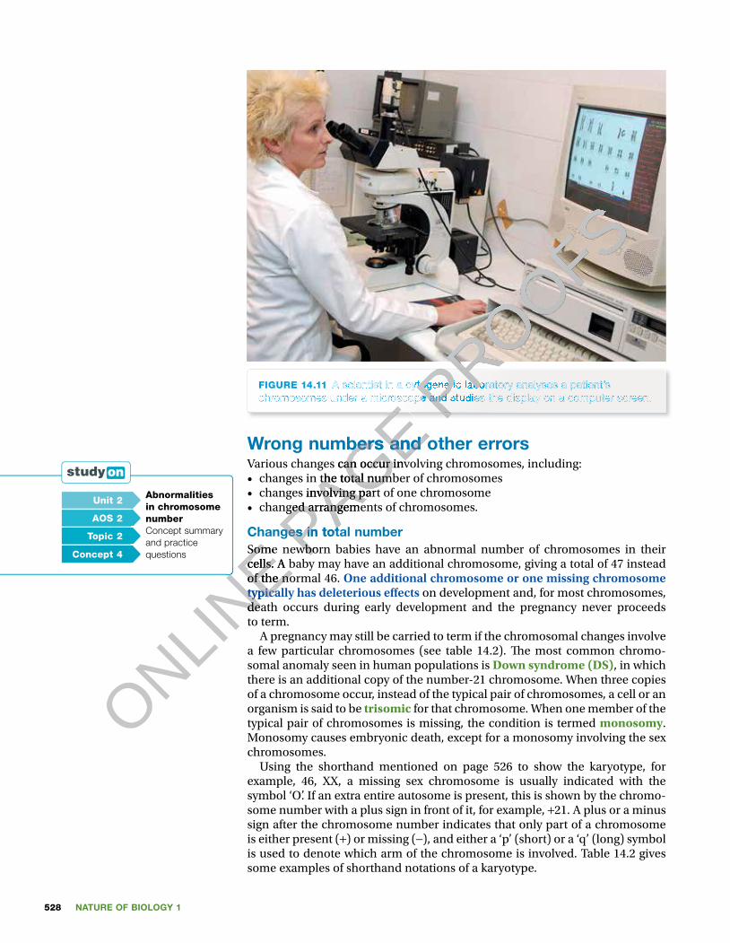
Today, in hospital cytogenetic laboratories, images of chromosome sets from cells are captured by a camera attached to a microscope (see � gure 14.11). � e images are then transferred to a computer where a scientist uses special software, such as CytoVision, that analyses the chromosomes from cells and automatically generates a karyotype (see � gure 14.11). � e chromosomes in a minimum of 15 cells must be examined before a karyotype can be decided. � is computer-based automation has increased the capacity of hospital lab-oratories to prepare the karyotypes that are important in diagnosis of con-ditions such as Down syndrome, where an extra number-21 chromosome is present, or Prader-Willi syndrome, in which a small deletion of the number-15 chromosome occurs.
FIGURE 14.10 Human chromosomes with their centromeres made visible with a probe labelled with a pink � uorescent dye that binds to the centromeric DNA of all chromosomes. The remainder of the chromosomes have been stained with a blue � uorescent dye. Can you identify a chromosome with a centromere near the end of the chromosome?
Unit 2 Autosomes and sex chromosomesConcept summary and practice questions
AOS 2
Topic 2
Concept 2
ONLINE tinctive ends. Chromosome ends are known as
ONLINE tinctive ends. Chromosome ends are known as
DNA made up of many thousands of repeats of short sequences of base pairs.
ONLINE DNA made up of many thousands of repeats of short sequences of base pairs.
In your chromosomes, the repeated sequence is TTAGGG. Telomeres pre-
ONLINE
In your chromosomes, the repeated sequence is TTAGGG. Telomeres pre-vent chromosomes sticking together and they enable complete replication of
ONLINE
vent chromosomes sticking together and they enable complete replication of chromosomes to occur. To review telomeres, refer to � gure 9.20 on page 405.
ONLINE
chromosomes to occur. To review telomeres, refer to � gure 9.20 on page 405.
ONLINE
ONLINE
� uorescent dye. Can you
ONLINE
� uorescent dye. Can you identify a chromosome with
ONLINE
identify a chromosome with a centromere near the end of
ONLINE
a centromere near the end of the chromosome?
ONLINE
the chromosome?
ONLINE
ONLINE
ONLINE
ONLINE
ONLINE
ONLINE
ONLINE
ONLINE
ONLINE P
AGE orescent probe that binds to the DNA of the centromere. � e cen-
PAGE orescent probe that binds to the DNA of the centromere. � e cen-tromere is surrounded by a structure, known as the
PAGE tromere is surrounded by a structure, known as the that is made of protein. � e kinetochore forms the attachment
PAGE that is made of protein. � e kinetochore forms the attachment point for the spindle � bres that are necessary for the orderly move-
PAGE point for the spindle � bres that are necessary for the orderly move-ment of chromosomes during both cell division (mitosis) and
PAGE ment of chromosomes during both cell division (mitosis) and
gamete formation (meiosis). � is orderly movement of chromosomes ensures
PAGE gamete formation (meiosis). � is orderly movement of chromosomes ensures that each daughter cell formed by mitosis has a double (diploid) set of chromo-
PAGE that each daughter cell formed by mitosis has a double (diploid) set of chromo-somes and that gametes formed by meiosis contain a single (haploid) set of
PAGE somes and that gametes formed by meiosis contain a single (haploid) set of chromosomes. PAGE chromosomes.
Human chromosomes, like the chromosomes of other eukaryotes, have dis-PAGE Human chromosomes, like the chromosomes of other eukaryotes, have dis-
tinctive ends. Chromosome ends are known as PAGE
tinctive ends. Chromosome ends are known as
PROOFSsize; the number-1 chromosomes are the longest, and the number-21 and
PROOFSsize; the number-1 chromosomes are the longest, and the number-21 and number-22 chromosomes are the smallest. � e larger the chromosome, the
PROOFSnumber-22 chromosomes are the smallest. � e larger the chromosome, the more DNA it contains and usually the greater the number of genes that it
PROOFSmore DNA it contains and usually the greater the number of genes that it
� e members of each matching pair of chromosomes, such
PROOFS� e members of each matching pair of chromosomes, such as the two number-5 chromosomes, are said to be
PROOFSas the two number-5 chromosomes, are said to beNonmatching chromosomes, such as a number-5 chromosome
PROOFSNonmatching chromosomes, such as a number-5 chromosome and a number-14 chromosome are said to be
PROOFSand a number-14 chromosome are said to be nonhomologous
PROOFSnonhomologous
At a particular location along its length, each chromosome has
PROOFSAt a particular location along its length, each chromosome has
a constriction that is known as a centromere.
PROOFSa constriction that is known as a centromere.somes, the DNA at the centromere contains about one million
PROOFSsomes, the DNA at the centromere contains about one million base pairs and much consists of repeated sequences of bases.
PROOFSbase pairs and much consists of repeated sequences of bases. Figure 14.10 shows the chromosomes from a dividing white blood PROOFS
Figure 14.10 shows the chromosomes from a dividing white blood cell where the chromosomes have been hybridised with a pink � u-PROOFS
cell where the chromosomes have been hybridised with a pink � u-orescent probe that binds to the DNA of the centromere. � e cen-PROOFS
orescent probe that binds to the DNA of the centromere. � e cen-tromere is surrounded by a structure, known as the PROOFS
tromere is surrounded by a structure, known as the
NATURE OF BIOLOGY 1528
FIGURE 14.11 A scientist in a cytogenetic laboratory analyses a patient’s chromosomes under a microscope and studies the display on a computer screen.
Wrong numbers and other errorsVarious changes can occur involving chromosomes, including:• changes in the total number of chromosomes• changes involving part of one chromosome• changed arrangements of chromosomes.
Changes in total numberSome newborn babies have an abnormal number of chromosomes in their cells. A baby may have an additional chromosome, giving a total of 47 instead of the normal 46. One additional chromosome or one missing chromosome typically has deleterious e� ects on development and, for most chromosomes, death occurs during early development and the pregnancy never proceeds to term.
A pregnancy may still be carried to term if the chromosomal changes involve a few particular chromosomes (see table 14.2). � e most common chromo-somal anomaly seen in human populations is Down syndrome (DS), in which there is an additional copy of the number-21 chromosome. When three copies of a chromosome occur, instead of the typical pair of chromosomes, a cell or an organism is said to be trisomic for that chromosome. When one member of the typical pair of chromosomes is missing, the condition is termed monosomy. Monosomy causes embryonic death, except for a monosomy involving the sex chromosomes.
Using the shorthand mentioned on page 526 to show the karyotype, for example, 46, XX, a missing sex chromosome is usually indicated with the symbol ‘O’. If an extra entire autosome is present, this is shown by the chromo-some number with a plus sign in front of it, for example, +21. A plus or a minus sign after the chromosome number indicates that only part of a chromosome is either present (+) or missing (−), and either a ‘p’ (short) or a ‘q’ (long) symbol is used to denote which arm of the chromosome is involved. Table 14.2 gives some examples of shorthand notations of a karyotype.
Unit 2 Abnormalities in chromosome numberConcept summary and practice questions
AOS 2
Topic 2
Concept 4
ONLINE Changes in total number
ONLINE Changes in total number
Some newborn babies have an abnormal number of chromosomes in their
ONLINE Some newborn babies have an abnormal number of chromosomes in their
cells. A baby may have an additional chromosome, giving a total of 47 instead
ONLINE cells. A baby may have an additional chromosome, giving a total of 47 instead
of the normal 46.
ONLINE
of the normal 46. typically has deleterious e� ects
ONLINE
typically has deleterious e� ectsdeath occurs during early development and the pregnancy never proceeds
ONLINE
death occurs during early development and the pregnancy never proceeds to term.
ONLINE
to term.
ONLINE
ONLINE P
AGE
PAGE
PAGE chromosomes under a microscope and studies the display on a computer screen.
PAGE chromosomes under a microscope and studies the display on a computer screen.
PAGE Wrong numbers and other errors
PAGE Wrong numbers and other errorsVarious changes can occur involving chromosomes, including:
PAGE Various changes can occur involving chromosomes, including:
changes in the total number of chromosomes
PAGE changes in the total number of chromosomeschanges involving part of one chromosome
PAGE changes involving part of one chromosomechanged arrangements of chromosomes.
PAGE changed arrangements of chromosomes.
Changes in total numberPAGE Changes in total numberSome newborn babies have an abnormal number of chromosomes in their PAGE
Some newborn babies have an abnormal number of chromosomes in their
PROOFS
PROOFS
PROOFS
PROOFS
A scientist in a cytogenetic laboratory analyses a patient’s PROOFS
A scientist in a cytogenetic laboratory analyses a patient’s chromosomes under a microscope and studies the display on a computer screen.PROOFS
chromosomes under a microscope and studies the display on a computer screen.PROOFS
529CHAPTER 14 Chromosomes: carriers of genes
TABLE 14.2 Some examples of chromosome changes and approximate incidence rates. Which syndrome is an example of a trisomy? A monosomy? The XYY condition does not have a clinical name.
Chromosome change Resulting syndromeApproximate incidence rate
Addition: whole chromosome
extra number-21 (47, +21) Down syndrome 1/700 live births
extra number-18 (47, +18) Edwards syndrome 1/3000 live births
extra number-13 (47, +13) Patau syndrome 1/5000 live births
extra sex chromosome (47, XXY) Klinefelter syndrome 1/1000 male births
extra Y chromosome (47, XYY) n/a 1/1000 male births
Deletion: whole chromosome
missing sex chromosome (46, XO) Turner syndrome 1/5000 female births
Deletion: part chromosome
missing part of short arm of number-4 (46, 4p−)
Wolf-Hirschhorn syndrome
1/50 000 live births
missing part of short arm of number-5 (46, 5p−)
cri-du-chat syndrome 1/25 000 live births
Changes to parts of chromosomesChanges can occur that involve part of a chromosome, such as:• duplication, in which part of a chromosome is duplicated (see � gure 14.12a)• deletion, in which part of a chromosome is missing (see � gure 14.12b), as in
cri-du-chat syndrome, for example, so named because a� ected babies have a cat-like cry (see table 14.2).
Normalchromosome 14
Normalchromosome
Normalchromosome 21
14/21 translocatedchromosome
Chromosomewith a segment
duplicated
Normalchromosome
Chromosomewith a segment
deleted
(a) Duplication
(c) Translocation
(b) Deletion
Duplicatedsegment
Site ofdeletionBreakage
points
Breakagepoints
New join21
14
FIGURE 14.12 (a) Normal chromosome and the same chromosome showing a duplication (b) Normal chromosome and the same chromosome showing a deletion (c) An example of a 14/21 translocation
ONLINE
ONLINE
ONLINE
ONLINE
ONLINE
ONLINE
ONLINE
ONLINE
ONLINE
ONLINE
ONLINE
ONLINE
ONLINE P
AGE Changes can occur that involve part of a chromosome, such as:
PAGE Changes can occur that involve part of a chromosome, such as:, in which part of a chromosome is duplicated (see � gure 14.12a)
PAGE , in which part of a chromosome is duplicated (see � gure 14.12a), in which part of a chromosome is missing (see � gure 14.12b), as in
PAGE , in which part of a chromosome is missing (see � gure 14.12b), as in
cri-du-chat syndrome, for example, so named because a� ected babies have
PAGE cri-du-chat syndrome, for example, so named because a� ected babies have a cat-like cry (see table 14.2).
PAGE a cat-like cry (see table 14.2).
(a) Duplication
PAGE (a) Duplication
PAGE
PAGE
PAGE
PAGE
PAGE
PAGE PROOFS
PROOFS
PROOFS
PROOFS
PROOFS
PROOFS
PROOFS
PROOFS
PROOFS
PROOFS
PROOFS
PROOFS
PROOFS
PROOFS
PROOFS
PROOFS
PROOFS
PROOFS
PROOFS
PROOFS
PROOFS
PROOFSPatau syndrome 1/5000 live births
PROOFSPatau syndrome 1/5000 live births
extra sex chromosome (47, XXY) Klinefelter syndrome 1/1000 male births
PROOFSextra sex chromosome (47, XXY) Klinefelter syndrome 1/1000 male births
1/1000 male births
PROOFS1/1000 male births
missing sex chromosome (46, XO) Turner syndrome 1/5000 female births
PROOFSmissing sex chromosome (46, XO) Turner syndrome 1/5000 female birthsmissing sex chromosome (46, XO) Turner syndrome 1/5000 female births
PROOFSmissing sex chromosome (46, XO) Turner syndrome 1/5000 female births
Wolf-Hirschhorn
PROOFSWolf-Hirschhorn syndrome
PROOFSsyndrome
cri-du-chat syndrome
PROOFScri-du-chat syndrome
Changes to parts of chromosomesPROOFS
Changes to parts of chromosomesChanges can occur that involve part of a chromosome, such as:PROOFS
Changes can occur that involve part of a chromosome, such as:, in which part of a chromosome is duplicated (see � gure 14.12a)PROOFS
, in which part of a chromosome is duplicated (see � gure 14.12a)
NATURE OF BIOLOGY 1530
Re-arrangements of chromosomes Structural changes may occur in which the location of a chromosome segment is altered so that it becomes relocated to a new region within the karyotype. Such a change is known as a translocation. One example is related to a special case of Down syndrome when part of the number-21 chromosome becomes physically attached to a number-14 chromosome (see � gure 14.12c). � e par-ental origin of the chromosomes is also important. Normally a child inherits one member of each chromosome pair from each of its parents. If both copies of a particular chromosome are inherited from one parent, instead of the usual one from each parent, abnormalities of development result. For example, Angelman syndrome, characterised by poor motor coordination and mental retardation, can result if an embryo inherits both of its number-15 chromo-somes from its mother and none from its father.
A case of Down syndrome Michael (see � gure 14.13a) is a young boy who has 47 chromosomes in his body cells, instead of the normal 46. His karyotype shows the presence of an extra number-21 chromosome, which is typical of the condition Down syn-drome. � is karyotype can be denoted as 47, XY, +21 (where ‘47’ denotes the total number of chromosomes, ‘XY’ denotes the sex chromosomes present, and ‘+21’ denotes the identity of the extra chromosome).
FIGURE 14.13 (a) Michael, pictured aged 11, is a DS boy who enjoys using a computer, which is one of his many interests. (b) A typical karyotype from a DS male like Michael. Which chromosome is present as a trisomy?
(a) (b)
About one in 700 babies born in Australia has an extra number-21 chromosome, but the rate di� ers according to the age of the mother (see � gure 14.14). � e risk of having a DS baby increases with maternal age; the risk for mothers aged 20 is about 1/2300, while the risk for mothers aged 40 is about 1/100.
20 25 30 35 40 45
0.03
0.02
0.01
0.00
Freq
uenc
y o
f D
Sp
er li
ve b
irth
s
Age of mother
12300
1880
1290
1100
146
FIGURE 14.14 Incidence of DS births for mothers of different ages. How does the risk of a DS baby for a 40-year-old woman compare with that for a 20-year-old? Increased father’s age also increases the risk, but to a lesser extent.
ONLINE
ONLINE
ONLINE
ONLINE
computer, which is one of his
ONLINE
computer, which is one of his
ONLINE
ONLINE
ONLINE
A typical
ONLINE
A typical karyotype from a DS male like
ONLINE
karyotype from a DS male like Michael. Which chromosome
ONLINE
Michael. Which chromosome is present as a trisomy?
ONLINE
is present as a trisomy?
ONLINE
ONLINE
ONLINE
0.03 ONLINE
0.03
PAGE PROOFS
one from each parent, abnormalities of development result. For example,
PROOFSone from each parent, abnormalities of development result. For example,
, characterised by poor motor coordination and mental
PROOFS, characterised by poor motor coordination and mental inherits both of its number-15 chromo-
PROOFSinherits both of its number-15 chromo-
Michael (see � gure 14.13a) is a young boy who has 47 chromosomes in his
PROOFSMichael (see � gure 14.13a) is a young boy who has 47 chromosomes in his body cells, instead of the normal 46. His karyotype shows the presence of an
PROOFSbody cells, instead of the normal 46. His karyotype shows the presence of an extra number-21 chromosome, which is typical of the condition Down syn-
PROOFSextra number-21 chromosome, which is typical of the condition Down syn-drome. � is karyotype can be denoted as 47, XY,
PROOFSdrome. � is karyotype can be denoted as 47, XY, +
PROOFS+21 (where ‘47’ denotes the
PROOFS21 (where ‘47’ denotes the
total number of chromosomes, ‘XY’ denotes the sex chromosomes present,
PROOFStotal number of chromosomes, ‘XY’ denotes the sex chromosomes present,
21’ denotes the identity of the extra chromosome).
PROOFS
21’ denotes the identity of the extra chromosome).
PROOFS
531CHAPTER 14 Chromosomes: carriers of genes
� e presence of an extra number-21 chromosome produces various symp-toms including a fold on the inner margin of the upper eyelid, a smaller than normal mouth cavity and distinctive creases on the palms of the hands and the soles of the feet. � e trisomy condition in which three separate copies of the number-21 chromosome are present in the karyotype is the most common form of DS. In most cases, the extra number-21 chromosome is transmitted via an abnormal egg with 24 chromosomes, including two number-21 chromosomes. If this egg is fertilised by a normal sperm (with 23 chromosomes including one number-21 chromosome), a DS embryo will result (see � gure 14.15).
(a)
2n = 46
21 21
(b)
2n = 47
21 2121
n = 23
Egg
21
21
n = 24
Egg
Sperm
21
21
21
Sperm
n = 23
Fertilisation
Fertilisation
n = 23
FIGURE 14.15 (a) Fertilisation of normal gametes (b) Possible gametes involved in fertilisation to produce a DS zygote (In both (a) and (b), the number-21 chromosomes are shown separately.)
An abnormal egg results when, during the process of egg formation by mei-osis in the ovary, the normal separation of the two copies of chromosome 21 to opposite poles of the spindle does not occur. � is type of error is known as a nondisjunction and is unpredictable. For parents who have a DS child who is a result of nondisjunction during egg formation in the mother (or during sperm production in the father), the risk of a second child with DS is low and is determined by the mother’s age. When a child with DS is born, new challenges arise for the family concerned. Read the box on page 535, about Jane, a young woman with DS, as told by her mother.
Another form of DS: translocationEach year in Australia, a few babies are born who show all the clinical signs of DS. � eir karyotype, however, shows just 46 chromosomes, instead of the 47 expected in DS. So, what has happened?
In these cases, a third number-21 chromosome is present, but it is not a separate chromosome. Instead, the extra number-21 is joined to another chromosome, for example, the number-14 chromosome, through a trans-lo cation (see � gure 14.16). As a result, the total number of chromosomes in somatic cells of this rarer form of DS is 46.
ONLINE
ONLINE
ONLINE
ONLINE
ONLINE An abnormal egg results when, during the process of egg formation by mei-
ONLINE An abnormal egg results when, during the process of egg formation by mei-
osis in the ovary,
ONLINE
osis in the ovary,
PAGE
PAGE
PAGE
PAGE n
PAGE n =
PAGE = 24
PAGE 24= 24=
PAGE = 24=
PAGE
PAGE
PAGE
PAGE
PAGE
PAGE
PAGE
PAGE
PAGE
PAGE
PAGE
PAGE
PAGE
PAGE
PAGE
PAGE
PAGE
PAGE
PAGE
PAGE
PAGE
PAGE
PAGE
PAGE
PAGE
PAGE
PAGE
PAGE
PAGE
PAGE
PAGE
PAGE
PAGE
PAGE
PAGE
PAGE
PAGE
PAGE
PAGE
PAGE
PAGE
PAGE
PAGE
PAGE
PAGE
PAGE
PAGE
PAGE
PAGE
PAGE
PAGE
PAGE
PAGE
PAGE
PAGE
PAGE
PAGE
PAGE
PAGE
PAGE
PAGE
PAGE
PAGE
PAGE
PAGE
PAGE
PAGE
PAGE
PAGE
PAGE
PAGE
PAGE
PAGE
PAGE
PAGE
PAGE
PAGE
PAGE
PAGE
PAGE
PAGE
PAGE
PAGE
PAGE
PAGE
PAGE
PAGE
PAGE
PAGE
PAGE
PAGE
PAGE
PAGE
PAGE
PAGE
PAGE
PAGE
PAGE
PAGE
PAGE
PAGE
PAGE
PAGE
PAGE
PAGE
PAGE
PAGE
PAGE
PAGE
PAGE
PAGE
PAGE
PAGE
PAGE
PAGE
PAGE
PAGE
PAGE PROOFS
PROOFS
PROOFS
PROOFS
EggPROOFS
Egg
Sperm
PROOFSSperm
PROOFS
PROOFSFertilisation
PROOFSFertilisation
NATURE OF BIOLOGY 1532
45
Type A Type B Type C Type D
Totalnumber of
chromosomesLeanne'sparents
View of number-14 and
number-21chromosomes Possible gametes
46
14 2114/21
14 1421 21
1421
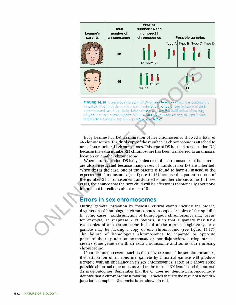
FIGURE 14.16 A translocation form of Down syndrome in which the condition is inherited. Note that the mother can produce four kinds of egg in terms of their chromosomal make-up. Baby Leanne resulted from the fertilisation of an egg of type C with a normal sperm. What would result when an egg of type B was fertilised? A fertilised type-D egg would die. Why?
Baby Leanne has DS. Examination of her chromosomes showed a total of 46 chromosomes. � e third copy of the number-21 chromosome is attached to one of her number-14 chromosomes. � is type of DS is called translocation DS, because the extra number-21 chromosome has been transferred to an unusual location on another chromosome.
When a translocation DS baby is detected, the chromosomes of its parents are also investigated because many cases of translocation DS are inherited. When this is the case, one of the parents is found to have 45 instead of the expected 46 chromosomes (see � gure 14.16) because this parent has one of the number-21 chromosomes translocated to another chromosome. In these cases, the chance that the next child will be a� ected is theoretically about one in three but in reality is about one in 10.
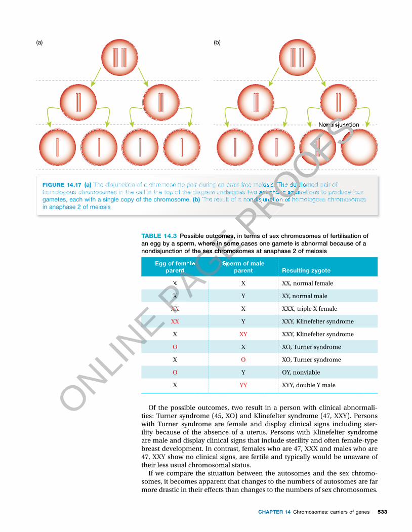
Errors in sex chromosomesDuring gamete formation by meiosis, critical events include the orderly disjunction of homologous chromosomes to opposite poles of the spindle. In some cases, nondisjunction of homologous chromosomes may occur, for example, at anaphase 2 of meiosis, such that a gamete may have two copies of one chromosome instead of the normal single copy, or a gamete may be lacking a copy of one chromosome (see � gure 14.17). � e failure of homologous chromosomes to separate to opposite poles of their spindle at anaphase, or nondisjunction, during meiosis creates some gametes with an extra chromosome and some with a missing chromosome.
If nondisjunction events such as these involve one of the sex chromosomes, the fertilisation of an abnormal gamete by a normal gamete will produce a zygote with an imbalance in its sex chromosomes. Table 14.3 shows some possible abnormal outcomes, as well as the normal XX female and the normal XY male outcomes. Remember that the ‘O’ does not denote a chromosome, it denotes that a chromosome is missing. Gametes that are the result of a nondis-junction at anaphase 2 of meiosis are shown in red.
ONLINE expected 46 chromosomes (see � gure 14.16) because this parent has one of
ONLINE expected 46 chromosomes (see � gure 14.16) because this parent has one of
the number-21 chromosomes translocated to another chromosome. In these
ONLINE the number-21 chromosomes translocated to another chromosome. In these
cases, the chance that the next child will be a� ected is theoretically about one
ONLINE cases, the chance that the next child will be a� ected is theoretically about one
in three but in reality is about one in 10.
ONLINE
in three but in reality is about one in 10.
Errors in sex chromosomes
ONLINE
Errors in sex chromosomes
PAGE Baby Leanne has DS. Examination of her chromosomes showed a total of
PAGE Baby Leanne has DS. Examination of her chromosomes showed a total of 46 chromosomes. � e third copy of the number-21 chromosome is attached to
PAGE 46 chromosomes. � e third copy of the number-21 chromosome is attached to one of her number-14 chromosomes. � is type of DS is called translocation DS,
PAGE one of her number-14 chromosomes. � is type of DS is called translocation DS, because the extra number-21 chromosome has been transferred to an unusual
PAGE because the extra number-21 chromosome has been transferred to an unusual location on another chromosome.
PAGE location on another chromosome.
When a translocation DS baby is detected, the chromosomes of its parents
PAGE When a translocation DS baby is detected, the chromosomes of its parents
are also investigated because many cases of translocation DS are inherited.
PAGE are also investigated because many cases of translocation DS are inherited. When this is the case, one of the parents is found to have 45 instead of the PAGE When this is the case, one of the parents is found to have 45 instead of the PAGE
expected 46 chromosomes (see � gure 14.16) because this parent has one of PAGE
expected 46 chromosomes (see � gure 14.16) because this parent has one of the number-21 chromosomes translocated to another chromosome. In these PAGE
the number-21 chromosomes translocated to another chromosome. In these
PROOFS
PROOFS14
PROOFS14
21
PROOFS21
PROOFS
PROOFS
PROOFS
PROOFS
PROOFS
PROOFS
PROOFS
PROOFS
PROOFS
PROOFS
PROOFS
PROOFS
PROOFS
PROOFS
PROOFS
PROOFS
PROOFS
PROOFS
PROOFS
PROOFS
PROOFS
PROOFS
PROOFS
PROOFS
PROOFS
PROOFS
PROOFS
PROOFS
PROOFS
PROOFS
PROOFS
PROOFS
PROOFS
PROOFSA translocation form of Down syndrome in which the condition is
PROOFSA translocation form of Down syndrome in which the condition is
inherited. Note that the mother can produce four kinds of egg in terms of their
PROOFSinherited. Note that the mother can produce four kinds of egg in terms of their chromosomal make-up. Baby Leanne resulted from the fertilisation of an egg
PROOFSchromosomal make-up. Baby Leanne resulted from the fertilisation of an egg of type C with a normal sperm. What would result when an egg of type B was
PROOFSof type C with a normal sperm. What would result when an egg of type B was fertilised? A fertilised type-D egg would die. Why?
PROOFS
fertilised? A fertilised type-D egg would die. Why?
PROOFS
533CHAPTER 14 Chromosomes: carriers of genes
(a) (b)
Nondisjunction
FIGURE 14.17 (a) The disjunction of a chromosome pair during an error-free meiosis. The duplicated pair of homologous chromosomes in the cell in the top of the diagram undergoes two anaphase separations to produce four gametes, each with a single copy of the chromosome. (b) The result of a nondisjunction of homologous chromosomes in anaphase 2 of meiosis
TABLE 14.3 Possible outcomes, in terms of sex chromosomes of fertilisation of an egg by a sperm, where in some cases one gamete is abnormal because of a nondisjunction of the sex chromosomes at anaphase 2 of meiosis
Egg of female parent
Sperm of male parent Resulting zygote
X X XX, normal female
X Y XY, normal male
XX X XXX, triple X female
XX Y XXY, Klinefelter syndrome
X XY XXY, Klinefelter syndrome
O X XO, Turner syndrome
X O XO, Turner syndrome
O Y OY, nonviable
X YY XYY, double Y male
Of the possible outcomes, two result in a person with clinical abnormali-ties: Turner syndrome (45, XO) and Klinefelter syndrome (47, XXY). Persons with Turner syndrome are female and display clinical signs including ster-ility because of the absence of a uterus. Persons with Klinefelter syndrome are male and display clinical signs that include sterility and often female-type breast development. In contrast, females who are 47, XXX and males who are 47, XXY show no clinical signs, are fertile and typically would be unaware of their less usual chromosomal status.
If we compare the situation between the autosomes and the sex chromo-somes, it becomes apparent that changes to the numbers of autosomes are far more drastic in their e� ects than changes to the numbers of sex chromosomes.
ONLINE
ONLINE
ONLINE P
AGE Possible outcomes, in terms of sex chromosomes of fertilisation of
PAGE Possible outcomes, in terms of sex chromosomes of fertilisation of an egg by a sperm, where in some cases one gamete is abnormal because of a
PAGE an egg by a sperm, where in some cases one gamete is abnormal because of a nondisjunction of the sex chromosomes at anaphase 2 of meiosis
PAGE nondisjunction of the sex chromosomes at anaphase 2 of meiosis
PAGE
PAGE
PAGE
PAGE
PAGE
PAGE
PAGE Egg of female
PAGE Egg of female
parent
PAGE parent
Sperm of male
PAGE Sperm of male
X
PAGE X
PAGE XPAGE X
XXPAGE
XX
PROOFS
PROOFS
PROOFS
PROOFS
PROOFS
PROOFS
PROOFS
PROOFS
PROOFS
PROOFS
PROOFS
PROOFS
PROOFS
PROOFS
PROOFS
PROOFS
PROOFSNondisjunction
PROOFSNondisjunctionNondisjunction
PROOFSNondisjunction
PROOFS
PROOFSThe disjunction of a chromosome pair during an error-free meiosis. The duplicated pair of
PROOFSThe disjunction of a chromosome pair during an error-free meiosis. The duplicated pair of
homologous chromosomes in the cell in the top of the diagram undergoes two anaphase separations to produce four
PROOFShomologous chromosomes in the cell in the top of the diagram undergoes two anaphase separations to produce four
The result of a nondisjunction of homologous chromosomes
PROOFS The result of a nondisjunction of homologous chromosomes
PROOFS
Possible outcomes, in terms of sex chromosomes of fertilisation of PROOFS
Possible outcomes, in terms of sex chromosomes of fertilisation of
NATURE OF BIOLOGY 1534
All cases of monosomy of an autosome are nonviable resulting in embryonic death, but the monosomy XO (Turner syndrome) is viable. � is indicates that two copies of each autosome are essential for prenatal development. In contrast, even with only one X chromosome, prenatal devel-opment proceeds and the a� ected female survives into adulthood. However, the absence of both X chromosomes creates a nonviable situation.
Only a few cases of trisomy of an autosome are viable, and of these only trisomy 21 (Down syndrome) normally survives into adulthood. In contrast, a person with a XXX trisomy shows no clinical signs. (We will see in chapter 15 why this is the case — it is called X inactivation.)
Chromosomal changes in cancerIn cancerous tissues multiple changes occur in the chromosomes of cells, such as where part of one chromosome becomes attached to a nonhomologous chromo-some, or extra copies of chromosomes are present or chromosomes are missing.
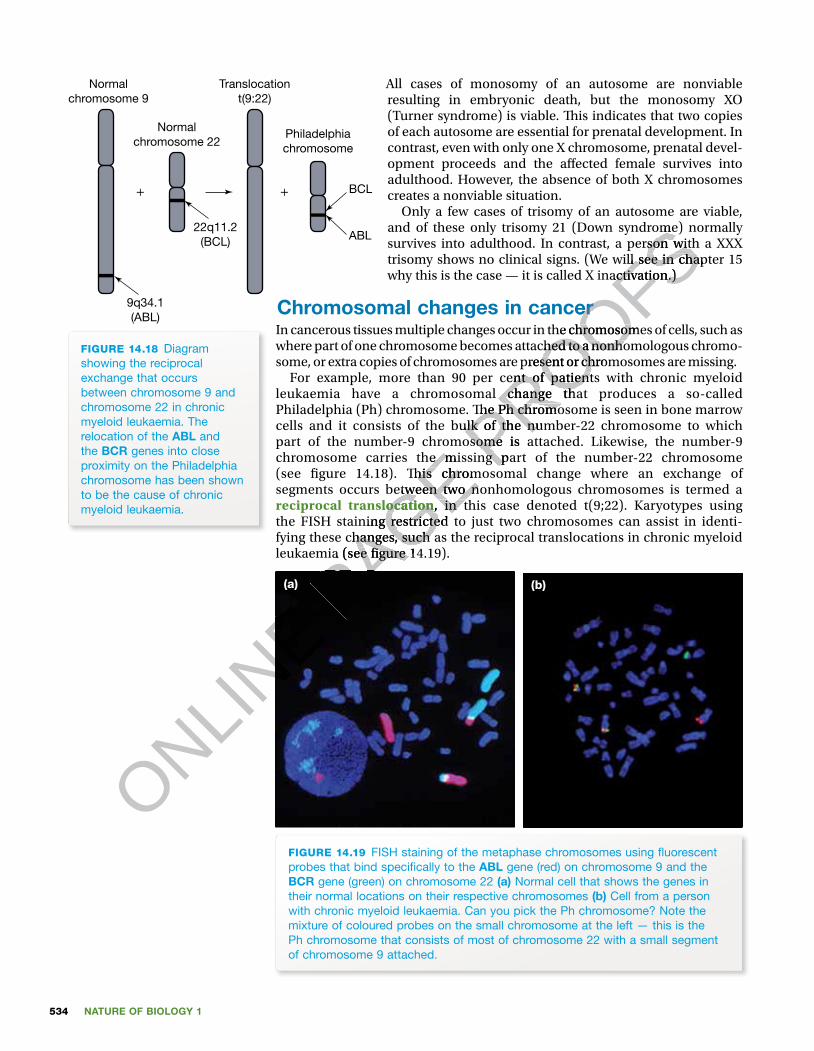
For example, more than 90 per cent of patients with chronic myeloid leukaemia have a chromosomal change that produces a so-called Philadelphia (Ph) chromosome. � e Ph chromosome is seen in bone marrow cells and it consists of the bulk of the number-22 chromosome to which part of the number-9 chromosome is attached. Likewise, the number-9 chromosome carries the missing part of the number-22 chromosome (see � gure 14.18). � is chromosomal change where an exchange of segments occurs between two nonhomologous chromosomes is termed a reciprocal translocation, in this case denoted t(9;22). Karyotypes using the FISH staining restricted to just two chromosomes can assist in identi-fying these changes, such as the reciprocal translocations in chronic myeloid leukaemia (see � gure 14.19).
(a) (b)
FIGURE 14.19 FISH staining of the metaphase chromosomes using � uorescent probes that bind speci� cally to the ABL gene (red) on chromosome 9 and the BCR gene (green) on chromosome 22 (a) Normal cell that shows the genes in their normal locations on their respective chromosomes (b) Cell from a person with chronic myeloid leukaemia. Can you pick the Ph chromosome? Note the mixture of coloured probes on the small chromosome at the left — this is the Ph chromosome that consists of most of chromosome 22 with a small segment of chromosome 9 attached.
Normalchromosome 9
Normalchromosome 22
Philadelphiachromosome
Translocationt(9:22)
22q11.2(BCL)
BCL
ABL
+ +
9q34.1(ABL)
FIGURE 14.18 Diagram showing the reciprocal exchange that occurs between chromosome 9 and chromosome 22 in chronic myeloid leukaemia. The relocation of the ABL and the BCR genes into close proximity on the Philadelphia chromosome has been shown to be the cause of chronic myeloid leukaemia.
ONLINE
ONLINE P
AGE part of the number-9 chromosome is attached. Likewise, the number-9
PAGE part of the number-9 chromosome is attached. Likewise, the number-9 chromosome carries the missing part of the number-22 chromosome
PAGE chromosome carries the missing part of the number-22 chromosome (see � gure 14.18). � is chromosomal change where an exchange of
PAGE (see � gure 14.18). � is chromosomal change where an exchange of segments occurs between two nonhomologous chromosomes is termed a
PAGE segments occurs between two nonhomologous chromosomes is termed a
PAGE reciprocal translocation
PAGE reciprocal translocation, in this case denoted t(9;22). Karyotypes using
PAGE , in this case denoted t(9;22). Karyotypes using
the FISH staining restricted to just two chromosomes can assist in identi-
PAGE the FISH staining restricted to just two chromosomes can assist in identi-fying these changes, such as the reciprocal translocations in chronic myeloid
PAGE fying these changes, such as the reciprocal translocations in chronic myeloid
PAGE leukaemia (see � gure 14.19).
PAGE leukaemia (see � gure 14.19).
PAGE PROOFS
and of these only trisomy 21 (Down syndrome) normally
PROOFSand of these only trisomy 21 (Down syndrome) normally survives into adulthood. In contrast, a person with a XXX
PROOFSsurvives into adulthood. In contrast, a person with a XXX trisomy shows no clinical signs. (We will see in chapter 15
PROOFStrisomy shows no clinical signs. (We will see in chapter 15 why this is the case — it is called X inactivation.)
PROOFSwhy this is the case — it is called X inactivation.)
Chromosomal changes in cancer
PROOFSChromosomal changes in cancerIn cancerous tissues multiple changes occur in the chromosomes of cells, such as
PROOFSIn cancerous tissues multiple changes occur in the chromosomes of cells, such as where part of one chromosome becomes attached to a nonhomologous chromo-
PROOFSwhere part of one chromosome becomes attached to a nonhomologous chromo-some, or extra copies of chromosomes are present or chromosomes are missing.
PROOFSsome, or extra copies of chromosomes are present or chromosomes are missing.
For example, more than 90 per cent of patients with chronic myeloid
PROOFSFor example, more than 90 per cent of patients with chronic myeloid
leukaemia have a chromosomal change that produces a so-called
PROOFSleukaemia have a chromosomal change that produces a so-called Philadelphia (Ph) chromosome. � e Ph chromosome is seen in bone marrow
PROOFS
Philadelphia (Ph) chromosome. � e Ph chromosome is seen in bone marrow cells and it consists of the bulk of the number-22 chromosome to which PROOFS
cells and it consists of the bulk of the number-22 chromosome to which part of the number-9 chromosome is attached. Likewise, the number-9 PROOFS
part of the number-9 chromosome is attached. Likewise, the number-9 chromosome carries the missing part of the number-22 chromosome PROOFS
chromosome carries the missing part of the number-22 chromosome
535CHAPTER 14 Chromosomes: carriers of genes
‘Jane is the third of four children born into our family. When she was born, little did we realise the extent to which our ideas about disability and life chances would change. We were shocked, saddened and confused about what it might mean to have a child with Down syndrome. We didn’t know very much about it; however, we were fortunate to have doctors who provided us with as much information as possible and urged us to treat her just like any other baby. Jane received the same attention, the same love and the same opportunities for learning and socialising that her brothers experienced.
Jane’s early development proceeded through the same stages as for other children, but more slowly. She sat up at 10 months, crawled at 15 months, walked when she was 26 months old and talked by the time she was three and a half. We came to believe that Jane could learn to do most things that other children learn, but that the learning process would take longer. She was happy to watch others but wouldn’t necessarily initiate action as much as other children do, so we ensured that Jane was actively involved in as many play experiences as possible.
When Jane was three and a half, she attended a Day Training Centre for the Intellectually Handicapped, as such centres were called then. She became involved in music, painting, solving jigsaws, and so on. However, she was still doing much the same things two years later and her opportunities for aca-demic learning were very limited. We believed that Jane was capable of learning much more than was expected of her at the centre. When Jane was � ve, she had the opportunity of attending the local kinder-garten for two days a week where she socialised with children with a much broader range of abilities. At six, Jane began to learn how to read. Having been a primary teacher, I taught Jane at home and was delighted to � nd that she learned with relative ease. From then on, we never assumed what Jane may or may not be capable of learning, but provided oppor-tunities for her to learn. At this time the integration debate began, and it reinforced my conviction that Jane should attend her local school with her brothers and neighbourhood peers. So when Jane was seven, she was admitted at our local school and had her needs met in the same way as students. Although Jane was two years older than most of the other chil-dren, she was very small and was at their level devel-opmentally, so her placement was appropriate.
In looking for secondary school options for Jane, we were delighted when she was accepted into a small Catholic girls’ school that catered for indi-vidual di� erences in an inclusive way. Jane attended secondary school for � ve years, after which time the
work was becoming too complex and di� cult for her, so a job placement seemed to be a more appropriate option. Her integration into the mainstream of edu-cation broadened Jane’s options for integration into the workforce. Since leaving school she has been employed in fast-food businesses and in retail stores. For nearly 15 years she has been employed in a Target store under a productivity-based pay scheme, whereby she is paid according to her level of prod-uctivity, as measured against the average worker. Under this scheme, she receives a disability pension reduced according to her wage. She now needs less support and has even had some long-service leave.
Jane knows that she has Down syndrome and has a simple understanding of what that means genetically, but she doesn’t see herself as disabled. Indeed, she continues to learn and has reached a level of indepen-dence that we would not have believed possible.
She has had piano lessons and is interested in TV, books, music and dancing. She is capable of travel-ling to and from work, shopping by herself, operating quite complex video, DVD and internet technology, arranging social outings with her friends and making appointments for herself. Jane has had lessons in cooking and budgeting to enable her to achieve greater independence. Although Jane received her ‘L’ plates ready for driving lessons, she didn’t in fact take them. � e important thing is that she had the opportunity to learn.
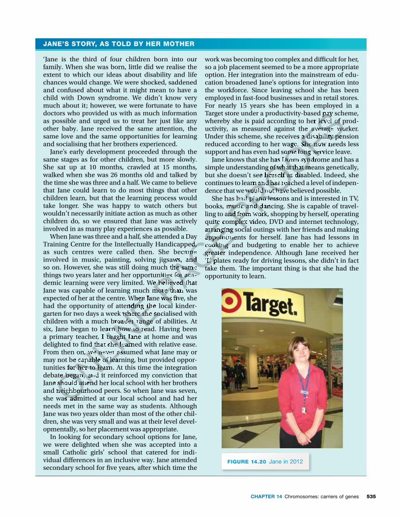
FIGURE 14.20 Jane in 2012
JANE’S STORY, AS TOLD BY HER MOTHER
ONLINE expected of her at the centre. When Jane was � ve, she
ONLINE expected of her at the centre. When Jane was � ve, she
had the opportunity of attending the local kinder-
ONLINE had the opportunity of attending the local kinder-
garten for two days a week where she socialised with
ONLINE garten for two days a week where she socialised with
children with a much broader range of abilities. At
ONLINE
children with a much broader range of abilities. At six, Jane began to learn how to read. Having been
ONLINE
six, Jane began to learn how to read. Having been a primary teacher, I taught Jane at home and was
ONLINE
a primary teacher, I taught Jane at home and was delighted to � nd that she learned with relative ease.
ONLINE
delighted to � nd that she learned with relative ease. From then on, we never assumed what Jane may or
ONLINE
From then on, we never assumed what Jane may or may not be capable of learning, but provided oppor-
ONLINE
may not be capable of learning, but provided oppor-tunities for her to learn. At this time the integration
ONLINE
tunities for her to learn. At this time the integration debate began, and it reinforced my conviction that
ONLINE
debate began, and it reinforced my conviction that Jane should attend her local school with her brothers ONLIN
E
Jane should attend her local school with her brothers and neighbourhood peers. So when Jane was seven, ONLIN
E
and neighbourhood peers. So when Jane was seven, she was admitted at our local school and had her ONLIN
E
she was admitted at our local school and had her
PAGE Training Centre for the Intellectually Handicapped,
PAGE Training Centre for the Intellectually Handicapped, as such centres were called then. She became
PAGE as such centres were called then. She became involved in music, painting, solving jigsaws, and
PAGE involved in music, painting, solving jigsaws, and so on. However, she was still doing much the same
PAGE so on. However, she was still doing much the same things two years later and her opportunities for aca-
PAGE things two years later and her opportunities for aca-demic learning were very limited. We believed that PAGE demic learning were very limited. We believed that Jane was capable of learning much more than was PAGE Jane was capable of learning much more than was expected of her at the centre. When Jane was � ve, she PAGE
expected of her at the centre. When Jane was � ve, she had the opportunity of attending the local kinder-PAGE
had the opportunity of attending the local kinder-
quite complex video, DVD and internet technology,
PAGE quite complex video, DVD and internet technology, arranging social outings with her friends and making
PAGE arranging social outings with her friends and making appointments for herself. Jane has had lessons in
PAGE appointments for herself. Jane has had lessons in cooking and budgeting to enable her to achieve
PAGE cooking and budgeting to enable her to achieve greater independence. Although Jane received her
PAGE greater independence. Although Jane received her ‘L’ plates ready for driving lessons, she didn’t in fact
PAGE ‘L’ plates ready for driving lessons, she didn’t in fact take them. � e important thing is that she had the
PAGE take them. � e important thing is that she had the
PROOFSTarget store under a productivity-based pay scheme,
PROOFSTarget store under a productivity-based pay scheme, whereby she is paid according to her level of prod-
PROOFSwhereby she is paid according to her level of prod-uctivity, as measured against the average worker.
PROOFSuctivity, as measured against the average worker. Under this scheme, she receives a disability pension
PROOFSUnder this scheme, she receives a disability pension reduced according to her wage. She now needs less
PROOFSreduced according to her wage. She now needs less support and has even had some long-service leave.
PROOFSsupport and has even had some long-service leave.
Jane knows that she has Down syndrome and has a
PROOFSJane knows that she has Down syndrome and has a
simple understanding of what that means genetically,
PROOFSsimple understanding of what that means genetically, but she doesn’t see herself as disabled. Indeed, she
PROOFSbut she doesn’t see herself as disabled. Indeed, she
PROOFScontinues to learn and has reached a level of indepen-
PROOFScontinues to learn and has reached a level of indepen-dence that we would not have believed possible.
PROOFSdence that we would not have believed possible.
She has had piano lessons and is interested in TV,
PROOFSShe has had piano lessons and is interested in TV,
books, music and dancing. She is capable of travel-PROOFS
books, music and dancing. She is capable of travel-ling to and from work, shopping by herself, operating PROOFS
ling to and from work, shopping by herself, operating quite complex video, DVD and internet technology, PROOFS
quite complex video, DVD and internet technology, arranging social outings with her friends and making PROOFS
arranging social outings with her friends and making
NATURE OF BIOLOGY 1536
KEY IDEAS
■ Each species has a characteristic number of chromosomes, known as the diploid number, typically present in body (somatic) cells.
■ Human chromosomes in body cells exist in pairs, normally 23 pairs. ■ The 23 pairs of human chromosomes include 22 pairs of autosomes, present in both sexes, and one pair of sex chromosomes, XX in the female and XY in the male.
■ Chromosome sets can be organised into karyotypes. ■ An ideogram is a stylised diagrammatic representation of chromosomes. ■ Additional or missing entire chromosomes or parts of chromosomes are readily identi� ed from an analysis of karyotypes.
■ Certain syndromes result from chromosomal changes. ■ Cancers are associated with chromosomal changes.
QUICK CHECK
1 Identify a key difference between the members of the following pairs.a Ideogram and karyotypeb Autosome and sex chromsomec Haploid and diploidd Centromere and kinetochoree Monosomy and trisomyf Deletion and duplication of a chromosome
2 Identify whether each of the following statements is true or false.a Colchicine is a poison that arrests dividing cells at metaphase. b The two number-3 chromosomes constitute a nonhomologous pair. c Prader-Willi syndrome involves a small deletion of chromosome 15.d Two forms of Down syndrome occur, a trisomy form and a translocation
form.3 Brie� y explain why the chromosomal make-up of a person is more easily
analysed using a karyotype than a metaphase spread.4 What is the Philadelphia chromosome?
Chromosomes: genes carriersMendel (see chapter 13) postulated that his factors were separate particles, behaving independently of each other. However, experiments by W Bateson (1851–1926) and RC Punnett (1875–1967) in England in the early 1900s showed
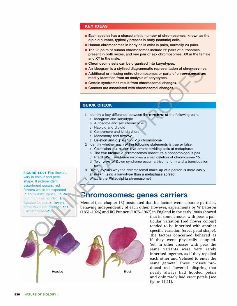
that in some crosses with peas a par-ticular variation (red � ower colour) tended to be inherited with another speci� c variation (erect petal shape). � e factors concerned behaved as if they were physically coupled. Yet, in other crosses with peas the same variants were very rarely inherited together, as if they repelled each other and ‘refused to enter the same gamete’. � ese crosses pro-duced red � owered o� spring that nearly always had hooded petals and only rarely had erect petals (see � gure 14.21).
Hooded Erect
FIGURE 14.21 Pea � owers vary in colour and petal shape. If independent assortment occurs, red � owers would be expected to show erect petals just as commonly as hooded, and likewise for purple � owers. What result did Bateson and Punnett observe?
ONLINE
ONLINE
ONLINE 4
ONLINE 4 What is the Philadelphia chromosome?
ONLINE What is the Philadelphia chromosome?
Chromosomes: genes carriers
ONLINE
Chromosomes: genes carriersMendel (see chapter 13) postulated that his factors were separate particles,
ONLINE
Mendel (see chapter 13) postulated that his factors were separate particles,
ONLINE
ONLINE
ONLINE
ONLINE
to show erect petals just as
ONLINE
to show erect petals just as commonly as hooded, and
ONLINE
commonly as hooded, and likewise for purple � owers.
ONLINE
likewise for purple � owers. What result did Bateson and
ONLINE
What result did Bateson and Punnett observe?
ONLINE
Punnett observe?
ONLINE P
AGE Centromere and kinetochore
PAGE Centromere and kinetochoreMonosomy and trisomy
PAGE Monosomy and trisomyDeletion and duplication of a chromosome
PAGE Deletion and duplication of a chromosomeIdentify whether each of the following statements is true or false.
PAGE Identify whether each of the following statements is true or false.
Colchicine is a poison that arrests dividing cells at metaphase.
PAGE Colchicine is a poison that arrests dividing cells at metaphase. The two number-3 chromosomes constitute a nonhomologous pair.
PAGE The two number-3 chromosomes constitute a nonhomologous pair. Prader-Willi syndrome involves a small deletion of chromosome 15.
PAGE Prader-Willi syndrome involves a small deletion of chromosome 15.Two forms of Down syndrome occur, a trisomy form and a translocation
PAGE Two forms of Down syndrome occur, a trisomy form and a translocation form.
PAGE form.
Brie� y explain why the chromosomal make-up of a person is more easily PAGE Brie� y explain why the chromosomal make-up of a person is more easily analysed using a karyotype than a metaphase spread.PAGE
analysed using a karyotype than a metaphase spread.What is the Philadelphia chromosome? PAGE
What is the Philadelphia chromosome?
PROOFS
PROOFS
PROOFSAn ideogram is a stylised diagrammatic representation of chromosomes.
PROOFSAn ideogram is a stylised diagrammatic representation of chromosomes.Additional or missing entire chromosomes or parts of chromosomes are
PROOFSAdditional or missing entire chromosomes or parts of chromosomes are
Certain syndromes result from chromosomal changes.
PROOFSCertain syndromes result from chromosomal changes. Cancers are associated with chromosomal changes.
PROOFSCancers are associated with chromosomal changes.
PROOFS
PROOFS
PROOFSIdentify a key difference between the members of the following pairs.
PROOFSIdentify a key difference between the members of the following pairs.
Autosome and sex chromsomePROOFS
Autosome and sex chromsome
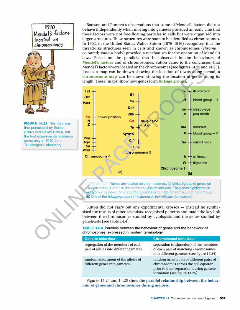
537CHAPTER 14 Chromosomes: carriers of genes
Bateson and Punnett’s observations that some of Mendel’s factors did not behave independently when moving into gametes provided an early clue that these factors were not free-� oating particles in cells but were organised into larger structures. � ese structures were soon to be identi� ed as chromosomes. In 1902, in the United States, Walter Sutton (1876–1916) recognised that the thread-like structures seen in cells and known as chromosomes (chromo = coloured; soma = body) provided a mechanism for the operation of Mendel’s laws. Based on the parallels that he observed in the behaviours of Mendel’s factors and of chromosomes, Sutton came to the conclusion that Mendel’s factors were located on the chromosomes (see � gures 14.22 and 14.23). Just as a map can be drawn showing the location of towns along a road, a chromosome map can be drawn showing the location of genes along its length. � ese ‘maps’ show how genes form linkage groups.
yellow skinw
H
p
ma
P
se
Na
h
Fl
blood group—H
pea comb
marbled
blood group—P
sleepy-eye
naked neck
silkiness
�ightless
(b) Chromosome 1
(a)
Chromosome 4Chromosome 5
LatBt
Pa
Det
Nlb
Gp
Te
Sym16
Cri
Fs
Brz
Was
Fa �ower positionunripe podcolour
NZ
PimAgeSil
Wsp
FIGURE 14.23 Genes are located on chromosomes. (a) Linked group of genes on chromosomes 4 and 5 of the edible pea (Pisum sativum). The genes highlighted in red are two of the seven genes that Mendel used in his crosses (refer to � gure 13.27). (b) One of the linkage groups in the domestic fowl (Gallus domesticus)
Sutton did not carry out any experimental crosses — instead he synthe-sised the results of other scientists, recognised patterns and made the key link between the chromosomes studied by cytologists and the genes studied by geneticists (see table 14.4).
TABLE 14.4 Parallels between the behaviour of genes and the behaviour of chromosomes, expressed in modern terminology
Genetic behaviour Chromosomal behaviour
segregation of the members of each pair of alleles into di� erent gametes
separation (disjunction) of the members of each pair of matching chromosomes into di� erent gametes (see � gure 14.24)
random assortment of the alleles of di� erent genes into gametes
random orientation of di� erent pairs of chromosomes across the cell equator prior to their separation during gamete formation (see � gure 14.25)
Figures 14.24 and 14.25 show the parallel relationship between the behav-iour of genes and chromosomes during meiosis.
FIGURE 14.22 This idea was � rst postulated by Sutton (1902) and Boveri (1903), but the � rst experimental evidence came only in 1910 from TH Morgan’s laboratory.
ONLINE
ONLINE
ONLINE FIGURE 14.23
ONLINE FIGURE 14.23 chromosomes 4 and 5 of the edible pea (
ONLINE chromosomes 4 and 5 of the edible pea (
ONLINE
ONLINE red are two of the seven genes that Mendel used in his crosses (refer to � gure 13.27).
ONLINE red are two of the seven genes that Mendel used in his crosses (refer to � gure 13.27). (b)
ONLINE (b)
ONLINE P
AGE
PAGE
PAGE Chromosome 4
PAGE Chromosome 4
PAGE Chromosome 5
PAGE Chromosome 5
PAGE
PAGE
PAGE Sym16
PAGE Sym16
Cri
PAGE Cri
PAGE
PAGE
PAGE
PAGE Fs
PAGE Fs
PAGE
PAGE
PAGE
PAGE
PAGE
PAGE
FIGURE 14.23 PAGE
FIGURE 14.23 PAGE PROOFS
Mendel’s factors were located on the chromosomes (see � gures 14.22 and 14.23).
PROOFSMendel’s factors were located on the chromosomes (see � gures 14.22 and 14.23). Just as a map can be drawn showing the location of towns along a road, a
PROOFSJust as a map can be drawn showing the location of towns along a road, a can be drawn showing the location of genes along its
PROOFS can be drawn showing the location of genes along its linkage groups
PROOFSlinkage groups.
PROOFS.
PROOFS
PROOFS
PROOFS
PROOFS
PROOFS
PROOFS
PROOFS
PROOFS
PROOFS
PROOFS
PROOFSyellow skin
PROOFSyellow skinw
PROOFSw
H
PROOFSH
PROOFS
PROOFS
PROOFS
PROOFS
PROOFS
PROOFS
PROOFS
PROOFS
PROOFS
PROOFS
PROOFS
PROOFS
PROOFS
PROOFS
PROOFS
PROOFS
PROOFS
PROOFS
PROOFS
PROOFS
PROOFS
PROOFS
PROOFS
PROOFS
unripe pod
PROOFS
unripe podcolourPROOFS
colour
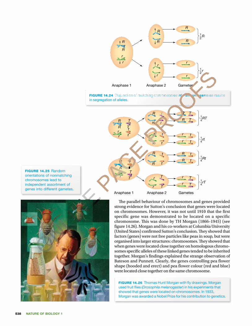
NATURE OF BIOLOGY 1538
1–2
R
1–2
r
Anaphase 1 Anaphase 2 Gametes
R
R
r
r
R
R
r
r
R
R
r
r
FIGURE 14.24 Disjunction of matching chromosomes into different gametes results in segregation of alleles.
1–4RT
1–4rt
1–4Rt
1–4rT
T
T T
TR
Rrr
T
Tt
t
R
R R
r
r
R
r
r
Anaphase 1 Anaphase 2 Gametes
t
t t
t
R
Rrr
T
T
t
tR
R R
r
r
R
r
r
t
t t
t
T
T T
T
FIGURE 14.25 Random orientations of nonmatching chromosomes lead to independent assortment of genes into different gametes.
� e parallel behaviour of chromosomes and genes provided strong evidence for Sutton’s conclusion that genes were located on chromosomes. However, it was not until 1910 that the � rst specific gene was demonstrated to be located on a specific chromosome. � is was done by TH Morgan (1866–1945) (see � gure 14.26). Morgan and his co-workers at Columbia University (United States) con� rmed Sutton’s conclusion. � ey showed that factors (genes) were not free particles like peas in soup, but were organised into larger structures: chromosomes. � ey showed that when genes were located close together on homologous chromo-somes speci� c alleles of these linked genes tended to be inherited together. Morgan’s � ndings explained the strange observation of Bateson and Punnett. Clearly, the genes controlling pea � ower shape (hooded and erect) and pea � ower colour (red and blue) were located close together on the same chromosome.
FIGURE 14.26 Thomas Hunt Morgan with � y drawings. Morgan used fruit � ies (Drosophila melanogaster) in his experiments that showed that genes were located on chromosomes. In 1933, Morgan was awarded a Nobel Prize for his contribution to genetics.
ONLINE
ONLINE
ONLINE P
AGE
PAGE R
PAGE R
R
PAGE Rr
PAGE rr
PAGE r
PAGE
PAGE
PAGE
PAGE
PAGE
PAGE
PAGE
PAGE
PAGE
PAGE
PAGE
PAGE
PAGE
PAGE
PAGE
PAGE
PAGE
PAGE
PAGE
PAGE
PAGE
PAGE
PAGE
PAGE
PAGE
PAGE
PAGE
PAGE
PAGE
PAGE
PAGE
PAGE t
PAGE t
t
PAGE t
PAGE
PAGE
PAGE
PAGE
PAGE
PAGE
PAGE
PAGE
PAGE
PAGE
PAGE PROOFSAnaphase 1 Anaphase 2 Gametes
PROOFSAnaphase 1 Anaphase 2 Gametes
PROOFS
PROOFSDisjunction of matching chromosomes into different gametes results
PROOFSDisjunction of matching chromosomes into different gametes results
PROOFS
PROOFS
PROOFS
PROOFS
PROOFS
PROOFS
PROOFS
PROOFS
PROOFS
PROOFST
PROOFST
PROOFS
PROOFS
PROOFS
PROOFS
PROOFS
PROOFS
PROOFST
PROOFST
PROOFS
PROOFS
PROOFS
PROOFS
PROOFSR
PROOFSR
R
PROOFSR
PROOFS
PROOFS
PROOFS
PROOFS
PROOFS
PROOFS
PROOFS
PROOFS
PROOFS
rPROOFS
rPROOFS
PROOFS
PROOFS
PROOFS
PROOFS
PROOFS
PROOFS
PROOFS
rPROOFS
rPROOFS
PROOFS
PROOFS
PROOFS
PROOFS
PROOFS
PROOFS
PROOFS
PROOFS
PROOFS
tPROOFS
tPROOFS
PROOFS
PROOFS
PROOFS
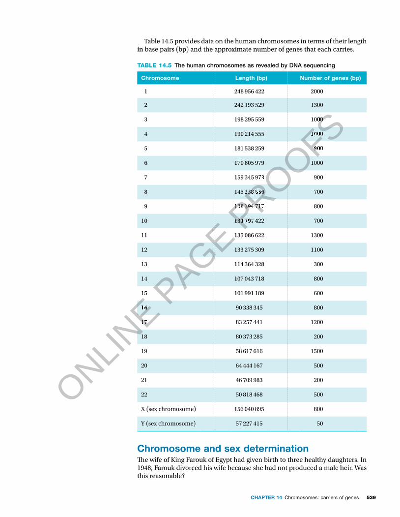
539CHAPTER 14 Chromosomes: carriers of genes
Table 14.5 provides data on the human chromosomes in terms of their length in base pairs (bp) and the approximate number of genes that each carries.
TABLE 14.5 The human chromosomes as revealed by DNA sequencing
Chromosome Length (bp) Number of genes (bp)
1 248 956 422 2000
2 242 193 529 1300
3 198 295 559 1000
4 190 214 555 1000
5 181 538 259 900
6 170 805 979 1000
7 159 345 973 900
8 145 138 636 700
9 138 394 717 800
10 133 797 422 700
11 135 086 622 1300
12 133 275 309 1100
13 114 364 328 300
14 107 043 718 800
15 101 991 189 600
16 90 338 345 800
17 83 257 441 1200
18 80 373 285 200
19 58 617 616 1500
20 64 444 167 500
21 46 709 983 200
22 50 818 468 500
X (sex chromosome) 156 040 895 800
Y (sex chromosome) 57 227 415 50
Chromosome and sex determination�e wife of King Farouk of Egypt had given birth to three healthy daughters. In 1948, Farouk divorced his wife because she had not produced a male heir. Was this reasonable?
ONLINE
ONLINE
ONLINE 16
ONLINE 16
17
ONLINE
17
PAGE
PAGE
PAGE
PAGE
PAGE
PAGE
PAGE
PAGE
PAGE 135
PAGE 135PROOFS
PROOFS
PROOFS
PROOFS
PROOFS
PROOFS
PROOFS
PROOFS
PROOFS
PROOFS1000
PROOFS1000
1000
PROOFS1000
900
PROOFS900
1000
PROOFS1000
973
PROOFS973
138
PROOFS138
PROOFS 636
PROOFS636
138PROOFS
138 PROOFS
394PROOFS
394 PROOFS
717PROOFS
717
133PROOFS
133 PROOFS
797PROOFS
797 422PROOFS
422
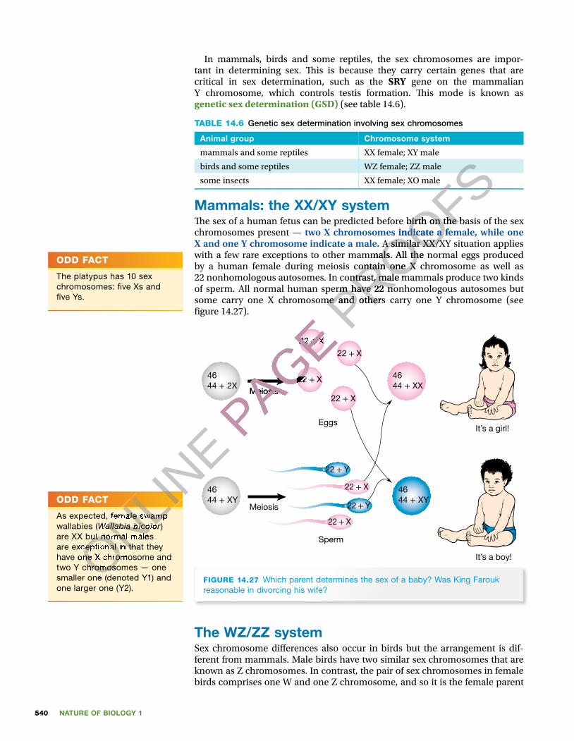
NATURE OF BIOLOGY 1540
In mammals, birds and some reptiles, the sex chromosomes are impor-tant in determining sex. � is is because they carry certain genes that are critical in sex determination, such as the SRY gene on the mammalian Y chromosome, which controls testis formation. � is mode is known as genetic sex determination (GSD) (see table 14.6).
TABLE 14.6 Genetic sex determination involving sex chromosomes
Animal group Chromosome system
mammals and some reptiles XX female; XY male
birds and some reptiles WZ female; ZZ male
some insects XX female; XO male
Mammals: the XX/XY system� e sex of a human fetus can be predicted before birth on the basis of the sex chromosomes present — two X chromosomes indicate a female, while one X and one Y chromosome indicate a male. A similar XX/XY situation applies with a few rare exceptions to other mammals. All the normal eggs produced by a human female during meiosis contain one X chromosome as well as 22 nonhomologous autosomes. In contrast, male mammals produce two kinds of sperm. All normal human sperm have 22 nonhomologous autosomes but some carry one X chromosome and others carry one Y chromosome (see � gure 14.27).
Meiosis
EggsIt’s a girl!
It’s a boy!
Sperm
Meiosis
22 + X
22 + X
22 + X
22 + Y
22 + Y
22 + X
22 + X
22 + X
4644 + XY
4644 + XY
4644 + 2X
4644 + XX
FIGURE 14.27 Which parent determines the sex of a baby? Was King Farouk reasonable in divorcing his wife?
The WZ/ZZ systemSex chromosome di� erences also occur in birds but the arrangement is dif-ferent from mammals. Male birds have two similar sex chromosomes that are known as Z chromosomes. In contrast, the pair of sex chromosomes in female birds comprises one W and one Z chromosome, and so it is the female parent
ODD FACT
The platypus has 10 sex chromosomes: � ve Xs and � ve Ys.
ODD FACT
As expected, female swamp wallabies (Wallabia bicolor) are XX but normal males are exceptional in that they have one X chromosome and two Y chromosomes — one smaller one (denoted Y1) and one larger one (Y2).
ODD FACT
We tend to think of sex as determined for life, but individual � sh of several species have the ability to change sex during their lifetimes.
ONLINE
ONLINE
ONLINE
ONLINE
As expected, female swamp
ONLINE
As expected, female swamp Wallabia bicolor
ONLINE
Wallabia bicolorare XX but normal males
ONLINE
are XX but normal males are exceptional in that they ONLIN
E
are exceptional in that they have one X chromosome and ONLIN
E
have one X chromosome and ONLINE
two Y chromosomes — one ONLINE
two Y chromosomes — one smaller one (denoted Y1) and ONLIN
E
smaller one (denoted Y1) and
PAGE Meiosis
PAGE Meiosis
PAGE
PAGE
PAGE
PAGE
PAGE
PAGE 22
PAGE 22 22
PAGE 22 +
PAGE + X
PAGE X
22
PAGE 22
PROOFS
PROOFS
PROOFS
PROOFS� e sex of a human fetus can be predicted before birth on the basis of the sex
PROOFS� e sex of a human fetus can be predicted before birth on the basis of the sex
two X chromosomes indicate a female, while one
PROOFStwo X chromosomes indicate a female, while one
A similar XX/XY situation applies
PROOFS A similar XX/XY situation applies
with a few rare exceptions to other mammals. All the normal eggs produced
PROOFSwith a few rare exceptions to other mammals. All the normal eggs produced by a human female during meiosis contain one X chromosome as well as
PROOFSby a human female during meiosis contain one X chromosome as well as 22 nonhomologous autosomes. In contrast, male mammals produce two kinds
PROOFS22 nonhomologous autosomes. In contrast, male mammals produce two kinds of sperm. All normal human sperm have 22 nonhomologous autosomes but
PROOFS
of sperm. All normal human sperm have 22 nonhomologous autosomes but some carry one X chromosome and others carry one Y chromosome (see PROOFS
some carry one X chromosome and others carry one Y chromosome (see
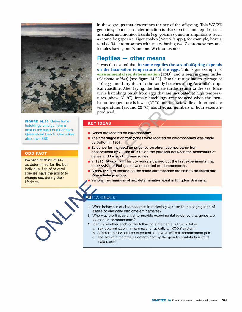
541CHAPTER 14 Chromosomes: carriers of genes
In mammals, birds and some reptiles, the sex chromosomes are impor-tant in determining sex. � is is because they carry certain genes that are critical in sex determination, such as the SRY gene on the mammalian Y chromosome, which controls testis formation. � is mode is known as genetic sex determination (GSD) (see table 14.6).
TABLE 14.6 Genetic sex determination involving sex chromosomes
Animal group Chromosome system
mammals and some reptiles XX female; XY male
birds and some reptiles WZ female; ZZ male
some insects XX female; XO male
Mammals: the XX/XY system� e sex of a human fetus can be predicted before birth on the basis of the sex chromosomes present — two X chromosomes indicate a female, while one X and one Y chromosome indicate a male. A similar XX/XY situation applies with a few rare exceptions to other mammals. All the normal eggs produced by a human female during meiosis contain one X chromosome as well as 22 nonhomologous autosomes. In contrast, male mammals produce two kinds of sperm. All normal human sperm have 22 nonhomologous autosomes but some carry one X chromosome and others carry one Y chromosome (see � gure 14.27).
Meiosis
EggsIt’s a girl!
It’s a boy!
Sperm
Meiosis
22 + X
22 + X
22 + X
22 + Y
22 + Y
22 + X
22 + X
22 + X
4644 + XY
4644 + XY
4644 + 2X
4644 + XX
FIGURE 14.27 Which parent determines the sex of a baby? Was King Farouk reasonable in divorcing his wife?
The WZ/ZZ systemSex chromosome di� erences also occur in birds but the arrangement is dif-ferent from mammals. Male birds have two similar sex chromosomes that are known as Z chromosomes. In contrast, the pair of sex chromosomes in female birds comprises one W and one Z chromosome, and so it is the female parent
ODD FACT
The platypus has 10 sex chromosomes: � ve Xs and � ve Ys.
ODD FACT
As expected, female swamp wallabies (Wallabia bicolor) are XX but normal males are exceptional in that they have one X chromosome and two Y chromosomes — one smaller one (denoted Y1) and one larger one (Y2).
ODD FACT
We tend to think of sex as determined for life, but individual � sh of several species have the ability to change sex during their lifetimes.
in these groups that determines the sex of the o� spring. � is WZ/ZZ genetic system of sex determination is also seen in some reptiles, such as snakes and monitor lizards (e.g. goannas), and in amphibians, such as some frog species. Tiger snakes (Notechis spp.), for example, have a total of 34 chromosomes with males having two Z chromosomes and females having one Z and one W chromosome.
Reptiles — other meansIt was discovered that in some reptiles the sex of o� spring depends on the incubation temperature of the eggs. � is is an example of environmental sex determination (ESD), and is seen in green turtles (Chelonia midas) (see � gure 14.28). Female turtles lay an average of 110 eggs and bury them in the sandy beaches along Australia’s trop-ical coastline. After laying, the female turtles return to the sea. Male turtle hatchlings result from eggs that are incubated at high tempera-tures (above 31 °C), female hatchlings are produced when the incu-bation temperature is lower (27 °C and below), while at intermediate temperatures (around 29 °C) about equal numbers of both sexes are produced.
KEY IDEAS
■ Genes are located on chromosomes. ■ The � rst suggestion that genes were located on chromosomes was made by Sutton in 1902.
■ Evidence for the location of genes on chromosomes came from observations by Sutton in 1902 on the parallels between the behaviours of genes and those of chromosomes.
■ In 1910, Morgan and his co-workers carried out the � rst experiments that demonstrated that genes were located on chromosomes.
■ Genes that are located on the same chromosome are said to be linked and form a linkage group.
■ Various mechanisms of sex determination exist in Kingdom Animalia.
QUICK CHECK
5 What behaviour of chromosomes in meiosis gives rise to the segregation of alleles of one gene into different gametes?
6 Who was the � rst scientist to provide experimental evidence that genes are located on chromosomes?
7 Identify whether each of the following statements is true or false.a Sex determination in mammals is typically an XX/XY system.b A female bird would be expected to have a WZ sex chromosome pair. c The sex of a mammal is determined by the genetic contribution of its
male parent.
FIGURE 14.28 Green turtle hatchlings emerge from a nest in the sand of a northern Queensland beach. Crocodiles also have ESD.
ONLINE
ONLINE
ONLINE
ONLINE
ONLINE ■
ONLINE ■
ONLINE
ONLINE
ONLINE
QUICK CHECK
ONLINE
QUICK CHECK
PAGE Genes are located on chromosomes.
PAGE Genes are located on chromosomes.The � rst suggestion that genes were located on chromosomes was made
PAGE The � rst suggestion that genes were located on chromosomes was made by Sutton in 1902.
PAGE by Sutton in 1902.Evidence for the location of genes on chromosomes came from
PAGE Evidence for the location of genes on chromosomes came from observations by Sutton in 1902 on the parallels between the behaviours of
PAGE observations by Sutton in 1902 on the parallels between the behaviours of genes and those of chromosomes.
PAGE genes and those of chromosomes. In 1910, Morgan and his co-workers carried out the � rst experiments that
PAGE In 1910, Morgan and his co-workers carried out the � rst experiments that demonstrated that genes were located on chromosomes.
PAGE demonstrated that genes were located on chromosomes.Genes that are located on the same chromosome are said to be linked and PAGE Genes that are located on the same chromosome are said to be linked and form a linkage group.PAGE form a linkage group.PAGE
Various mechanisms of sex determination exist in Kingdom Animalia.PAGE
Various mechanisms of sex determination exist in Kingdom Animalia.
PROOFSin some reptiles the sex of o� spring depends
PROOFSin some reptiles the sex of o� spring depends
� is is an example of
PROOFS� is is an example of
(ESD), and is seen in green turtles
PROOFS (ESD), and is seen in green turtles ) (see � gure 14.28). Female turtles lay an average of
PROOFS) (see � gure 14.28). Female turtles lay an average of 110 eggs and bury them in the sandy beaches along Australia’s trop-
PROOFS110 eggs and bury them in the sandy beaches along Australia’s trop-ical coastline. After laying, the female turtles return to the sea. Male
PROOFSical coastline. After laying, the female turtles return to the sea. Male turtle hatchlings result from eggs that are incubated at high tempera-
PROOFSturtle hatchlings result from eggs that are incubated at high tempera-
C), female hatchlings are produced when the incu-
PROOFSC), female hatchlings are produced when the incu-
C and below), while at intermediate
PROOFSC and below), while at intermediate
C) about equal numbers of both sexes are
PROOFSC) about equal numbers of both sexes are
PROOFS
PROOFS
PROOFS
Genes are located on chromosomes.PROOFS
Genes are located on chromosomes.The � rst suggestion that genes were located on chromosomes was made PROOFS
The � rst suggestion that genes were located on chromosomes was made
NATURE OF BIOLOGY 1542
BIOLOGIST AT WORK
Lisette Curnow — genetic counsellor
When I completed my degree in biological science, I knew that, while I enjoyed laboratory work and had a real interest in science, especially genetics, I couldn’t see myself in the lab long term. Having heard about genetic counselling as an emerging profession, I liked the idea of combining my genetic interest with dealing with people in a clinical setting. � e role of a genetic counsellor is to provide accurate information and options, together with counselling and support to indi-viduals or families whose lives are impacted in some way by a genetic condition in themselves, their chil-dren or their extended family. It is a particularly impor-tant � eld given the dramatic advances that are being made in genetics, and complex information needs to be imparted to the public in a clear and concise way.
� e convenor of the Postgraduate Diploma in Genetic Counselling (run through Melbourne Uni-versity) recommended that I try to develop some counselling skills to see if that was something I would like to do. I volunteered at Lifeline, a tele-phone counselling service that puts volunteers through a fairly comprehensive training program, an experience that I found to be invaluable in determining whether I was interested in counsel-ling and had any skills in the area. I subsequently completed the Graduate Diploma in genetic coun-selling, then travelled for 12 months, combining backpacking with six months of work experience at the Hospital for Sick Children in Toronto. On my return to Melbourne, I took up some volunteer work before embarking on my Master of Health Sciences (Genetic Counselling) degree. � is gave me a real interest in research and I developed skills I have been able to use in my job.
While completing my masters, I worked part time in cancer genetics at Peter MacCallum Cancer Insti-tute. � is work involved meeting with individuals with a very strong family history of cancer to assess whether they may carry an inherited mutation in one of the cancer-predisposing genes. While testing for cancer predisposition is still limited, it may be available for families who meet ‘high risk’ criteria for breast cancer and some forms of bowel cancer. Assessment of people to see if they meet these cri-teria is done by genetic counsellors and other rel-evant clinicians (such as geneticists, oncologists, gastroenterologists and breast surgeons) through the various cancer genetics services.
I later worked in developing cancer services at the Royal Children’s Hospital before moving into the role as genetic counsellor to the Royal Children’s Hospital. � is was a job with paediatric focus where part of my role involved dealing with abnormal cystic � brosis results from newborn screening (the heel-prick test that is carried out on every baby born in Victoria a couple of days after birth). I also attended the weekly paediatric clinic where I saw a range of patients, such as families who may have a child with a new diag-nosis of a genetic condition, or a family member who is concerned about their risk of being a carrier of a condition that is present in the family. I now work primarily in adult neurogenetics and coordinate the predictive testing program for Huntington’s disease (HD) in Victoria. � is is a program that enables indi-viduals at risk of HD (a debilitating adult-onset neu-rodegenerative disease) to � nd out whether they will develop the condition. � is role allows me to meet people who constantly amaze me with their resilience.
I am also fortunate to be involved in coordinating the genetics component of the Master of Genetic Counselling course, as well as frequently giving lec-tures to various groups. Finally, I squeeze as much research as possible into my schedule as I am perfectly positioned to access valuable information regarding the impact of today’s genetic technology on the public.
Our genetic knowledge is constantly evolving and, as genetics does not discriminate, the families we meet are necessarily varied and multifaceted. � is ensures that there is rarely a dull moment in my day-to-day work, and we regularly encounter di� -cult ethical dilemmas that prompt stimulating dis-cussions among my colleagues as we try to resolve the best way forward for our clients. � e interpret-ation of complex genetics and technology for the people it a� ects every day is an extremely chal-lenging yet rewarding � eld in which to work.
FIGURE 14.29 Lisette Curnow
ONLINE dren or their extended family. It is a particularly impor-
ONLINE dren or their extended family. It is a particularly impor-
tant � eld given the dramatic advances that are being
ONLINE tant � eld given the dramatic advances that are being
made in genetics, and complex information needs to
ONLINE made in genetics, and complex information needs to
be imparted to the public in a clear and concise way.
ONLINE
be imparted to the public in a clear and concise way.� e convenor of the Postgraduate Diploma in
ONLINE
� e convenor of the Postgraduate Diploma in Genetic Counselling (run through Melbourne Uni-
ONLINE
Genetic Counselling (run through Melbourne Uni-versity) recommended that I try to develop some
ONLINE
versity) recommended that I try to develop some counselling skills to see if that was something I
ONLINE
counselling skills to see if that was something I would like to do. I volunteered at Lifeline, a tele-
ONLINE
would like to do. I volunteered at Lifeline, a tele-phone counselling service that puts volunteers
ONLINE
phone counselling service that puts volunteers through a fairly comprehensive training program,
ONLINE
through a fairly comprehensive training program, an experience that I found to be invaluable in
ONLINE
an experience that I found to be invaluable in determining whether I was interested in counsel-ONLIN
E
determining whether I was interested in counsel-ling and had any skills in the area. I subsequently ONLIN
E
ling and had any skills in the area. I subsequently completed the Graduate Diploma in genetic coun-ONLIN
E
completed the Graduate Diploma in genetic coun-
PAGE the idea of combining my genetic interest with dealing
PAGE the idea of combining my genetic interest with dealing with people in a clinical setting. � e role of a genetic
PAGE with people in a clinical setting. � e role of a genetic counsellor is to provide accurate information and
PAGE counsellor is to provide accurate information and options, together with counselling and support to indi-
PAGE options, together with counselling and support to indi-viduals or families whose lives are impacted in some
PAGE viduals or families whose lives are impacted in some way by a genetic condition in themselves, their chil-PAGE way by a genetic condition in themselves, their chil-dren or their extended family. It is a particularly impor-PAGE dren or their extended family. It is a particularly impor-tant � eld given the dramatic advances that are being PAGE
tant � eld given the dramatic advances that are being
couple of days after birth). I also attended the weekly
PAGE couple of days after birth). I also attended the weekly paediatric clinic where I saw a range of patients, such
PAGE paediatric clinic where I saw a range of patients, such as families who may have a child with a new diag-
PAGE as families who may have a child with a new diag-nosis of a genetic condition, or a family member who
PAGE nosis of a genetic condition, or a family member who is concerned about their risk of being a carrier of a
PAGE is concerned about their risk of being a carrier of a condition that is present in the family. I now work
PAGE condition that is present in the family. I now work primarily in adult neurogenetics and coordinate the
PAGE primarily in adult neurogenetics and coordinate the
PROOFSavailable for families who meet ‘high risk’ criteria
PROOFSavailable for families who meet ‘high risk’ criteria for breast cancer and some forms of bowel cancer.
PROOFSfor breast cancer and some forms of bowel cancer. Assessment of people to see if they meet these cri-
PROOFSAssessment of people to see if they meet these cri-teria is done by genetic counsellors and other rel-
PROOFSteria is done by genetic counsellors and other rel-evant clinicians (such as geneticists, oncologists,
PROOFSevant clinicians (such as geneticists, oncologists, gastroenterologists and breast surgeons) through the
PROOFSgastroenterologists and breast surgeons) through the various cancer genetics services.
PROOFSvarious cancer genetics services.
I later worked in developing cancer services at the
PROOFSI later worked in developing cancer services at the
Royal Children’s Hospital before moving into the role
PROOFSRoyal Children’s Hospital before moving into the role as genetic counsellor to the Royal Children’s Hospital.
PROOFSas genetic counsellor to the Royal Children’s Hospital.
PROOFS� is was a job with paediatric focus where part of my
PROOFS� is was a job with paediatric focus where part of my role involved dealing with abnormal cystic � brosis
PROOFS
role involved dealing with abnormal cystic � brosis results from newborn screening (the heel-prick test PROOFS
results from newborn screening (the heel-prick test that is carried out on every baby born in Victoria a PROOFS
that is carried out on every baby born in Victoria a couple of days after birth). I also attended the weekly PROOFS
couple of days after birth). I also attended the weekly paediatric clinic where I saw a range of patients, such PROOFS
paediatric clinic where I saw a range of patients, such

543CHAPTER 14 Chromosomes: carriers of genes
BIOCHALLENGE
Use the Human Genome Resources weblink in your eBookPLUS.
The small diagram of the human chromosomes at the upper left-hand corner allows you to click on a chromosome and see the detailed genetic information that is carried on each chromosome.
1 Click on chromosome 21. The new screen display (see � gure 14.30) will show you an ideogram of chromosome 21 and will indicate the positions of 20 of the 726 genes located on this chromosome.
Note how the genes are designated by capital letters (and, in some cases, number(s)). Don’t be overwhelmed at the amount of information! This exercise is simply to show you the remarkable detail that now exists about the human genome.
2 Now click on the 9 at the top of the screen display to see an ideogram of the human chromosome 9.
One of the 20 genes shown on this ideogram is the ABO gene. Click on the ABO link and see the information that is available about this gene. Scan this information and answer the following questions:a What type of gene is the ABO gene?
b What protein does this gene encode?
3 a Now return to the starting page in the weblink and use the Find A Gene box, located in the left-hand column, to � nd the chromosome on which the HTT gene that
FIGURE 14.30 Screenshot from the National Center for Biotechnology Information (NCBI) website
controls production of the protein huntingtin is located. Once you have the chromosomal location, click on the gene name for further information about the gene, and identify an inherited disorder caused by an altered form (allele) of the gene. Record your data in table 14.7.
TABLE 14.7
Gene symbolChromosome
locationInherited disease
HTT
CFTR
DMD
HBB
b Repeat the procedure in part (a) for the following genes. i The CFTR gene that encodes the cystic � brosis
trans-membrane conductance regulator (refer to chapter 1, pp. 36–7)
ii The DMD gene that controls production of the muscle protein dystrophin, which if abnormal results in Duchenne muscular dystrophy
iii The HBB gene that controls production of the beta polypeptide chains of the adult haemoglobin A molecule
ONLINE P
AGE Now return to the starting page in the weblink and use
PAGE Now return to the starting page in the weblink and use
the Find A Gene box, located in the left-hand column,
PAGE the Find A Gene box, located in the left-hand column,
HTT
PAGE HTT gene that
PAGE gene that
PAGE i The
PAGE i The
chapter 1, pp. 36–7)
PAGE chapter 1, pp. 36–7) ii The
PAGE ii The
muscle protein dystrophin, which if abnormal
PAGE muscle protein dystrophin, which if abnormal
PROOFSaltered form (allele) of the gene. Record your data in
PROOFSaltered form (allele) of the gene. Record your data in
PROOFS
PROOFS
PROOFS
PROOFS
PROOFS
PROOFS
PROOFS
PROOFS
PROOFS
PROOFS
PROOFS
PROOFS
PROOFS
PROOFS
PROOFSChromosome
PROOFSChromosome location
PROOFSlocation
Inherited
PROOFSInherited
Repeat the procedure in part (a) for the following genes.PROOFS
Repeat the procedure in part (a) for the following genes. i The PROOFS
i The CFTRPROOFS
CFTR gene that encodes the cystic � brosis PROOFS
gene that encodes the cystic � brosis trans-membrane conductance regulator (refer to PROOFS
trans-membrane conductance regulator (refer to
NATURE OF BIOLOGY 1544
Unit 2 Chromosomes
Sit topic test
AOS 2
Topic 2Chapter review
Key wordsAngelman syndrome autosomecentromere chromosome mapcolchicinecytogeneticist deletion Down syndrome (DS)
duplicationembryo environmental sex
determination FISH staininggenetic sex
determination (GSD)homologous
hypotonic shock treatment
ideogram karyotypekinetochore linkage groups Mendel’s factors metaphase spread
monosomy nondisjunction nonhomologousreciprocal translocation sex chromosomes telomeretranslocation trisomic
Questions 1 Making connections ➜ Use at least eight of the
chapter key words to draw a concept map relating to chromosomes. You may use other words in drawing your map.
2 Demonstrating knowledge ➜ Where would you locate each of the following?a Human cells undergoing meiosisb Haploid cells in a catc Cells containing 20 unpaired chromosomes in a
moused � e genetic instruction for your ABO blood type
3 Demonstrating your understanding ➜ Use words and/or diagrams to identify the di� erences, if any, between the items in each of the following pairs.a Haploid and diploidb Autosome and sex chromosomec Somatic cell and germline celld Gene and allele
4 Recognising patterns ➜ A newborn baby showed facial abnormalities and other signs including deformed kidneys and nails, and an unusual way of clenching its � st. Its karyotype was prepared (see � gure 14.31). Examine this karyotype.
a What is the total number of chromosomes?b What is the sex of the baby?c What abnormality is visible in the karyotype?d What name is given to this condition?
5 Match the processes in column A to the outcome in column B.
Column A Column B
meiosis exchange of segments occurs between homologous chromosomes
crossing over haploid gametes produced from a diploid cell
disjunction of homologous chromosomes
diploid number of chromosomes restored, one set from each parent
independent disjoining of nonhomologous chromosomes
alleles of one gene segregate from each other
fusion of gametes at fertilisation
random combinations of di� erent genes produced in gametes
6 Demonstrating knowledge and understanding ➜ Some chromosomes, when present as a trisomy in a human, produce obvious signs or clinical conditions in the a� ected person and are typically diagnosed soon after birth.a Which chromosomes are these?b How would these conditions be diagnosed?c Brie� y explain how a trisomy can be
produced.d A student stated: ‘Strange how the number-1 and
the number-2 chromosomes are not examples of clinically recognised trisomies. It must be that they are never involved in a nondisjunction. It looks like only a few chromosomes are subject to that kind of error’.
Carefully consider this statement and indicate whether or not you agree with this student and give a reason for your decision.
FIGURE 14.31
ONLINE Somatic cell and germline cell
ONLINE Somatic cell and germline cell
A newborn baby showed
ONLINE
A newborn baby showed facial abnormalities and other signs including
ONLINE
facial abnormalities and other signs including deformed kidneys and nails, and an unusual way
ONLINE
deformed kidneys and nails, and an unusual way of clenching its � st. Its karyotype was prepared (see
ONLINE
of clenching its � st. Its karyotype was prepared (see � gure 14.31). Examine this karyotype.
ONLINE
� gure 14.31). Examine this karyotype.
ONLINE P
AGE
PAGE � e genetic instruction for your ABO blood type
PAGE � e genetic instruction for your ABO blood type
Use words
PAGE Use words
and/or diagrams to identify the di� erences, if any,
PAGE and/or diagrams to identify the di� erences, if any, between the items in each of the following pairs.
PAGE between the items in each of the following pairs.
PAGE
PAGE meiosis
PAGE meiosis
crossing over
PAGE crossing over
disjunction of
PAGE disjunction of
PROOFSnonhomologous
PROOFSnonhomologousreciprocal translocation
PROOFSreciprocal translocation sex chromosomes
PROOFSsex chromosomes telomere
PROOFStelomeretranslocation
PROOFStranslocation trisomic
PROOFStrisomic
What is the total number of chromosomes?
PROOFS What is the total number of chromosomes? What is the sex of the baby?
PROOFS What is the sex of the baby? What abnormality is visible in the karyotype?
PROOFS What abnormality is visible in the karyotype? What name is given to this condition?
PROOFS What name is given to this condition?
Match the processes in column A to the outcome in
PROOFSMatch the processes in column A to the outcome in column B.
PROOFS
column B.
PROOFS
PROOFS
Column A PROOFS
Column A
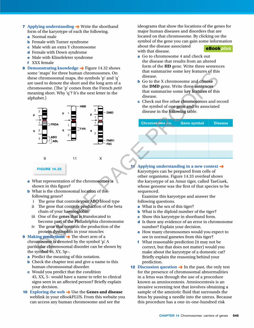
545CHAPTER 14 Chromosomes: carriers of genes
7 Applying understanding ➜ Write the shorthand form of the karyotype of each the following.a Normal maleb Female with Turner syndromec Male with an extra Y chromosomed Female with Down syndrome e Male with Klinefeleter syndromef XXX female
8 Demonstrating knowledge ➜ Figure 14.32 shows some ‘maps’ for three human chromosomes. On these chromosomal maps, the symbols ‘p’ and ‘q’ are used to denote the short and the long arm of a chromosome. (� e ‘p’ comes from the French petit meaning short. Why ‘q’? It’s the next letter in the alphabet.)
9 11 X
p
q
1
1
2
2
3
FRDA
ABOABL
p
q
1
1
2
HBB
TYR
p
q
1
2
1
2
DMD
FMR1F8C
FIGURE 14.32
a What representation of the chromosomes is shown in this � gure?
b What is the chromosomal location of the following genes? i � e gene that controls your ABO blood type ii � e gene that controls production of the beta
chain of your haemoglobiniii One of the genes that is translocated to
become part of the Philadelphia chromosomeiv � e gene that controls the production of the
protein dystrophin in your muscles 9 Making predictions ➜ � e short arm of a
chromosome is denoted by the symbol ‘p’. A particular chromosomal disorder can be shown by the symbol 46, XY, 5p–. a Predict the meaning of this notation.b Check the chapter text and give a name to this
human chromosomal disorder. c Would you predict that the condition
45, XX, 5– would have a name to refer to clinical signs seen in an a� ected person? Brie� y explain your decision.
10 Exploring the web ➜ Use the Genes and disease weblink in your eBookPLUS. From this website you can access any human chromosome and see the
ideograms that show the locations of the genes for major human diseases and disorders that are located on that chromosome. By clicking on the symbol of the gene you can gain some information about the disease associated with that disease.a Go to chromosome 4 and check out
the disease that results from an altered form of the HD gene. Write three sentences that summarise some key features of this disease.
b Go to the X chromosome and choose the DMD gene. Write three sentences that summarise some key features of this disease.
c Check out � ve other chromosomes and record the symbol of one gene and its associated disease in the following table.
Chromosome no. Gene symbol Disease
11 Applying understanding in a new context ➜ Karyotypes can be prepared from cells of other organisms. Figure 14.33 overleaf shows the karyotype of an Amur tiger, called TaeGuek, whose genome was the � rst of that species to be sequenced. Examine this karyotype and answer the following questions.a What is the sex of this tiger?b What is the diploid number of the tiger?c Show this karyotype in shorthand form.d Is there any evidence of an error in chromosome
number? Explain your decision.e How many chromosomes would you expect to
see in normal gametes from this tiger?f What reasonable prediction (it may not be
correct, but that does not matter) would you make about the karyotype of a domestic cat? Brie� y explain the reasoning behind your prediction.
12 Discussion question ➜ In the past, the only test for the presence of chromosomal abnormalities in a fetus was through the use of a procedure known as amniocentesis. Amniocentesis is an invasive screening test that involves obtaining a sample of the amniotic � uid that surrounds the fetus by passing a needle into the uterus. Because this procedure has a one-in-one-hundred risk
ONLINE What is the chromosomal location of the
ONLINE What is the chromosomal location of the
i � e gene that controls your ABO blood type
ONLINE
i � e gene that controls your ABO blood type ii � e gene that controls production of the beta
ONLINE
ii � e gene that controls production of the beta chain of your haemoglobin
ONLINE
chain of your haemoglobiniii One of the genes that is translocated to
ONLINE
iii One of the genes that is translocated to become part of the Philadelphia chromosome
ONLINE
become part of the Philadelphia chromosomeiv � e gene that controls the production of the
ONLINE
iv � e gene that controls the production of the protein dystrophin in your muscles
ONLINE
protein dystrophin in your musclesMaking predictions
ONLINE
Making predictions ➜
ONLINE
➜chromosome is denoted by the symbol ‘p’. A ONLIN
E
chromosome is denoted by the symbol ‘p’. A particular chromosomal disorder can be shown by ONLIN
E
particular chromosomal disorder can be shown by the symbol 46, XY, 5p–. ONLIN
E
the symbol 46, XY, 5p–. Predict the meaning of this notation.ONLIN
E
Predict the meaning of this notation.
PAGE What representation of the chromosomes is PAGE What representation of the chromosomes is
What is the chromosomal location of the PAGE
What is the chromosomal location of the PAGE
PAGE
PAGE
PAGE 11
PAGE 11
PROOFSthat summarise some key features of this
PROOFSthat summarise some key features of this
Go to the X chromosome and choose
PROOFS Go to the X chromosome and choose gene. Write three sentences
PROOFS gene. Write three sentences that summarise some key features of this
PROOFSthat summarise some key features of this
Check out � ve other chromosomes and record
PROOFS Check out � ve other chromosomes and record
the symbol of one gene and its associated
PROOFSthe symbol of one gene and its associated disease in the following table.
PROOFSdisease in the following table.
PROOFS
PROOFS
PROOFS
PROOFS
PROOFSChromosome no.
PROOFSChromosome no.
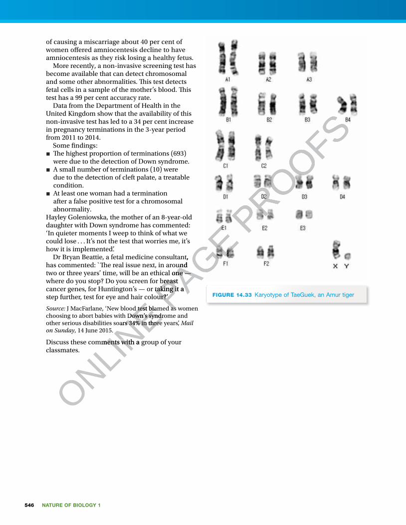
546 NATURE OF BIOLOGY 1
of causing a miscarriage about 40 per cent of women o� ered amniocentesis decline to have amniocentesis as they risk losing a healthy fetus. More recently, a non-invasive screening test has become available that can detect chromosomal and some other abnormalities. � is test detects fetal cells in a sample of the mother’s blood. � is test has a 99 per cent accuracy rate. Data from the Department of Health in the United Kingdom show that the availability of this non-invasive test has led to a 34 per cent increase in pregnancy terminations in the 3-year period from 2011 to 2014. Some � ndings:■ � e highest proportion of terminations (693)
were due to the detection of Down syndrome.■ A small number of terminations (10) were
due to the detection of cleft palate, a treatable condition.
■ At least one woman had a termination after a false positive test for a chromosomal abnormality.
Hayley Goleniowska, the mother of an 8-year-old daughter with Down syndrome has commented: ‘In quieter moments I weep to think of what we could lose . . . It’s not the test that worries me, it’s how it is implemented’. Dr Bryan Beattie, a fetal medicine consultant, has commented: `� e real issue next, in around two or three years’ time, will be an ethical one — where do you stop? Do you screen for breast cancer genes, for Huntington’s — or taking it a step further, test for eye and hair colour?’
Source: J MacFarlane, ‘New blood test blamed as women choosing to abort babies with Down’s syndrome and other serious disabilities soars 34% in three years’, Mail on Sunday, 14 June 2015.
Discuss these comments with a group of your classmates.
FIGURE 14.33 Karyotype of TaeGuek, an Amur tiger
ONLINE step further, test for eye and hair colour?’
ONLINE step further, test for eye and hair colour?’
J MacFarlane, ‘New blood test blamed as women
ONLINE J MacFarlane, ‘New blood test blamed as women
choosing to abort babies with Down’s syndrome and
ONLINE choosing to abort babies with Down’s syndrome and
ONLINE
other serious disabilities soars 34% in three years’,
ONLINE
other serious disabilities soars 34% in three years’, , 14 June 2015.
ONLINE
, 14 June 2015.
Discuss these comments with a group of your
ONLINE
Discuss these comments with a group of your
PAGE Dr Bryan Beattie, a fetal medicine consultant,
PAGE Dr Bryan Beattie, a fetal medicine consultant, has commented: `� e real issue next, in around
PAGE has commented: `� e real issue next, in around two or three years’ time, will be an ethical one —
PAGE two or three years’ time, will be an ethical one — where do you stop? Do you screen for breast
PAGE where do you stop? Do you screen for breast cancer genes, for Huntington’s — or taking it a PAGE cancer genes, for Huntington’s — or taking it a step further, test for eye and hair colour?’PAGE step further, test for eye and hair colour?’
J MacFarlane, ‘New blood test blamed as women PAGE
J MacFarlane, ‘New blood test blamed as women PAGE PROOFS