Generation and Analysis of Card9-Deficient Mice · Generation and Analysis of Card9-Deficient Mice...
93
Technische Universität München Lehrstuhl für Entwicklungsgenetik Generation and Analysis of Card9-Deficient Mice Olaf Groß Vollständiger Abdruck der von der Fakultät Wissenschaftszentrum Weihenstephan für Ernährung, Landnutzung und Umwelt der Technischen Universität München zur Erlangung des akademischen Grades eines Doktors der Naturwissenschaften (Dr. rer. nat.) genehmigten Dissertation. Vorsitzender: Univ.-Prof. Dr. Wolfgang Höll Prüfer der Dissertation: 1. Univ.-Prof. Dr. Wolfgang Wurst 2. Priv.-Doz. Dr. Jürgen Ruland 3. Priv.-Doz. Dr. Roland Hermann Lang Die Dissertation wurde am 22. Oktober 2007 bei der Technischen Universität München eingereicht und durch die Fakultät Wissenschaftszentrum Weihenstephan für Ernährung, Landnutzung und Umwelt am 29. Januar 2008 angenommen.
-
Upload
truongdiep -
Category
Documents
-
view
223 -
download
0
Transcript of Generation and Analysis of Card9-Deficient Mice · Generation and Analysis of Card9-Deficient Mice...
Technische Universität München
Lehrstuhl für Entwicklungsgenetik
Generation and Analysis of Card9-Deficient Mice
Olaf Groß
Vollständiger Abdruck der von der Fakultät Wissenschaftszentrum Weihenstephan
für Ernährung, Landnutzung und Umwelt der Technischen Universität München zur
Erlangung des akademischen Grades eines
Doktors der Naturwissenschaften (Dr. rer. nat.) genehmigten Dissertation.
Vorsitzender: Univ.-Prof. Dr. Wolfgang Höll
Prüfer der Dissertation: 1. Univ.-Prof. Dr. Wolfgang Wurst
2. Priv.-Doz. Dr. Jürgen Ruland
3. Priv.-Doz. Dr. Roland Hermann Lang
Die Dissertation wurde am 22. Oktober 2007 bei der Technischen Universität
München eingereicht und durch die Fakultät Wissenschaftszentrum Weihenstephan
für Ernährung, Landnutzung und Umwelt am 29. Januar 2008 angenommen.
2
Du bereitest vor mir einen Tisch
im Angesicht meiner Feinde.
Psalm 23,5

3
Index
ACKNOWLEDGMENTS 5
ZUSAMMENFASSUNG 5
ABSTRACT 9
FIGURE LIST 10
TABLE LIST 11
ABBREVIATIONS 12
1 INTRODUCTION 16
1.1 The Innate Immune Response 16 1.1.1 The Cellular Basis of Innate Immunity 17 1.1.2 Mechanisms of Pathogen Killing 18 1.1.3 The Interface between Innate and Adaptive Immunity. 19
1.2 The Adaptive Immune Response 19 1.2.1 B-Lymphocytes and the Humoral Immune Response 20 1.2.2 T-Lymphocytes interact with MHC molecules on host cells 21 1.2.3 NK Cells Participate in Viral and Cancer Defence 23
1.3 Immune cell activation 23 1.3.1 Receptors of Innate Immunity 25
1.4 Signal Transduction for Immune Cell Activation 28 1.4.1 NF-κB Signal Transduction 29 1.4.2 Antigen Receptor Signalling for NF-κB Activation 30 1.4.3 The Toll-like Receptor Pathway for NF-κB 32 1.4.4 Non-TLR Signalling in Innate Immunity: Dectin-1 33
1.5 Card9 35
2 MATERIALS AND METHODS 37
2.1 Material 37 2.1.1 Reagents 37 2.1.2 Primer list 37
2.2 Methods 38 2.2.1 Generation of Card9-Deficient Mice 38 2.2.2 Flow Cytometric Analysis 38

4
2.2.3 Measurement of Serum Immunoglobulin Concentrations 38 2.2.4 In Vivo Immunizations 39 2.2.5 Proliferation Assays 39 2.2.6 Generation and Stimulation of Bone Marrow Derived Dendritic Cells (BMDC) 39 2.2.7 Generation of Bone Marrow Derived Macrophages (BMDM) 40 2.2.8 Cytokine Production 41 2.2.9 Survival and Clearance of Candida albicans Infection 41 2.2.10 Restimulation of T-cells after Infection and Cytokine Measurement 41 2.2.11 Staphylococcus aureus Infection 42 2.2.12 Western Blot Analysis and Antibodies 42 2.2.13 Zymosan Uptake 43 2.2.14 Immunofluorescence 42 2.2.15 Gel Mobility Shift Assay 43 2.2.16 NF-κB Reporter Assay 43
3 RESULTS 44
3.1 Gene-Targeting of Card9 44
3.2 Regular Adaptive Immune Responses without Card9 46
3.3 Card9 Controls Zymosan-Induced Cell Activation 48
3.4 Card9 is Essential for Dectin-1/Syk Signalling 53
3.5 Card9 Relays Dectin-1/Syk Signalling to NF-κB 56
3.6 Card9 Signals via Bcl10 and Malt1 57
3.7 Card9-Dependent Activation of TH17 T Cell Responses 61
4 DISCUSSION 64
4.1 Card9 is Dispensable for Lymphocyte Activation 65
4.2 Card9 Controls Innate Immunity 65
4.3 Card9 in Anti-Fungal Immunity. 72
4.4 Activation of TH17 Responses by Card9 74
4.5 Potential Role of Card9 in the Signal Transduction of other ITAM- Receptors 75
4.6 Comparing Card9 and MyD88 Dependent Effects 76
4.7 Possible Involvement of Card9 in Cancer Development 78
4.8 Conclusion and outlook 79
CITATION INDEX 81
5
Acknowledgments
I would like to thank:
Jürgen Ruland for providing me with the opportunity to perform my doctoral thesis
work in his lab and for his constant advice and support.
Katrin Finger and Andreas Gewies, who both contributed a great deal of time and effort
to complete several aspects of this project in time for publication.
Uta Ferch and Stefanie Klemm for support and friendship during both good times and
bad.
All other members of AG Ruland, Kristina Brunner, Mercedes María Castiñeiras-
Vilariño, Christina Grupp, Nicole Hannesschläger, Philipp Jost, Stephanie Leeder, Katja
Meiners, Thomas Patzelt, Konstanze Pechloff, Dominik Straßer, Stefan Wanninger,
Stefanie Weiß and Stephanie Zimmermann.
Prof. Christian Peschel and the III. Medizinische Klinik, especially AG Bernhard and
AG Duyster.
Prof. Hermann Wagner for adopting me as an orphan of innate immunity and the
members of the Institut für Medizinische Mikrobiologie, Immunlogie und Hygiene.
Roland Lang and Tim Sparwasser for advice concerning myeloid cell preparation, TLR
stimulation and cytokine measurement.
Katharina Lahl, Michael Hammer and Martin Schäfer for their help in the same area
and for good companionship.
6
Irmgard Förster and Susie Weiß for help and support during stem cell culture and
microinjection.
Laura Layland for her valuable corrections of this manuscript.
Prof. Wolfgang Wurst for the representation of my thesis in the faculty
Wissenschaftszentrum Weihenstephan für Ernährung, Landnutzung und Umwelt and
for examining this thesis.
Prof. Wolfgang Höll for chairing the doctoral examination.
Roland Lang for examining this thesis.
Ursula und Günter Groß, to whom I not only owe my life and education, but also the
strength and will to carry on and to reach out for more.
7
Zusammenfassung
Das angeborene Immunsystem der höheren Organismen erkennt mikrobielle
Pathogene an molekularen Strukturen die typisch sind für spezielle mikrobielle
Komponenten. Toll-like Rezeptoren (TLRs) stellen eine Hauptgruppe dieser pattern-
recognition Rezeptoren (PRRs) dar, aber auch andere PRRs haben essentielle Funktion
für die Erkennung bestimmter Pathogene. Die Mechanismen, mit Hilfe derer diese PRRs
inflammatorische Signalwege aktivieren sind noch unzureichend verstanden. In dieser
Arbeit wurde die Funktion des Proteins Caspase-recruitment-domain Protein 9 (Card9)
untersucht. Mit Hilfe Card9 defizienter Mäuse konnte ein neuer nicht-TLR abhängiger
Signalweg definiert werden, der für die angeborenen Immunantwort sowie die
Aktivierung der erworbenen Immunantwort gegen Pilzinfektionen notwendig ist.
Card9 ist strukturell verwandt mit dem CARD-MAGUK (membrande-associated
guanylate kinase) protein 1 (Carma1). Carma1 vermittelt Nuclear Faktor kappa B (NF-
κB) Aktivierung über Bcl10 (B-cell lymphoma 10) und Malt1 (mucosa-associated-
lymphoid-tissue lymphoma-translocation gene 1) nach B Zell und T Zell Rezeptor
Aktivierung. Card9 bindet an Bcl10 und kann in Überexpressionsexperimenten
ebenfalls NF-κB aktivieren. Die physiologischen Funktionen von Card9 waren zu
Beginn dieser Arbeit unbekannt.
Hier konnte gezeigt werden, das T und B Zell Funktion in Card9 defizienten
Mäusen der in wildtyp Tieren entspricht, Card9 also nicht am Carma1/Bcl10 Signalweg
beteiligt ist. Am Modell von aus Knochenmark generierten Dendritischen Zellen (bone-
marrow derived dendritic cells, BMDCs) wurde gezeigt, dass die Zytokin-Antwort nach
Stimulation mit verschieden TLR-Liganden ebenfalls durch Card9 nicht beeinflusst
wird. Jedoch produzieren Card9 defiziente Dendritische Zellen im Vergleich zum
Wildtyp deutlich weniger Zytokine nach Stimulation mit dem Hefe-
Zellwandbestandteil Zymosan oder inaktivierten Candida albicans Zellen. Zudem sind
Card9 defiziente Tiere extrem Anfällig für Infektionen mit Candida albicans. Auf
molekularer Ebene wurde nachgewiesen, dass Card9 spezifische Signale vom β-Glucan
Rezeptor Dectin-1, dem wesentlichen PRR für Zymosan leitet. Hierbei kooperiert Card9
mit Bcl10 in der Aktivierung von NF-κB.
8
In weiteren Arbeiten konnte gezeigt werden, dass die Aktivierung adaptiver T-
Zell Antworten gegen Pilzinfektionen durch Dendritische Zellen ebenfalls durch den
Dectin-1/Card9 Signalweg kontrolliert wird. Durch Infektionsexperimente konnte
gezeigt werden, dass wildtyp Mäuse nach Candida albicans Infektion
antigenspezifische T-Zell Antworten aktivieren. In Card9 defizienten Tieren bleibt diese
Reaktion aus. Dabei ist von besonderem Interesse, dass durch diesen Signalweg
präferentiell TH17 T-Helferzellantworten induziert werden.
Diese Daten definieren Card9 als unentbehrlichen Teil eines neuen, TLR-
unabhängigen Signalwegs der angeborenen Immunität, der Dectin-1 nach
Pilzerkennung mit Bcl10 verbindet und NF-κB aktiviert und an die Aktivierung
adaptiver TH17 Immunantworten koppeln kann.
9
Abstract
Fungal infections are increasing worldwide with the dramatic rise in
immunodeficiencies including AIDS. However, the immune responses to such
infections remain poorly understood. Dectin-1 is the major mammalian pattern
recognition receptor for the fungal component zymosan. Dectin-1 represents the
prototype of innate non-Toll like receptors (TLRs) containing immunoreceptor tyrosine-
based activation motifs (ITAMs), which are related to those of adaptive antigen
receptors. In this work, Card9 was identified as a key transducer of Dectin-1 signalling.
Although dispensable for TLR/MyD88-induced responses, Card9 controls Dectin-1
mediated myeloid cell activation, cytokine production and innate anti-fungal immunity.
Card9 couples to Bcl10 and regulates via Bcl10/Malt1 zymosan-induced NF-κB
activation. Yet, Card9 is dispensable for antigen receptor signalling that utilizes Carma1
as a link to Bcl10/Malt1. Thus, these results define a novel innate immune pathway and
indicate that evolutionarily distinct ITAM receptors in innate and adaptive immune
cells employ diverse adaptor proteins to selectively engage the conserved Bcl10/Malt1
module.
10
Figure List
Figure 1: Receptors of innate and adaptive immunity. ...........................................................24
Figure 2: Signal transduction from B and T cell receptor to NF-κB via Bcl10........................31
Figure 3: Various receptors engage common downstream signalling components for NF-kB
activation. .........................................................................................................................34
Figure 4: Protein structure of Carma1, Card9 and Bcl10.. .....................................................36
Figure 5: Gene targeting strategy for the generation of Card9-deficient Mice.......................44
Figure 6: Verification of the deletion of Card9 and Mendelian Analysis ................................45
Figure 7: Flow cytometric analysis of lymphocytes development in Card9+/+ and -/- mice..46
Figure 8: Signal induced lymphocyte proliferation..................................................................47
Figure 9: NF-κB activation after antigen receptor stimulation. ...............................................48
Figure 10: Humoral immune response ...................................................................................49
Figure 11: Card9 Expression analysis by RT-PCR of the Card9 transcript in in tissues and
cell lines............................................................................................................................50
Figure 12: Cytokine production in Card9 deficient dendritic cells in response to zymosan
stimulation ........................................................................................................................51
Figure 13: Dose dependent TNF-α, IL-6, IL-2, IL-10 or IL-12 production from Card9+/+, +/-
or -/- BMDCs after 24 h of zymosan stimulation..............................................................52
Figure 14: Dose dependent TNF-α or IL-6 production from Card9+/- or -/- BMDCs after 24 h
of LPS stimulation ............................................................................................................53
Figure 15: Dose dependent Candida albicans (strain SC5314) induced TNF-α, IL-6, IL-2, IL-
10 or IL-12 production in Card9-/- BMDCs ......................................................................54
Figure 18: Normal TLR2 induced TNF-α production..............................................................58
Figure 19: Comparative cytokine responses from Card9-/-, MyD88-/- or Syk-inhibited DCs
upon zymosan stimulation................................................................................................59
Figure 20: Differentiation and Dectin-1 expression on BMDCs .............................................60
11
Figure 21: Regular Syk activation and phagocytosis in the absence of Card9 .....................61
Figure 22: Specific defect in zymosan induced NF-κB activation in Card9-/- cells ...............62
Figure 23: NF-κB activation after zymosan stimulation in BMDMs determined by EMSA ....64
Figure 24: Card9 and Bcl10 synergize for NF-κB activation ..................................................66
Figure 25: Bcl10 is required for Card9 induced NF-κB activation..........................................67
Figure 26: Curdlan is a pure β-Glucan that activates the Dectin-1/Card9 pathway. Production
of TNF-α, IL-12p40, IL-12p70, IL-10, IL-2, IL-6 (a) and IL-23 (b) in response to curdlan
depends on Card9............................................................................................................68
Figure 27: Selective Dectin-1 signalling induces Card9-dependent DC maturation..............69
Figure 28: Infection with C. albicans induces Card9-dependent TH17 antifungal responses 71
Figure 29: Simplified model for the differential control of Bcl10/Malt1 signalling in innate and
adaptive immunity ............................................................................................................75
Table list
Table 1: Pattern recognition receptor (PRR) families .............................................................25
Table 2: Selected PRRs from prominent families and their localisation, way of action and
recognised ligands ...........................................................................................................28
Table 3: Immunreceptor tyrosine-based activation motifs (ITAMs) in signalling adaptors and
receptors...........................................................................................................................35
12
Abbreviations
AIDS acquired immune deficiency syndrome
Alum aluminium hydroxide
AP adaptor protein complex
APC antigen presenting cell
APG acylpolygalactoside
ATF activating transcription factor
B cell B lymphocyte
Bcl B cell lymphoma associated protein
BCR B cell receptor
BMDC bone marrow derived dendritic cells
BMDM bone marrow derived macrophages
C complement component
CARD caspase recruitment domain
Carma CARD-MAGUK protein
CCL CC (cystein-cystein) chemokine ligands
CD cluster of differentiation
CFU colony forming units
IAP inhibitors of apoptosis
Clec C-type lectin domain family member
CLR C-type lectin like receptor
CR complement receptor;
CRD carbohydrate recognition domain
CRP C-reactive protein
C-type calcium-dependent type
Dap DNAX-activation protein
DC-SIGN dendritic cell-specific ICAM3-grabbing nonintegrin
DD death domain
Dectin dendritic cell-associated C- type lectin
DNA desoxyribocucleic acid
EAE experimental autoimmune encephalomyelitis
EMCV encephalomyocarditis virus
EMSA electromobility shift assays
Erk extracellular signal-regulated kinase
13
Fc crystalisable fragment
FEEL fasciclin EGF-like, laminin-type EGF-like, and link domain containing SR
FOX forkhead box
gp340 glycoprotein 340
GTP guanosyl triphosphate
GUK guanylate kinase
HIV human immunodeficiency virus
HSV herpes simplex virus
ICAM intercellular adhesion molecule
IFN Interferon
Ig Immunoglobulin
Iga/b B cell receptor immunoglobulin a and b chain
IkB inhibitors of NF-kB
IKK IkB kinase
IL interleucin
IL-1R interleucin-1 receptor
iNOS nitic oxide synthetase
IRAKs IL-1R-associated kinases
IRF Interferon Regulatory Factor
ITAM immunreceptor tyrosine-based activation motifs
Jnk c-Jun N-terminal kinase
kDa kilo Dalton (molecular mass)
LBP lipid binding protein;
Lck lymphocyte cell-specific protein-tyrosine kinase
LOX lectin-like oxidized LDL receptor
LPG lipophosphoglycan
LPS lipopolysaccharide
LRRs leucin rich repeats
LTA lipoteichoic acid
Lyn v-yes-1 Yamaguchi sarcoma viral related oncogene homolog
MAGUK membrande-associated guanylate kinase
MAIR myeloid-associated Ig-like receptor
Malt mucosa-associated-lymphoid-tissue lymphoma-translocation gene
MAP mitogen-activated protein
MAPK(KKK) mitogen-activated protein kinase (kinase kinase kinase)
MARCO macrophage receptor with collagenous structure;

14
MAVS mitochondrial antiviral-signaling protein
MBL mannose-binding lectin
MCMV murine cytomegalovirus
MDA melanoma-differentiation-associated gene
MDP muramyl dipeptide
MHC mayor histocompatibility complex
MRR macrophage mannose receptor
Mx myxovirus resistance protein
MyD myeloid differentiation primary-response protein
NACHT nucleoside triphosphatase protein domain contained in NAIP, CIITA, HET-E
and TP1
NAIP neuronal apoptosis inhibitor protein
NDV newcastle disease virus
NF-AT nuclear factor of activated T cells
NF-kB nuclear factor kappa-B
NK cells natural killer
NLR NACHT–leucine-rich repeat
NO nitic oxide
NOD nucleotide-binding oligomerization domain
OSCAR osteoclast-associated receptor
P phosphate group
p38 protein of 38 kDa
p65 protein of 65 kDa
PAMP pathogen associated molecular pattern
PCR polymerase chain reaction
PGRP peptidoglycan-recognition protein
PKC phosphotyrosin kinase C
PLCg phospholipase C gamma
PDZ perfect dark zero
PRR pattern recognition receptor
PTX pentraxin
PYD pyrin N-terminal homology domain
R receptor
Rel v-rel avian reticuloendotheliosis viral oncogene homolog
RIG-I retinoic-acid-inducible gene I
RIP receptor interacting protein

15
RLH RIG like helicases
RNA ribocucleic acid
ROS reactive oxygen species
RSV respiratory-syncytial virus
RT reverse transcriptase
SAP serum amyloid protein
SD standard deviation
SH Src homology domain
SIGN specific ICAM3-grabbing non-integrin
SIGNR IGN-related
SMAD SMA and MAD homolog
snRNP small nuclear Ribonucleoprotein
SP surfactant protein
SR scavenger receptor
Src v-src avian sarcoma (Schmidt-Ruppin A-2) viral oncogene homolog
STAT signal transduction and activator of transcription
Syk splenic tyrosine kinase
T cell T lymphocyte
TAD transcription activation domains
TAK TGF-beta activated-kinase
TCR T cell receptor
TH helper T cell
TIR Toll/IL-1R domain
TIRAP Toll-interleukin 1 receptor (TIR) domain-containing adapter protein
TLR Toll like receptor
TNF tumor-necrosis factor
TNFR tumor-necrosis factor receptor
TRAF TNF-receptor-associated factor
Treg regulatory T cell
TREM triggering receptor expressed on monocytes
TRIF tiled raster interchange format
ub ubiquitin
VSV vesicular stomatitis virus
WNV west nile virus
Y tyrosine, ITAM
ZAP-70 zeta-chain asociated protein of 70 kDa

16
1 Introduction
Long-lived, multi-cellular organisms are under the constant attack of microbial
pathogens. In order to deal with these intruders, multiple defence mechanisms have
developed in vertebrates and invertebrates and are collectively known as the immune
system. It consists of a plethora of different cell types, each utilising distinct approaches
to defend the organism. In mammals, most of these cells derive from stem cells, located
in the bone marrow. They float through the blood and lymph and accumulate in
lymphoid organs, such as the thymus, spleen, and lymph nodes or reside in the tissues
they protect (Fu and Chaplin 1999). Defence is activated through the recognition of
pathogens-specific foreign structures by receptors of the immune system. There are two
main classes of receptors, carried by different cells types, which discriminate the two
major branches of the immune system: innate and adaptive immunity. Whilst the
receptors of innate immune cells are germ line encoded (Janeway and Medzhitov
2002), the receptors of adaptive immune cells are produced by somatic chromosomal
rearrangements in the genes encoding the antigen-specific parts of the receptors
(Fugmann, Lee et al. 2000).
1.1 The Innate Immune Response
Innate immune responses are evolutionarily much older than adaptive responses
and are not restricted to vertebrates but also present in other phyla like insects
(Medzhitov, Preston-Hurlburt et al. 1997) or plants (Deyoung and Innes 2006). These
responses rely on a limited spectrum of so-called pattern recognition receptors (PRRs,
Figure 1) that recognize specific pathogen-associated molecular patterns (PAMPs)
(Janeway and Medzhitov 2002). Such molecules are indispensable for pathogen
cellular function but are absent in the host. Through the detection of PAMPs by their
PRRs, phagocytes, like neutrophils and macrophages can immediately react to
pathogen invasion without time delay due to clonal expansion. Following
phagocytosis, pathogens are killed by the exposure to reactive oxygen species, nitric
oxide and lytic enzymes (Underhill and Ozinsky 2002). In addition, the release of

17
distinct cytokines and other mediators by tissue-resident macrophages and dendritic
cells in the vicinity of pathogen entrance is the starting point of local inflammation. The
inflammatory response encompasses recruitment of lymphocytes and serum proteins
like complement components from the blood to the site of infection as well as
phagocyte activation.
1.1.1 The Cellular Basis of Innate Immunity
Several cell types perform innate immune reactions. These cell types differ in the
mechanisms and kinetics of anti-pathogen responses. The most prominent cells to
promote innate responses are neutrophils and macrophages, which make up the
phagocyte family. Neutrophils are the first cell type to respond to an infection and upon
recognition they ingest and kill microbes. Following pathogen uptake, neutrophils die
after a few hours (Eyles, Roberts et al. 2006). They are the most abundant type of
leukocyte in the blood. During inflammation, they rapidly cross the vessel endothelium
and invade the infected tissue. Macrophages on the other hand, reside in tissues or float
in the blood where they are termed monocytes. Monocytes also migrate into inflamed
tissues but unlike neutrophils, they don’t die after activation but survive in the tissue for
long periods of time, even after pathogen encounter (Gordon and Taylor 2005).
Tissue-resident macrophages produce messenger molecules, so-called cytokines
in response to the recognition of invading pathogens. Amongst the expressed cytokines
are tumour necrosis factor alpha (TNF-α), Interleukin-1 (IL-1) and chemoatractant
cytokines, termed chemokines (Brown 2006). They stimulate the endothelial cells of
capillary vessels close to the site of infection, which in turn produce adhesion
molecules and loosen their cell-cell contacts. Phagocytes then bind to the adhesion
molecules and according to the chemokine milieu produce high-affinity ligands for the
adhesion molecules on the endothelial cells. By this mechanism, phagocytes are
recruited from the bloodstream and enter the tissue. They now travel along the
chemokine gradient through the tissues to the very site of pathogen presence. Activated
lymphocytes enter inflammatory tissues by the same mechanisms (Abbas and Lichtman
2006).
18
1.1.2 Mechanisms of Pathogen Killing
Some of the PRRs work as phagocytosis activating receptors. In addition to PRRs,
phagocytes also recognize antibody coating of pathogens that the adaptive immune
system had already encountered earlier, by a special receptor. These so-called Fcγ
receptors (Figure 1) bind to the constant region (Fc region) of antibody molecules of the
IgG isotypes, but only when they are bound to antigen (Sedlik, Orbach et al. 2003). It
also mediates phagocytosis and cell activation. Upon recognition, pathogens are
engulfed by the cell membrane and are eventually taken up by membrane vesicles
called phagosomes. These vesicles fuse with lysosomes (lytic vesicles) forming
phagolysosomes, in which pathogens are killed. To perform this fuction, lysosomes
contain several microbicidal agents, such as reactive oxygen species (ROS), which are
produced by the enzyme phagocyte oxidase. Basically the enzyme converts oxygen
into superoxide anions and free radicals, which are toxic to the pathogens. Another
example is the inducible nitic oxide synthetase (iNOS) enzyme that produces toxic
nitric oxide from arginine. In addition, lysosomes contain proteolytic enzymes that
degrade pathogen proteins. All these enzymes are produced or activated in the
lysosomes after PRR pathogen recognition (Gordon 2003). Localization of pathogen
killing in this special membrane compartment protects the host cell from the toxic
effects of the microbicidal agents that would harm them as well (Sansonetti 2006).
In the case of pathogens too big to get phagocytosed like helmith parasites, ROS,
nitic oxide (NO) and proteolytic enzymes are secreted from specialised cells called
eosinophils. Due to the fact that release of microbicidal agents can cause massive tissue
damage, these cells are tightly controlled. They are only weakly activated by PRR
stimulation and actually require an additional activating signal from the adaptive
immune system to become fully activated. This signal comes from the Fcε receptor that
binds, in contrast to Fcγ, free antibody molecules of the IgE isotype (Kraft and Kinet
2007). IgE is specifically produced by B-cells against antigens derived from large,
multicellular, non-phagocytosable pathogens. Thereby, by producing distinct types of
antibodies, B-cells participate in the decision whether macrophages or eosinophils are
preferentially activated. T-cells also contribute to the macrophage and eosinophil
activation by production of the cytokine interferon gamma (IFN-γ) (Gordon 2003).
19
Using these pathways, the function of innate immune cells is controlled by the adaptive
immune system in order to orchestrate the response and to avoid over-reactions and
host tissue damage.
1.1.3 The Interface between Innate and Adaptive Immunity.
For the activation and shaping of T-cell and B-cell responses, another tissue-
resident cell type carrying PRRs plays a major role, namely dendritic cells (DCs) (Reis e
Sousa 2006). Like macrophages, dendritic cells reside in all tissues of the body. In some
tissues, they carry different names, such as langerhans cells in the epidermis, stern cells
in the liver and microglia in the brain. After recognition and phagocytosis of a
pathogen, these cells move from the site of pathogen-encounter to the closest
“draining” lymph node. Here they present antigen patterns of the pathogen to T cells
and activate T cell clones that carry receptors with high affinity for those antigens (Reis
e Sousa 2006). According to the composition of PRR stimuli received from the
pathogen, dendritic cells produce a varying pattern of cytokines - a process poorly
understood. This DC derived cytokine cocktail contains, among others, IL-12, IL-6,
TNF-α, IL-10, IL-2 and IL-23. Apparently, the composition of this mixture provides
information for the T-cell about the nature of the pathogen from which the antigen was
derived. T helper cells now differentiate into different types that in turn stimulate other
immune cells, like macrophages, eosinophils and B cells. Humoral responses are
thereby also tuned to produce either preferentially IgG or IgE (Calame, Lin et al. 2003).
Collaboration of innate and adaptive immunity is therefore essential for a strong,
directed and sustained immune response.
1.2 The Adaptive Immune Response
B lymphocytes and T lymphocytes (B and T cells) constitute adaptive immunity.
Their receptors are the B cell and T cell receptors respectively, which are summarised
as antigen receptors (Figure 1). Each naïve T or B cell clone carries a receptor with a
unique specificity (Bassing, Swat et al. 2002). Upon recognition of their specific ligand,
the antigen, the cells are activated, start to proliferate and carry out their specific
20
function whilst being inactive in the absence of antigen. The enormous variety of
antigen receptors originates from genetic changes in the genes encoding them (Jung
and Alt 2004). This genetic rearrangement process constantly generates new B and T
cells with new specificities throughout long time periods. New T cells are generated
during childhood and adolescence, while new B cells are produced throughout the
whole lifetime (Muljo and Schlissel 2000). Due to the fact that these changes occur
randomly, newly generated cells are tested before release into the periphery. Any
receptors that are auto-reactive are deleted as their receptor is activated by contact with
a self-antigen expressed by special antigen presenting cells, a process called negative
selection. Failure to delete these self-responding cells leads to autoimmune diseases.
During maturation, receptor function is also tested in a process termed positive
selection. Cells with receptors that are unable to mount a signal via their receptor die.
Therefore only functional but not auto-reactive B and T cells reach the stage of mature,
naïve lymphocytes (Nemazee 2000). The term T cells originates from the thymus, in
which this process is carried out (Starr, Jameson et al. 2003). B cells selection occurs in
the bone marrow (Niiro and Clark 2002).
1.2.1 B-Lymphocytes and the Humoral Immune Response
B cells are the source of antibodies, soluble proteins mainly found in the blood
and lymph. Due to this feature, B cell mediated immunity is called the humoral
response, which is directed against extracellular pathogens (Calame, Lin et al. 2003).
Antibodies carry two or more antigen recognition sites, binding to their respective
antigen. Recognition of antigen by the B-cell receptor that is, in fact, a membrane-
bound antibody molecule (Figure 1), triggers the B cell to proliferate and initiates
production of soluble antibodies. Clonal expansion leads to a greater number of
activated B cells, producing huge amounts of antibodies, which in turn coat the
pathogen, toxin or any other foreign molecule. The presence of more than one binding
site on one antibody molecule, numerous molecules of the same antigen and of several
different B cell clones, producing antibodies specific for different antigens on a
pathogens surface interlinks them, hindering their vital processes, like entering host
cells (DeFranco 2000). The antibody coating also marks the foreign component, i.e.

21
pathogen or toxin, for destruction by phagocytes, mainly macrophages. To mount this
activity, phagocytes carry a receptor for the non-antigen-specific part of the antibody
molecule, the so-called Fc-part (Fc-receptor, see Figure 1) that recognises antigen-
bound antibody, as mentioned above. In addition, recognition of antigen by antibody
also induces the activation of the complement system against the antigen (Heyman
2000). Complement is a system of proteins, recruited to the pathogen surface not only
via bound antibodies but also by direct recognition of foreign structures. Complement
forms pores in the microbe’s cell wall, relieving its membrane potential and eventually
killing it. Complement components in turn are recognized by phagocyte receptors. The
coating of a foreign particle such as a pathogen both with antibodies and complement
is summarised as opsonisation (Carroll 2004).
1.2.2 T-Lymphocytes Interact with MHC Molecules on Host Cells
In contrast to the humoral response, the cellular immune response is carried out
by T cells and is directed against intracellular pathogens, that is, infected host cells.
While B cells recognize any present antigen, T cells only recognize antigen after it is
presented to them on the surface of host cells by a special molecule, the mayor
histocompatibility complex (MHC) (Rammensee and Bevan 1984). The MHC presents
short peptides of all proteins present in a cell. While healthy host cells only present
self-peptides, infected cells also present peptides from the bacteria or viruses infecting
them (Ackerman and Cresswell 2004). These peptide-MHC complexes trigger antigen-
specific T cell clones to expand and react. Two different classes of T cells react on two
different forms of MHCs. Cytotoxic T cells interact with MHC class I that is present on
every host cell. Specific binding to MHC class I is facilitated by the co-receptor
molecule CD8 on cytotoxic T cells. Upon recognition of foreign antigen on a host cell,
indicating infection, cytotoxic T cells induce apoptosis within than cell und thus
prevents further reproduction of the pathogen and exposes them to a humoral response.
A second effect of antigen recognition is the induction of proliferation of the T cells,
ensuring a broad systemic killing of infected host cells (Hewitt 2003).
The second class of T cells is the helper T cells that are rendered by the
expression of the coreceptor CD4. CD4+ T cells are the main regulators of adaptive
22
immunity. They interact with MHC class II, which is only present on professional
antigen presenting cells (APCs), like dendritic cells and macrophages (Bryant, Lennon-
Dumenil et al. 2002). Full activation of CD4+ T cells requires three stimuli by the APC.
Besides stimulation of the antigen receptor (signal 1), so-called costimulatory receptors,
such as CD28 and CD40L, must also be triggered (signal 2). Their ligands are
molecules of the B7 family, namely CD80 and CD86 in mice and humans and CD83 in
humans alone, and CD40, upregulated on the APC (Sharpe and Freeman 2002). As
already mentioned in the discussion of dendritic cells, the third signal consists of
cytokines, soluble proteins released by the APC (Kapsenberg 2003).
Helper T cells fall into diverse classes that are distinguishable by their
production of different cytokines upon stimulation. Responses by interferon IFN-γ-
producing CD4+ T helper type 1 (TH1) cells promote immunity to viruses, intracellular
bacteria and protozoan parasites mainly through hyperactivation of macrophages. TH2
cells make interleukin 4 (IL-4), IL-5 and IL-13 and direct immunity to metazoan
parasites. TH2 responses do this mainly by modulating B cell responses in order to
produce special immunoglobulin isotypes like IgE, that in turn activate eosinophils, as
mentioned above (Sher and Coffman 1992) (Abbas, Murphy et al. 1996). Whether a
naïve T helper cell (TH0) differentiates into a TH1 or a TH2 cell depends on the cytokine
milieu accompanying the antigen receptor and co-receptor stimulation (Murphy and
Reiner 2002).
Another class of proinflammatory helper T cells, TH17 cells, has also been
recently added to the spectrum. They are named after their most prominent feature, the
production of IL-17 upon stimulation. IL-17 and/or its receptor have been linked to
resistance to infection by extracellular bacteria such as Klebsiella pneumoniae and
fungi such as Candida albicans (Ye, Rodriguez et al. 2001; Huang, Na et al. 2004).
Whereas TH1 cells are usually induced by the production of IL-12 by the APC, the
closely related cytokine IL-23 appears important for the production of TH17 cells. Their
specific function and antigen receptor specificities remain largely unknown to date.
Regulatory T cells (Tregs) on the other hand don’t induce but mainly suppress
immune reactions, preventing over-reactions and excessive tissue damage (Mills 2004).
Antigen presenting cells, T helper cells and regulatory T cells all contribute to the
orchestration and balance of immune reactions, ensuring the right kind and strength of
23
response. Together, they optimise the immune response for maximum pathogen killing
and minimum host tissue damage.
1.2.3 NK Cells Participate in Viral and Cancer Defence
Loss of MHC expression in any vertebrate cell is critical due to the fact that an
infection of such a cell would be obscured. Many viruses hide from cytotoxic T cell
action by downregulating MHC expression of their host cells. Therefore another cell
type, the natural killer (NK) cells kill any cell that doesn’t carry MHC class I on its
surface (Kim, Poursine-Laurent et al. 2005). Although NK cells are a part of innate
immunity, since they don’t carry antigen receptors, they are introduced here due to
their interaction with MHC and their relationship to T cells in terms of development
and mechanism of target cell killing. NK cells carry a wide spectrum of activating and
inactivating receptors (Yokoyama and Plougastel 2003). Any healthy cell of the body
carries proteins, mostly MHC alleles, on its surface that inactivate NK cells. If a cell fails
to inactivate a passing NK cell, it is killed. This is also an important mechanism for the
prevention of cancer. Developing cancer cells might loose expression of MHC class I or
other molecules important for the inactivation of NK cells and are thus eventually killed
(Pardoll 2003). NK cells also participate in the immune responses to viruses. They
recognise and kill virus-infected cells mostly by the action of receptors of the C-type
lectin family. In addition, they produce large amounts of class I interferons upon
recognition of viral infecton. These soluble messenger molecules locally or systemically
reduce host cell proliferation, thereby also slowing down virus production in host cells.
Interferons also help shape T cell responses for optimal virus defence (Katze, He et al.
2002).
1.3 Immune cell activation
Cells usually perceive their environment and obtain signals from it through
membrane-bound receptors. These receptors typically consist of an extracellular
recognition domain, a transmembrane domain and an intracellular signalling domain.
The recognition domain binds to its specific ligand, ideally just one chemical

24
compound or a small group of structurally related molecules, thereby facilitating ligand
specificity. Binding of the ligand leads to changes in the receptor molecule
conformation and/or modifies the cellular distribution relative to other membrane
bound proteins. Thereby, proper cellular processes are initiated in response to the
ligand recognition. The receptors of the immune system recognize foreign structures
and activate the immune cells to engage defence. The fundamental difference between
innate and adaptive immunity is the nature of their receptors, as outlined in the
following sections. In this section, the main focus lies on the introduction of the
receptors of innate immunity and their ligands. Nevertheless, some information about
further signal transduction is also included. A more general as well as more detailed
discussion of signal transduction and gene regulation follows in the next chapter.
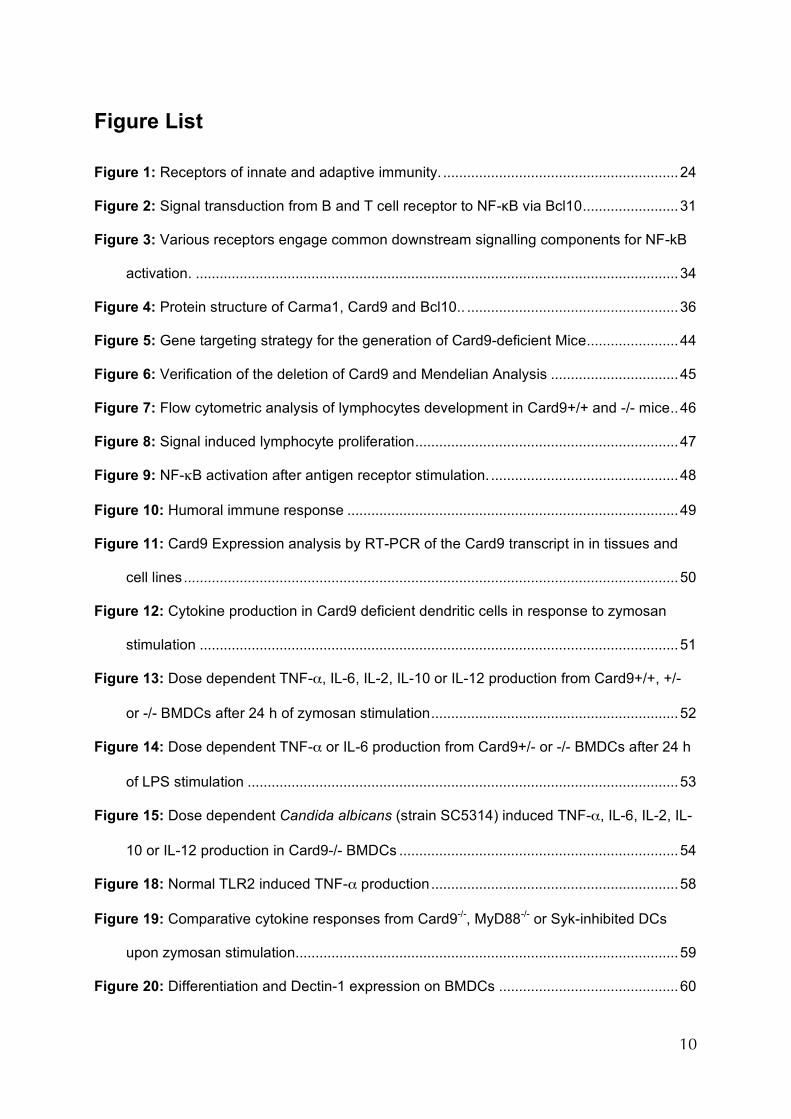
Figure 1: Receptors of innate and adaptive immunity. For abbreviations, see page 13ff.
25
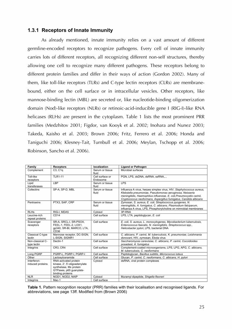
1.3.1 Receptors of Innate Immunity
As already mentioned, innate immunity relies on a vast amount of different
germline-encoded receptors to recognize pathogens. Every cell of innate immunity
carries lots of different receptors, all recognizing different non-self structures, thereby
allowing one cell to recognize many different pathogens. These receptors belong to
different protein families and differ in their ways of action (Gordon 2002). Many of
them, like toll-like receptors (TLRs) and C-type lectin receptors (CLRs) are membrane-
bound, either on the cell surface or in intracellular vesicles. Other receptors, like
mannose-binding lectin (MBL) are secreted or, like nucleotide-binding oligomerization
domain (Nod)-like receptors (NLRs) or retinoic-acid-inducible gene I (RIG-I)-like RNA
helicases (RLHs) are present in the cytoplasm. Table 1 lists the most prominent PRR
families (Medzhitov 2001; Figdor, van Kooyk et al. 2002; Inohara and Nunez 2003;
Takeda, Kaisho et al. 2003; Brown 2006; Fritz, Ferrero et al. 2006; Honda and
Taniguchi 2006; Klesney-Tait, Turnbull et al. 2006; Meylan, Tschopp et al. 2006;
Robinson, Sancho et al. 2006).
Family Receptors localization Ligand or Pathogen Complement C3, C1q Serum or tissue
fluid Microbial surfaces
Toll-like receptors
TLR1-11 Cell surface or Endosome
PGN, LPS, dsDNA, dsRNA, ssRNA,...
Lipid transferases
LBP Serum or tissue fluid
LPS
Collectins SP-A, SP-D, MBL Serum or tissue fluid
Influenza A virus, herpes simplex virus, HIV, Staphylococcus aureus, Klebsiella pneumoniae, Pseudomonas aeruginosa, Neisseria meningitidis, Haemophilus influenzae, E. coli,Pneumocystis carinii, Cryptococcus neoformans, Aspergillus fumigatus, Candida albicans
Pentraxins PTX3, SAP, CRP Serum or tissue fluid
Zymosan, S. aureus, E. coli, Streptococcus pyogenes, N. meningitidis, A. fumigatus, C. albicans, Plasmodium falciparum, influenza A virus, LPS, Phosphorylcholine on mimrobial membranes
RLHs RIG-I, MDA5 Cytosol 3P-RNA Leucine-rich repeat proteins
CD14 Cell surface LPS, LTA, peptidoglycan, E. coli
Scavenger receptors
SR-A, SRCL-I, SR-PSOX, FEEL-1, FEEL-2, LOX1, gp340, SR-BI, MARCO, LTA, CD36
Cell surface E. coli, S. aureus, L. monocytogenes, Mycobacterium tuberculosis, Enterococcus faecalis, N. meningitidis, Streptococcus spp., Helicobacter pylori, LPS, bacterial DNA
Classical C-type lectin
Mannose receptor, DC-SIGN, L-SIGN, SIGNR1
Cell surface C. albicans, P. carinii, M. tuberculosis, K. pneumoniae, Leishmania donovani, HIV, zymosan, Ebola virus
Non-classical C-type lectin
Dectin-1 Cell surface Saccharomyces cerevisiae, C. albicans, P. carinii, Coccidioides posadasii, A. fumigatus
Integrins CR3, CR4 Cell surface Complement-coated microorganisms, LPS, LPG, APG, C. albicans, M. tuberculosis, C. neoformans
Long PGRP PGRP-L, PGRP-I, PGRP-I Cell surface Peptidoglycan, Bacillus subtilis, Micrococcus luteus Other Lactosylceramide Cell surface Glucan, P. carinii, C. neoformans, C. albicans, H. pylori Interferon-induced proteins
RNA-activated protein kinase, 2', 5'-oligoadenylate synthetase, Mx protein GTPases, p65 guanylate-binding proteins
Cytosol dsRNA, viral protein complexes
NLR NOD1, NOD2, NAIP Cytosol Muramyl dipeptide, Shigella flexneri Integrins Mac-1 Cell surface Table 1. Pattern recognition receptor (PRR) families with their localisation and recognised ligands. For abbreviations, see page 13ff. Modified from (Brown 2006)

26
Toll-like receptors are structurally and presumably phylogenetically related to
Toll, an insect protein that also, among other ontogenetic properties, participates in the
insect immune response (Hoffmann and Reichhart 2002). TLRs consist of extracellular
leucin-rich repeats (LRRs) a transmembrane domain and an intracellular Toll/IL-1-
receptor (TIR) domain. TLR-7, -8 and -9 are localized in membranes of the endosome,
while the others are found on the cell surface. TLR-1, -2, -4, -5, -6, and -11 recognize
components of microbial surfaces, like lipopolysaccharide (TLR-4) in the cell wall of
gram-negative bacteria, peptidoglycan (TLR2) in gram-positive ones or flagellin (TLR5),
a component of the flagella of some motile bacteria. TLR-3, -7, -8 and -9 recognize
nucleic acids, i.e. double-stranded RNAs of viruses (TRL3) or CpG motifs, special DNA
sequences that are absent in mammals (TLR9) (Medzhitov 2001). The further events of
signalling and altered gene expression induced by TLRs upon PAMP recognition are
discussed later.
Toll-like receptors are membrane bound and directed to the extracellular space,
thereby detecting external or phagocytosed pathogens. Another class of membrane-
bound PRRs, the C-type lectin like receptors (CLRs) are the subject matter of section
1.4.4. In contrast, many pathogens, viruses and some bacteria reside in the cytoplasm
and soluble cytoplasmatic receptors are responsible for their perception. The most
prominent family of intracellular molecules monitoring bacteria is the NOD-
(Nucleotide binding and oligomerisation domain) like receptors (NLRs). This family
currently consists of 22 members (Meyland, Nature 2006), NOD2 being its most
prominent and best-studied member. NLRs typically contain an N-terminal CARD or
pyridine effector domain (PYD), a nucleotide binding and oligomerization (NACHT)
domain and a variable number of C-terminal leucin rich repeats (LRRs). NLRs are
synthesized in an autorepressed form, in which their LRRs are folded back to the rest of
the protein. This inhibits oligomerization by NACHT domains. Upon ligand recognition
by the LRRs, the protein unfolds and homooligomerizes. The CARD or PYD recruits
downstream effectors into the complex activating further signalling. NOD2 is involved
in the perception of intracellular muramyl dipeptide (MDP), a breakdown product of
bacterial peptidoglycan. It is unclear though, whether it is the receptor of MDP or
interacts with the actual receptor or a downstream effector of it. Like the TLRs, NOD2
activates mitogen-activated protein kinases (MAPK) and nuclear factor κB (NF-κB)

27
signalling cascades by CARD dependent binding of receptor interacting protein 2
(RIP2). NOD2 is indicated to be involved in immunity to Streptococcus, Mycobacteria
and Listeria species. Mutations in the LRRs of NOD2 have been associated with chronic
inflammatory disorders like Crohn’s disease. This is an inflammatory bowel disease
associated with increased production of proinflammatory cytokines such as TNF-α and
IL-1β and hyperactivation of NF-κB in gut associated lymphoid cells (Kaparakis,
Philpott et al. 2007).
Viruses also give away their presence in the host by carrying special PAMPs.
One of the most prominent of these is single- or double-stranded RNA (ssRNA or
dsRNA). Although TLR3 can recognize extracellular dsRNA, it is most important for the
infected cell to recognize intracellular viral RNA being produced in the cytoplasm
upon viral replication. Two members of the RIG like helicases (RLHs), RIG-I and
melanoma-differentiation-associated gene 5 (MDA5) recognize intracellular viral RNA.
The closely related proteins consist, like NOD2, of two N-terminal CARDs and, unlike
NOD2 of a C-terminal helicase domain. These receptors distinguish between host and
virus RNA by the presence of 5’ triphosphate groups in viral RNA that is absent in host
RNA (Thompson and Locarnini 2007). Upon binding of 5’ triphosphate RNA, they
interact by CARD-CARD interaction with MAVS, a protein located in the mitochondrial
membrane. Further signalling activates the transcription factors NF-κB and interferon
regulatory factor (IRF) 3 and 7, eventually leading, amongst other effects, to the
production and release of the antiviral cytokines interferon (IFN) α and IFNβ (type-I
interferons). These two receptors have been shown to be important for immune
responses to Japanese encephalitis virus, Newcastle disease virus, vesicular stomatitis
virus, Sendai virus and influenza virus (RIG-I), as well as encephalomyocarditis virus
(MDA5).
The different receptors and receptor families of innate immunity utilize distinct
signal transduction pathways to trigger diverse cellular reactions like activation,
proliferation, cytokine production, phagocytosis and oxidative burst. Many of these
reactions are based on altered gene transcription. Table 2 summarizes the signalling
mechanisms and induced actions of some membrane-bound receptors or receptor
families.
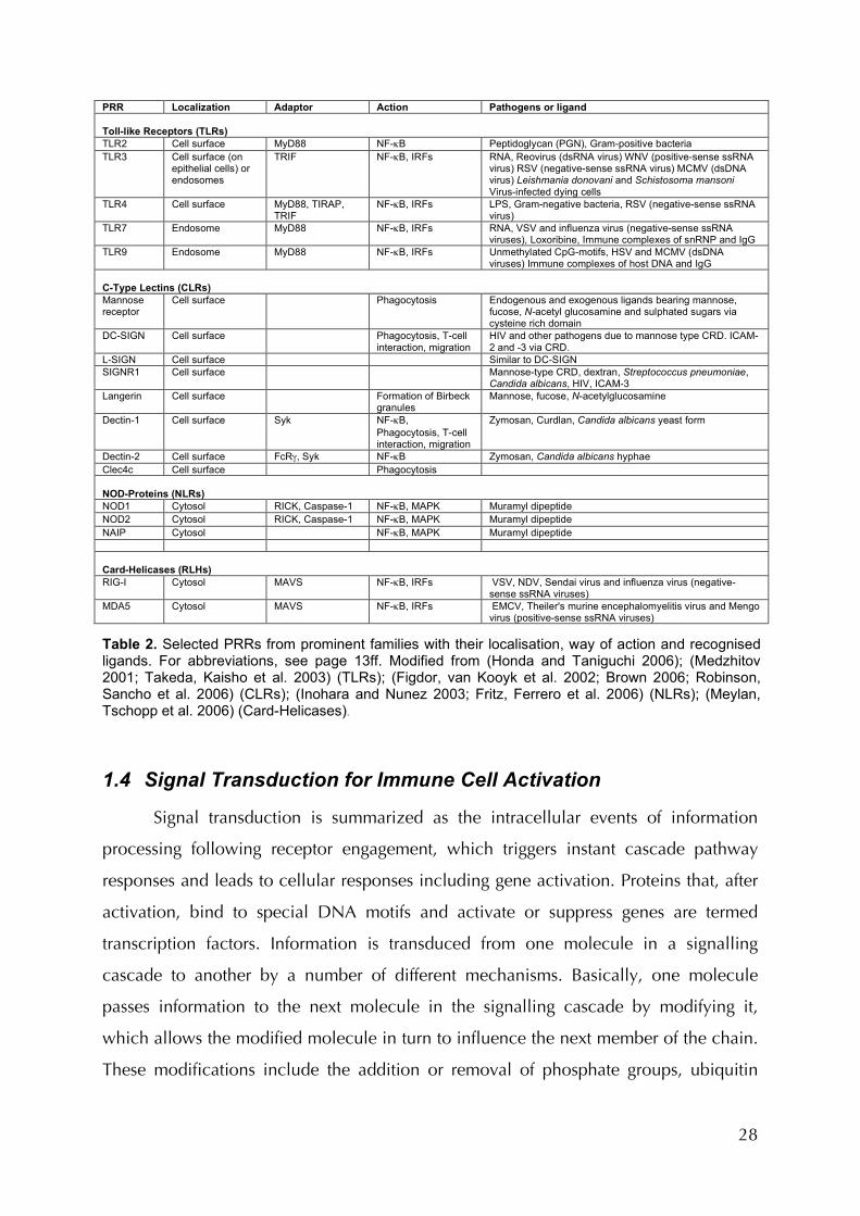
28
PRR Localization Adaptor Action Pathogens or ligand Toll-like Receptors (TLRs) TLR2 Cell surface MyD88 NF-κB Peptidoglycan (PGN), Gram-positive bacteria TLR3 Cell surface (on
epithelial cells) or endosomes
TRIF NF-κB, IRFs RNA, Reovirus (dsRNA virus) WNV (positive-sense ssRNA virus) RSV (negative-sense ssRNA virus) MCMV (dsDNA virus) Leishmania donovani and Schistosoma mansoni Virus-infected dying cells
TLR4 Cell surface MyD88, TIRAP, TRIF
NF-κB, IRFs LPS, Gram-negative bacteria, RSV (negative-sense ssRNA virus)
TLR7 Endosome MyD88 NF-κB, IRFs RNA, VSV and influenza virus (negative-sense ssRNA viruses), Loxoribine, Immune complexes of snRNP and IgG
TLR9 Endosome MyD88 NF-κB, IRFs Unmethylated CpG-motifs, HSV and MCMV (dsDNA viruses) Immune complexes of host DNA and IgG
C-Type Lectins (CLRs) Mannose receptor
Cell surface Phagocytosis Endogenous and exogenous ligands bearing mannose, fucose, N-acetyl glucosamine and sulphated sugars via cysteine rich domain
DC-SIGN Cell surface Phagocytosis, T-cell interaction, migration
HIV and other pathogens due to mannose type CRD. ICAM-2 and -3 via CRD.
L-SIGN Cell surface Similar to DC-SIGN SIGNR1 Cell surface Mannose-type CRD, dextran, Streptococcus pneumoniae,
Candida albicans, HIV, ICAM-3 Langerin
Cell surface Formation of Birbeck granules
Mannose, fucose, N-acetylglucosamine
Dectin-1 Cell surface Syk NF-κB, Phagocytosis, T-cell interaction, migration
Zymosan, Curdlan, Candida albicans yeast form
Dectin-2 Cell surface FcRγ, Syk NF-κB Zymosan, Candida albicans hyphae Clec4c Cell surface Phagocytosis NOD-Proteins (NLRs) NOD1 Cytosol RICK, Caspase-1 NF-κB, MAPK Muramyl dipeptide NOD2 Cytosol RICK, Caspase-1 NF-κB, MAPK Muramyl dipeptide NAIP Cytosol NF-κB, MAPK Muramyl dipeptide Card-Helicases (RLHs) RIG-I Cytosol MAVS NF-κB, IRFs VSV, NDV, Sendai virus and influenza virus (negative-
sense ssRNA viruses) MDA5 Cytosol MAVS NF-κB, IRFs EMCV, Theiler's murine encephalomyelitis virus and Mengo
virus (positive-sense ssRNA viruses) Table 2. Selected PRRs from prominent families with their localisation, way of action and recognised ligands. For abbreviations, see page 13ff. Modified from (Honda and Taniguchi 2006); (Medzhitov 2001; Takeda, Kaisho et al. 2003) (TLRs); (Figdor, van Kooyk et al. 2002; Brown 2006; Robinson, Sancho et al. 2006) (CLRs); (Inohara and Nunez 2003; Fritz, Ferrero et al. 2006) (NLRs); (Meylan, Tschopp et al. 2006) (Card-Helicases).
1.4 Signal Transduction for Immune Cell Activation
Signal transduction is summarized as the intracellular events of information
processing following receptor engagement, which triggers instant cascade pathway
responses and leads to cellular responses including gene activation. Proteins that, after
activation, bind to special DNA motifs and activate or suppress genes are termed
transcription factors. Information is transduced from one molecule in a signalling
cascade to another by a number of different mechanisms. Basically, one molecule
passes information to the next molecule in the signalling cascade by modifying it,
which allows the modified molecule in turn to influence the next member of the chain.
These modifications include the addition or removal of phosphate groups, ubiquitin
29
molecules or other components. Its interaction partner can also induce conformational
changes of a molecule. A third mechanism is the change of local molecule
concentrations, for example by oligomerisation, which influences the molecules
activity or by bringing interaction partners into closer proximity by binding to the same
large scaffold protein.
Mitogen-activated protein kinase (MAPK) pathways are a classical example of a
signalling cascade that is important in a lot of different cellular processes in presumably
all eukaryotic cells. MAPK pathways typically consist of a chain of proteins
phosphorylating the next protein in line after having been phosphorylated itself. This
leads to a nomenclature, were a MAP-Kinase (MAPK) gets phosphorylated by a MAPK-
Kinase (MAPKK) that in turn is phosphorylated and thereby activated by a MAPKK-
Kinase (MAPKKK) and so on (Elion 2000). Important MAP kinases in immunity are
extracellular signal-regulated kinase (ERK), c-Jun N-terminal kinase (Jnk) and p38
(protein of 38 kDa) that activate, amongst others, the transcription factor adaptor
protein complex 1 (AP-1) (Ashwell 2006). Further important transcription factors in
both innate and adaptive immunity are nuclear factor κB (NF-κB), nuclear factor of
activated T cells (NF-AT) and the IRF family (Honda and Taniguchi 2006).
1.4.1 NF-κB Signal Transduction
The transcription factor NF-κB is of remarkable importance in the immune
system. A vast number of processes in innate and adaptive immunity rely on the
activation of NF-κB. Upon PAMP recognition; virtually all PRRs activate NF-κB,
resulting in the production of proinflammatory cytokines and other responses. These
cytokines, i.e. TNF-α can in turn induce further NF-κB activation in their target cell by
the action of their specific receptors like the TNF-α receptor. NF-κB is also of central
importance in the activation of B- and T-cell through the antigen receptor (Karin and
Greten 2005).
In mammals, the term NF-κB designates a family of five transcription factors,
p50, p52, RelA (p65) c-Rel and RelB. Their common feature is the N-terminal DNA-
binding/dimerization domain (Rel homology domain), through which they can form
homo- and heterodimers. This domain facilitates also binding to distinct DNA motifs,
30
called κB sites, to alter gene expression. RelA, c-Rel and RelB also contain C-terminal
transcription activation domains (TADs) for the activation of target gene expression.
These sites are not contained in p50 and p52, which makes them negative regulators of
gene expression. These two family members can also form heterodimers with the other
members, in these cases they can act as positive regulators of gene expression. During
absence of a stimulatory signal, NF-κB is sequestered in the cytoplasm by inhibitors of
NF-κB proteins (IκBs). After stimulation, IκB proteins become phosphorylated and are
then ubiquitinated and thereby designated for proteasomal degradation. The
destruction of IκB allows NF-κB to translocate into the nucleus, subsequently activating
its target genes (Bonizzi and Karin 2004). In most cases the IκB proteins are
phosphorylated by the IκB kinase (IKK) complex. It consists of two catalytic subunits,
IKKα and IKKβ and a regulatory unit, NEMO. In the more abundant canonical pathway,
IKKβ and NEMO are required for gene activation, while IKKα is not essential but might
also contribute to the IKK complex. In the alternative pathway, IKKα alone is
responsible for the activation of NF-κB complexes inhibited by the IκB protein p100.
Activating signals and target genes of these two distinct pathways might overlap.
Upstream of IKK, individual receptors utilise different pathways for NF-κB activation.
NF-κB, dependent on the cell type, activates many different genes. These include genes
enhancing proliferation and survival in lymphocytes and cytokine and costimulatory
molecules in myeloid cells. The statement that NF-κB is of extraordinary importance in
immunity is underlined by its conservation throughout evolution. In fact, it is even the
primary regulator of innate immunity in insects like Drosophila (Govind 1999).
1.4.2 Antigen Receptor Signalling for NF-κB Activation
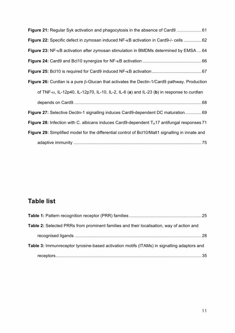
B and T cell receptor signalling activates many transcription factors including
NF-κB, for cell activation and proliferation (Figure 2). Membrane-bound subunits of the
antigen receptor complexes, Igα/β for BCR and CD3ζ for TCR contain immunreceptor
tyrosine-based activation motifs (ITAMs). Upon receptor activation, the tyrosines
become phosphorylated by SRC family kinase like lck or lyn.
31
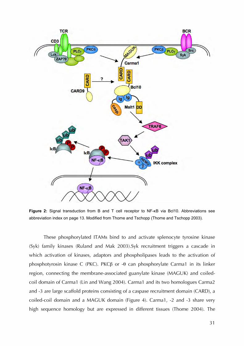
Figure 2: Signal transduction from B and T cell receptor to NF-κB via Bcl10. Abbreviations see
abbreviation index on page 13. Modified from Thome and Tschopp (Thome and Tschopp 2003).
These phosphorylated ITAMs bind to and activate splenocyte tyrosine kinase
(Syk) family kinases (Ruland and Mak 2003).Syk recruitment triggers a cascade in
which activation of kinases, adaptors and phospholipases leads to the activation of
phosphotyrosin kinase C (PKC). PKCβ or -θ can phosphorylate Carma1 in its linker
region, connecting the membrane-associated guanylate kinase (MAGUK) and coiled-
coil domain of Carma1 (Lin and Wang 2004). Carma1 and its two homologues Carma2
and -3 are large scaffold proteins consisting of a caspase recruitment domain (CARD), a
coiled-coil domain and a MAGUK domain (Figure 4). Carma1, -2 and -3 share very
high sequence homology but are expressed in different tissues (Thome 2004). The
32
coiled-coil and MAGUK domains of Carma1 mediate homo-oligomerisation of many
Carma1 molecules with each other. In this way, many molecules of the same signalling
components downstream of Carma1 are brought into close proximity with one another
(Rawlings, Sommer et al. 2006). It has been shown that homo-oligomerisation of some
downstream components of Carma1 is sufficient to activate NF-κB. Oligomerisation of
one of the components can often be achieved by simple overexpression. The
phosphorylation by PKC allows refolding of Carma1 (Matsumoto, Wang et al. 2005),
making the CARD accessible for interaction with Bcl10 (Figure 4). As implicated by the
name, CARD-containing proteins were initially identified as mediators of caspase
activation and apoptosis (Bouchier-Hayes and Martin 2002). The discovery of Bcl10s
role in antigen receptor activation has been the first report of an
antiapoptotic/proproliferative role of a CARD containing protein (Ruland, Duncan et al.
2001). Bcl10 binds to Malt1 (Ruland, Duncan et al. 2003), which is thereby also
included into the signalling complex (Figure 2). Recently, Klemm et al. demonstrated
an involvement of Bcl10 and Malt1 in myeloid cells, namely mast cells, for NF-κB
activation and cytokine production after stimulation of the ITAM receptor Fcε (Klemm,
Gutermuth et al. 2006). This implies that the Bcl10/Malt1 pathway is not restricted to
lymphoid cells and antigen receptors but also used in myeloid cells and might well be
engaged by further receptors and pathways. Downstream of Malt1 the pathway
converges with those of other receptors such as TNF receptor or TLRs by activating
TNF-receptor-associated factor 2 (TRAF-2) and TRAF-6. Finally, the signal is transduced
to the IKK complex via TGF-beta activated-kinase 1 (TAK1) (Figure 2).
1.4.3 The Toll-like Receptor Pathway for NF-κB
As outlined above, the 11 members of the Toll-like receptor family bind various
ligands derived from many pathogens. Most of these receptors use a common signalling
pathway to induce the activation of NF-κB. TLRs are type 1 transmembrane proteins
and consist of extracellular leucin rich repeats (LRRs) and an intracellular Toll/IL-1R
(TIR) domain (Medzhitov, Preston-Hurlburt et al. 1997). Variations in the LRRs of the
different TLR family members are responsible for the binding specificity to their specific
PAMPs. The intracellular TIR domain that the TLRs share with the interleucin-1 receptor
33
(IL-1R) mediates signalling by binding to the adaptor protein myeloid differentiation
primary-response protein 88 (MyD88) (Medzhitov, Preston-Hurlburt et al. 1998). This
binding is induced by dimerization of the receptors due to conformational changes
upon ligand recognition. MyD88 recruits IL-1R-associated kinases (IRAKs) 1 and 4 by
the death domain (DD) mediated protein-protein interaction (Akira and Takeda 2004).
IRAK4 then phosphorylates IRAK1, which in turn recruits tumor-necrosis factor (TNF)-
receptor-associated factor 6 (TRAF6), and eventually leads to nuclear translocation of
NF-κB.
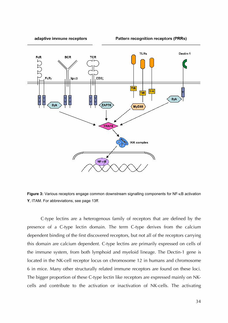
1.4.4 Non-TLR Signalling in Innate Immunity: Dectin-1
In addition to TLRs, other receptors also contribute to immune cell activation by
gene regulation in response to innate pathogen recognition, as summarised in Table 1.
Compared to antigen-receptor and TLR pathways, little is known about these pathways
(Figure 1, Table 2). Prototypic for such non-TLR PRRs is dendritic cell-associated C-
type lectin 1 (Dectin-1) (Taylor, Brown et al. 2002). The recognition of fungal infections
is primarily through this type II transmembrane receptor of the C-type lectin family.
Dectin-1 recognizes insoluble β-Glucans, which are present in the cell wall of
yeast cells. In its cytoplasmic tail, Dectin-1 carries an immunoreceptor tyrosine-based
activation motif (ITAM) (Brown 2006) by which it forwards activating signals. Receptors
and receptor interacting signalling molecules throughout innate and adaptive immunity
utilise ITAMs to forward signals (Table 3) (Barrow and Trowsdale 2006). As mentioned
above, the ITAM-carrying receptor associated molecules Igα/β and CD3, found in B
and T cell receptors, respectively signal via Syk (or its homologue ZAP-70) and Bcl10
(Ruland and Mak 2003) for lymphocyte activation and proliferation (Figure 3). The
ITAM of Dectin-1, like FcRγ, also signals via Syk for phagocytosis, oxidative burst, and
activation of gene transcription (Underhill, Rossnagle et al. 2005). Dectin-1s ITAM is
extraordinary when compared to those of other receptors due to the fact that it carries
only one YxxL motif, while usually two of these are required to interact with the two
SH2 domains of Syk (Table 3). It has been shown though, that this one tyrosine is
required and sufficient for Syk activation (Brown 2006). However, the pathways in
which Dectin-1/Syk mediate their responses remains largely elusive.
34
Figure 3: Various receptors engage common downstream signalling components for NF-κB activation
Y, ITAM. For abbreviations, see page 13ff.
C-type lectins are a heterogenous family of receptors that are defined by the
presence of a C-type lectin domain. The term C-type derives from the calcium
dependent binding of the first discovered receptors, but not all of the receptors carrying
this domain are calcium dependent. C-type lectins are primarily expressed on cells of
the immune system, from both lymphoid and myeloid lineage. The Dectin-1 gene is
located in the NK-cell receptor locus on chromosome 12 in humans and chromosome
6 in mice. Many other structurally related immune receptors are found on these loci.
The bigger proportion of these C-type lectin like receptors are expressed mainly on NK-
cells and contribute to the activation or inactivation of NK-cells. The activating
35
receptors of this family interact with stress or virus induced proteins on the surface of
host cells (Yokoyama and Plougastel 2003). They also utilize ITAMs for cellular
activation, either by carrying an ITAM in their cytoplasmatic portion or by utilizing
adapter proteins like DNAX-activation protein 12 (DAP12) or FcRγ, which both carry
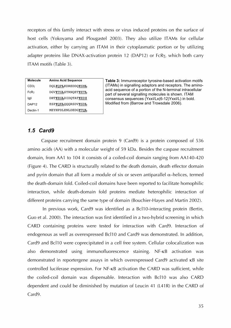
ITAM motifs (Table 3).
Molecule Amino Acid Sequence
CD3γ DQLYQPLKDREDDQYSHL
FcRγ DGVYTGLSTRNQETYETL
Igβ DHTYEGLDIDQTATYEDI
DAP12 ESPYQELQQQRSDVYSDL
Dectin-1 MEYHPDLENLDEDGYTQL
Table 3: Immunreceptor tyrosine-based activation motifs (ITAMs) in signalling adaptors and receptors. The amino-acid sequence of a portion of the N-terminal intracellular part of several signalling molecules is shown. ITAM consensus sequences (YxxI/Lx(6-12)YxxI/L) in bold. Modified from (Barrow and Trowsdale 2006).
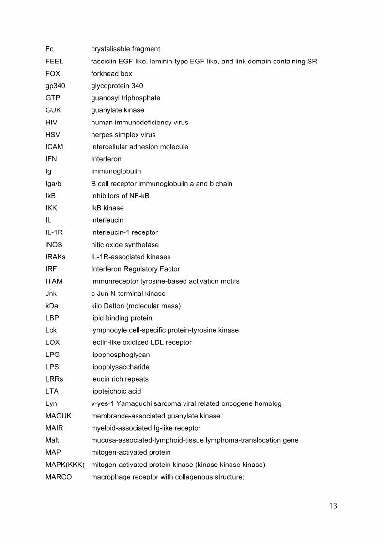
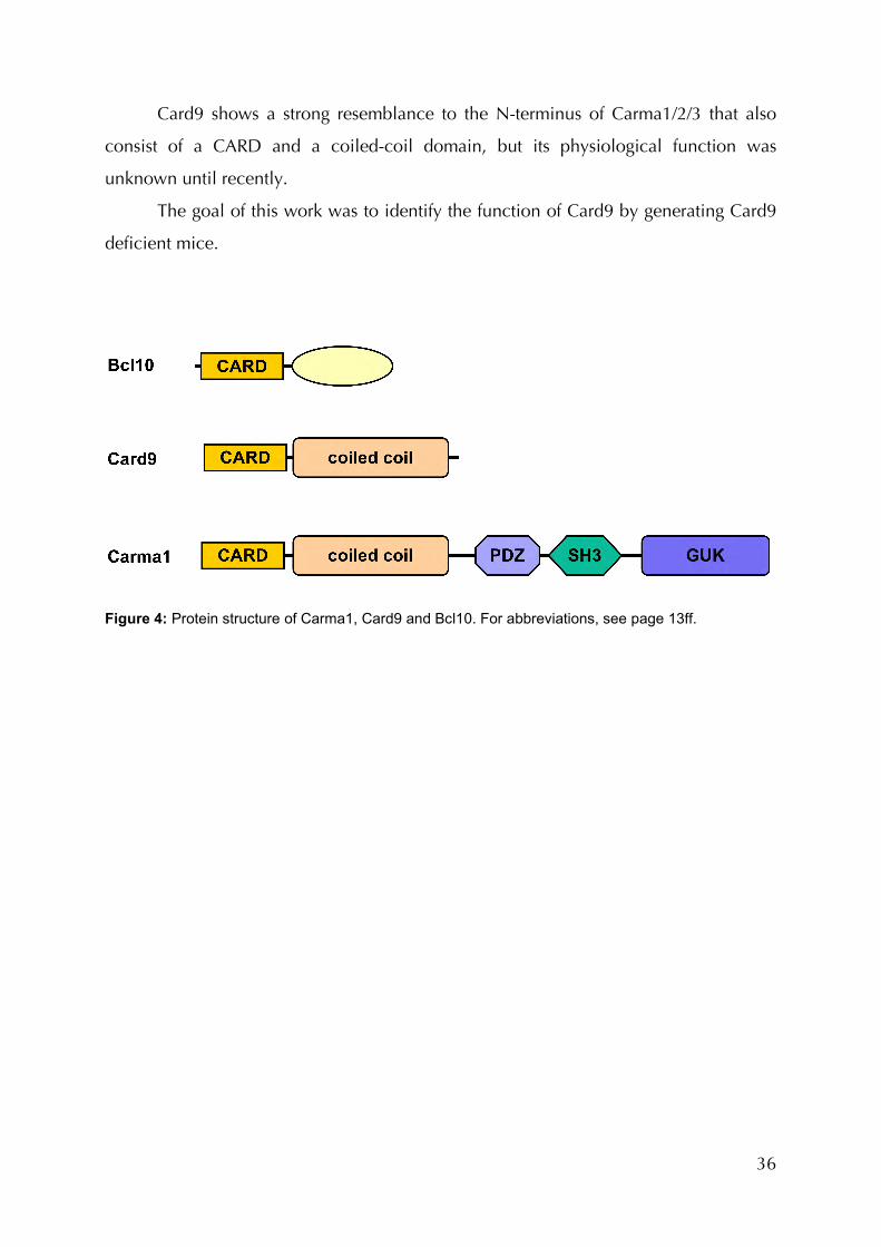
1.5 Card9
Caspase recruitment domain protein 9 (Card9) is a protein composed of 536
amino acids (AA) with a molecular weight of 59 kDa. Besides the caspase recruitment
domain, from AA1 to 104 it consists of a coiled-coil domain ranging from AA140-420
(Figure 4). The CARD is structurally related to the death domain, death effector domain
and pyrin domain that all form a module of six or seven antiparallel α–helices, termed
the death-domain fold. Coiled-coil domains have been reported to facilitate homophilic
interaction, while death-domain fold proteins mediate heterophilic interaction of
different proteins carrying the same type of domain (Bouchier-Hayes and Martin 2002).
In previous work, Card9 was identified as a Bcl10-interacting protein (Bertin,
Guo et al. 2000). The interaction was first identified in a two-hybrid screening in which
CARD containing proteins were tested for interaction with Card9. Interaction of
endogenous as well as overexpressed Bcl10 and Card9 was demonstrated. In addition,
Card9 and Bcl10 were coprecipitated in a cell free system. Cellular colocalization was
also demonstrated using immunofluorescence staining. NF-κB activation was
demonstrated in reportergene assays in which overexpressed Card9 activated κB site
controlled luciferase expression. For NF-κB activation the CARD was sufficient, while
the coiled-coil domain was dispensable. Interaction with Bcl10 was also CARD
dependent and could be diminished by mutation of Leucin 41 (L41R) in the CARD of
Card9.
36
Card9 shows a strong resemblance to the N-terminus of Carma1/2/3 that also
consist of a CARD and a coiled-coil domain, but its physiological function was
unknown until recently.
The goal of this work was to identify the function of Card9 by generating Card9
deficient mice.
Figure 4: Protein structure of Carma1, Card9 and Bcl10. For abbreviations, see page 13ff.
37
2 Materials and Methods
2.1 Material
2.1.1 Reagents
If not otherwise stated, all chemicals were from Sigma. All cell culture materials
were from Invitrogen; stem cell serum was from PAN biotech; serum with very low
endotoxin levels was from Perbio (Hyclone).
PCR and RT-PCR reagents were from Invitrogen; primers were designed using
Gene Construction Kit software and synthetized by MWG; DNA constructs were
sequenced by GATC.
2.1.2 Primer list
Primers for construction of the targeting construct:
Card9_SA_3_XbaI GCT CTA GAC AGC CCC ACA GCC ACA TGG TCC Card9_SA_5_NotI ATT GCG GCC GCG ACC AAG CTT TGT CCC ACT GCA C C9_SA_Scr_5_NotI ATT GCG GCC GCG GGA AGC TTG CAG CTC ATA G Card9_LA_XhoI_4.5kb GAG CTC GAG GTG TGT TAG TCT AAG AGT TC Card9_LA_ClaI+ CCA TCG ATG GCC TGA TGA TAC TCA CAG GCT
Primers for PCR screening of stem cells and mice: Card9_SA_ScPr_3 CCA TAG AGG ACT ATA GCT GCC TAC AG NEO Scr. (N73) GGG TGG GAT TAG ATA AAT GCC TGC TC Card9_WT_ScPr_1 TGG TTG ACC CAG TGG ACA GAC ATT TC Primers for cloning Card9 cDNA into pcDNA3.1 vector: hsCard9-JR15 CGC GGA TCC GTT ATG TCG GAC TAC GAG AAC hsCard9-JR16 CCG GAA TTC CTA CTA GGA GCC CTC AGT GTC Primers for RT-PCR: Card9_fwd5_E3 TGA GAA TGA CGA CGA GTG CTG Card9_rev4_E4 CTC CAA CGC CAT CAT AGA AGC
38
2.2 Methods
2.2.1 Generation of Card9-Deficient Mice
Gene targeting was performed by standard methods (Thomas and Capecchi
1987). A genomic Card9 clone was isolated from a 129/J bacterial artificial
chromosome (BAC) library (CHORI) and used as a PCR template to construct a
pBluescript (Stratagene) based targeting vector (Figure 5) that was electroporated into
E14K ES cells. Homologous recombination replaced a 2 kilobase (kb) genomic
fragment containing the exons encoding the CARD-domain of Card9 by a neomycin-
resistance gene. Clones resistant to G418 were selected and recombinants were
identified by PCR. Injection of three independent clones into C57/BL6 blastocysts
generated chimeric mice and Card9+/- mice. Germline transmission was confirmed by
PCR and Southern blot analysis of tail or thymus DNA. Card9+/- mice were intercrossed
to generate homozygous Card9-/- animals. All animals were housed at the facilities of
the Institut für Medizinische Mikrobiologie, Immunologie und Hygiene der
Technischen Universität München at the Klinikum rechts der Isar in accordance to
standard protocols and German and European laws and regulations. Littermate controls
were used in all experiments.
2.2.2 Flow Cytometric Analysis
Surface marker expression of thymocytes, splenocytes, lymph nodes, bone
marrow cells or bone marrow derived myeloid cells was analyzed using a flow
cytometer (FACScalibur, Becton Dickinson), CellQuest software and FlowJo Software
(Tree Star, Inc.) according to standard protocols. Flourescently labeled antibodies were
from Becton Dickinson, eBioscience, Caltag or Serotech.
2.2.3 Measurement of Serum Immunoglobulin Concentrations
Ig isotypes of 8-12 week old Card9+/+ and -/- mice were analyzed by ELISA on
serially diluted serum samples and anti-mouse IgG1, IgG2a, IgG2b, IgG3, IgA, or IgM
antibodies, as recommended by the manufacturer (Southern Biotechnology).
39
2.2.4 In Vivo Immunizations
Mice were immunized intraperitoneally (i.p.) with either 200 µg NP-Ova
(Calbiochem) in Alum (Sigma) as a T cell-dependent antigen or 25 µg TNP-Ficoll
(Biosearch Technologies) as a T cell-independent type II antigen. Blood from mouse
tails was collected at various time points. ELISA for the determination of antigen-
specific Ig isotypes was performed utilizing plate-bound TNP-Ficoll or NP-BSA instead
of capture antibodies to bind immunoglobulin (Tafuri, Shahinian et al. 2001).
2.2.5 Proliferation Assays
T and B cells were purified using magnetic beads (Dynabeads, Dynal) according
to manufacturers protocols. T cells were stimulated in 96 well U-bottom cell culture
plates (Becton Dickinson) with phorbol-12 myristate 13-acetate ester and calcium
ionophore (PMA + Iono, 10 ng/ml each), soluble anti-CD3 (0,1 – 10µg/ml), and soluble
anti-CD28 (2 µg/ml) in the presence or absence of IL-2 (50 U/ml). B cells were
stimulated with anti-IgM (1 - 10 µg/ml) and/or anti-CD40 (5 µg/ml). Cells were
harvested with a Skatronic 96 well harvester onto glass fibre membranes at 24 or 48 h
after an 8 h pulse with [3H]thymidine (1 µCi/well). Incorporation of [3H]thymidine was
measured with a scintillation Matrix 96 direct β-counter system (Canberra Packard).
2.2.6 Generation and Stimulation of Bone Marrow Derived Dendritic Cells (BMDC)
Dendritic cells were differentiated from bone marrow (BM) as previously
described (Sparwasser, Koch et al. 1998) by rinsing femora and tibiae bones of Card9
knock-out or control mice with culture medium using a syringe and 22-gauge needle to
elucidate bone marrow and erythrocytes were were lysed with RBC lysis buffer
(eBioscience). BM cells (4x10⁶) were seeded into a 100 mm standard petri dish in 10 ml
complete RPMI 1640 medium (Invitrogen), supplemented with 10% very low
endotoxin fetal bovine serum (Hyclone) and 2% GM-CSF containing cell culture
supernatant (Lutz, Kukutsch et al. 1999) or 20 ng/ml recombinant GM-CSF (Peprotech).
Cells producing GM-CSF were provided by Christian Meyer zum Büschenfelde. 10 ml

40
medium was added on day three and 10 ml (50%) were exchanged for fresh medium
on day six after spinning down half the culture volume at 300xg for 5 minutes. On day
six to eight, immature dendritic cells (CD11c+, MHCII+low), representing at least 50% of
the population, were transferred into 24 well tissue culture plates at a density of 2-5
x105 per well in 500 µl of GM-CSF containing medium. On day seven to nine, cells
were challenged with 500 µl two-fold stimuli or 50 µl 10-fold stimuli, diluted in GM-
CSF containing medium. Supernatants were harvested after a period of 5 to 48 hours for
further analysis of cytokine production. For analysis of activation marker, cells were
harvested on day 4 and plated on non-tissue culture treated 6 well plates with medium
without GM-CSF. Cells were stimulated after 3 h and activation marker expression was
measured by FACS after 16 h.
R848 and CpG were kind gift of Stefan Bauer and Tim Sparwasser, respectively.
All other TLR Ligands and MDP were purchased from Invivogen. Zymosan was either
from Sigma or Invivogen. Curdlan (Wako) was suspended in PBS at a concentration of
10 mg/ml. Candida albicans strain SC5314 was obtained from Rudolf Rupec. Candida
albicans cells for stimulation were grown on blood-agar or Sabourand plates at 30°C,
suspended and washed three times with cold PBS. Boiling for 1 h inactivated yeast
cells.
In order to inhibit Syk activity, Piceatannol and Syk-Inhibitor (Calbiochem) were
used. MyD88 deficient animals were kindly provided by Bernhard Holzmann.
2.2.7 Generation of Bone Marrow Derived Macrophages (BMDM)
BMDMs were prepared as previously described (Hammer, Mages et al. 2005). In
brief, femora and tibiae bones from mice (Card9-/- or heterozygous) were flushed with
medium to elucidate bone marrow and erythrocytes were were lysed with RBC lysis
buffer (eBioscience). Cells were cultured in complete Dulbecco's modified Eagle
Medium (DMEM, Invitrogen), containing 10% fetal bovine serum (Hyclone), and 10%
L-cell-conditioned medium (LCCM) as a source of M-CSF and incubated overnight in
10 cm cell culture dishes. Nonadherent cells were counted and replated at a density of
3 x 10⁵ cells/ml in DMEM with 10% LCCM in tissue culture plates. After 6-7 days of
differentiation, cultures were nearly confluent and the cells were used for experiments

41
after removal of nonadherent cells, washing with DMEM and resting overnight in
DMEM without M-CSF.
2.2.8 Cytokine Production
The amount of secreted TNF-α, IL-2, IL-6, IL-10, IL-12p40, IL-12p70, IL-23, IFN-
γ and IL-17 in culture supernatants was quantified using ELISA (Becton Dickinson or
eBioscience) according to manufacturers recommendations. To increase sensitivity,
supernatants were incubated on the ELISA plates (Nunc maxisorp) overnight at 4°C.
Values were measured with a TECAN Sunrise platereader.
2.2.9 Survival and Clearance of Candida albicans Infection
Card9-deficient and heterozygous control mice were infected with 103 – 106 cfu
Candida albicans (strain SC5314) per mouse in PBS via the tail vein. Infected mice were
monitored daily for signs of disease, which is shaggy fur, decreased motility and tearing
eyes. In order to determine pathogen load, mice were sacrificed on day four post
infection and the right kidney, the lung and the liver of these mice were disintegrated
by mashing them through a 100 µm cells strainer. Serial dilutions of the organs in PBS
were plated on ‘Candida ID2’ diagnostic plates (Biomerieux) at various dilutions. After
36h of incubation at 30°C, colonies were counted and pathogen load was calculated.
2.2.10 Restimulation of T-cells after Infection and Cytokine Measurement
Card9–/– mice and Card9+/– littermate control mice were infected intravenously
with 4x10³ to 1.2x10⁴ colony-forming units of Candida albicans strain SC5314. On
days 6–8 after infection, mice were sacrificed and either total splenocytes or splenocyte
samples depleted of CD4+ cells (2x10⁶ cells per well) were restimulated in 96-well U-
bottomed plates with heat-inactivated Candida albicans (1x10⁶ to 1x10⁸ per ml). After
48 h, IFN-γ and IL-17 in the supernatants were measured by sandwich ELISA.

42
2.2.11 Staphylococcus aureus Infection
Mice were infected with 5x10⁵ colony-forming units (cfu) Staphylococcus aureus
(ATCC 29219) as described before (Takeuchi, Hoshino et al. 2000) and monitored daily
for signs of disease. To determine pathogen titers, serial dilutions of homogenized
spleens were plated on CNA diagnostic plates (Biomerieux), incubated over night at
37°C and colonies counted.
2.2.12 Western Blot Analysis and Antibodies
Protein lysates from unstimulated or stimulated BMDC or BMDMs were
subjected to Western blott according to standard protocols (Ruland, Duncan et al.
2001) using antibodies against Syk, phospho-Syk (NEB), β-actin (Sigma) or Card9 (Santa
Cruz) and using equipment from Bio-RAD.
2.2.13 Zymosan Uptake
BMDCs were incubated with FITC-conjugated zymosan particles (100 µg ml-1,
Invitrogen) and internalization of zymosan particles was measured by FACS on a 15
minute basis. Cells were kept in a 37°C water bath between measurements. Cells were
counterstained and gated for CD11c.
2.2.14 Immunofluorescence
For immunofluorescence, BMDCs were seeded on 20 mm cover glasses in 6-
well plates at a density of 5x10⁵ cells/well. After stimulation for 1 h with 100 µg/ml
zymosan or 200 ng/µl LPS, the cells were washed once with PBS, fixed in ice cold
methanol for 5 min, and then blocked and permeabilized for 30 min at RT in blocking
buffer (BB), consisting of 10% FCS, and 0.1% Triton-X100 in PBS. Cells were stained
with anti-p65 polyclonal antibodies (C-20, Santa Cruz, 1:200 in BB, 1h at RT), followed
by a short incubation in BB supplemented with 2 µg/ml DAPI. Cells were then washed
twice with BB and incubated with a secondary antibody (Alexa Fluor 594, goat anti-
rabbit, Molecular Probes, 1:500 in BB, 1h at RT). After a further three washes with BB,
43
the samples were mounted in Vectashield embedding medium (Vector Laboratories)
and observed under a LSM confocal microscope (LSM510, Zeiss). Images were
evaluated with the Image Browser Software (Zeiss).
2.2.15 Gel Mobility Shift Assay
Nuclear protein extracts were prepared from unstimulated and stimulated cells
as previously described (Ruland, Duncan et al. 2001). Extracts (4 µg protein) were
incubated in 20 µl binding buffer with radio-labelled, double-stranded DNA probes
(NF-κB, 5′-ATC AGG GAC TTT CCG CTG GGG ACT TTC CG-3′; AP-1, 5′-CGC TTG
ATG ACT CAG CCG GAA-3′; NF-Y, 5′-AGA CCG TAC GTG ATT GGT TAA TCT CTT-
3′) and fractionated on a native 5% polyacrylamide gel. NF-κB and NF-Y binding
buffer: 5 mM HEPES (pH 7.8), 50 mM KCl, 0.5 mM dithiothreitol, 2 µg poly (dI-dC),
and 10% glycerol. AP-1 binding buffer: 10 mM Tris-HCl (pH 7.5), 100 mM KCl, 0.5
mM MgCl2, 0.1 mM EDTA, 0.5 mM dithiothreitol, 2 µg poly (dI-dC), and 10% glycerol.
2.2.16 NF-κB Reporter Assay
Bcl10+/- and -/- MEFs were plated at 1 x 10⁵ cells/well in a 96 well flat bottom
cell culture plate 24 h prior to transfection. The MEFs were co-transfected with 500 ng
Bcl10, Card9 or Carma3 (kindly provided by Gabriel Nunez, University of Michigan) or
Nod1 expression plasmids plus 6xκB-luciferase (250 ng) and pRL-TK (250 ng,
Promega). Transfectin (Bio-Rad) was used to transfect MEFs according to manufacturers
protocols. 293T cells were transfected with calcium phosphate according to standard
protocols or with Lipofectamin (Invitrogen) with Dectin-1 (50 ng / well in 96 wells)
and/or a combination of Card9 and Bcl10 expression vectors (5 ng each / well in 96
wells). Medium was renewed and cells were stimulated with 100 µg/ml zymosan after
24h. After 48 hrs, the cells were lysed and luciferase activity was determined using a
commercial assay kit (Promega) and a Berthold Luminometer.
Expression vector pUNO-Dectin-1 was from Invivogen, inserting Card9 cDNA
into pcDNA3.1 from Invitrogen generated Card9 expression vector.
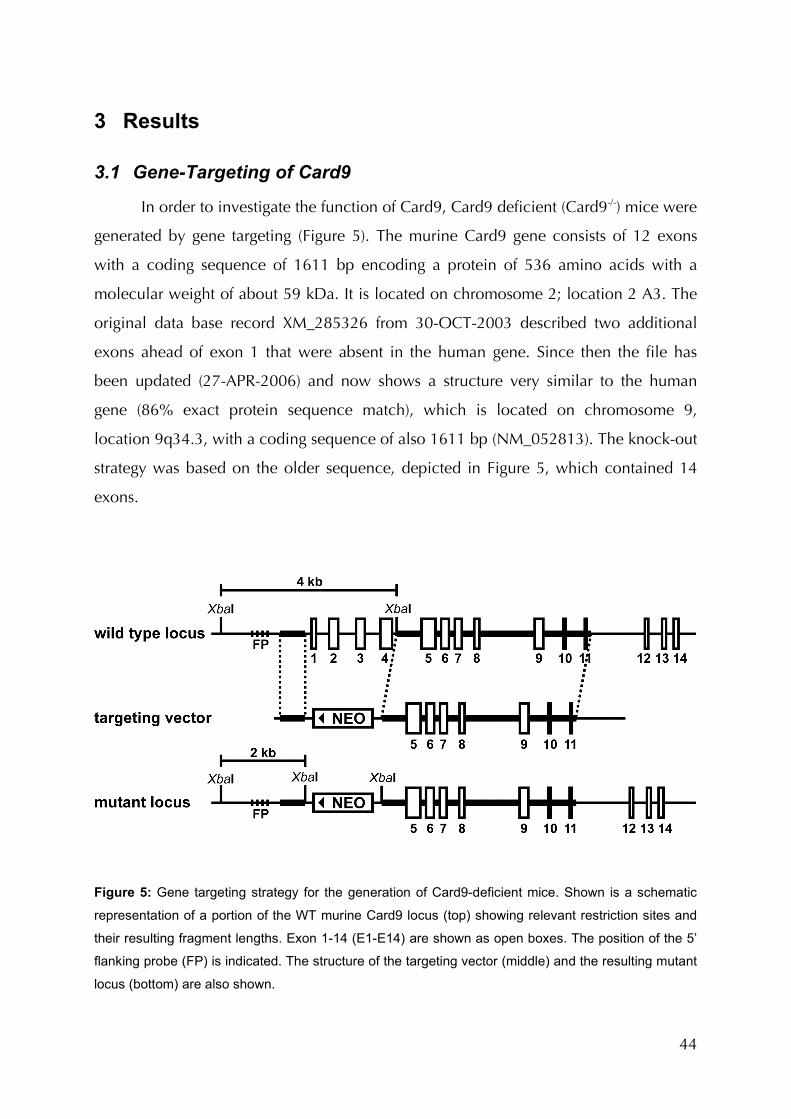
44
3 Results
3.1 Gene-Targeting of Card9
In order to investigate the function of Card9, Card9 deficient (Card9-/-) mice were
generated by gene targeting (Figure 5). The murine Card9 gene consists of 12 exons
with a coding sequence of 1611 bp encoding a protein of 536 amino acids with a
molecular weight of about 59 kDa. It is located on chromosome 2; location 2 A3. The
original data base record XM_285326 from 30-OCT-2003 described two additional
exons ahead of exon 1 that were absent in the human gene. Since then the file has
been updated (27-APR-2006) and now shows a structure very similar to the human
gene (86% exact protein sequence match), which is located on chromosome 9,
location 9q34.3, with a coding sequence of also 1611 bp (NM_052813). The knock-out
strategy was based on the older sequence, depicted in Figure 5, which contained 14
exons.
Figure 5: Gene targeting strategy for the generation of Card9-deficient mice. Shown is a schematic
representation of a portion of the WT murine Card9 locus (top) showing relevant restriction sites and
their resulting fragment lengths. Exon 1-14 (E1-E14) are shown as open boxes. The position of the 5’
flanking probe (FP) is indicated. The structure of the targeting vector (middle) and the resulting mutant
locus (bottom) are also shown.

45
To obliterate protein function, exons 1-2 (1-4), encoding the CARD, responsible
for protein-protein interaction and presumably protein function, were replaced by a
neomycin resistance gene. Germline transmitting chimeras were obtained from three
independent cell lines from two independent electroporations. Homologous
recombination was verified by Southern blot analysis using thymus DNA (Figure 6a).
Card9-/- mice were born at mendelian ratios from heterozygous (+/-) intercrosses (Figure
6b) without gross anatomical abnormalities. RT-PCR and Western blotting from the
splenocytes of Card9-deficient and wildtype mice confirmed the absence of Card9
mRNA transcript and protein (Figure 6c, d).
Figure 6: Verification of the deletion of Card9 on the DNA, RNA and protein level and Mendelian
Analysis. a Southern blot analysis of thymus DNA isolated from WT (+/+), heterozygous (+/-) and
Card9-deficient (-/-) mice. Genomic DNA was digested with XbaI and hybridized with the flanking
probe indicated in Figure 5. b Mendelian analysis of the offspring of Card+/- parents. c BMDM lysates
from Card9+/+ and -/- mice were analysed for Card9 protein content by Western blot. d Splenocyte
RNA isolated from Card9+/+ and -/- mice was subjected to RT-PCR analysis using primer pairs
specific for Card9 or β-actin.
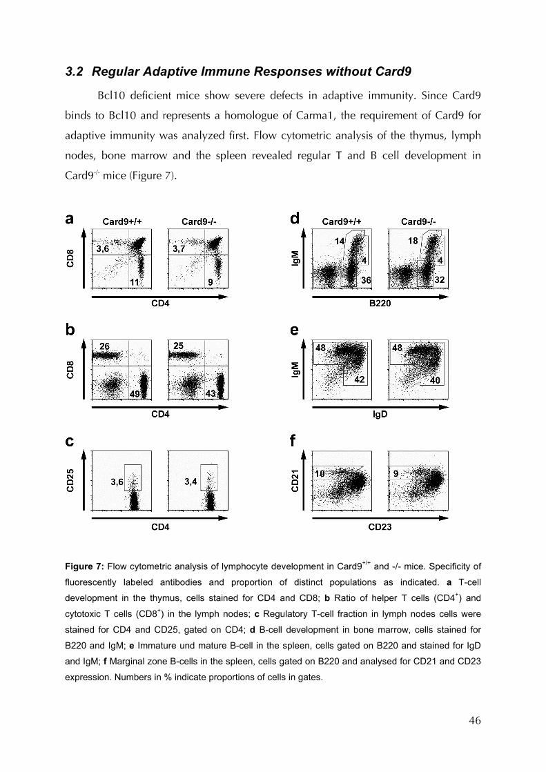
46
3.2 Regular Adaptive Immune Responses without Card9
Bcl10 deficient mice show severe defects in adaptive immunity. Since Card9
binds to Bcl10 and represents a homologue of Carma1, the requirement of Card9 for
adaptive immunity was analyzed first. Flow cytometric analysis of the thymus, lymph
nodes, bone marrow and the spleen revealed regular T and B cell development in
Card9-/- mice (Figure 7).
Figure 7: Flow cytometric analysis of lymphocyte development in Card9+/+ and -/- mice. Specificity of
fluorescently labeled antibodies and proportion of distinct populations as indicated. a T-cell
development in the thymus, cells stained for CD4 and CD8; b Ratio of helper T cells (CD4+) and
cytotoxic T cells (CD8+) in the lymph nodes; c Regulatory T-cell fraction in lymph nodes cells were
stained for CD4 and CD25, gated on CD4; d B-cell development in bone marrow, cells stained for
B220 and IgM; e Immature und mature B-cell in the spleen, cells gated on B220 and stained for IgD
and IgM; f Marginal zone B-cells in the spleen, cells gated on B220 and analysed for CD21 and CD23
expression. Numbers in % indicate proportions of cells in gates.
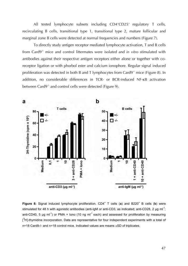
47
All tested lymphocyte subsets including CD4+CD25+ regulatory T cells,
recirculating B cells, transitional type 1, transitional type 2, mature follicular and
marginal zone B cells were detected at normal frequencies and numbers (Figure 7).
To directly study antigen receptor mediated lymphocyte activation, T and B cells
from Card9-/- mice and control littermates were isolated and in vitro stimulated with
antibodies against their respective antigen receptors either alone or together with co-
receptor ligation or with phorbol ester and calcium ionophore. Regular signal induced
proliferation was detected in both B and T lymphocytes from Card9-/- mice (Figure 8). In
addition, no considerable differences in TCR- or BCR-induced NF-κB activation
between Card9-/- and control cells were detected (Figure 9).
Figure 8: Signal induced lymphocyte proliferation. CD4+ T cells (a) and B220+ B cells (b) were
stimulated for 48 h with agonistic antibodies (anti-IgM or anti-CD3, as indicated; anti-CD28, 2 µg ml-1;
anti-CD40, 5 µg ml-1) or PMA + Iono (10 ng ml-1 each) and assessed for proliferation by measuring
[3H]-thymidine incorporation. Data are representative for four independent experiments with a total of
n=18 Card9-/- and n=18 control mice. Indicated values are means ±SD of triplicates.
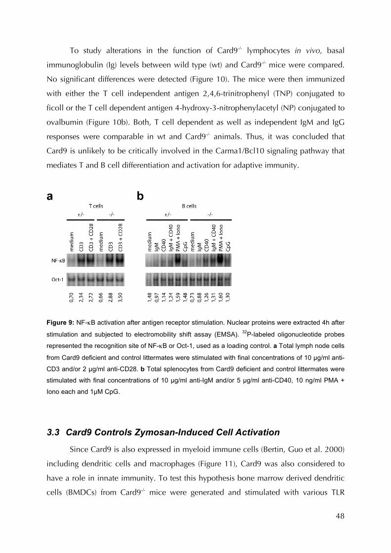
48
To study alterations in the function of Card9-/- lymphocytes in vivo, basal
immunoglobulin (Ig) levels between wild type (wt) and Card9-/- mice were compared.
No significant differences were detected (Figure 10). The mice were then immunized
with either the T cell independent antigen 2,4,6-trinitrophenyl (TNP) conjugated to
ficoll or the T cell dependent antigen 4-hydroxy-3-nitrophenylacetyl (NP) conjugated to
ovalbumin (Figure 10b). Both, T cell dependent as well as independent IgM and IgG
responses were comparable in wt and Card9-/- animals. Thus, it was concluded that
Card9 is unlikely to be critically involved in the Carma1/Bcl10 signaling pathway that
mediates T and B cell differentiation and activation for adaptive immunity.
Figure 9: NF-κB activation after antigen receptor stimulation. Nuclear proteins were extracted 4h after
stimulation and subjected to electromobility shift assay (EMSA). 32P-labeled oligonucleotide probes
represented the recognition site of NF-κB or Oct-1, used as a loading control. a Total lymph node cells
from Card9 deficient and control littermates were stimulated with final concentrations of 10 µg/ml anti-
CD3 and/or 2 µg/ml anti-CD28. b Total splenocytes from Card9 deficient and control littermates were
stimulated with final concentrations of 10 µg/ml anti-IgM and/or 5 µg/ml anti-CD40, 10 ng/ml PMA +
Iono each and 1µM CpG.
3.3 Card9 Controls Zymosan-Induced Cell Activation
Since Card9 is also expressed in myeloid immune cells (Bertin, Guo et al. 2000)
including dendritic cells and macrophages (Figure 11), Card9 was also considered to
have a role in innate immunity. To test this hypothesis bone marrow derived dendritic
cells (BMDCs) from Card9-/- mice were generated and stimulated with various TLR

49
ligands as well as ligands for non-TLR PRRs. As an initial readout, ligand-induced
production of tumor necrosis factor-α (TNF-α) was measured (Figure 12). No
reproducible differences were detected between wild type and Card9 deficient BMDCs
in response to TLR ligands Pam3CSK4 (TLR2), LPS (TLR4), R848 (TLR7), CpG DNA
(TLR9) or upon stimulation with the NOD2 agonist muramyl dipeptide (MDP) alone or
in combination with LPS.
Figure 10: Humoral immune response. a Basal immunoglobulin (Ig) concentrations. Ig isotypes in
sera of 8-12 week old wt (+/+, n=9) and Card9-/- (n=18) mice were determined by ELISA. b Regular
antibody responses. Left: control (+/-, filled symbols, n=8) and Card9-/- mice (open symbols, n=6)
were immunized with 25 µg TNP-ficoll for T cell-independent immune responses. Concentrations of
TNP specific IgM and IgG3 were determined before and after immunization. Right: for T cell-
dependent antibody responses, control (+/-, filled symbols, n=4) and Card9-/- mice (open symbols,
n=3) were injected with 200 µg NP-Ova absorbed on alum. NP-specific IgM and IgG1 concentrations
were measured before and after immunization. Results are representative for two independent
experiments.
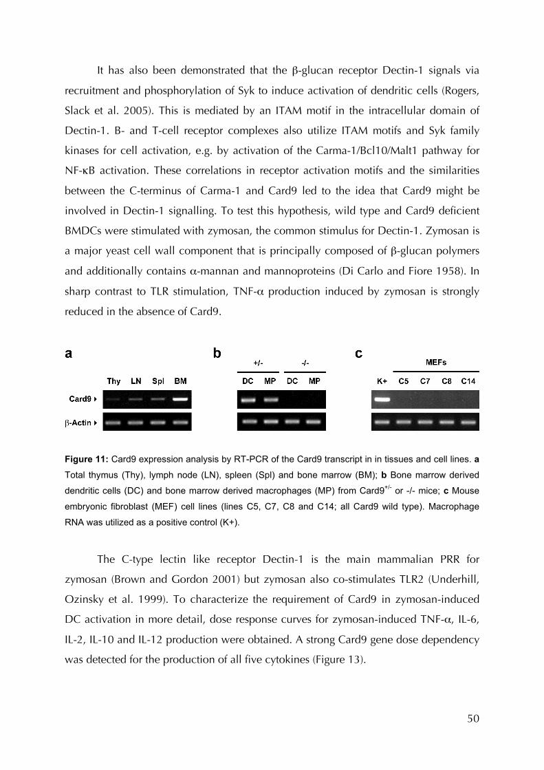
50
It has also been demonstrated that the β-glucan receptor Dectin-1 signals via
recruitment and phosphorylation of Syk to induce activation of dendritic cells (Rogers,
Slack et al. 2005). This is mediated by an ITAM motif in the intracellular domain of
Dectin-1. B- and T-cell receptor complexes also utilize ITAM motifs and Syk family
kinases for cell activation, e.g. by activation of the Carma-1/Bcl10/Malt1 pathway for
NF-κB activation. These correlations in receptor activation motifs and the similarities
between the C-terminus of Carma-1 and Card9 led to the idea that Card9 might be
involved in Dectin-1 signalling. To test this hypothesis, wild type and Card9 deficient
BMDCs were stimulated with zymosan, the common stimulus for Dectin-1. Zymosan is
a major yeast cell wall component that is principally composed of β-glucan polymers
and additionally contains α-mannan and mannoproteins (Di Carlo and Fiore 1958). In
sharp contrast to TLR stimulation, TNF-α production induced by zymosan is strongly
reduced in the absence of Card9.
Figure 11: Card9 expression analysis by RT-PCR of the Card9 transcript in in tissues and cell lines. a
Total thymus (Thy), lymph node (LN), spleen (Spl) and bone marrow (BM); b Bone marrow derived
dendritic cells (DC) and bone marrow derived macrophages (MP) from Card9+/- or -/- mice; c Mouse
embryonic fibroblast (MEF) cell lines (lines C5, C7, C8 and C14; all Card9 wild type). Macrophage
RNA was utilized as a positive control (K+).
The C-type lectin like receptor Dectin-1 is the main mammalian PRR for
zymosan (Brown and Gordon 2001) but zymosan also co-stimulates TLR2 (Underhill,
Ozinsky et al. 1999). To characterize the requirement of Card9 in zymosan-induced
DC activation in more detail, dose response curves for zymosan-induced TNF-α, IL-6,
IL-2, IL-10 and IL-12 production were obtained. A strong Card9 gene dose dependency
was detected for the production of all five cytokines (Figure 13).

51
Figure 12: Cytokine production in Card9 deficient dendritic cells in response to PRR stimulation. TNF-
α production in Card9-/- compared to Card9+/- BMDCs after stimulation with various TLR-ligands,
Muramyl dipeptide (MDP) and zymosan. BMDC were treated with 200 ng/ml Pam3CSK4, 100 ng/ml
Lipopolysaccharide (LPS), 10 µg/ml MDP, 1 µM CpG, 200 ng/ml R848 or 3 µg/ml zymosan or
unstimulated (medium). TNF-α concentration in the medium was determined after 24h by ELISA. ND,
not detected. Data are means ±SD of triplicates and representative of three independent experiments.
In contrast, TLR4 ligation with LPS induced regular dose dependent TNF-α or IL-
6 synthesis in Card9-/- BMDCs (Figure 14) whereas LPS induced IL-2 production in
neither Card9-/- nor control cells (data not shown).
It is believed that the recognition of zymosan or β-glucans triggers immune
responses that are primarily designed for the control of fungal pathogens (Brown and
Gordon 2003). Therefore, the role of Card9 in immune responses to whole fungal cells
was studied using Candida albicans as a model (Villamon, Gozalbo et al. 2004).
Consistent with a role of Card9 in zymosan recognition signalling, Card9-/- BMDCs
exhibited severe defects in Candida albicans-induced TNF-α, IL-2, IL-6 and IL-12
production (Figure 15). To determine the relevance of these findings in vivo, the
susceptibility to infection in Card9-/- mice and heterozygous littermates was compared
after injection of live Candida albicans (Figure 16). In several sets of experiments, mice
were infected with varying doses of Candida albicans cells, ranging from 103 to 106
colony forming units (cfu) (Villamon, Gozalbo et al. 2004). While Card9 deficient
animals survived for a maximum of nine days, control animals cleared the infection or
survived significantly longer, depending on the dose (Figure 17). Homozygous wildtype
control mice survived an infection even longer than Card9 heterozygous animals,
indicating a gene-dosage effect (Figure 17b).
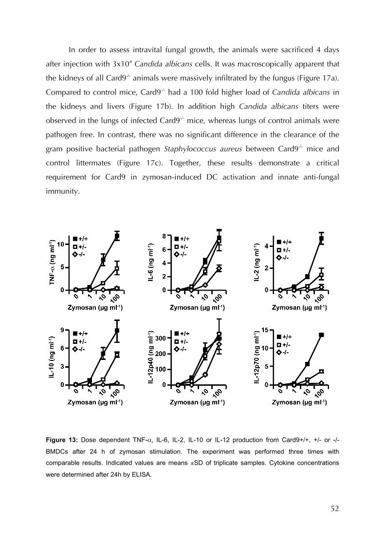
52
In order to assess intravital fungal growth, the animals were sacrificed 4 days
after injection with 3x10⁴ Candida albicans cells. It was macroscopically apparent that
the kidneys of all Card9-/- animals were massively infiltrated by the fungus (Figure 17a).
Compared to control mice, Card9-/- had a 100 fold higher load of Candida albicans in
the kidneys and livers (Figure 17b). In addition high Candida albicans titers were
observed in the lungs of infected Card9-/- mice, whereas lungs of control animals were
pathogen free. In contrast, there was no significant difference in the clearance of the
gram positive bacterial pathogen Staphylococcus aureus between Card9-/- mice and
control littermates (Figure 17c). Together, these results demonstrate a critical
requirement for Card9 in zymosan-induced DC activation and innate anti-fungal
immunity.
Figure 13: Dose dependent TNF-α, IL-6, IL-2, IL-10 or IL-12 production from Card9+/+, +/- or -/-
BMDCs after 24 h of zymosan stimulation. The experiment was performed three times with
comparable results. Indicated values are means ±SD of triplicate samples. Cytokine concentrations
were determined after 24h by ELISA.
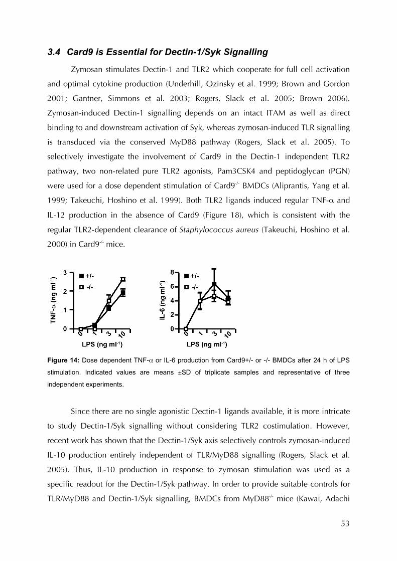
53
3.4 Card9 is Essential for Dectin-1/Syk Signalling
Zymosan stimulates Dectin-1 and TLR2 which cooperate for full cell activation
and optimal cytokine production (Underhill, Ozinsky et al. 1999; Brown and Gordon
2001; Gantner, Simmons et al. 2003; Rogers, Slack et al. 2005; Brown 2006).
Zymosan-induced Dectin-1 signalling depends on an intact ITAM as well as direct
binding to and downstream activation of Syk, whereas zymosan-induced TLR signalling
is transduced via the conserved MyD88 pathway (Rogers, Slack et al. 2005). To
selectively investigate the involvement of Card9 in the Dectin-1 independent TLR2
pathway, two non-related pure TLR2 agonists, Pam3CSK4 and peptidoglycan (PGN)
were used for a dose dependent stimulation of Card9-/- BMDCs (Aliprantis, Yang et al.
1999; Takeuchi, Hoshino et al. 1999). Both TLR2 ligands induced regular TNF-α and
IL-12 production in the absence of Card9 (Figure 18), which is consistent with the
regular TLR2-dependent clearance of Staphylococcus aureus (Takeuchi, Hoshino et al.
2000) in Card9-/- mice.
Figure 14: Dose dependent TNF-α or IL-6 production from Card9+/- or -/- BMDCs after 24 h of LPS
stimulation. Indicated values are means ±SD of triplicate samples and representative of three
independent experiments.
Since there are no single agonistic Dectin-1 ligands available, it is more intricate
to study Dectin-1/Syk signalling without considering TLR2 costimulation. However,
recent work has shown that the Dectin-1/Syk axis selectively controls zymosan-induced
IL-10 production entirely independent of TLR/MyD88 signalling (Rogers, Slack et al.
2005). Thus, IL-10 production in response to zymosan stimulation was used as a
specific readout for the Dectin-1/Syk pathway. In order to provide suitable controls for
TLR/MyD88 and Dectin-1/Syk signalling, BMDCs from MyD88-/- mice (Kawai, Adachi

54
et al. 1999) and wild type dendritic cells that were pre-incubated with the small
molecule Syk inhibitor piceatannol were now also included (Figure 19a). Consistent
with published data (Rogers, Slack et al. 2005), zymosan stimulation induced a strong
IL-10 production in wildtype BMDCs. This production was MyD88 independent but
robustly blocked by inhibiting Syk function. The Card9 deletion also completely
abolished zymosan-induced IL-10 synthesis, suggesting a role for Card9 in Dectin-1/Syk
dependent, TLR/MyD88 independent signalling events. Compared to zymosan, the TLR
ligands LPS and CpG induced regular albeit lower amounts of IL-10 in Card9-/- BMDCs
(Figure 19b). Next, zymosan-induced production of TNF-α, IL-6, IL-2 and IL-12 in wild
type BMDCs, wild type BMDCs that were preincubated with Syk inhibitor and in
Card9-/- BMDCs was compared (Figure 19c). Indeed, zymosan-induced TNF-α and IL-2
production are highly dependent on Syk and Card9.
Figure 15: Dose dependent Candida albicans (strain SC5314) induced TNF-α, IL-6, IL-2, IL-10 or IL-
12 production in Card9-/- BMDCs. Cells were stimulated for 24 h with indicated concentrations of heat-
inactivated Candida albicans cells. Cytokine production was measured by ELISA. Results are means
±SD of triplicates and representative for three independent experiments.

55
Figure 16: Strongly increased mortality
of Card9 deficient mice after Candida
albicans infection. Eight to ten week
old Card9 deficient, wild type and/or
heterozygous control female mice (five
or seven per group, respectively, from
two litters) were intravenously infected
with Candida albicans. Injected doses
were: in a 1x10⁶ CFU, in b 5x10⁵ CFU,
in c 1,5x10⁵ CFU. Survival was
checked on a 12h basis for 1 week and
afterwards once a day.
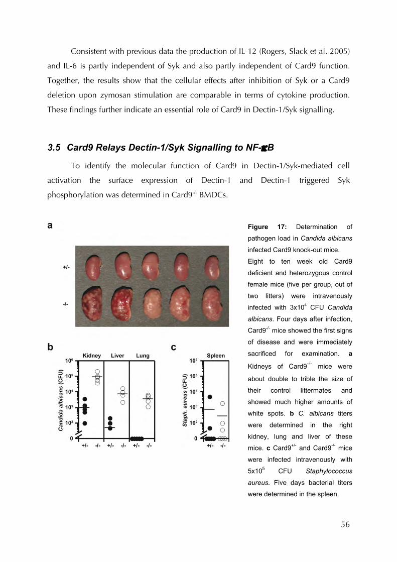
56
Consistent with previous data the production of IL-12 (Rogers, Slack et al. 2005)
and IL-6 is partly independent of Syk and also partly independent of Card9 function.
Together, the results show that the cellular effects after inhibition of Syk or a Card9
deletion upon zymosan stimulation are comparable in terms of cytokine production.
These findings further indicate an essential role of Card9 in Dectin-1/Syk signalling.
3.5 Card9 Relays Dectin-1/Syk Signalling to NF-κB
To identify the molecular function of Card9 in Dectin-1/Syk-mediated cell
activation the surface expression of Dectin-1 and Dectin-1 triggered Syk
phosphorylation was determined in Card9-/- BMDCs.
Figure 17: Determination of
pathogen load in Candida albicans
infected Card9 knock-out mice.
Eight to ten week old Card9
deficient and heterozygous control
female mice (five per group, out of
two litters) were intravenously
infected with 3x10⁴ CFU Candida
albicans. Four days after infection,
Card9-/- mice showed the first signs
of disease and were immediately
sacrificed for examination. a
Kidneys of Card9-/- mice were
about double to trible the size of
their control littermates and
showed much higher amounts of
white spots. b C. albicans titers
were determined in the right
kidney, lung and liver of these
mice. c Card9+/- and Card9-/- mice
were infected intravenously with
5x10⁵ CFU Staphylococcus
aureus. Five days bacterial titers
were determined in the spleen.

57
The receptor is regularly expressed (Figure 20) and upon stimulation with
zymosan normally activates Syk (Figure 21a). Syk activation in BMDCs not only
controls cytokine production but also regulates zymosan phagocytosis (Rogers, Slack et
al. 2005). To quantify Syk-dependent zymosan internalization, BMDCs were incubated
with or without Syk inhibitor and fluorescently-labeled zymosan before flow cytometric
analysis (Figure 21b). Regular Syk-dependent phagocytosis was observed in Card9-/-
BMDCs demonstrating that certain Dectin-1/Syk controlled pathways are not
dependent on Card9.Since overexpression studies have indicated that Card9 can
induce NF-κB (Bertin, Guo et al. 2000), the activation of this transcription factor was
probed by monitoring RelA nuclear translocation in zymosan- or LPS-stimulated Card9-
/- BMDCs (Figure 22). Using confocal microscopy similar nuclear translocation of RelA
in Card9-/- and control BMDCs was detected upon TLR4-activation with LPS.
Microscopy also clearly demonstrated that Card9-/- BMDCs internalized zymosan in the
normal manner. However, Card9-/- BMDCs exhibit a substantial impairment in
zymosan-induced NF-κB activation although some NF-κB induction presumably via
TLR2 could be observed. In addition, electromobility shift assays (EMSAs) for NF-κB
DNA binding activity were performed using nuclear extracts from bone marrow derived
macrophages BMDMs that had been stimulated with zymosan or the TLR ligands PGN,
LPS or R848 (Figure 23). TLR ligation induced regular NF-κB activation whereas
zymosan-induced NF-κB activation was diminished in the absence of Card9. Together,
these results indicate a role for Card9 in transducing zymosan-activated Dectin-1
signals to NF-κB.
3.6 Card9 Signals via Bcl10 and Malt1
Since Card9 can interact with Bcl10 (Bertin, Guo et al. 2000) it has been studied
whether the two proteins functionally cooperate (Figure 24). Consistent with previous
results (Bertin, Guo et al. 2000) single overexpression of Card9 or Bcl10 weakly
activates NF-κB. However, the co-overexpression of both proteins elicited more than
additive NF-κB induction, indicating synergism (Figure 24a).
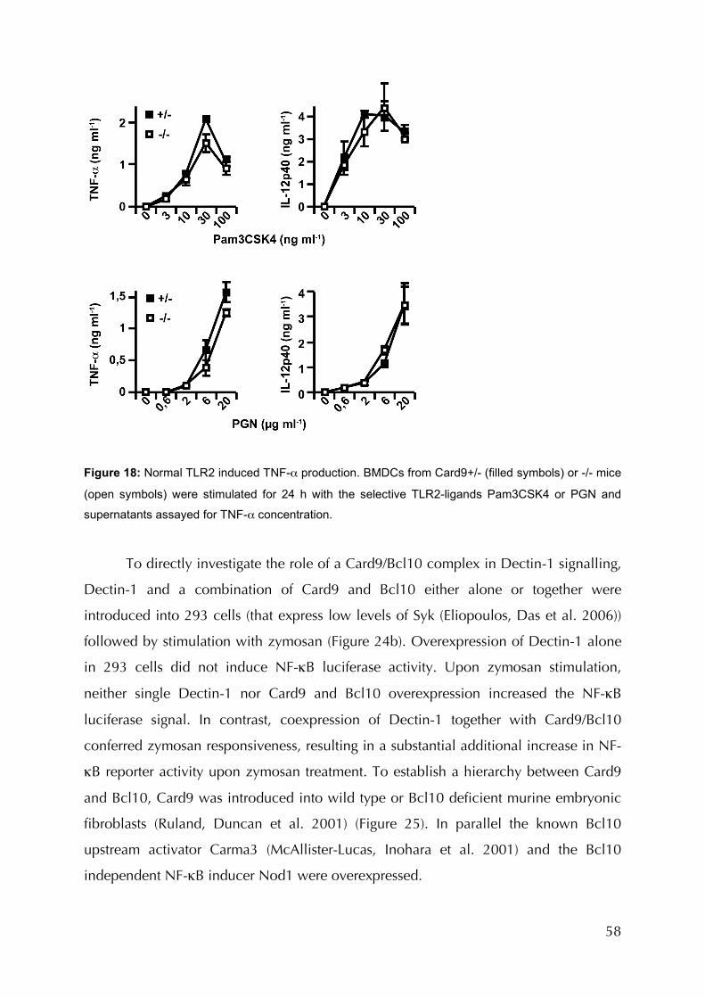
58
Figure 18: Normal TLR2 induced TNF-α production. BMDCs from Card9+/- (filled symbols) or -/- mice
(open symbols) were stimulated for 24 h with the selective TLR2-ligands Pam3CSK4 or PGN and
supernatants assayed for TNF-α concentration.
To directly investigate the role of a Card9/Bcl10 complex in Dectin-1 signalling,
Dectin-1 and a combination of Card9 and Bcl10 either alone or together were
introduced into 293 cells (that express low levels of Syk (Eliopoulos, Das et al. 2006))
followed by stimulation with zymosan (Figure 24b). Overexpression of Dectin-1 alone
in 293 cells did not induce NF-κB luciferase activity. Upon zymosan stimulation,
neither single Dectin-1 nor Card9 and Bcl10 overexpression increased the NF-κB
luciferase signal. In contrast, coexpression of Dectin-1 together with Card9/Bcl10
conferred zymosan responsiveness, resulting in a substantial additional increase in NF-
κB reporter activity upon zymosan treatment. To establish a hierarchy between Card9
and Bcl10, Card9 was introduced into wild type or Bcl10 deficient murine embryonic
fibroblasts (Ruland, Duncan et al. 2001) (Figure 25). In parallel the known Bcl10
upstream activator Carma3 (McAllister-Lucas, Inohara et al. 2001) and the Bcl10
independent NF-κB inducer Nod1 were overexpressed.
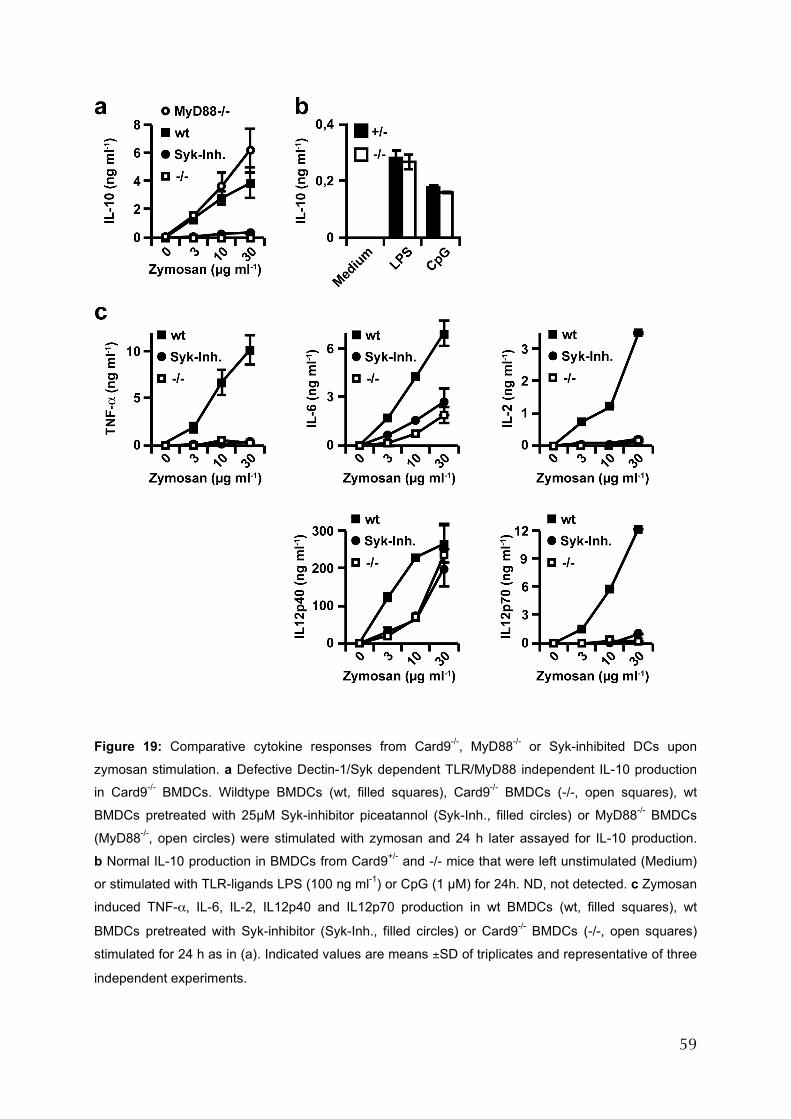
59
Figure 19: Comparative cytokine responses from Card9-/-, MyD88-/- or Syk-inhibited DCs upon
zymosan stimulation. a Defective Dectin-1/Syk dependent TLR/MyD88 independent IL-10 production
in Card9-/- BMDCs. Wildtype BMDCs (wt, filled squares), Card9-/- BMDCs (-/-, open squares), wt
BMDCs pretreated with 25µM Syk-inhibitor piceatannol (Syk-Inh., filled circles) or MyD88-/- BMDCs
(MyD88-/-, open circles) were stimulated with zymosan and 24 h later assayed for IL-10 production.
b Normal IL-10 production in BMDCs from Card9+/- and -/- mice that were left unstimulated (Medium)
or stimulated with TLR-ligands LPS (100 ng ml-1) or CpG (1 µM) for 24h. ND, not detected. c Zymosan
induced TNF-α, IL-6, IL-2, IL12p40 and IL12p70 production in wt BMDCs (wt, filled squares), wt
BMDCs pretreated with Syk-inhibitor (Syk-Inh., filled circles) or Card9-/- BMDCs (-/-, open squares)
stimulated for 24 h as in (a). Indicated values are means ±SD of triplicates and representative of three
independent experiments.

60
As expected, Nod1-induced NF-κB in WT and Bcl10-/- cells was equal, whereas
Carma3 only activated NF-κB in Bcl10+/- cells.
The requirement of Bcl10 and its downstream interaction partner Malt1 has also
been investigated in vivo in mice deficient for these factors. These experiments were
performed by Katrin Finger as part of her MD thesis work and are therefore not shown
here. For this data, please refer to (Gross, Gewies et al. 2006). In brief, Bcl10 or Malt1
deficient BMDCs show the same defects in Dectin-1 signalling as Card9-/- dendritic
cells do.
Figure 20: Differentiation and Dectin-1 expression on BMDCs. Prior to plating for stimulation on day
eight after killing of the donor mice, BMDCs from wild type, heterozygous and Card9 deficient mice
were FACS-analysed for surface marker expression. a Cells were stained with CD11c and MHCII to
determine the extent of differentiation and spontaneous maturation. Gates indicate the population of
total, CD11c+ dendritic cells and CD11c+ - MHCIIhigh, mature dendritic cells. Numbers represent
percentages of total viable cells. Viability was assayed by propidium iodide (PI) stain. b Dectin-1
expression on CD11c+ BMDCs. Wild type versus Card9 deficient (grey area) and isotype stained
(dashed line) cells.
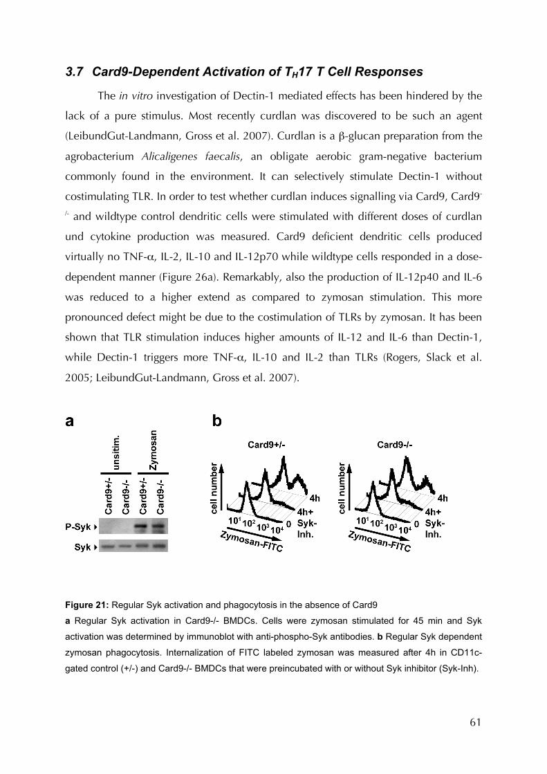
61
3.7 Card9-Dependent Activation of TH17 T Cell Responses
The in vitro investigation of Dectin-1 mediated effects has been hindered by the
lack of a pure stimulus. Most recently curdlan was discovered to be such an agent
(LeibundGut-Landmann, Gross et al. 2007). Curdlan is a β-glucan preparation from the
agrobacterium Alicaligenes faecalis, an obligate aerobic gram-negative bacterium
commonly found in the environment. It can selectively stimulate Dectin-1 without
costimulating TLR. In order to test whether curdlan induces signalling via Card9, Card9-
/- and wildtype control dendritic cells were stimulated with different doses of curdlan
und cytokine production was measured. Card9 deficient dendritic cells produced
virtually no TNF-α, IL-2, IL-10 and IL-12p70 while wildtype cells responded in a dose-
dependent manner (Figure 26a). Remarkably, also the production of IL-12p40 and IL-6
was reduced to a higher extend as compared to zymosan stimulation. This more
pronounced defect might be due to the costimulation of TLRs by zymosan. It has been
shown that TLR stimulation induces higher amounts of IL-12 and IL-6 than Dectin-1,
while Dectin-1 triggers more TNF-α, IL-10 and IL-2 than TLRs (Rogers, Slack et al.
2005; LeibundGut-Landmann, Gross et al. 2007).
Figure 21: Regular Syk activation and phagocytosis in the absence of Card9
a Regular Syk activation in Card9-/- BMDCs. Cells were zymosan stimulated for 45 min and Syk
activation was determined by immunoblot with anti-phospho-Syk antibodies. b Regular Syk dependent
zymosan phagocytosis. Internalization of FITC labeled zymosan was measured after 4h in CD11c-
gated control (+/-) and Card9-/- BMDCs that were preincubated with or without Syk inhibitor (Syk-Inh).

62
Activation of dendritic cells is a major step in the activation of adaptive immune
responses. It has been indicated that fungal immunity preferentially involves the
activation of the lately defined TH17 T helper cell subpopulation (Huang, Na et al.
2004). DC derived IL-23 is, in addition to IL-6 and TGF-β, essential for the activation of
TH17 cells (Ivanov, McKenzie et al. 2006). Utilizing curdlan, it was investigated
whether dendritic cells induce IL-23 in response to Dectin-1 stimulation in a Card9
dependent manner. Indeed, Curdlan induced IL-23 in a dose dependent manner in
wildtype, but not Card9-/- cells (Figure 26b). In contrast IL-23 production in response to
TLR stimulation was unaffected by the absence of Card9. In addition to certain
proinflammatory cytokines, namely IL-12p70 for TH1 and IL-23 for TH17 responses, the
upregulation of costimulatory factors on APCs is essential for the activation of sustained
helper T cell responses.
Figure 22: Specific defect in zymosan-induced NF-κB activation in Card9-/- cells. BMDCs from
Card9+/- or -/- mice were stimulated for 1 h with 100 µg/ml zymosan or 200 ng/µl LPS. RelA (red)
translocation into the nucleus (DAPI stained, blue; co-localization pink) was monitored by
immunofluorescence and quantified by determining the frequency of RelA positive nuclei in at least
100 individual cells.
63
Stimulation of DC with curdlan strongly induced upregulation of the
costimulatory molecules CD40, CD80 and CD86 on dendritic cells as demonstrated by
FACS analysis (Figure 27). In the absence of Card9, this upregulation was clearly
reduced. The defects appeared to be milder though, as compared to cytokine
production. The lack of IL-12p70 and IL-23 production as well as the reduced
upregulation of costimulatory factors strongly indicates that Card9 might play a role in
the activation of adaptive anti-fungal T cell responses. In order to test this, it was finally
determined whether TH1 and/or TH17 T cell responses were induced during fungal
infection and to what extent this depends on the Dectin-1/Card9 signalling pathway.
Large amounts of IFN-γ and IL-17 were produced by CD4+ spleen cells from mice
infected with Candida albicans after restimulation with heat-killed organisms (Fig. 28a).
Card9 was indispensable for the TH17 response and partially contributed to the TH1
response (Fig. 28b). Therefore, adaptive immune responses to Candida albicans
infection include the induction of TH17 cells and this process is dependent on innate
signalling via Card9.

64
4 Discussion
Collectively, these results identify a novel signalling pathway in innate immunity
with the potential to activate adaptive responses. Whereas Card9 is dispensable for
antigen receptor signalling, the data presented here suggests a model in which Card9
operates upstream of Bcl10 to transduce, together with Bcl10 and Malt1, Dectin-1
signals for NF-κB activation, cytokine production and dendritic cell controled
activation of TH17 T cells responses in vivo. For a model, see Figure 29. Dectin-1 was
recently discovered as the key non-TLR PRR for fungal β-glucan detection (Brown
2006). This receptor uses an intracellular YxxL ITAM motif to initiate signalling via Syk
through novel MyD88-independent pathways (Rogers, Slack et al. 2005). The results
presented here provide mechanistic insights into these pathways that can cooperate
with TLR signalling for full innate immune cell activation and for the induction of
pathogenspecific innate and adaptive responses (LeibundGut-Landmann, Gross et al.
2007). These findings also demonstrate that Bcl10–Malt1 signalling can be engaged by
an ITAM-containing PRR through the adaptor Card9, presumably in an analogous
manner to ITAM-containing lymphocyte antigen receptors that use Carma1 to couple to
Bcl10 and Malt1. This hypothesis suggests an evolutionarily conserved mechanism for
immune cell signalling and puts the concept forward that ITAM-receptors in innate and
adaptive immune cells can differentially connect to the conserved Bcl10/Malt1
complex via distinct CARD-coiled-coil adaptor proteins.
Figure 23: NF-κB activation was determined by EMSA in BMDMs stimulated for indicated time points
with 100 µg/ml zymosan (a), or for 1h with 10 µg/ml PGN, 500 ng/ml LPS or 500 ng/ml R848 (b)

65
4.1 Card9 is Dispensable for Lymphocyte Activation
Prior to the findings presented here, it’s been suggested that Card9 could
potentially play a role in Bcl10-mediated antigen receptor signalling (Thome and
Tschopp 2003). Bcl10-deficient mice show severe defects in virtually all NF-κB-related
responses mediated by the B- or T-cell receptor (Ruland, Duncan et al. 2001; Xue,
Morris et al. 2003). These mice show impaired lymphocyte development, manifested
by defects like the accumulation of CD4/CD8 double negative, CD44-/CD25-
thymocytes after the beta-checkpoint of thymocyte development and the total absence
of marginal zone B-cells in the spleen as well as recirculating B-cells in the bone
marrow. Consequently, humoral responses are severely reduced in the absence of
Bcl10. In these mice, B- and T-cells don’t proliferate after antigen-receptor stimulation
and NF-κB does not translocate to the nucleus. All these Bcl10 dependent lymphocyte
functions were unaffected by the absence of Card9. Therefore, Card9 is unlikely to
directly interfere with Bcl10 regulated antigen-receptor signalling.
4.2 Card9 Controls Innate Immunity
Card9 conducts signals downstream of Dectin-1, the receptor for fungal β-
glucans and is not involved in the reactions triggered by the well-defined Toll-like
receptor family. In myeloid cells, Dectin-1 mediates its signals via Syk, which binds to
the YxxL ITAM motif present in the cytoplasmic tail of Dectin-1 (Rogers, Slack et al.
2005). Dectin-1/Syk signalling activates multiple cellular outcomes in myeloid cells,
including transcription-dependent and -independent reactions (Brown 2006). Even
though independent ones, like phagocytosis, were found to be Card9-independent,
certain transcription-dependent functions, such as the production of cytokines were
clearly Card9 dependent. This defect was pinpointed to defective nuclear translocation
and activation of the transcription factor NF-κB after Dectin-1 ligation. Nevertheless,
additional transcription factors are also important and necessary for gene transcription
to mount sustained immune responses. Some of these are the signal transducer and
activator of transcription proteins (STATs), SMA- and MAD-related proteins (SMADs)
(Shuai and Liu 2005), forkhead-box (FOX) transcription factors (Coffer and Burgering
2004), interferon (IFN)-regulatory factor (IRF) family members (Lohoff and Mak 2005),
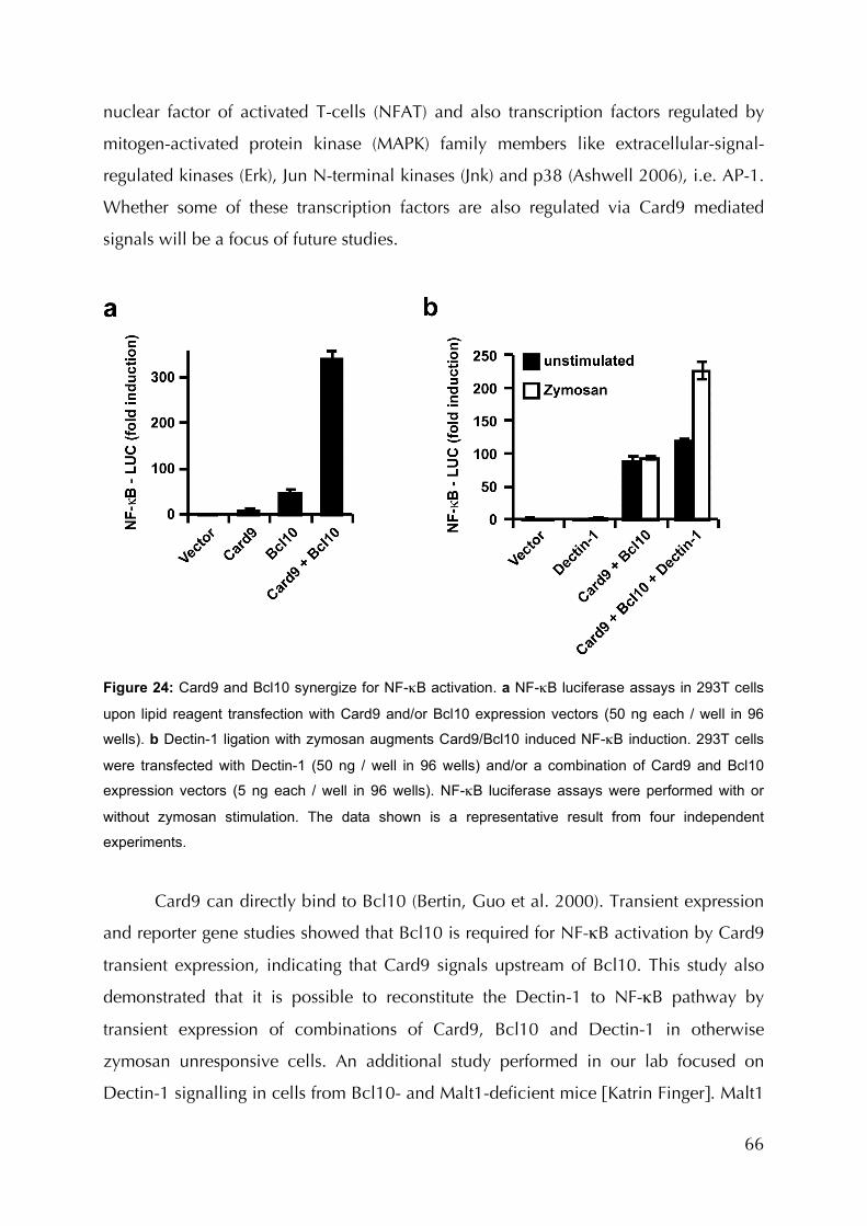
66
nuclear factor of activated T-cells (NFAT) and also transcription factors regulated by
mitogen-activated protein kinase (MAPK) family members like extracellular-signal-
regulated kinases (Erk), Jun N-terminal kinases (Jnk) and p38 (Ashwell 2006), i.e. AP-1.
Whether some of these transcription factors are also regulated via Card9 mediated
signals will be a focus of future studies.
Figure 24: Card9 and Bcl10 synergize for NF-κB activation. a NF-κB luciferase assays in 293T cells
upon lipid reagent transfection with Card9 and/or Bcl10 expression vectors (50 ng each / well in 96
wells). b Dectin-1 ligation with zymosan augments Card9/Bcl10 induced NF-κB induction. 293T cells
were transfected with Dectin-1 (50 ng / well in 96 wells) and/or a combination of Card9 and Bcl10
expression vectors (5 ng each / well in 96 wells). NF-κB luciferase assays were performed with or
without zymosan stimulation. The data shown is a representative result from four independent
experiments.
Card9 can directly bind to Bcl10 (Bertin, Guo et al. 2000). Transient expression
and reporter gene studies showed that Bcl10 is required for NF-κB activation by Card9
transient expression, indicating that Card9 signals upstream of Bcl10. This study also
demonstrated that it is possible to reconstitute the Dectin-1 to NF-κB pathway by
transient expression of combinations of Card9, Bcl10 and Dectin-1 in otherwise
zymosan unresponsive cells. An additional study performed in our lab focused on
Dectin-1 signalling in cells from Bcl10- and Malt1-deficient mice [Katrin Finger]. Malt1

67
binds to Bcl10 to transduce signals downstream of Bcl10 in lymphocytes. It became
apparent that Bcl10- and Malt1-deficient cells show the same defects in cytokine
production and NF-κB activation after Dectin-1 triggering as Card9 deficient cells. For
example, when stimulated with zymosan or Candida albicans cells, dendritic cells from
Bcl10-deficient or Malt1-deficient mice have defects in production of TNF-α, IL-2, IL-
10 and IL-6, while producing normal amounts of these cytokines in response to various
TLR-ligands like PGN, LPS or CpG. In addition, dendritic cells deficient in either Bcl10
or Malt1 are also unable to induce nuclear translocation of NF-κB when exposed to
zymosan. Thus, these results suggest a signalling pathway in which Card9 relays
Dectin-1/Syk signals via Bcl10 and Malt1 for NF-κB activation.
Figure 25: Bcl10 is required for Card9 induced NF-κB activation. Card9, Carma3 or Nod1 expression
plasmids were introduced into Bcl10+/- or -/- MEFs by lipid reagent transfection and NF-κB luciferase
assays performed.
Over the last years, signalling components downstream of Malt1 have been the
focus of extensive work. In T cells, the signal is transuded by TRAF2, TRAF6 and TAK1
(Rawlings, Sommer et al. 2006). It is therefore conceivable that similar mechanisms
could operate in myeloid cells, but are as yet not formally proven. The components
upstream of Card9 that connect the signal to Syk remain much more ambiguous. The
molecules relaying the analogue signals in lymphocytes include protein kinases C beta
and theta (PKCβ, PKCθ) and the membrane-associated guanylate kinase (MAGUK) and
linker-domain of Carma1. The MAGUK domain anchors Carma1 to the cell membrane

68
and mediates homophile interaction. The linker region between the MAGUK and
coiled-coil domain is a flexible region by which the N-terminus of Carma1 folds back
to the C-terminus, thereby presumably inhibiting interaction of the CARD with other
CARD-carrying proteins. PKCθ serine-phoshorylates the linker region of Carma1 and
thereby induces its activation (Sommer, Guo et al. 2005).
Figure 26: Curdlan is a pure β-Glucan that activates the Dectin-1/Card9 pathway. Production of
TNF-α, IL-12p40, IL-12p70, IL-10, IL-2, IL-6 (a) and IL-23 (b) in response to curdlan depends on
Card9. Cytokine production by BMDCs from Card9-/- or wild-type control mice after stimulation with
curdlan was measured by ELISA. Data are the mean ± S.D. of triplicate stimulations and are
representative of three independent experiments.
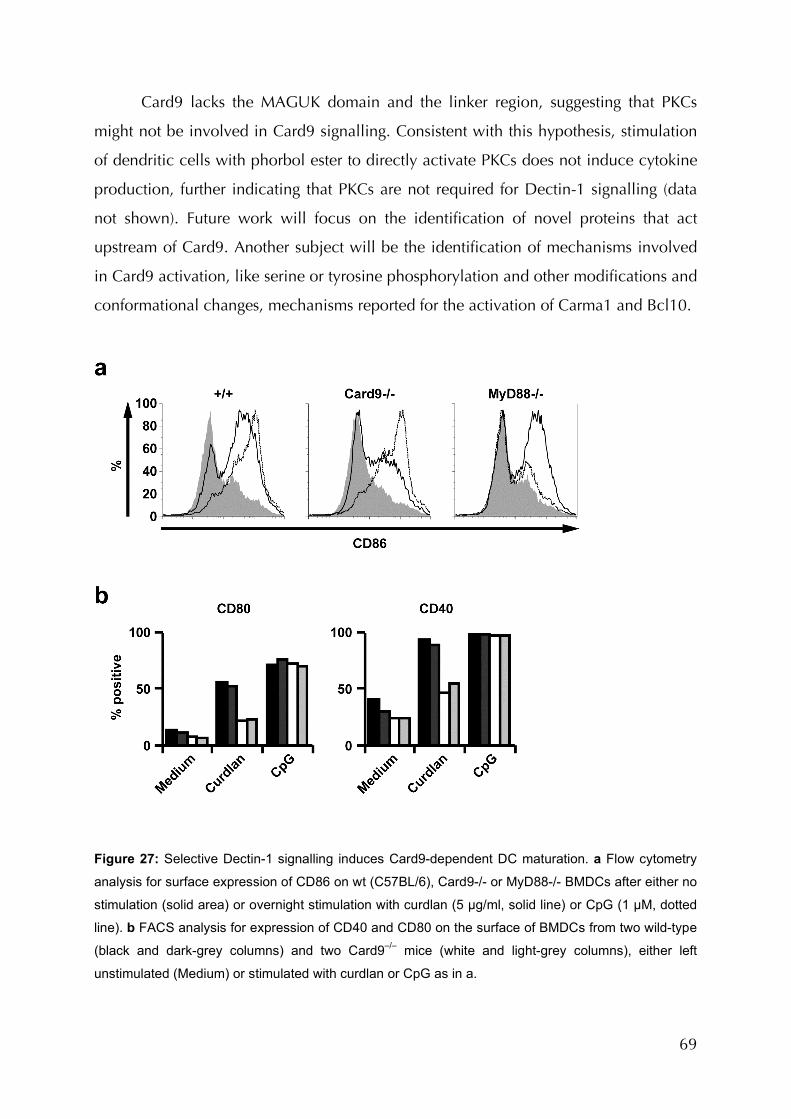
69
Card9 lacks the MAGUK domain and the linker region, suggesting that PKCs
might not be involved in Card9 signalling. Consistent with this hypothesis, stimulation
of dendritic cells with phorbol ester to directly activate PKCs does not induce cytokine
production, further indicating that PKCs are not required for Dectin-1 signalling (data
not shown). Future work will focus on the identification of novel proteins that act
upstream of Card9. Another subject will be the identification of mechanisms involved
in Card9 activation, like serine or tyrosine phosphorylation and other modifications and
conformational changes, mechanisms reported for the activation of Carma1 and Bcl10.
Figure 27: Selective Dectin-1 signalling induces Card9-dependent DC maturation. a Flow cytometry
analysis for surface expression of CD86 on wt (C57BL/6), Card9-/- or MyD88-/- BMDCs after either no
stimulation (solid area) or overnight stimulation with curdlan (5 µg/ml, solid line) or CpG (1 µM, dotted
line). b FACS analysis for expression of CD40 and CD80 on the surface of BMDCs from two wild-type
(black and dark-grey columns) and two Card9–/– mice (white and light-grey columns), either left
unstimulated (Medium) or stimulated with curdlan or CpG as in a.
70
On the basis of similarities and differences in Bcl10/Malt1 activation in innate
and adaptive immunity, a signalling model has been outlined (Figure 29). In adaptive
immunity, ITAM-containing molecules like the B-cell receptor Igα/β chains and T-cell
receptor CD3γ/ζ subunit utilize Carma1 to transduce signals to Bcl10/Malt1 and
eventually NF-κB. On the other side, innate ITAM-containing PRRs like Dectin-1 use
the Carma1 homologue Card9 to perform this action. Future research should address
the question of whether the differential use of Card9 and Carma1 is a feature of
different receptor families or different cell types and lineages.
After this data was published, two other groups also reported findings using
independently generated Card9 deficient mice (Underhill and Shimada 2007). Both
groups find similar expression patterns of Card9 as have been reported here and also
identified roles for Card9 in innate immunity, but with a different focus. The group of
Xin Lin reports that Card9 forwards signals from the intracellular PRR Nod2 for
activation MAP Kinases (Hsu, Zhang et al. 2007). They stimulated thioglycolate-elicited
macrophages with various PAMPs and found impaired IL-6 and TNF-α cytokine
responses with muramyl dipeptide (MDP, a Nod2 ligand) and poly(I:C) (a TLR3 ligand),
mild defects with peptidoglycan (PGN, a TLR2 ligand) and loxoribine (a TLR7 ligand)
and normal responses with lipopolysaccharide (LPS, a TLR4 ligand) or diacylated
lipopeptide FSL-1 (a TLR2/6 ligand). In terms of signal transduction, they reported
impaired activation of MAPK p38 upon stimulation with MDP or poly(I:C), while Erk
and Jnk as well as NF-κB activation were normal. Due to the fact, that poly(I:C), PGN
and loxoribine have, in addition to their TLR stimulating capacity also, like MDP the
potential to stimulate intracellular receptors, they conclude that Card9 participates in
the perception of intracellular infections. To test this theory, they infected macrophages
with the intracellular bacterium Listeria monocytogenes and vesicular stomatitis virus
(VSV). They then found impaired cytokine production as well as reduced p38 and Jnk
activation but normal Erk and NF-κB activation in response to both pathogens. In
overexpression experiments and coimmunoprecipitations direct interaction of Nod2
and Card9 was demonstrated. Reporterassay experiments also showed a collaboration
of Nod2 and Card9 for ATF-2 (a MAPK regulated transcription factor) activation.
Interaction of native proteins was shown in cellular extracts from macrophages upon
infection with Listeria monocytogenes. In infection experiments with the same
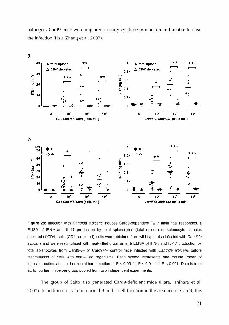
71
pathogen, Card9 mice were impaired in early cytokine production and unable to clear
the infection (Hsu, Zhang et al. 2007).
Figure 28: Infection with Candida albicans induces Card9-dependent TH17 antifungal responses. a
ELISA of IFN-γ and IL-17 production by total splenocytes (total spleen) or splenocyte samples
depleted of CD4+ cells (CD4+ depleted); cells were obtained from wild-type mice infected with Candida
albicans and were restimulated with heat-killed organisms. b ELISA of IFN-γ and IL-17 production by
total splenocytes from Card9–/– or Card9+/– control mice infected with Candida albicans before
restimulation of cells with heat-killed organisms. Each symbol represents one mouse (mean of
triplicate restimulations); horizontal bars, median. *, P < 0.05; **, P < 0.01; ***, P < 0.001. Data is from
six to fourteen mice per group pooled from two independent experiments.
The group of Saito also generated Card9-deficient mice (Hara, Ishihara et al.
2007). In addition to data on normal B and T cell function in the absence of Card9, this
72
group demonstrated Card9-dependent Dectin-1 activation. Moreover, they also tested
other receptors that don’t contain an ITAM themselves but signal via the ITAM
containing adapter proteins DAP12 or FcRγ. FcR (CD16), triggering receptor expressed
on monocytes 1 (TREM-1), osteoclast-associated receptor (OSCAR) and myeloid-
associated Ig-like receptor (MAIR) also activated dendritic cells in a Card9-dependent
manner. Upon TLR stimulation, Hara et al. additionally reported a partial defect in
TNF-α and IL-6 production for several ligands, suggesting a potential role of Card9 in
TLR signalling (Hara, Ishihara et al. 2007). Future work is therefore warranted to test
whether Card9 is a direct element of TLR signal transduction or whether it cooperates
with TLR pathways for full innate immune cell activation.
4.3 Card9 in Anti-Fungal Immunity.
In line with the finding that Card9 mediates fungal recognition in myeloid cells,
severe defects in fungal infections of Card9-deficient mice were observed. Intra-venous
infections lead to rapid lethality of Card9-/- animals after 4 to 9 days of infection and the
organs of Card9 knockout mice showed up to 100-fold higher loads of infiltrating
Candida albicans cells compared to wildtype littermates. How the defect in cytokine
production by dendritic cells and the increased susceptibility to fungi in vivo are
mechanistically connected is not clear as to now. One important role of tissue-resident
dendritic cells is the onset of inflammation after pathogen recognition by
proinflammatory cytokines like TNF-α and IL-6 (Romani 2004). Defective cytokine
production in the absence of Card9 during fungal infection would lead to reduced
inflammatory responses. Thus, it could be predicted that there would be a severe
reduction in the recruitment of lymphocytes or any other effector cells from the
bloodstream to the site of infection, which is key in controlling a pathogen. However,
the observed defects in cytokine production by dendritic cells are insufficient in
explaining the severity of infection. Full activation of adaptive immunity takes about
one week, and defects in adaptive immunity can therefore be excluded as a reason for
the early fatality in Card9 deficiency. Thus, other cell types, like neutrophils and
macrophages that also carry the Dectin-1 receptor are probably involved. Dectin-1-
deficient mice have already been studied in detail for defects in these cell types. Two
73
independent studies have addressed the role of Dectin-1 in innate immunity by
generating Dectin-1-deficient mice. The group of Gordon D. Brown has focused on the
role of Dectin-1 in macrophages and neutrophils (Taylor, Tsoni et al. 2007). The main
effector functions of these cells are phagocytosis of pathogens and killing by release of
reactive oxygen species (ROS), nitric oxide (NO) and lysosomal proteases. Dectin-1-
deficient macrophages and neutrophils showed a severely reduced ability to bind and
take up zymosan particles. The respiratory burst reaction was only reduced in
neutrophils from Dectin-1 knockouts, while it was found to be normal in macrophages.
In line with this, Dectin-1-/- neutrophils were significantly impaired in their ability to kill
live fungal cells in vitro, while macrophages showed no difference. It should be tested
whether that oxidative burst like phagocytosis is independent of Card9.
Dectin-1-/- mice show a defect in the recruitment of neutrophils, eosinophils and
macrophages into the peritoneal cavity upon infection with Candida albicans. This
phenomenon was linked to defective production of IL-6, the chemokines CCL2 and
CCL3 as well as granulocyte- and granulocyte-macrophage colony-stimulating factors.
Sources of these cytokines are resident macrophages of the peritoneal cavity that were
present in regular frequencies in Dectin-1 knockout mice. This finding provides further
evidence for the hypothesis that defective onset of inflammation in Card9 deficient
mice, due to defective cytokine as well as chemokine production in both dendritic cells
and tissue-resident macrophages, causes increased susceptibility to fungal infections.
Dectin-1 knockout mice showed increased susceptibility to disseminated
candidiasis. However, after infection with a dose of 104 cfu Candida albicans, some
Dectin-1 deficient mice survived long term and only succumbed to the infection after
the tenth day. In contrast, in a Card9 deficient background, no mouse survived more
than 9 days even with low doses of only 1000 cfu. In addition, IL-10 and IL-12
production from Dectin-1 deficient bone marrow derived dendritic cells in response to
zymosan stimulation was normal (Taylor, Tsoni et al. 2007), although these cells show
impaired cytokine production in response to curdlan stimulation (LeibundGut-
Landmann, Gross et al. 2007).
In a second report on independently generated Dectin-1 deficient mice, the
group of Tarakhovsky reports no defect in Candida albicans defence and only mild
defects in response to Pneumocystis carinii infection (Saijo, Fujikado et al. 2007). In this

74
study, they demonstrated defects in cytokine production using pure β-Glucans like
oxidized zymosan. In contrast, they show normal responses of Dectin-1-knockout
dendritic cells to zymosan or fungal cells in terms of cytokine production.
4.4 Activation of TH17 Responses by Card9
It was additionally demonstrated that Card9 signalling in dendritic cells mediates
the activation of TH17 responses in vivo and in vitro (Palm and Medzhitov 2007).
Production of the TH17 related cytokines IL-23 and IL-6, as well as the upregulation of T
cell co-stimulatory molecules on dendritic cells has been shown to be Card9
dependent. Consequently, Card9-deficient mice don’t induce anti-fungal TH17 T cells
upon infection. Thus, Card9-deficient mice might be a good model to specifically study
TH17 dependent responses. Emerging data suggests that TH17 T cell mediated immunity
plays a role in those infectious diseases that were shown to be largely independent of
TLR recognition. Card9 dependent C-type lectin recognition might be more important
for the recognition of these pathogens, which has to be tested, as outlined below in
more detail. TH17 T cells have also been connected to autoimmune diseases that were
previously thought to be TH1-related, like multiple sclerosis in the model of
experimental autoimmune encephalomyelitis (EAE). In addition, TH17 T cells were
found to be abundant in the lamina propria of the gut, although their function in this
tissue remains elusive. It’s been speculated that these cells develop in the interaction of
immune cells with the gut flora, but their development appears to be MyD88
independent (Ivanov, McKenzie et al. 2006). In correlation, IL-23 and TH17 T cells are
now implicated in the development of inflammatory bowel diseases such as Crohns
disease (Strober, Murray et al. 2006). Future work will address the question whether
Card9-dependent signalling contributes to the development of TH17 T cells in the gut
and the onset of inflammatory bowel diseases and other autoimmune diseases, possibly
by accelerating IL-23 and TH17 responses.
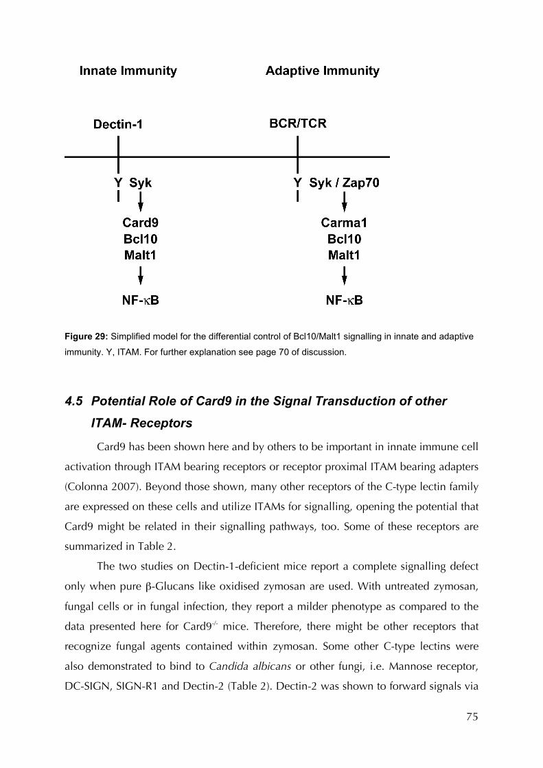
75
Figure 29: Simplified model for the differential control of Bcl10/Malt1 signalling in innate and adaptive
immunity. Y, ITAM. For further explanation see page 70 of discussion.
4.5 Potential Role of Card9 in the Signal Transduction of other
ITAM- Receptors
Card9 has been shown here and by others to be important in innate immune cell
activation through ITAM bearing receptors or receptor proximal ITAM bearing adapters
(Colonna 2007). Beyond those shown, many other receptors of the C-type lectin family
are expressed on these cells and utilize ITAMs for signalling, opening the potential that
Card9 might be related in their signalling pathways, too. Some of these receptors are
summarized in Table 2.
The two studies on Dectin-1-deficient mice report a complete signalling defect
only when pure β-Glucans like oxidised zymosan are used. With untreated zymosan,
fungal cells or in fungal infection, they report a milder phenotype as compared to the
data presented here for Card9-/- mice. Therefore, there might be other receptors that
recognize fungal agents contained within zymosan. Some other C-type lectins were
also demonstrated to bind to Candida albicans or other fungi, i.e. Mannose receptor,
DC-SIGN, SIGN-R1 and Dectin-2 (Table 2). Dectin-2 was shown to forward signals via
76
the ITAM bearing adapter protein FcRγ. It selectively recognises the hyphal morphology
of Candida albicans, whereas Dectin-1 recognises the yeast form (Gantner, Simmons et
al. 2005). Candida hyphae “hide” their β-Glucans from Dectin-1 by covering it with
other cell wall components, mostly mannans and mannoproteins. These mannoproteins
are subject to oxidization of zymosan that destroys Dectin-1 independent but
potentially Card9 dependent components of zymosan. To which component of
Candida albicans cells Dectin-2 binds is still unclear, thou. Dectin-2 might thereby be a
Card9-engaging anti-fungal proinflammatory receptor. Knockout mice deficient for
Dectin-2 would provide more data to this question but are not yet available.
Dectin-1 is the first member of a family of seven NK-cell receptor-like-C-type
lectins for which defined signalling capacities have been described (Brown, Herre et al.
2003; Cambi and Figdor 2003; Rogers, Slack et al. 2005). A group of six very similar
receptor-encoding genes is located in the vicinity of the Dectin-1 gene. They show high
sequence homology and might go back to a common ancestor gene that could have
multiplied at this locus, possibly by viral or retroviral action (Brown 2006). Four of
these genes, Clec2, Clec9a, Clec12a and Clec12b encode proteins possessing an YxxL
ITAM motif in their cytoplasmic tail. Although it is possible that Dectin-1 might be an
exception, these receptors might function similar to Dectin-1, possibly as PRRs, and
engage analogous pathways (Brown, Herre et al. 2003; Rogers, Slack et al. 2005).
Future work should therefore investigate the role of Card9/Bcl10/Malt1 signalling in
these, as of yet, undefined pathways.
FcγR possibly also engages Bcl10 for downstream signalling. The closely related
FcεR on mast cells was shown to signal via Bcl10 and Malt1 for NF-κB activation,
cytokine production and late phase hyperresponsiveness (Klemm, Gutermuth et al.
2006). FcγR and FcεR use the same Fc receptor γ subchain and some of the cell types
carrying one or the other are most closely related. Future work will investigate whether
FcγR indeed signals via Bcl10 like FcRε and if they engage Card9 or Carma1 to do this.
4.6 Comparing Card9 and MyD88 Dependent Effects
An interesting feature of Card9 deficient mice is, that they don’t develop
spontaneous fungal infection. MyD88-deficient mice frequently suffer from bacterial
77
infections like Staphylococcus aureus infections of the middle ear. MyD88 knockout
often die a few weeks after birth and seldom reach the age of one year. Compared to
those, Card9 mice live more than two years and eventually pass away from causes
other than infection (personal observation). Specific pathogen free (SPF) mouse facilities
are not free of opportunistic fungi like Candida or Aspergillus species (Harlan Inc.,
personal communication). The immune system of Card9 deficient mice therefore seems
to be able to deal with low-doses of fungal pathogens, probably by epithelial defence,
while MyD88 knockouts are often unable to do the same for some bacteria. However,
in fungal sepsis, very low doses of Candida already lead to the death of Card9 knockout
mice. Card9 therefore seems to take action when an infection becomes serious, i.e.
reaches the blood stream. C-type lectin domain receptors are relatively new in
evolution, first appearing in gnathostomata (Kono, Sakai et al. 2003; Robinson, Sancho
et al. 2006). Toll like receptors in contrast are much older and are conserved for
example in insects and nemaotodes (Hoffmann and Reichhart 2002). Even in plants,
LRR carrying PRRs are found. The abilities of C-type lectins in innate immunity might
correspond to the special selective pressures on higher vertebrates. Not much is known
about pathogen-host specificity in invertebrates. In vertebrates, pathogens-host
specificity often correlates with the severity of the disease (Wolfe, Dunavan et al.
2007). Opportunistic pathogens like Staphylococci or Candida species are usually non-
specific and only infect immunocompromised individuals. Many highly infectious
diseases like human immunodeficiency virus (HIV) and other viruses or certain
helmiths or protozoic pathogens like Plasmodium falciparum are highly host specific.
Interestingly, these are mostly non-bacterial pathogens. Many of the C-type lectin
receptors discussed above bind to such pathogens and some also bind to Mycobacteria
(Table 2). If further research would verify these findings in in vivo models, it might turn
out that C-type lectin receptors are more important in the recognition of eukaryotic or
viral, highly host-specific pathogens, while the main role of TLRs might be the
recognition of opportunistic prokaryotes and viruses. This would fit into place with the
observation that, while TLRs turned out to be of extraordinary importance for the
defence against bacteria, i.e. TLR2 for Staphylococcus aureus, the results for highly
infectious pathogens didn’t meet expectations (Bellocchio, Montagnoli et al. 2004;
Malmgaard, Melchjorsen et al. 2004; Villamon, Gozalbo et al. 2004; Power, Marshall
78
et al. 2006; Ryffel, Jacobs et al. 2006; Fremond, Togbe et al. 2007; Lepenies, Cramer et
al. 2007). It would also fit the fact that C-type lectin expression often varies greatly
between individuals or species, as outlined for DC-SIGN between men and mice and
for NK1.1 between different mice strains (Colucci, Caligiuri et al. 2003), possibly
reflecting the needs of pathogen-host specificity. If indeed some of these receptors
signal via Syk to Card9/Bcl10/Malt, this pathway might turn out to be of significant
importance for many infectious diseases.
A novel study on the contribution of MyD88/TRIF pathways for vaccination has
shown that TLR signalling also does not contribute to activation of adaptive responses
by the most common clinical adjuvants, Freunds adjuvant and aluminium hydroxid
(alum) (Gavin, Hoebe et al. 2006). However, it has been shown that whole
recombinant yeast cells expressing foreign antigen work as a potent adjuvant for
immunisation (Stubbs, Martin et al. 2001). Whether MyD88/TRIF and Card9 pathways
work synergistically together for activation of adaptive responses by any of theses
substances or whether both are dispensable for immunisation will allow further insights
on the mechanisms of adaptive immune activation by the commonly used but poorly
understood adjuvants.
4.7 Possible Involvement of Card9 in Cancer Development
Proteins of the Bcl10 pathway are recurrently involved in lymphomagenesis
(Farinha and Gascoyne 2005). There are two major mechanisms participating in
lymphoma development in this pathway. One is chromosomal translocation, bringing
genes under the control of a strong promoter like the immunoglobulin heavy chain
enhancer on chromosome 14 in humans. Bcl10 is an example for this mechanism
being translocated t(1;14)(p22;q32) (Willis, Jadayel et al. 1999). Chromosomal
translocation can also result in chimeric proteins like in the case of IAP2-Malt1 with the
translocation t(11;18)(q21;p21) (Morgan, Yin et al. 1999). The underlying mechanism
of all these pathogenic changes seems to be an increased and persistent oligomerisation
of proteins of this pathway, either due to fusion to proteins containing homo-
oligomerisation domains like IAP2, or by increasing the cellular concentration of
proteins with domains capable of homo-oligomerisation, like CARD domains and thus
79
eventually inducing NF-κB (Thome 2004). Card9 was found be upregulated in
lymphomas due to genomic gains of the Card9 locus. The Card9 gene is also reported
to be involved in lymphoma development through genomic gains. It is amplified in
translocation negative MALT-lymphoma and overexpressed in gastric B-cell lymphoma
(Kono, Sakai et al. 2003) (Nakamura, Nakamura et al. 2005) (Zhou, Ye et al. 2006).
Nevertheless, how Card9 actually contributes to lymphoma development is still
unclear. This work demonstrated that Card9 is an important activation mediator in
distinct cell types. Like experimental overexpression of Card9 can activate NF-κB in
cell lines, upregulation of Card9 due to genomic gains could lead to constitutive NF-κB
activation, thereby contributing to lymphomagenesis. Therefore, Card9 is, after Malt1
and Bcl10 the third member of Bcl10 pathways that can potentially promote cancer
development if genetically altered.
4.8 Conclusion and outlook
In contrast to immunity directed against viruses and bacteria, far less is known
about innate responses to fungal infections (Romani 2004). Yet, the incidence of these
infections is rapidly growing worldwide with the number of immunocompromised
patients, suffering from AIDS or undergoing immunosuppressive therapies for example
during organ transplantations. In order to potentially manipulate anti-fungal immunity,
it is necessary to understand the pathways controlling these responses. To this point,
molecular insights into fungal infections have mostly been derived from studies of the
receptor Dectin-1 (Brown 2006). The results presented here provide mechanistic
insights into these pathways. This might prove to be important for the development of
new anti-fungal immune therapies. This knowledge might also help to develop
therapeutics that interfere with Card9/Bcl10/Malt signalling in disease.
In recent years, numerous important infectious diseases, as well as common
adjuvants, were demonstrated to be TLR/MyD88 dependent to a minor degree
(Bellocchio, Montagnoli et al. 2004) (Villamon, Gozalbo et al. 2004). In turn, various
CLRs as well as other receptors and receptor families have been demonstrated to
recognize human pathogens. Many CLRs use ITAM motifs to forward signalling,
potentially via Card9. Future studies will test this hypothesis. Card9 might therefore be
80
a master regulator of innate immunity forwarding signals of many C-type lectins,
functioning in an analogous way to MyD88 for TLRs. TH17 T cell responses are
emerging to be of major importance for many infectious diseases as well as various
autoimmune diseases (Amadi-Obi, Yu et al. 2007) (Veldhoen, Hocking et al. 2006).
Like TLRs activate TH1 responses to many different pathogens via MyD88, Card9 might
also activate TH17 T cells for defence against pathogens other than fungi. In addition,
TH17 dependent autoimmunity might be related to Card9 dependent signals derived
either from pathogens or from host-derived stimuli. Future work will have to investigate
the potential involvement of Card9 in innate and adaptive immunity to various
pathogens as well as in TH17-mediated autoimmune models.
81
Citation Index
Abbas, A. K. and A. H. Lichtman (2006). "Basic Immunology." Saunders(Second Edition 2006-2007).
Abbas, A. K., K. M. Murphy, et al. (1996). "Functional diversity of helper T lymphocytes." Nature 383(6603): 787-93.
Ackerman, A. L. and P. Cresswell (2004). "Cellular mechanisms governing cross-presentation of exogenous antigens." Nat Immunol 5(7): 678-84.
Akira, S. and K. Takeda (2004). "Toll-like receptor signalling." Nat Rev Immunol 4(7): 499-511.
Aliprantis, A. O., R. B. Yang, et al. (1999). "Cell activation and apoptosis by bacterial lipoproteins through toll-like receptor-2." Science 285(5428): 736-9.
Amadi-Obi, A., C. R. Yu, et al. (2007). "TH17 cells contribute to uveitis and scleritis and are expanded by IL-2 and inhibited by IL-27/STAT1." Nat Med 13(6): 711-8.
Ashwell, J. D. (2006). "The many paths to p38 mitogen-activated protein kinase activation in the immune system." Nat Rev Immunol 6(7): 532-40.
Barrow, A. D. and J. Trowsdale (2006). "You say ITAM and I say ITIM, let's call the whole thing off: the ambiguity of immunoreceptor signalling." Eur J Immunol 36(7): 1646-53.
Bassing, C. H., W. Swat, et al. (2002). "The mechanism and regulation of chromosomal V(D)J recombination." Cell 109 Suppl: S45-55.
Bellocchio, S., C. Montagnoli, et al. (2004). "The contribution of the Toll-like/IL-1 receptor superfamily to innate and adaptive immunity to fungal pathogens in vivo." J Immunol 172(5): 3059-69.
82
Bertin, J., Y. Guo, et al. (2000). "CARD9 is a novel caspase recruitment domain-containing protein that interacts with BCL10/CLAP and activates NF-kappa B." J Biol Chem 275(52): 41082-6.
Bonizzi, G. and M. Karin (2004). "The two NF-kappaB activation pathways and their role in innate and adaptive immunity." Trends Immunol 25(6): 280-8.
Bouchier-Hayes, L. and S. J. Martin (2002). "CARD games in apoptosis and immunity." EMBO Rep 3(7): 616-21.
Brown, G. D. (2006). "Dectin-1: a signalling non-TLR pattern-recognition receptor." Nat Rev Immunol 6(1): 33-43.
Brown, G. D. and S. Gordon (2001). "Immune recognition. A new receptor for beta-glucans." Nature 413(6851): 36-7.
Brown, G. D. and S. Gordon (2003). "Fungal beta-glucans and mammalian immunity." Immunity 19(3): 311-5.
Brown, G. D., J. Herre, et al. (2003). "Dectin-1 mediates the biological effects of beta-glucans." J Exp Med 197(9): 1119-24.
Bryant, P. W., A. M. Lennon-Dumenil, et al. (2002). "Proteolysis and antigen presentation by MHC class II molecules." Adv Immunol 80: 71-114.
Calame, K. L., K. I. Lin, et al. (2003). "Regulatory mechanisms that determine the development and function of plasma cells." Annu Rev Immunol 21: 205-30.
Cambi, A. and C. G. Figdor (2003). "Dual function of C-type lectin-like receptors in the immune system." Curr Opin Cell Biol 15(5): 539-46.
Carroll, M. C. (2004). "The complement system in regulation of adaptive immunity." Nat Immunol 5(10): 981-6.
83
Coffer, P. J. and B. M. Burgering (2004). "Forkhead-box transcription factors and their role in the immune system." Nat Rev Immunol 4(11): 889-99.
Colonna, M. (2007). "All roads lead to CARD9." Nat Immunol 8(6): 554-5.
Colucci, F., M. A. Caligiuri, et al. (2003). "What does it take to make a natural killer?" Nat Rev Immunol 3(5): 413-25.
DeFranco, A. L. (2000). "B-cell activation 2000." Immunol Rev 176: 5-9.
Deyoung, B. J. and R. W. Innes (2006). "Plant NBS-LRR proteins in pathogen sensing and host defense." Nat Immunol 7(12): 1243-9.
Di Carlo, F. J. and J. V. Fiore (1958). "On the composition of zymosan." Science 127(3301): 756-7.
Elion, E. A. (2000). "Pheromone response, mating and cell biology." Curr Opin Microbiol 3(6): 573-81.
Eliopoulos, A. G., S. Das, et al. (2006). "The tyrosine kinase Syk regulates TPL2 activation signals." J Biol Chem 281(3): 1371-80.
Eyles, J. L., A. W. Roberts, et al. (2006). "Granulocyte colony-stimulating factor and neutrophils--forgotten mediators of inflammatory disease." Nat Clin Pract Rheumatol 2(9): 500-10.
Farinha, P. and R. D. Gascoyne (2005). "Molecular pathogenesis of mucosa-associated lymphoid tissue lymphoma." J Clin Oncol 23(26): 6370-8.
Figdor, C. G., Y. van Kooyk, et al. (2002). "C-type lectin receptors on dendritic cells and Langerhans cells." Nat Rev Immunol 2(2): 77-84.
84
Fremond, C. M., D. Togbe, et al. (2007). "IL-1 receptor-mediated signal is an essential component of MyD88-dependent innate response to Mycobacterium tuberculosis infection." J Immunol 179(2): 1178-89.
Fritz, J. H., R. L. Ferrero, et al. (2006). "Nod-like proteins in immunity, inflammation and disease." Nat Immunol 7(12): 1250-7.
Fu, Y. X. and D. D. Chaplin (1999). "Development and maturation of secondary lymphoid tissues." Annu Rev Immunol 17: 399-433.
Fugmann, S. D., A. I. Lee, et al. (2000). "The RAG proteins and V(D)J recombination: complexes, ends, and transposition." Annu Rev Immunol 18: 495-527.
Gantner, B. N., R. M. Simmons, et al. (2003). "Collaborative induction of inflammatory responses by dectin-1 and Toll-like receptor 2." J Exp Med 197(9): 1107-17.
Gantner, B. N., R. M. Simmons, et al. (2005). "Dectin-1 mediates macrophage recognition of Candida albicans yeast but not filaments." Embo J 24(6): 1277-86.
Gavin, A. L., K. Hoebe, et al. (2006). "Adjuvant-enhanced antibody responses in the absence of toll-like receptor signaling." Science 314(5807): 1936-8.
Gordon, S. (2002). "Pattern recognition receptors: doubling up for the innate immune response." Cell 111(7): 927-30.
Gordon, S. (2003). "Alternative activation of macrophages." Nat Rev Immunol 3(1): 23-35.
Gordon, S. and P. R. Taylor (2005). "Monocyte and macrophage heterogeneity." Nat Rev Immunol 5(12): 953-64.
Govind, S. (1999). "Control of development and immunity by rel transcription factors in Drosophila." Oncogene 18(49): 6875-87.
85
Gross, O., A. Gewies, et al. (2006). "Card9 controls a non-TLR signalling pathway for innate anti-fungal immunity." Nature 442(7103): 651-6.
Hammer, M., J. Mages, et al. (2005). "Control of dual-specificity phosphatase-1 expression in activated macrophages by IL-10." Eur J Immunol 35(10): 2991-3001.
Hara, H., C. Ishihara, et al. (2007). "The adaptor protein CARD9 is essential for the activation of myeloid cells through ITAM-associated and Toll-like receptors." Nat Immunol 8(6): 619-29.
Hewitt, E. W. (2003). "The MHC class I antigen presentation pathway: strategies for viral immune evasion." Immunology 110(2): 163-9.
Heyman, B. (2000). "Regulation of antibody responses via antibodies, complement, and Fc receptors." Annu Rev Immunol 18: 709-37.
Hoffmann, J. A. and J. M. Reichhart (2002). "Drosophila innate immunity: an evolutionary perspective." Nat Immunol 3(2): 121-6.
Honda, K. and T. Taniguchi (2006). "IRFs: master regulators of signalling by Toll-like receptors and cytosolic pattern-recognition receptors." Nat Rev Immunol 6(9): 644-58.
Hsu, Y. M., Y. Zhang, et al. (2007). "The adaptor protein CARD9 is required for innate immune responses to intracellular pathogens." Nat Immunol 8(2): 198-205.
Huang, W., L. Na, et al. (2004). "Requirement of interleukin-17A for systemic anti-Candida albicans host defense in mice." J Infect Dis 190(3): 624-31.
Inohara, N. and G. Nunez (2003). "NODs: intracellular proteins involved in inflammation and apoptosis." Nat Rev Immunol 3(5): 371-82.
Ivanov, II, B. S. McKenzie, et al. (2006). "The orphan nuclear receptor RORgammat directs the differentiation program of proinflammatory IL-17+ T helper cells." Cell 126(6): 1121-33.
86
Janeway, C. A., Jr. and R. Medzhitov (2002). "Innate immune recognition." Annu Rev Immunol 20: 197-216.
Jung, D. and F. W. Alt (2004). "Unraveling V(D)J recombination; insights into gene regulation." Cell 116(2): 299-311.
Kaparakis, M., D. J. Philpott, et al. (2007). "Mammalian NLR proteins; discriminating foe from friend." Immunol Cell Biol.
Kapsenberg, M. L. (2003). "Dendritic-cell control of pathogen-driven T-cell polarization." Nat Rev Immunol 3(12): 984-93.
Karin, M. and F. R. Greten (2005). "NF-kappaB: linking inflammation and immunity to cancer development and progression." Nat Rev Immunol 5(10): 749-59.
Katze, M. G., Y. He, et al. (2002). "Viruses and interferon: a fight for supremacy." Nat Rev Immunol 2(9): 675-87.
Kawai, T., O. Adachi, et al. (1999). "Unresponsiveness of MyD88-deficient mice to endotoxin." Immunity 11(1): 115-22.
Kim, S., J. Poursine-Laurent, et al. (2005). "Licensing of natural killer cells by host major histocompatibility complex class I molecules." Nature 436(7051): 709-13.
Klemm, S., J. Gutermuth, et al. (2006). "The Bcl10-Malt1 complex segregates Fc epsilon RI-mediated nuclear factor kappa B activation and cytokine production from mast cell degranulation." J Exp Med 203(2): 337-47.
Klesney-Tait, J., I. R. Turnbull, et al. (2006). "The TREM receptor family and signal integration." Nat Immunol 7(12): 1266-73.
Kono, T., T. Sakai, et al. (2003). "Molecular cloning and expression analysis of a novel caspase recruitment domain protein (CARD) in common carp Cyprinus carpio L." Gene 309(1): 57-64.
87
Kraft, S. and J. P. Kinet (2007). "New developments in FcepsilonRI regulation, function and inhibition." Nat Rev Immunol 7(5): 365-78.
LeibundGut-Landmann, S., O. Gross, et al. (2007). "Syk- and CARD9-dependent coupling of innate immunity to the induction of T helper cells that produce interleukin 17." Nat Immunol 8(6): 630-8.
Lepenies, B., J. P. Cramer, et al. (2007). "Induction of experimental cerebral malaria is independent of TLR2/4/9." Med Microbiol Immunol.
Lin, X. and D. Wang (2004). "The roles of CARMA1, Bcl10, and MALT1 in antigen receptor signaling." Semin Immunol 16(6): 429-35.
Lohoff, M. and T. W. Mak (2005). "Roles of interferon-regulatory factors in T-helper-cell differentiation." Nat Rev Immunol 5(2): 125-35.
Lutz, M. B., N. Kukutsch, et al. (1999). "An advanced culture method for generating large quantities of highly pure dendritic cells from mouse bone marrow." J Immunol Methods 223(1): 77-92.
Malmgaard, L., J. Melchjorsen, et al. (2004). "Viral activation of macrophages through TLR-dependent and -independent pathways." J Immunol 173(11): 6890-8.
Matsumoto, R., D. Wang, et al. (2005). "Phosphorylation of CARMA1 plays a critical role in T Cell receptor-mediated NF-kappaB activation." Immunity 23(6): 575-85.
McAllister-Lucas, L. M., N. Inohara, et al. (2001). "Bimp1, a MAGUK family member linking protein kinase C activation to Bcl10-mediated NF-kappaB induction." J Biol Chem 276(33): 30589-97.
Medzhitov, R. (2001). "Toll-like receptors and innate immunity." Nat Rev Immunol 1(2): 135-45.
Medzhitov, R., P. Preston-Hurlburt, et al. (1997). "A human homologue of the Drosophila Toll protein signals activation of adaptive immunity." Nature 388(6640): 394-7.
88
Medzhitov, R., P. Preston-Hurlburt, et al. (1998). "MyD88 is an adaptor protein in the hToll/IL-1 receptor family signaling pathways." Mol Cell 2(2): 253-8.
Meylan, E., J. Tschopp, et al. (2006). "Intracellular pattern recognition receptors in the host response." Nature 442(7098): 39-44.
Mills, K. H. (2004). "Regulatory T cells: friend or foe in immunity to infection?" Nat Rev Immunol 4(11): 841-55.
Morgan, J. A., Y. Yin, et al. (1999). "Breakpoints of the t(11;18)(q21;q21) in mucosa-associated lymphoid tissue (MALT) lymphoma lie within or near the previously undescribed gene MALT1 in chromosome 18." Cancer Res 59(24): 6205-13.
Muljo, S. A. and M. S. Schlissel (2000). "Pre-B and pre-T-cell receptors: conservation of strategies in regulating early lymphocyte development." Immunol Rev 175: 80-93.
Murphy, K. M. and S. L. Reiner (2002). "The lineage decisions of helper T cells." Nat Rev Immunol 2(12): 933-44.
Nakamura, S., S. Nakamura, et al. (2005). "Overexpression of caspase recruitment domain (CARD) membrane-associated guanylate kinase 1 (CARMA1) and CARD9 in primary gastric B-cell lymphoma." Cancer 104(9): 1885-93.
Nemazee, D. (2000). "Receptor selection in B and T lymphocytes." Annu Rev Immunol 18: 19-51.
Niiro, H. and E. A. Clark (2002). "Regulation of B-cell fate by antigen-receptor signals." Nat Rev Immunol 2(12): 945-56.
Palm, N. W. and R. Medzhitov (2007). "Antifungal defense turns 17." Nat Immunol 8(6): 549-51.
Pardoll, D. (2003). "Does the immune system see tumors as foreign or self?" Annu Rev Immunol 21: 807-39.

89
Power, M. R., J. S. Marshall, et al. (2006). "The myeloid differentiation factor 88 is dispensable for the development of a delayed host response to Pseudomonas aeruginosa lung infection in mice." Clin Exp Immunol 146(2): 323-9.
Rammensee, H. G. and M. J. Bevan (1984). "Evidence from in vitro studies that tolerance to self antigens is MHC-restricted." Nature 308(5961): 741-4.
Rawlings, D. J., K. Sommer, et al. (2006). "The CARMA1 signalosome links the signalling machinery of adaptive and innate immunity in lymphocytes." Nat Rev Immunol 6(11): 799-812.
Reis e Sousa, C. (2006). "Dendritic cells in a mature age." Nat Rev Immunol 6(6): 476-83.
Robinson, M. J., D. Sancho, et al. (2006). "Myeloid C-type lectins in innate immunity." Nat Immunol 7(12): 1258-65.
Rogers, N. C., E. C. Slack, et al. (2005). "Syk-dependent cytokine induction by Dectin-1 reveals a novel pattern recognition pathway for C type lectins." Immunity 22(4): 507-17.
Romani, L. (2004). "Immunity to fungal infections." Nat Rev Immunol 4(1): 1-23.
Ruland, J., G. S. Duncan, et al. (2001). "Bcl10 is a positive regulator of antigen receptor-induced activation of NF-kappaB and neural tube closure." Cell 104(1): 33-42.
Ruland, J., G. S. Duncan, et al. (2003). "Differential requirement for Malt1 in T and B cell antigen receptor signaling." Immunity 19(5): 749-58.
Ruland, J. and T. W. Mak (2003). "From antigen to activation: specific signal transduction pathways linking antigen receptors to NF-kappaB." Semin Immunol 15(3): 177-83.
Ruland, J. and T. W. Mak (2003). "Transducing signals from antigen receptors to nuclear factor kappaB." Immunol Rev 193: 93-100.
90
Ryffel, B., M. Jacobs, et al. (2006). "Toll-like receptors and control of mycobacterial infection in mice." Novartis Found Symp 279: 127-39; discussion 139-41, 216-9.
Saijo, S., N. Fujikado, et al. (2007). "Dectin-1 is required for host defense against Pneumocystis carinii but not against Candida albicans." Nat Immunol 8(1): 39-46.
Sansonetti, P. J. (2006). "The innate signaling of dangers and the dangers of innate signaling." Nat Immunol 7(12): 1237-42.
Sedlik, C., D. Orbach, et al. (2003). "A critical role for Syk protein tyrosine kinase in Fc receptor-mediated antigen presentation and induction of dendritic cell maturation." J Immunol 170(2): 846-52.
Sharpe, A. H. and G. J. Freeman (2002). "The B7-CD28 superfamily." Nat Rev Immunol 2(2): 116-26.
Sher, A. and R. L. Coffman (1992). "Regulation of immunity to parasites by T cells and T cell-derived cytokines." Annu Rev Immunol 10: 385-409.
Shuai, K. and B. Liu (2005). "Regulation of gene-activation pathways by PIAS proteins in the immune system." Nat Rev Immunol 5(8): 593-605.
Sommer, K., B. Guo, et al. (2005). "Phosphorylation of the CARMA1 linker controls NF-kappaB activation." Immunity 23(6): 561-74.
Sparwasser, T., E. S. Koch, et al. (1998). "Bacterial DNA and immunostimulatory CpG oligonucleotides trigger maturation and activation of murine dendritic cells." Eur J Immunol 28(6): 2045-54.
Starr, T. K., S. C. Jameson, et al. (2003). "Positive and negative selection of T cells." Annu Rev Immunol 21: 139-76.
Strober, W., P. J. Murray, et al. (2006). "Signalling pathways and molecular interactions of NOD1 and NOD2." Nat Rev Immunol 6(1): 9-20.
91
Stubbs, A. C., K. S. Martin, et al. (2001). "Whole recombinant yeast vaccine activates dendritic cells and elicits protective cell-mediated immunity." Nat Med 7(5): 625-9.
Tafuri, A., A. Shahinian, et al. (2001). "ICOS is essential for effective T-helper-cell responses." Nature 409(6816): 105-9.
Takeda, K., T. Kaisho, et al. (2003). "Toll-like receptors." Annu Rev Immunol 21: 335-76.
Takeuchi, O., K. Hoshino, et al. (2000). "Cutting edge: TLR2-deficient and MyD88-deficient mice are highly susceptible to Staphylococcus aureus infection." J Immunol 165(10): 5392-6.
Takeuchi, O., K. Hoshino, et al. (1999). "Differential roles of TLR2 and TLR4 in recognition of gram-negative and gram-positive bacterial cell wall components." Immunity 11(4): 443-51.
Taylor, P. R., G. D. Brown, et al. (2002). "The beta-glucan receptor, dectin-1, is predominantly expressed on the surface of cells of the monocyte/macrophage and neutrophil lineages." J Immunol 169(7): 3876-82.
Taylor, P. R., S. V. Tsoni, et al. (2007). "Dectin-1 is required for beta-glucan recognition and control of fungal infection." Nat Immunol 8(1): 31-8.
Thomas, K. R. and M. R. Capecchi (1987). "Site-directed mutagenesis by gene targeting in mouse embryo-derived stem cells." Cell 51(3): 503-12.
Thome, M. (2004). "CARMA1, BCL-10 and MALT1 in lymphocyte development and activation." Nat Rev Immunol 4(5): 348-59.
Thome, M. and J. Tschopp (2003). "TCR-induced NF-kappaB activation: a crucial role for Carma1, Bcl10 and MALT1." Trends Immunol 24(8): 419-24.
Thompson, A. J. and S. A. Locarnini (2007). "Toll-like receptors, RIG-I-like RNA helicases and the antiviral innate immune response." Immunol Cell Biol.

92
Underhill, D. M. and A. Ozinsky (2002). "Phagocytosis of microbes: complexity in action." Annu Rev Immunol 20: 825-52.
Underhill, D. M., A. Ozinsky, et al. (1999). "The Toll-like receptor 2 is recruited to macrophage phagosomes and discriminates between pathogens." Nature 401(6755): 811-5.
Underhill, D. M., E. Rossnagle, et al. (2005). "Dectin-1 activates Syk tyrosine kinase in a dynamic subset of macrophages for reactive oxygen production." Blood 106(7): 2543-50.
Underhill, D. M. and T. Shimada (2007). "A pair of 9s: it's in the CARDs." Nat Immunol 8(2): 122-4.
Veldhoen, M., R. J. Hocking, et al. (2006). "Signals mediated by transforming growth factor-beta initiate autoimmune encephalomyelitis, but chronic inflammation is needed to sustain disease." Nat Immunol 7(11): 1151-6.
Villamon, E., D. Gozalbo, et al. (2004). "Toll-like receptor 2 is dispensable for acquired host immune resistance to Candida albicans in a murine model of disseminated candidiasis." Microbes Infect 6(6): 542-8.
Villamon, E., D. Gozalbo, et al. (2004). "Toll-like receptor-2 is essential in murine defenses against Candida albicans infections." Microbes Infect 6(1): 1-7.
Willis, T. G., D. M. Jadayel, et al. (1999). "Bcl10 is involved in t(1;14)(p22;q32) of MALT B cell lymphoma and mutated in multiple tumor types." Cell 96(1): 35-45.
Wolfe, N. D., C. P. Dunavan, et al. (2007). "Origins of major human infectious diseases." Nature 447(7142): 279-83.
Xue, L., S. W. Morris, et al. (2003). "Defective development and function of Bcl10-deficient follicular, marginal zone and B1 B cells." Nat Immunol 4(9): 857-65.
93
Ye, P., F. H. Rodriguez, et al. (2001). "Requirement of interleukin 17 receptor signaling for lung CXC chemokine and granulocyte colony-stimulating factor expression, neutrophil recruitment, and host defense." J Exp Med 194(4): 519-27.
Yokoyama, W. M. and B. F. Plougastel (2003). "Immune functions encoded by the natural killer gene complex." Nat Rev Immunol 3(4): 304-16.
Zhou, Y., H. Ye, et al. (2006). "Distinct comparative genomic hybridisation profiles in gastric mucosa-associated lymphoid tissue lymphomas with and without t(11;18)(q21;q21)." Br J Haematol 133(1): 35-42.