EGFR-deficient mice have delayed primary endochondral
41
EGFR-deficient mice have delayed primary endochondral ossification due to defective osteoclast recruitment Ke Wang 1,2 , Hiroaki Yamamoto 1,2 , Jennie R. Chin 1,2 , Zena Werb 3 , and Thiennu H. Vu 1,2 Departments of 1 Medicine and 3 Anatomy, and 2 Lung Biology Center, University of California, San Francisco, CA 94143 Correspondence: Thiennu H. Vu Box 2911 University of California San Francisco, CA 94143-2911 Phone: (415) 514-4266 Fax: (415) 514-4365 Email: [email protected] Running title : Delayed osteoclast recruitment into EGFR-/- cartilage Key words : Epidermal growth factor receptor, bone development, endochondral ossification, osteoclast formation JBC Papers in Press. Published on September 28, 2004 as Manuscript M403114200 Copyright 2004 by The American Society for Biochemistry and Molecular Biology, Inc. by guest on April 4, 2019 http://www.jbc.org/ Downloaded from
Transcript of EGFR-deficient mice have delayed primary endochondral

EGFR-deficient mice have delayed primary endochondral ossification due to
defective osteoclast recruitment
Ke Wang1,2, Hiroaki Yamamoto1,2, Jennie R. Chin1,2 , Zena Werb3, and Thiennu H. Vu1,2
Departments of 1Medicine and 3Anatomy, and 2Lung Biology Center, University of
California, San Francisco, CA 94143
Correspondence: Thiennu H. Vu
Box 2911
University of California
San Francisco, CA 94143-2911
Phone: (415) 514-4266
Fax: (415) 514-4365
Email: [email protected]
Running title: Delayed osteoclast recruitment into EGFR-/- cartilage
Key words: Epidermal growth factor receptor, bone development, endochondral
ossification, osteoclast formation
JBC Papers in Press. Published on September 28, 2004 as Manuscript M403114200
Copyright 2004 by The American Society for Biochemistry and Molecular Biology, Inc.
by guest on April 4, 2019
http://ww
w.jbc.org/
Dow
nloaded from

Wang et al.
2
SUMMARY
The epidermal growth factor receptor (EGFR) and its ligands function in diverse cellular
functions including cell proliferation, differentiation, motility and survival. EGFR
signaling is important for the development of many tissues, including skin, lungs,
intestines, and the craniofacial skeleton. We have now determined the role of EGFR
signaling in endochondral ossification. We analyzed long bone development in EGFR-
deficient mice. EGFR deficiency caused delayed primary ossification of the cartilage
anlage and delayed osteoclast and osteoblast recruitment. Ossification of the growth
plates was also abnormal resulting in an expanded area of growth plate hypertrophic
cartilage and few bony trabeculae. The delayed osteoclast recruitment was not due to
inadequate expression of matrix metalloproteinases, including MMP-9, which have
previously been shown to be important for osteoclast recruitment. EGFR was expressed
by osteoclasts, suggesting that EGFR ligands may act directly to affect the formation
and/or function of these cells. EGFR signaling regulated osteoclast formation. Inhibition
of EGFR tyrosine kinase activity decreased the generation of osteoclasts from cultured
bone marrow cells.
by guest on April 4, 2019
http://ww
w.jbc.org/
Dow
nloaded from

Wang et al.
3
INTRODUCTION
Skeletal elements develop by two distinct mechanisms: intramembranous and
endochondral ossification (1). Endochondral ossification is a process by which a
cartilaginous template is first formed and then replaced by bone. During embryogenesis,
condensations of mesenchymal cells form, within which chondrocytes develop,
proliferate and differentiate to form a cartilage template that contains distinct zones of
resting, proliferative, and hypertrophic chondrocytes. The proliferation and
differentiation of chondrocytes within the cartilage template are spatially ordered, with
proliferating cells at the two ends of the template and progressively more mature cells
forming hypertrophic cartilage in the middle. Hypertrophic chondrocytes secrete a
specialized extracellular matrix (ECM) containing collagen X, which becomes calcified.
Endochondral ossification begins with the invasion of the calcified hypertrophic cartilage
by blood vessels, accompanied by osteoclasts and osteoblasts (primary ossification
center). The function of osteoclasts is to remove the hypertrophic cartilage ECM and that
of osteoblasts is to replace it with bone ECM. Longitudinal bone growth is accomplished
by the continuing proliferation and maturation of chondrocytes at the ends of the cartilage
template (the growth plates) to form more hypertrophic cartilage and its continual
removal and replacement by bone (growth plate ossification or formation of primary
spongiosa). Normal endochondral bone development requires the exquisite coordination
of hypertrophic cartilage formation, vascular invasion, and the development and function
of osteoclasts and osteoblasts (2; 3).
The epidermal growth factor receptor (EGFR) family of receptor tyrosine kinases
includes EGFR/ErbB1, HER2/ErbB2, HER3/ErbB3 and HER4/ErbB4 (4; 5). EGFR
by guest on April 4, 2019
http://ww
w.jbc.org/
Dow
nloaded from

Wang et al.
4
binds several ligands including epidermal growth factor (EGF), transforming growth
factor-α (TGF-α), betacellulin, epiregulin and amphiregulin. During mouse
development, EGFR and ligands are expressed in many tissues, including skeletal tissues
such as embryonic mandible, Meckel’s cartilage and limbs (6; 7; 8; 9; 10). EGFR
deficient mice have abnormal craniofacial cartilage and intramembranous bone formation
resulting in abnormal development with narrow, elongated snouts, underdeveloped jaw,
and high incidence of cleft palate, as well as abnormal development in many epithelial
organs including skin, intestines, and lungs (11; 12; 13; 14; 15; 16).
EFGR expression has been detected in the axial and appendicular skeleton at the
bone-cartilage junction (17), suggesting a role for this signaling pathway in
endochrondral bone formation. Over-expression of EGF in transgenic mice using the β-
actin promoter results in growth retardation and over-proliferation of osteoblasts,
consistent with a role for EGFR in osteoblastic cell growth (18). A recent study showed
that EGFR-deficient mice have impaired endochondral ossification, probably secondary
to a defect in hypertrophic chondrocyte maturation and osteoblastic cell proliferation
(19). However, in cultured fetal rat long bones, EGF stimulates bone resorption,
suggesting that EGFR signaling also plays a role in osteoclast function (20). In this
study, we showed that impaired recruitment of osteoclasts contributed to the impaired
endochondral bone formation in EGFR-deficient mice and that EGFR signaling is
necessary for osteoclast formation from bone marrow progenitors.
EXPERIMENTAL PROCEDURES
Reagents
by guest on April 4, 2019
http://ww
w.jbc.org/
Dow
nloaded from

Wang et al.
5
AG1478 was purchased from Calbiochem (San Diego, CA). Mouse macrophage colony
stimulating factor (mouse M-CSF) was purchased from R&D (Minneapolis, MN).
Recombinant murine RANK ligand (rm-sRANKL) was purchased from PeproTech, Inc.
(Rocky Hill, NJ). Minimum essential medium (MEM) alpha medium with ribonuleosides
and fetal bovine serum were purchased from GIBCO (Rockyville, Maryland) and Ficoll-
Hypaque was purchased from Amersham Bioscience (Piscataway, NJ).
Histological analyses
The generation of EGFR null allele by homologous recombination in ES cells was
previously described (11). EGFR+/+ and EGFR+/- mice were genotyped by PCR for the
targeted allele. Mice heterozygous for the null allele (EGFR+/-) were mated and the day
of the vaginal plug is designated as E0.5. Pregnant mice were sacrificed at E16.5 and
E18.5, the embryos were removed, and the long bones dissected for analyses. In some
experiments, long bones from newborn pups from heterozygous mating are collected.
EGFR-/- embryos or newborn pups were recognized by their obvious phenotype of open-
eyed (15). Bones were fixed in 4% paraformaldehyde in PBS overnight and decalcified
in 0.5M EDTA (pH 7.4) for 1-3 days at 4°C prior to processing for paraffin embedding.
E16.5 bones were not decalcified. For general morphology, sections were stained with
Masson Trichrome stains using a kit from Sigma (St. Louis, MO) according to the
instructions provided by the manufacturer.
In situ hybridization
Complementary DNAs corresponding to Cbfa-1 (Runx2), osteocalcin, collagen type I,
matrix metalloproteinase-9 (MMP-9), MMP-13 (collagenase-3), MMP-14 (MT1-MMP)
were used to generate 35S-UTP labeled anti-sense riboprobes using a transcription kit
by guest on April 4, 2019
http://ww
w.jbc.org/
Dow
nloaded from

Wang et al.
6
from Promega (Madison, WI, USA). In situ hybridization was performed as described
previously (21). Briefly, slides were deparaffinized, treated with proteinase K (20 µg/ml)
for 5 minutes at ambient temperature, and hybridized with 35S-labeled anti-sense
riboprobes in hybridization buffer (50% deionized formamide, 300 mM NaCl, 20 mM
Tris-HCl pH 8.0, 5 mM EDTA, 0.5 mg/ml yeast tRNA, 10% dextran sulfate, and 1X
Denhardt’s) in a humidified chamber at 55oC overnight. Following hybridization, the
slides were treated with RNAse A, washed to a final stringency of 50% formamide, 2X
SSC at 60oC, dipped in emulsion, exposed for 1-4 weeks, developed, and counterstained
with hematoxylin and eosin.
Gelatin substrate gel analyses
The long bones were dissected from E16.5 and E18.5 EGFR-/- and EGFR+/? embryos
and homogenized in lysis buffer (50 mM Tris-HCl, pH 7.4; 1% NP-40; 0.25% sodium
deoxycholate; 150 mM NaCl; 1 mM EGTA; 1 mM PMSF; 1ug/ml each aprotinin,
leupeptin, pepstatin). Insoluble aggregates and nuclei were removed by centrifugation
and protein concentration of the supernatants was quantified using BCA protein assay
reagent kit (Pierce, Pockford, IL). Samples (10ug of protein) were added to non-
denaturing loading buffer and separated on 10% SDS-polyacrylamide gels containing
1mg/ml gelatin (Sigma, St. Louis, MO). Gels were washed two times in 2.5% Triton X-
100 at ambient temperature for 30 minutes each, then incubated in substrate buffer (50
mM Tris-HCl, pH 8.0; 5 mM CaCl2; 0.02% Sodium azide) at 37°C overnight. Gels were
then fixed and stained in 30% isopropanol, 10% acetic acid and 0.1% coomasie blue.
Isolation of bone marrow mononuclear cells and osteoclast formation assay
by guest on April 4, 2019
http://ww
w.jbc.org/
Dow
nloaded from

Wang et al.
7
Bone marrow mononuclear cells were isolated from 6- to 8-week-old CD1 mice by a
modified procedure from a previously described method (22). Briefly, mice were killed
by cervical dislocation. Femora and tibiae were dissected free of soft tissue. The ends of
the bone were cut, and bone marrow cells were flushed out into a cell culture dish by
slowly injecting MEM medium at one end of the bone using a sterile 27-gauge needle.
The cell suspension was filtered through a cell strainer (Falcon, 70 µm nylon) and
pelleted by centrifugation at 1000 rpm at 4oC. Cells were then resuspended in MEM
containing 10% FCS, plated in a 100-mm cell culture dish at a density of 1x10⁷ cells/ml
and incubated at 37°C, 5% CO2 overnight. Next morning the non-adherent cells were
collected, centrifuged and purified on a Ficoll-Hypaque gradient. The monocyte cell layer
was aspirated carefully from the medium and washed with PBS. The cells were counted,
resuspended in MEM containing 2.5% FBS and placed in a 24-well plate at 4 x 10⁴
cells/ml. To each of these wells, growth factor (25 ng/ml M-CSF and 25 ng/ml RANKL)
and either vehicle (DMSO) or AG1478 at different concentrations (1.25 µM, 2.5 µM and
5 µM) were added. The culture medium was changed every 3 days. After 6 days of
culturing, osteoclast formation was evaluated by quantification of TRAP-positive
multinucleated osteoclastic cells (OCL) as described below.
TRAP staining
The osteoclast preparations were stained for tartrate-resistant acid phosphatase (TRAP)
activity using a leukocyte acid phosphatase kit from Sigma (St. Louis, MO). Briefly,
after culturing for 6 or 8 days, cells were rinsed with PBS, fixed with 37% formaldehyde
(formalin) in acetone-citrate buffer for 1 min, and stained according to the instructions
provided by the manufacturer. All the osteoclasts in one well were counted under the
by guest on April 4, 2019
http://ww
w.jbc.org/
Dow
nloaded from

Wang et al.
8
microscope after counterstaining with hematoxylin as TRAP+ cells containing at least
three nuclei. The results were expressed as means + SD of triplicate samples. TRAP
staining was also performed on the paraffin sections according to the instruction provided
by the manufacturer. The determination of the numbers and distribution of TRAP+ cells
in longitudinal sections of bones were performed as described previously (23).
Analysis of EGFR mRNA in osteoclasts by RT-PCR
Total RNAs from cultures of OCL cells, and from EGFR-/- and EGFR+/+ embryonic
heads were isolated using TRIzol Reagent (Invitrogen, Carlsbad, CA) according to the
instructions provided by the manufacturer and dissolved in 50 µl nuclease–free water.
Concentration of the RNA preparations was quantified by absorbance at 260 nm. EGFR
mRNA expression was determined using one tube access RT-PCR system (Promega,
Madison, WI, USA). Total RNA (1 µg) was added to a reverse transcription (RT) mixture
containing 1x AMV/Tfl reaction Buffer, 0.2 mmol/l dNTP mix, 1 mmol/l MgSO4, 0.1
U/µL AMV reverse transcriptase, 0.1 U/µl Tfl DNA polymerase and 1 µmol/l of each
EGFR primer in a total volume of 50 µl. The EGFR forward primer
(5’CTGCCAAGGCACAAGTAACA -3’) span nucleotides 304-323 of the mouse EGFR
gene and the reverse primer (5′-ATTGGGACAGCTTGGATCAC-3′) span nucleotides
783-802. RT reaction was carried out at 48º C for 40 minutes. PCR was carried out for
40 cycles of 94º C for 30 seconds, 56º C for 30 sec, 72º C for 30 sec. PCR products were
analyzed on a 1.5% agarose gel.
Northern blotting
Total RNAs from cultured osteoclasts, EGFR-/- and EGFR+/+ embryonic heads were
isolated as described above. Total RNA (20 µg) was electrophoresed through a 1%
by guest on April 4, 2019
http://ww
w.jbc.org/
Dow
nloaded from

Wang et al.
9
denaturing formaldehyde- agarose gel, transferred to Hybond-N+ membrane (Amersham
Biosciences, Piscataway, New Jersey) and then cross-linked by UV irradiation. Blots
were prehybridized for one hour at 68°C in QuikHybTM reagent (Stratagene, La Jolla,
CA) and then hybridized with random primed 32P-labeled EGFR probe overnight at 68oC.
Blots were washed at a final stringency of 60°C in 0.2x SCC+ 0.1% SDS and then
exposed to Hyperfilm MP (Amersham Pharmacia Biotech) at -70°C. The EGFR probe
used was provided by Dr. Janice Liu at the University of Washington, Seattle, WA (24).
The probe contains residues 969-1242 of the rat EGFR cDNA.
Bone marrow cell culture proliferation assay
Bone marrow mononuclear cells were isolated as described above and seeded in 96-well
culture plates at a density of 20,000 cells /well with MEM supplemented with 2.5% FBS,
25 ng/ml M-CSF, 25 ng/ml RANKL. Either vehicle (DMSO) or different concentrations
of AG1478 (1.25 µM, 2.5 µM and 5 µM) were added to the wells. Cells were then
incubated at 37°C, 5%CO2. After 48 hours, the number of viable cells was measured by
MTT (3-[4,5-dimethylthiazol-2-yl]-2,5-diphenyltetrazolium bromide) uptake method.
Briefly, 10 µl of a 5 mg/ml MTT solution was added to 100 µl culture medium in each
well and the cells were incubated at 37°C, 5% CO2, for 4 hours. 100 µl of 10%SDS in
0.01 N HCl was then added to each well overnight and the color reaction was determined
by absorbance at 570 nm. Six wells were used for each treatment, and experiments were
repeated 3 times.
Bone resorption assay
Osteoclast resorption was performed on calcium phosphate coated discs (BD Biosciences
Biocoat Osteologic discs). Bone marrow cells were isolated and cultured on osteologic
by guest on April 4, 2019
http://ww
w.jbc.org/
Dow
nloaded from

Wang et al.
10
discs in the presence of M-CSF and RANKL in MEM with 10% FCS. After osteoclasts
form, the medium was changed to resorption medium (MEM adjusted to pH 7.0 with HCl
with 10% FCS), and either DMSO or AG1478 (5 µM) was added. After 2 days, the discs
were bleached to remove cells, washed in water, and air-dried. Resorption pits were
visualized under light microscopy. Images were taken with a digital camera and analyzed
using Adobe Photoshop. Images were visualized in gray scale and inverted. Areas of
resorption pits were outlined, and under the histogram function, the percentage of pixels
in the top 25% of the gray scale range contained in the outlined area was calculated as the
percent resorption area. Statistical analysis was done using the student’s t-test.
RESULTS
EGFR-/- mice show delayed primary endochondral ossification and lengthened
growth plate hypertrophic cartilage zones
EGFR-null mice generally die within the first postnatal day due to severe respiratory
distress (14). Therefore, we studied skeletal development during embryonic
development. In the data that follow, we show the humerus; however similar phenotypes
were seen in the radius, ulna, and in the hind limbs. Primary ossification in the wild type
and EGFR+/- bones occurred normally with humeri at E16.5 showing completed
invasion of capillaries into the calcified hypertrophic cartilage (HC), with the resultant
removal of the middle section of the HC and replacement of this area with vascularized
tissues (Figure 1A). In contrast, in the EGFR-/- humeri, the middle section of the EGFR-
/- HC remained intact, indicating delayed primary ossification (Figure 1B).
At E18.5, ossification in the wild type/heterozygous humeri continued in the
longitudinal direction resulting in the formation of an area of trabecular bone (primary
by guest on April 4, 2019
http://ww
w.jbc.org/
Dow
nloaded from

Wang et al.
11
spongiosa) and a growth plate that contained an area of hypertrophic cartilage of
relatively small size (Figure 1C). However, at E18.5, the EGFR-/- humeri showed a
lengthened HC zone at the growth plate and ossification that had not proceeded very far
longitudinally (Figure 1D). This delay in ossification continued until birth, with
continuing accumulation of HC at the growth plates in newborn EGFR-/- mice compared
to their wild type or heterozygous littermates (Figure 1 E&F). Trabecular bone formation
was also impaired in EGFR-/- mice. In E18.5 wild type or heterozygous humeri, the
primary spongiosa area was large and contained many long trabeculae (Figure 1G), but in
the EGFR-/- humeri, there were only a few short trabeculae (Figure 1H). This
impairment in bone formation persisted until birth, and the differences in trabecular bone
were also seen in newborn mice (Figure 1 I&J). We concluded that primary ossification
of the cartilage templates and the subsequent ossification of the growth plates of the long
bones are impaired in the EGFR-/- mice. This occurs in the absence of overall growth
retardation in utero. Since the EGFR-/- mice die soon after birth, and those that survive
for a few days to weeks are severely growth retarded, we did not analyze postnatal
development.
Since heterogeneous mice did not show a bone phenotype, the EGFR wild type
and heterozygote embryos were used interchangeably in the results that follow. For
simplicity they are referred to as wild type.
EGFR-/- mice have delayed osteoclast recruitment into hypertrophic cartilage
We next determined the EGFR-dependent mechanisms in endochondral ossification. A
key event in primary ossification of the hypertrophic cartilage anlage is the recruitment of
osteoclasts. Mononuclear hematopoietic precursors are disseminated via the blood
by guest on April 4, 2019
http://ww
w.jbc.org/
Dow
nloaded from
Wang et al.
12
stream and deposited in the mesenchyme surrounding the bone rudiments. There, they
proliferate and differentiate into tartrate-resistant acid phosphatase (TRAP)+ cells that are
the precursors of multinucleated osteoclasts (3). These (pre)osteoclasts invade the
hypertrophic cartilage (HC) together with blood vessels and initiate the resorption of HC
(25). During this migration into HC the mononucleated (pre)osteoclasts fuse together to
form the multinucleated mature osteoclasts. We saw many TRAP+ cells within the
middle section of the HC in the E16.5 wild type humeri (Figure 2A). In contrast, TRAP+
cells were found mainly at the periphery of the HC in the EGFR-/- humeri (Figure 2B).
Quantification of the number of these cells on serial sections showed a significant
difference in the number of TRAP+ cells inside vs. outside the calcified HC between wild
type and EGFR-/- bones (Figure 2G). There were no apparent differences in the size of
the TRAP+ cells. There were also no differences in the number of nuclei per TRAP+ cell
between wild type and EGFR-/- mice (data not shown). The difference in the number of
TRAP+ cells inside the calcified HC between EGFR-/- and wild type bones diminished
by E18.5 (Figure 2 C-G). These results indicate that the delayed primary ossification of
EGFR deficient HC is coupled with a delay in osteoclast recruitment.
Impaired bone formation in EGFR-/- mice is due in part to delayed osteoblast
recruitment
During primary ossification, concurrent with vascular invasion and osteoclast
recruitment, osteoblasts also migrate from the bony collar into the hypertrophic cartilage.
Since bone formation was also impaired in the EGFR-/- humeri, we asked whether the
recruitment of osteoblasts into the EGFR-/- HC was also delayed. We assayed for the
expression of the transcription factor Cbfa-1, a marker of osteoblast differentiation (26;
by guest on April 4, 2019
http://ww
w.jbc.org/
Dow
nloaded from
Wang et al.
13
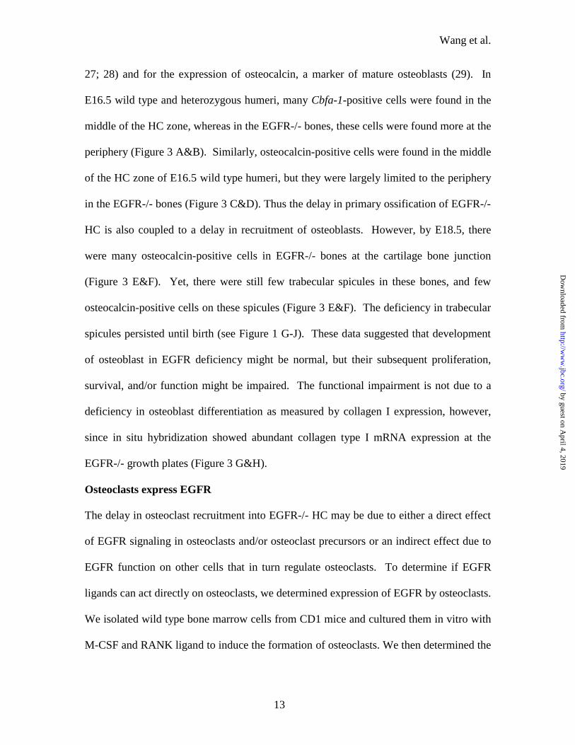
27; 28) and for the expression of osteocalcin, a marker of mature osteoblasts (29). In
E16.5 wild type and heterozygous humeri, many Cbfa-1-positive cells were found in the
middle of the HC zone, whereas in the EGFR-/- bones, these cells were found more at the
periphery (Figure 3 A&B). Similarly, osteocalcin-positive cells were found in the middle
of the HC zone of E16.5 wild type humeri, but they were largely limited to the periphery
in the EGFR-/- bones (Figure 3 C&D). Thus the delay in primary ossification of EGFR-/-
HC is also coupled to a delay in recruitment of osteoblasts. However, by E18.5, there
were many osteocalcin-positive cells in EGFR-/- bones at the cartilage bone junction
(Figure 3 E&F). Yet, there were still few trabecular spicules in these bones, and few
osteocalcin-positive cells on these spicules (Figure 3 E&F). The deficiency in trabecular
spicules persisted until birth (see Figure 1 G-J). These data suggested that development
of osteoblast in EGFR deficiency might be normal, but their subsequent proliferation,
survival, and/or function might be impaired. The functional impairment is not due to a
deficiency in osteoblast differentiation as measured by collagen I expression, however,
since in situ hybridization showed abundant collagen type I mRNA expression at the
EGFR-/- growth plates (Figure 3 G&H).
Osteoclasts express EGFR
The delay in osteoclast recruitment into EGFR-/- HC may be due to either a direct effect
of EGFR signaling in osteoclasts and/or osteoclast precursors or an indirect effect due to
EGFR function on other cells that in turn regulate osteoclasts. To determine if EGFR
ligands can act directly on osteoclasts, we determined expression of EGFR by osteoclasts.
We isolated wild type bone marrow cells from CD1 mice and cultured them in vitro with
M-CSF and RANK ligand to induce the formation of osteoclasts. We then determined the
by guest on April 4, 2019
http://ww
w.jbc.org/
Dow
nloaded from

Wang et al.
14
expression of EGFR in cultured osteoclasts by RT-PCR and Northern blotting. Total
RNAs were isolated from primary cultures of osteoclasts and from the heads of wild type
and EGFR-/- E18.5 embryos for positive and negative controls, respectively. RT-PCR
was performed using a forward primer in exon 1 and a reverse primer in exon 4 to
distinguish wild type EGFR mRNA and any residual mRNA from the targeted allele,
which has a disrupted exon 2. We observed the expression of EGFR mRNA in
osteoclasts and wild type embryonic heads (Figure 4A). As expected, no PCR product
corresponding to EGFR was seen in the EGFR-/- embryonic heads. On Northern blot,
two transcripts of approximately 9.6 kb and 5 kb were found in osteoclast cultures and
wild type embryonic heads (Figure 4B). No transcripts were found in EGFR-/-
embryonic heads. These data suggest that EGFR ligands may act directly on osteoclasts
to affect their function.
Delayed primary osteoclast recruitment in EGFR null mice is not due to deficiency
in MMPs
Previous studies showed that matrix metalloproteinases (MMPs) were necessary for the
migration of (pre)osteoclasts into calcified hypertrophic cartilage during primary
endochondral ossification (23). In particular, MMP-9 (gelatinase B) is required for the
timely recruitment of these cells. MMP-9 deficient mice show a delay in osteoclast
recruitment into the calcified hypertrophic cartilage during primary endochondral
ossification of the metatarsals (25). Since EGFR signaling can modulate the cellular
expression of MMPs, the delay in osteoclast recruitment into the EGFR-/- HC may be
due to deficiency in the expression of MMPs. To test this hypothesis, we analyzed the
expression of several MMPs by in situ hybridization. The expression of MMP-9 and
by guest on April 4, 2019
http://ww
w.jbc.org/
Dow
nloaded from

Wang et al.
15
MMP-14 (MT1-MMP), which are highly expressed in osteoclasts (30; 31; 32), was
consistent with the TRAP staining results. In E16.5 wild type humeri, MMP-9
expression was found in cells corresponding to osteoclasts in the vascularized bone
marrow cavity and at the cartilage-bone junction (Figure 5 A&C). In the E16.5 EGFR-/-
humeri, MMP-9 expression was found in cells at the periphery of the HC, consistent with
the location of osteoclasts in these skeletal elements at this time (Figure 5 B&D). There
were no significant differences in the amount of MMP-9 mRNA per cell, judged by the
intensity of the signal. By E18.5, there were just as many MMP-9 expressing cells at the
cartilage-bone junction of the growth plate and in the bone marrow cavity in the EGFR-/-
bones compared to wild type bones. This is consistent with the observation that the
differences in the number of osteoclasts between wild type and EGFR-/- bones have
diminished by this time. Substrate gel analyses showed that there was slightly less MMP-
9 protein in extracts of EGFR-null bones compared to wild type (Figure 5I). There was
no apparent change in the ratio of active vs. latent forms of MMP-9 proteins. These
results indicated that MMP-9 expression and activity in osteoclasts were not significantly
altered by EGFR deficiency, even though there might be overall decrease in the level of
MMP-9 due to decreased osteoclast number. Thus deficiency in MMP-9 activity was
unlikely to be the primary cause for the delayed recruitment of osteoclasts into EGFR-
null HC. Similar results were observed for the expression of MMP-14 (MT1-MMP). In
E16.5 wild type humeri, MMP-14 was expressed by cells with similar distribution to
osteoclasts, at the cartilage-bone junction and in the bone marrow cavity (Figure 6 A&C).
In contrast, cells expressing MMP-14 were located at the periphery of the unvascularized
HC in E16.5 EGFR-/- bones (Figure 6 B&D). At E18.5 the number and location of
by guest on April 4, 2019
http://ww
w.jbc.org/
Dow
nloaded from

Wang et al.
16
MMP-14 expressing cells were comparable in wild type, heterozygous and EGFR-/-
bones (data not shown).
MMP-13 (collagenase-3) may also be important for the vascularization of
hypertrophic cartilage during primary endochondral ossification. It can degrade native
collagen, which is a major component of the hypertrophic cartilage ECM (33; 34).
MMP-13 is highly expressed in hypertrophic chondrocytes and osteoblasts (35) and
therefore may act to degrade collagens in the hypertrophic cartilage ECM during
endochondral ossification. In E16.5 wild type humeri, MMP-13 was expressed by late
hypertrophic chondrocytes and by cells at the vascularization front, some of which may
be osteoblasts (Figure 6 E&G). In the E16.5 EGFR-/- bones, there was high expression
of MMP-13 in hypertrophic chondrocytes in the calcified HC (Figure 6 F&H). These
results indicated that deficiency in MMP-13 expression was not the cause for the delayed
primary ossification and osteoclast recruitment into EGFR-/- hypertrophic cartilage.
Osteoclast formation from bone marrow cells is attenuated by EGFR inhibitor
The delay in osteoclast recruitment into EGFR-/- hypertrophic cartilage may be due to
deficiency in either their formation or their migration. To address the role of EGFR
signaling in osteoclast formation, we inhibited EGFR signaling during the formation of
osteoclasts from bone marrow cells in vitro using the EGFR tyrosine kinase inhibitor
AG1478 (36). Wild type bone marrow cells from CD1 mice were isolated and cultured
in the presence of M-CSF and RANK ligand to induce the formation of osteoclasts. The
addition of AG1478 in these cultures attenuated osteoclast formation (Figure 7). Cultures
treated with vehicle showed the formation of multinucleated TRAP+ cells characteristic
of osteoclasts (Figure 7A). Addition of increasing concentration of AG1478 decreased
by guest on April 4, 2019
http://ww
w.jbc.org/
Dow
nloaded from

Wang et al.
17
the number of multinucleated TRAP+ cells that were formed in a dose-dependent manner
(Figure 7 B-D), reaching over 90% inhibition at 5 µM AG1478 (Figure 7E). Addition of
exogenous EGF or TGF-α had no additional effects on the formation of osteoclasts in
these cultures (data not shown).
Several genes have been shown to be important for osteoclast formation from
hematopoietic stem cells (37). We asked whether any of these genetic pathways was
down stream of EGFR signaling. Quantitative real time RT-PCR of RNA isolated from
bone marrow cells cultured in the presence of M-CSF and RANK ligand with or without
AG1478 showed no change in the expression of PU.1, c-fos, TRAF-6, MITF, and c-src
with AG1478 treatment (data not shown). Thus the regulation of osteoclast formation by
EGFR signaling does not appear to be mediated through these pathways. To determine if
the inhibition of osteoclast formation by AG1478 was due to an effect on cell
proliferation, we assayed for cell proliferation in the AG1478 treated bone marrow cell
cultures. Wild type bone marrow cells were isolated and cultured for 2 days in the
presence of M-CSF and RANK ligand with or without AG1748. Treatment with
AG1478 resulted in a dose-dependent decrease in MTT uptake, indicative of decreased
cell number (Figure 7F). This suggested that inhibition of EGFR signaling by AG1478
led to decreased cell proliferation. This may account in part for the decrease in osteoclast
formation caused by AG1478 treatment.
Osteoclast resorptive activity is not modulated by EGFR inhibitor
To determine if EGFR signaling modulates osteoclast function, we tested whether
osteoclast resorptive activity is inhibited in the presence of the EGFR tyrosine kinase
inhibitor AG1478. Osteoclasts derived from bone marrow progenitor cells were allowed
by guest on April 4, 2019
http://ww
w.jbc.org/
Dow
nloaded from

Wang et al.
18
to form on calcium phosphate coated discs and subsequently switched to resorptive media
in the presence of AG1478 or vehicle control for two days. Quantitative analyses of the
areas of resorption showed that there were no significant differences in the percent
resorption area between control and treated osteoclasts (Figure 8). Therefore EGFR
signaling does not appear to modulate osteoclastic bone resorption.
DISCUSSION
In this study we have investigated the function of EGFR signaling in endochondral bone
formation by analyzing long bone development in EGFR-null mice. Our data showed
that in EGFR-null mice primary ossification of the calcified hypertrophic cartilage anlage
was delayed and was associated with a delay in the recruitment of osteoclasts and
osteoblasts. Formation of the primary spongiosa was also abnormal leading to
accumulation of growth plate HC and decreased trabecular bone mass. The delay in
osteoclast recruitment might be due to inadequate formation of osteoclasts resulting from
EGFR deficiency.
EGFR signaling is necessary for normal osteoclast recruitment and function during
endochondral ossification.
A critical step in endochondral ossification is the invasion of capillaries into the calcified
hypertrophic cartilage zone in the diaphysis of the cartilage anlage. Vascularization of
calcified HC is accompanied by the recruitment of osteoclasts and osteoblasts. We found
that EGFR deficiency resulted in delayed recruitment of osteoclasts into calcified HC
during primary ossification. However, this delay was only temporary, consistent with the
model that lack of EGFR signaling causes defective the formation of osteoclasts from
precursor cells surrounding the bone rudiments, thus requiring a longer time for sufficient
by guest on April 4, 2019
http://ww
w.jbc.org/
Dow
nloaded from

Wang et al.
19
osteoclasts to accumulate. Alternatively, the migration of osteoclasts into calcified HC
may also be defective.
Even though osteoclast recruitment into EGFR-/- HC reached the same level as
wild type mice at E18.5, ossification of growth plate HC was still not complete, as
evidenced by the lengthened HC zones of the EGFR-/- growth plates up until birth. This
may be due to the accumulation of HC caused by the delay in primary ossification, and
the inability of the subsequently normal number of osteoclasts to overcome the initial
difference and therefore ossification in the longitudinal direction would always be behind
in the EGFR-null bones. Alternatively, growth plate ossification may also be abnormal
due to either abnormal formation of growth plate HC or abnormality in its removal. A
recent study also reported increased growth plate HC in the EGFR-/- mice (19). The
authors found expression of EGFR in chondroblasts but no differences in growth plate
chondrocyte proliferation, and suggested that EGFR negatively regulated hypertrophic
chondrocyte maturation. On the other hand, the removal and ossification of EGFR-null
growth plate HC may also be abnormal, proceeding at a slower rate resulting in
accumulation of growth plate HC. We found expression of EGFR on mature osteoclasts,
suggesting that these cells can respond directly to EGFR ligands. The function of EGFR
in osteoclasts is not known, but it may be to stimulate their proteolytic activity. EGFR
ligands have been found to stimulate osteoclastic bone resorption in vivo and in vitro in
bone organ cultures (20; 38; 39).
MMPs are necessary for the recruitment of osteoclasts into calcified hypertrophic
cartilage during primary endochondral ossification (25). The craniofacial and lung
developmental defects of EGFR-null mice may be due to alteration in MMP activity (15;
by guest on April 4, 2019
http://ww
w.jbc.org/
Dow
nloaded from

Wang et al.
20
16). We found that down-regulation MMP-9, MMP-13 and MMP-14 was unlikely to be
the primary mechanism for the delayed recruitment of osteoclasts into EGFR-/- HC.
However, this does not exclude EGFR regulation of other ECM degrading proteinases
that are important in osteoclast function. In addition, since MMP-13 acts synergistically
with MMP-9 (25), the initial delayed recruitment of osteoclasts expressing MMP-9 could
functionally blunt HC ECM degradation and exacerbating the delay in HC
vascularization.
Vascular endothelial growth factor (VEGF) is important for vascularization of and
osteoclast recruitment into calcified hypertrophic cartilage of the developing bones (40;
25). We found no significant difference in VEGF expression in EGFR-null bones by in
situ hybridization (K. Wang, unpublished data). This suggests that the delayed osteoclast
recruitment into EGFR-null HC is not due to deficiency in VEGF expression. However,
since MMP-9 may regulate the bioavailability of VEGF, the initial delay in osteoclast
recruitment may cause an initial decrease in MMP-9 activity leading to decreased
bioavailable VEGF, which leads to further delay in vascularization and recruitment of
osteoclasts.
EGFR deficiency leads to impaired trabecular bone formation
Trabecular bone mass is decreased in the EGFR-/- bones. This may partly be due to a
delay in the initial recruitment of osteoblasts during primary ossification. However,
trabecular spicules continued to be deficient until birth. Fewer osteoblasts were found in
the EGFR-/- primary spongiosa even though they are abundant at the cartilage bone
junction. This suggests that formation of osteoblasts in the EGFR-/- bones may be
normal, but that their subsequent proliferation/survival and/or function may be impaired.
by guest on April 4, 2019
http://ww
w.jbc.org/
Dow
nloaded from

Wang et al.
21
Studies of the LRP5 (LDL receptor related protein 5)- deficient mice showed that
osteoblast differentiation and proliferation were differentially regulated (42). Sibilia et
al. (2003) reported that primary EGFR-/- calvarial osteoblast cultures showed decreased
proliferation potential and increased differentiation as measured by their ability to form
bone nodules in vitro (19). Thus the decreased trabecular bone mass in the EGFR-/- mice
was likely due to decreased osteoblast proliferation. A direct effect of EGFR signaling
on osteoblasts is supported by previous studies showing that EGFR was expressed in
osteoblasts in vivo (17; 43), and that EGF stimulated osteoblast proliferation in vitro (44;
45).
EGFR signaling is necessary for the formation of osteoclasts
Previous studies have identified two essential factors for osteoclastogenesis: M-
CSF and RANKL (37). M-CSF is necessary for the generation of the
monocyte/macrophage cell lineage and RANKL for their differentiation into osteoclasts.
We found that induction of osteoclast formation from bone marrow cells in the presence
of RANKL and M-CSF was significantly inhibited in the presence of the EGFR tyrosine
kinase inhibitor AG1478. This is consistent with previous studies suggesting a role for
EGFR ligands in osteoclast formation. Addition of either TGF-α or EGF increased the
formation of multinucleated osteoclasts from cultured human bone marrow cells (46). In
our bone marrow cell cultures addition of exogenous EGF or TGF-alpha had no
additional effect on osteoclast formation. This may be because there were already
saturating amounts of endogenous EGFR ligands in these cultures, or that the endogenous
EGFR was trans-activated by other ligand-receptor signaling pathways (47). The role of
EGFR signaling in osteoclast formation may be a direct effect on osteoclast precursors to
by guest on April 4, 2019
http://ww
w.jbc.org/
Dow
nloaded from
Wang et al.
22
stimulate either their proliferation or differentiation. Alternatively, EGFR ligands may
act on other bone marrow cells to stimulate production of either secreted or cell-surface
factors that in turn act in a paracrine fashion on osteoclast precursors to effect their
growth and differentiation. Further studies are needed to distinguish between these two
possibilities.
ACKNOWLEDGEMENTS
This study was supported by a grant from the National Institutes of Health (AR46238 to
T.H.V. and Z.W.), and a grant from the Sandler Family Supporting Foundation to T.H.V.
by guest on April 4, 2019
http://ww
w.jbc.org/
Dow
nloaded from
Wang et al.
23
REFERENCES
1. Caplan, A. I. (1988) in Ciba Foundation Symposium Vol. 136, pp. 3021
2. Olsen, B. R., Reginato, A. M., and Wang, W. (2000) Annu Rev Cell Dev Biol 16,
191-220
3. Karsenty, G., and Wagner, E. F. (2002) Dev Cell 2, 389-406
4. Hackel, P. O., Zwick, E., Prenzel, N., and Ullrich, A. (1999) Curr Opin Cell Biol
11, 184-189
5. Bogdan, S., and Klambt, C. (2001) Curr Biol 11, R292-295
6. Partanen, A. M., Ekblom, P., and Thesleff, I. (1985) Dev Biol 111, 84-94
7. Dardik, A., Smith, R. M., and Schultz, R. M. (1992) Dev Biol 154, 396-409
8. Wiley, L. M., Wu, J. X., Harari, I., and Adamson, E. D. (1992) Dev Biol 149,
247-260
9. Canoun, C., Ma, C., Halpern, D., Shum, L., Bringas, P., Jr., Sank, A., and
Slavkin, H. C. (1993) J Surg Res 54, 638-647
10. Shum, L., Sakakura, Y., Bringas, P., Jr., Luo, W., Snead, M. L., Mayo, M.,
Crohin, C., Millar, S., Werb, Z., Buckley, S., and et al. (1993) Development 118,
903-917
11. Miettinen, P. J., Berger, J. E., Meneses, J., Phung, Y., Pedersen, R. A., Werb, Z.,
and Derynck, R. (1995) Nature 376, 337-341
12. Sibilia, M., and Wagner, E. F. (1995) Science 269, 234-238
13. Threadgill, D. W., Dlugosz, A. A., Hansen, L. A., Tennenbaum, T., Lichti, U.,
Yee, D., LaMantia, C., Mourton, T., Herrup, K., Harris, R. C., and et al. (1995)
Science 269, 230-234
by guest on April 4, 2019
http://ww
w.jbc.org/
Dow
nloaded from

Wang et al.
24
14. Miettinen, P. J., Warburton, D., Bu, D., Zhao, J. S., Berger, J. E., Minoo, P.,
Koivisto, T., Allen, L., Dobbs, L., Werb, Z., and Derynck, R. (1997) Dev Biol
186, 224-236
15. Miettinen, P. J., Chin, J. R., Shum, L., Slavkin, H. C., Shuler, C. F., Derynck, R.,
and Werb, Z. (1999) Nat Genet 22, 69-73
16. Kheradmand, F., Rishi, K., and Werb, Z. (2002) J Cell Sci 115, 839-848
17. Martineau-Doize, B., Lai, W. H., Warshawsky, H., and Bergeron, J. J. (1988)
Endocrinology 123, 841-858
18. Chan, S. Y., and Wong, R. W. (2000) J Biol Chem 275, 38693-38698
19. Sibilia, M., Wagner, B., Hoebertz, A., Elliott, C., Marino, S., Jochum, W., and
Wagner, E. F. (2003) Development 130, 4515-4525
20. Raisz, L. G., Simmons, H. A., Sandberg, A. L., and Canalis, E. (1980)
Endocrinology 107, 270-273
21. Albrecht, U., Eichele, G., Helms, J. A., and Lu, H. (1997) in Molecular and
Cellular Methods in Developmental Toxicology (Daston, G. P., ed), pp. 23-48,
CRC Press, Boca Raton
22. Fuller, K., Lean, J. M., Bayley, K. E., Wani, M. R., and Chambers, T. J. (2000) J
Cell Sci 113 ( Pt 13), 2445-2453
23. Blavier, L., and Delaisse, J. M. (1995) J Cell Sci 108 ( Pt 12), 3649-3659
24. Wilson, S. E., Chen, L., Mohan, R. R., Liang, Q., and Liu, J. (1999) Exp Eye Res
68, 377-397
by guest on April 4, 2019
http://ww
w.jbc.org/
Dow
nloaded from

Wang et al.
25
25. Engsig, M. T., Chen, Q. J., Vu, T. H., Pedersen, A. C., Therkidsen, B., Lund, L.
R., Henriksen, K., Lenhard, T., Foged, N. T., Werb, Z., and Delaissé, J. M. (2000)
Journal of Cell Biology 151, 879-890
26. Ducy, P., Zhang, R., Geoffroy, V., Ridall, A. L., and Karsenty, G. (1997) Cell 89,
747-754
27. Komori, T., Yagi, H., Nomura, S., Yamaguchi, A., Sasaki, K., Deguchi, K.,
Shimizu, Y., Bronson, R. T., Gao, Y. H., Inada, M., Sato, M., Okamoto, R.,
Kitamura, Y., Yoshiki, S., and Kishimoto, T. (1997) Cell 89, 755-764
28. Otto, F., Thornell, A. P., Crompton, T., Denzel, A., Gilmour, K. C., Rosewell, I.
R., Stamp, G. W., Beddington, R. S., Mundlos, S., Olsen, B. R., Selby, P. B., and
Owen, M. J. (1997) Cell 89, 765-771
29. Lian, J. B., Stein, G. S., Stewart, C., Puchacz, E., Mackowiak, S., Aronow, M.,
Von Deck, M., and Shalhoub, V. (1989) Connect Tissue Res 21, 61-68; discussion
69
30. Reponen, P., Sahlberg, C., Munaut, C., Thesleff, I., and Tryggvason, K. (1994) J
Cell Biol 124, 1091-1102
31. Sato, T., del Carmen Ovejero, M., Hou, P., Heegaard, A. M., Kumegawa, M.,
Foged, N. T., and Delaisse, J. M. (1997) J Cell Sci 110 ( Pt 5), 589-596
32. Irie, K., Tsuruga, E., Sakakura, Y., Muto, T., and Yajima, T. (2001) Tissue Cell
33, 478-482
33. Knauper, V., Lopez-Otin, C., Smith, B., Knight, G., and Murphy, G. (1996) J Biol
Chem 271, 1544-1550
by guest on April 4, 2019
http://ww
w.jbc.org/
Dow
nloaded from

Wang et al.
26
34. Billinghurst, R. C., Dahlberg, L., Ionescu, M., Reiner, A., Bourne, R., Rorabeck,
C., Mitchell, P., Hambor, J., Diekmann, O., Tschesche, H., Chen, J., Van Wart,
H., and Poole, A. R. (1997) J Clin Invest 99, 1534-1545
35. Sasano, Y., Zhu, J. X., Tsubota, M., Takahashi, I., Onodera, K., Mizoguchi, I.,
and Kagayama, M. (2002) J Histochem Cytochem 50, 325-332
36. Levitzki, A., and Gazit, A. (1995) Science 267, 1782-1788
37. Teitelbaum, S. L., and Ross, F. P. (2003) Nat Rev Genet 4, 638-649
38. Stern, P. H., Krieger, N. S., Nissenson, R. A., Williams, R. D., Winkler, M. E.,
Derynck, R., and Strewler, G. J. (1985) J Clin Invest 76, 2016-2019
39. Marie, P. J., Hott, M., and Perheentupa, J. (1990) Am J Physiol 258, E275-281
40. Gerber, H. P., Vu, T. H., Ryan, A. M., Kowalski, J., Werb, Z., and Ferrara, N.
(1999) Nat Med 5, 623-628
41. Mayr-Wohlfart, U., Waltenberger, J., Hausser, H., Kessler, S., Gunther, K. P.,
Dehio, C., Puhl, W., and Brenner, R. E. (2002) Bone 30, 472-477
42. Kato, M., Patel, M. S., Levasseur, R., Lobov, I., Chang, B. H., Glass, D. A., 2nd,
Hartmann, C., Li, L., Hwang, T. H., Brayton, C. F., Lang, R. A., Karsenty, G.,
and Chan, L. (2002) J Cell Biol 157, 303-314
43. Davideau, J. L., Sahlberg, C., Thesleff, I., and Berdal, A. (1995) Connect Tissue
Res 32, 47-53
44. Ng, K. W., Partridge, N. C., Niall, M., and Martin, T. J. (1983) Calcif Tissue Int
35, 624-628
45. Loza, J., Carpio, L., Lawless, G., Marzec, N., and Dziak, R. (1995) Bone 16,
341S-347S
by guest on April 4, 2019
http://ww
w.jbc.org/
Dow
nloaded from

Wang et al.
27
46. Takahashi, N., MacDonald, B. R., Hon, J., Winkler, M. E., Derynck, R., Mundy,
G. R., and Roodman, G. D. (1986) J Clin Invest 78, 894-898
47. Gschwind, A., Zwick, E., Prenzel, N., Leserer, M., and Ullrich, A. (2001)
Oncogene 20, 1594-1600
by guest on April 4, 2019
http://ww
w.jbc.org/
Dow
nloaded from

Wang et al.
28
FIGURE LEGENDS
Figure 1: Long bone development in wild type and EGFR-/- mice. Trichrome-Masson
stained tissue sections of the humerus of E16.5 (A&B), E18.5 (C&D, G&H), and
newborn (E&F, I&J) wild type or heterozygous (A, C, E, G, I) and EGFR-/- (B, D, F, H,
J) mice. At E16.5, vascularization of the calcified hypertrophic cartilage zone has
already occurred in the wild type humerus with vascularized tissues replacing
hypertrophic cartilage in the diaphysis (A, arrows), whereas invading capillaries remain
at the outer edge of the calcified hypertrophic cartilage zone in EGFR-/- humerus (B,
arrows). At E18.5, endochondral ossification has continued in the wild type humerus
resulting in an area of trabecular bone and a normal sized growth plate (C), whereas there
is still a large area of un-ossified hypertrophic cartilage in the EGFR-/- humerus (D). In
newborn mice, there continues to be a large area of hypertrophic cartilage at the growth
plate of EGFR-/- humerus (F) compared to wild type (E). Many long bony trabeculae are
present in the metaphysis in wild type humerus at E18.5 (G, arrows) and at birth (I,
arrows), but the bony trabeculae in EGFR-/- littermates are very few and short (H&J,
arrows). Bar, A-F: 200 µm, G-J: 100 µm. Since heterogeneous mice did not show a bone
phenotype, the EGFR wild type and heterozygote embryos were used interchangeably
and are indicated as EGFR+/?.
Figure 2: Effect of EGFR deficiency on the number and distribution of TRAP+ cells in
the developing humerus. Tissue sections of the humerus of E16.5 (A&B), E18.5 (C&D),
and newborn (E&F) wild type or heterozygous (A, C, E) and EGFR-/- (B, D, F) mice
stained for TRAP activity. In E16.5 wild-type humerus, many TRAP+ cells were
detected in the vascularized hypertrophic cartilage (A, arrows). However, in E16.5
by guest on April 4, 2019
http://ww
w.jbc.org/
Dow
nloaded from

Wang et al.
29
EGFR-/- humerus, most of the TRAP+ cells were found at the periphery of the
hypertrophic cartilage (B, arrows). In E18.5 (C, D) and newborn mice (E, F), there were
just as many TRAP+ cells inside the bone rudiment in EGFR-/- humerus (D&F, arrows)
as in the wild-type humerus (C&E, arrows). Bar, 100 µm. G: Quantification of the
number of TRAP + cells in wild type and EGFR-/- humeri at different stages. Horizontal
bars show mean counts of TRAP+ cells found either outside the calcified hypertrophic
cartilage at the perichondrium/periosteum or inside the calcified hypertrophic cartilage.
At E16.5, there is a significant difference in the total number of TRAP+ cells found
outside vs. inside the calcified hypertrophic cartilage between wild type and EGFR-/-
mice (p‹0.05).
Figure 3. Expression of Cbfa-1, osteocalcin, and collagen type I in the humerus of wild
type and EGFR-/- mice. A-H: bright field images of tissue sections of E16.5 (A-D) or
E18.5 (E-H) humeri from wild type (A, C, E, G) or EGFR-/- (B, D, F, H) mice hybridized
with 35S-labeled Cbfa-1 (A&B), osteocalcin (C-F), and collagen type I (G&H) anti-sense
probes. In E16.5 wild type humerus, many Cbfa-1-positive cells were found in the
middle section of the hypertrophic cartilage (A, arrows), whereas these cells are found
largely at the periphery of the hypertrophic cartilage in EGFR-/- humerus (B, arrows).
Similarly, osteocalcin (oc) expressing cells are found at the periphery of hypertrophic
cartilage in E16.5 EGFR-/- (D, arrows) and in the middle of wild type hypertrophic
cartilage (C, arrows). By E18.5, there were abundant osteocalcin-positive cells in the
metaphysis of EGFR-/- humerus. However, these cells are found mainly at the cartilage-
bone junction (F, arrows), and very few are found in the primary spongiosa, which also
contain very few trabecular spicules (F, arrowhead). In contrast, in the E18.5 wild type
by guest on April 4, 2019
http://ww
w.jbc.org/
Dow
nloaded from

Wang et al.
30
humerus there are abundant osteocalcin-positive cells both at the cartilage-bone junction
(E, arrows) and in the primary spongiosa on trabecular spicules (E, arrowheads).
Collagen type I expression was found in the metaphysis in both wild type and EGFR-/-
E18.5 humerus (G&H, arrows). Bar: 200 µm.
Figure 4. RT-PCR and Northern blot analysis of EGFR mRNA in cultured osteoclasts.
A: RT-PCR with EGFR specific primers of total RNA isolated from cultured osteoclasts,
wild type embryonic heads, and EGFR-/- heads. The expected 499 bp PCR product was
seen in RNA from osteoclast and wild type embryonic head, but not in EGFR-/- head. B:
Northern blot of total RNA isolated from cultured osteoclasts, wild type embryonic
heads, and EGFR-/- heads hybridized with a 32P-labeled EGFR probe. Two transcripts,
9.6 kb and 5.0 kb in size, were detected in RNA from osteoclasts and wild type
embryonic head, but not from EGFR-/- head.
Figure 5. Expression of MMP-9 in the humeri of wild type/homozygous and EGFR-/-
mice. A-D: bright field (A&B) and dark field (C&D) images of tissue sections of E16.5
humerus from wild type (A&C) or EGFR-/- (B&D) mice hybridized with 35S-labeled
MMP-9 anti-sense probe. MMP-9 expression was found in cells inside the vascularized
hypertrophic cartilage, including at the cartilage-bone junction in wild type humerus,
(A&C, arrows), and in cells at the outer edge of the calcified hypertrophic cartilage in
EGFR-/- humerus (B&D, arrows). E-H: bright field (E&F) and dark field (G&H)
images of tissue sections of E18.5 humerus from wild type (E&G) or EGFR-/- (F&H)
mice hybridized with 35S-labeled MMP-9 anti-sense probe. Similar number and
distribution of MMP-9 expressing cells were found in both wild type and EGFR-/-
humerus. I: Gelatin zymogram of tissue lysates from the long bones of wild type and
by guest on April 4, 2019
http://ww
w.jbc.org/
Dow
nloaded from
Wang et al.
31
EGFR-/- mice showing slightly decreased level of both latent and activated gelatinase B
(gelBa) in EGFR-/- bones and normal level of latent and activated gelatinase A (gelAa).
Bar (A-H), 200µm.
Figure 6. Expression of MMP-14 (MT1-MMP) and MMP-13 (collagenase 3) in the wild
type and EGFR-/- humeri. A-D: bright field (A&B) and dark field (C&D) images of
tissue sections of E16.5 humeri from wild type (A&C) or EGFR-/- (B&D) mice
hybridized with 35S-labeled MMP-14 anti-sense probe. Similar to MMP-9, MMP-14
expression was found in cells inside the vascularized hypertrophic cartilage, including at
the cartilage-bone junction in wild type humerus, (A&C, arrows), and in cells at the outer
edge of the calcified hypertrophic cartilage in EGFR-/- humerus (B&D, arrows). E-H:
bright field (E&F) and dark field (G&H) images of tissue sections of E16.5 humeri from
wild type (E&G) or EGFR-/- (F&H) mice hybridized with 35S-labeled MMP-13 anti-
sense probe. MMP-13 expression was found in the lower hypertrophic chondrocytes
adjacent to the vascularized area in wild type humerus (E&G, arrows), and in
chondrocytes of the calcified hypertrophic cartilage in EGFR-/- humerus (F&H, arrows).
Bar: 200µm.
Figure 7. Effect of the inhibition of EGFR signaling on osteoclast formation. A-D:
TRAP staining of bone marrow cells cultured for 6 days with RANKL (25ng/ml), M-CSF
(25ng/ml) and either vehicle or increasing concentrations of the EGFR tyrosine kinase
inhibitor AG1478 (1.25 µM, 2.5 µM, and 5 µM). In vehicle treated cultures, there were a
large number of multinucleated TRAP+ cells characteristic of osteoclasts (A). Treatment
with AG1478 caused a dose-dependent decrease in the number of multinucleated TRAP+
cells (B-D). E: Quantification of the number of osteoclasts developed in control cultures
by guest on April 4, 2019
http://ww
w.jbc.org/
Dow
nloaded from
Wang et al.
32
and cultures with different concentration of AG1478. Each histogram represents mean
number of total osteoclasts counted in three wells of a 24-well plate. F: Effects of EGFR
signaling inhibition on proliferation of bone marrow mononuclear cells. Wild type bone
marrow mononuclear cells were isolated and cultured for 2 days in the presence of
RANKL (25 ng/ml), M-CSF (25 ng/ml) and either vehicle or increasing concentrations of
the EGFR tyrosine kinase inhibitor AG1478 (1.25 µM, 2.5 µM, and 5 µM). MTT uptake
was assessed by absorbance and used as a measure of cell number. AG1478 caused a
dose-dependent decrease in cell number as reflected by decrease in absorbance. Each
histogram represents mean value of six replicates. The experiments were repeated three
times with similar results.
Figure 8. Effect of the inhibition of EGFR signaling on osteoclast function. A: Images
of the resorption pits formed by osteoclasts on calcium phosphate coated discs. Bone
marrow cells were cultured on calcium phosphate coated discs in the presence of M-CSF
and RANKL to form osteoclasts before switching to resorptive media containing vehicle
control (DMSO) or AG1478 (5 µM) for 2 days. B: Quantitative analyses of the area of
resorption as a percentage of the total area corresponding to an osteoclast. Data are
presented as mean and standard deviation (error bars) of 35 osteoclasts analyzed. There
was no significant difference between DMSO and AG1478 treated groups (p = 0.36)
by guest on April 4, 2019
http://ww
w.jbc.org/
Dow
nloaded from
Ke Wang, Hiroaki Yamamoto, Jennie R. Chin, Zena Werb and Thiennu H. Vuosteoclast recruitment
EGFR-deficient mice have delayed primary endochondral ossification due to defective
published online September 28, 2004J. Biol. Chem.
10.1074/jbc.M403114200Access the most updated version of this article at doi:
Alerts:
When a correction for this article is posted•
When this article is cited•
to choose from all of JBC's e-mail alertsClick here
by guest on April 4, 2019
http://ww
w.jbc.org/
Dow
nloaded from


![Transcriptional Network Controlling Endochondral Ossification · branous ossification and endochondral ossification.[1] During intramembranous ossification, osteoblasts produce type](https://static.fdocuments.in/doc/165x107/5e8cf0c24763783dcf0d78ef/transcriptional-network-controlling-endochondral-ossification-branous-ossification.jpg)