General Anatomy (heart) - WordPress.com · · 2017-03-19General Anatomy (Thorax) Thorax : trunk...
7
BY : Haif AlQahtani & Jenan almahfoudh General Anatomy (Thorax) Thorax : trunk (chest, abdomen and pelvis), between the neck and the abdomen, separated from the abdomen by the diaphragm, and the skeleton is thoracic cage ( sternum, 12 pairs of ribs and 12 thoracic (dorsal) vertebrae) Thoracic upper is upper opening : Thoracic inlet Lower opening closer to the diaphragm : Thoracic outlet Sternum subcutaneous bone (thoracic bone) it has three parts: upper part manuboriam, middle part body, lower part xiphoid process Jugular Notch : upper border of the manuboriam Manuboriam articulates with median end of clavical and first rib : sternoclavicular joint Sternal angle joins middle and upper part together > anatomical landmark (elevated area) it’s important: attachment of the 2 nd costal cartilage, 1. count the ribs and spaces between the ribs The line passes between the 4 th and 5 th 2. it divides the mediastinum into superior and inferior Where the trachea divide into primary bronchi is Carina The arch of the aorta starts and ends at the sternal angle Ribs: True 1-7 (own costal cartilages) False 8-10 (8-9-10 attach to 7) Floating 11-12 (no attachment, end in the anterior wall of the abdomen) Each rib has a head, neck, tubercle, angle, body Head has facets is a type of articulation (joining) Head joins body of thoracic vertebra (body of the vertebra) Tubercle articulates with the transverse process of the vertebra Subcostal groove: under the rib you find subcostal groove, intercostal vein and artery and nerve Neurovascular bundle inside the subcostal groove
Transcript of General Anatomy (heart) - WordPress.com · · 2017-03-19General Anatomy (Thorax) Thorax : trunk...

BY:HaifAlQahtani&Jenanalmahfoudh
GeneralAnatomy(Thorax)Thorax:trunk(chest,abdomenandpelvis),betweentheneckandtheabdomen,separatedfromtheabdomenbythediaphragm,andtheskeletonisthoraciccage(sternum,12pairsofribsand12thoracic(dorsal)vertebrae)Thoracicupperisupperopening:ThoracicinletLoweropeningclosertothediaphragm:ThoracicoutletSternumsubcutaneousbone(thoracicbone)ithasthreeparts:upperpartmanuboriam,middlepartbody,lowerpartxiphoidprocessJugularNotch:upperborderofthemanuboriamManuboriamarticulateswithmedianendofclavicalandfirstrib:sternoclavicularjointSternalanglejoinsmiddleandupperparttogether>anatomicallandmark(elevatedarea)it’simportant:attachmentofthe2ndcostalcartilage,1. counttheribsandspacesbetweentheribsThelinepassesbetweenthe4thand5th2. itdividesthemediastinumintosuperiorandinferiorWherethetracheadivideintoprimarybronchiisCarinaThearchoftheaortastartsandendsatthesternalangleRibs:True1-7(owncostalcartilages)False8-10(8-9-10attachto7)Floating11-12(noattachment,endintheanteriorwalloftheabdomen)Eachribhasahead,neck,tubercle,angle,bodyHeadhasfacetsisatypeofarticulation(joining)Headjoinsbodyofthoracicvertebra(bodyofthevertebra)TuberclearticulateswiththetransverseprocessofthevertebraSubcostalgroove:undertheribyoufindsubcostalgroove,intercostalveinandarteryandnerveNeurovascularbundleinsidethesubcostalgroove

BY:HaifAlQahtani&Jenanalmahfoudh
VentralRamiformintervertebralnervesSpine:vertebralcolumn>curvilinearstructureApartoftheaxialskeletal(oneinthemidline)Consistsof33-34irregularbones>vertebraeBonesdividedinto7intheneck(C-cervical),12inthechest(T-thoracic/dorsal)and5inthelowerback(L-lumbar)and5inthepelvis(S-sacral>formsacrum)and4-5inthepelvis(coccygeal>fusetoformcoccyx)total33-34Vertebraedividedintotwogrooves:Atypical:Typical:allthoracic:theyconsistsoftwopartsvertebralbodyandvertebralarchWedon’thavefacetsincervicalorlumbarit’sonlyinthoracicforarticulationwiththeheadoftheribsThreedimensions:anterior,post,lateralVertebralarchincludestwopedicles,twotransverseprocess,twoinferiorarticularfacets,twosuperiorarticularfacetsand2laminaland1spinalprocessVertebralorspinalforamenVertebralcolumnhasthenerves,bloodvesselsandeverything
SynovialjointformedbythesuperiorarticularfacetsandinferiorfacetsLaminalflatbonypartjoinatthemidlinetoformspinalprocessIntervertebralforamenthespace
Ventralbody>anteriorfacetsforarticulation
BY:HaifAlQahtani&Jenanalmahfoudh
AnatomyLecture5
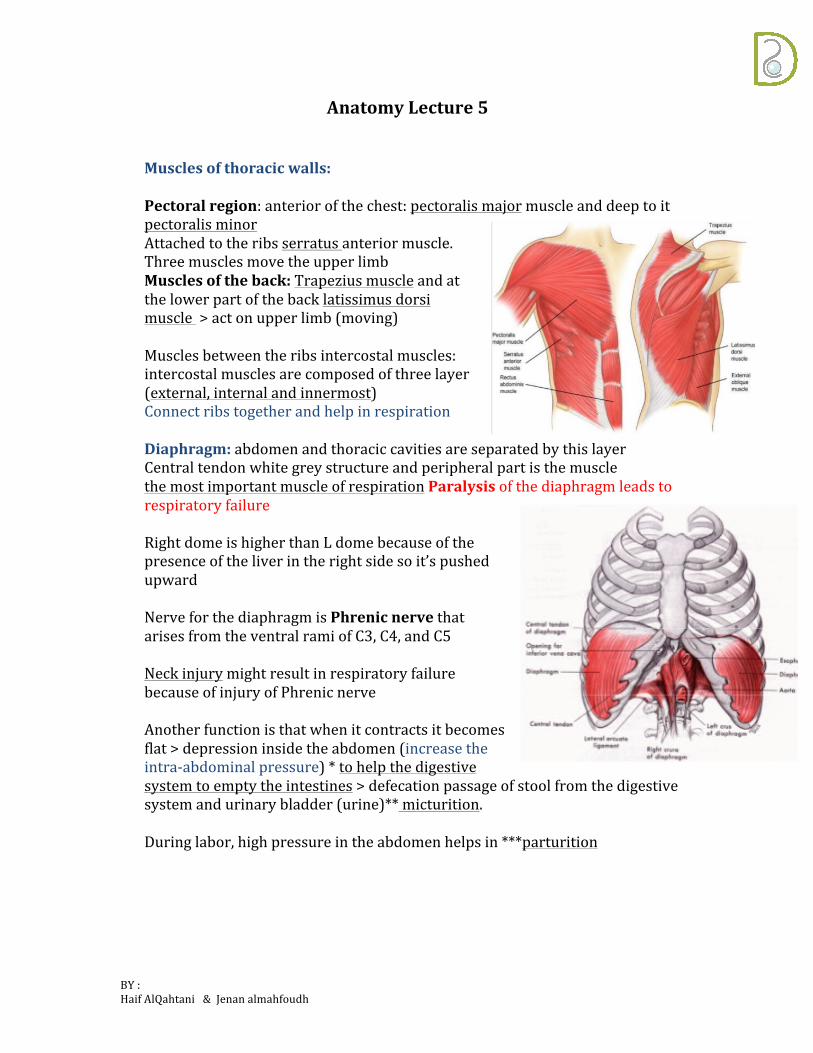
Musclesofthoracicwalls:Pectoralregion:anteriorofthechest:pectoralismajormuscleanddeeptoitpectoralisminorAttachedtotheribsserratusanteriormuscle.ThreemusclesmovetheupperlimbMusclesoftheback:Trapeziusmuscleandatthelowerpartofthebacklatissimusdorsimuscle>actonupperlimb(moving)Musclesbetweentheribsintercostalmuscles:intercostalmusclesarecomposedofthreelayer(external,internalandinnermost)ConnectribstogetherandhelpinrespirationDiaphragm:abdomenandthoraciccavitiesareseparatedbythislayerCentraltendonwhitegreystructureandperipheralpartisthemusclethemostimportantmuscleofrespirationParalysisofthediaphragmleadstorespiratoryfailureRightdomeishigherthanLdomebecauseofthepresenceoftheliverintherightsidesoit’spushedupwardNerveforthediaphragmisPhrenicnervethatarisesfromtheventralramiofC3,C4,andC5NeckinjurymightresultinrespiratoryfailurebecauseofinjuryofPhrenicnerveAnotherfunctionisthatwhenitcontractsitbecomesflat>depressioninsidetheabdomen(increasetheintra-abdominalpressure)*tohelpthedigestivesystemtoemptytheintestines>defecationpassageofstoolfromthedigestivesystemandurinarybladder(urine)**micturition.Duringlabor,highpressureintheabdomenhelpsin***parturition

BY:HaifAlQahtani&Jenanalmahfoudh
Threemajoropeningsinthediaphragm:esophagus(esophagealopening)T10,inferiorvenacava(cavalopening)T8andaorta(Aorticopening)T12WhentheaortapassesfromthoracictotheabdomenitchangesitsnametoabdominalaortainsteadofthoracicaortaThoraciccavity:• Lungsanditscoverings• Mediastinum
Lungs:Conicalinshapebaseandanapex,spongy,andcompressiblebecauseofelastictissue.Threesurfaces:facingtheribs>costalsurfaceInferiorsurfacefacingthediaphragm>diaphramaticsurfaceMedially>mediastinalsurfaceLoops:rightlung>threelobesandtwofissures(horizontal,obliquefissure)upper,middleandlowerLeftlung>twolobes,upperandlower>obliquefissure(onefissureonly)RightlungisheavierLeftlunggivesspacetotheheartsoapartofitismissingEachlungiscoveredbyaserousmembrane>PleuraeTwolayers:visceralpleuraclosetothelungsandparietalpleuraclosetotheribsHilumofthelungsareawherethingsenterandleavethelungandit’snotcoveredwithpleuraBetweenthetwolayerswehavepotentialclosedspace>pleuralcavityandit’sfilledwithfluid>pleuralfluidforlubricationThefluidwillcreateanegativepressuretokeepthelungsinflatedIfit’srupturedordamage>airwillenterandlayerswillseparatesothelungsgetsmaller>pneumothorax>problemsbreathingOrit’scanrupturedordamagebyincreaseinfluidorbloodinside>hydrothorax

BY:HaifAlQahtani&Jenanalmahfoudh
Respiratorysystem:Conductionzone:startsfromthetrachea>mainairway>dividestotwoprimarybronchidependingonthe#oflungs>rightprimarybronchusandleftPBandthebifurcationisCarinaDependingonthelobes:RPB>threesecondarybronchiLPB>twosecondarybronchusEachlobesubdividesintosmallersegments:Bronchopulmonarysegment>functionalunitofthelung>wedgeshaped+atertiarybronchus+BVsLPB>narrowerRPB>straightandwider>foreignbodymorelikelyenterstherightPBbecauseit’swiderandstraighterMediastinum:thoraciccavityminuslungsandpleuraDivideitbyimaginarylinefromlevelofsternalanglebetweenT4,T5Superior,inferiorandinferiorisdividedtomiddle,posterior(heartand
vertebralcolumn)andanteriorSuperior:
• SuperiorVenaCava• Brachiocephalicvein• ArchofAorta• Trachea• Vagusnerve• Phrenicnerve• Thoracicduct• Esophagus• Thymus• Leftrecurrentlaryngealnerve
-theveinintheheadandneck>internaljugularveinrightandleft-Fromtheupperlimbs>leftsubclavianveinandrightsubclavianveinBothjointoformbrachiocephalicvein>rightandleftBothofwhichjoinandformsuperiorvenacava>enterstheRAoftheheartBringthebloodfromtheupperhalfofthebody
min

BY:HaifAlQahtani&Jenanalmahfoudh
ArchoftheaortaThoracicduct>lymphaticchannelwhichendinthevenouscirculationThymusisaglandlocatedbehindthesternum>retrosternalglandLargerduringearlychildhoodandit’ssmalleraswegrowupTLymphocytesmaturebythymus>thymosinhelpinmaturingTlymphocytesVagus(10thcranialnerve)LeftRecurrentLaryngealnerve>branchesfromvagusandarisesinthechestandgoesbacktotheneck
Inferior:
• Anteriormediastinum:Liesanteriortothepericardium.Containsthethymus,lymphnodes,andfats.
• Middlemediastinum:Heart,pericardium,phrenicnerve,andmainbronchi• Posteriormediastinum:Esophagus,azygusvein,thoracicaorta,thoracicduct,
Intercostalveinsontherightsidetheyend>azygusveinOntheleftside>smallerveinhemiazygus

BY:HaifAlQahtani&Jenanalmahfoudh
FunctionalClassificationofNerve:
• SomaticorigininCNS>(directly)effecterskeletalmuscles• Visceral(sympatheticandparasympathetic):CNS>ganglion>effector
NerveinCNSpassthroughgangliontheneffector>thefibersarecalledpreganglionicfibersbetweenCNSandganglionBetweenganglionandeffector>postganglionicfibers
Parasympathetic:originatefrombrainandcraniosacraloutflowSympathetic:arisesfromthethoraxandlumbarregionofspinalcord>thoracolumbaroutflow
• Preganglionicfiberofsympatheticisshortbecauseit’sclosetotheganglion• PreganglionicfiberofParasympatheticislong(totheeffector)
Sympatheticchain(trunk):veinsofsympatheticganglionareconnectedtoeachother