Gamma Knife Radiosurgery - Metro Detroit...
8
3/23/2017 1 Gamma Knife Radiosurgery A Revolutionary Noninvasive Brain Surgery Procedure By Michelle Manders RN BSN What Is Gamma Knife? Gamma Knife is not really a knife. No incision is made Uses a high dose of radiation from Cobalt 60 sources A total of 201 beams of radiation intersect to focus precisely on the targeted abnormal tissue in the brain Gamma Knife Beams are so weak on their own that they don’t cause any damage to the brain or skin It is when the 201 beams intersect together that you get the treatment effect Unlike traditional forms of radiation, GK is one day treatment only Gamma Knife Usage is so precise that only the targeted tissue is affected thus sparing surrounding healthy tissue and structures Can target to within 0.1mm accuracy What is Radiosurgery Single fraction- one treatment only High dose Small target (s) Head fixation device is used Highly accurate What is the difference between Radiosurgery and Radiotherapy? Radiosurgery One fraction High single dose Very high precision Uses head frame Radiotherapy Multiple fractions Lower dose High precision
Transcript of Gamma Knife Radiosurgery - Metro Detroit...

3/23/2017
1
Gamma Knife
Radiosurgery
A Revolutionary Noninvasive Brain Surgery Procedure
By Michelle Manders RN BSN
What Is Gamma Knife?
Gamma Knife is not really a knife. No incision is made
Uses a high dose of radiation from Cobalt 60 sources
A total of 201 beams of radiation intersect to focus precisely on the targeted abnormal tissue in the brain
Gamma Knife
Beams are so weak on their own that they don’t cause any damage to the brain or skin
It is when the 201 beams intersect together that you get the treatment effect
Unlike traditional forms of radiation, GK is one day treatment only
Gamma Knife
Usage is so precise that only the targeted tissue is affected thus sparing surrounding healthy tissue and structures
Can target to within 0.1mm accuracy
What is Radiosurgery
Single fraction- one treatment only
High dose
Small target (s)
Head fixation device is used
Highly accurate
What is the difference between
Radiosurgery and Radiotherapy?
Radiosurgery
One fraction
High single dose
Very high precision
Uses head frame
Radiotherapy
Multiple fractions
Lower dose
High precision

3/23/2017
2
4 Steps in GK
Frame Placement
Imaging
Treatment planning
Delivery of radiation
Frame Placement
Pt arrives at 05:45am
Emla cream applied to forehead
Premeds given
Saline lock started
Decadron 10 mg IVP given
Valium IVP given
Frame is placed by Neurosurgeon
Frame is made out of Titanium and is compatible with the MRI
Injection of Lidocaine 1% with epi and bupivicaine .25% 50/50 mixture injected to numb up the skin
Stereotactic Frame
3/23/2017
3
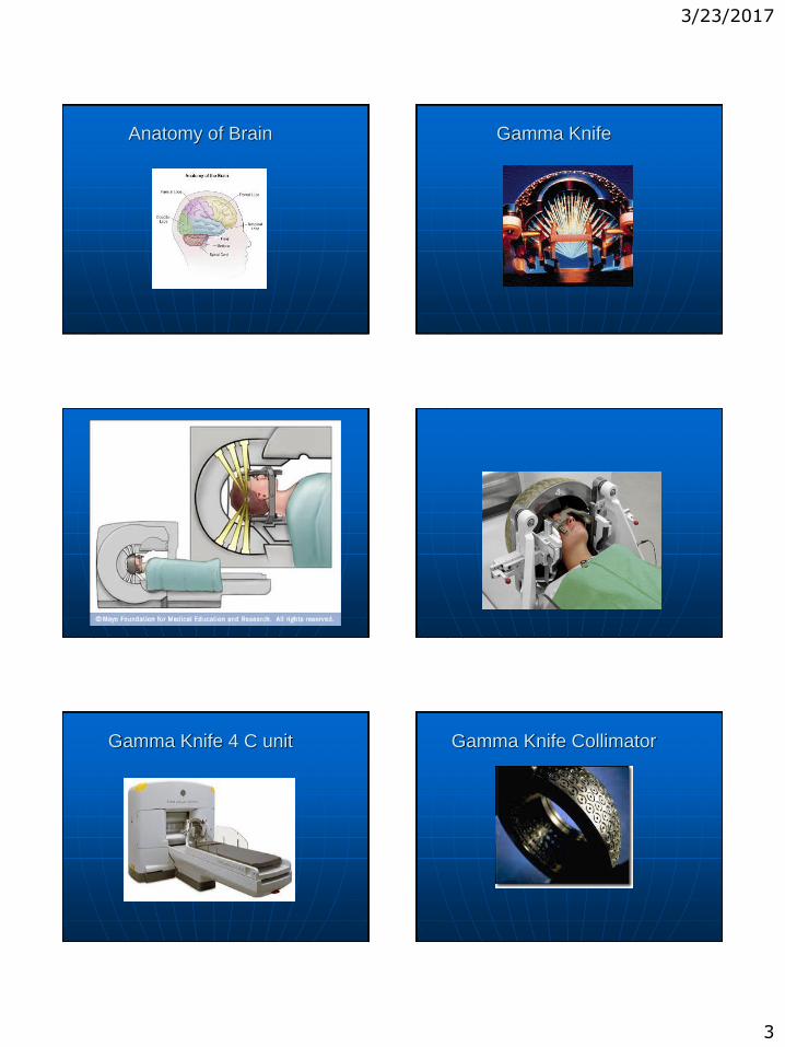
Anatomy of Brain Gamma Knife
Gamma Knife 4 C unit Gamma Knife Collimator

3/23/2017
4
There are 4 sizes of collimators used in order to conform to the shape of the patient’s tumor. The diameter of the holes are 4,8,14 and 18 mm
Either a single or a combination can be used to achieve an optimal treatment plan
History Of Gamma Knife
Developed in Sweden in 1950’s by Lars
Leksell and Borge Larsson
Very labor intensive in the beginning
Much more streamlined since the invention of CT and MRI
269 facilities World wide
123 centers in the USA-3 centers in Michigan-Beaumont Hospital in Royal Oak, Karmanos Cancer Institute in Detroit and Midland Michigan
Why do we only work in the brain?
The brain is easy to stabilize
Virtually no movement
Needs very high precision
Confined space
Conditions Treated With Gamma
Knife Primary brain tumors Metastatic brain tumors Resection cavaties Pituitary tumors Meningiomas Acoustic neuroma Trigeminal neuralgia Arteriovenous malformations
Glomus Jugulare tumors Ependymomas
Criteria For Gamma Knife
There should be 10 or less tumors present unless okayed by oversight committee (pt would otherwise have WBRT)
Size of tumor
Karnofsky Performance Scale (KPS) of 70 or more
Patient’s underlying disease is well controlled
How Does Radiation Work
Radiation works over a period of time
It causes damage to the DNA of the tumor cell which knocks out the ability of the cell to grow
The tumor slowly decreases in size and can eventually dissolve
In arteriovenous malformations (AVM) radiation causes damage by scarring the lumen of the vessel
Blood flow over time eventually slows and vessel ultimately occludes
Can take 2 -3 years to occur

3/23/2017
5
How Will It Be Decided Who Gets
Gamma Knife
Patient is seen by both a neurosurgeon and a radiation oncologist
If case is questionable then it is presented at the neurosurgery tumor board
The tumor board is a multidisciplinary team. As a group they will decide what the
best treatment option is for the patient
Treatment Options
Large tumor - surgery is an option
Primary brain tumor – biopsy needed to determine pathology
Surgery can be difficult based on location of tumor
Damage can occur to surrounding structures and brain function can be lost
Treatment Options
Chemotherapy –most don’t pass through the blood-brain barrier
Gamma Knife- noninvasive radiation treatment specifically given at the target site thus minimizing or avoiding side effects to normal tissue
Step 2 Imaging
MRI/CT of brain is performed on all patients
If pt has an implantable device CT only used
Slice thickness of MRI is 1 mm
The MRI box fits down over the frame. It contains copper sulfate in channels along the front ,back and sides of box
The CT box has strips of copper which light up on these images
The MRI and CT are fused to look at the accuracy of the MRI.
MR Fiducial Box MRI fiducial markers

3/23/2017
6
CT Fiducial Box CT Setup
Imaging
If pt is being treated for a vascular malformation a cerebral angiogram is done after the MRI and CT are completed
This gives more information of where the cluster or nidus of vessels is located and assists in treatment planning
Once all the imaging is completed bubble measurements are taken of the patients head
Bubble Measurement
Measurements
A plastic bubble sits down over the frame
Measurements are taken at set points around the circumference of the bubble
These numbers are entered into the computer and a 3D image is created of the patient’s skull
The length of the posts and pins are measured to ensure there is no collision with the equipment

3/23/2017
7
Step 3.Treatment planning
Computerized planning is done by the neurosurgeon, radiation oncologist and the physicist
All images taken are reviewed and used in the treatment planning
Can have 500-600 images per pt
Treatment Planning
Multiple Metastases Multiple Metastases
Step 4.Treatment
Treatment time is dependant upon the number, size and location of tumors
The actual procedure is painless and quiet Once treatment is completed the frame is
removed, pin sites cleansed and antibiotic ointment applied
Pt is observed for 1 hour The patient can resume their previous
activities Patient may go home with a Rx for steroid
and GI prophylaxis
Leksell Gamma Knife 4C

3/23/2017
8
Side Effects
Very few side effects
Headache
Discomfort at pin sites
Some bruising
Cerebral edema
Follow Up
Follow-up in 2 weeks
Pin sites assessed
Steroid taper
A follow-up MRI will be scheduled.
For metastatic tumors MRI will be repeated in 10-12 weeks
For benign condition most MRI’s are repeated in 3-6 months
Benefits of Gamma Knife
No surgical complications or side effects from general anesthesia
Risk of hemorrhage or infection is minimal as no incision made
No prolonged recovery time
More cost effective
Able to access areas of the brain that conventional surgery can’t
No delays for other treatment
Other Conditions Under Study for
Gamma Knife
Parkinsons and other movement
disorders
Epilepsy
Chronic intractable pain
Psychiatric Illnesses (depression, OCD)
Cluster Headaches/Migraines