Prognostic Factors of Gamma Knife Radiosurgery for ...
6
106 ISSN 1738-6217 J Kor Soc Ster Func Neurosurg 2015;11:106-111 potential for ischemia and seizure. 1)5) AVM has an annual hemorrhage rate of 2-3% that persists as long as the lesion exists. 1)3)5) Management of these lesions can be observation- al, although lesion obliteration is typically considered to miti- gate these risks. 1) Over the last decades, the management options for arterio- venous malformations (AVM) have expanded. 2) Although microsurgical resection has been used as the primary treat- ment option, stereotactic radiosurgery and embolization have been established as effective treatment options for AVM. 2)5)10) Moreover, recent advances in imaging technology and com- puterized radiation dose planning have improved the out- comes of radiosurgery. 2) Gamma knife radiosurgery (GKRS) has emerged as a popular treatment tool for AVMs without the immediate risk of hemorrhage. 23) GKRS is widely used CLINICAL ARTICLE Copyright © 2015 The Korean Society of Stereotactic and Functional Neurosurgery Prognostic Factors of Gamma Knife Radiosurgery for Cerebral Arteriovenous Malformation : The Results from 218 Consecutive Patients Deok Young Kim, MD 1 , Keun Young Park, MD 2 , Jae Whan Lee, MD, PhD 2 , Seung Kon Huh, MD, PhD 2 , Hyun Ho Jung , MD, PhD 1 , Jong Hee Chang, MD, PhD 1 , Jin Woo Chang, MD, PhD 1 , Yong Gou Park, MD 1 , Won Seok Chang, MD 1 1 Department of Neurosurgery, Yonsei Gamma Knife Center, Yonsei University College of Medicine, Seoul, Korea 2 Department of Neurosurgery, Severance Hospital Stroke Center, Yonsei University College of Medicine, Seoul, Korea Objective: We retrospectively evaluated our experience of the management and long-term outcomes of gamma knife radiosur- gery (GKRS) on arteriovenous malformation (AVM) as a primary treatment as well as adjunct therapy. GKRS is widely used in many centers worldwide for the management of AVM. However, the long-term results of GKRS are not well known. In the pres- ent study, we retrospectively evaluated our experience of focusing on prognostic factors in the management and long-term out- comes of GKRS on AVM. Materials and Methods: We performed a retrospective review of 218 patients. They were treated with GKRS in our institute from 2007 to 2013. The group included first-time GKRS for cerebral AVM, previous surgical resection, and embolization, exclud- ing previously GKRS-treated patients. We analyzed the association between several factors (gender, age, marginal dose, hemor- rhage, marginal dose, nidus volume) and the obliteration rate, which was the cumulative obliteration rate regardless of additional treatment, including repeat GKRS. Results: The univariate analysis of our study identified that a high radiation dose [margin dose >16.0Gy (50%), p=0.017] rather than a low radiation dose [margin dose ≤16.0Gy (50%)] and small AVM volume (≤15.0mL, p=0.003) rather than large AVM vol- ume ( >15.0mL) were positive predictors of AVM nidus obliteration. Our study showed that young age (≤18 years) had a better obliteration rate compared with others (> 18 years) in AVM after GKRS (p=0.053). Our findings did not show that gender (p= 0.427) or hemorrhage (p=0.191) had significant effects on nidus obliteration. Comparison of our results with prior studies showed that AVM volume and margin radiation dose had a significant role in the obliteration rate in common. Conclusion: We suggest that AVM with a small volume can be considered good indications for GKRS, and a higher marginal dose [margin dose >16.0Gy (50%)] can be an effective factor for higher obliteration with the carefulness of post-radiosurgical complications. Finally, we can expect favorable GKRS results for appropriately selected AVMs in the young population. KEY WORDS: Arteriovenous malformation (AVM) · Gamma knife radiosurgery (GKRS) · Obliteration rate · Prognostic factors. INTRODUCTION Cerebral arteriovenous malformations (AVMs) are patho- logic vascular lesions found in children and adults with a prevalence in adults of approximately 18 per 100,000. 1) AVM is defined by an abnormal connection between the venous and arterial circulation, resulting in an arteriovenous shunt and the gross appearance of a tangle of blood vessels. 1) The angioarchitecture of these lesions puts them at risk of hem- orrhage as well as subjecting the adjacent parenchyma to the Address for correspondence: Won Seok Chang, MD Department of Neurosurgery, Yonsei University College of Medicine, 50-1 Yonsei-ro, Seodaemun-gu, Seoul 03722, Korea Tel: +82-2-2228-2165, Fax: +82-2-393-9979 E-mail: [email protected] online © ML Comm
Transcript of Prognostic Factors of Gamma Knife Radiosurgery for ...

106
ISSN 1738-6217J Kor Soc Ster Func Neurosurg 2015;11:106-111
potential for ischemia and seizure.1)5) AVM has an annual hemorrhage rate of 2-3% that persists as long as the lesion exists.1)3)5) Management of these lesions can be observation-al, although lesion obliteration is typically considered to miti-gate these risks.1)
Over the last decades, the management options for arterio-venous malformations (AVM) have expanded.2) Although microsurgical resection has been used as the primary treat-ment option, stereotactic radiosurgery and embolization have been established as effective treatment options for AVM.2)5)10) Moreover, recent advances in imaging technology and com-puterized radiation dose planning have improved the out-comes of radiosurgery.2) Gamma knife radiosurgery (GKRS) has emerged as a popular treatment tool for AVMs without the immediate risk of hemorrhage.23) GKRS is widely used
CLINICAL ARTICLECopyright © 2015 The Korean Society of Stereotactic and
Functional Neurosurgery
Prognostic Factors of Gamma Knife Radiosurgery for Cerebral Arteriovenous Malformation : The Results from 218 Consecutive Patients
Deok Young Kim, MD1, Keun Young Park, MD2, Jae Whan Lee, MD, PhD2, Seung Kon Huh, MD, PhD2, Hyun Ho Jung , MD, PhD1, Jong Hee Chang, MD, PhD1, Jin Woo Chang, MD, PhD1, Yong Gou Park, MD1, Won Seok Chang, MD1
1Department of Neurosurgery, Yonsei Gamma Knife Center, Yonsei University College of Medicine, Seoul, Korea 2Department of Neurosurgery, Severance Hospital Stroke Center, Yonsei University College of Medicine, Seoul, Korea
Objective: We retrospectively evaluated our experience of the management and long-term outcomes of gamma knife radiosur-gery (GKRS) on arteriovenous malformation (AVM) as a primary treatment as well as adjunct therapy. GKRS is widely used in many centers worldwide for the management of AVM. However, the long-term results of GKRS are not well known. In the pres-ent study, we retrospectively evaluated our experience of focusing on prognostic factors in the management and long-term out-comes of GKRS on AVM.Materials and Methods: We performed a retrospective review of 218 patients. They were treated with GKRS in our institute from 2007 to 2013. The group included first-time GKRS for cerebral AVM, previous surgical resection, and embolization, exclud-ing previously GKRS-treated patients. We analyzed the association between several factors (gender, age, marginal dose, hemor-rhage, marginal dose, nidus volume) and the obliteration rate, which was the cumulative obliteration rate regardless of additional treatment, including repeat GKRS. Results: The univariate analysis of our study identified that a high radiation dose [margin dose >16.0Gy (50%), p=0.017] rather than a low radiation dose [margin dose ≤16.0Gy (50%)] and small AVM volume (≤15.0mL, p=0.003) rather than large AVM vol-ume (>15.0mL) were positive predictors of AVM nidus obliteration. Our study showed that young age (≤18 years) had a better obliteration rate compared with others (>18 years) in AVM after GKRS (p=0.053). Our findings did not show that gender (p= 0.427) or hemorrhage (p=0.191) had significant effects on nidus obliteration. Comparison of our results with prior studies showed that AVM volume and margin radiation dose had a significant role in the obliteration rate in common.Conclusion: We suggest that AVM with a small volume can be considered good indications for GKRS, and a higher marginal dose [margin dose >16.0Gy (50%)] can be an effective factor for higher obliteration with the carefulness of post-radiosurgical complications. Finally, we can expect favorable GKRS results for appropriately selected AVMs in the young population.
KEY WORDS: Arteriovenous malformation (AVM) · Gamma knife radiosurgery (GKRS) · Obliteration rate · Prognostic factors.
INTRODUCTION
Cerebral arteriovenous malformations (AVMs) are patho-logic vascular lesions found in children and adults with a prevalence in adults of approximately 18 per 100,000.1) AVM is defined by an abnormal connection between the venous and arterial circulation, resulting in an arteriovenous shunt and the gross appearance of a tangle of blood vessels.1) The angioarchitecture of these lesions puts them at risk of hem-orrhage as well as subjecting the adjacent parenchyma to the
Address for correspondence: Won Seok Chang, MD Department of Neurosurgery, Yonsei University College of Medicine, 50-1 Yonsei-ro, Seodaemun-gu, Seoul 03722, KoreaTel: +82-2-2228-2165, Fax: +82-2-393-9979E-mail: [email protected]
online © ML Comm
Deok Young Kim, et al : GKRS for AVM
107
in many centers worldwide for the management of AVM.2) GKRS is usually applied as a single treatment for small
AVMs (<3 cm) as well as a part of multimodal therapy in combination with microsurgical resection and emboliza-tion for large AVMs.2) Evidence from the literature reveals that GKRS had a significant impact on the obliteration of the AVM nidus in 65-94% patients over a 5-year observation interval.2) GKRS has been associated with a low risk of ra-diation-related complications, even for lesions located in ar-eas that are difficult to access via microsurgery.2) However, little information is available regarding the intermediate or long-term outcomes of GKRS on AVM.2) In the present study, we retrospectively evaluated our experience, focusing on prognostic factors in the management and long-term out-comes of GKRS on AVM.
MATERIALS AND METHODS
A total of 218 patients were treated with GKRS in our institute from 2007 to 2013. The group included first-time GKRS for cerebral AVM, previous surgical resection, and embolization excluding previously GKRS-treated patients. We analyzed the correlation between several factors (gender, age, marginal dose, hemorrhage, marginal dose, nidus vol-ume) and the obliteration rate, which was the cumulative obliteration rate regardless of additional treatment, includ-ing repeat GKRS.
Statistical analysis
Various factors that may affect the outcome were analyzed. Descriptive statistics is demonstrated as mean, median, stan-dard deviation (SD), percentages, and range. Kaplan-Meier method and log-rank analysis were used for actuarial rates of outcome and related factors. Multivariate analyses were performed by using a Cox regression analysis. All analyses were performed using commercially available statistical soft-ware SPSS Statistics (version 21.0 ; IBM Co., Armonk, NY, USA). Values of p<0.05 were considered statistically sig-nificant.
Patients’ demographics
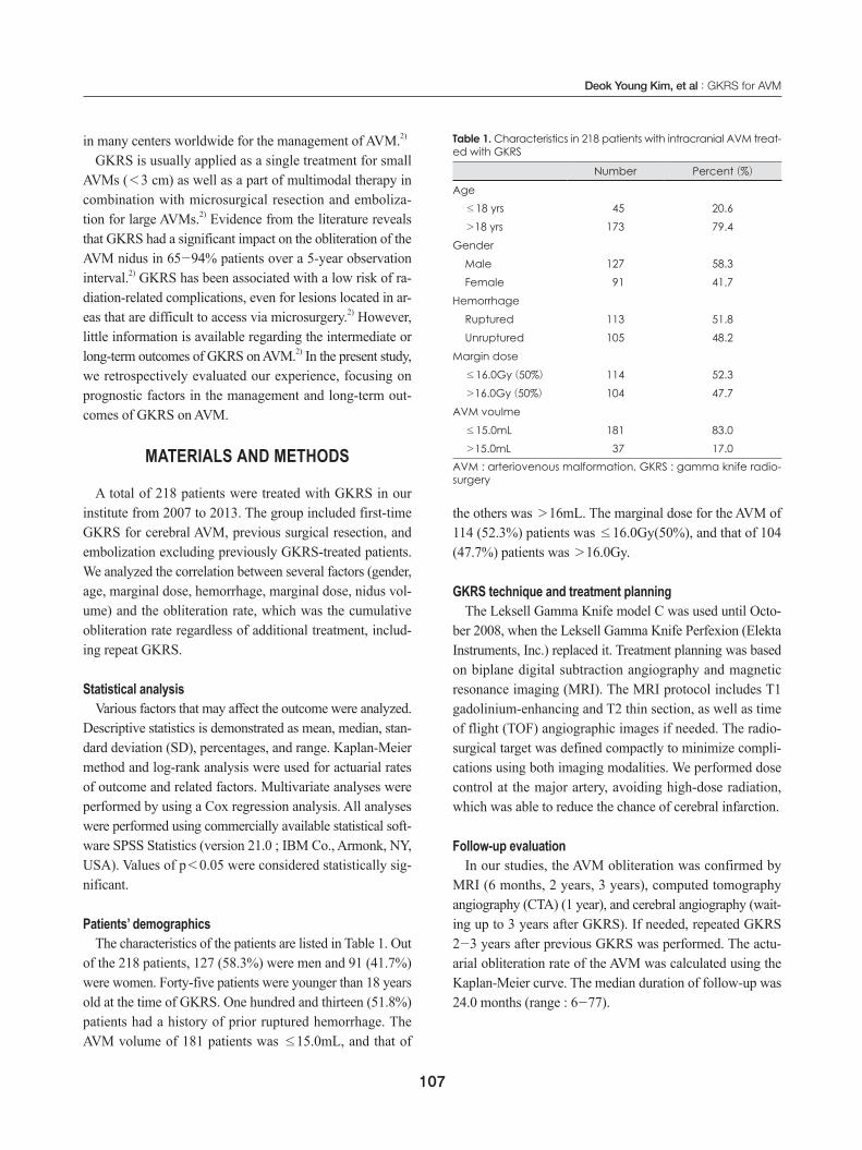
The characteristics of the patients are listed in Table 1. Out of the 218 patients, 127 (58.3%) were men and 91 (41.7%) were women. Forty-five patients were younger than 18 years old at the time of GKRS. One hundred and thirteen (51.8%) patients had a history of prior ruptured hemorrhage. The AVM volume of 181 patients was ≤15.0mL, and that of
the others was >16mL. The marginal dose for the AVM of 114 (52.3%) patients was ≤16.0Gy(50%), and that of 104 (47.7%) patients was >16.0Gy.
GKRS technique and treatment planningThe Leksell Gamma Knife model C was used until Octo-
ber 2008, when the Leksell Gamma Knife Perfexion (Elekta Instruments, Inc.) replaced it. Treatment planning was based on biplane digital subtraction angiography and magnetic resonance imaging (MRI). The MRI protocol includes T1 gadolinium-enhancing and T2 thin section, as well as time of flight (TOF) angiographic images if needed. The radio-surgical target was defined compactly to minimize compli-cations using both imaging modalities. We performed dose control at the major artery, avoiding high-dose radiation, which was able to reduce the chance of cerebral infarction.
Follow-up evaluationIn our studies, the AVM obliteration was confirmed by
MRI (6 months, 2 years, 3 years), computed tomography angiography (CTA) (1 year), and cerebral angiography (wait-ing up to 3 years after GKRS). If needed, repeated GKRS 2-3 years after previous GKRS was performed. The actu-arial obliteration rate of the AVM was calculated using the Kaplan-Meier curve. The median duration of follow-up was 24.0 months (range : 6-77).
Table 1. Characteristics in 218 patients with intracranial AVM treat-ed with GKRS
Number Percent (%)
Age≤18 yrs 045 20.6>18 yrs 173 79.4
GenderMale 127 58.3Female 091 41.7
HemorrhageRuptured 113 51.8Unruptured 105 48.2
Margin dose≤16.0Gy (50%) 114 52.3>16.0Gy (50%) 104 47.7
AVM voulme≤15.0mL 181 83.0>15.0mL 037 17.0
AVM : arteriovenous malformation, GKRS : gamma knife radio-surgery

J Kor Soc Ster Func Neurosurg 2015;11:106-111
108
RESULTS
We analyzed the association between several factors, in-cluding gender, age, hemorrhage, margin dose, and AVM volume, with the obliteration rate of the nidus. The results of AVM obliteration after GKRS are below Fig. 1.
Following a single GKRS procedure, 152 (69.7%) of 218 patients still had a residual nidus shown on MRI or angiog-raphy. Additionally, 17 patients harboring residual AVMs underwent repeat GKRS, and a total AVM obliteration was identified in 5 of these patients. After all, in 71 (32.6%) pa-tients, obliteration of the nidus was identified on follow-up angiography (60 patients) and MRI (11 patients). Clinical follow-up greater than 3 years was in 75 patients (34.4%), and 42 of these patients were confirmed as a total obliteration on both angiography and MRI. The actuarial angiographic or MRI obliteration rate was 10.64% at 24 months, 26.1% at 36 months, and 66.6% at 60 months, respectively (Fig. 1). Our overall obliteration rate was 32.6% through the follow-up duration (mean 33.8±2.7 months).
Factors related to AVM obliteration
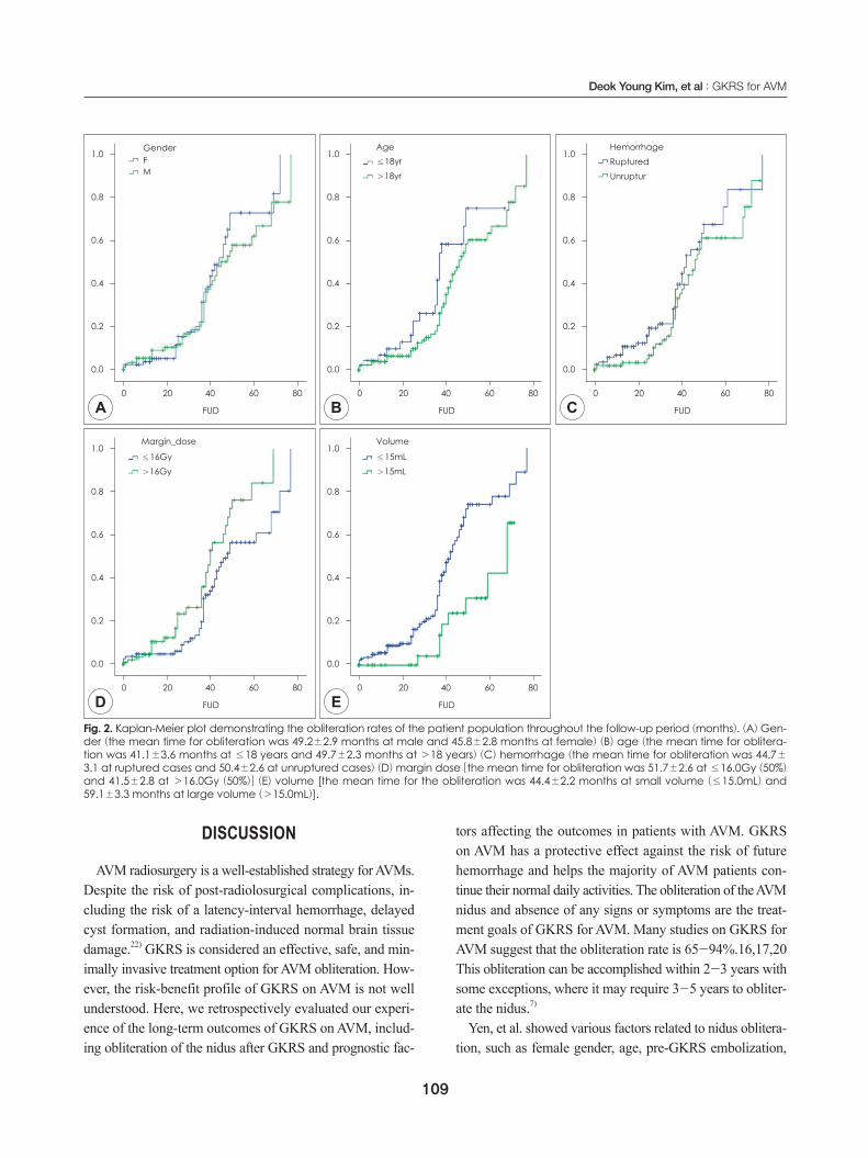
The results of factors related to AVM obliteration after GKRS are below Fig. 2. Statistical analysis demonstrated that a margin dose of more than 16.0Gy (50%) showed sta-tistical significance for better AVM obliteration (Log Rank p=0.017).
Small nidus volume (Log Rank p=0.003) was also asso-
ciated with increased AVM obliteration. Gender (Log Rank p=0.427) and hemorrhage (Log Rank p=0.191) were not re-lated to nidus obliteration. In our study, a younger age (Log Rank p=0.053) showed some association with AVM oblit-eration.
Statistical analysis demonstrated that gender (Log Rank p=0.427) was not related to AVM obliteration. Age (Log Rank p=0.053) was relatively associated with AVM obliter-ation. The mean time for obliteration was 41.1±3.6 months at ≤18 years and 49.7±2.3 months at >18 years. The younger group showed a shorter obliteration time. The mean time for the obliteration of ruptured AVM was 44.7±3.1 months, and that of unruptured AVM was 50.4±2.6 months. Hemorrhage (Log Rank p=0.191) was not related to nidus obliteration. Our study showed that a high median marginal dose [>16.0Gy (50%)] rather than a low median marginal dose [≤16.0Gy (50%)] was significantly associ-ated with an increased rate of AVM obliteration (Log Rank p=0.017). The group with the >16.0Gy (50%) marginal dose had a shorter obliteration time (41.5±2.8 months) than the one with ≤16.0Gy (50%) (51.7±2.6 months). Small volume (≤15.0mL) rather than large volume (>15.0mL) revealed a significant statistical difference of AVM obliter-ation (Log Rank p=0.003). The mean time for the oblitera-tion of small volume (≤15.0mL) was 44.4±2.2 months, and that of large volume (>15.0mL) was 59.1±3.3 months. In multivariate analysis, only small nidus volume (p=0.045) was significantly associated with increased rate of AVM obliteration (Table 2).
Complication associated with treatment
Overall, a post-GKRS complication was recorded for 40 patients (18.3%). Posttreatment hemorrhage occurred in 12 patients (5.5%), 1 of whom was brain death after emergen-cy hematoma removal. Eight (3.7%) patients developed brain edema and seven (3.2%) patients developed GKRS related cyst formation. One of 7 patients having brain cyst underwent Ommaya reservoir insertion. Four (1.8%) patient had newly developed post-GKRS seizures and two (0.9%) had preexisting seizures. Hydrocephalus, paresthesia on limbs, memory impairment, radiation necrosis, character change, and cerebral infarction were developed in each one (0.5%) patient. The patient with hydrocephalus recovered his heath after a ventriculoperitoneal shunt. In addition to being mentioned, there were not in life-threatening conditions. Most of them had responded to conservative treatment.
Fig. 1. Kaplan-Meier plot depicting the AVM obliteration rates fol-lowing GKRS in the present series.
1.0
0.8
0.6
0.4
0.2
0.0
0.00 20.00 40.00 60.00 80.00
Survival functionCensored
FUD (month)
Obl
itera
tion
base
d o
n an
giog
ram
or M
RI
Deok Young Kim, et al : GKRS for AVM
109
DISCUSSION
AVM radiosurgery is a well-established strategy for AVMs. Despite the risk of post-radiolosurgical complications, in-cluding the risk of a latency-interval hemorrhage, delayed cyst formation, and radiation-induced normal brain tissue damage.22) GKRS is considered an effective, safe, and min-imally invasive treatment option for AVM obliteration. How-ever, the risk-benefit profile of GKRS on AVM is not well understood. Here, we retrospectively evaluated our experi-ence of the long-term outcomes of GKRS on AVM, includ-ing obliteration of the nidus after GKRS and prognostic fac-
tors affecting the outcomes in patients with AVM. GKRS on AVM has a protective effect against the risk of future hemorrhage and helps the majority of AVM patients con-tinue their normal daily activities. The obliteration of the AVM nidus and absence of any signs or symptoms are the treat-ment goals of GKRS for AVM. Many studies on GKRS for AVM suggest that the obliteration rate is 65-94%.16,17,20 This obliteration can be accomplished within 2-3 years with some exceptions, where it may require 3-5 years to obliter-ate the nidus.7)
Yen, et al. showed various factors related to nidus oblitera-tion, such as female gender, age, pre-GKRS embolization,
Fig. 2. Kaplan-Meier plot demonstrating the obliteration rates of the patient population throughout the follow-up period (months). (A) Gen-der (the mean time for obliteration was 49.2±2.9 months at male and 45.8±2.8 months at female) (B) age (the mean time for oblitera-tion was 41.1±3.6 months at ≤18 years and 49.7±2.3 months at >18 years) (C) hemorrhage (the mean time for obliteration was 44.7±
3.1 at ruptured cases and 50.4±2.6 at unruptured cases) (D) margin dose [the mean time for obliteration was 51.7±2.6 at ≤16.0Gy (50%) and 41.5±2.8 at >16.0Gy (50%)] (E) volume [the mean time for the obliteration was 44.4±2.2 months at small volume (≤15.0mL) and 59.1±3.3 months at large volume (>15.0mL)].
1.0
0.8
0.6
0.4
0.2
0.0
0 20 40 60 80
FUDD
Margin_dose
≤16Gy>16Gy
1.0
0.8
0.6
0.4
0.2
0.0
0 20 40 60 80
FUDE
Volume
≤15mL>15mL
1.0
0.8
0.6
0.4
0.2
0.0
0 20 40 60 80
GenderFM
FUDA
1.0
0.8
0.6
0.4
0.2
0.0
0 20 40 60 80
FUDC
HemorrhageRupturedUnruptur
1.0
0.8
0.6
0.4
0.2
0.0
0 20 40 60 80
FUDB
Age≤18yr>18yr
J Kor Soc Ster Func Neurosurg 2015;11:106-111
110
AVM volume, location, deep-draining vein, single-draining vein, margin dose, maximum dose, isodense line, isocenters, radiation-induced changes, and radiosurgery-based AVM score.23) Their univariate analysis demonstrated that high margin dose (p=0.042), high maximum dose (p=0.035), deep-drain vein (p=0.042), and small nidus volume (p=0.015) were associated with increased AVM obliteration rate. Sex, age, prior embolization, locations of nidi, radiosurgery-based AVM scores, single-draining vein, isodose line, number of isocen-ters, and presence of radiation-induced imaging changes were not related to nidus obliteration.23) Bir, et al. identified female sex (p=0.04) and Spetzler-Martin Grade I-III (p=
0.002) as positive predictors of AVM nidus obliteration, but history of hemorrhage and history of embolization did not affect nidus obliteration. Some studies said that nidus vol-ume is a critical factor that impedes the complete oblitera-tion of AVMs.13)15) The lowest dose delivered to the AVM is an important factor related to the obliteration rate.13) It is rel-atively easy to deliver an insufficient radiation dose to the
nidus of a large AVM, and the operator wants to avoid ad-verse radiation effects (ARE).14)15) The above studies showed that AVM nidus volume and radiation dose had the effect on nidus obliteration in common.
In this study, we had an interest in the young age effect on the AVM obliteration rate as a prognostic factor. The opti-mal management of unruptured AVMs in pediatric patients is now incompletely understood.9) A Randomized Trial of Unruptured Brain AVMs (ARUBA) was a prospective, ran-domized controlled trial that showed superior outcomes with medical management compared with intervention for patients with unruptured AVMs.18) However, pediatric pa-tients (aged less than 18 years) were specifically excluded from ARUBA. Therefore, the efficacy of radiosurgery for unruptured pediatric AVMs is poorly understood.9) From our review of the major series, the reported obliteration rates derived from retrospective studies of pediatric patients vary widely, ranging from 45% to 84% in studies reporting 3-year radiographic outcomes.4)6)8)11)12)19)21)24)25)
We evaluated several factors, gender, age, hemorrhage, margin dose, and AVM volume, with the obliteration rate of the nidus. The univariate analysis of our study identified that a high radiation dose (margin dose >16.0Gy (50%), p=0.017) rather than a low radiation dose [margin dose ≤16.0Gy (50%)] and small AVM volume (≤15.0mL, p=0.003) rather than large AVM volume (>15.0mL) were positive predic-tors of AVM nidus obliteration. Our study showed that young age (≤18 years) had a better obliteration rate compared with others (>18 years) in AVM after GKRS (p=0.053). Our findings did not show that gender (p=0.427) or hemorrhage (p=0.191) had significant effects on nidus obliteration.
Comparison of our results with prior studies showed that AVM volume and margin radiation dose had a significant role in the obliteration rate in common. However, the age effect as a prognostic factor is not well defined. Yen, et al.23) and Bir, et al.2) showed that age was not related to nidus oblit-eration (Table 3). Previous studies reported a lower obliter-ation rate for GKRS in young-age AVM because the young
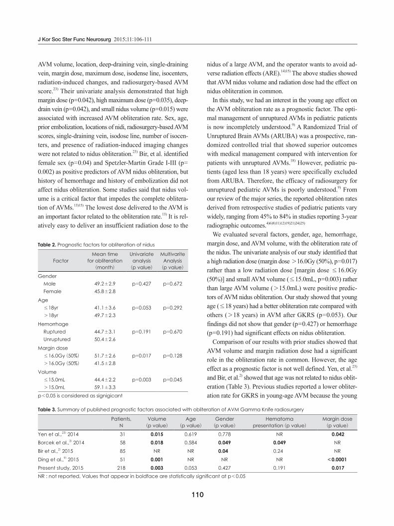
Table 2. Prognostic factors for obliteration of nidus
FactorMean time
for obliteration(month)
Univariate analysis(p value)
MultivariteAnalysis(p value)
GenderMaleFemale
49.2±2.945.8±2.8
p=0.427 p=0.672
Age≤18yr>18yr
41.1±3.649.7±2.3
p=0.053 p=0.292
HemorrhageRupturedUnruptured
44.7±3.150.4±2.6
p=0.191 p=0.670
Margin dose≤16.0Gy (50%)
>16.0Gy (50%)
51.7±2.641.5±2.8
p=0.017 p=0.128
Volume≤15.0mL>15.0mL
44.4±2.259.1±3.3
p=0.003 p=0.045
p<0.05 is considered as signigicant
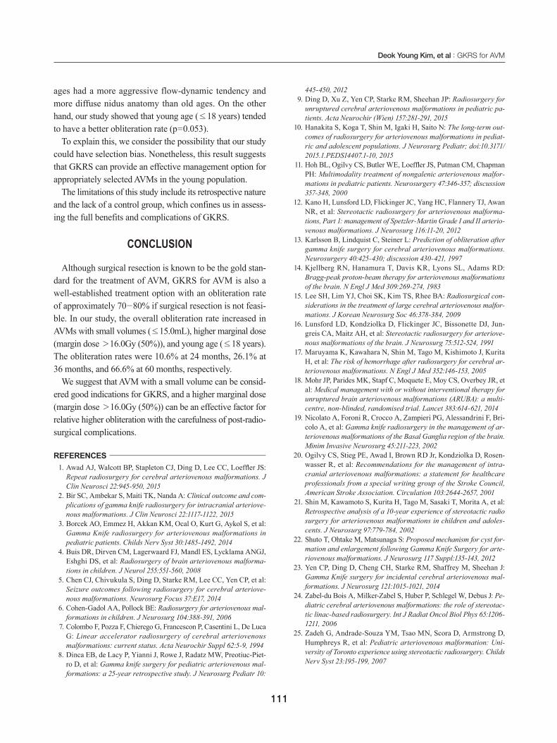
Table 3. Summary of published prognostic factors associated with obliteration of AVM Gamma Knife radiosurgery
Patients,N
Volume(p value)
Age(p value)
Gender(p value)
Hematoma presentation (p value)
Margin dose(p value)
Yen et al.,23) 2014 031 0.015 0.619 0.778 NR <0.042Borcek et al.,3) 2014 058 0.018 0.584 0.049 0.049 NRBir et al.,2) 2015 085 NR NR 0.040 0.240 NRDing et al.,9) 2015 051 0.001 NR NR NR <0.0001Present study, 2015 218 0.003 0.053 0.427 0.191 <0.017NR : not reported, Values that appear in boldface are statistically significant at p<0.05
Deok Young Kim, et al : GKRS for AVM
111
ages had a more aggressive flow-dynamic tendency and more diffuse nidus anatomy than old ages. On the other hand, our study showed that young age (≤18 years) tended to have a better obliteration rate (p=0.053).
To explain this, we consider the possibility that our study could have selection bias. Nonetheless, this result suggests that GKRS can provide an effective management option for appropriately selected AVMs in the young population.
The limitations of this study include its retrospective nature and the lack of a control group, which confines us in assess-ing the full benefits and complications of GKRS.
CONCLUSION
Although surgical resection is known to be the gold stan-dard for the treatment of AVM, GKRS for AVM is also a well-established treatment option with an obliteration rate of approximately 70-80% if surgical resection is not feasi-ble. In our study, the overall obliteration rate increased in AVMs with small volumes (≤15.0mL), higher marginal dose (margin dose >16.0Gy (50%)), and young age (≤18 years). The obliteration rates were 10.6% at 24 months, 26.1% at 36 months, and 66.6% at 60 months, respectively.
We suggest that AVM with a small volume can be consid-ered good indications for GKRS, and a higher marginal dose (margin dose >16.0Gy (50%)) can be an effective factor for relative higher obliteration with the carefulness of post-radio-surgical complications.
REFERENCES1. Awad AJ, Walcott BP, Stapleton CJ, Ding D, Lee CC, Loeffler JS:
Repeat radiosurgery for cerebral arteriovenous malformations. J Clin Neurosci 22:945-950, 2015
2. Bir SC, Ambekar S, Maiti TK, Nanda A: Clinical outcome and com-plications of gamma knife radiosurgery for intracranial arteriove-nous malformations. J Clin Neurosci 22:1117-1122, 2015
3. Borcek AO, Emmez H, Akkan KM, Ocal O, Kurt G, Aykol S, et al: Gamma Knife radiosurgery for arteriovenous malformations in pediatric patients. Childs Nerv Syst 30:1485-1492, 2014
4. Buis DR, Dirven CM, Lagerwaard FJ, Mandl ES, Lycklama ANGJ, Eshghi DS, et al: Radiosurgery of brain arteriovenous malforma-tions in children. J Neurol 255:551-560, 2008
5. Chen CJ, Chivukula S, Ding D, Starke RM, Lee CC, Yen CP, et al: Seizure outcomes following radiosurgery for cerebral arteriove-nous malformations. Neurosurg Focus 37:E17, 2014
6. Cohen-Gadol AA, Pollock BE: Radiosurgery for arteriovenous mal-formations in children. J Neurosurg 104:388-391, 2006
7. Colombo F, Pozza F, Chierego G, Francescon P, Casentini L, De Luca G: Linear accelerator radiosurgery of cerebral arteriovenous malformations: current status. Acta Neurochir Suppl 62:5-9, 1994
8. Dinca EB, de Lacy P, Yianni J, Rowe J, Radatz MW, Preotiuc-Piet-ro D, et al: Gamma knife surgery for pediatric arteriovenous mal-formations: a 25-year retrospective study. J Neurosurg Pediatr 10:
445-450, 20129. Ding D, Xu Z, Yen CP, Starke RM, Sheehan JP: Radiosurgery for
unruptured cerebral arteriovenous malformations in pediatric pa-tients. Acta Neurochir (Wien) 157:281-291, 2015
10. Hanakita S, Koga T, Shin M, Igaki H, Saito N: The long-term out-comes of radiosurgery for arteriovenous malformations in pediat-ric and adolescent populations. J Neurosurg Pediatr; doi:10.3171/2015.1.PEDS14407.1-10, 2015
11. Hoh BL, Ogilvy CS, Butler WE, Loeffler JS, Putman CM, Chapman PH: Multimodality treatment of nongalenic arteriovenous malfor-mations in pediatric patients. Neurosurgery 47:346-357; discussion 357-348, 2000
12. Kano H, Lunsford LD, Flickinger JC, Yang HC, Flannery TJ, Awan NR, et al: Stereotactic radiosurgery for arteriovenous malforma-tions, Part 1: management of Spetzler-Martin Grade I and II arterio-venous malformations. J Neurosurg 116:11-20, 2012
13. Karlsson B, Lindquist C, Steiner L: Prediction of obliteration after gamma knife surgery for cerebral arteriovenous malformations. Neurosurgery 40:425-430; discussion 430-421, 1997
14. Kjellberg RN, Hanamura T, Davis KR, Lyons SL, Adams RD: Bragg-peak proton-beam therapy for arteriovenous malformations of the brain. N Engl J Med 309:269-274, 1983
15. Lee SH, Lim YJ, Choi SK, Kim TS, Rhee BA: Radiosurgical con-siderations in the treatment of large cerebral arteriovenous malfor-mations. J Korean Neurosurg Soc 46:378-384, 2009
16. Lunsford LD, Kondziolka D, Flickinger JC, Bissonette DJ, Jun-greis CA, Maitz AH, et al: Stereotactic radiosurgery for arteriove-nous malformations of the brain. J Neurosurg 75:512-524, 1991
17. Maruyama K, Kawahara N, Shin M, Tago M, Kishimoto J, Kurita H, et al: The risk of hemorrhage after radiosurgery for cerebral ar-teriovenous malformations. N Engl J Med 352:146-153, 2005
18. Mohr JP, Parides MK, Stapf C, Moquete E, Moy CS, Overbey JR, et al: Medical management with or without interventional therapy for unruptured brain arteriovenous malformations (ARUBA): a multi-centre, non-blinded, randomised trial. Lancet 383:614-621, 2014
19. Nicolato A, Foroni R, Crocco A, Zampieri PG, Alessandrini F, Bri-colo A, et al: Gamma knife radiosurgery in the management of ar-teriovenous malformations of the Basal Ganglia region of the brain. Minim Invasive Neurosurg 45:211-223, 2002
20. Ogilvy CS, Stieg PE, Awad I, Brown RD Jr, Kondziolka D, Rosen-wasser R, et al: Recommendations for the management of intra-cranial arteriovenous malformations: a statement for healthcare professionals from a special writing group of the Stroke Council, American Stroke Association. Circulation 103:2644-2657, 2001
21. Shin M, Kawamoto S, Kurita H, Tago M, Sasaki T, Morita A, et al: Retrospective analysis of a 10-year experience of stereotactic radio surgery for arteriovenous malformations in children and adoles-cents. J Neurosurg 97:779-784, 2002
22. Shuto T, Ohtake M, Matsunaga S: Proposed mechanism for cyst for-mation and enlargement following Gamma Knife Surgery for arte-riovenous malformations. J Neurosurg 117 Suppl:135-143, 2012
23. Yen CP, Ding D, Cheng CH, Starke RM, Shaffrey M, Sheehan J: Gamma Knife surgery for incidental cerebral arteriovenous mal-formations. J Neurosurg 121:1015-1021, 2014
24. Zabel-du Bois A, Milker-Zabel S, Huber P, Schlegel W, Debus J: Pe-diatric cerebral arteriovenous malformations: the role of stereotac-tic linac-based radiosurgery. Int J Radiat Oncol Biol Phys 65:1206-1211, 2006
25. Zadeh G, Andrade-Souza YM, Tsao MN, Scora D, Armstrong D, Humphreys R, et al: Pediatric arteriovenous malformation: Uni-versity of Toronto experience using stereotactic radiosurgery. Childs Nerv Syst 23:195-199, 2007