Gamma Knife Radiosurgery - AMOS Onlineamos3.aapm.org/abstracts/pdf/68-19901-237350-85912.pdf · 1...
60
1 Gamma Knife Gamma Knife Radiosurgery Radiosurgery Paula L. Petti, Ph.D. FREMONT, CA Disclosure: P. Petti is a PFX system-start consultant for Elekta Instrument AB
Transcript of Gamma Knife Radiosurgery - AMOS Onlineamos3.aapm.org/abstracts/pdf/68-19901-237350-85912.pdf · 1...
1
Gamma Knife Gamma Knife RadiosurgeryRadiosurgery
Paula L. Petti, Ph.D.
FREMONT, CA
Disclosure: P. Petti is a PFX system-start consultant for Elekta Instrument AB

2
2
Lars Lars LeksellLeksell, neurosurgeon, introduces idea, neurosurgeon, introduces idea
of of stereotactic radiosurgery stereotactic radiosurgery in 1951in 1951
“The stereotactic technique enables theaccurate insertion of a needle electrode intoany given structure of the brain…
It would therefore befeasible to replace theneedle by narrowbeams of radiantenergy directed at thetarget in the brain andthereby produce alocal destruction of thetissue…”
L. Leksell, Acta Chir Scand 102:316-319, 1951

3
Leksell Gamma KnifeModel U: (Introduced 1986) Model C: (Introduced 1999)
Perfexion: (Introduced 2006)

4
Properties of Leksell Gamma
Knife® Radiosurgery
• ~200 60Co sources (6000 Ci total initial activity)
• Sources positioned and collimated to focus
radiation precisely at isocenter
• Prescription volume shaped to match the target
volume by:
– translating the patient in 3 orthogonal directions
between “shot” settings
– using appropriately sized collimators for each shot

5
Properties of 60Co
60Co decays via beta decay,producing 2 mono-energetic !-
rays, 1.17 and 1.33 MeV in the
process
The half life of 60Co is 5.27 yrs.
Depth dose distribution similar
to 4 MV photons
60Co isotope produced by bombarding 59Co withneutrons in a nuclear reactor " The U.S. Nuclear
Regulatory Commission (NRC) oversees its use

6
Indications for Gamma Knife
Radiosurgery
44% Malignant Tumors
35% Benign Tumors
1% Ocular
Vascular Disorders 13%
Functional
Disorders
8%
Malignant tumors: Brain metastases, glial tumors
Benign tumors: Meningioma, vestibular schwannoma, pituitary
adenoma
Vascular disorders: Arteriovenous malformation
Functional disorders: Trigeminal neuralgia
Examples:Data from Leksell
Gamma Knife Society,
worldwide 2009
statistics

7
Criteria for Gamma Knife
Radiosurgery
• Largest tumordimension # 3 to 4 cm
• Minimum distance tooptic apparatus $ 2 mm
• Max number of lesionsthat can reasonably betreated in a singlesession ~ 30
• Can treat very smalltumors (e.g. small brainmetastases withdimension of the order2 mm)
• Largest tumordimension > 4 cm
• Tumor “too close” tooptic apparatus
• Occasionally, patientpreference
Single-fraction GK SRS Multiple-fraction GK SRS
(Extend system)

8
Localization Systems for GK
Leksell Stereotactic Frame: attached
to patient via 4 pins
Single-fraction radiosurgery
Leksell Extend re-locatable frame
system: vacuum bite block
Fractionated radiosurgery
Images from Elekta brochures

9
GK Model Comparison
Treatment couch = the
patient positioning system
Patient position changed
automatically
Patient position changed
manually or semi-
automatically
Sources moveSource position fixed
All collimation is internalSecondary collimators are
external, must be manually
changed
Perfexion%Models U, C and 4C

10
PerfexionPerfexion™™ Collimator Collimator• 3 collimator sizes (4-, 8- and 16-mm)
• 576 collimating channels
• Material is tungsten
(Images courtesy of Elekta)
217800Steel
7.919300Tungsten
60Co
approx.
HVL(mm)
Density
(kg/m3)
Material

11
Illustration of Sector PositionsIllustration of Sector Positions
Sectors alternating 16-mm (red)
and 8-mm (green) collimators
Note different
positions of
sectors
5 sector Positions:
Home
8-mm
Blocked
4-mm
16-mm

12
Radiological AccuracyRadiological Accuracy• Alignment of the radiation focus and the mechanical
isocenter
Patient positioning
system defines
mechanical isocenter
Sources and collimating system
defines radiation focus
Critically important since the hallmark of Critically important since the hallmark of radiosurgeryradiosurgery is the ability to is the ability todeliver focal radiation with sub-millimeter precisiondeliver focal radiation with sub-millimeter precision

13
GK GK PerfexionPerfexion™™: Selected Technical: Selected Technical
SpecificationsSpecifications
< 3 sTypical couch repositioning time
<0.5 mmRadiological accuracy
< 3 sTypical collimator size setup
time
210 kg (460 lbs)Maximum patient weight
< 0.05 mmCouch positioning repeatability
14
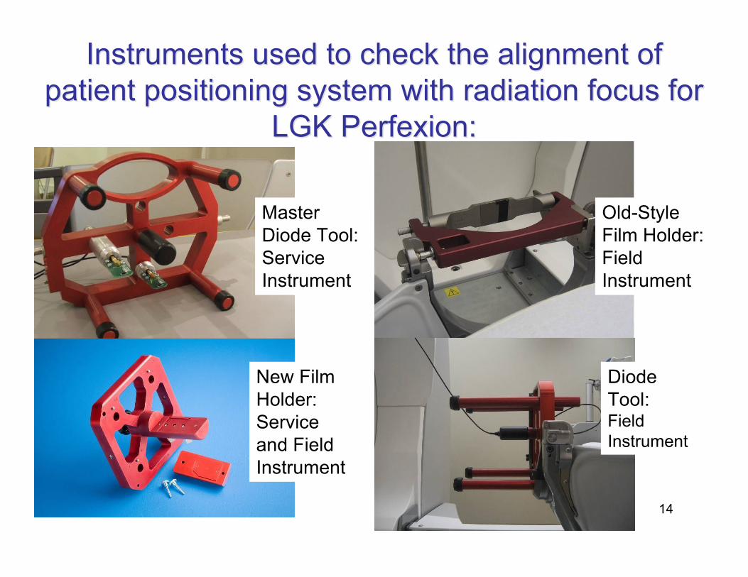
Instruments used to check the alignment ofInstruments used to check the alignment of
patient positioning system with radiation focus forpatient positioning system with radiation focus for
LGK LGK PerfexionPerfexion::
Master
Diode Tool:
Service
Instrument
New Film
Holder:
Service
and Field
Instrument
Old-Style
Film Holder:
Field
Instrument
Diode
Tool:Field
Instrument

15
Diode QA TestDiode QA Test• The diode is scanned through the irradiated field in X, Y and
Z for the 4-mm collimator. Average deviations are reported.
Calculated Deviation

16
Example of Results:Example of Results:
Pin-Point Test of Coincidence of PPS and RFP, New Film
Holder
-20
0
20
40
60
80
100
120
140
80 85 90 95 100 105 110 115 120
X Coordinate of PPS (mm)
Re
ad
ing
(b
ackg
rou
nd
su
btr
acte
d)
16mm X AxisFWHMPPS CenterRFP Center
!X = 0.02 mm

17
Results of Pin-Point Test
at WHHS Averaged over 4 years from 2007
to 2011
0.080.2716-mm
0.070.238-mm
0.070.164-mm
Standard
Deviation
(mm)
Averaged !r* over
4-year time period
(mm)
Collimator
222* zyxr !!!! ++=Results within the specified
0.5 mm radiological accuracy
of the Perfexion positioning
systemdeviation of radiation focal point from
center of patient positioning system=

18
End-to-End Tests
• Tests described on previous slides assessthe precision with which the radiation focalpoint is aligned to the mechanicalisocenter
• They do not test the accuracy of the entireGK procedure
• End-to-end tests are not done routinely forGK
• A few studies have been reported

19
End-to-End Experiments
• L. Ma et al., Med Phys.
35, 5110-14 (2008).
– Attached LGK SRS frame
to phantom
– Obtained CT scan
– Inserted film in phantom
– Irradiated film for 6
representative treatment
plans

20
Results from L. Ma Study
• E.g., for 50% isodose line,average DTA was 1.02 ±0.18 mm
• Considering all isodoselevels, no statisticallysignificant difference inuncertainty with increasingnumber of shots
21
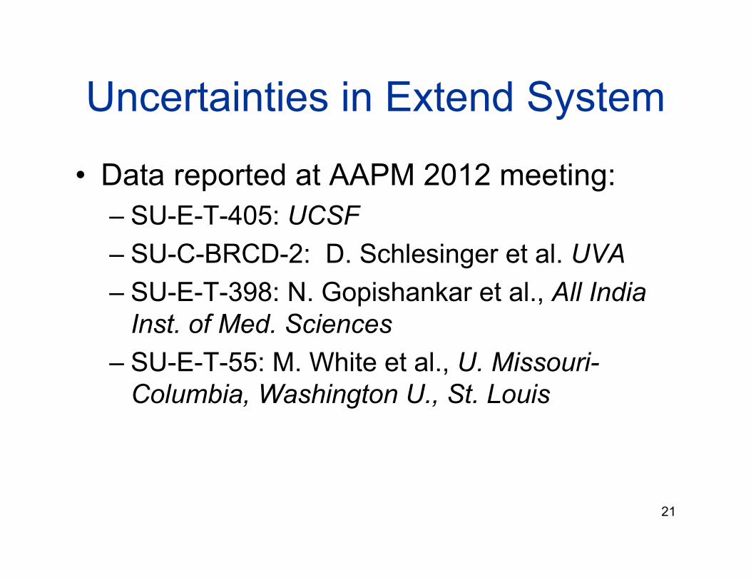
Uncertainties in Extend System
• Data reported at AAPM 2012 meeting:
– SU-E-T-405: UCSF
– SU-C-BRCD-2: D. Schlesinger et al. UVA
– SU-E-T-398: N. Gopishankar et al., All India
Inst. of Med. Sciences
– SU-E-T-55: M. White et al., U. Missouri-
Columbia, Washington U., St. Louis
22
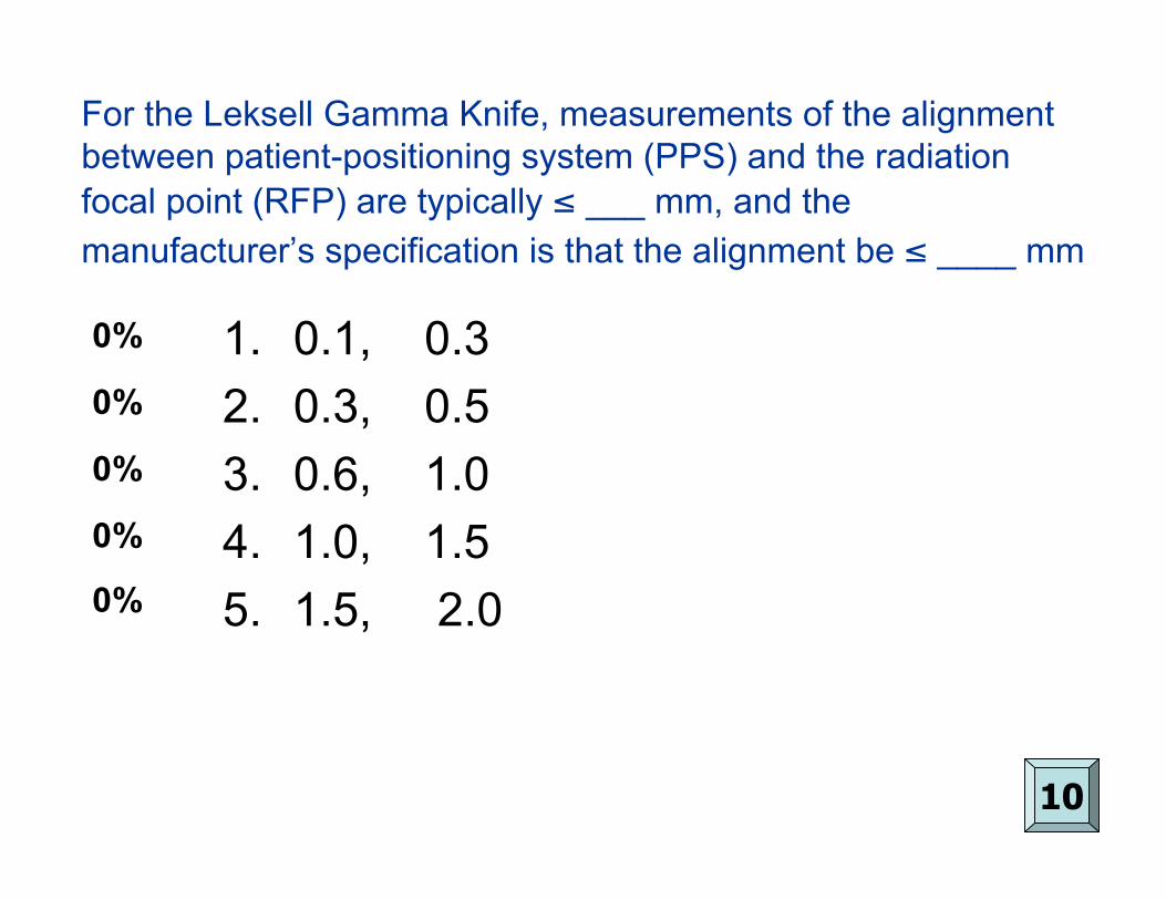
For the Leksell Gamma Knife, measurements of the alignment
between patient-positioning system (PPS) and the radiation
focal point (RFP) are typically # ___ mm, and the
manufacturer’s specification is that the alignment be # ____ mm
0%
0%
0%
0%
0%
10
1. 0.1, 0.3
2. 0.3, 0.5
3. 0.6, 1.0
4. 1.0, 1.5
5. 1.5, 2.0

23
For the Leksell Gamma Knife, measurements of the
alignment between patient-positioning system (PPS) and
the radiation focal point (RFP) are typically # ___ mm, and
the manufacturer’s specification is that the alignment be #____ mm
Answer: 2: M. Schell et al., AAPM Report No. 54,
“Stereotactic Radiosurgery,” p. 65 (1999). Also, notethat the values listed on slide 18 are # 0.3 mm.
Specifications for the radiological accuracy of the
Perfexion system are stated in Elekta’s documentation,
“LGK Perfexion, System Description.”
0.080.2716-mm
0.070.238-mm
0.070.164-mm
Standard
Deviation (mm)
Averaged !r* over 4-
year time period (mm)
Collimator
Typical
measure-
ment
results

24
Based on reports in the literature, the
distance-to-agreement (DTA) for the 50%
isodose level in a multi-shot GK treatment is
approx. ___ mm
0%
0%
0%
0%
0%
10
1. 0.1
2. 0.5
3. 1.0
4. 1.8
5. 2.5

25
Based on reports in the literature, the
distance-to-agreement (DTA) for the 50%
isodose level in a multi-shot GK treatment is
approx. ___ mm
Answer: 3: L. Ma et al., “Whole-procedure clinical
accuracy of Gamma Knife treatments of large lesions,” Med.
Phys. 35: 5110-5114, (2008)

26
Workflow for Frame-Based GK SRS
• Neurosurgeon attaches stereotactic frame
• Skull and frame measurements obtained
• MR and/or CT images acquired
• Treatment planning performed (by either
physicists or MDs)
• Treatment plan approved
• Treatment delivered
All steps completed in one day
Several patients can be treated per day depending on
complexity of cases and staffing

27
Frame attachment, Skull
Measurements
Stereotactic Frame defines LGK coordinate
system
Surface of the head determined by skull
scaling instrument (“bubble”)
Everything inside skull, as determined by
bubble, is assumed to be unit density

28
Skull Definition from CT
• Relatively new feature:– Determine skull outline from
CT scan
– Use CT to make tissueheterogeneity correction viaconvolution dose calculationalgorithm
– Pros: More accurate dosecalc
– Cons:
• Requires CT as well as MRfor treatment planning
• Existing data for doseprescriptions based on unit-density approximation

29
Image DefinitionImage DefinitionMRI MRI Fiducial Fiducial BoxesBoxes
Copper Sulfate channels
30
Distortions in MRI ImagesDistortions in MRI Images
• Sources of distortion in MR images:
– Different pulse sequences
– Different magnetic susceptibilities of individual
patients
– The presence of magnetic objects such as surgical
clips
• Investigators* have documented distortions in
MRI images used for GK SRS to be of the order
of 1 mm or less
*e.g., A. Ertl et al., Med Phys. 28, 166-170, (1999)

31
Assessing Distortion in MRAssessing Distortion in MR
ImagesImages
• Consider obtaining CT scans in patients for
whom a 1 mm shift in targeting could be
critical
– Fuse the CT with the MR (i.e., overlay
corresponding pixels of defined images)
– Or, simply assess targeted area on CT scan
• Perform phantom measurements to assess
distortions on MR as part of regular QA
32
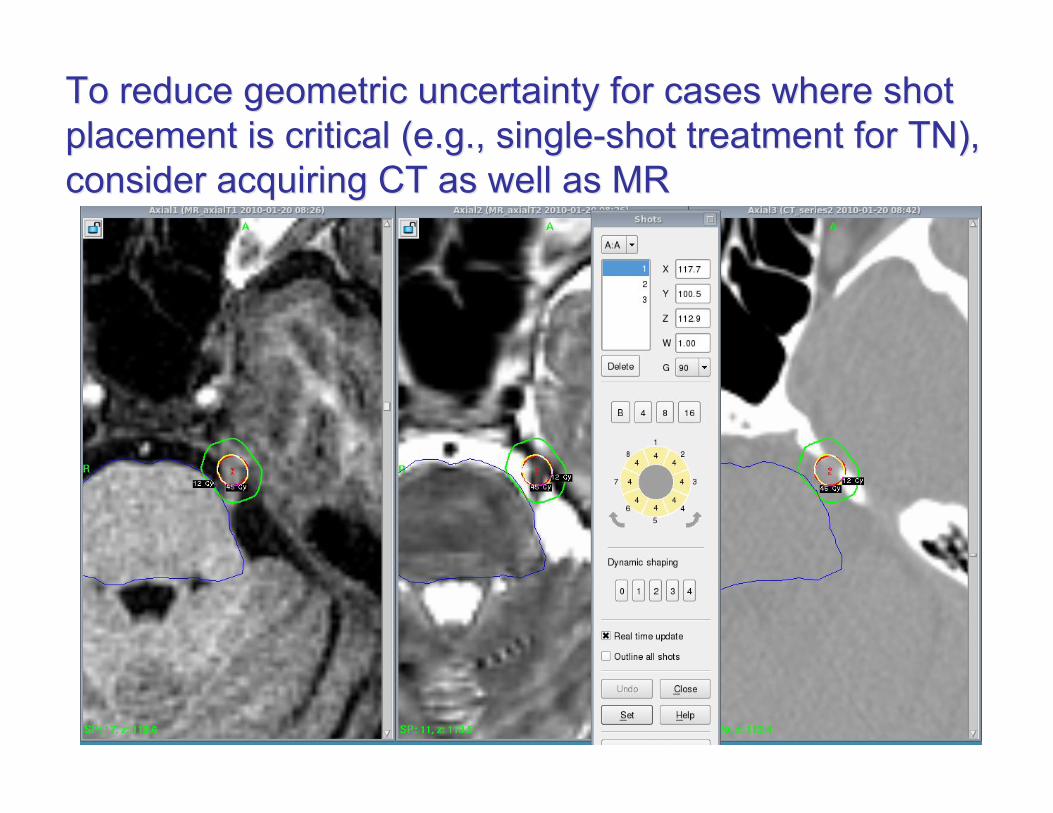
To reduceTo reduce geometric uncertainty for cases where shotgeometric uncertainty for cases where shot
placement is critical (e.g., single-shot treatment for TN),placement is critical (e.g., single-shot treatment for TN),
consider acquiring CT as well as MRconsider acquiring CT as well as MR

33
An Example of a QA PhantomAn Example of a QA Phantom
from CIRSfrom CIRS• Phantom filled with a
proprietary water-based
polymer that images
well on MR and CT.
The skull is made from
an epoxy-based tissue
substitute
• Rods running in
orthogonal directions
inside the phantom are
used to assess
distortions
• Can attach the Leksell
frame to phantom

34
Qualitative Assessment of DistortionsQualitative Assessment of Distortions
in MR Imagesin MR Images
CT MR T1 Fused MR/CT
Can visually assess spatial agreement between
images by examining overlap of fiducials

35
Quantitative Assessment ofQuantitative Assessment of
Distortions in MR ImagesDistortions in MR Images
• Identifycorrespondingpoints on CTand MR
• Use measuringtool in LGP toassessdifferences
Typically, I haveTypically, I have
found RMSfound RMS
differences ofdifferences of
about 0.5 mmabout 0.5 mm

36
Tumor Definition for GK
• T1 MR sequences
• T2 MR useful for benign lesions (e.g.
meningioma and vestibular schwannoma)
• CT useful for skull-base lesions
• Margins are generally not added to the
Gross Tumor Volume

37
Tumor and Critical Structure DefinitionTumor and Critical Structure DefinitionT1-, T2- MR and CT to Visualize Acoustic Tumor and CochleaT1-, T2- MR and CT to Visualize Acoustic Tumor and Cochlea
Cochlea most easily identified on
T2 MR or CT images
All three image sets used to identify tumor within auditory canal
38
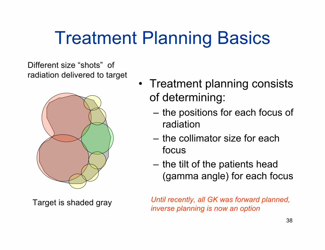
Treatment Planning Basics
• Treatment planning consists
of determining:
– the positions for each focus of
radiation
– the collimator size for each
focus
– the tilt of the patients head
(gamma angle) for each focus
Different size “shots” of
radiation delivered to target
Target is shaded gray Until recently, all GK was forward planned,
inverse planning is now an option

39
GK Treatment Planning Basics
Placement of first shot Placement of second shot

40
A GK shot-placing strategy for GK:A GK shot-placing strategy for GK: large shotslarge shots
placed centrally, smaller shots used to placed centrally, smaller shots used to ““fill infill in”” with with
smaller collimators smaller collimators ““nearnear”” the critical structures the critical structures
Max dose
to pituitary
stalk is 6.5
Gy
Max dose
to optic
structures
is 4 Gy
16mm3 of
brainstem
receives >
12 Gy
Cavernous
sinus
meningioma,
VP = 15 Gy

41
Plan for Small Vestibular Plan for Small Vestibular SchwannomaSchwannoma
Three 4-mm isocenters, each with some sector
blocking so that the cochlea receives ! 4Gy

42
Treatment Plan Evaluation: SomeTreatment Plan Evaluation: Some
Definitions:Definitions:
T
TD
V
VVc
!=
D
TD
V
VVs
!=
Target CoverageTarget Coverage Plan SelectivityPlan Selectivity
VT = Target Volume
D = Prescription dose
VD = Volume receiving dose D
(i.e., the prescription volume)

43
Coverage versus SelectivityCoverage versus Selectivity
• Excellent targetcoverage, poorselectivity
• Excellentselectivity, poortarget coverage
Target
Prescription
isodose
Prescription
IsodoseTarget

44
Conformity IndicesConformity Indices
scCP
!=
*E. Shaw et al., Int. J. Radiat. Oncol. Biol. Phys. 27, 1231-1239 (1993).
**I. Paddick, J. Neurosurg. (Suppl) 93, 219-222 (2000).
!
CS
=V
D
VT
=c
s
Usually " 1, but can be < 1 if
coverage is sub-optimal.
Always # 1
CP = 1 represents perfect conformity
RTOG conformity index*:
Paddick conformity index**:

45
Relationship between Shaw (RTOG)Relationship between Shaw (RTOG)
and and Paddick Paddick Conformity IndicesConformity Indices
• CP is inversely proportional to CS, with proportionalityconstant equal to the square of the target coverage
• CP = 1/CS if the target coverage is 100% (i.e., c = 1)
• In GK SRS we seem to be moving towards using CP
S
2
P
C
cC =

46
To understand the meaning and
implication of conformity index,
consider the dose plans shown
on the next three slides…..

47
Brain Met: VT = 0.15 cc

48
Meningioma: VT = 1.2 cc

49
Meningioma: VT = 5.5 cc

50
Considering the 3 plans shown on the preceding
slides, which of the following statements regarding
their Paddick conformity indices is true?
0%
0%
0%
0%
0%
10
1. CP (sm met) > CP (sm meningioma) > CP (lg mening)
2. CP (sm met) < CP (sm meningioma) < CP (lg mening)
3. CP (sm met) = CP (sm meningioma) > CP (lg mening)
4. CP (sm met) > CP (sm meningioma) = CP (lg mening)
5. CP (sm met) = CP (sm meningioma) = CP (lg mening)

51
Considering the 3 plans shown on the preceding
slides, which of the following statements regarding
their Paddick conformity indices is true?
Answer: 5:
CP (small met) = CP (small meningioma) = CP (large meningioma)
All 3 plans have the same Paddick conformity index.
(Recall that larger values of CP correspond to better conformity
and that CP = 1.0 represents “perfect” conformity.)
References: E. Shaw et al., “RTOG: Radiosurgery quality assurance
guidelines,” Rad. Oncol. Biol. Phys. 27, 1231-39 (1993), and I. Paddick,
“A simple scoring ratio to index the conformity of radiosurgical treatment
plans,” J. Neurosurg. (Suppl) 93, 219-22 (2000).

52
For all 3 plans:
Target Coverage = 1.0
Plan selectivity = 0.58
& CP = 0.58, CS = 1.72
However, the volume of non-target tissue
receiving the prescription dose is:
Small met: 0.1 cc
Small Meningioma: 0.9 cc
Large Meningioma: 3.9 cc

53
Actual Plan for Large Meningioma

54
J.L. Nakamura et al., “Dose conformity of
Gamma Knife radiosurgery and risk factors
for complications,” Int. J. Radiat. Oncol. Biol.
Phys. 51, 1313 – 19, (2001).
• 1181 evaluable lesions treated between 1993and 1998
• Symptomatic radiation toxicity associated withlarger:– Target volume (VT)
– Prescription volumes (VP)
– Larger volume of non-target tissue receiving theprescription dose
• But not with:– Worsening conformity index (VP/VT)

55
The moral of the story…
• Conformity index is simply a ratio of
volumes
• Does not tell us the absolute volume of
normal tissue irradiated
• Larger targets are likely to require better
conformity
– (probably > 0.75 for VT > 3 cm3)*
*Petti et al., Med Phys., 38, 2812-2819, (2011).

56
QA and Radiation Safety for
Gamma Knife
• Large doses delivered in a single fraction
• Imaging, planning and treatment performed in a single day
• Often, multiple patients treated in a single day
• GK team members are often under stress to compete
steps involved for GK treatment
+ distractions including the occasional necessity
to “multi-task”

57
Potential for Human Error
• Types of errors that have occurred
– Left/Right inversion errors
– Delivering the wrong treatment plan to a
patient
– Entering incorrect prescription dose
– For older model GKs, setting coordinates
incorrectly

58
Gamma Knife users must adhere to
recommendations put forth by the
0%
0%
0%
0%
0%
10
1. U.S. Food and Drug Administration
2. U.S. Environmental Protection Agency
3. National Institute of Health
4. U.S. Nuclear Regulatory Commission
5. National Institute of Standards and
Technology
59
Gamma Knife users must adhere to
recommendations put forth by the…
Answer: 4: The US Nuclear Regulatory
Commission*
Unlike other forms of radiosurgery, GK
SRS falls under the jurisdiction of the NRC
(because Co-60 is reactor bi-product material)
*NRC10 CFR Part 35.600 Subpart H, 10 CFR Part 35.600 Subpart H, ““Photon Emitting RemotePhoton Emitting Remote
Afterloader Afterloader Units, Units, Teletherapy Teletherapy Units and Gamma StereotacticUnits and Gamma Stereotactic
Radiosurgery Radiosurgery UnitsUnits”” and and http://www.http://www.nrcnrc..gov/materials/miau/med-use-gov/materials/miau/med-use-
toolkit/perfexion-guidancetoolkit/perfexion-guidance..pdfpdf

60
NRC Requirements for PFXNRC Requirements for PFX
• Training of AUs and AMPs, and documentationof training
• Preparation and use of “written directive”
• Routine QA
Purpose of both the NRC requirements and of many of the
technological developments in hardware and software for
the Gamma Knife over the years has been to reduce the
likelihood of human error
Thank you for your attention!Thank you for your attention!