Gagal Nafas
22
GAGAL NAFAS Generated by Foxit PDF Creator © Foxit Software http://www.foxitsoftware.com For evaluation only.
-
Upload
ryan-asada -
Category
Documents
-
view
49 -
download
1
description
irwan
Transcript of Gagal Nafas

GAGAL NAFAS
Generated by Foxit PDF Creator © Foxit Softwarehttp://www.foxitsoftware.com For evaluation only.

Gagal nafas adalah ketidakmampuan paru-paru untuk memenuhikebutuhan metabolik tubuh.
Klasifikasi:Gagal nafas ada 2 tipe:
1.Hypoxemic Respiratory failure. à PaO2 < 50 torrTerdapat pada pasien acute lung injury dan acute pulmonary edema.Etiologi:
• Mismatch of alveolar ventilation and pulmonary perfusion.• Decrease diffusion of axygen across the alveolocapillary membrane complex akibat interstitiel edema, inflammation dan fibrosis.
• Alveolar hypoventilation• High altitude.
Generated by Foxit PDF Creator © Foxit Softwarehttp://www.foxitsoftware.com For evaluation only.

P (A - a) O2 gradient sering digunakan untuk menentukan apakahhypoxemia disebabkan oleh hypercapnia.
P (A - a) O2 gradient: • < 10 torr in younger adult• < 20 torr in all patients
Bila gradient: • normal, berarti hypoxemia akibat hypercapnia• meningkat, berarti akibat parenchymed changes (V/Q mismatching, etc).
Penatalaksanaan:• Gejala awal: naikkan FiO2
• Penurunan difusi oksigen: diuretic atau causative.
Generated by Foxit PDF Creator © Foxit Softwarehttp://www.foxitsoftware.com For evaluation only.

2.Hypercapnic Respiratory Failure. à PaCO2 > 50 torr.
Terdapat pada pasien severe airflow obstruction, central respiratory failureatau neuromusculare failure.
Etiologi:Gangguan ventilasi: VA = ( VT - VD ) x RR.
A. VT atau RR menurun, akibat:• Airway obstruction ( COPD )• Central respiratory failure• Peripheral respiratory failure:• Neurologic disorders: spinal cord injury, myasthenia gravis, Guillain
Barre syndrome, neuromusculare blocker. • Respiratory muscle: fatique, malnutrition, dystrophy.
Generated by Foxit PDF Creator © Foxit Softwarehttp://www.foxitsoftware.com For evaluation only.

B. VD meningkat, akibat:• Regional airway pressure lebih tinggi dari regional perfusion pressure• Hypovolemia• Cardiac output buruk• Pulmonary emboli.
Penatalaksanaan:Atasi gejala dengan:• Minute ventilation ditingkatkan (MV = TV x RR)• Peak airway pressure diturunkan• Pulmonary blood flow yang turun diatasi.
Note: Setiap peningkatan 1 L O2 pada dewasa akan meningkatkan FiO2 4 %.
Generated by Foxit PDF Creator © Foxit Softwarehttp://www.foxitsoftware.com For evaluation only.
Manifestasi klinis:
Pada umumnya merupakan tanda dan gejala dari hypoxemia, hypercapnia atau keduanya, berupa:
• Perubahan status mental, dari agitasi sampai somnolens• Kerja nafas meningkat: nasal flaring, use of accessory respiratory muscles, intercostal/ suprasternal/supraclavicular retraction, hyperpnea, tachypnea or dyssynchronous breathing patterns or paradoxical.
• Bradypnea• Diaphoresis, tachycardia, hypertension dan gejala lain catecholamine release.
Generated by Foxit PDF Creator © Foxit Softwarehttp://www.foxitsoftware.com For evaluation only.

Laboratorium:Sebagai key test adalah arterial blood gas, untuk menentukan tipe darirespiratory failure yaitu PaO2 dan PaCO2.
Test tambahan adalah: elektrolit, hematokrit, laktat
Analisa gas darah (normal): • pH = 7,35 - 7,45• HCO3
- = 21 - 26 mEq/L• PaO2 = 85 - 95 mmHg• PaCO2 = 35 - 45 mmHg• BE = - 2,5 s/d + 2,5• SaO2 = 93 - 98%
Elektrolit (normal): • K + = 3,5 - 5,5 mEq/L• Na + = 135 - 145 mEq/L• Ca++ = 4,5 - 5,5 mEq/L• Mg++ = 1,9 - 2,5 mg/dL• Cl- = 99 - 105 mEq/L
Generated by Foxit PDF Creator © Foxit Softwarehttp://www.foxitsoftware.com For evaluation only.
KLINIS
Gagal Nafas ada 2 tipe:
1. Hypoxemic respiratory failure.Bila PaO2 < 50 torr.Penyebab: • Kelainan paru-paru: pneumonia, atelektase, ARDS,
intrapulmonal• Kelainan jantung: R to L shunt
Termasuk golongan: failure to oxygenate (gangguan oksigenasi). Terjadilow PaO2
Tindakan: • Increase minute volume• NIPPV (non invasive positive pressure ventilation)• IPPV
Generated by Foxit PDF Creator © Foxit Softwarehttp://www.foxitsoftware.com For evaluation only.

2. Hypercapnic respiratory failure.
Bila PaCO2 > 50 torr.Termasuk golongan failure to ventilate (gangguan ventilasi)Terjadi high PaCO2
Tindakan: • Increase FiO2
• Apply PPV (CPAP)• Increase mean airway pressure
Ada pula golongan failure to protect airway.Pada low GCSTindakan: intubasi oral atau tracheal.
Generated by Foxit PDF Creator © Foxit Softwarehttp://www.foxitsoftware.com For evaluation only.

Gambaran Klinis
1. Kompensasi respirasi:• Tachypnea• Kerja otot-otot nafas tambahan meningkat• Nasal flaring• Retraksi: intercostals, suprasternal, supraclaviculare
2. Peningkatan tonus simpatik:• Tachycardia• Hipertensi• Berkeringat banyak
3. End organ hipoksia:• Perubahan status mental• Bradycardia• hipotensi
4. Desaturasi Hb5. Polycytemia (hipoksemia kronik)
Generated by Foxit PDF Creator © Foxit Softwarehttp://www.foxitsoftware.com For evaluation only.

MEKANISME PATOFISIOLOGIK
Mekanisme dasar terjadinya gagal nafas:1. Shunting2. Dead space ventilation3. Abnormal diffusion4. Alveolar hypoventilation
Failure to oxygenate:1. Diffusion abnormality: • Pulmonary edema
• Pulmonary fibrosis• Interstitial lung disease
2. Normal, V/Q = 13. Dead space ventilation, V/Q > 1: • Pulmonary embolism
• Excessive PEEP4. Shunt, V/Q < 1: • Lung collapse
• Atelectasis• Consolidation
Generated by Foxit PDF Creator © Foxit Softwarehttp://www.foxitsoftware.com For evaluation only.

Shunting (Perfusi tanpa ventilasi).• V/Q mismatch, dimana alveoli tidak terventilasi namun tetap mengalamiperfusi.
• Relatif resisten terhadap terapi oksigen• Tersering pada penyakit kritis• Etiologi:
1. Intracardiac: R to L shunt: Tetrealogy of Fallot, Eisenmebgersyndrome
2. Paru-paru: pneumonia, edema paru, atelektasis, contusion paru
Dead space ventilation.• V/Q > 1 à kebalikan dari V/Q mismatch.• Gas melewati alveoli, tetapi tidak terjadi pertukaran gas, karena alveolitidak mengalami perfusi.
• Bila pasien dapat mengkompensir, pengurangan ventilasi.efektifàPaCO: ↑
• Etiologi:1. Low Cardiac output2. Tekanan intra alveolar ↑à kompresi atau regangan alveolar
Generated by Foxit PDF Creator © Foxit Softwarehttp://www.foxitsoftware.com For evaluation only.

Abnormal diffusion.• Alveolar surface area berkurang: membrane alveolar abnormal, jumlahalveoli menurun
• Etiologi:1. ARDS2. Fibrotic disease
Alveolar hypoventilation.Penyebab:• Kelainan neurologik:
. Pusat nafas: opioid, anesthetic, trauma kapitis
. Saraf cervical: C 3 - 5
. Spinal injury
. N. Phrenicus
. Chest trauma atau pembedahan
. Neuromuscular junction: myasthenia gravis• Kelainan muscular: myopathy
Generated by Foxit PDF Creator © Foxit Softwarehttp://www.foxitsoftware.com For evaluation only.

Generated by Foxit PDF Creator © Foxit Softwarehttp://www.foxitsoftware.com For evaluation only.

SETTING VENTILATOR MEKANIK
1. Volume Control Setting:• Tidal volume: 6 - 10 ml/kg
Pada ARDS: 4 - 6ml/kg• Respiratory rate: 10 - 15 x/men.
Pada COPD: RR harus dilihat agar mendapat base line PaCO2
• Inspiratory time• I : E ratio• FiO2: 100% atau di setting agar PaO2 > 60 mmHg atau SaO2 > 90 %• PEEP: . diberikan agar paru tidak kolaps
. dititrasi dengan pegangan: SaO2 > 88 - 90 %PaO2 > 55 - 70 mmHgFiO2 < 0,6
Tidal volume konstanInspiratory pressure bervariasiInspiratory flow konstanResiko barotraumas
Generated by Foxit PDF Creator © Foxit Softwarehttp://www.foxitsoftware.com For evaluation only.
2. Volume Assist - Control Ventilation. Setting:
• PEEP• RR: 12 - 14 x/men.• TV: 6 ml/kg• Peak flow: 50 L/men. Set triggering flow
Cek BGA setelah 30 - 60 menit.• Bila CO2 tinggià naikan RR (hati-hati auto PEEP), pertimbangkan
reduksi TV jadi 4 - 5 ml/kg• Hipoksia beratà PEEP naikan dan tambahkan Inspiratory pause
(agar mean airway pressure meningkat), tambahkan sedasi danmuscle relaxant, pertimbangkan PC, ARPV dan HFO.Yakinkan peak flow adequate.
Jika pasien sulit mengontrol RR dan jadi tachypneaàdalamkan sedasi, pertimbangkan bilevel ventilation -> amatipressure wave form, nilai flow inspiratoryà lihat compliance paru: • complaice turun (TV ↓) à PS ↑
• compliance membaik à PS ↓ (TV > 6 ml/kg) Bila perlu tambah sedasi.
Generated by Foxit PDF Creator © Foxit Softwarehttp://www.foxitsoftware.com For evaluation only.
3. Pressure Control Setting:• Inspiratory pressure (peak inspiratory pressure: 18 - 35 cmH2O)• Respiratory rate• I: E ratio• FiO2
• PEEP• Trigger of sensitivityTidal volume bervariasiInspiratory pressure konstanInspiratory flow bervariasiResiko hipoventilasi dan pneumothorax
Generated by Foxit PDF Creator © Foxit Softwarehttp://www.foxitsoftware.com For evaluation only.

4. Pressure Assist - Control Ventilation. Setting:• PEEP• RR: 12 - 14 x/men.• Inspiratory pressure untuk mendapatkan TV 6 ml/kg• Inspiratory time: 0,5 - 0,8 detik• Triggering flow
Cek BGA setelah 30 - 60 menit.• Bila CO2 tinggià naikan RR (hati-hati auto PEEP) Jika hipoksemia membaik, turunkan Inspiratory pressure
• Hipoksemia memburuk: PEEP naik dan Inspiratory time naik (1 - 1,5 detik), tambahkan sedasi dan muscle relaxant Alternatif mode: ARPV, HFO atau prone position
• Bila hiperkarbiaà turunkan TV Turunkan inspiratory time, bila hipoksemia membaik
Bila nafas spontan dan cedera paru ringan à PSVBila tidak dapat mengontrol RR dan tachypnea à naikan dosis obat dan pertimbangkan Bilevel ventilation
Generated by Foxit PDF Creator © Foxit Softwarehttp://www.foxitsoftware.com For evaluation only.

LANGKAH PENDEKATAN ASAM-BASA
Langkah I : Tetapkan gangguan yang paling nampak
Gangguan pH PCO2 HCO3
Asidosis metabolik turun naik (sekunder ) turun ( primer)Alkalosis metabolik naik naik ( sekunder) naik ( primer)Asidosis respiratorik turun naik ( primer) naik ( sekunder)Alkalosis respiratorik naik naik ( primer ) turun ( sekunder)
Generated by Foxit PDF Creator © Foxit Softwarehttp://www.foxitsoftware.com For evaluation only.
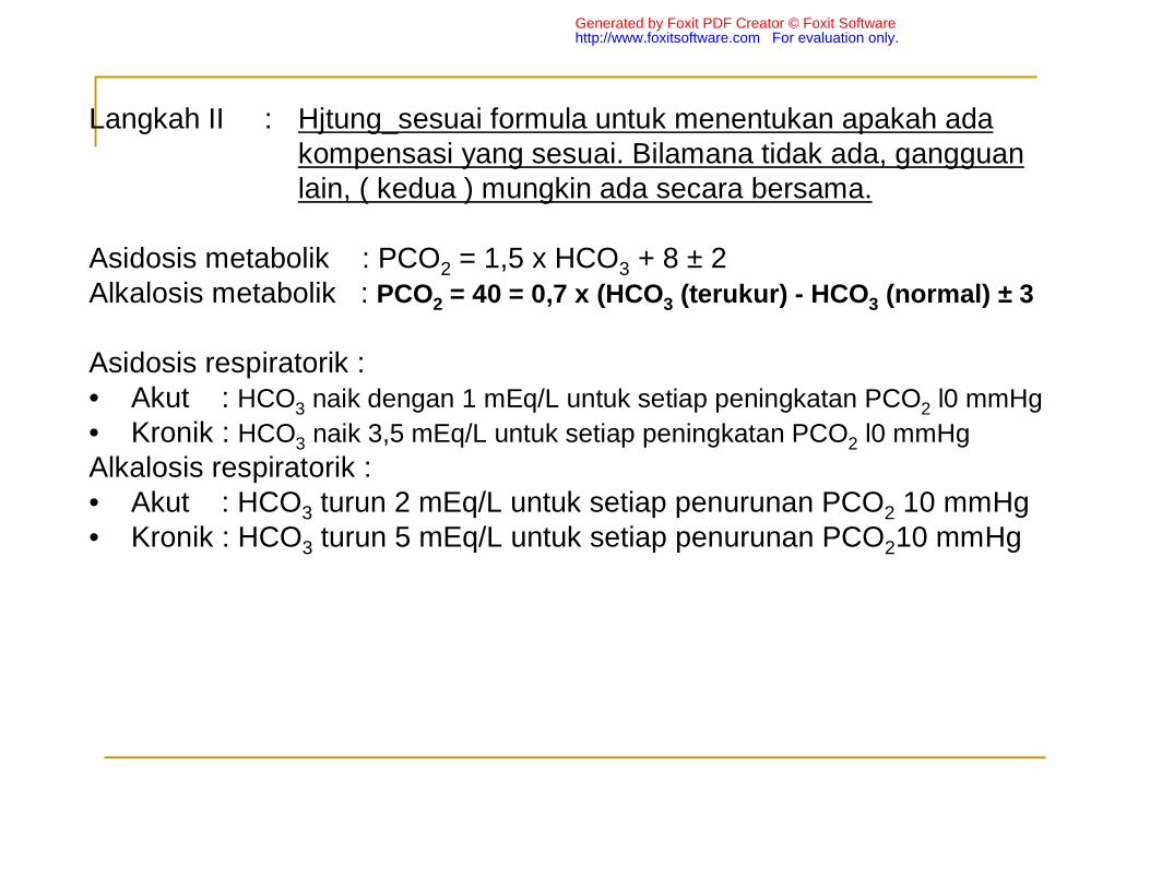
Langkah II : Hjtung_sesuai formula untuk menentukan apakah adakompensasi yang sesuai. Bilamana tidak ada, gangguanlain, ( kedua ) mungkin ada secara bersama.
Asidosis metabolik : PCO2 = 1,5 x HCO3 + 8 ± 2Alkalosis metabolik : PCO2 = 40 = 0,7 x (HCO3 (terukur) - HCO3 (normal) ± 3
Asidosis respiratorik :• Akut : HCO3 naik dengan 1 mEq/L untuk setiap peningkatan PCO2 l0 mmHg
• Kronik : HCO3 naik 3,5 mEq/L untuk setiap peningkatan PCO2 l0 mmHg
Alkalosis respiratorik :• Akut : HCO3 turun 2 mEq/L untuk setiap penurunan PCO2 10 mmHg• Kronik : HCO3 turun 5 mEq/L untuk setiap penurunan PCO210 mmHg
Generated by Foxit PDF Creator © Foxit Softwarehttp://www.foxitsoftware.com For evaluation only.
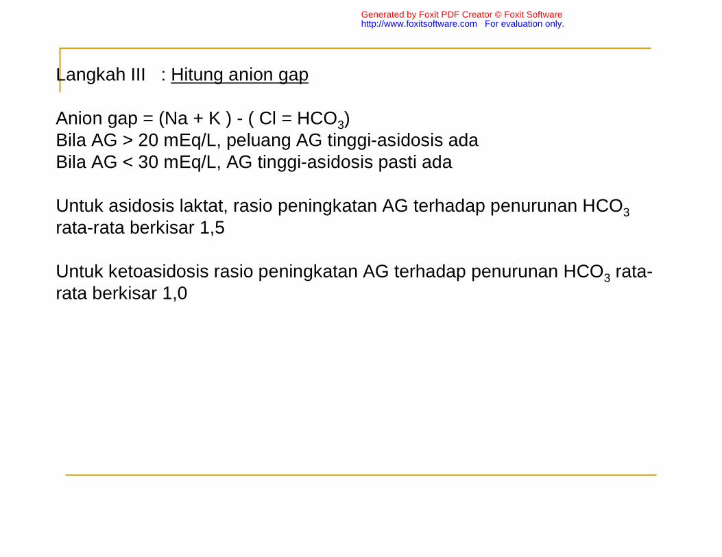
Langkah III : Hitung anion gap
Anion gap = (Na + K ) - ( Cl = HCO3)Bila AG > 20 mEq/L, peluang AG tinggi-asidosis adaBila AG < 30 mEq/L, AG tinggi-asidosis pasti ada
Untuk asidosis laktat, rasio peningkatan AG terhadap penurunan HCO3
rata-rata berkisar 1,5
Untuk ketoasidosis rasio peningkatan AG terhadap penurunan HCO3 rata-rata berkisar 1,0
Generated by Foxit PDF Creator © Foxit Softwarehttp://www.foxitsoftware.com For evaluation only.

PENYEBAB ASIDOSIS METABOLIK
1. AG meningkat• Ketoasidosis diabetik• Asidosis laktat• Ketoasidosis uremik• Ketoasidosis alkoholik• Intoksikasi
2. AG normal• Gagal ginjal ringan-sedang• Diare akut• Asidosis delusional• Asidosis tubulus ginjal ( Tipe I, II, III)• Terapi ketoasidosis diabetik.
Generated by Foxit PDF Creator © Foxit Softwarehttp://www.foxitsoftware.com For evaluation only.