Proteomic Biomarkers as Indicator of Aquatic Pollution in ...
Upload
pasteurtunisCategory
view
240download
1
Functional analysis of proteomic biomarkers and targeting glioblastoma stem cells
Radovan Komel1, Ivana Jovčevska1, Neja Zupanec1, Mirjana Liovič1, Filip Mihalič1,
Daniela Cesselli2 , Clara Limbaeck Stokin3, Serge Muyldermans4, Uroš Smrdel5, Andrej Vranič6, Boštjan Matos6
1Medical Centre for Molecular Biology Faculty of Medicine, University of Ljubljana, Slovenia
2Azienda Ospedaliero-Universitaria di Udine (AOUD),
S. Maria Della Misericordia, Udine, Italy 3Institute of Pathology, Faculty of Medicine, University of Ljubljana, Slovenia
4Department of Applied Biological Sciences and Engineering, Vrije Universiteit Brussel, Brussel, Belgium
5Institute of Oncology, Ljubljana, Slovenia 6University Clinical Centre, Neurosurgery Clinic, Ljubljana, Slovenia
Cours International Medicine Genomique
October 17-21, 2016 Inst. Pasteur, Tunis

What is a brain tumor?
A brain tumor is an abnormal growth of tissue in the brain or central spine
that can disrupt normal brain function.
Benign tumors Malignant tumors
• The least agressive type.
• Do not contain cancer cells.
• Slowly growing.
• Clear borders.
• Do not spread into other
tissue.
• They contain cancer cells.
• They do not have clear borders.
• They grow rapidly.
• They invade surrounding brain
tissue.
Primary Metastatic
• They start in cells of the brain.
• They may spread to other parts
of the brain or to the spine.
• They spread rarely to other
organs.
• They begin in another part of the
body and spread to the brain.
• They are more common than
primary brain tumors.

SLOVENIA
150 of malignant brain tumors / year
60 glioblastoma / year
340 mio inhabitants
2 mio inhabitants

The most common primary brain tumors
All brain tumors Malignant brain
tumors
Meningiomas 34%
Gliomas 30% 70-80%
Pituitary adenomas 13%
Nerve sheath tumors 9%
Gliomas are the most common primary intracranial
tumor, representing more than 70% of malignant brain
tumors. Although relatively rare, they cause significant
mortality and morbidity. Glioblastoma multiforme (GBM)
is the most common and deadliest of malignant primary
brain tumors in adults and is one of a group of tumors
referred to as gliomas. The incidence, or the number of
new diagnoses made annually is 2 to 3 per 100,000
people in the United States and Europe. GBM accounts
for 12% to 15% of all intracranial tumors and 50% to
60% of all gliomas.

There are three types of normal glial cells that can produce tumors. An
astrocyte will produce astrocytomas (including glioblastomas), an
oligodendrocyte will produce oligodendrogliomas, and ependymomas come
from ependymal cells. Tumors that display a mixture of these different cells are
called mixed gliomas.
“Glioma” is a general term used to describe any tumor that arises from the
supportive (“gluey”) tissue of the brain. This tissue, called “glia,” helps to keep
the neurons in place and functioning well.
www.ABTA.org, 2013

The normal differentiation process originates three main types of cells in the mature
CNS: neurons, glial cells (oligodendrocytes, astrocytes, ...), and ependymal cells (not
shown). Malignant transformation to glioma occurs through didefferentiation process
either from glial cells or from glia progenitor cells and/or from neuronal stem cells.
Celine S. et al., INTECH, 2013
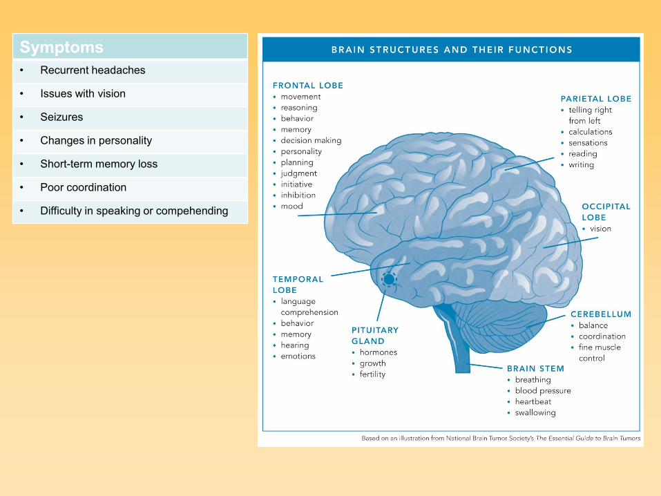
Symptoms
• Recurrent headaches
• Issues with vision
• Seizures
• Changes in personality
• Short-term memory loss
• Poor coordination
• Difficulty in speaking or compehending

Zhang P. et al, TIN, 2009
Cancer stem cells within a brain tumor can arise from normal stem cells or
from neural progenitor cells that harbor mutations. Cancer stem cells within
glioblastomas can be particularly resistant to cytotoxic therapies, and therefore
are a source of cells for cancer recurrence. To meet the challenge of a cure for
glioblastoma, new therapies that selectively suppress proliferation and/or kill
cancer stem cells must be found.
Characteristics and fates of normal neural stem cells and cancer stem cells.


Chromosomal region Type of alteration Candidate gliomas genes
1p36.31-pter Gains and deletions Not known
1p36.22-p36.31 Gains and deletions Not known
1p34.2-p36.1 Gains and deletions Not known
1q32 Gains Receptor interacting protein kinase (RIPK5)
Mouse double munite 4 (MDM4)
Phosphatidylinositol-4-phosphate-3-kinase
Catalytic subunit Type 2 beta (PK3C2B)
And others ...
4q Deletions NMA-related kinase (NEK1), NIMA
7p11.2-p12 Amplifications/gains Epidermal growth factor receptor (EGFR)
9p21-p24 Deletions Cyclin-dependent kinase inhibitor 2A (CDKN2)
10q23 Deletions Phosphatase and tensin homolog (PTEN)
10q25-q26 Deletions O-6-methylguanine-DNA-methyltransferase (MGMT)
11p Deletions Cyclin-dependent kinase inhibitor 1C (CDKN1C)
Related RAS viral oncogene homolog 2 (RRAS2)
12q13.3-q15 Amplifications Mouse double minute 2 homolog (MDM2)
Cyclin-dependent kinase 4 (CDK4)
And others ...
13p11-p13/13q14-q34 Loss Retinoblastoma 1 (RB1)
19q13 Loss Glioma tumor suppressor candidate region gene 1
GLTSCR2
DNA ligase I ATP dependent (LIG1)
Cytohexin 2 (CYTH2)
And many others ...
22q11.21-q12.2 Loss 28 genes incl. Integrase interactor 1 (INI1)
22q13.1-q13.3 Loss Not known
T.Mesti and J.Ocvirk: Radiol. & Oncol, 2016
The chromosomal alterations mostly observed in gliomas

Syndrome Gene name Chromosomal
location
Neurofibromatosis 1 Neurofibromin 1 (NF1) 17q11
Neurofibromatosis 2 Neurofibromin 2 (NF2) 22q12
Tuberous sclerosis Tuberous sclerosis 1 (TSC1)
Tuberous sclerosis 2 (TSC2)
9q34
16p13
Retinoblastoma Retinoblastoma 1 (RB1) 13q14
Li-Fraumeni
syndrome
Tumor suppressor p53 (TP53) 17p13
Turcot's syndrome
Multiple harmartoma
Adenomatous polyposis coli (APC)
DNA mismatch repair genes:
Recombinant human MutL homolog-1 (hMLH1)
MutS homolog 2 (hMSH2)
Mismatch repair endonuclease (PMS2)
Phosphate and tensin homolog (PTEN)
5q21
3p21.3
2p22-21
7p22
10q23.3
Inherited mutations present in patients with malignant gliomas
Differentiared astrocytes or precursor cells
p53 mutation (>65 percent) EGFR amplification (40 percent)
PDGF-A, PDGFR-alpha overexpression (60 %) overexpression (60 percent)
Low grade astrocytoma MDM2 amplification (<10 percent)
overexpression (50 percent)
LOH 19q (50 percent)
RB alteration (25 percent)
p16 deletion (30-40 percent)
Anaplastic astrocytoma
LOH 10p and 10q
LOH 10q PTEN mutation (30 percent)
PTEN mutation (5 percent)
DOC loss of expression (50 percent)
PDGFR-alpha amplification (<10 percent) RB alteration
Secondary glioblastoma Primary glioblastoma de novo
Development of
primary and
secondary
glioblastoma
T.Mesti and J.Ocvirk:
Radiol. & Oncol, 2016

Marker Protein type Function Prognostic
significance
CD 133 5-TM protein Only CD133+ cells may produce a
tumor. +++
- -
Nestin Filament marker in
neuronal progenitor
cells
Neuronal development /
dedifferentiation +++++
+/-
- -
CD 133 /nestin +
Podoplanin Mucin-type TMP Motility & invasion
spheroid formation ++
CD 15 Differentiation antigen Impl. of CD+ GB cells may produce
new tumors. + / -
A2B5 Surface glycoside A2B5 cells have CSC properties + / -
Musashi-1 Neuronal RNA-binding
protein
Neurosphere formation – CSC
properties ++ -
+/-
BM/1 Polycom-group protein Tumor suppressor pathway
regulator +/-
-
SOX2 Sox-family transcription
factor
Normal pluripotent cell development -
Id1 DNA-binding protein
inhibitor
Cell growth, senescence, and
differentiation / Proliferation, neo-
angiogenesis, invasion
++
Oct-4 Octamer-binding
transcription factor 4
Regulator of ESC self-renewal and
differentiation +
Dalhrot et al.: Int. J. Clin. Ex. Pathol., 2013
Review of twenty-seven independent studies on putative CSC markers in gliomas
• Overexpression
• Prognosis
• Prediction
• Survival

50 % 50 %
VHH:
• single domain, small size (15 kDa)
• high expression levels
• soluble and stable
• easy to handle compared to scFv
CAMELIDAE (camels, llamas) heavy chain ANTIBODIES

scFv VHH
V G L W
37 44 45 47
FY
E R VGLW
VL
VH
Covering of the hydrophobic patch by a long CDR3 allows soluble expression of VHH domains in the absence of VL counterpart.

Structural representation
Human VH camelid VHH
CDR1 CDR3
CDR2

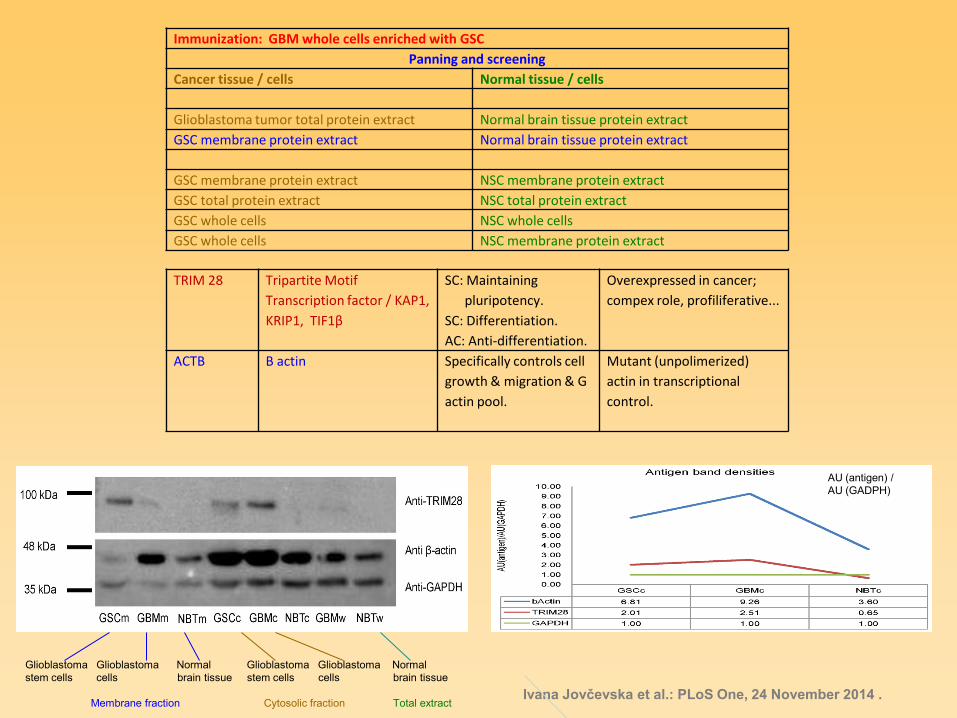
Immunization: GBM whole cells enriched with GSC
Panning and screening
Cancer tissue / cells Normal tissue / cells
Glioblastoma tumor total protein extract Normal brain tissue protein extract
GSC membrane protein extract Normal brain tissue protein extract
GSC membrane protein extract NSC membrane protein extract
GSC total protein extract NSC total protein extract
GSC whole cells NSC whole cells
GSC whole cells NSC membrane protein extract
Ivana Jovčevska et al.: PLoS One, 24 November 2014 .
TRIM 28 Tripartite Motif
Transcription factor / KAP1,
KRIP1, TIF1β
SC: Maintaining
pluripotency.
SC: Differentiation.
AC: Anti-differentiation.
Overexpressed in cancer;
compex role, profiliferative...
ACTB Β actin Specifically controls cell
growth & migration & G
actin pool.
Mutant (unpolimerized)
actin in transcriptional
control.
AU (antigen) /
AU (GADPH)
Glioblastoma Glioblastoma Normal Glioblastoma Glioblastoma Normal
stem cells cells brain tissue stem cells cells brain tissue
Membrane fraction Cytosolic fraction Total extract

Nanobodies (Nbs) and the corresponding antigens obtained atfer several rounds of
phage-display VHH-library panning followed by MS analysis
Nanobody Antigen Name
Nb141 ACTB Beta actin
Nb237 TRIM28 Tripartite motif-containing 28 (TRIM28), also known as
transcriptional intermediary factor 1β (TIF1β) and KAP1
(KRAB-associated protein-1)
Nb10 NUCL Nucleolin
Nb79
VIM
Vimentin
Nb179
NAP1L1
Nucleosome assembly protein 1-like 1
Nb314
DPYSL2/CRMP1
MTHFD1
Dihydropyrimidinase-related protein 2/Collapsin response
mediator protein 1
Methylenetetrahydrofolate dehydrogenase 1
Nb394 DPYSL2/CRMP1 Dihydropyrimidinase-related protein 2/Collapsin response
mediator protein 1
Nb225
Nb206
TufM Tu translation elongation factor, mitochondrial
Nb395 ALYREF THO complex, subunit 4
In ed. p.: I. Jovchevska et al., 2016

Starting QVQL Ending TVSS
Unique CDR3s: binding different antigens
GLEW of Nb10: VH germline origin during V-D-J recombination
R50: VHH origin

Nanobody:Antigen binding
• Nb141:β-actin
GSC GBM
• Nb237:TRIM28 GSC GBM
* * Y Y * * * * Y Y * *
Y – staining with commercial antibodies after
incubation with Nb141 (anti-ACTB) Y – staining with commercial antibodies after
incubation with Nb237 (anti-TRIM28)
I. Jovčevska et al.: PLoS One, 2014


TufM:
Tu translation elongation factor,
mitochondrial
Mitochondrial translational elongation.
Reported to be involved in exosome formation and as a component
of natural killer cells.
A nuclear gene encoded protein, synthesized in
cytoplasm and imported to mitochondria.
Reported down-regulated in lung cancer cells.
Reported unfavorable prognostic indicator in CRC.
Reported elevated expression in GC.

HaCat Anti K14 stained (red) / DAPI stained (blue) U251 DAPI stained (blue)
Nb 206 – antiTufM
FITC-Nb 206 (green): sub-membrane localisation + close
to nuclei of dividing cells.
FITC-Nb 206 (green): centrosomal localisation in dividing cells.

Nb206, model from 4WGV
Arg103 Asp260
EF-TU, 1D2E
Nano body - Divalent ion transporter, 4WGV
In ed. p.: J. Stojan et al., 2016

Vimentin Major cytoskeletal component of mesenchymal cells.
Used as a marker of :
• mesenchymally-derived cells;
• cells undergoing an epithelial-to-mesenchymal
transition (EMT) during both normal development
and metastatic progression.
Formation of lamellipodia and invadipodia during cell migration and invasion.
Three studies already reported VIM over-expression in GBM.
Reported involved in different forms of cancers.
Reported involved in glioma metastatic abilities.
Protein expression data for ALYREF, CRMP1 and VIM.
Loading control, GAPDH, is included to show equal protein amount among samples.

Nucleolin An eukaryotic nucleolar phosphoprotein.
Involved in the synthesis and maturation of ribosomes.
Located mainly in dense fibrillar regions of the nucleolus.
Two NUCL types reported in gliomas: cytoplasmic and surface.
Centrosomal nucleolin is required for microtubule network organization.
Surface NUCL reported dependent on association with actin cytoskeleton.
Reported as regulator of glioma cell migration.
Reported that surface nucleolin increase with the malignancy grade of glioma.

DPYSL2 / CRMP2:
Dihydropyrimidinase-related protein 2/
Collapsin response mediator protein 1
Expressed during neuronal development, and rarely in adult brain.
Reported under-expressed in GBM.
Reported overexpressed in CRC and GC.

NAP1L1:
Nucleosome assembly protein 1-like 1 A member of the nucleosome assembly protein (NAP) family.
Locates within the nucleus of dividing cells.
Involved in cell cycle progression, transciptional regulation
and modification of chromatin structure.
Posible role in modulating chromatin formation.
Contribute to the regulation of cell proliferation.
In dividing cells influences expression of pro-proliferative genes.
Reported over-expressed in pancreatic neuroendocrine neoplasm
metastases.
Reported involved in lymphoblastic leukemia, ovarian cancer,
breast cancer, hepatoblastoma, CRC and lung cancer.

MTHFD1: Methylenetetrahydrofolate dehydrogenase 1
A protein with three distinct enzymatic activities in the interconversion
of 1-carbon derivatives of tetrahydrofolate, substrates for
methionine, thymidulate and de novo purine synthesis.
The folate metabolic system MTHFD1 is up-regulated in cancer
and is involved in rapid cancer cell proliferation.
Reported role of folate metabolism in the development of primary brain
tumors (meningioma and glioma):
genotypes associated with increased 5,10 methylenetetrahydrofolate
levels associated with elevated risk.

ALYREF:
THO complex, subunit 4
Nuclear protein which functions as a molecular chaperone.
Involved in transport of mature mRNA out of the nucleus.
Probably regulates dimerization, DNA binding, and transcriptional
activity of basic region-leucine zipper (bZIP) proteins.
The protein is a part of NUP107 complex which has been reported
altered in 19% of GBMs.

Correlations among the proteins studied with OncoFinder.
Blue color: proteins investigated in this study. Black arrows: interactions through intermediate compounds.
Grey color: other proteins involved in the interaction network. Blue arrows: direct activations a protein by the preceding protein.
Interaction map of top differential proteins identified by Nanobodies.
In ed. p.: I. Jovchevska et al., 2016

Histograms of area under curve (AUC) values
x-axis: AUC
y-axis: frequency
Red bars: genes of interest
Grey bars: overall distribution
A: Histogram of gene distribution analyzed
as cancer vs. normal.
It suggests DPYSL2 as a great,
and NUCL, TRIM28, VIM and NAP1L as
good markers for distinguishing cancer from
normal samples.
B: Histogram of gene distribution analyzed
as GBM vs. LGG.
NAP1L1, NUCL, CRMP1, ACTB and VIM
are pointed out as genes that can be used
as markers to distinguish between tumor
classes.
In ed. p.: I. Jovchevska et al., 2016

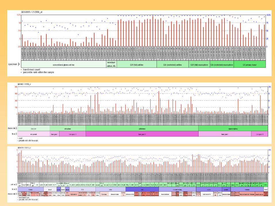
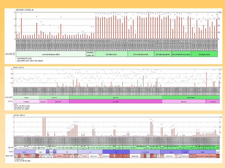
Genemania network

Number of surviving cells relative to control after 24 hours (Filip Mihalic, Mirjana Liovic, 2015).
0
0,5
1
1,5
2
untreated HaCat
α-β actin 10 μg/ml
α-β actin 100 μg/ml
α-206 10 μg/ml
α-206 100 μg/ml
α-Trim 28 10 μg/ml
α-Trim 28 100 μg/ml
HaCat
0
0,5
1
1,5
2
untreated TR146
α-β actin 10 μg/ml
α-β actin 100 μg/ml
α-206 10 μg/ml
α-206 100 μg/ml
α-Trim 28 10 μg/ml
α-Trim 28 100 μg/ml
TR146
0
0,5
1
1,5
2
untreated U251
α-β actin 10 μg/ml
α-β actin 100 μg/ml
α-206 10 μg/ml
α-206 100 μg/ml
α-Trim 28 10 μg/ml
α-Trim 28 100 μg/ml
U251
a-TRIM28/a-beta_actin nanoparticles have a greater effect on
glioblastoma cells

Wst-1 proliferation test on glioblastoma cell lines
Nb 206: anti-TufM
Nb 237: anti-TRIM28
Neja Zupanec et al., 2015
Wst-1 proliferation test on glioblastoma stem
cells (NCH 644 / NCH 421K) vs. neural stem
cells (NSC)


Archeosomes


https://youtu.be/P18fSQuCjNM?t=14
https://www.youtube.com/watch?v=P18fSQuCjNM
GPMV – Giant Plasma Membrane Vesicle formation

HACAT
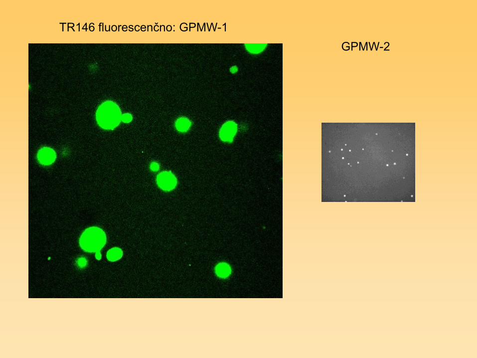
TR146 fluorescenčno: GPMW-1
GPMW-2

In ed. p.: M.Liovic et al., 2016


ACKNOWLEDGMENTS:
Applied Biological
Sciences & Engineering,
Vrije Universiteit Brussel,
Brussel, Belgium
Serge Muyldermans
Medical Centre for
Molecular Biology,
MF UL
Ivana Jovčevska
Neja Zupanec
Mirjana Liović
Nina Kočevar
Damjana Kastelic
Helena Klavžar
Radovan KOMEL
et al.
AOUD
Udine, Italy
Daniela Cesselli
Institute for Pathology,
Faculty of Medicine UL
Clara Limbaeck Stokin
DKFZ
Heidelberg, Germany
Damjana Kastelic
•Slovenian Research Agency
• Interreg GLIOMA
• Ad-Futura Science foundation
• VUB, Brussel
• Babraham, Cambridge, U.K.
• CNRS, Gif-sur-Yvette, France
NIB
Ljubljana
Tamara Lah Turnšek
Neža Podergajs
Martina Mršnik
Elettra Synchrotron
Trieste, Italy
Paola Storici