From the origin of NASH to the future of metabolic fatty ...
40
Geier et al. Manuscript ID gutjnl-2020-323202 1 Recent advances in basic science From the origin of NASH to the future of metabolic fatty liver disease Andreas Geier 1 , Dina Tiniakos 2 Helmut Denk 3 , Michael Trauner 4 1. Division of Hepatology, Department of Internal Medicine II, University Hospital Würzburg, Würzburg, Germany 2. Department of Pathology, Aretaieion Hospital, Medical School, National & Kapodistrian University of Athens, Athens, Greece & Translational & Clinical Research Institute; Faculty of Medical Sciences, Newcastle University, Newcastle upon Tyne, United Kingdom 3. Institute of Pathology, Medical University of Graz, Graz, Austria 4. Division of Gastroenterology and Hepatology, Department of Internal Medicine III, Medical University of Vienna, Vienna, Austria Corresponding author: Andreas Geier, M.D. Professor of Internal Medicine and Hepatology Division of Hepatology University Hospital Würzburg Oberdürrbacherstrasse 6 D-97080 Würzburg Germany Tel.: +49 9 31 2 01 - 4 01 70 Fax: +49 9 31 2 01 – 64 01 70 E-Mail: [email protected]; [email protected] Word count: 5000
Transcript of From the origin of NASH to the future of metabolic fatty ...

Geier et al. Manuscript ID gutjnl-2020-323202
1
Recent advances in basic science
From the origin of NASH to the future of metabolic
fatty liver disease
Andreas Geier1, Dina Tiniakos2 Helmut Denk3, Michael Trauner4
1. Division of Hepatology, Department of Internal Medicine II, University Hospital
Würzburg, Würzburg, Germany
2. Department of Pathology, Aretaieion Hospital, Medical School, National &
Kapodistrian University of Athens, Athens, Greece & Translational & Clinical
Research Institute; Faculty of Medical Sciences, Newcastle University, Newcastle
upon Tyne, United Kingdom
3. Institute of Pathology, Medical University of Graz, Graz, Austria
4. Division of Gastroenterology and Hepatology, Department of Internal Medicine III,
Medical University of Vienna, Vienna, Austria
Corresponding author:
Andreas Geier, M.D.
Professor of Internal Medicine and Hepatology
Division of Hepatology
University Hospital Würzburg
Oberdürrbacherstrasse 6
D-97080 Würzburg
Germany
Tel.: +49 9 31 2 01 - 4 01 70
Fax: +49 9 31 2 01 – 64 01 70
E-Mail: [email protected]; [email protected]
Word count: 5000

Geier et al. Manuscript ID gutjnl-2020-323202
2
Figures: 5
References: 178
Key words: NASH, fatty liver, obesity, fibrosis, molecular pathology, non-
invasive testing, genetics.
Abbreviations:
Alcohol-related liver disease (ALD), Enhanced Liver Fibrosis (ELF), Fatty Liver
Inhibition of Progression (FLIP), fibrosis-4 (FIB-4), glucokinase regulatory protein gene
(GCKR), high fructose corn syrup (HFCS), Mallory-Denk bodies (MDBs), metabolic
dysfunction-associated Fatty Liver Disease (MAFLD), NAFLD activity score (NAS),
NASH Clinical Research Network (NASH CRN), nonalcoholic fatty liver, bland
steatosis (NAFL), nonalcoholic fatty liver disease (NAFLD), nonalcoholic
steatohepatitis (NASH), non-invasive test (NIT), patatin-like phospholipase domain-
containing 3 (PNPLA3), single nucleotide polymorphism (SNP), Steatosis, Activity,
Fibrosis scoring system (SAF), transmembrane 6 superfamily member 2 (TM6SF2)
Author contribution:
Concept and supervision: AG, MT; acquisition of data: AG, DT, HD, MT; analysis and
interpretation of data: AG, DT, HD, MT; drafting of the manuscript: AG, MT; critical
revision of the manuscript for important intellectual content: AG, DT, HD, MT; obtained
funding: AG, DT, MT; material support: AG, DT, HD.
Funding:
This review has been conducted as part of the evidence synthesis efforts in the
LITMUS (Liver Investigation: Testing Marker Utility in Steatohepatitis) study. The
LITMUS study is funded by the Innovative Medicines Initiative (IMI2) Program of the
European Union (Grant Agreement 777377) The funder and the authors’ institutions
had no role in the development of this review.
Conflict of Interest:
AG has received grants from Intercept, Novartis, Exalenz, Falk Pharma and Kibion and
personal fees from Intercept, Novartis, Gilead, Pfizer, Falk Foundation, Bayer, MSD,
BMS, Ipsen, Sanofi-Aventis, CSL Behring, Sequana, Merz, Abbvie and Alexion.

Geier et al. Manuscript ID gutjnl-2020-323202
3
DT reports consultation fees from Intercept, Allergan, Cirius Therapeutics, Alimentiv,
Clinnovate Health UK and an educational grant from Histoindex Pte.
MT has received research grants from Albireo, Cymabay, Falk, Gilead, Intercept, MSD
and Takeda and travel grants from Abbvie, Falk, Gilead and Intercept. He further has
advised for Albireo, BiomX, Boehringer Ingelheim, Falk Pharma, Genfit, Gilead,
Intercept, Jannsen, MSD, Novartis, Phenex, Regulus and Shire and has served as
speaker for Falk Foundation, Gilead, Intercept and MSD. He is also co-inventor of
patents on the medical use of NorUDCA filed by the Medical Universities of Graz and
Vienna.

Geier et al. Manuscript ID gutjnl-2020-323202
4
Abstract / Key messages
Nonalcoholic fatty liver disease (NAFLD) has become the most common cause of
chronic liver disease worldwide. Understanding the pathological and molecular
hallmarks from its first description to definitions of disease entities, classifications and
molecular phenotypes is crucial for both appropriate clinical management and research
in this complex disease. We provide an overview through almost two hundred years of
clinical research from the beginnings as a nebulous disease entity of unknown origin
in the 19th century to the most frequent and vigorously investigated liver disease today.
The clinical discrimination between alcohol-related liver disease and NAFLD was
uncommon until the 1950s and likely contributed to the late acceptance of NAFLD as
a metabolic disease entity for long time. Although the term “fatty liver hepatitis” first
appeared in 1962, it was in 1980 that the term “nonalcoholic steatohepatitis” (NASH)
was coined and the histopathological hallmarks that are still valid today were defined.
The 2005 NASH CRN scoring was the first globally accepted grading and staging
system for the full spectrum of NAFLD and is still used to semiquantify main histological
features. In 2021, liver biopsy remains the only diagnostic procedure that can reliably
assess the presence of NASH and early fibrosis but increasing efforts are made
towards non-invasive testing and molecular classification of NAFLD subtypes.
Geier et al. Manuscript ID gutjnl-2020-323202
5
Bullet points
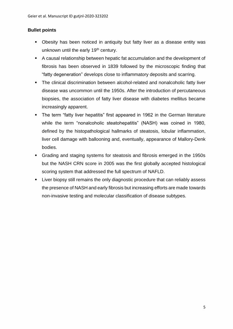
▪ Obesity has been noticed in antiquity but fatty liver as a disease entity was
unknown until the early 19th century.
▪ A causal relationship between hepatic fat accumulation and the development of
fibrosis has been observed in 1839 followed by the microscopic finding that
“fatty degeneration” develops close to inflammatory deposits and scarring.
▪ The clinical discrimination between alcohol-related and nonalcoholic fatty liver
disease was uncommon until the 1950s. After the introduction of percutaneous
biopsies, the association of fatty liver disease with diabetes mellitus became
increasingly apparent.
▪ The term “fatty liver hepatitis” first appeared in 1962 in the German literature
while the term “nonalcoholic steatohepatitis” (NASH) was coined in 1980,
defined by the histopathological hallmarks of steatosis, lobular inflammation,
liver cell damage with ballooning and, eventually, appearance of Mallory-Denk
bodies.
▪ Grading and staging systems for steatosis and fibrosis emerged in the 1950s
but the NASH CRN score in 2005 was the first globally accepted histological
scoring system that addressed the full spectrum of NAFLD.
▪ Liver biopsy still remains the only diagnostic procedure that can reliably assess
the presence of NASH and early fibrosis but increasing efforts are made towards
non-invasive testing and molecular classification of disease subtypes.

Geier et al. Manuscript ID gutjnl-2020-323202
6
Introduction
Nonalcoholic fatty liver disease (NAFLD) comprises a wide spectrum of liver damage,
ranging from nonalcoholic fatty liver (simple steatosis; NAFL), to nonalcoholic
steatohepatitis (NASH) with inflammation and hepatocyte injury to advanced fibrosis
and cirrhosis.[1] Although NAFLD histopathology resembles that of alcohol-related
liver disease (ALD), the clinical background is different. NAFLD is regarded as one
component of the metabolic syndrome, including obesity, insulin resistance or type 2
diabetes mellitus, hypertension, and dyslipidemia. A “multiple parallel hits” hypothesis
describes the pathogenesis of this complex disease from simple steatosis to
steatohepatitis.[2, 3] NAFLD affects 10–24% of the general population worldwide and
is the most common cause of chronic liver disease.[4]
The worldwide number of obese patients tripled from 1975 to 2018.[5] The introduction
of industrialized food rich in high fructose corn syrup (HFCS) in the late 1960s
coincided with the increase in the incidence of obesity, diabetes and metabolic
syndrome. Bray and colleagues (2004) first hypothesized that HFCS may be directly
associated with obesity.[6] However, carefully controlled long-term studies are still
required to substantiate the aetiological role of HFCS in metabolic syndrome-related
diseases, such as NAFLD.[7] Given the sheer frequency of patients with obesity,
metabolic syndrome and NAFLD, it is remarkable that this disease entity has been
overlooked by most clinicians for a long time, except for few pioneers, mostly
pathologists, who first described the clinical and pathological characteristics more than
150 years ago. Even as recently as 30 years ago, NASH was familiar only to a small
group of experts and the term did not exist in the medical vocabulary until 1980. Today,
the lack of awareness of NAFLD by the clinical community has contributed to a lack of
NASH-specific drugs and reliable biomarkers. The time has come to take a journey
through almost two hundred years of clinical research from the beginnings of NASH as
a nebulous disease entity of unknown origin in the 19th century to the most frequent
and vigorously investigated liver disease today.
Before autopsy
Obesity has been present in humans, putatively, since the European upper Paleolithic
age. This is illustrated by sculptures discovered across Europe, which are traditionally
referred to as "Venus figurines". The most prominent example is the Venus of

Geier et al. Manuscript ID gutjnl-2020-323202
7
Willendorf (30,000 BC) found in the Austrian Danube valley (Figure 1A).[8] In the
Neolithic age (7,000-3,000 BC), clay obese female figurines were still popular in the
Mediterranean area (Figure 1B).[9] Similarly, in ancient Egypt, parts of sub-Saharan
Africa, China and south Pacific islands, obesity has been considered a sign of success,
prosperity and good health, and in women implied fertility.[10]
In antiquity, obesity was first regarded as a pathologic state associated with feasts and
inactivity by the Indian physician Susruta (circa 6th century BC) Obesity, its co-
morbidities and their treatment have been described in Ayurvedic classic texts.[11] In
Europe, Hippocrates of Kos (460–377 BC), recognized the principal importance of
healthy nutrition.[12, 13] Later, Galen of Pergamon (129-216 AD) described obesity as
an illness he termed “polysarcia”.[13, 14] Although Galen`s ideas concerning the
pathophysiology of obesity are obsolete, some of his treatments such as diet and
exercise are relevant today.[14]
Age of clinical diagnosis and autopsy – term “steatosis”
Fatty liver disease was not recognized as a disease entity until the early 19th century.
The first autopsy series revealed that hepatic steatosis affected one third of the French
and German populations with female preponderance.[15-17] The highest frequency of
hepatic steatosis in the 1800s was observed in patients affected by tuberculosis.[15,
17-19]. In 1825, the first edition of Louis` celebrated anatomy and pathology textbook
contained the earliest use of the term “foie gras” (fatty liver).[15, 20] Fatty changes in
cirrhosis were noticed as early as 1836 by Addison who introduced the term “fatty liver”
in the English medical vocabulary.[21] At that time, deposition of free adipose tissue
into the liver parenchyma, omentum, and mesenteries was regarded as the aetiology
of hepatic steatosis.[19] The causal relationship between hepatic fat accumulation and
fibrosis development was first recognized by the Viennese pathologist Rokitansky in
1839.[22]
In 1856, Frerichs distinguished between “a liver merely abounding in fat” and “one
which has undergone fatty degeneration”.[17] The general reversibility of hepatic
steatosis after diet changes was also recognized.[17] In addition, progression to
terminal stages with “granular” (cirrhotic) liver, ascites and splenomegaly was
observed and liver failure was termed “foie inactive”.[18, 23]

Geier et al. Manuscript ID gutjnl-2020-323202
8
Intriguingly, it was already recognized by Frerichs that microscopic liver “fatty
degeneration” occurs close to inflammatory infiltrates and fibrosis (Figure 2).[17] In
contrast, “simple deposition of fat in cells which were not altered in their other
characters” was defined as “fatty infiltration”. This distinction resembles the
differentiation between bland steatosis (NAFL) and progressive fatty liver disease
(NASH and beyond) used today. By the end of the 19th century, hepatitis had been
accepted as “the second stage” thus representing the early recognition of a disease
spectrum in NAFLD.[24]
Frerichs described “distended cells”[17] while Lereboullet[25] coined the term “cloudy
swelling”.[26] The morphologic description is similar to what is now regarded as
hepatocellular ballooning. Grading of steatosis, assessed as percentage of affected
hepatocytes, was established by Frerichs.[17]
These histological observations were made on autopsy material. A clinical diagnosis
could only be made in vivo by palpation of a rounded margin of the enlarged liver.[16]
The clinical association of fatty liver with diabetes and obesity were first made by
Pepper in 1884[27] and Bartholow in 1885,[28] respectively.
Pathological classification and clinical descriptions of the disease – first biopsy
series
Most early reports on fatty liver disease came from pathologists.[29] In 1936, Hanssen,
a clinician at the Steno Memorial Hospital in Copenhagen, Denmark described
hepatomegaly due to fat accumulation in diabetic patients[30] but it was Connor, a
pathologist at the University of California San Francisco, USA who in 1938 described
the histopathological features of fatty liver disease in diabetics and its association with
development of cirrhosis indicating for the first time an aetiological link[31] and put the
stage for many other studies. In 1950s, possible link(s) of fatty liver with morbid obesity
were discussed.[32, 33]
Liver biopsies for the assessment of fatty liver disease were rarely performed before
1950 and often interpreted by clinicians themselves. Serial biopsies for the assessment
of fatty liver started in the early 1950s and documented the potentially progressive
nature of the disease.[34-38] The “one second biopsy of the liver” introduced by

Geier et al. Manuscript ID gutjnl-2020-323202
9
Menghini in the late 1950s contributed to the rapid dissemination of percutaneous
biopsy as a routine procedure.[39]
During this time, the association of fatty liver disease with diabetes mellitus became
increasingly apparent and most biopsy series included diabetic patients.[40] However,
discrimination between ALD and NAFLD was uncommon and many investigators
analyzed mixed-patient cohorts.[38]
Notably, the grading and staging systems for steatosis and fibrosis in the 1950s
differed from current standards. In the 1950s, livers were considered steatotic when
more than 10% (today 5%) of the parenchymal cells were fat-ladden. An accepted
grading scale extended from grade 1 (10% steatosis) to grade 4 (approaching
100%).[41] Prevalence rates of steatosis were around 60% in diabetic subjects[42-44]
compared to between 18 and 36% among non-diabetic patients.[42, 43]
There was a major discrepancy in the literature regarding the incidence of advanced
fibrosis and cirrhosis and the terms “cirrhosis” or “portal cirrhosis” were used as
synonyms for advanced fibrosis. The fibrosis staging scale ranged from 1, denoting
increased periportal connective tissue extending into the lobules, up to stage 4 with
“severe distortion of lobular architecture”.] Popper suggested stages A to D similar to
current fibrosis staging systems.[38] He concluded that subdivision by septa (stage B
beginning, stage C connecting) is the basic mechanism of cirrhosis formation.
From the turn of the century until 1955, general autopsy series in the United States
revealed a continuous increase in the prevalence of cirrhosis from 13% to
approximately 20%, with the majority representing the “fatty nutritional type” which
included both alcoholic and nonalcoholic fatty livers.[44] The prevalence of cirrhosis in
diabetic autopsy series in the 1930s to1950s ranged from 12.7% to as high as 44%
and indicated that advanced fibrosis was frequent in poorly controlled diabetic
patients.[40, 42, 45] In parallel to these observations, a continuous increase of primary
liver carcinoma was noticed in “fatty nutritional cirrhosis” thus completing the full
spectrum of NAFLD as it is appreciated today.[44]
Regarding pathophysiology of fatty liver disease, mitochondriopathy was first
described in 1952 in a French series of liver biopsies.[37] As a pathological correlate
to mitochondrial dysfunction, the authors observed mitochondrial degeneration and
rarefication in progressive disease. Megamitochondria within hepatocytes in NASH are

Geier et al. Manuscript ID gutjnl-2020-323202
10
currently regarded as morphologic equivalents of mitochondrial dysfunction; indeed,
NAFLD and NASH can be considered as mitochondrial diseases in view of important
function impairment of mitochondria and their role as reactive oxygen species (ROS)
sources.[46, 47]
Attempts to correlate clinico-chemical test results with liver histology failed, with
particularly poor results for early disease stages.[35, 40, 43, 48] Frequently, before
routine liver transaminase testing was introduced, even the combination of four
different liver function tests was normal.[42]
Definition of NASH – the emergence of “steatohepatitis”
With the emergence of over-nutrition after World War II and increasing clinical
relevance of obesity and related co-morbidity, attention was finally directed to NAFLD
as a part of the metabolic syndrome.[49, 50] Over time it became increasingly clear
that the underlying pathophysiology of NAFLD was fundamentally different from
alcoholic steatosis. Thaler in Vienna, Austria showed that steatosis affected half of
obese subjects, whereas the frequency in a random series of 10,900 liver biopsies was
26.5%.[51] The degree of steatosis in diabetic patients correlated with the extent of
obesity.[52] Although earlier studies had shown an increased prevalence of cirrhosis
in diabetics,[41, 53] it was Thaler who clearly noted “a cirrhotogenic role of diabetes
mellitus”.[50]
The term “fatty liver hepatitis” as a surrogate of “steatohepatitis” first appeared in 1962
in the German literature to describe fatty liver with necroinflammation.[49] In this
seminal paper, Thaler described the presence of inflammatory infiltrates after exclusion
of concurrent diseases, which could be responsible for the pathological picture. His
early reports on steatohepatitis included separate series of both alcohol-related and
nonalcoholic etiology, highlighting the similarity in histopathological appearance but
differences in their clinical course. Even today, we struggle with a substantial overlap
in real life, as reflected by the emergence of new acronyms, such as BASH and BAFLD
for “both” ALD and NAFLD.
Histopathological studies in obese or diabetic patients with symptomatic liver
disease[28, 54-70] documented lesions already known in ALD that are now diagnosed
as NAFL/NASH.[71]

Geier et al. Manuscript ID gutjnl-2020-323202
11
Despite considerable similarities, NAFLD/NASH and ALD differ in certain morphologic
aspects. For example, central veno-occlusive lesions are usually absent in
NAFLD/NASH in contrast to ALD, and the presence of abundant, large, well-formed,
Mallory-Denk bodies (MDBs) with surrounding neutrophil infiltration (satellitosis) points
to an alcohol-related aetiology.[72, 73] Previously, others generated the hypothesis
that the observed inflammatory changes could be caused by other pathogenetic
influences.[35] Thaler recognized that inflammation evolved independently of the
degree of fatty change and he first used the term “nonalcoholic”.[74, 75] Over time, the
term “fatty liver hepatitis” gained broader acceptance.[54]
The term “nonalcoholic steatohepatitis” in the English language was coined by Jurgen
Ludwig from the Mayo Clinic, Rochester, MN, USA in 1980, who defined the “hitherto
unnamed liver disease that histologically mimics alcoholic hepatitis and that also may
progress to cirrhosis”.(76) However, the term “nonalcoholic” was already in use by
European and Japanese authors to describe patients with this disease.[77, 78] The
Mayo team described the histopathological hallmarks characterized by the presence
of lobular hepatitis, focal necrosis with mixed inflammatory infiltrates, and in most
instances MDBs and fibrosis. For the first time, the disease had been clearly linked to
a pathophysiological scenario of obesity and co-morbidities such as diabetes mellitus.
In 1988, Thaler added the term “ballooning” for what had previously been described as
“cloudy swelling” of damaged hepatocytes to complete the NASH criteria of today`s
scoring systems.[75] Still, in 1990, both terms “fatty liver hepatitis” and “steatohepatitis”
were used.[79] These descriptions triggered an exponential growth of research on
NASH since 1980 (Figure 3). Recently, the term Metabolic dysfunction-Associated
Fatty Liver Disease (MAFLD) has been proposed to emphasize this association but
unfortunately it may also cover other forms of metabolic liver disease with steatosis.[1]
NASH is a progressive disorder characterized by steatosis, hepatocyte damage,
lobular inflammation and fibrosis with centrilobular (zone 3) pattern of injury in adult
patients (Figure 4). In pediatric NASH, portal predominance of the characteristic
lesions or mixed forms are more frequent than the zone 3 pattern which is commoner
in older children.[80] Together with lobular infiltration by neutrophils and/or
mononuclear cells, enlarged “ballooned” hepatocytes with lightly stained, rarified
cytoplasm and often a hyperchromatic nucleus with enlarged nucleolus are a
diagnostically decisive feature of NASH; they often, but not always, contain irregular
Geier et al. Manuscript ID gutjnl-2020-323202
12
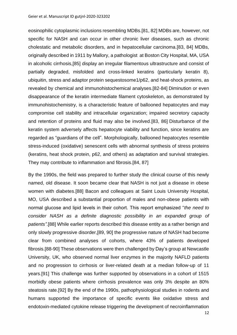
eosinophilic cytoplasmic inclusions resembling MDBs.[81, 82] MDBs are, however, not
specific for NASH and can occur in other chronic liver diseases, such as chronic
cholestatic and metabolic disorders, and in hepatocellular carcinoma.[83, 84] MDBs,
originally described in 1911 by Mallory, a pathologist at Boston City Hospital, MA, USA
in alcoholic cirrhosis,[85] display an irregular filamentous ultrastructure and consist of
partially degraded, misfolded and cross-linked keratins (particularly keratin 8),
ubiquitin, stress and adaptor protein sequestosome1/p62, and heat-shock proteins, as
revealed by chemical and immunohistochemical analyses.[82-84] Diminution or even
disappearance of the keratin intermediate filament cytoskeleton, as demonstrated by
immunohistochemistry, is a characteristic feature of ballooned hepatocytes and may
compromise cell stability and intracellular organization; impaired secretory capacity
and retention of proteins and fluid may also be involved.[83, 86] Disturbance of the
keratin system adversely affects hepatocyte viability and function, since keratins are
regarded as “guardians of the cell”. Morphologically, ballooned hepatocytes resemble
stress-induced (oxidative) senescent cells with abnormal synthesis of stress proteins
(keratins, heat shock protein, p62, and others) as adaptation and survival strategies.
They may contribute to inflammation and fibrosis.[84, 87]
By the 1990s, the field was prepared to further study the clinical course of this newly
named, old disease. It soon became clear that NASH is not just a disease in obese
women with diabetes.[88] Bacon and colleagues at Saint Louis University Hospital,
MO, USA described a substantial proportion of males and non-obese patients with
normal glucose and lipid levels in their cohort. This report emphasized “the need to
consider NASH as a definite diagnostic possibility in an expanded group of
patients”.[88] While earlier reports described this disease entity as a rather benign and
only slowly progressive disorder,[89, 90] the progressive nature of NASH had become
clear from combined analyses of cohorts, where 43% of patients developed
fibrosis.[88-90] These observations were then challenged by Day’s group at Newcastle
University, UK, who observed normal liver enzymes in the majority NAFLD patients
and no progression to cirrhosis or liver-related death at a median follow-up of 11
years.[91] This challenge was further supported by observations in a cohort of 1515
morbidly obese patients where cirrhosis prevalence was only 3% despite an 80%
steatosis rate.[92] By the end of the 1990s, pathophysiological studies in rodents and
humans supported the importance of specific events like oxidative stress and
endotoxin-mediated cytokine release triggering the development of necroinflammation

Geier et al. Manuscript ID gutjnl-2020-323202
13
in NASH patients.[93] Still, a likely contributor to the confusion was due to the term
“NASH” being frequently used not in the specific context of histopathological
steatohepatitis.[94]
As liver enzymes were introduced into routine automated testing platforms,
investigating apparently healthy patients with abnormal liver blood tests became a
relevant issue.[91] Two distinct groups of adult NAFLD patients emerged: those with
simple fatty liver who had an excellent prognosis and those with progressive NASH
and often fibrosis.[94] Poorer outcomes were observed more frequently in patients with
ballooning, MDBs or fibrosis.[95] Matteoni and colleagues from the Mayo Clinic
showed that more than 20% of patients with these lesions together with inflammation
developed cirrhosis over a 10-year follow-up in contrast to only 4% of those with simple
steatosis.[95]
In early 2000, the first studies on pediatric NAFLD documented progression to cirrhosis
in 3% of the cases.[96, 97] Most cases are aetiologically related to the metabolic
syndrome but some are due to inherited syndromes characterized by obesity and
insulin resistance.[71, 98] The first histological autopsy study, conducted at the
University of California San Diego, USA in 2006, showed a 9.6% prevalence of fatty
liver in children.[99]
Despite the emerging awareness among specialized liver centers, surprisingly little
attention was being paid to NASH in regular clinical practice.[94] One ongoing
challenge is the lack of a predictable correlation between abnormal standard liver tests
(such as aminotransferases) and the severity of histological lesions.[100, 101] This has
contributed to the under-recognition of NASH, particularly in patients with so-called
cryptogenic cirrhosis.[102, 103] Diagnosis of NAFLD in lean subjects is of particular
importance since 20% of these patients have NASH, >F2 fibrosis and carotid
atherosclerosis.[103] Furthermore, NAFLD is recognized as the most common cause
of cryptogenic cirrhosis but the diagnosis frequently appears to be delayed.[104]
Semi-quantification of NASH – Histological scoring systems
From the study of Matteoni and colleagues (1999) it became clear that specific
histopathological features discriminated between two prognostically different patient
groups.[95] Different scoring systems were established to quantify the lesions and
assign qualitative and semi-quantitative numerical values to the histopathological

Geier et al. Manuscript ID gutjnl-2020-323202
14
diagnoses of steatosis alone, steatohepatitis and fibrosis. In 1999, Brunt and
colleagues published a histological system for assessment of ‘activity’ grade and
staging fibrosis in NASH.[105] NASH grades (1–3) were based on the combination of
steatosis, hepatocellular ballooning, lobular and portal inflammation. Fibrosis staging
recognised early zone 3 sinusoidal and/or pericellular “chickenwire” fibrosis either
alone (stage 1) or in combination with periportal (stage 2) or bridging fibrosis (stage 3),
while stage 4 was cirrhosis.[105]
The necessity to define a widely accepted scoring system for NASH was highlighted
by a National Institutes of Health (NIH) symposium which fostered further efforts.[106]
The NASH Clinical Research Network (NASH CRN) established and validated the
NASH CRN score as the first globally accepted, scoring system that addressed the full
spectrum of NAFLD lesions and proposed the summative NAFLD activity score (NAS)
to semi-quantify disease activity in clinical trials.[107] The NAS (range 0-8) is
calculated by summing-up semi-quantitative scores for three of the most important
histological features of NAFLD: steatosis (0-3), lobular inflammation (0-2), and
hepatocellular ballooning (0-2) Kleiner and colleagues of the NASH CRN Pathology
Committee initially observed that NAS >5 correlated with NASH diagnosis whereas
biopsies with NAS scores of <3 correlated with “not NASH.” Subsequent work from the
same group made clear, however, that these NAS threshold values were not always
associated with the underlying histological diagnosis of simple steatosis or
steatohepatitis and, therefore, could not replace histopathological assessment.[108]
The NASH CRN pathologists have also coined the pattern-based “borderline NASH”
category for those cases that could not be binary classified. They also devised a
staging system for assessing fibrosis in NAFLD based on Brunt’s 5-tier staging (0-
4)[105] with only modification being the subdivision of stage 1 into 3 substages.[107]
In 2011, a research workshop of the American Association for the Study of Liver
Diseases reached consensus on key endpoints and the design of clinical trials for adult
NASH.[109] It was agreed that “definite steatohepatitis” is characterised by zone 3
accentuation of macrovesicular steatosis of any grade, hepatocellular ballooning of any
degree, and lobular inflammation of any degree and recommended that the NAS
should be used to semi-quantify disease activity.[110]
Grading and staging of morphologic features enhance information particularly in
clinical trials. NAFLD heterogeneity reflects individual variability in response to

Geier et al. Manuscript ID gutjnl-2020-323202
15
metabolic stress and susceptibility to hepatocyte lipotoxicity, depending on genetic and
environmental factor.[111] As shown by pioneers in the field, NAFLD displays a
continuous spectrum of hepatocytic, inflammatory and fibrous lesions. Therefore, the
binary categorization of NAFLD into NASH and “not NASH” is artificial in a continuous
disease process. [111] In 2012, Bedossa and colleagues developed a simple algorithm
to standardize the histological diagnosis of NASH and reduce inter-observer variability.
The diagnostic algorithm was informed by scores for steatosis (S0-S3), activity grade
(A0-A4 by adding scores for ballooning (0-2) and lobular inflammation (0-2)) and
fibrosis stage (F0-F4).[112] A group of expert hepatopathologists and general
pathologists from the Fatty Liver Inhibition of Progression (FLIP) consortium further
validated the FLIP algorithm.[113] The SAF scoring system (Steatosis, Activity,
Fibrosis) includes the same categories as NAS for the semi-quantitation of liver injury
but the diagnostic FLIP algorithm requires the simultaneous presence of steatosis,
ballooning and lobular inflammation for NASH diagnosis.[1, 111, 112]
Despite the use of widely accepted minimal diagnostic criteria for diagnosing NASH,
the issue of interobserver variability for assessing the characteristic histological
features still remains. In order to increase the reliability of NASH diagnosis, both NASH
CRN and FLIP SAF scoring systems are now simultaneously used in clinical trials and
registries worldwide and can be performed by expert and general pathologists equally
well if they are properly trained.[113, 114]
The role of fibrosis as prognostic indicator
The prognostic relevance of these histopathological scores has been validated in
several large registries with up to four decades follow-up. Fibrosis starts in zone 3
(perivenular, sinusoidal, pericellular) In later stages, portal, periportal and bridging
fibrosis leads, finally, to cirrhosis.[71-73] In 2015, it became clear that evaluation of
fibrosis stage may be even more fundamental than scoring necroinflammation or
diagnosing NASH since it emerged as the main prognostic factor.[115, 116] In two
independent cohorts, the American PRELHIN study and the cohort from Karolinska
and Linköping University Hospital, Sweden, NAS score alone was unable to predict
overall mortality, whereas fibrosis stage predicted both overall and disease-specific
mortality.[116] Similar results were obtained for SAF score, which was not associated
with increased mortality in NAFLD after adjustment for fibrosis.[117] In the largest
retrospective cohort study from Karolinska and Linköping University Hospital, Sweden

Geier et al. Manuscript ID gutjnl-2020-323202
16
from 1971 to 2009, the presence of NASH alone did not significantly increase the risk
of liver-specific morbidity or overall mortality during a mean 20-year follow-up.[118] In
recent systematic meta-analyses, the risk of liver-related mortality increased
exponentially with increasing fibrosis stage,[119] while biopsy-confirmed fibrosis was
associated with overall mortality risk in NAFL/NASH patients after adjusting for
confounding factors.[120] Further dissecting the natural course of advanced disease,
a multinational study of 458 patients documented that patients with NAFLD cirrhosis
have predominantly liver-related events, whereas those with bridging fibrosis
experience mainly extra-hepatic malignancy and cardiovascular events.[121] In 475
NASH patients enrolled in two negative phase 2b trials, the primary determinant of
clinical disease progression was fibrosis extent, fibrosis at baseline and its change over
time.[122]
Although cirrhosis is the major risk factor for hepatocellular carcinoma (HCC)
development in NAFLD, there is increasing evidence that NAFLD-associated HCC
frequently occurs in the absence of cirrhosis.[123, 124]
The role of NASH as a precursor lesion for fibrosis emerged as patients with NASH
developed severe liver disease slightly earlier than patients without NASH in the
Swedish cohort.[118] Recently, two positive NASH CRN clinical trials demonstrated a
strong association between improvements in fibrosis and resolution of
steatohepatitis.[125] In addition, a NASH CRN prospective study has shown that
changes in NAFLD activity are positively associated with changes in fibrosis.[126] The
general reversibility of ballooning, lobular inflammation and fibrosis after weight loss
has been documented in patients undergoing bariatric surgery.[127] Whether
histopathologic improvement from liver biopsy data in fact translates into a reduction
in overall mortality and liver-related events is currently being investigated in phase 3
studies.
In 2021, liver biopsy still remains the only diagnostic procedure that can reliably assess
the various NAFLD patterns, and particularly to diagnose NASH and early fibrosis.[71,
114]
Genetic basis of NAFLD phenotypes
At the turn of 21st century, large scale sequencing techniques became more widely
available at reasonable cost. Genome-wide association (GWAS) and candidate gene

Geier et al. Manuscript ID gutjnl-2020-323202
17
studies have contributed to our understanding of inter-individual variation in the
progression and outcome of ALD and NAFLD.[128] Genetic variants influence the risk
and fate of NAFLD, particularly intensity and effects of oxidative stress, severity of
steatosis and fibrosis, response to endotoxin, release of cytokines/chemokines, and
immune response. In 2008, Romeo and colleagues published the first GWAS in
NAFLD.[129] Of relatively modest size by current standards, this study examined 9,229
nonsynonymous single nucleotide polymorphisms (SNPs) in a North American
population of diverse ethnicity from the Dallas Heart Study and identified a single highly
significant association between increased hepatic triacylglycerol accumulation
measured using non-invasive proton magnetic resonance spectroscopy and the
patatin-like phospholipase domain-containing 3 (PNPLA3) gene locus. Ethnic
differences were described for the p.I148M SNP that was most common in Hispanics,
lower in subjects of European ancestry (0.23) and lowest in African-Americans.[129]
Together with further SNPs associated with increased hepatic triacylglycerol content,
PNPLA3 has been confirmed in a subsequent GWAS meta-analysis using 2.4 million
SNPs from 7176 individuals of European ancestry by Speliotes and colleagues.[130]
This variant may explain, at least in part, special phenotypes like pediatric and lean
NAFLD.[131-132] The search for further fatty liver genes revealed transmembrane 6
superfamily member 2 (TM6SF2), glucokinase regulatory protein gene (GCKR), and
HSD17B13, encoding for a retinol dehydrogenase, among an increasing list of
associated loci.[128, 133-135] Combined effects of these risk alleles were observed in
replication cohorts.[134]. In recent years, additional data supported the particular
contribution of PNPLA3 and other gene loci to the development of fibroinflammation
and HCC.[128] In the largest histology-based NAFLD GWAS in a cohort of 1,483
European patients with biopsy-proven NAFLD and 17,781 genetically matched
controls, PNPLA3 was confirmed as risk factor for the full histological spectrum of
NAFLD, while TM6SF2, GCKR and HSD17B13 were also confirmed as NAFLD risk
modifiers.[136] Combining the number of inherited NAFLD risk genes with the
multitude of exogenous NAFLD risk factors (exposome), three-dimensional risk-space
models allow visualisation of disease trajectory in NAFLD risk gene carriers over
time.[137] It is tempting to speculate that genotyping will guide our clinical practice in
the future but it has not as yet entered clinical practice because the effect magnitude
of PNPLA3 risk allele for NASH development or liver cancer is rather modest.[138,
139]

Geier et al. Manuscript ID gutjnl-2020-323202
18
Towards non-invasive diagnosis of NASH and fibrosis in NAFLD – future trends
Research on NAFLD has made remarkable progress over the past two centuries
(Figure 5) but major issues remain. Despite progress in non-invasive tests (NITs) for
the evaluation of liver fibrosis in NAFLD, such as elastography devices and blood
tests,[140] the diagnosis of NASH is still based on liver biopsy, an invasive procedure
not suitable for the large proportion of general population affected by NAFLD. In 2007,
the NAFLD fibrosis score (NFS) was introduced as a simple scoring system to
distinguish NAFLD with and without advanced fibrosis.[141] Subsequently, further
fibrosis tests (Table 1), including fibrosis-4 (FIB-4) index,[142]
Fibrotest/Fibrosure,[143] Enhanced Liver Fibrosis (ELF) test,[144] and liver stiffness
measurement by vibration-controlled transient elastography,[145] have entered clinical
practice. Most importantly for primary care, these NITs show excellent AUROCs for
the diagnosis of advanced fibrosis and cirrhosis.[146] The non-invasive diagnosis of
advanced fibrosis has been further improved by the sequential combination of different
NITs thereby refining the patient referral pathway between primary care or
diabetologists and liver specialists.[147] Longitudinal retrospective studies have
demonstrated that NITs calibrated on liver fibrosis are themselves prognostic markers
able to stratify the risk of liver-related outcomes and mortality in NAFLD.[148, 149]
These data reinforce the relevance of using NITs instead of liver biopsy for the
management of NAFLD patients in clinical practice.
NITs, such as the multiparametric NIS4 test,[150] have not yet achieved sufficient
accuracy and validation for the non-invasive diagnosis of NASH in routine practice.
This, therefore, remains a significant challenge, and large consortia (LITMUS in
Europe, NIMBLE in USA) are currently working to identify and validate new NASH
biomarkers.
Pathological diagnosis in the near future will be supported by artificial intelligence.
Future attempts to improve NASH diagnosis will apply machine learning to develop
fully automated software applications for quantification of steatosis, inflammation,
ballooning, and fibrosis.[151] Advances in microscopy techniques, such as second
harmonic generation/two-photon excitation fluorescence imaging, could potentially
improve reproducibility and standardization of liver biopsy assessment.[152]

Geier et al. Manuscript ID gutjnl-2020-323202
19
A great deal of our previously discussed knowledge has been derived from animal
experiments under standardized conditions.[153] Efforts are now focused on next-
generation mouse models, which are particularly suitable for genetic manipulation but
out of the focus of this work.[154]
What will the future bring in regard to NAFLD? As the global epidemic of obesity fuels
metabolic conditions, the clinical and economic burden of NAFLD will become
enormous.[155] Models based on published estimates predict a growth of up to 30%
in total NAFLD cases between 2016–2030.[156] NASH prevalence will increase by 15–
56%, while advanced liver disease and liver-related mortality will more than double as
a result of aging Western populations. Obeticholic acid could represent an important
milestone in the history of NASH if it becomes the first licensed treatment based on the
favorable results of the interim analysis of the pivotal phase 3 trial.[157] For those with
morbid obesity, rates of bariatric procedures will further increase.[158] Given the
rapidly growing global burden of NAFLD/NASH, efforts to discover accurate, non-
invasive diagnostic and prognostic biomarkers, to develop effective treatments for
individuals with advanced NASH and to implement preventive methods must
continue.[4] The remark of Louis from 1843 on NAFLD as the most frequent and
significant disease of the liver is still true today and guides our efforts in the future.[15]
Geier et al. Manuscript ID gutjnl-2020-323202
20
Acknowledgements
The authors thank Ms Alexandra Weisgram for her excellent secretarial assistance and
Dr Camilla Graham for language editing and insightful comments.
This work has been funded by the Innovative Medicines Initiative 2 Joint Undertaking
under grant agreement No. 777377 (LITMUS) This Joint Undertaking receives support
from the European Union’s Horizon 2020 research and innovation programme and
EFPIA.
This work is dedicated to all pioneers in the field with visionary concepts that
built the basis for research and clinical development in nonalcoholic fatty liver
disease.
Geier et al. Manuscript ID gutjnl-2020-323202
21
References
1. Eslam M, Newsome PN, Sarin SK, Anstee QM, Targher G, Romero-Gomez M, Zelber-Sagi S, et al. A new definition for metabolic dysfunction-associated fatty liver disease: An international expert consensus statement. J Hepatol 2020;73:202-209.
2. Tilg H, Moschen AR. Evolution of inflammation in nonalcoholic fatty liver disease: the multiple parallel hits hypothesis. Hepatology 2010;52(5):1836-1846.
3. Tilg H, Adolph TE, Moschen AR. Multiple parallel hits hypothesis in NAFLD - Revisited after a decade. Hepatology 2020 in press
4. Younossi ZM. Non-alcoholic fatty liver disease - A global public health perspective. J Hepatol 2019;70:531-544.
5. WHO. Obesity and overweight: WHO; 2018 February 16.
6. Bray GA, Nielsen SJ, Popkin BM. Consumption of high-fructose corn syrup in beverages may play a role in the epidemic of obesity. Am J Clin Nutr 2004;79(4):537-543.
7. Jensen T, Abdelmalek M, Sullivan S, Nadeau KJ, Green M, Roncal C, Nakagawa T, et al. Fructose and sugar: A major mediator of nonalcoholic fatty liver disease. J Hepatol 2018;68 (5):1063-1075.
8. Antl-Weiser W. The time of the Willendorf figurines and new results of palaeolithic research in Lower Austria. Anthropologie 2009;47:131-141.
9. Christopoulou-Aletra H, Papavramidou N, Pozzilli P. Obesity in the Neolithic era: a Greek female figurine. Obes Surg 2006;16(8):1112-1114
10. Sellayah D, Cagampang FR, Cox RD. On the Evolutionary Origins of Obesity: A New Hypothesis. Endocrinology 2014; 155(5):1573–1588.
11. Tipton CM. Susruta of India, an unrecognized contributor to the historyof exercise physiology. J Appl Physiol 2008; 104:1553–1556.
12. Smith R. Let food be thy medicine... Br Med J 2004;328:0.
13. Kleisiaris CF, Sfakianakis C, Papathanasiou IV. Health care practices in ancient Greece: The Hippocratic ideal. J Med Ethics Hist Med 2014;7:6.
14. Papavramidou NS, Papavramidis ST, Christopoulou-Aletra H. Galen on obesity: etiology, effects, and treatment. World J Surg 2004;28:631-635.
15. Louis PCA. Recherches anatomiques et pathologiques sur la phtisie. 2nd ed: J. B. Bailliere, 1843.
16. Lereboullet A. Memoire sur la Structure intime du Foie et sur la Nature de l` Alteration connue sous le Nom de Foie Gras: J. B. Bailliere, 1853.
17. Frerichs FT. A Clinical Treatise on Diseases of the Liver: M. W. Wood, New York, 1879 (German Edition 1856)
18. Bock CE. Lehrbuch der pathologischen Anatomie und Diagnostik. Leipzig: Georg Wigand`s Verlag, 1852.
19. Rokitansky C. A Manual of Pathological Anatomy. Philadelphia: Blanchard & Lea, 1855.
20. Cruveilhier J. Anatomie pathologique du Corps Humain. Paris: J. B. Bailliere, 1829/1842.
21. Addison T. Observations on fatty degeneration of the liver. Guy`s Hospital Reports 1836;1:476.

Geier et al. Manuscript ID gutjnl-2020-323202
22
22. Rokitansky CA. Skizze der Grössen- und Formenabweichungen der Leber. Bruchstück. Medizinisches Jahrbuch des kaiserlich-königlichen Österreichischen Staates 1839;20:577.
23. Gauchas A. Etude sur la Steatose Hepatique consideree au Point de Vue Chirurgical. Paris: A. Delahaye & E. Lecrosnier, Editeurs, 1882.
24. Bollinger O. Altlas and Essentials of Pathological Anatomy. New York: William Wood & Company, 1898.
25. Lereboullet MA. Intimate Structure of the Liver and the Nature of the Alteration Known under the Name of Fatty Liver. Edinb Med Surg J 1854;81:582-585.
26. Ziegler E. A Textbook of Pathological Anatomy and Pathogenesis. London: MacMillan & Co, 1884.
27. Pepper W. Saccharine diabetes. Med Rec 1884;25:9-12.
28. Bartolow P. Diseases of the liver. In: Pepper W, Starr L editors. A system of practical medicine (vol II) Philadelphia: Lea Brothers & Co; 1885. p. 1050.
29. Eppinger H. Die Leberkrankheiten. Allgemeine und Spezielle Pathologie der Leber. Wien: Springer, 1937.
30. Hanssen P. Enlargement of the liver in diabetes mellitus. JAMA 1936; 106 (11): 914.
31. Connor CL. Fatty infiltration of the liver and the development of cirrhosis in diabetes and chronic alcoholism. Am J Pathol 1938;14:347-364.
32. Zelman S. The liver in obesity. Arch Int Med 1952;90:141–56.
33. Westwater JD, Fainer D. Liver impairment in the obese. Gastroenterology 1958;34:686-693.
34. Weisbrod FG, Schiff L, et al. Needle biopsy of the liver; experiences in the differential diagnosis of jaundice. Gastroenterology 1950;14:56-72.
35. Welin G. Needle biopsy and liver function in earlier stages of fatty cirrhosis of the liver. Acta Med Scand Suppl. 1950;246:260-267.
36. Kalk H. Über die Beziehungen zwischen Fettleber und Diabetes. Dtsch Med Wochenschr 1959;84:1896-1901.
37. Martin MMJF. Considerations histo-pathologiques et cliniques sur les Steatoses du foie. Le Journal de Medicine de Lyon 1952:187-198.
38. Popper H, Szanto, PB, Elias, H. Transition of fatty liver into cirrhosis. Gastroenterology 1955;28:183-192.
39. Menghini G. One-second needle biopsy of the liver. Gastroenterology 1958;35:190-199.
40. Leevy CM, Ryan CM, Fineberg JC. Diabetes mellitus and liver dysfunction; etiologic and therapeutic considerations. Am J Med 1950;8:290-299.
41. Jaques WE. The incidence of portal cirrhosis and fatty metamorphosis in patients dying with diabetes mellitus. N Engl J Med 1953;249:442-445.
42. Reinberg MH, Lipson M. The association of Laennec's cirrhosis with diabetes mellitus. Ann Intern Med 1950;33:1195-1202.
43. Ulevitch H, Gall EA, Abernathy EL, Schiff L. Needle biopsy of the liver. VII. Observations in fatty vacuolization of the liver. Gastroenterology 1951;18:1-7.
44. Robbins SL. Clinicopathologic correlations in fatty nutritional cirrhosis. Am J Gastroenterol 1958;30:387-392.
Geier et al. Manuscript ID gutjnl-2020-323202
23
45. Schleusner V. Uber die Zusammenhange zwischen Diabetes mellitus und Erkrankungen der Leber und der Gallenwege. [Dissertation]. Hamburg: University of Hamburg; 1938.
46. Pessayre D, Fromenty B. NASH: a mitochondrial disease. J Hepatol 2005;42:928-940.
47. Koliaki C, Szendroedi J, Kaul K, Jelenik T, Nowotny P, Jankowiak F, Herder C, et al. Adaptation of hepatic mitochondrial function in humans with non-alcoholic fatty liver is lost in steatohepatitis. Cell Metab 2015;21:739-746.
48. Jansen L. [Diagnosis of fatty liver]. Dtsch Med Wochenschr 1956;81:742-744.
49. Thaler H. [Fatty liver, its causes and concomitant diseases]. Dtsch Med Wochenschr 1962;87:1049-1055.
50. Thaler H. Relation of steatosis to cirrhosis. Clin Gastroenterol 1975;4:273-280.
51. Thaler H. [Etiology and therapy of fatty liver]. Dtsch Med J 1972;23:648-653.
52. Behringer HT. Zusammenhänge zwischen Diabetes mellitus und Fettleber. Deutsche Medizinische Wochenschrift 1970;95:836-838.
53. Bloodworth JMB. Diabetes mellitus and cirrhosis of the liver. Arch Intern Med 1961;108:95–101.
54. Adler M, Schaffner F. Fatty liver hepatitis and cirrhosis in obese patients. Am J Med 1979;67:811-816.
55. Marubbio AT, Buchwald H, Schwartz MZ, Vargo R. Hepatic lesions of central pericellular fibrosis in morbid obesity and after jejunoileal bypass. Am J Clin Pathol 1976;66:684–691.
56. Galambos JT, Wills CE. Relationship between 505 paired liver tests and biopsies to 242 obese patients. Gastroenterology 1976;74:1191–1195.
57. Nasrallah SM, Wills CE, Galambos JT. Hepatic morphology in obesity. Dig Dis Sci 1981;26:325–327.
58. Andersen T, Christoffersen P, Gluud C. The liver in consecutive patients with morbid obesity: a clinical, morphological and biochemical study. Int J Obes 1984;8:107–115.
59. Eriksson S, Eriksson KF, Bondesson L. Nonalcoholic steatohepatitis in obesity: a reversible condition. Acta Medica Scand 1986;220:83–88.
60. Klain J, Fraser D, Goldstein J, et al. Liver histology abnormalities in the morbidly obese. Hepatology 1989;10:873–876.
61. Watanabe A, Kobayashi M, Yoshitomi S, Nagashima H. Liver fibrosis in obese patients with fatty livers. J Med 1989;20:357–362.
62. Zimmerman HJ, MacMurray FG, Rappaport H, Alpert LK. Studies of the liver in diabetes mellitus. J Lab Clin Med 1950;36:912–921.
63. Itoh S, Tsukada N, Motomure Y, Ichinoe A. Five patients with nonalcoholic diabetic cirrhosis. Acta Hepato-Gastroenterol 1979;26:90–97.
64. Nagore N, Scheuer PJ. The pathology of diabetic hepatitis. J Pathol 1988;156: 155-160.
65. Silverman JF, O’Brien KF, Long S, et al. Liver pathology in morbidly obese patients with and without diabetes. Am J Gastroenterol 1990;85:1349–1355.
66. Batman PA, Scheuer PJ. Diabetic hepatitis preceding the onset of glucose intolerance. Histopathology 1985;9:.237–243.

Geier et al. Manuscript ID gutjnl-2020-323202
24
67. Peters RL. Hepatic morphologic changes after jejunoileal bypass. Prog Liver Dis 1979;6:581–594.
68. Hamilton DL, Vest K, Brown BS, et al. Liver injury with alcoholic-like hyalin after gastroplasty for morbid obesity. Gastroenterology 1983;85:722–726.
69. Vyberg M, Ravn V, Andersen B. Pattern of progression in liver injury following jejunoileal bypass for morbid obesity. Liver 1987;7:271–276.
70. Silverman EM, Sapala JA, Appelman HD. Regression of hepatic steatosis in morbidly obese persons after gastric bypass. Am J Clin Pathol 1995;104:23–31.
71. Tiniakos DG, Anstee QM, Brunt EM, Burt AD. Fatty liver disease. In: Burt AD, Ferrell LD, Hubscher SG (Eds) "MacSween´s Pathology of the Liver", 8th Ed, Philadelphia: Elsevier, 2021 in press.
72. Brunt EM. Histological assessment of nonalcoholic fatty liver disease in adults and children. Clin Liver Dis (Hoboken) 2012;1:108-111.
73. Skoien R, Richardson MM, Jonsson JR, Powell EE, Brunt EM, Neuschwander-Tetri BA, et al. Heterogeneity of fibrosis patterns in non-alcoholic fatty liver disease supports the presence of multiple fibrogenic pathways. Liver Int 2013;33:624-632.
74. Schaffner F, Thaler H. Nonalcoholic fatty liver disease. Prog Liver Dis 1986;8:283-298.
75. Thaler H. Fatty change. Baillieres Clin Gastroenterol 1988;2:453-462.
76. Ludwig J, Viggiano TR, McGill DB, Oh BJ. Nonalcoholic steatohepatitis: Mayo Clinic experiences with a hitherto unnamed disease. Mayo Clin Proc 1980;55:434-438.
77. Itoh S, Tsukada Y, Motomura Y, Ichinoe A. Five patients with nonalcoholic diabetic cirrhosis. Acta Hepatogastroenterol 1979;26(2):90-97.
78. Massarrat S, Jordan G, Sahrhage G, Korb G, Bode JC, Dölle W. Five-year follow-up study of patients with nonalcoholic and nondiabetic fatty liver. Acta Hepatogastroenterol 1974;21(3):176-186.
79. Wanless IR, Lentz JS. Fatty liver hepatitis (steatohepatitis) and obesity: an autopsy study with analysis of risk factors. Hepatology 1990;12:1106-1110.
80. Carter-Kent C, Yerian LM, Brunt EM, et al. Nonalcoholic steatohepatitis in children: a multicenter clinicopathological study. Hepatology 2009;50:1113-1120.
81. Denk H, Franke WW, Kerjaschki D, Eckerstorfer R. Mallory bodies in experimental animals and man. Int Rev Exp Pathol 1979;20:77-121.
82. Zatloukal K, French SW, Stumptner C, Strnad P, Harada M, Toivola DM, Cadrin M, et al. From Mallory to Mallory-Denk bodies: what, how and why? Exp Cell Res 2007;313:2033-2049.
83. Strnad P, Zatloukal K, Stumptner C, Kulaksiz H, Denk H. Mallory-Denk-bodies: lessons from keratin-containing hepatic inclusion bodies. Biochim Biophys Acta 2008;1782:764-774.
84. Denk H, Abuja PM, Zatloukal K. Animal models of NAFLD from the pathologist's point of view. Biochim Biophys Acta Mol Basis Dis 2019;1865:929-942.
85. Mallory FB. Cirrhosis of the liver. Five different types of lesions from which it may arise. Bull Johns Hopkins Hosp 1911;22:69-75.
86. Lackner C, Gogg-Kamerer M, Zatloukal K, Stumptner C, Brunt EM, Denk H. Ballooned hepatocytes in steatohepatitis: the value of keratin immunohistochemistry for diagnosis. J Hepatol 2008;48:821-828.

Geier et al. Manuscript ID gutjnl-2020-323202
25
87. Papatheodoridi AM, Chrysavgis L, Koutsilieris M, Chatzigeorgiou A. The role of senescence in the development of nonalcoholic fatty liver disease and progression to nonalcoholic steatohepatitis. Hepatology 2020;71:363-374.
88. Bacon BR, Farahvash MJ, Janney CG, Neuschwander-Tetri BA. Nonalcoholic steatohepatitis: an expanded clinical entity. Gastroenterology 1994;107:1103-1109.
89. Lee RG. Nonalcoholic steatohepatitis: a study of 49 patients. Hum Pathol 1989;20:594-598.
90. Powell EE, Cooksley WG, Hanson R, Searle J, Halliday JW, Powell LW. The natural history of nonalcoholic steatohepatitis: a follow-up study of forty-two patients for up to 21 years. Hepatology 1990;11:74-80.
91. Teli MR, James OF, Burt AD, Bennett MK, Day CP. The natural history of nonalcoholic fatty liver: a follow-up study. Hepatology 1995;22:1714-1719.
92. Andersen T, Gluud C. Liver morphology in morbid obesity: a literature study. Int J Obes 1984;8:97-106.
93. Day CP, James OF. Hepatic steatosis: innocent bystander or guilty party? Hepatology 1998;27:1463-1466.
94. James OF, Day CP. Non-alcoholic steatohepatitis (NASH): a disease of emerging identity and importance. J Hepatol 1998;29:495-501.
95. Matteoni CA, Younossi ZM, Gramlich T, Boparai N, Liu YC, McCullough AJ. Nonalcoholic fatty liver disease: a spectrum of clinical and pathological severity. Gastroenterology 1999;116:1413-1419.
96. Sathya P, Martin S, Alvarez F. Nonalcoholic fatty liver disease (NAFLD) in children. Curr Opin Pediatr 2002; 14(5):593-600.
97. Roberts EA. Nonalcoholic steatohepatitis in children. Curr Gastroenterol Rep. 2003;5(3):253-259.
98. Schwimmer JB, Deutsch R, Rauch J, et al. Obesity, insulin resistance and other clinicopathological correlates of pediatric nonalcoholic fatty liver disease. J Pediatr 2003;143:500–505.
99. Schwimmer JB, Deutsch R, Kahen T, et al. Prevalence of fatty liver in children and adolescents. Pediatrics 2006;118:1388–1393.
100. Ma X, Liu S, Zhang J, Dong M, Wang Y, Wang M, Xin Y. Proportion of NAFLD patients with normal ALT value in overall NAFLD patients: a systematic review and meta-analysis. BMC Gastroenterol 2020;20:10.
101. Fracanzani AL, Valenti L, Bugianesi E, Andreoletti M, Colli A, Vanni E, Bertelli C, et al. Risk of severe liver disease in nonalcoholic fatty liver disease with normal aminotransferase levels: a role for insulin resistance and diabetes. Hepatology 2008;48:792-798.
102. Caldwell SH, Oelsner DH, Iezzoni JC, Hespenheide EE, Battle EH, Driscoll CJ. Cryptogenic cirrhosis: clinical characterization and risk factors for underlying disease. Hepatology 1999;29:664-669.
103. Fracanzani AL, Petta S, Lombardi R, Pisano G, Russello M, Consonni D, Di Marco V, et al. Liver and cardiovascular damage in patients with lean nonalcoholic fatty liver disease, and association with visceral obesity. Clin Gastroenterol Hepatol 2017;15(10):1604-1611.
104. Clark JM, Diehl AM. Nonalcoholic fatty liver disease: an underrecognized cause of cryptogenic cirrhosis. JAMA. 2003 Jun 11;289(22):3000-3004.

Geier et al. Manuscript ID gutjnl-2020-323202
26
105. Brunt EM, Janney CG, Di Bisceglie AM, et al. Nonalcoholic steatohepatitis: a proposal for grading and staging the histological lesions. Am J Gastroenterol 1999;94:2467–2474.
106. James O, Day C. Non-alcoholic steatohepatitis: another disease of affluence. Lancet 1999;353:1634-1636.
107. Kleiner DE, Brunt EM, Van Natta M, Behling C, Contos MJ, Cummings OW, Ferrell LD, et al. Design and validation of a histological scoring system for nonalcoholic fatty liver disease. Hepatology 2005;41:1313-1321.
108. Brunt EM, Kleiner DE, Wilson LA, Belt P, Neuschwander-Tetri BA, Network NCR. Nonalcoholic fatty liver disease (NAFLD) activity score and the histopathologic diagnosis in NAFLD: distinct clinicopathologic meanings. Hepatology 2011;53:810-820.
109. Sanyal AJ, Brunt EM, Kleiner DE, Kowdley KV, Chalasani N, Lavine JE, Ratziu V, et al. Endpoints and clinical trial design for nonalcoholic steatohepatitis. Hepatology 2011;54:344-353.
110. Suzuki A, Diehl AM. Nonalcoholic steatohepatitis. Annu Rev Med 2017;68:85-98.
111. Bedossa P. Current histological classification of NAFLD: strength and limitations. Hepatol Int 2013;7 Suppl 2:765-770.
112. Bedossa P, Poitou C, Veyrie N, Bouillot JL, Basdevant A, Paradis V, Tordjman J, et al. Histopathological algorithm and scoring system for evaluation of liver lesions in morbidly obese patients. Hepatology 2012;56:1751-1759.
113. Bedossa P, FLIP Histopathology Consortium. Utility and appropriateness of the fatty liver inhibition of progression (FLIP) algorithm and steatosis, activity, and fibrosis (SAF) score in the evaluation of biopsies of nonalcoholic fatty liver disease. Hepatology 2014;60:565-575.
114. Brunt EM, Kleiner DE, Carpenter DH, Rinella M, Harrison SA, Loomba R, Younossi Z, et al; American Association for the Study of Liver Diseases NASH Task Force. Nonalcoholic fatty liver disease: Reporting histologic findings in clinical practice.
Hepatology 2020; in press .
115. Angulo P, Kleiner DE, Dam-Larsen S, Adams LA, Bjornsson ES, Charatcharoenwitthaya P, Mills PR, et al. liver fibrosis, but no other histologic features, is associated with long-term outcomes of patients with nonalcoholic fatty liver disease. Gastroenterology 2015;149:389-397 e310.
116. Ekstedt M, Hagstrom H, Nasr P, Fredrikson M, Stal P, Kechagias S, Hultcrantz R. Fibrosis stage is the strongest predictor for disease-specific mortality in NAFLD after up to 33 years of follow-up. Hepatology 2015;61:1547-1554.
117. Hagstrom H, Nasr P, Ekstedt M, Kechagias S, Stal P, Bedossa P, Hultcrantz R. SAF score and mortality in NAFLD after up to 41 years of follow-up. Scand J Gastroenterol 2017;52:87-91.
118. Hagstrom H, Nasr P, Ekstedt M, Hammar U, Stal P, Hultcrantz R, Kechagias S. Fibrosis stage but not NASH predicts mortality and time to development of severe liver disease in biopsy-proven NAFLD. J Hepatol 2017;67:1265-1273.
119. Dulai PS, Singh S, Patel J, Soni M, Prokop LJ, Younossi Z, Sebastiani G, et al. Increased risk of mortality by fibrosis stage in nonalcoholic fatty liver disease: Systematic review and meta-analysis. Hepatology 2017;65:1557-1565.

Geier et al. Manuscript ID gutjnl-2020-323202
27
120. Taylor RS, Taylor RJ, Bayliss S, Hagström H, Nasr P, Schattenberg JM, Ishigami M, et al. Association between fibrosis stage and outcomes of patients with nonalcoholic fatty liver disease: a systematic review and meta-analysis. Gastroenterology 2020;158(6):1611-1625.
121. Vilar-Gomez E, Calzadilla-Bertot L, Wai-Sun Wong V, Castellanos M, Aller-de la Fuente R, Metwally M, Eslam M, et al. Fibrosis severity as a determinant of cause-specific mortality in patients with advanced nonalcoholic fatty liver disease: A multi-national cohort study. Gastroenterology 2018;155:443-457 e417.
122. Sanyal AJ, Harrison SA, Ratziu V, Abdelmalek MF, Diehl AM, Caldwell S, Shiffman ML, et al. The natural history of advanced fibrosis due to nonalcoholic steatohepatitis: data from the simtuzumab trials. Hepatology 2019;70:1913-1927.
123. Bertot LC, Adams LA. Trends in hepatocellular carcinoma due to non-alcoholic fatty liver disease. Expert Rev Gastroenterol Hepatol 2019;13(2):179-187.
124. Piscaglia F, Svegliati‐Baroni G, Barchetti A, Pecorelli A, Marinelli S, Tiribelli C,
et al; HCC‐NAFLD Italian Study Group. Clinical patterns of hepatocellular carcinoma in nonalcoholic fatty liver disease: a multicenter prospective study. Hepatology 2016;63:827‐838.
125. Brunt EM, Kleiner DE, Wilson LA, Sanyal AJ, Neuschwander-Tetri BA, Nonalcoholic Steatohepatitis Clinical Research N. Improvements in histologic features and diagnosis associated with improvement in fibrosis in nonalcoholic steatohepatitis: Results from the Nonalcoholic Steatohepatitis Clinical Research Network treatment trials. Hepatology 2019;70:522-531.
126. Kleiner DE, Brunt EM, Wilson LA, Behling C, Guy C, Contos M, et al; Nonalcoholic Steatohepatitis Clinical Research Network. Association of histologic disease activity with progression of nonalcoholic fatty liver disease. JAMA Netw Open 2019; 2(10):e1912565.
127. Lassailly G, Caiazzo R, Buob D, Pigeyre M, Verkindt H, Labreuche J, Raverdy V, et al. Bariatric surgery reduces features of nonalcoholic steatohepatitis in morbidly obese patients. Gastroenterology 2015; 149(2):379-388.
128. Anstee QM, Seth D, Day CP. Genetic factors that affect risk of alcoholic and nonalcoholic fatty liver disease. Gastroenterology 2016;150:1728-1744 e1727.
129. Romeo S, Kozlitina J, Xing C, Pertsemlidis A, Cox D, Pennacchio LA, Boerwinkle E, et al. Genetic variation in PNPLA3 confers susceptibility to nonalcoholic fatty liver disease. Nat Genet 2008;40:1461-1465.
130. Speliotes EK, Yerges-Armstrong LM, Wu J, Hernaez R, Kim LJ, Palmer CD, Gudnason V, et al. Genome-wide association analysis identifies variants associated with nonalcoholic fatty liver disease that have distinct effects on metabolic traits. PLoS Genet 2011;7:e1001324.
131. Krawczyk M, Liebe R, Maier IB, Engstler AJ, Lammert F, Bergheim I. The Frequent adiponutrin (PNPLA3) variant p.Ile148Met is associated with early liver injury: analysis of a german pediatric cohort. Gastroenterol Res Pract. 2015; 2015: 205079.
132. Krawczyk M, Bantel H, Rau M, Schattenberg JM, Grünhage F, Pathil A, Demir M. Could inherited predisposition drive non-obese fatty liver disease? Results from German tertiary referral centers. J Hum Genetics 2018;63(5):621-626.
133. Kozlitina J, Smagris E, Stender S, Nordestgaard BG, Zhou HH, Tybjaerg-Hansen A, Vogt TF, et al. Exome-wide association study identifies a TM6SF2 variant that confers susceptibility to nonalcoholic fatty liver disease. Nat Genet 2014;46:352-356.
Geier et al. Manuscript ID gutjnl-2020-323202
28
134. Krawczyk M, Rau M, Schattenberg JM, Bantel H, Pathil A, Demir M, Kluwe J, et al. Combined effects of the PNPLA3 rs738409, TM6SF2 rs58542926, and MBOAT7 rs641738 variants on NAFLD severity: a multicenter biopsy-based study. J Lipid Res 2017;58:247-255.
135. Kozlitina J. Genetic risk factors and disease modifiers of nonalcoholic steatohepatitis. Gastroenterol Clin North Am 2020;49(1):25-44.
136. Anstee QM, Darlay R, Cockell S, Meroni M, Govaere O, Tiniakos D, Burt AD, et al; EPoS Consortium Investigators. Genome-wide association study of non-alcoholic fatty liver and steatohepatitis in a histologically characterised cohort. J Hepatol 2020; in press
137. Krawczyk M, Liebe R, Lammert F. Toward genetic prediction of nonalcoholic fatty liver disease trajectories: PNPLA3 and beyond. Gastroenterology 2020;158:1865-1880 e1861.
138. Krawczyk M, Stokes CS, Romeo S, Lammert F. HCC and liver disease risks in homozygous PNPLA3 p.I148M carriers approach monogenic inheritance. J Hepatol 2015;62:980-981.
139. Anstee QM, Liu YL, Day CP, Reeves HL. Reply to: HCC and liver disease risk in homozygous PNPLA3 p.I148M carriers approach monogenic inheritance. J Hepatol 2015;62:982-983.
140. Geier A, Boursier J. Non-invasive diagnosis of patients with 'at-risk' NAFLD : only fibrosis counts? Gut 2020;69(7):1164-1165.
141. Angulo P, Hui JM, Marchesini G, Bugianesi E, George J, Farrell GC, Enders F, et al. The NAFLD fibrosis score: a noninvasive system that identifies liver fibrosis in patients with NAFLD. Hepatology 2007;45:846-854.
142. McPherson S, Stewart SF, Henderson E, Burt AD, Day CP. Simple non-invasive fibrosis scoring systems can reliably exclude advanced fibrosis in patients with non-alcoholic fatty liver disease. Gut. 2010;59(9):1265-1269.
143. Ratziu V, Massard J, Charlotte F, Messous D, Imbert-Bismut F, Bonyhay L, Tahiri M, et al. Diagnostic value of biochemical markers (FibroTest-FibroSURE) for the prediction of liver fibrosis in patients with non-alcoholic fatty liver disease. BMC Gastroenterol 2006;6:6.
144. Guha IN, Parkes J, Roderick P, Chattopadhyay D, Cross R, Harris S, et al. Noninvasive markers of fibrosis in nonalcoholic fatty liver disease: Validating the European Liver Fibrosis Panel and exploring simple markers. Hepatology 2008;47:455–460.
145. Yoneda M, Yoneda M, Fujita K, Inamori M, Tamano M, Hiriishi H, Nakajima A. Transient elastography in patients with non-alcoholic fatty liver disease (NAFLD). Gut 2007;56:1330-1331.
146. Anstee QM, Lawitz EJ, Alkhouri N, Wong VW, Romero-Gomez M, Okanoue T, Trauner M, et al. Noninvasive tests accurately identify advanced fibrosis due to NASH: baseline data from the STELLAR trials. Hepatology 2019;70:1521-1530.
147. Boursier J, Guillaume M, Leroy V, Irles M, Roux M, Lannes A, Foucher J, et al. New sequential combinations of non-invasive fibrosis tests provide an accurate diagnosis of advanced fibrosis in NAFLD. J Hepatol 2019;71:389-396.
148. Angulo P, Bugianesi E, Bjornsson ES, Charatcharoenwitthaya P, Mills PR, Barrera F, Haflidadottir S, et al. Simple noninvasive systems predict long-term outcomes of patients with nonalcoholic fatty liver disease. Gastroenterology 2013;145:782-789 e784.
Geier et al. Manuscript ID gutjnl-2020-323202
29
149. Boursier J, Vergniol J, Guillet A, Hiriart JB, Lannes A, Le Bail B, Michalak S, et al. Diagnostic accuracy and prognostic significance of blood fibrosis tests and liver stiffness measurement by FibroScan in non-alcoholic fatty liver disease. J Hepatol 2016;65:570-578.
150. Harrison SA, Ratziu V, Boursier J, Francque S, Bedossa P, Majd Z, et al. A blood-based biomarker panel (NIS4) for non-invasive diagnosis of non-alcoholic steatohepatitis and liver fibrosis: a prospective derivation and global validation study. Lancet Gastroenterol Hepatol 2020;5(11):970-985.
151. Forlano N; Mullish BH GN, Maurice JB, Angkathunyakul N, Lloyd J, Tzallas AT, Tsipouras M, et al. High-Throughput, machine learning–based quantification of steatosis, inflammation, ballooning, and fibrosis in biopsies from patients with nonalcoholic fatty liver disease. Clin Gastroenterol Hepatol 2020;in press.
152. Liu F, Goh GB, Tiniakos D, Wee A, Leow WQ, Zhao JM, Rao HY, et al. qFIBS: A novel automated technique for quantitative evaluation of fibrosis, inflammation, ballooning, and steatosis in patients with nonalcoholic steatohepatitis. Hepatology 2019.
153. Diehl AM. Lessons from animal models of NASH. Hepatol Res 2005;33:138-144.
154. Jahn D, Kircher S, Hermanns HM, Geier A. Animal models of NAFLD from a hepatologist's point of view. Biochim Biophys Acta Mol Basis Dis 2019;1865:943-953.
155. Younossi ZM, Blissett D, Blissett R, Henry L, Stepanova M, Younossi Y, Racila A, et al. The economic and clinical burden of nonalcoholic fatty liver disease in the United States and Europe. Hepatology 2016;64:1577-1586.
156. Estes C, Anstee QM, Arias-Loste MT, Bantel H, Bellentani S, Caballeria J, Colombo M, et al. Modeling NAFLD disease burden in China, France, Germany, Italy, Japan, Spain, United Kingdom, and United States for the period 2016-2030. J Hepatol 2018;69:896-904.
157. Younossi ZM, Ratziu V, Loomba R, Rinella M, Anstee QM, Goodman Z, Bedossa P, et al. Obeticholic acid for the treatment of non-alcoholic steatohepatitis: interim analysis from a multicentre, randomised, placebo-controlled phase 3 trial. Lancet 2019;394:2184-2196.
158. Arterburn DE, Telem DA, Kushner RF, Courcoulas AP. Benefits and risks of bariatric surgery in adults: A Review. JAMA 2020;324(9):879-887.
159. Miele L, Forgione A, La Torre G, Vero V, Cefalo C, Racco S, Vellone VG, et al. Serum levels of hyaluronic acid and tissue metalloproteinase inhibitor-1 combined with age predict the presence of nonalcoholic steatohepatitis in a pilot cohort of subjects with nonalcoholic fatty liver disease. Transl Res 2009;154(4):194-201.
160. Santos VN, Leite-Mór MM, Kondo M, Martins JR, Nader H, Lanzoni VP, Parise ER. Serum laminin, type IV collagen and hyaluronan as fibrosis markers in non-alcoholic fatty liver disease. Braz J Med Biol Res 2005;38(5):747-753.
161. Kumagai E, Mano Y, Yoshio S, Shoji H, Sugiyama M, Korenaga M, Ishida T, et al. Serum YKL-40 as a marker of liver fibrosis in patients with non-alcoholic fatty liver disease. Sci Rep 2016;6:35282.
162. Boyle M, Tiniakos D, Schattenberg JM, Ratziu V, Bugianessi E, Petta S, Oliveira CP, et al. Performance of the PRO-C3 collagen neo-epitope biomarker in non-alcoholic fatty liver disease. JHEP Rep 2019;1(3):188-198.

Geier et al. Manuscript ID gutjnl-2020-323202
30
163. Yilmaz Y, Eren F. Serum biomarkers of fibrosis and extracellular matrix remodeling in patients with nonalcoholic fatty liver disease: association with liver histology. Eur J Gastroenterol Hepatol 2019;31(1):43-46.
164. Hasegawa T, Yoneda M, Nakamura K, Makino I, Terano A. Plasma transforming growth factor-beta1 level and efficacy of alpha-tocopherol in patients with non-alcoholic steatohepatitis: a pilot study. Aliment Pharmacol Ther 2001;15(10):1667-1672.
165. Yang HR, Kim HR, Kim MJ, Ko JS, Seo JK. Noninvasive parameters and hepatic fibrosis scores in children with nonalcoholic fatty liver disease. World J Gastroenterol 2012;18(13):1525-30.
166. Peleg N, Issachar A, Sneh-Arbib O, Shlomai A. AST to Platelet Ratio Index and fibrosis 4 calculator scores for non-invasive assessment of hepatic fibrosis in patients with non-alcoholic fatty liver disease. Dig Liver Dis. 2017 Oct;49(10):1133-1138.
167. Feldstein AE, Wieckowska A, Lopez AR, Liu YC, Zein NN, McCullough AJ.
Cytokeratin-18 fragment levels as noninvasive biomarkers for nonalcoholic
steatohepatitis: a multicenter validation study. Hepatology 2009;50:1072–1078.
168. Huang Y, Adams LA, Joseph J, Bulsara MK, Jeffrey GP. The ability of Hepascore to predict liver fibrosis in chronic liver disease: a meta-analysis. Liver Int 2017;37(1):121-131.
169. Abdelmalek MF, Diehl AM, Guy CD, Portenier D, Sudan R, Li XJ, et al. Serum-based biomarker accurately stratifies hepatic fibrosis in patients with nonalcoholic steatohepatitis. Hepatology 2017;66:55a–56a.
170. Aykut UE, Akyuz U, Yesil A, Eren F, Gerin F, Ergelen R, Celikel CA, et al. Scand J Gastroenterol 2014;49(11):1343-1348. A comparison of FibroMeter NAFLD Score, NAFLD fibrosis score, and transient elastography as noninvasive diagnostic tools for hepatic fibrosis in patients with biopsy-proven non-alcoholic fatty liver disease.
171. Martínez SM, Crespo G, Navasa M, Forns X. Noninvasive assessment of liver fibrosis. Hepatology 2011;53(1):325-335.
172. Ratziu V, Giral P, Charlotte F, et al. Liver fibrosis in overweight patients. Gastroenterology 2000;118: 1117-1123.
173. Harrison SA, Oliver D, Arnold HL, Gogia S, Neuschwander-Tetri BA. Development and validation of a simple NAFLD clinical scoring system for identifying patients without advanced disease. Gut 2008;57:1441–1447.
174. Boursier J, Anty R, Vonghia L, Moal V, Vanwolleghem T, Canivet CM, Michalak S et al. Screening for therapeutic trials and treatment indication in clinical practice: MACK-3, a new blood test for the diagnosis of fibrotic NASH. Aliment Pharmacol Ther 2018;47(10):1387-1396.
175. Boursier J, de Ledinghen V, Leroy V, Anty R, Francque S, Salmon D, Lannes A, et al. A stepwise algorithm using an at-a-glance first-line test for the non-invasive diagnosis of advanced liver fibrosis and cirrhosis. J Hepatol 2017;66(6):1158-1165.
176. Ampuero J, Pais R, Aller R, Gallego-Durán R, Crespo J, García-Monzón C, Boursier J, et al; HEPAmet Registry. Development and validation of HEPAmet fibrosis scoring system-a simple, noninvasive test to identify patients with nonalcoholic fatty liver disease with advanced fibrosis. Clin Gastroenterol Hepatol 2020;18(1):216-225.e5.
177. Newsome PN, Sasso M, Deeks JJ, Paredes A, Boursier J, Chan WK, Yilmaz Y, et al. FibroScan-AST (FAST) score for the non-invasive identification of patients with nonalcoholic steatohepatitis with significant activity and fibrosis: a prospective

Geier et al. Manuscript ID gutjnl-2020-323202
31
derivation and global validation study. Lancet Gastroenterol Hepatol 2020;5(4):362-373.
Geier et al. Manuscript ID gutjnl-2020-323202
32
Figure Legends
Figure 1 Venus figurines of obese women.
(A) Venus of Willendorf. The figurine is estimated to have been created around 30,000
BC in the Austrian Danube valley near Krems and is displayed at the Museum of
Natural History, Vienna, Austria (© NHM Vienna, with permission) (B) Neolithic Venus
figurine. Clay naturalistic figurine of a seated obese woman from Farsala, Thessaly
(Athanassakeion Archaeological Museum of Volos, Greece with permission of the
Hellenic Ministry of Culture and Sports/Archaeological Resources Fund) Middle
Neolithic period 5800-5300 BC.[9]
Figure 2 Historical pathology specimen of a fatty liver from T. Frerichs, Atlas of
Pathological Anatomy (Part I, 1861)
(A) “Fatty liver of an advanced grade”. Massive steatosis sparing the vincinity of the
central veins; (B) cut surface with pale-yellow parenchyma; (C) after removal of the fat
by boiling in ether, only meshes formed by the vessels remain.
Figure 3 Numbers of annual PubMed/MEDLINE references for the term “NASH”
from 1980 through 2020.
References were identified for the search term “NASH” (excluding author names),
accessed on April 8th, 2020.
Figure 4 Histology of steatosis and nonalcoholic steatohepatitis (NASH)
(A) Macrovesicular steatosis. The hepatocytes are distended by large fat droplets;
chromotrope-aniline blue, x200. (B) NASH characterized by ballooned hepatocytes
with lightly stained cytoplasm, some of them containing indistinct cytoplasmic
inclusions resembling Mallory-Denk bodies (MDBs; arrows); H&E stain, x200. (C)
NASH with ballooned hepatocytes, some of them containing MDBs (arrows) Most
hepatocytes are surrounded by pericellular fibrosis (blue rims around hepatocytes);
chromotrope-aniline blue stain x200. (D, inset) Immunohistochemistry using antibodies
to sequestosome1/p62 reveals p62-containing MDBs (red), x200.

Geier et al. Manuscript ID gutjnl-2020-323202
33
Figure 5 Timeline of major developments in the field of NAFLD over the past 200
years
Major landmarks in the field until 2000 are shown with solid lines and black font, while
developments after 2000 that cannot yet be viewed in a historical context are shown
in dashed lines and lighter font.

Geier et al. Manuscript ID gutjnl-2020-323202
34
Tables
Table 1. Non-invasive tests (NITs) for fibrosis assessment in NAFLD
Direct serum markers Indirect serum
markers/panels
Combined
scores/Algorithms
Hyaluronic Acid[159]
Laminin[160]
YKL-40[161]
Procollagen type-III N-terminal
peptide (Pro-C3)[162]
Metalloproteinases
MMP-1,MMP-2[163]
TIMPs[159]
TGF-β1*[164]
AST/ALT[142]
γ-GT
Platelet count
Albumin
PGA[165]
APRI[166)
Cytokeratin-18*[167]
Fibrotest®[143]
Hepascore®[168]
Fibrospect®[169]
Fibrometer®[170]
Forns score[171]
BAAT score[172]
BARD Score[173]
ELF[144]
FIB-4 score[142]
NAFLD fibrosis score[141]
MACK-3 score*[174]
eLIFT[175]
HEPAmet fibrosis score[176]
FAST score*[177]
NIS4 score*TM[150]
* Denotes tests also assessed for non-invasive diagnosis of NASH
YLK-40: chondrex, human cartilage glycoprotein-39; PIIINP: Procollagen III amino
terminal peptide; TIMPs: tissue inhibitors of metalloproteinases; TGF-β1: Transforming
growth factor-β; AST: aspartate aminotransferase; ALT: alanine aminotransferase; γ-
GT: gamma-glutamyl transferase; PGA: Prothrombin time, Gamma-glutamyl
transpeptidase, Apoliprotein AI); APRI: AST to Platelet Ratio Index; BAAT: BMI, age,
ALT, Triglyceride score; BARD: BMI, AST/ALT Ratio, Diabetes score; ELF: European
Liver Fibrosis panel; eLIFT: easy Liver Fibrosis Test; FIB-4 score: Fibrosis-4; FAST:
Fibroscan-AST; NAFLD fibrosis score (NFS): Non-Alcoholic Fatty Liver Disease

Geier et al. Manuscript ID gutjnl-2020-323202
35
fibrosis score; MACK-3: combination of HOMA, AST and CK18; NIS-4: Non-Invasive
diagnosis for non-alcoholic Steatohepatitis panel.

Geier et al. Manuscript ID gutjnl-2020-323202
36
Figure 1.

Geier et al. Manuscript ID gutjnl-2020-323202
37
Figure 2.

Geier et al. Manuscript ID gutjnl-2020-323202
38
Figure 3.
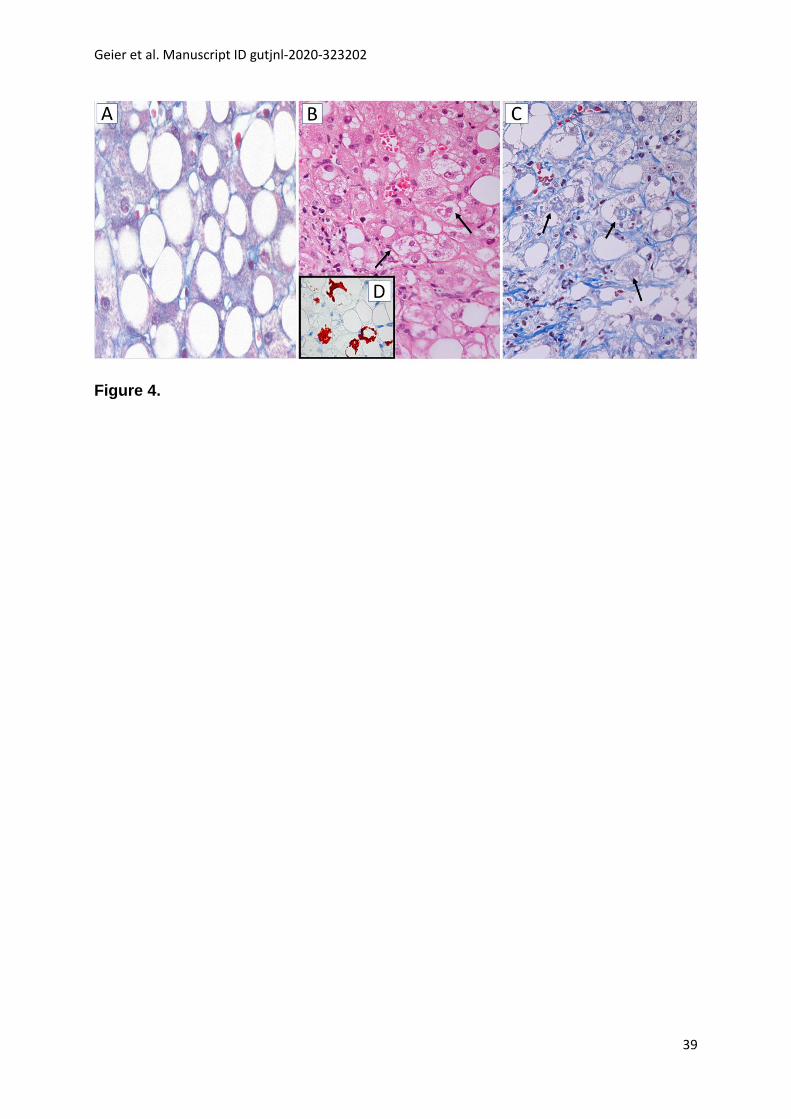
Geier et al. Manuscript ID gutjnl-2020-323202
39
Figure 4.

Geier et al. Manuscript ID gutjnl-2020-323202
40
Figure 5.