
Corn oil versus lard: Metabolic effects of omega-3 fatty ...
25
Research paper Corn oil versus lard: Metabolic effects of omega-3 fatty acids in mice fed obesogenic diets with different fatty acid composition Jana Pavlisova a , Kristina Bardova a , Barbora Stankova b , Eva Tvrzicka b , Jan Kopecky a , Martin Rossmeisl a, * a Department of Adipose Tissue Biology, Institute of Physiology of the Czech Academy of Sciences, Prague, Czech Republic b 4th Department of Internal Medicine, 1st Faculty of Medicine of Charles University and the General Teaching Hospital, Prague, Czech Republic a r t i c l e i n f o Article history: Received 30 April 2015 Accepted 2 July 2015 Available online 4 July 2015 Keywords: Omega-3 fatty acids Hepatic steatosis Obesity Insulin resistance Stearoyl-CoA desaturase-1 a b s t r a c t Mixed results have been obtained regarding the level of insulin resistance induced by high-fat diets rich in saturated fatty acids (SFA) when compared to those enriched by polyunsaturated fatty acids (PUFA), and how metabolic effects of marine PUFA of n-3 series, i.e. docosahexaenoic acid (DHA) and eicosa- pentaenoic acid (EPA), depend on dietary lipid background. Here we compared two high-fat diets, in which the major lipid constituent was based either on SFA in the form of pork lard (LHF diet) or PUFA of n-6 series (Omega-6) as corn oil (cHF diet). Both cHF and LHF parental diets were also supplemented with EPAþDHA (~30 g/kg diet) to produce cHFþF and LHFþF diet, respectively. Male C57BL/6N mice were fed the experimental diets for 8 weeks. Insulin sensitivity was assessed by hyperinsulinemic-euglycemic clamps in mice fed LHF and cHF diets, and then metabolic effects of cHFþF and LHFþF diets were assessed focusing on the liver and epididymal white adipose tissue (eWAT). Both LHF and cHF induced comparable weight gain and the level of insulin resistance, however LHF-fed mice showed increased hepatic steatosis associated with elevated activity of stearoyl-CoA desaturase-1 (SCD1), and lower plasma triacylglycerol levels when compared to cHF. Despite lowering hepatic SCD1 activity, which was concomitant with reduced hepatic steatosis reaching the level observed in cHFþF mice, LHFþF did not decrease adiposity and the weight of eWAT, and rather further impaired insulin sensitivity relative to cHFþF, that tended to improve it. In conclusion, high-fat diets containing as much as ~35 weight% as lipids induce similar weight gain and impairment of insulin sensitivity irrespective whether they are based on SFA or Omega-6. Although the SFA-rich diet containing EPAþDHA efficiently reduced hepatic steatosis, it did so without a corresponding improvement in insulin sensitivity and in the absence of effect on adiposity. © 2015 Elsevier B.V. and Soci! et! e Française de Biochimie et Biologie Mol ! eculaire (SFBBM). All rights reserved. Abbreviations: AA, arachidonic acid; Acaca, acetyl-CoA carboxylase isoform 1 gene; Acly, ATP citrate lyase gene; Acot1, acyl-CoA thioesterase 1 gene; Acox1, peroxisomal acyl-CoA oxidase 1 gene; Adgre1, adhesion G protein-coupled receptor E1 gene; Apob, apolipoprotein B gene; Arg1, arginase 1 gene; ATM, adipose tissue macrophages; AUC, area under the glucose curve; Ccl2, chemokine (C-C motif) ligand 2 gene; cHF, corn oil-based high-fat diet; cHFþF, cHF diet supplemented with EPA/DHA in the form of a triacylglycerol concentrate; CLS, crown-like structures; Dgat1, diacylglycerol O-acyltransferase 1 gene; DHA, docosahexaenoic acid; Ehhadh, enoyl-CoA, hydratase/3- hydroxyacyl CoA dehydrogenase gene; Elovl5, fatty acid elongase 5 gene; EPA, eicosapentaenoic acid; eWAT, epididymal white adipose tissue; Fasn, fatty acid synthase gene; GIR, glucose infusion rate; GTO, glucose turnover; Hadha, hydroxyacyl-CoA dehydrogenase/3-ketoacyl-CoA thiolase/enoyl-CoA hydratase gene; HGP, hepatic glucose production; LHF, pork lard-based high-fat diet; LHFþF, LHF diet supplemented with EPA/DHA in the form of a triacylglycerol concentrate; HOMA-IR, homeostatic model assessment of insulin resistance; LA, linoleic acid; Mttp, microsomal triglyceride transfer protein gene; NEFA, non-esterified fatty acids; Nos2, inducible nitric oxide synthase 2 gene; Omega-3, marine polyunsaturated fatty acids of n-3 series; Omega-6, polyunsaturated fatty acids of n-6 series; PUFA, polyunsaturated fatty acids; SCD1, stearoyl-CoA desaturase-1; Scd1, stearoyl-CoA desaturase-1 gene; SFA, saturated fatty acids; Srebf1, sterol regulatory element-binding transcription factor 1 gene; VLDL, very low-density lipoproteins; WAT, white adipose tissue. * Corresponding author. Department of Adipose Tissue Biology, Institute of Physiology of the Czech Academy of Sciences, Videnska 1083, 142 20 Prague 4, Czech Republic. E-mail address: [email protected] (M. Rossmeisl). Contents lists available at ScienceDirect Biochimie j o u r n a lhomepage: w w w . e l s e v i e r . c o m / l o c a t e / b i o c h i http://dx.doi.org/10.1016/j.biochi.2015.07.001 0300-9084/© 2015 Elsevier B.V. and Soci ! et! e Française de Biochimie et Biologie Mol ! eculaire (SFBBM). All rights reserved. Biochimie 124 (2016) 150e162
Transcript of Corn oil versus lard: Metabolic effects of omega-3 fatty ...
Research paper
Corn oil versus lard: Metabolic effects of omega-3 fatty acids in micefed obesogenic diets with different fatty acid composition
Jana Pavlisovaa, Kristina Bardova a, Barbora Stankova b, Eva Tvrzicka b, Jan Kopecky a,Martin Rossmeisl a, *
a Department of Adipose Tissue Biology, Institute of Physiology of the Czech Academy of Sciences, Prague, Czech Republicb 4th Department of Internal Medicine, 1st Faculty of Medicine of Charles University and the General Teaching Hospital, Prague, Czech Republic
a r t i c l e i n f o
Article history:
Received 30 April 2015Accepted 2 July 2015Available online 4 July 2015
Keywords:
Omega-3 fatty acidsHepatic steatosisObesityInsulin resistanceStearoyl-CoA desaturase-1
a b s t r a c t
Mixed results have been obtained regarding the level of insulin resistance induced by high-fat diets richin saturated fatty acids (SFA) when compared to those enriched by polyunsaturated fatty acids (PUFA),and how metabolic effects of marine PUFA of n-3 series, i.e. docosahexaenoic acid (DHA) and eicosa-pentaenoic acid (EPA), depend on dietary lipid background. Here we compared two high-fat diets, inwhich the major lipid constituent was based either on SFA in the form of pork lard (LHF diet) or PUFA ofn-6 series (Omega-6) as corn oil (cHF diet). Both cHF and LHF parental diets were also supplementedwith EPAþDHA (~30 g/kg diet) to produce cHFþF and LHFþF diet, respectively. Male C57BL/6N mice werefed the experimental diets for 8 weeks. Insulin sensitivity was assessed by hyperinsulinemic-euglycemicclamps in mice fed LHF and cHF diets, and then metabolic effects of cHFþF and LHFþF diets wereassessed focusing on the liver and epididymal white adipose tissue (eWAT). Both LHF and cHF inducedcomparable weight gain and the level of insulin resistance, however LHF-fed mice showed increasedhepatic steatosis associated with elevated activity of stearoyl-CoA desaturase-1 (SCD1), and lower plasmatriacylglycerol levels when compared to cHF. Despite lowering hepatic SCD1 activity, which wasconcomitant with reduced hepatic steatosis reaching the level observed in cHFþF mice, LHFþF did notdecrease adiposity and the weight of eWAT, and rather further impaired insulin sensitivity relative tocHFþF, that tended to improve it. In conclusion, high-fat diets containing as much as ~35 weight% aslipids induce similar weight gain and impairment of insulin sensitivity irrespective whether they arebased on SFA or Omega-6. Although the SFA-rich diet containing EPAþDHA efficiently reduced hepaticsteatosis, it did so without a corresponding improvement in insulin sensitivity and in the absence ofeffect on adiposity.
© 2015 Elsevier B.V. and Soci!et!e Française de Biochimie et Biologie Mol !eculaire (SFBBM). All rightsreserved.
Abbreviations: AA, arachidonic acid; Acaca, acetyl-CoA carboxylase isoform 1 gene; Acly, ATP citrate lyase gene; Acot1, acyl-CoA thioesterase 1 gene; Acox1, peroxisomalacyl-CoA oxidase 1 gene; Adgre1, adhesion G protein-coupled receptor E1 gene; Apob, apolipoprotein B gene; Arg1, arginase 1 gene; ATM, adipose tissue macrophages; AUC,area under the glucose curve; Ccl2, chemokine (C-C motif) ligand 2 gene; cHF, corn oil-based high-fat diet; cHFþF, cHF diet supplemented with EPA/DHA in the form of atriacylglycerol concentrate; CLS, crown-like structures; Dgat1, diacylglycerol O-acyltransferase 1 gene; DHA, docosahexaenoic acid; Ehhadh, enoyl-CoA, hydratase/3-hydroxyacyl CoA dehydrogenase gene; Elovl5, fatty acid elongase 5 gene; EPA, eicosapentaenoic acid; eWAT, epididymal white adipose tissue; Fasn, fatty acid synthasegene; GIR, glucose infusion rate; GTO, glucose turnover; Hadha, hydroxyacyl-CoA dehydrogenase/3-ketoacyl-CoA thiolase/enoyl-CoA hydratase gene; HGP, hepatic glucoseproduction; LHF, pork lard-based high-fat diet; LHFþF, LHF diet supplemented with EPA/DHA in the form of a triacylglycerol concentrate; HOMA-IR, homeostatic modelassessment of insulin resistance; LA, linoleic acid; Mttp, microsomal triglyceride transfer protein gene; NEFA, non-esterified fatty acids; Nos2, inducible nitric oxide synthase 2gene; Omega-3, marine polyunsaturated fatty acids of n-3 series; Omega-6, polyunsaturated fatty acids of n-6 series; PUFA, polyunsaturated fatty acids; SCD1, stearoyl-CoAdesaturase-1; Scd1, stearoyl-CoA desaturase-1 gene; SFA, saturated fatty acids; Srebf1, sterol regulatory element-binding transcription factor 1 gene; VLDL, very low-densitylipoproteins; WAT, white adipose tissue.
* Corresponding author. Department of Adipose Tissue Biology, Institute of Physiology of the Czech Academy of Sciences, Videnska 1083, 142 20 Prague 4, Czech Republic.E-mail address: [email protected] (M. Rossmeisl).
Contents lists available at ScienceDirect
Biochimie
j o u r n a lhomepage: w w w . e l s e v i e r . c o m / l o c a t e / b i o c h i
http://dx.doi.org/10.1016/j.biochi.2015.07.0010300-9084/© 2015 Elsevier B.V. and Soci!et!e Française de Biochimie et Biologie Mol !eculaire (SFBBM). All rights reserved.
Biochimie 124 (2016) 150e162
1. Introduction
Obesity with excessive accumulation of white adipose tissue(WAT) is associated with a cluster of metabolic abnormalities (i.e.metabolic syndrome), which predispose to the development ofcardiovascular disease. Obesity is also accompanied by insulinresistance that is driven by the activation of an inflammatoryresponse in WAT, and later also at the systemic level (reviewed inRef. [1]). While pharmacological interventions for the treatment ofmetabolic disturbances in obesity require the use of multipleagents, and are often associated with side-effects, lifestyle modifi-cations including increased physical activity and dietary manipu-lations remain an essential component of any prevention and/ortreatment strategy (see e.g. Ref. [2]).
With regard to nutrition and its role in the development ofobesity, not only the quantity but also the type of dietary lipids is animportant determinant. For instance, the chain lengths of dietaryfatty acids, degree of their unsaturation, and double bond config-uration all could have a significant impact on their metabolism[3,4]. Of note, the typical Western diet of today is dramaticallydifferent to that of Paleolithic people, especially with regard todietary fat; modern people have switched from a diet of animalprotein consisting of wild game, which was rich in polyunsaturatedfatty acids (PUFA) and contained more PUFA of n-3 series (Omega-3), to one containing more fat per unit of weight and much lessOmega-3 in favor of increased content of PUFA of n-6 series(Omega-6), namely linoleic acid (LA; 18:2n-6) and arachidonic acid(AA; 20:4n-6; reviewed in Refs. [5,6]). A recent study by Alvheimet al. [7] reported that in the United States dietary content of LAincreased from 1 to 8 energy% during the 20th century; further-more, a significant increase in LA, likely derived from foods con-taining corn oil, in the diet of Americans is largely due torecommendations to minimize saturated fatty acids (SFA) intake tohelp preventing cardiovascular disease [5]. Regarding the pre-sumed detrimental role of SFA, it has been shown that the dietcontaining a higher proportion of monounsaturated fatty acids ascompared to SFA improves insulin sensitivity in healthy subjectsreceiving controlled, isoenergetic diets for 3 months, however onlywhen a total fat intake was below 37 energy% (the KANWU study;[8]). Furthermore, feeding mice a high-fat diet enriched withmonounsaturated fatty acids resulted in improved insulin sensi-tivity coincident with attenuated adipose interleukin-1 b secretion,reduced interleukin-1 b precursor priming, and sustained adiposeAMP-activated protein kinase activation as compared with mice fedSFA-rich high-fat diet [9]. In healthy humans, overeating SFA pro-moted hepatic and visceral fat storage, whereas excess energy fromPUFAs instead promoted lean tissue [10,11]. Similar data showingmore detrimental metabolic effects of SFA-when compared toPUFA-based high-fat diets have been obtained also in rodentmodels of obesity [12e16].
Increased dietary intake of Omega-3, either plant-derived ( a-linolenic acid; 18:3n-3) or especially fish-derived [i.e. eicosa-pentaenoic acid (EPA; 20:5n-3) and docosahexaenoic acid (DHA;22:6n-3)], has been associated with a lower risk of cardiovasculardisease [17]. Several studies in obese humans also demonstrated areduction of adiposity after supplementation with fish-derivedOmega-3 [18,19]. However, different results have been obtainedregarding the effect of Omega-3 supplementation on glucose ho-meostasis in animal models and human patients with obesity andinsulin resistance. While in obese adults with impaired glucosetolerance Omega-3 administration did not improve glycemic con-trol ([20] and reviewed in Ref. [21]), admixing EPAþDHA in theform of fish oils (i.e. Omega-3 as triacylglycerols) to various high-fatdiets rich in Omega-6 partially prevented obesity, dyslipidemia,insulin resistance, and WAT inflammation in various rodent models
[22e27]. The reason for reduced efficacy of fish oil in states ofobesity and insulin resistance especially in humans is not clear,however background lipids and/or other macronutrients in the dietcould also play a role in influencing metabolic efficacy of dietaryEPAþDHA supplementation, as suggested by some experimentalstudies in rodents ([13,14,28] and reviewed in Ref. [29]).
In the present study, using our established model of obesity-prone C57BL/6 mice fed a corn oil-based high-fat diet (see e.g.Refs. [25e27]), as well as mice fed a commercial high-fat diet basedon pork lard, we aimed (i) to evaluate the potency of either SFA- orOmega-6-based high-fat diets to induce obesity and insulin resis-tance, the latter assessed by hyperinsulinemic-euglycemic clamps,and (ii) to explore the ability of dietary supplementation withEPAþDHA in the form of triacylglycerols to induce beneficial effectson glucose and lipid metabolism depending primarily on the lipidbackground of the diet.
2. Materials and methods
2.1. Animals and dietary interventions
Before the experiments, male C57BL/6N mice (Charles RiverLaboratories, Sulzfeld, Germany) were maintained for 2 weeks on a12-h light/dark cycle (light from 6:00 a.m.) at 22 " C, with ad libitumaccess to water and a low-fat standard diet (Chow; extruded SsniffR/M-H diet; Ssniff Spezialdieten GmbH, Soest, Germany). As ageneral approach, single-caged mice at the age of 12 weeks wererandomly divided into two experimental groups in order to getaccustomed for 2 weeks to one of the following obesogenic diets:corn oil-based high-fat diet (cHF; lipids ~32% wt/wt; corn oil con-tains ~50% of LA), prepared at the Department of Adipose TissueBiology of the Institute of Physiology in Prague (see e.g. Ref. [25]),and lard-based high-fat diet (LHF; lipids ~35% wt/wt; Ssniff EF acc.D12492 (I) mod., product #E15742-34; Ssniff Spezialdieten GmbH,Soest, Germany; http://www.ssniff.com/documents/gereinigte_diaeten_experimentaldiaeten.pdf). Following a 2-week adaptationperiod, each of the above mentioned groups was randomly dividedinto two subgroups, one of which continued on a respectiveparental high-fat diet (cHF or LHF) while the other subgroupreceived the same parental diet supplemented with Omega-3 in theform of a triacylglycerol concentrate of EPAþDHA (Epax 1050 TG;DHA, ~47% wt/wt, EPA, ~11% wt/wt; Epax, Sandvika, Norway). Thus,52.5 g of the Epax 1050 TG was used to replace an equivalent part ofthe major lipid source in each diet (corn oil or lard), resulting in theconcentration of ~30 g of EPAþDHA per 1 kg of diet. The supple-mented diets were designated either as cHFþF (see also ref. [25])and LHFþF, depending on whether the background diet was cHF orLHF,respectively. For details regarding the macronutrient compo-sition of experimental diets, see Supplementary Table 1; for fattyacid composition in dietary lipids, see Supplementary Table 2.Chow-fed mice served always as lean controls.
Two independent experiments were performed: (i) the firstexperiment lasting 8 weeks compared the capacity of the LHF andcHF diet (i.e. without EPAþDHA supplementation) to induce insulinresistance and associated impairments of glucose homeostasis,assessed by hyperinsulinemic-euglycemic clamps (for details onclamp procedure, see the Section 2.2), and (ii) the next experimentlasting 6 weeks (plus the 2-week adaptation period e see above),which investigated metabolic effects of dietary Omega-3 withrespect to dietary background; thus, it involved five experimentalgroups including cHF, LHF, cHFþF, LHFþF, and the Chow.
Body weight of single-caged mice was monitored weekly, whilea fresh ration was given every 2 days. The calculation of averageenergy intake was based on food consumption measurementsassessed in each mouse during a 24-h period once a week. Mice
J. Pavlisova et al. / Biochimie 124 (2016) 150e162 151
were killed following a 6-h fast (food removed at 6:00 a.m.) bycervical dislocation under diethylether anesthesia between 12:00p.m. and 14:00 p.m. The weights of tissues including white adiposetissue (WAT) from the epididymal depot (eWAT), mesenteric WAT,subcutaneous WAT from dorsolumbar depot, as well as the liverwere recorded; aliquots of eWAT and liver were stored in 4%formaldehyde for the histological evaluation, and also snap-frozenin liquid nitrogen and stored at #70 " C for subsequent biochemicalanalyses. Adiposity index (in %) was defined as a sum of the weightsof eWAT, mesenteric WAT, and subcutaneous WAT relative to bodyweight of the respective animal. All animal experiments wereapproved by the Animal Care and Use Committee of the Institute ofPhysiology, Czech Academy of Sciences (Approval Number: 127/2013) and followed the guidelines.
2.2. Hyperinsulinemic-euglycemic clamp
The clamp procedure performed in conscious mice was amodification of our previously published approach [25,26]. Briefly,approximately 1 week before the end of experiment, i.e. after 7weeks on experimental diets, mice were equipped with a perma-nent catheter inserted into v. jugularis. Following a post-operativeperiod that lasted a minimum of 2 days, mice were fasted for 6 h(6:00 a.m.e12:00 p.m.) and then infused with insulin HumulinR(Lilly USA, Indianapolis, IN) at a constant rate of 4.8 mU/min per kgbody weight, while D-[3- 3H]glucose (Perkin Elmer, Boston, MA)was infused at a rate of 0.26 mCi/min. Throughout the infusion,glucose concentration in tail blood was determined by the use ofglucometers Contour™PLUS (Bayer, Leverkusen, Germany). Eugly-cemia (~5.5 mmol/l) was maintained by periodically adjusting avariable infusion of glucose solution (30% for lean animals, 15% forobese animals). Every 10 min during the last hour of a 3-h infusionperiod, blood samples were collected for the analysis of D-[3- 3H]-glucose specific activity and other parameters as described before[26]. At the end of the clamp, mice were killed by cervical dislo-cation in diethylether anesthesia. Liver and EDTA-plasma werecollected for biochemical analyses.
2.3. Glucose tolerance
Intraperitoneal glucose tolerance tests were performed similarlyas before [30]. Briefly, an injection of D-glucose was applied at adose of 1 g per kg body wt. to overnight fasted mice, and glucoselevels in tail blood were monitored by glucometers (see above)before and 15, 30, 60, 120, and 180 min after the glucose injection.The level of glucose intolerance was then quantified as an areaunder the glucose curve (AUC), either as incremental or total (i.e.including blood glucose at the basal state) AUC.
2.4. Plasma metabolites and hormones
The levels of total cholesterol and total triacylglycerols in EDTA-plasma were assessed colorimetrically using commercial kits Bio-La-test TG L250S and Bio-La-Test CHOL L250S, respectively (ErbaLachema, Brno, Czech Republic); non-esterified fatty acids (NEFA)were measured by the kit Wako NEFA-HR (Wako Chemicals USA,Inc., Richmond, VA). Plasma levels of insulin and total adiponectinwere determined by the Sensitive rat insulin RIA kit and MouseAdiponectin ELISA kit, respectively (Merck Millipore Co., Billerica,MA). The homeostasis model assessment (HOMA) was applied toquantify insulin resistance (HOMA-IR index) using the followingformula: fasting plasma insulin (mU/l) x fasting plasma glucose(mmol/l)/22.5.
2.5. Tissue lipids and fatty acid composition
Liver samples (~50 mg) were digested with 150 ml of 3 M KOH in65% EtOH at 70 " C for 2 h. Resulting homogenates were clearedfrom debris by a brief centrifugation and the concentration of totalglycerolipids was assessed by the Bio-La-Test TG L250S (see Section2.4). In the second experiment, the total lipid content in the liverwas assessed by gravimetry following the Folch extraction proce-dure [31]. Fatty acid composition of the triacylglycerol and phos-pholipid fraction in the liver was analyzed by gas chromatographyas before [32].
2.6. Gene expression analysis
Total RNA was isolated either from samples of liver stored inRNAlater (Ambion, Austin, TX, USA) or from the frozen eWATsamples using TRI Reagent (SigmaeAldrich, St. Louis, MO). Thelevels of mRNA in the liver and eWAT were analyzed by real-timequantitative PCR (qPCR) as described earlier [33]. The expressionof lipogenic genes including fatty acid synthase (Fasn), acetyl-CoAcarboxylase isoform 1 (Acaca), ATP citrate lyase (Acly), stearoyl-CoA desaturase-1 (Scd1), fatty acid elongase 5 (Elovl5), sterol reg-ulatory element-binding transcription factor 1 (Srebf1), fatty acidoxidation genes such as enoyl-CoA, hydratase/3-hydroxyacyl-CoAdehydrogenase (Ehhadh), hydroxyacyl-CoA dehydrogenase/3-ketoacyl-CoA thiolase/enoyl-CoA hydratase (Hadha), acyl-CoAthioesterase 1 (Acot1), and acyl-CoA oxidase 1 (Acox1), genesencoding enzymes involved in a formation of very-low density li-poproteins (VLDL) including apolipoprotein B (Apob), diacylglycerolO-acyltransferase 1 (Dgat1), and microsomal triglyceride transferprotein (Mttp), as well as macrophage markers such as adhesion Gprotein-coupled receptor E1 gene (Adgre1), chemokine (C-C motif)ligand 2 (Ccl2), inducible nitric oxide synthase 2 (Nos2), arginase 1(Arg1) were analyzed. Results were normalized to a geometricmean of expressions of several housekeeping genes includingeukaryotic translation elongation factor 2, eukaryotic translationelongation factor 1 a 1, and peptidylprolyl isomerase B. Primersequences are shown in Supplementary Table 3.
2.7. Light microscopy and immunohistochemical analysis
Samples of eWAT and liver fixed in 4% formaldehyde wereembedded in paraffin, and 5-mm sections were stained usinghematoxylin-eosine as before [30]. In eWAT, a macrophage markerMAC-2/galectin-3 was detected by using specific antibodies in or-der to calculate a relative density of crown-like structures (CLS), i.e.the aggregates of adipose tissue macrophages (ATM) in inflamedtissue [34]. Digital images were captured using Olympus AX70 lightmicroscope and a DP 70 camera (Olympus, Tokyo, Japan). Adipocytemorphometry was performed using a Lucia IMAGE version 4.81(Laboratory Imaging, Prague, Czech Republic).
2.8. Statistics
The data are presented as means ± SEM. The comparisons werejudged to be significant at p $ 0.05. The effects of obesogenic high-fat diets (cHF or LHF) as compared to the Chow, as well as the effectsof EPAþDHA supplemented diets (cHFþF and LHFþF) as comparedto their respective parental diets (cHF and LHF), were determinedusing the Student's t-test or ManneWhitney test for the data withand without normal distribution, respectively. For the overallcomparisons among all parental high-fat diets and their EPAþDHAsupplemented forms, Two Way ANOVA followed by the Holm-Sidak method for pairwise multiple comparisons was used. Allanalyses were performed using SigmaStat 3.5 software.
J. Pavlisova et al. / Biochimie 124 (2016) 150e162152
3. Results
3.1. Comparable effects of SFA- and Omega-6-enriched high-fatdiets on obesity-associated insulin resistance, but distinct effects onthe level of hepatic steatosis
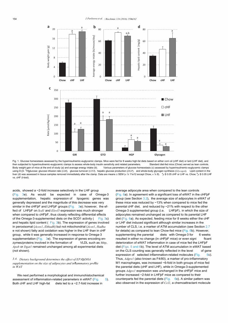
In the first experiment, before the dietary intervention withEPAþDHA, obesity- and insulin resistance-inducing capacity of thetwo parental high-fat diets differing in the major lipid constituentwas analyzed. Body weight gain after 8 weeks of dietary inter-vention with high-fat diets enriched either in Omega-6 (cHF group)or SFA (LHF group) showed a ~3-fold increase when compared tolean Chow-fed controls, however no significant differences in bodyweight gain were observed between the cHF and LHF group(Fig. 1a), despite a marginal increase in the average energy intake incase of the cHF group (Fig. 1b). At the end of experiment,hyperinsulinemic-euglycemic clamps were performed in bothgroups of mice to evaluate whole-body insulin sensitivity (Fig. 1c).Thus, under insulin-stimulated conditions, the amount of exoge-nous glucose required to maintain euglycemia during the clamp,i.e., the glucose infusion rate (GIR), was ~2.4-fold lower in bothgroups of mice fed high-fat diets than in Chow-fed mice, suggestinga profound diet-induced insulin resistance ( Fig. 1c; GIR). Insulinresistance in mice fed either the cHF or LHF diet was also charac-terized by a ~1.6-fold reduction in the glucose turnover rate (GTO)despite a ~4.1-fold increase in hepatic glucose production (HGP),which suggests an impaired suppression of HGP by insulin in micefed high-fat diets (Fig. 1c). As might be expected in the states ofobesity-induced insulin resistance, the whole-body glycogen syn-thesis was markedly reduced in both the cHF and LHF group ascompared with lean, insulin-sensitive Chow-fed mice (Fig. 1c;Glycogen). No differences in the whole-body glycolysis were foundamong the groups (not shown). Thus, high-fat feeding in generalproduced a full spectrum of known defects in glucose metabolism;however there were no significant differences between the cHF andLHF group with regard to GTO, HGP,or whole-body glycogen syn-thesis (Fig. 1c). In contrast, the content of glycerolipids in the liversof clamped mice was significantly increased only in the LHF groupwhen compared to lean Chow-fed mice ( Fig. 1d).
3.2. Differential effects of EPAþDHA supplementation on adiposityand glucose homeostasis depending on dietary background of theirsupplementation
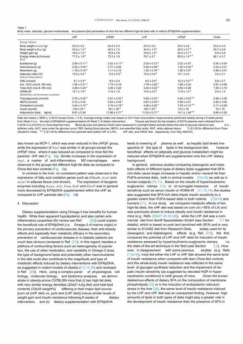
Although the obesogenic cHF and LHF diets showed a compa-rable potency to impair glucose homeostasis (see Section 3.1), theability of dietary EPAþDHA to improve various aspects of meta-bolism under obesogenic conditions could differ depending onwhether they are supplemented onto a lard- or corn oil-enrichedhigh-fat background. Therefore, metabolic effects of a 6-week di-etary intervention with EPAþDHA in the form of triacylglycerolsusing the LHF and cHF background (i.e. the LHFþF and cHFþFsupplemented diets, respectively) were analyzed in mice followinga 2-week adaptation period (see Section 2.1). As shown in Table 1,both the cHF and LHF diet induced a significant but comparableweight gain of ~18e19 g, which was similar to that in the “ clamp”experiment (Section 3.1). The average energy intake of mice fed thecHF and LHF diet was comparable; however, when these diets weresupplemented with EPAþDHA, the average energy intake in theLHFþF group was ~12% and ~17% higher than in the LHF and cHFþFgroup, respectively, corresponding to a larger weight gain in LHFþFmice (Table 1). Furthermore, a tendency for decreased weight gainin the cHFþF group was associated with a significant reduction inthe adiposity index, which was in turn largely attributable to apreferential reduction in the weight of eWAT as compared to other
fat depots ( Table 1). No such effects were observed in LHFþF mice.The cHF and LHF diets impaired glucose homeostasis to a similarlevel; however, while EPAþDHA in the form of LHFþF dietincreased fasting blood glucose (FBG) and tended to further dete-riorate glucose homeostasis, the cHFþF diet reduced fasting plasmainsulin and in general tended to improve insulin sensitivity, as alsosuggested by a significant reduction of HOMA-IR by ~35% whencompared to the LHFþF diet (Table 1). The levels of total adipo-nectin, i.e. the adipokine with insulin-sensitizing properties, werehigher in mice fed LHF diet as compared to cHF-fed mice, howeverthey increased similarly in both the LHFþF and cHFþF groups withEPAþDHA supplementation (Table 1). Plasma levels of tri-acylglycerols, NEFA and total cholesterol, analyzed after a 6-h fast,were reduced to a similar extent in both the cHFþF and LHFþFgroup as compared to their respective parental dietary groups(Table 1). Interestingly, plasma triacylglycerol levels were signifi-cantly lower in mice fed the LHF diet as compared to cHF group(Table 1).
3.3. Pronounced hepatic steatosis in SFA-enriched lard-based LHFdiet is associated with up-regulation of SCD1, which is reduced by
EPAþDHA supplementation
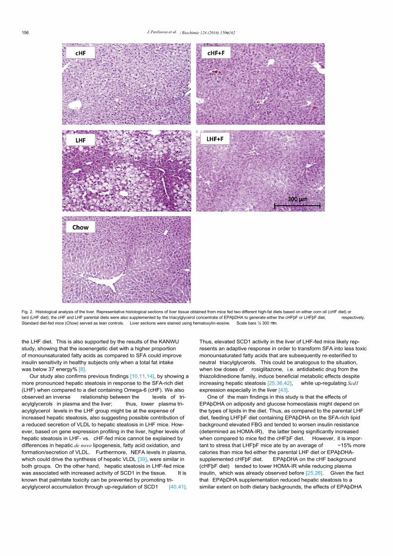
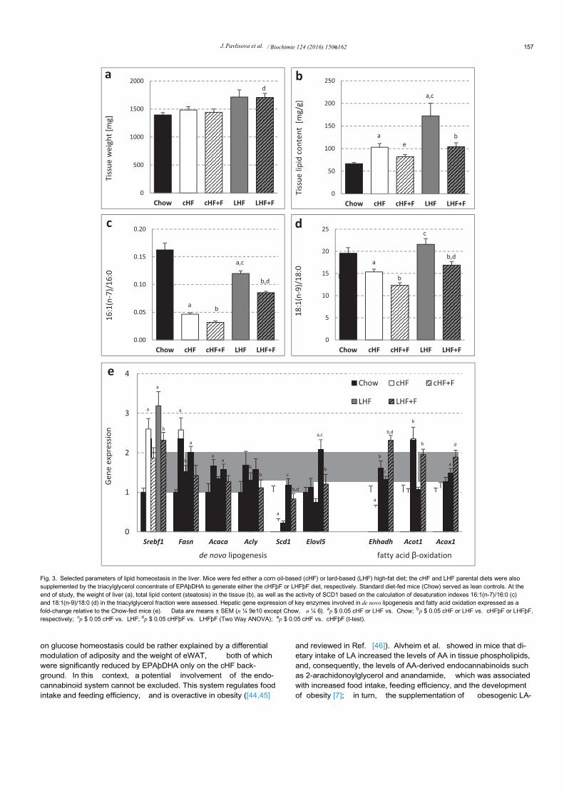
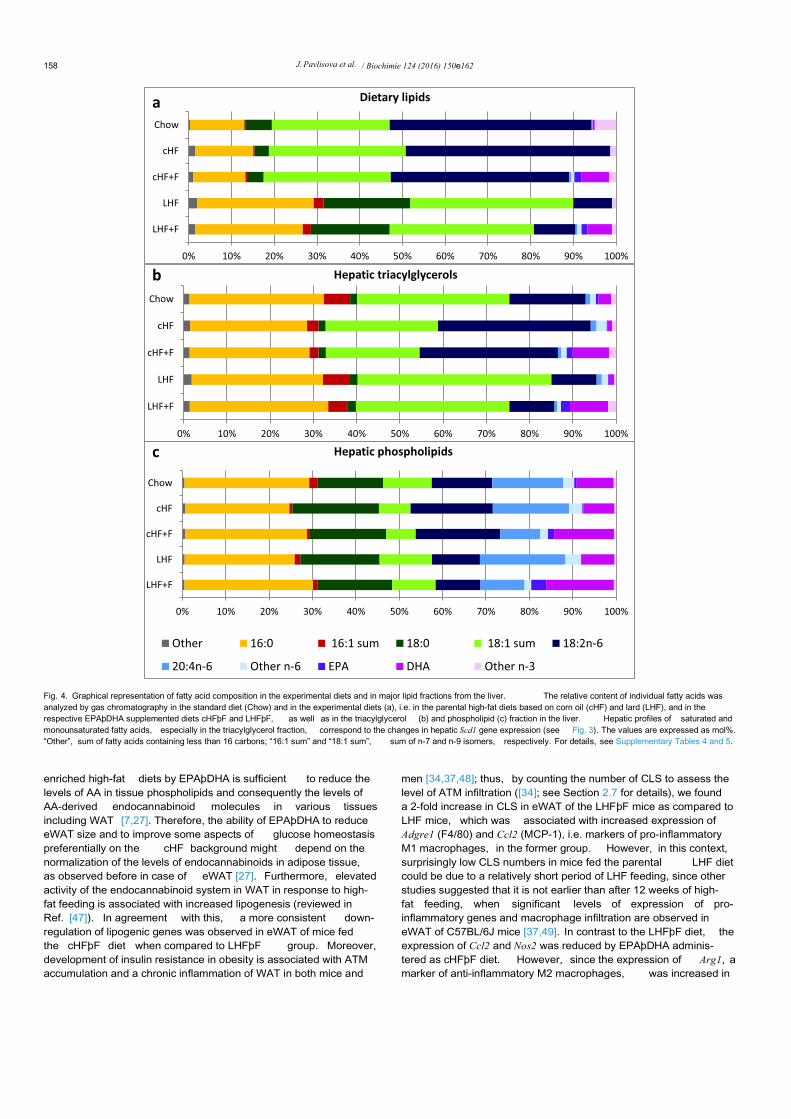
Histological analysis of liver sections stained with hematoxylin-eosin (Fig. 2) indicated a higher level of hepatic steatosis in the LHFthan in the cHF group, while EPAþDHA supplementation reducedit. The liver weight was increased only in the LHF and LHFþF groupbut not in the cHF-based groups (Fig. 3a). Confirming the results ofhistological analysis in the liver (see above), the amount of tissuelipids measured either by the estimation of released glycerol (notshown) or by gravimetry ( Fig. 3b) was much higher in the LHF thanin cHF group, showing a ~2.6-fold and ~1.6-fold elevation in tissuelipid accumulation, respectively, as compared to lean Chow-fedmice (Fig. 3b). Regarding the effects of EPAþDHA supplementa-tion, there was a robust reduction of tissue lipids by ~40% inresponse to EPAþDHA administered as LHFþF, while there was a~20% decrease in the cHFþF group (Fig. 3b). Although dietarycontent of SFA (e.g. 16:0 and 18:0) was markedly increased in boththe LHF and LHFþF diet (Fig. 4a), the SFA levels in hepatic tri-acylglycerol (Fig. 4b) and total phospholipid (Fig. 4c) fractions werealready quite similar between the LHF and cHF groups, which iscompatible with elevated stearoyl-CoA desaturase-1 (SCD1)enzyme activity in the livers of LHF mice. Indeed, marked hepaticsteatosis in the parental LHF group was associated with a largeelevation in the activity of SCD1, as documented by increaseddesaturation indexes 16:1(n-7)/16:0 and 18:1(n-9)/18:0 in hepatictriacylglycerol fraction (Fig. 3c and d); see also the data on fatty acidcomposition in hepatic triacylglycerol fraction in SupplementalTable 4. Similar differences in the indexes of hepatic SCD1 activitybetween the cHF and LHF group were also observed at the level oftissue phospholipids (Supplemental Table 5). Of note, administra-tion of EPAþDHA reduced the levels of AA in the hepatic phos-pholipid fraction by ~50% in mice fed the cHFþF diet as well as inmice fed the LHFþF diet ( Supplemental Table 5), which is consis-tent with the observed metabolic effects of EPAþDHA administra-tion on the liver, irrespective of the dietary background.
Furthermore, the magnitude of changes in the lipid contentinduced by Omega-3 either in the form of the cHFþF or LHFþF dietcorresponded with the suppressive effect of these diets on hepaticSCD1 activity (Fig. 3c and d). Interestingly, the cHF and LHF dietsinduced similar increases in the expression of lipogenic genes suchas Fasn, Acaca, Acly, as well as their transcriptional regulator Srebf1,however hepatic Scd1 expression was more than 4-fold higher inthe LHF than in cHF group (Fig. 3e). Similarly, the expression ofElovl5, which is implicated in the elongation of long-chain fatty
J. Pavlisova et al. / Biochimie 124 (2016) 150e162 153
acids, showed a ~2-fold increase selectively in the LHF group(Fig. 3e). As would be expected in case of Omega-3supplementation, hepatic expression of lipogenic genes wasgenerally depressed and the magnitude of this decrease was verysimilar in the cHFþF and LHFþF groups (Fig. 3e); however, the ef-fect of LHFþF on Scd1 and Elovl5 expression was much strongerwhen compared to cHFþF, thus closely reflecting differential effectsof the Omega-3-supplemented diets on the SCD1 activity ( Fig. 3c)and hepatic lipid content ( Fig. 3b). The expression of genes involvedin peroxisomal (Acox1, Ehhadh) but not mitochondrial (Acot1, Hadhae not shown) fatty acid oxidation was higher in the LHF than in cHFgroup, while it was generally increased in response to Omega-3supplementation (Fig. 3e). The expression of genes encoding en-zymes/proteins involved in the formation of VLDL such as Mttp,Apob or Dgat1 remained unchanged among all experimental diets(not shown).
3.4. Dietary background determines the effect of EPAþDHAsupplementation on the size of adipocytes and inflammatory profilein WAT
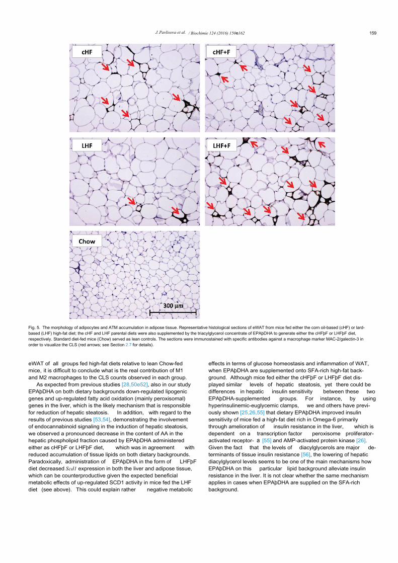
We next performed a morphological and immunohistochemicalassessment of inflammation-related parameters in eWAT (Fig. 5).Both cHF and LHF high-fat diets led to a ~2.7-fold increase in
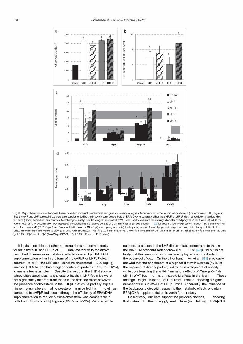
average adipocyte area when compared to the lean controls(Fig. 6a). In agreement with a significant loss of eWAT in the cHFþFgroup (see Section 3.2), the average size of adipocytes in eWAT ofthese mice was reduced by ~13% when compared to mice fed theparental cHF diet, and reduced by ~21% with respect to the otherOmega-3 supplemented group (i.e. LHFþF), in which the size ofadipocytes remained unchanged as compared to its parental LHFdiet (Fig. 6a). As expected, feeding mice for 8 weeks either the cHFor LHF diet induced significant although similar increases in thenumber of CLS, i.e. a marker of ATM accumulation (see Section 2.7for details) as compared to lean Chow-fed mice (Fig. 6b). However,supplementing the parental diets with Omega-3 for 6 weeksresulted in either no change (in cHFþF mice) or even signi ficantdeterioration of eWAT inflammation in case of mice fed the LHFþFdiet (Figs. 5 and 6b). The level of ATM accumulation in eWAT basedon the CLS counting was generally reflected in the level of geneexpression of selected inflammation-related molecules (Fig. 6c).Thus, Adgre1 (also known as F4/80), a marker of pro-inflammatoryM1 macrophages, was increased ~6-fold in both groups of mice fedthe parental diets (cHF and LHF), while in Omega-3-supplementedgroups Adgre1 expression was unchanged in the cHFþF mice andfurther increased ~2-fold in LHFþF mice as compared to theircounterparts fed the parental diets (Fig. 6c). A similar pattern wasalso observed in the expression of Ccl2, a chemoattractant molecule
Fig. 1. Glucose homeostasis assessed by the hyperinsulinemic-euglycemic clamps. Mice were fed for 8 weeks high-fat diets based on either corn oil (cHF diet) or lard (LHF diet), andthen subjected to hyperinsulinemic-euglycemic clamps to assess whole-body insulin sensitivity and related parameters. Standard diet-fed mice (Chow) served as lean controls.Body weight gain of mice at the end of study (a) and average energy intake (b). Various parameters of glucose homeostasis (c) assessed by hyperinsulinemic-euglycemic clampsusing D-[3- 3H]glucose: glucose infusion rate (GIR), glucose turnover (GTO), hepatic glucose production (HGP), and whole-body glycogen synthesis (Glycogen). Lipid content in theliver (d) was assessed in tissue samples removed immediately after the clamp. Data are means ± SEM (n ¼ 11e12 except Chow, n ¼ 8). ap $ 0.05 cHF or LHF vs. Chow; bp $ 0.05 LHFvs. cHF (t-test).
J. Pavlisova et al. / Biochimie 124 (2016) 150e162154
also known as MCP-1, which was even reduced in the cHFþF group,while the expression of Nos2 was similar in all groups except thecHFþF mice, where it was reduced as compared to mice fed theparental cHF diet (Fig. 6c). Similar increases in the expression ofArg1, a marker of anti-inflammatory M2 macrophages, wereobserved in the groups fed different high-fat diets as compared toChow-fed lean controls (Fig. 6c).
In contrast to the liver, no consistent pattern was observed in theexpression of fatty acid oxidation genes such as Ehhadh, Acot1 andAcox1 in adipose tissue (not shown). The expression of lipogenicenzymes including Acaca, Acly, Fasn, Scd1 and Elovl5 was in generalmore decreased by EPAþDHA supplemented within the cHF ascompared to LHF parental diet (Fig. 6d).
4. Discussion
Dietary supplementation using Omega-3 has benefits for humanhealth. While their apparent hypolipidemic and also certain anti-inflammatory properties (for review see Ref. [35]) could explainthe beneficial role of EPAþDHA (i.e. Omega-3 of marine origin) inthe primary prevention of cardiovascular disease, their anti-obesityeffects and especially their metabolic efficacy in the secondaryprevention of cardiovascular disease or in diabetic patients aremuch less obvious (reviewed in Ref. [21]). In this regard, besides aplethora of confounding factors such as heterogeneity of popula-tion, the use of other medication, and variability in Omega-3 dose,the type of background lipids and potentially other macronutrientsin the diet could also contribute to the magnitude and type ofmetabolic effects induced by dietary interventions with EPAþDHA,as suggested in rodent models of obesity ([13,14,28] and reviewedin Ref. [29]). Here, using a complex panel of physiological, cellbiology, molecular biology, and lipidomics analyses, we demon-strate in obesity-prone C57BL/6N mice that (i) two high-fat dietswith very similar energy densities (20e21 kJ/g diet) and total lipidcontents (32e35 weight%) differing in their major lipid source[corn oil (cHF diet) vs. pork lard (LHF diet)] induce similar levels ofweight gain and insulin resistance following 8 weeks of dietaryintervention, and (ii) dietary supplementation with EPAþDHA
leads to lowering of plasma as well as hepatic lipid levels irre-spective of the type of lipids in the background diet, howeverbeneficial effects on adiposity and glucose metabolism might bereduced when EPAþDHA are supplemented onto the LHF dietarybackground.
In general, previous studies comparing obesogenic and meta-bolic effects of different types of dietary lipids suggested that SFA-rich diets cause larger increases in hepatic and/or visceral fat thanPUFA-enriched diets, both in animal models [13e16] as well as inhuman subjects [10,11]. Based on the results of hyperinsulinemic-euglycemic clamps [12] or on surrogate measures of insulinsensitivity such as serum insulin or HOMA-IR [11,13,14], the studiesalso suggested that SFA-rich diets impair insulin sensitivity to agreater extent than PUFA-based diets in both rodents [12e14] andhumans [11]. In our study, we compared metabolic effects of twohigh-fat diets; the cHF diet was based on corn oil (~50% of LA) andwas previously shown to induce obesity and insulin resistance inmice (e.g. Refs. [25e27,30,33,36]), while the LHF diet was a com-mercial diet from Ssniff Spezialdieten GmbH (see Section 2.1 fordetails), which is based on pork lard (enriched with SFA) and is verysimilar to D12492 diet from Research Diets, widely used for itsobesogenic and diabetogenic effects (e.g. Ref. [37]). We firstcompared the potential of LHF and cHF diets for induction of insulinresistance assessed by hyperinsulinemic-euglycemic clamps, i.e.the state-of-the-art technique in the field (see Section 2.2). How-ever, in disagreement with some previous studies (e.g. Refs.[11e14]), mice fed either the LHF or cHF diet showed the same levelof insulin resistance when compared with lean Chow-fed controls,and this whole-body insulin resistance was reflected in the samelevel of glycogen synthesis reduction and the impairment of he-patic insulin sensitivity (as suggested by elevated HGP in hyper-insulinemic conditions) in both groups of mice. Given the knowndeleterious effects of dietary SFA on the composition of membranephospholipids [12] or in the induction of endoplasmic reticulumstress in the liver [38], the same level of insulin resistance inducedby the LHF and cHF diet was an unexpected finding. However, highamounts of lipids in both types of diets might play a greater role inthe development of insulin resistance than the presence of SFA in
Table 1Body mass, adiposity, glucose homeostasis, and plasma lipid parameters of mice fed two different high-fat diets with or without EPAþDHA supplementation.
cHF cHFþF LHF LHFþF Chow
Energy balance
Body weight e initial (g) 23.3 ± 0.2 23.4 ± 0.3 23.2 ± 0.2 23.4 ± 0.2 23.4 ± 0.3Body weight e final (g) 43.2 ± 1.2 a 42.0 ± 1.0 44.4 ± 1.5 a 45.5 ± 0.7 d 30.7 ± 0.5Weight gain (g) 18.2 ± 1.2 a 16.8 ± 0.9 19.5 ± 1.4 a 20.4 ± 0.7 d 5.9 ± 0.4Energy intake (kJ/mouse) 77.2 ± 1.9 a 73.2 ± 1.6 76.7 ± 2.7 85.9 ± 1.6 b,d 68.1 ± 0.7WAT
Epididymal (g) 2.56 ± 0.11 a 2.02 ± 0.11 b 2.53 ± 0.13 a 2.32 ± 0.07 0.54 ± 0.04Subcutaneous (g) 0.83 ± 0.04 a 0.77 ± 0.05 0.94 ± 0.06 a 1.00 ± 0.05 d 0.24 ± 0.01Mesenteric (g) 1.18 ± 0.15 a 1.11 ± 0.12 1.20 ± 0.18 a 1.28 ± 0.07 0.28 ± 0.01Adiposity index (%) 10.5 ± 0.4 a 9.3 ± 0.5 b 10.4 ± 0.4 a 10.1 ± 0.3 3.4 ± 0.1Glucose homeostasis
FBG (mmol/l) 8.7 ± 0.4 a 8.5 ± 0.4 9.0 ± 0.6 a 10.3 ± 0.4 b,d 5.8 ± 0.7Incr. AUC (mol & 180 min) 1.90 ± 0.21 a 1.73 ± 0.19 1.79 ± 0.25 a 1.69 ± 0.22 0.91 ± 0.06Total AUC (mol & 180 min) 3.49 ± 0.24 a 3.28 ± 0.22 3.42 ± 0.32 a 3.55 ± 0.26 1.94 ± 0.15HOMA-IR 15.7 ± 1.6 a 11.6 ± 1.6 15.4 ± 2.5 a 17.9 ± 1.5 d 2.8 ± 1.0Metabolites and hormones in plasma
Triacylglycerols (mmol/l) 0.75 ± 0.05 a 0.61 ± 0.03 b 0.60 ± 0.03 c 0.46 ± 0.02 b,d 0.58 ± 0.05NEFA (mmol/l) 0.72 ± 0.03 0.63 ± 0.04 b 0.67 ± 0.04 a 0.59 ± 0.01 0.83 ± 0.05Cholesterol (mmol/l) 4.04 ± 0.13 a 3.16 ± 0.18 b 4.48 ± 0.22 a 3.75 ± 0.14 b,d 2.11 ± 0.05Insulin (pmol/l) 318 ± 24 a 241 ± 29 b 297 ± 35 a 313 ± 19 79 ± 15Adiponectin (total; mg/ml) 8.87 ± 0.54 11.88 ± 0.77 b 11.03 ± 0.77 a 14.7 ± 1.49 b,d 8.80 ± 0.25
Data are means ± SEM (n ¼ 9e10 except Chow, n ¼ 6). Average energy intake was based on 24-h food consumption measurements performed weekly during a 5-week period,from Week 3 (i.e. the start of EPAþDHA supplementation) till Week 7 of dietary intervention. Tissues and blood (for the isolation of EDTA-plasma) were collected from 6-h (6:00 a.m.e12:00 p.m.) food-deprived mice. Blood glucose and plasma insulin were measured in overnight fasted animals before the start of glucose tolerance test. A.U.,arbitrary units; AUC, area under the glucose curve; FBG, fasting blood glucose; NEFA, non-esterified fatty acids; WAT, white adipose tissue. ap $ 0.05 for difference from Chow(Student's t-test); b,c,dp $ 0.05 for difference from parental diets (either cHF or LHF), cHF diet, and cHFþF diet, respectively (Two Way ANOVA).
J. Pavlisova et al. / Biochimie 124 (2016) 150e162 155
the LHF diet. This is also supported by the results of the KANWUstudy, showing that the isoenergetic diet with a higher proportionof monounsaturated fatty acids as compared to SFA could improveinsulin sensitivity in healthy subjects only when a total fat intakewas below 37 energy% [8].
Our study also confirms previous findings [10,11,14], by showing amore pronounced hepatic steatosis in response to the SFA-rich diet(LHF) when compared to a diet containing Omega-6 (cHF). We alsoobserved an inverse relationship between the levels of tri-acylglycerols in plasma and the liver; thus, lower plasma tri-acylglycerol levels in the LHF group might be at the expense ofincreased hepatic steatosis, also suggesting possible contribution ofa reduced secretion of VLDL to hepatic steatosis in LHF mice. How-ever, based on gene expression profiling in the liver, higher levels ofhepatic steatosis in LHF- vs. cHF-fed mice cannot be explained bydifferences in hepatic de novo lipogenesis, fatty acid oxidation, andformation/secretion of VLDL. Furthermore, NEFA levels in plasma,which could drive the synthesis of hepatic VLDL [39], were similar inboth groups. On the other hand, hepatic steatosis in LHF-fed micewas associated with increased activity of SCD1 in the tissue. It isknown that palmitate toxicity can be prevented by promoting tri-acylglycerol accumulation through up-regulation of SCD1 [40,41].
Thus, elevated SCD1 activity in the liver of LHF-fed mice likely rep-resents an adaptive response in order to transform SFA into less toxicmonounsaturated fatty acids that are subsequently re-esterified toneutral triacylglycerols. This could be analogous to the situation,when low doses of rosiglitazone, i.e. antidiabetic drug from thethiazolidinedione family, induce beneficial metabolic effects despiteincreasing hepatic steatosis [25,36,42], while up-regulating Scd1expression especially in the liver [43].
One of the main findings in this study is that the effects ofEPAþDHA on adiposity and glucose homeostasis might depend onthe types of lipids in the diet. Thus, as compared to the parental LHFdiet, feeding LHFþF diet containing EPAþDHA on the SFA-rich lipidbackground elevated FBG and tended to worsen insulin resistance(determined as HOMA-IR), the latter being significantly increasedwhen compared to mice fed the cHFþF diet. However, it is impor-tant to stress that LHFþF mice ate by an average of ~15% morecalories than mice fed either the parental LHF diet or EPAþDHA-supplemented cHFþF diet. EPAþDHA on the cHF background(cHFþF diet) tended to lower HOMA-IR while reducing plasmainsulin, which was already observed before [25,26]. Given the factthat EPAþDHA supplementation reduced hepatic steatosis to asimilar extent on both dietary backgrounds, the effects of EPAþDHA
Fig. 2. Histological analysis of the liver. Representative histological sections of liver tissue obtained from mice fed two different high-fat diets based on either corn oil (cHF diet) orlard (LHF diet); the cHF and LHF parental diets were also supplemented by the triacylglycerol concentrate of EPAþDHA to generate either the cHFþF or LHFþF diet, respectively.Standard diet-fed mice (Chow) served as lean controls. Liver sections were stained using hematoxylin-eosine. Scale bars ¼ 300 mm.
J. Pavlisova et al. / Biochimie 124 (2016) 150e162156
on glucose homeostasis could be rather explained by a differentialmodulation of adiposity and the weight of eWAT, both of whichwere significantly reduced by EPAþDHA only on the cHF back-ground. In this context, a potential involvement of the endo-cannabinoid system cannot be excluded. This system regulates foodintake and feeding efficiency, and is overactive in obesity ([44,45]
and reviewed in Ref. [46]). Alvheim et al. showed in mice that di-etary intake of LA increased the levels of AA in tissue phospholipids,and, consequently, the levels of AA-derived endocannabinoids suchas 2-arachidonoylglycerol and anandamide, which was associatedwith increased food intake, feeding efficiency, and the developmentof obesity [7]; in turn, the supplementation of obesogenic LA-
Fig. 3. Selected parameters of lipid homeostasis in the liver. Mice were fed either a corn oil-based (cHF) or lard-based (LHF) high-fat diet; the cHF and LHF parental diets were alsosupplemented by the triacylglycerol concentrate of EPAþDHA to generate either the cHFþF or LHFþF diet, respectively. Standard diet-fed mice (Chow) served as lean controls. At theend of study, the weight of liver (a), total lipid content (steatosis) in the tissue (b), as well as the activity of SCD1 based on the calculation of desaturation indexes 16:1(n-7)/16:0 (c)and 18:1(n-9)/18:0 (d) in the triacylglycerol fraction were assessed. Hepatic gene expression of key enzymes involved in de novo lipogenesis and fatty acid oxidation expressed as afold-change relative to the Chow-fed mice (e). Data are means ± SEM (n ¼ 9e10 except Chow, n ¼ 6). ap $ 0.05 cHF or LHF vs. Chow; bp $ 0.05 cHF or LHF vs. cHFþF or LHFþF,respectively; cp $ 0.05 cHF vs. LHF; dp $ 0.05 cHFþF vs. LHFþF (Two Way ANOVA); ep $ 0.05 cHF vs. cHFþF (t-test).
J. Pavlisova et al. / Biochimie 124 (2016) 150e162 157
enriched high-fat diets by EPAþDHA is sufficient to reduce thelevels of AA in tissue phospholipids and consequently the levels ofAA-derived endocannabinoid molecules in various tissuesincluding WAT [7,27]. Therefore, the ability of EPAþDHA to reduceeWAT size and to improve some aspects of glucose homeostasispreferentially on the cHF background might depend on thenormalization of the levels of endocannabinoids in adipose tissue,as observed before in case of eWAT [27]. Furthermore, elevatedactivity of the endocannabinoid system in WAT in response to high-fat feeding is associated with increased lipogenesis (reviewed inRef. [47]). In agreement with this, a more consistent down-regulation of lipogenic genes was observed in eWAT of mice fedthe cHFþF diet when compared to LHFþF group. Moreover,development of insulin resistance in obesity is associated with ATMaccumulation and a chronic inflammation of WAT in both mice and
men [34,37,48]; thus, by counting the number of CLS to assess thelevel of ATM infiltration ([34]; see Section 2.7 for details), we founda 2-fold increase in CLS in eWAT of the LHFþF mice as compared toLHF mice, which was associated with increased expression ofAdgre1 (F4/80) and Ccl2 (MCP-1), i.e. markers of pro-inflammatoryM1 macrophages, in the former group. However, in this context,surprisingly low CLS numbers in mice fed the parental LHF dietcould be due to a relatively short period of LHF feeding, since otherstudies suggested that it is not earlier than after 12 weeks of high-fat feeding, when significant levels of expression of pro-inflammatory genes and macrophage infiltration are observed ineWAT of C57BL/6J mice [37,49]. In contrast to the LHFþF diet, theexpression of Ccl2 and Nos2 was reduced by EPAþDHA adminis-tered as cHFþF diet. However, since the expression of Arg1, amarker of anti-inflammatory M2 macrophages, was increased in
Fig. 4. Graphical representation of fatty acid composition in the experimental diets and in major lipid fractions from the liver. The relative content of individual fatty acids wasanalyzed by gas chromatography in the standard diet (Chow) and in the experimental diets (a), i.e. in the parental high-fat diets based on corn oil (cHF) and lard (LHF), and in therespective EPAþDHA supplemented diets cHFþF and LHFþF, as well as in the triacylglycerol (b) and phospholipid (c) fraction in the liver. Hepatic profiles of saturated andmonounsaturated fatty acids, especially in the triacylglycerol fraction, correspond to the changes in hepatic Scd1 gene expression (see Fig. 3). The values are expressed as mol%.“Other”, sum of fatty acids containing less than 16 carbons; “16:1 sum” and “18:1 sum”, sum of n-7 and n-9 isomers, respectively. For details, see Supplementary Tables 4 and 5.
J. Pavlisova et al. / Biochimie 124 (2016) 150e162158
eWAT of all groups fed high-fat diets relative to lean Chow-fedmice, it is difficult to conclude what is the real contribution of M1and M2 macrophages to the CLS counts observed in each group.
As expected from previous studies [28,50e52], also in our studyEPAþDHA on both dietary backgrounds down-regulated lipogenicgenes and up-regulated fatty acid oxidation (mainly peroxisomal)genes in the liver, which is the likely mechanism that is responsiblefor reduction of hepatic steatosis. In addition, with regard to theresults of previous studies [53,54], demonstrating the involvementof endocannabinoid signaling in the induction of hepatic steatosis,we observed a pronounced decrease in the content of AA in thehepatic phospholipid fraction caused by EPAþDHA administeredeither as cHFþF or LHFþF diet, which was in agreement withreduced accumulation of tissue lipids on both dietary backgrounds.Paradoxically, administration of EPAþDHA in the form of LHFþFdiet decreased Scd1 expression in both the liver and adipose tissue,which can be counterproductive given the expected beneficialmetabolic effects of up-regulated SCD1 activity in mice fed the LHFdiet (see above). This could explain rather negative metabolic
effects in terms of glucose homeostasis and inflammation of WAT,when EPAþDHA are supplemented onto SFA-rich high-fat back-ground. Although mice fed either the cHFþF or LHFþF diet dis-played similar levels of hepatic steatosis, yet there could bedifferences in hepatic insulin sensitivity between these twoEPAþDHA-supplemented groups. For instance, by usinghyperinsulinemic-euglycemic clamps, we and others have previ-ously shown [25,26,55] that dietary EPAþDHA improved insulinsensitivity of mice fed a high-fat diet rich in Omega-6 primarilythrough amelioration of insulin resistance in the liver, which isdependent on a transcription factor peroxisome proliferator-activated receptor- a [55] and AMP-activated protein kinase [26].Given the fact that the levels of diacylglycerols are major de-terminants of tissue insulin resistance [56], the lowering of hepaticdiacylglycerol levels seems to be one of the main mechanisms howEPAþDHA on this particular lipid background alleviate insulinresistance in the liver. It is not clear whether the same mechanismapplies in cases when EPAþDHA are supplied on the SFA-richbackground.
Fig. 5. The morphology of adipocytes and ATM accumulation in adipose tissue. Representative histological sections of eWAT from mice fed either the corn oil-based (cHF) or lard-based (LHF) high-fat diet; the cHF and LHF parental diets were also supplemented by the triacylglycerol concentrate of EPAþDHA to generate either the cHFþF or LHFþF diet,respectively. Standard diet-fed mice (Chow) served as lean controls. The sections were immunostained with specific antibodies against a macrophage marker MAC-2/galectin-3 inorder to visualize the CLS (red arrows; see Section 2.7 for details).
J. Pavlisova et al. / Biochimie 124 (2016) 150e162 159
It is also possible that other macronutrients and componentsfound in the cHF and LHF diet may contribute to the abovedescribed differences in metabolic effects induced by EPAþDHAsupplementation either in the form of the cHFþF or LHFþF diet. Incontrast to cHF, the LHF diet contains cholesterol (290 mg/kg),sucrose (~9.5%), and has a higher content of protein (~22% vs. ~12%),to name a few examples. Despite the fact that the LHF diet con-tained cholesterol, plasma cholesterol levels in LHF-fed mice werenot significantly different from those in the cHF-fed mice; however,the presence of cholesterol in the LHFþF diet could partially explainhigher plasma levels of cholesterol in mice fed this diet ascompared to cHFþF-fed mice, although the efficiency of EPAþDHAsupplementation to reduce plasma cholesterol was comparable inboth the LHFþF and cHFþF group (#16% vs. #22%). With regard to
sucrose, its content in the LHF diet is in fact comparable to that inthe AIN-93M standard rodent chow (i.e. 10%; [57]), thus it is notlikely that this amount of sucrose would play an important role inthe observed effects. On the other hand, Ma et al. [58] previouslyshowed that the enrichment of a high-fat diet with sucrose (43%; atthe expense of dietary protein) led to the development of obesitywhile counteracting the anti-inflammatory effects of Omega-3 (fishoil) in WAT but not its anti-steatotic effects in the liver. Thesefindings might support our current results showing a highernumber of CLS in eWAT of LHFþF mice. Apparently, the influence ofthe background diet with respect to the metabolic effects of dietaryEPAþDHA supplementation is worth further study.
Collectively, our data support the previous findings, showingthat instead of their triacylglycerol form (i.e. fish oil), EPAþDHA
Fig. 6. Major characteristics of adipose tissue based on immunohistochemical and gene expression analyses. Mice were fed either a corn oil-based (cHF) or lard-based (LHF) high-fatdiet; the cHF and LHF parental diets were also supplemented by the triacylglycerol concentrate of EPAþDHA to generate either the cHFþF or LHFþF diet, respectively. Standard diet-fed mice (Chow) served as lean controls. Morphological analysis of histological sections of eWAT was used to evaluate the average diameter of adipocytes in the tissue (a), while theoverall level of ATM accumulation was assessed by calculating the relative density of CLS in the tissue (b; see Section 2.7 for details). Gene expression in eWAT: (c) the markers ofpro-inflammatory M1 (Ccl2, Adgre1, Nos2) and anti-inflammatory M2 (Arg1) macrophages, and (d) the key enzymes of de novo lipogenesis, expressed as a fold change relative to theChow-fed mice. Data are means ± SEM (n ¼ 9e10 except Chow, n ¼ 6). ap $ 0.05 cHF or LHF vs. Chow; bp $ 0.05 cHF or LHF vs. cHFþF or LHFþF, respectively; cp $ 0.05 cHF vs. LHF;dp $ 0.05 cHFþF vs. LHFþF (Two Way ANOVA); ep $ 0.05 cHF vs. cHFþF (t-test).
J. Pavlisova et al. / Biochimie 124 (2016) 150e162160
should ideally be administered as marine phospholipids ([27,36]and reviewed in Ref. [59]) and/or in combination with otherpharmacological and lifestyle interventions (e.g. antidiabetic drugs[25] or calorie restriction [18,33,60]) to ensure their highestpossible efficacy, especially with regard to their insulin-sensitizingeffects in both the prevention and reversal (i.e. treatment) ofobesity and associated metabolic disturbances.
In conclusion, this study demonstrates that a long-term dietaryintervention with high-fat diets differing primarily in their majorlipid constituent (Omega-6 vs. SFA) and containing as much as ~35weight% as lipids leads to a similar weight gain and impairment ofglucose homeostasis. Supplementing these high-fat diets withmarine Omega-3 (i.e. EPAþDHA, ~30 g/kg diet) in the form of tri-acylglycerols results in comparable reductions of lipid metabolitesin plasma and relatively mild effects on glucose homeostasis.Administration of EPAþDHA on a dietary background containingmainly Omega-6 and starch beneficially affected at least some pa-rameters of glucose homeostasis and reduced accumulation of fat.On the other hand, the anti-steatotic effects of EPAþDHA in theliver are even stronger when these fatty acids are supplemented onthe SFA-rich dietary background.
Acknowledgments
Supported by grants from the Czech Science Foundation 13-00871S (JK) and 14-09347S (MR). We thank Epax (Sandvika, Nor-way) for a generous gift of the Omega-3 concentrate, and staffmembers of the Department of Adipose Tissue Biology (Institute ofPhysiology of the Czech Academy of Sciences, Prague, Czech Re-public) for their help in dissecting the animals and the analysis ofglucose tolerance.
Appendix A. Supplementary data
Supplementary data related to this article can be found at http://dx.doi.org/10.1016/j.biochi.2015.07.001 .
References
[1] M. Mraz, M. Haluzik, The role of adipose tissue immune cells in obesity andlow-grade inflammation, J.Endocrinol. 222 (2014) R113eR127.
[2] W.C. Knowler, E. Barrett-Connor, S.E. Fowler, R.F. Hamman, J.M. Lachin,E.A. Walker, D.M. Nathan, Reduction in the incidence of type 2 diabetes withlifestyle intervention or metformin, New Engl. J.Med. 346 (2002) 393e403.
[3] T. Raclot, R. Groscolas, Differential mobilization of white adipose tissue fattyacids according to chain length, unsaturation, and positional isomerism,J.Lipid Res. 34 (1993) 1515e1526.
[4] J.L.Joannic, S. Auboiron, J.Raison, A. Basdevant, F. Bornet, B. GuyGrand, Howthe degree of unsaturation of dietary fatty acids influences the glucose andinsulin responses to different carbohydrates in mixed meals, Am. J. Clin. Nutr.65 (1997) 1427e1433.
[5] J. Kim, Y. Li, B.A. Watkins, Fat to treat fat: emerging relationship betweendietary PUFA, endocannabinoids, and obesity, Prostaglandins Other Lipid.Mediat. 104e105 (2013) 32e41 .
[6] G. Ailhaud, F. Massiera, P. Weill, P. Legrand, J.M. Alessandri, P. Guesnet,Temporal changes in dietary fats: role of n-6 polyunsaturated fatty acids inexcessive adipose tissue development and relationship to obesity, Prog. LipidRes.45 (2006) 203e236 .
[7] A.R. Alvheim, M.K. Malde, D. Osei-Hyiaman, L.Y. Hong, R.J. Pawlosky,L. Madsen, K. Kristiansen, L. Froyland, J.R.Hibbeln, Dietary linoleic acid ele-vates endogenous 2-AG and anandamide and induces obesity, Obesity 20(2012) 1984e1994.
[8] B. Vessby, M. Unsitupa, K. Hermansen, G. Riccardi, A.A. Rivellese, L.C.Tapsell,C. Nalsen, L. Berglund, A. Louheranta, B.M. Rasmussen, G.D. Calvert,A. Maffetone, E. Pedersen, I.B. Gustafsson, L.H. Storlien, Substituting dietarysaturated for monounsaturated fat impairs insulin sensitivity in healthy menand women: the KANWU Study, Diabetologia 44 (2001) 312e319.
[9] O.M. Finucane, C.L.Lyons, A.M. Murphy, C.M. Reynolds, R. Klinger, N.P.Healy,A.A. Cooke, R.C.Coll, L. McAllan, K.N. Nilaweera, M.E. O'Reilly, A.C. Tierney,M.J. Morine, J.F. Alcala-Diaz, J. Lopez-Miranda, D.P. O'Connor, L.A. O'Neill,F.C.McGillicuddy, H.M. Roche, Monounsaturated fatty acid-enriched high-fatdiets impede adipose NLRP3 inflammasome-mediated IL-1beta secretion andinsulin resistance despite obesity, Diabetes 64 (2015) 2116e2128.
[10] F. Rosqvist, D. Iggman, J. Kullberg, J. Cedernaes, H.E. Johansson, A. Larsson,L. Johansson, H. Ahlstrom, P. Arner, I. Dahlman, U. Riserus, Overfeedingpolyunsaturated and saturated fat causes distinct effects on liver and visceralfat accumulation in humans, Diabetes 63 (2014) 2356e2368.
[11] H. Bjermo, D. Iggman, J. Kullberg, I. Dahlman, L. Johansson, L. Persson,J. Berglund, K. Pulkki, S. Basu, M. Uusitupa, M. Rudling, P. Arner, T. Cederholm,H. Ahlstrom, U. Riserus, Effects of n-6 PUFAs compared with SFAs on liver fat,lipoproteins, and inflammation in abdominal obesity: a randomizedcontrolled trial, Am. J.Clin. Nutr. 95 (2012) 1003e1012.
[12] L.H. Storlien, A.B. Jenkins,D.J. Chisholm, W.S. Pascoe,S. Khouri, E.W. Kraegen,Influence of dietary fat composition on development of insulin resistance inrats. Relationship to muscle triglyceride and omega-3 fatty acids in musclephospholipid, Diabetes 40 (1991) 280e 289.
[13] X. Wang, M. Cheng, M. Zhao, A. Ge, F. Guo, M. Zhang, Y. Yang, L. Liu, N. Yang,Differential effects of high-fat-diet rich in lard oil or soybean oil on osteo-pontin expression and inflammation of adipose tissue in diet-induced obeserats, Eur. J.Nutr. 52 (2013) 1181e1189.
[14] M. Zhao, B. Zang, M. Cheng, Y. Ma, Y. Yang, N. Yang, Differential responses ofhepatic endoplasmic reticulum stress and inflammation in diet-induced obeserats with high-fat diet rich in lard oil or soybean oil, PLoS One 8 (2013)e78620.
[15] R.E.Newman, W.L. Bryden, E. Fleck, J.R.Ashes, W.A. Buttemer, L.H. Storlien,J.A.Downing, Dietary n-3 and n-6 fatty acids alter avian metabolism: meta-bolism and abdominal fat deposition, Brit. J.Nutr. 88 (2002) 11e18.
[16] H. Wang, L.H. Storlien, X.F. Huang, Effects of dietary fat types on body fatness,leptin, and ARC leptin receptor, NPY, and AgRP mRNA expression, Am. J.Physiol. Endoc. M. 282 (2002) E1352eE1359.
[17] P.M. Kris-Etherton, W.S. Harris, L.J. Appel, A.H.A.N.C.A.H. Association, Omega-3fatty acids and cardiovascular disease: new recommendations from theAmerican Heart Association, Arterioscler. Thromb. Vasc. Biol. 23 (2003)151e152.
[18] T.A. Mori, D.Q. Bao, V. Burke, I.B. Puddey, G.F. Watts, L.J. Beilin, Dietary fish as amajor component of a weight-loss diet: effect on serum lipids, glucose, andinsulin metabolism in overweight hypertensive subjects, Am. J.Clin. Nutr. 70(1999) 817e825.
[19] C. Couet, J. Delarue, P. Ritz, J.M. Antoine, F. Lamisse, Effect of dietary fish oil onbody fat mass and basal fat oxidation in healthy adults, Int. J. Obes. 21 (1997)637e643 .
[20] P. Fasching, K. Ratheiser, W. Waldhausl, M. Rohac, W. Osterrode, P. Nowotny,H. Vierhapper, Metabolic effects of fish-oil supplementation in patients withimpaired glucose tolerance, Diabetes 40 (1991) 583e589.
[21] P. Flachs, M. Rossmeisl, J. Kopecky, The effect of n-3 fatty acids on glucosehomeostasis and insulin sensivity, Physiol. Res. (2014) 93e118.
[22] L.H. Storlien, E.W. Kraegen, D.J.Chisholm, G.L. Ford, D.G. Bruce, W.S. Pascoe,Fish oil prevents insulin resistance induced by high-fat feeding in rats, Science237 (1987) 885e888.
[23] J. Ruzickova, M. Rossmeisl, T. Prazak, P. Flachs, J. Sponarova, M. Vecka,E. Tvrzicka, M. Bryhn, J.Kopecky, Omega-3 PUFA of marine origin limit diet-induced obesity in mice by reducing cellularity of adipose tissue, Lipids 39(2004) 1177e1185 .
[24] P. Flachs, V. Mohamed-Ali, O. Horakova, M. Rossmeisl, M.J. Hosseinzadeh-Attar, M. Hensler, J. Ruzickova, J. Kopecky, Polyunsaturated fatty acids ofmarine origin induce adiponectin in mice fed high-fat diet, Diabetologia 49(2006) 394e397 .
[25] O. Kuda, T. Jelenik, Z. Jilkova, P. Flachs, M. Rossmeisl, M. Hensler, L. Kazdova,N. Ogston, M. Baranowski, J. Gorski, P. Janovska, V. Kus, J. Polak, V. Mohamed-Ali, R. Burcelin, S. Cinti, M. Bryhn, J. Kopecky, n-3 Fatty acids and rosiglitazoneimprove insulin sensitivity through additive stimulatory effects on muscleglycogen synthesis in mice fed a high-fat diet, Diabetologia 52 (2009)941e951.
[26] T. Jelenik, M. Rossmeisl, O. Kuda, Z.M. Jilkova, D. Medrikova, V. Kus,M. Hensler, P. Janovska, I. Miksik, M. Baranowski, J. Gorski, S. Hebrard,T.E. Jensen, P. Flachs, S. Hawley, B. Viollet, J. Kopecky, AMP-activated pro-tein kinase {alpha}2 subunit is required for the preservation of hepaticinsulin sensitivity by n-3 polyunsaturated fatty acids, Diabetes 59 (2010)2737e2746.
[27] M. Rossmeisl, J.Z. Macek, O. Kuda, T. Jelenik, D. Medrikova, B. Stankova,B. Kristinsson, G.G. Haraldsson, H. Svensen, I. Stoknes, P. Sjovall,Y. Magnusson, M.G. Balvers, K.C. Verhoeckx, E. Tvrzicka, M. Bryhn, J. Kopecky,Metabolic effects of n-3 PUFA as Phospholipids are superior to triglycerides inmice fed a high-fat diet: possible role of endocannabinoids, Plos One 7 (2012)e38834.
[28] A. Sato, H. Kawano, T. Notsu, M. Ohta, M. Nakakuki, K. Mizuguchi, M. Itoh,T. Suganami, Y. Ogawa, Antiobesity effect of eicosapentaenoic acid in high-fat/high-sucrose diet-induced obesity: importance of hepatic lipogenesis, Dia-betes 59 (2010) 2495e2504.
[29] L. Madsen, K. Kristiansen, Of mice and men: factors abrogating the antiobesityeffect of omega-3 fatty acids, Adipocyte 1 (2012) 173e176 .
[30] M. Rossmeisl, T. Jelenik, Z. Jilkova, K. Slamova, V. Kus, M. Hensler,D. Medrikova, C. Povysil, P. Flachs, V. Mohamed-Ali, M. Bryhn, K. Berge,A.K. Holmeide, J. Kopecky, Prevention and reversal of obesity and glucoseintolerance in mice by DHA derivatives, Obesity 17 (2009) 1023e1031 .
[31] J.Folch, M. Lees, G.H. Sloane Stanley, A simple method for the isolation andpurification of total lipides from animal tissues, J. Biol. Chem. 226 (1957)497e509.
J. Pavlisova et al. / Biochimie 124 (2016) 150e162 161
[32] E. Tvrzicka, M. Vecka, B. Stankova, A. Zak, Analysis of fatty acids in plasmalipoproteins by gas chromatography-flame ionization detection. Quantitativeaspects, Anal. Chim. Acta 465 (2002) 337e350.
[33] P. Flachs, R. Ruhl, M. Hensler, P. Janovsk, P. Zouhar, V. Kus, J.Z. Macek, E. Papp,O. Kuda, M. Svobodova, M. Rossmeisl, G. Tsenov, V. Mohamed-Ali, J. Kopecky,Synergistic induction of lipid catabolism and anti-inflammatory lipids inwhite fat of dietary obese mice in response to calorie restriction and n-3 fattyacids, Diabetologia 54 (2011) 2626e2638.
[34] S. Cinti, G. Mitchell, G. Barbatelli, I. Murano, E. Ceresi, E. Faloia, S. Wang,M. Fortier, A.S. Greenberg, M.S. Obin, Adipocyte death defines macrophagelocalization and function in adipose tissue of obese mice and humans, J. LipidRes.46 (2005) 2347e2355.
[35] P.C.Calder, Marine omega-3 fatty acids and inflammatory processes: effects,mechanisms and clinical relevance, BBA Mol. Cell Biol. L 1851 (2015)469e484.
[36] M. Rossmeisl, D. Medrikova, E.M. van Schothorst, J. Pavlisova, O. Kuda,M. Hensler, K. Bardova, P. Flachs, B. Stankova, M. Vecka, E. Tvrzicka, A. Zak,J. Keijer, J. Kopecky, Omega-3 phospholipids from fish suppress hepaticsteatosis by integrated inhibition of biosynthetic pathways in dietary obesemice, BBA Mol. Cell. Biol. L 1841 (2014) 267e278.
[37] H. Xu, G.T. Barnes, Q. Yang, G. Tan, D. Yang, C.J.Chou, J. Sole, A. Nichols,J.S.Ross, L.A. Tartaglia, H. Chen, Chronic inflammation in fat plays a crucialrole in the development of obesity-related insulin resistance, J. Clin. Invest.112 (2003) 1821e1830 .
[38] Y. Wei, D. Wang, F. Topczewski, M.J.Pagliassotti, Saturated fatty acids induceendoplasmic reticulum stress and apoptosis independently of ceramide inliver cells, Am. J.Physiol. Endocrinol. Metab. 291 (2006) E275eE281.
[39] G.F.Lewis, Fatty acid regulation of very low density lipoprotein production,Curr. Opin. Lipidol. 8 (1997) 146e153.
[40] L.L. Listenberger, X.L. Han, S.E. Lewis, S. Cases, R.V. Farese, D.S. Ory,J.E.Schaffer, Triglyceride accumulation protects against fatty acid-inducedlipotoxicity, Proc. Natl. Acad. Sci. U. S. A. 100 (2003) 3077e3082.
[41] A. Peter, C. Weigert, H. Staiger, K. Rittig, A. Cegan, P. Lutz, F. Machicao,H.U. Haring, E. Schleicher, Induction of stearoyl-CoA desaturase protects hu-man arterial endothelial cells against lipotoxicity, Am. J. Physiol. Endoc. M.295 (2008) E339eE349.
[42] V. Kus, P. Flachs,O. Kuda, K. Bardova, P. Janovska,M. Svobodova, Z.M. Jilkova,M. Rossmeisl, R. Wang-Sattler, Z. Yu, T. Illig, J. Kopecky, Unmasking differ-ential effects of rosiglitazone and pioglitazone in the combination treatmentwith n-3 fatty acids in mice fed a high-fat diet, Plos One 6 (2011)e27126ee27127.
[43] O. Kuda, B. Stankova, E. Tvrzicka, M. Hensler, T. Jelenik, M. Rossmeisl, P. Flachs,J.Kopecky, Prominent role of liver in elevated plasma palmitooleate levels inresponse to rosiglitazone in mice fed high-fat diet, J. Physiol. Pharmacol. 60(2009) 135e 140.
[44] M. Bluher, S. Engeli, N. Kloting, J. Berndt, M. Fasshauer, S. Batkai, P. Pacher,M.R. Schon, J. Jordan, M. Stumvoll, Dysregulation of the peripheral and adi-pose tissue endocannabinoid system in human abdominal obesity, Diabetes55 (2006) 3053e3060.
[45] S. Engeli, J. Bohnke, M. Feldpausch, K. Gorzelniak, J. Janke, S. Batkai, P. Pacher,J.Harvey-White, F.C.Luft, A.M. Sharma, J.Jordan, Activation of the peripheralendocannabinoid system in human obesity, Diabetes 54 (2005) 2838e2843 .
[46] V. Di Marzo, The endocannabinoid system in obesity and type 2 diabetes,Diabetologia 51 (2008) 1356e1367.
[47] I. Matias, S. Petrosino, A. Racioppi, R. Capasso,A.A. Izzo, V. Di Marzo, Dysre-gulation of peripheral endocannabinoid levels in hyperglycemia and obesity:effect of high fat diets, Mol. Cell. Endocrinol. 286 (2008) S66eS78.
[48] S.P. Weisberg, D. McCann, M. Desai, M. Rosenbaum, R.L. Leibel, A.W. Ferrante,Obesity is associated with macrophage accumulation in adipose tissue, J. Clin.Invest. 112 (2003) 1796e1808.
[49] K.J. Strissel, Z. Stancheva, H. Miyoshi, J.W. Perfield, J. DeFuria, Z. Jick,A.S. Greenberg, M.S. Obin, Adipocyte death, adipose tissue remodeling, andobesity complications, Diabetes 56 (2007) 2910e2918 .
[50] M. Teran-Garcia, A.W. Adamson, G. Yu, C. Rufo, G. Suchankova, T.D. Dreesen,M. Tekle, S.D. Clarke, T.W. Gettys, Polyunsaturated fatty acid suppression offatty acid synthase (FASN): evidence for dietary modulation of NF-Y bindingto the Fasn promoter by SREBP-1c, Biochem. J.402 (2007) 591e600.
[51] S. Kajikawa, T. Harada, A. Kawashima, K. Imada, K. Mizuguchi, Highly purifiedeicosapentaenoic acid prevents the progression of hepatic steatosis byrepressing monounsaturated fatty acid synthesis in high-fat/high-sucrosediet-fed mice, Prostag. Leukotr. Ess.80 (2009) 229e238 .
[52] Z.Y. Du, T. Ma, B. Liaset, A.H. Keenan, P. Araujo, E.J. Lock, L. Demizieux,P. Degrace, L. Froyland, K. Kristiansen, L. Madsen, Dietary eicosapentaenoicacid supplementation accentuates hepatic triglyceride accumulation in micewith impaired fatty acid oxidation capacity, BBA Mol. Cell. Biol. L 1831 (2013)291e299.
[53] D. Osei-Hyiaman, M. DePetrillo, P. Pacher, J. Liu, S. Radaeva, S. Batkai,J. Harvey-White, K. Mackie, L. Offertaler, L. Wang, G. Kunos, Endocannabinoidactivation at hepatic CB1 receptors stimulates fatty acid synthesis and con-tributes to diet-induced obesity, J.Clin. Invest. 115 (2005) 1298e1305.
[54] D. Osei-Hyiaman, J. Liu, L. Zhou, G. Godlewski, J. Harvey-White, W.I. Jeong,S. Batkai, G. Marsicano, B. Lutz, C. Buettner, G. Kunos, Hepatic CB1 receptor isrequired for development of diet-induced steatosis, dyslipidemia, and insulinand leptin resistance in mice, J.Clin. Invest. 118 (2008) 3160e3169.
[55] S. Neschen, K. Morino, J. Dong, Y. Wang-Fischer, G.W. Cline, A.J.Romanelli,J.C. Rossbacher, I.K. Moore, W. Regittnig, D.S. Munoz, J.H. Kim, G.I. Shulman, N-3 fatty acids preserve insulin sensitivity in vivo in a PPAR{alpha}-dependentmanner, Diabetes 56 (2007) 1034e1041 .
[56] V.T. Samuel, K.F. Petersen, G.I. Shulman, Lipid-induced insulin resistance:unravelling the mechanism, Lancet 375 (2010) 2267e2277 .
[57] P.G.Reeves, F.H. Nielsen, G.C.Fahey Jr., AIN-93 purified diets for laboratoryrodents: final report of the American Institute of Nutrition ad hoc writingcommittee on the reformulation of the AIN-76A rodent diet, J. Nutr. 123(1993) 1939e1951 .
[58] T. Ma, B. Liaset, Q. Hao, R.K. Petersen, E. Fjaere, H.T. Ngo, H.H. Lillefosse,S.Ringholm, S.B.Sonne, J.T.Treebak, H. Pilegaard, L. Froyland, K. Kristiansen,L. Madsen, Sucrose counteracts the anti-inflammatory effect of fish oil inadipose tissue and increases obesity development in mice, Plos One 6 (2011)e21647.
[59] L. Burri, N. Hoem, S. Banni, K. Berge, Marine omega-3 phospholipids: meta-bolism and biological activities, Int. J.Mol. Sci. 13 (2012) 15401e15419 .
[60] M. Kunesova, R. Braunerova, P. Hlavaty, E. Tvrzicka, B. Stankova, J. Skrha,J. Hilgertova, M. Hill, J. Kopecky, M. Wagenknecht, V. Hainer, M. Matoulek,J. Parizkova, A. Zak, S. Svacina, The influence of n-3 polyunsaturated fatty acidsand very low calorie diet during a short-term weight reducing regimen onweight loss and serum fatty acid composition in severely obese women,Physiol. Res.55 (2006) 63e72 .
J. Pavlisova et al. / Biochimie 124 (2016) 150e162162
Omega-3 phospholipids from fish suppress hepatic steatosis byintegrated inhibition of biosynthetic pathways in dietary obese miceMartin Rossmeisl a, Dasa Medrikova a,1, Evert M. van Schothorst b, Jana Pavlisovaa, Ondrej Kuda a,Michal Hensler a, Kristina Bardova a, Pavel Flachsa, Barbora Stankovac, Marek Veckac, Eva Tvrzickac, Ales Zakc,Jaap Keijerb, Jan Kopeckya,⁎a Department of Adipose Tissue Biology, Institute of Physiology Academy of Sciences of the Czech Republic v.v.i., Prague, Czech Republicb Human and Animal Physiology, Wageningen University, Wageningen, Netherlandsc 4th Department of Internal Medicine, 1st Faculty of Medicine of Charles University and the General Teaching Hospital, Prague, Czech Republic
a b s t r a c ta r t i c l e i n f o
Article history:
Received 3 June 2013Received in revised form 14 November 2013Accepted 21 November 2013Available online 1 December 2013
Keywords:
ObesityNAFLDMarine phospholipidsPUFALipogenesisHigh-fat diet
Non-alcoholic fatty liver disease (NAFLD) accompanies obesity and insulin resistance. Recent meta-analysis sug-gested omega-3 polyunsaturated fatty acids DHA and EPA to decrease liver fat in NAFLD patients. Anti-inflammatory, hypolipidemic, and insulin-sensitizing effects of DHA/EPA depend on their lipid form, with marinephospholipids showing better efficacy than fish oils. We characterized the mechanisms underlying beneficial ef-fects of DHA/EPA phospholipids, alone or combined with an antidiabetic drug, on hepatosteatosis. C57BL/6Nmice were fed for 7 weeks an obesogenic high-fat diet (cHF) or cHF-based interventions: (i) cHF supplementedwith phosphatidylcholine-rich concentrate from herring (replacing 10% of dietary lipids; PC), (ii) cHF containingrosiglitazone (10 mg/kg diet; R), or (iii) PC + R. Metabolic analyses, hepatic gene expression and lipidome pro-filing were performed. Results showed that PC and PC + R prevented cHF-induced weight gain and glucose intol-erance, while all interventions reduced abdominal fat and plasma triacylglycerols. PC and PC + R also loweredhepatic and plasma cholesterol and reduced hepatosteatosis. Microarray analysis revealed integrated down-regulation of hepatic lipogenic and cholesterol biosynthesis pathways by PC, while R-induced lipogenesis wasfully counteracted in PC + R. Gene expression changes in PC and PC + R were associated with preferential enrich-ment of hepatic phosphatidylcholine and phosphatidylethanolamine fractions by DHA/EPA. The complex down-regulation of hepatic lipogenic and cholesterol biosynthesis genes and the antisteatotic effects were unique toDHA/EPA-containing phospholipids, since they were absent in mice fed soy-derived phosphatidylcholine. Thus, in-hibition of lipid and cholesterol biosynthesis associated with potent antisteatotic effects in the liver in response toDHA/EPA-containing phospholipids support their use in NAFLD prevention and treatment.
© 2013 Elsevier B.V. All rights reserved.
1. Introduction
Non-alcoholic fatty liver disease (NAFLD), a premorbid conditionthat can lead to fibrosis and cirrhosis, is frequently driven by obesityand insulin resistance [1]. Pharmacological interventions to treatobesity-associated diseases require multiple agents and are often associ-ated with adverse side effects, as in the case of thiazolidinedione (TZD)antidiabetic drugs, thus lifestyle interventions remain an essential com-ponent of any treatment strategy. Marine fish oils, namely long-chainpolyunsaturated fatty acids (FA) of n-3 series (omega-3), such asdocosahexaenoic acid (DHA; 22:6n-3) and eicosapentaenoic acid(EPA; 20:5n-3), were shown to reduce the incidence and mortality ofcardiovascular disease [2,3]. This beneficial effect was attributed to a re-duction in plasma triacylglycerol (TG) levels as well as to the anti-inflammatory action of omega-3, both in rodents [4–6] and in humans[7,8]. Omega-3 supplementation potentiates health benefits of reducedcalorie intake in humans [9] and obese mice [10]. While omega-3 im-proved insulin sensitivity in rodent models of metabolic syndrome[11], they could not revert insulin resistance in diabetic patients
Biochimica et Biophysica Acta 1841 (2014) 267–278
Abbreviations: cHF, corn oil-based high-fat diet; Chow, standard low-fat diet; CLS,crown-like structures; DHA, docosahexaenoic acid; EPA, eicosapentaenoic acid; FA, fattyacids; FDR, false discovery rate; GEO, Gene Expression Omnibus; GSEA, Gene SetEnrichment Analysis; HDL-C, high-density lipoprotein cholesterol; HMW, high molecularweight; HOMA, homeostasis model assessment; LA, linoleic acid; NAFLD, non-alcoholicfatty liver disease; NEFA, non-esterified fatty acids; omega-3, long-chain n-3 polyunsatu-rated fatty acids of marine origin; omega-3 PL, omega-3 as marine phospholipids;omega-3 TG, omega-3 as triacylglycerols in fish oil; oPLS-DA, orthogonal partial leastsquares-discriminant analysis; PC, cHF supplemented with marine phospholipids; PCA,principal component analysis; PPAR, peroxisome proliferator-activated receptor; PC + R,cHF supplemented with marine phospholipids and rosiglitazone; qPCR, real-time quanti-tative RT-PCR; R, cHF supplemented with an antidiabetic drug rosiglitazone; SHP, smallheterodimer partner; TC, total cholesterol; TG, triacylglycerol; TZD, thiazolidinedione;VIP, variable important to projection; VLDL-TG, very low-density lipoprotein triacylglycer-ol; WAT, white adipose tissue⁎ Corresponding author at: Department of Adipose Tissue Biology, Institute of
Physiology AS CR, v.v.i., Videnska 1083, 142 20 Prague 4, Czech Republic. Tel.: +420296442554; fax: +420 296442599.
E-mail address: [email protected] (J. Kopecky).1 Present address: Joint Research Division Molecular Metabolic Control, German Cancer
Research Center, Center for Molecular Biology, University of Heidelberg, Network AgingResearch, University Hospital Heidelberg, Germany.
1388-1981/$ – see front matter © 2013 Elsevier B.V. All rights reserved.http://dx.doi.org/10.1016/j.bbalip.2013.11.010
Contents lists available at ScienceDirect
Biochimica et Biophysica Acta
j o u r n a lh o m e p a g e : w w w . e l s e v i e r . c o m / l o c a t e / b b a l i p
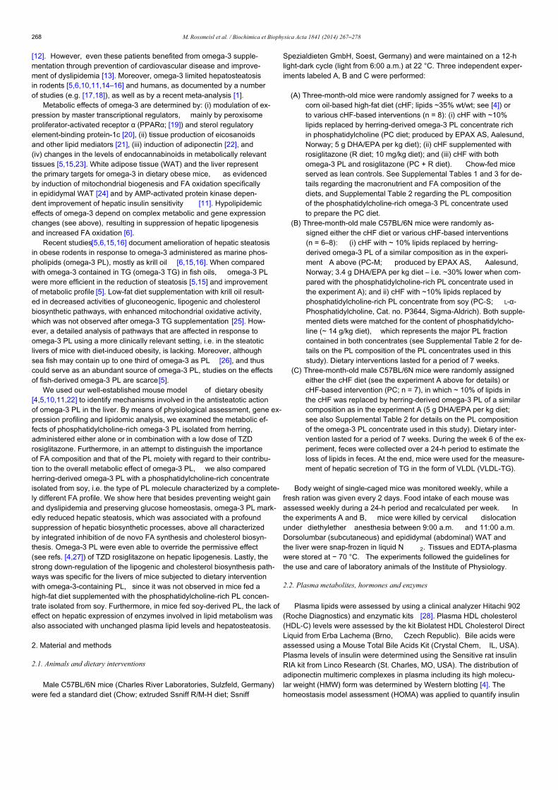
[12]. However, even these patients benefited from omega-3 supple-mentation through prevention of cardiovascular disease and improve-ment of dyslipidemia [13]. Moreover, omega-3 limited hepatosteatosisin rodents [5,6,10,11,14–16] and humans, as documented by a numberof studies (e.g. [17,18]), as well as by a recent meta-analysis [1].
Metabolic effects of omega-3 are determined by: (i) modulation of ex-pression by master transcriptional regulators, mainly by peroxisomeproliferator-activated receptor α (PPARα; [19]) and sterol regulatoryelement-binding protein-1c [20], (ii) tissue production of eicosanoidsand other lipid mediators [21], (iii) induction of adiponectin [22], and(iv) changes in the levels of endocannabinoids in metabolically relevanttissues [5,15,23]. White adipose tissue (WAT) and the liver representthe primary targets for omega-3 in dietary obese mice, as evidencedby induction of mitochondrial biogenesis and FA oxidation specificallyin epididymal WAT [24] and by AMP-activated protein kinase depen-dent improvement of hepatic insulin sensitivity [11]. Hypolipidemiceffects of omega-3 depend on complex metabolic and gene expressionchanges (see above), resulting in suppression of hepatic lipogenesisand increased FA oxidation [6].
Recent studies[5,6,15,16] document amelioration of hepatic steatosisin obese rodents in response to omega-3 administered as marine phos-pholipids (omega-3 PL), mostly as krill oil [6,15,16]. When comparedwith omega-3 contained in TG (omega-3 TG) in fish oils, omega-3 PLwere more efficient in the reduction of steatosis [5,15] and improvementof metabolic profile [5]. Low-fat diet supplementation with krill oil result-ed in decreased activities of gluconeogenic, lipogenic and cholesterolbiosynthetic pathways, with enhanced mitochondrial oxidative activity,which was not observed after omega-3 TG supplementation [25]. How-ever, a detailed analysis of pathways that are affected in response toomega-3 PL using a more clinically relevant setting, i.e. in the steatoticlivers of mice with diet-induced obesity, is lacking. Moreover, althoughsea fish may contain up to one third of omega-3 as PL [26], and thuscould serve as an abundant source of omega-3 PL, studies on the effectsof fish-derived omega-3 PL are scarce[5].
We used our well-established mouse model of dietary obesity[4,5,10,11,22] to identify mechanisms involved in the antisteatotic actionof omega-3 PL in the liver. By means of physiological assessment, gene ex-pression profiling and lipidomic analysis, we examined the metabolic ef-fects of phosphatidylcholine-rich omega-3 PL isolated from herring,administered either alone or in combination with a low dose of TZDrosiglitazone. Furthermore, in an attempt to distinguish the importanceof FA composition and that of the PL moiety with regard to their contribu-tion to the overall metabolic effect of omega-3 PL, we also comparedherring-derived omega-3 PL with a phosphatidylcholine-rich concentrateisolated from soy, i.e. the type of PL molecule characterized by a complete-ly different FA profile. We show here that besides preventing weight gainand dyslipidemia and preserving glucose homeostasis, omega-3 PL mark-edly reduced hepatic steatosis, which was associated with a profoundsuppression of hepatic biosynthetic processes, above all characterizedby integrated inhibition of de novo FA synthesis and cholesterol biosyn-thesis. Omega-3 PL were even able to override the permissive effect(see refs. [4,27]) of TZD rosiglitazone on hepatic lipogenesis. Lastly, thestrong down-regulation of the lipogenic and cholesterol biosynthesis path-ways was specific for the livers of mice subjected to dietary interventionwith omega-3-containing PL, since it was not observed in mice fed ahigh-fat diet supplemented with the phosphatidylcholine-rich PL concen-trate isolated from soy. Furthermore, in mice fed soy-derived PL, the lack ofeffect on hepatic expression of enzymes involved in lipid metabolism wasalso associated with unchanged plasma lipid levels and hepatosteatosis.
2. Material and methods
2.1. Animals and dietary interventions
Male C57BL/6N mice (Charles River Laboratories, Sulzfeld, Germany)were fed a standard diet (Chow; extruded Ssniff R/M-H diet; Ssniff
Spezialdieten GmbH, Soest, Germany) and were maintained on a 12-hlight-dark cycle (light from 6:00 a.m.) at 22 °C. Three independent exper-iments labeled A, B and C were performed:
(A) Three-month-old mice were randomly assigned for 7 weeks to acorn oil-based high-fat diet (cHF; lipids ~35% wt/wt; see [4]) orto various cHF-based interventions (n = 8): (i) cHF with ~10%lipids replaced by herring-derived omega-3 PL concentrate richin phosphatidylcholine (PC diet; produced by EPAX AS, Aalesund,Norway; 5 g DHA/EPA per kg diet); (ii) cHF supplemented withrosiglitazone (R diet; 10 mg/kg diet); and (iii) cHF with bothomega-3 PL and rosiglitazone (PC + R diet). Chow-fed miceserved as lean controls. See Supplemental Tables 1 and 3 for de-tails regarding the macronutrient and FA composition of thediets, and Supplemental Table 2 regarding the PL compositionof the phosphatidylcholine-rich omega-3 PL concentrate usedto prepare the PC diet.
(B) Three-month-old male C57BL/6N mice were randomly as-signed either the cHF diet or various cHF-based interventions(n = 6–8): (i) cHF with ~ 10% lipids replaced by herring-derived omega-3 PL of a similar composition as in the experi-ment A above (PC-M; produced by EPAX AS, Aalesund,Norway; 3.4 g DHA/EPA per kg diet – i.e. ~30% lower when com-pared with the phosphatidylcholine-rich PL concentrate used inthe experiment A); and ii) cHF with ~10% lipids replaced byphosphatidylcholine-rich PL concentrate from soy (PC-S; L-α-Phosphatidylcholine, Cat. no. P3644, Sigma-Aldrich). Both supple-mented diets were matched for the content of phosphatidylcho-line (~ 14 g/kg diet), which represents the major PL fractioncontained in both concentrates (see Supplemental Table 2 for de-tails on the PL composition of the PL concentrates used in thisstudy). Dietary interventions lasted for a period of 7 weeks.
(C) Three-month-old male C57BL/6N mice were randomly assignedeither the cHF diet (see the experiment A above for details) orcHF-based intervention (PC; n = 7), in which ~ 10% of lipids inthe cHF was replaced by herring-derived omega-3 PL of a similarcomposition as in the experiment A (5 g DHA/EPA per kg diet;see also Supplemental Table 2 for details on the PL compositionof the omega-3 PL concentrate used in this study). Dietary inter-vention lasted for a period of 7 weeks. During the week 6 of the ex-periment, feces were collected over a 24-h period to estimate theloss of lipids in feces. At the end, mice were used for the measure-ment of hepatic secretion of TG in the form of VLDL (VLDL-TG).
Body weight of single-caged mice was monitored weekly, while afresh ration was given every 2 days. Food intake of each mouse wasassessed weekly during a 24-h period and recalculated per week. Inthe experiments A and B, mice were killed by cervical dislocationunder diethylether anesthesia between 9:00 a.m. and 11:00 a.m.Dorsolumbar (subcutaneous) and epididymal (abdominal) WAT andthe liver were snap-frozen in liquid N 2. Tissues and EDTA-plasmawere stored at − 70 °C. The experiments followed the guidelines forthe use and care of laboratory animals of the Institute of Physiology.
2.2. Plasma metabolites, hormones and enzymes
Plasma lipids were assessed by using a clinical analyzer Hitachi 902(Roche Diagnostics) and enzymatic kits [28]. Plasma HDL cholesterol(HDL-C) levels were assessed by the kit Biolatest HDL Cholesterol DirectLiquid from Erba Lachema (Brno, Czech Republic). Bile acids wereassessed using a Mouse Total Bile Acids Kit (Crystal Chem, IL, USA).Plasma levels of insulin were determined using the Sensitive rat insulinRIA kit from Linco Research (St. Charles, MO, USA). The distribution ofadiponectin multimeric complexes in plasma including its high molecu-lar weight (HMW) form was determined by Western blotting [4]. Thehomeostasis model assessment (HOMA) was applied to quantify insulin
268 M. Rossmeisl et al. / Biochimica et Biophysica Acta 1841 (2014) 267–278
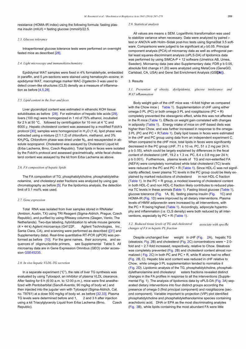
resistance (HOMA-IR index) using the following formula: fasting plas-ma insulin (mIU/l) × fasting glucose (mmol/l)/22.5.
2.3. Glucose tolerance
Intraperitoneal glucose tolerance tests were performed on overnightfasted mice as described [28].
2.4. Light microscopy and immunohistochemistry
Epididymal WAT samples were fixed in 4% formaldehyde, embeddedin paraffin, and 5 μm-sections were stained using hematoxylin–eosine; inepididymal WAT, macrophage marker MAC-2/galectin-3 was used todetect crown-like structures (CLS) density as a measure of inflamma-tion as before [4,5,28].
2.5. Lipid content in the liver and feces
Liver glycerolipid content was estimated in ethanolic KOH tissuesolubilisates as before [28]. For estimation of hepatic bile acids [29],livers (100 mg) were homogenized in 1 ml of 75% ethanol, incubatedfor 2 h at 50 °C, followed by centrifugation for 10 min at 4 °C and6000 g. Hepatic cholesterol content was assessed by a modified Folch'sprotocol [30]; samples were homogenized in H 2O (1:4), lipid phase wasextracted using a mixture (2:1:1.2) of chloroform, methanol, and 3%KH2PO4. Chloroform phase was dried under N2, and resuspended in ab-solute isopropanol. Cholesterol was assayed by Cholesterol Liquid kit(Erba Lachema, Brno, Czech Republic). Total lipids in feces were isolatedby the Folch's protocol and assessed gravimetrically, while fecal choles-terol content was assayed by the kit from Erba Lachema as above.
2.6. FA composition of hepatic lipids
The FA composition of TG, phosphatidylcholine, phosphatidyletha-nolamine, and cholesteryl ester fractions was analyzed by using gaschromatography as before [5]. For the lipidomics analysis, the detectionlimit of 0.1 mol% was used.
2.7. Gene expression
Total RNA was isolated from liver samples stored in RNAlater(Ambion, Austin, TX) using TRI Reagent (Sigma-Aldrich, Prague, CzechRepublic), and purified by using RNeasy columns (Qiagen, Venlo, TheNetherlands). Two-dye labeling, hybridization to whole mouse genome(4 × 44 k) Agilent microarrays (G4122F, Agilent Technologies, Inc.,Santa Clara, CA), and scanning were performed as described ([31] andSupplementary data). Real-time quantitative RT-PCR (qPCR) was per-formed as before [10]. For the gene names, their acronyms, and se-quences of oligonucleotide primers, see Supplemental Table 5. Allmicroarray data are in Gene Expression Omnibus (GEO) under acces-sion GSE45235.
2.8. In vivo hepatic VLDL-TG secretion
In a separate experiment (“C”), the rate of liver TG synthesis wasevaluated by using Tyloxapol, an inhibitor of plasma VLDL clearance.After fasting for 6 h (6:00 a.m. to 12:00 p.m.), mice were first anesthe-tized with Pentobarbital (Sanofi-Aventis; 90 mg/kg of body wt.) andthen injected into the jugular vein with Tyloxapol (Sigma-Aldrich, Cat.no. T8761) at a dose 500 mg/kg of body wt. as before [32,33]. PlasmaTG levels were determined before and 1, 2 and 3 h after injectionusing a kit Triacylglyceroly Liquid from Erba Lachema (Brno, CzechRepublic).
2.9. Statistical analysis
All values are means ± SEM. Logarithmic transformation was usedto stabilize variance when necessary. Data were analyzed by paired t-test or ANOVA with Holm–Sidak post-hoc tests using SigmaStat 3.5 soft-ware. Comparisons were judged to be significant at p b0.05. Principalcomponent analysis (PCA) of microarray data as well as orthogonal par-tial least squares-discriminant analysis (oPLS-DA) of lipidomics datawas performed by using SIMCA-P + 12 software (Umetrics AB, Umea,Sweden). Microarray data (see also Supplementary data; FDR p b 0.05,absolute fold change ≥1.20) were analyzed using MetaCore (GeneGO,Carlsbad, CA, USA) and Gene Set Enrichment Analysis (GSEA;[34]).
3. Results
3.1. Prevention of obesity, dyslipidemia, glucose intolerance and
WAT inflammation
Body weight gain of the cHF mice was ~4-fold higher as comparedwith the Chow mice ( Table 1). Supplementation of cHF using eitheromega-3 PL (PC) or both omega-3 PL and rosiglitazone (PC + R)completely prevented the obesogenic effect, while this was not affectedin the R mice (Table 1). Effects on weight gain correlated with changesin adiposity (Table 1). Energy intake of mice on cHF-based diets washigher than Chow, and was further increased in response to the omega-3 PL (PC and PC + R;Table 1). Daily lipid losses in feces were estimatedin the cHF and PC group using data from an independent experiment C.When compared to the cHF mice, total lipids in feces were significantlydecreased in the PC group (cHF, 71 ± 10 vs. PC, 51 ± 2 mg per 24 h;p b 0.05), which could be largely explained by differences in the fecalcontent of cholesterol (cHF, 19.2 ± 1.3 vs. PC, 8.4 ± 0.8 mg per 24 h;p b 0.001). Furthermore, plasma levels of TG and non-esterified FA(NEFA) were completely normalized while total cholesterol (TC) levelswere reduced in the PC and PC + R (Table 1). Since HDL-C was not signif-icantly affected, lower plasma TC levels in the PC group could be likely ex-plained by marked reductions of cholesterol in non HDL-C fraction(Table 1). In the PC + R group, a modest lowering of cholesterol contentin both HDL-C and non HDL-C fraction likely contributes to reduced plas-ma TC levels in these animals (Table 1). Fasting blood glucose (Table 1),glucose tolerance (Fig. 1A, B), fasting plasma insulin (Fig. 1C) andHOMA-IR (Fig. 1D) were improved by all dietary interventions. Plasmalevels of HMW adiponectin were increased by all interventions, withthe PC + R being highest (Table 1). Accordingly, adipocyte hypertro-phy and inflammation (i.e. CLS density) were both reduced by all inter-ventions, especially by PC + R (Table 1).
3.2. Reduced tissue levels of TG and cholesterol associate with specificchanges of FA in hepatic PL fraction
Despite unchanged liver weight in cHF (Fig. 2A), hepatic TG(steatosis; Fig. 2B) and cholesterol (Fig. 2C) concentrations were ~ 2.0-fold and ~ 2.7-fold increased, respectively, relative to Chow. Steatosiswas completely prevented (Fig. 2B) and cholesterol content almost nor-malized ( Fig. 2C) in both PC and PC + R, while R alone had no effect(Fig. 2B, C). Hepatic bile acid content was reduced in cHF relative toChow, while omega-3 PL supplementation tended to normalize it(Fig. 2D). Lipidomic analysis of the TG, phosphatidylcholine, phosphati-dylethanolamine and cholesteryl esters fractions revealed distinctchanges in the FA profiles in response to all the interventions (Supple-mental Fig. 1). The analysis of lipidomics data by oPLS-DA (Fig. 3A) sep-arated dietary interventions into four distinct groups according thepresence of omega-3 (first principal component) and rosiglitazone (sec-ond component). Variable important to projection (VIP) plot identifiedphosphatidylcholine and phosphatidylethanolamine species containingarachidonic acid, DHA or EPA as the most discriminating analytes(Fig. 3B), while lipids containing the most abundant FA were little
269M. Rossmeisl et al. / Biochimica et Biophysica Acta 1841 (2014) 267–278
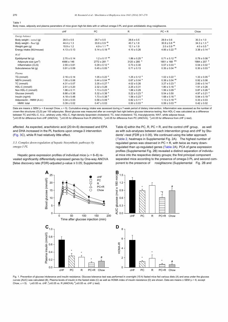
affected. As expected, arachidonic acid (20:4n-6) decreased and EPAand DHA increased in the PL fractions upon omega-3 intervention(Fig. 3C), while R had relatively little effect.
3.3. Complex down-regulation of hepatic biosynthetic pathways byomega-3 PL
Hepatic gene expression profiles of individual mice (n = 6–8) re-vealed significantly differentially expressed genes by One-way ANOVA[false discovery rate (FDR)-adjusted p-value ≤ 0.05; Supplemental
Table 4] within the PC, R, PC + R, and the control cHF group, as wellas with sub-analyses between each intervention group and cHF by Stu-dents' t-test (FDR p b 0.05). We continued using the latter approach(Table 2, heatmaps in Supplemental Fig. 2A). The highest number ofregulated genes was observed in PC + R, with twice as many down-regulated than up-regulated genes (Table 2A). PCA of gene expressionprofiles (Supplemental Fig. 2B) revealed a distinct separation of individu-al mice into the respective dietary groups; the first principal componentseparated mice according to the presence of omega-3 PL and second com-ponent to the presence of rosiglitazone (Supplemental Fig. 2B and
Table 1Body mass, adiposity and plasma parameters of mice given high-fat diets with or without omega-3 PL and given antidiabetic drug rosiglitazone.
cHF PC R PC + R Chow
Energy balance
Body weight – initial (g) 28.5 ± 0.5 28.7 ± 0.5 28.6 ± 0.5 28.9 ± 0.6 30.3 ± 1.0Body weight – final (g) 44.4 ± 1.3 33.6 ± 0.9 ab 40.7 ± 1.8 30.9 ± 0.8 ab 34.3 ± 1.3 d
Weight gain (g) 15.9 ± 1.2 4.9 ± 1.1 ab 12.1 ± 1.9 2.0 ± 0.9 ab 4.0 ± 0.5 d
Energy intake (MJ/mouse) 4.13 ± 0.13 5.14 ± 0.18 ab 4.10 ± 0.26 4.95 ± 0.22 ab 3.38 ± 0.14 d
WAT
Epididymal fat (g) 2.70 ± 0.14 1.2 ± 0.17 ab 1.86 ± 0.25 a 0.71 ± 0.12 ab 0.79 ± 0.08 d
Adipocyte size (μm2) 4066 ± 146 2772 ± 281 a 3120 ± 285 a 1851 ± 168 abc 1984 ± 207 d
Inflammation (CLS) 2.55 ± 0.61 0.29 ± 0.12 a 0.73 ± 0.30 0.07 ± 0.03 a 0.04 ± 0.02 d
Subcutaneous fat (g) 0.91 ± 0.09 0.45 ± 0.05 a 0.71 ± 0.13 0.35 ± 0.04 ab 0.30 ± 0.03 d
Plasma
TG (mmol/l) 2.16 ± 0.14 1.05 ± 0.22 a 1.25 ± 0.12 a 1.02 ± 0.22 a 1.33 ± 0.05 d
NEFA (mmol/l) 1.00 ± 0.06 0.44 ± 0.04 ab 0.67 ± 0.04 a 0.38 ± 0.04 ab 0.92 ± 0.06TC (mmol/l) 4.31 ± 0.07 3.35 ± 0.27 a 4.02 ± 0.26 3.27 ± 0.23 a 2.60 ± 0.14 d
HDL-C (mmol/l) 2.51 ± 0.20 2.32 ± 0.28 2.25 ± 0.31 1.80 ± 0.16 d 1.91 ± 0.26Non HDL-C (mmol/l) 1.96 ± 0.11 1.13 ± 0.23 a 1.88 ± 0.29 1.56 ± 0.09 d 0.87 ± 0.26 d
Glucose (mmol/l) 6.66 ± 0.36 5.32 ± 0.38 d 5.22 ± 0.23 d 5.54 ± 0.50 4.97 ± 0.21 d
Insulin (ng/ml) 4.18 ± 0.48 1.73 ± 0.38 a 1.58 ± 0.23 a 1.68 ± 0.16 a 0.94 ± 0.19 d
Adiponectin - HMW (A.U.) 0.34 ± 0.03 0.56 ± 0.09 a 0.82 ± 0.11 a 1.13 ± 0.18 ac 0.33 ± 0.03HMW: total 0.39 ± 0.02 0.47 ± 0.03 0.50 ± 0.03 a 0.59 ± 0.03 ac 0.42 ± 0.02
Data are means ± SEM (n = 8 except Chow, n = 5). Cumulative energy intake was assessed during a 7-week period of dietary intervention. Inflammation was assessed as the number ofcrown-like structures (CLS) per 100 adipocytes. Blood glucose was measured after an overnight fast right before glucose tolerance testing. Non HDL-C was calculated as a differencebetween TC and HDL-C. A.U., arbitrary units; HDL-C, High-density lipoprotein cholesterol; TC, total cholesterol; TG, triacylglycerols; WAT, white adipose tissue.ap≤0.05 for difference from cHF (ANOVA); bp≤0.05 for difference from R (ANOVA); cp≤0.05 for difference from PC (ANOVA); dp≤0.05 for difference from cHF (t-test).
cHF PC R PC+R Chow
Pla
sma
insu
lin (
nmo
l/l)
0.0
0.2
0.4
0.6
0.8
1.0C
**
*#
Time after glucose injection (min)0 50 100 150 200
Blo
od g
luco
se (
mm
ol/l
)
0
5
10
15
20
25cHFPCRPC+RChow
A
cHF PC R PC+R Chow
AU
C g
luco
se (
mo
l x 1
80
min
)
0.0
0.3
0.6
0.9
1.2
1.5B
*
*†
#
cHF PC R PC+R Chow
HO
MA
-IR
0
2
4
6
8D
*
*
* #
Fig. 1. Prevention of glucose intolerance and insulin resistance. Glucose tolerance test was performed in overnight (15-h) fasted mice fed various diets (A) and area under the glucosecurves (AUC) was calculated (B). Plasma levels of insulin in the fasted state (C) as well as HOMA index of insulin resistance (D) are shown. Data are means ± SEM (n = 8, exceptChow, n = 5). ⁎p≤0.05 vs. cHF; †p≤0.05 vs. R (ANOVA). #p≤0.05 vs. cHF (t test).
270 M. Rossmeisl et al. / Biochimica et Biophysica Acta 1841 (2014) 267–278
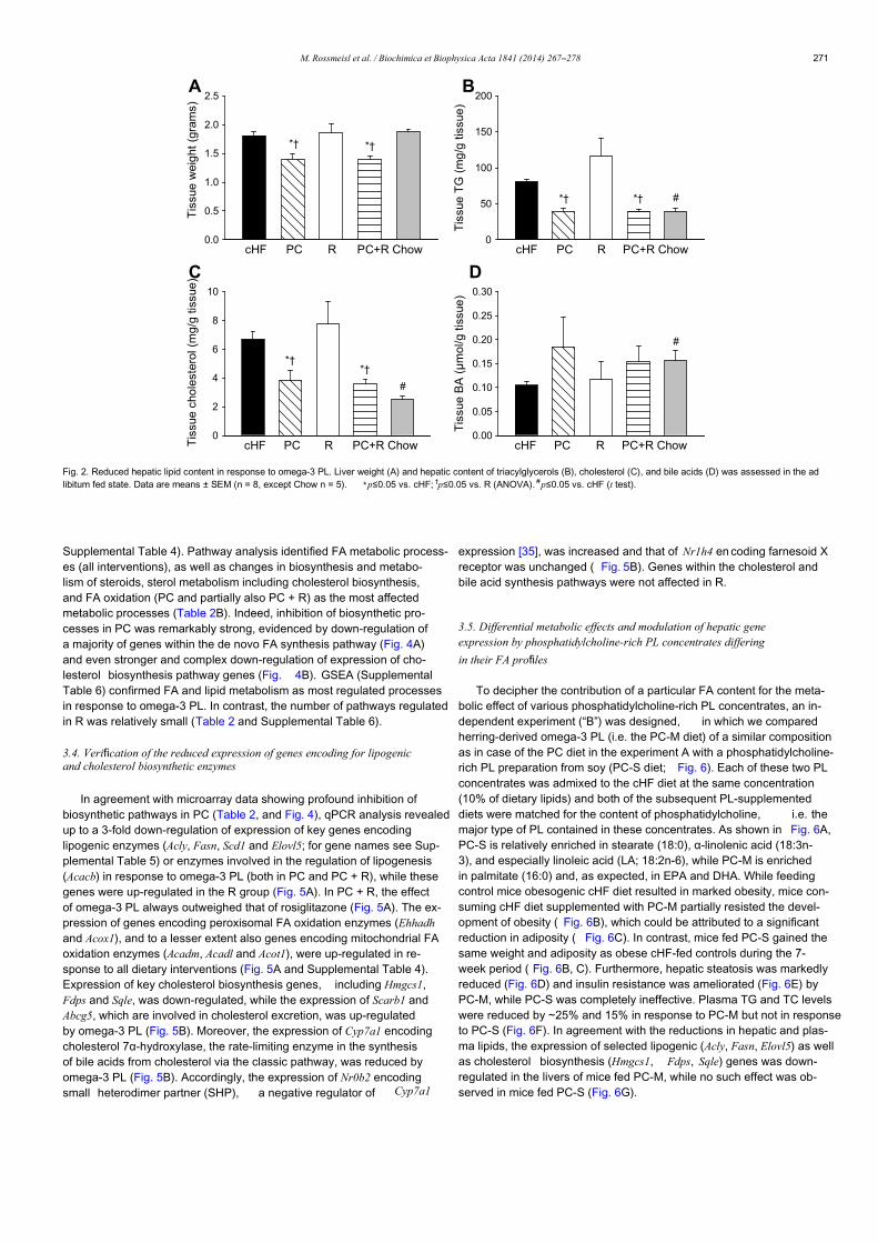
Supplemental Table 4). Pathway analysis identified FA metabolic process-es (all interventions), as well as changes in biosynthesis and metabo-lism of steroids, sterol metabolism including cholesterol biosynthesis,and FA oxidation (PC and partially also PC + R) as the most affectedmetabolic processes (Table 2B). Indeed, inhibition of biosynthetic pro-cesses in PC was remarkably strong, evidenced by down-regulation ofa majority of genes within the de novo FA synthesis pathway (Fig. 4A)and even stronger and complex down-regulation of expression of cho-lesterol biosynthesis pathway genes (Fig. 4B). GSEA (SupplementalTable 6) confirmed FA and lipid metabolism as most regulated processesin response to omega-3 PL. In contrast, the number of pathways regulatedin R was relatively small (Table 2 and Supplemental Table 6).
3.4. Verification of the reduced expression of genes encoding for lipogenicand cholesterol biosynthetic enzymes
In agreement with microarray data showing profound inhibition ofbiosynthetic pathways in PC (Table 2, and Fig. 4), qPCR analysis revealedup to a 3-fold down-regulation of expression of key genes encodinglipogenic enzymes (Acly, Fasn, Scd1 and Elovl5; for gene names see Sup-plemental Table 5) or enzymes involved in the regulation of lipogenesis(Acacb) in response to omega-3 PL (both in PC and PC + R), while thesegenes were up-regulated in the R group (Fig. 5A). In PC + R, the effectof omega-3 PL always outweighed that of rosiglitazone (Fig. 5A). The ex-pression of genes encoding peroxisomal FA oxidation enzymes (Ehhadhand Acox1), and to a lesser extent also genes encoding mitochondrial FAoxidation enzymes (Acadm, Acadl and Acot1), were up-regulated in re-sponse to all dietary interventions (Fig. 5A and Supplemental Table 4).Expression of key cholesterol biosynthesis genes, including Hmgcs1,Fdps and Sqle, was down-regulated, while the expression of Scarb1 andAbcg5, which are involved in cholesterol excretion, was up-regulatedby omega-3 PL (Fig. 5B). Moreover, the expression of Cyp7a1 encodingcholesterol 7α-hydroxylase, the rate-limiting enzyme in the synthesisof bile acids from cholesterol via the classic pathway, was reduced byomega-3 PL (Fig. 5B). Accordingly, the expression of Nr0b2 encodingsmall heterodimer partner (SHP), a negative regulator of Cyp7a1
expression [35], was increased and that of Nr1h4 en coding farnesoid Xreceptor was unchanged ( Fig. 5B). Genes within the cholesterol andbile acid synthesis pathways were not affected in R.
3.5. Differential metabolic effects and modulation of hepatic geneexpression by phosphatidylcholine-rich PL concentrates differing
in their FA profiles
To decipher the contribution of a particular FA content for the meta-bolic effect of various phosphatidylcholine-rich PL concentrates, an in-dependent experiment (“B”) was designed, in which we comparedherring-derived omega-3 PL (i.e. the PC-M diet) of a similar compositionas in case of the PC diet in the experiment A with a phosphatidylcholine-rich PL preparation from soy (PC-S diet; Fig. 6). Each of these two PLconcentrates was admixed to the cHF diet at the same concentration(10% of dietary lipids) and both of the subsequent PL-supplementeddiets were matched for the content of phosphatidylcholine, i.e. themajor type of PL contained in these concentrates. As shown in Fig. 6A,PC-S is relatively enriched in stearate (18:0), α-linolenic acid (18:3n-3), and especially linoleic acid (LA; 18:2n-6), while PC-M is enrichedin palmitate (16:0) and, as expected, in EPA and DHA. While feedingcontrol mice obesogenic cHF diet resulted in marked obesity, mice con-suming cHF diet supplemented with PC-M partially resisted the devel-opment of obesity ( Fig. 6B), which could be attributed to a significantreduction in adiposity ( Fig. 6C). In contrast, mice fed PC-S gained thesame weight and adiposity as obese cHF-fed controls during the 7-week period ( Fig. 6B, C). Furthermore, hepatic steatosis was markedlyreduced (Fig. 6D) and insulin resistance was ameliorated (Fig. 6E) byPC-M, while PC-S was completely ineffective. Plasma TG and TC levelswere reduced by ~25% and 15% in response to PC-M but not in responseto PC-S (Fig. 6F). In agreement with the reductions in hepatic and plas-ma lipids, the expression of selected lipogenic (Acly, Fasn, Elovl5) as wellas cholesterol biosynthesis (Hmgcs1, Fdps, Sqle) genes was down-regulated in the livers of mice fed PC-M, while no such effect was ob-served in mice fed PC-S (Fig. 6G).
cHF PC R PC+R ChowTis
sue
cho
lest
erol
(m
g/g
tis
sue
)
0
2
4
6
8
10
#
*†*†
A
cHF PC R PC+R Chow
cHF PC R PC+R Chow
cHF PC R PC+R Chow
Tis
sue
wei
ght
(gr
ams)
0.0
0.5
1.0
1.5
2.0
2.5
*† *†
Tis
sue
TG
(m
g/g
tis
sue
)
0
50
100
150
200
*† *† #
Tis
sue
BA
(µ
mo
l/g t
issu
e)
0.00
0.05
0.10
0.15
0.20
0.25
0.30
#
B
C D
Fig. 2. Reduced hepatic lipid content in response to omega-3 PL. Liver weight (A) and hepatic content of triacylglycerols (B), cholesterol (C), and bile acids (D) was assessed in the adlibitum fed state. Data are means ± SEM (n = 8, except Chow n = 5). ⁎p≤0.05 vs. cHF; †p≤0.05 vs. R (ANOVA). #p≤0.05 vs. cHF (t test).
271M. Rossmeisl et al. / Biochimica et Biophysica Acta 1841 (2014) 267–278
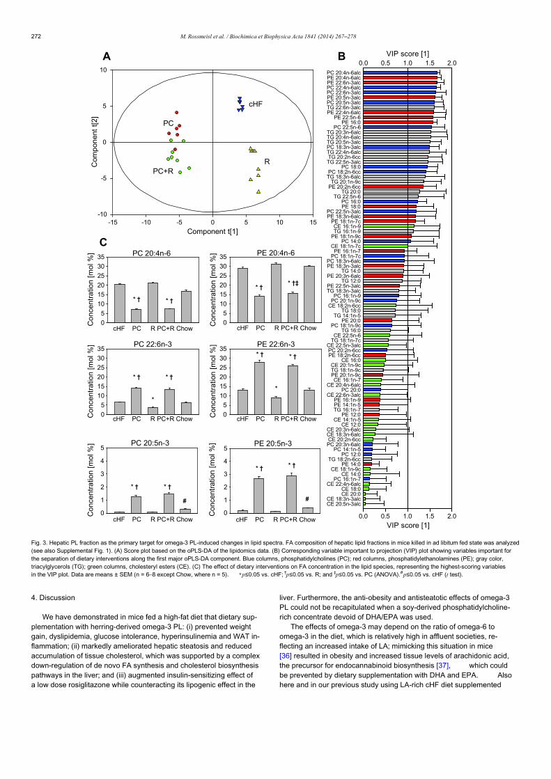
4. Discussion
We have demonstrated in mice fed a high-fat diet that dietary sup-plementation with herring-derived omega-3 PL: (i) prevented weightgain, dyslipidemia, glucose intolerance, hyperinsulinemia and WAT in-flammation; (ii) markedly ameliorated hepatic steatosis and reducedaccumulation of tissue cholesterol, which was supported by a complexdown-regulation of de novo FA synthesis and cholesterol biosynthesispathways in the liver; and (iii) augmented insulin-sensitizing effect ofa low dose rosiglitazone while counteracting its lipogenic effect in the
liver. Furthermore, the anti-obesity and antisteatotic effects of omega-3PL could not be recapitulated when a soy-derived phosphatidylcholine-rich concentrate devoid of DHA/EPA was used.
The effects of omega-3 may depend on the ratio of omega-6 toomega-3 in the diet, which is relatively high in affluent societies, re-flecting an increased intake of LA; mimicking this situation in mice[36] resulted in obesity and increased tissue levels of arachidonic acid,the precursor for endocannabinoid biosynthesis [37], which couldbe prevented by dietary supplementation with DHA and EPA. Alsohere and in our previous study using LA-rich cHF diet supplemented
Component t[1]-15 -10 -5 0 5 10 15
Com
pon
ent
t[2]
-10
-5
0
5
10
PC
PC+R
cHF
R
Co
ncen
tra
tion
[mol
%]
0
5
10
15
20
25
30
35
* † * †
PC 20:4n-6
BA
C
Con
cent
ratio
n [m
ol %
]
0
5
10
15
20
25
30
35
* †* †‡
PE 20:4n-6
Con
cent
ratio
n [m
ol %
]
0
5
10
15
20
25
30
35* †
*
* †
PE 22:6n-3
cHF PC R PC+R Chow
cHF PC R PC+R Chow
cHF PC R PC+R Chow
cHF PC R PC+R Chow
cHF PC R PC+R Chow
cHF PC R PC+R Chow
Co
ncen
tra
tion
[mol
%]
0
5
10
15
20
25
30
35
* †
*
* †
PC 22:6n-3
Co
ncen
tra
tion
[mol
%]
0
1
2
3
4
5PC 20:5n-3
* †
#
* †
Co
ncen
trat
ion
[m
ol %
]
0
1
2
3
4
5
* †* †
#
PE 20:5n-3
VIP score [1]0.0 0.5 1.0 1.5 2.0
VIP score [1]0.0 0.5 1.0 1.5 2.0
PC 20:4n-6alcPE 20:4n-6alcPE 22:6n-3alcPC 22:4n-6alcPC 22:6n-3alcPE 20:5n-3alcPC 20:5n-3alcTG 22:6n-3alcPE 22:4n-6alc
PE 22:5n-6PE 16:0
PC 22:5n-6TG 20:3n-6alcTG 20:4n-6alcTG 20:5n-3alcPC 18:3n-3alcTG 22:4n-6alcTG 20:2n-6ccTG 22:5n-3alc
PC 18:0PC 18:2n-6ccTG 18:3n-6alc
TG 20:1n-9cPE 20:2n-6cc
TG 20:0TG 22:5n-6
PC 16:0PE 18:0
PC 22:5n-3alcPE 18:3n-6alc
PE 18:1n-7cCE 16:1n-9TG 16:1n-9
PE 18:1n-9cPC 14:0
CE 18:1n-7cPE 16:1n-7
PC 18:1n-7cPC 18:3n-6alcPE 18:3n-3alc
TG 14:0PE 20:3n-6alc
TG 12:0PE 22:5n-3alcTG 18:3n-3alc
PC 16:1n-9PC 20:1n-9c
CE 18:2n-6ccTG 18:0
TG 14:1n-5PE 20:0
PC 18:1n-9cTG 16:0
CE 22:5n-6TG 18:1n-7c
CE 22:5n-3alcPC 20:2n-6ccPE 18:2n-6cc
CE 16:0CE 20:1n-9cTG 18:1n-9cPE 20:1n-9cCE 16:1n-7
CE 20:4n-6alcPC 20:0
CE 22:6n-3alcPE 16:1n-9PE 14:1n-5TG 16:1n-7
PE 12:0CE 14:1n-5
CE 12:0CE 20:3n-6alcCE 18:3n-6alcCE 20:2n-6ccPC 20:3n-6alc
PC 14:1n-5PC 12:0
TG 18:2n-6ccPE 14:0
CE 18:1n-9cCE 14:0
PC 16:1n-7CE 22:4n-6alc
CE 18:0CE 20:0
CE 18:3n-3alcCE 20:5n-3alc
Fig. 3. Hepatic PL fraction as the primary target for omega-3 PL-induced changes in lipid spectra. FA composition of hepatic lipid fractions in mice killed in ad libitum fed state was analyzed(see also Supplemental Fig. 1). (A) Score plot based on the oPLS-DA of the lipidomics data. (B) Corresponding variable important to projection (VIP) plot showing variables important forthe separation of dietary interventions along the first major oPLS-DA component. Blue columns, phosphatidylcholines (PC); red columns, phosphatidylethanolamines (PE); gray color,triacylglycerols (TG); green columns, cholesteryl esters (CE). (C) The effect of dietary interventions on FA concentration in the lipid species, representing the highest-scoring variablesin the VIP plot. Data are means ± SEM (n = 6–8 except Chow, where n = 5). ⁎p≤0.05 vs. cHF; †p≤0.05 vs. R; and ‡p≤0.05 vs. PC (ANOVA).#p≤0.05 vs. cHF (t test).
272 M. Rossmeisl et al. / Biochimica et Biophysica Acta 1841 (2014) 267–278
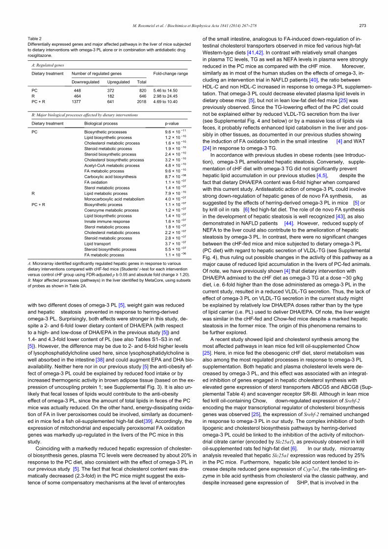
with two different doses of omega-3 PL [5], weight gain was reducedand hepatic steatosis prevented in response to herring-derivedomega-3 PL. Surprisingly, both effects were stronger in this study, de-spite a 2- and 6-fold lower dietary content of DHA/EPA (with respectto a high- and low-dose of DHA/EPA in the previous study [5]) and1.4- and 4.3-fold lower content of PL (see also Tables S1–S3 in ref.[5]). However, the difference may be due to 2- and 6-fold higher levelsof lysophosphatidylcholine used here, since lysophosphatidylcholine iswell absorbed in the intestine [38] and could augment EPA and DHA bio-availability. Neither here nor in our previous study [5] the anti-obesity ef-fect of omega-3 PL could be explained by reduced food intake or byincreased thermogenic activity in brown adipose tissue (based on the ex-pression of uncoupling protein 1; see Supplemental Fig. 3). It is also un-likely that fecal losses of lipids would contribute to the anti-obesityeffect of omega-3 PL, since the amount of total lipids in feces of the PCmice was actually reduced. On the other hand, energy-dissipating oxida-tion of FA in liver peroxisomes could be involved, similarly as document-ed in mice fed a fish oil-supplemented high-fat diet[39]. Accordingly, theexpression of mitochondrial and especially peroxisomal FA oxidationgenes was markedly up-regulated in the livers of the PC mice in thisstudy.
Coinciding with a markedly reduced hepatic expression of cholester-ol biosynthesis genes, plasma TC levels were decreased by about 20% inresponse to the PC diet, also consistent with the effect of omega-3 PL inour previous study [5]. The fact that fecal cholesterol content was dra-matically decreased (2.3-fold) in the PC mice might suggest the exis-tence of some compensatory mechanisms at the level of enterocytes
of the small intestine, analogous to FA-induced down-regulation of in-testinal cholesterol transporters observed in mice fed various high-fatWestern-type diets [41,42]. In contrast with relatively small changesin plasma TC levels, TG as well as NEFA levels in plasma were stronglyreduced in the PC mice as compared with the cHF mice. Moreover,similarly as in most of the human studies on the effects of omega-3, in-cluding an intervention trial in NAFLD patients [40], the ratio betweenHDL-C and non HDL-C increased in response to omega-3 PL supplemen-tation. That omega-3 PL could decrease elevated plasma lipid levels indietary obese mice [5], but not in lean low-fat diet-fed mice [25] waspreviously observed. Since the TG-lowering effect of the PC diet couldnot be explained either by reduced VLDL-TG secretion from the liver(see Supplemental Fig. 4 and below) or by a massive loss of lipids viafeces, it probably reflects enhanced lipid catabolism in the liver and pos-sibly in other tissues, as documented in our previous studies showingthe induction of FA oxidation both in the small intestine [4] and WAT[24] in response to omega-3 TG.
In accordance with previous studies in obese rodents (see Introduc-tion), omega-3 PL ameliorated hepatic steatosis. Conversely, supple-mentation of cHF diet with omega-3 TG did not significantly preventhepatic lipid accumulation in our previous studies [4,5], despite thefact that dietary DHA/EPA content was 6-fold higher when comparedwith this current study. Antisteatotic action of omega-3 PL could involvestrong down-regulation of hepatic genes of de novo FA synthesis, assuggested by the effects of herring-derived omega-3 PL in mice [5] orby krill oil in rats [6] fed high-fat diet. The role of de novo FA synthesisin the development of hepatic steatosis is well recognized [43], as alsodemonstrated in NAFLD patients [44]. However, reduced supply ofNEFA to the liver could also contribute to the amelioration of hepaticsteatosis by omega-3 PL. In contrast, there were no significant changesbetween the cHF-fed mice and mice subjected to dietary omega-3 PL(PC diet) with regard to hepatic secretion of VLDL-TG (see SupplementalFig. 4), thus ruling out possible changes in the activity of this pathway as amajor cause of reduced lipid accumulation in the livers of PC-fed animals.Of note, we have previously shown [4] that dietary intervention withDHA/EPA admixed to the cHF diet as omega-3 TG at a dose ~30 g/kgdiet, i.e. 6-fold higher than the dose administered as omega-3 PL in thecurrent study, resulted in a reduced VLDL-TG secretion. Thus, the lack ofeffect of omega-3 PL on VLDL-TG secretion in the current study mightbe explained by relatively low DHA/EPA doses rather than by the typeof lipid carrier (i.e. PL) used to deliver DHA/EPA. Of note, the liver weightwas similar in the cHF-fed and Chow-fed mice despite a marked hepaticsteatosis in the former mice. The origin of this phenomena remains tobe further explored.
A recent study showed lipid and cholesterol synthesis among themost affected pathways in lean mice fed krill oil-supplemented Chow[25]. Here, in mice fed the obesogenic cHF diet, sterol metabolism wasalso among the most regulated processes in response to omega-3 PLsupplementation. Both hepatic and plasma cholesterol levels were de-creased by omega-3 PL, and this effect was associated with an integrat-ed inhibition of genes engaged in hepatic cholesterol synthesis withelevated gene expression of sterol transporters ABCG5 and ABCG8 (Sup-plemental Table 4) and scavenger receptor SR-BI. Although in lean micefed krill oil-containing Chow, down-regulated expression of Srebf-2encoding the major transcriptional regulator of cholesterol biosynthesisgenes was observed [25], the expression of Srebf-2 remained unchangedin response to omega-3 PL in our study. The complex inhibition of bothlipogenic and cholesterol biosynthesis pathways by herring-derivedomega-3 PL could be linked to the inhibition of the activity of mitochon-drial citrate carrier (encoded by Slc25a1), as previously observed in krilloil-supplemented rats fed high-fat diet [6]. In our study, microarrayanalysis revealed that hepatic Slc25a1 expression was reduced by 25%in the PC mice. Furthermore, hepatic bile acid content tended to in-crease despite reduced gene expression of Cyp7a1, the rate-limiting en-zyme in bile acid synthesis from cholesterol via the classic pathway, anddespite increased gene expression of SHP, that is involved in the
Table 2Differentially expressed genes and major affected pathways in the liver of mice subjectedto dietary interventions with omega-3 PL alone or in combination with antidiabetic drugrosiglitazone.
A: Regulated genes
Dietary treatment Number of regulated genes Fold-change range
Downregulated Upregulated Total
PC 448 372 820 5.46 to 14.50R 464 182 646 2.98 to 24.45PC + R 1377 641 2018 4.69 to 10.40
B: Major biological processes affected by dietary interventions
Dietary treatment Biological process p-value
PC Biosynthetic processes 9.6 × 10 −11
Lipid biosynthetic process 1.2 × 10 −10
Cholesterol metabolic process 1.6 × 10 −10
Steroid metabolic process 1.9 × 10 −10
Steroid biosynthetic process 2.4 × 10 −10
Cholesterol biosynthetic process 3.2 × 10 −10
Acetyl-CoA metabolic process 4.8 × 10 −10
FA metabolic process 9.6 × 10 −10
Carboxylic acid biosynthesis 8.7 × 10 −08
FA oxidation 1.1 × 10 −07
Sterol metabolic process 1.4 × 10 −07
R Lipid metabolic process 7.9 × 10 −10
Monocarboxylic acid metabolism 4.0 × 10 −07
PC + R Biosynthetic process 1.1 × 10 −07
Coenzyme metabolic process 1.2 × 10 −07
Lipid biosynthetic process 1.4 × 10 −07
Innate immune response 1.6 × 10 −07
Sterol metabolic process 1.8 × 10 −07
Cholesterol metabolic process 2.2 × 10 −07
Steroid metabolic process 2.8 × 10 −07
Lipid transport 3.7 × 10 −07
Steroid biosynthetic process 5.5 × 10 −07
FA metabolic process 1.1 × 10 −06
A: Microrarray identified significantly regulated hepatic genes in response to variousdietary interventions compared with cHF-fed mice (Students' t-test for each interventionversus control cHF group using FDR-adjusted p b 0.05 and absolute fold change ≥ 1.20).B: Major affected processes (pathways) in the liver identified by MetaCore, using subsetsof probes as shown in Table 2A.
273M. Rossmeisl et al. / Biochimica et Biophysica Acta 1841 (2014) 267–278
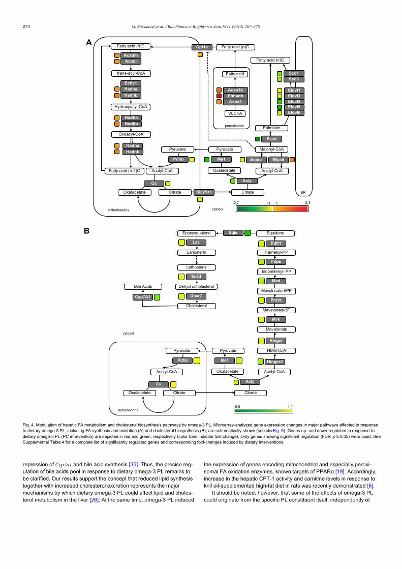
repression of Cyp7a1 and bile acid synthesis [35]. Thus, the precise reg-ulation of bile acids pool in response to dietary omega-3 PL remains tobe clarified. Our results support the concept that reduced lipid synthesistogether with increased cholesterol excretion represents the majormechanisms by which dietary omega-3 PL could affect lipid and choles-terol metabolism in the liver [26]. At the same time, omega-3 PL induced
the expression of genes encoding mitochondrial and especially peroxi-somal FA oxidation enzymes, known targets of PPARα [19]. Accordingly,increase in the hepatic CPT-1 activity and carnitine levels in response tokrill oil-supplemented high-fat diet in rats was recently demonstrated [6].
It should be noted, however, that some of the effects of omega-3 PLcould originate from the specific PL constituent itself, independently of
A
B
Hmgcs1
HMG-CoA
Mevalonate
Mevalonate-5P
Mevalonate-5PP
Isopentenyl- PP
Farnesyl-PP
SqualeneEpoxysqualene
Lanosterin
Lathosterol
Dehydrocholesterol
Cholesterol
Pyruvate
Acetyl-CoA Acetyl-CoA
Pyruvate
Oxalacetate
CitrateCitrate
Me1
AclyCs
Pdhb
Oxalacetate
mitochondria
cytosol
Hmgcr
Mvk
Pmvk
Mvd
Fdps
Fdft1
Sqle
Lss
Sc5d
Dhcr7
Bile Acids
Cyp7A1
0,1-5,5-
Pyruvate
Acetyl-CoA
Fatty acid (n)C
Palmitate
Malonyl-CoA
Acetyl-CoA
Pyruvate
Oxalacetate
CitrateCitrate
trans-acyl-CoA
Fatty acid (n)C
Fasn
MlycdAcacaMe1
AclyCs
Pdhb
Elovl1
Elovl6Elovl5Elovl3Elovl2
Scd1Scd3
Fatty acid (n-2)C
Oxoacyl-CoA
Hydroxyacyl-CoA
Hadhb
Hadha
AcadmAcadl
Echs1
Hadhb
Hadha
Hadhb
Hadha
Oxalacetate
mitochondria cytosol
ER
Fatty acid (n)C
VLCFA
Acaa1b
Acox1Ehhadh
Fatty acid
peroxisome
3,27,2- 1-1
Slc25a1
Cpt1a
Fig. 4. Modulation of hepatic FA metabolism and cholesterol biosynthesis pathways by omega-3 PL. Microarray-analyzed gene expression changes in major pathways affected in responseto dietary omega-3 PL, including FA synthesis and oxidation (A) and cholesterol biosynthesis (B), are schematically shown (see alsoFig. 5). Genes up- and down-regulated in response todietary omega-3 PL (PC intervention) are depicted in red and green, respectively (color bars indicate fold change). Only genes showing significant regulation (FDR, p b 0.05) were used. SeeSupplemental Table 4 for a complete list of significantly regulated genes and corresponding fold-changes induced by dietary interventions.
274 M. Rossmeisl et al. / Biochimica et Biophysica Acta 1841 (2014) 267–278
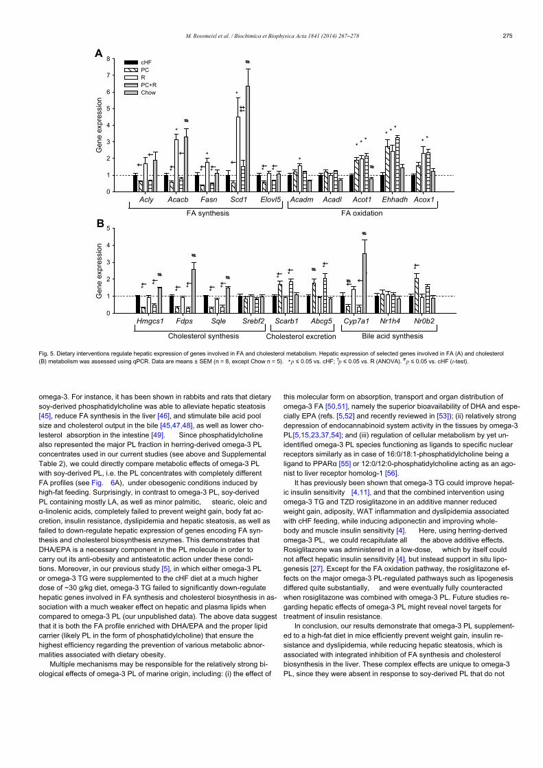
omega-3. For instance, it has been shown in rabbits and rats that dietarysoy-derived phosphatidylcholine was able to alleviate hepatic steatosis[45], reduce FA synthesis in the liver [46], and stimulate bile acid poolsize and cholesterol output in the bile [45,47,48], as well as lower cho-lesterol absorption in the intestine [49]. Since phosphatidylcholinealso represented the major PL fraction in herring-derived omega-3 PLconcentrates used in our current studies (see above and SupplementalTable 2), we could directly compare metabolic effects of omega-3 PLwith soy-derived PL, i.e. the PL concentrates with completely differentFA profiles (see Fig. 6A), under obesogenic conditions induced byhigh-fat feeding. Surprisingly, in contrast to omega-3 PL, soy-derivedPL containing mostly LA, as well as minor palmitic, stearic, oleic andα-linolenic acids, completely failed to prevent weight gain, body fat ac-cretion, insulin resistance, dyslipidemia and hepatic steatosis, as well asfailed to down-regulate hepatic expression of genes encoding FA syn-thesis and cholesterol biosynthesis enzymes. This demonstrates thatDHA/EPA is a necessary component in the PL molecule in order tocarry out its anti-obesity and antisteatotic action under these condi-tions. Moreover, in our previous study [5], in which either omega-3 PLor omega-3 TG were supplemented to the cHF diet at a much higherdose of ~30 g/kg diet, omega-3 TG failed to significantly down-regulatehepatic genes involved in FA synthesis and cholesterol biosynthesis in as-sociation with a much weaker effect on hepatic and plasma lipids whencompared to omega-3 PL (our unpublished data). The above data suggestthat it is both the FA profile enriched with DHA/EPA and the proper lipidcarrier (likely PL in the form of phosphatidylcholine) that ensure thehighest efficiency regarding the prevention of various metabolic abnor-malities associated with dietary obesity.
Multiple mechanisms may be responsible for the relatively strong bi-ological effects of omega-3 PL of marine origin, including: (i) the effect of
this molecular form on absorption, transport and organ distribution ofomega-3 FA [50,51], namely the superior bioavailability of DHA and espe-cially EPA (refs. [5,52] and recently reviewed in [53]); (ii) relatively strongdepression of endocannabinoid system activity in the tissues by omega-3PL[5,15,23,37,54]; and (iii) regulation of cellular metabolism by yet un-identified omega-3 PL species functioning as ligands to specific nuclearreceptors similarly as in case of 16:0/18:1-phosphatidylcholine being aligand to PPARα [55] or 12:0/12:0-phosphatidylcholine acting as an ago-nist to liver receptor homolog-1 [56].
It has previously been shown that omega-3 TG could improve hepat-ic insulin sensitivity [4,11], and that the combined intervention usingomega-3 TG and TZD rosiglitazone in an additive manner reducedweight gain, adiposity, WAT inflammation and dyslipidemia associatedwith cHF feeding, while inducing adiponectin and improving whole-body and muscle insulin sensitivity [4]. Here, using herring-derivedomega-3 PL, we could recapitulate all the above additive effects.Rosiglitazone was administered in a low-dose, which by itself couldnot affect hepatic insulin sensitivity [4], but instead support in situ lipo-genesis [27]. Except for the FA oxidation pathway, the rosiglitazone ef-fects on the major omega-3 PL-regulated pathways such as lipogenesisdiffered quite substantially, and were eventually fully counteractedwhen rosiglitazone was combined with omega-3 PL. Future studies re-garding hepatic effects of omega-3 PL might reveal novel targets fortreatment of insulin resistance.
In conclusion, our results demonstrate that omega-3 PL supplement-ed to a high-fat diet in mice efficiently prevent weight gain, insulin re-sistance and dyslipidemia, while reducing hepatic steatosis, which isassociated with integrated inhibition of FA synthesis and cholesterolbiosynthesis in the liver. These complex effects are unique to omega-3PL, since they were absent in response to soy-derived PL that do not
cHFPCRPC+RChow
A
B
† **†*†*†
†
*
#†
*†
#†‡
*†*†
*
#
**
***
***
#*†
*†
*†
*†#*†
*†
#*†
*†
*† *†#
##
†
†#
Hmgcs1 Fdps Sqle Srebf2 Scarb1 Abcg5 Cyp7a1 Nr1h4 Nr0b2
Gen
e ex
pre
ssio
n
0
1
2
3
4
5
Cholesterol synthesis Cholesterol excretion Bile acid synthesis
Acly Acacb Fasn Scd1 Elovl5 Acadm Acadl Acot1 Ehhadh Acox1
Gen
e ex
pre
ssio
n
0
1
2
3
4
5
6
7
8
FA synthesis FA oxidation
Fig. 5. Dietary interventions regulate hepatic expression of genes involved in FA and cholesterol metabolism. Hepatic expression of selected genes involved in FA (A) and cholesterol(B) metabolism was assessed using qPCR. Data are means ± SEM (n = 8, except Chow n = 5). ⁎p ≤ 0.05 vs. cHF; †p ≤ 0.05 vs. R (ANOVA). #p ≤ 0.05 vs. cHF (t-test).
275M. Rossmeisl et al. / Biochimica et Biophysica Acta 1841 (2014) 267–278
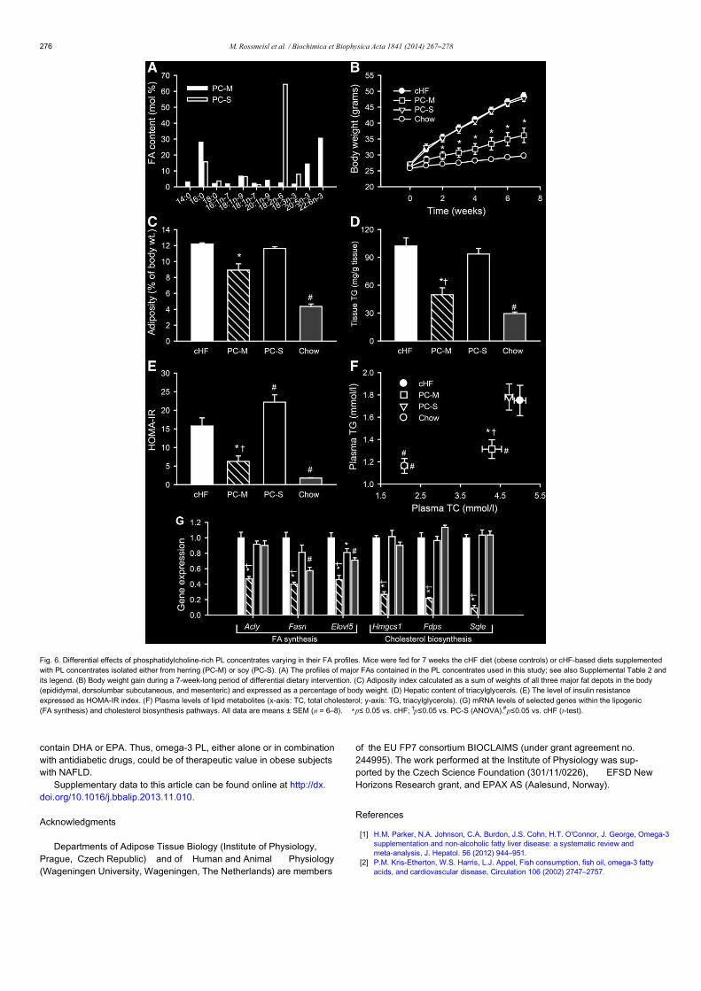
contain DHA or EPA. Thus, omega-3 PL, either alone or in combinationwith antidiabetic drugs, could be of therapeutic value in obese subjectswith NAFLD.
Supplementary data to this article can be found online at http://dx.doi.org/10.1016/j.bbalip.2013.11.010.
Acknowledgments
Departments of Adipose Tissue Biology (Institute of Physiology,Prague, Czech Republic) and of Human and Animal Physiology(Wageningen University, Wageningen, The Netherlands) are members
of the EU FP7 consortium BIOCLAIMS (under grant agreement no.244995). The work performed at the Institute of Physiology was sup-ported by the Czech Science Foundation (301/11/0226), EFSD NewHorizons Research grant, and EPAX AS (Aalesund, Norway).
References
[1] H.M. Parker, N.A. Johnson, C.A. Burdon, J.S. Cohn, H.T. O'Connor, J. George, Omega-3supplementation and non-alcoholic fatty liver disease: a systematic review andmeta-analysis, J. Hepatol. 56 (2012) 944–951.
[2] P.M. Kris-Etherton, W.S. Harris, L.J. Appel, Fish consumption, fish oil, omega-3 fattyacids, and cardiovascular disease, Circulation 106 (2002) 2747–2757.
Fig. 6. Differential effects of phosphatidylcholine-rich PL concentrates varying in their FA profiles. Mice were fed for 7 weeks the cHF diet (obese controls) or cHF-based diets supplementedwith PL concentrates isolated either from herring (PC-M) or soy (PC-S). (A) The profiles of major FAs contained in the PL concentrates used in this study; see also Supplemental Table 2 andits legend. (B) Body weight gain during a 7-week-long period of differential dietary intervention. (C) Adiposity index calculated as a sum of weights of all three major fat depots in the body(epididymal, dorsolumbar subcutaneous, and mesenteric) and expressed as a percentage of body weight. (D) Hepatic content of triacylglycerols. (E) The level of insulin resistanceexpressed as HOMA-IR index. (F) Plasma levels of lipid metabolites (x-axis: TC, total cholesterol; y-axis: TG, triacylglycerols). (G) mRNA levels of selected genes within the lipogenic(FA synthesis) and cholesterol biosynthesis pathways. All data are means ± SEM (n = 6–8). ⁎p≤ 0.05 vs. cHF; †p≤0.05 vs. PC-S (ANOVA).#p≤0.05 vs. cHF (t-test).
276 M. Rossmeisl et al. / Biochimica et Biophysica Acta 1841 (2014) 267–278
[3] D. Mozaffarian, R.N. Lemaitre, I.B. King, X. Song, H. Huang, F.M. Sacks, E.B. Rimm, M.Wang, D.S. Siscovick, Plasma phospholipid long-chain omega-3 fatty acids and totaland cause-specific mortality in older adults: a cohort study, Ann. Intern. Med. 158(2013) 515–525.
[4] O. Kuda, T. Jelenik, Z. Jilkova, P. Flachs, M. Rossmeisl, M. Hensler, L. Kazdova,N. Ogston, M. Baranowski, J. Gorski, P. Janovska, V. Kus, J. Polak, V.Mohamed-Ali, R. Burcelin, S. Cinti, M. Bryhn, J.Kopecky, n-3 Fatty acids androsiglitazone improve insulin sensitivity through additive stimulatory effectson muscle glycogen synthesis in mice fed a high-fat diet, Diabetologia 52(2009) 941–951.
[5] M. Rossmeisl, J.Z. Macek, O. Kuda, T. Jelenik, D. Medrikova, B. Stankova, B.Kristinsson, G.G. Haraldsson, H. Svensen, I. Stoknes, P. Sjovall, Y. Magnusson, M.G.Balvers, K.C. Verhoeckx,E. Tvrzicka, M. Bryhn, J. Kopecky,Metabolic effects of n-3PUFA as phospholipids are superior to triglycerides in mice fed a high-fat diet: pos-sible role of endocannabinoids, PLoS One 7 (2012) e38834.
[6] A. Ferramosca, A. Conte, L. Burri, K. Berge, N.F. De, A.M. Giudetti, V. Zara, A krill oil sup-plemented diet suppresses hepatic steatosis in high-fat fed rats, PLoS One 7 (2012)e38797.
[7] H.M. Roche, M.J. Gibney, Effect of long-chain n-3 polyunsaturated fatty acids onfasting and postprandial triacylglycerol metabolism, Am. J. Clin. Nutr. 71 (2000)232S–237S.
[8] B.K. Itariu, M. Zeyda, E.E.Hochbrugger, A. Neuhofer, G. Prager, K. Schindler, A.Bohdjalian, D. Mascher, S. Vangala, M. Schranz, M. Krebs, M.G. Bischof, T.M.Stulnig, Long-chain n-3 PUFAs reduce adipose tissue and systemic inflammationin severely obese nondiabetic patients: a randomized controlled trial, Am. J. Clin.Nutr. 96 (2012) 1137–1149.
[9] T.A. Mori, D.Q. Bao, V. Burke, I.B. Puddey, G.F. Watts, L.J. Beilin, Dietary fish as amajor component of a weight-loss diet: effect on serum lipids, glucose, and in-sulin metabolism in overweight hypertensive subjects, Am. J.Clin. Nutr. 70(1999) 817–825.
[10] P. Flachs, R. Ruhl, M. Hensler, P. Janovska, P. Zouhar, V. Kus, J.Z. Macek, E. Papp, O.Kuda, M. Svobodova, M. Rossmeisl, G. Tsenov, V. Mohamed-Ali, J. Kopecky, Synergis-tic induction of lipid catabolism and anti-inflammatory lipids in white fat of dietaryobese mice in response to calorie restriction and n-3 fatty acids, Diabetologia 54(2011) 2626–2638.
[11] T. Jelenik, M. Rossmeisl, O. Kuda, Z.M. Jilkova, D. Medrikova, V. Kus, M. Hensler, P.Janovska,I. Miksik, M. Baranowski, J.Gorski, S.Hebrard, T.E.Jensen,P. Flachs, S.Hawley, B. Viollet, J. Kopecky, AMP-activated protein kinase alpha2 subunit is re-quired for the preservation of hepatic insulin sensitivity by n-3 polyunsaturatedfatty acids, Diabetes 59 (2010) 2737–2746.
[12] C.E. Friedberg, M.J. Janssen, R.J. Heine, D.E. Grobbee, Fish oil and glycemic control indiabetes. A meta-analysis, Diabetes Care 21 (1998) 494–500.
[13] J.A. Nettleton, R. Katz, n-3 long-chain polyunsaturated fatty acids in type 2 diabetes:a review, J. Am. Diet. Assoc. 105 (2005) 428–440.
[14] A. Gonzalez-Periz, R.Horrillo, N. Ferre, K. Gronert, B. Dong, E. Moran-Salvador, E.Titos, M. Martinez-Clemente, M. Lopez-Parra, V. Arroyo, J. Claria, Obesity-inducedinsulin resistance and hepatic steatosis are alleviated by omega-3 fatty acids: arole for resolvins and protectins, FASEB J. 23 (2009) 1946–1957.
[15] B. Batetta, M. Griinari, G. Carta, E. Murru, A. Ligresti, L. Cordeddu, E. Giordano, F.Sanna, T. Bisogno, S. Uda, M. Collu, I. Bruheim, M.V. Di, S. Banni, Endocannabinoidsmay mediate the ability of (n-3) fatty acids to reduce ectopic fat and inflammatorymediators in obese Zucker rats, J. Nutr. 139 (2009) 1495–1501.
[16] S. Tandy, R.W. Chung, E. Wat, A. Kamili, K. Berge, M. Griinari, J.S. Cohn, Dietary krilloil supplementation reduces hepatic steatosis, glycemia, and hypercholesterolemiain high-fat-fed mice, J. Agric. Food Chem. 57 (2009) 9339– 9345.
[17] L. Spadaro, O. Magliocco, D. Spampinato, S. Piro, C. Oliveri, C. Alagona, G. Papa, A.M.Rabuazzo, F. Purrello, Effects of n-3 polyunsaturated fatty acids in subjects with non-alcoholic fatty liver disease, Dig. Liver Dis. 40 (2008) 194–199.
[18] N. Tanaka, K. Sano, A. Horiuchi, E. Tanaka, K. Kiyosawa, T. Aoyama, Highly purifiedeicosapentaenoic acid treatment improves nonalcoholic steatohepatitis, J.Clin.Gastroenterol. 42 (2008) 413–418.
[19] L.M. Sanderson, P.J. de Groot, G.J. Hooiveld, A. Koppen, E. Kalkhoven, M. Muller, S.Kersten, Effect of synthetic dietary triglycerides: a novel research paradigm fornutrigenomics, PLoS One 3 (2008) e1681.
[20] H. Sampath, J.M. Ntambi, Polyunsaturated fatty acid regulation of genes of lipid me-tabolism, Annu. Rev. Nutr. 25 (2005) 317–340.
[21] P. Flachs, M. Rossmeisl, M. Bryhn, J. Kopecky, Cellular and molecular effects of n-3polyunsaturated fatty acids on adipose tissue biology and metabolism, Clin. Sci.(Lond.) 116 (2009) 1–16.
[22] P. Flachs, V. Mohamed-Ali, O. Horakova, M. Rossmeisl, M.J. Hosseinzadeh-Attar,M. Hensler, J.Ruzickova, J.Kopecky, Polyunsaturated fatty acids of marine ori-gin induce adiponectin in mice fed high-fat diet, Diabetologia 49 (2006)394–397.
[23] F. Piscitelli, G. Carta, T. Bisogno, E. Murru, L. Cordeddu, K. Berge, S. Tandy, J.S. Cohn,M. Griinari, S. Banni, V. Di Marzo, Effect of dietary krill oil supplementation on theendocannabinoidome of metabolically relevant tissues from high fat-fed mice,Nutr. Metab. (Lond.) 8 (2011) 51.
[24] P. Flachs, O. Horakova, P. Brauner, M. Rossmeisl, P. Pecina, N.L. Franssen-vanHal, J. Ruzickova, J. Sponarova, Z. Drahota, C. Vlcek, J. Keijer, J. Houstek, J.Kopecky, Polyunsaturated fatty acids of marine origin upregulate mitochon-drial biogenesis and induce beta-oxidation in white fat, Diabetologia 48(2005) 2365–2375.
[25] L. Burri, K. Berge, K. Wibrand, R.K. Berge, J.L. Barger, Differential effects of krill oil andfish oil on the hepatic transcriptome in mice, Front. Genet. 2 (2011) 45.
[26] L. Burri, N. Hoem, S.Banni, K. Berge, Marine omega-3 phospholipids: metabolismand biological activities, Int. J. Mol. Sci. 13 (2012) 15401–15419.
[27] O. Kuda, B. Stankova, E. Tvrzicka, M. Hensler, T. Jelenik, M. Rossmeisl, P.Flachs, J. Kopecky, Prominent role of liver in elevated plasma palmitooleatelevels in response to rosiglitazone in mice fed high-fat diet, J. Physiol.Pharmacol. 60 (2009) 135–140.
[28] M. Rossmeisl, T. Jelenik, Z. Jilkova, K. Slamova, V. Kus, M. Hensler, D. Medrikova, C.Povysil, P.Flachs,V. Mohamed-Ali, M. Bryhn, K. Berge, A.K. Holmeide, J. Kopecky,Prevention and reversal of obesity and glucose intolerance in mice by DHA deriva-tives, Obesity 17 (2009) 1023–1031.
[29] C.A.Argmann, S.M. Houten, M.F. Champy, J.Auwerx, Lipid and bile acid analysis,Curr. Protoc. Mol. Biol. 29 (2006)29B.2.1–29B.2.24.
[30] J. Folch, M. Lees, G.H. Sloane Stanley, A simple method for the isolation and pu-rification of total lipides from animal tissues, J. Biol. Chem. 226 (1957)497–509.
[31] E.F. Hoek-van den Hil, J. Keijer, A. Bunschoten, J.J.Vervoort, B. Stankova, M.Bekkenkamp, L. Herreman, D. Venema, P.C. Hollman, E. Tvrzicka, I.M. Rietjens, E.M.van Schothorst, Quercetin induces hepatic lipid omega-oxidation and lowersserum lipid levels in mice, PLoS One 8 (2013) e51588.
[32] M. Rossmeisl, J. Kovar, I. Syrovy, P. Flachs, D. Bobkova, F. Kolar, R. Poledne, J.Kopecky, Triglyceride-lowering effect of respiratory uncoupling in white adiposetissue, Obes. Res. 13 (2005) 835–844.
[33] G. Haemmerle, R. Zimmermann, J.G. Strauss, D. Kratky, M. Riederer, G.Knipping, R. Zechner, Hormone-sensitive lipase defic iency in mice changesthe plasma lipid profile by affecting the tissue-specific expression pattern oflipoprotein lipase in adipose tissue and muscle1, J. Biol. Chem. 277 (2002)12946–12952.
[34] A. Subramanian, P. Tamayo,V.K. Mootha, S. Mukherjee, B.L. Ebert, M.A. Gillette, A.Paulovich, S.L. Pomeroy, T.R. Golub, E.S.Lander, J.P. Mesirov, Gene set enrichmentanalysis: a knowledge-based approach for interpreting genome-wide expressionprofiles, Proc. Natl. Acad. Sci. U. S. A. 102 (2005) 15545–15550.
[35] S. Modica, R.M. Gadaleta, A. Moschetta, Deciphering the nuclear bile acid receptorFXR paradigm, Nucl. Recept. Signal. 8 (2010) e005.
[36] A.R. Alvheim, M.K. Malde, D. Osei-Hyiaman, L.Y. Hong, R.J. Pawlosky, L. Madsen, K.Kristiansen, L. Froyland, J.R.Hibbeln, Dietary linoleic acid elevates endogenous2-AG and anandamide and induces obesity, Obesity (Silver Spring) 20 (2012)1984–1994.
[37] S. Banni, V. Di Marzo, Effect of dietary fat on endocannabinoids and related media-tors: consequences on energy homeostasis, inflammation and mood, Mol. Nutr.Food Res. 54 (2010) 82–92.
[38] A.J. Rampone, L.W. Long, The effect of phosphatidylcholine and lysophospha-tidylcholine on the absorption and mucosal metabolism of oleic acid and cholesterolin vitro, Biochim. Biophys. Acta 486 (1977) 500–510.
[39] J. Fiamoncini, N. Turner, S.M. Hirabara, T.M. Salgado, A.C. Marcal, S. Leslie, S.M. da Silva,F.C. Deschamps, J. Luz, G.J. Cooney, R. Curi, Enhanced peroxisomal beta-oxidation is as-sociated with prevention of obesity and glucose intolerance by fish oil-enriched diets,Obesity (Silver Spring) 21 (2013) 1200–1207.
[40] F. Sofi, I. Giangrandi, F. Cesari, I. Corsani, R. Abbate, G.F. Gensini, A. Casini, Effects of a1-year dietary intervention with n-3 polyunsaturated fatty acid-enriched olive oilon non-alcoholic fatty liver disease patients: a preliminary study, Int. J.Food Sci.Nutr. 61 (2010) 792–802.
[41] H.M. de Vogel-van den Bosch, N.J. de Wit, G.J. Hooiveld, H. Vermeulen, J.N. van derVeen, S.M. Houten, F. Kuipers, M. Muller, R. van der Meer, A cholesterol-free,high-fat diet suppresses gene expression of cholesterol transporters in murinesmall intestine, Am. J. Physiol. Gastrointest. Liver Physiol. 294 (2008)G1171–G1180.
[42] C. Desmarchelier, C. Dahlhoff, S. Keller, M. Sailer, G. Jahreis, H. Daniel, C57Bl/6N miceon a western diet display reduced intestinal and hepatic cholesterol levels despite aplasma hypercholesterolemia, BMC Genomics 13 (2012) 84.
[43] C. Postic, J. Girard, Contribution of de novo fatty acid synthesis to hepatic steatosisand insulin resistance: lessons from genetically engineered mice, J.Clin. Invest.118 (2008) 829–838.
[44] K.L. Donnelly, C.I. Smith, S.J.Schwarzenberg, J.Jessurun, M.D. Boldt, E.J.Parks,Sources of fatty acids stored in liver and secreted via lipoproteins in patients withnonalcoholic fatty liver disease, J. Clin. Invest. 115 (2005) 1343–1351.
[45] E. Polichetti, A. Janisson, P.L. De La Porte, H. Portugal, J. Leonardi, A. Luna, D.P. La, F.Chanussot, Dietary polyenylphosphatidylcholine decreases cholesterolemia in hyper-cholesterolemic rabbits: role of the hepato-biliary axis, Life Sci. 67 (2000) 2563–2576.
[46] Y. Kabir, T. Ide, Effect of dietary soybean phospholipid and fats differing in the de-gree of unsaturation on fatty acid synthesis and oxidation in rat liver, J. Nutr. Sci.Vitaminol. (Tokyo) 41 (1995) 635–645.
[47] E. Polichetti, N. Diaconescu, P.L.De La Porte, L. Malli, H. Portugal, A.M. Pauli, H.Lafont, B. Tuchweber, I. Yousef, F. Chanussot, Cholesterol-lowering effect ofsoyabean lecithin in normolipidaemic rats by stimulation of biliary lipid secretion,Br. J. Nutr. 75 (1996) 471–478.
[48] M.J. LeBlanc, S. Brunet, G. Bouchard, T. Lamireau, I.M. Yousef, V. Gavino, E. Levy, B.Tuchweber, Effects of dietary soybean lecithin on plasma lipid transport and hepaticcholesterol metabolism in rats, J. Nutr. Biochem. 14 (2003) 40–48.
[49] I. Mastellone, E. Polichetti, S. Gres, C. de la Maisonneuve, N. Domingo, V. Marin,A.M. Lorec, C. Farnarier, H. Portugal, G. Kaplanski, F. Chanussot, Dietarysoybean phosphatidylcholines lower lipidemia: mechanisms at the levels ofintestine, endothelial cell, and hepato-biliary axis, J. Nutr. Biochem. 11(2000) 461–466.
[50] X. Tang, Z.J. Li, J. Xu, Y. Xue, J.Z. Li, J.F. Wang, T. Yanagita, C.H. Xue, Y.M. Wang, Shortterm effects of different omega-3 fatty acid formulation on lipid metabolism in micefed high or low fat diet, Lipids Health Dis. 11 (2012) 70.
[51] Y. Tanaka, T. Ohkubo, N. Fukuda, H. Hibino, Effect of molecular forms ondistribution of docosahexaenoic acid into organs in mice, J. Oleo. Sci. 52 (2003) 89–97.
277M. Rossmeisl et al. / Biochimica et Biophysica Acta 1841 (2014) 267–278

[52] K.C. Maki, M.S. Reeves, M. Farmer, M. Griinari, K. Berge, H. Vik, R. Hubacher, T.M. Rains,Krill oil supplementation increases plasma concentrations of eicosapentaenoic anddocosahexaenoic acids in overweight and obese men and women, Nutr. Res.29(2009) 609–615.
[53] J.P. Schuchardt, A. Hahn, Bioavailability of long-chain omega-3 fatty acids, Prosta-glandins Leukot. Essent. Fatty Acids 89 (2013) 1–8.
[54] D. Osei-Hyiaman, J. Liu, L. Zhou, G. Godlewski, J. Harvey-White, W.I. Jeong, S. Batkai,G. Marsicano, B. Lutz, C. Buettner, G. Kunos, Hepatic CB1 receptor is required for de-
velopment of diet-induced steatosis, dyslipidemia, and insulin and leptin resistancein mice, J. Clin. Invest. 118 (2008) 3160–3169.
[55] M.V. Chakravarthy, I.J. Lodhi, L. Yin, R.R.V. Malapaka, H.E. Xu, J. Turk, C.F.Semenkovich, Identification of a physiologically relevant endogenous ligand forPPARalfa in liver, Cell 138 (2009) 476–488.
[56] J.M. Lee, Y.K. Lee, J.L. Mamrosh, S.A. Busby, P.R. Griffin, M.C. Pathak, E.A. Ortlund, D.D.Moore, A nuclear-receptor-dependent phosphatidylcholine pathway with antidia-betic effects, Nature 474 (2011) 506–510.
278 M. Rossmeisl et al. / Biochimica et Biophysica Acta 1841 (2014) 267–278


![Universitas Brawijaya€¦ · contains omega-3 fatty acid which is equivalent to omega 3 fatty acid in cod liver oil t l], Omega-3 fatty acid product of tuna canning has limited stability](https://static.fdocuments.in/doc/165x107/5feaff47d35ceb2fbf6799fa/universitas-brawijaya-contains-omega-3-fatty-acid-which-is-equivalent-to-omega-3.jpg)


