Imperfect Inspection of a System With Unrevealed Failure ...
Upload
hoangtuyenCategory
view
214download
0
Linköping University Post Print
Fragmented sleep: An unrevealed problem in
peritoneal dialysis patients
Pia Yngman Uhlin, Anna Johansson, Anders Fernström,
Sussanne Börjeson and Ulla Edéll-Gustafsson
N.B.: When citing this work, cite the original article.
Original Publication:
Pia Yngman Uhlin, Anna Johansson, Anders Fernström, Sussanne Börjeson and Ulla Edéll-
Gustafsson, Fragmented sleep: An unrevealed problem in peritoneal dialysis patients, 2011,
SCANDINAVIAN JOURNAL OF UROLOGY AND NEPHROLOGY, (45), 3, 206-215.
http://dx.doi.org/10.3109/00365599.2011.557025
Copyright: Informa Healthcare
http://informahealthcare.com/
Postprint available at: Linköping University Electronic Press
http://urn.kb.se/resolve?urn=urn:nbn:se:liu:diva-67151

1
TITLE: Fragmented sleep – an unrevealed problem in peritoneal dialysis patients
JOURNAL: Scandinavian journal of Urology and Nephrology
AUTHORS: Yngman-Uhlin, Pia1,2
, Johansson, Anna1,3
, Fernström, Anders2, Sussanne
Börjeson, Sussanne1, Edéll-Gustafsson, Ulla
1
1Department of Medical
and Health Sciences, Division of Nursing Science, Faculty of Health
Sciences, Linköping University, Linköping, Sweden
2Department of Nephrology, County Council of Östergötland, Sweden,
3Department of Cardiology, Skövde Hospital, Skövde, Sweden
Correspondence to:
Pia Yngman-Uhlin, RN, MSc, PhD-student e-mail: [email protected]
Department of Medical and Health Sciences telephone: +46101037792
Faculty of Health Sciences fax: +4613123285
SE-581 85 Linköping
SWEDEN
Number of figures: 1
Number of tables: 5
Short running title: Sleep fragmentation in peritoneal dialysis
2
Title: Fragmented sleep – an unrevealed problem in peritoneal dialysis patients
ABSTRACT
Objective: The aim of this study was to describe the sleep-wake cycle, sleep quality, fatigue
and HRQoL measured with questionnaires, actigraphy and sleep a diary during a one-week
period in patients with peritoneal dialysis treatment at home. Further, to explore differences
compared to patients with coronary artery disease (CAD) and individuals from the general
population.
Material and Methods: In this study one week, actigraphy registration, four questionnaires
(Uppsala Sleep Inventory, SF-36, FACIT-fatigue, International Restless Legs Study Groups´
form) and a sleep diary were used.
Results: In total data from 68 participants and 470 nights were collected. PD-patients (n=28)
had more fragmented sleep (p<0.001) and worse sleep efficiency (SE) (p<0.0001) compared
to the CAD (n=22) and the population (n=18) groups. Pruritus (57%), restless legs (46%) and
fatigue (89%) were prevalent in PD-patients. Pruritus correlated to fragmented sleep (r=-0.45,
p=0.01) and SE (r=-0.49, p =0.01). In HRQoL, the physical component score was decreased
in the PD and angina groups (p<0.01) compared to the population group.
Conclusions: To our knowledge this study is the first to demonstrate that PD-patients have a
deteriorated sleep with serious fragmentation measured with a one-week actigraphy
registration. Further, PD-patients exhibit worse sleep quality compared to CAD patients and
individuals in the population. More evaluation of sleep in clinical practice is highly
recommended since PD-patients are vulnerable individuals with extended self-care
responsibilities and at risk for co-morbidity secondary to insufficient. Future research on
whether PD-patient´ sleep problems and fatigue can be improved by an individual non-
pharmacological intervention programme is required.

3
Keywords: actigraphy, health related quality of life, insomnia, peritoneal dialysis, sleep
disturbance, coronary artery disease, fatigue, pruritus, restless legs

4
INTRODUCTION
Despite the facts that sleep problems is a prevalent issue in peritoneal dialysis patients there is
a lack of research that objectively evaluates the sleep wake-cycle. In a systematic review
study including 61 studies evaluating the prevalence of uremic symptoms of end stage renal
disease (ESRD) the weighted mean prevalence of fatigue and/or tiredness and sleepiness was
high; 71% and 44%, respectively [1]. Difficulties to initiate and maintain sleep and waking
too early i.e. insomnia vary from 45-71% in home dialysis patients [2]. Potential sleep
disturbing factors are pruritus and restless legs syndrome (RLS) with a prevalence of 55% and
30%, respectively [1]. In addition, high rates (57%) of sleep apnoea in PD-patients have been
reported [3]. Decreased sleep quality changes both metabolic [4] and immunological [5, 6]
functions. This should be considered since the two most common causes of mortality in
dialysis are cardiovascular diseases and infections [7].
Disturbed sleep also affects Health related Quality of Life (HRQoL) [8]
cognitive functions [9] and daytime impairments which are important functions for PD-
patients with extended self-care responsibilities for their treatment at home. Disrupted sleep
further increases negative mood characteristics [10]. Sleep problems might be underestimated
due to accumulation of a symptom burden with overlapping and co-existing disease related
symptoms.
Koch et. al. [11] studied sleep-wake parameters for a consecutive seven-day
period using questionnaires, actigraphy (a device that objectively estimates sleep and
activity), sleep log and melatonin rhythm in different dialysis groups including six PD-
patients who demonstrated impaired sleep efficiency and increased wake-time compared to
normal values. Further more detailed evaluations of the sleep-wake cycle and sleep
behaviours in larger groups of PD-patients are needed as well as investigations into whether

5
PD-patients at home differ compared to other patients with chronic illness and individuals in
the general population.
The aim of this study was to describe the sleep-wake cycle, sleep quality, fatigue
and health related quality of life measured with questionnaires, actigraphy and a sleep diary
during a one-week period in patients with peritoneal dialysis treatment at home. Further, to
explore differences compared to patients with coronary artery disease (CAD) and individuals
from the general population.
MATERIAL AND METHODS
In this descriptive, comparative study patients with PD treatment at home were recruited from
two university, two county council and two general hospitals in Sweden. PD-patients were
matched for age, gender and month of data collection with two comparison groups.
Identical inclusion criteria for PD- and CAD-patients were: living at home, over 18 years of
age, ability to read and understand the Swedish language, free from malignancy, persistent
sequelas from stroke or any other disorder of importance, no known addiction to alcohol
and/or to drug abuse and no current treatment for a mental disorder. Additional criteria for the
PD-patients were: ongoing PD treatment more than three months and matching criteria to the
comparison groups i.e. age, gender and month for data collection.
The total population of PD-patients was 115 individuals, md (Q1-Q3), 60 yrs
(53-69 yrs), 69 men and 46 women, 62 yrs, (53-75 yrs) and 59 yrs, (51.5-65 yrs), respectively.
Out of these, 48 patients met the inclusion criteria and were asked to participate. Thirteen
were unwilling to participate and three did not answer. Thirty-two patients accepted
participation. Of these four dropped out due to progress in the disease. Finally, 28 PD-patients
were included (Table 1). Renal diagnoses were; glomerulonephritis, diabetic nephropathy,
vasculitis, nephrosclerosis and polycystic kidney disease.
6
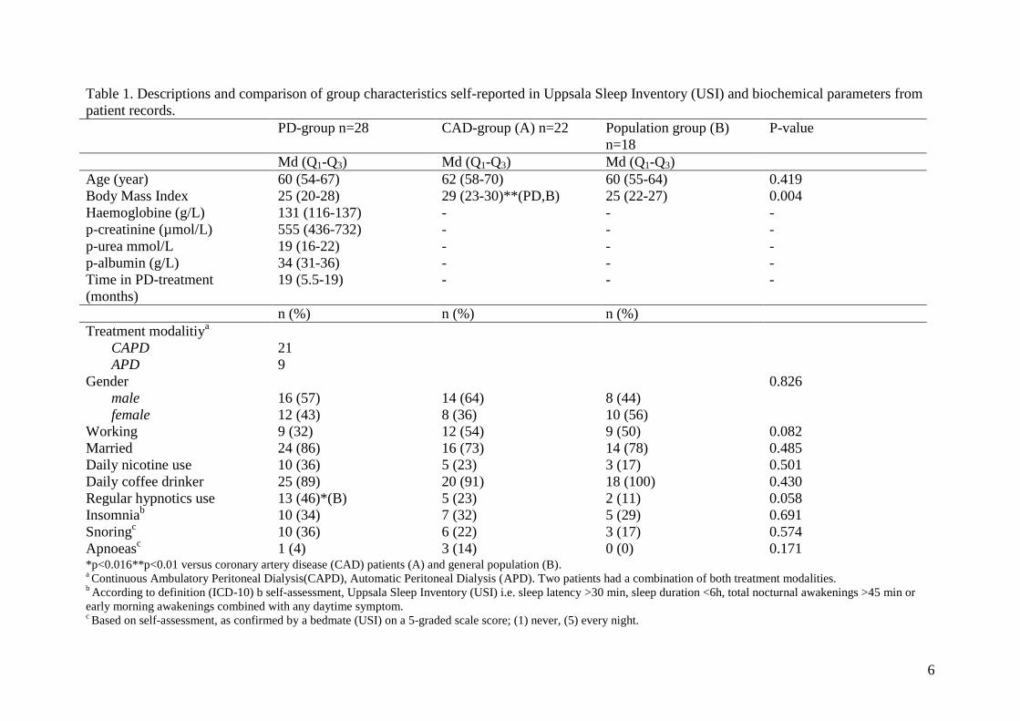
Table 1. Descriptions and comparison of group characteristics self-reported in Uppsala Sleep Inventory (USI) and biochemical parameters from
patient records.
*p<0.016**p<0.01 versus coronary artery disease (CAD) patients (A) and general population (B). a Continuous Ambulatory Peritoneal Dialysis(CAPD), Automatic Peritoneal Dialysis (APD). Two patients had a combination of both treatment modalities.
b According to definition (ICD-10) b self-assessment, Uppsala Sleep Inventory (USI) i.e. sleep latency >30 min, sleep duration <6h, total nocturnal awakenings >45 min or
early morning awakenings combined with any daytime symptom. c Based on self-assessment, as confirmed by a bedmate (USI) on a 5-graded scale score; (1) never, (5) every night.
PD-group n=28 CAD-group (A) n=22 Population group (B)
n=18
P-value
Md (Q1-Q3) Md (Q1-Q3) Md (Q1-Q3)
Age (year) 60 (54-67) 62 (58-70) 60 (55-64) 0.419
Body Mass Index 25 (20-28) 29 (23-30)**(PD,B) 25 (22-27) 0.004
Haemoglobine (g/L) 131 (116-137) - - -
p-creatinine (µmol/L) 555 (436-732) - - -
p-urea mmol/L 19 (16-22) - - -
p-albumin (g/L) 34 (31-36) - - -
Time in PD-treatment
(months)
19 (5.5-19) - - -
n (%) n (%) n (%)
Treatment modalitiya
CAPD 21
APD 9
Gender 0.826
male 16 (57) 14 (64) 8 (44)
female 12 (43) 8 (36) 10 (56)
Working 9 (32) 12 (54) 9 (50) 0.082
Married 24 (86) 16 (73) 14 (78) 0.485
Daily nicotine use 10 (36) 5 (23) 3 (17) 0.501
Daily coffee drinker 25 (89) 20 (91) 18 (100) 0.430
Regular hypnotics use 13 (46)*(B) 5 (23) 2 (11) 0.058
Insomniab 10 (34) 7 (32) 5 (29) 0.691
Snoringc 10 (36) 6 (22) 3 (17) 0.574
Apnoeasc 1 (4) 3 (14) 0 (0) 0.171

7
Comparison groups
As uraemia is a chronic disease like CAD which is also prevalent in the uraemic population a
comparison with a group of chronic CAD patients was included. They were derived from a
larger study (n=680) with a history of stable angina pectoris (Canadian Cardiovascular
Society Class I-II), listed for percutaneous coronary intervention at a University Hospital in
Sweden, two years ago. From that study 22 patients fitted the matching criteria of the PD-
group. Of these 22, ten patients were treated by surgery, eight pharmacologically, three
conservatively and one had missing data. The matching by month was performed as the
daylight in northern Europe varies substantially during the year. Further, 18 general
population individuals from the Swedish Government Person and Address Register Database
[12], who were already matched controls to the CAD group also matched the PD-patients.
Procedures
A list of PD-patients who met the inclusion criteria was obtained from the clinic. Patients
were consecutively contacted by post providing information about the study. Thereafter,
patients who agreed to participate were sent four questionnaires, an actigraph and a sleep
diary by post.
Measurements
Actigraphy
For seven days all participants wore an actigraph (Actiwatch-L®, Cambridge
Neurotechnology Ltd), applied at the non-dominating wrist like a wristwatch. The actigraph
measures the sleep-wake cycle, activity and indirect sleep. The device is an accelerometer and
records limb movements. Periods of movement are scored as wakefulness and inactivity as

8
Table 2. Description of actigraphy variables measured in the three groups.
Actigraphy
Variables Description Scale
Sleep variables
Bedtime, sleep latency, wake time during night, number of wake bouts, sleep duration and nap
time.
-
Movment and
Fragmentation index
This index is an indicator for disrupted sleep i.e. percentage of minutes moving + percentage
of immobility.
>50 = poor sleep, <20 = good
sleep.
Sleep efficiency
Ratio of actual sleep time divided by time in bed expressed in per cent. <85% = poor sleep.
Interdaily stability
Quantifies the degree of similarity concerning the activity between individual days. Range; 0-1. Values of 0,6 =
normal.
Interdaily variability
Quantifies the fragmentation of periods of rest/sleep and activity/wakefulness. A higher value
is similar to more fragmented rhythm.
Range; 0-2, <1= typical value
The amplitude of the
rhythm (AMP)
Differences between the average activity of the five least (night) and the ten most (day) active
hours, are sensitive to the overall activity.
-
Relative amplitude Ratio of AMP and average activity of the five least and the ten most active hours gives a
correction for the sensitivity.
Range 0-1, values close to 1=
more active rhythm.

9
sleep. With advanced algorithms a number of variables are obtained [13] (Table 2). Data were
processed in the software package Actiwatch Sleep Analysis 2001, version 1.9 [14].
Questionnaires
The PD-patients were sent four questionnaires and a sleep diary. Two of the questionnaires
and the sleep diary had also been completed by the comparison groups.
Simultaneously with wearing the actigraph a study specific sleep diary was
completed day by day. In the morning; bedtime, sleep latency, nocturnal awakenings and
sleep duration and in the evening; final morning awakenings, naps and daytime symptoms.
Habitual sleep during the last four weeks was assessed by the Uppsala Sleep
Inventory (USI) [15] in a modified form [16] measuring sleep variables; bedtime, sleep
latency, nocturnal awakenings, morning awakening, sleep duration, assumed sleep duration,
numbers and duration of naps. Items about sleep, pruritus, RLS and daytime functioning were
answered in a 5-point scale scored from “never” (1) to “very often” (5). Further, snoring and
apnoeic behaviour, as confirmed by a bedmate were scored in the same way. USI has earlier
been validated in a Swedish population [17]. In this study the definition of insomnia
corresponds to ICD-10, sleep duration <6h, sleep latency >30 min, five or more nocturnal
awakenings or a nocturnal wake time >45 min combined with one or more daytime
symptoms. If these persist for three weeks or more, the patients are diagnosed with insomnia
[18].
HRQoL was assessed with a generic instrument, Short form-36 (SF-36), where
the questions relate to the last four weeks [19]. This is the most widely used questionnaire for
the evaluation of health outcome assessing eight health domains; physical functioning, role
limitations due to physical health problems, bodily pain, general health, vitality, social
function, role limitations due to emotional problems and mental health. Each domain scores

10
0-100, where a higher score indicates better HRQoL [20]. The eight domains are
dichotomized in two principal dimensions; physical (PSC) and mental (MSC) component
summary. SF-36 has a good reliability and validity [19, 21] with a Cronbach-alpha between
0.79 and 0.91 [19] and was in this study 0.75 in both PSC and MSC. The above
questionnaires and the sleep diary were completed by the three groups.
Questionnaires completed by the PD-group only
The International Restless Legs Study Groups´ validated questionnaire (IRLS) evaluates the
severity of RLS symptoms. It is a ten item, five-point scale scored from “none” (0) to “very
severe” (4) symptoms, total range; 0-40. The index scoring is divided as: negligibly (0-10),
moderate (11-20), severe (21-30) and very severe (31-40) [22]. Cronbach-alpha for IRLS has
been reported to 0.93-0.95 [22] and was in this study 0.95.
Degree of fatigue was measured by the Functional Assessment of Chronic
Illness Therapy (FACIT-) fatigue scale [23]. The scale is a 13-item, five-point scale score
from “not at all” (0) to “very much” (4), total range; 0-52. Cut-off score for fatigue is 43 or
below [23]. The scale essentially assesses activity i.e. functional fatigue. Cronbach-alpha for
FACIT-fatigue scale has been reported to 0.94 [23] and was in this study 0.87. Diagnoses and
biochemical parameters were collected from patients´ records at the time of actigraphy
registration for each patient.
This study was approved by the Regional Ethical Review Board in Linköping.
Principles according to the Helsinki declaration (WMA 2004) have been followed. Written
informed consent was obtained from all participants.

11
Statistical analyses
Description of data was given by frequencies, percentage, median (md) and inter-quartile
range (Q1-Q3). Non-parametric statistics were used when data were assessed on interval or
nominal level and not normally distributed. Analysis of variance between the three groups
was performed with Kruskal-Wallis and for comparison between two groups the Mann-
Whitney U- test was used. Dichotomous variables were analysed using the Chi-square test.
Actigraphy and sleep diaries variables were averaged over the seven days of recording.
Wilcoxon´s signed rank test between groups was used when appropriate. Spearman’s rank
order correlations coefficient (r) was used to explore associations between variables. The
internal consistency was calculated with Cronbach´s alpha, at the three index scales; SF-36,
IRLS, FACIT-fatigue. Internal drop outs occurred in four items (two patients) in the FACIT-
fatigue scale and were replaced by a patient-specific mean value. Adjustment for multiple
comparisons with Bonferroni post hoc analysis [24] was performed and a two-tail p-value of
< 0.016 was considered as statistically significant, otherwise a p-value of < 0.05 was
accepted. SPSS software package 18.0 for Windows was used for all analysis.
RESULTS
Sleep-wake cycle
In total data from 68 participants and 470 nights were collected by actigraphy and sleep
diaries. The PD-patients had significantly more disrupted sleep, explored by the movement
and fragmentation index (MFI) (Table 3). In contrast to the comparison groups, none of the
PD-patients had MFI below 20 but 11 (39.3%) had above 50, which indicates seriously
disrupted sleep (Figure 1). The nocturnal sleep duration did not differ between the groups
(Table 3). However, nocturnal awakening time in PD-patients was 30 minutes longer than in
the CAD-group and 36 minutes longer than in the population group (p<0.001) (Table 3)

12
Table 3. Group comparison of sleep variables assessed by actigraphy and sleep diary during one week and the questionnaire Uppsala Sleep
Inventory (USI) covering habitual sleep over 4 weeks. PD-group n=28 CAD group (A) n=22 Population group (B) n=18 P-value
Sleep variables Md (Q1-Q3) Md (Q1-Q3) Md (Q1-Q3) (Kruskal-Wallis)
Sleep quality (score) 1=bad, 5=very good 3 (2-4)**(A) 2 (1-3) 2 (1-4) 0.006
Sleep latency (min)
Actigraphy 30 (22-59)***(B) 24 (10-41) 13 (7-26) 0.002
Sleep diary 28 (16-75) 16 (12-34) 18 (9-34) 0.1
USI 25 (6-60)**(A) 10 (1-20) 10 (3-23) 0.007
Sleep duration (hour.minutes)
Actigraphy 6.42 (5.58-7.17) 6.50 (6.05-7.37) 6.43 (6.30-7.12) 0.903
Sleep diary 7.06 (5.54-8.00) 6.55 (6.03-7.25) 6.17 (5.50-7.05) 0.132
USI 7.00 (5.00-8.00) 7.00 (4.42-8.00) 7.00 (5.06-7.00) 0.951
Go to bed (time)
Actigraphy 22:41 (21:41-23:04) 22:55 (22:33-23:20) 23:03 (22:41-23:19) 0.079
Sleep diary 22:23 (21:29-22:55) 22:27 (22:00-22:56) 22:35 (22:27-23:03) 0.278
USI 22:00 (21:00-23:00)**(B) 22:00 (21:42-22:30)*(B) 23:00 (22:00-23:00) 0.009
Wake time during night (min)
Actigraphy 94 (70-122)**(A)***(B) 64 (41-91) 58 (47-73) 0.001
Sleep diary 20 (8-52) 24 (10-37) 21 (12-43) 0.973
USIa 4 (2-5) 3 (1-4) 4 (1-6) 0.056
Wake bouts (freq)
Actigraphy 32 (27-36)**(A) 26 (19-34) 24 (20-31) 0.051
USI 2 (1-3)**(B) 3 (1-4)*(B) 2 (0-2) 0.015
Nap time (min)
Actigraphy 59 (39-92) **(B) 48 (30-57) 32 (14-47) 0.009
USI 45 (10-60) 45 (11-60) 30 (11-60) 0.463
Fragmentation indexb
Actigraphy 49 (41-64) ***(A)***(B) 35 (25-42) 34 (26-37) 0.0001
Sleep efficiencyc (%)
Actigraphy 71 (68-80)**(A)***(B) 78 (75-85) 83 (82-87) <0.0001
Sleep Sufficient Indexd (%)
USI 80 (72-94)*(A) 93 (83-100) 93 (86-100) 0.027 *(<0.016), ** (<0.01), *** (<0.001) versus coronary artery disease (CAD) patients (A) and general population (B). Mann-Whitney U test adjusted with Bonferroni. a Score; <5 min (1), 5-15 min (2), 15-30 min (3), 30-60 min (4), 1-2 h (5), 2-3 h (6), >3 h (7).
b Fragmentation index (i.e. movement and fragmentation index) indicates restless and fragmented sleep; summary of percentage of minutes moving and percentage of
immobility, bad sleep (>50) and very good sleep (<20). c Sleep efficiency; ratio of sleep duration and time in bed in per cent, below 85% indicates insufficient sleep efficiency.
d Sleep Sufficient Index; ratio of sleep duration and self-estimated need for sleep in percent, below 80% indicates insufficient sleep efficiency.

13
Figure 1: Illustration of the distribution of fragmented sleep in the three groups. Above 50
indicates very disrupted sleep and below 20 means well consolidated sleep.
measured by actigraphy. Interdaily variability and –stability were within the normal range,
which indicates stable sleep-wake cycles (Table 4).
Quality of sleep and sleep disturbing factors
PD-patients had 7% lower sleep efficiency than the CAD-group and 12% lower than the
population group (p<0.001) (Table 3). One third (n=10) of the PD-patients could be
categorised as insomniacs (Table 1). PD-patients classified as insomniacs had significantly
decreased sleep quality, md (Q1-Q3) 2 score (1.25-3.0 score) compared to non-insomniacs 3
14
score (2.0-4.0 score), (p<0.05). Further, on the dichotomized question “Do you get too little
sleep?” 61% (n=17) of PD-patients gave an affirmative answer compared to 23% (n=5) in the
CAD group (p<0.05) and 50% (n=9) in the population group (not significant). All 22
insomniacs in the three groups were in agreement to the specific question (p<0.05).
In PD-patients pruritus was a common sleep disturbing factor at sleep onset.
Sixteen patients (57%) had moderate to very severe pruritus at sleep onset and a correlation to
MFI (r=-0.42, p=0.034) was found. Nocturnal pruritus also correlated to MFI (r=-0.49,
p<0.01), sleep efficiency (r=-0.49, p<0.01), bodily pain (r=-0.39, p <0.05) and general health
(r=-0.42, p<0.05). RLS was reported by 13 patients (46%) as being moderate to very severe
problems. Correlations to sleep quality (USI) (r=-0.39, p<0.05), sleep efficiency (actigraphy)
(r=-0.54, p<0.01), role physical (r=-0.39, p <0.05), vitality (r=-0.36, p<0.05) were found.
Further, sleep duration (r=-0.40, p<0.05) and sleep latency measured by actigraphy (r=-0.40,
p<0.05) and sleep diary (r=0.45, p<0.05) were associated to RLS.
Daytime symptoms and health related quality of life
Sleepiness and physical tiredness were the most prevalent daytime symptoms in all three
groups (Table 4). The prevalence of fatigue was high in PD-patients, 25 (89%) scored below
the cut off (43) and fatigue-score was correlated to MFI (r=-0.43, p<0.05). PD- and CAD-
patients reported worse physical function in SF-36 compared to the population (Table 5). The
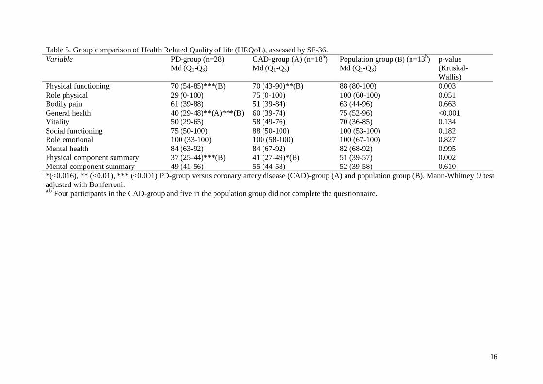
PD-patients also reported worse general health.
No differences concerning the sleep variables were found between patients on automatic PD
(APD) and manual PD (CAPD). Influence of the seasonal variation of light was not detected
on any of the variables (data not shown).

15
Table 4. Group comparison of circadian rhythm assessed by actigraphy during one week and daytime symptoms assessed by Uppsala Sleep
Inventory (USI) questionnaire referring to the last four weeks.
PD-group n=28 CAD-group n=22 (A) Population group n=18 (B)
P-value
Md (Q1-Q3) Severe
Problems
Freq (%)e
Md (Q1-Q3) Severe
Problems
Freq (%)e
Md (Q1-Q3) Severe
Problems
Freq (%)e
(Kruskal-
Wallis)
Daily activity
(Actigraphy)
Interdaily stabilitya 0.58 (0.45-0.64) - 0.54 (0.47-0.63) - 0.57 (0.46-0.64) - 0.957
Interdaily variabilityb 0.83 (0.70-1.01) - 0.82 (0.69-0.95) - 0.82 (0.69-1.07) - 0.815
Amplitudec 13387 (8743-19396)*(B) - 15672 (12361-21401) - 21288 (14391-26448) - 0.04
Relative amplituded 0.84 (0.76-0.87)**(B) - 0.88 (0.85-0.90) - 0.90 (0.85-0.93) - 0.002
Daytime symptoms (USI)
Score;(1) no problem-
(5) very big problems
No refreshing sleep 3 (2-4) 18 (62) 2 (2-3) 10 (44) 3 (1-3) 9 (56) 0.315
Sleepiness 3 (2-4) 21 (72) 3 (2-4) 14 (61) 3 (1-3) 9 (56) 0.617
Physical tiredness 3 (2-4) 22 (76) 3 (2-4) 13 (56) 2 (1-4) 6 (38) 0.235
Mental tiredness 2 (2-2) 7 (24) 2 (2-3) 8 (35) 2 (1-3) 5 (31) 0.854
Exhaustion 2 (1-2) 7 (24) 2 (1-3) 8 (35) 2 (1-3) 6 (38) 0.662
*(<0.016), ** (<0.01), *** (<0.001) PD-group versus coronary artery disease (CAD)-group (A) and population group (B). Mann-Whitney U test
adjusted with Bonferroni. a quantifies the resemblance between the activity pattern on individual days, ranges from 0 (low) to1 (high), 0.6 is typical.
b quantifies the fragmentation of sleep and activity, ranges from 0 (low) to 2 (high), below 1 is typical.
c Amplitude of the rhythm i.e. difference of average activity and the five least and the ten most active hours.
d Range from low (0) to high (1) activity. Ratio of the rhythm amplitude and the sum of average activity counts of the five least and the ten most
active hours. e Reporting severe to very severe problems (score 4-5)
16
Table 5. Group comparison of Health Related Quality of life (HRQoL), assessed by SF-36.
Variable PD-group (n=28)
Md (Q1-Q3)
CAD-group (A) (n=18a)
Md (Q1-Q3)
Population group (B) (n=13b)
Md (Q1-Q3)
p-value
(Kruskal-
Wallis)
Physical functioning 70 (54-85)***(B) 70 (43-90)**(B) 88 (80-100) 0.003
Role physical 29 (0-100) 75 (0-100) 100 (60-100) 0.051
Bodily pain 61 (39-88) 51 (39-84) 63 (44-96) 0.663
General health 40 (29-48)**(A)***(B) 60 (39-74) 75 (52-96) <0.001
Vitality 50 (29-65) 58 (49-76) 70 (36-85) 0.134
Social functioning 75 (50-100) 88 (50-100) 100 (53-100) 0.182
Role emotional 100 (33-100) 100 (58-100) 100 (67-100) 0.827
Mental health 84 (63-92) 84 (67-92) 82 (68-92) 0.995
Physical component summary 37 (25-44)***(B) 41 (27-49)*(B) 51 (39-57) 0.002
Mental component summary 49 (41-56) 55 (44-58) 52 (39-58) 0.610
*(<0.016), ** (<0.01), *** (<0.001) PD-group versus coronary artery disease (CAD)-group (A) and population group (B). Mann-Whitney U test
adjusted with Bonferroni. a,b
Four participants in the CAD-group and five in the population group did not complete the questionnaire.

17
DISCUSSION
To our knowledge this is the first study describing a one-week sleep-wake cycle and
demonstrating that fragmented sleep is a reality for PD-patients. Compared with the CAD and
the population groups the PD-patients had more deteriorated sleep.
The fragmented sleep can explain that PD-patients had significantly lower sleep
quality, compared to the comparison groups, as fragmented sleep reduces the restorative deep
sleep [25]. In addition, PD-patients seem to have an increased need for recovery which was
reflected by the Sleep Sufficient Index which was lower than in the comparison groups.
Although an optimized treatment regime, a uraemic state still persists contributing to more
tiredness [1] and an increased need for recovery, i.e. sleep [26]. About one-third of all
participants from all three groups fulfilled the insomnia criteria which can be one explanation
to the fragmented sleep. Insomnia has been described as a prevalent problem in dialysis
populations [16, 27] earlier. Although the proportion of insomniacs was similar in all three
groups, only the PD-patients had significantly higher fragmentation and decreased sleep
quality. There might, however, possibly be other explanations for the fragmentation of sleep.
In this study 46% of the PD-patients reported RLS, another potential sleep
disturber and prevalent in ESRD [1] and PD [16] patients. The high prevalence may explain
the inability to consolidate sleep [16, 27] causing fragmented sleep or insomnia depending on
when the symptoms peak: at the sleep onset or during the night. In this study no significant
association between RLS and fragmented sleep was found, although such an association has
been described previously [28]. RLS in our study was correlated to decreased sleep quality.
Unruh and co-workers (2004) showed that RLS in dialysis patients was associated with
decreased physical function, decreased well-being and even an increased risk for mortality
[29]. Another prevalent source for sleep disruption is pruritus, which in our study was
reported by 57% of the PD patients. Pruritus has earlier been reported to vary between 40-

18
55% in uremic populations [1, 16, 30] and one study supports a relationship between pruritus
and mortality, in haemodialysis patients, attributed to poor sleep [30]. This study bears no
evidence that disturbed sleep is related to APD treatment. Previous studies have shown only
tendencies and no significant instances of APD treatment causing a more disturbed sleep.
Contrary, our research group previously found that APD-patients reported significantly better
sleep quality than CAPD-patients (p=0.03) [16].
Only one PD-patient (4%) reported apnoeic behaviour, but this sleep behaviour
is difficult to self-report. Evidence has been presented for a tenfold higher prevalence of
sleep apnoea (57%) in uraemic patients [3] than in people with normal renal function (2-4%)
[25]. Obese people suffer more from sleep apnoea. Although the CAD-patients in our study
had significantly higher BMI than the PD-and the population group (p=0.004) no significant
differences were found in the self-reported sleep apnoea. However, this is difficult to evaluate
since only 4 out of 68 patients reported sleep apnoea in this study. One possible explanation
could be that an increased intra-abdominal pressure from the dialysis solution can affect the
breathing in PD-patients. In haemodialysis water overload can cause oedema and obstruction
in the airways. Furthermore, a central destabilization of respiratory control and acidosis can
be another explanation [31]. Sleep fragmentation caused by sleep apnoea is an alarming
problem in peritoneal dialysis patients leading to nocturnal hypoxemia which is a serious risk
for fatal cardiovascular incidences [32, 33]. The discrepancy between self-assessed and
actigraphy-registered wake bouts in this study can be explained by the fact that actigraphy-
registered fragmentation is mostly unconscious awakenings i.e. micro arousals, with an
increased EEG frequency [34] and decreased slow-wave sleep and sleep quality. It is essential
to keep in mind that apnoea, RLS and pruritus can induce fragmented sleep or insomnia,
either separately or in combination. Therefore evaluation of symptoms is essential because the
clinical picture is so complex.

19
This study showed a correlation between fragmented sleep and fatigue which is
new knowledge concerning PD-patients. Poor sleep has also been shown to have a substantial
impact on fatigue for patients with primary Sjögrens´s syndrome and rheumatoid arthritis
[35]. The prevalence of fatigue among the PD-patients in this study was extremely high, but
consistent with previous studies, 55-88% [16, 36]. Despite the fact that the PD-patients in this
study had more fragmented sleep, longer sleep latency onset and worse sleep efficiency, than
the comparison groups, they did not report more daytime symptoms related to poor sleep. In
healthy subjects daytime symptoms have been reported after only one night with fragmented
sleep [37]. Our findings can partly be explained by the fact that symptoms of fatigue are, both
by patients and healthcare personnel, considered to be a part of the disease [26, 38] and
therefore are easily overlooked. Nevertheless, in this study and in a previous study by our
research group [16] napping was a frequently used behaviour (74%) and presumably a way to
manage daytime sleepiness attributed to the fragmented sleep. Extended naps, with a duration
of more than 30 minutes, can mean an awakening from slow-wave sleep which is
characterized by confusion, grogginess and decreased performance i.e. sleep inertia [39].
Symptoms of sleep inertia can be misinterpreted as fatigue and vice versa as fatigue is
characterized as diminished energy and mental capacity and an increased need for rest [40].
Healthcare personnel may be able to help patients to address their symptoms and to keep
structured daily routines to promote good sleep behaviours. Sleep scheduling interventions
reducing nap frequency and nap time [41] are one way of management. However, sleep
apnoea and insomnia are not the only factors affecting daytime function, other co-existing
problems such as depression also have to be considered [42].
The overall activity, measured by actigraphy, was significantly lower in the PD-
group compared to the comparison groups. The decreased activity in PD-patients was also
reflected in SF-36 where PD- and CAD- patients reported decreased physical functioning. In

20
addition for the PD- patients the high prevalence of fatigue for the PD- patients can partly be
addressed to decreased physical function when the FACIT-fatigue scale essentially assesses
functional fatigue. Interestingly, decreased physical functioning has showed an association to
a high prevalence of RLS previously [29].
The strength of our study is that data from more than a total of 400 days were
collected in an everyday context with a device for repeated measurements which does not
affect habitual life or routines. Further, data were collected through a combination of
questionnaires and a sleep diary where several parameters showed similar results. Another
strength in the study is the combined evaluation of RLS by both a questionnaire and
actigraphy which has been described as a valid and reliable method [28] since RLS is closely
related to sleep disorders. Finally the reliability calculated FACIT-fatigue and SF-36 were
satisfactory.
This study also had some limitations: First, a small sample size and the findings
must be generalized carefully. However, the PD-group represents approximately 4% of the
total PD population in Sweden and had a gender distribution similar to the national sample.
Second, actigraphy measures sleep/wake periods indirectly when activity indicates
wakefulness. This can implicate that actigraphy underestimates sleep latency when a
motionless period precedes sleep or overestimates daytime sleep when the patient is resting.
Nevertheless, sleep fragmentation assessed by actigraphy has shown high sensitivity (89%)
and specificity (95%) for diagnosing sleep apnoea [43, 44]. Actigraphy has also measure
acceptable accuracy to polysomnographical variables; number of nocturnal awakenings,
waking after sleep onset, total sleep time and sleep efficiency in insomnia patients [45].
Finally, we did not reach the same number of participants in the three groups, partly due to
the three matching variables and in particular the month for data collection. Therefore,
between groups statistics was chosen. Another comparison group could have been patients

21
with chronic kidney disease not yet on dialysis, in order to study how dialysis per se might
cause sleep disturbances.
Conclusion
To our knowledge this study is the first to demonstrate that PD-patients have a deteriorated
sleep with serious fragmentation measured by one-week actigraphy registration. Further, PD-
patients exhibit worse sleep quality compared to CAD patients and individuals in the
population. More evaluation of sleep in clinical practice is highly recommended since PD-
patients are vulnerable individuals with extended self-care responsibilities and at risk for co-
morbidity secondary to insufficient. Future research to investigate whether PD-patients sleep
problems and fatigue can be improved by an individual non-pharmacological intervention
programme is required.
REFERENCES
1. Murtagh FE, Addington-Hall J, Higginson IJ. The prevalence of symptoms in
end-stage renal disease: a systematic review. Adv Chronic Kidney Dis 2007 Jan;14(1):82-99.
2. Hanly P. Sleep disorders and home dialysis. Adv Chronic Kidney Dis 2009
May;16(3):179-88.
3. Kumagai TI, Y. Kawarazaki, W. Shimizu, H Kaname, S. Fujita T. Effects of
nocturnal oxygen theraphy on sleep apnea syndrome in peritoneal dialysis patients. Clinical
Nephrology 2008;70(4):332-9.
4. Knutson KL. Impact of sleep and sleep loss on glucose homeostasis and appetite
regulation. Sleep Med Clin 2007 Jun;2(2):187-97.
5. Redwine L, Hauger RL, Gillin JC, Irwin M. Effects of sleep and sleep
deprivation on interleukin-6, growth hormone, cortisol, and melatonin levels in humans. J
Clin Endocrinol Metab 2000 Oct;85(10):3597-603.
6. Chiu YL, Chuang YF, Fang KC, Liu SK, Chen HY, Yang JY, et al. Higher
systemic inflammation is associated with poorer sleep quality in stable haemodialysis
patients. Nephrol Dial Transplant 2009 Jan;24(1):247-51.
7. Benz RL, Pressman, M. R., Hovick, E. T., Peterson, D. D. Potential novel
predictors of mortality in end-stage renal disease patients with sleep disorders. Am J Kidney
Dis 2000 Jun;35(6):1052-60.
8. Bilgic A, Akman B, Sezer S, Ozisik L, Arat Z, Ozdemir FN, et al. Predictors for
quality of life in continuous ambulatory peritoneal dialysis patients. Nephrology (Carlton)
2008 Oct;13(7):587-92.
9. Kutner NG, Zhang, R., Huang, Y., Bliwise, D. L. Patient-reported sleep
difficulty and cognitive function during the first year of dialysis. Int Urol Nephrol
2008;40(1):203-10.

22
10. Stepanski E, Lamphere, J., Roehrs, T., Zorick, F., Roth, T. Experimental sleep
fragmentation in normal subjects. Int J Neurosci 1987 Apr;33(3-4):207-14.
11. Koch BC, Nagtegaal JE, Hagen EC, Wee PM, Kerkhof GA. Different melatonin
rhythms and sleep-wake rhythms in patients on peritoneal dialysis, daytime hemodialysis and
nocturnal hemodialysis. Sleep Med 2010 Mar;11(3):242-6.
12. Edell-Gustafsson U, Carstensen J, Regestein Q, Swahn E, Svanborg E.
Hyperarousal, depression and quality of life--validity and reliability of the Swedish version of
the Hyperarousal Scale. Scand J Caring Sci 2006 Mar;20(1):58-67.
13. Camtech. Actigraphy: Activity and Sleep Analysis. Cambridge
Neurotechnology Ltd Information Bulletin No 4B 2007.
14. Ltd Neurotechnology Cambridge. Actiwatch Sleep Analysis. 1.09 ed2001.
15. Hetta J, Almqvist M, Ågren H, Hambert G, Liljenberg G, Roos BE, editors.
Prevalence of sleep disturbances and related symptoms in a middle-aged Swedish population.
Stuttgart: Gustaf Fischer-Verlag; 1985.
16. Yngman-Uhlin P, Edell-Gustafsson U. Self-reported subjective sleep quality and
fatigue in patients with peritoneal dialysis treatment at home. Int J Nurs Pract 2006
Jun;12(3):143-52.
17. Gislason T, Almqvist M. Somatic diseases and sleep complaints. An
epidemiological study of 3,201 Swedish men. Acta Med Scand 1987;221(5):475-81.
18. World Health Organization. The ICD-10 Classification of Mental and
Behavioural Disorders: Diagnostic Criteria for Research
Geneva: WHO; 1993.
19. Sullivan M, Karlsson J, Ware JE, Jr. The Swedish SF-36 Health Survey--I.
Evaluation of data quality, scaling assumptions, reliability and construct validity across
general populations in Sweden. Soc Sci Med 1995 Nov;41(10):1349-58.
20. Sullivan M, Karlsson J, Taft C. SF-36 Hälsoenkät: Svensk manual och
tolkningsguide (Swedish Manual and Interpretation Guide). 2 ed. Gothenburg: Sahlgrenska
University Hospital; 2002.
21. Ware JE, Jr., Sherbourne CD. The MOS 36-item short-form health survey (SF-
36). I. Conceptual framework and item selection. Med Care 1992 Jun;30(6):473-83.
22. Walters AS, LeBrocq C, Dhar A, Hening W, Rosen R, Allen RP, et al.
Validation of the International Restless Legs Syndrome Study Group rating scale for restless
legs syndrome. Sleep Med 2003 Mar;4(2):121-32.
23. Yellen SB, Cella DF, Webster K, Blendowski C, Kaplan E. Measuring fatigue
and other anemia-related symptoms with the Functional Assessment of Cancer Therapy
(FACT) measurement system. J Pain Symptom Manage 1997 Feb;13(2):63-74.
24. Field A, editor. Discovering statistics using SPSS (and sex and drugs and
rock´n´roll). 3 ed. Dubai: Oriental Press; 2009.
25. Young TP, M. Dempsey, J. Skatrud, J. Weber, S. Badr, S. . The occurence of
sleepdisordered breathing among middle-age adults. The New England Journal of medicine
1993;328(17):1230-5.
26. Yngman-Uhlin P, Friedrichsen M, Gustavsson M, Fernstrom A, Edell-
Gustafsson U. Circling around in tiredness: perspectives of patients on peritoneal dialysis.
Nephrol Nurs J 2010 Jul-Aug;37(4):407-13.
27. Sabry AA, Abo-Zenah H, Wafa E, Mahmoud K, El-Dahshan K, Hassan A, et al.
Sleep disorders in hemodialysis patients. Saudi J Kidney Dis Transpl 2010 Mar;21(2):300-5.
28. Allen RP. Improving RLS diagnosis and severity assessment: polysomnography,
actigraphy and RLS-sleep log. Sleep Med 2007 Aug;8 Suppl 2:S13-8.

23
29. Unruh ML, Levey AS, D'Ambrosio C, Fink NE, Powe NR, Meyer KB. Restless
legs symptoms among incident dialysis patients: association with lower quality of life and
shorter survival. Am J Kidney Dis 2004 May;43(5):900-9.
30. Pisoni RL, Wikstrom B, Elder SJ, Akizawa T, Asano Y, Keen ML, et al.
Pruritus in haemodialysis patients: International results from the Dialysis Outcomes and
Practice Patterns Study (DOPPS). Nephrol Dial Transplant 2006 Dec;21(12):3495-505.
31. Mavanur M, Sanders M, Unruh M. Sleep disordered breathing in patients with
chronic kidney disease. Indian J Med Res 2010 Feb;131:277-84.
32. Kuniyoshi FH, Pusalavidyasagar S, Singh P, Somers VK. Cardiovascular
consequences of obstructive sleep apnoea. Indian J Med Res 2010 Feb;131:196-205.
33. Loewen A, Siemens A, Hanly P. Sleep disruption in patients with sleep apnea
and end-stage renal disease. J Clin Sleep Med 2009 Aug 15;5(4):324-9.
34. American Sleep Disorders Association Atlas Task force. EEG arousals: scoring
rules and examples. SLEEP 1992;15:173-84.
35. Goodchild CE, Treharne GJ, Booth DA, Bowman SJ. Daytime patterning of
fatigue and its associations with the previous night's discomfort and poor sleep among women
with primary Sjogren's syndrome or rheumatoid arthritis. Musculoskeletal Care 2010 Mar
15;8:107-17.
36. Ossareh S, Roozbeh, J. Krishnan, M. Bargman, JM. Oreopoulos, DG. Fatigue in
peritoneal dialysis patients. Int Urol and Nephrol 2003;35:535-41.
37. Roehrs T, Merlotti L, Petrucelli N, Stepanski E, Roth T. Experimental sleep
fragmentation. Sleep 1994 Aug;17(5):438-43.
38. Yngman-Uhlin P, Johansson A, Fernström A, Börjeson S, Edell-Gustafsson U.
Circling around in tiredness; Perspectives of patients on peritoneal dialysis. Neprology
Nursing Journal, accepted for publication 2011-06-04.
39. Milner CE, Cote KA. Benefits of napping in healthy adults: impact of nap
length, time of day, age, and experience with napping. J Sleep Res 2009 Jun;18(2):272-81.
40. Cella D, Lai JS, Chang CH, Peterman A, Slavin M. Fatigue in cancer patients
compared with fatigue in the general United States population. Cancer 2002 Jan 15;94(2):528-
38.
41. Morin C, Espie C. Insomnia - A clinical guide to assessment and treatment. New
York: Springer; 2004.
42. Abdel-Kader K, Unruh ML, Weisbord SD. Symptom burden, depression, and
quality of life in chronic and end-stage kidney disease. Clin J Am Soc Nephrol 2009
Jun;4(6):1057-64.
43. Stepanski EJ. The effect of sleep fragmentation on daytime function. Sleep 2002
May 1;25(3):268-76.
44. Aubert-Tulkens G, Culee C, Harmant-Van Rijckevorsel K, Rodenstein DO.
Ambulatory evaluation of sleep disturbance and therapeutic effects in sleep apnea syndrome
by wrist activity monitoring. Am Rev Respir Dis 1987 Oct;136(4):851-6.
45. Lichstein KL, Stone KC, Donaldson J, Nau SD, Soeffing JP, Murray D, et al.
Actigraphy validation with insomnia. Sleep 2006 Feb 1;29(2):232-9.