FLEXIBLE BRONCHOSCOPY EDUCATION PROJECT · 2017-12-07 · Patient-centered practical approach...
132
Bronchoscopy International 2011 © THE BRONCHOSCOPY EDUCATION PROJECT Training Manual Subject (Part II): Endobronchial Ultrasound Bronchoscopy Competency Program Henri Colt MD, FCCP Professor of Medicine University of California, Irvine [email protected] Bronchoscopy International A non-profit organization dedicated to education, © and the global dissemination of knowledge* www.Bronchoscopy.org *The Foundation for the Advancement of Medicine is a 501-C3 non-profit organization
Transcript of FLEXIBLE BRONCHOSCOPY EDUCATION PROJECT · 2017-12-07 · Patient-centered practical approach...

Bronchoscopy International 2011©
THE BRONCHOSCOPY EDUCATION PROJECT
Training Manual
Subject (Part II): Endobronchial Ultrasound Bronchoscopy Competency Program
Henri Colt MD, FCCP Professor of Medicine
University of California, Irvine [email protected]
Bronchoscopy International
A non-profit organization dedicated to education, ©
and the global dissemination of knowledge* www.Bronchoscopy.org
*The Foundation for the Advancement of Medicine is a 501-C3 non-profit organization

Bronchoscopy Education Project Part II
Bronchoscopy International 2011©
Funding statement Funding for The Bronchoscopy Education Project Materials is a result of philanthropic endeavors and from the general support of the University of California, Irvine for Dr. Colt’s work. No corporate support was either solicited or received for this work. Copyright statement Copyright 2011. Henri Colt MD. All Rights Reserved. Permission to use, copy, modify, and di stribute any part o f this software including any source c ode a nd doc umentation f or e ducational, r esearch, a nd non -profit pur poses, without fee, and without a written agreement is hereby granted, provided that the above copyright notice, and this paragraph appear in all copies of the documentation. Bronchoscopy Education Project Disclaimer Confidentiality of data potentially relating to individual patients and visitors has been respected. We strive to honor or exceed any legal requirements of medical/health information privacy as they apply to the United States and to the state of California. Images and case descriptions provided here-in, therefore, are not intended for the diagnosis of any specific patient. Any information found on the website or in this text should not be used as a substitute for medical care. The authors disclaim liability, injury, or damage incurred as a consequence from the use of information. Reference to websites, instruments, or equipment portrayed in this material is not of commercial nature, nor does the inclusion imply endorsement. Should documents or images from this material be copied for personal use, we request that materials remain unedited and unmodified unless for teaching purposes, that no fee be charged for copies or access to the information, that copyright notices and disclaimers remain attached, and that credit be given to The Bronchoscopy Education Project (suggested format Bronchoscopy International© www.http://bronchoscopy.org
22

Bronchoscopy Education Project Part II
Bronchoscopy International 2011©
The Bronchoscopy Education Project is a uniform curriculum designed to provide bronchoscopy educators with competency-oriented tools and materials that can be incorporated in whole or in part into various training programs. Materials can be used to train student bronchoscopists and assess progress along the learning curve from novice to competent practitioner. This project is based on FIVE key concepts:
1. Mandatory reading, including review of open-access web-based materials
2.
in the form of video clips, photo atlases, and written manuals provides a uniform foundation of theoretical and practical knowledge. Step-by-step instruction
3.
, simulation scenarios, training models, and small group workshops allow technical skill and experiential knowledge acquisition for existing and newly introduced technologies and procedures. Checklists
4.
using a uniform template enhance procedural standardization, patient safety, and implementation across procedural platforms. Patient-centered practical approach exercises
5.
help practitioners rationalize the various components of the decision-making process (strategy and planning; equipment, techniques and results; outcomes and quality improvement), thus reinforcing their acquisition of cognitive, technical, affective and experiential knowledge. Assessment tools,
readily applied in the clinical as well as simulation setting enhance learning, and document progression along the learning curve from novice to competent practitioner.
The Bronchoscopy Education Project includes three parts, to be completely developed by Bronchoscopy International and a host of international experts. As they come to completion, these components, designed using a uniform template and development philosophy, will be disseminated and implemented at the national and international level with the added endorsement and collaboration of university medical centers, regional physician groups, national societies, and international organizations. A series of Train the Trainers seminars are being conducted to familiarize a cadre of bronchoscopy educators with general educational philosophies and
33

Bronchoscopy Education Project Part II
Bronchoscopy International 2011©
methodologies, and to provide opportunities to learn and practice various elements of The Bronchoscopy Education Project. We assume this cadre of educators will use some or all project materials in future regional or institution-based teaching programs. As a result of this work, it is our hope to facilitate the work of our professional colleagues by providing a uniform instructional framework that can be expanded, researched, and improved upon, and to alleviate patients from the burdens of procedure-related training. Increasingly knowledgeable and competent bronchoscopists will thus enhance their practice through a more rapid implementation of new technologies, and a better use of existing ones, all to the benefit of our patients. Part I: Introductory Course in Flexible Bronchoscopy. This course addresses bronchoscopic inspection, lavage, brushing, endobronchial biopsy, transbronchial lung biopsy and conventional transbronchial needle aspiration. Part II: Endobronchial Ultrasound and EBUS-Transbronchial Needle Aspiration. This course addresses Endobronchial Ultrasound physics, equipment (processors, bronchoscopes, needles, radial and linear array transducers), techniques including EBUS-TBNA, mediastinal anatomy, lung cancer staging according to universally accepted IASLC guidelines, and EBUS-radiographic-bronchoscopic correlations. Part III: Introduction to Interventional Flexible Bronchoscopy. This course addresses flexible bronchoscopic resection techniques including electrosurgery and Nd:YAG laser, foreign body removal techniques and instrumentation, difficult airway management including difficult intubation and hemoptysis, flexible bronchoscopic stent and airway valve insertion, bronchoscopic techniques of electromagnetic navigation, and bronchial thermoplasty. Train the Trainers Seminars: These hands-on seminars are specifically designed to familiarize participants with materials and techniques necessary for teaching each of the three other components of The Bronchoscopy Education Project. Each seminar targets mastery of didactic and associated reading materials, and provides opportunity to practice using checklists, assessment tools, practical approach patient-centered exercises, and simulation or role playing exercises. Debriefing and 360 degree feedback techniques are employed to foster teamwork, provide individual intrinsic value, and enhance individual as well as group performance. .
Henri Colt MD., FCCP Eric Edell MD., FCCP [email protected] [email protected]
44

Bronchoscopy Education Project Part II
Bronchoscopy International 2011©
Welcome to The EBUS Bronchoscopy Education Project. The
purpose of this project is to provide bronchoscopy educators and training program directors with competency-oriented tools and materials with which to train student bronchoscopists and assess progress along the learning curve from novice to competent practitioner. Material can be incorporated in whole or in part, as needed by each program. The foundation for this project is a standardized curriculum (schedule, content, checklists, assessment tools, training models, and train-the-trainers instruction) pertaining to Bronchoscopy Education Project Part II, Endobronchial Ultrasound and EBUS-Transbronchial Needle Aspiration. This course addresses endobronchial ultrasound physics, equipment (processors, bronchoscopes, needles, radial and linear array transducers), techniques including EBUS-TBNA, mediastinal anatomy, lung cancer staging according to universally accepted IASLC guidelines, and EBUS-CT-White light bronchoscopy correlations
Modeled on this curriculum, work is in progress for programs pertaining to (a) interventional flexible bronchoscopy, and (b) rigid bronchoscopy. The already completed Bronchoscopy Education Project Part pertains to an Introduction to Flexible Bronchoscopy This project is ongoing and will be updated at www.bronchoscopy.org as components become available. We invite your comments as you use these materials.
Henri Colt MD., FCCP Eric Edell MD., FCCP [email protected] [email protected]
55

Bronchoscopy Education Project Part II
Bronchoscopy International 2011©
This page intentionally left blank.
66

Bronchoscopy Education Project Part II
Bronchoscopy International 2011©
Table of Contents
Page Content 3………………………Welcome and Purpose of this Training Manual
5………………………Overview of the Bronchoscopy Education Project
7………………………Description of the BEP Part II Program (EBUS)
9………………………Section 1: Program Completion with Checklist
15.….…………………Section 2: Regional Courses
21.……………….……Section 3: Reading assignments
45……………………. Section 4: Endobronchial Ultrasound Step-by-Step§
47..……………………Section 5: Simulation Workshops
67…………………… Section 6: Observed Real-Patient Scenarios
71..……………………Section 7: Practical Approach Sessions
89…………………… Section 8: Proctored EBUS-TBNA
93…………………… Section 9: Assessment Tools*§
107…..……………… Section 10: Checklists§
127………………….. Conclusion
*Answer grids should not be released to students so that assessment tools can continue to be used during local and regional programs. For copies of assessment tools and answer grids, please contact Henri Colt MD at [email protected] §
Checklists, Assessment Tools, and Step-by-Step Narratives created by Mohsen Davoudi MD, Septimiu Murgu, and Henri Colt MD
77

Bronchoscopy Education Project Part II
Bronchoscopy International 2011©
This page intentionally left blank.
88

Bronchoscopy Education Project Part II
Bronchoscopy International 2011©
Endobronchial Ultrasound and EBUS-TBNA Curriculum contains:
1. Endobronchial Ultrasound and EBUS-TBNA Competency Program Completion Checklist 2. Regional EBUS and EBUS-TBNA courses comprised of didactic lectures, interactive
sessions and simulation-based hands-on workshops using a pre-test/post-test model to document cognitive knowledge and technical skill acquisition..
3. Reading assignments: • The EBUS Bronchoscopist©, a single module, 30 question/answer Web-based
study guide with downloadable PDF files and 10 question post-test (mandatory). • Lung Cancer Staging (Revised IASLC) and EBUS Lymph Node Map Module with
didactic lecture, synopsis and video (optional). • EBUS-CT-White Light Bronchoscopy correlations Module with didactic lecture,
synopsis (poster) and video (optional). • Ultrasound Physics and Terminology Module with didactic lecture, synopsis, and
video (optional). 4. The EBUS Step-by-Step procedural skill acquisition 5. A series of simulation workshops that include:
• An EBUS Informed Consent, EBUS Patient Safety, and EBUS Procedural Pause simulation.
• An EBUS Airway Access and Image Acquisition simulation including introduction of the EBUS bronchoscope through a laryngeal mask airway, endotracheal tube, and oral bite block. Uses inanimate models in order to demonstrate the use of color Doppler, gain, depth, frequency and focus adjustments.
• An EBUS-TBNA needle and scope handling simulation using inanimate models and/or high-fidelity computer-based simulation to sample lymph node stations.
6. A series of observed real-patient encounters which include: • An EBUS airway access and image acquisition encounter (using the EBUS
processor needle and scope handling checklist). • An EBUS-TBNA encounter (using EBUS-STAT).
7. A series of interactive (instructor-student) Practical Approach to Procedural Decision-making workshops.
8. A proctored real patient EBUS-TBNA bronchoscopy with competency assessment. 9. A collection of assessment tools used to monitor progress:
• Endobronchial Skills and Tasks Assessment Tool (EBUS-STAT) • Endobronchial Ultrasound Self-Assessment Tool (EBUS-SAT)
10. A collection of Checklist tools used to monitor progress: • EBUS Informed Consent checklist • EBUS Sedation-Anesthesia checklist • EBUS Procedural Pause checklist • EBUS Processor and needle checklist • EBUS Practical Approach checklist • EBUS Proctored EBUS-TBNA checklist
Most of the materials for this project can be accessed via Bronchoscopy International at http://www.Bronchoscopy.org
99

Bronchoscopy Education Project Part II
Bronchoscopy International 2011©
This page intentionally left blank.
1010

Bronchoscopy Education Project Part II
Bronchoscopy International 2011©
Section 1
EBUS and EBUS-TBNA Competency Program Completion
1111

Bronchoscopy Education Project Part II
Bronchoscopy International 2011©
This page intentionally left blank.
1212
Bronchoscopy Education Project Part II
Bronchoscopy International 2011©
Recommendations for Using the EBUS and EBUS-TBNA
Competency Program Completion Checklist
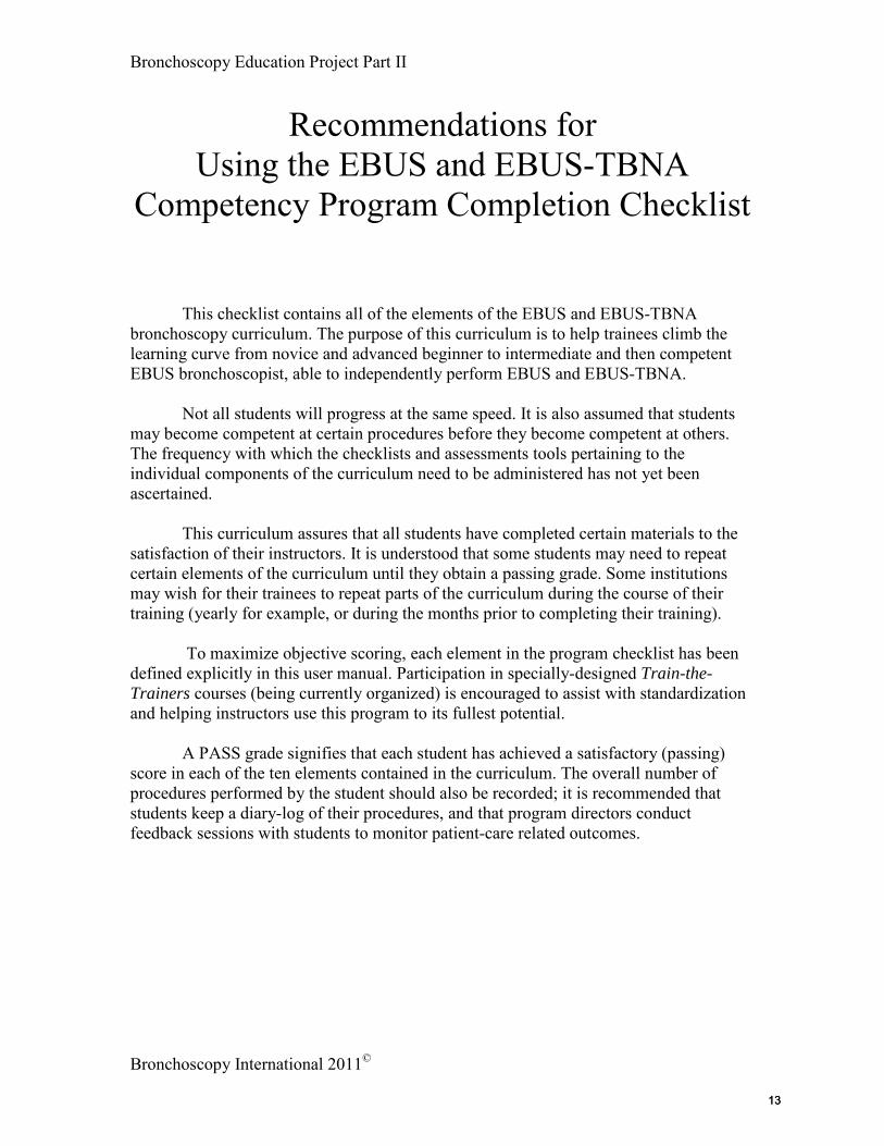
This checklist contains all of the elements of the EBUS and EBUS-TBNA bronchoscopy curriculum. The purpose of this curriculum is to help trainees climb the learning curve from novice and advanced beginner to intermediate and then competent EBUS bronchoscopist, able to independently perform EBUS and EBUS-TBNA.
Not all students will progress at the same speed. It is also assumed that students may become competent at certain procedures before they become competent at others. The frequency with which the checklists and assessments tools pertaining to the individual components of the curriculum need to be administered has not yet been ascertained. This curriculum assures that all students have completed certain materials to the satisfaction of their instructors. It is understood that some students may need to repeat certain elements of the curriculum until they obtain a passing grade. Some institutions may wish for their trainees to repeat parts of the curriculum during the course of their training (yearly for example, or during the months prior to completing their training). To maximize objective scoring, each element in the program checklist has been defined explicitly in this user manual. Participation in specially-designed Train-the-Trainers courses (being currently organized) is encouraged to assist with standardization and helping instructors use this program to its fullest potential. A PASS grade signifies that each student has achieved a satisfactory (passing) score in each of the ten elements contained in the curriculum. The overall number of procedures performed by the student should also be recorded; it is recommended that students keep a diary-log of their procedures, and that program directors conduct feedback sessions with students to monitor patient-care related outcomes.
1313
Bronchoscopy Education Project Part II
Bronchoscopy International 2011©
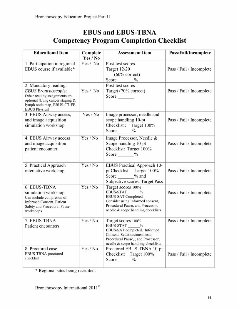
EBUS and EBUS-TBNA Competency Program Completion Checklist
Educational Item Complete
Yes / No Assessment Item Pass/Fail/Incomplete
1. Participation in regional EBUS course if available*
Yes / No Post-test scores Target 12/20 (60% correct) Score _______%
Pass / Fail / Incomplete
2. Mandatory reading: EBUS Bronchoscopist Other reading assignments are optional (Lung cancer staging & lymph node map, EBUS-CT-FB, EBUS Physics)
Yes / No
Post-test scores Target (70% correct) Score _______
Pass / Fail / Incomplete
3. EBUS Airway access, and image acquisition simulation workshop
Yes / No Image processor, needle and scope handling 10-pt Checklist : Target 100% Score ______%
Pass / Fail / Incomplete
4. EBUS Airway access and image acquisition patient encounter
Yes / No Image Processor, Needle & Scope handling 10-pt Checklist: Target 100% Score _______%
Pass / Fail / Incomplete
5. Practical Approach interactive workshop
Yes / No EBUS Practical Approach 10-pt Checklist: Target 100% Score _______% and Subjective scores: Target Pass
Pass / Fail / Incomplete
6. EBUS-TBNA simulation workshop Can include completion of Informed Consent, Patient Safety and Procedural Pause workshops
Yes / No Target scores 100% EBUS-STAT ______% EBUS-SAT Completed Consider using Informed consent, Procedural Pause, and Processor, needle & scope handling checklists
Pass / Fail / Incomplete
7. EBUS-TBNA Patient encounters
Yes / No Target scores 100% EBUS-STAT ______% EBUS-SAT completed. Informed Consent, Sedation/anesthesia, Procedural Pause, , and Processor, needle & scope handling checklists
Pass / Fail / Incomplete
8. Proctored case EBUS-TBNA proctored checklist
Yes / No Proctored EBUS-TBNA 10-pt Checklist: Target 100% Score ______%
Pass / Fail / Incomplete
* Regional sites being recruited.
1414

Bronchoscopy Education Project Part II
Bronchoscopy International 2011©
Section 2
Regional Courses
Regional courses comprised of didactic lectures, interactive sessions and simulation-based hands-on workshops using a pre-test/post-test model to
document cognitive knowledge and technical skill acquisition.
1515

Bronchoscopy Education Project Part II
Bronchoscopy International 2011©
This page intentionally left blank.
1616

Bronchoscopy Education Project Part II
Bronchoscopy International 2011©
User Instructions
Regional Courses (1 day) EBUS and EBUS-TBNA
Learning bronchoscopy in the clinical setting promotes learner anxiety, subjects patients to the burden of procedure-related education [1], and results in a highly variable learning experience [2]. Clinical responsibilities often interfere with reading of bronchoscopy-related material, and, in the absence of periodic assessments of bronchoscopy-related knowledge, trainees are unlikely to be compliant with educational endeavors they perceive as optional or reliant on individual motivation, especially if there are no pass/fail grading consequences [3]. The current subspecialty bronchoscopy learning environment is further rendered less-than-ideal for beginners because of concerns regarding patient safety, fiscal constraints, and an increasing impetus to document procedural competency [4-6]. Whilst not supplanting on-the-job training that occurs with subspecialty rotations, short postgraduate courses comprised of lectures and simulation-based hands-on instruction, have thus become a popular means towards enhancing procedure-related learning [7-9]. In accordance with continued medical education (CME) guidelines, these programs identify learner objectives and provide opportunities for feedback from students regarding the perceived quality of the course. The purpose of regional courses is to provide standardized learning material to bronchoscopy trainees. By regionalizing the process, program directors can enlist participants from numerous regional programs, thereby reducing course-related expenditures pertaining to travel and lodging. Already, several courses have become highly popular in the Carolinas, Southern California, and Midwest. Other regional courses are planned in the Northeast, Texas, and Southeast. During course participation, students are exposed to standardized course material delivered using didactic lectures, interactive sessions, hands-on training using patient models, low-fidelity and high-fidelity simulation, debriefing exercises, and problem-based learning modules. Pre-test/post-test assessments help document knowledge and technical skill acquisition, thereby setting a new baseline for students in subspecialty training. It has long been recognized that assessment drives learning, and that rigorous assessment inspires learning, reinforces confidence, and reassures the public. Proving that course participation is responsible for learning gains is difficult. For example, demonstrating the short-term benefit of an educational intervention is controversial because of debates regarding the value of pre-test and post-test assessments, and because of the obvious difficulty constituting a control group to which studies of an educational intervention can be compared [10-13]. Studies of long-term retention are problematic because causality is subject to the effects of normal maturation and ongoing training history [14].
1717
Bronchoscopy Education Project Part II
Bronchoscopy International 2011©
The true value of pre-test/post-test assessments has also been controversial because of the effects of many extraneous variables, which include the Hawthorne effect (knowing that one is being tested may affect the results), the halo effect (the human tendency to respond positively or negatively to an instructor), and the practice effect (of a pre-test on a subsequent post-test). In the context of procedure-based training, the calculation of various measures of learning gain, including class-average and single-student normalized gain provides an objective and informative means to document learner performance and demonstrate robustness of the educational intervention. Patients should not bear the burden of procedure-related training. Participation in regional courses, using simulation-based deliberate practice to acquire technical skill, and documenting a rapid climb up the initially steep slope of the novice’s learning curve should result in decreased patient suffering and improved procedure-related decision making. Diverse opinions regarding educational methodologies, curricular structure, and measures of effectiveness persist in regards to short one- or two-day programs [15-16]. Additional studies are therefore needed, not only to document the effectiveness of regional courses, but also to determine how such courses might favorably impact patient outcomes. Selected References
1. Silvestri GA. The evolution of bronchoscopy training. Respiration. 2008;76(1):92-101.
2. Haponik EF, Russell GB, Beamis JF, et al. Bronchoscopy training: current fellows' experiences and some concerns for the future. Chest. 2000 Sep;118(3):572-573.
3. Wahidi MM, Silvestri GA, Coakley RD, Ferguson JS, Shepherd RW, Moses L, Conforti J, Que L, Anstrom KJ, McGuire F, Colt H, Downie GH. A prospective multi-center study of competency metrics and educational interventions in the learning of bronchoscopy among starting pulmonary fellows. Chest E-pub, Oct, 2009.
4. Reznick RK, MacRae H. Teaching Surgical Skills – Changes in the Wind. N Engl J Ned. 2006; 355:2664-2669.
5. Carraccio C, Englander R. Evaluating competence using a portfolio: a literature review and web-based application to the ACGME competencies. Teach Learn Med. 2004 Fall;16(4):381-387.
6. Carraccio C, Wolfsthal SD, Englander R, et al. Shifting Paradigms: From Flexner to Competencies. Academic Medicine. 2002;77(5):361-367.
7. Norman G. The American College of Chest Physicians evidence-based educational guidelines for continuing medical education interventions: a critical review of evidence-based educational guidelines. Chest. 2009;135(3):834-837.
8. Schijven MP, Schout BM, Dolmans VE, Hendrikx AJ, Broeders IA, Borel Rinkes IH. Perceptions of surgical specialists in general surgery, orthopaedic surgery, urology and gynaecology on teaching endoscopic surgery in The Netherlands. Surg Endosc. 2008;22(2):472-482.
1818

Bronchoscopy Education Project Part II
Bronchoscopy International 2011©
9. Davis D, Bordage G, Moores LK, Bennett N, Marinopoulos SS, Mazmanian PE, Dorman T, McCrory D. The Science of Continuing Medical Education. Chest.2009;135:8S-16S.
10. Cronbach LJ, Furby L. How should we measure “change” or should we? Psychol. Bull 1970;74:68-80.
11. Hake, R.R. 2009. "Should We Measure Change? Yes!" online at <http://www.physics.indiana.edu/~hake/MeasChangeS.pdf> (2.5 MB) and as ref. 43 at <http://www.physics.indiana.edu/~hake>.
12. Melzer DE. Normalized learning gain: A key measure of student learning [Addendum to: Melzer DE. The relationship between mathematics preparation and conceptual learning gains in physics: A possible “hidden variable” in diagnostic pretest scores. Am J. Phys 2002;70:1259-1267. Online at http://scitation.aip.org/getpdf/servlet/GetPDFServlet?filetype=pdf&id=AJPIAS000070000012001259000001&idtype=cvips [Accessed June 16, 2009].
13. USDE. 2003. U.S. Department of Education, Identifying and Implementing Educational Practices Supported by Rigorous Evidence: A User Friendly Guide. Institute of Education Sciences, National Center for Education Evaluation and Regional Assistance. The entire guide is online at <http://www.ed.gov/rschstat/research/pubs/rigorousevid/index.html> / “PDF (140KB)”.
14. Shadish, W.R., T.D. Cook, & D.T. Campbell. 2002. Experimental and Quasi-Experimental Designs for Generalized Causal Inference. Houghton Mifflin - information at <http://tinyurl.com/y3e7vw>.
15. Colt HG, Davoudi M, Murgu SD, Zamanian Rohani N. Measuring learning gain during a one-day introductory bronchoscopy course. Surgical Endoscopy. 2010; In Press.
16. Colt HG, Davoudi M, Quadrelli S, Zamanian Rohani N. Use of Competency-based metrics to determine effectiveness of a postgraduate thoracoscopy course. Respiration. 2010; In Press.
1919

Bronchoscopy Education Project Part II
Bronchoscopy International 2011©
Example of a One day EBUS & EBUS-TBNA Program
7:30-8:15 am Registration, pre-test and survey of practice experience
8:15-8:30 Welcome, introduction, and learning objectives
8:30-9:00 EBUS-TBNA Indications, diagnostic yield, complications
9:00-9:30 EBUS-TBNA Technique and smear preparation
9:30-10:00 Mediastinal anatomy (IASLC) and lymph node structure
10:00-10:15 EBUS-CT-White light bronchoscopy correlations
10:15-10:30 BREAK
10:30-11:00 EBUS: Other applications
11:00-11:30 Practical Approach to EBUS-TBNA
11:30-12:00 Interactive session: EBUS-TBNA True/False exercises
12:00-1:00 LUNCH
Afternoon hands-on training
1:00-3:30 pm
Hands-on training: 4 stations - 35 minutes per station
(1) EBUS Step by Step: handling the scope and the needle (x2)
(2) EBUS Step by Step: handling the scope and the image (x2)
(3) Nodal and vascular mediastinal anatomy: pattern recognition
(4) Patient-based Practical Approach to EBUS-TBNA
3:30-4:15 Post course technical skills and cognitive learning assessments
4:15-4:30 Wrap up, certificate of course completion
2020

Bronchoscopy Education Project Part II
Bronchoscopy International 2011©
EBUS and EBUS-TBNA: Hands-On Workstations
Workstation 1: EBUS Step by Step: Scope and Needle Handling
Learning Objectives:
1. To be able to demonstrate how to move the EBUS scope into position. 2. To be able to demonstrate techniques of needle insertion and EBUS-TBNA. 3. To demonstrate communication skills with bronchoscopy assistants.
Description:
The instructor will review the EBUS-TBNA Step by Step Instructional Video with the group. EBUS-TBNA will be performed through the carina into subcarinal adenopathy of the specially designed lo-fidelity EBUS airway model, followed by EBUS-TBNA at levels 7, 4R and 4l. The instructor will describe different ways with which to assure patient and operator safely, and ways in which the EBUS bronchoscope is protected. Each team member will demonstrate needle insertion techniques, while another team member serves as the bronchoscopy assistant, assuring patient, operator, and equipment safety. This instructional session focuses on repetition and focused practice.
Workstation 2: EBUS Step by Step: Scope and Image Handling
Learning Objectives:
1. To be able to turn on and set up the EBUS processor 2. To be able to introduce the EBUS scope using Laryngeal mask intubation
(LMA), endotracheal intubation, or the bronchoscopic transoral route. 3. To be able to manipulate the scope atraumatically inside the artificial airway
and inside the upper and lower airways. 4. To demonstrate the use of gain, and depth and Doppler for image acquisition. 5. To be able to trouble-shoot complications and equipment malfunction.
Description:
The instructor will review the EBUS Physics Instructional Video with the group. Students will be asked to make changes on the image processor in order to better visualize the target lymph node at levels 7, 4R or RL using the specially-designed lo-fidelity model. Intubation in the patient with an EBUS scope can be difficult. Because EBUS can be performed using different intubation techniques, the operator will demonstrate the ability to manipulate the scope in the artificial airway prior to obtaining an EBUS image.
2121

Bronchoscopy Education Project Part II
Bronchoscopy International 2011©
Workstation 3: Nodal and vascular mediastinal anatomy: pattern recognition
Learning Objectives:
1. To be able to precisely elaborate a strategy for nodal sampling using computed tomography scans for planning an EBUS-TBNA procedure.
2. To be able to recognize site-specific EBUS imaging of mediastinal and hilar nodal and vascular anatomy.
Description:
The instructor will review the EBUS Lymph node map Instructional Video with the group. The instructor will demonstrate how a CT scan can be used to plan an EBUS-TBNA procedure, after which team members will be asked to elaborate a procedural strategy based on an imaging study. During this small group, interactive session, participants will then study a collection of site-specific images corresponding to nodal and vascular, mediastinal and hilar anatomy. .
Workstation 4: Patient-based Practical Approach to EBUS-TBNA
Learning Objectives:
1. The learner should be able to identify case-specific elements that are important for procedural strategy and planning.
2. The learner should be able to describe technical elements critical for the execution of EBUS-TBNA, including review of CT scan and choosing nodal sampling strategies.
3. The learner should be able to identify procedure-related risks, benefits, and alternatives, as well as have a plan of action in case of procedure-related complications.
Description:
In this interactive session, the instructor opens the floor to the learners, and together they walk through a patient’s clinical scenario using the 4-Box Practical Approach model, working their way through initial patient evaluation, assessment of procedural strategies (including indications and contraindications, expected results, and risk-benefit analysis), discuss procedural techniques and results (including choosing among technical options and instruments, knowing the anatomic risks, results, and possible complications and how to deal with each), and devising long-term management plans (including assessment of the results, plan for follow-up diagnostic and therapeutic options, along with quality improvement for the procedural team). This “Practical Approach” exercise combines cognitive, theoretical technical, affective, and experiential learning.
2222

Bronchoscopy Education Project Part II
Bronchoscopy International 2011©
Section 3
Reading Assignments
Suggested reading*
• The EBUS Bronchoscopist©
based study guide, PDF file and 10 question post-test (mandatory). : One module, 30 question/answer web-
• Lung Cancer Staging (Revised IASLC) and EBUS Lymph Node Map: module with didactic lecture, synopsis, and instructional video (optional reading).
• EBUS-CT-White Light Bronchoscopy Correlations: module with didactic lecture, synopsis (poster), and instructional video (optional reading).
• EBUS Physics and Terminology: module with didactic lecture, synopsis, and instructional video (optional reading).
* Checklists, videos, and post-tests used at the discretion of program director.
2323

Bronchoscopy Education Project Part II
Bronchoscopy International 2011©
This page intentionally left blank.
2424

Bronchoscopy Education Project Part II
Bronchoscopy International 2011©
User Instructions Mandatory reading of the single module, 30 question/answer
EBUS Bronchoscopist©
with post-tests
The web-based EBUS Bronchoscopist© is a laddered curriculum of theoretic bronchoscopic knowledge that can be accessed free of charge in English. It has been prepared with the assistance and input of numerous EBUS experts worldwide who contributed question/answer series. This and other materials can be downloaded from The Bronchoscopy International website (www.Bronchoscopy.org) an HON code certified (Health on the Net) voted by the American Thoracic Society as the best on-line resource for bronchoscopy education. The aim of the EBUS Bronchoscopist © is to complement a traditional apprenticeship model of training in EBUS and EBUS-TBNA by emphasizing important facets of knowledge and skill required for competency. Elements addressed in the EBUS Bronchoscopist © are intentionally written so that contrary opinions might occasionally be provided by instructors. In this fashion, dialogue is promoted, but access to a certain amount of “essential” material is guaranteed. The question-answer sets of the EBUS Bronchoscopist©
contain information pertaining to mediastinal and hilar nodal and vascular anatomy, lymph node mapping, scope insertion and placement within the airways, patient preparation, indications, contraindications and complications, techniques and solutions to technical problems, image processing and troubleshooting, ultrasound physics and image artifacts, site specific pattern recognition, as well as lung cancer staging and restaging.
In order to document that a student has been exposed to material contained in the EBUS Bronchoscopist ©
, a passing score on post-tests is warranted. A score of 70 and above (7/10 correct responses) is warranted to check off the module as completed on the EBUS Bronchoscopy Education Competency Checklist.
Selected References 1. Colt H G, D avoudi M , Q uadrelli S . Pilot s tudy o f web-based b ronchoscopy e ducation us ing t he
Essential B ronchoscopist in d eveloping c ountries ( Mauritania a nd M ozambique). R espiration 2007;74:358-359.
2. Colt HG, Ngoc van Tran, Quadrelli S, than Pham van. Creation of an interventional technical plateau at Cho ray Hospital, Vietnam. J Bronchol 2007;14:289-292.
3. Davoudi M , Q uadrelli S , O sann K , an d C olt HG. A competency-based t est of br onchoscopic knowledge using the Essential Bronchoscopist: an initial concept study. Respirology, 2008;13:736-743.
4. Davoudi M, Colt HG. Bronchoscopy s imulation: a b rief r eview. Adv Health Sci Educ 2009;14:287-296.
5. Goldberg R, Colt HG, Davoudi M, Cherisson L. Realistic and affordable lo-fidelity 6. Quadrelli S , G alíndez F , D avoudi M C olt H G,. R eliability o f a 2 5 ite m low stakes multiple c hoice
assessment of bronchoscopic knowledge. Chest 2009;135:315-321. 7. Wahidi MM, Silvestri GA, Coakley RD, Ferguson JS, Shepherd RW, Moses L, Conforti J, Que L,
Anstrom KJ, McGuire F, Colt H, Downie GH. A prospective multi-center study of competency metrics and e ducational i nterventions i n t he l earning of br onchoscopy a mong s tarting pul monary f ellows. Chest 2010, online pub.
2525

Bronchoscopy Education Project Part II
Bronchoscopy International 2011©
This page intentionally left blank.
2626

Bronchoscopy Education Project Part II
Bronchoscopy International 2011©
User Instructions
Suggested reading of the (1) Lung cancer staging (revised IASLC) and EBUS lymph node map module (2) EBUS-CT-White Light bronchoscopy correlations module, and (3) EBUS physics and terminology module
The purpose of these readings is to provide students with exposure to basic principles pertaining to Lung cancer staging, EBUS-TBNA mediastinal and hilar lymph node assessments, and EBUS-related ultrasound physics. Reading these assignments will complement reading the EBUS Bronchoscopist. While it is presumed that institutions have their own regulations and protocols, many do not have a formal program of education in these three areas. Inappropriate understanding of general principles in these three areas can adversely affect patient management. It is for this reason that knowledge in these three areas is necessary, and it is also why we have prepared special instructional videos so that learners can repeatedly study relevant materials at their own leisure as well as during the course of their training. We recommend at least ONE formal session during which a didactic lecture on each of these three subjects is provided (after students have reviewed the synopsis and other pertinent material such as posters and instructional videos available on www.Bronchoscopy.org, and the Bronchoscopy International YouTube and Facebook sites).
2727

Bronchoscopy Education Project Part II
Bronchoscopy International 2011©
This page intentionally left blank.
2828

Bronchoscopy Education Project Part II
Bronchoscopy International 2011©
Lung Cancer Staging and EBUS Lymph node Map
SYNOPSIS
The purpose of this synopsis is to provide readers with a brief overview of the IASLC Revised Lung Cancer Staging System, particularly effects of nodal stage on prognosis and survival, as well as to provide a description of how EBUS can map mediastinal and hilar nodal and vascular anatomy. It is assumed that institutions and practitioners have different biases and regulations. Herein a short summary is provided so that EBUS bronchoscopists might enhance or reinforce their knowledge. Readers are encouraged to follow guidelines and protocols established in their own institutions. IASLC Lung Cancer Staging Figure 1: Revised lymph node maps from 7th edition
From IASLC and
2929

Bronchoscopy Education Project Part II
Bronchoscopy International 2011©
Figure 2: Applying the new IASLC map to CT scans
From IASLC and
3030

Bronchoscopy Education Project Part II
Bronchoscopy International 2011©
Figure 3: Definitions of Nodal stages
From IASLC and
3131

Bronchoscopy Education Project Part II
Bronchoscopy International 2011©
3232

Bronchoscopy Education Project Part II
Bronchoscopy International 2011©
Prognosis and survival based on stage according to IASLC 7th
edition staging system
IASLC 7th Edition
Goldstraw P, Crowley J, Chanksy K, Giroux D, Groome P, Rami-Porta R, Postums P, Rusch V, Sobin L. The IASLC Lung Cancer Staging Project: Proposals for the Revision of the TNM Stage Groupings in the Forthcoming (Seventh) Edition of the TNM Classification of Malignant Tumours. J Thorac Oncol 2007: 2; 706-704.
Also in Chest 2009;136:260-271
3333

Bronchoscopy Education Project Part II
Bronchoscopy International 2011©
IASLC 7th EditionNSCLC
Clinical Stage
Goldstraw P, Crowley J, Chanksy K, Giroux D, Groome P, Rami-Porta R, Postums P, Rusch V, Sobin L. The IASLC Lung Cancer Staging Project: Proposals for the Revision of the TNM Stage Groupings in the Forthcoming (Seventh) Edition of the TNM Classification of Malignant Tumours. J Thorac Oncol 2007: 2; 706-704.
IASLC 7th Edition
Goldstraw P, Crowley J, Chanksy K, Giroux D, Groome P, Rami-Porta R, Postums P, Rusch V, Sobin L. The IASLC Lung Cancer Staging Project: Proposals for the Revision of the TNM Stage Groupings in the Forthcoming (Seventh) Edition of the TNM Classification of Malignant Tumours. J Thorac Oncol 2007: 2; 706-704.
NSCLCPathologic Stage
EBUS Lymph Node Map (See video): Describes scope and transducer placement
3434

Bronchoscopy Education Project Part II
Bronchoscopy International 2011©
1. Station 2R, the upper right paratracheal region, extends across the midline to the left
lateral border of the trachea. The upper border is the apex of the right lung and pleural space, and, the upper border of the sternal manubrium. The lower border is at the intersection of the caudal margin of the innominate vein with the trachea. The EBUS probe is placed against the right tracheal wall in the upper trachea at the level of the 4-5th
2. Station 2L, the upper left paratracheal region, includes nodes extending to the left of the left lateral border of the trachea. The upper border is the apex of the left lung and pleural space, and in the midline, the upper border of the sternal manubrium. The lower border is the superior aspect of the aortic arch. To visualize this station, the EBUS scope is placed against the left tracheal wall in the upper trachea at the level of the 4-5
tracheal rings. The transducer is oriented towards the 3-o’clock position.
th
3. Station 4R, the right lower paratracheal region, includes right lower paratracheal nodes, and pretracheal nodes extending to the left lateral border of trachea. The upper border is the intersection of the caudal margin of the innominate vein with the trachea. The lower border is the lower margin of the azygos vein. The EBUS scope is placed proximal to the main carina and turned towards the 3-o’clock position. 4R nodes, however, are often pretracheal, in a more anterior position, so the scope may need to be turned counterclockwise towards the 12 o’clock position.
tracheal rings. The transducer is oriented towards the 9-o’clock position.
4. Station 10R is immediately adjacent to the right main bronchus and hilar vessels, including the proximal portions of the pulmonary veins and main pulmonary artery. The upper border is the lower margin of the azygos vein. The lower border is the interlobar region between the right upper lobe and bronchus intermedius. To visualize this hilar nodal station, the EBUS scope can be gently advanced from the carina to the
3535

Bronchoscopy Education Project Part II
Bronchoscopy International 2011©
origin of the right upper lobe bronchus. The transducer is oriented antero-laterally towards the 2-o’clock position.
5. Station 7 is subcarinal. The upper border of this region is the main carina of the trachea. Its lower borders are the distal end of the bronchus intermedius on the right, and the upper border of the lower lobe bronchus on the left. The scope is placed in the proximal right or left main bronchus. The transducer is oriented medially.
6. Station 4L includes nodes to the left of the left lateral border of the trachea, medial to the ligamentum arteriosum. Its upper border is the upper margin of the aortic arch. Its lower border is the upper margin of the left main pulmonary artery. The scope is placed in the proximal left main bronchus at the level of main carina. The scope is turned to the left, towards the 9-o’clock position.
7. Station 10L is immediately adjacent to the left main bronchus and hilar vessels, including the proximal portions of the pulmonary veins and main pulmonary artery. The upper border is the upper margin of the left pulmonary artery. The lower border is the interlobar region between the left upper and lower lobes. The EBUS scope is placed in the proximal left upper lobe bronchus. The tip of the scope is pressed to the bronchial wall towards the 11-o’clock position.
8. Station 11L is comprised of nodes located in the region between the origins of the left upper and lower lobar bronchi. The scope is advanced towards the proximal part of the lower lobe bronchus. The transducer is oriented laterally towards the left in order to scan for interlobar lymph nodes.
9. Station 11R superior includes nodes between the right upper lobe bronchus and the bronchus intermedius. The tip of the EBUS scope is placed in the proximal bronchus intermedius, just below the subcarina separating the upper lobe bronchus from the bronchus intermedius. The area between the 2-o’clock and 4-o’clock positions is scanned.
10. Station 11R inferior is between the middle lobe and right lower lobe bronchi. The EBUS scope is positioned in the proximal right lower lobe bronchus. The transducer can then be oriented towards the right lateral wall.
__________________________________________
3636

Bronchoscopy Education Project Part II
Bronchoscopy International 2011©
EBUS-CT-WHITE LIGHT BRONCHOSCOPY
SYNOPSIS
The purpose of this synopsis is to provide the reader with a brief overview of EBUS-CT-White light bronchoscopy correlations in order to enhance procedural strategy and planning. A poster is provided so that EBUS bronchoscopists might familiarize themselves with radiographic anatomic landmarks and their EBUS/white light bronchoscopy correlates. Readers are encouraged to study the instructional video for further information, and to discuss procedural planning strategies and techniques with colleagues and EBUS experts. This is just one example of how imaging studies can be used to help plan EBUS-TBNA. Readers are encouraged to develop and discuss other methodologies with their peers. See Poster in your curriculum booklet as well as video
1. Specific Computed Tomography views, primarily the coronal views, can help plan an EBUS procedure. In this example we shall use the lower paratracheal station 4L limited by the superior margin of the aortic arch and the upper rim of the left main pulmonary artery. The lymph node is usually located lateral to the trachea at the level of the main carina.
3737

Bronchoscopy Education Project Part II
Bronchoscopy International 2011©
2. The bronchoscope is placed in the lower trachea, approximately at the origin of the left main bronchus. With the balloon inflated, the transducer is turned towards the left to visualize the left paratracheal region. Station 4L is ALWAYS to the left of the left lateral border of the trachea.
3. The coronal computed tomography view correlates well with this EBUS scanning plane. The aortic arch is seen proximal, and the left pulmonary artery is seen distal to the lymph node. Actually, the EBUS image is projected on the monitor as if the scope were horizontal. The green dot on the monitor represents the point where the needle exits the scope. This corresponds to the cephalad aspect of the body. This dot is by default, towards the 1’o’clock position on the monitor.
4. Several adjustments can be made to the coronal CT image in order to bring the scope to a horizontal position and the green dot cephalad (towards the 1 o’clock position on the monitor) to match the EBUS image.
5. Rotate the CT image clockwise in order to bring the scope to a horizontal position and the green dot towards 1 o’clock. Because the green dot is cephalad, the vascular structure at 3’o’clock is proximal and represents the aorta, while the vascular structure at 9’o’clock is more distal and represents the left pulmonary artery.
__________________________________________
3838

Bronchoscopy Education Project Part II
Bronchoscopy International 2011©
EBUS PHYSICS and TERMINOLOGY
SYNOPSIS
Ultrasound (US) is an imaging moda lity ba sed on the r eflective properties of sound w aves. U ltrasonography t hus represents m echanical en ergy t hat caus es compression a nd r arefaction of a c onducting m aterial or s ubstance know n a s m edium. This t echnology us es s ound w aves with f requencies of 20kH z or hi gher, i naudible t o humans1
.
Echogenicity represents t he ex tent t o which a tissue or s ubstance gives r ise to reflections of ul trasonic w aves. W hen US i mages a re di splayed on a gray s cale, t he strongest echo signal is white, and when no sound wave is reflected, the image is black or in ul trasound t erms, a nechoic ( Figure 1). The int ensity o f the s ignal determining echogenicity depends on the r eflected wave am plitude. The t erms us ed in ultrasonography to describe a certain t issue or s tructure i nclude: isoechoic, comparable with t he s urrounding t issue; hypoechoic, weaker t han the s urrounding t issue and hyperechoic, stronger than the surrounding tissue (Figure 1). Frequency and Wavelength represent a specific number of vibration cycles per second (measured in units of hertz). Endoscopic US frequencies are in the range of 5-30 MHz2
. Current dedicated EBUS-TBNA bronchoscopes allow changes in frequency from 5 t o12 M Hz. The wavelength of U S r epresents t he di stance be tween two successive pulses: the higher the frequency, the shorter the related wavelength.
Propagation is the process through which sound advances through various tissues. The t ransducer s ends ou t a br ief pul se of s ound t hat pe netrates t he t issue. T he s ound waves are reflected back to a transducer, which serves as the sensor and source of signal. US i s r eflected at tissue boundaries and interfaces, l ike l ight of f a m irror (F igure 1); it reflects very well wherever there is a significant change in the propagation medium. The degree of reflection is determined by t he acou stic i mpedance o f t he adjacent t issue, largely related to tissue de nsity. When the U S b eam s trikes an interface, i t unde rgoes
1 Kurimoto N. Diagnosis of depth penetration in the tracheobronchial tree. In: Kurimoto N (ed.). Endobronchial Ultrasonography. Kinpodo, Kyoto, 2001; pp 33–38. Also, see Greenleaf JF. ABCs of Ultrasound Imaging. In: Ernst A, Feller-Kopman D (eds). Ultrasound Guided Procedures and Investigations. A manual for the clinician.Taylor & Francis, New York, 2006; pp 1-16 2 Nishina K, Hirooka K, Wiegand J, Dremel H. Principles and Practice of endoscopic ultrasound. In Bolliger CT et al (eds): Clinical Chest Ultrasound: From the ICU to the bronchoscopy suite. Prog Respir Res. Basel, Karger, 2009, vol 37,pp 110-127
3939

Bronchoscopy Education Project Part II
Bronchoscopy International 2011©
refraction, s cattering, a nd a ttenuation a s i t pa sses t hrough t issue, all of which de grade image quality on examining deeper structures (Figure 1). Refraction represents a ch ange i n di rection of t he i ncident U S be am, w hile scattering is the spread of the US beam in different directions. Attenuation is the loss of energy caused by absorption (when the vibration of the US wave is converted to heat due to friction). Attenuation depends on the medium, being much higher in air than in water and on the frequency, increasing with higher frequencies (Figure 1). The bigger the difference in acoustic properties between two media, the larger the pr oportion of r eflected U S a nd t he s maller the t ransmitted U S. S uch a n a coustic interface results i n a s trong e cho s ignal ( i.e. l ung t issue or a ir i n t he bronchial s ystem) (Figure 1); the lower the density, the higher the attenuation (i. e .higher attenuation in air than in water) (Figure 1). Penetration refers t o the di stance be tween an imaged area and the t ransducer. The time delay between the energy entering the tissue and returning to the US transducer determines the depth from w hich the s ignal ar ises ( longer t imes equ al greater de pths since d epth=velocity X time/2). Large t ransducers t hat t ransmit pow erful be ams will increase penetration depth (it goes without saying, therefore, that penetration depth is less in EBUS than in thoracic US) (Figure 1). The maximum penetration depth depends on the f requency us ed: h igher f requencies ( i.e. 20 M Hz) do not pe netrate a s de ep a s t he lower frequencies (7.5 MHz). Thus superficial structures are better visualized with higher frequency and deeper structures with lower frequency transducers. For these reasons, in bronchoscopy, the 20 MHz E BUS r adial pr obe can be used for ima ging a irway w all layers, while the 7.5 MHz is used for visualizing deeper structures such as lymph nodes and blood vessels (Figure 1). Resolution refers to the level of detail of an image; it is the capacity of a system to distinguish small objects from others and is determined by the frequency and duration of the transmitted sound phase. Resolution is categorized in two types: axial, representing the ability to resolve ob jects w ithin t he i maging pl ane a t di fferent de pths, a nd lateral, representing t he abi lity t o resolve obj ects i n the imaging pl ane t hat ar e l ocated side b y side. Image A rtifacts are due t o image di stortion c aused b y no rmal phe nomena of refraction, scattering an d attenuation which may interfere w ith the a bility to properly identify an US target (i.e. lymph node, blood vessel). Yet, image artifacts can be useful because they help describe the properties of tissues (i.e. calcification or necrosis within a lymph node). Understanding the different types of artifacts helps us identify and obtain a clear i mage o f a r eal t arget a nd pe rform a s afe ne edle a spiration w ithout punc turing vascular s tructures o r l ung p arenchyma. Common types of a rtifacts s een dur ing EBUS imaging include reverberation and attenuation artifacts.
• Reverberation artifacts occur w hen a hi ghly r eflective t issue i s pa rallel t o the transducer, such as when the water filled balloon of the EBUS probe/scope is not in contact w ith the a irway w all, and the U S waves ar e r epeatedly reflected
4040

Bronchoscopy Education Project Part II
Bronchoscopy International 2011©
between the tissue surface (airway wall) and the transducer3
•
. The resulting effect is t hat s trong f alse e choes a ppear as m ultiple e qually s paced l ines on t he ultrasound image (Figure 2). Attenuation artifacts are c omprised of the tadpole tail s ign and the acoustic shadow. T issues w ith low a coustic i mpedance ( i.e. ne crotic l ymph node s, mediastinal cysts) result in lower attenuation than tissues with higher impedance. For t he tadpole ta il a rtifact, t he echo a t t he di stal bor der of t he l ow i mpedance structure will be higher and US will display the area distal to the low impedance structure m ore b rightly compared t o t he s urrounding t issue (Figure 2). The acoustic shadow artifact
3 Nishina K, Hirooka K, Wiegand J, Dremel H. Principles and Practice of endoscopic ultrasound. In Boliger CT et al (eds): Clinical Chest Ultrasound: From the ICU to the bronchoscopy suite. Prog Respir Res. Basel, Karger, 2009, vol 37,pp 110-127
is exactly the reverse of the tadpole tail as the area behind a high impedance structure is displayed with lower brightness than the rest of the surrounding tissue. Since the US beam is almost completely reflected at the border or attenuated within the high impedance structure, the posterior area does not receive US waves and appears as a hypoechoic shadow (Figure 2).
4141

Bronchoscopy Education Project Part II
Bronchoscopy International 2011©
Figure 1: Ultrasound terminology and principles
Figure 1. Principles and terminology of ultrasound: (A). A structure (i.e. blood vessel) which does not reflect the sound waves at all is termed anechoic. (B). A hyperechoic pattern (i.e. cartilaginous nodule in tracheopathica osteochondroplastica) occurs when the echoes are stronger than those of the surrounding tissues. (C). A hypoechoic structure ( i.e. lymph node) occurs when the echoes are weaker than those from the surrounding tissues. (D). This lymph node is labeled isoechoic, because the echoes are of comparable amplitude with the surrounding tissue (i. e. lung). (E). The US beam undergoes reflection, refraction, scattering, and attenuation as it passes through tissue; all of these degrade image quality on examining deeper structures but the last three processes cause the US waves reflected back to the transducer to be much weaker. (F). Increased attenuation and low penetration depth occur when structures are visualized with high frequency probes: the hypertrophic stenotic tissue and the intact cricoid cartilage are clearly visualized, but the deeper structures are not when the 20 MHz radial probe is used. (G). When the curved linear EBUS transducer with 7.5 MHz frequency is used the attenuation is decreased, the penetration depth is increased and the EBUS image shows the right lower paratracheal lymph node, the superior vena cava and the distal normal lung parenchyma but details of the airway wall structures cannot be assessed. (H). Attenuation also depends on the medium, being low in fluid (i.e. blood vessels). (I). Attenuation is high in air (i.e. normal lung tissue). (J). The depth of penetration is also directly dependent on the size of the transducer. A relatively superficial penetration (i.e. 5 cm) is achieved using the small EBUS curved linear transducer. (K). A deep penetration (i.e. 15 cm) can be obtained by using large thoracic transducers.
4242

Bronchoscopy Education Project Part II
Bronchoscopy International 2011©
Figure 2: Image artifacts
Figure 2. Image artifacts. (A). Reverberation artifacts: when the water filled balloon of the EBUS bronchoscope is not in intimate contact with the airway wall, the US waves are repeatedly reflected between the highly reflective tissue surface (airway wall) and the transducer; the resulting effect is that strong false echoes appear as multiple equally spaced lines on the ultrasound image. (B). In the tadpole tail artifact, the echo at the distal border of the low impedance structure ( i.e. azygous vein) will be higher and US will display the area distal to the low impedance structure more brightly compared to the surrounding tissue (thick arrows). (C). The acoustic shadow artifact is the reverse effect of the tadpole tail; the area behind a high impedance structure (i.e. calcification within the lymph node) is displayed with lower brightness than the rest of the surrounding tissue. The US beam is attenuated within the high impedance structure and the posterior area appears as a hypoechoic shadow (thick arrows).
__________________________________________
4343

Bronchoscopy Education Project Part II
Bronchoscopy International 2011©
This page intentionally left blank.
4444

Bronchoscopy Education Project Part II
Bronchoscopy International 2011©
Section 4
EBUS Step-by-Step
The EBUS Bronchoscopy Step-by-Step presentation is based on the idea that with patience, each step can be mastered. To become a good tennis player, one cannot master the forehand, backhand, serve, volley, smash and all other strokes at the same time; separately and repeatedly, the different strokes are practiced and then combined to play a beautiful game. The same holds true for the multiple steps needed to perform EBUS-TBNA. It is also necessary to communicate clearly to a bronchoscopy assistant who helps handle the needle and syringe, applies suction, and subsequently handles the specimen. EBUS step by step therefore provides learners with a:
• Systematic Approach: Deconstructing a complex task into constituent elements
• Development of Muscle Memory: Motor learning through the sub-
conscious process of improving motor skills, smoothness and accuracy of movements, thus creating maximum efficiency and economy of movement. The major prerequisite for development of muscle memory is repeated, deliberate practice.
• Development of Spatial Awareness: To learn to flow in space, always occupying the desired position. In EBUS bronchoscopy, this additionally requires the accurate identification of airway anatomy, manipulation of the 30 degree view EBUS scope, and appropriate positioning of the transducer within the airway.
4545

Bronchoscopy Education Project Part II
Bronchoscopy International 2011©
This page intentionally left blank.
4646

Bronchoscopy Education Project Part II
Bronchoscopy International 2011©
EBUS Bronchoscopy Step-by-Step
The same principles as those applied to Flexible Bronchoscopy Step-by-Step are followed. Optimum hand position and posture should be maintained at all times. The bronchoscope’s position within the airway should be known at all times, recalling that the angle of view is 30 degrees. The airway wall should be respected and trauma avoided during nodal sampling. Careful attention should be paid to the order in which lymph nodes are sampled. Contamination of needle entry sites or of the needle sheath and aspirating needle must be consistently avoided. Steps should be practiced while standing both at the “patient’s” head and side, although it is most likely practitioners will be performing EBUS-TBNA from the head of the patient. It is best that practice be done initially on inanimate models and/or a virtual reality (VR) simulator. Remember: Decision; Intent; Control; Confidence; Economy of Movement.
Background: A curved array ultrasound transducer is built into the distal end of the EBUS bronchoscope which has a thirty degree field of vision using white light bronchoscopy. The processor has adjustable depth and gain. It also has Doppler capabilities to identify blood flow in vessels, distinguishing them from lymph nodes. The 22 gauge disposable needle and sheath protrude at an angle. The lockable sheath prevents injury to the scope during needle protrusion. An inner stylet prevents aspiration of bronchial cells. Prior to EBUS, white light bronchoscopic inspection is performed and the airways are cleared of secretions. A touch control button on the ultrasound processor alternates between the bronchoscopic and ultrasound image. After airway inspection, the balloon can be inflated on the EBUS scope so that a small crescent of it is seen. It may be necessary to trouble shoot the balloon and needle/sheath apparatus (see Processor and Needle checklist as well as simulation scenarios). After sonographic mapping of the nodal and vascular structures of the mediastinum, the operator selects the target node(s) based on a predetermined strategy and proceeds with EBUS-TBNA. The following is just one example using a commercially available needle-syringe ensemble. EBUS-TBNA in 15 steps (see EBUS step-by-step video)
1. The needle is inserted into the working channel. 2. The housing is secured to the bronchoscope by sliding the flange. 3. The sheath is released by twisting the inferior screw. 4. With the node visualized by US, the sheath is advanced out of the end
of the scope until it slightly touches the airway wall. It is now safe to advance the needle.
5. The needle screw, located superiorly, is then released. 6. The needle is advanced by jabbing it into the lymph node.
4747

Bronchoscopy Education Project Part II
Bronchoscopy International 2011©
7. During this process the needle may push the airway wall away from the balloon. The transducer –wall interface might become lost and the image may show reverberation artifact. This problem is overcome by gently advancing the scope or further inflating the balloon.
8. The needle is visualized within the lymph node. 9. The stylet is moved in and out a few times to dislodge bronchial
epithelium or cellular debris that may have entered the needle. 10. The stylet is then withdrawn from the scope. 11. The syringe is attached to the needle and suction is applied. 12. The needle is moved back and forth within the node approximately 10 -15
times under ultrasound visualization. 13. Suction is released by removing the syringe from the scope. 14. The needle is retracted into the sheath. 15. The needle housing is unlocked and the needle and the sheath are removed
together.
Disclaimer: The authors recognize that many needle-syringe ensembles will be commercially available. Readers are encouraged to carefully review product inserts before performing EBUS-TBNA.
4848

Bronchoscopy Education Project Part II
Bronchoscopy International 2011©
Section 5
Simulation Workshops
A series of simulation workshops that include: • EBUS Informed consent, Patient safety and Procedural pause. • EBUS Airway access and Image acquisition using inanimate
models in order to demonstrate introduction of the EBUS bronchoscope through a laryngeal mask airway, endotracheal tube, and oral bite and the use of color Doppler, gain, depth, frequency and focus adjustments using the EBUS image processor
• EBUS-TBNA needle and scope handling, using inanimate models and/or high-fidelity computer-based simulation to practice needle handling and sampling of various lymph node stations.
* Checklists and assessment tools used at the discretion of program director.
4949

Bronchoscopy Education Project Part II
Bronchoscopy International 2011©
This page intentionally left blank.
5050

Bronchoscopy Education Project Part II
Bronchoscopy International 2011©
User Instructions Simulation workshops
The purpose of these workshops is for students to practice skills pertaining to EBUS and EBUS-TBNA without endangering or causing undue emotional or physical discomfort to patients. Using a combination of patient models, affordable low-fidelity case-based simulation, computer-based high-fidelity simulation, and interactive discussions and debriefing sessions, trainees and instructors work together to build a mutually productive educational environment consistent with the needs outlined in the ACGME Outcome Project. Various assessment tools and ten-point checklists are used to document knowledge and skill acquisition in accordance with the elements required by ACGME (patient care, medical knowledge, practice-based learning and improvement, interpersonal communication skills, professionalism, and systems-based practice). Case-based scenarios can be created by each training program, or scenarios already developed and tested can be used (some are downloadable from the Bronchoscopy.org website). Airway models, many of which are already being used internationally, can be purchased from organizations such as the American Association for Bronchology and Interventional Pulmonology and the Foundation for the Advancement of Medicine a (501-C3 nonprofit organization), as well as from private companies. Some can be loaned to institutions for specific courses.
Examples of Simulation Models
• Model constructed using a Laerdal upper airway and tracheal endoscopic
ultrasound teaching phantom with bifurcation and non-echogenic structures (ATS laboratories Inc. Bridgeport CT)
5151

Bronchoscopy Education Project Part II
Bronchoscopy International 2011©
Training Program
Informed Consent, Patient Safety, Procedural
Pause (Time-Out)
Learning materials (Items 1-6 should be reviewed prior to workshop participation) 1. Informed consent/research and procedures: read the essay from The Picture of Health: Medical ethics and the movies (Oxford University Press). View film clip from Extreme Measures. 2. Informed consent/competence and capacity: read the essay from The Picture of Health: Medical ethics and the movies (Oxford University Press). View film clip from A Beautiful Mind. 3. Simulation session: read case descriptions, debriefing and concepts. 4. Read the manuscript Psychological Aspects of Flexible Bronchoscopy (by Colt, Goldman, Edell, and Knippa). 5. Read Medical Informed Consent: general considerations for physicians (by Patrick et al). 6. Read the text abstracted from Institute for Clinical Systems Improvement (ICSI) Guidelines for safe site invasive procedures non-operating room, downloaded from http://www.icsi.org/ January 2010. 7. Read the text abstracted from Center for Disease Control CDC Universal Precautions downloaded from cdc.gov January 2010. 8. Participation in group session simulation workshop (duration 90 minutes) during which materials are reviewed and case-based simulations pertaining to informed consent, patient safety, and procedural pause are performed. 9. Interactive session with critical review of a scene from the film Death of Mr. Lazarescu. 10. Interactive session (one-on-one assessment) with instructor for scoring and feedback purposes.
5252

Bronchoscopy Education Project Part II
Bronchoscopy International 2011©
INFORMED CONSENT, PATIENT SAFETY, and PROCEDURAL PAUSE (Time-Out)*
Case Information Part 1: Demographics
Case Title: Informed Consent, Patient Safety, Procedural Pause (Time Out) Subject Name: (1) Jack Jones (2) Thomas Lee (3) Diane Yin Scenario Name: Informed consent-patient safety-procedural pause Simulation Developer(s): H. Colt Date(s) of Development: September 2010 Appropriate for following learning groups
Post graduate education, Physicians in practice Residents Specialties: Pulmonary Anesthesiology Surgery Critical Care Medical Students
Simulated patients 3 scenarios Scenario # 1
(10 minutes, with 10 minutes debriefing): Obtain informed consent for EBUS-TBNA from a patient with suspected bronchogenic carcinoma and mediastinal adenopathy.
Scenario # 2
: (10 minutes, with 10 minutes debriefing): Identify important elements of history and physical in a patient with coronary artery disease and hypertension being evaluated for EBUS-TBNA.
Scenario # 3:
(10 minutes with 10 minutes debriefing): Review all of the elements of a Procedural Pause (Time Out) for a patient about to undergo EBUS-TBNA under general anesthesia.
Scenario description: The instructor will read the scenario to the team. A specially trained patient educator will be the subject of the simulation. A team member will be designated to lead the simulation, and together with other team members, the team will proceed to perform each of the scenarios with guidance and specific instruction from the instructor. It is assumed that approximately ten minutes will be devoted to each scenario, with 10 minutes for an instructor-led debriefing. The instructor may choose to perform a ten minutes debriefing after moving the team through each of the three scenarios. * Template for Simulation Patient. Design Modified from original template by Jeffrey M. Taekman, M.D, Duke University Simulation and Patient Safety Center
5353

Bronchoscopy Education Project Part II
Bronchoscopy International 2011©
Part 2: Curricular Information
Educational Rationale: There has been little or no emphasis on methods for obtaining informed consent for
interventional pulmonary procedures, including EBUS-TBNA. We believe that developing and applying guidelines for informed consent is necessary in view of the increasing number and complexity of interventional procedures to ensure that specific information about each procedure, as well as benefits, potential complications, and alternatives are shared with the patient. In addition, in an environment that respects cultural diversity, this information should be shared in respect with patient-defined goals, values and priorities, including participation of family members, when desired or warranted, in the information sharing and decision-making process. Morbidity and mortality from medical errors is a growing concern for the public, and for healthcare professionals. Patient safety has become of outmost importance, especially in regards to interventional pulmonary diagnostic and therapeutic procedures, where, at least in the United States, where the legal system does not consider interventional pulmonologists to be practicing potentially dangerous or life-threatening procedures. Patient safety also includes knowledge and performance of the procedural pause, now mandatory in the Unites States in both the bronchoscopy suite and the operating room theater. We believe that it is possible to implement greater patient safety measures if bronchoscopists were regularly informed and instructed about these patient safety practices. Learning Objectives:
• The learner should be able to characterize the informed consent process according to accepted criteria
• The learner should be able to characterize the informed consent process in the setting of an emergency airway procedure where interaction is only possible with a family member.
• The learner should be able to identify specific questions while obtaining the patient’s history that help to ensure patient safety.
• The learner should be able to enumerate the elements of a procedural pause and lead the bronchoscopy healthcare team in a “time-out.”
Guided Study Questions:
• What are the key elements of informed consent? • In respect for cultural diversity, what elements should be taken into consideration? • What key elements of the patient history are important for enhancing patient safety during
an interventional diagnostic or therapeutic pulmonary procedure? • What are the key elements of the procedural pause? Why is such a “time out” necessary?
References (in addition to those provided for this session)
• Braddock CH et al, How doctors and patients discuss routine clinical decisions. J. Gen Intern Med 1997;12:339-345
• Etchells E et al. Patient safety in surgery: error detection and prevention. World J Surg 2003;27:936-942.
• Colt HG. Functional evaluation before and after interventional bronchoscopy. In, Interventional Bronchoscopy, Prog Resp Research 2000; 30:55-65, Karger Eds.
5454

Bronchoscopy Education Project Part II
Bronchoscopy International 2011©
• Joint Commission for the Accreditation of Healthcare Organizations. National Patient Safety Goals, 2006.
• ICSI Guidelines safe site invasive procedures non-operating room. Available from
http://www.guideline.gov/summary/summary.aspx?ss=15&doc_id=13702 from
Didactics:
• Not applicable Assessment Instruments:
• Informed consent checklist • Procedural pause checklist • Patient safety and procedure-related precautions checklist
Part 3: Preparation
Monitors Required:
Not applicable
Other equipment required:
Not applicable
Time Duration
For each scenario Set-up 5 minutes Preparation 2 minutes Simulation 10 minutes Debrief 10 minutes
5555

Bronchoscopy Education Project Part II
Bronchoscopy International 2011©
Part 4: Supporting Files (case scenario handouts) Scenario # 1 (Informed consent): A 74 year old male a history of weight loss has been diagnosed with a lung mass and mediastinal adenopathy on PET-CT. He is referred for EBUS-TBNA for diagnosis and possible staging.
You must obtain informed consent from the patient for EBUS-TBNA in order to determine the cause for these radiographic findings. Scenario # 2 (Patient safety): A 50 year old asian male with a history of chest pain, known coronary artery disease and systemic hypertension is referred for EBUS-TBNA of a large subcarinal lymph node. Previous flexible bronchoscopy and bronchoalveolar lavage were nondiagnostic.
In addition to obtaining informed consent, you must identify elements from the history that will help assure patient safety during and after the procedure. Scenario # 3 (Procedural pause): A 60 year old woman with known lung cancer and a history of tuberculosis currently on treatment is about to undergo EBUS-TBNA for staging purposes. She has a small right paratracheal lymph node. She has no known distant metastases. She is currently on the gurney in the operating room where she will be intubated for the procedure.
In addition to the procedural pause, you must identify procedure-related elements such as universal precautions, droplet precautions, and airborne pathogen precautions instituted, before, during and after the procedure.
5656

Bronchoscopy Education Project Part II
Bronchoscopy International 2011©
Part 5: Debriefing Scenario # 1: Informed Consent Elements of informed decision making include: (1) discussion of the clinical issue, (2) description of the procedure, (3) discussion of the risks and potential benefits of the procedure, (4) discussion of the therapeutic alternatives, and potential consequences from choosing those alternatives, (5) discussion of the implications of declining treatment, (6) assessment of the patient’s and/or family member’s understanding, (7) discussion of the uncertainties associated with the decision, and (8) asking the patient and family to express a preference. Scenario # 2: Patient Safety and Procedure-Related Precautions Elements crucial to safe bronchoscopic intervention, moderate or deep sedation, and general anesthesia include (1) review of medical history such as COPD, pulmonary embolus, deep venous thrombosis, rheumatoid arthritis, ankylosing spondylitis , infectious lung disease, other illnesses potentially affecting the airway, cardiac disease, pacemaker, coronary artery disease, obstructive sleep apnea, CO2 retention, laryngospasm or bronchospasm, asthma; (2) review of surgical history such as neck surgery, lung surgery, spine surgery; (3) dentures or loose teeth that might interfere with appropriate response to procedure-related complications; (4) bleeding disorder; (5) allergies to medications including local anesthetics, antibiotics, or reactions to general anesthetic drugs; (6) medication usage including anticoagulation, antiplatelet agents or clopidogrel (Plavix); (7) living situation and family or friend support system; (8) proximity to medical center and physician services; (9) pregnancy; (10) inquiry regarding advanced directives and health care decision making. (12) Universal precautions should always be used to protect the patient and the health care team from spread of blood borne infections such as Hepatitis and HIV. (13) Droplet precautions are warranted in case of risk for infectious lung disease which are droplet-transmitted (14) Airborne pathogens precautions are warranted in selected cases, especially in case of suspicion for tuberculosis or influenza. (15) Resuscitation cart must always be readily available, regularly checked and restocked. Scenario # 3: Procedural Pause (Time-Out)
The procedural pause is performed immediately prior to the start of a procedure and must include specific elements to assure patient safety and avoid wrong procedure-wrong site-wrong patient events. A visual memory (triggers) is helpful to assure that all elements are addressed. These include (1) verification of patient, (2) verification of procedure, (3) verification of site and side, (4) verification of consistency with signed informed consent, (5) verification of availability of medical records and equipment, (6) declaration of need for medication or fluids, (7) description of allergies, drug reactions, (8) declaration and communication regarding other safety concerns. Initiated by the team leader, a verbal acknowledgement is required by all members of the health care team. During the time-out, each person in the room should stop what they are doing and actively participate in the process. No individual is exempt, and active participation requires that each individual state clearly that they agree with the elements of the time-out. Any discrepancies and disagreements must be addressed before the procedure is begun. If any distractions occur during the time-out, such as if another individual enters the room or a telephone rings, the time-out must be restarted.
5757
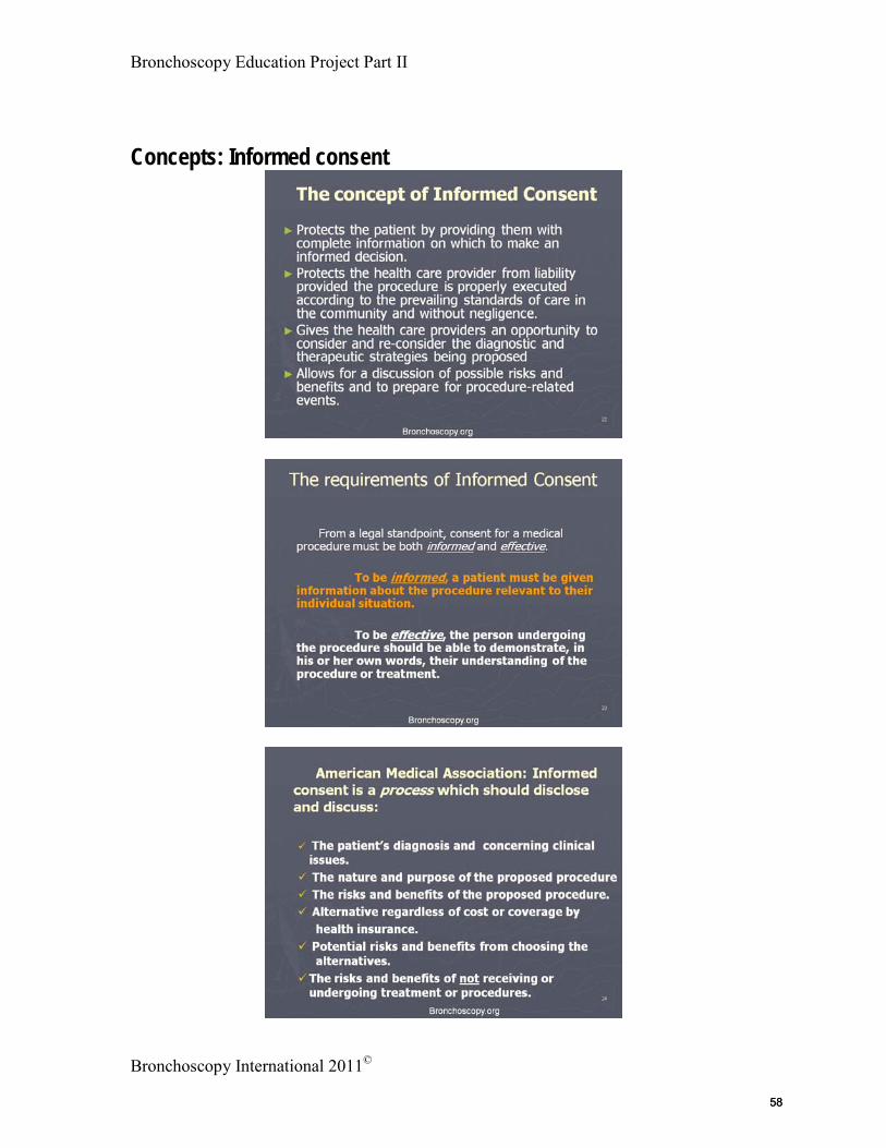
Bronchoscopy Education Project Part II
Bronchoscopy International 2011©
Concepts: Informed consent
5858

Bronchoscopy Education Project Part II
Bronchoscopy International 2011©
Concepts: Patient Safety and procedure-related precautions
1
History, Risk factors, and Universal precautions
1. Medical history such as COPD, pulmonary embolus, deep venous thrombosis, rheumatoid arthritis, ankylosing spondylitis , infectious lung disease or other illnesses potentially affecting the airway, cardiac disease, pacemaker, coronary artery disease, obstructive sleep apnea, CO2 retention, laryngospasm or bronchospasm, asthma.
2. Surgical history such as neck surgery, lung surgery, spine surgery; 3. Dentures or loose teeth that might interfere with appropriate response to
procedure-related complications4. Bleeding disorder5. Allergies to medications including local anesthetics, antibiotics, or reactions
to general anesthetic drugs6. Medication usage including anticoagulation, antiplatelet agents or
Clopidogrel7. Living situation and family or friend support system8. Proximity to medical center and physician services9. Pregnancy10. Inquiry regarding advanced directives and health care decision making.11. Universal precautions should always be used to protect the patient and the
health care team from spread of blood borne infections such as Hepatitis and HIV.
12. Gloves, Hand-washing, Gowns, Surface disinfection, Sharp containers
2
Droplet precautions
►Droplet precautions are warranted in patients known or suspected to be infected with microorganisms transmitted by droplets (larger than 5 microns in size) that can be generated by coughing, sneezing, talking, or during the procedure. Surgical masks, facial shield, or goggles patient transport precautions Droplet precaution sign on procedure room door Cough/respiratory hygiene etiquette
3
Airborne pathogens precautions
► Hand hygiene► Cough/respiratory hygiene etiquette► Cleaning and disinfection of contaminated surfaces► Negative airflow with external exhaust► N-95 respiratory or other National Institute for
Occupational Safety and Health recommended device.
► Power air purifying respiratory (PAPR) might be considered in selected high risk cases.
► Airborne precautions sign
5959

Bronchoscopy Education Project Part II
Bronchoscopy International 2011©
Concepts: Procedural pause (Time-Out)
Bronchoscopy.org1
The concept of a “Procedural Pause”, also known as a “Time Out”
► This safety protocol eliminates events involving the wrong patient, wrong site or wrong procedure.
► The protocol has been endorsed by more than fifty professional organizations, and is applicable to all high-risk procedures.
► The protocol is included in the USA Joint Commission for the Accreditation of Healthcare Organization National Patient Safety Goals project and was originally approved in 2004.
► The protocol also includes other components important in fostering a culture of patient safety, such as purposeful team communication and ensuring patient understanding.
Bronchoscopy.org2
Requirements of an active “Time Out”
► Performed immediately prior to the start of the procedure.
► Ensures that the correct patient, site, positioning, and procedure to be performed are correctly identified.
► Ensures that pertinent imaging studies, medical records and equipment are available.
► Initiated by the provider and includes active verbal acknowledgement by all members of the health care team and any other persons present.
► All environmental distractions should be eliminated as much as possible.
3
Time Out: visual memory guide1. Verification of patient
This is patient (read name badge), confirm with patient or family.
2. Verification of procedure I am Dr….We are going to perform …Patient agrees…
3. Verification of side and site 4. Nurse verifies consistency with signed informed
consent.5. Team members verify and declare availability of
pertinent medical records, imaging studies and equipment.
6. Declare need for antibiotics, fluids or moderate sedation.
7. Describe allergies or drug reactions8. Declare of safety issues based on medical history
6060

Bronchoscopy Education Project Part II
Bronchoscopy International 2011©
EBUS Airway Access and Image Acquisition
Case Information Part 1: Demographics Case Title: Airway access and image acquisition Subject Name: (1) Eddie Ebus Scenario Name: Airway access and image acquisition Simulation Developer(s): H. Colt Date(s) of Development: September 2010 Appropriate for following learning groups
Post graduate education, Physicians in practice Specialties: Pulmonary Anesthesiology Surgery Critical Care Thoracic surgeons performing EBUS
Simulated patient 1 scenario Scenario # 1 (10 minutes, with 10 minutes debriefing): Access the airway using the 30 degree field of view EBUS bronchoscope through an oral airway, a Laryngeal Mask Airway and an endotracheal tube. Then demonstrate controls on the Ultrasound image processor including On/off switch, Gain, Depth, Color Doppler, Frequency, and measurement calipers. Scenario description: The instructor will read the scenario to the team. It may be necessary for the instructor to demonstrate airway access techniques and image processor controls to the team. The learner will then proceed to access the airway using three techniques. After the third technique, and with the EBUS bronchoscope inside the EBUS model, the learner will demonstrate use of the image processor controls for altering the quality of the ultrasound image. It may be necessary to use more than one model for this exercise (an intubation head for airway access for example, and the EBUS box trainer with bifurcated airway and lymph node stations 2,4 and 7 for the image acquisition portion of the simulation). It is assumed that approximately ten minutes will be devoted to the scenario, with 10 minutes for an instructor-led debriefing. The instructor may further choose to perform an additional ten minutes debriefing that could include viewing the EBUS Physics instructional video. * Template for Simulation Patient. Design Modified from original template by Jeffrey M. Taekman, M.D, Duke University Simulation and Patient Safety Center
6161
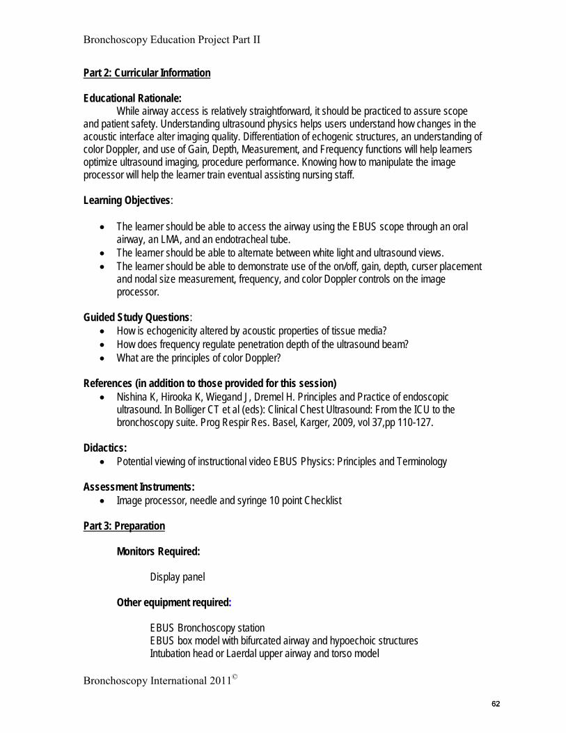
Bronchoscopy Education Project Part II
Bronchoscopy International 2011©
Part 2: Curricular Information Educational Rationale:
While airway access is relatively straightforward, it should be practiced to assure scope and patient safety. Understanding ultrasound physics helps users understand how changes in the acoustic interface alter imaging quality. Differentiation of echogenic structures, an understanding of color Doppler, and use of Gain, Depth, Measurement, and Frequency functions will help learners optimize ultrasound imaging, procedure performance. Knowing how to manipulate the image processor will help the learner train eventual assisting nursing staff. Learning Objectives:
• The learner should be able to access the airway using the EBUS scope through an oral airway, an LMA, and an endotracheal tube.
• The learner should be able to alternate between white light and ultrasound views. • The learner should be able to demonstrate use of the on/off, gain, depth, curser placement
and nodal size measurement, frequency, and color Doppler controls on the image processor.
Guided Study Questions:
• How is echogenicity altered by acoustic properties of tissue media? • How does frequency regulate penetration depth of the ultrasound beam? • What are the principles of color Doppler?
References (in addition to those provided for this session)
• Nishina K, Hirooka K, Wiegand J, Dremel H. Principles and Practice of endoscopic ultrasound. In Bolliger CT et al (eds): Clinical Chest Ultrasound: From the ICU to the bronchoscopy suite. Prog Respir Res. Basel, Karger, 2009, vol 37,pp 110-127.
Didactics: • Potential viewing of instructional video EBUS Physics: Principles and Terminology
Assessment Instruments:
• Image processor, needle and syringe 10 point Checklist Part 3: Preparation
Monitors Required:
Display panel
Other equipment required:
EBUS Bronchoscopy station EBUS box model with bifurcated airway and hypoechoic structures Intubation head or Laerdal upper airway and torso model
6262

Bronchoscopy Education Project Part II
Bronchoscopy International 2011©
Time Duration
For this scenario
Set-up 10 minutes Preparation 10 minutes Simulation 10 minutes Debrief 10-20 minutes
Part 4: Supporting Files (case scenario handouts) Scenario # 1 (Airway access and image acquisition): A 70 year old male has slightly enlarged lymph nodes at the subcarinal (7) and lower right paratracheal stations (4R). He is referred for EBUS-TBNA for diagnosis.
You must access the airway and demonstrate how to use the controls on the image processor in order to optimize image acquisition at these lymph node levels.
6363

Bronchoscopy Education Project Part II
Bronchoscopy International 2011©
EBUS-TBNA Needle and Scope Handling
Case Information Part 1: Demographics Case Title: EBUS-TBNA of stations 7 and 4R Subject Name: (1) Eddie Ebus Scenario Name: EBUS-TBNA of stations 7 and 4R Simulation Developer(s): H. Colt Date(s) of Development: September 2010 Appropriate for following learning groups
Post graduate education, Physicians in practice Specialties: Pulmonary Anesthesiology Surgery Critical Care Thoracic surgeons performing EBUS
Simulated patient 1 scenario Scenario # 1 (10 minutes, with 10 minutes debriefing): With the EBUS bronchoscope in position inside the bifurcated airway model, the EBUS needle is used to puncture the lymph node visualized using EBUS. Scenario description: The instructor will read the scenario to the team. It may be necessary for the instructor to demonstrate scope and needle handling techniques. The learner will then perform EBUS-TBNA of level 7 and 4R lymph node stations. The learner will articulate each step of needle handling, while another learner serves as the bronchoscopy assistant. It is assumed that approximately ten minutes per person will be devoted to the scenario, with 10 minutes for an instructor-led debriefing. The instructor may further choose to perform an additional ten minutes debriefing that includes viewing the EBUS curiosities instructional video. * Template for Simulation Patient. Design Modified from original template by Jeffrey M. Taekman, M.D, Duke University Simulation and Patient Safety Center
6464

Bronchoscopy Education Project Part II
Bronchoscopy International 2011©
Part 2: Curricular Information Educational Rationale:
Learning to perform EBUS-TBNA using a step-by-step approach assures safe needle handling and acquisition of an adequate specimen without harming the bronchoscope or patient. Knowing how to manipulate the needle will additionally help the learner train assisting nursing staff. Learning Objectives:
• The learner should be able to manipulate the EBUS scope and needle in the performance of EBUS-TBNA.
• The learner should be able to assist with EBUS-TBNA • The learner should be able to demonstrate the various steps required for handling the
scope, maintaining the ultrasound image, and handling the needle before, during and after EBUS-TBNA.
Guided Study Questions:
• Why does the needle sometimes not penetrate the lymph node? • What alterations in balloon inflation and scope position might be necessary to optimize
image acquisition? • What precautions should be used during needle insertion and removal?
References (in addition to those provided for this session)
• NA
Didactics: • Potential viewing of instructional video: EBUS Curiosities
Assessment Instruments:
• EBUS-STAT Part 3: Preparation
Monitors Required:
Display panel
Other equipment required:
EBUS Bronchoscopy station EBUS box model with bifurcated airway and hypoechoic structures
Time Duration
For this scenario
Set-up 10 minutes
6565

Bronchoscopy Education Project Part II
Bronchoscopy International 2011©
Preparation 10 minutes Simulation 10 minutes Debrief 10-20 minutes
Part 4: Supporting Files (case scenario handouts) Scenario # 1 (EBUS-TBNA): A 70 year old male has slightly enlarged lymph nodes at the subcarinal (7) and lower right paratracheal stations (4R). He is referred for EBUS-TBNA for diagnosis.
You must image the airway in order to visualize stations 4 and 7 after which you must perform EBUS-TBNA at levels 7 or 4R. One of the other participants will serve as your assistant. Please verbalize all needle and scope related commands.
6666

Bronchoscopy Education Project Part II
Bronchoscopy International 2011©
Section 6
Observed Real-Patient Encounters
A series of observed real-patient encounters that include: • An informed consent, patient safety, procedural pause
encounter. • An EBUS-TBNA image processor, needle and scope handling
encounter.
* Checklists and assessment tools used at the discretion of program director.
6767

Bronchoscopy Education Project Part II
Bronchoscopy International 2011©
This page intentionally left blank.
6868

Bronchoscopy Education Project Part II
Bronchoscopy International 2011©
User Instructions Real patient observations
The purpose of real patient observations is to allow the instructor to assess the student in the clinical setting. This is similar to how medical students and other house officers are observed and judged prior to being deemed competent to perform a physical examination or certain procedures.
Various assessment tools and ten-point checklists are used to document knowledge and skill acquisition in accordance with elements required by ACGME (patient care, medical knowledge, practice-based learning and improvement, interpersonal communication skills, professionalism, and systems-based practice). It is assumed that students will be observed throughout their subspecialty training, and that specific instances may be chosen for assessment purposes. The frequency of such assessments is still undetermined, but the purpose is to be able to monitor the student’s progression along the learning curve, so that instructors might be able to provide constructive feedback and advice, in addition to knowledge and technical expertise. These assessments can also be used for physicians in practice performing procedures under the guidance or observation of a proctor (certified trainer).
6969

Bronchoscopy Education Project Part II
Bronchoscopy International 2011©
This page intentionally left blank.
7070

Bronchoscopy Education Project Part II
Bronchoscopy International 2011©
Section 7
Practical Approach Sessions
A series of interactive (instructor-student) Practical Approach to Procedural Decision-making workshops
7171

Bronchoscopy Education Project Part II
Bronchoscopy International 2011©
This page intentionally left blank.
7272

Bronchoscopy Education Project Part II
Bronchoscopy International 2011©
User Instructions Practical Approach workshops
The purpose of these practical approach sessions is to help learners think through the decision making process. Often, instructors will use a practical approach to help students gain insights into the strategy and planning, technical performance, and response to complications elements of a minimally invasive procedure. These exercises are done orally, and the assessment is subjective, based on the instructor’s perception of how the learner responds to questions and outlines a procedural strategy. Learners should be taught to use scientific evidence as well as expert opinion to formulate plans and achieve desired results. A dialogue is thus encouraged between instructor and student in order to address alternatives and differences in technique, as well as expected outcomes. It is expected that sessions should usually last no more than 30 minutes. The student may be given a scenario, and using a model of the four box template the student might be asked to address each of the four boxes, with specific emphasis on one or two items based on instructor preference. Numerous exercises are available on the Bronchoscopy.org website to serve as examples, but any scenario the instructor chooses or devises can be used for teaching purposes.
7373

Bronchoscopy Education Project Part II
Bronchoscopy International 2011©
The Practical Approach© is an interactive learning program: The purpose of The Practical Approach is to help learners gain the cognitive,
technical, experiential, and affective skills necessary to perform bronchoscopy. Competency is sought in the three major elements of a procedure; strategy and planning, execution, and response to procedure-related adverse events or complications.
Using a four box approach inspired from Albert Jonsen’s1
• By working through case scenarios, learners are prompted to think about the how and why of their actions, based on background information, pertinent literature, and experience.
classic work in medical ethics, learners rationalize various components of the decision making process.
• Consistent with the ACGME professionalism competency guides 2, 3
, case scenarios prompt learners to address various components of the informed consent process, and discuss outcomes based on possible as well as real results.
The Practical Approach helps learners become competent bronchoscopists according to current recommendations set forth by the American College of Graduate Medical Education 4
• Gather essential and accurate information about their patients. whereby trainees learn to:
• Make informed decisions about diagnostic and therapeutic interventions based on patient information and preferences, up-to-date scientific evidence and clinical judgment.
• Use information technology to support patient care decisions and patient education.
• Develop patient management plans. • Counsel and educate patients and their families. • Communicate effectively and demonstrate caring and respectful behaviors
when interacting with patients and their families. • Provide health care services aimed at preventing health problems or
maintaining health. • Work with health care professionals, including those from other
disciplines to provide patient-focused care.
References 1. Jonsen AR, Siegler M, Winslade WJ. Clinical Ethics. 6th
2. ACGME Web site:http//www.acgme.org/outcome/comp/compfull.asp
Ed, McGraw Hill, NY 2006.
3. Apelgren K. ACGME e-Bulletin. August 2006 4. ACGME Competencies at http://www.acgme.org
7474

Bronchoscopy Education Project Part II
Bronchoscopy International 2011©
PRACTICAL APPROACH MODULE
EBUS-TBNA for lymph nodes < 1 cm in a patient with solitary pulmonary nodule
Based on the information below, please describe your procedural decision making and answer the three questions at the end of the exercise. Do your best to complete each item of the Four Boxes. If the case scenario contains no information pertaining to an item, please address it as NOT AVAILABLE. Each case scenario may have greater emphasis on one or more items listed in the “Practical Approach”.
This 55 year old man has a 1.5 cm solitary pulmonary nodule in the mediobasal segment of right lower lobe which was incidentally noted while he was undergoing CT of the abdomen for nephrolithiasis. The scan show a 1.5 cm RLL nodule, 5.5 SUV on PET and a 7 mm subcarinal lymph node on CT which is not PET-avid. He has no risk factors for cancer. Vital signs are normal. Physical exam is unremarkable. CBC, coagulation and chemistry panel, and spirometry are normal. PPD is negative. He is an attorney specializing in real estate law. He lives with his wife and 1 child. He has no advanced directives. He desires treatment in case of lung cancer.
After addressing items of the four boxes, please consider the following:
► Describe major elements of informed consent. ► Describe the current evidence about staging CT/PET negative mediastinal
lymphadenopathy in patients with known or suspected lung cancer. ► List three reasons for a poor sample on smear.
7575

Bronchoscopy Education Project Part II
Bronchoscopy International 2011 ©
This page intentionally left blank.
7676

Bronchoscopy Education Project Part II
Bronchoscopy International 2011©
Slide 1
www.Bronchoscopy.org1
EBUS-TBNA for lymph nodes < 1 cm in a patient with known solitary pulmonary nodule
►Describe major elements of informed consent.
►Describe the current debate about staging CT/PET negative mediastinal lymphadenopathy in patients with known or suspected lung cancer.
►List three reasons for a poor sample on smear.
___________________________________
___________________________________
___________________________________
___________________________________
___________________________________
___________________________________
___________________________________
Slide 2
www.Bronchoscopy.org 2
Case description (practical approach #)
► 55 year old man with 1.5 cm solitary pulmonary nodule in the mediobasal segment of right lower lobe which was incidentally noted while he was undergoing CT of abdomen for nephrolithiasis. He has no risk factors for cancer. Vital signs are normal. Physical exam is unremarkable. CBC, coagulation and chemistry panel, and spirometry are normal. PPD is negative. He is an attorney specializing in real estate law. He lives with his wife and 1 child. He has no advanced directives. He desires all available treatment options in case he has lung cancer.
___________________________________
___________________________________
___________________________________
___________________________________
___________________________________
___________________________________
___________________________________
Slide 3
www.Bronchoscopy.org 3
Case description (practical approach #)
► 1.5 cm RLL spn, 5.5 SUV on PET
► 7 mm subcarinal lymph node on CT which is not PET avid
___________________________________
___________________________________
___________________________________
___________________________________
___________________________________
___________________________________
___________________________________
77

Bronchoscopy Education Project Part II
Bronchoscopy International 2011©
Slide 4
www.Bronchoscopy.org 4
Initial Evaluations
►Exam►ECOG performance status 0
►Comorbidities►Nephrolithiasis
►Support system►Wife and teenage child all healthy and actively
involved with patient’s care.
►Patient preferences►Desires all available active treatment options.
___________________________________
___________________________________
___________________________________
___________________________________
___________________________________
___________________________________
___________________________________
Slide 5
www.Bronchoscopy.org 5
Procedural Strategies► Indications Invasive lymph node staging in a patient with
radiographically (CT and PET) normal mediastinum PET scan and Integrated PET/CT have demonstrated a
high NPV for mediastinal nodal disease with NPV of ~90%*
5.6% of patients with radiographically stage 1 disease were found to have N2 disease**
Risk factors and incidence of occult N2 mets include*** ► larger tumor size (≥6.0 cm) – 57% prevalence►central location- 21.6% prevalence►high PET SUV (≥4.0)- 10.5% prevalence►adeno ca cell type- 9.0% prevalence
*Eur Respir J 2009; 33: 201–212**J Thorac Cardiovasc Surg 2006;131:822-829***Ann Thorac Surg 2007;84:177-181
___________________________________
___________________________________
___________________________________
___________________________________
___________________________________
___________________________________
___________________________________
Slide 6 Procedural Strategies
►Indications Should this patient undergo lymph node staging
before or after needle aspiration biopsy of his peripheral nodule? Should he undergo invasive lymph node staging?
►Invasive staging should be reserved for patients with 1 or more risk factors for occult N2 disease* ** ***
►Routine use of invasive lymph node staging for patients with clinical stage 1 non small cell lung ca and no risk factors for occult N2 disease is not cost effective**
www.Bronchoscopy.org 6
*Ann Thorac Surg 2007;84:177-181**J Thorac Cardiovasc Surg 2006;131:822-829*** Eur J Cardiothorac Surg. 2007 Jul;32(1):1-8
___________________________________
___________________________________
___________________________________
___________________________________
___________________________________
___________________________________
___________________________________
78
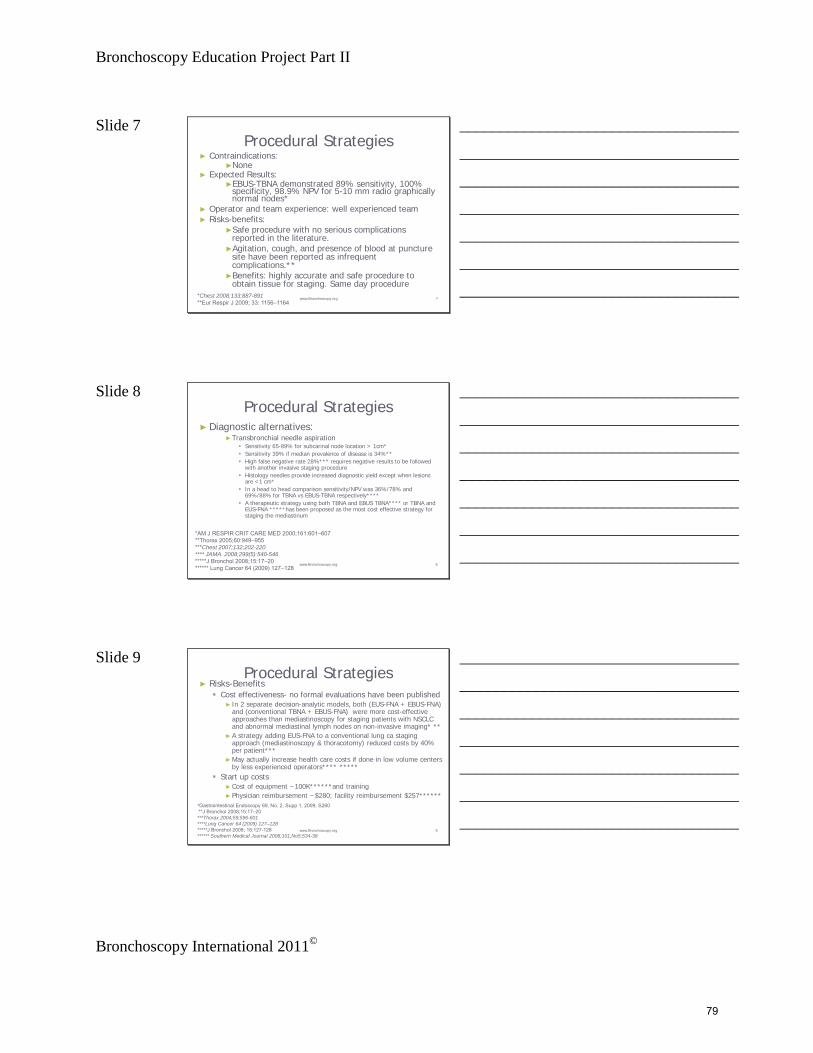
Bronchoscopy Education Project Part II
Bronchoscopy International 2011©
Slide 7 Procedural Strategies
► Contraindications: ►None
► Expected Results: ►EBUS-TBNA demonstrated 89% sensitivity, 100%
specificity, 98.9% NPV for 5-10 mm radio graphically normal nodes*
► Operator and team experience: well experienced team► Risks-benefits:
►Safe procedure with no serious complications reported in the literature.
►Agitation, cough, and presence of blood at puncture site have been reported as infrequent complications.**
►Benefits: highly accurate and safe procedure to obtain tissue for staging. Same day procedure
www.Bronchoscopy.org 7*Chest 2008;133;887-891**Eur Respir J 2009; 33: 1156–1164
___________________________________
___________________________________
___________________________________
___________________________________
___________________________________
___________________________________
___________________________________
Slide 8 Procedural Strategies
► Diagnostic alternatives:►Transbronchial needle aspiration
Sensitivity 65-89% for subcarinal node location > 1cm* Sensitivity 39% if median prevalence of disease is 34%** High false negative rate 28%*** requires negative results to be followed
with another invasive staging procedure Histology needles provide increased diagnostic yield except when lesions
are <1 cm* In a head to head comparison sensitivity/NPV was 36%/78% and
69%/88% for TBNA vs EBUS-TBNA respectively**** A therapeutic strategy using both TBNA and EBUS TBNA**** or TBNA and
EUS-FNA *****has been proposed as the most cost effective strategy for staging the mediastinum
www.Bronchoscopy.org 8
*AM J RESPIR CRIT CARE MED 2000;161:601–607**Thorax 2005;60:949–955***Chest 2007;132;202-220**** JAMA. 2008;299(5):540-546*****J Bronchol 2008;15:17–20****** Lung Cancer 64 (2009) 127–128
___________________________________
___________________________________
___________________________________
___________________________________
___________________________________
___________________________________
___________________________________
Slide 9 Procedural Strategies
► Risks-Benefits Cost effectiveness- no formal evaluations have been published
► In 2 separate decision-analytic models, both (EUS-FNA + EBUS-FNA) and (conventional TBNA + EBUS-FNA) were more cost-effective approaches than mediastinoscopy for staging patients with NSCLC and abnormal mediastinal lymph nodes on non-invasive imaging* **
► A strategy adding EUS-FNA to a conventional lung ca staging approach (mediastinoscopy & thoracotomy) reduced costs by 40% per patient***
► May actually increase health care costs if done in low volume centers by less experienced operators**** *****
Start up costs► Cost of equipment ~100K******and training► Physician reimbursement ~$280; facility reimbursement $257******
*Gastrointestinal Endoscopy 69, No. 2, Supp 1, 2009, S260 **J Bronchol 2008;15:17–20***Thorax 2004;59;596-601****Lung Cancer 64 (2009) 127–128*****J Bronchol 2008; 15:127-128****** Southern Medical Journal 2008;101,No5;534-38
9www.Bronchoscopy.org
___________________________________
___________________________________
___________________________________
___________________________________
___________________________________
___________________________________
___________________________________
79

Bronchoscopy Education Project Part II
Bronchoscopy International 2011©
Slide 10 Procedural Strategies
►Diagnostic alternatives:►Percutaneous needle aspiration of nodule►Endoscopic ultrasound in a head to head comparison*
sensitivity and negative predictive value (69% and 89% respectively) was identical to EBUS for accessible nodes
►Mediastinoscopy gold standard. Mediastinoscopy is invasive staging procedure of choice in
radiographically negative mediastinum** ***► 42% sensitivity in clinical stage 1 ****►Difficult to access level 7 nodes
►VATS most invasive of alternatives. Only provides access to ipsilateral nodes. 75% sensitivity****. Benefits include definitive lobar resection at same time if node negative.
www.Bronchoscopy.org 10
*JAMA. 2008;299(5):540-546**Chest 2007;132;178S-201S***Eur J Cardiothorac Surg. 2007 Jul;32(1):1-8****Chest 2007;132;202-220
___________________________________
___________________________________
___________________________________
___________________________________
___________________________________
___________________________________
___________________________________
Slide 11 Procedural Strategies
► Informed consent: ►There were no barriers to learning identified. Patient has good
insight into his disease and realistic expectations.
www.Bronchoscopy.org 11
Drawing from Herth FJ et al.. Chest 2003;123:604 –7EBUS image from patient.
___________________________________
___________________________________
___________________________________
___________________________________
___________________________________
___________________________________
___________________________________
Slide 12
BI #. Practical Approach Title12
Procedural techniques and resultsAnesthesia and perioperative care
► Conscious sedation►May be performed in bronchoscopy suite►Cost savings compared to general anesthesia.►Visualization and biopsy of smaller nodes technically more
difficult than with general anesthesia.
► General anesthesia with LMA►Better visualization of higher nodes compared with ET tube►May be performed in bronchoscopy suite►May not be appropriate in severe obesity or severe untreated
GERD
► General anesthesia with ET tube►Usually performed in OR►EBUS scope directed more centrally in airway which may make
biopsies more difficult 12www.Bronchoscopy.org
___________________________________
___________________________________
___________________________________
___________________________________
___________________________________
___________________________________
___________________________________
80

Bronchoscopy Education Project Part II
Bronchoscopy International 2011©
Slide 13 Procedural techniques and results:
Moderate Sedation or General Anesthesia?
► As more and more centers begin EBUS-TBNA programs, it is important that the procedure is performed in the correct environment and with realistic expectations of patients, operators, and equipment performance characteristics.
► If a consensus is reached that complete nodal staging is feasible and accurate with EBUS-TBNA, it may be more optimally performed under general anesthesia until the bronchoscopist feels comfortable in targeting these smaller lymph nodes in multiple stations in a conscious patient.
Chest 2008;134;1350-135113www.Bronchoscopy.org
___________________________________
___________________________________
___________________________________
___________________________________
___________________________________
___________________________________
___________________________________
Slide 14 Procedural Techniques and Results
► Instrumentation EBUS scope- direct real time US imaging with curved
array ultrasound transducer incorporated in distal end of bronchoscope
Ultrasound processor►Adjustable gain and depth►B mode and Doppler capabilities
Needle►22 gauge acrogenic needle with stylet►Needle guide system locks to scope►Lockable needle and sheath►Precise needle projection up to 4 cm
www.Bronchoscopy.org 14
___________________________________
___________________________________
___________________________________
___________________________________
___________________________________
___________________________________
___________________________________
Slide 15 Procedural Techniques and Results
► Anatomic dangers and other risks►Major blood vessels- azygous, PA, aorta, SVC and Left atrium
Risk of canulating major vessel significantly reduced with real time B mode and Doppler mode imaging
“Minor” oozing of blood at puncture site was reported in 1 study. There have been no reports of major bleeding*
►Pneumothorax and pneumomediastinum** Have been reported with blind TBNA but no reports in literature
with EBUS guided FNA.
►A case of bacterial pericardial effusion and nodal infection have recently been reported as complications following EBUS with full needle extension***.
www.Bronchoscopy.org 15
Chest 2004;126;122-128**Eur Respir J 2002; 19:356–373***Eur Respir J 2009; 33:935-938
___________________________________
___________________________________
___________________________________
___________________________________
___________________________________
___________________________________
___________________________________
81

Bronchoscopy Education Project Part II
Bronchoscopy International 2011©
Slide 16 Procedural Techniques and Results
► Aspirate cytology Adequate/representative in presence of frankly
malignant cells, lymphocytes, lymphoid tissue, or clusters of anthracotic pigment-laden macrophages*
Inadequate/nonrepresentative if there are no cellular components, scant lymphocytes (defined as <40 per HPF) blood only, or cartilage or bronchial epithelial cells only* **
A quantitative cut off value of at least 30% of cellularity composed of lymphocytes has been arbitrarily proposed by some experts***
Higher yield may be obtained by obtaining aspirates from the periphery of nodes****
*Am J Clin Pathol 2008;130:434-443**Chest 2008;134;368-374;***Chest 2004;126;1005-1006****Techniques in GI Endoscopy, Vol 2, No 3, 2000: pp 136-141 16www.Bronchoscopy.org
___________________________________
___________________________________
___________________________________
___________________________________
___________________________________
___________________________________
___________________________________
Slide 17 Procedural Techniques and Results► Number of aspirates* if ROSE not utilized Best yield with 3 aspirates per station (see table) Two aspirations per LN station can be acceptable when at
least one tissue core specimen is obtained by the first two aspirations
►Sensitivity 91.7%, NPV 96.0%, and accuracy 97.2%
If operator believes targeting is inadequate or insufficient another aspirate should be performed
BI #. Practical Approach Title 17
* Chest 2008;134;368-374;17www.Bronchoscopy.org
___________________________________
___________________________________
___________________________________
___________________________________
___________________________________
___________________________________
___________________________________
Slide 18 Maximum results after 3 aspirates
Chest 2008;134;368-37418www.Bronchoscopy.org
Sample adequacy reached 100% in three aspirations. Median procedure time was 1 h when three nodal stations were punctured. Results suggest that a fourth aspiration is not necessary in EBUS-TBNA for the mediastinal staging of NSCLC. The sensitivity of the first aspiration was 68.9%. The sensitivity increased to 83.7% after two aspirations and to 95.3% after three aspirations.
___________________________________
___________________________________
___________________________________
___________________________________
___________________________________
___________________________________
___________________________________
82

Bronchoscopy Education Project Part II
Bronchoscopy International 2011©
Slide 19 Procedural Techniques and Results
►Results and procedure-related complications Due to the absence of history suggestive of
noncancer diagnosis and high SUV on PET the level 7 node was sampled with EBUS under general anesthesia using a 9.0 ET tube. The cytology was diagnostic for non small cell
carcinoma (adenocarcinoma). There were no complications.
www.Bronchoscopy.org 19
___________________________________
___________________________________
___________________________________
___________________________________
___________________________________
___________________________________
___________________________________
Slide 20
www.Bronchoscopy.org 20
Long-term Management Plan► Outcome assessment
Patient was referred for multidisciplinary evaluation to include cardiothoracic surgery, oncology, and radiation oncology for potential trial enrollment for neoadjuvant treatment of stage IIIA adenocarcinoma of the lung.*
► Follow-up tests and procedures Patient will follow up in 1 month to ensure he has been evaluated
by all the above specialties.► Referrals
See above.► Quality improvement
The occult N2 metastasis was identified prior to the patient undergoing VATS or open thoracotomy for a non resectable tumor.
5 year survival for IIIA non-small cell lung ca is 23%.
*Chest 2007;132;243S-265S
___________________________________
___________________________________
___________________________________
___________________________________
___________________________________
___________________________________
___________________________________
Slide 21
Q 1: Describe the major elements of informed consent
21www.Bronchoscopy.org
___________________________________
___________________________________
___________________________________
___________________________________
___________________________________
___________________________________
___________________________________
83

Bronchoscopy Education Project Part II
Bronchoscopy International 2011©
Slide 22 The concept of Informed Consent
► Protects the patient by providing them with complete information on which to make an informed decision.
► Protects the health care provider from liability provided the procedure is properly executed according to the prevailing standards of care in the community and without negligence.
► Gives the health care providers an opportunity to consider and re-consider the diagnostic and therapeutic strategies being proposed
► Allows for a discussion of possible risks and benefits and to prepare for procedure-related events.
22www.Bronchoscopy.org
___________________________________
___________________________________
___________________________________
___________________________________
___________________________________
___________________________________
___________________________________
Slide 23 The requirements of Informed Consent
From a legal standpoint, consent for a medical procedure must be both informed and effective.
To be informed, a patient must be given information about the procedure relevant to their individual situation.
To be effective, the person undergoing the procedure should be able to demonstrate, in his or her own words, their understanding of the procedure or treatment.
23www.Bronchoscopy.org
___________________________________
___________________________________
___________________________________
___________________________________
___________________________________
___________________________________
___________________________________
Slide 24 American Medical Association: Informed
consent is a process which should disclose and discuss:
The patient’s diagnosis and concerning clinical issues.
The nature and purpose of the proposed procedure The risks and benefits of the proposed procedure. Alternative regardless of cost or coverage by
health insurance. Potential risks and benefits from choosing the
alternatives.The risks and benefits of not receiving or
undergoing treatment or procedures.24www.Bronchoscopy.org
___________________________________
___________________________________
___________________________________
___________________________________
___________________________________
___________________________________
___________________________________
84

Bronchoscopy Education Project Part II
Bronchoscopy International 2011©
Slide 25
Q 2: Describe the current debate about staging CT/PET negative
mediastinal lymphadenopathy in patients with known or suspected lung cancer.
25www.Bronchoscopy.org
___________________________________
___________________________________
___________________________________
___________________________________
___________________________________
___________________________________
___________________________________
Slide 26
www.Bronchoscopy.org 26
Q 2: Describe debate about staging CT/PET negative mediastinal lymphadenopathy
PET scan and Integrated PET/CT have demonstrated a high NPV for mediastinal nodal disease with NPV of ~90%. 5.6% of patients with radiographically stage 1
disease have N2 disease. Risk factors and incidence of occult N2 disease:
►larger tumor size (≥6.0 cm) – 57% prevalence►central location- 21.6% prevalence►high PET SUV (≥4.0)- 10.5% prevalence►adeno ca cell type- 9.0% prevalence
___________________________________
___________________________________
___________________________________
___________________________________
___________________________________
___________________________________
___________________________________
Slide 27 Q 2: Describe debate about staging CT/PET
negative mediastinal lymphadenopathy
►Indications Should these patients undergo invasive lymph
node staging?►Invasive staging should be reserved for patients with
1 or more risk factors for occult N2 disease.►Routine use of invasive lymph node staging for
patients with clinical stage 1 non small cell lung ca and no risk factors for occult N2 disease is not cost effective.
www.Bronchoscopy.org 27
___________________________________
___________________________________
___________________________________
___________________________________
___________________________________
___________________________________
___________________________________
85

Bronchoscopy Education Project Part II
Bronchoscopy International 2011©
Slide 28 However
►Adenocarcinoma is a frequent histologic type of NSCLC. Data suggest that patients with adenocarcinoma (contrary to squamous cell carcinoma) may have false negative PET/CT findings of the mediastinum
►This suggests that adenocarcinoma should be carefully staged preoperatively even in mediastinal PET/CT-negative cases.
Chest; Prepublished online December 31, 2008; 28www.Bronchoscopy.org
___________________________________
___________________________________
___________________________________
___________________________________
___________________________________
___________________________________
___________________________________
Slide 29 EBUS for PET negative, normal mediastinum
► Patients highly suspicious for NSCLC with CT scans showing no enlarged lymph nodes (no node > 1 cm) and a negative PET finding of the mediastinum underwent EBUS-TBNA.
► Identifiable lymph nodes at locations 2R, 2L, 4R, 4L, 7, 10R, 10L, 11R, and 11L were aspirated.
► All patients underwent subsequent surgical staging. Diagnoses based on aspiration results were compared with those based on surgical results.
► One hundred patients (mean age, 52.4 years; 59 men) were included. After surgery, 97 patients (mean age, 52.9 years; 57 men) had NSCLC confirmed and were included in the analysis.
Herth F et al. CHEST 2008; 133:887–89129www.Bronchoscopy.org
___________________________________
___________________________________
___________________________________
___________________________________
___________________________________
___________________________________
___________________________________
Slide 30 ► In this group, 156 lymph nodes ranging 5 to 10 mm in
size were detected and sampled. Malignancy was detected in nine patients but missed in
one patient. Mean diameter of the punctured lymph nodes was 7.9 mm. The sensitivity of EBUS-TBNA for detecting malignancy was
89%, specificity was 100%, and the negative predictive value was 98.9%. No complications occurred.
► EBUS-TBNA can be used to accurately sample and stage patients with clinical stage 1 lung cancer and no evidence of mediastinal involvement on CT and PET.
► Potentially operable patients with no signs of mediastinal involvement may benefit from presurgical staging with EBUS-TBNA.
Herth F et al. CHEST 2008; 133:887–89130www.Bronchoscopy.org
___________________________________
___________________________________
___________________________________
___________________________________
___________________________________
___________________________________
___________________________________
86
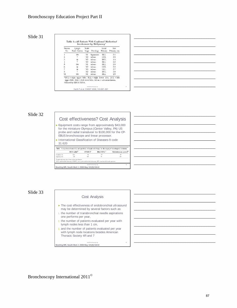
Bronchoscopy Education Project Part II
Bronchoscopy International 2011©
Slide 31
Herth F et al. CHEST 2008; 133:887–89131www.Bronchoscopy.org
___________________________________
___________________________________
___________________________________
___________________________________
___________________________________
___________________________________
___________________________________
Slide 32 Cost effectiveness? Cost Analysis
► Equipment costs range from approximately $43,000 for the miniature Olympus (Center Valley, PA) US probe and radial transducer to $100,000 for the CP-EBUS bronchoscope and linear processor.
► International Classification of Diseases-9 code 31.620
Bowling MR, South Med J. 2008 May 101(5) 534-832www.Bronchoscopy.org
___________________________________
___________________________________
___________________________________
___________________________________
___________________________________
___________________________________
___________________________________
Slide 33 Cost Analysis
► The cost effectiveness of endobronchial ultrasound may be determined by several factors such as:
1. the number of transbronchial needle aspirations one performs per year,
2. the number of patients evaluated per year with lymph nodes less than 1 cm,
3. and the number of patients evaluated per year with lymph node locations besides American Thoracic Society 4R and 7
Bowling MR, South Med J. 2008 May 101(5) 534-833www.Bronchoscopy.org
___________________________________
___________________________________
___________________________________
___________________________________
___________________________________
___________________________________
___________________________________
87
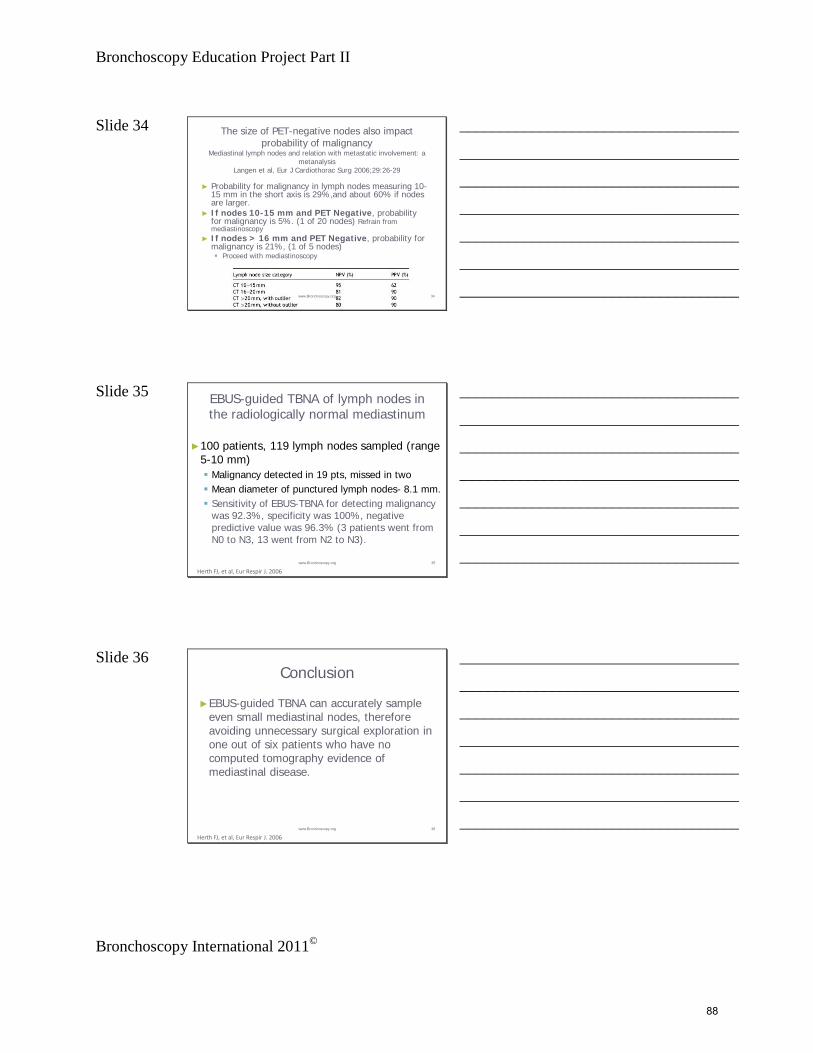
Bronchoscopy Education Project Part II
Bronchoscopy International 2011©
Slide 34 The size of PET-negative nodes also impact probability of malignancy
Mediastinal lymph nodes and relation with metastatic involvement: a metanalysis
Langen et al, Eur J Cardiothorac Surg 2006;29:26-29
► Probability for malignancy in lymph nodes measuring 10-15 mm in the short axis is 29%,and about 60% if nodes are larger.
► If nodes 10-15 mm and PET Negative, probability for malignancy is 5%. (1 of 20 nodes) Refrain from mediastinoscopy
► If nodes > 16 mm and PET Negative, probability for malignancy is 21%, (1 of 5 nodes) Proceed with mediastinoscopy
34www.Bronchoscopy.org
___________________________________
___________________________________
___________________________________
___________________________________
___________________________________
___________________________________
___________________________________
Slide 35 EBUS-guided TBNA of lymph nodes in the radiologically normal mediastinum
►100 patients, 119 lymph nodes sampled (range 5-10 mm) Malignancy detected in 19 pts, missed in two Mean diameter of punctured lymph nodes- 8.1 mm. Sensitivity of EBUS-TBNA for detecting malignancy
was 92.3%, specificity was 100%, negative predictive value was 96.3% (3 patients went from N0 to N3, 13 went from N2 to N3).
Herth FJ, et al, Eur Respir J. 2006
35www.Bronchoscopy.org
___________________________________
___________________________________
___________________________________
___________________________________
___________________________________
___________________________________
___________________________________
Slide 36 Conclusion
►EBUS-guided TBNA can accurately sample even small mediastinal nodes, therefore avoiding unnecessary surgical exploration in one out of six patients who have no computed tomography evidence of mediastinal disease.
Herth FJ, et al, Eur Respir J. 2006
36www.Bronchoscopy.org
___________________________________
___________________________________
___________________________________
___________________________________
___________________________________
___________________________________
___________________________________
88

Bronchoscopy Education Project Part II
Bronchoscopy International 2011©
Slide 37 More cost analysis: EBUS guided TBNA
of mediastinal lymph nodes for lung cancer staging
►Hypothesis Patients found to have N2/N3 disease would
not require further investigation with PET imaging and mediastinoscopy
Callister ME, et al, Thorax. 2008
37www.Bronchoscopy.org
___________________________________
___________________________________
___________________________________
___________________________________
___________________________________
___________________________________
___________________________________
Slide 38 EBUS guided TBNA of mediastinal lymph nodes for lung cancer staging: a projected
cost analysis►Forty-seven patients underwent
mediastinoscopy as a staging procedure for lung cancer at Leeds Teaching Hospitals in 2006.
►Twenty-eight patients were shown to have malignant disease in N2 or N3 nodes, of which 27 were deemed accessible to EBUS-TBNA (all had mediastinal lymphadenopathy on initial CT scan).
Callister ME, et al, Thorax. 2008
38www.Bronchoscopy.org
___________________________________
___________________________________
___________________________________
___________________________________
___________________________________
___________________________________
___________________________________
Slide 39 Conclusion
►Mean EBUS-TBNA sensitivity for malignancy in recently published series was 92.3%
►Projected that 25 patients would have had mediastinal malignancy demonstrated by EBUS-TBNA and would therefore not have undergone CT-PET and mediastinoscopy.
Callister ME, et al, Thorax. 2008
BUT THESE PATIENTS ALL HAD EVIDENCE OF ENLARGED MEDIASTINAL NODES
39www.Bronchoscopy.org
___________________________________
___________________________________
___________________________________
___________________________________
___________________________________
___________________________________
___________________________________
89

Bronchoscopy Education Project Part II
Bronchoscopy International 2011©
Slide 40
And…for many multidisciplinary teams, PET is recommended
anyway because of a known 10-14 percent incidence of unsuspected extrathoracic metastases that can
be discovered by PET.
40www.Bronchoscopy.org
___________________________________
___________________________________
___________________________________
___________________________________
___________________________________
___________________________________
___________________________________
Slide 41
Q3: List three reasons for a poor sample on smear
41www.Bronchoscopy.org
___________________________________
___________________________________
___________________________________
___________________________________
___________________________________
___________________________________
___________________________________
Slide 42 Avoid:
• Sample coagulation : delay in processing the sample may completely hinder the process ofsmearing on the slide because of its coagulation, even when the blood content of thesample is not too much. Sample coagulation may happens in the needle lumen or on theslide surface.
• Air drying artefacts : if Papanicolaou (fig. A&B) stain is required, the smear must be still wetwhen fixative is applied, otherwise staining artefacts are the rule. Conversely, with Diff-Quick stain, the quantity of sample fluid must be just enough to smear, enabling the sampleto dry as soon as possible.
• Air bubbles : their presence in the material put on the slide causes uneven smeardistribution. This problem happens mainly with needle aspiration technique.
• Thick smear : the correct technique of smearing gives as final result distribution of cells inmonolayer on slide surface; cells overlapping makes the diagnostic process difficult orimpossible because it is impossible to evaluate precisely cellular details. To avoid thisproblem smear only a small drop of sample on the slide surface.
• Crushed smear : is the result of excessive pressure applied to smear the sample; this maycompletely destroy diagnostic material. Don’t worry, this may take some time to learn !
Needle biopsy of peripheral lung nodule (adenocarcinoma). Fig. A and B come fromthe same sampling and staining process. The slide of fig.A was correctly smearedand fixed while wet, giving precise color profile (pale blue); the slide of fig. B wascorrectly smeared but fixed when completely dried giving wrong color profile(pinkish): same neoplastic cells, same staining procedure, different color profile !
A B
Sampling and SmearsFrom G. Marcielli, with permission
42www.Bronchoscopy.org
___________________________________
___________________________________
___________________________________
___________________________________
___________________________________
___________________________________
___________________________________
90

Bronchoscopy Education Project Part II
Bronchoscopy International 2011 ©
Section 8
Proctored EBUS & EBUS-TBNA
Proctored patient-care in the procedural setting, with competency assessment
9191

Bronchoscopy Education Project Part II
Bronchoscopy International 2011 ©
This page intentionally left blank.
9292

Bronchoscopy Education Project Part II
Bronchoscopy International 2011 ©
User Instructions Proctored EBUS and EBUS-TBNA
The purpose of proctoring a procedure is to document that a physician is indeed able to independently perform all the elements of EBUS & EBUS-TBNA. These include strategy and planning, technique, patient safety measures and the ability to respond appropriately to procedure-related adverse events.
The Proctored EBUS-TBNA ten-point assessment tool can be used for grading purposes. This tool summarizes information contained in the Informed Consent, Procedural Pause, Practical Approach, and Sedation/Anesthesia checklists, as well as items contained in the Processor-Needle-Syringe checklist and EBUS-STAT assessment tool.
The overall assessment should also include the faculty’s general observations of the
learner’s practices during the course of training. From an assessment perspective, this is the last item with which to “assess” a learner’s ability to competently perform EBUS
bronchoscopy. The intensity with which proctoring occurs, and the number of “proctored” cases that may be required within a training program or health care
institution for physicians in-practice is variable.
9393

Bronchoscopy Education Project Part II
Bronchoscopy International 2011 ©
EBUS PROCTORED EBUS & EBUS-TBNA 10-Point CHECKLIST* Student _________________________________ Training Year _______________
Faculty __________________________________ Date _______________________ Simulation EBUS Workshop Patient-based EBUS Scenario
Educational Item* Items 1-10 are scored 10 points each (no partial points given)
Satisfactory Yes/No
1. Formulation of effective plan and strategy: Informed consent obtained, signed and in medical record History and physical Review imaging studies Cost-effective practice Use of information resources Applies evidence-based medicine Use of systems resources
Yes / No
2. Patient safety: ASA assessment Airway assessment Allergies Medications Comorbidities
Yes / No
3. Patient safety: Positioning Supplemental oxygen Vital signs Suction Bite-block and/or ET tube if applicable Verifies ability to respond to complications and accessibility of resuscitation equipment
Yes / No
4. Patient and procedural team safety: Knowledgeable use of image processor Hand disinfection Universal precautions “Time-Out”.
Yes / No
5. Procedure: Premedication Moderate sedation Topical anesthetic General anesthesia as applicable
Yes / No
6. Procedure: Scope insertion Empathy and Communication with patient
Yes / No
7. Procedure: Image processor controls Balloon attachment and troubleshooting Needle, stylet, and syringe function and troubleshooting
Yes / No
8. Procedure: Alternate between white light and EBUS Image acquisition Recognition of mediastinal and hilar nodal anatomy Recognition of mediastinal and hilar vascular structures Lymph node sampling Smear preparation and station labeling
Yes / No
9. Communication with staff, patient, and patient family Puts needs of patient first Punctuality Respect Listening skills Personal appearance Initiative & Motivation Empathy Honesty Accepts responsibility
Yes / No
10. Documentation/procedure note Informative Accurate Communication with colleagues
Yes / No
* Each of the 10 items contains all of the elements required by ACGME (patient care, medical knowledge, practice-based learning and improvement, interpersonal communication skills, professionalism, and systems-based practice).
FINAL GRADE PASS FAIL SCORE _______/100
9494
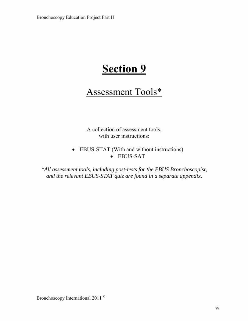
Bronchoscopy Education Project Part II
Bronchoscopy International 2011 ©
Section 9
Assessment Tools*
A collection of assessment tools, with user instructions:
• EBUS-STAT (With and without instructions)
• EBUS-SAT
*All assessment tools, including post-tests for the EBUS Bronchoscopist, and the relevant EBUS-STAT quiz are found in a separate appendix.
9595

Bronchoscopy Education Project Part II
Bronchoscopy International 2011 ©
This page intentionally left blank.
9696

Bronchoscopy Education Project Part II
Bronchoscopy International 2011 ©
Scoring Recommendations for EBUS Assessment Tools
The goal of this assessment tools is to be able to monitor a student’s progress along the learning curve from novice (Score < 60) to advanced beginner (Score 60-79), intermediate (score 80-99), and competent (score 100). The instructor should be able to ascertain, by observing the student’s performance that each of the ten elements in the tool are covered satisfactorily. (For EBUS-STAT tools, this could be done on once or twice a year) Repeated testing will demonstrate increases in knowledge and technical skill acquisition as the student climbs the learning curve from novice to advanced beginner, intermediate and competent bronchoscopist for the procedure being assessed. To maximize objective scoring, each task has been defined explicitly in this user manual for each checklist and assessment tool. Participation in specially-designed Train-the-Trainers courses being currently organized is encouraged to assist with standardization and to help instructors use this program to its fullest potential. Scores can be plotted on a graph, and each institution/program can obviously choose its own cut-offs for a PASS grade, although we recommend that a final PASS grade be achieved with a score of 100, in order for the student to be judged competent to perform bronchoscopy independently. In the absence of a large pilot study demonstrating standard normograms as is done for high-stakes testing, consensus of world renowned experts was obtained to delineate cut-off scores for the following four categories. Category Score Novice < 60 Advanced Beginner 60-79 Intermediate 80-99 Competent 100 Specific instructions marked by an asterisk (*) are provided in each of the following assessment tools. Instructions: To administer the EBUS-STAT, trainees are asked to perform a complete EBUS-TBNA, while at all times stating what they are doing. Thus, items 1, 2, assess ability to maneuver the EBUS bronchoscope, item 3&6 assesses the ability to obtain a satisfactory image and control the image processor, and items 4, 5, andv7 assess the ability to identify targets, mediastinal anatomy, and perform EBUS-TBNA. Items 8 -10 are scored using the associated quiz images. It is not necessary to test for all the items in the same patient, and some of the EBUS-STAT can be performed using a simulator setting. EBUS-SAT provides an opportunity for bidirectional feedback that can help identify strengths and weaknesses of individual bronchoscopists, but also of the training program. Ideally, learners should complete the EBUS-SAT prior to the session, providing the completed form to the instructor for consideration.
9797

Bronchoscopy Education Project Part II
Bronchoscopy International 2011 ©
EBUS Skills and Tasks
Assessment Tool (EBUS-STAT)
• Items 1-10 may be scored separately. • A passing score of 100 is expected to reflect competency. • Scores >100 should initiate instructor-learner feedback • Assessments may be performed in models or in patients. • Knowledge of nomenclature (common language) for EBUS
imaging can be assessed using the quiz. Recognition of findings can also be assessed during procedures, in which case instructors may prefer to use the quiz as a learning guide.
• The combined use of the 10 items pertains to technical skills needed to climb the learning curve from novice to competent bronchoscopist able to independently perform EBUS and EBUS-TBNA.
• This assessment tool is complementary to other methods of assessment such as practical approach exercises, checklists, logbooks of numbers of procedures performed, and outcomes.
9898

Bronchoscopy Education Project Part II
Bronchoscopy International 2011 ©
EBUS-STAT 10 Point Assessment Tool Student: _________________________________ Year of Training _________ Faculty _________________________________ Date ______________
Educational Item* Items 1-10 each scored separately
Satisfactory Yes/No
1. Able to maneuver the scope through upper airway into trachea, without trauma or difficulty (5 points for single item tested) Mouth and Vocal cords ET Tube Laryngeal mask airway
Yes / No
Score ____/5 2. Able to maneuver scope using white light bronchoscopy within tracheobronchial tree without trauma (4 points, no partial points) Scope centered in airway lumen avoiding airway wall trauma
Yes / No
Score ____/4
3. Ultrasound image obtained without artifacts (5 points, no partial points) Absence of artifacts on image, any target
Yes / No
Score ____/5 4. Identify major mediastinal vascular structures (4 points per item) Aorta Pulmonary artery Superior vena cava Azygos vein Left atrium
Yes / No
Score ____/20 5. Identify lymph node station (Select 3 targets, 5 points each) 2R 2L 4R 10R 7 4L 10L 11L 11Rs 11Ri
Yes / No Score ____/15
6. Able to operate EBUS processor (2 points each item) Gain Depth Doppler
Yes / No Score ____/6
7. Performance of EBUS-TBNA (1 point each, target 15 points) Advance needle through working channel (neutral position) Secure needle housing by sliding the flange Release sheath screw Advance and lock sheath when it touches wall Release needle screw Advance needle using jab technique Visualize needle entering target node Move stylet in and out a few times Remove stylet Attach syringe Apply suction Pass needle in and out of node 10-15 times Release suction Retract needle into sheath Unlock and remove needle and sheath
Yes / No Score ____/15
8. Image analysis: CT scans (1 point each, target 10 points) Image 1 Image 2 Image 3 Image 4 Image 5 Image 6 Image 7 Image 8 Image 9 Image 10
Yes / No
Score ____/10 9. Image analysis: EBUS views (1 point each, target 10 points) Image 1 Image 2 Image 3 Image 4 Image 5 Image 6 Image 7 Image 8 Image 9 Image 10
Yes / No
Score ____/10 10. Decision-making tasks: (2 points each, target 10 points) Image 1 Image 2 Image 3 Image 4 Image 5
Yes / No Score ____/10
* The combined use of the 10 items pertains to technical skills needed to climb learning curve from novice to advanced beginner to intermediate to competent bronchoscopist able to perform EBUS-TBNA independently.
FINAL GRADE PASS FAIL SCORE __________/100
9999

Bronchoscopy Education Project Part II
Bronchoscopy International 2011 ©
User Instructions EBUS-STAT 10 Point Assessment Tool
Student: _________________________________ Year of Training _________ Faculty _________________________________ Date ______________
Educational Item* Items 1-10 each scored separately
Satisfactory Yes/No
1. Able to maneuver the scope through upper airway into trachea, without trauma or difficulty (5 points for single item tested) Mouth and Vocal cords ET Tube Laryngeal mask airway * Educators may wish to “test” only one technique applicable to their institution. This is an “All or None” exercise. No partial points are given. When EBUS-STAT is used as a learning instrument, all THREE techniques should be demonstrated in order to obtain FIVE points.
Yes / No
Score ____/5
2. Able to maneuver scope using white light bronchoscopy within tracheobronchial tree without trauma (4 points, no partial points) Scope centered in airway lumen avoiding airway wall trauma *The learner should be able to maneuver between lateral and medial walls without traumatizing main and minor carinas.
Yes / No
Score ____/4
3. Ultrasound image obtained without artifacts (5 points, no partial points) Absence of artifacts on image, any target *Targeting any nodal station, a sharp image without artifacts should be obtained.
Yes / No
Score ____/5 4. Identify major mediastinal vascular structures (4 points per item) Aorta Pulmonary artery Superior vena cava Azygos vein Left atrium *Each of the vascular structures should be identified on demand. It may be necessary to score this item in several patients.
Yes / No
Score ____/20
5. Identify lymph node station (Select 3 targets, 5 points each) 2R 2L 4R 10R 7 4L 10L 11L 11Rs 11Ri * Target stations are selected based on indication, anatomy, and instructor preference. To identify all nodal stations, more than one patient may be necessary. In this case, intructors may choose to readminister EBUS-STAT. Stations 2, 4, and 7 can be scored in a low-fidelity model.
Yes / No Score ____/15
6. Able to demonstrate EBUS processor functions (2 points each item) Gain Depth Doppler *Except for Doppler, these functions can be tested in either a low-fidelity model or patient.
Yes / No Score ____/6
7. Performance of EBUS-TBNA (1 point each, target 15 points) Advance needle through working channel (neutral position) Secure needle housing by sliding the flange Release sheath screw Advance and lock sheath when it touches wall Release needle screw Advance needle using jab technique Visualize needle entering target node Move stylet in and out a few times Remove stylet Attach syringe Apply suction Pass needle in and out of node 10-15 times Release suction Retract needle into sheath
Yes / No Score ____/15
100100

Bronchoscopy Education Project Part II
Bronchoscopy International 2011 ©
Unlock and remove needle and sheath *Ideally, while performing EBUS-TBNA, steps should be listed in order by the learner, according to the product manual. As needles and techniques evolve, the steps may change, but principles remain the same to assure equipment, operator, and patient safety, and obtain an adequate specimen. 8. Image analysis: CT scans (1 point each, target 10 points) Image 1 Image 2 Image 3 Image 4 Image 5 Image 6 Image 7 Image 8 Image 9 Image 10 * This is a written test for which 1 point is given for each correct answer, to be used with associated slide-show or print-out.
Yes / No
Score ____/10
9. Image analysis: EBUS views (1 point each, target 10 points) Image 1 Image 2 Image 3 Image 4 Image 5 Image 6 Image 7 Image 8 Image 9 Image 10 * This is a written test for which 1 point is given for each correct answer, to be used with associated slide-show or print-out.
Yes / No
Score ____/10
10. Decision-making tasks: (2 points each, target 10 points) Image 1 Image 2 Image 3 Image 4 Image 5 This is a written test for which 2 points are given for each correct answer, to be used with associated slide-show or print-out.
Yes / No Score ____/10
* The combined use of the 10 items tests competencies needed to climb the learning curve from novice to advanced beginner to intermediate to competent bronchoscopist able to independently perform EBUS-TBNA.
FINAL GRADE PASS FAIL SCORE __________/100
101101

Bronchoscopy Education Project Part II
Bronchoscopy International 2011 ©
This page intentionally left blank.
102102

Bronchoscopy Education Project Part II
Bronchoscopy International 2011 ©
EBUS Self-Assessment Tool
(EBUS-SAT)
• Items 1-10 may be assessed separately. • There is no “passing score” for this assessment tool. • The purpose of this assessment tool is to initiate instructor-
learner feedback and to identify areas that warrant further reinforcement.
• Assessments may be performed in models or in patients. • Assessments may be performed before and after a series of
training sessions or clinical procedures. • This assessment tool is complementary to other methods of
assessment such as EBUS-STAT, practical approach exercises, checklists, logbooks of numbers of procedures performed, and outcomes.
Endobronchial Ultrasound Self Assessment Tool (EBUS-SAT)
103103

Bronchoscopy Education Project Part II
Bronchoscopy International 2011 ©
Please answer each question by writing the number that most closely represents your experience with EBUS and EBUS-TBNA using the following scale. 1 2 3 4 5 Not comfortable Comfortable Very comfortable 1. I am able to introduce the EBUS bronchoscope without difficulty ___ 2. I am able to atraumatically maneuver the EBUS bronchoscope ___ 3. I am able to identify major mediastinal vascular structures ___ 4. I am able to identify lymph node stations 2R and 2L ___ 5. I am able to identify lymph node stations 4R and 10R, 7 and 4L ___ 6. I am able to identify lymph node stations 10L and 11L, 11Rs and 11Ri ___ 7. I am able to use gain, depth and Doppler functions ___ 8. I am able to recognize ultrasound image distortions/artifacts ___ 9. I am able to obtain to obtain an adequate EBUS-TBNA sample ___ 10. I am comfortable independently performing EBUS-TBNA in patients ___ Anatomy Abnormalities Technique Equipment Interpretation of findings I would like to learn more about (circle all that apply above) 1 2 3 4 5 Poor Below average Average Good Excellent Using the above scale please rate this training program as I have the following comments ________________________________________________________________________________________________________________________________________________________________________________________________________________________________________________________________________________________________________________________________________________________________________
104104

Bronchoscopy Education Project Part II
Bronchoscopy International 2011 ©
User Instructions Endobronchial Ultrasound Self Assessment Tool (EBUS-SAT)
The purpose of this assessment tool is to provide bidirectional feedback between learner and instructor. There are no wrong answers. Well performed, this interaction will allow opportunities to ascertain strengths and weaknesses of a training program and educational methodologies. In addition, an open discussion will allow both learner and instructor to identify the learner’s zones of proximal development and reflective capacity 4
. Educators may wish to ask learners to complete the EBUS-SAT prior to the encounter, and to then review each element of the questionnaire with the learner in order to identify perceived and real strengths or weaknesses in the performance of various elements of the EBUS-TBNA. Learners should answer each question by writing the number that most closely represents their experience with EBUS and EBUS-TBNA using the following scale.
1 2 3 4 5 Not comfortable Comfortable Very comfortable 1. I am able to introduce the EBUS bronchoscope without difficulty ___ 2. I am able to atraumatically maneuver the EBUS bronchoscope ___ 3. I am able to identify major mediastinal vascular structures ___ 4. I am able to identify lymph node stations 2R and 2L ___ 5. I am able to identify lymph node stations 4R and 10R, 7 and 4L ___ 6. I am able to identify lymph node stations 10L and 11L, 11Rs and 11Ri ___ 7. I am able to use gain, depth and Doppler functions ___ 8. I am able to recognize ultrasound image distortions/artifacts ___ 9. I am able to obtain to obtain an adequate EBUS-TBNA sample ___ 10. I am comfortable independently performing EBUS-TBNA in patients ___ Anatomy Abnormalities Technique Equipment Interpretation of findings I would like to learn more about (circle all that apply above) 1 2 3 4 5 Poor Below average Average Good Excellent Using the above scale please rate this training program as I have the following comments
4 The constructivist psychologist Lev Vygotsky (1896-1934) believed that learning and development depend on social interaction. Focusing primarily on how children learn, he described a zone of proximal development (ZPD) as “the distance between the actual development level as determined by independent problem solving and the level of potential development as determined through problem solving under adult guidance or in collaboration with more capable peers” (L.S. Vygotsky: Mind in Society: Development of Higher Psychological Processes, p. 86, John-Steiner, Cole, Scribner, and Souberman Editors, Harvard University Press ,1980). Tinsley and Lebak expanded on this theory, describing a zone of reflective capacity in which adults increased their ability for critical reflection through feedback, analyses, and evaluation of one another’s work in a collaborative working environment (Lebak, K. & Tinsley, R. Can inquiry and reflection be contagious? Science teachers, students, and action research. Journal of Science Teacher Education;2010:21;953-970).
105105

Bronchoscopy Education Project Part II
Bronchoscopy International 2011 ©
This page intentionally left blank.
106106

Bronchoscopy Education Project Part II
Bronchoscopy International 2011 ©
Section 10
Checklists
A collection of checklists, with user instructions:
• EBUS Sedation-Anesthesia • EBUS Informed consent • EBUS Procedural pause • EBUS Image processor, needle and scope handling • EBUS Practical Approach • EBUS Proctored EBUS-TBNA
107107

Bronchoscopy Education Project Part II
Bronchoscopy International 2011 ©
This page intentionally left blank.
108108

Bronchoscopy Education Project Part II
Bronchoscopy International 2011 ©
Scoring Recommendations for EBUS CHECKLISTS
(Sedation/Anesthesia, Informed Consent, Procedural Pause, Image Processor, needle and scope handling, Practical Approach,
Proctored EBUS-TBNA,)
The goal of these checklists is to be able to monitor a student’s progress along the learning curve from novice (Score < 60) to advanced beginner (Score 60-79), intermediate (score 80-99), and competent (score 100). The instructor should be able to ascertain, by observing the student’s performance that each of the TEN elements in each tool are covered satisfactorily. The frequency with which these tools should be used remains to be studied and is currently at the discretion of program directors. Repeated testing will demonstrate knowledge and skill acquisition as the student climbs the learning curve from novice to advanced beginner, intermediate and competent bronchoscopist for the procedure being assessed. To maximize objective scoring, each task in the checklists has been defined explicitly in this user manual. Participation in specially-designed Train-the-Trainers courses being currently organized is encouraged to assist with standardization, and to help instructors use this program to its fullest potential. Scores can be plotted on a graph, and each institution/program can obviously choose its own cut-offs for a PASS grade, although we recommend that a final PASS grade be achieved with a score of 100, in order for the student to be judged competent to perform bronchoscopy independently. In the absence of a large pilot study demonstrating standard normograms as is done for high-stakes testing, consensus of world renowned experts was obtained to delineate cut-off scores for the following four categories. Category Score Novice < 60 Advanced Beginner 60-79 Intermediate 80-99 Competent 100 Specific instructions marked by an asterisk (*) are provided in each of the following checklists.
109109

Bronchoscopy Education Project Part II
Bronchoscopy International 2011 ©
This page intentionally left blank.
110110

Bronchoscopy Education Project Part II
Bronchoscopy International 2011 ©
EBUS SEDATION/ANESTHESIA 10-Point CHECKLIST*
Student _________________________________ Training Year _______________ Faculty __________________________________ Date _______________________ Interactive session Patient environment
Educational Item* Items 1-10 are scored 10 points each (no partial points given)
Satisfactory Yes/No
2. Definitions Moderate sedation Deep sedation General anesthesia
Yes / No
2. Able to obtain sedation-anesthesia informed consent
Yes / No
3. Able to describe ASA classification 1 2 3 4 5
Yes / No
3. Able to identify high risk patients
Yes / No
4. Able to describe potential contraindications
Yes / No
5. Able to list equipment that must be available
Yes / No
6. Sedation/anesthetic agents: role, dosage, precautions Midazolam Fentanyl Propofol
Yes / No
7. Reversal agents: role, dosage, precautions Flumazenil Naloxone
Yes / No
8. Able to describe how to respond to complications such as Vomiting Seizure Hypotension
Yes / No
9. Able to describe how to respond to over-sedation and Hypotension Airway obstruction
Yes / No
10. Able to describe how to respond to over-sedation and Hypoxemia Respiratory failure
Yes / No
* Each of the 10 items contains all of the elements required by ACGME (patient care, medical knowledge, practice-based learning and improvement, interpersonal communication skills, professionalism, and systems-based practice).
FINAL GRADE PASS FAIL SCORE _______/100
111111
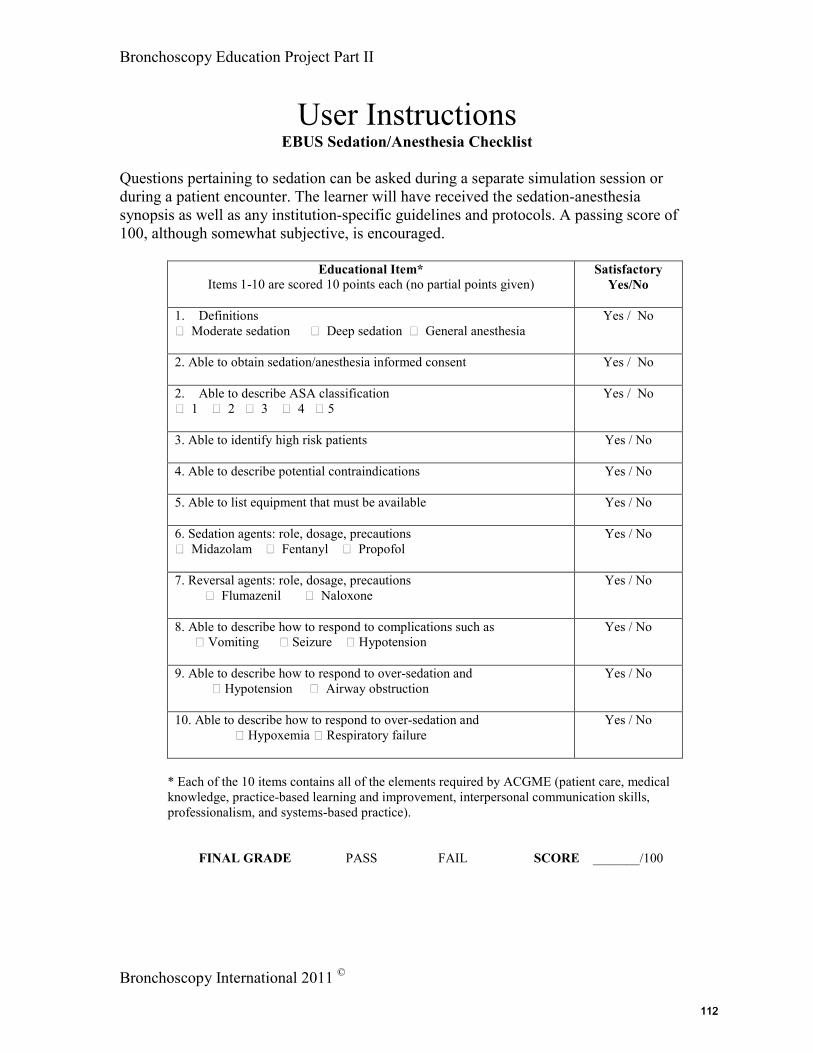
Bronchoscopy Education Project Part II
Bronchoscopy International 2011 ©
User Instructions EBUS Sedation/Anesthesia Checklist
Questions pertaining to sedation can be asked during a separate simulation session or during a patient encounter. The learner will have received the sedation-anesthesia synopsis as well as any institution-specific guidelines and protocols. A passing score of 100, although somewhat subjective, is encouraged.
Educational Item*
Items 1-10 are scored 10 points each (no partial points given)
Satisfactory Yes/No
1. Definitions Moderate sedation Deep sedation General anesthesia
Yes / No
2. Able to obtain sedation/anesthesia informed consent
Yes / No
2. Able to describe ASA classification 1 2 3 4 5
Yes / No
3. Able to identify high risk patients
Yes / No
4. Able to describe potential contraindications
Yes / No
5. Able to list equipment that must be available
Yes / No
6. Sedation agents: role, dosage, precautions Midazolam Fentanyl Propofol
Yes / No
7. Reversal agents: role, dosage, precautions Flumazenil Naloxone
Yes / No
8. Able to describe how to respond to complications such as Vomiting Seizure Hypotension
Yes / No
9. Able to describe how to respond to over-sedation and Hypotension Airway obstruction
Yes / No
10. Able to describe how to respond to over-sedation and Hypoxemia Respiratory failure
Yes / No
* Each of the 10 items contains all of the elements required by ACGME (patient care, medical knowledge, practice-based learning and improvement, interpersonal communication skills, professionalism, and systems-based practice).
FINAL GRADE PASS FAIL SCORE _______/100
112112

Bronchoscopy Education Project Part II
Bronchoscopy International 2011 ©
EBUS INFORMED CONSENT 10-Point CHECKLIST*
Student _________________________________ Training Year _______________ Faculty __________________________________ Date _______________________ Simulation EBUS-TBNA Workshop Patient-based EBUS-TBNA Scenario
Educational Item*
Items 1-10 are scored 10 points each (no partial points given)
Satisfactory Yes/No
1. Able to define “Informed Consent”: Informed decision-making regarding indications and expected outcomes, conflict of interest Protection from liability Provides opportunity to assess management strategies Provides opportunity to discuss risks, benefits, and alternatives
Yes / No
3. Able to discuss diagnosis and pertinent clinical issues
Yes / No
4. Able to describe the purpose of the procedure
Yes / No
5. Able to describe the nature of the procedure
Yes / No
6. Able to describe procedure-related risks
Yes / No
7. Able to describe procedure-related benefits
Yes / No
7. Able to describe alternative procedures regardless of cost or health care coverage
Yes / No
8. Able to describe potential risks and benefits from choosing the alternatives
Yes / No
9. Able to describe the risks and benefits of not performing the procedure or not choosing any of the alternatives
Yes / No
10. Able to demonstrate “effectiveness” of the informed consent process by asking the patient to explain in his or her own words, their understanding of the procedure
Yes / No
* Each of the 10 items contains all of the elements required by ACGME (patient care, medical knowledge, practice-based learning and improvement, interpersonal communication skills, professionalism, and systems-based practice).
FINAL GRADE PASS FAIL SCORE _______/100
113113

Bronchoscopy Education Project Part II
Bronchoscopy International 2011 ©
User Instructions EBUS Informed consent Checklist
Educational Item*
Items 1-10 are scored 10 points each (no partial points given)
1. Able to define “Informed Consent” Informed decision-making regarding indications and expected outcomes, conflict of interest Protection from liability Provides opportunity to assess management strategies Provides opportunity to discuss risks, benefits, and alternatives * The instructor may wish that the student be able to address some of the history of informed consent, and explain how and why informed consent plays a prominent role in medicine today. 2. Able to discuss diagnosis and pertinent clinical issues * Which diagnostic elements and clinical information help guide a patient’s choice of procedures? Why is this particular procedure being performed based on diagnosis and clinical issues? This provides the background to the informed consent process and opens the door for a dialogue with the patient so that the patient understands that simple authorization to perform the procedures is not what is being requested. 3. Able to describe the purpose of the procedure * Based on the clinical picture, the procedure is placed into context and the different elements of the procedure (such as to sample lymph nodes, exclude infection, or to diagnose or stage a malignancy) are described. 4. Able to describe the nature of the procedure * The procedure is described in layman’s terms. 5. Able to describe procedure-related risks *Risks applicable to the procedure are noted; the student may offer a few questions and answers, such as when can the patient eat, will the procedure hurt, is there a chance for bleeding, lung collapse, or infection. Some patients may fear death, and so this also may need to be addressed. A description of risks can increase both state and trait anxiety, and therefore, patients should probably be asked about their anxiety level and whether medication or other interventions are desired. 6. Able to describe procedure-related benefits *Benefits should be described clearly, such as diagnosis leads to early treatment, may avoid need for more invasive tests, provide information to other doctors to assist with therapeutic and diagnostic strategies, certain illnesses might be excluded so as to simplify further work-up. 7. Able to describe alternative procedures regardless of cost or health care coverage * Alternatives to EBUS & EBUS-TBNA should be cited and described; these might include, for example, invasive procedures such as open surgery, mediastinoscopy, percutaneous needle aspiration, and noninvasive procedures
114114

Bronchoscopy Education Project Part II
Bronchoscopy International 2011 ©
such as radiographic studies, etc. 8. Able to describe potential risks and benefits from choosing the alternatives * The risks and benefits from each of the alternatives should be addressed and explained. They can be compared to those of EBUS & EBUS-TBNA, and the physician can provide expert opinion as to why EBUS is being proposed and recommended. 9. Able to describe the risks and benefits of not performing the procedure or not choosing any of the alternatives * If EBUS is not performed, the patient should be told about potential consequences, whether or not alternatives are chosen, such as, for example, delayed diagnosis, prolonged illness, endangerment, need for different modes of surveillance or subsequent diagnostic tests or therapeutic measures. 10.Able to demonstrate “effectiveness” of the informed consent process by asking the patient to explain in his or her own words, their understanding of the procedure * The patient should be asked to describe EBUS or EBUS-TBNA and its consequences in general terms. This also provides an opportunity for dialogue.
* Each of the 10 items contains all of the elements required by ACGME (patient care, medical knowledge, practice-based learning and improvement, interpersonal communication skills, professionalism, and systems-based practice).
FINAL GRADE PASS FAIL SCORE _______/100
115115

Bronchoscopy Education Project Part II
Bronchoscopy International 2011 ©
This page intentionally left blank.
116116

Bronchoscopy Education Project Part II
Bronchoscopy International 2011 ©
EBUS PROCEDURAL PAUSE 10-Point CHECKLIST*
Student _________________________________ Training Year _______________ Faculty __________________________________ Date _______________________ Simulation EBUS Workshop Patient-based EBUS Scenario
Educational Item* Items 1-10 are scored 10 points each (no partial points given)
Satisfactory Yes/No
1. Able to define “Procedural Pause” as: Time-Out Wrong patient, site, procedure Team communication/patient understanding Mandatory in USA
Yes / No
4. Able to describe requirements of the procedural pause: Immediately before procedure Correct site, position, procedure Correct patient Pertinent medical records and equipment Verbal acknowledgements by all team members Elimination of environmental distractions
Yes / No
5. Able to describe the team leader’s role Yes / No 6. Able to describe the nursing team’s role Yes / No 7. Able to describe the patient’s role Yes / No 8. Able to describe other person’s roles (technicians, other
physicians)
Yes / No
7. Able to list the elements that must be covered: Patient Procedure Side and site Informed consent Medical records and equipment Medications Allergies/drug reactions Safety concerns based on history
Yes / No
8. Able to address behaviors in case of distractions Yes / No 9. Able to describe behaviors in case of disagreements Yes / No 10. Able to describe other elements pertaining to assuring a culture of safety: Communication Ability to prevent and respond to complications Universal, Droplet, and Airborne pathogen precautions
Yes / No
Each of the 10 items contains all of the elements required by ACGME (patient care, medical knowledge, practice-based learning and improvement, interpersonal communication skills, professionalism, and systems-based practice).
FINAL GRADE PASS FAIL SCORE _______/100
117117

Bronchoscopy Education Project Part II
Bronchoscopy International 2011 ©
User Instructions EBUS Procedural Pause Checklist
1. Able to define “Procedural Pause” as Time Out Wrong patient, site, procedure Team communication/patient understanding Mandatory in USA 2. Able to describe requirements of the procedural pause Immediately before procedure Correct site, position, procedure Correct patient Pertinent medical records and equipment Verbal acknowledgements by all team members Elimination of environmental distractions 3. Able to describe the team leader’s role * Mark the site if applicable, state name, patient, and procedure being performed, lead the time out, assure that all distractions are avoided during the time, requests a new time-out in case distractions occur, assures that time-out is being done according to protocol, addresses discrepancies, cancels procedure if all elements are not ascertained, modifies procedural strategy if applicable according to results of the time out. 4. Able to describe the nursing team’s role * Assures patient identification using at least two independent identifiers, assures right side right patient, right procedure, reviews and reads informed consent, assures appropriate medical records and equipment are available, assures appropriate response to complications or adverse events is possible. 5. Able to describe the patient’s role *If alert, able to state name, agree with procedure and site, signal family members who might be present. 6. Able to describe other person’s roles (technicians other physicians) * Able to state name and role. 7. Able to list the elements that must be covered Patient Procedure Side and site Informed consent Medical records and equipment Medications Allergies/drug reactions Safety concerns based on history 8. Able to address behaviors in case of distractions *No one should enter or leave the room during the time-out; any interruptions should prompt renewing the time-out; if a new or second procedure is being performed, time-out should be repeated; individuals should remain silent so that all present can focus on the time-out being performed. 9. Able to describe behaviors in case of disagreements * Verbal comments, behavior modification during or after the time-out; anyone should be able to disagree with what is being said during the time-out if it is inconsistent with the informed consent, predesignated procedural strategy, or clinical suspicions. 10. Able to describe other elements pertaining to assuring a culture of safety Communication Ability to prevent and respond to complications Universal, Droplet, and Airborne pathogen precautions *It is understood that differences between institutions and countries exist. Guidelines specific to each institution or country of practice can be given to students. General rules and concepts, however, should be understood and are covered in the special simulation session.
118118

Bronchoscopy Education Project Part II
Bronchoscopy International 2011 ©
EBUS Image Processor, Needle and Scope Handling 10-Point CHECKLIST*
Student _________________________________ Training Year ____________ Faculty __________________________________ Date ____________________ Interactive session Patient environment
Educational Item*
Items 1-10 are scored 10 points each (no partial points given)
Satisfactory Yes/No
1. Airway access and alternate among EBUS and White light bronchoscopy Airway access Oral/LMA/ET Tube Alternate EBUS/WLB
Yes / No
2. Image quality adjustments: Depth Describe Demonstrate
Yes / No
3. Image quality adjustments: Gain Describe Demonstrate
Yes / No
4. Image quality adjustments: Frequency Describe Demonstrate
Yes / No
5. Image quality adjustments: Doppler Describe Demonstrate
Yes / No
6. Image target measurements: Cursor placement Able to describe cursor placement for measuring lymph node size Able to demonstrate cursor placement and size measurements
Yes / No
7. Scope: Able to demonstrate balloon placement and functionality for optimal image acquisition Balloon placement onto scope Balloon integrity check Removal of air bubbles
Yes / No
8. Needle: Able to check mechanics and integrity of: □ Needle adjuster lock □ sheath adjuster knob □ Connecting slide
Yes / No
9. Needle: Integrity, retraction, and slider □ Integrity □ Retracts into sheath □ Slider moves freely and locks
Yes / No
10. Needle: Stylet and aspiration syringe integrity and function □ Moves freely □ Aspiration syringe functions
Yes / No
* Each of the 10 items contains all of the elements required by ACGME (patient care, medical knowledge, practice-based learning and improvement, interpersonal communication skills, professionalism, and systems-based practice).
FINAL GRADE PASS FAIL SCORE _______/100
119119

Bronchoscopy Education Project Part II
Bronchoscopy International 2011 ©
User Instructions EBUS Image Processor, Needle and Scope Handling Checklist
Questions pertaining to EBUS can be asked during a separate simulation session or during a patient encounter. The learner will have received the EBUS image processor and needle synopsis as well as any institution-specific guidelines and protocols. A passing score of 100, although somewhat subjective, is encouraged.
Educational Item*
Items 1-10 are scored 10 points each (no partial points given)
Satisfactory Yes/No
1. Able to alternate between EBUS and White light bronchoscopy * The instructor should assure that the operator knows how to switch between white light and EBUS bronchoscopy imaging and screen displays
Yes / No
2. Image quality adjustments: Depth Describe Demonstrate * The instructor verifies that the learner can describe meaning of depth, and demonstrate depth control on the processor
Yes / No
3. Image quality adjustments: Gain Describe Demonstrate * The instructor verifies that the learner can describe principles of gain, and demonstrate gain control on the processor
Yes / No
4. Image quality adjustments: Frequency Describe Demonstrate * The instructor verifies that the learner can describe principles of frequency, and demonstrate frequency control on the processor
Yes / No
5. Image quality adjustments: Doppler Describe Demonstrate * The instructor verifies that the learner can describe principles of Doppler, and demonstrate Doppler control on the processor
Yes / No
6. Image target measurements: Cursor placement Able to describe cursor placement for measuring lymph node size Able to demonstrate cursor placement and size measurements
Yes / No
7. Scope: Able to demonstrate balloon placement and functionality for optimal image acquisition Balloon placement onto scope Balloon integrity check Removal of air bubbles * The instructor verifies that the learner can place and troubleshoot balloon-related issues.
Yes / No
9. Needle: Able to check mechanics and integrity of: □ Needle adjuster lock □ sheath adjuster knob □ Connecting slide
*The instructor assures that the learned knows how the needle functions (eg. is not bent) and is able to troubleshoot malfunction
Yes / No
9. Needle: Integrity, retraction, and slider □ Integrity □ Retracts into sheath □ Slider moves freely and locks
Yes / No
120120

Bronchoscopy Education Project Part II
Bronchoscopy International 2011 ©
10. Needle: Stylet and aspiration syringe integrity and function □ Moves freely □ Aspiration syringe functions *The instructor assures that the learner can verify proper functioning of the stylet and aspiration syringe before performing EBUS-TBNA
Yes / No
* Each of the 10 items contains all of the elements required by ACGME (patient care, medical knowledge, practice-based learning and improvement, interpersonal communication skills, professionalism, and systems-based practice).
FINAL GRADE PASS FAIL SCORE _______/100
121121

Bronchoscopy Education Project Part II
Bronchoscopy International 2011 ©
This page intentionally left blank.
122122

Bronchoscopy Education Project Part II
Bronchoscopy International 2011 ©
EBUS PRACTICAL APPROACH 10 Point CHECKLIST*
Student _________________________________ Training Year _______________ Faculty __________________________________ Date _______________________ Instructor-learner 30 minute session Daily rounds & EBUS consultation
Educational Item* Items 1-10 are scored 10 points each (no partial points given)
Satisfactory Yes/No
1. Initial evaluation A Physical examination, laboratory tests, and functional assessment Comorbidities
Yes / No
2. Initial evaluation B Support system Preferences and expectations
Yes / No
3. Procedural strategies A Indications, contraindications, expected results Operator and team experience and expertise
Yes / No
4. Procedural strategies B Risk-benefits and therapeutic alternatives Informed consent
Yes / No
5. Techniques and results A Anesthesia and perioperative care Techniques and instruments
Yes / No
6. Techniques and results B Anatomic dangers and other risks Results and complications
Yes / No
7. Long-term management plan A Outcome assessment Follow-up tests and procedures
Yes / No
8. Long-term management plan B Referrals to other specialists Quality improvement and team evaluation
Yes / No
9. Able to answer case-specific questions Question 1 Question 2 Question 3
Yes / No
10. General ability to provide evidence for and rationally justify decision making Subjective assessment of learner ability
Yes / No
*These 10 items contains all of the elements required by ACGME (patient care, medical knowledge, practice-based learning and improvement, interpersonal communication skills, professionalism, and systems-based practice).
FINAL GRADE PASS FAIL SCORE _______/100
123123

Bronchoscopy Education Project Part II
Bronchoscopy International 2011 ©
User Instructions EBUS Practical Approach Checklist
This practical approach exercise is held as a 30-minute session between learner and instructor, similar to an “oral examination”. Of course, programs including Practical Approach-like discussions for each EBUS bronchoscopy consultation may choose to forego a formal session. Using a structured format (the four boxes), learners and instructors are more certain to cover in as great a depth as desired all aspects of a procedure: strategy and planning, techniques and instruments, and response to complications. Because each element is important, items are equally weighted at ten points each with no partial points given. A passing score of 100, although somewhat subjective, is encouraged.
Educational Item* Items 1-10 are scored 10 points each (no partial points given)
Satisfactory Yes/No
2. Initial evaluation A Physical examination, laboratory tests, and functional assessment Comorbidities
Yes / No
2. Initial evaluation B Support system Preferences and expectations
Yes / No
3. Procedural strategies A Indications, contraindications, expected results Operator and team experience and expertise
Yes / No
4. Procedural strategies B Risk-benefits and therapeutic alternatives Informed consent
Yes / No
5. Techniques and results A Anesthesia and perioperative care Techniques and instruments
Yes / No
6. Techniques and results B Anatomic dangers and other risks Results and complications
Yes / No
7. Long-term management plan A Outcome assessment Follow-up tests and procedures
Yes / No
8. Long-term management plan B Referrals to other specialists Quality improvement and team evaluation
Yes / No
9. Able to answer case-specific questions Question 1 Question 2 Question 3
Yes / No
10. General ability to provide evidence for and rationally justify decision making Subjective assessment of learner ability
Yes / No
*These 10 items contains all of the elements required by ACGME (patient care, medical knowledge, practice-based learning and improvement, interpersonal communication skills, professionalism, and systems-based practice).
FINAL GRADE PASS FAIL SCORE _______/100
124124

Bronchoscopy Education Project Part II
Bronchoscopy International 2011 ©
PROCTORED EBUS & EBUS-TBNA 10-Point CHECKLIST* Student _________________________________ Training Year _______________
Faculty __________________________________ Date _______________________ Simulation EBUS Workshop Patient-based EBUS Scenario
Educational Item* Items 1-10 are scored 10 points each (no partial points given)
Satisfactory Yes/No
1. Formulation of effective plan and strategy: Informed consent obtained, signed and in medical record History and physical Review imaging studies Cost-effective practice Use of information resources Applies evidence-based medicine Use of systems resources
Yes / No
2. Patient safety: ASA assessment Airway assessment Allergies Medications Comorbidities
Yes / No
3. Patient safety: Positioning Supplemental oxygen Vital signs Suction Bite-block and/or ET tube if applicable Verifies ability to respond to complications and accessibility of resuscitation equipment
Yes / No
4. Patient and procedural team safety: Knowledgeable use of image processor Hand disinfection Universal precautions “Time-Out”.
Yes / No
5. Procedure: Premedication Moderate sedation Topical anesthetic General anesthesia as applicable
Yes / No
6. Procedure: Scope insertion Empathy and Communication with patient
Yes / No
7. Procedure: Image processor controls Balloon attachment and troubleshooting Needle, stylet, and syringe function and troubleshooting
Yes / No
8. Procedure: Alternate between white light and EBUS Image acquisition Recognition of mediastinal and hilar nodal anatomy Recognition of mediastinal and hilar vascular structures Lymph node sampling Smear preparation and station labeling
Yes / No
9. Communication with staff, patient, and patient family Puts needs of patient first Punctuality Respect Listening skills Personal appearance Initiative & Motivation Empathy Honesty Accepts responsibility
Yes / No
11. Documentation/procedure note Informative Accurate Communication with colleagues
Yes / No
* Each of the 10 items contains all of the elements required by ACGME (patient care, medical knowledge, practice-based learning and improvement, interpersonal communication skills, professionalism, and systems-based practice).
FINAL GRADE PASS FAIL SCORE _______/100
125125

Bronchoscopy Education Project Part II
Bronchoscopy International 2011 ©
User Instructions
Proctored EBUS-TBNA Checklist
Educational Item* Items 1-10 each scored 10 points (no partial points given)
1. Formulation of effective plan and strategy Informed consent obtained, signed and in medical record History and physical Review imaging studies Cost-effective practice Use of information resources Applies evidence-based medicine Use of systems resources * The instructor should be certain that the student has obtained the informed consent and that it is signed and available. The student should be able to describe pertinent clinical and radiographic findings and to use evidence-based medicine in addition to any other information gained from other resources, such as faculty expert opinion, advice, and requests by referring physicians, to justify the procedure being performed. The student should be able to describe the use of institutional resources, and use them according to institutional practices, such as computed tomography, ultrasound, other instruments and equipment. This also includes nursing surveillance, respiratory therapy assistance, on-site cytology, and need for procedure suite, operating theater or intensive care unit. Cost-effectiveness can be discussed in regards to other diagnostic or therapeutic modalities, but also how EBUS bronchoscopy may or may not increase or decrease health-related expenditures in the context of the patient’s diagnosis and treatment plan. 2. Patient safety: ASA assessment Airway assessment Allergies Medications Comorbidities *ASA and airway assessments should be performed according to the institution’s guidelines. Allergies, medications, especially anticoagulants and antiplatelet agents should be noted, comorbidities that might increase the risk of adverse events should also be described and identified. 3. Patient safety: Positioning Supplemental oxygen Vital signs Suction Bite-block and/or ET tube, if applicable Verifies ability to respond to complications and accessibility of resuscitation equipment *These measures should be taken according to institutional biases and protocols. 4. Patient and procedural team safety: 4. Patient and procedural team safety: Knowledgeable use of image processor Hand disinfection Universal precautions “Time-Out”. *Students should be observed as they comply with these safety measures. Additional information pertaining to time-out, universal precautions, and knowledge of fluoroscopy are provided in the modules. Protocols may vary among institutions. 5. Procedure: Premedication Moderate sedation Topical anesthetic General anesthesia as
126126

Bronchoscopy Education Project Part II
Bronchoscopy International 2011 ©
applicable *These should be administered according to institutional biases and according to protocols. 6. Procedure: Scope insertion Empathy and Communication with patient *Performed according to institutional practices. Empathy and communication with the patient should be observed. Students should be able to speak comfortably and reassuringly with their patient. Open dialogues should be encouraged to enhance confidence and decrease patient anxiety. 7. Procedure: Image processor controls Balloon attachment and troubleshooting Needle, stylet, and syringe function and troubleshooting *Performed according to institutional practices. 8. Procedure: Alternate between white light and EBUS Image acquisition Recognition of mediastinal and hilar nodal anatomy Recognition of mediastinal and hilar vascular structures Lymph node sampling Smear preparation and station labeling *Performed according to institutional practices. It is recognized that not all patients will undergo all of these procedures, therefore, if the institution desires, a different proctored checklist can be completed for each (or each set) of the procedures listed. 9. Communication with staff, patient, and patient family Puts needs of patient first Punctuality Respect Listening skills Personal appearance Initiative & Motivation Empathy Honesty Accepts responsibility *These are for the most part subjective assessments, and also require feedback from nursing staff. Students should be told that they will be judged on these items during the course of their training, so that they can obtain feedback from their instructors and improve their performance in these areas. 10.Documentation/procedure note Informative Accurate Communication with colleagues *Procedure notes may vary according to institution; however, in general, the note should be informative, telling a story about the procedure that referring physicians can understand. The note should be accurate regarding what was done, why it was done, and how it was done. Procedure-related adverse events should be described. Communication with colleagues should be observed, by watching how the student interacts with the nursing team, other physicians, and with referring physicians. While this element is also, for the most part subjective, in general, communication should be informative, accurate polite, and considerate. * Each of the 10 items contains all of the elements required by ACGME (patient care, medical knowledge, practice-based learning and improvement, interpersonal communication skills, professionalism, and systems-based practice).
FINAL GRADE PASS FAIL SCORE _______/100
127127

Bronchoscopy Education Project Part II
Bronchoscopy International 2011 ©
This page intentionally left blank.
128128

Bronchoscopy Education Project Part II
Bronchoscopy International 2011 ©
CONGRATULATIONS
You have now completed the administration of the EBUS Bronchoscopy Competency
Program, an integral part of the Bronchoscopy Education Project.
Please send us your comments regarding your participation in this international endeavor by contacting your national bronchology association, emailing us
at www.bronchoscopy.org or by contacting
Dr. Henri Colt at [email protected] and Dr. Eric Edell at [email protected].
If you wish to further pursue an in-depth study of bronchoscopy education, thus becoming a certified bronchoscopy trainer, please consider our Train The Trainers Program.
129129

Bronchoscopy Education Project Part II
Bronchoscopy International 2011 ©
130130
Bronchoscopy Education Project Part II
Bronchoscopy International 2011 ©
This page intentionally left blank.
131131

Bronchoscopy Education Project Part II
Bronchoscopy International 2011 ©
This page intentionally left blank.
132132