
FlavonoidsIsolatedfromKoreaCitrusaurantiumL.InduceG2/M...
12
Hindawi Publishing Corporation Evidence-Based Complementary and Alternative Medicine Volume 2012, Article ID 515901, 11 pages doi:10.1155/2012/515901 Research Article Flavonoids Isolated from Korea Citrus aurantium L. Induce G2/M Phase Arrest and Apoptosis in Human Gastric Cancer AGS Cells Do-Hoon Lee, 1 Kwang-Il Park, 1 Hyeon-Soo Park, 1 Sang-Rim Kang, 1 Arulkumar Nagappan, 1 Jin-A Kim, 2 Eun-Hee Kim, 3 Won-Sup Lee, 4 Young-Sool Hah, 5 Hyon-Jong Chung, 1 Su-Jin An, 1 and Gon-Sup Kim 1 1 Research Institute of Life science and College of Veterinary Medicine, Gyeongsang National University, Gazwa, Jinju 660-701, Republic of Korea 2 Korea National Animal Research Resource Center and Korea National Animal Bio-resource Bank, Gyeongsang National University, Gazwa, Jinju 660-701, Republic of Korea 3 Department of Nursing Science, International University of Korea, Jinju 660-759, Republic of Korea 4 Department of Internal Medicine, Institute of Health Sciences, Gyeongsang National University School of Medicine and Gyeongnam Regional Cancer Center, Gyeongsang National University Hospital, Jinju 660-702, Republic of Korea 5 Clinical Research Institute, Gyeongsang National University Hospital, Jinju 660-702, Republic of Korea Correspondence should be addressed to Gon-Sup Kim, [email protected] Received 5 August 2011; Accepted 14 September 2011 Academic Editor: Jae Youl Cho Copyright © 2012 Do-Hoon Lee et al. This is an open access article distributed under the Creative Commons Attribution License, which permits unrestricted use, distribution, and reproduction in any medium, provided the original work is properly cited. Aim of the Study. Citrus species is used in traditional medicine as medicinal herb in several Asian countries including Korea. Flavonioids became known as various properties, such as anti-oxidants, anti-inflammation and anti-cancer, and so forth. The present study, the anti-cancer effect of flavonioids isolated from Citrus aurantium L. in human gastric cancer AGS cells has been investigated. Materials and Methods. The anti-proliferative activity was assayed using MTT assay. Cell cycle analysis was done using flow cytometry and apoptosis detection was done using by hoechst fluorescent staining and Annexin V-propidium iodide double staining. Western blot was used to detect the expression of protein related with cell cycle and apoptosis. Results. Flavonoids isolated from Citrus aurantium L. have the effect of anti proliferation on AGS cells with IC50 value of 99 μg/mL. Flavonoids inhibited cell cycle progression in the G2/M phase and decrease expression level of cyclin B1, cdc 2, cdc 25c. Flavonoids induced apoptosis through activate caspase and inactivate PARP. Conclusions. Flavonoids isolated from Citrus aurantium L. induced G2/M phase arrest through the modulation of cell cycle related proteins and apoptosis through activation caspase. These finding suggest flavonoids isolated from Citrus aurantium L. were useful agent for the chemoprevention of gastric cancer. 1. Introduction Citrus fruits are a very popular food source because of their nutrient, flavor, and intrinsic attributes [1] and have long been the basis of commonly used traditional medicines in several Asian countries. Immature peels of citrus fruit are used to treat indigestion and have demonstrated potential as a chemotherapeutic agent [2, 3]. Among nutrients of citrus fruits, flavonoids have been more recently recognized as having various medicinal benefits that include antioxidant, antimicrobial, anti-inflammatory, and anticancer activities [4, 5]. Citrus fruits have numerous bioactive flavonoids such as naringin, naringenin, narirutin, nobiletin, quercetin, kaempferol, hesperidin, neohesperidin, didymin, and pon- cirin [6]. In particular, naringin, nobiletin, and hesperidin display anticancer effects via cell cycle arrest and apoptosis [7–9]. Gastric cancer is the most prevalent cancer in Korea. Ac- cording to Ministry of Health and Welfare statistics, there were 28,078 cases of gastric cancer in Korea in 2008, repre- senting 15.7% of all cancer cases in the country. Globally, gastric cancer is the fourth leading cancer and is the second- leading cause of cancer-related death, following lung can- cer [10]. Treatment of gastric cancer consists generally of
Transcript of FlavonoidsIsolatedfromKoreaCitrusaurantiumL.InduceG2/M...
Hindawi Publishing CorporationEvidence-Based Complementary and Alternative MedicineVolume 2012, Article ID 515901, 11 pagesdoi:10.1155/2012/515901
Research Article
Flavonoids Isolated from Korea Citrus aurantium L. Induce G2/MPhase Arrest and Apoptosis in Human Gastric Cancer AGS Cells
Do-Hoon Lee,1 Kwang-Il Park,1 Hyeon-Soo Park,1 Sang-Rim Kang,1
Arulkumar Nagappan,1 Jin-A Kim,2 Eun-Hee Kim,3 Won-Sup Lee,4
Young-Sool Hah,5 Hyon-Jong Chung,1 Su-Jin An,1 and Gon-Sup Kim1
1 Research Institute of Life science and College of Veterinary Medicine, Gyeongsang National University, Gazwa,Jinju 660-701, Republic of Korea
2 Korea National Animal Research Resource Center and Korea National Animal Bio-resource Bank, Gyeongsang National University,Gazwa, Jinju 660-701, Republic of Korea
3 Department of Nursing Science, International University of Korea, Jinju 660-759, Republic of Korea4 Department of Internal Medicine, Institute of Health Sciences, Gyeongsang National University School of Medicine andGyeongnam Regional Cancer Center, Gyeongsang National University Hospital, Jinju 660-702, Republic of Korea
5 Clinical Research Institute, Gyeongsang National University Hospital, Jinju 660-702, Republic of Korea
Correspondence should be addressed to Gon-Sup Kim, [email protected]
Received 5 August 2011; Accepted 14 September 2011
Academic Editor: Jae Youl Cho
Copyright © 2012 Do-Hoon Lee et al. This is an open access article distributed under the Creative Commons Attribution License,which permits unrestricted use, distribution, and reproduction in any medium, provided the original work is properly cited.
Aim of the Study. Citrus species is used in traditional medicine as medicinal herb in several Asian countries including Korea.Flavonioids became known as various properties, such as anti-oxidants, anti-inflammation and anti-cancer, and so forth. Thepresent study, the anti-cancer effect of flavonioids isolated from Citrus aurantium L. in human gastric cancer AGS cells has beeninvestigated. Materials and Methods. The anti-proliferative activity was assayed using MTT assay. Cell cycle analysis was doneusing flow cytometry and apoptosis detection was done using by hoechst fluorescent staining and Annexin V-propidium iodidedouble staining. Western blot was used to detect the expression of protein related with cell cycle and apoptosis. Results. Flavonoidsisolated from Citrus aurantium L. have the effect of anti proliferation on AGS cells with IC50 value of 99 μg/mL. Flavonoidsinhibited cell cycle progression in the G2/M phase and decrease expression level of cyclin B1, cdc 2, cdc 25c. Flavonoids inducedapoptosis through activate caspase and inactivate PARP. Conclusions. Flavonoids isolated from Citrus aurantium L. induced G2/Mphase arrest through the modulation of cell cycle related proteins and apoptosis through activation caspase. These finding suggestflavonoids isolated from Citrus aurantium L. were useful agent for the chemoprevention of gastric cancer.
1. Introduction
Citrus fruits are a very popular food source because of theirnutrient, flavor, and intrinsic attributes [1] and have longbeen the basis of commonly used traditional medicines inseveral Asian countries. Immature peels of citrus fruit areused to treat indigestion and have demonstrated potential asa chemotherapeutic agent [2, 3]. Among nutrients of citrusfruits, flavonoids have been more recently recognized ashaving various medicinal benefits that include antioxidant,antimicrobial, anti-inflammatory, and anticancer activities[4, 5]. Citrus fruits have numerous bioactive flavonoids
such as naringin, naringenin, narirutin, nobiletin, quercetin,kaempferol, hesperidin, neohesperidin, didymin, and pon-cirin [6]. In particular, naringin, nobiletin, and hesperidindisplay anticancer effects via cell cycle arrest and apoptosis[7–9].
Gastric cancer is the most prevalent cancer in Korea. Ac-cording to Ministry of Health and Welfare statistics, therewere 28,078 cases of gastric cancer in Korea in 2008, repre-senting 15.7% of all cancer cases in the country. Globally,gastric cancer is the fourth leading cancer and is the second-leading cause of cancer-related death, following lung can-cer [10]. Treatment of gastric cancer consists generally of
2 Evidence-Based Complementary and Alternative Medicine
surgery, chemotherapy, and/or radiotherapy. While the treat-ments can be effective, about half of gastric cancer patientsare untreatable [11]. The need for more effective treatmentsis pressing.
The programmed cell death mechanism of apoptosis isthe major regulator of cell proliferation and is a focus ofcancer research as an effective way of eliminating precan-cerous and/or cancerous cells [1]. Caspases play a pivotalrole in apoptosis; their overexpression and cleavage is aprecursor of apoptosis in mammalian cells. To date, 14caspases have been identified based on their function. Theyconstitute three functional groups: inflammatory caspases(caspase-1, -4, and -5), apoptotic initiator caspases (caspase-2, -8, -9, and -10), and apoptotic effector caspases (caspase-3, -6, and -7) [12]. Caspases possess inactive zymogens thatconsist of p20 (large) and p10 (small) subunits. In responseto apoptotic signals, the inactive zymogens are cleaved,yielding active forms of the proteins that are associated withthe induction of apoptosis [13]. Also, the Bcl-2 family ofproteins, whose activity is directed to act at the mitochon-drial outer membrane, are major regulators of apoptosis.The Bcl-2 family consists of proapoptotic and antiapoptoticmembers [14]. Apoptotic Bcl-2 proteins such as Bax and Bakcreated pores in the mitochondrial outer membrane, throughwhich cytochrome c is released to the cytosol. Binding ofcytochrome c to Apaf-1 creates an apoptosome complexthat activates caspase-9, which in turn activates caspase-3. Antiapoptotic Bcl-2 proteins such as Bcl-2 and Bcl-xLpreserve the mitochondrial membrane structure throughinteraction with apoptotic Bcl-2 proteins [15].
Cell cycle arrest is also a major regulator of cell prolif-eration. In eukaryotic cells, the cell cycle comprises the G1,S, G2, and M phases. Checkpoints to the cycle are presentin the G1 and G2 phases. In cell cycle progression, cyclinsand cyclin-dependent kinases (cdks) play a central role asregulators. In the mid-G1 phase, cell cycle progression iscontrolled by a cyclin D-cdk4/cdk6 complex. In late-G1,progression is controlled by the cyclin E-cdk2 complex. Inthe G2 phase, cell cycle progression is controlled by thecyclin A/B-cdc2 complex [16–18]. DNA damage can inhibitcell proliferation by the inactivation of cyclins and cdks andsubsequent cell cycle arrest.
As referred to earlier, flavonoids isolated from Citrusaurantium L. have the anticancer effect. However, the mecha-nisms of the anticancer activity of Korea Citrus aurantium L.still remain unknown. In the present study, we demonstratethat flavonoids isolated from Korea Citrus aurantium L. causecell cycle arrest and apoptosis in AGS human gastric cancercells. The expression levels of several important proteins areshown to be strongly related with the cell cycle and apoptosis.Finally, the occurrence of cell cycle arrest and apoptosis wasdetermined to ascertain the anticancer mechanism of theisolated flavonoids.
2. Materials and Methods
2.1. Antibodies and Reagents. Cyclin B1, cdc 2, cdc 25c, andß-actin were purchased from Millipore (Billerica, MA, USA).
Antibodies for Bcl-xL, Bax, cleaved poly(ADP-ribose) poly-merase (PARP), and caspases-3, -6, -8, and -9 were purchasedfrom Cell Signaling Technology (Danvers, Mass, USA).Horseradish peroxidase- (HRP-) coupled goat anti-mouseIgG and anti-rabbit IgG were purchased from Santa CruzBiotechnology (Santa Cruz, CA, USA). RPMI-1640 waspurchased from Hyclone (Logan, UT, USA). Fetal bovineserum (FBS) and antibiotics (streptomycin/penicillin) werepurchased from Gibco (BRL Life Technologies, Grand Island,NY, USA). 3-(4,5-Dimethylthiazol-2-yl)-2,5-dephenyltetra-zolium bromide (MTT), dimethylsulfoxide (DMSO), andRNase A were obtained from Sigma-Aldrich (St. Louis,MO, USA). Fluorescein isothiocyanate (FITC) annexin-Vapoptosis detection kit 1 was purchased from BD Pharmin-gen (San Diego, CA, USA). Enhanced chemiluminescence(ECL) kit was purchased from Amersham Life Science(Buckinghamshire, UK). Materials and chemicals used forelectrophoresis were obtained from Bio-Rad Laboratories(Hercules. CA, USA).
2.2. Isolation of Flavonoids. The analyses of isolation of fla-vonoids were conducted at the Department of Chemistry,Gyeongsang National University by Professor Sung ChulShin. High-performance liquid chromatography (HPLC)was performed as descried. Briefly, HPLC analysis wasperformed using a 1100 series LC system installed with aG1322A degasser, a G1312A pump, a G1313A autosampler,and a G1316A oven (Agilent Technologies, Palo Alto, CA,USA). Chromatographic separation was carried out ona Zorbax StableBond Analytical SB-C18 column (4.6 ×250 mm, 5 μm; Agilent Technologies). The binary solventsystem was made up of 0.1% aqueous formic acid (A)and methanol/acetonitrile (1 : 1) (B). Elution was performedusing a linear gradient (0%–25% B) over 10 min and 40%–70% B over 10 min, followed by 30 min of isocratic elution,declined from 40%–25% B over 5 min and followed by10 min of isocratic elution. The flow rate was 0.5 mL/minin column temperature of 35◦C and an infusion volumeof 10 μL in each experiment. Chromatographic data werecollected and controlled using a ChemStation, Rev.B.0301(Agilent Technologies). Spectral data were collected (200–400 nm, 2 nm resolution) for the entire progression, and theflavonoids were quantified by extracting each chromatogramat 280 nm (Figure 1). All flavanones and flavones werequantified using the external calibration curves of hesperetinand nobiletin, respectively. Tandem mass spectrometry(MS/MS) experiments were conducted using a 3200 QTRAPLC-MS/MS system (Applied Biosystems, Foster City, CA,USA) with a Turbo V-source and a Turbo Ion Spray probe(Applied Biosystems). The mass spectrometer was operatedin the positive mode with selected ion monitoring (SIM),BioAnalyst, version 1.4.2 (AB Sciex, Zagreb, Croatia). Theelectron spray voltage was set at 5.2 kV and the sourcetemperature at 500◦C. The mass spectra were recordedbetween m/z 100 and m/z 1000 with a step size of 0.06 amu.
2.3. Cell Culture. AGS human gastric cancer cells obtainedfrom the Korea Cell Line Bank (Seoul, Korea) were cultured

Evidence-Based Complementary and Alternative Medicine 3
1
2
3
45 6 7
8
910
11
12
13 14
280 nm
10 20 30 40 50 (min)
0
200
400
600
800
1000
mAUMWD1 A, Sig = 280.5, Ref = off (1214-CITRUS\1214-NAOH-C.D)
Figure 1: HPLC chromatogram patterns of Korea Citrus aurantium L. at 280 nm. (1) naringin, (2) hesperidin, (3) poncirin, (4) isosi-ennsetin, (5) hexamethoxyflavone, (6) sineesytin, (7) hexamethoxyflavone, (8) tetramrthnl-o-isoscutellaeein, (9) nobiletin, (10) hep-tamethoxyflavone, (11) 3-hydoxynobiletin, (12) tangeretin, (13) hydroxypentamethoxyflavone, and (14) hexamethoxyflavone.
in RPMI-1640 medium supplemented with 10% heat-inac-tivated fetal calf serum and 100 μg/mL of penicillin andstreptomycin in a humidified atmosphere of 5% CO2 at37◦C.
2.4. Cell Viability Assay and Morphological Examination.Cell viability was determined using MTT 24 h after theexperimental treatments. Briefly, cells were plated in wellsof 12-well plates and incubated for 24 h at 37◦C. The cellswere untreated or treated with varying concentrations offlavonoids (10, 50, 100, 150, and 200 μg/mL) for 24 h at 37◦C.DMSO (0.1%) was used as a vehicle control. MTT solution(5 mg/mL in phosphate buffered saline; PBS) was diluted to0.5 mg/mL by medium. After 3 h incubation at 37◦C, MTT-containing medium was removed and the crystals that hadformed were dissolved by the addition of DMSO to eachwell. After mixing, the absorbance of the cells was measuredat 540 nm by using an enzyme-linked immunosorbent assayplate reader. For morphological examination, cells weregrown on 6-well plates, treated with flavonoids for 24 h, andthen examined under light microscopy (×400).
2.5. Hoechst 33258 Fluorescent Staining. AGS cells from ex-ponentially growing cultures were seeded in 12-well cultureplates. AGS cells were untreated or treated with varyingconcentrations of flavonoids (20, 40, 60, 80, and 100 μg/mL)for 24 h at 37◦C. The cells were then washed in ice-coldphosphate-buffered saline (PBS) and fixed in a solution of3.7% paraformaldehyde for 15 min at room temperature.To identify the apoptotic AGS cells, they were stainedwith Hoechst 33258 (5 μg/mL in PBS) for 10 min at roomtemperature. The nuclei structure of the cells was examined
by Leica fluorescence microscopy. The apoptotic cells wereobserved at 1000x magnification.
2.6. Cell Cycle Analysis. Upon reaching 70%–80% conflu-ence, AGS cells were untreated or treated with varyingconcentrations of flavonoids (20, 40, 60, 80, and 100 μg/mL)for 24 h at 37◦C. Then, the cells were washed twice withcold PBS, trypsinized, and centrifuged. The cells werefixed in 70% (v/v) ethanol for 24 h at 4◦C. The cells werewashed with PBS and stained with propidium iodide (PI;50 μg/mL) including RNase A (0.1 mg/mL) in PBS (pH 7.4)for 30 min in the dark. PI-stained samples were analyzed witha FACScan flow cytometer (Becton Dickinson, San Jose, CA,USA). In each sample, 10,000 cells were sorted. The data wereanalyzed using CellQuest software (Becton Dickinson).
2.7. Apoptosis Assays. Apoptotic cells were detected using aFITC annexin-V apoptosis detection kit 1 (BD Pharmingen,San Diego, CA, USA). Briefly, cells were untreated ortreated with varying concentrations of flavonoids (20, 40,60, 80, and 100 μg/mL) for 24 h at 37◦C. The cells weretrypsinized and washed with PBS. The washed cells wereresuspended in Annexin-V binding buffer containing 10 mMHEPES/NaOH, pH 7.4, 140 mM NaCl and 2.5 mM CaCl2according to the manufacturer’s protocol. The cells werestained simultaneously with FITC-conjugated Annexin-Vand PI at room temperature for 15 min in the dark, priorto the addition of binding buffer. The apoptotic cells weremeasured using a FACScan flow cytometer. The cells weresorted into intact cells (Annexin V− PI−), early apoptoticcells (Annexin V+ PI−), late apoptotic cells (Annexin V+PI+), and necrotic cells (Annexin V− PI+).
4 Evidence-Based Complementary and Alternative Medicine
2.8. Caspase-3 Activity. Caspase-3-activity was determinedby a colorimetric assay using a caspase-3 activation kit ac-cording to the protocol of the manufacturer (Millipore,Billerica, MA, USA). In brief, the cells were lysed in thesupplied lysis buffer and incubated for 10 min in an icebath. Then, the samples were centrifuged for 5 minutesin a microcentrifuge (10,000×g). The supernatants werecollected and incubated with Assay buffer and Caspase-3 Substrate (Ac-DEVD-pNA) for 1 h at 37◦C. The opticaldensity of the reaction mixture was quantified using anenzyme-linked immunosorbent assay plate reader at 405 nm.
2.9. Western Blot Analysis. Protein concentration was deter-mined using a Bradford assay (Bio-Rad). An equal proteinamount was separated by 12% sodium dodecyl sulfate-polyacrylamide gel electrophoresis (SDS-PAGE), and theresolved proteins were transferred onto 0.45 mm Immob-ilon-P polyvinylidene fluoride (PVDF) membranes (Milli-pore). Each membrane was blocked with TBST (10 mM Tris-HCl, pH 7.4, 150 mM NaCl, 0.1% Tween-20) containing 5%skin milk. Blocking was performed for 1 h at room temper-ature. Then, each membrane was incubated with primaryantibody (1 : 1000 dilution) for overnight at 4◦C, washed fivetimes for 10 minutes each time with TBST, and incubatedwith HRP-conjugated secondary antibody (1 : 2000 dilution)for 2 h at room temperature. Each membrane was washed5 times for 10 minutes each time with TBST. Protein bandswere visualized by ECL and Western Blotting DetectionReagents and exposed to X-ray films (Fuji, Tokyo, Japan).Each band was analyzed using the Image J program (http://rsb.info.nih.gov/).
2.10. Statistical Analysis. All experiments were performedin triplicate. Results are expressed as the mean ± standarddeviation of at least three separate experiments. Statisticalanalysis was determined by Student’s t-test using SPSSversion 10.0 for Window (SPSS, Chicago, IL, USA). A valueof P < 0.05 was considered to be significant.
3. Results
3.1. Flavonoids Inhibit Cell Viability. In order to estimate theeffect of cell viability inhibition, we tested the cytotoxicityof various concentrations (0–200 μg/mL) of flavonoids for24 using the MTT assay. As shown in Figure 2, flavonoidsinhibited cell viability in a dose-dependent manner. Com-pared with the control group, the viability of cells treatedwith flavonoid concentrations of 100 μg/mL declined byabout 48% after 24 h (IC50 approximately 99 μg/mL). Visualobservations revealed a dose-dependent change in cell shapesuch as cell shrinkage.
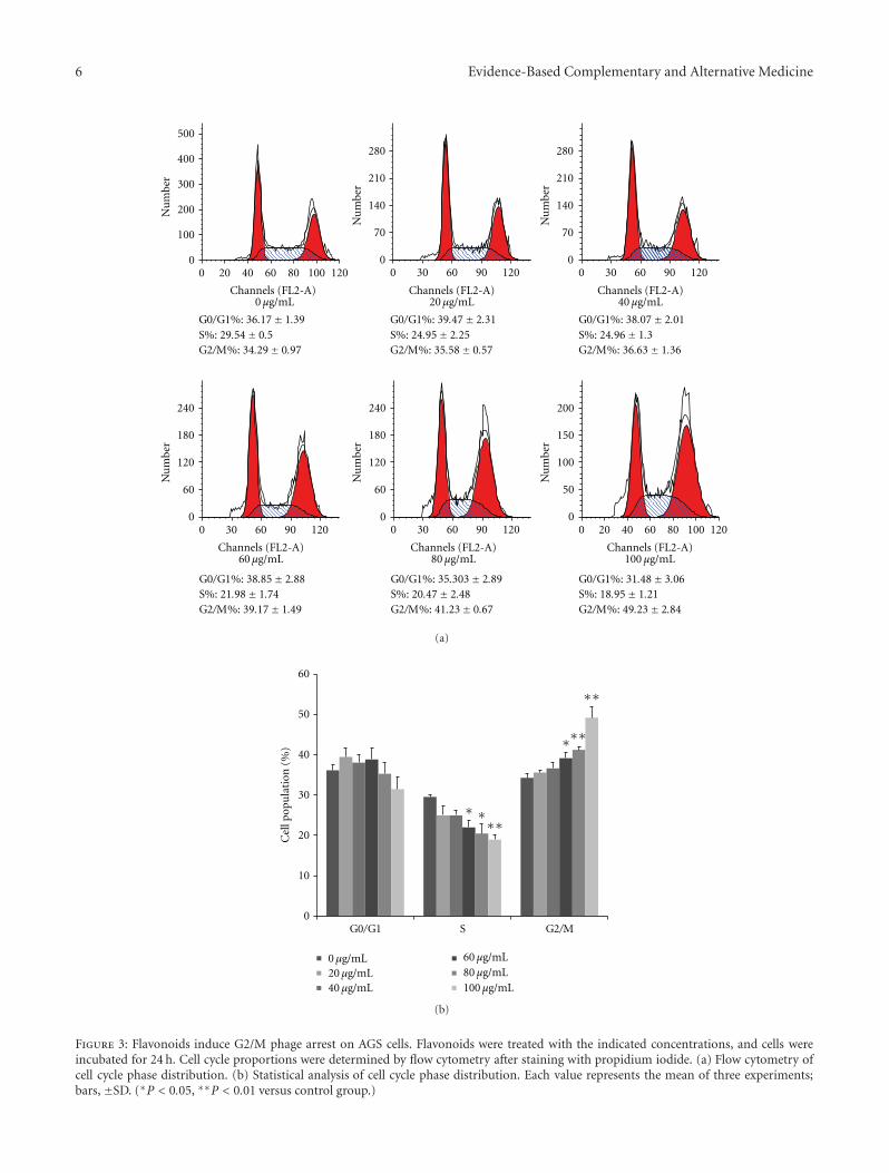
3.2. Flavonoids Induce G2/M Phase Cell Cycle Arrest. Asshown in Figure 4, flavonoids arrested the cell cycle in adose-dependent fashion at the G2/M phase. The S phase wassignificantly decreased in AGS cells treated with flavonoidconcentrations of 60, 80, and 100 μg/mL. Comparison ofcontrol cells and cells treated with 100 μg/mL of flavonoids
for 24 h revealed an increase in the G2/M phase from 34.29%to 49.23% (P < 0.01) and a decrease in the S phase from29.54% to 18.95% (P < 0.01). But, flavonoids did not influ-ence the G0/G1 phase. These data indicated that flavonoidscaused G2/M phase arrest in AGS cells.
3.3. Flavonoids Inhibit Expression of Cyclin B1, cdc 2, and cdc25c. Cyclin B1, cdc 2, and cdc 25c are important proteinsrelated to the G2/M phase. G2/M phase was controlled by acomplex formed cyclin B1 and cdc 2; the complex is regulatedby cdc 25c. Because flavonoids induce G2/M phase arrestin AGS cells, we evaluated the expression of proteins thatregulate the G2/M phase transition using Western blot. Asshown in Figure 4, the protein levels of cyclin B1, cdc 2,and cdc 25c decreased in a dose-dependent manner, withsignificant inhibition occurring at flavonoid concentrationsof 80 and 100 μg/mL. These data indicated that flavonoidsreduce the expression of cyclin B1, cdc 2, and cdc 25c.
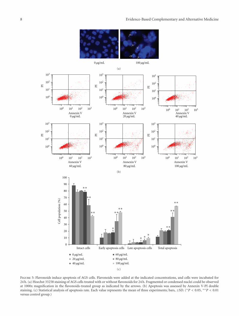
3.4. Flavonoids Induce Apoptosis. Apoptosis is another reasonof inhibited cell proliferation. The induction of apoptosisby flavonoid exposure was presently assessed using Hoechststaining and Annexin V-PI double staining. Treatment with100 μg/mL flavonoids for 24 h in AGS cells produced intenseHoechst-positive staining for condensed nuclei indicativeof apoptosis (Figure 5(a)). Also treatment with 0, 20, 40,60, 80, and 100 μg/mL of the flavonoids for 24 h progres-sively decreased the proportion of intact cells (Figure 5(b)),coincident with a dose-dependent increase in apoptosis.Comparison of control group cells and cells treated with100 μg/mL of the tested flavonoids for 24 h revealed adecrease in prevalence of intact cells from 88.26% to 42.42%(P < 0.01). In contrast, early apoptotic cells increased from10.29% to 48.07% (P < 0.01), and late apoptotic cellsincreased from 1.34% to 9.44% (P < 0.05). These dataindicated that flavonoids induced apoptosis in AGS cells.
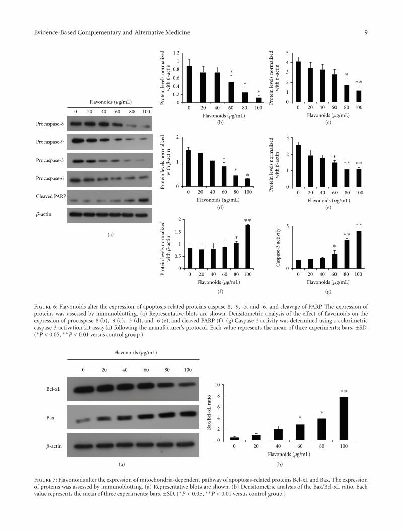
3.5. Flavonoids Activate Caspases and Shift the Bax/Bcl-xLRatio. The observation that the flavonoids induced apopto-sis of AGS cells prompted an examination of the expressionlevels of some apoptosis regulatory proteins, including Bcl-xL, Bax, procaspase-3, -6, -8, and -9, and cleaved PARP. Bcl-xL and Bax are major proteins of the mitochondria apoptosispathway. Western blot results showed that expression level ofBcl-xL was decreased in a dose-dependent manner, whereasthe level of Bax increased. The effect was the significantlyincreased Bax/Bcl-xL ratio in the presence of flavonoidconcentrations of 60, 80, and 100 μg/mL (Figure 7). Caspasesare also major proteins of apoptosis. Presently, the levelsof procaspase-3, -6,-8, and -9 were diminished in a dose-dependent manner (Figure 6(a)). The observation of dimin-ished procaspase-3 expression prompted an examination ofcaspase-3 -activity in AGS cells treated with flavonoids.Treatment with the flanovoids significantly increased cas-pase-3 activity at 60, 80, and 100 μg/mL (Figure 6(g)). Cleav-age of PARP is the main hallmark for caspase-dependentapoptosis. Cleaved PARP also increased in a dose-dependentmanner. The collective data favored the suggestion that

Evidence-Based Complementary and Alternative Medicine 5
0
20
40
60
80
100
120
0 10 50 100 150 200
Flavonoids (μg/mL)
Cel
lvia
bilit
y(%
ofco
ntr
ol)
(a)
0 μg/mL
60 μg/mL
40 μg/mL20 μg/mL
100 μg/mL80 μg/mL
(b)
Figure 2: Effect of flavonoids on the viability of AGS cells. (a) AGS cells were treated with various concentrations of the flavonoids for24 h. Cell viability was then determined by an MTT assay. Cell viability is represented as the percentage relative absorbance compared tothe controls. (b) Morphology of cells treated with or without flavonoids for 24 h and examined under light microscopy (×400). Each valuerepresents the mean of three experiments.
flavonoids induced apoptosis through a shift in Bax/Bcl-xLratio and the activation of caspases.
4. Discussion and Conclusions
Plant-derived herbal medicines have been used for a longtime in several Asian countries. Several studies reported anti-cancer activity in vitro using a natural herb extract [19, 20].In recent years, interest in flavonoids isolated from medicinalherbs such as Silybum marianum, Alpinia officinarum, andHypericum perforatum has been increasing because of theiranticancer effects on various cancer cells. Furthermore,natural flavonoids advantageously do not possess any sideeffects [21, 22]. In this study, the main flavonoids isolatedfrom Citrus aurantium L. were naringin, nobiletin, andhesperidin. These components confer superb anticancereffects that appear through cell cycle arrest and apoptosis invarious cancers [8, 9, 23] (Table 1).
In the present study, the anticancer effects of flavonoidsisolated from Citrus aurantium L. on AGS gastric carcinomacells were investigated. Viability and motility assays demon-strated that flavonoids suppressed proliferation of AGScells compared with untreated control cells. Regulating thecell cycle and apoptosis is crucial to retaining homeostasisbetween cell division and cell death [24].
Cell cycle aperiodicity is a classic feature of tumor cells[25]. In the cell cycle, the G2/M checkpoint represents apotential target for cancer therapy. When cells that containdamaged DNA enter mitosis, the G2/M checkpoint helpsprevent cell cycle progression in an effort to repair of DNAthat was damaged in late S or G2 phase [26]. Quercetinsuppresses the proliferation of human cervical cancer cellsby inducing G2/M phase cell cycle arrest [27]. Also, genistein
induces G2/M phase cell cycle arrest in nasopharyngeal car-cinoma cells [28]. Presently, AGS cells treated with variousconcentrations of flavonoids accumulated mostly in theG2/M phase in a dose-dependent manner, whereas the Sphase decreased. In terms of regulating the cell cycle, cdcsand cyclins play a most influential role. The complex formedbetween cyclin B and cdc 2 is crucial in the transition fromthe G2 to M phase [29]. At the end of the G2 phase, cdc2 is activated by dephosphorylation on Thr14/Tyr15 andcomplexes with cyclin B. Cdc 25c is very important in cdc2 activation. Phosphorylation of cdc 25c plays an importantpart in irreversible G2/M phase arrest [30]. Presently, thetested flavonoids downregulated cyclin B1, cdc 2, and cdc 25cprotein levels, which were required for the processing of theG2/M phase. Flavonoids such as luteolin and licochalconeA suppress the growth of AGS cells by blocking cell cycleprogression at the G2/M phases. In these phases, cyclin B,cdc 2, and cdc 25c play critical roles. Similar effects reportedthat luteolin and licochalcone A induce the downregulationof the production of cyclin B, cdc 2, and cdc 25c [31, 32]. Thecollective previous and present results support a powerfulinhibitory effect of cyclin B, cdc 2, and cdc 25c and providemolecular evidence of the antiproliferative effect directed atG2/M phase arrest. Decrease of cyclin B1, cdc 2, and cdc 25cexpression might be a molecular mechanism through whichflavonoids induce G2/M arrest (Figure 3).
Apoptosis is programmed cell death that can occur bya variety of internal or external stimuli, and these signalsare controlled by two distinct pathways. One is an extrinsicpathway (death receptor pathway), and the other is anintrinsic pathway (mitochondria pathway) [33]. Caspaseshave a major role in the extrinsic and intrinsic pathways ofapoptosis. In the regulation of apoptosis, caspases are divided
6 Evidence-Based Complementary and Alternative Medicine
G0/G1%: 36.17± 1.39S%: 29.54± 0.5G2/M%: 34.29± 0.97
0 μg/mL
0
100
200
300
400
500
Nu
mbe
r
Nu
mbe
r
Nu
mbe
rN
um
ber
Nu
mbe
r
Nu
mbe
r
0 20 40 60 80 100 120
0 20 40 60 80 100 120
Channels (FL2-A) Channels (FL2-A) Channels (FL2-A)
Channels (FL2-A) Channels (FL2-A) Channels (FL2-A)
0 30 60 90 120 0 30 60 90 120
0 30 60 90 1200 30 60 90 120
0
70
140
210
280
0
70
140
210
280
0
60
120
180
240
00
60
120
180
240
50
100
150
200
G0/G1%: 39.47± 2.31S%: 24.95± 2.25G2/M%: 35.58± 0.57
20 μg/mL
G0/G1%: 38.07± 2.01S%: 24.96± 1.3G2/M%: 36.63± 1.36
40 μg/mL
G0/G1%: 38.85± 2.88S%: 21.98± 1.74G2/M%: 39.17± 1.49
60 μg/mL
G0/G1%: 35.303± 2.89S%: 20.47± 2.48G2/M%: 41.23± 0.67
80 μg/mL
G0/G1%: 31.48± 3.06S%: 18.95± 1.21G2/M%: 49.23± 2.84
100 μg/mL
(a)
0 μg/mL20 μg/mL40 μg/mL
60 μg/mL80 μg/mL100 μg/mL
0
10
20
30
40
50
60
G0/G1 S G2/M
∗ ∗∗∗
∗∗
∗∗∗
Cel
lpop
ula
tion
(%)
(b)
Figure 3: Flavonoids induce G2/M phage arrest on AGS cells. Flavonoids were treated with the indicated concentrations, and cells wereincubated for 24 h. Cell cycle proportions were determined by flow cytometry after staining with propidium iodide. (a) Flow cytometry ofcell cycle phase distribution. (b) Statistical analysis of cell cycle phase distribution. Each value represents the mean of three experiments;bars, ±SD. (∗P < 0.05, ∗∗P < 0.01 versus control group.)

Evidence-Based Complementary and Alternative Medicine 7
0 20 40 60 80 100
Cyclin B1
Cdc 25c
Cdc 2
β- actin
Flavonoids (μg/mL)
0
0.5
1
1.5
0 20 40 60 80 100 0 20 40 60 80 100
0 20 40 60 80 100
Flavonoids (μg/mL)
Flavonoids (μg/mL)Flavonoids (μg/mL)
Pro
tein
leve
lsn
orm
aliz
edw
ithβ
-act
in
Pro
tein
leve
lsn
orm
aliz
edw
ithβ
-act
in
Pro
tein
leve
lsn
orm
aliz
edw
ithβ
-act
in
0
1
2
3
0
1
2
3
4
5
∗∗
∗∗∗ ∗∗
∗∗∗
(a)
(b) (c)
(d)
Figure 4: Flavonoids alter the expression of cell-cycle-related proteins cdc 2, cyclin B1, and cdc 25c. The expression of proteins was assessedby immunoblotting. (a) Representative blots are shown. Densitometric analysis of the effect of flavonoids on the expression of cyclin B1 (b),cdc 2 (c), and cdc 25c (d). Each value represents the mean of three experiments; bars, ±SD. (∗P < 0.05, ∗∗P < 0.01 versus control group.)
Table 1: The quantitative value and the retention time of identified flavonoids isolated from Citrus aurantium L.
No. Rt MS[M+H]+ Compound Quantity ± SD (mg/kg)
1 16.46 581 Naringin 299.2 ± 0.5
2 18.02 611 Hesperidin 210.3± 1.3
3 29.56 595 Poncirin 108.6 ± 1.1
4 41.67 373 Isosinensetin 30.4± 0.1
5 42.88 403 Hexamethoxyflavone 15.5± 0.05
6 43.40 373 Sinensetin 19.7 ± 0.1
7 44.60 403 Hexamethoxyflavone 23.6± 0.07
8 45.91 343 Tetramethyl-o-isoscutellarein 35.6 ± 0.1
9 46.52 403 Nobiletin 200.5± 0.1
10 47.50 433 Heptamethoxyflavone 168.9 ± 1.3
11 48.91 419 3-Hydroxynobiletin 28.7 ± 0.1
12 50.78 373 Tangeretin 81.5 ± 0.09
13 52.06 389 Hydroxypentamethoxyflavone 12.8± 0.04
14 52.72 403 Hexamethoxyflavone 7.0 ± 0.04
into two groups: initiator caspases (caspase-2, -8, -9, and-10) and effector caspases (caspase-3, -6, and -7). Initiatorcaspases are activated as a result of protein complex forma-tion, such as the death-inducing signaling complex (DISC)and apoptosome [24, 34]. The effector caspases-3 and -6are activated through the extrinsic pathway that involvescaspase-8 that involves caspase-9. Caspase-3 is a prominenteffector that cleaves the various elements related to apoptosis.Because of similarities in their substrate specificity, caspase-6 is able to partially assume the functional role of caspase-3[35, 36]. PARP, as a DNA repair enzyme, is activated by DNAbreaks and promotes the attachment of ADP-ribose poly-mers to various nuclear factors, expediting repair. PARP is
an important factor for cell viability. But, the cleavage ofPARP helps cellular disassembly and is a marker of cells un-dergoing apoptosis. During apoptosis, caspases induce PARPcleavage and inactivation [37]. Caspase signaling is initiatedand proceeds by proteolytic autocatalysis and by cleavage ofdownstream caspases and substrates such as PARP [24]. TheBax/Bcl-xL ratio is a key factor of mitochondria pathwayapoptosis. Proapoptotic proteins, such as Bax, translocate tothe mitochondrial membrane and make the mitochondrialouter membrane permeable. Antiapoptotic proteins, suchas Bcl-xL, maintain the mitochondrial membrane statusby the interactions between proapoptotic proteins [38].Therefore, the equilibrium between the expression levels of
8 Evidence-Based Complementary and Alternative Medicine
0 μg/mL 100 μg/mL
(a)
0 μg/mL 20 μg/mL 40 μg/mL
60 μg/mL 80 μg/mL 100 μg/mL
100
101
102
103
100
101
102
103
100
101
102
103
100
101
102
103
100
101
102
103
100
101
102
103
100 101 102 103 100 101 102 103100 101 102 103
100 101 102 103100 101 102 103100 101 102 103
PI
PI
PI
PIPI
PI
Annexin V Annexin V Annexin V
Annexin VAnnexin VAnnexin V
(b)
0 μg/mL
20 μg/mL
40 μg/mL
60 μg/mL
80 μg/mL
100 μg/mL
∗∗
∗∗
∗∗
∗∗
∗∗
∗∗
∗∗∗∗
∗∗∗
∗
Cel
lpop
ula
tion
(%)
0
10
20
30
40
50
60
70
80
90
100
Intact cells Early apoptosis cells Late apoptosis cells Total apoptosis
(c)
Figure 5: Flavonoids induce apoptosis of AGS cells. Flavonoids were added at the indicated concentrations, and cells were incubated for24 h. (a) Hoechst 33258 staining of AGS cells treated with or without flavonoids for 24 h. Fragmented or condensed nuclei could be observedat 1000x magnification in the flavonoids-treated group as indicated by the arrows. (b) Apoptosis was assessed by Annexin V-PI doublestaining. (c) Statistical analysis of apoptosis rate. Each value represents the mean of three experiments; bars, ±SD. (∗P < 0.05, ∗∗P < 0.01versus control group.)
Evidence-Based Complementary and Alternative Medicine 9
Procaspase-8
Procaspase-9
Procaspase-3
Procaspase-6
Cleaved PARP
β-actin
0 20 40 60 80 1000 20 40 60 80 100 0 20 40 60 80 100
0 20 40 60 80 1000 20 40 60 80 100
0 20 40 60 80 100 0 20 40 60 80 100
Flavonoids (μg/mL)
Flavonoids (μg/mL) Flavonoids (μg/mL)
Flavonoids (μg/mL)Flavonoids (μg/mL)
Flavonoids (μg/mL) Flavonoids (μg/mL)
Pro
tein
leve
lsn
orm
aliz
edw
ithβ
-act
in
Pro
tein
leve
lsn
orm
aliz
edw
ithβ
-act
inP
rote
inle
vels
nor
mal
ized
wit
hβ
-act
in
Pro
tein
leve
lsn
orm
aliz
edw
ithβ
-act
inP
rote
inle
vels
nor
mal
ized
wit
hβ
-act
in
0
0.5
1
0
0.2
0.4
0.6
0.8
1
1.2
1.5
2
0
0 0
1
1
2
2
3
5
0
1
2
3
4
5
∗
∗∗
∗
∗∗
∗∗∗
∗
∗∗
∗∗∗ ∗∗
∗
∗∗∗∗
(e)
(a)
(b) (c)
(d)
(f)
Cas
pase
-3ac
tivi
ty
(g)
Figure 6: Flavonoids alter the expression of apoptosis-related proteins caspase-8, -9, -3, and -6, and cleavage of PARP. The expression ofproteins was assessed by immunoblotting. (a) Representative blots are shown. Densitometric analysis of the effect of flavonoids on theexpression of procaspase-8 (b), -9 (c), -3 (d), and -6 (e), and cleaved PARP (f). (g) Caspase-3 activity was determined using a colorimetriccaspase-3 activation kit assay kit following the manufacturer’s protocol. Each value represents the mean of three experiments; bars, ±SD.(∗P < 0.05, ∗∗P < 0.01 versus control group.)
Bcl-xL
Bax
β-actin
20 40 60 80 100
Flavonoids (μg/mL)
0
(a)
20 40 60 80 100
Flavonoids (μg/mL)
00
2
4
6
8
10
∗∗
∗∗
Bax
/Bcl
-xL
rati
o
(b)
Figure 7: Flavonoids alter the expression of mitochondria-dependent pathway of apoptosis-related proteins Bcl-xL and Bax. The expressionof proteins was assessed by immunoblotting. (a) Representative blots are shown. (b) Densitometric analysis of the Bax/Bcl-xL ratio. Eachvalue represents the mean of three experiments; bars, ±SD. (∗P < 0.05, ∗∗P < 0.01 versus control group.)
10 Evidence-Based Complementary and Alternative Medicine
pro- and antiapoptotic proteins is critical for cell survival ordeath. Presently, apoptosis analysis showed that flavonoidspromote apoptosis. Especially, early apoptotic cells increased.Apoptosis induced by flavonoids was also consistent withthe observed changes in morphology. Flavonoids causeddownregulation of procaspase-3, -6, -8, and -9, which con-sequently increased the activity of caspase-3. The activationof caspase-3 induced PARP cleavage. In previous studies, theflanovoids genistein and eupatilin induced apoptosis in AGScells through the activation of caspase-3 and cleaved PARP[39, 40]. Caspase-3 is a very important key of apoptosis andhas been recognized as the crucial executioner caspase. Also,cleavage of PARP is a biochemical hallmark of apoptosis.Consistent with this, the flavonoids isolated from KoreaCitrus aurantium L. induced apoptosis by activating caspase-3 and cleaving PARP.
In conclusion, flavonoids can inhibit the proliferation ofAGS cells through both G2/M arrest via the modulation ofcell-cycle-related proteins, cyclin B1, cdc 2, and cdc 25c andtriggering of apoptosis via upregulation of caspase-3 activityand cleavage of PARP. Although additional investigations areneeded to prove the anticancer effects of flavonoids, thisstudy highlights the application potential of flavonoids iso-lated from Korea Citrus aurantium L. in gastric cancer chem-otherapy.
Acknowledgments
This work was supported by the National Research Founda-tion (NRF) of Korea Grant funded by the Korean govern-ment (MEST) (no. 2009-0084454) and the National R&DProgram for Cancer Control, Ministry for Health, Welfareand Family affairs, Republic of Korea (no. 0820050).
References
[1] I. M. Ghobrial, T. E. Witzig, and A. A. Adjei, “Targetingapoptosis pathways in cancer therapy,” Ca-A Cancer Journalfor Clinicians, vol. 55, no. 3, pp. 178–194, 2005.
[2] F. Deyhim, E. Lopez, J. Gonzalez, M. Garcia, and B. S. Patil,“Citrus juice modulates antioxidant enzymes and lipid profilesin orchidectomized rats,” Journal of Medicinal Food, vol. 9, no.3, pp. 422–426, 2006.
[3] S. S. Kim, J. S. Baik, T. H. Oh, W. J. Yoon, N. H. Lee, andC. G. Hyun, “Biological activities of Korean Citrus obovoidesand Citrus natsudaidai essential oils against acne-inducingbacteria,” Bioscience, Biotechnology and Biochemistry, vol. 72,no. 10, pp. 2507–2513, 2008.
[4] Y. Li, H. Fang, and W. Xu, “Recent advance in the researchof flavonoids as anticancer agents,” Mini-Reviews in MedicinalChemistry, vol. 7, no. 7, pp. 663–678, 2007.
[5] A. Quı́lez, B. Berenguer, G. Gilardoni et al., “Anti-secretory,anti-inflammatory and anti-Helicobacter pylori activities ofseveral fractions isolated from Piper carpunya Ruiz & Pav,”Journal of Ethnopharmacology, vol. 128, no. 3, pp. 583–589,2010.
[6] C. D. Harapu, A. Miron, M. Cuciureanu, and R. Cuciureanu,“Flavonoids—bioactive compounds in fruits juice,” RevistaMedico-Chirurgicala a Societatii de Medici si Naturalisti dinLasi, vol. 114, pp. 1209–1214, 2010.
[7] Y. C. Hsiao, Y. S. Hsieh, W. H. Kuo et al., “The tumor-growth inhibitory activity of flavanone and 2′-OH flavanonein vitro and in vivo through induction of cell cycle arrestand suppression of cyclins and CDKs,” Journal of BiomedicalScience, vol. 14, no. 1, pp. 107–119, 2007.
[8] G. Luo, X. Guan, and L. Zhou, “Apoptotic effect of citrus fruitextract nobiletin on lung cancer cell line A549 in vitro and invivo.,” Cancer Biology & Therapy, vol. 7, no. 6, pp. 966–973,2008.
[9] J. R. Patil, K. N. Chidambara Murthy, G. K. Jayaprakasha, M.B. Chetti, and B. S. Patil, “Bioactive compounds from mexicanlime (Citrus aurantifolia) juice induce apoptosis in humanpancreatic cells,” Journal of Agricultural and Food Chemistry,vol. 57, no. 22, pp. 10933–10942, 2009.
[10] K. D. Crew and A. I. Neugut, “Epidemiology of gastric cancer,”World Journal of Gastroenterology, vol. 12, no. 3, pp. 354–362,2006.
[11] S. A. Hundahl, J. L. Phillips, and H. R. Menck, “The nationalcancer data base report on poor survival of U.S. gastric carci-noma patients treated with gastrectomy: fifth edition Ameri-can joint committee on cancer staging, proximal disease, andthe “different disease” hypothesis,” Cancer, vol. 88, no. 4, pp.921–932, 2000.
[12] Z. Jin and W. S. El-Deiry, “Overview of cell death signalingpathways,” Cancer Biology and Therapy, vol. 4, no. 2, pp. 139–163, 2005.
[13] H. R. Stennicke and G. S. Salvesen, “Caspases—Controllingintracellular signals by protease zymogen activation,” Biochim-ica et Biophysica Acta, vol. 1477, no. 1-2, pp. 299–306, 2000.
[14] J. Lindsay, M. D. Esposti, and A. P. Gilmore, “Bcl-2 pro-teins and mitochondria-specificity in membrane targeting fordeath,” Biochimica et Biophysica Acta, vol. 1813, pp. 532–539,2010.
[15] P. Li, D. Nijhawan, I. Budihardjo et al., “Cytochrome c anddATP-dependent formation of Apaf-1/caspase-9 complex ini-tiates an apoptotic protease cascade,” Cell, vol. 91, no. 4, pp.479–489, 1997.
[16] T. M. Guadagno, M. Ohtsubo, J. M. Roberts, and R. K.Assoian, “A link between cyclin A expression and adhesion-dependent cell cycle progression,” Science, vol. 262, no. 5139,pp. 1572–1575, 1993.
[17] R. W. King, P. K. Jackson, and M. W. Kirschner, “Mitosis intransition,” Cell, vol. 79, no. 4, pp. 563–571, 1994.
[18] W. Krek and E. A. Nigg, “Differential phosphorylation of ver-tebrate p34(cdc2) kinase at the G1/S and G2/M transitions ofthe cell cycle: identification of major phosphorylation sites,”EMBO Journal, vol. 10, no. 2, pp. 305–316, 1991.
[19] K. I. Park, H. S. Park, S. R. Kang et al., “Korean Scutel-laria baicalensis water extract inhibits cell cycle G1/S tran-sition by suppressing cyclin D1 expression and matrix-metalloproteinase-2 activity in human lung cancer cells,” Jour-nal of Ethnopharmacology, vol. 133, pp. 634–641, 2010.
[20] Z. Xu, X. Chen, Q. Zhang, L. Chen, and Y. Wang, “Corydalisyanhusuo W.T. Wang extract inhibits MCF-7 cell proliferationby inducing cell cycle G2/M arrest,” American Journal ofChinese Medicine, vol. 39, no. 3, pp. 579–586, 2011.
[21] Y. J. Moon, X. Wang, and M. E. Morris, “Dietary flavonoids:effects on xenobiotic and carcinogen metabolism,” Toxicologyin Vitro, vol. 20, no. 2, pp. 187–210, 2006.
[22] R. Pierini, J. M. Gee, N. J. Belshaw, and I. T. Johnson, “Flavon-oids and intestinal cancers,” British Journal of Nutrition, vol.99, no. 1, pp. ES53–ES59, 2008.
Evidence-Based Complementary and Alternative Medicine 11
[23] K. N. Chidambara Murthy, G. K. Jayaprakasha, V. Kumar, K.S. Rathore, and B. S. Patil, “Citrus limonin and its gluco-side inhibit colon adenocarcinoma cell proliferation throughapoptosis,” Journal of Agricultural and Food Chemistry, vol. 59,no. 6, pp. 2314–2323, 2011.
[24] M. O. Hengartner, “The biochemistry of apoptosis,” Nature,vol. 407, no. 6805, pp. 770–776, 2000.
[25] C. Y. Jin, Y. H. Choi, D. O. Moon et al., “Induction of G2/Marrest and apoptosis in human gastric epithelial AGS cells byaqueous extract of Agaricus blazei,” Oncology Reports, vol. 16,no. 6, pp. 1349–1355, 2006.
[26] Y. Wang, P. Ji, J. Liu, R. R. Broaddus, F. Xue, and W. Zhang,“Centrosome-associated regulators of the G2/M checkpoint astargets for cancer therapy,” Molecular Cancer, vol. 8, article 8,2009.
[27] R. Vidya Priyadarsini, R. Senthil Murugan, S. Maitreyi, K.Ramalingam, D. Karunagaran, and S. Nagini, “The flavonoidquercetin induces cell cycle arrest and mitochondria-mediatedapoptosis in human cervical cancer (HeLa) cells throughp53 induction and NF-κB inhibition,” European Journal ofPharmacology, vol. 649, no. 1–3, pp. 84–91, 2010.
[28] H. Han, C. Zhong, X. Zhang et al., “Genistein induces growthinhibition and G2/M arrest in nasopharyngeal carcinomacells,” Nutrition and Cancer, vol. 62, no. 5, pp. 641–647, 2010.
[29] S. M. Lee, J. I. Kwon, Y. H. Choi, H. S. Eom, and G. Y. Chi,“Induction of G2/M arrest and apoptosis by water extractof Strychni Semen in human gastric carcinoma AGS cells,”Phytotherapy Research, vol. 22, no. 6, pp. 752–758, 2008.
[30] Y. S. Hwang, W.-Y. Chung, J. Kim, H.-J. Park, E.-C. Kim,and K.-K. Park, “Buddlejasaponin IV induces cell cycle arrestat G2/M phase and apoptosis in immortalized human oralkeratinocytes,” Phytotherapy Research, vol. 25, no. 10, pp.1503–1510, 2011.
[31] B. Wu, Q. Zhang, W. Shen, and J. Zhu, “Anti-proliferative andchemosensitizing effects of luteolin on human gastric cancerAGS cell line,” Molecular and Cellular Biochemistry, vol. 313,no. 1-2, pp. 125–132, 2008.
[32] X. Y. Xiao, M. Hao, X. Y. Yang et al., “Licochalcone A inhibitsgrowth of gastric cancer cells by arresting cell cycle progressionand inducing apoptosis,” Cancer Letters, vol. 302, pp. 69–75,2011.
[33] A. H. Wyllie, “Apoptosis (The 1992 Frank Rose MemorialLecture),” British Journal of Cancer, vol. 67, no. 2, pp. 205–208,1993.
[34] M. Olsson and B. Zhivotovsky, “Caspases and cancer,” CellDeath and Differentiation, vol. 18, pp. 1441–1449, 2011.
[35] G. M. Cohen, “Caspases: the executioners of apoptosis,”Biochemical Journal, vol. 326, no. 1, pp. 1–16, 1997.
[36] T. S. Zheng, S. Hunot, K. Kuida et al., “Deficiency in caspase-9 or caspase-3 induces compensatory caspase activation,”Nature Medicine, vol. 6, no. 11, pp. 1241–1247, 2000.
[37] Y. A. Lazebnik, S. H. Kaufmann, S. Desnoyers, G. G. Poirier,and W. C. Earnshaw, “Cleavage of poly(ADP-ribose) poly-merase by a proteinase with properties like ICE,” Nature, vol.371, no. 6495, pp. 346–347, 1994.
[38] H. W. Findley, L. Gu, A. M. Yeager, and M. Zhou, “Expressionand regulation of Bcl-2, Bcl-xl, and Bax correlate withp53 status and sensitivity to apoptosis in childhood acutelymphoblastic leukemia,” Blood, vol. 89, no. 8, pp. 2986–2993,1997.
[39] C. Y. Jin, C. Park, J. Cheong et al., “Genistein sensitizes TRAIL-resistant human gastric adenocarcinoma AGS cells throughactivation of caspase-3,” Cancer Letters, vol. 257, no. 1, pp. 56–64, 2007.
[40] M.-J. Kim, D.-H. Kim, H.-K. Na, T. Y. Oh, C.-Y. Shin, andY.-J. Surh, “Eupatilin, a pharmacologically active flavonederived from Artemisia plants, induces apoptosis in humangastric cancer (AGS) cells,” Journal of Environmental Pathology,Toxicology and Oncology, vol. 24, no. 4, pp. 261–269, 2005.

Submit your manuscripts athttp://www.hindawi.com
Stem CellsInternational
Hindawi Publishing Corporationhttp://www.hindawi.com Volume 2014
Hindawi Publishing Corporationhttp://www.hindawi.com Volume 2014
MEDIATORSINFLAMMATION
of
Hindawi Publishing Corporationhttp://www.hindawi.com Volume 2014
Behavioural Neurology
EndocrinologyInternational Journal of
Hindawi Publishing Corporationhttp://www.hindawi.com Volume 2014
Hindawi Publishing Corporationhttp://www.hindawi.com Volume 2014
Disease Markers
Hindawi Publishing Corporationhttp://www.hindawi.com Volume 2014
BioMed Research International
OncologyJournal of
Hindawi Publishing Corporationhttp://www.hindawi.com Volume 2014
Hindawi Publishing Corporationhttp://www.hindawi.com Volume 2014
Oxidative Medicine and Cellular Longevity
Hindawi Publishing Corporationhttp://www.hindawi.com Volume 2014
PPAR Research
The Scientific World JournalHindawi Publishing Corporation http://www.hindawi.com Volume 2014
Immunology ResearchHindawi Publishing Corporationhttp://www.hindawi.com Volume 2014
Journal of
ObesityJournal of
Hindawi Publishing Corporationhttp://www.hindawi.com Volume 2014
Hindawi Publishing Corporationhttp://www.hindawi.com Volume 2014
Computational and Mathematical Methods in Medicine
OphthalmologyJournal of
Hindawi Publishing Corporationhttp://www.hindawi.com Volume 2014
Diabetes ResearchJournal of
Hindawi Publishing Corporationhttp://www.hindawi.com Volume 2014
Hindawi Publishing Corporationhttp://www.hindawi.com Volume 2014
Research and TreatmentAIDS
Hindawi Publishing Corporationhttp://www.hindawi.com Volume 2014
Gastroenterology Research and Practice
Hindawi Publishing Corporationhttp://www.hindawi.com Volume 2014
Parkinson’s Disease
Evidence-Based Complementary and Alternative Medicine
Volume 2014Hindawi Publishing Corporationhttp://www.hindawi.com


![139 ' # '8& *#1 & 2Cepharanthine Stephania sp. p-gp [46] Crebanine Stephania venosa 13 g/ml Cyclin A, D & Caspases 3,9,8 & PARP and G 0/G 1 phase [48] Curine Chondrodendron platyphyllum](https://static.fdocuments.in/doc/165x107/60b799c100ac547f7a14e78e/139-8-1-2-cepharanthine-stephania-sp-p-gp-46-crebanine-stephania.jpg)


