Fetal movement counts in pregnancy: a comparison of · PDF fileFETAL MOVEMENT COUNTS IN...
61
FETAL MOVEMENT COUNTS IN PREGNANCY: A COMPARISON OF THE CARDIFF AND SADOVSKY METHODS by Diane Dwyer Heubusch A thesis submitted to the faculty of The University of Utah in partial fulfillment of the requirements for the degree of Master of Science Department of NurSing The University of Utah June 1987
Transcript of Fetal movement counts in pregnancy: a comparison of · PDF fileFETAL MOVEMENT COUNTS IN...
FETAL MOVEMENT COUNTS IN PREGNANCY:
A COMPARISON OF THE CARDIFF AND
SADOVSKY METHODS
by
Diane Dwyer Heubusch
A thesis submitted to the faculty of The University of Utah
in partial fulfillment of the requirements for the degree of
Master of Science
Department of NurSing
The University of Utah
June 1987

THE UNIVERSITY OF UTAH GRADUATE SCHOOL
SUPERVISORY COMMITTEE APPROVAL
of a thesis submitted by
This thesis has been read by each member of the following supervisory committee and by majority vote has been found to be satisfactory.
� J�, 19?2
o De borah �'�Jl8r
"----- ..)
THE UNIVERSITY OF UTAH GRADUATE SCHOOL
FINAL READING APPROVAL
To the Graduate Council of The University of Utah:
I have read the thesis of DisJ!le DW1er He.b.sch mUs
final form and have found that (1) its format. citations, and bibliographic style are
consistent and acceptable; (2) its illustrative materials including figures, tables, and charts are in place; and (3) the final manuscript is satisfactory to the Supervisory
Committee and is ready for submission to the Graduate School.
Karia T. Kirchhoff Member. Supervi�ory Committee
Approved for the Major Department
Lil:lda AIIIOS Chairman, Dean
Approved for the Graduate Council
B. Gale Dick Dean of The Graduate School

Copyright ~ Diane Dwyer Heubusch
All Rights Reserved
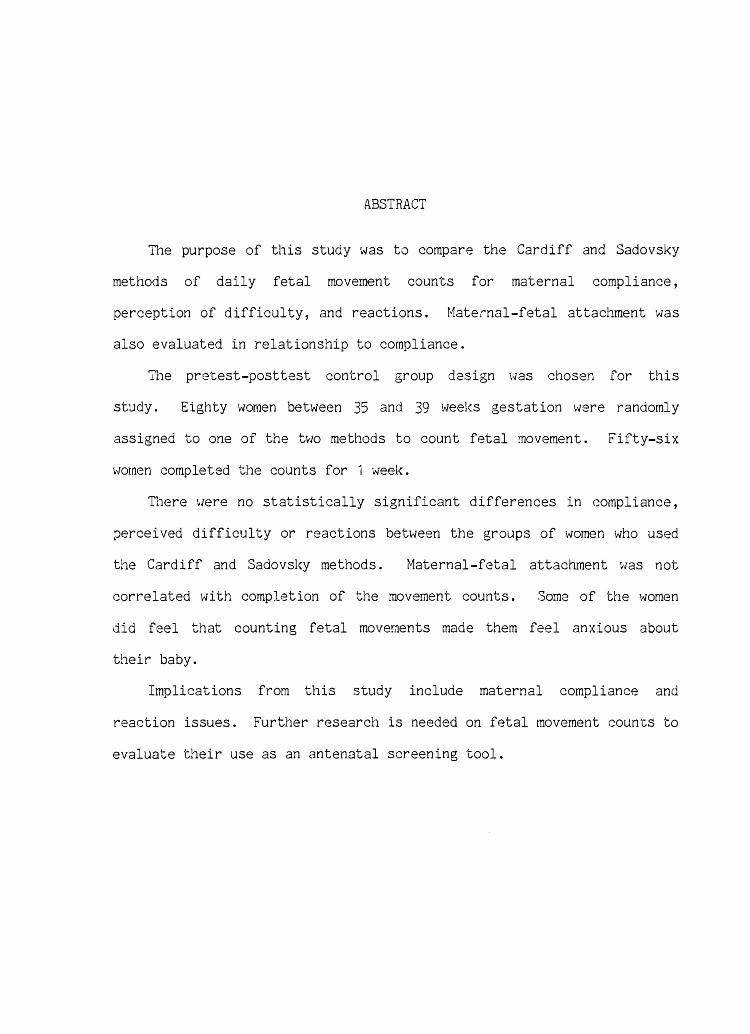
ABSTRACT
The purpose of this study was to compare the Cardiff and Sadovsky
methods of daily fetal movement counts for maternal compliance,
perception of difficulty, and reactions. flilaternal-fetal attachment was
also evaluated in relationship to compliance.
The pretest-posttest control group design was chosen for this
study. Eighty women between 35 and 39 weeks gestation ""rere randomly
assigned to one of the tHO methods to count fetal movement. Fifty-six
women completed the counts for 1 week.
There vlere no statistically significant differences in compliance,
percei ved difficulty or reactions betHeen the groups of women who used
the Cardiff and Sadovsky methods. Maternal-fetal attachment Has not
correlated with completion of the movement counts. Some of the women
did feel that counting fetal movements made them feel anxious about
their baby.
Implications from this study include maternal compliance and
reaction issues. Further research is needed on fetal movement counts to
evaluate their use as an antenatal screening tool.
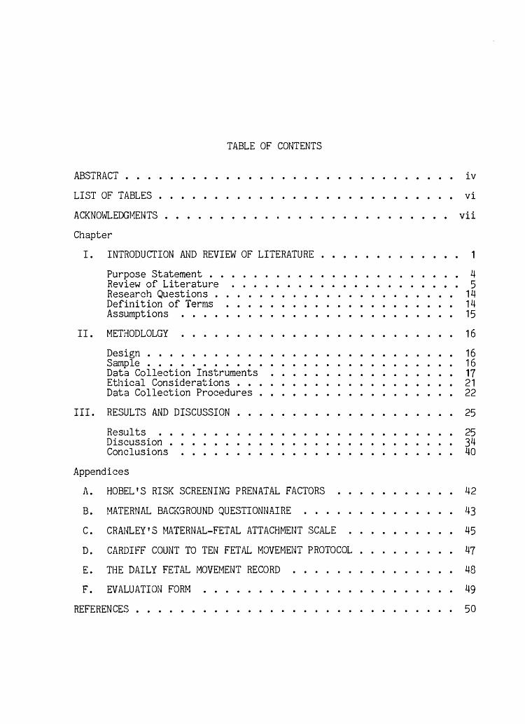
TABLE OF CONTENTS
ABSTRACT •.•
LIST OF TABLES
ACKNOWLEDGHENTS
Chapter
I. INTRODUCTION N~D REVIEW OF LITERATURE •••.
Purpose Statement . • • • . • . • • • • • • • • Review of Literature ••••••.••• Research Questions • Definition of Terms Assumptions
II. METHODLOLGY
Design • • • Sample . • • . • • • • • • Data Collection Instruments Ethical Considerations • • Data Collection Procedures .
III. RESULTS AND DISCUSSION .
Results • • • • Discussion • • Conclusions
Appendices
A. HOBEL'S RISK SCREENING PRENATAL FACTORS
B.
C.
D.
E.
MATERNAL BACKGROUND QUESTIONNAIRE • • • •
CRANLEY'S t1ATERNAL-FETAL ATTACHMENT SCALE
CARDIFF COUNT TO TEN FETAL MOVEMENT PROTOCOL •
THE DAILY FETAL MOVEMENT RECORD
F. EVALUATION FORB
REFERENCES • • • . • • •
iv
vi
vii
1
4 5
14 14 15
16
16 16 17 21 22
25
25 • • • • 34
40
42
• • • • • 43
• . . • 45
47
48
49
50
Table
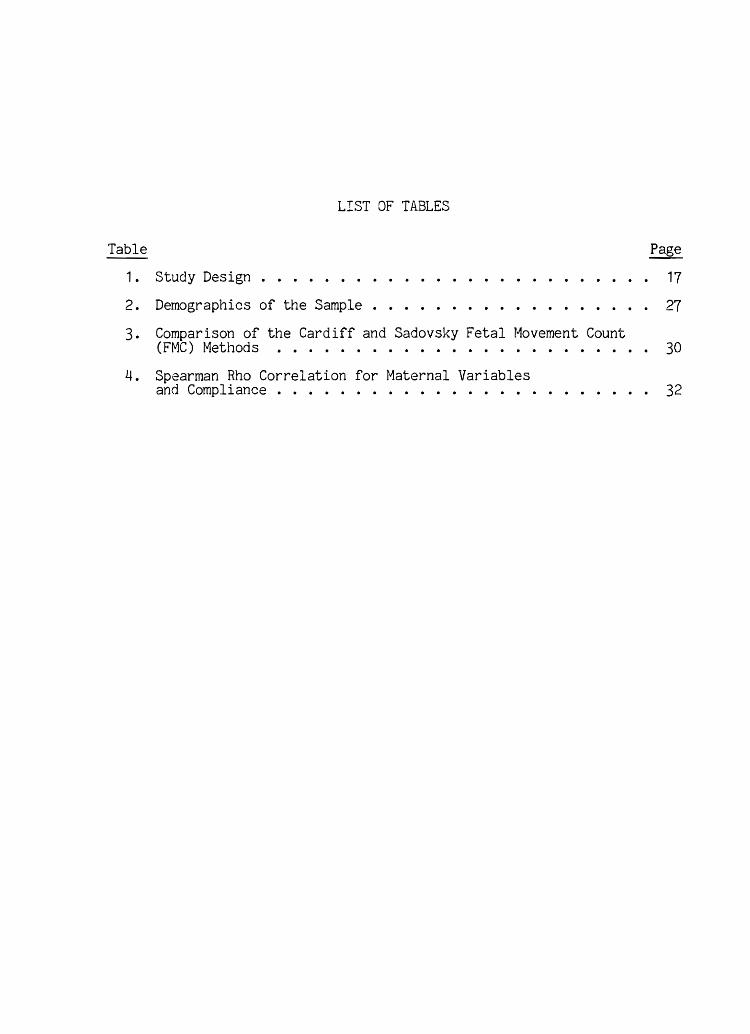
1. Study Design
LIST OF TABLES
Page
17
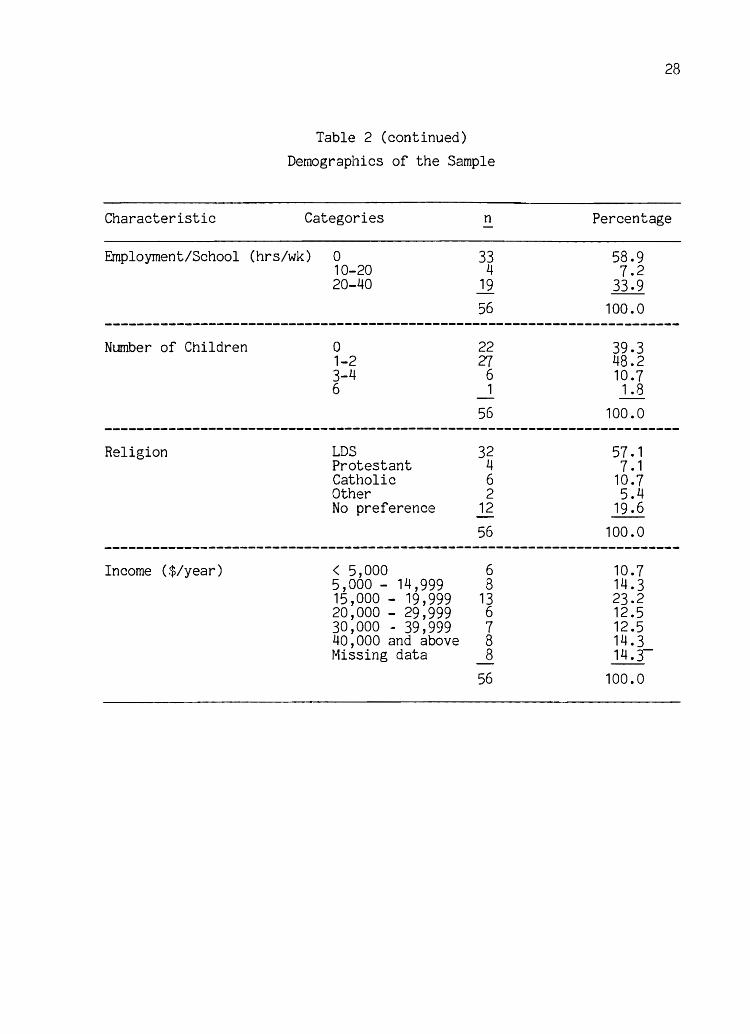
2. Demographics of the Sample • 27
3. Comparison of the Cardiff and Sadovsky Fetal Hovement Count (FMC) Method s • • • • • • • • • • • • • • • • • • • • • • • . 30
4. Spearman Rho Correlation for Maternal Variables and Compliance • • • • • • • • • • • • • • • • • 32
ACKNO~lLEDGi'4ENTS
I would like to acknowledge those individuals who made
contributions towards the completion of this thesis. Appreciation is
extended to my committee, Karin Kirchhoff, Deborah Greener, and Leslie
Carey. A special thanks goes to Karin for her guidance, support, and
flexibility during this study, and to Deborah and Leslie for their
commitment and clinical knowledge.
I would also like to extend my sincere appreciation and love to my
husband Joseph, and my sons, Jacob and Cole, for their patience and care
through the completion of this project.
CHAPTER I
INTRODUCTION AND REVIEW OF LITERATURE
Wi thin the field of midv/ifery and obstetrics, maternal perception
of fetal movement in utero has long been an indicator of fetal well
being. Early textbooks defined maternal recognition of fetal movement
as "quickening," a milestone for dating the pregnancy and a verification
of viability. A decrease or cessation of movement vias further
acknowledged as a sign of fetal death. In the past 20 years the
assessment of fetal movement has been refined as an accurate and valid
tool for fetal surveillance.
Health care providers have made achievements in decreasing poor
outcomes of pregnancy by using newer technological procedures for the
assessment of fetal well-being. The nonstress test, contraction stress
test, ultrasound scan, and biophysical profile are accepted screening
and diagnostic tools in antepartal care. Yet these procedures are
costly, invasive, and carry risks, mal<ing them impractical for use as
routine screening tools in low risk pregnancies.
Fetal death in utero occurs at the rate of 9.2 per 1,000 live
births annually in the United States (Pritchard, MacDonald & Gant, 1985,
p.2). A small proportion of these deaths can be attributed to
congeni tal anomalies, yet approximately one half of these are normal
fetuses that die in utero without a known cause. Although the number is
not large, each death is a tragedy and many such deaths occur in "low
2
risk" pregnancies. Perinatal mortality, stated as the number of
stillbirths and the number of neonatal deaths per 1,000 live births,
occurs at a rate of 17.7% in the United States. This high number is
primarily a result of preterm delivery, intrauterine growth retardation,
diabetes, and lethal congenital anomalies (Depp, 1982, p. 803). Early
identification of the first three entities and evaluation of the
fetoplacental unit can reduce poor outcomes. A compromised fetus
entering the stress of the intrapartal period may further be
compromised, resulting in permanent damage. A low cost, practical, and
valid screening tool is needed to identify at-risk fetuses needing
further evaluation. Daily fetal movement counts (FMCs) through maternal
assessment may serve this purpose.
Acti ve fetal movement has been associated with posi ti ve neonatal
outcomes, including high Apgar scores and cord pH values, adequate birth
weight (above 2500 grams), and improved neonatal mortality and morbidity
statistics (Ehrstrom, 1979; Leader, Bailee & Van Schaalkwyk, 1981;
Neldam, 1980; Pearson & Weaver, 1976; Sadovsky, Yaffe & Polishuk, 1974).
Decreased fetal movement has been correlated with deteriorating
placental function, intrauterine growth retardation, low Apgar scores,
meconium stained fluid, and intrapartal fetal distress (Leader et al.,
1981; Liston, Cohen, Mennuti & Gabbe, 1982; Mathews, 1975; Neldam, 1980;
Sadovsky & Yaffe, 1973; Sadovsky, 1981). Cessation of fetal movement
with an audible fetal heart rate is a signal that in utero demise will
occur within 12 to 48 hours (Sadovsky & Yaffe, 1973). Because of these
studies, formal methods of fetal movement counting have been developed.
3
Today wi thin clinical practice two main methods of fetal movement
counting are taught to women. In one method, known as the "Cardiff
Count to Ten" (Pearson, 1977) or the fixed number approach, the woman
counts fetal movements up to 10, and records the time period in which
she reached the number. The other method, developed by Sadovsky (1973),
is a fixed time approach in which the woman counts for three half hour
to hour periods per day and calculates the number of movements per 12
hours. If fetal movement falls below set criteria when either method is
used, the woman is to notify her health care provider for further
evaluation. Evaluation and management sequelae for decreased fetal
movement include a nonstress test for fetal reactivity and health. If a
nonreactive nonstress test is obtained, further management may include
observation, a contraction stress test or the induction of labor. These
approaches have become standards of practice within the field of
perinatology, particularly among high risk clientele. In low risk
pregnancies, fetal movement counts are more corrnnonly taught if the
pregnancy begins to show signs of pregnancy-induced hypertension,
intrauterine growth retardation, gestational diabetes, or becomes post
dates. These situations indicate decreasing placental function
requiring closer observation of fetal health.
The counting of fetal movement is a sound recommendation in
antepartum management, yet only one large prospect i ve controlled study
has been conducted on the effect of formal fetal movement counting on
the stillbirth rate (Neldam, 1980). This study involved 2,250 pregnant
women. Subjects in the treatment group were formally instructed in a
method to count fetal movement and documentation of movement was made in
4
the chart. The control group was not specifically instructed in
counting fetal movement but were always asked whether they were feeling
a decrease in fetal movement. Management for decreased fetal movement
was the same for both groups. A significant difference (E < 0 _ 0 1) in
the stillbirth rate was found between the two groups _ Among infants
weighing more than 1500 grams, eight intrauterine deaths occurred in the
control group and no deaths in the monitoring group (Neldam, 1980).
Maternal counting of fetal movement can be used as a screening tool
for low and high risk pregnancies. Few studies have demonstrated which
method of counting has increased compliance. Furthermore, few studies
have included maternal variables that may affect compliance. Maternal
fetal attachment may be related to the woman's compliance with antenatal
screening care such as fetal movement counts. As a screening tool,
fetal movement counts rely fully on the woman's participation. However,
even fewer studies have evaluated the mother's reaction to the
completion of fetal movement counts. In order to be clinically useful,
movement counts must be well accepted by the mother, easy to use, and
not cause unnecessary anxiety with her pregnancy_
Purpose Statement
The purpose of this study is to compare the Cardiff and Sadovsky
methods of daily fetal movement counts for maternal compliance.
Maternal-fetal attachment will be evaluated in relationship to
compliance. The overall maternal reaction to the use of daily fetal
movement counts in pregnancy will also be assessed.
5
Review of Literature
Fetal movement has been discussed from a variety of perspectives
within the literature. This review begins with a synopsis of studies of
the normal ranges and types of fetal movement during pregnancy, and of
the factors which may affect movement, i.e., gestational age, maternal
food intake, circadian cycles, and neurobehavioral states of the fetus.
Secondly, studies are reviewed in which fetal movement counts are
compared as a surveillance tool to other measures of well-being, such as
the nonstress test. Finally, the different formal methods of fetal
movement counts are described. A few studies are presented which have
tested the methods for compliance, accuracy, and maternal reaction.
Characteristics of Fetal Movement
Fetal movement in utero is known to begin mainly as reflexive
activity at 7 to 8 menstrual weeks. Movements increase in frequency and
range in a cephalocaudal progression as gestation advances. It is not
until the 16th to 21st weeks that fetal movement is perceptible to the
mother. At this time, the movements are infrequent, weak, and difficult
to distinguish from peristaltic movements of the intestines. Gradually,
the movements become stronger, more frequent, and discernible from
peristaltic movements (Coleman, 1981; Sadovsky & Polishuk, 1977).
The relationship between gestational age and the number of fetal
movements per day has been studied by several investigators with varying
results. Sadovsky (1981) studied 127 pregnant women with normal
outcomes, finding daily means of fetal movement increasing from 200 in
the 20th week to a maximum of 575 in the 32nd week, and gradually
decreasing to a mean of 282 at delivery. Ehrstrom (1980) also found
6
fetal activity increased from the 24th week to the 32nd week and then
slowly decreased until term. However, Patrick, Campbell, Carmichael,
Natale and Richardson (1982) found mean fetal movements per hour to be
similar at 30 -31, 34-35, and 36-39 weeks gestation.
Several investigators have found decreased fetal movements \-li th
postmaturity (Edwards & Edwards, 1970; Sadovsky & Polishuk, 1977; Wood,
Gilbert, & O'Connor, 1979). Physiologic changes of pregnancy have been
thought to be the rationale used to support these findings. As
gestation advances, space and fluid ratio decreases leaving less room
for fetal movement. Timor-Tritsch (1979), studying the types of fetal
movement, suggested that decreased movement at term correlated with a
maturing fetal neurological system and longer sleep cycles. Decreased
placental perfusion in a postmature placenta has also been suggested as
a causative factor.
The effects of maternal meals and serum glucose concentrations on
the frequency of fetal movement have also been studied. In a well
controlled study, serum glucose values were obtained every 30 minutes
for 2 hours after meals and compared to fetal movement counts. Maternal
plasma glucose concentrations increased significantly to peaks one hour
following meals, but there was no relationship between the number of
gross fetal body movements and glucose concentrations (Patrick et al.,
1982).
There appears to be a diurnal variation in fetal movement with
increased periods of fetal activity between the hours of 2000 to 0100
(Ehrstrom, 1979; Goodin & Lowe, 1974; Spellacy, Cruz, Gilman & Buhi,
1977; Wood, Walters & Trigg, 1977). Women may perceive an increase in
7
fetal activity during evening and bedtime hours when there is more focus
on the fetus. Another factor affecting the frequency of fetal movement
includes the cyclic asleep/active pattern of the fetus. Multiple
investigators have examined the behavioral states of the fetus and
identified cyclic activity. Periods of fetal rest with little or no
movement have been found to average 22 minutes, but may last up to 75
minutes (Patrick et al., 1982; Timor-Tritsch, Zador, Dieker, Hetz &
Rosen, 1978).
The patterns of fetal movement have been studied using real time
ultrasound, pressure transducers, and pleismographs. One classification
system divided the patterns of fetal movement based on duration, shape,
and amplitude of the wave form created by fetal movement on the
maternal abdomen. Four types of movement were identified: rolling or
stretching movements lasting greater than 3 seconds, simple trunk and
limb movements lasting less than 3 seconds, high frequency types (i.e.,
hiccoughs), and fetal respiratory movements (Timor-Tritsch, 1979).
Other classification systems used maternal perceptions and
categorized movements as weak, strong, or rolling movements. At 20
weeks, movements were predominantly weal( with the stronger and rolling
movements progressively increasing until 36 to 37 weeks. From 37 weeks
on, strong movements decreased with a slight increase in weaker
movements (Sadovsky, Laufer & Allen, 1979).
The overall daily range of perceived fetal movement varies among
mothers with frequencies from 4 to 1646 per day. Individual variation
is between 30 to 40 movements for the same fetus and may fluctuate
8
between 200 to 700 per day. The clinical value of the absolute number
of fetal movements has not been firmly established (Sadovsky, 1985).
Normal outcomes have been found with fetal movements as low as four
to ten per day, although this pattern occurs in less than 2.5% of the
population. A clear pattern of decreased fetal movement over time
coupled with a low daily movement pattern, or fetal movement in an
active fetus decreased to less than 10 movements in 12 hours indicates a
compromised fetus (Sadovsky, Laufer, & Allen, 1979; Sadovsky, 1985).
Relationship of Fetal Movement Counts
to the Nonstress Test
Movement of the fetus has been compared to changes in fetal heart
rate. Lee, Dilereto, and O'Lane (1975) demonstrated that acceleration
of the fetal heart in association with fetal movements is considered
normal and an expression of well-being. This correlation has led in
part to the development of the nonstress test (NST). A reactive
nonstress test is commonly defined as acceleration of the fetal heart 15
beats for fifteen seconds in association with fetal movement.
Yet studies on the relationship between nonreactive/reactive
nonstress tests to fetal movement records have not been fully
conclusive. Rayburn (1982) found a positive correlation between fetal
activity and the reactive nonstress test. In contrast, O'Leary and
Andrinopoulos (1981) found through statistical analysis that the NST
results and fetal movement count record were not related. Sadovsky and
Polishuk (1977) studied fetal heart rate in association with fetal
movement in 141 normal and pathologic pregnancies. They concluded that
no consistent change in fetal heart rate occurs with each fetal
9
movement. Fetal movement of less than 1 second duration may not be
accompanied by acceleration of the fetal heart, and this does not
indicate a compromised fetus. Yet fetal movement lasting longer than 3
seconds duration is associated 98% of the time with an increase of the
fetal heart rate. Clinically, fetal movement counts should be used with
other tests when evaluating a compromised fetus, rather than being the
sole indicator of fetal status (Gantes, Schy, Bartasius & Roberts,
1986).
Fetal movement counts have also been studied in relationship to
biochemical tests of placental function such as serum and urinary
estriol levels. Rayburn and McKean (1980) concluded that low serum
estriol values and a documented slow fetal activity pattern indicated
severe fetal distress and impending death. Yet these low estriol values
and positive fetal activity were within normal limits. Other
investigators have found that the counting of fetal movement was more
useful than the determination of serum estriol values in predicting
perinatal mortality and morbidity (Harper, Greenberg, Farahani, Glassman
& Kierney, 1981; Pearson & Weaver, 1976; Sadovsl<y & Polishuk, 1977).
The Daily Fetal Movement Record
Maternal perception of fetal movement is a reliable index of fetal
acti vi ty • Using ultrasound and electromechanical devices to record
fetal movement, women's subjective sensation was 82-90% accurate
(Hertogs, Roberts, Cooper, Griffin & Campbell, 1979; Rayburn & McKean,
1980; Sadovsky, Polishuk, Mahler & Mall<in, 1973; Sadovsky et al., 1979).
Maternal sensitivity to fetal movement has not been found to be affected
10
by maternal age, parity, obesity, duration of fetal movement, or the
presence of an anterior placenta. In contrast, maternal perception has
been found to be influenced by the mother's character, occupation, and
willingness to participate. Some mothers mistake fetal breathing
movements, fetal hiccoughs, and Braxton-Hicks contractions for fetal
movement.
The first formal method for fetal movement counts was developed by
Sadovsky in 1973. Sadovsky noted in clinical practice that a decrease
or cessation of fetal movement, but with an audible fetal heart rate,
was followed by fetal demise within 12 to 48 hours (Sadovsky & Yaffe,
1973). Using this information, he developed a protocol for fetal
movement counts which continues to be updated. The woman begins to
count fetal movement at 27 weeks gestation in a high risk pregnancy. The
woman counts movements for 30 to 60 minutes, three times a day in the
morning, afternoon, and evening. If she notes more than four movements
during each period, the woman can be reassured the fetal movement is
within a normal range. If less than four movements, the woman continues
to count for 1, 2 or more hours. When there are less than 10 fetal
movements in 6 hours, the woman is to notify the health care provider
for a nonstress test (Sadovsky, 1985). The sum total of the three 30 to
60 minute recordings are multiplied by 4 to give a 12 hour daily count.
In a low risk pregnancy, women begin to count fetal movements at 32 to
36 weeks gestation. These women are taught to pay attention to fetal
movements two to three times a day. If fetal movements are markedly
reduced, the women are treated as high risk patients.
11
Pearson developed a different method of recording fetal movements
known as the Cardiff Count to Ten chart. The count begins at 0900 and
continues in half hour blocks until the 10th movement is perceived.
This time is recorded on a chart. By 2100 if the movement count is less
than 10, the actual number of movements perceived is recorded. The
client is to notify her provider immediately if movements are not felt
for 1 day or less than 10 movements by 2100 hours (Pearson, 1911). This
method is the most widely used system in the United Kingdom.
Variations have been developed for both formal count systems.
Utilizing Sadovsky's method, count periods have been decreased to 10 to
20 minute periods (Spellacy et al., 1911; Wood, Walters & Trigg, 1911).
Problems encountered with decreasing the count time included increasing
false alarms. The fetal sleep cycle ranging from 20 to 10 minutes was
thought to overlap with the counting periods, thus producing the false
alarms. Neldam (1980) instructed the mothers to count once a week in
the morning, at noon, and in the evening, until the 32nd week.
Thereafter they were to count three times weekly, three times a day.
This method may increase compliance with fewer count periods, but at the
risk of decreasing the sensiti vi ty of the method. A third variation
developed by Grant and Hepburn ( 1984) combined the methods to form an
indi vidualized approach to the counting of fetal movement. The pilot
test of this new system was associated with fewer false alarms for
decreased movement. Although the individualized approach was developed
to decrease time per day women would need to count, it was found the
time women spent counting was similar in length to Sadovsky' s method,
yet less time than the Cardiff method.
12
Many studies have used Sadovsky' s, Pearson's, and variations of
these methods to investigate the validity of movement counts and the
relationship to neonatal outcomes. Within these studies problems have
been identified with the methods of counting and documenting.
Investigators have commented that women are unsure what constitutes a
movement, become confused about how to complete the chart, and have had
inadequate teaching and reinforcement to increase compliance. Very few
studies have focused on the differences in the counting methods, or
which method improves the validity and compliance within a population .
Clark ( 1985) found when using the Cardiff method in a low risk
population that compliance was inadequate: 27% of the subjects filled
out the chart less than 50% of the time, and 65% of those who
experienced a movement alarm signal did not notify their provider. In
contrast, a 98% compliance rate has been obtained by other investigators
(Draper, Field, Thomas & Hare, 1986; Fischer, Fullerton, & Trezise,
1981). In studies using variations of Sadovsky's method, the
investigators reported no specific compliance rates, but indicated that
this method provided improved reliability, accuracy, and completion with
the shorter count periods (Neldam, 1980; Rayburn, 1980; Valentin,
Lofgren, Marsal & Gulberg, 1984; Wood et ale 1977).
Research investigating the woman's perception and reactions to
doing fetal movement counts is minimal. Because the screening tool
places the responsibility of monitoring on the mother, it has been
questioned if this places unnecessary anxiety on the pregnancy (Mathews,
1973; McIlwaine, Howat, Dunn & McNaughton, 1980; Thompson & Wheeler,
1985). Draper et ale (1986) found that two thirds of the women in their
13
population were reassured by completing a fetal movement chart, and one
third were worried. The main reasons expressed for concern were an
inadequate knowledge about fetal movement.
Summary
In review, fetal movement in pregnancy has been studied thoroughly.
Fetal movement is first perceived by the mother between 16 and 20 weeks
gestation. From then on the fetus remains active, varying in the number
and types of movements per day. The active fetus is associated with
positive neonatal outcomes and documented decreased fetal movement with
intrapartal fetal distress or impending in utero demise. Because of
studies which correlate fetal movement to other antepartal tests of
well-being, the instruction of counting fetal movement has become an
accepted part of antenatal care.
Both the Cardiff and Sadovsky methods of counting fetal movements
have been developed and are sensi ti ve to identifying fetuses at risk
needing further evaluation. But in order for a screening tool to be
beneficial in clinical practice it needs to be applicable to clientele
of various ages, occupations, levels of education, and socioeconomic
status. The purpose of this study is to evaluate in a low risk
population \..rhich method of fetal movement counting encourages
compliance. The relationship of maternal-fetal attachment to compliance
with FMCs will also be investigated. Maternal reaction to completing
both methods will be examined with the purpose of further developing
fetal movement counts as a useful screening tool in pregnancy.
14
Research Questions
1. Are there differences in maternal compliance between the Cardiff
and Sadovsky methods of counting fetal movement?
2. Are there differences in perceived difficulty between the
Cardiff and Sadovsky methods of counting fetal movement?
3. Is maternal-fetal attachment related to compliance with the
counting of fetal movement?
4. What are maternal reactions to using formal methods of fetal
movement counting, and do they differ between the methods?
Definition of Terms
Compliance: a ratio of the number of days the movement count
record was completed as directed to the number of days possible to
complete the movement count record.
Maternal Perception of Difficulty: a composite score of questions
through 7 on the Fetal Movement Count Evaluation Form.
Maternal Reactions: the mother's attitudes, beliefs, and feelings
as expressed by responses to questions 8, 9, and 10 on the Fetal
Movement Count Evaluation Form.
Cardiff Count to Ten Method: a formal method of daily assessing
fetal activity and well-being by the mother, which includes the counting
of fetal movements beginning each morning until the 18th movement is
perceived.
Sadovsky Daily Fetal Movement Record: a formal method of daily
assessing fetal activity and well-being by the mother which involves the
counting of fetal movement for three half hour to hour periods in the
morning, noon, and evening.
15
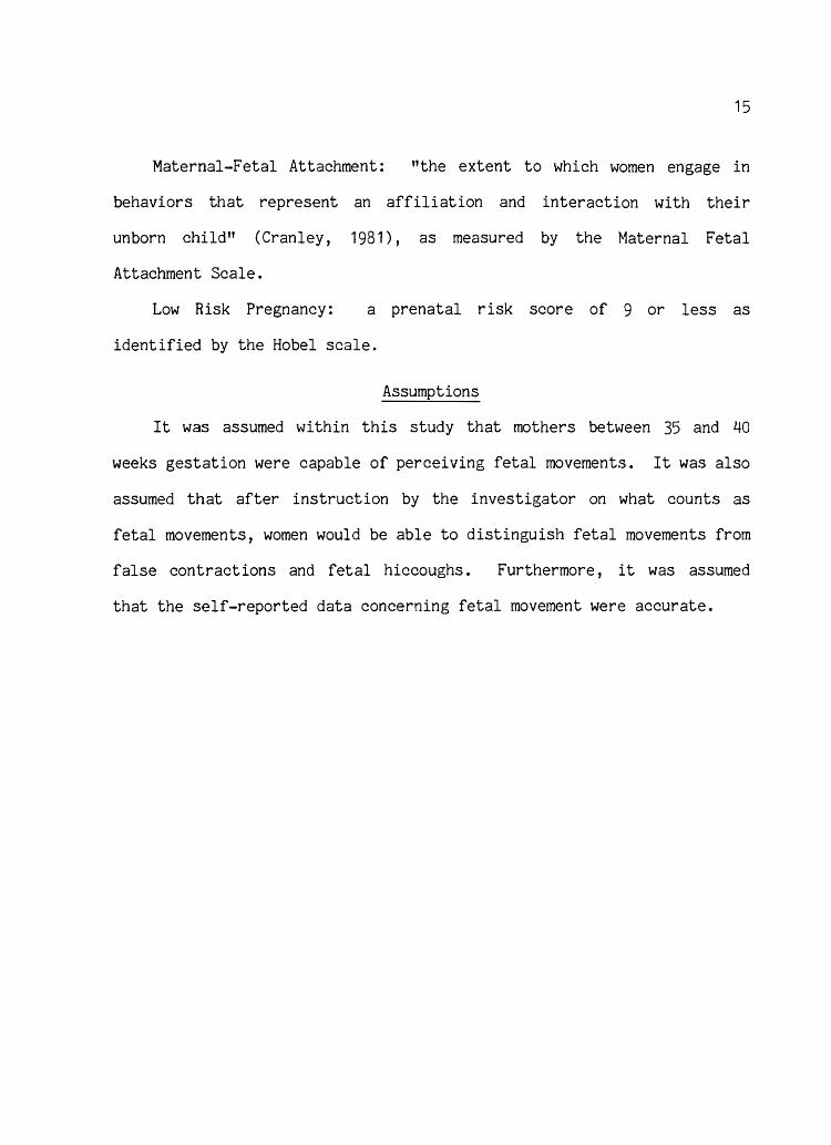
Maternal-Fetal Attachment: "the extent to which women engage in
behaviors that represent an affiliation and interaction with their
unborn child" (Cranley, 1981), as measured by the Maternal Fetal
Attachment Scale.
Low Risk Pregnancy: a prenatal risk score of 9 or less as
identified by the Hobel scale.
Assumptions
It was assumed within this study that mothers between 35 and 40
weeks gestation were capable of perceiving fetal movements. It was also
assumed that after instruction by the investigator on what counts as
fetal movements, women would be able to distinguish fetal movements from
false contractions and fetal hiccoughs. Furthermore, it was assumed
that the self-reported data concerning fetal movement were accurate.
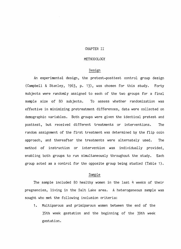
CHAPTER II
METHODOLOOY
Design
An experimental design, the pretest-posttest control group design
(Campbell & Stanley, 1963, p. 13), was chosen for this study. Forty
subjects were randomly assigned to each of the two groups for a final
sample size of 80 subjects. To assess whether randomization was
effective in minimizing pretreatment differences, data were collected on
demographic variables. Both groups were given the identical pretest and
posttest, but received different treatments or interventions. The
random assignment of the first treatment was determined by the flip coin
approach, and thereafter the treatments were alternately used. The
method of instruction or intervention was individually provided,
enabling both groups to run simultaneously throughout the study. Each
group acted as a control for the opposite group being studied (Table 1).
Sample
The sample included 80 healthy women in the last 4 weeks of their
pregnancies, living in the Salt Lake area. A heterogeneous sample was
sought who met the following inclusion criteria:
1. Multiparous and primiparous women between the end of the
35th week gestation and the beginning of the 39th week
gestation.
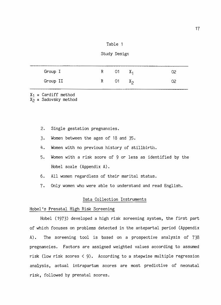
Group I
Group II
X1 = Cardiff method X2 = Sadovsky method
Table 1
Study Design
R
R
01
01
2. Single gestation pregnancies.
3. Women between the ages of 18 and 35.
4. Women with no previous history of stillbirth.
02
02
5. Women with a risk score of 9 or less as identified by the
Hobel scale (Appendix A).
6. All women regardless of their marital status.
7. Only women who were able to understand and read English.
Data Collection Instruments
Hobel's Prenatal High Risk Screening
17
Hobel (1973) developed a high risk screening system, the first part
of which focuses on problems detected in the antepartal period (Appendix
A) . The screening tool is based on a prospective analysis of 738
pregnancies. Factors are assigned weighted values according to assumed
risk (low risk scores < 9). According to a stepwise multiple regression
analysis, actual intrapartum scores are most predictive of neonatal
risk, followed by prenatal scores.
18
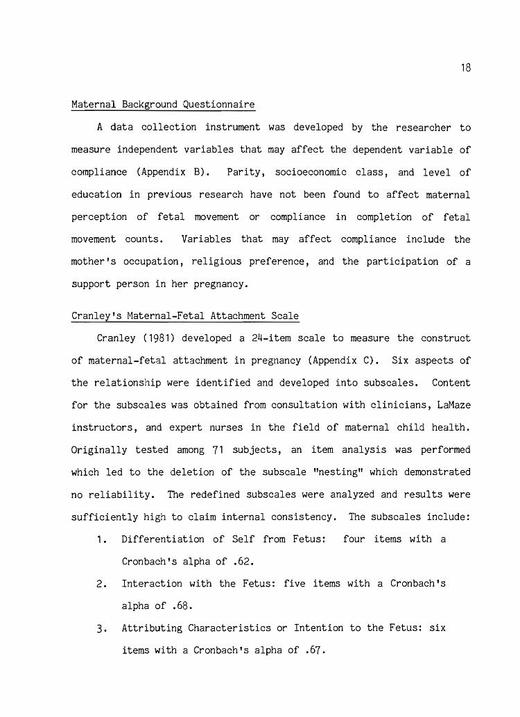
Maternal Background Questionnaire
A data collection instrument was developed by the researcher to
measure independent variables that may affect the dependent variable of
compliance (Appendix B). Parity, socioeconomic class, and level of
education in previous research have not been found to affect maternal
perception of fetal movement or compliance in completion of fetal
movement counts. Variables that may affect compliance include the
mother's occupation, religious preference, and the participation of a
support person in her pregnancy.
Cranley's Maternal-Fetal Attachment Scale
Cranley (1981) developed a 24-item scale to measure the construct
of maternal-fetal attachment in pregnancy (Appendix C). Six aspects of
the relationship were identified and developed into subscales. Content
for the subscales was obtained from consultation with clinicians, LaMaze
instructors, and expert nurses in the field of maternal child health.
Originally tested among 71 subjects, an item analysis was performed
which led to the deletion of the subscale "nesting" which demonstrated
no reliability. The redefined subscales were analyzed and results were
sufficiently high to claim internal consistency. The subscales include:
1 • Differentiation of Self from Fetus: four items with a
Cronbach's alpha of .62.
2. Interaction with the Fetus: five items with a Cronbach's
alpha of .68.
3. Attributing Characteristics or Intention to the Fetus: six
items with a Cronbach's alpha of .67.
4. Giving of Self: five items with a Cronbach's alpha of
.52.
5. Role Taking: four items with a Cronbach's alpha of .73.
19
Cranley found the coefficient of reliability for the total scale of
24 items is .85. Intercorrelations were performed among the subs cales
and the total scale to examine construct validity. All subscales are
positively associated with the total scale (r = .61 to .83). According
to Cranley, this provides some statistical evidence that the subscales
measure different aspects of the construct of maternal-fetal attachment
(Cranley, 1981).
Cardiff and Sadovsky Protocols
for Fetal Movement Counts
The formal methods of counting fetal movement used in this study
are known as the Cardiff Count to Ten Method (Pearson, 1977 & Appendix
D), and the Daily Fetal Movement Record (Sadovsky & Yaffe, 1973 &
Appendix E). Both methods have been used in clinical practice and by
researchers investigating the relationship of fetal activity/inactivity
to pregnancy outcomes. Through both retrospective and prospective
studies, active fetal movement has been correlated with positive
outcomes and considered a reliable measure of well-being. The
subjecti ve maternal perception of fetal movement has been compared to
the objective measures of fetal movement, i.e., ultrasound recordings.
The correlation of maternal perception to movements sensed by ultrasound
was found to be between 82% to 90%, affirming the reliability of fetal
movement counts performed by the mother.

20
Criterion-related validity for fetal movement counts has been
developed through the correlation of reactive nonstress tests to active
movement counts, and nonreactive tests at times consistent with
decreased fetal movement. In addition, an accepted fetal surveillance
tool in practice is the biophysical profile. This tool includes the
assessment of fetal body movements as one of the parameters for scoring
well-being.
Investigators have examined the predictive values of fetal movement
counts. In a prospective study by Leader et. al (1981), using four half
hour count periods per day similar to Sadovsky's protocol, a specificity
(the proportion of normal fetuses giving a normal result) of 91 % is
obtained. The sensitivity (the proportion of compromised fetuses giving
an abnormal result) is 86%. Overall the predictive value (the
likelihood of compromise if the result is abnormal) is only 46%, thought
to be low, secondary to the tendency of mothers to undercount fetal
movements.
Liston et al. (1982), in a prospective study using the Cardiff
protocol for counting fetal movements, found a sensitivity of 64% and
specificity of 98%. The predictive value is 55% vJith the level of
significance set at .001. In comparison to other biophysical and
biochemical tests of well-being with sensitivities of 21% to 76%,
specificities of 69% to 97%, and predictive values of 55% to 83%, the
counting of fetal movement can be considered a valid measure of
assessing fetal well-being.
The "movement alarm signal n (MAS, less than 10 fetal movements in
12 hours) used as the parameter to identify fetal distress has been
21
compared in both the Cardiff and Sadovsky protocols through a
retrospective study in 252 high risk pregnancies (Sadovsky, Ohel,
Havazeleth, Steinwell & Penchas, 1983). The alarm signal was compared
to other definitions of decreased fetal movement. The criteria for
establishing a poor outcome include intrauterine death after the 26th
weel< of pregnancy, intrauterine growth retardation (defined as fetal
weight less than the tenth percentile), 1-minute Apgar scores of 6 or
less, and meconium stained fluid. All definitions of decreased fetal
movement were found to be highly sensitive (the conditional probability
that the test would be positive once the disease state was in
existence), but only the MAS showed specificity (the conditional
probability that the test would be negative once the disease state did
not exist), thus decreasing the false negative prediction rate.
Overall, while the maternal perception of fetal movement does not detect
all fetal movements, has false positive alarm Signals, and is subject to
patient compliance, the tool is universally available, is inexpensive,
and may alert the clinician to possible fetal compromise.
Ethical Considerations
Prior to initiating this research, approval was obtained from the
Institutional Review Board for Research with Human Subjects at the
University of Utah. The study was also approved by the research review
board within the clinical institution where data were collected.
The participants for the study were voluntarily recruited from
prenatal clinics during the last 4 weeks of their pregnancies. The
investigator approached prospecti ve participants indi vidually and
22
verbally explained the purpose of the research study and its components
(Appendices A - F). The approximate time involved for participation was
also explained. Subjects were told that the benefits of the study for
themselves included learning a simple tool they could use daily to
assess fetal well-being. It was also explained that the study was
noninvasive and carried no identifiable risks, but might inconvenience
their time and cause them to increase their focus on the fetus in utero.
Confidentiality would be maintained through the use of code numbers on
all forms provided. If the individuals agreed to participate, a written
consent form was signed and a copy was given to them explaining the
study, their rights, and telephone numbers to use for any questions.
They were told they were free to withdraw from the study at any time,
and it would have no affect on their health care.
Data Collection Procedures
The study was conducted at Family Health Plan, a large health
maintenance organization serving the Salt Lake City area. The
organization's prenatal clinics, clients, and office hours were used for
data collection.
Ini tially prospective participants for the study were obtained by
the investigator, who reviewed the daily scheduled appointment sheet.
Return prenatal visits for women between the end of the 35th gestational
week and beginning of the 39th gestational week were identified using
estimated dates of confinement. The clinic charts were then reviewed by
the investigator to assess if the individual met the inclusion criteria
for the study. These included primiparous and multiparous women between
23
the ages of 18 and 35, English-speaking, and with a risk score of 9 or
less as identified by the Hobel Prenatal High Risk Screen (Appendix A).
Those identified as meeting the inclusion criteria were approached for
voluntary participation in the study_
When the client registered for her appointment, the investigator
approached the individual and verbally explained the study _ At this
time informed consent for participation vias obtained. If time was
available before the woman's visit with the health care provider, she
was asked to complete the Maternal Background Questionnaire (Appendix B)
and the Cranley's Maternal-Fetal Attachment Scale (Appendix C). If
unable to complete prior to being called for her visit, she was asked to
finish the instruments after her prenatal visit. When both instruments
were completed, the participant was individually instructed in a method
to formally count fetal movements by the investigator _ The first
subject in the study was assigned to the Cardiff Count to Ten Fetal
Movement Protocol (Appendix D), which was randomly determined by a flip
coin approach. The next participant was placed in the opposite
intervention, and instructed in Sadovsky's Daily Fetal Movement Record
Protocol (Appendix E). From then on subjects were alternately assigned
to one of the two methods to count fetal movement.
Instructions were provided using standard written protocols of the
method, which were also given to the client (Appendices D & E). The
instructions provided also contained the chart the woman was to complete
on a daily basis for 1 week. Any questions on how to count and graph
the count were answered by the investigator using a sample sheet of the
method in which they were instructed. The participant was then asked to
24
bring the movement chart back at her next prenatal visit in 1 week, or
to return the movement chart by mail using a provided envelope. She was
told that her time at the next visit would be approximately 15 minutes
to complete participation in the study.
Each participant was contacted at the woman's next prenatal visit
or through a telephone call. The completed charts (Appendix D or E)
"/ere collected at the visit, or the client was again asked to return
the chart by mail. Attrition information was also obtained at this
time. The participant was asked to complete the provided evaluation
form (Appendix F) to conclude her participation in the study. If
interested in continuing to use the method in which she was instructed,
she was provided with additional forms on which to complete her counts.
Questionnaires were checl<ed for completeness and data were coded
and analyzed at the University of Utah Computer Center using the
Statistical Package for Social Sciences (SPSS). Descriptive statistics
were used for analysis of the demographic variables. The variables of
parity, age, income, level of education, and maternal-fetal attachment
were assessed in relationship to the dependent variable of compliance
using Spearman's correlation coefficient. Compliance data for both the
Cardiff and Sadovsky methods were obtained on an interval scale and
analyzed using inferential statistics including t-tests and chi-square
analyses. Both descriptive and inferential statistics were used to
analyze the participants' evaluation of the fetal movement counts.
CHAPTER III
RESULTS AND DISCUSSION
Results
The data obtained from the questionnaires were computed at the
Uni versi ty of Utah Computer Center using the Statistical Package for
Social Sciences (SPSS) program. General descriptive statistics were
computed on the demographic variables and the questionnaire responses.
Independent !-tests were used to analyze differences between completers
and noncompleters of fetal movement counts (FMCs). To determine if
there were any significant differences in compliance or maternal
reactions between the Cardiff and Sadovsl<y methods, independent t-tests
and chi-square analyses were computed.
Fifty-six women in this study completed fetal movement counts for
one week, an overall compliance rate of 70%. Twenty-four women (30%)
did not complete the counts. Of these 24, 6 (7.5%) stated they withdrew
from the study because they were busy working. Four (5%) did not
complete the counts because they delivered 2 to 3 days after entering
the study. Fourteen (17.5%) stated they completed the counts but failed
to return the charts. Of the 24 women who did not complete the counts,
66% were assigned the Sadovsky method.
Mean scores were calculated on the total MFA scale. Mean scores
were also calculated on the subscales To identify any differences
between subjects that completed FMCs and those that did not, independent
t-tests were computed. There were no significant differences in
26
demographic variables or maternal-fetal attachment between the two
groups.
Demographics of the Sample
Fifty-six of the 80 women who entered the study completed it.
USing Hobel's risk screening tool for prenatal factors, 66% of the women
had no identifiable risk (Table 2). The remaining 34% carried a risk
score of 1 to 7, still wi thin the low risk pregnancy category. The
gestational week in pregnancy ranged from 35 to 39, (!i = 36.3, S.D. =
5.1). The ages of the women ranged from 18 to 35, (!i= 25.9, S.D. = 5.1). According to ethnic groups, 91% were Anglo-American, 2% Black
American, 5% Hispanic, and 2% Polynesian (Table 2). The background
questionnaire revealed that 53.6% completed high school and the
remainder had some college education. Fifty-nine percent were not
working at the time of the study and 41% were working part- to full
time. Thirty-nine percent of the women were primigravidas and 61%
reported having one to four children, 1 subject having six children.
Almost half (43.8%) of the women reported an annual income of
greater than $20, 000 per year. In a listing of religious preference,
57.1% were Latter Day Saints, \rJith the remaining listing Catholic,
Protestant, Jewish, and no religious preference. All the subjects
reported having a support person in their pregnancy.
To assess whether randomization was effecti ve in reducing
pretreatment differences between the two groups that completed the
Cardiff and Sadovsky FMCs, independent t-tests were computed on
Table 2
Demographics of the Sample
Characteristic Categories
Risk Score 0 1-5 6-7
.!i = 1.143, S.D. = 2.03, Range = 0-7
Gestational Week 35 36 37 38 39
~ = 36.37, S.D. = 1.214, Range = 35-39
Maternal Age
.!i = 25.94, S.D.
Ethnic Group
Education (in years)
18-20 21-25 26-30 31-35
= 5. 16, Range = 18-35
Caucasian Black Hispanic Polynesian
8-12 13-14 15-16
M = 12.64, S.D. = 1.6, Range = 8-20
n
37 17 2 -
56
17 16 10 11 2 -
56
10 20 14 12 -56
51 1 3 1 -
56
30 18 8
56
27
Percentage
66.0 30.4 3.6
100.0
30.4 28.6 17.9 19.6 3.6
100.0
17.9 35.8 25.6 21.5
100.0
91.0 1 .8 5.4 1.8
100.0
53.6 32.2 14.2
100.0
Characteristic
Employment/School
Number of Children
Religion
Income ($/year)
Table 2 (continued)
Demographics of the Sample
Categories
(hrs/wk) 0 10-20 20-40
o 1-2 3-4 6
LDS Protestant Catholic Other No preference
< 5,000 5,000 - 14,999 15,000 - 19,999 20,000 - 29,999 30,000 - 39,999 40,000 and above Missing data
n
33 4
19
56
22 27
6 1
56
32 4 6 2
12 -56
6 8
13 6 7 8 8
56
28
Percentage
58.9 7.2
33.9 100.0
39.3 48.2 10.7 1.8
100.0
57.1 7 • 1
10.7 5.4
19.6 100.0
10.7 14.3 23.2 12.5 12.5 14.3 14.3-
100.0
29
demographic and the attachment variables. No significant differences
were identified between the two groups (Table 3).
Instrument Reliability
Cronbach 's alpha coefficient of reliability was computed on the
total scale and subscales that were used to measure the construct of
maternal-fetal attachment. The Cronbach's alpha coefficient is .80 for
the total scale, whereas the subscales have coefficients ranging from
.43 to .66. The instrument women used to evaluate their difficulty in
participation with their assigned methods of fetal movement counts was
also analyzed for reliability. A Cronbach's alpha coefficient of .80
was obtained.
Research Question 1
Are there differences in maternal compliance between the Cardiff
and Sadovsky methods of counting fetal movement?
Eighty subjects were randomly assigned to the two groups to count
fetal movements, 40 in each group. Fifty-six subjects completed the
fetal movement counts; 31 (55.4%) completed the Cardiff method and 25
(44.6%) completed the Sadovsky method.
The number of possible days to count fetal movement ranged from 3
to 7 days (~= 6.17, S.D. = 1.47), with 78.6% counting between 6 and 7
days, and 75% counting the full 7 days. To identify any differences in
compliance between the two methods, independent t-tests on the
percentage of days completed were computed. There was no significant
difference between the two groups (Table 3).
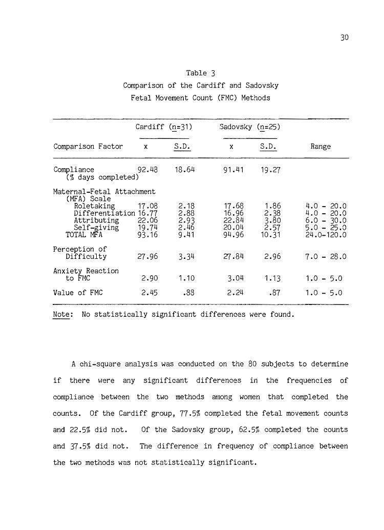
Table 3
Comparison of the Cardiff and Sadovsky
Fetal Movement Count (FMC) Methods
Cardiff (!!=31 ) Sadovsky (n=25)
Comparison Factor x S.D. x S.D.
Compliance 92.48 18.64 91 .41 19.27 (% days completed)
Maternal-Fetal Attachment (MFA) Scale
Roletaking 17.08 2.18 17.68 1.86 Differentiation 16.77 2.88 16.96 2.38 Attributing 22.06 2.93 22.84 3.80 Self-giving 19.74 2.46 20.04 2.57
TOTAL MFA 93.16 9.41 94.96 10.31
Perception of Difficulty 27.96 3.34 27 .84 2.96
Anxiety Reaction to FMC 2.90 1 .10 3.04 1 • 13
Value of FMC 2.45 .88 2.24 .87
Note: No statistically significant differences were found.
30
Range
4.0 - 20.0 4.0 - 20.0 6.0 - 30.0 5.0 - 25.0 24.0-120.0
7.0 - 28.0
1.0 - 5.0
1.0 - 5.0
A chi-square analysis was conducted on the 80 subjects to determine
if there vlere any significant differences in the frequencies of
compliance between the two methods among women that completed the
counts. Of the Cardiff group, 77.5% completed the fetal movement counts
and 22.5% did not. Of the Sadovsky group, 62.5% completed the counts
and 37.5% did not. The difference in frequency of compliance between
the two methods was not statistically significant.
31
To evaluate which factors may be related to compliance with fetal
movement counts, Spearman correlation coefficients were calculated for
the relationship between compliance and several of the independent
variables identified in the study. There were no significant
correlations between risk factors, gestational week, maternal age, level
of education, parity, income, and maternal-fetal attachment to
compliance with fetal movement counts (Table 4).
Research Question 2
Are there differences in perceived difficulty between the Cardiff
and Sadovsky methods of counting fetal movement?
Identical posttests or questionnaires were given to both groups of
women after completing the fetal movement counts. The evaluation
questionnaire was developed using an ordinal scale, higher scores
indicating a lesser degree of difficulty. Mean scores and frequencies
were calculated from the responses on the questionnaire. Overall 89.3%
felt comfortable completing fetal movement counts; the remaining 10.7%
were uncertain. Fifty-five women (98.3%) indicated they felt they knew
how to complete the charts, and only one individual (1.8%) felt
uncertain. Fifty-five women (98.3%) indicated they knew what counted as
a fetal
certain.
complete,
complete.
movement, and again one individual indicated she was not
Most women (82.2%) definitely did not find the method hard to
7.1% were uncertain, and 10.7% responded it was hard to
Also, most women (82.2%) did not feel fetal movement counts
took too much of their time, 10.7% were uncertain, and 7. 1 % indicated
that they did.
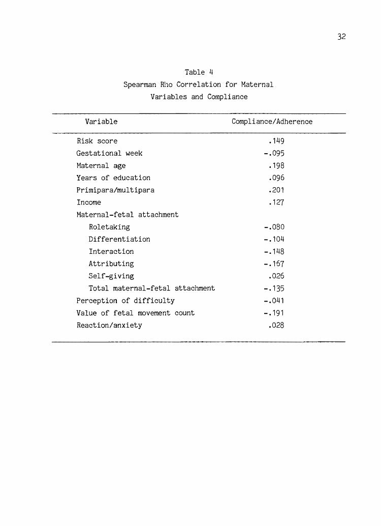
Table 4
Spearman Rho Correlation for Maternal
Variables and Compliance
Variable
Risk score
Gestational week
Maternal age
Years of education
Primipara/multipara
Income
Maternal-fetal attachment
Roletaking
Differentiation
Interaction
Attributing
Self-giving
Total maternal-fetal attachment
Perception of difficulty
Value of fetal movement count
Reaction/anxiety
Compliance/Adherence
• 149
-.095 .198
.096
.201
.127
-.080 -.104
-.148
-.167
.026
-.135
-.041
-. 191
.028
32
33
To identify differences in the perceived difficulty between the two
methods, independent t-tests were computed. No statistically
significant differences were found between the Cardiff and Sadovsky
methods on the maternal rating for difficulty.
Research Question 3
Is there a relationship between maternal- fetal attachment and
compliance with fetal movement counting?
Scores and frequencies were tabulated from the women's responses on
Cranley's Maternal Fetal Attachment Scale (MFAS). Most (85.4%)
indicated that they at times engaged in behaviors or attitudes
represented on the scale. Over three-fourths (78.2%) indicate they
frequently do, and 7.2% state that they do most of the time.
Within the group that did complete FMCs, maternal-fetal attachment
was evaluated in relationship to compliance. Using Spearman correlation
coefficients, it was found there was no significant relationship between
the subscales or total scale lneasuring attachment and compliance to FMCs
(Table 4).
Research Question 4
What are maternal reactions to using formal methods of fetal
movement counts and do they differ between methods?
Of the 56 women that completed the fetal movement counts, 23
(41.1$) indicated that counting fetal movement did not make them feel
anxious about their baby. Nine (16.1%) were uncertain, and 24 (42.8%)
felt counting fetal movement did cause them to feel anxious about their
baby. Thirty-three (59%) women felt fetal movement counting was a
34
worthwhile tool in pregnancy, 6 (10.7%) felt it was not, and 17 (30.3%)
were uncertain. To evaluate if there was a difference in maternal
reactions between the methods of fetal movement counts, independent ~
tests were calculated. There was no statistically significant
difference between the two methods.
Discussion
The purpose of this study was to evaluate, in a low risk
population, which method of fetal movement counting encourages
compliance. The sample in this study had several characteristics that
are comparable to the general population in the last trimester of
pregnancy. These include the subject's age, gestational week, level of
education, income, and parity. Characteristics that may not reflect the
general population include this sample's religious preference and ethnic
group. HO\-/ever, these two characteristics are representative of the
population residing in the metropolitan Salt Lake City area.
The scores obtained on the i~aternal-Fetal Attachment Scale are
higher in this sample compared to those obtained by Cranley ( 1981 ) .
Overall, this sample indicated that 98% of the women engaged in
behaviors or attitudes represented on the scale. Cranley found a lower
posi ti ve response of 78%. The responses on the questionnaire revolve
around developmental tasks in pregnancy, hence groups with similar
gestational periods should score similar frequencies of response. It
may be that this sample is skewed with higher maternal-fetal attachment
behaviors and attitudes.
35
Research Question 1
Are there differences in maternal compliance between the Cardiff
and Sadovsky methods of counting fetal movement?
In analyses of the two methods, Cardiff and Sadovsky, there were no
statistically significant differences found in compliance. Compliance
using the Cardiff method was 77.5% (~ = 31), and 62.5% (N = 25) with the
Sadovsky method. Studies which have evaluated the Cardiff method for
compliance have yielded a wide range in adherence. Draper et ale (1986)
states there was 98% compliance with the Cardiff method, but used only
verbal reports that the women completed the charts. Fischer et ale
(1981) found a 50% (N = 332) compliance rate; 19.8% (N = 128) stated
they completed the charts but failed to return them. Similar to this
investigator's study, there was no statistical significance in maternal
variables between the groups that complied Vlith the methods and those
that did not.
Clark et ale (1985) found that only 42% in her study completed
Cardiff charts daily as instructed, and 27% failed to keep the charts
50% of the days. The population in this study was different in that 72%
had family incomes less than $12,000 and also carried risk factors which
included a previous stillbirth or premature infant. The instruction on
using the charts were also given by a variety of health care providers.
Fewer studies have been conducted to investigate compliance using
the Sadovsky method to count fetal movement. Studies using this method
primarily use samples of women with high risk pregnancies, comparing
fetal movement to neonatal outcomes. Observations were made on
completed records obtained, and few studies indicated the initial number
36
of participants. Harper et ale (1981) using the Sadovsky method
collected 82.7% (!! = 91) completed charts. The sample was a mixture of
high and low risk pregnancies. Ehrstrom (1979) reported no specific
compliance rates, but mentioned 14% withdrew from his study due to lack
of time or other reasons. Thompson et ale (1985) found a 55% compliance
rate using daily 1-hour count periods.
Generally, the compliance rate in this study of collected charts
and percentage of completed days is similar if not higher than what
other studies have reported. The compliance rate is significant,
because the use of fetal movement counts as a screening tool places the
total responsibility for assessment on the mother. Although not
statistically significant, the Cardiff method had a higher compliance
rate.
In order for fetal movement counts to be most effective in
detecting decreased fetal movement, women must use them daily. In this
study, there were no maternal variables that correlated with compliance.
This can be interpreted that fetal movement counts can be universally
instructed with an equally expected adherence, regardless of the women's
age, socioeconomic status, parity, occupation, or level of education.
Most importantly, this study identified compliance with formal
methods of counting fetal movement. TI1e actual rate is lower than the
investigator anticipated. The percentage of days of adherence to
counting was high in those that completed, which may have been the
effect of testing on the women who chose to participate. Instruction
was provided separately from the women's actual prenatal care which may
have affected the women's sense of value in completing fetal movement
37
counts. However, the degree of adherence identified in this study and
previous research points to the issue that formal methods of counting
fetal movement are not readily accepted as valuable by the mother, who
carries the responsibility of completing them. In obstetrical and nurse
midwifery care, these results point to the need for reinforcement to
develop FMCs as a useful screening tool in antenatal care.
Research Question 2
Are there differences in difficulty between the Cardiff and
Sadovsky methods of counting fetal movement?
There were no significant differences in women's perception of
difficulty between the two methods. Most women (89.3%) felt comfortable
completing the charts. This result is similar to the 90% positive
response rate in ease of use found by Valentin et ale (1984). Fischer
et ale ( 1981 ) found that of the Cardiff charts returned, 98% were
completed accurately.
difficulty completing
In contrast, Clark et ale (1985) found women had
the chart when presented \V'i th different
si tuations, i. e., how to mark the chart if the count began later than 9
a.m. Clark's study was perhaps influenced by the five different levels
of careproviders that instructed 'vlOmen in the use of the fetal movement
counts. No specific results on women's perception of difficulty with
the Sadovsky method have been reported.
Women in this study had ample time to ask questions if uncertain
how to complete the chart. Verbal and written instruction was provided
by the investigator only, and was not combined with any other prenatal
38
instructions. This could have decreased the women's perception of
difficulty with use of the charts.
Other responses collected in this study indicate that women found
no particular difficulty with the completion of fetal movement counts.
Only 7.1% felt that counting fetal movement took too much of their time,
regardless of the method used. These data were from the women whom
completed the counts; it is likely the 30% noncompletion rate may have
been related to the time demands placed on women to complete FMCs.
The results from this study imply that the actual completion of the
FM count chart is not difficult given verbal and written instructions.
Both methods can be taught easily and both methods appear to be
relatively simple for the women to complete.
Research Question 3
Is there a relationship between maternal-fetal attachment and
compliance with fetal movement counts?
Maternal-fetal attachment (MFA) is a process which begins
physically and psychologically during pregnancy and not solely with the
birth of the infant. Quickening, or the recognition of fetal movement,
is significant in the developmental changes and tasks of pregnancy_ It
is at this point the mother begins to identify the fetus as a separate
entity from herself. By the last 4 weeks of pregnancy, healthy
psychological responses would include an already established degree of
maternal-fetal attachment.
This study utilized the instrument developed by Cranley (1981 )
which measures the construct of maternal-fetal attachment. MFA was
39
evaluated in relationship to compliance with fetal movement counts.
FMCs place full responsibility on the mother to be able to observe and
assess her infant's health. It would seem that women who are strongly
attached to their infants would be more likely to comply with FMCs. But
no significant differences were found between the women who completed or
did not complete fetal movement counts and their scores on the MFAs.
The degree of attachment does not appear to reflect the woman's
participation in antenatal screening tools such as FMCs.
Similar to Cranley's (1981) study, no significant relationship was
found between MFA and other maternal variables such as age, parity, and
socioeconomic class. The data from this study support the concept that
maternal-fetal attachment is an ongoing developmental process and is not
affected by demographic variables.
Research Question 4
What are maternal reactions to using formal methods of fetal
movement counts and do they differ between methods?
Women in this study had mixed responses to the completion of
formally counting fetal movements. Less than half (41.1%) of the women
felt that counting fetal movements did not cause them to feel anxious
about their baby; 42.8% felt it did cause them to feel anxious, and
16.1% were uncertain. There were no statistically significant
differences between the Cardiff and Sadovsky methods. Draper et al.
(1986) found 55% were reassured with fetal movement counts, 23% were
worried, and 17% were neither reassured nor worried. Other
investigators have found women generally comfortable with the
40
completion of fetal movement counts, but use no specific data to support
these findings (Ehrstrom et al., 1979; Rayburn, 1980).
Several authors have questioned the amount of anxiety that may be
placed on the mother with formally counting fetal movements (Hathews,
1973; McIlwaine, Howat, Dunn & McNaughton, 1980; Thompson & Wheeler,
1985). The data from this study and other investigators suggest that
FMCs increase focus on the fetus, and may cause a significant part of
the population to have anxious feelings regarding fetal health. It is
possible that this same percentage of the population may feel anxious
regarding any other fetal tests of well-being, including glucose
tolerance tests, ultrasounds, and nonstress tests. The difference with
fetal movement counts is that it requires the mother to assess fetal
well-being daily. Perhaps the anxiety women report is solely related to
her involvement and responsibility with observing fetal health. These
resul ts implicate the need for frequent discussion with the woman
regarding her participation in antenatal screening tools such as FMCs.
Conclusions
Normal fetal movements are an indicator of fetal well-being,
whereas reduced fetal activity may precede fetal death. Women are
sensitive to 80% to 90% of fetal movements that can be detected by
ultrasound and electronic devices. Several protocols have been
developed and are used in antepartal management for maternal counting of
fetal movements. The two main protocols on Vlhich many studies have been
based are the Cardiff Count to Ten Method and Sadovsky' s Daily Fetal
Hovement Record. Both of these protocols have tested sensi ti vi ty and
41
specificity to be included as a valuable screening tool in pathological
and normal pregnancies. There is evidence that maternal monitoring of
fetal movements can lead to a lowered stillbirth rate.
The difference with fetal movement counts is the fact that the
assessment of fetal health is placed on the mother. The use of daily
fetal movement counts has also not been a part of routine prenatal care.
Compliance rates that have been found in past research and in this study
may reflect these two concepts. Women may be more likely to adhere to
counts if there were instruction and reinforcement at prenatal visits
rather than random questioning regarding fetal movement. What has not
been adequately investigated is the different methods women can use to
count fetal movement and which method they might prefer.
It is also interesting that researchers investigating women's
reactions to fetal movement counts find a significant portion of the
population do have anxious feelings regarding formally counting fetal
movement. The amount of anxiety and negative effects need to be more
specifically identified to evaluate the value of formal fetal movement
counts in normal pregnancies. Further research is needed in these areas
on the counting of fetal movement. Counts will only be clinically
useful if women adhere to them with a certain degree of comfort.
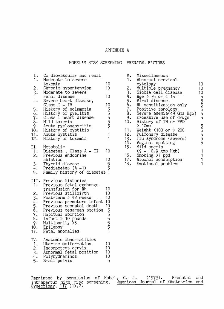
APPENDIX A
HOBEL'S RISK SCREENING PRENAT~4L FACTORS
I. Cardiovascular and renal V. Miscellaneous 1 • Moderate to severe 1 • Abnormal cervical
toxemia 10 cytology 10 2. Chronic hypertension 10 2. Multiple pregnancy 10 3. Moderate to severe 3. Sickle cell disease 10
renal disease 10 4. Age > 35 or < 15 5 4. Severe heart disease, 5. Viral disease 5
Class I - IV 10 6. Rh sensitization only 5 5. History of eclampsia 5 7. Positive serology 5 6. History of pyelitis 5 8. Severe anemia«9 Gms Hgb) 5 7. Class I heart disease 5 9. Excessive use of drugs 5 8. Mild toxemia 5 10. History of TB or PPD 9. Acute pyelonephritis 5 > 10rnrn 5
10. History of cystitis 1 11 • Weight <100 or > 200 5 11 • Acute cystitis 1 12. Pulmonary disease 5 12. History of toxemia 1 13. Flu syndrome (severe) 5
14. Vaginal spotting 5 II. Metabolic 15. Mild anemia 1 • Diabetes • Class A - II 10 (9 - 10.9 gms Hgb) 1 2. Previous endocrine 16. Smoking >1 ppd 1
ablation 10 17. Alcohol consumption 1 3. Thyroid disease 5 18. Emotional problem 1 4. Prediabetes (A -1) 5 5. Family history of diabetes
III. Previous histories 1 • Previous fetal exchange
transfusion for Rh 10 2. Previous stillbirth 10 3. Post-term> 42 \.-leeks 10 4. Previous premature infant 10 5. Previous neonatal death 10 6. Previous cesarean section 5 7. Habitual abortion 5 8. Infant > 10 pounds 5 9. i"1ultiparity >5 5
10. Epilepsy 5 11 • Fetal anomalies 1
IV. Anatomic abnormalities 1 • Uterine malformation 10 2. Incompetent cervix 10 3. Abnormal fetal position 10 4. Polyhydraminos 10 5. Small pelvis 5
Reprinted by permission of Hobel, C. J. (1973). Prenatal and intrapartum hi~h risk screening. American Journal of Obstetrics and Gynecology, III (1),2.

APPENDIX B
MATERNAL BACKGROUND QUESTIONNAIRE
week
Directions: Please complete the following.
1.In what week of your pregnancy are you?
2. What is your due date?
-----
3.
4.
------What is your age? _____ ---..:years
What is your ethnic group?
Anglo American
Black American
Asian
Hispanic
Polynesian_
Other(please specify) -------5. What is your highest level of education? (please circle the number
corresponding to completed years of education)
Elementary 1 2 3 4 5 6
Junior high/ High School 7 8 9 10 11 12
College 13 14 15 16
Postgraduate 17 18 19 20 21 or more
6. What is your primary occupation?
7. How many hours per week do you work and/or go to school?
o hours per week
o to 10 hours per week
10 to 20 hours per week
20 to 40 hours per week
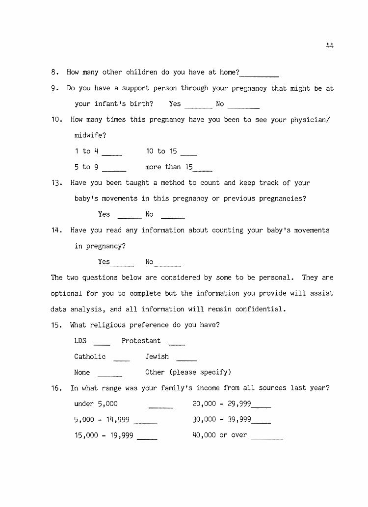
44
8. How many other children do you have at home? -----9. Do you have a support person through your pregnancy that might be at
your infant's birth? Yes No ----10. How many times this pregnancy have you been to see your physician/
midwife?
1 to 4 --5 to 9 ---
10 to 15
more than 1
13. Have you been taught a method to count and keep track of your
baby's movements in this pregnancy or previous pregnancies?
Yes No
14. Have you read any information about counting your baby's movements
in pregnancy?
Yes No ---The two questions below are considered by some to be personal. They are
optional for you to complete but the information you provide will assist
data analysis, and all information will remain confidential.
15. What religious preference do you have?
LDS Protestant
Catholic
None
Jewish
Other (please specify)
16. In what range was your family's income from all sources last year?
under 5,000 20,000 - 29,999 --5,000 - 14,999 30,000 - 39,999 __
15,000 - 19,999 -- 40,000 or over ----

APPENDIX C
CRANLEY'S ~ffiTERNAL FETAL ATTACHMENT SCALE
Directions: Please place a check in one of the colur~s for each
statement on the left to describe your feelings about this pregnancy.
1 • I talk to my unborn baby.
2. I feel all the trouble of being pregnant is worth it
3. I enjoy watching my turmny jiggle as the baby kicks inside.
4. I picture myself feeding the baby.
5. I'm really looking forward to seeing what the baby looks like.
6. I wonder if the baby feels cramped in there.
7. I refer to my baby by a nickname.
8. I imagine myself taking care of the baby.
9. I can almost guess Vlhat my baby t s personality \ilill be from the 'vlay he/she moves around.
Definitely Yes
UncerYes tain No
--
Definitely
No
--
Reprinted by permission of Cranley, M. (1981). Development of a tool for the measurement of maternal attachment during pregnancy. Nursing Research, 30 (5),282.

46
APPENDIX D
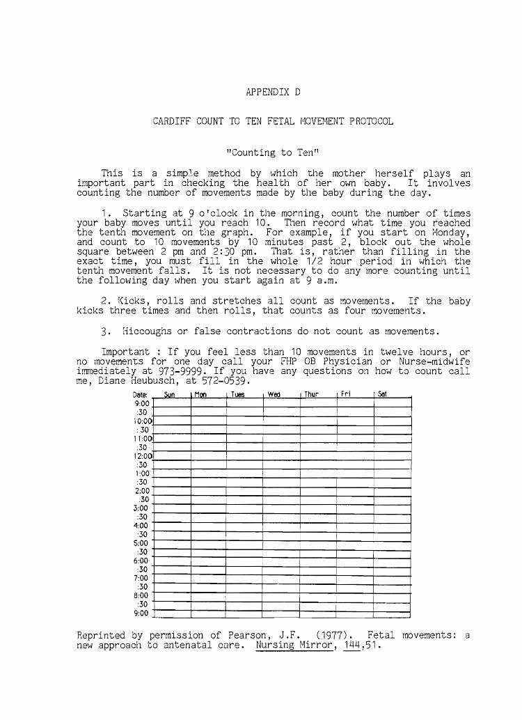
CARDIFF COUNT TO TEN FETAL MOVEMENT PROTOCOL
"Counting to Ten"
This is a simp~.e method by ~Jhich the mother herself plays an important part in checking the health of her own baby. It involves counting the number of movements made by the baby during the day.
1. Starting at 9 o'clock in the morning, count the number of times your baby moves until you reach 10. Then record what time you reached the tenth movement on the graph. For example, if you start on i"1onday, and count to 1 0 movements by 1 0 minutes past 2, block out the whole square between 2 pm and 2: 30 pm. That is, rather than filling in the exact time, you must fill in the whole 1 / 2 hour period in which the tenth movement falls. It is not necessary to do any more counting until the following day when you start again at 9 a.m.
2. Kicks, rolls and stretches all count as movements. If the baby kicks three times and then rolls, that counts as four movements.
3. Hiccoughs or false contractions do not count as movements.
Important : If you feel less than 10 movements in twelve hours, or no movements for one day call your FHP 08 Physician or Nurse-midwife immediately at 973-9999. If you have any questions on how to count call me, Diane Heubusch, at 572-0539.
Date: 9:00 :30
10:00 : 30
11:00 :30
12:00 :30 1:00 :30 2:00 :30
3:00 :30
4:00 :30
5:00 :30
6:00 :30
7:00 :30
8:00 :30
9:00
Sun Mon Tues Woo Thur 1 Fri Sot
I
I
I
I
I
Reprinted by permission of Pearson, J.F. ( 1977) . Fetal movements: a new approach to antenatal care. Nursing Mirror, 144,51.
APPENDIX E
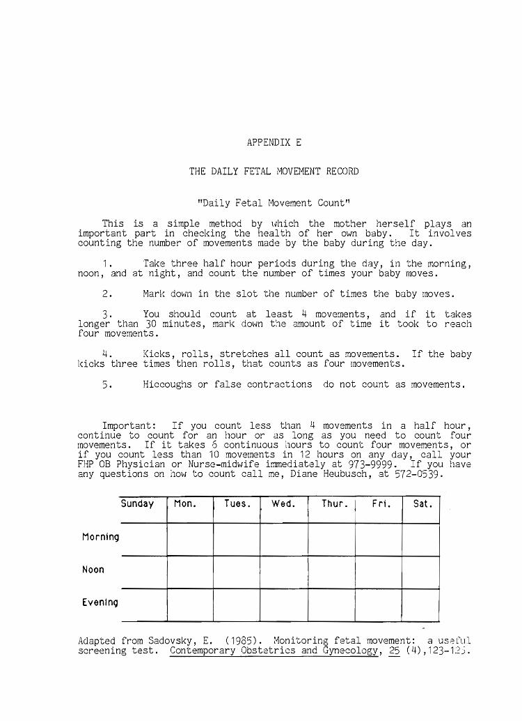
THE DAILY FETAL ivlOVEMENT RECORD
"Daily Fetal Hovement Count"
This is a simple method by \.Jhich the mother herself plays an important part in checking the health of her own baby. It in vol ves counting the number of movements made by the baby during the day.
1. Take three half hour periods during the day, in the morning, noon, and at night, and count the number of times your baby moves.
2. Marl( down in the slot the number of times the baby moves.
3. You should count at least 4 movements, and if it takes longer than 30 minutes, mark down the amount of time it took to reach four movements.
4. Kicks, rolls, stretches all count as movements. If the baby l<:icks three times then rolls, that counts as four movements.
5. Hiccoughs or false contractions do not count as movements.
Important: If you count less than 4 movements in a half hour, continue to count for an hour or as long as you need to count four movements. If it takes 6 continuous hours to count four movements, or if you count less than 10 movements in 12 hours on any day, call your FHP OB Physician or Nurse-mid\tJife immediately at 973-9999. If you have any questions on how to count call me, Diane Heubusch, at 572-0539.
Sunday Mon. Tues. Wed. Thur. Fri. Sat.
Morning
Noon I
Even1nq
Adapted from Sadovsky, E. (1985). :"''lonitoring fetal movement: a useful screening test. Contemporary Obstetrics and Gynecology, (4),123-12).

APPENDIX F
EVALUATION FORM
Please place a checl< in one of the columns for each statement on the
left that best fits your feelings.
1. Did you feel comfortable completing the fetal movement chart this week?
2. Did you feel you knew how to fill in the chart correctly?
3. Did you l{noH what counted as a fetal movement?
4. Did you find this method to count fetal movement hard to do?
5. Was the chart difficult to complete?
6. Did you feel counting fetal movement this way tool{ too much of your time?
7. Did the counting of fetal movement interfere with your day too much?
8. Did counting fetal movements make you feel anxious about your baby?
9. In general do you feel counting fetal movements is a worthwhile tool for mothers to use during their pregnancy?
Definitely Yes
UncerYes tain No
Definitely
No
10. Please make any corrunents be 1 m·J about the method you used this week to count fetal movements.
REFERENCES
Campbell, D.J., & Stanley, J. quasiexperimental designs for College PubIlshlng Company.
C. (1963) . Experimental and research. Chicago: Rand-McNaTIY
Clark, J. (1985). Factors contributing to client nonuse of the Cardiff Count to Ten Fetal Activity Chart. Journal of Nurse Midwifery, 30(6), 320-325.
Coleman, C. ( 1981 ) . Fetal movement counts: An assessment tool. Journal of Nurse Nidwifery, 26(1), 15-23.
Cranley, M.S. ( 1981 ) • Development of a tool for the measurement of meternal attachment during pregnancy. Nursing Research, 30 (5) 281-284.
Depp, R. ( 1982) . Clinical evaluation of fetal status. In D. D. Danforth (Ed.), Obstetrics and Gynecology (4th ed.) (pp. 802-835) •. Danforth, D.D. (Ed.) Phlladelpola: Harper & Row Publishers.
Draper, J., Field, S., Thomas, H. & Hare, M.J. (1986). Women's views on keeping fetal movement charts. British Journal of Obstetrics and Gynecology, 93, 334-338.
Edwards, D.D., & Edwards, J.S. (1970). Fetal movement: Development and time course. Science, 169, 95-97.
Ehrstrom, C. (1979). Fetal movement monitorin~ in normal and high risk pregnancy. Acta Obstet Gynecol Scand, 80 (1), suppl., 1-32.
Fischer, S., Fullerton, J.T., & Trezise, L. (1981). Fetal movement and fetal outcome in a low risk population. Journal of Nurse Midvlifery, 26 (1), 25-30.
Gantes, M., Schy, D.S., Bartasius, V.H. & Roberts, J. (1986). The use of daily fetal movement records in a clinical setting. Journal of Obstetric, Gynecologic and Neonatal Nursing, 12 (5), 390-393.
Goodwin, R.C., & Lowe, E.W. (1974). Multiphasic fetal monitoring. American Journal of Obstetrics and Gynecology, 119 (3) 341-357.
Grant, A., & Hepburn, M. (1984). Merits of an individualized approach to fetal movement counting compared with fixed time and fixed number methods. British Journal of Obstetrics and Gynecology, 94, 1087-1090.
Harper l R.G., Greenberg, M, Farahani, G., Glassman, I. & Kierney, C.M.P. ( 19t51 ) • Fetal movement, biochemical, biophysical parameters and outcome of pregnancy. American Journal of Obstetrics and Gynecology, 141 (1), 39-42.
51
Hellman, L.M., Pritchard, J.A., & Gant, N.F. (1985). Williams obstetrics (17th ed.). New York: Appleton-Century-Crofts.
Hobel, C. J., Hyvarinen, M.A., Okada, D.M., & Oh, W. (1973). Prenatal and intrapartum high risk screening. American Journal of Obstetrics and Gynecology, 117 (1), 1-9.
Hertogs, K., Roberts, A.B., Cooper, D., Griffin, D.R., & Campbell, S. (1979). r.1aternal perception of fetal motor activity. British Medical Journal, ~, 1183-1185.
Leader, R., Baillie, P., & Van Schaaalkwyk, D.J. (1981). Fetal movements and fetal outcome: A prospective study. Obstetrics and Gynecology, 57(4): 431-436.
Lee, C.Y., Dilereto, P.C., & O'Lane, J.M. (1975). A study of fetal heart rate acceleration patterns. Obstetrics and Gynecology, 45, 142-146.
Liston, R.M., Cohen, A.lrJ., Mennuti, M.T., & Gabbe, S.G. (1982). Antepartum fetal evaluation by maternal perception of fetal movement. Obstetrics and Gynecology, 60(4), 424-426.
Mathews, D.D. (1973). Fetal movements and fetal well being. Lancet, 1, 1315.
Mathews, D.D. (1975). Maternal assessment of fetal activity in small for dates infants. Obstetrics and Gynecology, 45 (5), 488-493.
McIlwaine, G.M., Howat, R.C.L., Dunn, F .H., & McNaughton, lvi.C. (1980). Perinatal practice and compensation handicap. British Medical Journal, 281, 1067.
Neldam, S. (1980) . Fetal movements as an indicator of fetal well being. Lancet, 1, 1222-1224.
0' Learly, J.A., & Andrinopoulos, G. C. (1981) • Correlation of daily fetal movements and the nonstress test as tools for assessment of fetal welfare. American Journal of Obstetrics and Gynecology, 139 ( 1 ), 1 07-1 08 •
Patrick, J., Campbell, K., Carmichael, L., Natale, R., & Richardson, B. (1982). Patterns of gross fetal body movements over 24 hour observation intervals during the last ten weeks of pregnancy. American Journal of Obstetrics and Gynecology, 142, 363-371.
Pearson, J. (1977). Fetal movements: A new approach to antenatal care. Nursing Mirror, 144, 49-51.
Pearson, J., & Weaver, J.B. (1976). Fetal activity and fetal well being: An evaluation. British Medical Journal, 1, 1305-1307.
52
Rayburn, W.F. (1980) . Clinical significance of perceptible fetal motion. American Journal of Obstetrics and Gynecology, 138, 210-212.
Rayburn, W.F. ( 1982) • Antepartum fetal assessment, monitoring fetal activity. Clinics in Perinatology, 1 (2), 231-252.
Rayburn, W. R., & ivfcKean, H. ( 1980) . Maternal perception of fetal movement and perinatal outcome. Obstetrics and Gynecology, 56 (2), 161-164.
Sadovsky, E., Polishuk, W., Mahler, Y., & Malkin, A. (1973). Correlation between electromagnetic recording and maternal assessment of fetal movement. Lancet, 1, 1141.
Sadovsky, E., & Yaffe, H. (1973). Daily fetal movement recording and fetal prognosis. Obstetrics and Gynecology, ~ (6), 845-850.
Sadovsky, E., Yaffe, H., & Pol ishuk, W. ( 1974) • Fetal movement in pregnancy and urinary estriol in prediction of impending fetal death in utero. Israel Journal of Medical Science, lQ, 1096-1099.
Sadovsky, E., Laufer, 1'1., & Allen, J • W. ( 1979) . The incidence of differant types of fetal movements during pregnancy. British Journal of Obstetrics and Gynecology, 86, 10-14.
Sadovsky, E. (1981). Fetal movements and fetal health. Seminars in Perinatology, 2 (2), 131-143.
Sadovsky~ E., Ohel, H., Havazeleth, A., Steinwell, D. & Penchas, S. (1983). The definition and significance of decreased fetal movements. Acta Obstetrics Gynecol Scand, 62, 409-413.
Sadovsl<y, E. (1985). Monitoring fetal movement: A useful screening test. Contemporary Obstetrics and Gynecology, 25 (4), 123-135.
Spellacy, W. N., Cruz, A.C., Gilman, S.R. & Buhi, W.C. (1977). Fetal movements and placental lactogen levels for fetal placental evaluation. Obstetrics and Gynecology, 49, 113-115.
Timor-Tritsch, I. (1979). Fetal movement: A brief review. Clinical Obstetrics and Gynecology, 22 (3), 583-592.
Timor-Tritsch, I., Zador, I., Dieker Hetz, R.H. & Rosen, M.G •• (1978). Classification of fetal movement. American Journal of Obstetrics and Gynecology, 126, 70-77.
Thompson, S.L. & Wheeler, T. (1985). Compliance and maternal fetal movement counting. Lancet, 2 (8464) 1122.
Valentin, L., Lofgren, K., Marsal, T., & Gulberg, B. (1984). Subjective recordings of fetal movement. Acta Obstetrics Gynecol Scand, 63, 223-228.
Wood, C., Gilbert, M. & O'Connor, A. (1979). Subjective recording of fetal rnovements. British Journal of Obstetrics and Gynaecology, 86, 836-842.

53
Wood, C., Walters, W.A.W., & Trigg, P. (1977). Methods of recording fetal movement. British Journal of Obstetrics and Gynecology, 84 (8), 561-567.