Fernando Danilo Gonzalez-Nilo UNIVERSIDAD NACIONAL …
16
AFRL-AFOSR-CL-TR-2017-0007 Multimode bio-nano sensor Fernando Danilo Gonzalez-Nilo UNIVERSIDAD NACIONAL ANDRES BELLO Final Report 05/23/2017 DISTRIBUTION A: Distribution approved for public release. AF Office Of Scientific Research (AFOSR)/ IOS Arlington, Virginia 22203 Air Force Research Laboratory Air Force Materiel Command
Transcript of Fernando Danilo Gonzalez-Nilo UNIVERSIDAD NACIONAL …

AFRL-AFOSR-CL-TR-2017-0007
Multimode bio-nano sensor
Fernando Danilo Gonzalez-NiloUNIVERSIDAD NACIONAL ANDRES BELLO
Final Report05/23/2017
DISTRIBUTION A: Distribution approved for public release.
AF Office Of Scientific Research (AFOSR)/ IOSArlington, Virginia 22203
Air Force Research Laboratory
Air Force Materiel Command
a. REPORT
Unclassified
b. ABSTRACT
Unclassified
c. THIS PAGE
Unclassified
REPORT DOCUMENTATION PAGE Form ApprovedOMB No. 0704-0188
The public reporting burden for this collection of information is estimated to average 1 hour per response, including the time for reviewing instructions, searching existing data sources, gathering and maintaining the data needed, and completing and reviewing the collection of information. Send comments regarding this burden estimate or any other aspect of this collection of information, including suggestions for reducing the burden, to Department of Defense, Executive Services, Directorate (0704-0188). Respondents should be aware that notwithstanding any other provision of law, no person shall be subject to any penalty for failing to comply with a collection of information if it does not display a currently valid OMB control number.PLEASE DO NOT RETURN YOUR FORM TO THE ABOVE ORGANIZATION.1. REPORT DATE (DD-MM-YYYY) 31-05-2017
2. REPORT TYPEFinal
3. DATES COVERED (From - To)15 Mar 2015 to 14 Jun 2016
4. TITLE AND SUBTITLECross-Discipline Bio-Nanostructured Enhanced Photonic Multimode-Sensor Science
5a. CONTRACT NUMBER
5b. GRANT NUMBERFA9550-15-1-0140
5c. PROGRAM ELEMENT NUMBER61102F
6. AUTHOR(S)Fernando Danilo Gonzalez-Nilo
5d. PROJECT NUMBER
5e. TASK NUMBER
5f. WORK UNIT NUMBER
7. PERFORMING ORGANIZATION NAME(S) AND ADDRESS(ES)UNIVERSIDAD NACIONAL ANDRES BELLOREPUBLICA 237SANTIAGO, 8370146 CL
8. PERFORMING ORGANIZATIONREPORT NUMBER
9. SPONSORING/MONITORING AGENCY NAME(S) AND ADDRESS(ES)AFOSR/SOARDU.S. Embassy SantiagoAv. Andres Bello 2800Santiago, Chile
10. SPONSOR/MONITOR'S ACRONYM(S)AFRL/AFOSR IOS
11. SPONSOR/MONITOR'S REPORTNUMBER(S)
AFRL-AFOSR-CL-TR-2017-0007 12. DISTRIBUTION/AVAILABILITY STATEMENTA DISTRIBUTION UNLIMITED: PB Public Release
13. SUPPLEMENTARY NOTES
14. ABSTRACTThis cross-discipline experimental study aimed to combine soft material science with nanotechnology andmulti-physics modeling to produce adaptable bio-nanostructure based on Quantum Dots and Proteins,dropping therefore the cytotoxicity and open new applications in biomedicine, among other areas.We observe from MALDI-TOF analysis two type of proteins in the coating of the Quantum Dots synthetizedin bacterias: proteins that interact with nucleotides or DNA and chaperone proteins. All these sequencesrecollected were annotated and analyzed using bioinformatics tools. At the same time, some of the proteinswith structural information were model and characterized the residues accessible to the solvent, in order toevaluate a molecular mechanism of the condensation process of the proteins on the surface of theQuantum Dots.15. SUBJECT TERMSmulti-mode sensor, SOARD
16. SECURITY CLASSIFICATION OF: 17. LIMITATION OFABSTRACT
SAR
18. NUMBEROFPAGES
16
19a. NAME OF RESPONSIBLE PERSONPOKINES, BRETT
19b. TELEPHONE NUMBER (Include area code)(703) 835-2309
Standard Form 298 (Rev. 8/98)Prescribed by ANSI Std. Z39.18
Page 1 of 1FORM SF 298
6/6/2017https://livelink.ebs.afrl.af.mil/livelink/llisapi.dll

Final Report ASFORD 2016
CROSS-DISCIPLINE BIO-NANOSTRUCTURED ENHANCED PHOTONIC MULTIMODE-SENSOR SCIENCE
(Proposal #14IOS015)(FA9550-15-1-0140)
Principal Investigators: Dr. Fernando Danilo Gonzalez Nilo
Dr. Jose Manuel Perez Donoso Center for Bioinformatics and Integrative Biology
Facultad de Ciencias Biologicas Universidad Andres Bello
Avenida Republica 239, Santiago, Chile.
Objective: The proposed cross-discipline experimental study aims to combine soft material science, laser spectroscopy, nano-technology, biophotonics and multi-physics modeling to produce adaptable bio-nanostructure enhanced multi-mode sensor science
Andres Bello research team actions:
Production of new QDs/ NPs by using green synthesis methods (biomimetic and biosynthesis). Different types of NPs have been produced by green methods to be used as non-toxic, highly fluorescent and stable dyes. We use these methodologies to produce Au and Ag NPs and also CdTe, CdS and CdSe QDs, which were tested in the different sensors to be used in this proposal (Pérez-Donoso and cols., PLOS ONE 2012, Monrás and cols., PLOS ONE 2013).
Tracking of biocompatible QDs; A decade after their introduction to biology quantum dots are proven powerful probes for fluorescence imaging and are being developed for a range of additional applications including the detection of disease, fluorescent assays for drug discovery, single protein tracking, and intracellular reporting.
Once the QD has been generated modifying the surface may be required so that the dot can be directed to a specific target. There are three primary ways to target a biocompatible quantum dot: with antibodies, with peptides, or with small molecules. Through this process quantum dots can be targeted to a specific type of cell or to a specific organelle inside the cell. Once the quantum dot is in contact with the cell it is possible to monitor the endocytosis process and its final degradation through optical analysis and tracking programs
Protein and DNA engineering. - The properties of proteins to be used in sensors were studies using bioinformatics tools as described the proposal. The CBIB at Universidad Andres Bello has long experience in molecular simulations, structural predictions and experimental analysis of proteins and DNA.
The experimental approach developed during the proposal involved both objectives. The biosynthesis of CdS NPs was evaluated in different bacterial strains (some of them extremophiles), NPs were characterized, and the bound biomolecules (particularly proteins) were determined. The interaction of the proteins determined in the different biosynthesis systems with the QDs were studied by means of in silico approaches using different bioinformatics tools.
Biosynthesis of Cd-QDs by extremophile microorganisms: The biosynthesis of NPs was evaluated in three different bacterial strains. One mesophile (Escherichia coli), one acidophile (Acidithiobacillus thiooxidans), and one halophile bacterium (Halomonas). Bacterial strains with different properties were tested in order to evaluate the capability of cells to produce QDs under
DISTRIBUTION A. Approved for public release: distribution unlimited.

these extreme conditions, but also because NPs generally are highly sensitive to acidic pH (below 4.0) and high salt concentrations (4-5 % NaCl).
Figure 1.- Biosynthesis of NPs by three different acidophilic bacteria. A.- Cell pellets obtained under biosynthesis conditions at pH 3.0. B.- Absorbance and emission spectra of purified QDs produced by acidophilic bacteria.
Figure 2.- Biosynthesis of NPs at different times by Acidithiobacillus ferrooxidans. A.- Time evolution of QDs-associated fluorescence in cells suspensions and NPs purifications confirming the presence of QDs. B.- Absorbance and emission spectra of purified QDs produced at different times.
DISTRIBUTION A. Approved for public release: distribution unlimited.

Figure 3.- Biosynthesis of NPs at different times by an acidophilic bacterial isolate. A.- Time evolution of QDs-associated fluorescence in NPs purifications confirming the presence of QDs. B.- Absorbance and emission spectra of purified QDs produced at different times.
Characterization of biosynthesized QDs: For the first time we described the biosynthesis of fluorescent NPs in halophile and acidophilic microorganisms. Cells produce NPs of different color after exposure to biosynthesis conditions (metal exposure, cysteine amendment). Interestingly the NPs produced under these conditions display characteristic properties than those produced by the mesophile strain E. coli in terms of size, metal composition, spectroscopic properties, band gap, quantum yield, and Z-potential among others. However, QDs produced by acidophilic bacteria display increased tolerance to acidic pH (tolerate pH below 4,5) in circumstances than QDs produce by E. coli decompose when exposed to pH 5. Regarding osmolality, QDs produced by halophile bacteria display increased tolerance to NaCl, tolerating concentrations higher than 12% (E. coli produced QDs decompose after exposure to 3-4 % NaCl).
Figure 4.- TEM of NPs produced at acidic pH by acidophilic bacteria. A and B.- Purified fractions of NPs biosynthesized at pH 3.0. C.- Acidithiobacillus ferrooxidans producing QDs (arrows indicate the presence of nanomtric material). D.- Acidithiobacillus ferrooxidans in the absence of metals (control).
DISTRIBUTION A. Approved for public release: distribution unlimited.
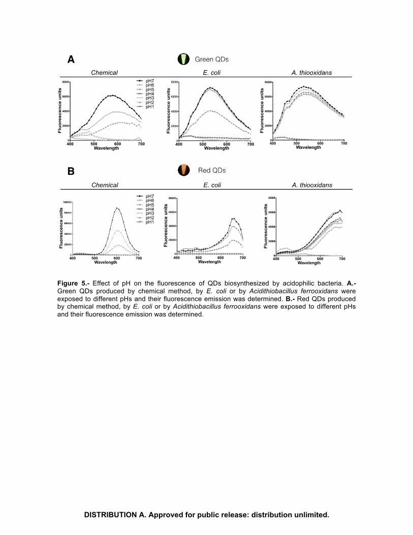
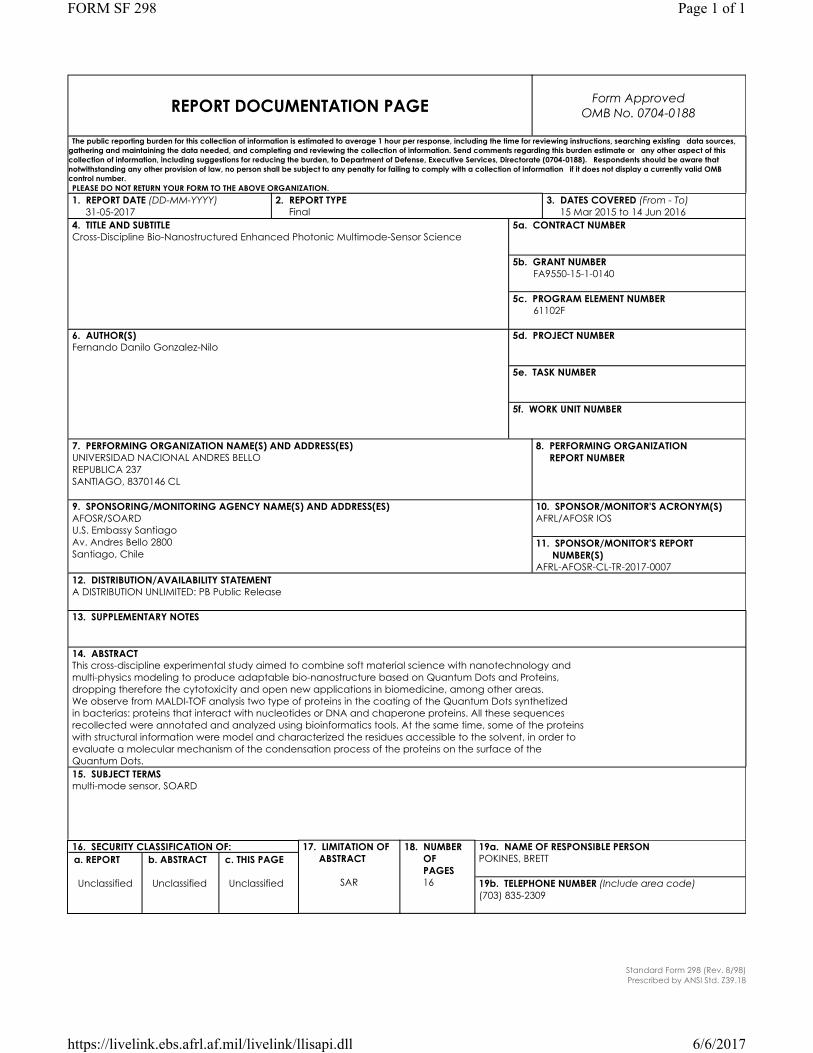
Figure 5.- Effect of pH on the fluorescence of QDs biosynthesized by acidophilic bacteria. A.- Green QDs produced by chemical method, by E. coli or by Acidithiobacillus ferrooxidans were exposed to different pHs and their fluorescence emission was determined. B.- Red QDs produced by chemical method, by E. coli or by Acidithiobacillus ferrooxidans were exposed to different pHs and their fluorescence emission was determined.
DISTRIBUTION A. Approved for public release: distribution unlimited.
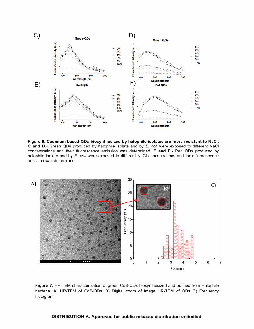
Figure 6. Cadmium based-QDs biosynthesized by halophile isolates are more resistant to NaCl. C and D.- Green QDs produced by halophile isolate and by E. coli were exposed to different NaCl concentrations and their fluorescence emission was determined. E and F.- Red QDs produced by halophile isolate and by E. coli were exposed to different NaCl concentrations and their fluorescence emission was determined.
Figure 7. HR-TEM characterization of green CdS-QDs biosynthesized and purified from Halophile bacteria. A) HR-TEM of CdS-QDs. B) Digital zoom of image HR-TEM of QDs C) Frequency histogram.
0 1 2 3 4 5 6 70
5
10
15
20
25
30
Fre
cue
ncy
(%
)
Size (nm)
A)B)
C)
DISTRIBUTION A. Approved for public release: distribution unlimited.

Extracellular biosynthesized green CdS-QDs were purified and characterized by HR-TEM (Figure 7A). The electron microscopy image shows that nanoparticles are uniformly distributed in a matrix of low crystallinity. A digital zoom of the HRTEM image shows that these QDs are regular polyhedra (Figure 7B), similar to models of icosahedral nanoparticles (Ref et al. 2016). Figure 7C show the size frequency histogram and the average particle size was 3,56nm.
1000 1500 2000 2500 3000 3500 4000
78
81
84
87
90
93
96
99
Ref
lect
ance
(%)
Wavenumbers (cm-1)
Cysteine QDs green
Figure 8. FTIR characterization of green CdS-QDs, biosynthesized and purified from Halophile bacteria. A) Spectrum FTIR of cysteine and B) Spectrum FTIR of QDs.
The FTIR spectra for cysteine and CdS-QDs can be observed in Figure 7. For both spectra, is observed a peak close to 1,575 cm-1 corresponding to carboxylate group, this band is typical of the formation of an amino-acid Zwitterion (Razium et al. 2014). The characteristic band for N–H stretching modes observed at 3,346 and 3,250 cm−1 indicates the presence of a –NH2 group. Also, the characteristic S-H stretching mode is clearly seen at 2,550cm−1, as expected from the pure cysteine. In the spectrum of CdS–QDs, it is noted the disappearance of the S–H group band at 2,550 cm−1. This is likely from the result of covalent bond formation between the thiol and the Cd at the CdS-QDs surface or proteins forming disulfide bonds (S-S).
In addition, proteins associated to the different NPs were determined to understand the properties of the different QDs produce by bacteria and also to find potential unique proteins involved in QDs synthesis or stabilization, both topics non-studied to date in any kind of biosynthesized NP.
Proteins associated to QDs produced by the different bacterial strains: We analyzed the proteins associated to QDs produced by the three bacterial strains of interest. To perform this, the NPs present in bacterial supernatants were purified and washed three times to eliminate organic molecules associated by week interactions to the QDs. After washing, the QDs were dialyzed to eliminate all the organic matter that could be bound non-specifically to the NPs. Then biosynthesized NPs were loaded in an SDS-PAGE to identify the presence of protein bands after Comassie and Silver staining. In the QDs produced by the three bacterial strains were observed proteins in the 20-60 kDa ranges with different profiles in each one. A section of the gel containing the most representative protein band of each QDs was extracted and sent for sequencing at the service of proteomic Analysis at the Center for functional Genetics in the University of Albany. The MALDI-TOF analysis of the proteins bound to QDs indicated the presence of an important number of proteins. Through a Blast analysis we determined the identity of the obtained sequences.
A)B)
DISTRIBUTION A. Approved for public release: distribution unlimited.

Protein Hits:
gi|189027968
RecName: Full=Elongation factor G; Short=EF-G
gi|446062467
30S ribosomal protein S1 [Salmonella enterica]
gi|447119484
heat-shock protein Hsp90 [Salmonella enterica]
gi|759355562
molecular chaperone DnaK [Pluralibacter gergoviae]
gi|502668509
molecular chaperone DnaK [Citrobacter rodentium]
gi|447215540
keto-acid formate acetyltransferase [Salmonella enterica]
gi|414145433
Chain A, Open Conformation Of Atp-Bound Hsp70 Homolog Dnak
gi|489954940
MULTISPECIES: keto-acid formate acetyltransferase [Enterobacter]
gi|873952861
elongation factor G [Brenneria goodwinii]
gi|513029196
transketolase [Salmonella enterica]
gi|161364100
hypothetical protein SPAB_02488 [Salmonella enterica subsp. enterica serovar Paratyphi B str. SPB7]
gi|492015287
elongation factor G [Pasteurella multocida]
gi|385867940
Chain A, The Crystal Structure Of Ompa Domain Of Ompa From Salmonella Enterica Subsp. Enterica Serovar Typhimurium Str. 14028s
gi|555263798
oligopeptidase A [Salmonella enterica]
gi|160867533
hypothetical protein SARI_04378 [Salmonella enterica subsp. arizonae serovar
DISTRIBUTION A. Approved for public release: distribution unlimited.
62:z4,z23:-]
gi|353077834
polyribonucleotide nucleotidyltransferase [Salmonella enterica subsp. enterica serovar Infantis str. SARB27]
gi|326623540
threonyl-tRNA synthetase [Salmonella enterica subsp. enterica serovar Dublin str. SD3246]
gi|62126355
pyruvate dehydrogenase, dihydrolipoyltransacetylase component [Salmonella enterica subsp. enterica serovar Choleraesuis str. SC-B67]
gi|62130425
fumarate reductase, anaerobic, flavoprotein subunit [Salmonella enterica subsp. enterica serovar Choleraesuis str. SC-B67]
gi|585341020
polynucleotide phosphorylase/polyadenylase [Escherichia coli]
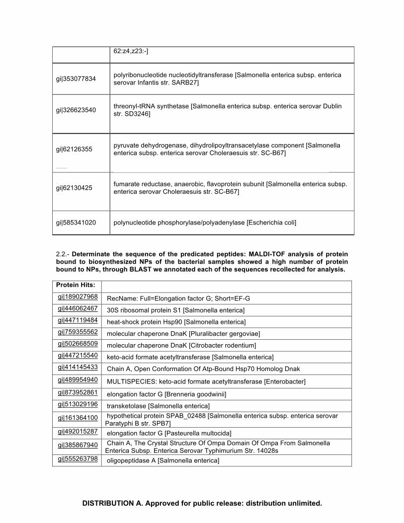
2.2.- Determinate the sequence of the predicated peptides: MALDI-TOF analysis of protein bound to biosynthesized NPs of the bacterial samples showed a high number of protein bound to NPs, through BLAST we annotated each of the sequences recollected for analysis. Protein Hits: gi|189027968 RecName: Full=Elongation factor G; Short=EF-G gi|446062467 30S ribosomal protein S1 [Salmonella enterica] gi|447119484 heat-shock protein Hsp90 [Salmonella enterica] gi|759355562 molecular chaperone DnaK [Pluralibacter gergoviae] gi|502668509 molecular chaperone DnaK [Citrobacter rodentium] gi|447215540 keto-acid formate acetyltransferase [Salmonella enterica] gi|414145433 Chain A, Open Conformation Of Atp-Bound Hsp70 Homolog Dnak gi|489954940 MULTISPECIES: keto-acid formate acetyltransferase [Enterobacter] gi|873952861 elongation factor G [Brenneria goodwinii] gi|513029196 transketolase [Salmonella enterica]
gi|161364100 hypothetical protein SPAB_02488 [Salmonella enterica subsp. enterica serovar Paratyphi B str. SPB7]
gi|492015287 elongation factor G [Pasteurella multocida]
gi|385867940 Chain A, The Crystal Structure Of Ompa Domain Of Ompa From Salmonella Enterica Subsp. Enterica Serovar Typhimurium Str. 14028s
gi|555263798 oligopeptidase A [Salmonella enterica]
DISTRIBUTION A. Approved for public release: distribution unlimited.

gi|160867533 hypothetical protein SARI_04378 [Salmonella enterica subsp. arizonae serovar 62:z4,z23:-]
gi|353077834 polyribonucleotide nucleotidyltransferase [Salmonella enterica subsp. enterica serovar Infantis str. SARB27]
gi|326623540 threonyl-tRNA synthetase [Salmonella enterica subsp. enterica serovar Dublin str. SD3246]
gi|62126355 pyruvate dehydrogenase, dihydrolipoyltransacetylase component [Salmonella enterica subsp. enterica serovar Choleraesuis str. SC-B67]
gi|62130425 fumarate reductase, anaerobic, flavoprotein subunit [Salmonella enterica subsp. enterica serovar Choleraesuis str. SC-B67]
gi|585341020 polynucleotide phosphorylase/polyadenylase [Escherichia coli] 3. Establish structure-activity relationships of the selected peptides in part 2, through an experimental-theoretical strategy. 3.1. Establish structure-activity relationships: from sequences of the peptides, we performed a bioinformatics analysis. To perform the bioinformatics analysis, we used the BLAST software (Altschul, S.F., Gish, W., Miller, W., Myers, E.W. & Lipman, D.J. (1990) "Basic local alignment search tool." J. Mol. Biol. 215:403-410) and the UniprotKB (The UniProt Consortium; UniProt: a hub for protein information. Nucl Acids Res 2014; 43 (D1)) data base. With the query to these data bases, we were able of identify that these sequences have some commons characteristics, despite that belonged to different species and performed different functions. For example, we identified that these proteins are involved in metal bound site, ADP, ATP and Zinc Finger bound site. When we analyzed the motif present in these proteins by MEME-suite tool (Timothy L. Bailey, Mikael Bodén, Fabian A. Buske, Martin Frith, Charles E. Grant, Luca Clementi, Jingyuan Ren, Wilfred W. Li, William S. Noble, "MEME SUITE: tools for motif discovery and searching", Nucleic Acids Research, 37:W202-W208, 2009), we found three shared motifs present in these proteins. One of these motifs belongs to Piruvate formate liaze (PFL1). Motif 1
Motif 2: Gly radical
MEME (with SSC) 28.12.2015 15:30
0
1
2
3
4
bits
1F 2
S
3
G
4D 5P 6IT
7W 8AV
9T 10
E
11
AVS
12
I
13
G
14
G
15
VM
16
SAG
17
IEVL
18
QD
19
G
20
ETR
21
PT
22
RL
23
V
24
T
25
K
26
SN
27
AS
28
F
29
R
30
MYF
31
L
32
NH
33
T
34
L35
DHSTY
36
TN
37
ML
38
G39
P40
SA
41
P42
E43
P44
N45
IL
46
T47
IV
48
L49
W50
SDISTRIBUTION A. Approved for public release: distribution unlimited.

Motif 3: PFL-like
In order to evaluate how these proteins bound to the nanoparticles we performed a BLAST search over the totality of the sequences, against Protein Data Bank (PDB) (H.M. Berman, J. Westbrook, Z. Feng, G. Gilliland, T.N. Bhat, H. Weissig, I.N. Shindyalov, P.E. Bourne (2000) The Protein Data Bank Nucleic Acids Research, 28: 235-242.), to get a crystallographic structure, and performed a molecular dynamics simulation and structural analysis. From the proteins set, we found in BLAST that some sequences have a crystal structure, and we choose the most commons protein present in the set, the Hsp 70 chaperone homolog Dnak PDB id: 4B9Q, this protein is involved in the ATP binding and belong to E.coli strain. In our analysis, we found that this protein has an electronegative character, and also have a domain called Hspa-9 Like-NBD, this domain has as function bind ATP and ADP. When we analyzed this chaperone protein and the residues that bind ATP and other phosphorylated sources, we found that this has arginine residues in common, and the capacity to bind ATP or ADP and the nanoparticle of this protein are related with SASA measure and the solvent accessibility that have this kind of residues.
MEME (no SSC) 28.12.2015 16:12
0
1
2
3
4bits
1T 2
S3N 4V 5V 6Y 7
G8
QK
9K 10
T11
G12
STN
13
T14
P15
D16
G17
R18
KR
19
KA
20
G21
TAE
22
LP
23
F24
GA
25
P26
G27
A28
N29
P30
LM
31
H32
G
33
R
34
D
35
KRTQ
36
SK
37
G
38
A
39
VL
40
A
41
S
42
L
43
SANT
44
S
45
V
46
A
47
K
48
L
49
P
50
FY
MEME (no SSC) 28.12.2015 18:55
0
1
2
3
4
bits
1LIY
2R 3K 4T 5H 6N 7HQ
8
G9V 10
F11
D12
AV
13
Y14
ST
15
KSDP
16
ED
17
IM
18
RKL
19
LNQRA
20
VCA
21
R22
RK
23
AS
24G
25IV
26LI
27
T28
G29
L30
P31
D32
GA
33
Y34
G35
R36
G37
R38
I39
I40
G41
D42
Y43
R44
R45
V46
PA
47
L48
Y49
G50
IV
00,10,20,30,40,50,60,70,80,91
Porcentajederesiduossobre20%
del
sasa
SASA
ASP
ARG
LYS
GLU
DISTRIBUTION A. Approved for public release: distribution unlimited.

4. Evaluation of molecular models to describe the potential mechanism of interaction between metallic nanoparticle coated with peptide corone and biological membranes. To evaluate the behavior of the nanoparticle with the chaperone protein, we developed a force field using mechanical quantum calculation, with the aim to have a set of parameters ready to use in CHARMM36 force field that describe the atomic potential for Cd and Se atoms. With the appropriated developed force field, a molecular dynamic was performed with NAMD 2.9 software (James C. Phillips, Rosemary Braun, Wei Wang, James Gumbart, Emad Tajkhorshid, Elizabeth Villa, Christophe Chipot, Robert D. Skeel, Laxmikant Kale, and Klaus Schulten. Scalable molecular dynamics with NAMD. Journal of Computational Chemistry, 26:1781-1802, 2005.) for 10ns, to evaluate the possible interactions that nanoparticle could have with the protein Figure 9-a). Although the protein has an electronegative tendency in general, there are some areas in the protein that are positive enough to have an adequate nanoparticle-protein interaction (figure 9-b). Some key residues bind to metal nanoparticles, are cysteines (Figure 9-c-d).
a) b)
c) d)
Figure 9: Dnak chaperone protein and nanoparticle simulation in a). the surface of electrostatic potential in b). key residue that interacted with nanoparticle c). SASA of residues over time d). Also, one of the peptides found in the purification process that has interaction with the nanoparticle, belong to Dnak Chaperone protein. With the sequences of this peptide we made a structural
DISTRIBUTION A. Approved for public release: distribution unlimited.

prediction using different algorithms, the prediction showed that peptide has an alpha helix structure, and using this information we built a crystal molecular model of this using Schrodinger Suite Models. A molecular dynamics simulation was performed by 20 ns with the peptide model and the nanoparticle, using the NAMD 2.9 software. The molecular dynamics was performed under isobaric-isothermal NPT ensemble, with periodic boundary conditions and TIP3 water box. After the end of molecular simulation, we were able to observe that the peptide binds to the surface of the nanoparticle by amino-terminal end, where are involved amino acid mainly positively charged, like lysine and arginine. Also we performed a search using BLAST algorithms, against non-redundant data base (nr), and we found that this peptide is part of Nucleotic Binding domain sugar kinase, and belongs specifically to motif Hsp70s, proteins that have this motif are strangely regulated for caloric stress, toxic chemicals, heavy metals like cadmium, arsenic and cupper.
a) b)
Figure 10: Structural prediction of the peptide with different algorithms a) and structural model of the peptides and nanoparticle b). When we performed molecular dynamics simulations of the Dnak Chaperone protein, we observed that this was not able to bind to peptide, because the protein has negative charge principally and this is repelled for the peptide, that has the same charge.
DISTRIBUTION A. Approved for public release: distribution unlimited.

Figure 11: Molecular dynamics and structural model of the Dnak chaperone and nanoparticle. Other of the proteins that were found bind to the nanoparticle was the elongation factor. A molecular dynamics simulation was performed between the nanoparticle and the elongation factors, with the aim to observe how the nanoparticle is able to change the secondary structure of the protein when binds to the nanoparticle. This suggests that when the secondary structure of the protein changes, others proteins could act like chaperones with the aim to try repair the loss of secondary structure. We also noted that protein binds to nanoparticle by charged amino acid like ALA y LYS.
Figure 12: Interaction Protein-nanoparticle Cd-Se. Conclusion:
In general terms, we observe from MALDI-TOF analysis two type of proteins in the coating of the Quantum Dots synthetized in bacterias: proteins that interact with nucleotides or DNA and chaperone proteins. All these sequences recollected were annotated and analyzed using bioinformatics tools. At the same time, some of the proteins with structural information were model and characterized the residues accessible to the solvent, in order to evaluate a molecular mechanism of the condensation process of the proteins on the surface of the Quantum Dots.
DISTRIBUTION A. Approved for public release: distribution unlimited.
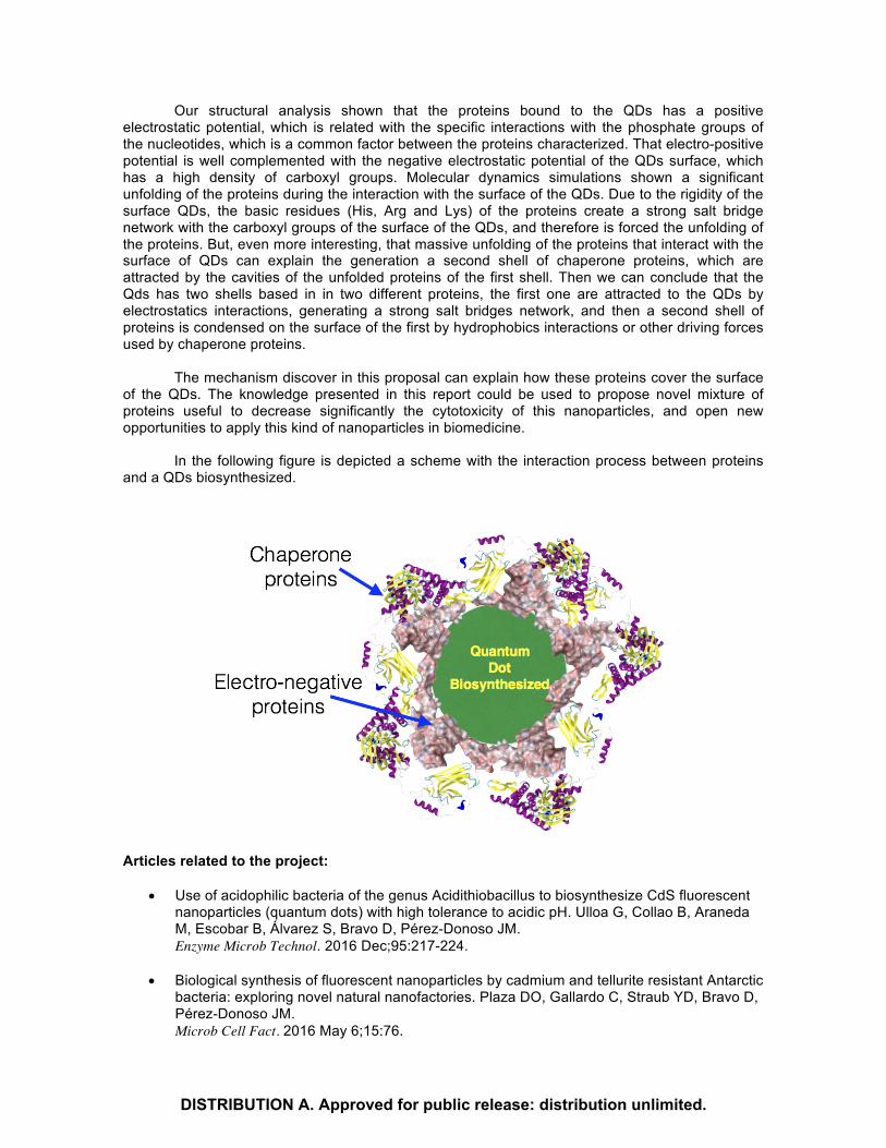
Our structural analysis shown that the proteins bound to the QDs has a positive electrostatic potential, which is related with the specific interactions with the phosphate groups of the nucleotides, which is a common factor between the proteins characterized. That electro-positive potential is well complemented with the negative electrostatic potential of the QDs surface, which has a high density of carboxyl groups. Molecular dynamics simulations shown a significant unfolding of the proteins during the interaction with the surface of the QDs. Due to the rigidity of the surface QDs, the basic residues (His, Arg and Lys) of the proteins create a strong salt bridge network with the carboxyl groups of the surface of the QDs, and therefore is forced the unfolding of the proteins. But, even more interesting, that massive unfolding of the proteins that interact with the surface of QDs can explain the generation a second shell of chaperone proteins, which are attracted by the cavities of the unfolded proteins of the first shell. Then we can conclude that the Qds has two shells based in in two different proteins, the first one are attracted to the QDs by electrostatics interactions, generating a strong salt bridges network, and then a second shell of proteins is condensed on the surface of the first by hydrophobics interactions or other driving forces used by chaperone proteins.
The mechanism discover in this proposal can explain how these proteins cover the surface
of the QDs. The knowledge presented in this report could be used to propose novel mixture of proteins useful to decrease significantly the cytotoxicity of this nanoparticles, and open new opportunities to apply this kind of nanoparticles in biomedicine.
In the following figure is depicted a scheme with the interaction process between proteins and a QDs biosynthesized.
Articles related to the project:
• Use of acidophilic bacteria of the genus Acidithiobacillus to biosynthesize CdS fluorescent nanoparticles (quantum dots) with high tolerance to acidic pH. Ulloa G, Collao B, Araneda M, Escobar B, Álvarez S, Bravo D, Pérez-Donoso JM. Enzyme Microb Technol. 2016 Dec;95:217-224.
• Biological synthesis of fluorescent nanoparticles by cadmium and tellurite resistant Antarctic bacteria: exploring novel natural nanofactories. Plaza DO, Gallardo C, Straub YD, Bravo D, Pérez-Donoso JM. Microb Cell Fact. 2016 May 6;15:76.
DISTRIBUTION A. Approved for public release: distribution unlimited.