Femtosecond Laser Technology in Modern Eye care · Femtosecond Laser Technology in Modern Eye Care...
30
9/15/2013 1 Femtosecond Laser Technology in Modern Eye Care Bill Tullo, OD, FAAO Vice President of Clinical Services TLC Laser Eye Centers DiplomateAAO Cornea Contact Lens & Refractive Surgery 2 Hrs 31917-RS Disclosures • Bill Tullo, OD – TLCVision–employee Femtosecond Technology • Sub-micrometer surgery • Minimally invasive • Incremental Improvements – Shorter pulses – Lower energy – Lower cost – Less complex – More accessible
Transcript of Femtosecond Laser Technology in Modern Eye care · Femtosecond Laser Technology in Modern Eye Care...

9/15/2013
1
Femtosecond Laser Technology in
Modern Eye Care
Bill Tullo, OD, FAAO
Vice President of Clinical Services
TLC Laser Eye Centers
Diplomate AAO Cornea Contact Lens & Refractive
Surgery
2 Hrs
31917-RS
Disclosures
• Bill Tullo, OD
– TLCVision – employee
Femtosecond Technology
• Sub-micrometer surgery
• Minimally invasive
• Incremental Improvements
– Shorter pulses
– Lower energy
– Lower cost
– Less complex
– More accessible

9/15/2013
2
Femtosecond Photodisruptive Infrared Laser = 1053 nm
• Nanosecond = 10 –9
• Picosecond = 10 –12
• Femtosecond = 10 –15
Definitions:
Femtosecond Laser
� Femtosecond laser
� (Nd:Glass) 1053 nm (near infrared)
� Each pulse of focused laser light lasts approximately
10-13
seconds (100 femtoseconds)
• In one second, light travels 7.5
times around the globe
• In 100 femtoseconds, light travels
across a human hair
• Power = Energy/Time, extremely
high power attained at relatively
low energy
Femtosecond Laser
• Surgical effect is achieved through “Photodisruption” at a molecular level
– No thermal or shock wave transmission to surrounding tissues
• Laser pulses focused to precise locations (+ 5 microns)
• Computer controlled optical delivery system places thousands of pulses next to each other

9/15/2013
3
Femtosecond Laser Applications
• Ophthalmic Surgery
– Corneal
• Incisions
• LASIK Flaps – 1st Commercial
Application
• Keratoplasty
• Refractive error and Presbyopia
– Cataract
• Capsulorhexis
• Nuclear disection
– Retinal
• Dental Surgery – painless and
safer
Chung SH, Mazur E. Surgical Applications of Femtosecond Lasers. J of Biophoton 2009;2(10) 557-572.
Optical Delivery System
• Glass lens applanates
cornea to fixate eye &
maintains precise
distance from laser head
to focal point Glass Lens
Cornea
Optical Delivery System
• Laser is set to desired
depth
– Defined distance from
bottom of glass applanation
lens (in microns)
• Pulses delivered in a prescribed
pattern creating a horizontal and
vertical cleavage plane in the
cornea

9/15/2013
4
An expanding bubble of gas & water is createdseparating the corneal lamellae
2 Microns
Femtosecond Photodisruption
The bi-products of photodisruption (CO2 & water) are absorbed by the mechanism of the endothelial
pump, leaving a cleavage plane in the cornea
Femtosecond Photodisruption
Advanced PerformanceTighter spot placement facilitates easier flap lifts

9/15/2013
5
Femtosecond Laser Raster Pattern
> 80% of all LASIK flaps are now created using femtosecond
technology
Femtosecond Lasers - LASIK
• AMO Intralase
– iFS
• Ziemer
– LDV
• Femtec
– 2010 Perfect Vision
• Zeiss
– VisuMax
• Alcon
– Wavelight FS200
• Technolas PV
– Victus

9/15/2013
6
Advantages of Femtosecond Flap
• Independent specific diameter
• Independent specific thickness
• Precise flap centration
• Variable hinge size/location
• Customized edge side cut
• Smooth evenly hydrated stromal bed
• Conserve tissue
• Planar shape
Why Femtosecond Technology?
• Femtosecond Technology is SAFER than any
mechanical metal blade microkeratome.
• Femtosecond Technology is MORE
EFFICACIOUS providing better visual outcomes
and quality of vision than any mechanical
metal blade microkeratome.
Safer
� There are fewer flap-related complications
� Eliminated common sight threatening complications
� Less loss of BCVA and more gain in BCVA
� Biomechanically stronger and more stable reducing the risk of post-LASIK ectasia

9/15/2013
7
Significant Vision Threatening Events -
Mechanical Microkeratome
• Globe Perforation
• Button-hole flap
• Thick Flap
• Thin Flap
• Free Cap
• Non-planar flap
• Epithelial slough
• Variable uncontrolled flap diameter
• Suction obtaining/maintaining
• Flap folds/striae
• Epithelial Ingrowth
• Neurotrophic Dry Eye
• Biomechanical destabilization
• Human error
– Microkeratome Assembly
– Microkeratome Sterilization
– Metal Blade quality/inspection
Comparison of Biomechanical Response Femtosecond
vs. LASEK vs. Blade
“ The biomechanical effect of LASIK with IntraLase is equivalent to that of surface ablation.”
Jorge L. Alió, MD, PhDESCRS, Sep 2005
Femtosecond LASIKWhy Stay Near the Surface?
• Anterior 40% of Cornea has strongest cohesive tensile strength
• Posterior 60% of Cornea is 50% weaker than the anterior 40%
• Increasing age is associated increased corneal cohesive tensile strength
– Randleman, J. et al, Journal of Refractive Surgery, January, 2008

9/15/2013
8
Photo credit: http://wieyemd.ophth.wisc.edu/ocupath/Cornea/HTML/cornealist.html
2x
Why are Femtosecond flaps more
EFFICACIOUS than mechanical
microkeratome flaps?
Better flaps = Better Vision!
Better Efficacy
• Flaps are planar not meniscus shape
• Stromal bed is smoother
• Less high order aberrations
• Stromal beds are evenly hydrated
• Faster visual recovery

9/15/2013
9
Uniform Flap Thickness
IntraLase Thin and planar
KeratomeThick and meniscus
Zeiss Visante images, courtesy of Jon Dishler MD
110
108 108
112
179
156
187
170
INTRALASE
Mechanical Microkeratome
Smooth Stromal Beds

9/15/2013
10
Durrie DS, Kezirian GM. Femtosecond laser versus mechanical keratome flaps in wavefront-guided laser in situ keratomileusis. J Cataract Refract Surg. 2005;31(1):120-126.
Better Efficacy
• Better quality of vision (Night Vision) as seen in Contrast Sensitivity studies
• Less quality of visual disturbances (haze, glare and light sensitivity)
• Less post-operative dry eye symptoms
• Technology continues to improve over time
IntraLase FS Laser Flap Creation Times
By Generation

9/15/2013
11
2nd Generation 3rd Generation
Real-Time
4th Generation 5th Generation iFS™ laser
Generation 2-5
Inverted Bevel-In Side Cut Angle
• Provides better wound healing for enhanced biomechanical stability of the post LASIK cornea1,2
• Significantly stronger flap adhesion post-operatively for improved wound healing
– 3x more force required for iFS™ laser (150° side cut) vs. microkeratome during flap lift2
• Virtually effortless flap lift, replacement, and positioning for maximum flap stability3,4
• Significantly reduced flap gutter3
1. Prof J Marshall, PhD. Data on file, AMO Development, LLC.2. Prof M Knorz, MD. Presented ASCRS 2008.3. P Binder, MD, A Chayet, MD. Presented ASCRS 2008.4. A. Chayet, MD. Data on file, AMO Development, LLC.
Elliptical Flaps� Preserves peripheral vital
lamellar fibers

9/15/2013
12
Femtosecond Science – LASIK Flaps
• Less intraoperative complications > x10
• Eliminates most devastating flap complications
• Reduce risk of ectasia – increased flap accuracy
• Less post-op complications– Dry eye
– Flap Striae
– Epithelial Ingrowth
• Faster visual recovery
• Better Night Vision– Improved low contrast VA
– Reduced HOA
• Reduced enhancement rates
Femtosecond Laser Keratomes
• What I tell patients
– Less flap-making risk
• Less long-term consequences if problems
– Thinner flaps
• More tissue left
• Less dryness
– More 20/20’s-better visual outcome
predictability
– Better low contrast [night] vision
Intacs for Keratoconus

9/15/2013
13
History of Intacs
• Originally FDA-approved in 1999 for mild myopia (-1.0 to
-3.0 D
• Intacs are two tiny, clear crescent-shaped pieces of a
plastic polymer that are inserted into the cornea.
• FDA, July 2004 -allow corrections of keratoconus using
intacs largely because of Intacs' safety record and
because only a few treatment options, such as corneal
transplants, are available for keratoconus.
Femto-Intacs Patient Selection
• Keratometry less than 58D
• No central scarring
• 500 microns thick at area of tunnel
• REALISTIC EXPECTATIONS!
Femto-Intacs Goals
• Delay need for corneal transplant
• Increase BCVA
• Improve ability to fit/wear CL’s
• Increase UCVA

9/15/2013
14
Intacs Complications
• Infection
• Difficulty with night vision
• Glare, halos, blurry and fluctuating vision.
• Neovascularization
• Blepharitis
• Intacs may produce no corrective effect in
fewer than 5 percent of individuals with
keratoconus.
Intacs for Keratoconus

9/15/2013
15
Femto-Intacs Advantages• Less surgeon dependent
• Reduced risk anterior/posterior perforation1
• Increased reduction in HOA (increased BCVA)2
• More precise placement3
• More effect3
• Easier to center on pupil
• Faster vision recovery
• Less pain
• Greater patient satisfaction!
1. Piñero DP, Alio JL. Intracorneal ring segments in ectatic corneal disease - a review Clin Experiment Ophthalmol. 2010 Mar;38(2):154-67
2. Piñero DP, Alio JL, El Kady B, Coskunseven E, Morbelli H, Uceda-Montanes A, Maldonado MJ, Cuevas D, Pascual I. Refractive and aberrometric outcomes of intracorneal ring segments for keratoconus: mechanical versus femtosecond-assisted procedures. Ophthalmology. 2009 Sep;116(9):1675-87. Epub 2009 Jul 29.
3. Rabinowitz YS Intacs for keratoconus Curr Opin Ophthalmol. 2007 Jul;18(4):279-83.
Corneal Inlay Pocket
Example: Acufocus
• Designed to improve
near vision in patients
with Presbyopia
– Easily implanted
– Minimal impact on
distance vision
– Removable
Central aperture:1.6 mm
Overall diameter: 3.8 mm
Corneal Inlay Pocket
• Example Acufocus

9/15/2013
16
IntraLase Enabled Keratoplasty
History: Advanced Shaped PK
� Massimo Busin (2003)
� Similar technique to the posterior two-level graft
� Adapted from modern day penetrating keratoplasty using modern instruments
Valve-Sealing Edge Design
SutureNot Tight
Intraocular Pressure
Prevents Leakage

9/15/2013
17
IntraLase Enabled Keratoplasty
Example Pattern Combinations
TopHat Shape
• Provides large endothelial surface transplantation
Mushroom Shape
• Preserves more host endothelium

9/15/2013
18
IntraLase Advanced Keratoplasty
The Zig-Zag shaped incision has shown a smooth corneal contour immediately after surgery with less distortion of the corneal optics and less astigmatism.
*Personal communication, Roger Steinert, M.D.
Slitlamp
1 week
3 months
6 months
Astigmatism
Typical 1 yr post-op result with
standard trephine cut PKP =
8 diopters of astigmatism
IntraLase Advanced Keratoplasty
at 3 months post-op =
½ diopter of astigmatism

9/15/2013
19
IEK Surgical Video
Femtosecond Lenticular Extraction –
Off label in US
• SMILE
– Small Incision Lenticular Extraction
• FLEx
– Femtosecond Lenticular Extraction
• Re-LEx
– Carl Zeiss Meditec’s brand of FLEx with SMILE
Femtosecond Lenticular Extraction
– Off Label

9/15/2013
20
FEMTO-AK
ALTERNATIVE TO THE
BLADE
INTRALASE ENABLED ASTIGMATIC KERATOTOMY - IEAK
BENEFITS OF FEMTO-AK/LRI
� Incomparable safety
� Decisive control of all surgical parameters
� Fully computerized control
� Maximal patient comfort
� Minimal learning curve
� Precision & predictability in the creation of AK resections + 10 Microns
FEMTO-AK/LRI PROCEDURE

9/15/2013
21
Femto-AK/LRI Conclusions
• The correction of astigmatism with the femtosecond laser is safe and effective 1,2
• Femtosecond assisted astigmatic keratotomy is more predictable and can correct more astigmatism than mechanized astigmatic keratotomy3.
1. Kook D, Bühren J, Klaproth OK, Bauch AS, Derhartunian V, Kohnen T. Astigmatic keratotomy with the femtosecond laser : Correction of high astigmatisms after keratoplastyOphthalmologe. 2010 Oct 6.
2. Kumar NL, Kaiserman I, Shehadeh-Mashor R, Sansanayudh W, Ritenour R, Rootman DSIntraLase-enabled astigmatic keratotomy for post-keratoplasty astigmatism: on-axis vector analysis. Ophthalmology. 2010 Jun;117(6):1228-1235.e1. Epub 2010 Feb 16.
3. Hoffart L, Proust H, Matonti F, Conrath J, Ridings B. Correction of postkeratoplastyastigmatism by femtosecond laser compared with mechanized astigmatic keratotomy Am J Ophthalmol. 2009 May;147(5):779-87, 787.e1. Epub 2009 Feb 20
Femtosecond Lasers
� Investigational Surgical Applications
◦ Presbyopia Correction (off label)
� Photo-disruption within the lens to restore
flexibility and the ability to accommodate◦ Peripheral lens incisions (enhanced lens elasticity)
1-day post-op 1-week post-op
Intracor Procedure
Intrastromal Femtosecond Ablation for
Presbyopia

9/15/2013
22
Intracor
• Circular intrastromal rings
• Central steepening of ant/post cornea to a multifocal hyperprolate shape
• Number, spacing, size of intrastromalcylindrical rings varied
• Variable power enhances depth of focus for better near vision
4 days preop
INTRACOR Presbyopia
Mike P. Holzer, MD
4 days preop 1 hour postop 1 week postop
1 hour postop
Cavitation gas in ring cuts
1 day postop
Gas escaped from cornea
Intracor Video
9/15/2013
23
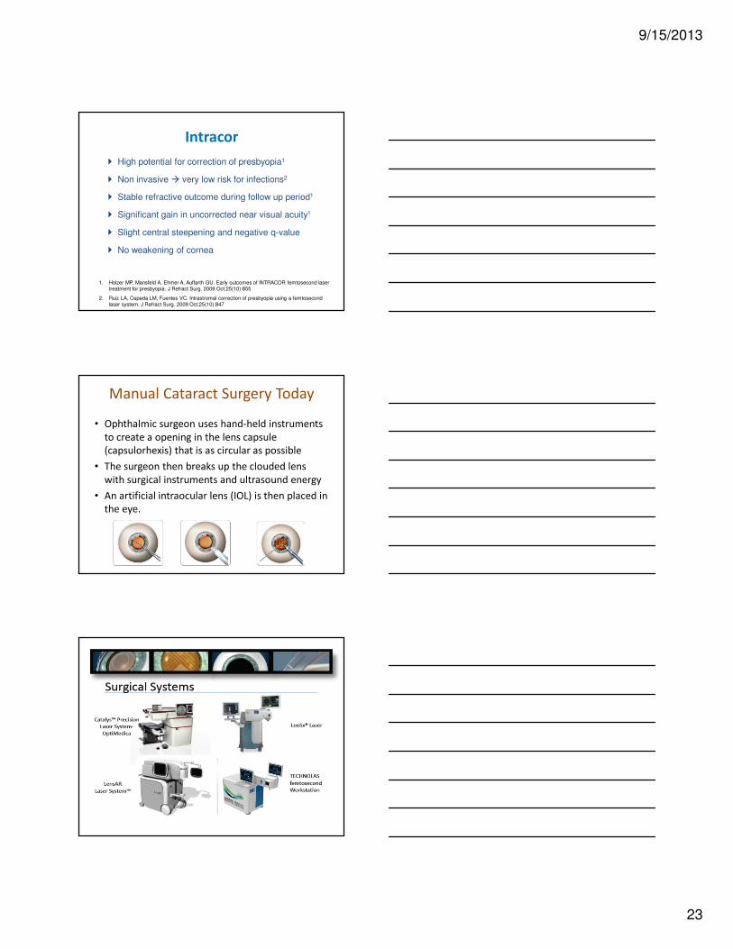
Intracor
� High potential for correction of presbyopia1
� Non invasive � very low risk for infections2
� Stable refractive outcome during follow up period1
� Significant gain in uncorrected near visual acuity1
� Slight central steepening and negative q-value
� No weakening of cornea
1. Holzer MP, Mansfeld A, Ehmer A, Auffarth GU. Early outcomes of INTRACOR femtosecond laser treatment for presbyopia. J Refract Surg. 2009 Oct;25(10):855
2. Ruiz LA, Cepeda LM, Fuentes VC. Intrastromal correction of presbyopia using a femtosecond laser system. J Refract Surg. 2009 Oct;25(10):847
Manual Cataract Surgery Today
• Ophthalmic surgeon uses hand-held instruments
to create a opening in the lens capsule
(capsulorhexis) that is as circular as possible
• The surgeon then breaks up the clouded lens
with surgical instruments and ultrasound energy
• An artificial intraocular lens (IOL) is then placed in
the eye.

9/15/2013
24
FEMTO CATARACT SURGERY
• The Players– LenSx –
• Aug 2009 – Anterior Capsulotomy
• Dec 2009 – Corneal Incisions• April 2010 – Lens
Fragmentation
– LensAR –• 3/21/11 Anterior
Capsulotomyand Lens Fragmentation
• Presbyopia licensing
– OptiMedica –• Dec 22, 2011 FDA Approval• Proprietary OCT with Catalys
– Technolas PV Victus• Both Refractive and Cataract• August 2012 – Anterior
Capsulotomy and Corneal Incisions
Femtosecond Laser for Cataract SurgeryLenSx, LensAR, OptiMedica, Technolas
� Lens Fragmentation - Liquefy, soften or “chop” the lens
� Refractive Capsulotomy - Create a perfectly centered and
sized
� Corneal Incisions - Create all required with perfect
dimension & architecture
� LRI Corneal Incisions - Provide a refractive solution to pre-
existing astigmatism
Clear Corneal Incisions

9/15/2013
25
LenSx
• Live Video
• OCT
• Procedure Templates
• Touch Screen
• Data Entry
• Ergonomic
• Space saving design

9/15/2013
26
OCT Image-Guided Surgery
Procedure Precision & Integration
Manual vs. Catalys Cataract Surgery 1 month postop
Manual Surgery Catalys Surgery

9/15/2013
27
More predictable outcomes
More predictable and precise cuts
Better outcomes
Catalys Clinical Results:
Capsulotomy ShapeManual Capsulorhexis
Catalys Capsulotomy
Images courtesy of OptiMedica Corp.
• Perfect centration• Precision diameter: < 0.25 mm• No radial tears • Easy and complete removal of capsule• No adverse events
Size Deviation from Intended Size
ShapeWhere 1.0 = perfect circle
PositionDeviation from Intended
29 ± 26 µm 0.936 ± 0.038 77 ± 47 µm
Clinical Trial Results: Precise Capsulotomy & Effective
Fragmentation
• 96% reduction in effective phaco time
Capsulotomy data courtesy of OptiMedica; effective phaco time data courtesy of Prof. Burkhard Dick, MD, PhD. Ruhr University Eye Hospital. Bochum, Germany, Member of OptiMedica Medical Advisory Board.Images courtesy of OptiMedica

9/15/2013
28
Corneal Incision and Lens Removal
Automated reproducibility allows every
surgeon to address astigmatismReduces or eliminates ultrasound
and related complications
0.1
0.105
0.11
0.115
0.12
0.125
0.13
0.135
0
5
10
15
20
25
30
LenSx + Phaco (n=21) Phaco Only (n=54)
Eff
ecti
ve P
haco
Tim
e (
min
s)
Av
e P
haco
Po
wer
(%)
Ave Power Effective Phaco Time
Comparison of Phaco Efficiency in Sighted Human Eyes
Self Sealing Cataract Incisions
Reproducible Refractive Incisions
Eff
ect
ive
Ph
aco
Tim
e
(se
con
ds)
Standard Cataract Surgery Catalys
Effective Phaco
Time (s)
Femto
(n=57)
0.16 ± 0.21
Standard
(n=52)
4.07 ± 3.14
MK-00185 Rev B
Catalys Clinical Results:
Impact on Effective Phaco Time
Data courtesy of Prof. Burkhard Dick, MD, PhD. Ruhr University Eye Hospital. Bochum, Germany, Member of
OptiMedica Medical Advisory Board.
96% reduction in effective phaco time compared to standard
Eff
ecti
ve P
haco
Tim
e (s
eco
nd
s)
LOCS IICatalys
LOCS IIICatalys
LOCS IV / IV+Catalys
LOCS IIStandard
LOCS IIIStandard
LOCS IVStandard
LOCS II: Standard II: Femto III: Standard III: Femto IV: Standard IV/IV+: Femto
EPZ (s) 1.96 ± 1.29 0.02 ± 0.05 3.32 ± 1.83 0.10 ± 0.16 6.21 ± 3.68 0.24 ± 0.25
Catalys Clinical Results:Impact on Effective Phaco Time
n= 13 n= 10 n= 18 n= 19 n= 21 n= 28
Data courtesy of Prof. Burkhard Dick, MD, PhD. Ruhr University Eye Hospital. Bochum, Germany, Member of
OptiMedica Medical Advisory Board. MK-00251 Rev A

9/15/2013
29
Patient Experience
• During procedure
– Docking: Slight pressure from vacuum pressure of
interface (no pain or loss of vision)
– During laser: A kaleidoscope of lights as the
procedure occurs
• Immediate to One-Day Post-op
– Same regimen as existing practices
– Visual recovery may be faster because of reduced
ultrasound energy
– Patient may notice slight hemorrhaging on the
conjunctivaMK-00251 Rev A
Patient Experience
• Clinical Workup
– No major changes to standard procedure
– Things to note:
• How well patient dilates
• Is patient able to keep still during procedure
• Post-Surgery Follow-up
– Same regimen as existing practices
– Things to note:
• Visual recovery may be faster because of reduced ultrasound energy
• Patient may notice slight hemorrhaging on the conjunctiva
MK-00251 Rev A
• Femtosecond laser applications in liquefaction was
safe, effective and efficient
• Capsulotomy size, shape and reproducibility is
statistically improved over manual techniques1
• Corneal incisions are reproducible and have precise
dimensions and geometry
1. Nagy Z, Takacs A, Filkorn T, Sarayba M. Initial clinical evaluation of an intraocular femtosecond laser in cataract surgery. J Refract Surg. 2009 Dec;25(12):1053

9/15/2013
30
Future Femtosecond Developments
• Higher Power Oscillators
• Laser fiber technology
• Solid State Lasers
• Combined with advanced imaging techniques
– Sub-diffraction imaging
– Quantum dots
• Cellular surgery and manipulation
• Deep tissue applications
QUESTIONS?