Fas/FasL pathway and cytokines in keratinocytes in atopic ... · RESEARCH ARTICLE Fas/FasL pathway...
12
RESEARCH ARTICLE Fas/FasL pathway and cytokines in keratinocytes in atopic dermatitis – Manipulation by the electromagnetic field Lukasz Szymanski ID 1 *, Aleksandra Cios 1 , Slawomir Lewicki ID 2 , Pawel Szymanski 1 , Wanda Stankiewicz 1 1 Department of Microwave Safety, Military Institute of Hygiene and Epidemiology, Warsaw, Poland, 2 Department of Regenerative Medicine and Cell Biology, Military Institute of Hygiene and Epidemiology, Warsaw, Poland * [email protected] Abstract Background Atopic dermatitis (AD) is one of the most frequent skin diseases. Changes of the keratino- cytes functionality play a major role in the development of AD. For example, activation of the Fas (CD95)/FasL (CD178) pathway in AD does not lead to extensive apoptosis in skin. Bind- ing of the Fas receptor to its protein ligand—FasL, which are present on the (AD)-modified keratinocytes, should result in the sequential induction of cell death, but there is no evidence of extensive apoptosis of these cells. This suggests that non-apoptotic mechanism of Fas/ FasL pathway is commonly encountered, although not examined in the case of AD, phe- nomenon. An electromagnetic field, which was used to influence cultured cells in this study, can modulate proliferation, apoptosis, differentiation, and metabolism in various cells. Objective Here, we evaluate the possibility to manipulate the immune activation of AD keratinocytes and their response to the electromagnetic field, which was not tested before. Methods Keratinocytes isolated from the skin of healthy subjects (n = 20) and patients with atopic der- matitis (n = 20) as well as HaCaT and PCS-200-010 cell were exposed to the 900 MHz elec- tromagnetic field for 60 minutes. Cytometric analysis of viability, Fas/FasL, p-ERK, p-p38 and p-JNK expression and Luminex analysis of cytokine concentration were performed in two-time points: 4 and 24 hours after the exposition. Results This research has shown upregulated Fas, FasL, p-ERK, p-p38, and p-JNK expression along with increased cytokine secretion (IL-1β, IL-4, IL-8, IL-10, IL-12p70, IL-13, IL-17A, IL- 31 and TNFα) by keratinocytes derived from the skin of patients with the AD when compared with healthy control. Exposure of keratinocyte cultures obtained from AD patients to EMF PLOS ONE | https://doi.org/10.1371/journal.pone.0205103 October 4, 2018 1 / 12 a1111111111 a1111111111 a1111111111 a1111111111 a1111111111 OPEN ACCESS Citation: Szymanski L, Cios A, Lewicki S, Szymanski P, Stankiewicz W (2018) Fas/FasL pathway and cytokines in keratinocytes in atopic dermatitis – Manipulation by the electromagnetic field. PLoS ONE 13(10): e0205103. https://doi.org/ 10.1371/journal.pone.0205103 Editor: Guillermo Lo ´pez Lluch, Universidad Pablo de Olavide, SPAIN Received: March 7, 2018 Accepted: August 5, 2018 Published: October 4, 2018 Copyright: © 2018 Szymanski et al. This is an open access article distributed under the terms of the Creative Commons Attribution License, which permits unrestricted use, distribution, and reproduction in any medium, provided the original author and source are credited. Data Availability Statement: All data used for this publication was uploaded to the public repository Zenodo https://doi.org/10.5281/zenodo.1300679. Funding: This study was funded by Polish Ministry of Science and Higher Education grant for Young Scientist. Competing interests: The authors have declared that no competing interests exist.
Transcript of Fas/FasL pathway and cytokines in keratinocytes in atopic ... · RESEARCH ARTICLE Fas/FasL pathway...

RESEARCH ARTICLE
Fas/FasL pathway and cytokines in
keratinocytes in atopic dermatitis –
Manipulation by the electromagnetic field
Lukasz SzymanskiID1*, Aleksandra Cios1, Sławomir LewickiID
2, Pawel Szymanski1,
Wanda Stankiewicz1
1 Department of Microwave Safety, Military Institute of Hygiene and Epidemiology, Warsaw, Poland,
2 Department of Regenerative Medicine and Cell Biology, Military Institute of Hygiene and Epidemiology,
Warsaw, Poland
Abstract
Background
Atopic dermatitis (AD) is one of the most frequent skin diseases. Changes of the keratino-
cytes functionality play a major role in the development of AD. For example, activation of the
Fas (CD95)/FasL (CD178) pathway in AD does not lead to extensive apoptosis in skin. Bind-
ing of the Fas receptor to its protein ligand—FasL, which are present on the (AD)-modified
keratinocytes, should result in the sequential induction of cell death, but there is no evidence
of extensive apoptosis of these cells. This suggests that non-apoptotic mechanism of Fas/
FasL pathway is commonly encountered, although not examined in the case of AD, phe-
nomenon. An electromagnetic field, which was used to influence cultured cells in this study,
can modulate proliferation, apoptosis, differentiation, and metabolism in various cells.
Objective
Here, we evaluate the possibility to manipulate the immune activation of AD keratinocytes
and their response to the electromagnetic field, which was not tested before.
Methods
Keratinocytes isolated from the skin of healthy subjects (n = 20) and patients with atopic der-
matitis (n = 20) as well as HaCaT and PCS-200-010 cell were exposed to the 900 MHz elec-
tromagnetic field for 60 minutes. Cytometric analysis of viability, Fas/FasL, p-ERK, p-p38
and p-JNK expression and Luminex analysis of cytokine concentration were performed in
two-time points: 4 and 24 hours after the exposition.
Results
This research has shown upregulated Fas, FasL, p-ERK, p-p38, and p-JNK expression
along with increased cytokine secretion (IL-1β, IL-4, IL-8, IL-10, IL-12p70, IL-13, IL-17A, IL-
31 and TNFα) by keratinocytes derived from the skin of patients with the AD when compared
with healthy control. Exposure of keratinocyte cultures obtained from AD patients to EMF
PLOS ONE | https://doi.org/10.1371/journal.pone.0205103 October 4, 2018 1 / 12
a1111111111
a1111111111
a1111111111
a1111111111
a1111111111
OPENACCESS
Citation: Szymanski L, Cios A, Lewicki S,
Szymanski P, Stankiewicz W (2018) Fas/FasL
pathway and cytokines in keratinocytes in atopic
dermatitis – Manipulation by the electromagnetic
field. PLoS ONE 13(10): e0205103. https://doi.org/
10.1371/journal.pone.0205103
Editor: Guillermo Lopez Lluch, Universidad Pablo
de Olavide, SPAIN
Received: March 7, 2018
Accepted: August 5, 2018
Published: October 4, 2018
Copyright: © 2018 Szymanski et al. This is an open
access article distributed under the terms of the
Creative Commons Attribution License, which
permits unrestricted use, distribution, and
reproduction in any medium, provided the original
author and source are credited.
Data Availability Statement: All data used for this
publication was uploaded to the public repository
Zenodo https://doi.org/10.5281/zenodo.1300679.
Funding: This study was funded by Polish Ministry
of Science and Higher Education grant for Young
Scientist.
Competing interests: The authors have declared
that no competing interests exist.
resulted in a decrease of 1β, IL-4, IL-10, IL-12, I L-13, IL-17, IL-31 and TNFα levels. Kerati-
nocytes derived from the skin of AD patients are characterized by elevated Fas and FasL
expression when compared to healthy control.
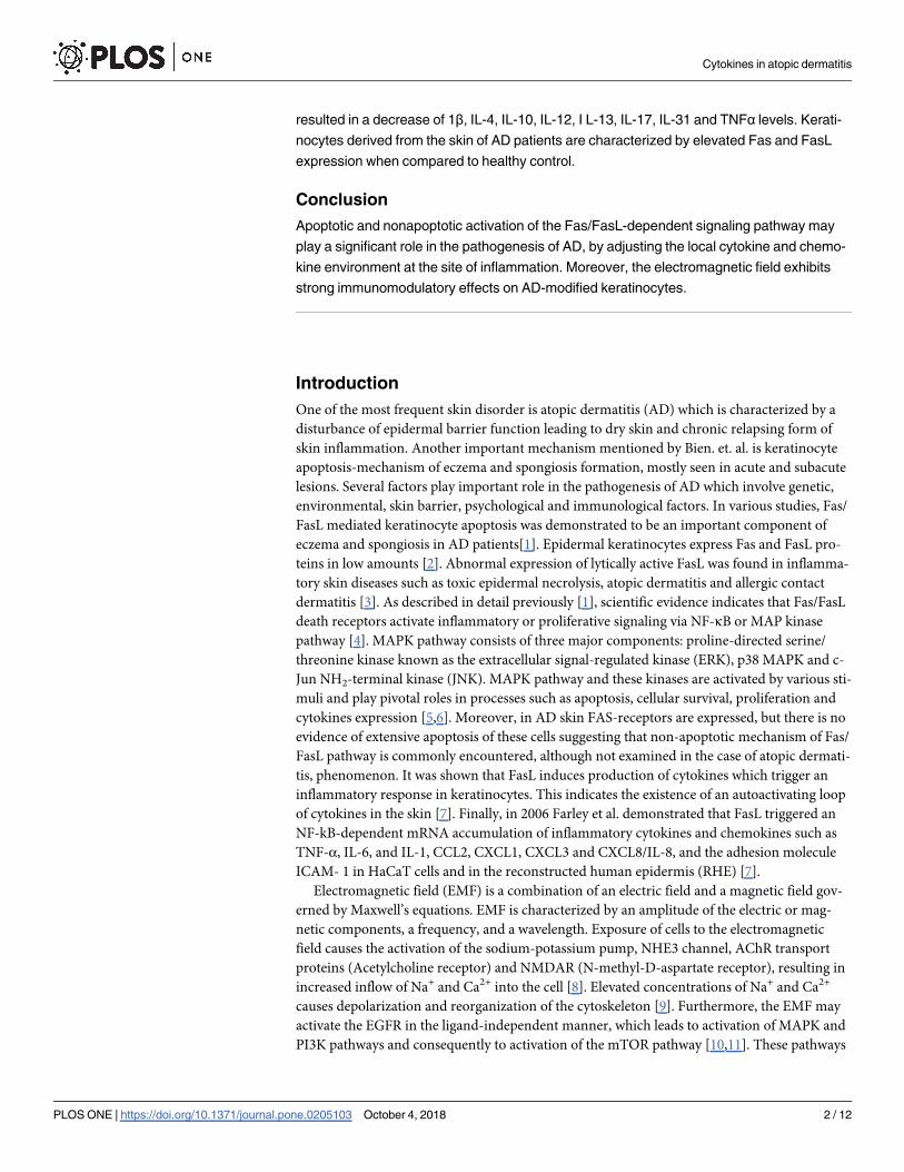
Conclusion
Apoptotic and nonapoptotic activation of the Fas/FasL-dependent signaling pathway may
play a significant role in the pathogenesis of AD, by adjusting the local cytokine and chemo-
kine environment at the site of inflammation. Moreover, the electromagnetic field exhibits
strong immunomodulatory effects on AD-modified keratinocytes.
Introduction
One of the most frequent skin disorder is atopic dermatitis (AD) which is characterized by a
disturbance of epidermal barrier function leading to dry skin and chronic relapsing form of
skin inflammation. Another important mechanism mentioned by Bien. et. al. is keratinocyte
apoptosis-mechanism of eczema and spongiosis formation, mostly seen in acute and subacute
lesions. Several factors play important role in the pathogenesis of AD which involve genetic,
environmental, skin barrier, psychological and immunological factors. In various studies, Fas/
FasL mediated keratinocyte apoptosis was demonstrated to be an important component of
eczema and spongiosis in AD patients[1]. Epidermal keratinocytes express Fas and FasL pro-
teins in low amounts [2]. Abnormal expression of lytically active FasL was found in inflamma-
tory skin diseases such as toxic epidermal necrolysis, atopic dermatitis and allergic contact
dermatitis [3]. As described in detail previously [1], scientific evidence indicates that Fas/FasL
death receptors activate inflammatory or proliferative signaling via NF-κB or MAP kinase
pathway [4]. MAPK pathway consists of three major components: proline-directed serine/
threonine kinase known as the extracellular signal-regulated kinase (ERK), p38 MAPK and c-
Jun NH2-terminal kinase (JNK). MAPK pathway and these kinases are activated by various sti-
muli and play pivotal roles in processes such as apoptosis, cellular survival, proliferation and
cytokines expression [5,6]. Moreover, in AD skin FAS-receptors are expressed, but there is no
evidence of extensive apoptosis of these cells suggesting that non-apoptotic mechanism of Fas/
FasL pathway is commonly encountered, although not examined in the case of atopic dermati-
tis, phenomenon. It was shown that FasL induces production of cytokines which trigger an
inflammatory response in keratinocytes. This indicates the existence of an autoactivating loop
of cytokines in the skin [7]. Finally, in 2006 Farley et al. demonstrated that FasL triggered an
NF-kB-dependent mRNA accumulation of inflammatory cytokines and chemokines such as
TNF-α, IL-6, and IL-1, CCL2, CXCL1, CXCL3 and CXCL8/IL-8, and the adhesion molecule
ICAM- 1 in HaCaT cells and in the reconstructed human epidermis (RHE) [7].
Electromagnetic field (EMF) is a combination of an electric field and a magnetic field gov-
erned by Maxwell’s equations. EMF is characterized by an amplitude of the electric or mag-
netic components, a frequency, and a wavelength. Exposure of cells to the electromagnetic
field causes the activation of the sodium-potassium pump, NHE3 channel, AChR transport
proteins (Acetylcholine receptor) and NMDAR (N-methyl-D-aspartate receptor), resulting in
increased inflow of Na+ and Ca2+ into the cell [8]. Elevated concentrations of Na+ and Ca2+
causes depolarization and reorganization of the cytoskeleton [9]. Furthermore, the EMF may
activate the EGFR in the ligand-independent manner, which leads to activation of MAPK and
PI3K pathways and consequently to activation of the mTOR pathway [10,11]. These pathways
Cytokines in atopic dermatitis
PLOS ONE | https://doi.org/10.1371/journal.pone.0205103 October 4, 2018 2 / 12

regulate many important processes in the cell, therefore the cell stimulation by electromagnetic
fields can lead to more intense apoptosis, increased cell proliferation, changed viability and cell
differentiation. Finally, an exposure to EMF can inhibit the release of proinflammatory TNFα,
IL-1, IL-6, IL-10 while stimulating the release of anti-inflammatory IL-10 [12].
We hypothesize that it is possible to alter the cytokine secretion and Fas/FasL expression in
AD keratinocytes using EMF. Therefore, the purpose of this study was to analyze cytokine and
chemokine secretion profile and Fas/FasL expression of active AD keratinocytes, as well as to
evaluate effects of the EMF exposition in these cells. Moreover, the percentage of p-ERK, p-
p38, and p-JNK positive cells were evaluated. Finally, this research was designed to evaluate
the suitability of commercially available cell lines as a material for AD and EMF studies.
Materials and methods
The research was approved by the local Ethics Committee associated with Military Institute of
Aviation Medicine (decision number 2/2016). The material was isolated under signed con-
scious consent of the patient form. All research was performed in accordance with relevant
guidelines and regulations.
Cell lines
Normal, primary human keratinocytes (control group)–adherent cell isolated from the fore-
arm epidermis of 20 single donors (n = 20). The inclusion criterion included the absence of
cancer, systemic and chronic diseases and the lack of pharmacological treatment (at least one
month before isolation of the cells) as well as the absence of aspecific inflammation and not
excessive use of tobacco and alcohol. Cells at the second and third passage were used for
experiments.
AD-altered, primary human keratinocytes (study group)–adherent cells isolated from
the epidermis of 20 single donors (n = 20). Skin grafts were extracted from different locations
on the donor body, depending on the location of the AD lesion. Only untreated patients who
did not receive steroid or antibiotic medication with EASI score greater than 20 were included
in the study. EASI score greater than 20 (on a 0–72 scale) indicates an acute AD [13]. Cells at
the second and third passage were used for experiments.
The basic information about patients were collected in Table 1.
Table 1. Basic information about patients in control and study group.
Control group
Female Male
Average Standard deviation Average Standard deviation
Age [years] 44 9,8 44,8 8
Weight [kg] 60,6 7,1 74,8 2,6
Height [cm] 165,6 2,3 173,7 4,9
BMI 22,41 24,49
Study group
Female Male
Average Standard deviation Average Standard deviation
Age [years] 42,8 9,3 46,5 6,5
Weight [kg] 61,4 6 77,1 6,7
Height [cm] 166,8 3,8 175,6 5,9
BMI 21,87 24,86
https://doi.org/10.1371/journal.pone.0205103.t001
Cytokines in atopic dermatitis
PLOS ONE | https://doi.org/10.1371/journal.pone.0205103 October 4, 2018 3 / 12

PCS-200-010 (ATCC)—Primary Epidermal Keratinocytes; Normal, Human, Neonatal. An
adherent cell line from a single donor, obtained from a newborn’s foreskin. Cell doubling time
was around 26 hours.
HaCaT—adherent, a non-cancerous line of human keratinocytes. The cell line was immor-
talized by a spontaneous mutation leading to aneuploidy. HaCaT cells despite the aneuploidy
are characterized by the standard features (structure and morphology, expression markers,
ability to differentiate) for normal human keratinocytes [14]. HaCaT cell line was a gift from
Independent Laboratory of Nanobiology and Biomaterials, Military Institute of Hygiene and
Epidemiology, Warsaw, Poland.
Skin-derived cells were isolated using enzymatic digestion [15]. Briefly, adipose tissue was
removed using scissors and the remaining tissue was incubated in the Dispase II solution (5 u/
mL, ThermoFisher Scientific, Poland, Poland) for 60 min. Later the epidermal layer was
removed and incubated with 1:1 trypsin-accutase solution (0,25% Trypsin-EDTA, StemPro
Accutase, ThermoFisher Scientific, Poland) for 20 min. After that, Trypsin Neutralizer Solu-
tion (ThermoFisher Scientific, Poland) was added to inhibit enzymatic digestions before cells
were centrifuged (500g, 5min) and seeded in the culture flask. Cells were cultured in standard
conditions (37˚C; 5% CO2) in EpiLife Medium (ThermoFisher Scientific, Poland) supple-
mented with Penicillin-Streptomycin (1000 u/mL and 10 mg/mL, respectively, ThermoFisher
Scientific, Poland) and Human Keratinocyte Growth Supplement (ThermoFisher Scientific,
Poland). After reaching about 85–90% confluency cells were transferred to the 6 well plates (3
x 105 cells per well, 4 wells per sample) and incubated for 2 days. HaCaT and PCS-200-010
cells were also seeded at the density of 3 x 10⁵ cells per well, 4 wells per sample.
With a freshly changed culture media, one of the plates was exposed to EMF for 60 minutes
in custom and validated anechoic chamber integrated with a CO2 incubator. The experiments
were performed at least in three independent time points.
Electromagnetic exposure system and conditions
Custom assembled EMF system consisting of the electromagnetic generator, power amplifier,
antenna, anechoic chamber and CO2 incubator was used in experiments (Fig 1).
Fig 1. Custom electromagnetic field exposure system. The system consists of the electromagnetic generator, power
amplifier, antenna, anechoic chamber and CO2 incubator.
https://doi.org/10.1371/journal.pone.0205103.g001
Cytokines in atopic dermatitis
PLOS ONE | https://doi.org/10.1371/journal.pone.0205103 October 4, 2018 4 / 12

Temperature variation in the incubator and cell culture medium was validated. No changes
in temperature were observed during 1-hour, 900 MHz exposition. What is more, homogene-
ity of the electromagnetic field and specific absorption rate (SAR) distribution was also vali-
dated [16].
Exposure conditions selected based on previous experiments performed in Department of
Microwave Safety, Military Institute of Hygiene and Epidemiology [17–19], were as follows:
• Frequency F = 900 MHz
• Wavelength = 33 cm
• Effective electric field E = 20 V / m
• Ti pulse duration = 570 μs
• pulse repetition period Tp = 1.14 ms,
• SAR = 0.024 W / kg
• Time: 60 min
4 and 24 h after the EMF exposure the analyzes were performed. Atopic dermatitis is a type
I IgE-mediated hypersensitivity reaction with an immediate reaction occurring after several
minutes and a late reaction occurring between 6th and 10th hour after exposure to an allergen.
However, it is known that the mast cells are mainly responsible for the immediate and late
reaction. In this study, the response of keratinocytes was evaluated. Time points of 4h and 24h
were selected because literature data indicate that after stimulation, secretion of preformed
cytokines occurs between 2nd and 4th hour, and the secretion of de novo formed cytokines
occurs between 20th and 24th hour after exposure [20,21]. What is more, a pilot kinetic study
of cytokine secretion was performed allowing for the selection of time points.
Luminex and cytometric analysis
Cell viability assays (rh AnnexinV-APC, 7-AAD Viability Staining Solution, Binding Buffer
for Annexin V) was performed as previously described [22]. Fas (Anti-Human CD95 (DX2)
FITC) and FasL (Anti-Human CD178 (CD95L) (NOK-1) PE) expression were analyzed by
flow cytometry (FACS Calibur, BD, USA). Appropriate controls were performed before con-
ducting the analysis. Cytokines and chemokine concentration analysis were performed using
Luminex MagPIX platform and ProcartaPlex magnetic beads. The reagents were purchased
from Affymetrix eBioscience, Austria.
MAPK pathway activation analysis was performed using flow cytometry (FACS Calibur,
BD, USA) and phospho-specific antibodies (Phospho-ERK1/2 (Thr202, Tyr204) Monoclonal
Antibody (MILAN8R), Phospho-p38 MAPK (Thr180, Tyr182) Monoclonal Antibody
(4NIT4KK) and Phospho-SAPK/JNK (Thr183/Tyr185) (G9) Monoclonal Antibody). Results
were validated with appropriate isotype controls.
Reactive oxygen species (ROS) generation was measured using FACS Calibur cytometer
and CM-H2DCFDA General Oxidative Stress Indicator. Antibodies and CM-H2DCFDA were
acquired from ThermoFisher Scientific, Poland.
Statistical analysis
Evaluation of data distribution using the Shapiro-Wilk test showed a normal distribution of
the obtained results except for the data from ROS analysis. Statistical evaluation of the results
was performed using one-way analysis of variance (ANOVA) with post-hoc Tukey’s test for
Cytokines in atopic dermatitis
PLOS ONE | https://doi.org/10.1371/journal.pone.0205103 October 4, 2018 5 / 12
cytometric results and post-hoc Bonferroni’s test for data obtained with Luminex platform.
ROS generation was evaluated using Mann-Whitney test. Results of Fas/FasL expression and
cytokines secretion are presented as a median ± range. Cytometric results of a percent of cells
with phosphorylated protein kinase (ERK, p38 or JNK) are presented as a mean ± SD. Results
at p<0.05 were considered statistically significant. GraphPad Prism ver.7 software was used to
perform statistical calculations (La Jolla, CA, USA).
Results
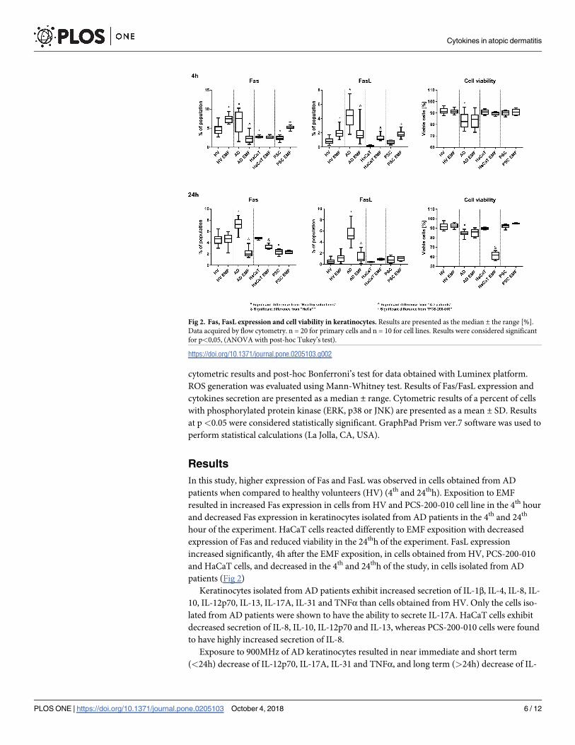
In this study, higher expression of Fas and FasL was observed in cells obtained from AD
patients when compared to healthy volunteers (HV) (4th and 24thh). Exposition to EMF
resulted in increased Fas expression in cells from HV and PCS-200-010 cell line in the 4th hour
and decreased Fas expression in keratinocytes isolated from AD patients in the 4th and 24th
hour of the experiment. HaCaT cells reacted differently to EMF exposition with decreased
expression of Fas and reduced viability in the 24thh of the experiment. FasL expression
increased significantly, 4h after the EMF exposition, in cells obtained from HV, PCS-200-010
and HaCaT cells, and decreased in the 4th and 24thh of the study, in cells isolated from AD
patients (Fig 2)
Keratinocytes isolated from AD patients exhibit increased secretion of IL-1β, IL-4, IL-8, IL-
10, IL-12p70, IL-13, IL-17A, IL-31 and TNFα than cells obtained from HV. Only the cells iso-
lated from AD patients were shown to have the ability to secrete IL-17A. HaCaT cells exhibit
decreased secretion of IL-8, IL-10, IL-12p70 and IL-13, whereas PCS-200-010 cells were found
to have highly increased secretion of IL-8.
Exposure to 900MHz of AD keratinocytes resulted in near immediate and short term
(<24h) decrease of IL-12p70, IL-17A, IL-31 and TNFα, and long term (>24h) decrease of IL-
Fig 2. Fas, FasL expression and cell viability in keratinocytes. Results are presented as the median ± the range [%].
Data acquired by flow cytometry. n = 20 for primary cells and n = 10 for cell lines. Results were considered significant
for p<0,05, (ANOVA with post-hoc Tukey’s test).
https://doi.org/10.1371/journal.pone.0205103.g002
Cytokines in atopic dermatitis
PLOS ONE | https://doi.org/10.1371/journal.pone.0205103 October 4, 2018 6 / 12

4, IL-10, IL-13 levels in the supernatant. In the 24thh of the study, exposure to EMF resulted in
a decrease of the IL-1β mean concertation level in the AD derived cell culture supernatant and
near immediate and short-term increase of IL-1β concentration in healthy cell culture super-
natants. EMF exposure also increased secretion of IL-8 (in the 24th hour of the study) by kera-
tinocytes obtained from healthy volunteers and patients with the AD. On the contrary,
exposition of the PCS-200-010 to the EMF resulted in a decrease of the IL-8 level in cell culture
supernatants. No statistically significant differences were found in the supernatant concentra-
tions of the investigated cytokines between exposed and unexposed HaCaT cells (Fig 3).
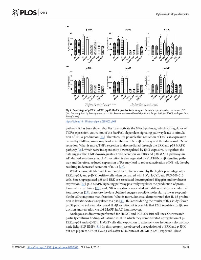
Phosphorylated-ERK (p-ERK) kinase was more abundantly expressed in cells obtained
from AD patients when compared to HV (4th and 24thh). HaCaT and PCS-200-010 cells were
found to have fewer p-ERK positive cells than HV (4th and 24thh). EMF exposure resulted in
decreased number of p-ERK positive cells in AD derived keratinocytes (4thh) and increased in
HaCaT cells (4thh).
P-p38 was found in a higher percentage in AD derived keratinocytes and a lower percent-
age of cells in HaCaT and PCS-200-010 when compared to HV (4thh and 24thh). Exposition to
EMF resulted in decreased number of p-p38 positive cells in AD derived keratinocytes (4thh)
and PCS-200-010 cells (4th and 24thh).
P-JNK was found in a higher percentage in AD derived keratinocytes when compared to
HV (4thh and 24thh). EMF exposure resulted in a decreased percentage of p-JNK positive cells
in AD derived keratinocytes and increased in HaCaT cells (4thh and 24thh) (Fig 4).
In order to investigate increased apoptosis rate in HaCaT cells after EMF exposition, rela-
tive ROS generation was measured using flow cytometry. 60 min EMF exposure resulted in
increased ROS production in HaCaT cells when compared to untreated control (2.426 vs 1 in
4th h and 1,937 vs 1.042 in 24th h, respectively).
Discussion
Immunomodulating properties of the electromagnetic field can significantly affect the profile
of chemokines and cytokines produced by keratinocytes, thus the recruitment of the different
T-cell subpopulations. Furthermore, several studies have suggested Fas-mediated elimination
of antigen-presenting cells as an important mechanism down-regulating the induction of auto-
immune responses. Restricting DC priming functions through Fas–FasL interactions were
shown to be a potent mechanism employed by CD4+/CD25+ regulatory T cells (Tregs) to
restrict CD8+ T-cell-mediated allergic immune responses in the skin. Therefore, death recep-
tors, such as Fas/FasL, may induce non-apoptotic signals and initiate cellular inflammatory
responses rather than modulate apoptosis. Thus, Fas/FasL may shape the quality and quantity
of the cytokine cocktail produced during the effector phase of the AD. These functions may
have important and distinct pathophysiological consequences during different stages of AD
inflammatory reaction.
In this study, we demonstrated that EMF downregulates the expression of Fas, FasL, per-
centage of p-ERK, p-JNK and p-p38 MAPK positive cells, and the secretion of IL-1β, IL-4, IL-
10, IL-12p70, IL-13, IL-17A, IL-31 and TNFα in the AD keratinocytes, which should have a
positive impact on the weakening of the symptoms. Although the molecular mechanism of
EMF-Fas interaction is not yet known, it was shown that exposure of human endothelial cell
line, EA.hy926 to the EMF caused the decrease in the expression of Fas mRNA [23]. Based on
literature analysis, it is highly probable that the NF-κβ pathway and the MAPK-dependent
pathway may be a potential site for regulation of the cytokine microenvironment in the AD
that is affected by EMF. For example, FasL and TNF are functionally related molecules that ini-
tiate apoptosis. Although each of the molecules initiates apoptosis by another signaling
Cytokines in atopic dermatitis
PLOS ONE | https://doi.org/10.1371/journal.pone.0205103 October 4, 2018 7 / 12

Fig 3. IL-1β, IL-4, IL-8, IL-10, IL-12p70, IL-13, IL-17A, IL-31 and TNFα concentration in keratinocytes. Results
are presented as the median ± the range [pg/mL]. Data acquired using Luminex MagPix. n = 15 for primary cells and
n = 10 for cell lines. Results were considered significant for p<0,05, (ANOVA with post-hoc Bonferroni’s test).
https://doi.org/10.1371/journal.pone.0205103.g003
Cytokines in atopic dermatitis
PLOS ONE | https://doi.org/10.1371/journal.pone.0205103 October 4, 2018 8 / 12
pathway, it has been shown that FasL can activate the NF-κβ pathway, which is a regulator of
TNFα expression. Activation of the Fas/FasL-dependent signaling pathway leads to stimula-
tion of TNFα production [24]. Therefore, it is possible that reduction of Fas/FasL expression
caused by EMF exposure may lead to inhibition of NF-κβ pathway and thus decreased TNFαsecretion. What is more, TNFα secretion is also mediated through the ERK and p38 MAPK
pathway [25], which were independently downregulated by EMF exposure. Altogether, the
data suggest that EMF downregulates TNFα secretion via ERK and p38 MAPK pathways in
AD derived keratinocytes. IL-31 secretion is also regulated by STAT6/NF-κβ signaling path-
way and therefore, reduced expression of Fas may lead to reduced activation of NF-κβ, thereby
resulting in decreased secretion of IL-31 [26].
What is more, AD derived keratinocytes are characterized by the higher percentage of p-
ERK, p-p38, and p-JNK positive cells when compared with HV, HaCaT, and PCS-200-010
cells. Since, upregulated p38 and ERK are associated downregulated filaggrin and involucrin
expression [27], p38 MAPK signaling pathway positively regulates the production of proin-
flammatory cytokines [28], and JNK is negatively associated with differentiation of epidermal
keratinocytes [29], therefore the data obtained suggests possible molecular pathway responsi-
ble for AD symptoms manifestation. What is more, Sun et al. demonstrated that IL-1β produc-
tion in keratinocytes is regulated via p38 [30], thus considering the results of this study (fewer
p-p38 positive cells and decreased IL-1β secretion) it is possible that EMF regulates IL-1β pro-
duction and secretion via p38 MAPK in AD keratinocytes.
Analogous studies were performed for HaCaT and PCS-200-010 cell lines. Our research
partially confirms findings of Patruno et. al. in which they demonstrated upregulation of p-
ERK, p-p38 and p-JNK in HaCaT cells after exposition to extremely low frequency electromag-
netic field (ELF-EMF) [31]. In this research, we observed upregulation of p-ERK and p-JNK
but not p-p38 MAPK in HaCaT cells after 60 minutes of 900 MHz EMF exposure. These
Fig 4. Percentage of p-ERK, p-JNK, p-p38 MAPK positive keratinocytes. Results are presented as the mean ± SD
[%]. Data acquired by flow cytometry. n = 18. Results were considered significant for p<0,05, (ANOVA with post-hoc
Tukey’s test).
https://doi.org/10.1371/journal.pone.0205103.g004
Cytokines in atopic dermatitis
PLOS ONE | https://doi.org/10.1371/journal.pone.0205103 October 4, 2018 9 / 12

results are in no way contradictory because effects of EMF exposure are both cell and fre-
quency (wavelength) specific.
Because of different base expression of Fas/FasL and cytokines secretion, and different
immune response of the PCS-200-010 and HaCaT cells to EMF exposure, those cell lines are
not a suitable model for investigation of EMF exposition effects and can’t replace, primary
human keratinocytes derived from the skin. Interestingly, decrease in HaCaT cell viability,
after EMF exposure, is probably caused by activation of ERK, JNK MAP kinase and mTOR
(PI3K / Akt) pathways, as well as, increased levels of reactive oxygen species that led to
increased apoptosis of these cells.
The electromagnetic field may have therapeutic potential in skin diseases like atopic derma-
titis however it also has limitations. Overexposure, different frequency or higher SAR may
cause thermal effects that will be harmful to cells and organs [32]. Effects of EMF exposure are
also cell type-dependent [33]. Moreover, Huang et al. observed that EMF may also differently
affect the same type of cells i.e. epidermis keratinocytes [34]. We also confirmed this observa-
tion. Exposure to EMF results in reduced survival in HaCaT cells but not in primary dermal
keratinocytes. Therefore, every potential therapeutic strategy involving the use of the EMF
should be carefully studied in pre-clinical and clinical trials.
In conclusion, both apoptotic and nonapoptotic activation of the Fas/FasL-dependent sig-
naling pathway may play a significant role in the pathogenesis of AD, by adjusting the local
cytokine and chemokine environment at the site of inflammation. Moreover, to the best of our
knowledge, this is the first publication to present that the electromagnetic field exhibits strong
immunomodulatory effects on AD-modified keratinocytes. Finally, obtained data suggests
that MAPK pathways mediated by ERK, JNK, and p38 protein kinases are responsible for AD
symptoms manifestation. These findings suggest that EMF may be potentially used as adjuvant
therapy in atopic dermatitis, however before human treatment its biological potential should
be evaluated in pre-clinical studies.
Author Contributions
Conceptualization: Lukasz Szymanski, Sławomir Lewicki, Wanda Stankiewicz.
Data curation: Lukasz Szymanski.
Formal analysis: Lukasz Szymanski.
Funding acquisition: Lukasz Szymanski.
Investigation: Lukasz Szymanski, Aleksandra Cios.
Methodology: Lukasz Szymanski.
Resources: Pawel Szymanski.
Supervision: Wanda Stankiewicz.
Writing – original draft: Lukasz Szymanski, Aleksandra Cios.
Writing – review & editing: Lukasz Szymanski, Sławomir Lewicki, Wanda Stankiewicz.
References
1. Bień K, Żmigrodzka M, Orłowski P, Fruba A, Szymański Ł, Stankiewicz W, et al. Involvement of Fas/
FasL pathway in the murine model of atopic dermatitis. Inflamm Res. 2017; 1–12.
2. Lee SH, Jang JJ, Lee JY, Kim SY, Park WS, Shin MS, et al. Fas ligand is expressed in normal skin and
in some cutaneous malignancies. Br J Dermatol. 1998; 139: 186–191. PMID: 9767230
Cytokines in atopic dermatitis
PLOS ONE | https://doi.org/10.1371/journal.pone.0205103 October 4, 2018 10 / 12

3. Boehm I. Apoptosis in physiological and pathological skin: implications for therapy. Curr Mol Med. 2006;
6: 375–394. PMID: 16900661
4. Wajant H, Pfizenmaier K, Scheurich P. Non-apoptotic Fas signaling. Cytokine Growth Factor Rev.
2003; 14: 53–66. PMID: 12485619
5. Kennedy NJ, Davis RJ. Chapter 164—Mammalian MAP Kinases. In: Bradshaw RA, Dennis EA, editors.
Handbook of Cell Signaling ( Second Edition). San Diego: Academic Press; 2010. pp. 1315–1328.
https://doi.org/10.1016/B978-0-12-374145-5.00164–9
6. Kim EK, Choi E-J. Pathological roles of MAPK signaling pathways in human diseases. Biochim Biophys
Acta BBA—Mol Basis Dis. 2010; 1802: 396–405. https://doi.org/10.1016/j.bbadis.2009.12.009 PMID:
20079433
7. Farley SM, Dotson AD, Purdy DE, Sundholm AJ, Schneider P, Magun BE, et al. Fas ligand elicits a cas-
pase-independent proinflammatory response in human keratinocytes: implications for dermatitis. J
Invest Dermatol. 2006; 126: 2438–2451. https://doi.org/10.1038/sj.jid.5700477 PMID: 16858424
8. Mycielska ME, Djamgoz MBA. Cellular mechanisms of direct-current electric field effects: galvanotaxis
and metastatic disease. J Cell Sci. 2004; 117: 1631–1639. https://doi.org/10.1242/jcs.01125 PMID:
15075225
9. Zhang HL, Peng HB. Mechanism of Acetylcholine Receptor Cluster Formation Induced by DC Electric
Field. PLOS ONE. 2011; 6: e26805. https://doi.org/10.1371/journal.pone.0026805 PMID: 22046365
10. Wu D, Ma X, Lin F. DC Electric Fields Direct Breast Cancer Cell Migration, Induce EGFR Polarization,
and Increase the Intracellular Level of Calcium Ions. Cell Biochem Biophys. 2013; 67: 1115–1125.
https://doi.org/10.1007/s12013-013-9615-7 PMID: 23657921
11. Wolf-Goldberg T, Barbul A, Ben-Dov N, Korenstein R. Low electric fields induce ligand-independent
activation of EGF receptor and ERK via electrochemical elevation of H+ and ROS concentrations. Bio-
chim Biophys Acta BBA—Mol Cell Res. 2013; 1833: 1396–1408. https://doi.org/10.1016/j.bbamcr.
2013.02.011 PMID: 23481041
12. Rosado MM, Simko M, Mattsson M-O, Pioli C. Immune-Modulating Perspectives for Low Frequency
Electromagnetic Fields in Innate Immunity. Front Public Health. 2018; 6. https://doi.org/10.3389/fpubh.
2018.00085 PMID: 29632855
13. Leshem YA, Hajar T, Hanifin JM, Simpson EL. What the Eczema Area and Severity Index score tells us
about the severity of atopic dermatitis: an interpretability study. Br J Dermatol. 2015; 172: 1353–1357.
https://doi.org/10.1111/bjd.13662 PMID: 25580670
14. Boukamp P, Petrussevska RT, Breitkreutz D, Hornung J, Markham A, Fusenig NE. Normal keratiniza-
tion in a spontaneously immortalized aneuploid human keratinocyte cell line. J Cell Biol. 1988; 106:
761–771. PMID: 2450098
15. Aasen T, Belmonte JCI. Isolation and cultivation of human keratinocytes from skin or plucked hair for
the generation of induced pluripotent stem cells. Nat Protoc. 2010; 5: 371. https://doi.org/10.1038/nprot.
2009.241 PMID: 20134422
16. Sobiech J, Kieliszek J, Puta R, Stankiewicz W. Wykorzystanie komory TEM do ekspozycji na
pole elektromagnetyczne obiektow biologicznych. Przegląd Elektrotechniczny. 2015;R. 91, nr 12:
218–220.
17. Stankiewicz W, Dabrowski MP, Kubacki R, Sobiczewska E, Szmigielski S. Immunotropic influence of
900 MHz microwave GSM signal on human blood immune cells activated in vitro. Electromagn Biol
Med. 2006; 25: 45–51. https://doi.org/10.1080/15368370600572961 PMID: 16595333
18. Zmyślony M, Politanski P, Rajkowska E, Szymczak W, Jajte J. Acute exposure to 930 MHz CW electro-
magnetic radiation in vitro affects reactive oxygen species level in rat lymphocytes treated by iron ions.
Bioelectromagnetics. 2004; 25: 324–328. https://doi.org/10.1002/bem.10191 PMID: 15197754
19. Dabrowski MP, Stankiewicz W, Kubacki R, Sobiczewska E, Szmigielski S. Immunotropic Effects in Cul-
tured Human Blood Mononuclear Cells Pre-exposed to Low-Level 1300 MHz Pulse-Modulated Micro-
wave Field. Electromagn Biol Med. 2003; 22: 1–13. https://doi.org/10.1081/JBC-120020347
20. Bort A, Alvarado-Vazquez PA, Moracho-Vilrriales C, Virga KG, Gumina G, Romero-Sandoval A, et al.
Effects of JWH015 in cytokine secretion in primary human keratinocytes and fibroblasts and its suitabil-
ity for topical/transdermal delivery. Mol Pain. 2017; 13. https://doi.org/10.1177/1744806916688220
PMID: 28326930
21. Keller M, Ruegg A, Werner S, Beer H-D. Active caspase-1 is a regulator of unconventional protein
secretion. Cell. 2008; 132: 818–831. https://doi.org/10.1016/j.cell.2007.12.040 PMID: 18329368
22. Leśniak M, Zdanowski R, Suska M, Brewczyńska A, Stankiewicz W, Kloc M, et al. Effects of Hexachlo-
rophene, a Chemical Accumulating in Adipose Tissue, on Mouse and Human Mesenchymal Stem
Cells. Tissue Eng Regen Med. 2018; 15: 211–222. https://doi.org/10.1007/s13770-017-0103-9
Cytokines in atopic dermatitis
PLOS ONE | https://doi.org/10.1371/journal.pone.0205103 October 4, 2018 11 / 12

23. Nylund R, Leszczynski D. Proteomics analysis of human endothelial cell line EA.hy926 after exposure
to GSM 900 radiation. Proteomics. 2004; 4: 1359–1365. https://doi.org/10.1002/pmic.200300773
PMID: 15188403
24. Lu B, Wang L, Medan D, Toledo D, Huang C, Chen F, et al. Regulation of Fas (CD95)-induced apopto-
sis by nuclear factor-kappaB and tumor necrosis factor-alpha in macrophages. Am J Physiol Cell Phy-
siol. 2002; 283: C831–838. https://doi.org/10.1152/ajpcell.00045.2002 PMID: 12176740
25. Kjellerup RB, Kragballe K, Iversen L, Johansen C. Pro-inflammatory cytokine release in keratinocytes is
mediated through the MAPK signal-integrating kinases. Exp Dermatol. 2008; 17: 498–504. https://doi.
org/10.1111/j.1600-0625.2007.00672.x PMID: 18081851
26. Maier E, Werner D, Duschl A, Bohle B, Horejs-Hoeck J. Human Th2 but not Th9 cells release IL-31 in a
STAT6/NF-κB-dependent way. J Immunol Baltim Md 1950. 2014; 193: 645–654. https://doi.org/10.
4049/jimmunol.1301836 PMID: 24943220
27. Tan Q, Yang H, Liu E, Wang H. P38/ERK MAPK signaling pathways are involved in the regulation of
filaggrin and involucrin by IL17. Mol Med Rep. 2017; 16: 8863–8867. https://doi.org/10.3892/mmr.2017.
7689 PMID: 28990053
28. Mutou Y, Tsukimoto M, Homma T, Kojima S. Immune Response Pathways in Human Keratinocyte
(HaCaT) Cells are Induced by Ultraviolet B via p38 Mitogen-activated Protein Kinase Activation. J
Health Sci. 2010; 56: 675–683. https://doi.org/10.1248/jhs.56.675
29. Gazel A, Banno T, Walsh R, Blumenberg M. Inhibition of JNK Promotes Differentiation of Epidermal
Keratinocytes. J Biol Chem. 2006; 281: 20530–20541. https://doi.org/10.1074/jbc.M602712200 PMID:
16648634
30. Sun Y, Zhang J, Zhai T, Li H, Li H, Huo R, et al. CCN1 promotes IL-1β production in keratinocytes by
activating p38 MAPK signaling in psoriasis. Sci Rep. 2017; 7. https://doi.org/10.1038/srep43310 PMID:
28266627
31. Patruno A, Pesce M, Grilli A, Speranza L, Franceschelli S, Lutiis MAD, et al. mTOR Activation by PI3K/
Akt and ERK Signaling in Short ELF-EMF Exposed Human Keratinocytes. PLOS ONE. 2015; 10:
e0139644. https://doi.org/10.1371/journal.pone.0139644 PMID: 26431550
32. Zhi W-J, Wang L-F, Hu X-J. Recent advances in the effects of microwave radiation on brains. Mil Med
Res. 2017; 4. https://doi.org/10.1186/s40779-017-0139-0 PMID: 29502514
33. D’Angelo C, Costantini E, Kamal MA, Reale M. Experimental model for ELF-EMF exposure: Concern
for human health. Saudi J Biol Sci. 2015; 22: 75–84. https://doi.org/10.1016/j.sjbs.2014.07.006 PMID:
25561888
34. Huang C-Y, Chuang C-Y, Shu W-Y, Chang C-W, Chen C-R, Fan T-C, et al. Distinct Epidermal Keratino-
cytes Respond to Extremely Low-Frequency Electromagnetic Fields Differently. PLOS ONE. 2014; 9:
e113424. https://doi.org/10.1371/journal.pone.0113424 PMID: 25409520
Cytokines in atopic dermatitis
PLOS ONE | https://doi.org/10.1371/journal.pone.0205103 October 4, 2018 12 / 12