Fall, 20 12 - Napa Valley College Pages - Napa Valley College...
91
Napa Valley College Health Occupations NURS 372 Intravenous Certification Fall, 2012 Janice Ankenmann RN MSN CCRN FNP-C
Transcript of Fall, 20 12 - Napa Valley College Pages - Napa Valley College...

Napa Valley College Health Occupations
NURS 372
Intravenous Certification
Fall, 2012
Janice Ankenmann RN MSN CCRN FNP-C

1
Intravenous Therapy Class Purpose: To teach licensed personnel (whom IV starts and care are part of their scope of practice) the fundamental principles and competencies required to begin, maintain and discontinue intravenous therapy. Objectives: At the completion of 30 total hours of training (24 hours didactic and 6 hours of clinical practice), the participant will be able to:
1. demonstrate understanding and knowledge of the veni-puncture procedure and intravenous therapy as evidenced by the successful completion of the written objectives for each of the four sessions,
2. demonstrate competency in starting, maintaining, and discontinuing IV therapy as evidenced by three individually supervised successful IV starts on live subjects with demonstrated ability to maintain and discontinue IV therapy utilizing appropriate aseptic technique.
Methods: Methods employed to teach the fundamentals of veni-puncture and IV therapy will include lecture, participative discussion, visual aids, written materials and review, as well as, clinical laboratory experience using practice, actual equipment/supplies and live subjects.

2
INTRAVENOUS THERAPY SESSION I
0730-0800 Introductions 0800-0830 Legal Aspects 0830-0930 Anatomy and Physiology of Circulation 0930-0940 Break 0940-1100 Site Selection 1100-1200 Lunch 1200-1330 Fundamentals of Fluids and Electrolytes 1330-1340 Break 1340-1500 1500- 1615
Indications for IV Therapy Clinical Scenario Review
1615- 1630 Objectives Review; Questions and Answers
LEARNING OBJECTIVES
1. Define the terms “intravenous fluids” and “superimpose” as stated under article 8 of the Vocational Nursing Practice Act.
2. Identify two documents that govern the practice of individual LVN’s regarding IV
therapy.
3. Understand clearly that LVN’s do not give IV meds under any circumstances.
4. Identify five factors to be considered in selecting appropriate sites for IV catheter placement.
5. Identify why the lower extremities are not routinely used for IV therapy.
6. Define “electrolytes” and give four examples.
7. Idenify five signs and symptoms that could indicate fluid/electrolyte imbalance.
8. Identify three clinical situations where you would suspect a patient may be at risk
for fluid/electrolyte imbalance.
9. Name four methods of transport across the semi-permeable cell membrane.
10. List five indications for IV therapy.
11. Identify the differences between isotonic, hypotonic and hypertonic solutions and give two examples each.
12. Name three ways to identify veins vs. arteries.

3
INTRAVENOUS THERAPY SESSION II
0730-0800 Review; Question and Answer 0800-0900 Review of Aseptic Technique and Universal Precautions 0900-1000 Venipuncture and IV Equipment 1000-1010 Break 1010-1100 Venipuncture Procedure 1100-1200 Lunch 1200-1300 Nursing Responsibilities (includes site inspection, dressing changes) 1300-1310 Break 1310-1345 Clinical Scenario Review 1345-1615 Skills Lab: venipuncture practice on models; handling equipment;
using a tourniquet, Butterfly sticks 1615-1630 Objectives Review; Questions and Answers
LEARNING OBJECTIVES
1. Identify three reasons why you might use a butterfly versus an angiocath when starting an IV.
2. Identify three benefits to using the antiocath.
3. Identify two reasons you would use an IV pump.
4. Identify two situations where you would use a microdrip tubing vs. macrodrip.
Why?
5. Describe infection control guidelines pertaining to IV therapy/venipuncture.
6. Know your workplace policy regarding IV site care and who may do what with central lines.
7. Describe the potential clinical findings that could be observed when caring for a
patient receiving IV therapy.
8. List five nursing responsibilities related to the care of the IV system.
9. Under direct supervision by an instructor, demonstrate appropriate technique for: -choosing a venipuncture site, -cleansing the skin, -choosing an appropriate IV device, -utilizing a tourniquet, -inserting the IV device, -securing the IV device, -attaching IV tubing/peripheral lock and -dressing the site.

4
INTRAVENOUS THERAPY SESSION III
0730-0800 Review; Question and Answer 0800-0930 Special Clients (pediatric, geriatric, anticoagulated) 0930-0940 Break 0940-1000 Potassium via Infusion 1000-1100 Calculating IV Fluid Drip Rates 1100-1200 Lunch 1200-1230 Review procedure for angiocath insertion 1230-1240 Break 1240-1530 Skills Lab: angiocath insertion 1530-1610 1610-1630
Clinical Scenario Review Objectives Review; Questions and Answers
LEARNING OBJECTIVES
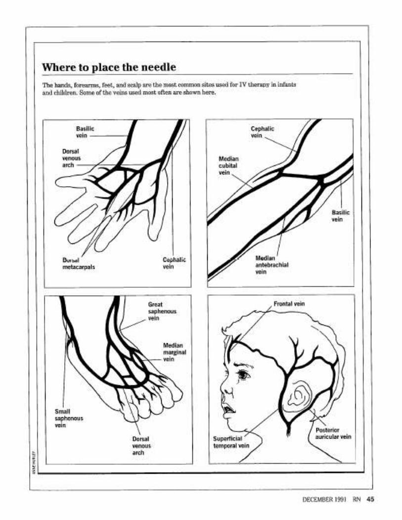
1. Identify the three most frequently used anatomical sites used for venipuncture in the infant.
2. Describe the preparation and approach to the pediatric client and parent(s) used
prior to venipuncture.
3. Describe two considerations to be evaluated prior to starting an IV on the geriatric patient.
4. Identify three patient populations that may be anticoagulated. How might this
affect the venipuncture procedure?
5. Identify three important nursing considerations to remember when administering IV solutions that contain KCl.
6. Demonstrate the ability to accurately calculate IV drip rates given a set of
hypothetical clinical scenarios.
7. Under direct supervision of an RN instructor, demonstrate on live subjects the correct procedure for:
-venipuncture with an angiocath, -dressing an IV site, -flushing a peripheral lock, -hanging IV fluid as ordered and calculating drip rates, and -discontinuing IV therapy and angiocath.

5
INTRAVENOUS THERAPY SESSION IV
0730-0800 Review; Question and Answer 0800-1000 Complications of IV Therapy/Venipuncture 1000-1010 Break 1010-1100 TPN, PPN, Lipid Administration 1100-1145 Blood Administration 1145-1245 Lunch 1245-1345 Clinical Scenario Review 1345-1445 Objectives Review; Questions and Answers 1445-1455 Break 1500-1600 Final Exam 1600-1615 Review of Final Exam 1615-1630 Program Evaluation/ Certificates of Completion
LEARNING OBJECTIVES
1. Identify the signs, symptoms and usual treatment of four local complications of venipuncture and IV therapy.
2. Identify the signs, symptoms and usual treatment of four systemic complications
of venipuncture and IV therapy.
3. Identify two measures that can be employed to prevent local and/or systemic complications.
4. Discuss the differences between TPN and PPN.
5. Identify three reasons for the use of parenteral nutrition.
6. Explain why parenteral nutrition poses a high risk for infection.
7. Identify four potential symptoms of transfusion reaction.
8. Identify five “rules” to follow for safe blood administration.

6
HOW ARE YOU FEELING TODAY?
7
Resources
Booth, K. (2008) Intravenous Therapy for Healthcare Personel. McGraw Hill.
ISBN 978-0-07-328112-4
Gahart, B. Nazareno, A. (2010) Intravenous Medications (25th
edition). St.
Louis, Mo: Mosby/ Elsevier.
Hogan, Gingrich, Overby, Ricci. ( 2007) Fluids, Electrolytes & Acid-Base Balance
(2nd
ed) Prentice Hall Nursing.
Lewis, Dirksen, Heitkemper, Buchner, Camera. (2011) Medical Surgical Nursing
(8th
ed) Elsevier Mosby.
Josephson, Dianne, L. (2004) Intravenous Infusion Therapy for Nurses (2nd
edition). Delmar Learning. ISBN 1-4018-0935-9
Mosby’s Nursing Video Skills: Advanced Skills (institutional version 3.0). ISBN
978-0-323-05293-1
Perry, AG. & Potter, PA. (2012) Nursing Interventions and Clinical Skills (8th
ed.). St. Louis, Mo. C.V.Mosby Co.
Philips, L. (2010) Manual of IV Theraputics (5th
edition). Philadelphia, Pa: F.A.
Davis.
(Plus articles contained within class materials)
8
Indications For
IV Therapy

9
INDICATIONS FOR I.V. THERAPY
A. To establish or maintain fluid and electrolyte balance. B. To administer continuous or intermittent medication. C. To administer a bolus injection. D. To administer blood and/or blood component therapy. E. To administer intravenous anesthetics. F. To maintain or correct a patient’s nutritional state. G. To administer diagnostic substances. H. To monitor hemodynamic function of central venous circulation.

10
Legal Aspects

11
LEGAL ASPECTS OF IV THERAPY/BLOOD WITHDRAWAL
CONSIDERATIONS: NEGLIGENCE ASSAULT and BATTERY FALSE IMPRISONMENT SLANDER and LIBEL MALPRACTICE PERSONAL LIABILITY * PATIENT BILL OF RIGHTS * CALIFORNIA VOCATIONAL NURSING PRACTICE ACT * CALIFORNIA CODE OF REGULATIONS Ruling regarding LVN’s and IV MEDS CENTRAL LINES TPN/ PPN LOCAL ANESTHETICS PERIPHERAL LOCKS

12
Legal Aspects of IV Therapy/Phlebotomy In California
In 1985, eighteen states had rulings allowing LVN’s to administer IV Therapy. Other states believe that the understanding for IV Therapy cannot be built on the scope of knowledge required for the basic practice nurse education. California is one of those states who do allow LVN’s to administer IV Therapy. Considerations
1. Negligence: Failure to do something or doing something that a responsible person would not do. Not acting in a reasonable and prudent manner with resultant damage to a person or his property.
2. Assault and Battery: Assault is an attempt or threat to physically injure a
person. Battery is the unauthorized touching of a person. “Coercion on a rational adult patient in order to insert a cannula constitutes assault and battery”. (1)
3. False Imprisonment: The act of confining or restraining a person without his
consent for no clinical or legal reason.
4. Slander and Libel: Slander is the oral statement made with intent to dishonor or defame another person when mad in the presence of a third person. Libel is a false or malicious writing that is intended to defame or dishonor a person and is published so other people can see it.
5. Malpractice: Is the negligent conduct of professional persons. The professional
misconduct, or unreasonable lack of skill of professional duties.
6. Personal Liability: “Every person is liable for his own conduct (his own wrongdoing).” (2) The physician cannot protect the nurse by giving a verbal or written order. We are directly responsible for what we each do as nurses, regardless of who ordered it done.
A. Observation is the legal and professional responsibility of the nurse. Frequent observation is imperative for early detection and prevention of a problem (ISMP).
B. Nurse-Patient Relationship Role is significant in the prevention of initiating legal liability against the nurse. Take time to develop an efficient, interpersonal relationship. A patient that is resentful, uncooperative, and dissatisfied with his care is the one most likely to take legal action when a problem arises. By giving skilled efficient care along with genuine respect and concern for the patient, a nurse may be more likely to avoid malpractice claims.
C. Be aware of your institution’s policies and procedure.

13

14

15

16

17

18

19

20
Site Selection

21
SITE SELECTIONS FOR I.V. THERAPY
I. Factors to Consider A. Patient’s medical history B. Patient’s age, size, general condition C. Condition of patient’s veins D. Type and rate of I.V. fluid or medication to be infused E. Expected duration of I.V. therapy II. Options in Adults: A. Veins in the hand: Metacarpal and digital veins.
B. Veins in the lower arm: Cephalic and basilica veins – larger arm veins do not become phlebotic as quickly as hand veins.
C. Veins in upper arm (above antecubital fossa) – these are deeper veins; good choice for infusing irritating solutions because they are less prone to phlebitis than lower veins.
D. Veins of the inner aspect of the arm and wrist – use only if absolutely necessary. They are thin walled, and are often associated with bruising, phlebitis, and infiltration.
E. Veins of legs, feet and ankles – generally used only with doctor’s approval. Using them may compromise circulation in the legs and cause thrombophlebitis or embolism.
F. Avoid using the following sites: Veins below a previous I.V. infiltration Veins below a phlebotic area Sclerosed or thrombosed veins Areas of skin inflammation, disease, bruising or breakdown An arm affected by radical mastectomy, edema, blood clot or
infection An arm with arteriovenous shunt or fistula
G. Avoid arteries: Generally located deeper than the veins Risk greater in the antecubital fossa because arteries and veins lie
close together To locate nearby arteries, palpate for pulsation Unlike superficial veins, arteries do not look bluish

22

23

24

25
Infection Control

26

27
STOP the spread of germs…
WASH YOUR HANDS
PROPER HAND WASHING TECHNIQUE
1. Use continuously running water.
2. Use plenty of soap.
3. Apply soap with vigorous contact on all surfaces of hands.
4. Wash hands for 10 – 15 seconds.
5. Keep hands down at all times, so any run off will go into the sink, and not down the arms.
6. Avoid splashing, rinse thoroughly.
7. Dry well with paper towels.
8. Discard the towels into a bag provided for that purpose.
9. Use a paper towel to turn off faucet.
Good handwashing is the single most effective means of preventing the spread of
infection.

28
UNIVERSAL PRECAUTIONS
All patients should be assumed to be infections for blood-borne pathogens. Gloves must be worn when handling a patient’s body fluids. Protective mask, eye goggles, and gown should also be worn if there is potential for mucous membrane exposure to blood or other body fluids. Take care to prevent injuries when using needles, scalpels, and other sharp instruments or devices. Never recap needles. Always dispose of needles immediately in a biohazard container. Immediately and thoroughly wash hands and other skin surfaces that are contaminated with blood, body fluids containing visible blood, or other body fluids to which universal precautions apply. Review your institutional guidelines for observing universal precautions.

29
ASEPSIS / SAFETY
ASEPSIS: a condition free from germs; free from infections; sterile, free from any form
of life (Tabers Cyclopedic Medical Dictionary)
Universal Precautions
Handwashing Potential Sources of Infection with Venipuncture and IV Catheters 1. autoinfection external spread hematogenous seeding 2. hands of medical personnel 3. contaminated disinfectants 4. contaminated equipment IV Site Dressings……..
1. Should be occlusive (according to institutions policy; transparent dressing; sterile 2x2 securely taped).
2. Should allow visualization of the site or the dressing should be changed daily.
3. Should be changed when wet or appears soiled.
SAFETY>>>>>>>>
1. Never re-cap a dirty needle! Use needleless systems or the safety!!
2. Pay attention to what you are doing…sticks often occur because the phlebotomist is distracted.
3. If your patient is unable to co-operate, get assistance. (remember patient
rights!)
4. Always properly dispose of sharps in a sharps container, bloody supplies in a biohazardous waste container.

30
What to do if you stick yourself…… If you are with a patient, excuse yourself, making sure that they are safe (no tourniquet on their arm, etc.) and let them know if that someone else will be in to finish the procedure.
1. Remove your contaminated gloves and dispose of them properly.
2. Squeeze the puncture site to promote bleeding.
3. Wash the area well with soap and water.
4. Record the patient’s name and identification number.
5. Immediately report the incident to your supervisor and follow your institutions policy/procedure regarding reporting, treatment and follow up.
What to do if you splash blood on your skin or into your eye…..
6. If you are with a patient, excuse yourself, making sure that they are safe (no tourniquet on their arm, etc.) and let them know if that someone else will be in to finish the procedure.
Wash the area with generous quantities of water. Wipe up any spilled blood with paper towels and disinfectant (10% bleach or other). Dispose of paper towels in a biohazardous waste container. Record the patient’s name and identification number. Immediately report the incident to your supervisor and follow your institutions policy/procedure regarding reporting, treatment, and follow-up.

31
Intravenous Access Taping Methods
Using the transparent dressing shown you can secure the vascular cannula with tape using the U, H, or chevron method, as illustrated below.
The U method
The H method
The chevron method

32
Complications of
IV Therapy

33
COMPLICATIONS OF I.V. THERAPY
I. Local Complications A. Hematoma: a pooling of blood into the subcutaneous space. 1. Signs and symptoms a. discoloration b. swelling c. tenderness 2. Treatment a. firm pressure b. elevated extremity on pillow 3. Prevention a. insert I.V. cannula at correct angle b. when discontinuing an I.V. cannula, correct application of pressure
B. Infiltration: the extravasation of injected fluid into the subcutaneous tissue.
1. Signs and symptoms a. swelling at site or dependent areas b. slowing of infusion rate c. blanching in severe cases d. patient discomfort at site e. coolness of area 2. Treatment a. exercise unless contraindicated b. if severe, frequent assessment of capillary refill c. if chemical composition of infusate causes tissue damage, an antidote may need to be administered d. warm compresses 3. Prevention a. appropriate selection of I.V. site and cannula b. proper stabilization of I.V. cannula c. use of tension loop d. proper placement of restraints e. validation of proper placement of cannula before admistration of an I.V. medication

34
C. Phlebitis/Thrombophlebitis 1. Phlebitis is an inflammation of the vein 2. Thrombophlebitis is a clot formation with phlebitis 3. Signs and symptoms a. tenderness b. redness c. heat d. edema e. palpable venous cord 4. Treatment a. relocate I.V. site b. elevate affected extremity c. vigorous exercise contraindicated with thrombophlebitis 5. Prevention a. proper selection of site and I.V. cannula size b. proper prepping of sit before insertion of cannula c. avoiding multiple venipuncture d. adequate taping of I.V. cannula e. adequate hemodilution of caustic solutions or medications f. adequate reconstitution of medications g. adequate flushing of I.V. line between incompatible medications h. following institutional policy related to I.V. infusion, I.V. tubing, I.V. dressing, and I.V. site changes
D. Infection: is the result of an invasion of pathogens that are localized in the surrounding tissues
1. Signs and symptoms a. purulent drainage b. tenderness c. swelling d. erythema e. induration 2. Treatment a. establish a new I.V. site b. remove old I.V. and send catheter tip for culturing along with any portion of administration set or solution that is suspected as being a part of the causative factor c. monitor patient over next 48 hours for development of septicemia d. follow institutional policy related to I.V. infusion, I.V. tubing, I.V. dressing, and I.V. site changes e. instruct patient in their role of maintaining integrity of the I.V. system

35
II. Systemic complications A. Septicemia: is a systemic proliferation of pathogens 1. Signs and symptoms a. gradual or sudden rise of temperature b. shaking chills c. tachycardia d. headache e. gastric symptoms f. can progress to septic shock 2. Treatment a. relocate I.V. site using different lot number for all medical equipment used in establishing the I.V. b. collect and culture appropriate specimens from catheter, tubing, and/or solutin; separate blood cultures must be obtained
B. Air embolism: is when air inadvertently enters the venous system. This is more frequently associated with central lines
1. Signs and symptoms a. chest pain b. shortness of breath c. cyanosis d. low back pain e. hypotension f. weak thready pulse g. loss of conciousness h. loud churning murmur over precordium C. Catheter embolism: occurs when a portion of the catheter breaks off and flows into the vascular system 1. Signs and symptoms a. depends on size of catheter embolism and final destination in vascular system b. range from no symptoms to massive pulmonary embolism symptoms 2. Treatment a. apply tourniquet to most proximal joint of the torso b. further treatment depends on patient’s clinical status 3. Prevention a. never restylet over-the-needle catheters b. never withdraw through-the-needle catheters through the introducer needle c. never use scissors during dressing change or removal of the dressing of either a peripheral or central I.V. catheter

36
D. Speed shock: results from a rapid introduction of a medication into the circulatory system 1. Signs and symptoms may vary depending on the patient and can Include a. flushing of faces b. headache c. severe shock to death 2. Treatment depends on the patient’s clinical status 3. Prevention a. appropriate reconstitution of all drugs b. appropriate adding of drugs to an I.V. solution already hanging c. appropriate hemodilution of all drugs given I.V.
E. Circulatory overload: is decompensation of the circulatory system due to excessive volume of fluid
1. Signs and symptoms vary according to the patient’s age, patient’s excessive volume of fluid a. hypoxemia b. dyspnea c. productive cough d. frothy sputum e. jugular vein distention 2. Treatment a. reduce infusion rate to keep open rate b. place patient in high Fowlers c. observe vital signs d. administer oxygen and diuretics according to the physician’s plan of care 3. Prevention a. frequent monitoring of I.V. flow rate b. assessment of the patient’s fluid and electrolyte balance c. use of intermittent infusion or mechanical devices III. Nursing documentation related to complications includes A. Patient signs and symptoms B. Nursing care implemented C. Patient outcome evaluated

37
Nursing Responsibilities

38
NURSING RESPONSIBILITIES regarding VENIPUNCTURE and IV THERAPY
PATIENT TEACHING
always explain what procedure you will be performing on a patient and why, prior to starting any part of the procedure (unless emergent)
patients and families also need to know any activity restrictions that may
be imposed, as well as what to report to the nurse (soreness, redness, swelling, pain, a cool or numb sensation at the insertion site)
stress the importance of keeping the site clean, the dressing intact, not
attempting to adjust the IV fluid rate if applicable, and the need to not kink the IV tubing
be honest about the fact that venipuncture does hurt and reassure them
that the discomfort will be of short duration…allay anxiety….in a very anxious patient, their own level of adrenalin in reaction to fear will cause vasoconstriction, thereby making venipuncture difficult
INFECTION CONTROL GUIDELINES
handwashing adhere to universal precautions adhere to facility policies re: venipuncture, IV dressing changes (every three days
and prn), IV tubing changes (usually every 72 hours of routine IV’s), etc. change IV solutions every 24 hours maintain a closed IV system frequently monitor site/extremity for signs and symptoms of infection do not re-cap sharps; utilize the safety; and dispose of them in sharps containers maintain aseptic technique when initiating/discontinuing venipuncture procedures
and/or handling any parts of the IV system PATIENT OBSERVATION *reported to the MD and/or RN, as appropriate *documented in the medical record as per facility policy
site lung sounds jugular vein distention urine output skin turgor appearance of mucous membranes laboratory reports radiology reports mental status

39
Special Clients

40
CONSIDERATIONS WITH THE AGED PATIENT
REGARDING IV THERAPY AND PHLEBOTOMY
As we age, our body structures change with decreasing body function and decreased body function and decreased ability to recuperate from injury and stress. We experience diminished pulmonary, renal, cardiac, and GI function. This is turn creates the tendency to have difficulty maintaining homeostasis….fluid and electrolyte imbalances are common. Considerations:
Mental function: does the patient understand what you are saying to them? are they confused normally or is this a symptom of electrolyte/fluid imbalance?
Senses: can the patient hear your explanation of treatment? can they
see what you are doing or do you need to explain each stop so as to not surprise them?
Cardiac: do they have dependent edema? arteriosclerosis? Is this
affecting peripheral pulses or the lumen of the veins?
Musculoskeletal: does the patient have decreased range of motion affecting their ability to position their limb for venipuncture? will the placement of an IV inhibit their ability to provide self care? site affect their ability to provide self care?
Skin: muscle atrophy and decreased skin turgor may affect your ability to
access veins (they may “roll”); skin may have rashes/abrasions/etc. due to medications and poor healing…this may affect your ability to secure a dressing over an IV site without harming the skin further.

41
CONSIDERATIONS WITH THE ANTICOAGULATED PATIENT
REGARDING IV THERAPY AN PHLEBOTOMY ANTICOAGULANT: 1) delaying or preventing blood coagulation, 2) an agent which delays or prevents blood coagulation (Taber’s Cyclopedic Medical Dictionary) Examples: HEPARIN, ASPIRIN, COUMADIN, PLAVIX, LOVENOX, FRAGMIN CONSIDERATIONS:
use as small a needle/angiocath as possible to meet the patient’s needs
always observe for hematoma and continued bleeding in high risk patients….apply pressure to the site for 5-10 minutes or longer prn to stop the bleeding
access patients as distal as possible…if the vein blows or hematoma forms, it will
allow access the same vessel higher up the extremity
consider the use of a central line or arterial line to access specimens rather than providing an additional puncture

42
INTRAVENOUS INFUSIONS IN PEDIATRICS
OUTLINE
I. Indications for Intravenous Therapy
fluid replacement open line (emergency) blood withdrawal diagnostic testing medications
II. Physical Considerations size (weight) activity level LOC physical assessment
III. Other Considerations parents communication different expectations different
IV. Site Selection activity length of time what’s infusing why needed history
V. Selection of Device VI. Equipment Needed VII. Intravenous Insertion Techniques VIII. Care and Maintenance of Intravenous Sites
MAINTENANCE INTRAVENOUS FLUIDS CHILDS WEIGHT CC/KG/24 HOURS Up to 10 KG 100 CC/KG/24 HOURS 11-20 KG 1000 CC + 50 CC/KG EACH KG OVER 10 KG/24 HOURS 21-30 KG 1500 CC + 25 CC/KG EACH KG OVER 10 KG/24HOURS
OR
HOURLY INTRAVENOUS FLUIDS
4 CC/Kg for the first 10 Kg 40 cc + 2 cc/Kg for each Kg over 10 Kg (up to 20 Kg)
60 cc + 1 cc/Kg for each Kg over 20 Kg

43
IV Equipment

44
EQUIPMENT FOR I.V. THERAPY
I. Cannula: A. Types 1. Steel needle (scalp vein or butterfly): greatly increases the risk of vein injury and infiltration. Uses:
short term IV therapy – only few hours dye injections bolus injections not requiring an IV line short term chemotherapy
2. Over-the-needle catheters (angiocath, winged-cannula): varies from 3/4” to 1” long; diameters range from 16 gauge to 24 gauge. Uses:
short or long term therapy delivery of viscous fluid intermittent therapy
3. Inside the needle catheters: longer in length than over-the-needle catheters. Uses:
long term therapy delivery of viscous fluid delivery of fluid or medication via a central line
4. Peripheral Locks (Heparin Lock; Saline Lock) for intermittent administration of medications or fluids Uses:
to keep an intravenous access without fluid administration B. Choosing the right size. Consider using:
to reduce the risk of phlebitis, catheter should be as small in diameter as possible
24 gauge cannula for infants, children, and adults with extremely small veins
20-22 gauge for medical patients (larger gauge catheter if larger vein is used to infuse caustic or viscous solution)
18 gauge for surgical patients and blood administration 16 gauge for trauma patients and those undergoing major
surgery

45
II. IV Solutions: A. Correct as prescribed B. No leaks, particulate matter, discoloration C. Compatabilities III. IV Tubings: A. Macrodrip – 10 gtts/cc or 15 gtts/cc B. Microdrip/ Pediatric – 60 gtts/cc C. Secondary lines – for IVPB D. Volutrol/Metri-set E. Blood Tubing- 10gtts/cc IV. Pumps and Controllers
A. Pumps deliver IV fluids using positive pressure. They are used in most acute care facilities on all IV’s. “Smart pumps” are also available. These smart pumps allow the user to label the IV lines with the name of the fluid/ medication infusing and can even help to calculate drip rates/ drug dosing. Remember to use the specialty tubing for the specific pump being used!
B. Controllers deliver IV fluids using gravity as the source of infusion pressure.
V. Venipuncture Set: (includes prepping solution, tourniquet, dressing, label, tape) Additional equipment/supplies: IV pole, armboard or handboard, gloves, additional lighting, if needed. VI. Peripheral Locks/ Extension Tubing:
A. Peripheral locks: A “dead space” whereby patency is maintained with NS or Heparin.
Used for intermittent medications, emergency vascular assess, and intermittent blood withdrawal. Can be positive pressure (to prevent backflow into the catheter tip).
B. Extension Tubing: used to extend the length of the IV tubing to allow better movement for the patient or more access ports for the caregivers.
VII. Filters: A. Incorporated into IV tubing; “membrane”. B. Used to filter particulate matter from solutions, medications and blood products infused into the patient. C. Refer to your facility policies for use.

46

47
Central Lines
The following is intended as a general overview of central lines. For more detailed information, further resources will need to be accessed. There are many types of central lines commonly used---tunneled, non-tunneled, and access ports that are surgically placed. The type of line used is determined by the physician based on patients needs: length of time catheter will be needed, purpose of its use, age and mobility of the patient, immune response of the patient, to name a few. There are many risks of central lines including infection, catheter migration/ emboli, air emboli, hemorrhage, nerve damage, pain, and on insertion, pneumothorax. The advantages of central lines are that they:
prevent frequent painful sticks for a patient who needs frequent access,
the lines are longer and often placed in larger vessels so that more caustic infusions will not cause phlebitis and damage to the vessels
they can be used for blood draws as well as administration of medications and fluids.
There are also PICC lines (peripherally inserted central venous catheters) that are placed by specially trained RN’s in the client’s room, but under sterile conditions. These lines can be used for intermittent or continuous use (one to two months on average), and can be used either in the acute care facility or at home. One of the advantages of PICC lines is that it does not require a surgical procedure and can be placed by a nurse, which significantly lowers the cost of insertion. Safety precautions with a PICC lines includes never administering medications IV bolus with a syringe smaller than a 10ml volume and to use a turbulent flow technique when flushing the line. The insertion, care, use, monitoring and discontinuation of central lines is not within the scope of practice of the Vocational Nurse. Common uses of central lines may include frequent blood draws, administration of chemotherapy and administration of TPN. PICC lines may be used for the administration of PPN, IV fluids, and IV antibiotics.

48
Common Central Line Access Sites

49

50
Starting and
Discontinuing IV’s

51
VENIPUNCTURE PROCEDURE 1. Getting Started: a. check the physicians order b. wash your hands c. assemble your equipment d. know he patient 2. Approach the Patient: a. introduce yourself b. check the patients ID band/ check allergies c. let the patient know what you plan to do and why d. obtain the patient’s consent (verbally) e. set up your equipment f. observe the patient’s arms and hands; ask which is their dominant hand (it is
better to stick the non-dominant extremity for long term IV sites, if possible, for patient comfort)
3. Dilate the vein: a. apply the tourniquet b. palpate the vein c. choose the appropriate equipment (vacutainer, butterfly, etc., gauge of needle/catheter) for the patient d. techniques for vein dilatation include lowering arm to the level of the heart; lightly tap on the vein; have patient open and close fist; apply warm, moist towels 4. Prepare the site: a. use scissors to cut excessive hair and/or use a hair clipper **check your hospital policy/procedure** b. put on clean gloves c. cleanse the insertion site with antiseptic (per facility policy)……..most facilities
utilize povidone iodine x 30 seconds of contact and/or ethyl alcohol 70% x one minute vigorous scrub and allow to dry
5. Insertion: a. inspect sharp b. hold skin taut c. angle sharp to skin 1. superficial veins- 15 degrees 2. deeper veins- 20-30 degrees d. entry methods 1. ONE STEP…..usually used with steel sharps
enter skin and vein in one smooth step observe for flashback obtain specimen (or if using an angiocath or butterfly, reduce
insertion angle until the sharp is almost parallel with the skin and carefully thread the cannula into the vein lumen)

52
2. TWO STEP…..can be used with over-the-needle or through-the-needle catheters
enter skin along side the vein lower cannula until it’s almost parallel with the skin aim directly into and enter the vein observe for flashback if using an over-the-needle catheter, carefully advance the
entire unit approximately 1/4 inch to assure full catheter tip entry into the vein lumen before threading……NEVER REINSERT STYLET NEEDLE INTO AN OVER-THE NEEDLE CATHETER
6. Withdrawal of the sharp a. release the tourniquet b. apply digital pressure to the puncture site or just above it if using a catheter with stylet c. slightly withdraw the sharp from the vein (leaving the cannula in place) and click
to retract the sharp from the cannula into the safety chamber, with a careful, even motion at the same angle you inserted it…..be sure to discard the sharp directly into a sharps container
d. connect IV tubing and/or peripheral lock and secure; apply dressing per facility policy
or if sampling blood, continue holding pressure to the site until there is no further bleeding (one to three minutes on most patients) and apply a bandaid, cotton ball or sterile gauze to the site *if an antecubital stick for blood sampling, do not have the patient bend their arm at the elbow to compress the site* PROBLEMS WITH INSERTION *high skin resistance
angle of cannula too low skin tautness not being maintained during insertion extremely tough skin
*catheter threading resistance due to decreased blood flow
remove tourniquet attach IV tubing to catheter and slowly initiate infusion if unsuccessful, remove sharp (or catheter and stylet together)
*peelback with over-the-needle catheters
use proper angle hold stylet, not hub after flashback, advance entire unit (needle and catheter) approximately 1/4
inch to assure that catheter tip is inside the vein lumen

53
*infiltration causes include unsuccessful entry into the vein; puncture of the posterior
wall of the vein; needle or catheter dislodged from the vein lumen actions include stop the IV infusion; remove IV canula; apply pressure at the
insertion site to prevent hematoma; apply dressing to site *hematoma
causes include puncture of the posterior wall of the vein; puncture of an artery; needle or catheter dislodged from the vein lumen; increased pressure on a friable vessel from the tourniquet has caused the vessel to “blow”
actions include hold pressure to the site until bleeding has stopped; check for patency of the IV site…if not patent remove IV canula; observe site at intervals of about 5-10 minutes after bleeding has stopped to be sure hematoma is not getting larger; if veins “blow” during insertion, try venipuncture without using a tourniquet
BEFORE DECIDING WHICH SHARP/CATHETER TO USE CONSIDER THE FOLLOWING: 1. the purpose of the venipuncture or IV start 2. the patient’s history 3. solution to be infused, if any 4. vein depth and amount of subcutaneous tissue over the vein 5. the patient’s level of activity 6. the length of time the catheter may be in place. Advantages of butterflies (wing tipped devices)
easier to insert than angiocaths easier to insert in children and the elderly with small, short or tortuous veins lower risk of infection than angiocaths…..more than vacutainer or needle and
syringe Advantages of angiocaths (over-the-needle catheters)
more comfort for the patient once inserted less trauma to the vein lower risk of infiltration and thrombus formation ability to administer more viscous fluids into larger veins with less trauma to
the vessel and discomfort for the patient

54
DISCONTINUATION OF Peripheral IV ACCESS
1. Explain procedure and rationale to patient/family
2. Wear clean gloves
3. Remove dressing
4. Place sterile gauze pad over catheter site immediately after withdrawal of the catheter
5. Withdraw catheter in an even, continuous manner, nearly flush with the skin
6. Visually check that the cannula removed corresponds with the length of catheter
inserted
7. Apply firm pressure until bleeding has stopped (about 1 – 3 minutes)
8. Place a bandaid over the site
9. Document as per your facility policy (usually the reason for discontinuation, that the catheter was intact on removal, what the site looks like, the amount of fluid infused and the patient’s response).

55
Fluids
and Electrolytes
and IV Solutions

56
FLUIDS and ELECTROLYTES
1) HOMEOSTASIS
definition:
2) FLUID BALANCE definition:
3) BODY WATER DISTRIBUTION
a) intracellular
b) extracellular
c) total body water i) variables ii) men iii) women iv) infant
4) BLOOD VOLUME
a) average adult b) plasma c) cells
5) COMPOSITION of BODY FLUID
a) solutes:
i) non-electrolytes
ii) electrolytes (1) ions
(a) cations (b) anions

57
6) COMPOSITION OF INTRACELLULAR AND EXTRACELLULAR FLUID
a) Na+ b) K+ c) Ca- - d) Mg++ e) Cl- f) HCO3 g) Glucose
7) PURPOSE of ELECTROLYTES
a)
b) c)
8) FLUID REPLACEMENT
*remember weight gains or losses are significant* a) Considerations
i) –
ii) –
iii) –
iv) –
9) MOVEMENT of WATER and SOLUTIONS
a) diffusion
b) active transport
c) filtration
d) osmosis

58
10) OSMOLALITY 11) TONICITY
a) isotonic
b) hypotonic
c) hypertonic 12) IMBALANCES
a) dehydration/hypovolemia
b) overhydration/hypervolemia
c) diuretic therapy
d) electrolyte imbalances
e) nursing observations

59
IV Solutions
Fluid Osmolality Use
D5W 5% dextrose in water
278mOsmo/L
Isotonic
Provides free water and no
electrolytes; 170 calories/L
D10 W
10% dextrose in water
556 mOsmo/L
Hypertonic
Provides free water, no
electrolytes; 340 calories/L
½ NS
0.45% Normal Saline
154 mOsmo/L
hypotonic
Provides free water, sodium and
chloride. No calories.
NS
0.9% Normal Saline
308mOsmo/L
Isotonic
Used to expand intravascular
volume and replace extracellular
fluid losses; contains Cl and Na
in excess of plasma levels;
provides no free water, no
calories and no lytes.
D5 ¼ NS
Dextrose & 0.25% Normal
Saline
355 mOsmo/L
Isotonic
Provides Na, Cl, free water;
170calories/L.
D5 ½ NS
Dextrose & ).45% Normal
Saline
432 mOsmo/L
Hypertonic
Provides free water, Na, Cl.
170 calories/L.
D5NS
Dextrose & >09% Normal
Saline
586 mOsm/L
Hypertonic
Provides free water. 170
calories/L.
LR
Lactated Ringers
309 mOsmo/L
Isotonic
Used as an intravascular volume
expander. Similar to plasma,
with no HCO3, higher levels of
Cl and Mg; No free water, no
calories.

60
POTASSIUM
K+ composition of body fluid: 5 meq/I intravascular 4 meq/l interstitial 141 meq/l intracellular
Functions in the body include:
Neuromuscular -transmission and conduction of nerve impulses -contraction of skeletal and smooth muscle
Cardiac -nerve conduction -contraction of the myocardium
Cellular -enzyme action for cellular energy production -deposits glycogen in liver cells -regulates osmolality of intracellular fluids
ADMINISTRATION of IV POTASSIUM CHLORIDE Indication: Prevention and treatment of hypokalemia Usual Dosage: 40 meq/liter of fluid over six or more hours via IV pump Side Effects/Adverse Reactions:
confusion bradycardia dysrhythmias nausea/vomiting diarrhea oliguria pain at infusion site
NURSING CONSIDERATIONS 1. Monitor K+ level, I&O, EKG (look for peaked t waves) 2. Administer through larger veins to decrease pain/ inflammation 3. Administer slowly when possible 4. NEVER GIVE IM or IV BOLUS 5. May lead to hyperkalemia, especially if pt is receiving potassium sparing diuretics

61
FACTORS AFFECTING IV FLOW RATE
Height of solution in relation to the patient’s heart
Clot in the cannula
Change in position of the cannula
Vasoconstriction
Trauma to the vein
Clogged or closed vent
Impinged tubing
Out of order pump/controller if applicable
CARE OF THE IV SYSTEM
Height of the IV container
Correct IV solution and properly labeled per facility policy, with a current
expiration date
Intact container
Correct flow rate
Absence of particulate matter/precipitates
Drip chamber 1/3 to 1/2 full
Dressing intact and per policy
IV tubing and site properly labeled and currently dated as per policy
ALTERATIONS IN FLOW RATE
Observe for infiltration/hematoma
Check for fluid level in the bottle/bag
Check for kinking in the tubing
Make sure the clamp is open

62
CALCULATING DRIP
RATES
It is understood that the majority of facilities now use IV pumps and even smart pumps for all IV infusions, where calculation of drip factors is unnecessary. The ability to double check volume to be administered per hour is recommended as there can be mechanical errors. The ability to determine appropriate tubing size and therefore flow of IV fluids for safe administration without a pump in case of emergency is a good skill to have and we will therefore be practicing this in class.

63
CALCULATING DRIP FACTORS
DRIP FACTORS Microdrip (pediatric) tubing 60 gtts/cc Macrodrip (regular) tubing 15 gtts/cc
*Some brands are 10gtts/cc and some are 20gtts/cc; be sure to check the package label to know what your facility uses*
Blood administration sets 10gtts/cc FORMULA SAMPLE CALCULATIONS MD Order: Lactated Ringers 1000cc at 75 cc/hr. How many drops per minute would you infuse this at? Calculation: _75 X 15gtts = ? 60 1.25 X 15 = 18.75 or 19 gtts/minute HELPFUL HINTS
When using microdrip tubing, the # cc/hour = # drops/minute
If your IV set has a drip factor of 10 gtts/cc, the # gtts/minute = # cc/hour 60
If your IV set has a drip factor of 15 gtts/cc, the # gtts/minute = # cc/hour 4
If your IV set has a drip factor of 20 gtts/cc, the # gtts/minute = # cc/hour 3
Amount to be infused X drop factor = # drops/minute 60

64
CALCULATING DRIP FACTORS PROBLEMS 1. D5 .45 1000cc with 40 meq KCL – Infuse at 70 cc/hr.
Macrodrip: Microdrip: 2. D5W 500cc – Infuse over 24 hours.
Macrodrip: Microdrip: 3. NS 1 liter with 20 meq KCL – Infuse over 8 hours.
Macrodrip: Microdrip: 4. Lactated Ringers 2 L in 24 hours.
Macrodrip: Microdrip:

65
CALCULATING DRIP FACTORS
5. PPN at 125 cc/hr.
Macrodrip: Microdrip: 6. pRBC’s, one unit (300 cc) over 2 hours.
Macrodrip: Microdrip: 7. NS, 3 L in 24 hours.
Macrodrip: Microdrip: 8. 5% dextrose in water 1 liter in 4 hours.
Macrodrip: Microdrip:

66
TPN &
PPN

67
TPN/PPN COMPOSITION
DEXTROSE IN WATER: Provides calories for metabolism AMINO ACIDS: Provides protein necessary for tissue repair POTASSIUM: Needed for cellular activity and tissue synthesis FOLIC ACID: Necessary for DNA formation; promotes growth and development VITAMIN D: Essential for bone metabolism and maintenance of serum calcium levels TRACE ELEMENTS: (i.e. zinc, manganese, cobalt) Helps in wound healing and red blood cell synthesis SODIUM: Helps control water distribution and maintain a normal fluid balance CHLORIDE: Regulates the acid-base equilibrium and maintains osmotic pressure VITAMIN B COMPLEX: Helps in final absorption of carbohydrates and protein CALCUIM: Needed for bone and teeth development; aids in blood clotting PHOSPHATE: Minimizes the threat of peripheral paresthesias MAGNESUIM: Helps absorb carbohydrates and protein ACETATE: Added to prevent metabolic acidosis VITAMIN K: (optional) Helps prevent bleeding disorders
VITAMIN C: Helps in wound healing

68
PARENTERAL NUTRITION
Total Parenteral Nutrition (TPN) The intravenous administration of protein, calories, electrolytes, minerals, and vitamins to achieve tissue synthesis and anabolic status in a patient whose condition precludes adequate oral/enteral intake. Indications are GI fistulas, inflammatory bowel syndrome, short bowel syndrome, pancreatitis, acute renal failure, prolonged ileus, trauma, large burs, large nitrogen loss from infected wound, respiratory failure, sepsis, hepatic failure, cancer. Composition
Carbohydrate in form of dextrose (40-50%) o To provide calories and prevent gluconeogenesis (breakdown of
protein for energy).
Amino Acids o Synthetic crystalline amino acids provide nitrogen that is essential
for tissue growth and repair Positive nitrogen balance = anabolism = protein “spared” for
tissue maintenance Negative nitrogen balance = catabolism = protein is used for
cell energy
Electrolytes o Type and amounts determined by patient need
Water and fat-soluble vitamins o Usually added; more research is usually needed to determine
accurate IV vitamin requirements
Fat emulsion o To prevent essential fatty acid deficiency and help control
hyperglycemia; can be given as 3 in 1 solution Partial (Peripheral) Parenteral Nutrition Provides nutrient concentration capable of infusing in peripheral veins (i.e. glucose < 15%) Use is limited to supplemental therapy because sufficient calories cannot be provided peripherally. Fat emulsion can be administered via peripheral vein because it is an isotonic solution.
Differences between TPN and PPN TPN PPN
Administered only by RN’s Administered by RN’s and LVN’s
Given only via central line May be given via peripheral line
Contains more glucose and amino acids Contains more lipids
Often has insulin added No insulin is added

69
PARENTERAL NUTRITION
OBJECTIVES OF THERAPY
The average American diet is: 10-15% proteins
40-45% carbohydrates 40-45% lipids/fats
Meet energy needs o Metabolism of carbohydrates
The body metabolizes CHO for immediate use and storage. Approximately half the CHO we ingest is converted to glucose which circulates in the blood and is taken in by the cells to be utilized for energy. All the other sugars are stored for quick energy reserves. This is the body’s primary source of immediate energy.
o Metabolism of lipids This is the chief stored energy source. One gram of fat
produces 9 kcal. After hydrolysis, the lipoproteins take on one of two paths. Some are modified and are distributed among the adipose tissues which can be released as free fatty acids to the heart, kidney, muscle, and other cells where they are oxidized for energy. The rest of the lipoproteins are distributed in the adipose tissue cells.
o Metabolism of protein If the energy needs of the body are met by CHO and lipid
intake, the protein intake is used for the replacement, growth, and repair of tissues, the maintenance of circulating proteins, and the production of enzymes
Meet metabolic needs o Maintenance o Tissue repair o Suppressing bowel function if necessary (bowel rest)
Correct imbalances
CRITICAL POINTS IN MANAGEMENT
Start and discontinue therapy slowly Always use IV pump Monitor serum glucose (accu-checks) Obtain accurate daily weights and I&O Asepsis, Asepsis, Asepsis – all throughout therapy! Report any problems to RN or MD immediately IV tubing changes per facility policy (usually every 24 hours) Use in line filters per facility policy Consider using a dedicated line for TPN/ PPN, as per facility policy

70
PARENTERAL NUTRITION
INTRAVENOUS NUTRITIONAL SUPPLEMENTATION
REMEMBER those SIGNS and SYMPTOMS........ HYPOGLYCEMIA HYPERGLYCEMIA generalized muscle weakness headache faintness frequent urination pounding of heart heavy, labored breathing impaired vision nausea irritability confusion excessive sweating increased thirst hunger weakness trembling loss of appetite headache generalized aches personality changes “fruity” breath
71
BLOOD and
Blood product Administration

72
TRANSFUSION THERAPY Main Objectives
Maintenance of blood volume
Maintenance of oxygen-carrying capacity of blood
Maintenance of coagulation Principles of Replacement
Identify cause of deficiency
Replace only the deficient component
Maintain safety Blood and blood components
Whole Blood o 1 unit = 500cc o 45% RBC o “Fresh” =< 24 hour hold o Lifespan
RBC in our body – 120 days Banked blood, preservative solution added (usually acid-
citrate-dextrose), stored 1-6 degree C – shelf-life up to 35 days
o Uses Replacement of blood volume lost in massive bleeding Exchange transfusions
o Limitations Deficient in platelets clotting factors, granulocytes Large volume Preservatives bind calcium
Packed Red Blood Cells o 1 unit = 250-300cc o 2/3 of plasma removed by centrifugation o Preservative added – shelf life up to 42 days o 1 unit can increase Hct. Approximately 3% (usually not evident until
1-2 hours after transfusion) o Uses
To increase oxygen-carrying capacity in chronic anemia and slow hemorrhage
Reduces danger of hypervolemia o Limitations
No viable platelets or granulocytes
Leukocyte-poor Red Blood Cells o 70-90% of leukocytes, platelets, and debris removed by
centrifugation or filtration
73
Leukocyte-poor Red Blood Cells (continued) o Uses
Reduces risk of nonhemolytic febrile reactions in susceptible individuals
o Limitations Preparation may reduce red cell mass to 70% 24 hour shelf life
Plasma o 1 Unit = 225cc o Liquid remaining after RBC’s removed from whole blood o Stored liquid form or frozen (up to 5 years) o Uses
Replaces plasma protein without overloading circulation with RBCs
Replacement of clotting factors Supplies other components/derivatives: platelet
concentrates, cryoprecipitate, serum albumin, plasma protein fraction, factor VIII and IX concentrates, immune serum globulins (i.e. HBIG)
Platelets o 1 unit (suspended in 30-50cc plasma) can increase platelet count
by 5,000-10,000 o Uses
Thrombocytopenia Platelet dysfunction
Rules for Safe Blood Administration
Inspect the blood bag for tears or inadequately sealed closures; if there is a problem, return to blood bank
Inspect the blood for discoloration or gas bubbles; if these are present, the blood is probably contaminated and should be discarded (look for a purple, red, or brown color to the plasma; indicative of hemolysis).
Proper Identification o Cross check the blood identification data against the patient’s
identification to assure giving the right blood to the right patient. Identify the patient by asking for a name and date of birth (or other identifier specified by facility) by checking the wrist ID band.
o Use particular caution when the patient has a common name; many errors have occurred by not checking beyond the last name and the first initial.
o Always check patient identifiers, blood type and the
expiration date of the blood with another registered nurse or a physician for validation

74
Rules for Safe Blood Administration (continued)
o Check the assigned blood bank number on the unit with the number on the requisition, in addition to the blood type, Rh factor, and the expiration date and double check this with another licensed person.
Be aware that a unit of blood (500ml) can usually be administered to an adult in a 1 1/2 to 2 hour period.
Unless the patient is severely hypovolemic, blood should not be given faster than 500ml in 30 minutes.
Patients with cardiac, renal, or liver damage may require a much slower than normal rate (such as 1 unit over a 3-4 hour period).
Use a small container of isotonic saline (0.9% NaCl) as a starter solution for blood administration.
o Do not use dextrose and water solutions since they cause aggregation of red blood cells (rouleaux formation). Do not use calcium containing solutions (such as lactated Ringer’s) since they may cause formation of clots in the in the blood administration set.
Collect baseline data (temperature, pulse, respiration, lung sounds, complaints of pain, and urine color) prior to starting the transfusion. This data serves as a comparison for changes occurring during the transfusion.
Start blood within 30 minutes after it is delivered to the nursing unit – do not store it in the unit refrigerator.
o Rapid deterioration of RBCs occurs after blood has been exposed to room temperature for more than 2 hours. If the patient requires a slow administration rate, the time of exposure to room temperature becomes critical. Unit refrigeration is inadequate for storage of blood because it is not controlled and has no alarm to fluctuations in temperature (accidental freezing of blood renders it unsuitable for use).
Give blood through a blood filter to remove the particulate matter formed during storage
o All blood administration sets are equipped with standard filters to trap particulate matter
o A condition know as post-transfusion lung syndrome can occur when multiple units have been transfused through a standard (170 micron) filter (allowing particulate matter to enter the pulmonary system).
o It has been recommended that microaggregate filters (40 micron and smaller) be used when more than 2 units of blood are to be given in 1 day and in all patients with respiratory insufficiency.
The first 15 minutes of the transfusion is a critical time and the patient should be carefully monitored for adverse reactions. (see section dealing with complications). After the first 50ml has infused with no adverse reactions, the rate may be increased to allow the blood to infuse in a 1 1/2 to 2 hour period (unless a slower rate is indicated).

75
Rules for Safe Blood Administration (continued)
Check the patient at least every 30 minutes (including vital signs as indicated ) throughout the transfusion for adverse reactions.
If a reaction occurs: o Stop the transfusion o Keep the vein open with the starter solution (usually isotonic normal
saline). o Contact the physician to report the patient’s condition and to
receive treatment orders o Other procedures vary with the type of reaction (discussed later). o Document in the patient’s medical record time of reaction,
symptoms, MD notification, lab notification, urine specimen sent to lab, transfusion set-up sent to lab, transfusion reaction study (incident report form) completed, treatment of patient condition.
Be aware that a unit of blood should not be infused longer than 4 hours. If the transfusion of 1 unit will take longer than 4 hours, the unit should be divided by the blood bank into 2 containers to avoid prolonged exposure to room temperature.
Warm blood, when indicated, by means of a mechanical blood warmer or a warm-water coil apparatus. Never use hot water or a microwave to warm blood since excessive heat destroys red blood cells.
Change the IV tubing every 2 to 4 units of blood and not less often than every 24 hours.
Record vital signs immediately post-transfusion and 30 minutes later. Transfusion Reactions
Febrile, Non-Hemolytic Fever Chills Headache Apprehension Anxiety Facial flushing Mild Anaphylaxis Flushing Itching Hives
Anaphylaxis Circulatory Overload Sepsis Hepatitis B Hepatitis C HIV Iron Overload

76
When a Transfusion Reaction Occurs
1. STOP THE TRANSFUSION. 2. Keep the IV open with 0.9% normal saline. 3. Report the reaction to both the transfusion service (lab) and
attending physician immediately. 4. Treat symptoms per physician’s orders and monitor vital signs. 5. Do clerical check at bedside of identifying tags and numbers. 6. Send blood bag with attached administration set and labels to the
transfusion service. 7. Collect blood and urine samples and send to lab.* 8. Document thoroughly on transfusion reaction form and in patient
chart. * Check with the transfusion service to determine the specific blood and urine samples needed to evaluate reactions.

77
CLINICAL SCENARIOS

78
IV Therapy Clinical Scenarios 1. Your Charge Nursed asked you to superimpose a liter of D5/0.45 NaCl on one of your
patients, Greg Smith. When you walked into Mr. Smith’s room, you noted two bags of solution are infusing:
About 50cc in the bag: Almost Empty still infusing What will you do? 2. The doctor ordered Ringers Lactate, 1000cc, to infuse at 200cc/hour. What kind of IV
tubing will you use? What should the flow rate be – gtts/min? 3. In report, you were told that your patient is receiving D5W TKO and that a new 500cc
bag was hung an hour ago. When you made your rounds, you noted that there’s 250cc’s left in the bag. What will you do?
D5
0.45
NaCl
Dilantin

79
4. Your patient has been quite restless. When you make your rounds, you note that blood has backed up in the IV tubing and the flow has stopped. What will you do?
5. Your patient was supposed to receive D5/NS at 125cc/hour. In report you were told that
a new bag was hung 3 hours ago. When you make your rounds, you note that the bag still contains 1000cc’s and the IV is infusing very slowly. What will you do?
6. Your patient is very upset because there are some small bubbles in the IV tubing. What
will you do? 7. The doctor ordered an IV of NS, 2000cc over 12 hours. What kind of tubing will you
use? What should the flow rate be – gtts/min? 8. In moving your patient from the wheelchair to his bed, the IV tubing was accidentally
disconnected from the hub and the end of the tubing landed on the wheelchair. What will you do?

80
9. Your patient is receiving 0.45 NaCl at 150cc/hour. She complained of discomfort at the site. Upon inspection, you note that the area is slightly reddened and tender. She has poor veins. What will you do?
10. Your 70 year old patient is receiving D5/NS at 125cc/hour. He started complaining of
increasing difficulty in breathing. He is tachypneic and on auscultation you can hear crackles (gurgling sounds). His neck veins are distended in spite of the fact that you have elevated the head of the bed 90 degrees. What will you do?
11. When you entered the room on your rounds you noticed that the bag of D5W hanging
has an expiration date of January 2011. What will you do? 12. You have entered Mrs. Johnson’s room to check the flow rate of her IV. When you
check the bag, you note “D5NS #5” is hanging. On the MAR, it is indicated that bag #5 should be is D5/1/2 NS. What will you do?

81
13. Your patient is receiving Normal Saline as a maintenance IV & another solution with medication. As you are adjusting the flow rate of the NS you note that the solution is cloudy with particles floating in it . What will you do?
14. When you observe the IV site of your patient, you note a white, raised area about 3” in
diameter, cool to touch & tender. What would you suspect? What will be your action(s)? 15. The patient arrives from ER with a glass bottle of D5 W with Nitroglycerin in it. As
you inspect the bottle for labeling, you note a hairline crack in the bottle, possibly due to transfer efforts. What will your actions be and why?
OR: your pt arrives with a bag of the same IV fluids and Medication, but the patient does
not seem to be obtaining relief of his pain……what might you consider checking? 16. Your IV pump just broke down and you cannot find a replacement machine, so you
must run the IV off the pump until you can get another one. The IV tubing set in use is a macrodrip set (15 drops/cc). You need to deliver the IV at 75cc/hr. What should the flow rate be– in gtts/min?

82
17. The pediatrician orders 500cc NS to be infused in 8 hours. What kind of IV tubing will you use? What should the flow rate be– gtts/min?
18. The physician order reads “run IV 100cc D5/.45% NS at an 8 hour rate”. What kind of
IV tubing will you use? What should the flow rate be– gtts/min? 19. A 6 y/o male was brought to ER with fever, dehydration, & lethargy. You need to start
an IV. How would you prepare the patient & parents? Where would you look for a phlebotomy site? Why? 20. An 87 y/o woman with CHF is brought to the clinic on a 103 degree day. Her daughter
states she is lethargic, won’t eat/drink, & hasn’t used the bathroom all day. What symptoms would you expect to observe for?
What labs would you be expected to draw? What IV solution might you be starting? What treatment would you expect?

84