Faculty of Sciences Department of...
190
مقراطيت الدي الشعبيتيت الجسائريت الجمهىرPeoples Democratic Republic Of Algeria علمي البحث ال ولعالي اتعليم ال وزارةMinistry Of Higher Education And Scientific Research . جامعتار عنابت باجي مختUniversity of Badji Mokhtar - Annaba Faculty of Sciences Department of Biology A THESIS SUBMITTED FOR THE FULFILLMENT OF DOCTORAL DEGREE (L.M.D) in Reproduction and Development Speciality: Animal Biology ENTITLED Presented by M iss : SIOUDA Wafa Member of the Jury: KHELILI Kamel (Pr) Chairman University of Annaba ABDENNOUR Cherif (Pr) Supervisor University of Annaba NECIB Youcef (Pr) Examiner University of Constantine LALAOUI Koraichi (Pr) Examiner University of Constantine 2016 The detoxification of mercury by Algerian medicinal plants (Urtica dioica and Raphanus sativus) in Wistar rats
Transcript of Faculty of Sciences Department of...

الشعبيت الديمقراطيت الجمهىريت الجسائريت
Peoples Democratic Republic Of Algeria
وزارة التعليم العالي و البحث العلمي
Ministry Of Higher Education And Scientific Research
. باجي مختار عنابت جامعت
University of Badji Mokhtar - Annaba
Faculty of Sciences
Department of Biology
A THESIS SUBMITTED FOR THE FULFILLMENT OF DOCTORAL DEGREE
(L.M.D) in Reproduction and Development
Speciality: Animal Biology
ENTITLED
Presented by Miss
: SIOUDA Wafa
Member of the Jury:
KHELILI Kamel (Pr) Chairman University of Annaba
ABDENNOUR Cherif (Pr) Supervisor University of Annaba
NECIB Youcef (Pr) Examiner University of Constantine
LALAOUI Koraichi (Pr) Examiner University of Constantine
2016
The detoxification of mercury by Algerian
medicinal plants (Urtica dioica and
Raphanus sativus) in Wistar rats

Acknowledgement
At the end of this research, I am pleased to be able to thank all the people who have
accompanied me and supported throughout the work described in this thesis.
Foremost, I would thank "Allah", for giving me the strength, patience and the courage to
bring this work to fruition.
Mainly, I would like to express my sincere gratitude to my dear supervisor, Professor
Abdennour Cherif, for the continuous support of my Ph.D. study, for his patience,
motivation, enthusiasm, and immense knowledge. His guidance helped me in all the time of
research and writing of this thesis, supporting my attendance at various conferences, engaging
me in new ideas, and demanding a high quality of work in all my endeavours. He encouraged
me to not only grow as an experimentalist and a biologist but also as an independent thinker.
Without their assistance and dedicated involvement in every step throughout the process, this
thesis would have never been accomplished.
An important part of this work has been carried out at the Laboratory of Animal
Ecophysiology, University Badji Mokhtar, Annaba; under the responsibility of Professor
Boulaakoud Med
Saleh, I want to thank him for his assistance and his warm welcome to the
laboratory.
Besides my supervisor, I would also like to show gratitude to my committee, including: Prof
KHELILI Kamel, Prof NECIB Youcef and Prof LALAOUI Koraichi for their
encouragement, insightful comments, and hard questions.
In particular, I would like to thank the engineers of animal Ecophysiology laboratory
"Benchikh Rym & Tata Taher" for the many services they have rendered me during the
realization of this work.
Getting through my dissertation required more than academic support, and I have many, many
people to thank for listening to and, at times, having to tolerate me over the past five years. I
cannot begin to express my gratitude and appreciation for their friendship.
Finally, and most importantly, none of this could have happened without my family,
specifically, my parents, for their faith in me and allowing me to be as ambitious as I wanted.
It was under their watchful eye that I gained so much drive and an ability to tackle challenges
head on. This dissertation stands as a testament to their unconditional love and
encouragement.

Abstract
ABSTRACT
Objective: The aim of this study is to evaluate the protective role of two Algerian plants
(stinging nettle leaves Urtica dioica and red radish roots Raphanus sativus) against the
oxidative stress induced by chronic mercury exposure. Materials and methods: A total of
42 males (6 groups) and 42 females Albinos wistar rats (6 groups) were distributed as
follows: The group (1) has served as a control (0+0); group (2) has received an
experimental diet containing 0.8 g HgCl2/kg food (Hg+0); group (3) has received the
infusion of nettle 1.5 ml/rat per os (UD+0); group (4) has received fresh radish juice
1ml/rat per os (RS+0); group (5) has received 0.8 g HgCl2/kg food +1.5ml nettle
infusion/rat per os (Hg+UD) and group (6) has received 0.8 g HgCl2/kg food +1ml radish
juice/rat per os (Hg+RS), for 30 consecutive days. Biometric, biochemical and fertility
markers, in addition to reduced glutathione level (liver, kidney and testis) and the
histological profiles of liver, kidney, testis and epididymis were evaluated.
Results of Urtica dioica:
Mercury induced negative effect on growth and organs absolute weights (liver, kidney,
testis and epididymis) in the two sexes compared to controls.
Compared to the control, in both males and females, the levels of glucose, triglycerides,
urea, creatinine, ALT, AST and ALP were significantly raised in the Hg group. In the latter
group, the concentrations of minerals; Mg, Fe and Ca were significantly decreased.
Besides, Hg+UD group has only showed raised AST activity and reduced Mg level.
Concerning the fertility markers, Hg has provoked significant decrease in the
spermatozoa’s concentration and motility and in plasma testosterone level as well.
Furthermore, hepatic, renal and testicular GSH concentrations have declined significantly
in the Hg treated rats compared to the control. A remarkable enhancement of the fertility
markers and also in the GSH level of the UD group, accompanied with normal levels of
these markers in the Hg+UD group.
Histological profiles showed some hepatic and renal impairment in rats exposed to Hg. It
revealed marked degeneration of most seminiferous tubules, with few sperms in the
epididymis ducts. However, the Hg+UD rats have demonstrated an improved histological
structure with the presence of important numbers of sperms. In addition, an increased
sperms’ numbers were noted in the UD supplemented rats.

Abstract
Results of Raphanus sativus:
Mercury disrupted rats’ growth and the absolute organs weights of both males and females
rats (liver, kidney, testis and epididymis).
In males and females exposed Hg, data indicated a significant increase of glucose,
triglycerides, urea and creatinine levels, in addition to the activities of AST, ALT and ALP.
The levels of minerals Mg, Fe and Ca were affected in the Hg group. Fortunately, fresh
juice of radish supplementation has kept the levels of glucose, triglycerides, urea,
creatinine, ALT, ALP and Fe within their normal biochemical ranges.
On other side, the exposure of rats to Hg caused significant decrease in the fertility markers
(spermatozoa’s concentration, motility and testosterone level) and in the hepatic and renal
GSH content. Though, no significant difference was seen between the Hg+RS group and
the control concerning the fertility markers and GSH levels. A clear enhancement of
reproductive markers and testicular GSH was noted in the RS group compared to the
control.
The histological profiles showed injuries of the hepatic tissues and vascular impairment
accompanied by degeneration of renal glomerulus with marked testicular degeneration of
the most seminiferous tubules, characterized with a few sperms in the lumen of epididymis
ducts in male rats treated by the Hg. However, the supplementation of R. sativus has
ameliorated the histological architecture of liver and kidney, especially the reproductive
organs by increasing the number of sperms.
In conclusion, nettle and red radish have moderated some damaging effects of mercury on
both sexes against the hepatic, the renal and the reproductive functions, in addition to
boosting testicular GSH level. Their supplementations would be a natural, easy and cheap
method to protect exposed individuals from adverse effects of mercury.
Key words: Mercury, toxicity, nettle, radish, fertility, oxidative stress, rat.

Résumé
RESUME
Objectif: L'objectif de cette étude est d'évaluer le rôle protecteur de deux plantes algériennes
(l’ortie Urtica dioica et le radis rouge Raphanus sativus) contre le stress oxydatif induit par
l'exposition chronique au mercure. Matériels et méthodes: 42 rats mâles (6 groupes) et 24
femelles (6 groupes) de genre Albinos wistar de 7 rats chacun : le groupe (1) a servi comme
un témoin (0+0); le groupe (2) a reçu une diète expérimentale contenant 0.8 g de HgCl2/kg de
nourriture (Hg+0); le groupe (3) a reçu l’infusion d'ortie 1.5 ml /rat per os (UD+0); le groupe
(4) a reçu le jus frais de radis rouge 1 ml/rat/ per os (RS+0); le groupe (5) a reçu 0.8g
HgCl2/kg de nourriture+1.5 ml d’infusion d'ortie /rat per os (Hg+UD); le groupe (6) a reçu
0.8g HgCl2/kg de nourriture+ 1 ml jus de radis rouge/rat per os (Hg+RS), pendant 30 jours
consécutifs. Les paramètres biométriques, biochimiques et les marqueurs de fertilité, en plus,
la concentration de GSH (le foie, les reins et les testicules) et les profils histologiques (foie,
reins, testicules et l'épididyme) ont été évalués.
Résultats d'Urtica dioica:
Le Mercure a induit un effet négatif sur la croissance des rats et le poids absolu de quelques
organes (foie, reins, testicules et l’épididyme) chez les deux sexes comparativement aux
témoins.
En comparaison avec le témoin, chez les mâles et les femelles, la concentration du glucose,
triglycérides, urée, créatinine, ALT, AST et ALP ont élevés d’une façon significative dans le
groupe d’Hg. Dans ce dernier, les concentrations de minéraux; Mg, Fe et Ca ont diminué
d’une manière significative. En outre, le groupe Hg+UD a seulement montré une
augmentation de l’activité de l’AST et une réduction de la concentration de Mg. Concernant
les marqueurs de fertilité, l’Hg a provoqué une diminution significative de la concentration et
la mobilité des spermatozoïdes et la concentration de testostérone plasmatique. De plus, une
diminution de GSH (foie, reins et testicules) d’une façon significative dans les rats traités par
l’Hg par rapport au témoin. Une remarquable amélioration des marqueurs de fertilité et le taux
de GSH ont été observés dans le groupe d’UD avec des niveaux normaux dans le groupe
Hg+UD. Les profils histologiques ont montré une certaine déficience glomérule rénal et
hépatique chez les rats exposés au mercure. Il a révélé une dégénérescence marquée dans la
plupart des tubules séminifères, avec un peu de spermatozoïdes dans les conduits
épididymaires. Toutefois, le groupe d’Hg+UD a démontré une amélioration de la structure

Résumé
histologique avec la présence d'un nombre important des cellules spermatiques. En plus, une
augmentation de nombre des spermatozoïdes a été noté dans le groupe traite par UD.
Résultats de Raphanus sativus :
Le Mercure a perturbé la croissance des rats et le poids absolu des organes (foie, reins,
testicules et épididymes) chez les mâles et les femelles. Dans le groupe d’Hg, chez les deux
sexes, les données ont montré une augmentation significative du glucose, triglycérides, urée et
créatinine, et les activités d’AST, ALT et ALP. Les concentrations des minéraux Mg, Fe et Ca
ont été affectés dans le groupe traité par Hg. Heureusement, le jus de radis rouge a maintenu
le taux du glucose, triglycérides, urée, créatinine, ALT, ALP et Fe dans leurs valeurs
biochimiques normaux. D’une autre côté, l'exposition des rats à Hg a provoqué une
diminution significative des marqueurs de fertilité (la concentration et la mobilité des
spermatozoïdes, ainsi que le taux de testostérone) et le taux de GSH hépatique et rénale.
Cependant, aucune différence significative n'a été observée entre Hg+RS et le témoin
concernant les marqueurs de fertilité et le taux de GSH. Une amélioration remarquable des
marqueurs de reproduction et GSH testiculaire a été notée dans le groupe RS par rapport au
témoin. Les profils histologiques ont montré une dégénérescence des tissus hépatiques et une
altération vasculaire accompagnée d'une dégénérescence du glomérule rénal avec un déclin
testiculaire des tubules séminifères, complété avec un peu de spermatozoïdes dans les
conduits épididymaires chez les rats mâles traités par l’Hg. Toutefois, la supplémentation de
R. sativus a amélioré l'architecture histologique du foie et des reins, notamment les organes
reproducteurs en augmentant le nombre de spermatozoïdes.
En conclusion : L’ortie et le radis rouge ont modéré certains effets nocifs du mercure chez les
deux sexes contre l'insuffisance dans les fonctions hépatique, rénale et les marqueurs de
reproduction, en plus, de stimuler le taux de GSH testiculaire. Leurs dispositions
complémentaires seraient un moyen facile, naturel et utile pour défendre quiconque exposé au
mercure de ses effets toxiques.
Mots clés:
Mercure, toxicité, ortie, radis, antioxydant, stress oxydatif, rat.

الملخص
الملخص
و انلدم األحش dioica Urtica )انوشاص اندضائشت ي انباحاث خثال انىهائ ذوسان حوى إن انؼم هزا هذف
(Raphanus sativus 6)ا كشر 42وصع : طرقالو المواد .نهضئبنانضي اناخى ػ انخؼشع انخأكسذ اندهذ ػذ
ا( ظاي2)كىج حهو ؛(0+0) شاهذكا بثابت (1) كا ه: كىج خشر ي ىع وسخاس (أكىاج 6) أث 42 و( أكىاج
1.5 بؼذل باث انوشاص وغ( 3)كىج حهوو (؛ Hg+0) ءؿزاؽ كهىسذ انضئبن/كهؾ 0.0 حخى ػه اؿزائ
ػخشر /يم1 بؼذل اصجطان األحش لدمنا ( ػظش4)كىج أػطك ح ؛ (UD+0) ىخضهان ؽشن ػيم/خشر
خشر/انوشاص وغ ي يم 1.5ء + ؿزاؽ كهىسذ انضئبن/كهؾ 0.0( 5)كىج أػطكا ؛ (RS+0) ىخضهان ؽشن
(Hg+UD)خشر نلدما ػظشيم ي 1 +ء ؿزاؽ كهىسذ انضئبن/كهؾ 0.0( 6) كىج حهو وهذ ؛/RS)+Hg) ،نلخشة
انخظىبت، باإلػاكت إن يسخىي ؤششاث يو انبىكاوت ،انبى يخشتانؼاش جسه يخخانت. ىيا 30 دايج
.(وانبشبخ ،انخظت ،انكه انكبذ) خششحتان ساثذاسوانانكه وانخظت( ،اندهىحاثى )انكبذ
القراص:نتائج
يواست وانبشبخ( انخظت انكه، )انكبذ،نألػؼاء انطهن ىصػه انو اندشرا ى ػه سهبتأثاسا انضئبنحذد أ
. ك كهخا اندس بانشاهذ
(،ALT, ASTاألي ) اهالث ،انكشخا انىسا، انثالثت، انذهى دهىكىص،ان يسخىياسحلغ ،بانشاهذ يواست
خلاعا سدم ،األخشا هز. ػذ ضئبنهركىس وإاد اندشرا انؼشػت نػذ كبش بشكم (ALP) انواػذ انلىسلخاصو
ك ااسحلاػ هشاص+انضئبنكوذ اظهش كىج ،رنك إن باإلػاكتيهحىظ. بشكم انـسىو، انحذذ وانكانسىو حشكض ك
. كوؾ انـسىو حشكض كا اخلاػو (AST) اهم األياالضى شاؽ
ك وكزنك ،انىت انحىااث حشكت و حشكضاخلاػا كبشا ك انضئبن حذدأكوذ ،انخظىبتؤششاث ب خؼهن كا
انخظت( بشكم كبش وانكبذ ،)انكه اندهىحاثىيسخىي اخلغ كوذ ،رنك إن باإلػاكت. هشيى حسخىسخشو حشكض
ييسخىكزنك ك انخظىبت و يؤششاثحع وخىد ححس ك نىكا .انشاهذبيواست انؼاندت بانضئبن اندشراك
ػذ حهك انخ يظحىبت بؼذالث ؽبؼت نهز انؤششاث ،كوؾ بانوشاص انؼاندتدشرا ان يذكم األػؼاء نخهىحاثى
.يؼا انوشاص وأوسامانضئبن أػطج يؼذ
هكبباث انكهىتن انذيىت يظحىبا بخذيش ألوػتنيغ حخشب وانكه انكبذ سح ك حهق تخششحان ساثاذسانأظهشث
ك يؼظى األابب انىت، يغ ػذد ههم ي انحىااث ايهحىظ احذهىس نىحع كانهضئبن. تانؼشػ ااندشرنذي
يغ وخىد تسدألاحشكب ك اححس هشاص +هضئبن دشرا انؼشػت نانانىت ك هىاث انبشبخ. ويغ رنك، أظهشث
ػذد انحىااث انىت ك اندىػت انخ حهوج كصادة ػذد كبش ي انخالا انىت. باإلػاكت إن رنك، نىحظج
.نىحذ بانوشاص تدانؼان
:الفجلنتائج
كهخا ػذ)انكبذ، انكه، انخظت وانبشبخ( نألػؼاءوانىص انطهن ااندشرى ػه ااػطشاب انضئبنسبب
. اندس

الملخص
اندهىكىص، انذهى انثالثت، انىسا يؼذل ك اكبش ااسحلاػ دػذ انزكىس واإلا ضئبنانؼانح بان لىجان أظهش
ك ايهحىظ اسدم اخلاػك ح . (ALPو AST ،ALT) صادة ك شاؽ االضاث، إن خاب وانكشخا
ػظش انلدم األحش كع احضئبن. نحس انحع، بان تؼايهانذي اندشرا نحشكض يؼذ انـسىو، انحذذ وانكانسىو
انحىت انطبؼت. ؼذالثان ػذوانحذذ ALT ،ALP، انكشخاػه يسخىاث اندهىكىص، انذهى انثالثت، انىسا،
انحىااث حشكتانخظىبت )حشكض ويؤششاث نهضئبن اخلاػا كبشا ك ااندشر ي احت أخشي، سبب حؼشع
ويغ رنك، نى الحع أ كشم ب انكهى وانكبذ. اثىحاندهىيؼذل ك وانىت ، وكزنك هشيى حسخىسخشو(
نىحع ححس هذ . واثىحاندهى انخظىبت و يؤششاثكا خض شاهذوان انلدم االحش +انؼاندت بانضئبن اندىػت
. شاهذانبيواست نىحذ األحشانلدم ز حهو انلىج انك انخظت اثىحخهى و بتىانخظ يؤششاثك كبش
نؼظى وحهق انذيىت يظحىبا بخذيشانكبباث انكهىت ألوػتنيغ حخشب سدت انكبذأل حهلا انسدت انذساست أظهشث
. ويغ انؼشػ نهضئبنهدشرا ن بشبخانهاة حدىق ، يغ ػذد ههم ي انحىااث انىت ك هخظتن ألابب انىتا
األػؼاء انخاسهت ػ ؽشن صادة خاطت بت انسدت نهكبذ وانكه، انححس ادي ان ػظش انلدم إػاكتكا ،رنك
ػذد انحىااث انىت.
ػه ػذ كهخا اندس ضئبن بؼغ اثاس انؼاسة نهي وهال أاألحش انلدمو انوشاص باحن أيك :جاالستنتا
هاح انبخخ ك إػاكت إ. انخظت خهىحاثى، باإلػاكت إن ححلض يؤششاث انخظىبتو ىتانكهو تانكبذ انىظائق
هضئبن.ن سايتاننخأثشاث ػذ ا صاشخحات األسههت و يلذة ن ،ؽبؼتكى وسهت ح أ
.خشر ،حأكسذخهذ ، خظىبت كدم، هشاص، ست، ،صئبن :الكلمات الذالة

LISTS OF FIGURES
Figure 01: Schematic view of Hg environmental recycling from the atmospheric
emission, deposition, exposure and bioaccumulation.
08
Figure 02: The proposed scheme illustrating cellular events resulting from
inorganic mercury toxicity.
10
Figure 03: Possible mechanisms for metal-induced oxidative stress. 12
Figure 04: The balance between antioxidant and pro-oxidant. 13
Figure 05: Main organs and systems affected by environmental and occupational
exposure to heavy metals.
15
Figure 06: Picture of stinging nettle Urtica dioica taken from North east Algeria
in spring.
20
Figure 07: Picture of Red radish Raphanus sativus taken from North east
Algeria.
25
Figure 08: Rat housing, gavage, and the preparation of nettle and radish extracts. 53
Figure 09: Summary of the experimental procedure. 54
Figure 10: Glutathione and DTNB reaction. 67
Figure 11: The calibration curve of BSA (1mg/ml) for the assay of proteins in
the homogenates.
68
Figure 12: Materials and steps needed for the preparation of slides for the
histological study.
71
Figure 13: Total body weight variations in the groups treated with Hg and /or
U.dioica for 30 days.
74
Figure 14: Total body weight variations in the groups treated with Hg and/or R.
sativus for 30 days.
76
Figure 15: Absolute weight variations of liver in groups treated by Hg and/or U.
dioica for 30 days.
78
Figure 16 : Absolute weight variations of kidney in groups treated by Hg and/or
U. dioica for 30 days.
78
Figure 17: Absolute weight variations of liver in groups treated by Hg and/or R.
sativus for 30 days.
80

Figure 18: Absolute weight variations of kidney in groups treated by Hg and/or
R. sativus for 30 days.
80
Figure 19: The effects of Hg and/or U.dioica on glucose level of rats after 30
days.
82
Figure 20: The effects of Hg and/or U.dioica on triglycerides level of rats after
30 days.
82
Figure 21: The effects of Hg and/or U.dioica on urea level of rats after 30 days. 83
Figure 22: The effects of Hg and/or U.dioica on creatinine level of rats after 30
days.
83
Figure 23: The effects of Hg and /or U. Dioica on AST activity of rats after 30
days.
84
Figure 24: The effects of Hg and /or U. Dioica on ALT activity of rats after 30
days.
84
Figure 25: The effects of Hg and /or U. Dioica on ALP activity of rats after 30
days.
85
Figure 26: The effects of Hg and /or U. Dioica on Mg concentration of rats after
30 days.
85
Figure 27: The effects of Hg and/or U. Dioica on Fe concentration of rats after
30 days.
86
Figure 28 : The effects of Hg and/or U. Dioica on Ca concentration of rats after
30 days.
86
Figure 29:
The effects of Hg and/or R. sativus on glucose level of rats after 30
days.
88
Figure 30: The effects of Hg and/or R. sativus on triglycerides level of rats after
30 days.
88
Figure 31: The effects of Hg and/or R. Sativus on urea’s level of rats after 30
days.
89
Figure 32: The effects of Hg and/or R. Sativus on creatinine level of rats after 30
days.
89
Figure 33: The effects of Hg and/or R. Sativus on AST activity of rats after 30
days.
90
Figure 34: The effects of Hg and /or R. Sativus on ALT activity of rats after 30
days.
90

Figure 35: The effects of Hg and /or R. Sativus on ALP activity of rats after 30
days.
91
Figure 36: The effects of Hg and /or R. Sativus on Mg concentration of rats after
30 days.
91
Figure 37: The effects of Hg and /or R. Sativus on Fe concentration of rats after
30 days.
92
Figure 38: The effects of Hg and /or R. Sativus on Ca concentration of rats after
30 days.
92
Figure 39: The effects of Hg and /or U. Dioica on hepatic GSH of rats after 30
days.
94
Figure 40: The effects of Hg and/or U. Dioica on renal GSH of rats after 30
days.
94
Figure 41 The effects of Hg and/or R. Sativus on hepatic GSH of rats after 30
days.
96
Figure 42 The effects of Hg and/or R. Sativus on renal GSH of rats after 30
days.
96
Figure 43: Histological profiles of male rats liver showing the control, the Hg
,the UD, the RS and the Hg+UD and Hg+RS groups after 30 days
treatments (magnification x 250).
99
Figure 44: Histological profiles of female rats liver showing the control, the Hg
,the UD, the RS and the Hg+UD and Hg+RS groups after 30 days
treatments (magnification x 250).
100
Figure 45: Histological profiles of male rats kidney showing the control, the Hg
,the UD, the RS and the Hg+UD and Hg+RS groups after 30 days
treatments (magnification x 250).
101
Figure 46: Histological profiles of female rats kidney showing the control, the
Hg ,the UD, the RS and the Hg+UD and Hg+RS groups after 30 days
treatments (magnification x 250).
102
Figure 47: Absolute weight variations of rats’ testis in the groups treated by Hg
and /or U. dioica for 30 days.
133
Figure 48: Absolute weight variations of rats’ epididymis in the groups treated
by Hg and/or U. dioica for 30 days.
133
Figure 49: Absolute weight variations of rats’ testis in the groups treated by Hg
and /or R. sativus for 30 days.
134

Figure 50: Absolute weight variations of rats’ epididymis treated by Hg and/or
R. sativus for 30 days.
135
Figure 51: The effects of Hg and /or R. sativus on spermatozoa’s concentration
of rats after 30 days.
136
Figure 52 : The effects of Hg and/or R. sativus on spermatozoa’s motility of rats
after 30 days.
136
Figure 53: The effects of Hg and/or R. sativus on testosterone concentration of
rats after 30 days.
137
Figure 54 : The effects of Hg and/or R. sativus on testicular GSH concentration
of rats after 30 days.
137
Figure55 : The effects of Hg and/or R. sativus on spermatozoa’s concentration of
rats after 30 days.
138
Figure 56 : The effects of Hg and/or R. sativus spermatozoa’s motility of rats
after 30 days.
139
Figure 57: The effects of Hg and /or R. sativus on testosterone concentration of
rats after 30 days.
139
Figure 58 : The effects of Hg and/or R. Sativus on testicular GSH of rats after 30
days.
140
Figure 59: Histological profiles of male rats testis showing the control, the UD,
the Hg and the Hg+UD groups after 30 days treatments (x 250).
142
Figure 60 : Histological profiles of male rats epididymis showing the control, the
Hg, the UD, the RS and the Hg+UD and Hg+RS groups after 30 days
treatments (x 250).
143

LIST of TABLES
Table 01: Summary of the important therapeutic properties of Urtica dioica. 22
Table 02: Summary of the most therapeutics proprieties of Cruciferae. 26
Table 03: Scientific classification of Urtica dioica. 50
Table 04: Scientific classification of Raphanus sativus. 51
Table 05: Total body weight variations in the traited groups by Hg and/or
U.dioica for 30 days.
75
Table 06: Total Body weight variations in the traited groups by Hg and/or R.
sativus for 30 days.
77
Table 07: Organ’s weight variations in the treated groups by Hg and/or
U.dioica for 30 days.
79
Table 08: Organ’s body weight variations in the treated groups by Hg and/or
R. sativus for 30 days.
81
Table 09: The ameliorative Effect of U.dioica on biochemical markers of
rats after 30 days Hg intoxication
87
Table 10: The ameliorative Effect of R .sativus on biochemical markers of
rats after 30 days Hg intoxication.
93
Table 11: The ameliorative effects of U.dioica on hepatic and renal GSH
(nmol/mg protein) of rats after 30 days Hg intoxication.
95
Table 12: The ameliorative effects of R. sativus on hepatic and renal GSH
(nmol/mg protein) of rats after 30 days Hg intoxication.
97
Table 13: The ameliorative effect of U.dioica on testis and epididymis
organ’s weight of male rats after 30 days Hg intoxication.
134
Table 14: The ameliorative effect of R. sativus on testis and epididymis
organ’s weight of male rats after 30 days Hg intoxication.
135
Table 15: The ameliorative effect of U. dioica on reproductive markers of
male rats after 30 days Hg intoxication.
138
Table 16: The ameliorative effect of R. sativus on reproductive markers of
male rats after 30 days Hg intoxication.
140

LIST OF CONTENTS
ABSTRACT
RESUME
ملخص
LIST OF FIGURES
LIST OF TABLES
LIST OF ABREVIATIONS
Introduction ……………………………………………………………………………. 1
References………………………………………………………………………………. 4
Chapter I: Litterature review
Litterature review……………………………………………………………………… 7 Industrial revolution…………………………………………………………………… 7
Mercury………………………………………………………………………………… 7 Mercury entry………………………………………………………………………….. 9
The accumulation of Hg………………………………………………………………. 9 Reactive oxygen species (ROS)……………………………………………………….. 11
Glutathione (GSH)…………………………………………………………………….. 11 The elimination of mercury…………………………………………………………… 12
Acute toxicity of Hg……………………………………………………………………. 14 Chronic exposure of Hg……………………………………………………………….. 16
Medicinal plants………………………………………………………………………... 17 Health benefits of plants………………………………………………………………. 17
Stinging Nettle………………………………………………………………………….. 18 The benefit of U. dioica………………………………………………………………… 18
Nettle chemical constituents…………………………………………………………… 19 Cardiovascular diseases’ treatment…………………………………………………... 21
Hypoglycaemic effect of nettle………………………………………………………… 21 The anti-inflammatory of U. dioica…………………………………………………… 21
The side effects of U.dioica…………………………………………………………….. 21 The therapeutic applications of nettle………………………………………………… 22
Brassicaceae family…………………………………………………………………….. 23 Red radish………………………………………………………………………………. 24
Health and nutritional benefits of Radish……………………………………………. 24

Flavonoids and vitamin C of radish…………………………………………………... 25
The therapeutic uses of radish………………………………………………………… 26 Radish Glucosinolates………………………………………………………………….. 27
Anthocyanins of red radish……………………………………………………………. 27 References………………………………………………………………………………. 29
Chapter II : Materials & Methods
Materials & Methods…………………………………………………………………... 50
1. Materials……………………………………………………………………………... 50 1.1. Biological Material……………………………………………………………….. 50 1.2. Chemicals………………………………………………………………………… 50 1.3. Preparation of Plant Material……………………………………………………... 50
1.4. Experimental procedure………………………………………………………….. 52 1.5. Collection of Biological Samples………………………………………………… 52
2. Methods………………………………………………………………………………. 55 2.1. Biochemical markers……………………………………………………………. 55 2.1.1.Glucose……………………………………………………………………….. 55 2.1.2.Triglycerides………………………………………………………………….. 55 2.1.3. Urea………………………………………………………………………….. 57 2.1.4.Creatinine……………………………………………………………………... 58 2.1.5.ALT…………………………………………………………………………. 59 2.1.6.AST………………………………………………………………………..... 60 2.1.7.ALP…………………………………………………………………………… 61 2.1.8.Magnesium……………………………………………………………………. 61 2.1.9.Iron…………………………………………………………………………….. 62 2.1.10.Calcium………………………………………………………………………. 63
2.2. Fertility profile…………………………………………………………………... 64 2.2.1. Spermatozoa’s concentration…………………………………………………. 64 2.2.2.Spermatozoa’s motility…………………………………………………………. 65 2.2.3.Testosterone…………………………………………………………………….. 65
2.3. GSH dosage……………………………………………………………………... 66 2.4. Histological profiles…………………………………………………………… 69 3. Statistical analysis………………………………………………………………… 71
References……………………………………………………………………………… 72
Chapter III : Biochemical markers
Results: Biochemical markers………………………………………………………… 74 1. The body weight…………………………………………………………………. 74
1.1.Urtica dioica………………………………………………………………………… 74 1.2.Rapahunus sativus…………………………………………………………………… 76
2. The absolute organs’ weights…………………………………………………….. 78 2.1. Urtica dioica…………………………………………………………………………… 78 2.2. Raphanus sativus……………………………………………………………………….. 80 3. The biochemical markers………………………………………………………….. 82 3.1. Urtica dioica…………………………………………………………………………… 82 3.2. Raphanus sativus……………………………………………………………………….. 88
4. GSH…………………………………………………………………………………... 94 4.1. Urtica dioica……………………………………………………………………………. 94 4.2. Raphanus sativus ………………………………………………………………... 96 5. Histological profile…………………………………………………………………... 98 5.1. Liver………………………………………………………………………………. 98 5. 2. Kidney……………………………………………………………………………. 98
Discussion………………………………………………………………………………. 103 References………………………………………………………………………………. 114
Chapter IV: Reproductive profile
Results: Reproductive profile………………………………………………………... 133
1. Organ’s weights…………………………………………………………………... 133 1.1. Urtica dioica……………………………………………………………………………. 133 1.2. Raphanus sativus………………………………………………………………………. 134 2. The reproductive markers………………………………………………………... 136 2.1. Urtica dioica…………………………………………………………………………… 136 2.2. Raphanus sativus……………………………………………………………………… 138 3. The Histological examinations……………………………………………………. 141
Discussion……………………………………………………………………………... 144 References……………………………………………………………………………... 149
Conclusions & perspectives………………………………………………………….. 156 Article
Research activities

LIST OF ABREVIATIONS
ALP : Alkaline phosphatase
ALT : Alanine aminotransferase
ANOVA : Analysis of variance
AST : Aspartate aminotransferase
BSA : Bovine serum albumin
Ca : Calcium
DTNB : 5,5′-Dithiobis (2-nitrobenzoic acid)
EDTA : Ethylene diamine tetraacetic acid
Fe : Iron
Fe+2 : Ferrous ions
Fe+3 : Ferric ions
GSH : Reduced glutathion
H2O2 : Hydrogen peroxide
Hg : Mercury
HgCl2 : Mercury chloride
LDH : Lactate dehydrogenase
Mg : Magnesium

NaCl : Sodium chloride
NADH : Nicotinamide adenine dinucleotide
OH : Hydroxyl radical
RS : Raphanus sativus
TBS : Tris-buffered saline
UD : Urtica dioica

INTRODUCTION

Introduction
Page 1
INTRODUCTION
n latest years, there is a growing health concern about several effects of
chronic exposure to various environmental contaminants such as trace
metals, which arise from different sources, especially the anthropogenic
activities. Trace metals are considered to be the most hazardous substance which could
enter into the body of both animals and humans by many ways.
In Algeria, Hg used to be refined in “Mercurial complex of Azzaba”, which
produces mercury from rocks found in the form of HgS. The production of Hg was up to
900t/year during the eighties. The operations of rocks’ crushing and grinding resulted in
contamination of the environment, and exposing workers to the risk of poisonings
(Abdennour et al., 2002). The levels of mercury found in the groundwater of the region
are very high and exceed the standard range 1µg/L, thereby demonstrating a contamination
of water bodies in the surrounding of the factory (Benhamza, 2007) where it getting up to
170µg/l (Radji, 2006).
Additionally, many medicinal uses of Hg including various medications, creams,
teething powders, cosmetics and paints (Guzzi et al., 2008). Besides, mercuric chloride
acted as a corrosive sublimate for treatment of syphilis, from its first appearance in the
West during the 15th
century up to World War II (O’Shea et al., 1990). Moreover, in
agriculture Hg is used as a pesticide, fungicide, dental amalgams and vaccines and in
chemistry as a mediator in production of other mercury compounds (Ratcliffe et al., 1996).
Mercury has been regarded as a priority pollutant by many international agencies
(Wang et al., 2004), because it is widely used in different fields of human life. It has been
known that mercury toxicity could provoke neurological, digestive, heamatological, renal,
respiratory, immune, and reproductive disorders, which are dependent upon the dose, the
chemical form and the exposure route (Bridges and Zalups, 2010). In fact, mercury has a
high affinity and a stable complex to sulfhydryl (SH) groups and other biomolecules which
might disturb some structures as enzymes (Omotayo et al., 2011) and metabolic processes
(Wiggers et al., 2008). Consequently, oxidative stress was proposed as one of the most
mechanisms of Hg pathological exposure (Clarkson, 1997).
I

Introduction
Page 2
Mediterranean region for centuries is very rich in traditional methods by the use of
medicinal plants for treating various common illnesses (Saad et al., 2006). In some
developing countries 70–95% of the populations rely on these traditions for the first care
(Robinson and Zhang, 2011).
In Algeria as in all countries of the Maghreb, plants are used mainly by the elderly
who are still using traditional folk medicine which remains the major source to cure minor
and serious ailments (Reguieg, 2011). The rapid growth of medicinal plants utilisation is
due to several factors, including their availability, cheap or free and also herbal products
are safe and effective.
Many therapeutic properties such as neuroprotective, cardioprotective,
chemoprotective, anti-carcinogenic, hepatoprotective and anti-inflammatory (Campanella
et al., 2003; Visioli et al., 2000), antistress, growth promotion, appetite stimulation and
immunostimulation activities (Citarasu et al., 2010) have been qualified to herbal
preparations. However, healing plants are rich in antioxidants that may be used to improve
human body, in particular by reducing or suppressing active oxygen species and free
radicals (Gülçin et al., 2002).
Introduction
Page 3
The objectives
The objectives of this study is to evaluate the therapeutic efficiency of nettle Urtica
dioica and red radish Raphanus sativus, local natural plants, to test their protective roles
against chronic mercury intoxication. Nettle is generally unusable by the majority of the
population due to the ignorance of the nutritional and healing properties. Although it is
widely distributed across northern Algeria, nettle is used only by some people, especially
the elderly, who know its medicinal benefits for long time.
Red radish is characterised by an exceptional tastes and aromas which has an important
nutritious and health benefits. It is also available in the market, especially during the cold
season, but its consumption is very limited.
In this study, male and female Wistar rats were chronically intoxicated by inorganic
mercury (HgCl2) and supplemented with U. dioica and R. Sativus in an attempt to detoxify
this metal. For this reason, the following markers have been evaluated:
Biochemical markers (glucose, triglycerides, urea, creatinine, AST, ALT, ALP,
Mg, Fe, Ca and glutathione);
Reproductive markers (testosterone and sperm concentration and motility);
Histological profile of liver, kidney, testis and epididymis.
Such biomarkers are generally assayed in order to know the health status of the
organism and in particular the functions of vital organs and tissues as that of blood, liver,
kidneys, testes and epididymis.

Introduction
Page 4
REFERENCES
A
Abdennour C., Khelili K., Boulakoud M.S., Nezzal A., Boubsil S., Slimani S. (2002).
Urinary markers of workers chronically exposed to mercury vapor. Environ. Res. 89 :
245–249.
B Benhamza M. (2007). Contribution de la géophysique à l’étude hydrogéologique de la
zone mercurielle Nord Numidique (Azzaba)- détermination du degré de pollution. Thèse de
doctorat d’état de l’université d’Annaba, Algérie. 103 p.
Bridges CC., Zalups RK. (2010). Transport of inorganic mercury and methylmercury in
target tissues and organs. J Toxicol Environ Health B Crit Rev. 13(5): 385-410.
C
Campanella L., Bonanni A., Tomassetti M. (2003). Determination of the antioxidant
capacity of samples of different types of tea, or of beverages based on tea or other herbal
products using a superoxide dismutase biosensor. J. Pharm. Biomed. Anal. 32: 725–736.
Citarasu T. (2010). Herbal biomedicines: A new opportunity to aquaculture industry.
Aquacult. Int. 18: 403-414.
Clarkson T.W. (1997). Clinical and laboratory science. The toxicology of mercury.
Critic. Rev. 34: 369-403.

Introduction
Page 5
G
Gülçin I., Oktay M., Küfrevioglu Ö. (2002). Determination of antioxidant activity of
lichen Cetraria islandica (L.) Ach. Journal of Ethnopharmacology. 79: 325–329.
Guzzi G., Pigatto D. (2008). Urinary Mercury Levels in Children with Amalgam Fillings.
Environ Health Perspect.116 (7): A286–A287.
O
Omotayo T.I., Rocha J.B.T., Ibukun E.O., Kade I.J. (2011). Inorganic mercury
interacts with thiols at the nucleotide and cationic binding sites of the ouabain-sensitive
cerebral electrogenic sodium pump.Negeria. Neurochem. Int. 58: 776–784.
O'Shea J. G. (1990). "Two minutes with Venus, two years with mercury" mercury as an
antisyphilitic chemotherapeutic agent. Journal of the Royal Society of Medicine. 83: 392-5.
R
Radji H. (2006). Qualité et potabilité des eaux de la région d’Azzaba- Constat de la
pollution mercurielle après l'arrêt de l'usine de mercure. Mémoire d'ingénieur d'état en
hydrogéologie, université d'Annaba, 98p.
Ratcliffe H., Swanson G., Fischer L.J. (1996). Human Exposure to Mercury: A Critical
Assessment of the Evidence of Adverse Health Effects. Journal of Toxicology and
Environmental Health. 49: 221- 270.
Reguieg L. (2011). Using medicinal plants in Algeria. Am. J. Food. Nutr. 1(3): 126-127.

Introduction
Page 6
Robinson M.M., Zhang. X. (2011). The World Medicines Situation. Traditional
Medicines: Global situation, issues and challenges. WHO Press, World Health
Organization. Geneva, Switzerland.
S
Saad B., Azaizeh H., Said O. (2006). Tradition and perspectives of Arab herbal
medicine: A Review. Evidence-based Complementary and Alternative Medicine. 2: 475-
479.
V
Visioli F., Caruso D., Galli C., Viappiani S., Galli G., Sala A. (2000). Olive oils rich
in natural catecholic phenols decrease isoprostane excretion in humans. Biochemical and
Biophysical Research Communications. 278: 797 – 799.
W
Wang Q., Kim D., Dionysiou D.D., Soriala G.A., Timberlakeb D. (2004). Sources and
remediation for mercury contamination in aquatic systems a literature review. Environ.
Pollut. 131: 323-336.
Wiggers G.A., Peçanha F.M., Briones A.M., Pérez- Girón J.V., Miguel M., Vassallo
D.V., Cachofeiro V., Alonso M.J., Salaices M. (2008). Low mercury concentrations
cause oxidative stress and endothelial dysfunction in conductance and resistance arteries.
Am. J. Physiol. Heart Circ. Physiol. 295: 1033-1043.

CCHHAAPPTTEERR II:: LLIITTTTEERRAATTUURREE RREEVVIIEEWW

Chapter I Literature review
Page 7
Mercury pollution
ndustrial revolution has increased global atmospheric levels of mercury up
to 300%. Mercury is distributed into the atmosphere from natural sources,
one third from degassing of the earth‘s crust, and from industrialized sources,
two thirds primarily from the burning of coal (WHO, 2003). Due to atmospheric transport
Hg is deposited locally and globally, in soil and water. Mercury undergoes a series of
complex chemical and physical transformations as it cycles between air, sol, and water.
Humans, plants, and animals are daily exposed to Hg and accumulated during this cycle;
named ‘the Hg cycle‘ (Mostafalou and Abdollahi, 2013). Upon deposition, terrestrial and
aquatic microbes transform elemental and inorganic Hg into a methylated, organic form
that is extremely absorbable through ingestion. The organic form, methyl Hg (CH3Hg), is
100 times more poisonous than inorganic Hg (NRC, 2000). Mercury from the environment
entering human body comes from the food chain, principally from the ingestion of fish.
Other major sources include natural gas, crude oils, the refining of petroleum products,
sewage treatment facilities, batteries, light bulbs and thermometers (Ngim et al., 1989;
Ratcliffe et al., 1996).
Global anthropogenic emissions of Hg are estimated to range between 2000 and
6000 metric tons per year. China alone is believed to emit about 1000 tons of Hg annually.
The only long-term sink for removal of Hg from the biosphere is thought to be deep-sea
sediments (Clarkson, 1997; Goldman and Shannon, 2001).
Mercury has a unique physical (density, liquid states at environmental
temperatures and volatility) and chemical (ease of reduction) properties, which make it a
useful industrial reagent. It is true, that Hg may cause many problems when it exceeds the
safe limit. Mercury has been traditionally used by Chinese since 1000 years before the
birth of Christ as the red dye pigment vermilion, moreover, in the Greco-Roman world.
Since then its toxicity has become well famous in metal workers, dyers and paint
manufacturers.
For thousands of years, Hg has been playing a positive role in the field of medicine and
dentistry in a variety of therapeutics as pharmaceuticals containing mercuric chloride such
as antiseptics and disinfectants (Merck Index, 1983).
I

Chapter I Literature review
Page 8
On the basis of toxicological characteristics, there are three forms of mercury; elemental
mercury (liquid and vapor), inorganic mercury (as HgCl2) and organic compounds (CH3Hg
methyl mercury and ethylmercury found in vaccine preservative).
The metallic element (Hg0) is a liquid metal at normal ambient temperatures and pressures.
Most of the mercury encountered in the atmosphere is elemental mercury gas (Clarkson,
2002; Mousavi et al., 2011). Though, dimethyl mercury was revealed to be the final
product in the aquatic bacterial methylation of mercury and is the most toxic form of
mercury (Wood et al., 1968).
CH3CH3HgCH3Hg Hg++Hg
Dimethyl Mercury Methyl Mercury Mercuric Ions Elemental (atomic) Lipid soluble Lipid Soluble Water Soluble Liquid metal/ Lipid
Soluble
Figure N°01. Schematic view of Hg environmental recycling from the atmospheric
emission, deposition, exposure and bioaccumulation (Mostafalou and Abdollahi, 2013).
Chapter I Literature review
Page 9
Mercury entry to the body is mainly by digestion of mercury salts from herbal
remedies, or, through the skin as mercury salts which used for their antiseptic, fungicidal
and bactericidal properties (WHO, 2003). In addition, the utilisation of skin lighteners
would result in a significant exposure due to its dermal absorption (IPCS, 1991).
Following inhalation, the level of absorption of inorganic mercury aerosols depend on
particle size but on average, approximately 10 % is absorbed, as most particles will be
found in the respiratory system (IPCS, 1991; WHO, 2003).
Mercury poisoning could happen via inhalation, ingestion, or absorption through the skin.
The main route of inorganic mercury exposure is through ingestion (WHO, 2003). In
humans, approximately 5–10 % of inorganic mercury in food is absorbed after ingestion
(DEFRA, 2002).
Mercury intoxication causes some serious illnesses and may influence different organs
(Sener et al., 2003; Holmes et al., 2009), depending upon the dose, the chemical form, the
exposure route (Bridges and Zalups, 2010; Hong et al., 2012), and the period of
exposure. A recent study showed that mercurial compounds would readily cross the
placental barrier and the blood–brain barrier, damaging the developing brain of the foetus
(Christinal and Sumathi, 2013).
The accumulation of Hg reaches the highest concentration (about 85-90%) in the
kidneys followed by the liver. Thus, the kidney is the critical target organ for chronic
mercuric chloride poisoning (Emanuelli et al., 1996; Clarkson, 1997), which can be taken
up by the proximal tubules (Verma et al., 2010; Parket et al., 2012), bound to
metallothionein (Piotrowski et al., 1974; Cherian et al., 1978). The second largest depot
of mercury occurs in the liver, resulting in severe hepatic damages (Wadaan, 2009).
Inorganic mercury is complexed with glutathione in the liver and secreted in the bile as a
glutathione-mercury complex or cysteine mercury, and lesser amounts is found in
epithelial tissues, and testes. Furthermore, HgCl2 may combine with plasma proteins or
enter into red blood cells, then it distributes to all tissues. Hg content of hair, blood and nail
are correlated well with the Hg recent exposure (NRC, 2000). Hair, nail and have high
sulfhydryl groups and mercury has high tendency to bind sulfur.
Chapter I Literature review
Page 10
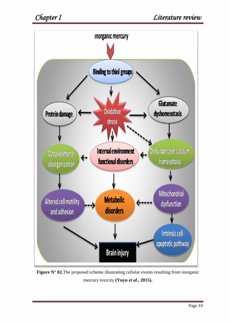
Figure N° 02.The proposed scheme illustrating cellular events resulting from inorganic
mercury toxicity (Yuyu et al., 2015).

Chapter I Literature review
Page 11
Reactive oxygen species (ROS) such as superoxide is well known to increase in
the intracellular following exposure to Hg (Bando et al., 2005). Mercury has a great
affinity for SH groups of endogenous biomolecules such as a various enzymes, amino
acids, and glutathione (GSH), as a result, Hg has bound to any free thiol available and the
thiol in the highest concentration will be the most frequently bound (Divine et al., 1999),
which comprises about 10% to 50% antioxidant capacity of both membrane and plasma
proteins (Salonen et al., 2000). The reaction rate is almost instantaneous (Clarkson,
2002). In other words, higher concentrations of thiols appear to protect against mercury
accumulation, both in vivo and in vitro (Divine et al., 1999). Glutathione is the most
common low molecular weight sulfhydryl-containing compound in mammalian cells,
present in millimolar amounts in most cells (Sen, 2003).
Glutathione (GSH) is known to interact with Hg causing the depletion of the
former (Zalups and Lash, 1997; Gatti et al., 2004), and can bind to the cysteine residues
of certain enzymes, inhibiting their activity (Frasco et al., 2007), which induce
biochemical damage to tissues that provoke an oxidative stress (Perottoni et al., 2004)
and causes severe oxidative changes (Hansen et al., 2006), furthermore, Hg played toxic
health effects to tissues and genes through diverse mechanisms such as interruption of
microtubule formation, changing intracellular calcium balance, disrupting membrane
potential, troubling or inhibition of enzymes, inhibition of protein and DNA synthesis and
disturbing immune functions (Yee et al., 1996), mitochondrial damage, lipid peroxidation
(NRC, 2000), oxidation of membrane lipids can lead to the loss of cellular or organelle
membrane integrity which can eventually result in cell death and pathological injury
(Girotti, 1998). As consequence, the reactive oxygen species (ROS) generated by
exposure to the mercurial forms may be one of the causes of the toxic effects (Hussain et
al., 1997; Shanker et al., 2005).

Chapter I Literature review
Page 12
Figure N° 03. Possible mechanisms for metal-induced oxidative stress (Ercal et al., 2001).
The elimination of mercury may be achieved mainly through urine, faeces, or
expired air although a number of other, minor routes exist (ATSDR, 1999) with a body
burden half-life of approximately 1-2 months (Clarkson, 1989; WHO, 2003), although
significant amounts are shed through sweat, tears, breast milk, and saliva (Berlin et al.,
2007). Following an acute exposure to mercuric chloride, the half-life elimination from
urine was estimated to be 25 days (Suzuki et al., 1992). Approximately 60-75% of
absorbed Hg was excreted as sulfhydryl mercury compounds, primarily with cysteine and
little if any metallic mercury was in the urine (Winship, 1985). Urinary excretion involves
active tubular transport and glomerular filtration, which is probably passive (Berlin, 1986).
The form of Hg found in the faeces is predominantly inorganic form. Intestinal
flora can convert organic mercury to inorganic, which then promotes its faecal excretion
(Rowland et al., 1980). Half-lives appear to be multiphasic, as with metallic mercury, with
human studies suggesting an effective half-life of 42 days for 80% of an oral dose; the
other 20% did not appear to have a measurable rate of excretion (Rahola et al., 1971).

Chapter I Literature review
Page 13
Elimination of inorganic Hg from the blood and brain is a biphasic process such as
an initial rapid elimination phase followed by as lower phase. Inorganic mercury may also
be reduced to form elemental mercury, which is exhaled as elemental mercury vapour or
excreted in the breast milk (WHO, 2003). Mercury vapor absorbed via the lungs converted
to divalent mercury (Hg2+
) in tissues and excreted in bile as glutathione conjugates, which
are after eliminated in faeces (Custodio et al., 2005).
The half-lives of elimination of the various forms are influenced by species, strain,
dose and sex. Age is also an important factor on body burden, with neonates and children
showing greater rates of absorption and retention. Body half-life estimates have been made
of 70–80 days for MeHg, 58 days for elemental mercury and 1–2 months for inorganic
forms (NRC, 2000).
Figure N°04: The balance between antioxidant and pro-oxidant (Favier, 2006).

Chapter I Literature review
Page 14
Immuno-dysregulation by inorganic mercury might provoke either auto immunity
or immuno-suppression depending on the genetic of rat strains tested (Robert et al., 1995).
Immune dysfunctions include hypersensitivity reactions to mercury exposure, as asthma,
dermatitis, various types of autoimmunity, suppression of natural killer cells (Ilback et al.,
1991) and disruption of various other lymphocyte subpopulations (Berlin et al., 2007). A
direct interaction between the immune system and Hg exposure leads to the reduction of
white blood cells‘ activation (Gallagher et al., 1995). Even at subacute, chronic exposure,
immune-modulatory effects of mercury were seen (Hemdan et al., 2007).
Acute toxicity of Hg could affect the different systems including the kidneys,
gastro-intestinal tract, lungs, nervous and immune systems (EU, 2007). Clinical symptoms
of acute intoxication include swelling of the salivary glands, stomatitis, loosening of the
teeth, nephritis, and hepatitis occur (Stockinger, 1981).
Ingestion of mercuric chloride can cause jaundice, an increase in liver enzymes
(WHO, 2003), histopathological and ultrastructural lesions of liver evidenced by
degeneration and cell necrosis (El-Shenawy and Hassan, 2008).
Coronary heart disease, myocardial infarction, hypertension (Houston, 2011) and
tachycardia, following the ingestion of inorganic mercury were reported (ATSDR, 1999).
Inhalation of mercury vapours and contact with mercurous chloride in teething
powders and ointments can be the reason of acrodynia, a pink discolouration of hands and
feet frequently accompanied by insomnia, irritability, and light sensitivity (WHO, 2008).
Acute oral poisoning causes gastroenteritis and colitis, and then damages the kidney
(Asano et al., 2000), this may produce death, and surviving patients commonly develop
renal tubular necrosis with anuria (Barnes et al., 1980). Kidneys are particularly
susceptible to mercury salts resulting in different alterations such as acute renal tubular
necrosis and autoimmune glomerulonephritis (Tchounwou et al., 2003).

Chapter I Literature review
Page 15
Figure N° 05. Main organs and systems affected by environmental and occupational
exposure to heavy metals (Garcia-Nino and Pedraza-Chaverri, 2014).

Chapter I Literature review
Page 16
Chronic exposure of humans to elemental or inorganic mercury might be
experienced in some occupational situations which include damage to the central nervous
system, progressive anaemia, gastric disturbance, excessive salivation and metallic taste in
the mouth (Boadi et al., 1992). Chronic exposure of chloralkali workers and mercury
miners has also been suggestive of cardiovascular toxicity (Kobal et al., 2004).
Nervous system is known to be the most sensitive target organ, as results, it would
produce neurological problems including tremors, abnormal irritability or responsiveness
to stimulation, emotional ability, insomnia, memory loss, neuromuscular changes,
headaches, polyneuropathy, and deficits in cognitive and motor function tests (WHO,
2008). Furthermore, chronic inorganic Hg salts intoxication may lead to development of lip
tremor, tongue, severe salivation, losing teeth, anorexia, and weight loss (Nelson et al.,
2011).
Reproduction of animals was affected when treated chronically with mercuric
chloride over 3 months, where it showed an alteration in testicular tissue and a decrease in
spermatogenic cell counts (IPCS, 1991; Rao et al., 2001; Abdennour et al., 2011). In the
same way, dentists exposed to metallic mercury have spontaneous abortions, stillbirths,
congenital malformations and irregular, painful or haemorrhagic menstrual disorders
(Sikorski et al., 1987). Furthermore, prolongation of the oestrous cycle as seen in animals
exposed to mercury (Baranski and Szymczk, 1973). Also, exposure of mice to HgCl2
throughout pre-mating; mating and gestation periods caused marked effects on fertility and
pup survival indices (Khan et al., 2004). Mercury concentrations in cord-blood correlate
also well with that of foetal-brain during the third trimester (NRC, 2000).

Chapter I Literature review
Page 17
Herbal medicine
Medicinal plants are an essential resource of healthiness to humanity since the
oldest time (Ladeji et al., 2003). According to World Health Organization, traditional
medicines, including herbal medicines have been, and continue to be, used in every
country around the world. Complementary and Alternative Medicine (CAM), including
herbal remedies are used throughout the world and almost civilizations which represented
the original sources of the greatest treatments and drugs (Cooper, 2005; Tsao, 2005).
Arabic medicine has contributed greatly to the advancement of modern medicine
in Europe and remains one of the principle sources of western medicine (Azaizeh et al.,
2006). Traditional Arab-Islamic medicine is still being plasticised by the Mediterranean as
well as most Arab and Islamic countries. However, the recent form of Arab herbal
medicines has historical origins in Greco-Arab and Islamic medicine, which was the leader
in the golden age (seventh to fifteenth century) of the Islamic civilization. In general,
Medicine and pharmacology are considered to be one of the most illustrious and best
known faces which Arabs most shined (Oumeish, 1998; Saad and Said, 2010; Saad,
2011).
The plant kingdom has provided an endless source of medicinal plants first used
in their simple forms as herbal teas, syrups, infusions, ointments, liniments and powders.
In human body, the chemical constituents of medicinal plants interact directly or indirectly
with the body chemistry. Once the active constituents are absorbed into the blood stream,
these constituents circulate and affect the blood system to spring the essential benefits and
hold off the waste as well as free radicals bounded to the fibres of the plants and, as
consequence, purify valuable contents into body cells.
Depending on the type of plants, one part including (flowers, leaves, branch or roots) or
whole sectors (aerial parts or roots) with their unending advantages with healing properties
could be expected to be used widely for the treatment of acute or chronic diseases, food
products, synthesis of beneficial drugs and nutritional improvement, moreover, ensure the
psychological health.
Health benefits of herbal teas of many plants are rich in phenolic compounds,
which are confirmed to apply an important antioxidant activity and are suggested to be
involved in human diet as health promoters (Seeram et al., 2001; Kim et al., 2005;
Piccolella et al., 2008; Khoo et al., 2011).

Chapter I Literature review
Page 18
Stinging nettle
Stinging Nettle known as Urtica dioica (Urticaceae) is a wild-growing, annual and
perennial herb belongs to the family of Urticaceae and is originating in Eurasia; the nettle
has spread in all temperate regions of the world. It is found more in Northern Europe than
in southern Europe, North Africa, Asia and widely distributed in North and South America
(Borchers et al., 2000). It grows in nitrogen-rich soils between 0 and 1800 m (Pignatti et
al., 1982) and temperate zones. Raw material of the nettle such as herbs (Urticae herba),
leaves (Urticae folium), and roots (Urticae radix) are recommended as helper supportive
treatment for many diseases and is one of the valuable plants used in phytotherapy as both
monotherapy and in combination therapy.
The nettle is one of the few plants that we can identify with closed eyes. Regarded as a
"bad grass", it is in reality a plant rich in vitamins and minerals and is equipped with many
virtues. Its use is multiple sections; it is used in agriculture, food, cosmetics, dyeing the
textile industry and for medicinal purposes (Bertrand and Jeanne, 2008).
U.dioica has been frequently consumed by humans for medicinal purposes. Its stinging
effect is widely known by many who have been surprised by its bite. Its genus name Urtica
is derived from uro, to burn, or urere, meaning to sting (Grieve, 1931).The stinging nettles
species name dioica is Latin for ―two houses‖, from the Greek word oikia, meaning house,
and refers to the plant‘s dioecious nature, bearing male and female flowers on separate
plants (Woodville, 1810).
The benefit of U. dioica is known for long time, where people have taken benefits
of this sting by flailing arthritic or paralytic limbs with fresh nettle to stimulate circulation
and bring warmth to joints and extremities in a treatment known as ―urtication‖ (Green,
1824).
Ancient Egyptians also reportedly used the infusion for the relief of arthritis and lumbago
(Harrison, 1966). The Roman troops were flailed themselves with stinging nettle to keep
warm. This practice of urtication became more popular in folk medicine as a remedy for
arthritis, rheumatism, and muscular paralysis and perhaps, it was the most ancient
medicinal use of this herb.
Nettle is a traditional remedy used for years against the anaemia and the lack of energy: it
is said that it is an excellent herb to its high content of iron and other minerals. It is also
said that it stimulates the digestive functions (Wichtl and Anton, 2003).

Chapter I Literature review
Page 19
The herbal tea of nettles is always proposed by herbalists as traditional remedy for gout
and rheumatism. In Germany, the herbal tea of nettle is used as diuretic, but it is not
powerful enough to be associated with a treatment of hypertension or heart problems.
While in Russia, the nettle is also used for the biliary disorders and liver (Valnet, 1983).
Nettle chemical constituents contain a fairly wide variety of chemicals although
only a few compounds belonging to various classes of natural products have been
identified. U.dioica has been used for hundreds of years in folk and officinal medicine as
haemostatic and vitamin substances (Dar et al., 2013). It is rich in vitamins such as
ascorbic acid (20–60 mg/100 g of dry material), vitamins B, K ( 0.16–0.64 mg) and E, and
pro-vitamin A (Guil-Guerrero et al., 1999), and minerals such as calcium (853–1050
mg/100g), iron (2–200 mg/100g dry material), magnesium (175 mg/100g), phosphorus
(50–265 mg/100g), potassium (532–613 mg/100g), and sodium (16–58 mg/100g) (Frank
et al., 1998). Other important substances present are all of the essential amino acids and a
very high content of chlorophyll (0.08–0.3% in fresh leaves and 0.6–1% in dry leaves)
(Frank et al., 1998), tannins, phytoncides, glycoside urticin, organic acids, sterols,
chlorophyll (up to 5%) and alkaloids (Reprintseva et al., 2011) and rarely carbohydrates
(Martinez-Para et al., 1980). Furthermore, Mavi et al (2004) reported that U. dioica
contains phenolic compounds, especially flavonoids, which have antioxidant potential. The
compounds responsible for the stinging/burning action of the hairs of leaves are
acetylcholine, histamine, and serotonin (Czarnetzki et al., 1990).
Nettle is recommended as an important medicinal herb in human health, it is nutritionally
high in minerals (iron, manganese, potassium, and calcium), chlorophyll, amino acids,
lecithin, carotenoids, and vitamins A, C and D as well as flavonoids, tanins, sterols, fatty
acids, polysaccharides, and lectins (Asgarpanah et al., 2012).

Chapter I Literature review
Page 20
Figure N° 06: Picture of stinging nettle Urtica dioica taken from North east Algeria in
spring.
Antioxidants of nettle are found in both leaves and roots and are used
therapeutically. So, nettle is a natural powerful antioxidant and as a possible food
supplements (Kanter et al., 2005).
The various antioxidants seven in low doses may be effective as strong hydrogen
giving ability (or their reducing power), free radical scavenging and metal chelating
activities. The phenolic components appear as mostly responsible for the antioxidant
activity of aqueous extracts (Khalil et al., 1999; Gülcin et al., 2004). In addition,
antioxidants are associated with decreased DNA damage, lipid peroxidation, boost the
immune performance and reduce cells‘ metamorphosis (Torbeyns, 2012–2013).
It has been demonstrated that U.dioica leaf extracts administered to rats as pre-treatment
has decreased the oxidative stress in muscles, suggesting that nettle is giving a protection
to cells against oxidative stress (Cetinus et al., 2005).

Chapter I Literature review
Page 21
Cardiovascular diseases’ treatment has been reinforced by the traditional use of
U.dioica (El Haouari et al., 2006; Daher et al., 2006). Moreover; the herb was indicated
in the treatment of oedema as a result of cardiac or renal insufficiency (Wichtl and Anton,
2003). The study in animals indicates that the extracts of U.dioica significantly inhibit
platelet aggregations and improve lipid profile (Daher et al., 2006).
Hypoglycaemic effect of nettle has been stated in ancient medical texts. The
hypoglycaemic component has been termed ―Hypoglycaemic principle= ‗urticin‘ and
nettle has been used for the treatment of high blood sugar (Said et al., 2008).This effect
may be caused in part by the reduction of intestinal glucose absorption (Bnouham et al.,
2003). Hypoglycemic activity of U.dioica was detected in a large pharmacological screen
of European species with known potential of anti-diabetic effects (Kavalali et al., 2003).
Most importantly, nettle has been applied as a diuretic in the treatment of urinary, bladder
and kidney problems (Kavalali, 2003). It was demonstrated the diuretic, the natriuretic and
the hypotensive effects of stinging nettle in animals (Tahri et al., 2000), accompanied with
increased excretion of chlorides and urea. Thus, flavonoids and high potassium content
may contribute to the diuretic action (Bradley, 1992).
The anti-inflammatory of U. dioica extracts is attracting attention for their actions
(Gülçin et al., 2004) and it has been reported to possess inhibitory effects on prostatic
hyperplasia (Zhang et al., 2008). The leaves and seeds are suggested to be useful for
patients suffering from neutrophil function deficiency (Basaran et al., 1997) that could
stimulate the proliferation of human lymphocytes (Wagner et al., 1989).
U.dioica has been used as a remedy for rheumatism, as it provides temporary relief from
pain (Alford, 2007), and allergic rhinitis (Sezik et al., 1997). Besides, nettle extract in
synthetic condition can halt the viral propagation such as those causing aids and hepatitis
(Chrubasik et al., 2007), and it was used against liver insufficiency (Ye¸silada et al.,
1993) and to treat stomachache (Ye¸silada et al., 2001). Moreover, U. dioica can be used
in the prevention of liver damage of rats (Lebedev et al., 2001; Kanter et al., 2005).
The side effects of U.dioica are seen as rare phenomena of allergy, skin infections
and oedema (Wichtl and Anton, 2003). The use of fresh nettles can be the origin of pitting
and rarely lead to severe allergic reactions in sensitive individuals (Alternative Medicine

Chapter I Literature review
Page 22
Review, 2007). However, the internal use of nettle is not associated with significant
adverse effects.
The therapeutic applications of nettle are summarised in table N° 01.
Table N° 01: Summary of the important therapeutic properties of Urtica dioica.
Therapeutic Properties Actions References
-Treatment of prostate
cancer and benign
hypertrophy of the
prostate.
The effects of nettle roots in the
treatment of HBP.
-Konrad et al,
2000;
-Schneider and
Rubben, 2004;
-Durak et al., 2004.
-Safarinejad, 2005;
-Hoffman, 2006;
-Hypotensive. The roots of nettle can produce a
hypotensive through vasodilator
effects.
-Newail et al., 1996;
-Blumenthal, 2000;
-Tahri et al., 2000;
-Testai et al., 2002;
-Legssyer et al.,
2002.
-Diuretic.
-Increases the urinary excretion -Blumenthal, 2000;
-Tahri et al., 2000.
-Hepatic-protective,
-Depurative.
-Elimination of accumulated toxins
in the body.
-The leaves help to regulate the
inflammatory factors.
-Turkdogan et al.,
2003;
-Yener et al., 2008.
-Anti-platelet
aggregation.
High iron content in the leaves. -El Houari et al.,
2006.
-Anti-allergic.
-Useful in the treatment of allergies
to pollen, long-term treatment.
- Effects on the key receivers and the
enzymes associated with allergic
-Gulcin et al., 2004;
-llhami et al., 2004;
-Roschek et al.,
2009.

Chapter I Literature review
Page 23
rhinitis(leaves)
-Anti-inflammatory,
-Immuno- stimulator.
- Inhibitory activity on rat legs
oedema(the roots)
-An immuno-stimulant neutrophils
(flavonoids glycosides from leaves).
-Glusker and
Rossi, 1986;
- Wagner, 1994.
-Effect on brain function
and memory.
The leaves are capable to reduce the
transcription of inflammation factors,
and stimulate the cerebral
performance.
-Wichtl and Anton,
2003.
-Antihyperglycemic,
-Anti-diarrheic,
-Antioxidants.
- Inhibits significantly glucose
absorption of rat small intestine.
-Due to the presence of tannins.
-In relation to different oxidative
systems (phenols, flavonoids).
-Guillaume, 1991;
-Bnouham et al.,
2003;
-Cetinus et al.,
2005.
-El Haouari et al.,
2006;
-Morel, 2008;
-Anti-asthenic (against
fatigue),
-Anti-anemic.
-Vitamins A, B2, B5, C, E, K, its
minerals magnesium, phosphorus,
sulfur, silicic acid, potassium,
calcium , its eight essential amino
acids and its principles iron, folic
acid, chlorophyll.
-Dahout and
Wuyts, 1991.
Red radish
Brassicaceae family is the eldest cultivated plants known to humans. Some
confirmation has been proved that a brassica (cruciferous) vegetable was widely consumed
as early as 10,000 years ago (Snowdon et al., 2007) with varied kind of uses; biofuel,
edible oil, human food and animal feed.
Cruciferae vegetables including broccoli, cabbage, cauliflower, radish, garden
cress, horse radishes, and wasabi, belong to family of Cruciferaceae which have an

Chapter I Literature review
Page 24
exceptional tastes and aromas but also come with both important nutritious and health
valuable benefits.
Cruciferae vegetables are rich in carotenoids, vitamin C, fibre, flavonoids, and in
particular, a group of health-promoting metabolites known as glucosinolates which linked
to cancer prevention as well as having antioxidant properties (Fahey et al., 1997; Barillari
et al., 2005). This is maintained by strong epidemiological evidence for the association of
Cruciferae vegetables consumption with a highly significant reduction of inflammatory
response (Holst and Williamson, 2004; Moon and Kim, 2012). Moreover, Cruciferae
was reported to reduce the risk of many sorts of cancer (Verhoeven et al., 1996; Higdon et
al., 2007).
Consequently, numerous studies have made attention on finding the bioactive compounds
from this family as a source of potential chemopreventive agents. The family has a unique
sulphur-containing compounds responsible for their strong aroma and spicy (or bitter) taste
(Drewnowski and Gomez- carneros, 2000).
Red radish R .sativus L. is originated from Europe and Asia. It can be cultivated in
moderate climates at altitudes between 190 and1240 m. It is 30–90 cm high and its roots
are thick and of various sizes, forms, and colours. They are comestible with a spicy taste.
R. sativus is a root crop spicy or sweet in taste with a lot of juice. Roots have many
variable shape and skin color, but the round, red skinned variety is the best one; is
generally cultivated all over the world for its edible roots and leaves and its great source of
medicinal compounds (Herman-Lara et al., 2012).
Health and nutritional benefits of Radish is multiple. It is rich in folic acid,
vitamin C and plenty of anthocyanins in red radish extract (Patil et al., 2009). About 12 to
34 kinds of anthocyanins have been identified in the red radish (Fuleki, 1969; Wu and
Prior, 2005).
Different parts of radish; including, roots and leaves have been reported to possess a wide
range of pharmacological activities (Nadkarni, 1976; Gilani and Ghayur, 2004).
Radishes have been applied as laxative, stimulant, digestive, appetizer, and in other
disorders of stomach digestion, and as bile flow stimulants (Chevallier, 1996).
Furthermore, the juices of the fresh leaves have been used as a diuretic and laxative agent
(Chopra et al., 1986). The leaves, seeds and roots have also been used to treat asthma and

Chapter I Literature review
Page 25
other chest diseases (Duke and Ayensu, 1985). In particular, radishes have been used in
traditional medicine as carminative and stomachic agents, especially as anti-cancer and/or
anti-inflammatory agents (Duke and Ayensu, 1985). Some isolated glucosinolates of R.
sativus, seeds are responsible on cancer-chemoprotective property (Barillari et al., 2005;
Duan et al., 2006).
Aqueous extract of radish was reported to cause an increase in the contractions of the
duodenum, jejunum, and ileum (Yong et al., 2000).
Flavonoids and vitamin C of radish may inhibit lipid peroxidation, promote liver
and red blood cell catalase, and inhibit XOD activities in animal‘s tissues. Radish can be
suggested for the treatment and prevention of cardiovascular disease and cancer (Jin et al.,
2001).The leaves and roots of R. sativus are used as hepatoprotective (Zaman and
Ahmad, 2004), cardioprotective (Zaman, 2004), antioxidant (Barillari et al., 2006) and
anti-urolithiatic (Vargas et al., 1999) effects. Furthermore, the freshly squeezed root juice
of R. sativus has been recommended to stimulate antiulcer activity (Alqasoumi et al.,
2008).
Figure N° 07. Picture of Red radish Raphanus sativus grown in Algeria taken from the
east of Algeria (Al-Hadjer).

Chapter I Literature review
Page 26
The therapeutic uses of radish are summarised in table N° 02.
Table N° 02: Summary of the most therapeutics proprieties of Cruciferae.
Therapeutics proprieties Actions References
-Beneficial in reducing the risk of
incidence and progression of type
II diabetes by improving insulin
sensitivity;
-Beneficial in Coronary heart
disease (CHD) and inflammation.
-Total fat levels tend to
be relatively low and
contain negligible
amounts of detrimental
saturated fats.
-There are appreciable
levels of polyunsaturated
fatty acids (PUFAs).
-(Harris et al., 2008);
-(Risérus et al., 2009);
-(Lopez, 2010);
-(Ortega et al., 2012).
-Good drugs for anaemia, rickets,
influenza, mortality, poor immune
function, and cognitive decline.
The cruciferous
vegetables are good
source of many vitamins
(A and K, C) and
minerals (Fe, Ca, Se,
Zn).
-(Lee and Kader,
2000);
-(Beck et al., 2001);
-(Rayman, 2012);
-(Chapapis et al.,
2012).
-Reduced incidence of some
cancers;
-Supposed dietary preventive
routes for several cancers as
colon, lung, and potentially breast
and prostate cancers;
-Stimulates expression of body‘s
own protective antioxidant and
detoxification proteins, and the
damaging effects as cell cycle
arrest and apoptosis.
Glucosinolates and their
catabolites (metabolism-
derived breakdown
products).
-(London et al., 2000) ;
-(Plate et al., 2003);
-(Hayes et al., 2008);
-(Steinbrecher et al.,
2009).
-Reduce risks of age-related
chronic diseases, like cancer and
cardiovascular disease, and
advantageous for gut micro-biota.
-Flavonoids & phenols
-Glutathione reductase
(roots and leaves).
-(Harborne et al.,
1993);
-(Lugasi et al., 2000);
-(Vitoria et al., 2001).

Chapter I Literature review
Page 27
-Anti-oestrogenic properties;
-Anti-inflammatory, diuretic, and
anti-HIV activities;
-Hypo-lipidic activity.
–(Clifford, 2004);
-(Graf et al., 2005);
-(Lee et al., 2006);
-(Terao et al., 2008).
-Antidiabetic;
-Antiherpes;
-Analgesic;
-Activation of macrophage.
Lipopolysaccharides
(roots).
-(Genichiro et al.,
1993).
Radish Glucosinolates are a class of metabolites found in the seeds of R. sativus;
their hydrolysis products possess health-promoting including anticancer properties (Holst
and Williamson, 2004). Glucosinolates from Brassica genus might exert neuro-protective
effect through modulation of inflammatory responses in the central nervous system
(Noyan-Ashraf et al., 2005; Cuzzola et al., 2013).
Anthocyanins of red radish is a red pigments found in the outer skin.
Anthocyanins are water-soluble natural pigments belong to the flavonoids class, which act
as an antioxidants helping to defend plant tissue from UV irradiation (Holton and
Cornish, 1995). Anthocyanins were found to have the strongest antioxidant power out of
150 flavonoids (Elliott et al., 1992). In addition, anthocyanins are very effective
antioxidants, and are the most characteristic bioactive nutrients in berries and other ‗‗red–
purple healthy food‘‘. Anthocyanins are the largest group of water-soluble pigments in the
plant kingdom responsible for the attractive red, orange, blue and purple colours of most
fruits (Mazza and Miniati, 1993) and vegetables and health benefits (Konczak and
Zhang, 2004).
Moreover, anthocyanin can play an antioxidants, or antibacterial agents (Kong et al.,
2003), which can be clarified by several antioxidant mechanisms, including hydrogen
donation, metal chelating and protein binding (Kong et al., 2003). The functions of
anthocyanins result from the chemical structure of these compounds, particularly from the
presence of hydroxyl groups in the ring moiety. They can give electrons or transfer
hydrogen atoms from hydroxyl moieties to free oxidative radicals (Castnˇeda–Ovando et
al., 2009). Additionally, anthocyanins also chelate metal ions, such as Fe3+
and Cu2+

Chapter I Literature review
Page 28
(Seeram and Nair, 2002). Besides, Anthocyanins have shown to exert anticarcinogenic
activities against multiple cancer cells in vitro (Renis et al., 2007; Shih et al., 2007) and in
animal tumour models in vivo (Cooke et al., 2006; Lala et al., 2006). Potential cancer
chemo-preventive mechanisms of anthocyanins include scavenging of reactive oxygen
species, inhibition of cell proliferation, and promotion of cell apoptosis and differentiation
(Stoner, 2008). Epidemiological and experimental studies indicate that anthocyanin intake
protects against the risk of cardiovascular diseases and other chronic degenerative
conditions (Seeram, 2008; Wang and Stoner, 2008).

Chapter I Literature review
Page 29
REFERENCES
A
Abdennour C., Khelili K., Boulakoud MS., Nezzal A., Boubsil S., Slimani S. (2002).
Urinary markers of workers chronically exposed to mercury vapor. Environ. Res. 89: 245-
249.
Alford L. (2007). The use of nettle stings for pain. Altern Ther Health Med. 13(6): pp 58.
Alqasoumi S., Yahya M.A., Al-Howiriny T., Rafatullah S. (2008). Gastroprotective
effect of radish (Raphanus sativus L.) on experimental gastric ulcer models in rats.
Farmacia LVI. (2): 204–214.
Alternative Medicine Review Volume 12, Number 3 September 2007.
Asano S., Eto K., Kurisaki E., Gunji H., Hiraiwa K., Sato M., Sato H., Sato H.,
Hasuike M., Hagiwara N., Wakasa H.(2000). Review article: acute inorganic mercury
vapor inhalation poisoning. Pathol Int. 50:169–174.
Asgarpanah J., Mohajerani R. (2012). Phytochemistry and pharmacologic properties of
Urtica dioica L. J. Med. Plants Res. 6(46): 5714-5719.
ATSDR. (1999). Toxicological Profile for Mercury Agency for Toxic Substances and
Disease Registry. U.S. Department of Health and Human Services, Public Health Service,
Atlanta, GA.
Azaizeh H., Saad B., Khalil K., Said. O. (2006). The State of the Art of Traditional Arab
Herbal Medicine in the Eastern Region of the Mediterranean: A Review. eCAM .3 (2):229–
235.
B
Bakir F., Damluji S.F., Amin-Zaki L., Murtadha M., Khalidi A., al-Rawi N.Y.,
Tikriti S., Dahahir H.I., Clarkson T.W., Smith J.C., Doherty R.A. (1973).
Methylmercury poisoning in Iraq. Science. 181: 230–241.

Chapter I Literature review
Page 30
Bando I., Reus MIS., Andres D., Cascales M. (2005). Endogenous antioxidant
defense system in rat liver following mercury chloride oral intoxication. J Biochem
Mol Toxi. 19: 154–60.
Baranshi B., Szmczyk I. (1973). Effects of mercury vapor upon reproductive function
of female white rats. Medycyna pracy. 24: 248.
Barillari J., Canistro D., Paolini M., Ferroni F., Pedulli G.F., Iori R., Valgimigli L.
(2005). Direct antioxidant activity of purified glucoerucin, the dietary secondary
metabolite contained in rocket (Eruca sativa Mill) seeds and sprouts. J. Agric. Food.
Chem. 53: 2475–2482
Barillari J., Cervellati R., Costa S. (2006). Antioxidant and cholereticproprities of
Raphanus sativus L. sprout extract. Journal of agricultural and food chemistry. 54:
9773-9778.
Barnes JL., McDowell EM., McNeil JS. (1980). Studies on the pathophysiology of
acute renal failure. V. Effect of chronic saline loading on the progression of proximal
tubular injury and functional impairment following administration of mercuric chloride
in the rat. Virchows Archiv Abteilung B Cell Pathology. 32 (3):233–260.
Basaran AA., Ceritoglu I., Undeger U. (1997). Immunomodulatory activities of some
Turkish medicinal plants. Phytother Res. 11: 609-11.
Beck MA., Nelson HK., Shi Q. (2001). Selenium deficiency increases the pathology
of an influenza virus infection. FASEB J. 15: 1481–1483.
Benhamza M. (2007). Contribution de la géophysique à l‘étude hydrogéologique de la
zone mercurielle Nord Numidique (Azzaba)- détermination du degré de pollution.
Thèse de doctorat d‘état de l‘université d‘Annaba, Algérie. 103 p
Berlin M., Zalups RK., Fowler BA. (2007). Mercury. In: Nordberg GF, Fowler BA,
Nordberg M, Friberg LT, editors. Handbook on the Toxicology of Metals. 3rd edition.
chapter 33. New York, NY, USA.
Berlin M. (1986). Mercury. In Handbook on the Toxicology of Metals (L. Friberg, G.F.
Nordberg, V.B. Vouk, and E. Kessler, Eds.). 2nd ed. Vol. II: Specific metals. Elsevier.
pp. 387-445.
Bertrand B. (2008). Les secrets de l‘ortie, 10e édition, édition de Terran.
Blumenthal M., Busse WR., Goldberg A., Gruenwald J., Hall T., Riggins CW.,
Rister RS. (2000). The Complete German Commission E Monographs: Therapeutic

Chapter I Literature review
Page 31
Guide to Herbal Medicines. Austin: American Botanical Council and Boston. Integr.
Med. Commun. p. 216.
Bnouham M., Merhfour F-Z., Ziyyat A., Mekhfi H., Aziz M., Legssyer A. (2003).
Antihyperglycemic activity of the aqueous extract of Urtica dioica. Fitoterapia.
7(4):677-681.
Boadi WY., Urbach J., Brandes JM., Yannai S. (1992). In vitro effect of mercury on
enzyme activities and its accumulation in the first trimester human placenta. Environ
Res .57:96–106.
Borchers A., Keen T., Hannum C.L., Gershwin, M. E. (2000). Cocoa and chocolate:
composition, bioavailability, and health implications. Journal of Medicinal Foods.
3:77-103.
Bradley S. (1992). ―Occasional (rare) allergic reactions have been observed ‖.
Bridges C.C., Zalups R.K. (2010). Transport of inorganic mercury and
methylmercury in target tissues and organs. J. Toxicol. Environ. Health B Crit. Rev. 13:
385-410.
C
Campanella L., Bonanni A., Tomassetti M. (2003). Determination of the
antioxidant capacity of samples of different types of tea, or of beverages based on tea
or other herbal products using a superoxide dismutase biosensor. J. Pharm. Biomed.
Anal. 32: 725–736.
Castañeda-Ovando A., Pacheco-Hernández Ml., Páez - Hernández Me., Rodríguez
Ja., Galán -Vidal Ca. ( 2009). Chemical studies of anthocyanins: A review. Food
Chem. 113: 859–871.
Cetinus AS., Goze I., Sarac B., Vural N. (2005). Scavenging effect and
antispasmodic activity of the essential oil of Cyclotrichium niveum. Fitoterapia. 78:
129-133.
Chasapis CT., Loutsidou AC., Spiliopoulou CA., Stefanidou ME. (2012). Zinc and
human health: an update. Arch Toxicol .86: 521–534.
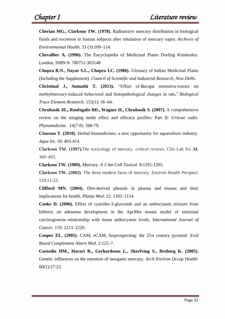
Chapter I Literature review
Page 32
Cherian MG., Clarkson TW. (1978). Radioactive mercury distribution in biological
fluids and excretion in human subjects after inhalation of mercury vapor. Archives of
Environmental Health. 33 (3):109–114.
Chevallier A. (1996). The Encyclopedia of Medicinal Plants Dorling Kindersley.
London, ISBN 9- 780751-303148
Chopra R.N., Nayar S.L., Chopra I.C. (1986). Glossary of Indian Medicinal Plants
(Including the Supplement). Council of Scientific and Industrial Research, New Delhi.
Christinal J., Sumathi T. (2013). ―Effect of Bacopa monniera extract on
methylmercury-induced behavioral and histopathological changes in rats,‖ Biological
Trace Element Research. 155(1): 56–64.
Chrubasik JE., Roufogalis BD., Wagner H., Chrubasik S. (2007). A comprehensive
review on the stinging nettle effect and efficacy profiles. Part II: Urticae radix.
Phytomedicine. 14(7-8): 568-79.
Citarasu T. (2010). Herbal biomedicines: a new opportunity for aquaculture industry.
Aqua Int. 18: 403-414.
Clarkson TW. (1997).The toxicology of mercury. critical reviews. Clin Lab Sci 34,
369–403.
Clarkson TW. (1989). Mercury. A J Am Coll Toxicol. 8:1291-1295.
Clarkson TW. (2002). The three modern faces of mercury. Environ Health Perspect.
110:11-23.
Clifford MN. (2004). Diet-derived phenols in plasma and tissues and their
implications for health. Planta Med. 12: 1103–1114.
Cooke D. (2006). Effect of cyanidin-3-glucoside and an anthocyanin mixture from
bilberry on adenoma development in the ApcMin mouse model of intestinal
carcinogenesis–relationship with tissue anthocyanin levels. International Journal of
Cancer. 119: 2213–2220.
Cooper EL. (2005). CAM, eCAM, bioprospecting: the 21st century pyramid. Evid
Based Complement Altern Med. 2:125–7.
Custodio HM., Harari R., Gerhardsson L., Skerfving S., Broberg K. (2005).
Genetic influences on the retention of inorganic mercury. Arch Environ Occup Health.
60(1):17-23.

Chapter I Literature review
Page 33
Cuzzola V.F., Galuppo M., Iori R., De Nicola G.R., Cassata G., Giacoppo S.,
Bramanti P., Mazzon E. (2013). Beneficial effects of (RS)-gluco raphanin on the
tight junction dysfunction in a mouse model of restraint stress. Life Sci.
Czarnetzki B., Thiele M., Rosenbach T. (1990). Immunoreactive leukotrienes in
nettle plants (Urtica urens ). Int. Arch. Aller. A. Imm. 91: 43-46.
D
Daher C. F., Baroody K. G., Baroody G.M. (2006). Effect of Urtica dioica extract
intake upon blood lipid profile in the rats. Fitoterapia. 77:183-8.
Dahout C. H., Wuyts D. (1991). Lactation (Gynécologie, obstétrique), Plantes
galactagogues, Editions techniques, Encycl. Méd. Nat., (Paris, France), Phytothérapie,
Aromathérapie.3-20.
Dar SA., Ganai FA., Yousuf AR., Balkhi MU., .Bhat TM., Sharma P. (2013).
Pharmacological and toxicological evaluation of Urtica dioica. Pharm. Biol.
51(2):170-180.
Department for Environment Food and Rural Affairs (DEFRA) and Environment
Agency (EA). (2002). Contaminants in soil: Collation of toxicological data and intake
values for humans. Mercury. R&D Publications TOX 7.
Divine KK., Ayala-Fierro F., Barber DS., Carter DE. (1999). Glutathione, albumin,
cysteine, and cysgly effects on toxicity and accumulation of mercuric chloride in LLC-
PK1 cells. J Toxicol Environ Health .57:489-505.
Drewnowski A., Gomez-Carneros C. (2000). Bitter taste, phytonutrients, and the
consumer: a review. Am. J. Clin. Nutr.72: 1424–1435.
Duan S.W., Bianchi T.S. (2006). Seasonal changes in the abundance and composition
of plant pigments in particulate organic carbon in the lower Mississippi and Pearl
Rivers (USA). Estuaries and Coasts. 29:427–442.
Duke J.A., Ayensu E. S. (1985). Medicinal Plants of China, Reference Publ. Inc.
Durak I., Biri H., Devrim E., Sozen S., Avci A. (2004). Aqueous extract of Urtica
dioica makes significant inhibition adenosine deaminase activity in prostate tissue from
patients with prostate cancer. Cancer Biol. Ther. 3(9):855-857.

Chapter I Literature review
Page 34
E
El Haouari M., Bnouham M., Bendahou M. (2006). Inhibition of rat platelet
aggregation by Urtica dioica leaves extracts. Phytother Res. 20:568-572.
Elliott AJ., Scheiber SA., Thomas C., Pardini RS. (1992). Inhibition of glutathione
reductase by flavonoids. A structure-activity study. Biochem Pharmacol . 44:1603-8.
El-Shenawy SMA., Hassan NS. (2008). Comparative evaluation of the protective
effect of selenium and garlic against liver and kidney damage induced by mercury
chloride in the rats. Pharmacol. Rep. 60: 199-208.
Emanuelli T., Rocha JBT., Periera ME., Prociuncula LO., Morsch VM., Martins
AF., Souza DOG. (1996). Effect of mercuric chloride intoxication and dimercaprol
treatment on delta aminolevulinate dehydratase from brain, liver and kidney of adult
mice. Pharm Toxicol. 79, 136–43.
Ercal N., Gurer-Orhan H., Aykin-Burns N. (2001). Toxic metals and oxidative
stress Part 1: mechanisms involved in metal induced oxidative damage. Curr. Top.
Med. Chem. 1: 529–539.
EU (European Union Employment and Social Affairs). (2007). Health and Safety at
Work). Recommendation from the Scientific Committee on occupational exposure
limits for elemental mercury and inorganic divalent mercury compounds.
F
Fahey J.W., Zalcmann A.T., Talalay P. (1997). The chemical diversity and
distribution of glucosinolates and isothiocyanates among plants. Phytochemistry. 56:5-
51
Favier A. (2003). Le stress oxydant: Interet conceptuel et experimental dans la
comprehension des mecanismes des maladies et potentiel therapeutique. Mecanismes
biochimiques. L'actualité Chimique. 108-115.

Chapter I Literature review
Page 35
Frank B. (1981). Urtica in Hagers Handbuch. 710-36. Data from Chrubasik J.E et al.
Phytomedicine 14 (2007).
Frasco M.F., Colletier J. P., Weik M., Carvalho F., Guilhermino L., Stojan J.,
Fournier D. (2007). Mechanisms of cholinesterase inhibition by inorganic mercury.
FEBS J. 274: 1849–1861
Fuleki T., Francis F. J. (1969). Quantitative methods for anthocyanins. 1. Extraction
and determination of total anthocyanin in cranberries. Food Sci. 33: 72-77.
G
Gallagher J., Noelle R., McCann F. (1995). Mercury Suppression of a Potassium
Current in Human B Lymphocytes. Cellular Signaling 7: 331-338.
Ganapathi P., Madhusudhan M.C., Ravindra Babu. (2009). Extraction,
dealcoholisation and concentration of anthocyanin from red radish peels. Chemical
Engineering and Processing. 48(1): 364-369.
García-Niño W. R., Pedraza-Chaverrí J. (2014). Protective effect of curcumin
against heavy metals-induced liver damage. Food and Chemical Toxicology. 69:182–
201.
Gatti A.B., Perez S.C.J.G.A., Lima M.I.S. (2004). Atividade Alelopática de extratos
aquosos de Aristolochia esperanzae O. Ktze na germinação e crescimento de Lactuca
sativa L. e Raphanus sativus L. Acta Botanica Brasilica. 18: 459-472.
Genichiro S., Kiyoshi Y., Daisuke T., Denichi M., Haruyuki O. (1993). Eur. Pat.
Appl. EP. 462,022 (Cl. A61K37/20), 18 dec 1991, JP Appl. 90/155,428, 15 Jun 1993.
Gilani A.H., Ghayur M.N. (2004). Pharmacological basis for the gut stimulatory
activity of Raphanus sativus leaves. Journal of Ethnopharmacology. 95: 169 – 172.
Girotti A. W. (1998). Lipid hydroperoxide generation, turnover, and effector action in
biological systems. J. Lipid Res. 39: 1529-1542.
Glusker JP., Rossi M. (1986). Molecular aspects of chemical carcinogens and
bioflavonoids. Progress in Clin and Bio Res. 21: 95–410.
Chapter I Literature review
Page 36
Goldman LR., Shannon MW. (2001). American Academy of Pediatrics: Committee
on Environmental Health. Technical report: Mercury in the environment: implications
for pediatricians. Pediatrics .108:197–205.
Graf BA., Milbury PE., Blumberg JB. (2005). Flavonols, flavones, flavanones, and
human health: Epidemiological evidence. J Medicin Food . 8: 281–90.
Green T. Universal herbal. 2nd ed London: Caxton Press; 1824, 729 p.
Grieve A. (1931). Modern Herbal. Penguin, ISBN 0-14-046-440-9.
Gu¨ lc¸in I., Ku¨ frevioglu OI., Oktay M., Bu¨ yu¨ kokuroglu ME. (2004).
Antioxidant, antimicrobial, antiulcer and analgesic activities of nettle (Urtica dioica
L.). J Ethnopharmacol. 90:205-15.
Guil-Guerrero J.L., Rodriguez-Garcia I. (1999). Lipids classes, fatty acids and
carotenes of the leaves of six edible wild plants. Z. Lebensm.-Unters. Forsch. 209 :
313-316.
Guillaume G. (1991). Place de la phytothérapie en rhumatologie, Editions techniques,
Encycl. Méd. Nat., (Paris, France), Phytothérapie, Aromathérapie, D-4, 9.
Guzzi G., La Porta CA. (2008). Molecular mechanisms triggered by mercury.
Toxicology. 244 (1):1–12
H
Hansen J., Zhang H., Jones D. (2006). Differential oxidation of thioredoxin-1,
thiordoxin- 2 and flutathione by metal ions. Free Radic Biol. Medi. 1: 40 (1): 138.
Harborne, J.B. (1963). Flavonoid sophorosides. Experientia. 19: 7–8.
Harborne JB., Baxter H. (1993). Phytochemical dictionary (A handbook of bioactive
compounds from plants) Taylor and Francis Ltd. pp: 300-308.
Harris CS., Lambert J., Saleem A., Coonishish J., Martineau LC., Cuerrier A.,
Haddad PS., Arnason JT., Bennett SAL. (2008). Anti-diabetic activity of extracts
from needle, bark, and cone of picea glauca: Organ-specific protection from glucose
toxicity and glucose deprivation. Pharm Biol. 46:126-134.
Harrison RK. (1966). Healing herbs of the Bible. Leiden: EJ Brill. 58 p.
Hayes JD., Kelleher MO., Eggleston IM. (2008). The cancer chemopreventive
actions of phytochemicals derived from glucosinolates. Eur. J. Nutr. 47: 73-88.

Chapter I Literature review
Page 37
Hemdan N. (2007). Immunomodulation by Mercuric Chloride in Vitro: Application of
Different Cell Activation Pathways. Clin Exp Immunol. 148(2): 325–337.
Herman- Lara E., Martínez-Sánchez C. E., Pacheco-Angulo H., Carmona-García
R., Ruiz-Espinosa H., & Ruiz-López I. (2012). Mass transfer modeling of
equilibrium and dynamic periods during osmotic dehydration of radish in NaCl
solutions. Food and Bioproducts Processing.
Hoffman D.J., Heinz G.H. (2006). Effects of mercury and selenium on glutathione
metabolism and oxidative stress in mallard ducks. Environ. Toxicol. Chem. 17: 161-
166.
Holmes P., James KAF., Levy LS. (2009). Is low level environmental mercury
exposure of concern to human health. Sci Total Environ. 408:171–82.
Holst B., Williamson G. (2004). A critical review of the bioavailability of
glucosinolates and related compounds. Natural Product Report. 21:425-447.
Holton TA., Cornish EC. (1995). Genetics and biochemistry of anthocyanin
biosynthesis. Plant Cell .7:1071–1083
Hong Y.S., Kim Y.M., Lee K.E. (2012). Methylmercury exposure and health effects.
J. Prev. Med. Public Health. 45: 353–363.
Houston MC. (2011). Role of mercury toxicity in hypertension, cardiovascular
disease, and stroke. J Clin Hypertens. 13:621–627.
Hussain S., Rodgers D.A., Duhart H.M., Ali S.F. (1997). Mercuric chloride-induced
reactive oxygen species and its effect on antioxidant enzymes in different regions of rat
brain. J. Environ. Sci. Health. 32: 395–409.
I
Ilback NG., Sundberg J., Oskarsson A. (1991). Methyl mercury exposure via
placenta and milk impairs natural killer (NK) cell function in newborn rats. Toxicology
Letters. 58 (2):149–158.
Ilhami G.O., Irfan K., Münir O., Mehmet E .B. (2004). Antioxidant, antimicrobial
antiulcer and analgesic activities of nettle (Urtica dioica L.). J. of Ethnopharmacology.
90:205-215.
International Programme on Chemical Safety (IPCS). (1990). Methylmercury.
Environmental Health Criteria 101. Geneva, Switzerland: World Health Organization.

Chapter I Literature review
Page 38
J
Jin A.S., Kyung K.M. (2001). Effect of dry powders, ethanol extracts and juices of
radish and onion on lipid metabolism and antioxidative capacity in rats. Han’guk
Yongyanghak Hoeji .34:513–524.
K
Kanter M., Coskun O., Budancamanak M. (2005). Hepatoprotective effects of
Nigella sativa L and Urtica dioica L on lipid peroxidation, antioxidant enzyme systems
and liver enzymes in carbon tetrachloride-treated rats. World J. Gastroenterol.
11:6684-8.
Kavalali G., Tuncel H., Goksel S., Hatemi H. H. (2003). Hypoglycemic activity of
Urtica pilulifera in streptozotocin-diabetic rats. J. Ethnopharmacol. 84: 241-5.
Khalil Z., Liu T., Helwe R.D. (1999). Free radicals contribute to the reduction in
peripheral vascular responses and the maintenance of thermal hyperalgesia in rats with
chronic constriction injury. Pain. 79: 31–37.
Khan I.A., Allgood J., Walker L.A., Abourashed E.A., Schelenk D., Benson W.H.
(2001). Determination of heavy metals and pesticides in ginseng products. Journal of
AOAC International. 84: 936– 939.
Khoo M.G., Clausen R.M., Pedersen H.B., Larsen E. (2011). Bioactivity and total
phenolic content of 34 sour cherry cultivars. Journal of Food Composition and
Analysis. 24: 772–776.
Kim D.O., Heo J.H., Kim J.Y., Yang S.H., Lee Y.C. (2005). Sweet and sour cherry
phenolics and their protective effects on neuronal cells. Journal of Agricultural and
Food Chemistry. 53: 9921–9927.
Kim D.O., Heo J.H., Kim J.Y., Yang S.H., Lee Y.C. (2005). Sweet and sour cherry
phen-olics and their protective effects on neuronal cells. Journal of Agricultural and
Food Chemistry. 53: 9921–9927.
Kobal AB., Horvat M., Prezelj M., Briski AS., Krsnik M., Dizdarevic T., Mazej
D., Falnoga I., Stibilj V., Arneric N., Kobal D., Osredkar J. (2004). The impact of

Chapter I Literature review
Page 39
long-term past exposure to elemental mercury on antioxidative capacity and lipid
peroxidation in mercury miners. J Trace Elem Med Biol. 17 (4):261-74.
Konczak I., Zhang W. (2004). Anthocyanins-More than nature´s colours, J Biomed
Biotechnol. 239-40.
Kong J.M. (2003). Analysis and biological activities of anthocyanins. Phytochemistry.
64: 923–933.
Konrad L., Muller H. H., Lenz C., Laubinger H., Aumuller G., Lichius J. J.
(2000). Antiproliferative effect on human Prostate cancer cells by a stinging nettle root
(Urtica dioica) extract. Planta Med. 66: 44–47.
Kudo A., Fujikawa Y., Miyahara S., Zheng J., Takigami H., Sugahara M.,
Muramatsu T., (1998). Lessons from minamata mercury pollution, Japan – after a
continuous 22 years of observation. Water Sci. Technol. 38: 187–193.
Ladeji O., Omekarah I., Solomon M. (2003). Hypoglycemic properties of aqueous
bark extract of Ceiba pentandra in streptozotocin induced diabetic rats. Journal of
Ethnopharmacology. 84: 139–142.
L
Lala G., Malik M., Zhao C., He J., Kwon Y., Giusti M. M., Magnuson B. A.
(2006). Anthocyanin-rich extracts inhibit multiple biomarkers of colon cancer in rats.
Nutr Cancer. 54: 84-93.
Lebedev A. A., Batakov E. A., Kurkin V. A., Lebedeva, E. A., Zapesochnaya, G.
G., Avdeeva E. V., Simonova G. V., Volotsueva A. V. (2001). The antioxidative
activity of a complex hepatoprotective preparation. Silybokhol Rast Res. 37:69-75.
Lee KW., Lee HJ. (2006). The roles of polyphenols in cancer chemoprevention.
Biofactors. 26:105-121.
Lee SK., Kader AA. (2000). Preharvest and postharvest factors influencing vitamin C
content in horticultural crops. Postharvet Biol. Technol. 20: 207-220.
Legssyer A., Ziyyat A., Mekhfi H., Bnouham M., Tahri A., Serhrouchni M.,
Hoerter J., Fischmeister R.(2002). Cardiovascular effects of Urtica dioica L. in
isolated rat heart and aorta. Phytother. Res. 16:503–507.
Li P., Feng X.B., Qiu G.L., Shang L.H., Li Z.G. (2009). Mercury pollution in Asia: a
review of the contaminated sites. J. Hazard. Mater. 168: 591–601.

Chapter I Literature review
Page 40
London SJ., Yuan JM., Chung FL., Gao YT., Coetzee GA., Ross RK., Yu MC.
(2000). Isocyanates, glutathione S-transferase M1 and T1 polymorphisms, and lung
cancer risk: a prospective study of men in Shanghai, China. Lancet. 356: 724-9.
Lopez-Huertas E. (2010). Health effects of oleic acid and long chain omega-3 fatty
acids (EPA and DHA) enriched milks. A review of intervention studies. Pharmacol
Res. 61: 200-207.
Lugasi A., Hovari J. (2000). Flavonoid aglycons in foods of plant origin. I.
Vegetables. Acta Aliment. 29: 345–352.
M
Martinez-Para M.C., Fidanza F., Torija-Isasa M.E. (1980). La ortiga en la
alimentacion.V. Estudio de la proteina. Anal. Bromatol. 32: 309–314.
Mau J.L., Chao G.R., Wu K.T. (2001). Antioxidant properties of methanolic extracts
from several mushrooms. Journal of Agricultural and Food Chemistry. 49: 5461–5467.
Mavi A. (2004). Antioxidant properties of some medicinal plants: Prangos ferulacea
(Apiaceae), Sedum sempervivoies (Crassulaceae), Rosa pimpinellifolia (Resaceae),
Gallium verum subsp. Verum (Rubiaceae) Urtica dioica (Urticaceae). Biol. Pharm.
Bull. (27):702-5.
Mazza G., Miniati E. (1993). Anthocyanins in fruits, vegetables and grains. Boca
Raton: CRC Press. 363 p.
Moon P.D., Kim H.M. (2012). Anti-inflammatory effect of phenethylisothiocyanate,
an active ingredient of Raphanus sativus Linne. FoodChem.131: 1332–1339.
Morel Jean-Michel. (2008). Traité pratique de phytothérapie, Editions Grancher,
collection « le corps et l‘esprit », p 74, 209, 233, 287.
Mostafalou S., Abdollahi M. (2013). Environmental pollution by mercury and related
health concerns: Renotice of a silent threat. Arh Hig Rada Toksikol. 64:179–181.
Moumen Y., Abdennour C., Loudjani F. (2011). Influence of palm date and vitamin
C supplementation on testicular functions of domestic rabbit Oryctolagus Cuniculus
under mercury exposure. Annals of Biological Research. 2(3), 359-367.

Chapter I Literature review
Page 41
Moumen Y., Abdennour C. (2008). Influence of Vitamin C on Testicular Functions
of Domestic Rabbit Oryctolagus Cuniculus Under Mercury Exposure. E. J. Sci. Res.
22(2): 197-204.
Mousavi A., Chávez R.D., Ali A.M.S., Cabanissa S.E. (2011). Mercury in natural
waters: a mini-review. Environ. Forensics. 12: 14–18.
N
Nadkarni K. (1976). Crocus sativus, Nigella sativa. In‘ Nadkarni, K.M.,
Editor.Indian materia medica, Popular Prakashan, Bombay. pp. 386–411.
National Research Council. (2000). Toxicological Effects of Methylmercury.
Washington, DC: National Academy Press. 33-35.
Nelson LS., Lewin NA., Howland MA., Hoffman RS., Goldfrank LR.,
Flomenbaum NE. (2011). Goldfrank‘s toxicologic emergencies. New York:
McGrawHill.
Newail CA, Anderson L and Phillipson J (1996). Herbal Medicines - A Guide for
Health Care Professionals. The Pharmaceutical Press, London.
Ngim C., Devathasan G. (1989). Epidemiologic study on the association between
body burden mercury level and idiopathic Parkinson‘s disease. Neuroepidemiology.
8:128–141.
Noyan-Ashraf M.H., Sadeghinejad Z., Juurlink B.H. (2005). Dietaryapproachto
decrease aging-related CNS inflammation. Nutr.Neurosci.8: 101–110.
O
O’Shea JG. (1990). ‗Two minutes with Venus, two years with mercury‘—mercury
as an antisyphilitic chemotherapeutic agent. J Royal Society Med. 83: 392–395.
Ortega A., Varela LM., Bermudez B., Lopez S., Abia R., Muriana FJ. (2012).
Dietary fatty acids linking postprandial metabolic response and chronic diseases. Food
Funct. 3: 22-7.

Chapter I Literature review
Page 42
Oumeish O. Y. (1998). The philosophical, cultural, and historical aspects of
complementary, alternative, unconventional, and integrative medicine in the Old
World. Archives of Dermatology. 134(11): 1373-1386.
P
P ark JD., Zheng W.(2012). Human exposure and health effects of Inorganic and
elemental mercury. JPrev Med Publ Health. 45:344–352.
Perottoni J., Rodrigues OED., Paixao MW., Zeni G., Lobato LP., Braga AL.,
Rocha JBT., Emanuelli T. (2004). Renal and hepatic ALA-D activity and selected
oxidative stress parameters of rats exposed to inorganic mercury and organoselenium
compounds. Food Chem Toxicol. 42: 17– 28.
Pfeiffer WC., Lacerda LD. (1988). Mercury inputs into the Amazon Region, Brazil.
Environmental Technology Letters. 9:325-30
Piccolella S., Fiorentino A., Pacifico S., D’abrosca B., Uzzo P., Monaco P. (2008).
Antioxidant properties of sour cherries (Prunus cerasus L.): role of colourless
phytochemicals from the methanolic extract of ripe fruits. Journal of Agricultural and
Food Chemistry. 56, 1928–1935.
Pignatti S. (1982). Flora d‘Italia. Edagricole, Bologna, vol: 1-3.
Piotrowski JK., Trojanowska B., Wisneiwska-Knypl JM., Baloanowska W
.(1974). Mercury binding in the kidney and liver of rats repeatedly exposed to mercuric
chloride: induction of metallothionein by mercury and cadmium. Toxicol Appl
Pharmacol. 27:11-19.
Plate AYA., Gallaher DD. (2003). Breakdown products of glucosinolates and reduced
risk of colon cancer. Faseb J. 17 A 1153.
R
Rafati-Rahimzadeh M., Moghaddamnia AA. (2010). Organophosphorus
compound poisoning. J Babol Univ Med Sci. 12:71–85.
Rafati-Rahimzadeh M., Rafati-Rahimzadeh M., Moghaddamnia AA (2013).
Arsenic compounds toxicity. J Babol Univ Med Sci. 15:51–68

Chapter I Literature review
Page 43
Rahola T., Hattula T., Lorolainen A. (1971). The Elimination of Hg-methylmercury
in man. Scandinavian Journal of Clinical and Laboratory Investigation. 27
(supplement 116): p. 77.
Rao MV., Sharma PSN. (2001). Protective effect of vitamin E against mercuric
chloride reproductive toxicity in male mice. Reproductive Toxicology. 15 (6):705–
712.
Ratcliffe H., Swanson G., Fischer L.J. (1996). Human Exposure to Mercury: A
Critical Assessment of the Evidence of Adverse Health Effects. Journal of Toxicology
and Environmental Health. 49: 221- 270.
Rayman MP. (2012). Selenium and human health. Lancet. 379: 1256-1268.
Renis M., Calandra L., Scifo C., Tomasello B., Cardile V., Vanella L., Bei R., La
Fauci L., Galvano F. (2008). Response of cell cycle/stress-related protein expression
and DNA damage upon treatment of CaCo2 cells with anthocyanins. Br J Nutr. 100:
27-35.
Reprintseva E.V. (2011). Marketing assessment of Geneva: ICH Secretariat, pp: 8.
multivitamin medicines. In the All-Russian scientific-practical conference with
international participation ―Traditions and innovations of pharmaceutical science and
practice‖. Kursk: Kursk. Gos. Med. Univ. pp: 194-196.
Risérus U., Willett WC., Hu FB. (2009). Dietary fats and prevention of type 2
diabetes. Prog. Lipid Res. 48: 44-51.
Robert A., Curtitis D., Michael P. (1995). ―Metal Toxicology.‖ 2nd Academic Press,
Robinson M. M., Zhang X. (2011). The World Medicine situation. Traditional
medicine: Global situation, issues and challenges, WHO Press, Geneva.
Roschek B. Jr., Fink RC., McMichael M., Alberte RS. (2009). Nettle extract (Urtica
dioica) affects key receptors and enzymes associated with allergic rhinitis. Phytother
Res. 23(7): 920-926.
Rowland I., Davies M., Evans J. (1980). Tissue content of mercury in rats given
methylmercury chloride orally: Influence of intestinal flora. Arch Environ Health.
35:155-160.

Chapter I Literature review
Page 44
S
Saad B., Azaizeh H., Said O. (2005). Tradition and perspectives of Arab herbal
medicine: a review. Evid Based Complement Alternat Med. 2(4): 475-479.
Saad B., Said O. (2010). Greco-Arab and Islamic Herbal Medicine: Traditional
System, Ethics, Safety, Efficacy and Regulatory Issues, Wiley-Blackwell John Wiley
& Sons, Inc.
Safarinejad MR. (2005). Urtica dioica for treatment of benign prostatic hyperplasia: a
prospective, randomized, double-blind, placebo-controlled, crossover study. J Herb
Pharmacother. 5 (4):1-11.
Said O., Saad B., Fulder F., Khalil K.,Kassis E. (2008). Weight Loss in Animals and
Humans Treated with (Weigh level), a Combination of Four Medicinal Plants Used In
Traditional Arabic and Islamic Medicine. Evid. Based Complement. Altern. Med.
Said O., Saad B., Fulder S., Khalil K., Kassis E. (2011). Weight loss in animals and
humans treated with ―weighlevel‖, a combination of four medicinal plants used in
traditional arabic and islamic medicine. Evid Based Complement Alternat
Med. 2011:874538.
Salonen JT., Seppanen K., Nyyssonen K. (1995). Intake of mercury from fish, lipid
peroxidation, and the risk of myocardial infarction and coronary, cardiovascular, and
any death in eastern Finnish men. Circulation. 91:645–655.
Salonen JT., Seppanen K., Lakka TA. (2000). Mercury accumulation and accelerated
progression of carotid atherosclerosis: a population based prospective 4-year follow-up
study in men in eastern Finland. Atherosclerosis. 148:265–273.
Schneider T., Rubben H. (2004). Stinging nettle root extract (Bazoton-uno) in long
term treatment of benign prostatic syndrome (BPS). Results of a randomized, double-
blind, placebo controlled multicenter study after 12 months]. Urologe A. 43 (3):302–6.
Seeram N. P., Nair M. G. (2002). Inhibition of lipid peroxidation and structure–
activity-related studies of the dietary constituent‘s anthocyanins, anthocyanidins, and
catechins. Journal of Agricultural and Food Chemistry. 50: 5308–5312.
Seeram NP., Aviram M., Zhang Y., Henning SM., Feng L., Dreher M., Heber D.
(2008). Comparison of antioxidant potency of commonly consumed polyphenol-rich
beverages in the United States. J Agric Food Chem. 56(4):1415-22.

Chapter I Literature review
Page 45
Seeram NP., Momin RA., Nair MG., Bourquin LD. (2001). Cyclooxygenase
inhibitory and antioxidant cyanidin glycosides in cherries and berries. Phytomedicine.
8 (5):362-9.
Sen C. (1997). Nutritional biochemistry of cellular glutathione. J Nutr Biochem. 8:660-
672.
Sener G., Sehirli A.O., Ayanoglu-Dulger G. (2003). Melatonin protects against
mercury (II)-induced oxidative tissue damage in rats. Pharmacol. Toxicol. 93: 290–
296.
Sezik E., Ye¸silada F., Tabata M., Honda G., Takaishi Y., Fujita T., Tanaka T.,
Takeda Y., (1997). Traditional medicine in Turkey VIII. Folk medicine in East
Anatolia Erzurum Erzincan Agrı, Kars, I ˇ gdır ˇ provinces. Economic Botany. 51: 195–
211.
Shanker G., Syversen T., Aschner J.L., Aschner M. (2005). Modulatory effect of
glutathione status and antioxidants on methylmercury-induced free radical formation in
primary cultures of cerebral astrocytes. Mol. Brain Res. 137: 11– 22.
Shih P.H. (2007). Anthocyanins induce the activation of phase II enzymes through the
antioxidant response element pathway against oxidative stress induced apoptosis.
Journal of Agricultural and Food Chemistry. 55: 9427–9435.
Sikorski R., Juszkiewicz T., Paszkowski J.K. (1987). Women in dental surgeries:
Reproductive hazards in occupational exposure to metallic mercury. International
archives of occupational and environmental health. 59: 551–557.
Snowdon R., Lühs W., Friedt W. (2007). Oilseed Rape. Oilseeds. 2: 55-114.
Steinbrecher A., Linseisen J. (2009). Dietary intake of individual glucosinolates in
participants of the EPIC-Heidelberg cohort study. Ann. Nutr. Metab. 54: 87-96.
Stockinger HE. (1981). The metals. In: Clayton GD, Clayton FE, eds. Patty’s
industrial hygiene and toxicology, 3rd ed. Vol. 2A. New York, NY, John Wiley &
Sons. pp. 1769–1792.
Stoner G.D. (2008). Anthocyanins and their role in cancer prevention. Cancer Letters.
269: 281–290.
Suzuki T., Takemoto TI., Shishido S., Kani K.(1977). Mercury in human amniotic
fluid. Scandinavian Journal of Work, Environment and Health. 3 (1):32–35.

Chapter I Literature review
Page 46
T
Tahri A., Yamani S., Legssyer A., Aziz M., Mekhfi H., Bnouham M., Ziyyat A.
(2000). Acute diuretic, natriuretic and hypotensive effects of a continuous perfusion of
aqueous extract of Urtica dioica in the rat. J Ethnopharmacol. 73 (1-2):95-100.
Tchounwou PB., Ayensu WK., Ninashvili N., Sutton D. (2003). Environmental
exposure to mercury and its toxicopathologic implications for public health. Environ
Toxicol .18:149–75.
Terao J., Kawai Y., Murota K. (2008). Vegetable flavonoids and cardiovascular
disease. Asia Pac. J. Clin. Nutr. 17: 291-293.
Testai L., Chericoni S., Calderone V., Nencioni G., Nieri P., Morelli I., Martinotti
E. (2002). Cardiovascular effects of Urtica dioica L. (Urticaceae) roots extracts: in
vitro and in vivo pharmacological studies. J Ethnopharmacol. 73(1-2):95-100
The Merck Index. (1983). 10th ed. (M. Windholz, Ed.). Merck and Company,
Rahway, NJ.
Toldy A., Stadler K., Sasvari M., Jakus J., Jung KJ., Chung HY. (2005).The effect
of exercise and nettle supplementation on oxidative stress markers in the rat brain.
Brain Res Bull. 65(6): 487-93.
Torbeyns T. (2012–2013). Correlates of Changes in Body Mass Index and Fat
Percent-age in Students after 1.5 Years at University.
Tsao JCI., Zeltzer LK. (2005). Complementary and alternative medicine approaches
for pediatric pain: a review of the state-of-the-science. Evid Based Complement Altern
Med. 2:149–59.
Türkdoğan MK., Ozbek H., Yener Z., Tuncer I., Uygan I., Ceylan E.(2003). The
role of Urtica dioica and Nigella sativa in the prevention of carbon tetrachloride-
induced hepatotoxicity in rats. Phytother Res. 17: 942–946.
V
Valnet Jean. (1983). Phytothérapie: traitement des maladies par les plantes. Ed.
Maloine 5e éd, rév. et augm. pp 712.

Chapter I Literature review
Page 47
Vargas B.R., Perez G.R.M., Perez G.S., Zavala S.M.A., Perez G.C. (1999).
Antiurolithiatic activity of Raphinus sativus L. aqueous extract on rats. J.
Ethnopharmacol. 68: 335–338.
Verma S., Kumar R., Khadwal A., Singhi S. (2010). Accidental inorganic mercury
chloride poisoning in a 2-year old child. Indian J Pediatr. 77:1153–1155.
Visioli F., Caruso D., Galli C., Viappiani S., Galli G., Sala A. (2000). Olive oils rich
in natural catecholic phenols decrease isoprostane excretion in humans. Biochemical
and Biophysical Research Communications. 278: 797 – 799.
Vitória A.P., Lea P.J., Azevedo R.A. (2001). Antioxidant enzymes responses to
cadmium in radish tissues. Phytochemistry. 57: 701-710.
W
Wadaan MAM. (2009). Effects of mercury exposure on blood chemistry and liver
histopathology of male rats. J. Pharmacol. Toxicol. 4: 126-131.
Wagner H., Willer F., Kreher B. (1989). Biologically active compounds from the
aqueous extract of Urtica dioica. Planta Medica.55: 452–454.
Wagner H., Willer F., Samtleben R., Boos G. (1994). Search for the antiprostatic
principle of stinging nettle (Urtica dioica) roots. Phytomedicine. 1:213-224
Wang H.T., Yang X.L., Zhang Z.H., Xu H.B. (2003). Both calcium and ROS as
common signals mediate Na2SeO3-induced apoptosis in SW480 human colonic
carcinoma cells. J. Inorg. Biochem. 97: 221–230.
Wang LS., Stoner GD. (2008). Anthocyanins and their role in cancer prevention.
Cancer Lett. 269(2): 281-90.
Wichtl M., Anton R. (2003). Plantes thérapeutiques : Tradition, pratique officinale,
sciences et thérapeutique, 2e édition. Paris (F): Lavoisier.
Winship KA. (1985). Toxicity of mercury and its inorganic salts. Adverse Drug React.
Acute Poisoning Review. 4(3):129-160.
Wood J.M., Kennedy F., Rosen C. (1968). Synthesis of Methyl Mercury Compounds
by Extracts of a Methanogenic Bacterium. Nature. 220: 173-174.
World Health Organisation (WHO) (2003). Elemental mercury and inorganic
mercury compounds: Human health aspects. Concise International Chemical
Assessment Document. CICAD 50. Geneva.

Chapter I Literature review
Page 48
Wu X., Prior RL. (2005). Standardized methods for the determination of antioxidant
capacity and phenolics in foods and dietary supplements. J Agric Food
Chem. 53(10):4290-302.
Y
Ye¸ silada E., Honda G., Sezik E., Tabata M., Goto K., Ikeshiro, Y. (1993).
Traditional medicine in Turkey IV. Folk medicine in the Mediterranean subdivision. J.
Ethnophar. 39: 31–38.
Ye¸silada E., Sezik E., Honda G., Takaishi Y., Takeda Y., Tanaka T. (2001).
Traditional medicine in Turkey X. Folk medicine in Central Anatolia. Journal of
Ethnopharmacology. 75: 95–115.
Yee S., Choi BH. (1996). Oxidative stress in neurotoxic effects of methylmercury
poisoning. Neurotoxicology. 17:17-26.
Yener Z., Celik I., Ilhan F., Bal R. (2008). Effects of Urtica dioica L. seed on lipid
peroxidation, antioxidants and liver pathology in aflatoxin-induced tissue injury in rats.
Food Chem. Toxicol. 47: 418-424.
Yong J.K., Kug C.Y., Min K.H., Kyu C.B. (2000). Radish extract stimulates motility
of the intestine via the muscarinic receptors. J. Pharm. Pharmacol. 52: 1031–1036.
Yuyu W., Dazhi W., Lin L., Minghua W. (2015). Quantitative proteomic analysis
reveals proteins involved in the neurotoxicity of marine medaka Oryzias
melastigma chronically exposed to inorganic mercury. Chemosphere.119: 1126–1133.
Z
Zalups RK., Lash LH. (1997). Depletion of glutathione in the kidney and the renal
disposition of administered inorganic mercury. Drug Metab Dispos. 25:516–523.
Zaman R.U., Ahmad M. (2004). Evaluation of Hepatoprotective effects of Raphanus
sativus L. J. Biol. Sci. 4: 463–469.

Chapter I Literature review
Page 49
Zhang Y., Li X., Zou D., Liu W., Yang J., Zhu N., Huo L., Wang M., Hong J., Wu
P., Ren G., Ning G. (2008). Treatment of type 2 diabetes and dyslipidemia with the
natural plant alkaloid berberine. J Clin Endocrinol Metab. 93(7): 2559-65.

Chapter IIII: Materials & Methods

Chapter II : Materials & Methods
Page 50
MATERIALS & METHODS
1. Materials:
1.1. Biological Material
The experimental study was carried out on 50 males and 50 females of albinos Wistar
rats weighing between 170–230 g which were obtained from Pasteur Institute (Algiers). The
rearing of rats was achieved at the “Pet Center” of the University Badji Mokhtar-Annaba.
Animals were housed in polyethylene cages lined with litter of wood chips and cleaned daily.
The group number and the dates were written on each cage. Rats were adapted for approxi-
mately one month under standard husbandry conditions, room temperature 23 ± 1 0C and nat-
ural photoperiod. They were given standard diet obtained from ONAB (Bejaia) and potable
water. The quantity of food and water were evaluated daily.
1.2. Chemicals
Pure inorganic mercury has been used (pharmaceutical company PROLABO, made in
EC). It is easily soluble in distilled water. A concentration of 0.8 g/kg bw was chosen accord-
ing to literature.
1.3. Preparation of Plant Material
Stinging nettle
The nettle (Urtica dioica) was collected from clean area of Souk Ahras region (north-
east Algeria) at the beginning of spring, cleaned with distilled water and prepared by infusing
fresh leaves in boiling water (16 gr in 25 ml of distilled water) during 5 minutes to get a green
solution.
Table N° 03. Scientific classification of Urtica dioica (Quezel and Santa ,1963).
Rank Scientific Name and Common Name
Kingdom Plantae – Plants
Subkingdom Tracheobionta – Vascular plants
Superdivision Spermatophyta – Seed plants
Division Magnoliophyta – Flowering plants

Chapter II : Materials & Methods
Page 51
Class Magnoliopsida – Dicotyledons
Subclass Hamamelididae
Order Urticales
Family Urticaceae – Nettle family
Genus Urtica L. – nettle
Species Urtica dioica L. – stinging nettle
Red radish
The Freshly harvested radish roots were collected from clean area of Annaba region
(east Algeria) and washed to remove soil and other surface contaminants. They were crushed
in a juicer for few minutes to obtain a relatively thick fresh juice.
Table N° 04. Scientific classification of Raphanus sativus:
Rank Scientific Name and Common Name
Kingdom Plantae – Plants
Subkingdom Tracheobionta – Vascular plants
Superdivision Spermatophyta – Seed plants
Division Magnoliophyta – Flowering plants
Class Magnoliopsida – Dicotyledons
Subclass Dilleniidae
Order Capparales
Family Brassicaceae ⁄ Cruciferae – Mustard family
Genus Raphanus L. – radish
Species Raphanus sativus L. – cultivated radish

Chapter II : Materials & Methods
Page 52
1.4. Experimental procedure:
After one month of adaptation under the same laboratory conditions, rats were distrib-
uted into six groups of seven each, which they were treated for 30 days.
Group 1: The control group received potable water and standard diet ad libitum (0 + 0).
Group 2: Rats received potable water and standard diet contaminated with mercury chlo-
ride (0.8 g Hg/kg diet) ad libitum (0 + Hg).
Group 3: In addition to potable water and standard diet ad libitum, rats received infusion
of nettle (1.5 ml) by gavage (0 + UD).
Group 4: In addition to potable water and standard diet ad libitum, rats received the fresh
juice of red radish (1 ml) by gavage (0 + RS).
Group 5: Rats received potable water and standard diet contaminated with mercury chlo-
ride (0.8 g Hg/kg diet) ad libitum + infusion of nettle (1.5 ml) by gavage
(Hg+UD).
Group 6: Rats received potable water and standard diet contaminated with mercury chlo-
ride (0.8 g Hg/kg diet) ad libitum + fresh juice of red radish (1 ml) by gavage
(Hg+RS).
1.5. Biological Samples:
Animals were sacrificed by decapitation after one month of treatment and blood was
immediately collected in tubes containing the anticoagulant:
Labelled polypropylene test tubes containing heparin. Blood was centrifuged at 4000
rpm/15 minute, and then the plasma biochemical markers were evaluated (glucose, tri-
glycerides, urea, creatinine, aspartate aminotransferase (AST), alanine aminotransfer-
ase (ALT), alkaline phosphatase (ALP), magnesium (Mg), iron (Fe), calcium (Ca) and
testosterone.
After sacrifice, liver, kidney, testis and epididymis were immediately weighed and
then stored in the freezer (-20)°C for the determination of the parameter of the oxidant
stress (GSH) and a part of these organs were collected and preserved in 10% neutral
buffered formalin to achieve histological profile.

Chapter II : Materials & Methods
Page 53
Pet center Wistar rats inside the cages Gavage of rats
Fresh stinging nettle leaves Nettle leaves infusion
Raw red radish roots Fresh radish juice
Figure N° 08. Rat housing, gavage, and the preparation of nettle and radish extracts.

Chapter II : Materials & Methods
Page 54
F
fig
Figure N° 09: Summary of the experimental procedure.
42 rats (males and females) divided into 6
groups
After adaptation
Control
Group
UD
group
RS group Hg group Hg+UD
group
Hg+RS
group
After 30 days of
Experimentation Sacrifice of rats by decapitation
Collection of blood Removal of organs Collection of semen
(Male)
1- Biochemical markers:
Glucose;
Triglycerides;
Urea and creatinine;
AST, ALT, ALP;
Mg, Fe, Ca;
Testosterone.
Fertility markers:
Spermatozoa’s
Concentration ;
Spermatozoa’s
motility.
1-GSH dosage:
Liver;
Kidney;
Testis (Male).
2-Histogical profile:
Liver;
Kidney;
Testis;
Epididymis. MALE

Chapter II : Materials & Methods
Page 55
2. Methods
2.1. Biochemical markers
Biochemical markers were measured by automated apparatus (Diatron PICTUS 200),
except glucose. Testosterone concentration has been estimated by Electrochemiluminescence
immunoassay (ECLIA) using the automated apparatus (Cobas e 411).
1. Glucose:
Principle:
The determination of blood glucose was performed by a glucose meter that uses test
strips. The latter are intended for use in vitro diagnostic for the test of blood glucose. They are
designed to measure the glucose in the blood. The test strip contains the glucose-oxidase, an
enzyme that oxidizes the glucose in the blood and which product of the acid D-gluconic and
of hydrogen peroxide.
Operating Mode:
The player turns on automatically by simple insertion of the test strip Accu- Chek (in the
direction of the arrows, and up to the stop).
The symbol of a gout flashes.
Remove the drop of blood on the deposit zone orange of the test strip.
The measurement is completed in approximately 5 seconds, and the result ap-
pears on the screen and the blood glucose is given in g/L.
2. Triglycerides
The dosage of triglycerides has been carried out by the enzymatic method colorimetric
according to the technical data sheet of the Spinreact Kit (Spain).
Principle:
The triglyceride incubated with the lipoprotein lipase (LPL), release the glycerol and
free fatty acids. Glycerol is converted to glycerol-3-phosphate (G3P) and adenosine-5-
diphosphate (ADP) by the glycerol kinase and adenosine triphosphate (ATP). The glycerol-3-
phosphate (G3P) is then converted by the glycerol phosphate dehydrogenase (GPO) in active
ingredient dihydroxyacetone phosphate (DAP) and hydrogen peroxide (H2O2). In the last
reaction, the hydrogen peroxide (H2O2) reacts with 4-aminophenazone (4-AP) and p-
chlorophenol in the presence of peroxidase (POD) to give a red color.

Chapter II : Materials & Methods
Page 56
The protocol therefore includes the following reactions:
LPL
Triglycerides + H2O Glycerol + Free Fatty Acids
Glycerol kinase
Glycerol+ ATP Glycerol-3-Phosphate + Adenosine-5diphosphate
GPO
The glycerol-3-phosphate+O2 Dihydroxyacetone phosphate +H2O2
POD
H2O2 + 4-AP + p-Chlorophenol Quinone + H2O
The intensity of the red color is proportional to the concentration of triglycerides in the
sample (Bucolo and David, 1973).
Reagents :
R 1: Buffer COOD pH 7,5
p-Chlorophenol
50 mM/L
2 mM/L
R 2: Enzymes Lipoprotein lipase (LPL)
Glycerol kinase (GK)
Glycerol-3-oxydase (GPO)
Peroxydase (POD)
4-Aminophénazone (4-AP)
ATP
150000 U/L
500 U/L
2500 U/L
440 U/L
0,1 mM/L
0,1 mM/L
Triglycerides CAL Triglycerides aqueous (standard) 200 mg/dL
Work reagent: dissolve the contents of one bottle of R2 in a vial of R1 and mix
slightly. This reagent is stable for 6 weeks at 2-8°C or a week at 15-25 OC.
Blank Buffer simple
Reagent of work
(mL)
1,0 1,0 1,0
Buffer (µ.L) -- 10 --
Simple (µ.L) -- -- 10

Chapter II : Materials & Methods
Page 57
3. Urea
The dosage of urea has been carried out by the enzymatic method colorimetric accord-
ing to the technical data sheet of the Spinreact Kit (Spain).
Principle:
The technique used for the determination of the rate of urea is the enzymatic method
using the urease according to the following reaction:
Urease
Urea + H2O (NH4)2+CO2
Nitroprusside
NH4 + Salicylate + NaClO Indophenol
The ions of ammonia forms responded with the salicylate and hypochlorite (NaClO) in the
presence of the catalytic converter nitroprusside to form the green indophenol (Kaplan,
1984), according to the following reactions.
The intensity of the color formed is proportional to the concentration of urea in the sample.
Reagents :
R1: Buffer Phosphate pH 6.7 50 mmol/L
EDTA 2 mmol/L
Sodium salicylate 400 mmol/L
Sodium nitroprusside 10 mmol/L
R2 : NaClO Sodium hydrochlorite (NaClO) 140 mmol/L
Sodium hydroxyde 150 mmol/L
R 3 : Enzymes Urease 30000 U/L
Urea CAL Urea aqueous (Standard) 50 mg/dL
Work reagent: Dissolve a tablet of reagent 3 (enzymes) in a bottle of reagent 1 (buff-
er) and lightly mix. The reagent 2 (NaClO) is prepared for use.

Chapter II : Materials & Methods
Page 58
Operating Mode:
Blanc Buffer simple
Reagent of work
(mL)
1,0 1,0 1,0
Buffer (µ.L) -- 10 --
Simple (µ.L) -- -- 10
4. Creatinine
The assay of creatinine has been achieved by the kinetic method colorimetric according
to the technical data sheet of the Spinreact Kit (Spain).
Principle:
The test is based on the reaction of creatinine with the picrate of sodium as described by Jaffe.
The creatinine reacts with the alkaline picrate forming a complex red. The time interval is
chosen for the measures in such a way that it avoids interference with other components of
serum. The intensity of the color is proportional to the concentration of creatinine in the sam-
ple (Murray, 1984).
Reagents:
R1:Picric reagent Picric acid 17,5 mM/L
R 2: Alcalin reagent Sodium hydroxide 0,29 mM/L
Creatinine CAL Creatinine aqueous
(standard)
2 mg/dL
Work reagent: Mix one volume of reagent 1 with a volume of reagent 2. The reagent
is stable for 10 days at 15-25OC.
Blanc Buffer simple
Reagent of work
(mL)
1,0 1,0 1,0
Buffer (µ.L) -- 100 --
Simple (µ.L) -- -- 100

Chapter II : Materials & Methods
Page 59
5. ALT
The assay of aspartate aminotransferase has been achieved by the kinetic method ac-
cording to the technical data sheet of the Spinreact Kit (Spain).
Principle:
The alanine aminotransferase (ALT) also known as the glutamate-pyruvate transami-
nase (GPT) catalyses the reversible transfer of an amino group from the alanine at α-
cetoglutarate forming glutamate and pyruvate. The pyruvate is reduced to lactate by lactate
dehydrogenase (LDH) and the NADH formed according to the following reactions:
ALT
Alanine + α-cetoglutarate Glutamate + Pyruvate
LDH
Pyruvate + NADH+H+
Lactate + NAD+
Reagents :
R 1: Buffer TRIS pH 7,8
L-alanine
100 mM/L
500 mM/L
R2: Substrate NADH
Lactate deshydrogenase (LDH)
α-cetoglutarate
0,18 mM/L
1200 U/L
15 mM/L
Work reagent: dissolve a tablet of R2 in a vial of R1 and mix slightly. This reagent is
stable for 21 days at 2-8°C or 72 hours at 15-25OC.
Operating mode:
Work reagent (mL) 1.0
Sample (µL) 100

Chapter II : Materials & Methods
Page 60
6. AST
The assay of aspartate aminotransferase has been achieved by the kinetic method ac-
cording to the technical data sheet of the Spinreact Kit (Spain).
Principle:
Aspartate aminotransferase (AST) also called the glutamate oxaloacetate transaminase
(GOT) catalyses the reversible transfer of an amino group from the aspartate at α-
cetoglutarate forming glutamate and oxaloacetate. The oxaloacetate is reduced to malate by
malate dehydrogenase (MDH) and the NADH H according to the following reactions:
AST
Aspartate + α-cetoglutarate Glutamate + Oxaloacetate
MDH
Oxaloacetate +NADH + H+
Malate + NAD+
Reagents :
R 1: Buffer TRIS pH 7,8
L-Aspartate
80 mM/L
200 mM/L
R 2: Substrate NADH
Lactate deshydrogenase (LDH)
Malate deshydrogenase (MDH)
α-cetoglutarate
0,18 mM/L
800 U/L
600 U/L
12 mM/L
Work reagent: dissolve a tablet of R2 in a vial of R1 and mix slightly. This reagent is
stable for 21 days at 2-8°C or 72 hours at 15-25OC.
Operating mode:
Work reagent (mL) 1.0
Sample (µL) 100

Chapter II : Materials & Methods
Page 61
7. ALP
The assay of alkaline phosphatase has been achieved by the kinetic method according to the
technical data sheet of the Spinreact Kit (Spain).
Principle:
The alkaline phosphatase (PAL) catalyses the hydrolysis of p-nitrophenyl phosphate at pH
10.4, freeing up the p-nitrophenol and the phosphate, according to the following reaction:
ALT
ALT p-nitrophenyl phosphate+ H2O p-Nitrophenyl+ Phosphate
The rate of the decrease of the concentration in NADH+ H+, measured by photometry, is
proportional to the catalytic activity of alkaline phosphatase (Wenger, 1984).
Reagents :
R1: Buffer Diethanolamine (DEA) pH 10,4
Chloride de magnesium
1 mM/L
0,5 mM/L
R 2: Substrate
P-Nitophenylphosphate (pNPP) 10 mM/L
Work reagent: dissolve a tablet of R2 in a vial of R1 and mix slightly. This reagent is
stable for 21 days at 2-8 °C or 72 hours at 15-25OC.
Operating mode:
Work reagent (mL) 1.2
Sample (µL) 20
8. Magnesium
The assay of magnesium has been achieved by the kinetic method according to the technical
data sheet of the Spinreact Kit (Spain).

Chapter II : Materials & Methods
Page 62
Principle:
Magnesium forms a coloured complex when reacts with Magon sulfonate in alkaline solu-
tion.
The intensity of the color formed is proportional to the magnesium concentration in the sample
(Farrell et al., 1984).
Reagents :
R
Xylidyl Blue
Thioglycolic acid
DMSO
0.1 mmol/L
0.7 mmol/L
3000 mmol/L
9. Iron
The assay of iron has been achieved by the kinetic method according to the technical
data sheet of the Spinreact Kit (Spain).
Principle
The iron is dissociated from transferring-iron complex in weakly acid medium. Liberated
iron is reduced into the bivalent form by means of ascorbic acid. Ferrous ions give with Fer-
roZine a coloured complex:
Acid Ascorbic
Transferrin (Fe+3
) 2 + e
- 2 Fe
+2 + Transferrin
FerroZine
Fe+2
Coloured complex
The intensity of the color formed is proportional to the iron concentration in the sample.
(Itano, 1978)
Reagents :
R 1 : Buffer
Acetate pH 4.9
100 mmol/L
R 2 : Reductant
Ascorbic acid
99.7%
R 3 :Color
FerroZine
40 mmol/L
IRON CAL
Iron aqueous primary
standard
100 g/dL

Chapter II : Materials & Methods
Page 63
Work reagent: Dissolve the contents of one tube R 2 Reductant in one bottle of R 1
Buffer.
Mode operating:
WR blank Standard Sample blank Sample
WR(mL) 1.0 1.0 1.0 1.0
R 3 (drops)
1 1 -- 1
Distilled wa-
ter (L)
200 -- -- --
Standard (L)
-- 200 -- --
Sample (L)
-- -- 200 200
10. Calcium
The assay of calcium has been achieved by the kinetic method according to the technical
data sheet of the Spinreact Kit (Spain).
Principle
The measurement of calcium in the sample is based on formation of color complex be-
tween calcium and o-cresolphtalein in alkaline medium:
OH
+
Ca++
+ o-Cresolphtalein Colored complex
The intensity of the colour formed is proportional to the calcium concentration in the
sample (Farell et al., 1984).
Reagents:
R1: Buffer Ethanolamine 500 mmol/L
R2: Chromogen 0-Cresolphatein
8-Hidroxyquinolein
0,62mmol/L
69mmol/L
CALCIUM CAL Calcuim aqueous primary standard 10mg/dL

Chapter II : Materials & Methods
Page 64
Work reagent:
All the reagents are ready to use. To prepare monoreagent, mix according to this proportion:
50 vol. of R1 and 1 vol. of R2.
Mode operating:
Blank Standard Sample
R 1 (mL) 2.0 2.0 2.0
R 2 (drops) 1 1 1
Standard (L)
-- 20 --
Sample (L)
-- -- 20
2.2. Fertility profile
The spermogram has been realized according to the method of (WHO, 1993) by making
a small incision at the epididymis level in order to obtain a drop of semen. One drop of sperm
of nearly 1μl was added to 49 μl of physiological solution (0.9% NaCl) and then spermato-
zoa’s concentration and motility were estimated.
Spermatozoa’s concentration :
The concentration of spermatozoa is measured by using a blade of counting (cell of
Malassez). A drop of the same preparation filed on the blade of Malassez and covered with a
coverslip, the counting is done in 5 fields of the blade to the magnification (x 40). The con-
centration of spermatozoa is calculated by the following method (WHO, 1993).
C (number x 106 /ml) = D x V x n / N
D: dilution factor (50 times)
V: volume of the blade of counting
n: number of sperm calculated in 5 fields.
N: number of small squares in the blade.

Chapter II : Materials & Methods
Page 65
Spermatozoa’s motility :
A drop of semen is deposited on a cell of Malassez and then covered with a coverslip.
The preparation is examined under optical microscope (x40). The field of observation is di-
vided into 5 distinct zones to classify 100 sperm, and then we calculate the percentage of mo-
bile sperm (WHO, 1993).
Testosterone
The dosage of Testosterone has been carried out by applying the test of e Electrochemi-
luminescence (ECLIA) which is suitable for immunological assays using the automated appa-
ratus (Cobas e 411).
Principle
The test of this hormone depends of the competition by using a specific antibody directed
against testosterone. The endogenous testosterone released by the action of the acid 8- ani-
lino-naphthalene sulfonic (ANS) and of the norgestrel, enters into competition with the exog-
enous testosterone marked in the ruthenium to the binding sites of anti-testosterone antibody
biotinylated.
Reagents: are contained in a cabinet Elecsys-testosterone:
Microparticles lined
Streptavidin
-One vial 6.5 ml
Microparticles lined
Streptavidin
-Conservative
0,72 mg/mL
R1: antibodies anti-
testosterone-biotin
- Antibodies (sheep) anti-testosterone
marked to the biotin, ph7
- 2-Bromoestradiol (reagent of cytokine
release)
- Buffer pH 6.0
- Conservative
40ng/ml
20mmol/L
50mmol/L
R2: peptide testosterone - Derived from testosterone brand in the
ruthenium
- Buffer pH 6.0
- Conservative
1.5ng/ml
50mmol/L

Chapter II : Materials & Methods
Page 66
Operating mode:
1st incubation: 20 µl of sample are incubated with a monoclonal antibody anti-
testosterone specific biotinylated. The binding sites of the antibody brand are occu-
pied by the analyse content in the sample (as a function of its concentration).
2nd
incubation: Microparticles lined with streptavidin and derivatives of testosterone
brand in the ruthenium are added to the reaction cup. The complex is fixed to the sol-
id phase by a link streptavidin-biotin. "The reaction mixture is transferred in the
measuring cell; the microparticles are maintained at the level of the electrode by a
magnet.
The elimination of the free fraction is performed by the passage of Procell. A potential
difference applied to the electrode triggers the production of luminescence which is measured
by a photomultiplier.
The results are obtained using a calibration curve. The latter is generated, for the parser
uses, by a calibration in 2 points and a reference curve stored in the bar code of the reagent.
2.3. GSH dosage
Preparation of homogenate:
1 g of liver, kidney or testis of rats of different groups studied, has been used. After
grinding and homogenization of tissues in the TBS (Tris 50 mm, NaCl 150 mM, pH 7.4) .We
proceeded to a centrifugation of the cell suspension (9000 holes/min, 4°C, 15 min), and then
the supernatant is aliquoted into tubes eppendorfs and then kept at (-20)°C pending to perform
the determinations of the parameters of the oxidant stress.
Determination of the rate of reduced glutathione (GHS) at tissue:
Glutathione is an intracellular thiol the most abundant present in all animal cells.
Principle:
For the determination of glutathione, the colorimetric method by the reagent of Ellman
(DTNB) is the commonest method (Ellman, 1959). The reaction is to cut off the molecule of
acid 5.5 ' dithiodis-2-nitrobenzoique (DTNB) by the GHS, which frees the thionitrobenzoique
acid (TNB) (figure) which presents an absorbance at 412 nm.

Chapter II : Materials & Methods
Page 67
Figure N° 10: Glutathione and DTNB reaction.
Operating Mode
o Collect 0.8 ml of homogenate;
o Add 0.2 ml of the solution with sulfosalicylic acid (0.25 %) and leave for 15 min in an
ice bath;
o Centrifuge at 1000 tours/min for 15 min;
o Collect 0.5 ml of the supernatant;
o Add 1ml of buffer Tris-EDTA (containing 0.02 M EDTA, pH 9.6);
o Add 0,025 ml of the acid 5,5 dithio-bis-2-nitrobenzoique (DTNB) to 0.01 M ;
o Leave for 5 min at an ambient temperature and read the optical densities at 412 nm
against the reagent blank.
Calculation of the concentration
The concentration is calculated of the GHS expressed in nanomoles per milligram of pro-
tein (nmol/mg prot) according to the following formula:
OD × L × 1.525
GSH = 13100 × 0.8 × 0.5 × mg prot
OD: Optical density ;
L: Total volume of solutions used in the deproteinisation ;
1,525: Total volume of solutions used in the dosing of the GHS at the level of the
supernatant;
13100: Coefficient of absorbance of the grouping -SH to 412 nm ;
0.8: Volume of homogenate ;
0.5: Volume of the supernatant.

Chapter II : Materials & Methods
Page 68
Determination of total protein in the tissue level
Principle
The tissue proteins were quantified according to the colorimetric method of Bradford
(1976) who uses the Gloss Blue Coomassie G250 (BBC) as a reagent and the serum albumin
of beef (BSA) as standard. The BBC reacts with the amino groups (-NH2) for protein to form
a complex of blue color. The emergence of this color reflects the degree of ionization of the
acid and intensity establishes the concentration of protein which is measured spectrophoto-
metrically at 595nm.
Operating Mode:
o Collect 0.1 ml of the homogenate ;
o Add 5 ml of brilliant blue Coomassie (BBC) (G250), as a reagent ;
o Shake and leave the mixture to ambient temperature during 5 min for the stabilization
of the color ;
o Measure the absorbance of the sample at 595nm against the white containing distilled
water in place of the homogenate. The optical density obtained is reported on the cali-
bration curve (0 to 1 mg/ml of BSA) previously carried out in the same conditions.
Figure N° 11: The calibration curve of BSA (1mg/ml) for the assay of proteins in the homog-
enates.

Chapter II : Materials & Methods
Page 69
2.4. Histological profiles:
After sacrifice, liver, kidney, testis and epididymis were immediately collected and pre-
served in 10% neutral buffered formalin, where it examined according to the classical method
of Martoja Martoja (1967) according to the following steps:
Fixing of the samples
The fixing represents the time essential to the histological technique, it is designed to im-
mobilise the structures in respecting their morphologies. The fixing is done in 10% neutral
buffered formalin, fixer commonly used.
Dehydration
The paraffin is not miscible in water; the sample must be fully dehydrated before the inclu-
sion in the paraffin. The fixer is eliminated; the sample is rinsed with distilled water and then
dehydrated by ethyl alcohol 700°, 900°, and 1000°.
The paraffin is not miscible with the alcohol used for the dehydration, for this we proceeded
to a substitution by the toluene renewed 3 times (duration of the bathroom 30 minutes).
Inclusion
The sample is included in paraffin that has been melted in an oven at 600 0C for 08 hours,
the blocks is done using bars of Leuckard.
Cuts and colouring
The blocks of inclusion are entries on a sample holder. The slices are made using a micro-
tome; the series of slices are connected between them in the form of ribbon, which facilitates
the replenishment of three-dimensional structures observed. The thickness of the slices is 4 to
7 μ.
The ribbons of slices obtained are pasted on glass slides degreased, posed on a heating
plate (to lower temperature to the melting temperature of the paraffin wax). The heat allows
the spreading of the ribbons of slices.
Coat the blades of glycealbumine, cover with distilled water and install the tapes above.
The slices deparaffinise are coloured to: haemalum-eosin, dehydrate the preparation in two
bathrooms of alcohol and fit by the deposit of a drop of Eukitt on which we place a coverslip.
Finally, we proceeded to the observation at the optical microscope equipped with a camera.

Chapter II : Materials & Methods
Page 70
The fitting is to attach a coverslip glass on histological sections after staining. This step al-
lows the:
Mechanical protection of slices.
Protection of chemical dyes.
Dry the slides and then observe the optical microscope equipped with a camera.
Fixation Cassette
Circulator Inclusion apparatus

Chapter II : Materials & Methods
Page 71
Microtome Deshydratation
Blocks of coloration Colored blades
Figure N° 12. Materials and steps needed for the preparation of slides for the histological
study.
3. Statistical analysis
The mean ± SEM values were calculated to determine the significance of intergroup.
Each parameter was analysed separately using two ways analysis of variance (ANOVA). To
find the difference between the groups, Student ‘t’ test was used. P <0.05 was considered to
be significant.
Chapter II : Materials & Methods
Page 72
REFERENCES
B
Bradford M. (1976). A rapid and sensitive method for the quantities of microgram
quantities of protein utilizing the principle of protein binding. Anal. Bioch, 72: 248–254.
Buccolo G., David H. (1973). Quantitative determination of serum triglycerides by use of
enzymes. Clin. Chem. 1973; 19 (5): 476-482.
E
Ellman G.L. (1959). Tissue sulfhydryl groups. Archives of Biochemistry and Biophys-
ics. 82:70–77.
F
Farrell E C. (1984). Magnesium. Clin Chem The C.V. Mosby Co. St Louis. Toronto.
Princeton. 1065-1069.
I
Itano M M D. (1978). Cap Serum Iron Survey. (70): 516-522.
J
Jollow DJ., Mitchell JR., Zamppaglione Z., Gillette JR. (1974). Bromobenzene in-
duced liver necrosis. Protective role of glutathione and evidence for 3, 4- bromobenzene
oxide as the hepatotoxic metabolites. USA. Pharm. 11: 51–157.

Chapter II : Materials & Methods
Page 73
K
Kaplan A. (1984). Glucose. Clin. Chem. Toronto. Princeton. 1032-1036.
Kaplan A. (1984). Urea. Clin. Chem. Toronto. Princeton. 1257-1260, 437 and 418.
Kaplan A. et al. (1984). Bilirubin. Clin. Chem. Toronto. Princeton. 1238-1241.
Kaplan A. et al. (1984). Lipids. Clin. Chem. Toronto. Princeton. 918-919.
Kaplan A. et al. (1984). Triglycerides. Clin. Chem. Toronto. Princeton. 437.
M
Martoja R., Martoja M. (1967). Initiation aux techniques de l’histologie animale. Ed
Masson and Cie. pp: 345-346.
Murray R.L. (1984). Alanine aminotransferase. Clin. Chem. Toronto. Princeton. 1088-
1090.
Murray R.L. (1984). Aspartate aminotransferase. Clin. Chem. Toronto. Princeton. 1112-
1116.
Murray R.L. (1984). Creatinine. Clin. Chem. Toronto. Princeton. 1261-1266.
Q
Quezel P., Santa S. (1962-1963). Nouvelle flore de l’Algérie et des régions désertiques
méridionales. CNRS. 2 Vol: 1170p.
W
Wenger C., et al. (1984). Alkaline phosphatase. Clin. Chem. Toronto. Princeton. 1094-
1098.
World Health Organization (WHO). (1993). Analyse human sperm and interaction with
sperm cervical mucus. Ed. INSERM. pp: 55-56.

CCHHAAPPTTEERR IIIIII:: RREESSUULLTTSS OOFF
BBIIOOCCHHEEMMIICCAALL MMAARRKKEERRSS

Chapter III The Biochemical markers
Page 74
RESULTS
1. The body weight
1.1. Urtica dioica
Final mean total body weights of male rats (163.2±15.3 g) after 30 days Hg
exposure has decreased considerably compared to the initial weight (182.2±23.5 g) with
a percentage of (−10.44%). Whereas, those of UD and Hg+UD mean total body weight
have increased by (+13.66%) and (+9%), respectively. However, the final total body
weight of the control has risen by (+16%) after 30 days.
Final mean total body weights of female rats (152.3±16.4g) after 30 days Hg
exposure has decreased considerably compared to the initial weight (171± 33.8g) with a
percentage of (−12.27%). Whereas, those of UD and Hg+UD mean total body weight
have increased by (+15.11%) and (-5.42%), respectively. However, the final total body
weight of the control has risen by (+16.35%) after 30 days.
Figure N° 13: Total body weight variations in the groups treated with Hg and /or
U.dioica for 30 days.

Chapter III The Biochemical markers
Page 75
Body weight variations were summarised table N°05.
*S
ign
ifican
tly d
ifferent w
hen
com
pared
to th
e con
trol;
#S
ign
ifican
tly d
ifferent b
etween
gro
up
s.
Fin
al
(g)
Initia
l
(g)
Ta
ble N
° 05
: To
tal bo
dy
weig
ht v
ariations in
the traited
gro
up
s by
Hg
and/o
r U.d
ioica
for 3
0 d
ays.
21
0.3
± 2
3#
18
0.5
±2
1,3
Male
C
20
1.3
± 2
1#
17
3±
27
.7
Fem
ale
16
3.2
±1
5.3
*#
18
2.2
±2
3.5
Male
Hg
15
2.3
±1
6.4
*#
17
1±
33
.8
Fem
ale
20
8.3
±1
6.6
#
18
3.2
±2
1.0
9
Male
UD
20
0.3
±1
4.8
#
17
4±
22
.2
Fem
ale
19
9.2
±2
3.1
*#
18
1.6
±2
0.3
Male
Hg+
UD
16
5.5
±2
0.3
*#
17
5.2
±1
2.6
Fem
ale

Chapter III The Biochemical markers
Page 76
1.2. Raphanus sativus
Final mean total body weights of male rats (163.2 ± 15.3 g) after 30 days Hg
exposure has decreased considerably compared to the initial weight (182.2 ± 23.5 g)
with a percentage of (−10.44%). Whereas, those of RS and Hg+RS mean total body
weight have increased by (+13.66%) and (+9%), respectively. However, the final total
body weight of the control has risen by (+16%) after 30 days.
Final mean total body weights of female rats (152.3 ± 16.4 g) after 30 days Hg
exposure has decreased considerably compared to the initial weight (171 ± 33.8 g) with
a percentage of (−12.27%). Whereas, those of RS and Hg+RS mean total body weight
have increased by (+22.8%) and (+6.5%), respectively. However, the final total body
weight of the control has risen by (+16.35%) after 30 days.
Figure N° 14: Total body weight variations in the groups treated with Hg and/or R.
sativus for 30 days.

Chapter III The Biochemical markers
Page 77
Body weight variations were summarised table N°06.
*S
ign
ifican
tly d
ifferent w
hen
com
pared
to th
e con
trol;
#S
ign
ifican
tly d
ifferent b
etween
gro
up
s.
Fin
al
(g)
Initia
l
(g)
Ta
ble N
° 06
: To
tal Bo
dy
weig
ht v
ariations in
the traited
gro
up
s by
Hg
and/o
r R. sa
tivus fo
r 30
day
s.
210.3
± 2
3#
180.5
±21.3
Male
C
201.3
± 2
1#
173±
27.7
Fem
ale
163.2
±15.3
*#
182.2
±23.5
Male
Hg 152.3
±16.4
*#
171
± 3
3.8
Fem
ale
211.6
±18.9
#
185.3
±22.1
Male
RS
21
0.3
±21
.8#
17
1.3
±26
.4
Fem
ale
19
5.2
±24
.6*#
18
0.6
±19
.6
Male
Hg+
RS
188.8
±21.9
#
176.5
±21.9
Fem
ale

Chapter III The Biochemical markers
Page 78
2. The absolute organs’ weights
2.1. Urtica dioica
Our results indicate that there has been a significant increase in the absolute liver
and kidney weight in Hg of male‘s rats comparing to the control group. Concerning the
absolute kidney weight in males, there is a significant difference between all groups.
Meanwhile, there has been a decrease in the absolute liver weight in female rats treated
by Hg. On the other side, the supplementation of Urtica Dioica presents no significant
difference between (Hg+ UD) group and the control.
Figure N° 15: Absolute weight variations of liver in groups treated by Hg and/or U.
dioica for 30 days.
(*)
0
0,2
0,4
0,6
0,8
1
1,2
C Hg UD Hg+UD
Kid
ney
(g
)
B
Kidney
male
female
Figure N° 16: Absolute weight variations of kidney in groups treated by Hg and/or U.
dioica for 30 days.

Chapter III The Biochemical markers
Page 79
Organ‘s weight variations were summarised in table N° 07.
*S
ign
ifican
tly d
ifferent w
hen
com
pared
to th
e con
trol;
#S
ign
ifican
tly d
ifferent b
etween
gro
up
s.
Kid
ney(g
)
Liver(g
)
Ta
ble N
° 07
: Org
an‘s w
eight v
ariations in
the treated
gro
ups b
y H
g an
d/o
r U.d
ioica
for 3
0 d
ays.
0.6
4 ±
0.0
3#
6.6
± 1
.79
Male
C
0.7
9 ±
0.1
2
7.2
5 ±
0.9
2
Fem
ale
1.0
4 ±
0.0
9*
#
8.3
5 ±
1.0
1*
Male
Hg
0.7
1 ±
0.1
1
5.1
2 ±
0.4
5*
Fem
ale
0.7
4 ±
0.1
0#
7.4
0 ±
1.5
8
Male
UD
0.8
± 0
.09
7.8
9 ±
1.5
5
Fem
ale
0.7
4 ±
0.1
7#
6.4
9 ±
1.9
Male H
g+
UD
0.7
5 ±
0.1
2#
5.8
2 ±
1.1
1*
Fem
ale

Chapter III The Biochemical markers
Page 80
2.2. Raphanus sativus
Concerning organs, an increase of absolute liver and kidney weight of males
treated by Hg was observed. In other side, there has been a decrease in the absolute liver
weight of females treated by Hg. Contrary; the combined treatment with R. sativus has
led to a slight increase in absolute weights of males‘ liver and kidney, accompanied
with a significant decrease of females‘ liver weight.
(*)
(*)
(*)
0
2
4
6
8
10
C Hg RS Hg+RS
Liv
er
(g)
A
Liver
Male
Female
Figure N° 17: Absolute weight variations of liver in groups treated by Hg and/or R.
sativus for 30 days.
(*)
0
0,2
0,4
0,6
0,8
1
1,2
C Hg RS Hg+RS
Kid
ney
(g
)
B
Kidney
Male
Female
Figure N° 18: Absolute weight variations of kidney in groups treated by Hg and/or R.
sativus for 30 days.

Chapter III The Biochemical markers
Page 81
Organ‘s body weight variations were summarised in table N° 08.
*S
ign
ifican
tly d
ifferent w
hen
com
pared
to th
e con
trol;
#S
ign
ifican
tly d
ifferent b
etween
gro
up
s.
Kid
ney(g
)
Liver(g
)
Ta
ble N
° 08
: Org
an‘s b
ody
weig
ht v
ariations in
the treated
gro
ups b
y H
g an
d/o
r R. sa
tivus fo
r 30
day
s.
0.6
4 ±
0.0
3#
6.6
± 1
.79
Male
C
0.7
9 ±
0.1
2
7.2
5 ±
0.9
2
Fem
ale
1.0
4 ±
0.0
9*
#
8.3
5 ±
1.0
1*
Male
Hg 0
.74
1±
0.1
5
7.0
6 ±
1.2
9
Fem
ale
0.7
±0
.16
#
6.5
6±
1.0
1
Male
RS
0.7
1 ±
0.1
1
5.1
2 ±
0.4
5*
Fem
ale
0.8
2±
0.0
9#
7.5
1±
0.5
4
Male H
g+
RS
0.7
6±
0.0
7
5.7
9 ±
0.9
*
Fem
ale

Chapter III The Biochemical markers
Page 82
3. The biochemical markers
3.1. Urtica dioica
During the course of present investigations, it was observed that the treatment with
Hg caused a significant increase in the concentration of glucose and triglycerides in
male Wistar rats compared to the control, but no difference was recorded in both sexes
of rats treated with Hg+UD and UD alone.
Figure N° 19. The effects of Hg and/or U.dioica on glucose level of rats after 30 days.
Figure N° 20. The effects of Hg and/or U.dioica on triglycerides level of rats after 30
days.

Chapter III The Biochemical markers
Page 83
This study demonstrates in both sexes that the treatment of rats with Hg has led to
a pronounced elevation of urea and creatinine concentration. The supplementation of
rats with Hg+UD or U.dioica alone recorded no noticeable change of these markers.
Besides, creatinine concentration shows a signification between all groups in both
sexes.
(*) (*)
0
0,1
0,2
0,3
0,4
0,5
0,6
C Hg UD Hg+UD
Ure
a (
g/l
)
C
Urea
Male
Female
Figure N° 21. The effects of Hg and/or U.dioica on urea level of rats after 30 days.
(*)(*)
0
0,2
0,4
0,6
0,8
1
1,2
1,4
C Hg UD Hg+UD
Cre
ati
nin
e (m
g/l
)
D
Creatinine
Male
Female
Figure N° 22. The effects of Hg and/or U.dioica on creatinine level of rats after 30
days.

Chapter III The Biochemical markers
Page 84
These results indicate that the plasma AST, ALT and ALP activities were
significantly increased by mercury in both sexes compared to the control group, but U.
dioica has exerted its effectiveness towards the ALT and ALP only. Furthermore, ALP
activity was significantly different between all groups only in female rats.
(*)
(*)
(*)
(*)
0
50
100
150
200
250
C Hg UD Hg+UD
AS
AT
(ui/
l)
E
AST
Male
Female
Figure N° 23: The effects of Hg and /or U. Dioica on AST activity of rats after 30 days.
(*) (*)
0
10
20
30
40
50
60
70
80
90
C Hg UD Hg+UD
AL
AT
(ui/
l)
F
ALT
Male
Femele
Figure N° 24: The effects of Hg and /or U. Dioica on ALT activity of rats after 30
days.
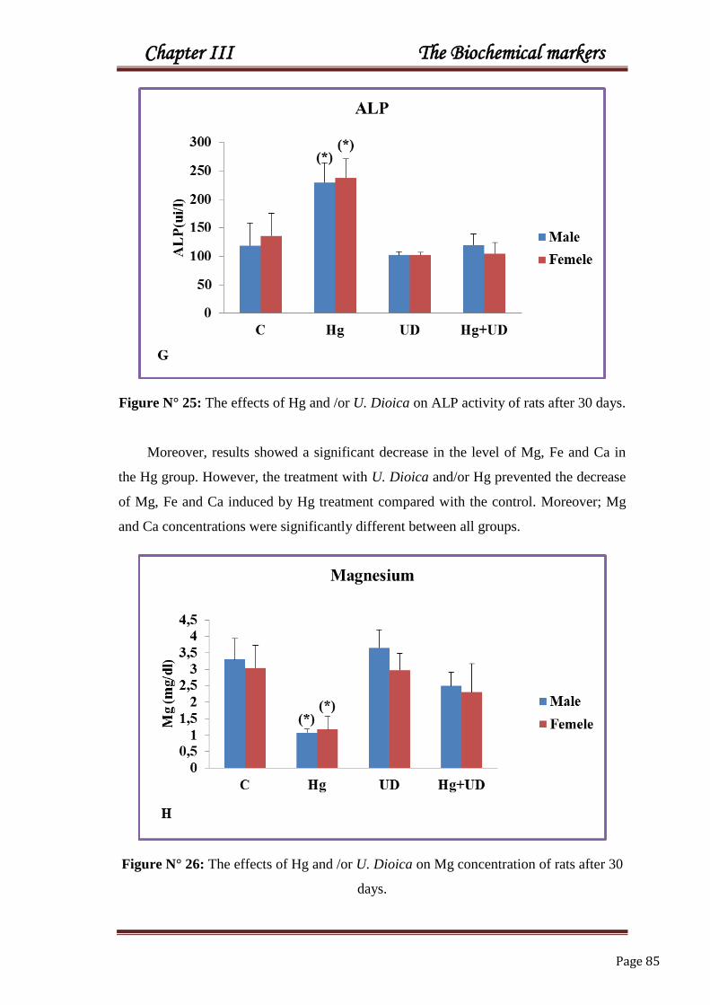
Chapter III The Biochemical markers
Page 85
Figure N° 25: The effects of Hg and /or U. Dioica on ALP activity of rats after 30 days.
Moreover, results showed a significant decrease in the level of Mg, Fe and Ca in
the Hg group. However, the treatment with U. Dioica and/or Hg prevented the decrease
of Mg, Fe and Ca induced by Hg treatment compared with the control. Moreover; Mg
and Ca concentrations were significantly different between all groups.
Figure N° 26: The effects of Hg and /or U. Dioica on Mg concentration of rats after 30
days.

Chapter III The Biochemical markers
Page 86
Figure N° 27: The effects of Hg and/or U. Dioica on Fe concentration of rats after 30
days.
Figure N° 28: The effects of Hg and/or U. Dioica on Ca concentration of rats after 30
days.

Chapter III The Biochemical markers
Page 87
Biochemical markers were summarised in table N° 09.
*S
ign
ifican
tly d
ifferent w
hen
com
pared
to th
e con
trol;
#S
ign
ifican
tly d
ifferent b
etween
gro
up
s.
Ca
(mg/l)
Fe (µ
g/d
l)
Mg
(mg/l)
AL
P (U
I/L)
AL
T (U
I/L)
AS
T (U
I/L)
Crea
tinin
e
(mg/l)
Urea
(g/l)
Trig
lycerid
es (g
/l)
Glu
cose (g
/l)
Ta
ble N
°09
: The am
eliorativ
e Effect o
f U.d
ioica
on
bio
chem
ical mark
ers of rats after 3
0 d
ays H
g
into
xicatio
n.
82
.6±
6.0
7#
10
7.6
±1
.2
3.3
±0.6
3#
11
8.9
±2
0.6
26
.05±
4.3
8
64
.3±
16
.2
0.4
4±
0.4
5#
0.2
5±
0.0
6
0.3
2±
0.0
2
0.8
8±
0.1
6
Ma
le
C
80
.8±
11
.8
10
8.3
±2
8.3
3.0
4±
0.6
7#
13
5.9
±3
9.7#
26
.73±
8.0
5
64
.5±
15
.4
0.4
3±
1.0
5#
0.2
5±
0.0
5
0.2
8±
0.0
7
0.8
9±
0.2
2
Fem
ale
69
.14±
9.8
8*#
56
.5±
5.6
8*
1.0
8±
0.1*
#
22
9.4
±2
0.8*
67
.31±
9*
16
9.5
±3
6.9*
0.8
1±
0.3
4*#
0.4
7±
0.0
6*
0.5
9±
0.1*
1.5
7±
0.6
4*
Male
Hg
54
±14
.7*
61
.3±
39
.5*
1.1
7±
0.3
8*#
23
7.3
±3
4.1
*#
64
.4±
12
.6*
20
4.2
±3
3.2*
0.8
6±
0.1
5*#
0.4
7±
0.0
6*
0.4
9±
0.2
3
……
……
..
Fem
ale
89
.42±
5.6
3#
10
9.5
±1
3.7
3.6
5±
0.5
4#
10
2.4
±1
0.3
25
.95±
3.1
8
66
.9±
15
.2
0.4
0±
0.3
2#
0.2
7±
0.0
6
0.3
2±
0.0
2
0.8
± 0
.13
Male
UD
88
.5±
61
.63
11
9.3
±1
9.6
2.9
7±
1.0
1#
10
.08±
5.1
6#
24
.23±
7.1
7
67
.1±
14
.2
0.4
0±
0.1
2#
0.2
4±
0.0
7
0.2
4±
0.0
6
0.9
± 0
.15
Fem
ale
76
.2±
12
.4#
10
2.9
±1
1.1
2.4
9±
0.5
3*#
12
0±
15
.4
26
.86±
6.0
3
10
5.1
±1
6.5*
0.4
0±
0.3
9#
0.2
3±
0.0
5
0.3
3±
0.0
4
0.8
6±
0.1
6
Ma
le Hg
+U
D
74
.37±
9.1
4
83
.7±
29
.5
2.3
±0.5
2*#
10
4.6
±1
9.2#
19
.67±
6.0
8
12
6.9
±1
0.2*
0.4
3±
0.1
2#
0.2
6±
0.0
6
0.3
5±
0.0
7
0.8
50±
.17
Fem
ale

Chapter III The Biochemical markers
Page 88
3.2. Raphanus sativus
The mercury treated males caused a significant elevation of plasma glucose and
triglycerides when compared to the control. On the other hand, rats received R. Sativus
alone or combined with Hg for 30 days did not show any negative effects.
Figure N° 29. The effects of Hg and/or R. sativus on glucose level of rats after 30 days.
Figure N°30. The effects of Hg and/or R. sativus on triglycerides level of rats after 30
days.

Chapter III The Biochemical markers
Page 89
Results demonstrate increased levels of plasma urea and creatinine in rats
intoxicated by HgCl2 in both sexes compared to the control. Fortunately, the
supplementation of R. sativus corrected the increased urea and creatinine provoked by
Hg. Besides, creatinine concentration shows a signification between all groups in both
sexes.
Figure N° 31: The effects of Hg and/or R. Sativus on urea level of rats after 30 days.
(*)
(*)
0
0,2
0,4
0,6
0,8
1
1,2
1,4
C Hg RS RS+Hg
Cre
ati
nin
e (m
g/l
)
D
Creatinine
Male
Female
Figure N° 32: The effects of Hg and/or R. Sativus on creatinine level of rats after 30
days.

Chapter III The Biochemical markers
Page 90
In the present result, a noticeable increase of AST, ALT and ALP activities has
been observed in the group under Hg stress. However, the presence of R. sativus has
suppressed the toxic effect, and the leakage of enzymes into the blood was decreased,
except in the combined treatment (Hg+RS), only AST was significantly different than
that of the control Furthermore, ALP activity was significantly different between all
groups only in females.
(*)
(*)
(*)
(*)
0
50
100
150
200
250
C Hg RS Hg+RS
AS
AT
(ui/
l)
E
AST
Male
Female
Figure N° 33: The effects of Hg and/or R. Sativus on AST activity of rats after 30 days.
(*) (*)
0
10
20
30
40
50
60
70
80
90
C Hg RS Hg+RS
AL
AT
(ui/
l)
F
ALT
Male
Female
Figure N° 34: The effects of Hg and /or R. Sativus on ALT activity of rats after 30
days.
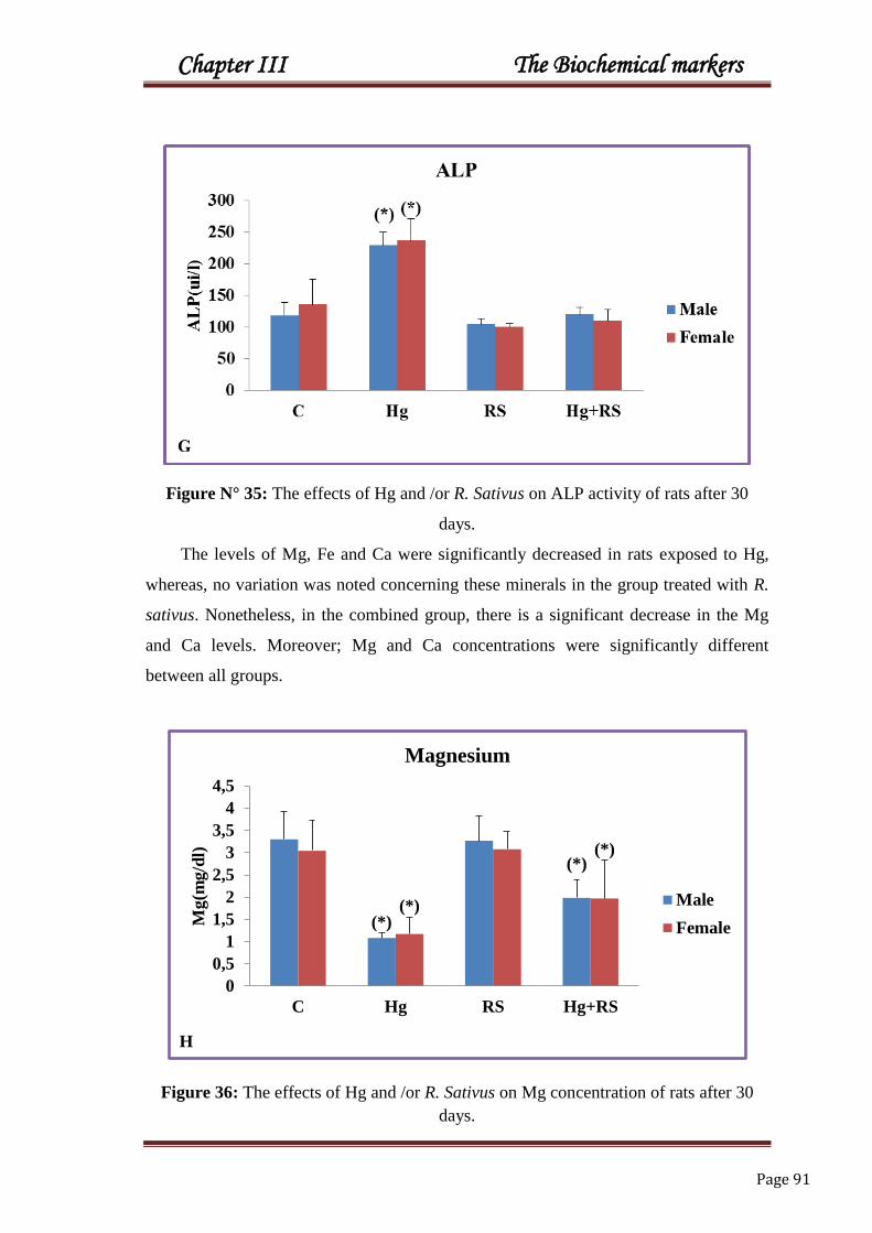
Chapter III The Biochemical markers
Page 91
Figure N° 35: The effects of Hg and /or R. Sativus on ALP activity of rats after 30
days.
The levels of Mg, Fe and Ca were significantly decreased in rats exposed to Hg,
whereas, no variation was noted concerning these minerals in the group treated with R.
sativus. Nonetheless, in the combined group, there is a significant decrease in the Mg
and Ca levels. Moreover; Mg and Ca concentrations were significantly different
between all groups.
(*)
(*)
(*)
(*)
0
0,5
1
1,5
2
2,5
3
3,5
4
4,5
C Hg RS Hg+RS
Mg
(mg
/dl)
H
Magnesium
Male
Female
Figure 36: The effects of Hg and /or R. Sativus on Mg concentration of rats after 30
days.

Chapter III The Biochemical markers
Page 92
Figure N°37: The effects of Hg and /or R. Sativus on Fe concentration of rats after 30
days.
Figure N° 38: The effects of Hg and /or R. Sativus on Ca concentration of rats after 30
days.

Chapter III The Biochemical markers
Page 93
Biochemical markers were summarised in table N° 10.
*S
ign
ifican
tly d
ifferent w
hen
com
pared
to th
e con
trol;
#S
ign
ifican
tly d
ifferent b
etween
gro
up
s.
Ca
(mg/l)
Fe (µ
g/d
l)
Mg
(mg/l)
AL
P (U
I/L)
AL
T (U
I/L)
AS
T (U
I/L)
Crea
tinin
e
(mg/l)
Urea
(g/l)
Trig
lycerid
es (g
/l)
Glu
cose (g
/l)
Ta
ble N
°10
: The am
eliorativ
e Effect o
f R .sa
tivus o
n b
ioch
emical m
arkers o
f rats after 30
day
s Hg
into
xicatio
n.
82
.6±
6.0
7#
10
7.6
±1
0.2
3.3
±0
.63
#
11
8.9
±2
0.6
26
.05
±4
.38
64
.3±
16
.2
0.4
4±
0.4
5#
0.2
5±
0.0
6
0.3
2±
0.0
2
0.8
8±
0.1
6
Ma
le
C 8
0.8
±11
.8
10
8.3
±2
8.3
3.0
4±
0.6
7#
13
5.9
±3
9.7
#
26
.73
±8
.05
64
.5±
15
.4
0.4
3±
1.0
5#
0.2
5±
0.0
5
0.2
8±
0.0
7
0.8
9±
0.2
2
Fem
ale
69
.14
±9
.88
*#
56.5
± 5
.68
*
1.0
8±
0.1
*#
22
9.4
±2
0.8
*
67.3
1±
9*
16
9.5
±3
6.9
*
0.8
1±
0.3
4*
#
0.4
7±
0.0
6*
0.5
9±
0.1
*
1.5
7±
0.6
4*
Male
Hg 54±
14.7
*
61.3
±39.5
*
1.1
7±
0.3
8*
#
23
7.3
±3
4.1
*#
64.4
±12.6
*
20
4.2
±3
3.2
*
0.8
6±
0.1
5*
#
0.4
7±
0.0
6*
0.4
9±
0.2
3
……
……
Fem
ale
85
.44±
9.0
8#
104,5
3±
9.1
8
3.2
6±
0.5
6#
104.9
3±
8.9
3
24.1
8±
4.8
6
63.6
±13.5
0.4
2±
0.2
9#
0.2
5±
0.0
5
0.3
2±
0.0
5
0.8
4±
0.1
3
Male
RS
83
.2±
12
11
4.9
±2
8.6
3.0
7±
0.3
9#
10
1.2
1±
5.2
1#
25
.04±
10
.8
64
.2±
16
.7
0.4
1±
0.2
4#
0.2
6±
0.0
5
0.3
4±
0.1
3
0.8
4±
0.2
4
Fem
ale
72
.04±
7.4
8*#
96
.59
±6
.55
1.9
8±
0.4
1*
#
12
0.4
±11
26
.37
±5
.8
10
2.9
±1
4.2
*
0.4
8±
0.3
#
0.2
5±
0.0
7
0.4
0±
0.1
4
0.8
9±
0.1
3
Male H
g+
RS
69
.6 ±
10
.3
94
.85
±1
7.9
1.9
6 ±
0.8
5*
#
11
0.8
±1
7#
22
.65
±6
.39
10
5.2
±1
5*
0.4
7±
0.2
1#
0.2
7±
0.0
9
0.4
4±
0.1
5
0.8
7±
0.1
1
Fem
ale

Chapter III The Biochemical markers
Page 94
4. GSH
4.1. Urtica dioica
A significant decrease of hepatic GSH level was observed in males and females
exposed to mercury, but its concentration was remarkably raised in the UD group,
followed by Hg+UD group with less extent. Renal GSH concentration was significantly
lower in the Hg exposed animals compared to the control and the UD group as well.
Figure N°39: The effects of Hg and /or U. Dioica on hepatic GSH of rats after 30 days.
Figure N°40: The effects of Hg and/or U. Dioica on hepatic GSH of rats after 30 days.

Chapter III The Biochemical markers
Page 95
GSH data was summarised in table N° 11.
*S
ign
ifican
tly d
ifferent w
hen
com
pared
to th
e con
trol;
#S
ign
ifican
tly d
ifferent b
etween
gro
up
s.
Kid
ney
(nm
ol/m
g
pro
t)
Liv
er
(nm
ol/m
g
pro
t)
Ta
ble N
°11
: Th
e amelio
rative effects o
f U.d
ioica
on
hep
atic and
renal G
SH
(nm
ol/m
g p
rotein
) of rats
after 30
day
s Hg
into
xicatio
n.
1.0
8±
0.1
9
1.2
4±
0.1
1#
Male
C
1.0
9±
0.0
9
2.3
4±
0.1
1#
Fem
ale
0.4
±0.2
5*
0.4
5±
0.2
2*
#
Male
Hg 0
.61±
0.1
4*
1.1
4±
0.2
2*
#
Fem
ale
1.4
9±
0.4
8
2.7
8±
0.1
4*
#
Male
UD
1.9
8±
0.4
5
2.5
8±
0.1
4*
#
Fem
ale
0,9
6±
0,0
8
1.0
6±
0.4
1#
Male H
g+
UD
1.0
8±
0.1
1
1.7
2±
0.4
1#
Fem
ale

Chapter III The Biochemical markers
Page 96
4.2. Raphanus sativus
The levels of hepatic and renal GSH are significantly decreased in Hg group
compared to the control. Compared to the control, no observed variations were recorded
in hepatic and renal GSH when supplemented with the R. sativus and also with
(Hg+RS) in both sexes.
(*)
(*)
0,000
0,500
1,000
1,500
2,000
2,500
3,000
3,500
C Hg RS Hg+RS
GS
H(n
mol/
mg
pro
tein
s)
A
Hepatic GSH
Male
Female
Figure N° 41: The effects of Hg and/or R. Sativus on hepatic GSH of rats after 30 days.
Figure N°42: The effects of Hg and/or R. Sativus on renal GSH of rats after 30 days.

Chapter III The Biochemical markers
Page 97
GSH data are summarised in table N°12.
*S
ign
ifican
tly d
ifferen
t wh
en co
mp
ared
to th
e con
trol;
#S
ign
ifican
tly d
ifferent b
etween
gro
up
s.
Kid
ney
(nm
ol/m
g
pro
t)
Liv
er
(nm
ol/m
g
pro
t)
Ta
ble N
°12
: The am
eliorativ
e effects of R
. sativu
s on
hep
atic and
renal G
SH
(nm
ol/m
g p
rotein
) of rats after 3
0
day
s Hg
into
xicatio
n.
1.0
8±
0.1
9#
1.2
4±
0.1
1#
Male
C
1.0
9±
0.0
9
2.3
4±
0.1
1#
Fem
ale
0.4
±0.2
5*
#
0.4
5±
0.2
2*
#
Male
Hg
0.6
1±
0.1
4*
1.1
4±
0.2
2*
#
Fem
ale
1.0
5 ±
0.0
2#
2.7
8±
0.1
4*
#
Male
RS
1.2
5±
0.0
5
2.5
8±
0.1
4*
#
Fem
ale
1.1
4±
0.1
9#
1.0
6±
0.4
1#
Male H
g+
RS
1.2
1 ±
0.2
#
1.7
2±
0.4
1#
Fem
ale

Chapter III The Biochemical markers
Page 98
5. Histological profile
5.1. Liver
Histological studies on liver tissues (figure 43, 44) following administration of HgCl2
during 30 days showed remarkable histo-pathological changes in the liver of both
females and males characterized by a congestion of the centrilobular vein and a
destruction of liver cells at this dose; dissimilar to the control when observed. It has also
been established that HgCl2 treatment can cause liver injury and even failure in both
animals and humans. These severe alterations affect liver function and can contribute to
a malfunction of this organ.
Treatment with U. dioica and R. sativus alone or/and in a combination with Hg lowered
the injury in liver tissues. This study proved that both U. dioica and R. sativus plants
protects the liver and increase the antioxidant defence system in rats intoxicated with
HgCl2.
5. 2. Kidney
The representative pictures of histo-pathological examination in the kidney tissue are
shown in figure (figure 45, 46). Kidney sections from the control group and U. dioica
and R. sativus rats showed intact histological structure of glomeruli and renal tubules.
However, abnormalities in kidney of Hg treated rats were detected in renal tissues
compared to the control. The main characteristic findings were the appearance of
vacuolization and swelling in the endothelium and swelling in the lining epithelium of
tubules. However, the co-administration of the U. dioica and R. sativus with Hg showed
marked improvement in their histological structure in comparison to the Hg only.

Chapter III The Biochemical markers
Page 99
MALE
Control
Hg
UD RS
(Hg+UD)
(Hg+RS)
Figure N° 43. Histological profiles of male rats liver showing the control, the Hg ,the
UD, the RS and the Hg+UD and Hg+RS groups after 30 days treatments (x 250).
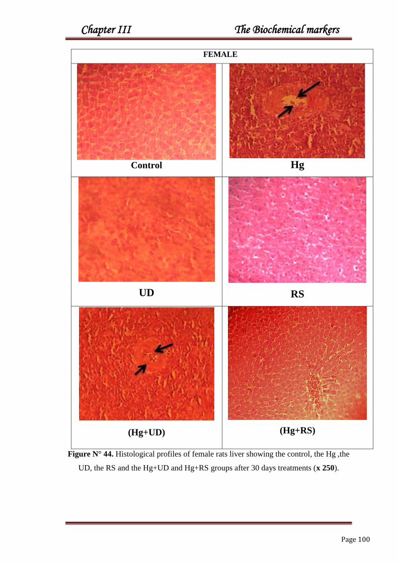
Chapter III The Biochemical markers
Page 100
FEMALE
Control Hg
UD
RS
(Hg+UD)
(Hg+RS)
Figure N° 44. Histological profiles of female rats liver showing the control, the Hg ,the
UD, the RS and the Hg+UD and Hg+RS groups after 30 days treatments (x 250).

Chapter III The Biochemical markers
Page 101
MALE
Control
Hg
UD
RS
(Hg+UD)
(Hg+RS)
Figure N° 45. Histological profiles of male rats kidneys showing the control, the Hg
,the UD, the RS and the Hg+UD and Hg+RS groups after 30 days treatments (x 250).

Chapter III The Biochemical markers
Page 102
FEMALE
Control
Hg
UD
RS
(Hg+UD)
(Hg+RS)
Figure N°46. Histological profiles of female rats kidneys showing the control, the Hg
,the UD, the RS and the Hg+UD and Hg+RS groups after 30 days treatments (x 250).

Chapter III The Biochemical markers
Page 103
DISCUSSION
Body weight is frequently used for observing the growth of animals and its
nutritional status (Ndlovu et al., 2007). In effect, the administration of Hg in rats
showed a significant decrease of the total body weight in both males and females. This
could be related to the decrease of the daily consumption of food and water, which is in
accordance with the report of National Toxicology Program Working Group (NTP,
1993) that the exposure of rats to mercuric chloride reduced food and water intake
leading to weight loss. Such result suggests that this metal presents adverse effects on
rat body growth. This reduction is used as an indicator of the deterioration of the status
of rats‘ general health. Heavy metals are well-known to disturb organ/body weight ratio
when added to diet at toxic amounts (Mahaffey et al., 1981). Our results are supported
by Van Vleet et al. (1981) who showed a marked decrease in body weight gain within
the experimental period, induced by Cd and Pb administration.
Besides, in these experimental conditions, it was noted that the addition of U. dioica and
R. sativus to rats treated by the Hg has caused an enhancement of the body weight. This
could be due to the reduction of the formation of free radicals by the action of
antioxidants presented in both plants. This results supported by Yang et al. (2003) and
Sayama et al. (2000), who found that green tea and body weight gain are associated in
broilers and rats.
Organs’ weights of liver and kidney were increased in males exposed to Hg. This is
would be explained by the tissues hypertrophy caused by this metal. Contrary, a
decrease in liver and kidney weights were noted in females. The increase of organ-body
weight ratio is suggestion of inflammation or infection; whereas a decrease of this ration
could be explained by cell constriction (Moore and Dalley, 1999). Our results are in
accordance with that of Nielsen and Andersen (1991), Eto et al. (2007) who found that
the Methyl mercury produced a reduction in liver weight at 3.0 mg/kg body weight/day.
Also Klein et al. (1972) observed a decrease in liver weight of treated rats at high
doses, correlating these data with low protein levels, glycogen depletion and changes in
the adipose tissue structure. Though inorganic Hg form is considered as a nephrotoxic
agent (Clarkson, 1997), it can pose renal deficiency, modifies body and organ weights
of rats exposed to during the precocious phase of development (Peixoto and Pereira,
2007).

Chapter III The Biochemical markers
Page 104
Glucose level in this study has increased in males exposed to Hg, but no change was
recorded in rats treated with U. dioica and R. sativus. Previously, Hg intoxication was
suggested to increase the energy supply to cope with the metal stress (Margarat et al.,
1999). Interestingly, Durczok et al. (2002) demonstrated that glucose uptake was
deeply affected in several tissues of neonatal rats exposed chronically to methylmercury
chloride, confirming the existence of complex molecular mechanisms of Hg-induced
dysglycaemia depending on the dose administered, the duration of exposure and the
individual age. On the other hand, a considerable hypoglycemia was registered in HgCl2
intoxicated rats, which was more obvious after 60 days exposure (Merzoug et al.,
2009). It is suggested that Hg might damage endocrine function via its ability to inhibit
the complex binding of hormone-receptor or through the inhibition of the enzymes; the
pancreas is a vulnerable organ to the toxic effects of Hg. Insulin, that appear to be the
most affected hormone by mercury, because it has many sulfur-binding sites which can
bind Hg inducing then a dysregulation of blood glucose levels (Chen et al., 2006).
On the other hand, the treatment of diabetes with U. dioica has reduced blood sugar to
its normal level (Mehri et al., 2011; Namazi et al., 2012) and had a hypoglycemic
effect as well on pancreatic beta cells of hyperglycemic male rats (Golalipour et al.,
2007). Moreover, when U. dioica mixed with various plants, the extract had antidiabetic
activity (Petlevski et al., 2001). Similarly, it might be said that green tea and ginger
extract have decreased the elevated glucose concentration in diabetic male rabbits
(Ahmed elkirdasy et al., 2015).
Fortunately, in our investigation the supplementation of R. sativus corrected the
hyperglycemic status provoked by Hg. Most of plants contain glycosides, alkaloids,
flavonoids, carotenoids, etc., that are often concerned by having antidiabetic effect.
Root juice of red radish has been stated to reduce blood glucose levels in streptozotocin
diabetic male rats (Shukla et al., 2011), which was attributed to the presence of ferulic
acid in the roots of this plant (Ohnishi et al., 2004).
Triglycerides’ concentration has risen remarkably in males Hg treated group,
but has augmented slightly in female Hg group. However, it is not the case for the
combined treatment of (Hg+UD) & (Hg+RS). Methylmercury had elevated serum
triglycerides and cholesterol concentration in male rats, while the treatment with
micronutrients (zinc and selenium) has reduced triglycerides by up to 77% and 52%,

Chapter III The Biochemical markers
Page 105
respectively (Joshi et al., 2010). Similarly, Veena et al. (2010) reported that the
exposure of rats to Pb has significantly elevated serum cholesterol concentration.
Dissimilar, in male rabbits, Hg has not affected the level of serum triglycerides and total
cholesterol after 5 week‘s exposure (Maarouf et al., 2008). Moreover, Merzoug et al.
(2009) showed a significant decline of triglycerides in the treated male rats after 2
weeks Hg exposure. Lately, some research mentioned that Hg promotes cardiovascular
disorders via metabolic changes, suggesting that blood cholesterol and triglycerides
would consequently be involved in Hg-induced cardiovascular risks (Mozaffarian,
2009).
On the other side, U. dioica extract has shown an apparent effect on rats at doses of 100
and 300 mg/kg by reducing the levels of total cholesterol and LDL, accompanied with a
remarkable decrease of liver enzymes and animal‘s weights when fed a high cholesterol
diet (Nassiri-Asl et al., 2009). The U. dioica leaves contain chlorophyll which was
proved to have hypolipidemic effects (Mahjoub et al., 2012). Here, the
supplementation of R. sativus to rats‘ clearly decreased triglycerides level comparing
with the control group. Furthermore, the supplementation of black radish juice to lipid-
rich diet resulted in a significant lowering of these parameters in male rats (Lugasi et
al., 2005). Moreover, total cholesterol level in liver homogenate was significantly
reduced by administrating aqueous coriander extract (Kansal et al., 2012), also,
dandelion leaves are helpful by correcting triglycerides and cholesterol levels in female
Pb rats (Mansouri et al., 2014).
Urea and creatinine in this study have a remarkable elevation in male and
female rats exposed to Hg. It is well known that inorganic mercury causes severe kidney
damage after acute and chronic exposure (Zalups, 1997). Accordingly, serum urea was
elevated after acute administration of HgCl2 in mice which due to the toxic effects of
Hg on kidney and liver cells (Ghosh et al., 2008). Urea is the end-product of the amino
acids catabolism; so its increase is an indicator of nephrotoxicity. Any increase in serum
creatinine level could indicate a decreased excretion or a damaged renal function
(Lakshmi et al., 2010). So it can be said that urea and creatinine concentrations are the
most traditional indicators for kidney function and renal structural integrity (Ali et al.,
2001). This elevation in creatinine and urea levels is in accordance with Oriquat et al.

Chapter III The Biochemical markers
Page 106
(2013), Smaoui et al. (2000) and Necib et al. (2012). Hg is able to accumulate in renal
tissues and induces renal impairment (Zalup et al., 1995).
The supplementation of rats with Hg+UD recorded no noticeable change of these
markers. In addition, flavonoids and the high potassium content of U. dioica contribute
in the diuretic action, which allow the body to excrete much wastes including mercury,
and that why stinging nettle is being used as a diuretic agent (Roschek et al., 2009).
Moreover, the administration of methylmercury to male rats has led to an increase in
urea serum and creatinine, while the co-administration of zinc and selenium has
protected the animals from mercury poisoning remarkably (Joshi et al., 2010). Also,
Pistacia lentiscus oil was beneficial to maintain normal serum urea level, which has
been increased in male rabbits treated by mercury chloride for 37 days (Maarouf et al.,
2008). Rabbits pretreated with Juglans sinensis were able to keep their regular urine
volume than rabbits given mercury chloride alone. So, the increase of serum creatinine
level by Hg was attenuated by Juglans sinensis pretreatment (Ahn et al., 2002).
In this study, no increased plasma urea and creatinine was recorded when rats were
treated with R. sativus in both sexes. This is in agreement with the proposal of Kumar
et al. (2013) that demonstrated a nephroprotection of R. sativus in gentamicin induced
nephrotoxicity, which was mediated via its antioxidant effect in male Wistar rats. Necib
et al. (2012) concluded that after sodium selenite has reduced the Hg toxicity in female
Wistar rats.
Amino-transferases in the present study have increased remarkably in the
plasma of rats intoxicated with Hg in two sexes. This elevation of these parameters has
been supported by Toshiko et al. (1998). ALT is a cytoplasmic enzyme found in very
high amounts in the liver and an increase of this specific enzyme designates
hepatocellular damage, while AST is less specific than ALT as an indicator of liver
function (Aliyu et al., 2006). Thus, the activity of AST and ALT enzymes in body fluid
could be used as an indicator of tissues damage provoked by oxidative stress (Svoboda,
2001), which is certainly come from the damage of the plasma membrane through the
fixing of Hg ions to its constituent proteins (Recknagel et al., 1989). Such enzymes are
suggested to be sensitive indicators of tissues damage, since they are liberated from
cells even when the magnitude of lesions is not sufficient for morphological detection
(Recknagel et al., 1989). As a consequence, the significant modifications of plasma

Chapter III The Biochemical markers
Page 107
AST and ALT activity could be the results of the hepatocellular injury or cellular
apoptosis of liver, spleen or muscles (Kaoud et al., 2011). Gutierrez et al. (1992)
found that Pb caused significant leakage of ALT and AST into the medium, which
indicate cellular leakage and loss of functional integrity of hepatic membrane
architecture made by trace metals.
In the current study, the presence of U. dioica and R. Sativus has removed such affect,
by reducing the leakage of enzymes into the plasma. Thus, U. dioica leaves are known
to contain minerals and other chelating agents which enable to reduce Hg toxicity
(Frank et al., 1998). Furthermore, after five weeks, serum AST activity was higher in
male rabbits intoxicated with mercury chloride, but in the Pistacia lentiscus oil rabbits
combined with Hg, the activity of this enzyme was almost similar to the control
(Maarouf et al., 2008). In male Swiss albino mice, serum AST and ALT were
decreased by a single injection of mercuric chlorides after 40 days, meanwhile when Hg
was given with the blue green algae Spirulina fusiformis, the enzymatic activities have
not been affected (Sharma et al., 2007). Effectively, the treatment with U. dioica
protected rats against Aflatoxin-induced hepatotoxicity, as evidenced by decreased AST
and ALT activities (Yener et al., 2009). Besides, U. dioica treatment for 60 days has
exhibited remarkable reduction in liver enzyme activities and also has increased the
reduced antioxidant enzyme levels in tetrachloromethane-treated male rats (Kanter et
al., 2005). The present results are in line with the previous studies, which investigated
the protective role of U. dioica (Mitscher et al., 1997;Celik and Tuluce, 2007).
On other side, current studies proved that R. sativus leaves shown a competitive efficacy
of reducing the elevated transaminase under the paracetamol stress on albino rabbits
(Rukhsana and Mubasher, 2006). Also, Hussein et al. (2012) demonstrated that
plasma ALT and AST activity increased significantly in fish exposed to Hg. However,
the addition of Spirulina platensis enhanced ALT and AST activity to be nearly as the
control group. Our results are consistent with the recent finding of ALT, AST and ALP
activities in male rats intoxicated with Pb (Haleagrahara et al., 2010), while, vitamin C
supplementation reversed these effects and attained the enzyme activities to normal
levels because it has hepatic-protective property related to its antioxidative
characteristic. The presence of dandelion has well-kept the activity of these enzymes,
probably by reducing hepatic failure induced by Pb (Mansouri et al., 2014). Such herbs
are assumed to motivate the elimination of bile, which increases the liver's ability to

Chapter III The Biochemical markers
Page 108
clean up the plasma (Bernard, 2011). The results of the present study were also in
accordance with those of Kalantari et al. (2009) who indicated that the treatment with
radish extract has reduced the activity of AST and ALT in rats given carbon
tetrachloride.
Alkaline phosphatise activity was clearly augmented by Hg intoxication in
male and female rats. ALP is an enzyme found in many organs, especially liver, and can
be liberated into the blood when parenchymal liver cells are damaged. It is reported that
the treatment of rats by HgCl2 at 5 mg / kg body weight for six months increased the
activity of alkaline phosphatase in both sexes of rats and B6C3F1 mice (NTP, 1993).
However, acute administration of HgCl2 has caused many toxic effects on kidney and
liver and it has been associated with increased serum ALP activity (Ghosh et al., 2008).
In addition, Rao et al. (2001) have observed a reduction in enzyme activity of male
mice exposed to HgCl2 to (1.25 mg / kg / day) for 45 days. Meanwhile, Hilmy (1981)
showed a diminution in ALP activity of Aphanius dispar Rüpp (Teleostei) from Red Sea
following acute and long-term mercury exposure that it may be attributed to slow but
constant inhibition of this enzyme by Hg ions. Therefore, it appears that the duration of
exposure to HgCl2 could affect organ function differently; the kidney and liver are the
main sites of Hg deposition and therefore are target organs for its toxicity (Kojima et
al., 1989).
However, in the group fed with U. dioica and R. sativus, no significant changes were
reported. In Swiss albino mice, the efficiency of the blue green algae Spirulina
fusiformis was noticeable to keep alkaline phosphatase activity within normal ranges
against the inhibitory effect of mercury (Sharma et al., 2007). Additionally, both of
Spirulina fusiformis and Ocimum sanctum have returned the enzymatic activity to their
normal biochemical range (Sharma et al., 2005). Dash et al. (2013) and they showed
that the high levels of serum ALT, AST, ALP and bilirubin in the toxicant males and
females were the consequence of CCl4-induced liver dysfunction, whereas the
pretreatment with different doses of Raphanus sativus reversed the levels of these
parameters towards normal.
Minerals levels (Mg, Fe and Ca) were significantly decreased in both sexes of
rats exposed to Hg, whereas; no variation was noted in rats supplied with U. dioica and

Chapter III The Biochemical markers
Page 109
R. sativus. This lowering effect was happened probably because mercury is known to
cause lack of appetite (Zahir et al., 2005), as well as, it is able to provoke many
dysfunctions in the absorption of nutrients (Farmanfarmaian et al., 1989). However,
no changes in the levels of Mg, Fe and Ca have been reported in rats supplemented with
U. dioica and R. sativus which might due to the high content of minerals and vitamins
found in these medicinal plants. The nettle is nutritionally rich in minerals (iron,
manganese, potassium, and calcium), carotenoids, and vitamins A, C and D also
flavonoids (Asgarpanah et al., 2012), as well red radish is a good source of many
minerals (Fe, Ca, Se and Zn) and vitamins (A, K and C) (Rayman, 2012).
Glutathione is the main thiol antioxidant and the conjugating agent; it was
known to bind electrophilic molecular species and free radicals intermediate. In the
present investigation the GSH level showed a noticeable significant depletion following
Hg exposure in different organs (liver, kidney and testis). Reduction in glutathione level
results in the detoxification of peroxides formed by the increased of oxidative stress
(Verma et al., 2001). Such depletions may due to the high affinity of Hg to thiol
groups, as results it disturbs GSH metabolism and damage cell functions (Hultberg et
al., 2001). Therefore, the metal–GSH conjugation manner is necessary in the
elimination of toxic Hg into the bile. HgCl2-induced oxidative stress is usually
attributed to the formation of highly reactive hydroxyl radical (OH), the stimulator of
lipid peroxidation and the sources of damage to the cell membrane (Bharathi and
Jagadeesan, 2012). It has been reported that mercuric chloride administration decreases
renal and hepatic GSH content and increases lipid peroxide formation (Nath et al.,
1996, Jagadeesan and Sankarsami Pillai, 2007).
In fact, most of the presented remedies help the healing or regeneration of the liver
(Neubauer et al., 1998). Beneficial remedies from medicinal plants are considered to be
effective and safe for the alternative treatments for hepatotoxicity (Ranawat et al.,
2009). The co-administration of U.dioica with Hg has resulted in keeping glutathione at
control level in males and females rats. The administration of U. Dioica extracts
demonstrated an antioxidant and hepatoprotective effect against anxious stimulus by
tetrachloromethane in Wistar rats (Kanter et al., 2005). Many studies proved that the
flavonoids and phenols are good antioxidant against Hg induced pathogenesis and also
act as effective chelators for several toxic metal ions (Farag et al., 2013). Numerous

Chapter III The Biochemical markers
Page 110
active components in Spirulina fusiformis could promote the activity of ROS
scavenging enzyme systems and protect against Hg induced renal damages (Sharma et
al., 2007). The co-administration of vitamin E with HgCl2 resulted in maintaining of
glutathione at control levels in male Wistar rats (Rao and Sharma, 2001). Besides, the
reasons for such an effect of the U. Dioica infusions could be due to the presence of
flavonoids in the plants which have strong antioxidant and metal-chelating properties
and can therefore protect cells from free radicals (Celik and Tuluce, 2007). However, it
has been suggested that antioxidants might help the treatment of Hg injuries (Gado and
Aldahmash, 2013).
In our experimental work, we proved that the supplementation of red radish juice can
improve hepatic, renal and testicular GSH level in in both sexes Hg treated rats; thus,
the present study suggests that deleterious ROS or lipid peroxides responsible for HgCl2
toxicity could be corrected by fresh juice of R. sativus, which in turn reflected in
significant enhancement of hepatic, renal and testicular GSH as compared to HgCl2
treated group. It has been reported that red radish has a high amount of total phenolics
(Stratil et al., 2006), and flavonoids (Lugasi et al., 2005), as well as health benefits,
including antioxidant activities (Matsufuji et al., 2007). Salah Abbès et al. (2008)
indicated that radish extract is rich on antioxidants. Radish seed can scavenge the free
radicals in the cell constituents and defend liver from damages, due to its antioxidant
property as source of vitamin C and sulfur containing compounds (Naghibi et al.,
2003). Baek et al. (2008) found that an oral administration of the sulfur radish extract in
mice clearly reduced the liver failure. Additionally, the supplementation of red radish
juice stopped the hepatic damage provoked by paracetamol treatment (Popovic et al.,
1993). Also, raw extract of R. sativus showed a hepatoprotective activity against
zearalenone-induced peroxidative damage in female Balb/c mice (Salah-Abbes et al.,
2008).
Liver histology in this investigation has showed histological changes in males
and female rats received Hg, where it characterized by a dilation of sinusoidal, a
congestion of the centrilobular vein as well as a phenomenon of cellular necrosis
(cytolyses) compared to the control. The changes of the liver histoarchitecture could
mainly due to the toxic effect of mercury. Our results are in accordance with Jadhaf et
al. (2007) who recorded an alteration in the standard levels of various blood chemical

Chapter III The Biochemical markers
Page 111
markers correlated by the histological necrosis and liver degenerative changes of male
rats exposed orally to HgCl2 for 8 weeks. These variations were attributed to noxious
effect of mercuric chloride on the endothelial cells lining blood vessels (Mohamed,
2004).
Ibegbu et al. (2014) showed congestion of hepatoportal blood vessels, congestion of
central vein due to the toxic effect of mercuric chloride. It has been revealed that under
conditions of liver toxicity by mercury, some cells become infiltrated and caused an
increase in cell permeability resulting in the release of transaminases and ALP into the
blood (Kumar et al., 2005), and it also damaged the hepatocytes (Ibegbu et al., 2013),
which may become irreversible, leading to liver cirrhosis (Quirino et al., 2012).
The severe histopathological alterations of liver in the actual study were confirmed by
the marked elevation of amino-transferases, indicating the impaired liver function.
Furthermore, many researchers indicated that intoxication with Pb, Hg and Cd produced
numerous hepatic injuries in experimental animals (Jabeen and Chaudhry, 2011). This
supports the earlier reports on liver histopathological changes made by mercury
intoxication (Ketterer, 1998; Sharma et al., 2000).
However, the examination of liver of groups (Hg+UD) and (Hg+RS) showed that the
pathological changes had almost completely disappeared compared to Hg group. This
could be associated with the ability of our medicinal herbal to eliminate the reactive
oxygen species (ROS) once formed, via very rapid electron transfer system that stops
lipid peroxidation (El-Tohamy and El Nattat, 2010).
Free oxygen radicals have a huge intermediary role in Ischemia-reperfusion injuries of
several organs, including liver. Some researchers suggest that antioxidant molecules
may show a protection from Ischemia-reperfusion injury. The U. dioica is recognized to
be a strong antioxidant, reacting with free radicals. For this reason, it is predictable to be
protective in hepatic rat injury (Terzi et al., 2010; Mittman et al., 1990). Oguz et al.
(2013) concluded that U. dioica is helpful for liver regeneration after liver damage over
oxidative stress, proliferation and apoptosis after partial hepatectomy in Wistar rats.
Kandis et al. (2010) indicated that U. dioica has a protective effect on damage created
during Ischemia-reperfusion in male Wistar rats.
A recent study has reported that vitamin E reversed Pb, Hg, Cd and Cu-induced liver
damage (Al-Attar, 2011), and the addition of vitamin E declined the histopathological
and biochemical alterations caused by Pb intoxication in female Sprague-Dawley albino

Chapter III The Biochemical markers
Page 112
rats (Sajitha et al., 2010), because this vitamin E was confirmed to act as an
antioxidant, and to stop the liver degeneration. The dilatations in the sinusoids were
considerably reduced, and the hepatocytes maintained at normal structure in the co-
exposed rats with Vitamin E and aluminium (Kutlubay et al., 2007).
On the other side, raw extract of R. sativus showed a hepatoprotective activity against
zearalenone-induced peroxidative damage in female Balb/c mice (Salah-Abbes et al.,
2009). This clearly demonstrates the fact that the anthocyanins of R. sativus have
hepatic cell membrane stabilizing effect and cell regeneration ability, which is due to
their anti-lipid peroxidation and/or free radical-scavenging activity. Silymarin is also
known to exert its hepatoprotective action through a similar mechanism of action.
Parenthetically, both these plants are polyphenolic compounds and belong to the
flavonoids family. Thus, the hepatoprotective actions of the red radish anthocyanins
fraction are due to their antioxidant and free radical scavenging characteristics common
to flavonoids (Dash et al., 2013).
Kidney histological study is one of the conformable tests for the nephrotoxic effect of
mercury which reassures the formation of reactive oxygen species (ROS) and lipid
peroxidation of the membrane lipid and protein denaturation. In this current research,
kidneys of females and males Hg-treated rats after 30 days of Hg exposure showed
severe necrosis compared to the control. Mercuric chloride is well known to accumulate
in the renal cortex and disturb the morphology and function of kidney (Greaves, 2000).
Oxidative stress has a dangerous role in the pathophysiology of numerous kidney
diseases, and many complications are mediated by oxidative stress made by mercury
toxicity (Ghaima et al., 2013). The kidney is an organ highly susceptible to damage
caused by ROS, likely due to the plenty of long-chain polyunsaturated fatty acids on the
composition of renal lipids. Aminoglycosides are nephrotoxic agents; their
nephrotoxicity is chiefly attributed to induction of ROS and depletion of antioxidant
enzyme activities in kidney (Alarifi et al., 2012). Jadhav et al. (2007) have observed
degenerative and necrotic changes in the kidney of male rats exposed to mercury via
drinking water. Normal morphology of the renal parenchyma with well-defined
glomeruli and tissues was seen at lower dose of Sidh Makardhwaj added to mercury
intoxication (Sharma et al., 2014). These results agree with others who demonstrated
kidney lesions induced by mercury (Said et al., 2008; Oda and El-Ashmawy, 2012).

Chapter III The Biochemical markers
Page 113
Such lesions were attributed to the Hg direct toxic effect on the development and
function of endothelial lining of renal blood vessels.
Conversely, the treatment with U. dioica and R. sativus showed an enhancement on the
kidney architecture in two the sexes, Also, the pretreatment with silymarin reduced the
intensity of the lesions. Wherein, kidneys showed focal areas of degenerative and
necrotic tubular changes, mostly confined to the renal cortex and corticomedullary
junction. Furthermore, slides from kidney section revealed the protective role of nettle
in preventing the appearance of renal abnormal histological variations after treatment
with Gentamicin in mice albino (Moghaddam et al., 2010). Also, Kumar et al. (2013)
assumed that the nephroprotection showed by R. Sativus in gentamicin induced
nephrotoxicity was mediated through its powerful antioxidant effect. The protective
activity of nettle is possible due to its richness of phenols. Phenolic compounds have
antioxidant properties by scavenging free radicals and active oxygen species such as
single oxygen, free radicals and hydroxyl radicals (Nale et al., 2012). Also, the
investigation done by Toldy et al. (2005) has proposed a neuro-protective property of
nettle extract due to its antioxidant activity. There were no histopathological injuries
detected in animals supplied with Moringa oleifera leaf extract, but the group given Hg
had a destruction of the kidney cyto-architecture. This could be related to the
hepatoprotective by the antioxidant properties possessed by Moringa oleifera leaf
extract against Hg–induced oxidative stress in adult Wistar rats (Ezejindu et al., 2014).

Chapter III The Biochemical markers
Page 114
REFERENCES
A
Ahn J., Gruen I.U., Fernando L.N. (2002). Antioxidant properties of natural
plant extracts containing polyphenolic compounds in cooked ground beef. Journal
of Food Science. 67: 1364–1369.
Alarifi S., Al-Doaiss A., Alkahtani S., Al-Farraj S.A., Al-Eissa M.S., Al-
Dahmash B. (2012). Blood chemical changes and renal histological alternations
induced by gentamicin in rats. Saudi J Biol Sci. 19 (1): 103–110.
Al-Attar A.M. (2011). Protective effect of Avicennia alba leaves extract on gastric
mucosal damage induced by ethanol. Res. J. Med. Plant. 5: 477-490.
Ali B., Bashar A., Alhadrani G. (1999). ―Reproductive hormonal status of rats
treated with date pits.‖ Food Chemistry. 66: 437-441.
Aliyu R., Adebayo A.H., Gatsing D., Garba I.H. (2006). The effect of ethanolic
leaf extract of Commiphora africana (Burseraceae) on rat liver and kidney
functions. J. Pharmacol. Toxicol. 2: 373-379.
Asgarpanah J., Razieh M. (2012). Phytochemistry and pharmacologic properties
of Urtica dioica L. Jinous. Journal of Medicinal Plants Research. 6(46): 5714-5719.

Chapter III The Biochemical markers
Page 115
B
Baek S.E., Park M., Suh J.H., ChoI H.S. (2008). Protective effects of an extract
of young radish (Raphanus sativus L) cultivated with sulfur (Sulfur-Radish Extract)
and of sulforaphane on carbon tetrachloride-induced Hepatotoxicity. Bioscience,
Biotechnol. and Biochem. 72 (5): 1176-1182. Bernard B. (2011). Le pissenlit, l‘or du pré. Ed. de terran.p:115.
Bharathi E., Jagadeesan G. (2012). Influence of s-allyl cysteine against mercuric
chloride induced nephrotoxicity in albino rats. Journal of Chemical and
Pharmaceutical Research. 4(3):1470-1474
C
Carlton W.W., McGavin M.D. (1995). Thomson‘s Special Veterinary Pathology
(2nd edn). Pp: 454-541. Mosby, St. Louis.
Celik I., Tuluce Y. (2007). Elevation protective role of Camellia sinensis and
Urtica dioica infusion against trichloroacetic acid-exposed rats. Phytother. Res. 21:
1039-1044.
Chen YW., Huang CF., Tsai KS., Yang RS., Yen CC., Yang CY. (2006).
Methylmercury induces pancreatic beta-cell apoptosis and dysfunction. Chem Res
Toxicol. 19 (8):1080–1085.

Chapter III The Biochemical markers
Page 116
Clarkson TW. (1997). The toxicology of mercury: critical reviews. Clin Lab Sci.
34: 369–403.
D
Dash R.N., Habibuddin M., Baruah D. B. (2013). Anthocyanins fraction of red
radish (Raphanus sativus L) protects hepatic damage induced by carb on
tetrachloride in albino rats. J Exp Integr Med. 3(1):43-50.
Durczok A., Szkilnik R., Brus R., Nowak P., Labus L., Konecki J., Drabek K.,
Kuballa G., Rycerski W., Mengel K. (2002). Effect of organic mercury exposure
during early stage of ontogenic development on the central dopaminergic system in
adult rats. Polish J. Environ. Stud. 11(4): 307-314.
E
Elkirdasy A., Shousha S., Alrohaimi A H., Faiz Arshad M. (2015).
Hematological And Immunobiochemical Study Of Green Tea And Ginger Extracts
In Experimentally Induced Diabetic Rabbits. Acta Poloniae Pharmaceutica Ñ Drug
Research. 72 (3): 497-506.
El-Tohamy., M.M., El-Nattat.,W.S. (2010). Effect of Antioxidant on Lead-
Induced Oxidative Damage and Reproductive Dysfunction in Male Rabbits. Journal
of American Science. 6 (11):613-622.

Chapter III The Biochemical markers
Page 117
Eto K., Yasutake A., Korogi Y., Akima M., Shimozeki T., Tokunaga H. (2007).
Methylmercury poisoning in common marmosets—MRI findings and peripheral
nerve lesions. Toxicol Pathol. 30(6):723–734.
Ezejindu D. N., Akingboye A. J., Ezejindu C. N. (2014). Hepatoprotective Effects
of Moringa Oleifera Leaf Extract on Mercury Induced Renotoxicity in Adult Wistar
Rats. International Journal of Scientific and Research Publications. 4(3):2250-
3153.
F
Farag M.A., Weigend M., Luebert F., Brokamp G., Wessjohann L.A. (2013).
Phytochemical, phylogenetic, and anti-inflammatory evaluation of 43 Urtica
accessions (stinging nettle) based on UPLC-Q-TOF-MS metabolomics profiles.
Phytochemistry. 96: 170-183.
Farmanfarmaian A., Pugliese K.A., Sun L.Z. (1989). Mercury inhibits the
transport of D-glucose by the intestinal brush border membrane vesicles of fish.
Mar. Environ. Res. 28: 247-251.
Frank B., Bohn I., Uehleke B. (1998). Urtica. Volume 3. In: Blaschek W, Hänsel
R, Keller K, Reichling J, Rmipler H, Schneider (Hrsg.) G, editors. Hagers
Handbuch. Berlin: Springer. p 710-36.
G

Chapter III The Biochemical markers
Page 118
Gado A.M., Aldahmash B.A. (2013). Antioxidant effect of Arabic gum against
mercuric chloride-induced nephrotoxicity. Drug Des. Dev. Ther. 7: 1245-1252.
Ghaima KK., Hashim NM., Ali SA. (2013). Antibacterial and antioxidant
activities of ethyl acetate extract of nettle (Urtica dioica) and dandelion (Taraxacum
officinale). J Appl Pharm Sci. 3(5): 96-99.
Ghosh A., Sil PC. (2008). A protein from Cajanus indicus Spreng protects liver and
kidney against mercuric chloride-induced oxidative stress. Biol Pharm Bull. 31
(9):1651-8.
Golalipour MJ., Kabiri Balajadeh B., Ghafari S., Azarhosh R., Khori V. (2011).
Protective Effect of Urtica dioica L. (Urticaceae) on Morphometric and
Morphologic Alterations of Seminiferous Tubules in STZ Diabetic Rats. Iran J
Basic Med Sci. 14(5): 472–477.
Grieve M. (1971). A Modern Herbal. New York, NY: Dover Publications. 2:574-
579.
Gutierrez R.C., Bucio O.L., Souza A.V., Aranda A.G., Carabez T.A., Chavez
C.E. (1992). Morphological and Functional Changes in WRL-68 Cells Treated with
Heavy Metals. Proceedings of the Western Pharmacology Society. 35: 57-60.
H

Chapter III The Biochemical markers
Page 119
Haleagrahara N., Jackie T., Chakravarthi S., Rao M. (2010).Protective effect
of Etlingera elatior (torch ginger) extract on lead acetate-induced hepatotoxicity in
rats .J. Toxicol. Sci. 35: 663–671
Hilmy AM., Shabana MB., Saied MM. (1981). Mercury levels in some Red Sea
marine pelagic and benzic food chain individuals and their biota. Comp Biochem
Physiol C. 68 (2):195-7.
Hultberg B., Andersson A., Isaksson A. (2001). Interaction of metals and thiols in
cell damage and glutathione distribution: potentiation of mercury toxicity by
dithiothreitol. Toxicology. 156: 93–100.
Hussain S., Rodgers D.A., Duhar H.M., Ali S.F. (1997). Mercuric chloride-
induced reactive oxygen species and its effect on antioxidant enzymes in different
regions of rat brain. J. Environ. Sci. Health B. 32: 395–409.
I
Ibegbu A.O., Ayuba M., Animoku A.A., Brosu D., Umana U. E., Musa S.A.
(2014). Effect of Ascorbic acid on Mercury-induced changes on the liver in Adult
Wistar Rats. Journal of Dental and Medical Sciences. 13(10):10-16.
Ibegbu AO., Alatise AT., Babatunde AO., Dassah, JS., Umana UE., Hamman
WO., Musa SA., Akpulu PS. (2013). Effects of Vitamin E on Phostoxin-Induced
changes in the Liver and Biochemical Parameters of Adult Wistar Rats. J. Biol.
Environ Sci. 7(21):121-129.

Chapter III The Biochemical markers
Page 120
J
Jabeen F., Chaudhry A.S. (2011). Chemical compositions and fatty acid profiles
of three freshwater fish species. Food Chem. 125: 991-996.
Jadhav SH., Sarkar SN., Aggarwal M., Tripathi HC. (2007). Induction of
oxidative stress in erythrocytes of male rats subchronically exposed to a mixture of
eight metals found as groundwater contaminants in different parts of India. Arch.
Environ. Contam. Toxicol. 52(1): 145- 151.
Jagadeesan G., Sankarsami Pillai S. (2007). Hepatoprotective effects of taurine
against mercury induced toxicity in rats. J Environ Biol. 28 (4):753-6.
Joshi A.K., Crossa I., Arun B., Chand R., Trethowan R., Vargas M., Ortiz
Monasterio I. (2010). Genotype environment interaction for zinc and iron
concentration of wheat grain in eastern Gangetic plains of India. Field Crops Res.
116: 268-277.
K
Kalantari H., Kooshapur H., Rezaii F., Ranjbari N., Moosavi M. (2009).
Study of protective effect of Raphanus sativus (Radish) seed in liver toxicity
induced by carbon tetrachloride in mice. Jundishapur Journal of Natural
Pharmaceutical Products. 4(1): 24-31

Chapter III The Biochemical markers
Page 121
Kandis H., Karapolat S., Yildirim U., Saritas A., Gezer S., Memisogullari
R.(2010). Effects of Urtica dioica on hepatic ischemia-reperfusion injury in rats.
Clinics (Sao Paulo). 65 (12):1357-61.
Kansal L., Sharma A., Lodi S. (2012). Remedial effect of coriandrum sativum
(coriander) extracts on lead induced oxidative damage in soft tissues of Swiss
Albino mice. International Journal of Pharmacy and Pharmaceutical Sciences.
77:1153–1155.
Kanter M., Coskun O., Budancamanak M. (2005). Hepatoprotective effects of
Nigella sativa L. and Urtica dioica L. on lipid peroxidation, antioxidant enzyme
systems and liver enzymes in carbon tetrachloride-treated rats. World J.
Gastroenterol. 11(42): 6684-6688.
Kaoud H.A., El-Dahshan A.R. (2011). Bioaccumulation and histopathological
alterations of the heavy metals in Oreochromis niloticus fish. Nat. Sci. 8 (4):147–
156.
Ketterer B. (1998). Glutathione-stransferase and prevention of cellular free radical
damage. Free rad Rese. 28: 647-658.
Kishor Kumar S., Sridher Rao K., Sridhar Y., Shankaraiah P. (2013). Effect Of
Raphanus Sativus Linn. Against gentamicin induced nephrotoxicity in rats. Journal
of Advanced Pharmaceutical Sciences. 3(1):355-365.
Klein R., Herman SP., Brubaker P., Lucier GW. (1972). A model of acute
methyl mercury intoxication in rats. Arch Path. 93(5): 408–418.
Kojima S., Shimada H., Kiyozumi M. (1989). Comparative effects of chelating
agents on distribution, excretion and renal toxicity of inorganic mercury in rats. Res.
Commun. Chem. Pathol. Pharmacol. 64:471-484.

Chapter III The Biochemical markers
Page 122
Kumar S., Sathwara NG., Gautam AK., Agarwal K., Shah B., Kulkarni PK.,
Patel K., Patel A., Dave LM., Parikh DJ., Saiyed HN. (2005). Semen quality of
industrial workers occupationally exposed to chromium. J Occup Health. 47: 424-
430.
Kumar S., Rao K., Sridhar Y., Shankaraiah P. (2013). Effect of Raphanus
sativus linn. against gentamicin- induced nephrotoxicity in rats. Journal of
Advanced Pharmaceutical Sciences. 3(1):355-365.
Kutlubay R., Can E., Nancy L. (2007).Vitamin E Protection from Testicular
Damage caused in traperitoneal aluminium. International J. of toxicology. 26(4):
297-306.
L
Lakshmi BVS., Sudhakar M. (2010). Protective effect of Zingiber officinale on
gentamicin induced nephrotoxicity in rats. Int J Pharmacol. 6: 58-62.
Lugasi A., Blazovics A., Hagymasik K., Kocsis I., Kery A. (2005). Antioxidant
effect of squeezed juice from black radish (R. sativus L var niger) in alimentary
hyperlipidaemia in rats. Phytother Res. 19: 587-591.
Lugasi A., Hovari J. (2000). Flavonoid aglycons in foods of plant origin. I.
Vegetables. Acta Aliment. 29: 345–352.
M

Chapter III The Biochemical markers
Page 123
Maarouf T., Abdennour C., Houaine N. (2008). Influence of Pistacia
lentiscus oil on serum biochemical parameters of domestic rabbit Oryctolagus
cuniculus in mercury induced toxicity. E. J. Sci. Res. 24(4): 591-600.
Maffey KR., Gartside PS., Glueck CJ. (1986). Blood lead levels and dietary
calcium intake in 1- to 11-year-old children: the Second National Health and
Nutrition Examination Survey, 1976-1980. Pediatrics. 78: 257–262.
Mahjoub S., Davari S., Moazezi Z., Qujeq D. (2012). Hypolipidemic effects of
ethanolic and aqueous extracts of Urtica dioica in rats. World Appl. Sci. J. 17(10):
1345-1348.
Mahmoud A.H., Motawa H.M., Wahba H.E., Ebrahim A.Y. (2004). Study of
some antioxidant parameters in mice livers affected with Urtica pilulifera extracts.
Asian J. Biochem. 1: 67-74.
Mansouri O., Abdennour C. (2011). Evaluation of the therapeutic efficiency of
raw garlic on reproduction of domestic rabbits under lead induced toxicity. Ann Biol
Res. 2:389-393.
Margarat A., Jagadeesan G., Sethupathy S. (1999). Recovery of liver tissue by
penicillamine in mercury-poisoned mice, Mus musculus (Linn). India J. Environ.
Toxicol. 9(1): 24-26.
Matsufuji H., Ishikawa K., Nunomura O., Chino M., Takeda M. (2007).
Antioxidant content of different coloured sweet peppers, white, green, yellow,
orange and red (Capsicum annuum L.). International Journal of Food Science and
Technology. 42: 1482-1488.
Chapter III The Biochemical markers
Page 124
Mehri A., Ranjbar-Hasani S., Larijani B., Abdollahi M. (2011). A systematic
review of efficacy and safety of Urtica dioica in treatment of diabetes. Int. J.
Pharmacol. 7(2): 161-170.
Merzoug S., Toumi M.L., Oumeddour A., Boukhris N., Baudin B., Tahraoui A.,
Bairi A. (2009). Effect of inorganic mercury on biochemical parameters in Wistar
rat. J. Cell Anim. Biol. 3(12): 222-230.
Mitscher L.A., Drake S., Gollapudi S.R., Okwute S.K. (1997). A modern look at
folkloric use of anti-infective agents. Journal of Natural Products. 50: 1025 1040.
Mittman P. (1990). Randomized, double-blind study of freeze-dried Urtica dioica
in the treatment of allergic rhinitis. Planta Med. 56:44-47.
Moghaddam AH., Javaheri M., Nabavi SF., Mahdavi MR., Nabavi SM.,
Ebrahimzadeh MA. (2010). Protective role of pleurotus porrigens (Angelʼs wings)
against gentamicin-induced nephrotoxicity in mice. Eur Rev Med Pharmacol Sci.
14: 1011-4.
Moore K.L., Dalley A.F. (1999). Clinical Oriented Anatomy. Woller Klumner
Corporation, Philadelphia.
Mozaffarian D. (2009). Fish, mercury, selenium and cardiovascular risk: Current
evidence and unanswered questions. Int. J. Environ. Res. Public Health. 6: 1894-
1916.
N

Chapter III The Biochemical markers
Page 125
Naghibi F., Pourmorad F., Honary S., Shamsi M. (2003). Decontamination of
Water Polluted with Phenol Using Raphanus sativus Root. Iranian Journal of
Pharmaceutical Research. 2: 29-32.
Namazi N., Esfanjan A.T., Heshmati J., Bahrami A. (2012). The effect of hydro
alcoholic nettle (Urtica dioica) extracts on insulin sensitivity and some
inflammatory indicators in patients with type 2 diabetes: A randomized double-blind
control trial. Pak. J. Biol. Sci. 14: 775-779.
Nassiri-Asl M., Abbasi E., Hajiabadi M., Nouri A., Farhoudi H. (2009). The
interaction between quinine and trimethylamine in the formalin test.
Pharmacologyonline. 3: 1008-1014.
Nath K A., Croatt A J., Likely S., Behrens T W., Warden D. (1996). Renal
oxidant injury and oxidant response induced by mercury. Kidney International. 50
(3):1032–1043.
National Toxicology Program. (1993). Toxicology and carcinogenesis studies of
mercuric chloride (CAS No. 7487- 94-4) in F344 rats and B6C3F1 mice. Natl.
Toxicol. Program Tech. Rep. Ser. 408: 1-260.
Ndlovu T., Chimonyo M., Okoh A. I., Muchenje V., Dzama K ., Raats J.G.
(2007). Assessing the nutritional status of beef cattle: current practices and future
prospects. Afr. J. Biotechnol. 6(24): 2727-2734.
Necib Y., Bahia A, Zerizer S., Abdennour C., Boulakoud M.S. (2013). Effect of
Argan oil (Argania spinosa. L) on Kidney Function Impairment and Oxidative
Stress Induced by Mercuric Chloride in Rats. Int. J. Pharm. Sci. Rev. Res., 22(2):
144-148.

Chapter III The Biochemical markers
Page 126
Neugebauer W., Winterhalter P., Schrier P. (1998). 3-hydroxy-alpha-ionyl-beta-
d-glucopyranosides from stinging nettles (Urtica dioica L.). Natural Products
Letters. 6:177–80.
Nielsen J.B., Andersen O. (1991).Time dependent disposition of mercury after oral
dosage. Metal compounds in environment and life. Science and Technology Letters,
Middlesex, UK. 341-348.
O
Oda S.S., El-Ashmawy I.M. (2012). Adverse effects of the anabolic steroid,
boldenone undecylenate, on reproductive functions of male rabbits. I nt. J. Exp.
Path. 93: 172–178.
Oguz S., Kanter M., Erboga M., Toydemir T., Sayhan MB., Onur H. (2013).
Effects of Urtica dioica on oxidative stress, proliferation and apoptosis after partial
hepatectomy in rats. Toxicol Ind Health. 31(5):475-84.
Ohnishi M., Matuo T., Tsuno T., Hosoda A., Nomura E., Taniguchi H., Sasaki
H., Morishita H. (2004). Antioxidant activity and hypoglycemic effect of ferulic
acid in STZ-induced diabetic mice and KK-Ay mice. Biofactors. 21: 315–319.
Oriquat G.A., Saleem T.H., Naik R.R., Moussa S.Z., Al-Gindy R.M. (2012). A
sub-chronic toxicity study of mercuric chloride in the rat. JJBS. 5(2): 141-146.
P

Chapter III The Biochemical markers
Page 127
P eixoto NC., Pereira ME. (2007). Effectiveness of ZnCl2 in protecting against
nephrotoxicity induced by HgCl2 in newborn rats. Ecotoxicol Environ Safety.
66:441-6.
Petlevski R., Hadzija M., Slijepcevic M., Juretic D. (2001). Effect of 'antidiabetis'
herbal preparation on serum glucose and fructosamine in NOD mice. J
Ethnopharmacol. 75 (2-3):181-4.
Popovic M., Lukie V., Jakovljevie V., Mikov M. (1993). The effect of the radish
juice on liver function. fitoterapia. 64:229–231.
Q
Quirino CJ., Marcília AF., Renério FJ. (2012). Depression, Insomnia, and
Memory Loss in a Patient With Chronic Intoxication by Inorganic Mercury. The
Journal of Neuropsychiatry and Clinical Neurosciences. 1: 191-205.
R
Ranawat L., Bhatt J., Patel J. (2009). Hepatoprotective activity of ethanolic
extracts of the bark of Zanthoxylum armatum DC in CCl4 induced hepatic damage in
rats. J Ethnopharmacol. 127:777-80.

Chapter III The Biochemical markers
Page 128
Rao MV., Sharma PSN. (2001). Protective effect of vitamin E against mercuric
chloride reproductive toxicity in male mice. Reproductive Toxicology. 15 (6):705–
712.
Rayman MP. (2012). Selenium and human health. Lancet. 379: 1256-1268.
Recknagel R.O., Glende E.A., Dolak J.A., Walter, R.L. (1989). Mechanism of
carbon tetrachloride toxicity. Pharmacol. Ther. 43: 139-154.
Richardson M. (1996). The Safety of Dental Amalgam. Canada: Minister of Health.
Roschek Jr., Ryan C., Randall S. (2009). Nettle extract (Urtica dioica) affects
key receptors and enzymes associated with allergic rhinitis. Phytotherapy
Research. 23(7): 920–926.
Rukhsana A., Mubasher A. (2006). Studies of Raphanus sativus as Hepato
Protective Agent. Journal of Medical Sciences. 6: 662-665.
S
Said O., Fulder S., Khalil K., Azaizeh H., Kassis E., Saad B. (2008).
Maintaining a physiological blood glucose level with ‗glucolevel‘, a combination of
four anti-diabetes plants used in the traditional Arab herbal medicine. Evid Based
Complement Alternat Med. 5:421- 8.
Sajitha GR., Jose R., Andrews A., Ajantha KG., Augustine P., Augusti KT.
(2010). Garlic oil and vitamin E prevent the adverse effects of lead acetate and
ethanol separately as well as in combination in the drinking water of rats. Indian J
Clin Biochem. 25: 280–288.

Chapter III The Biochemical markers
Page 129
Salah-Abbes B., Abbes B., Haous Z., Oueslati R. (2009). Raphanus sativus
extract prevents and ameliorates zearalenone-induced peroxidative hepatic damage
in Balb/c mice.J. Pharm. Pharmacol. 61: 1545–1554
Sayama K., Lin SX., Zheng GD., Oguni I.(2000). Antiobee effects of green tea
powder and its components. International Symposium on Green tea. KFN
Conference. pp. 55–62.
Sharma B., Singh S., Siddiqi N.J. (2014). Biomedical implications of heavy metals
induced imbalances in redox systems. BioMed Research International. p 26.
Sharma M.K., Kumar M., Kumar A. (2005). Study of mercury induced toxicity in
liver and its modulation by Spirulina fusiformis and Ocimum sanctum. In:
proceeding of third world congress on cellular and molecular biology held at jena,
Germany. Cell. Mol. Biol. 46, 227
Sharma MK., Sharma A., Kumar A., Kumar M. (2007). Spirulina fusiformis
provides protection against mercuric chloride induced oxidative stress in Swiss
albino mice. Food Chem. Toxicol. 45: 2412-2419.
Shukla S., Chatterji S., Mehta S., Rai PK., Singh RK., Yadav DK ., Watal G.
(2011). Antidiabetic effect of Raphanus sativus root juice. Pharm Biol. 49(1): 32-
37.
Smaoui M., Rebaï T., El Feki A. (1997). Incidence en fonction du temps des
émissions d'origine automobile sur la croissance générale, la fertilité et la variation
de certains paramètres biochimiq ues chez le rat. 8e Congrès Arabe de Biologie,
Amman, Jordanie.134-5.
Smaoui M., Yhorbel F., Boujelbene M., Makni-Ayadi F., El Féki A. (2000).
Impact de l‘Exposition Chronique au Gaz d‘Echappement d‘Origine Automobile sur

Chapter III The Biochemical markers
Page 130
certains Biomarqueurs touchant la Fonction Hormonale Sexuelle Mâle, la Fonction
Rénale et l‘Hémogramme chez le Rat, Pollut. Atmosph. 167 (2): 439-449.
Stratil P., Klejdus B., Kubáň V. (2006). Determination of total content of phenolic
compounds and their antioxidant activity in vegetables evaluation of
spectrofotometric methods. Journal of Agriculture and Food Chemistry. 54: 607–
616.
Svoboda M. (2001). Stress in fishes (a review). Bull. VURH Vodnany. 4:169-191.
T
Terzi R., Saglam A., Kutlu N., Nar H., Kadioglu A. (2010). Impact of soil
drought stress on photochemical efficiency of photosystem II and antioxidant
enzyme activities of Phaseolus vulgaris cultivars. Turk. J. Bot. 34: 1-10.
Toldy A., Krisztian S., Maria S., Judit J., Kyung J.J. (2005). The effect of
exercise and nettle supplementation on oxidative stress markers in the rat brain.
Brain Research Bulletin. 65: 487–493.
Toshiko T. K., Mieko S., Akira N., Nobuki Y., Nobumasa L. (1998). Strain
difference in sensitivity of mice to renal toxicity of inorganic mercury. J.
Pharmacol. Exp. Ther. 285 (1): 335-341.
V

Chapter III The Biochemical markers
Page 131
Van Vleet J.F., Boon G.D., Ferrans V.J. (1981). Induction of lesions of
selenium-vitamin E deficiency in ducklings fed silver, copper, cobalt, tellurium,
cadmium, or zinc: protection by selenium or vitamin E supplements. American
Journal of Veterinary Research. 42:1206-1217.
Veena B., Radhakrishnan C.K., Chacko J. (1997). Heavy metal induced
biochemical effects in an estuarine teleost India. J. Mar. Sci. 26: 74-78.
Verma S., Kumar R., Khadwal A., Singhi S. (2010). Accidental inorganic
mercury chloride poisoning in a 2-year old child. Indian J Pediatr. 77:1153-1155.
Y
Y ang C.S., Lambert J.D., Sang S. (2003). Antioxidative and anti-carcinogenic
activities of tea polyphenols. Arch Toxicol. 83(1):11–21.
Yener Z., Celik I., Ilhan F., Bal R. (2009). Effects of Urtica dioica L. seed on lipid
peroxidation, antioxidants and liver pathology in aflatoxin-induced tissue injury in
rats. Food Chem. Toxicol. 47: 418-424.
Z

Chapter III The Biochemical markers
Page 132
Zahir F., Rizwi SJ., Haq SK., Khan RH. (2005). Low dose mercury toxicity and
human health. Environ Toxicol Pharmacol. 20 (2):351-60.
Zalups R.K. (1995). Progressive losses of renal mass and the renal and hepatic
deposition of administered inorganic mercury. Toxicol. Appl. Pharmacol. 130 (1):
121-131.
Zalups R.K. (1997). Enhanced renal outer medullary uptake of mercury associated
with uninephrectomy: implication of a luminal mechanism. J. Toxicol. Environ.
Health. 50: 173–194.
Zalups R.K. (2000). Molecular interactions with mercury in the kidney. Pharmacol.
Rev. 52: 113-143.

CCHHAAPPTTEERR IIVV:: RREESSUULLTTSS OOFF
RREEPPRROODDUUCCTTIIVVEE PPRROOFFIILLEE

Chapter IV The Reproductive Profile
Page 133
RESULTS
1. Organ’s weights
1.1. Urtica dioica
In males, a reduction in the testis and epididymis absolute weight of Hg group was seen
compared to the control. In contrast, rats exposed to U. dioica had slight rise in absolute
weights of these organs.
Figure N°47: Absolute weight variations of rats’ testis in the groups treated by Hg and /or U.
dioica for 30 days.
Figure N°48: Absolute weight variations of rats’ epididymis in the groups treated by Hg
and/or U. dioica for 30 days.

Chapter IV The Reproductive Profile
Page 134
Testis and epididymis organ’s absolute weight were summarised in table N° 13.
Table N° 13: The ameliorative effect of U.dioica on reproductive profile of male rats after 30
days Hg intoxication.
C Hg UD Hg + UD
Testis (g) 1.45 ±0.22 1.18 ±0.15* 1.51 ±0.09 1.57±0.18
Epididymis (g) 0.55±0.06 0.31 ±0.1* 0.55±0.07 0.51±0.07
*: Significantly different when compared to the control;
#: Significantly different between groups.
1.2. Raphanus sativus
In male Wistar rats, we recorded a reduction in the testis and epididymis weight in Hg
groups compared to the control group. Meanwhile, R. sativus addition presented an
enhancement of weight of these organs comparing to the control group.
Figure N° 49: Absolute weight variations of rats’ testis in the groups treated by Hg and /or R.
sativus for 30 days.

Chapter IV The Reproductive Profile
Page 135
Figure N° 50: Absolute weight variations of rats’ epididymis treated by Hg and/or R. sativus
for 30 days.
Testis and epididymis organ’s absolute weight were summarised in table N° 14.
Table N° 14: The ameliorative effect of R. sativus on testis and epididymis organ’s weight of
male rats after 30 days Hg intoxication.
C Hg RS Hg + RS
Testis (g) 1.45 ±0.22 1.18 ± 0.15* 1.45± 0.09 1.5± 0.2
Epididymis (g) 0.55±0.06 0.31 ±0.1* 0.57± 0.04 0.57± 0.08
*: Significantly different when compared to the control;
#: Significantly different between groups.

Chapter IV The Reproductive Profile
Page 136
2. The reproductive markers
2.1. Urtica dioica
Mercury group in the present study provoked pronounced reduction of the epididymal
sperm concentration and motility. Concerning the motility, there is a significant difference
between all groups. On other hand, a noticeable improvements in all sperm's parameters were
recorded when U.dioica was added to rats.
(*)
0
100
200
300
400
500
C Hg UD Hg+UD
con
cen
trati
on
(10
6/m
l)
B
Concentration
Figure N°51. The effects of Hg and /or U. dioica on spermatozoa’s concentration of rats
after 30 days.
Figure N° 52. The effects of Hg and/or U. dioica on spermatozoa’s motility of rats after 30
days.

Chapter IV The Reproductive Profile
Page 137
Testosterone concentration showed clearly a significant decrease in the Hg group
compared to the control. However, the treatment with U.dioica presented an improvement in
the level of testosterone compared to the control group.
Figure N° 53. The effects of Hg and/or U.dioica on testosterone level of rats after 30 days.
Testicular GSH in rats stressed by mercury shows a decreased concentration comparing
to the control group, however, the treatment with U.dioica raised significantly the
concentration of testicular GSH toward control group. Besides, there is a difference between
all the groups.
Figure N° 54. The effects of Hg and/or U. dioica on testicular GSH concentration of rats
after 30 days.

Chapter IV The Reproductive Profile
Page 138
The Reproductive markers were summarised in table N° 15.
Table N°15: The ameliorative effect of U. dioica on reproductive markers of male rats after
30 days Hg intoxication.
C Hg UD Hg + UD
Concentration
(106)
311.3 ± 78.9 111.3 ± 23.1* 315.5 ± 76.3 223.5 ± 83
Motility (%) 66.05 ± 7.79# 31.9 ± 10.6
*# 71.39 ± 4.17
# 54.1 ± 13.9
#
Testosterone
(ng/ml)
2.07 ± 0.41 0.97 ± 0.11* 2..1 ± 0.36 1.52 ± 0.42
Testicular GSH
(nmol/mg
proteins)
0.17±0.01# 0.09±0.02
*# 1.27 ±0.14
*# 0.36 ±0.39
#
*: Significantly different when compared to the control;
#: Significantly different between groups.
2.2. Raphanus sativus
Results show that the mean sperm concentration and motility decreased significantly in
Hg group compared to control group. Also, there is a significant difference between all groups
However, sperm concentration and motility of (RS) and (Hg+RS) groups were not statically
different than that of the control.
(*)
0
100
200
300
400
500
C Hg RS Hg+RS
Con
cen
trati
on
(10
6/m
l)
A
Concentration
Figure N° 55. The effects of Hg and/or R. sativus on spermatozoa’s concentration of rats
after 30 days.

Chapter IV The Reproductive Profile
Page 139
Figure N° 56. The effects of Hg and/or R. sativus spermatozoa’s motility of rats after 30
days.
Testosterone concentration showed a clear significant decrease in the Hg group compared
to the control. However, the treatment with R. sativus has presented a markedly normal level
of testosterone compared to the control.
(*)
0
0,5
1
1,5
2
2,5
3
C Hg RS Hg+RS
Test
est
ero
ne (
ng/m
l)
C
Testesterone
Figure N° 57. The effects of Hg and /or R. sativus on testosterone level of rats after 30 days.

Chapter IV The Reproductive Profile
Page 140
Testicular GSH concentration was decreased significantly in the Hg group. Noticeably,
males treated with R. Sativus alone showed a highly significant increase of testicular GSH
concentration. GSH level of the (Hg+RS) was not different than that of the control. But, there
is a significant difference between all groups.
Figure N°58: The effects of Hg and/or R. Sativus on testicular GSH of rats after 30 days.
Reproductive profile were summarised in table N° 16.
Table N° 16. The ameliorative effect of R. sativus on reproductive markers of male rats after
30 days Hg intoxication.
Groups C Hg RS Hg + RS
Concentration
(106)
311.3 ± 78.9 111.3 ± 23.1*
365.6± 58 73.05± 4.44
Motility (%) 66.05 ± 7.79# 31.9 ± 10.6
*# 73.05± 4.44
# 58.86± 6.75
#
Testosterone
(ng/ml)
2.07 ± 0.41 0.97 ± 0.11*
2.39± 0.33 1.79±0.59
Testicular GSH
(nmol/mg
proteins)
0.17±0.01# 0.09±0.02*
# 1.22± 0.2
*# 0.2± 0.02
#
*: Significantly different when compared to the control;
#: Significantly different between groups.

Chapter IV The Reproductive Profile
Page 141
3. The Histological examinations
Figures (59, 60) illustrated the histological examination of testis and epididymis tissues
of different groups. Microscopic assessment of male reproductive system in control group
revealed normal seminiferous tubules, sperms with normal morphology, and concentration, as
shown in Figures (59, 60). Figures demonstrate that HgCl2 intoxication caused histological
changes where spermatozoa were rarely seen in tubules, different degrees of degeneration in
the lining spermatogenic tubules, and delocalization of seminiferous tubules. The histological
changes were reduced remarkably in testis and epididymis in animals supplemented with
Urtica dioica leaves and Raphanus sativus juice which showing sperm with normal
morphology and concentration close to control group. UD and RS treated animals showed
improved concentration of sperms and stabilization of organized seminiferous tubules and
many newly formed spermatogenic cells arranged properly inside the tubules better than the
control group. The results obtained from histological architecture were in consistency with the
reproductive markers.
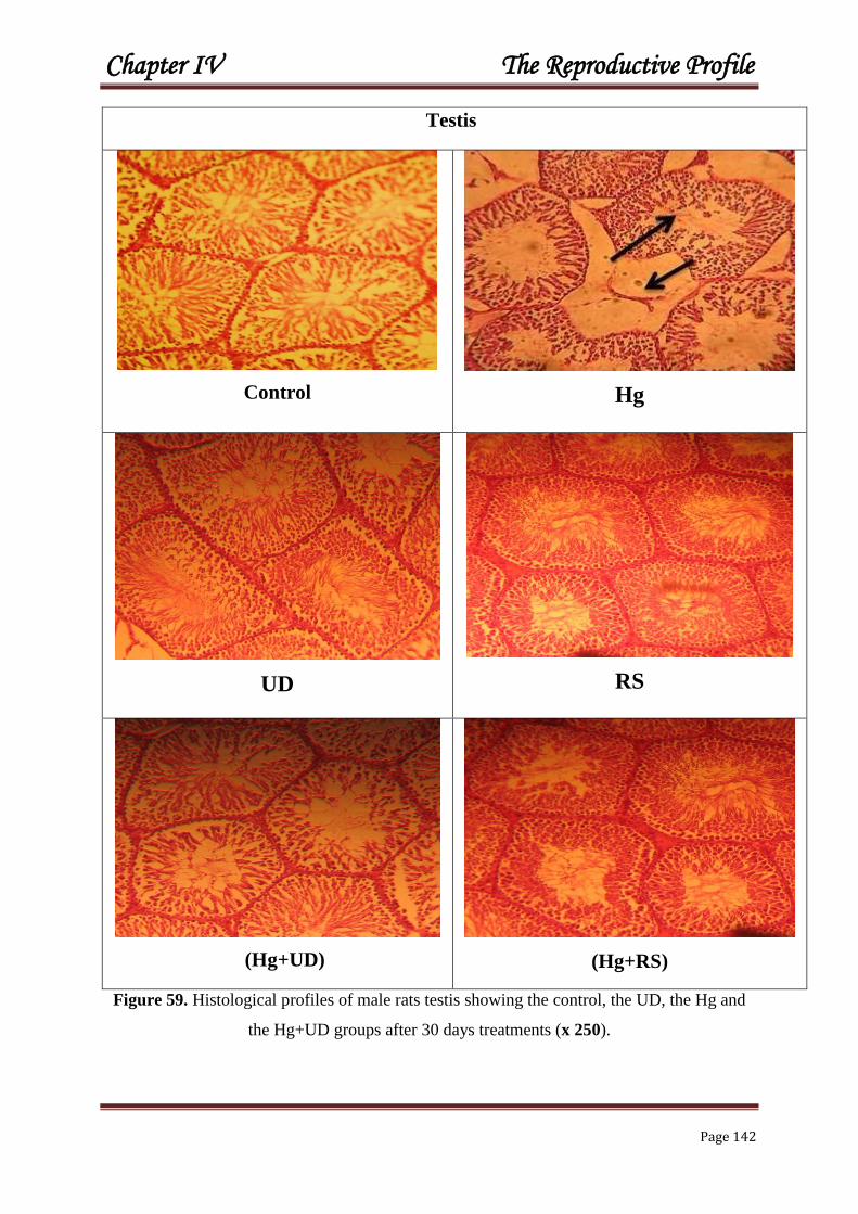
Chapter IV The Reproductive Profile
Page 142
Testis
Control
Hg
UD
RS
(Hg+UD)
(Hg+RS)
Figure 59. Histological profiles of male rats testis showing the control, the UD, the Hg and
the Hg+UD groups after 30 days treatments (x 250).

Chapter IV The Reproductive Profile
Page 143
Epididymis
Control
Hg
UD
RS
(Hg+UD)
(Hg+RS)
Figure 60. Histological profiles of male rats epididymis showing the control, the Hg, the UD,
the RS and the Hg+UD and Hg+RS groups after 30 days treatments (x 250).
Chapter IV The Reproductive Profile
Page 144
DISCUSSION
The weight of reproductive organs of male Wistar rats showed a diminution of testis
and epididymis weight in the Hg group; such results are in line with Al-Naimi et al. (2013)
who found a decreased testicular weight produced by the degenerative effect of mercuric
chloride. The effect of HgCl2 induced a decrease in sperm count by inhibiting the
spermatogenesis, in parallel to a reduction in testis and epididymis weights. Adverse effects of
HgCl2 on rat testicular tissues have been reported with marked testicular spermatogenic
degeneration at the spermatocyte level (Chowdhury et al., 1986). Mercury was reported to
provoke alterations in testis along with biochemical changes (Massanyi et al., 2007).
However, the increase in body weight and testicular weight in the supplemented group may be
due to an increase in the intake of food and water, and also to the protective roles of
compounds found in these plants against Hg toxicity. Thus, the observe elevation of the ratio
organ-body weight (brain, kidney and liver) after supplying the leaves of Chrysocoma ciliata
may be due to the functional capacity amelioration of these organs (Ashafa et al., 2009).
Reproductive markers have showed a reduction in the concentration and motility of
epididymal sperm. It proves that male reproductive system is a target system to Hg exposure.
Accordingly, another study revealed that sperm count and motility of the epididymis was
markedly reduced by HgCl2 (Basu et Dickerson, 1996). Such result is in agreement with that
of Moumen et al. (2011). Thus, the epididymis is known to play an important role in
providing the microenvironment for sperm maturation and storage. Consequently, the
decrease in epididymal sperm number/motility might be explained on the basis that Hg has
traversed the blood–testis barrier and gained access to germinal cells, leading to testicular
functional inactivation (Sharma et al., 1996). Previously, it was found that oral exposure to
mercuric chloride produced adverse effects on the reproductive performance of mice (Khan
et al., 2004). Subsequently, mammalian spermatozoa are rich in poly unsaturated fatty acids
and are thus very vulnerable to ROS attack; the later induced a decrease of sperm motility,
presumably by a rapid loss of intracellular ATP, causing testicular damage (De Lamirande
and Gagnon, 1992). Furthermore, it was reported that the exposure of Wistar rats to Mn
induces a reduction of number of spermatids and spermatocytes in the seminiferous tubules
(Badou et al., 2013). In addition, the study of El-Tohamy and El-Nattat (2010) proved that
Pb treatment can impair male reproductive function by reducing the sperm count and volume.

Chapter IV The Reproductive Profile
Page 145
Certainly, heavy metals are known to inhibit the enzymatic reactions by stopping the
steroidogenesis process (Smaoui et al., 1997). The epidemiological study carried out in Hong
Kong had demonstrated that the infertility of both men and women are linked to the high
levels of environmental mercury (Dickman et al., 1998).
However, a noticeable improvement in all sperm’s markers was observed when animals were
treated with Hg+UD and Hg+RS, which suggests the protective roles played by these
medicinal herbs. Additionally, the supplementation of essential S. aromaticum oil to rats
previously poisoned by Mn, has helped to regenerate the majority of seminiferous tubules and
interstitial cells, and has filled lights seminiferous tubules with spermatozoa (Badou et al.,
2013). Recently, Shodehinde and Ganiyu Oboh (2013) indicated that polyphenols’ rich-red
radish might act as great antioxidants that can inhibit lipid peroxidation, preventing testes
from the oxidative stress. Furthermore, vitamin C was reported to ameliorate the harmful
effect of Pb in rabbits, by improving sperm motility, reproductive function, and decreasing the
production of ROS (El-Tohamy, 2010). Additionally, vitamin E, glutathione, zinc and
selenium are considered as beneficial for the treatment of male infertility (Sinclair, 2000).
Accordingly, U. dioica contains many minerals and vitamins, especially vitamin C and E, as
well as flavonoids (Asgarpanah et al., 2012), which can act as an antioxidants to counteract
Hg intoxication. In fertile individuals, sperm motility levels, especially progressive sperms,
are directly related to the reproductive capability. Moreover, U. dioica can act as an anti-
oxidant and improve the sperm cells quality and count by the elevation of the anti-oxidant in
comparison with the nicotine group (Golalipour et al., 2011). Furthermore, it appears that U.
dioica supplementation could improve the quality of spermatozoa and inhibits nicotine-
induced toxic effects on sperm parameters in male mice (Jalili et al., 2014). Many studies
revealed that animals receiving vitamin E alone or combined with HgCl2 had sperm markers
similar to the normal values (Basu and Dickerson, 1996).
Testosterone level in this study has been negatively affected by mercury after one
month exposure. In fact, mercury can react with the sulfhydryl’s groups of proteins, which
could inhibit enzymes, interrupt the synthesis of hormones and other cell components.
Additionally, since Leydig cells are the only source of testosterone production; mercury
intoxication had possibly has moderated their functions. However, oxidative stress is a result
of the imbalance between ROS and antioxidants in the body, which mercury is responsible on
this status, leading, to spermatozoa’s damage, deformity and finally provoking male infertility

Chapter IV The Reproductive Profile
Page 146
(Agarwal et al., 2008). Similarly, Moumen et al. (2011) revealed that the exposure of
domestic rabbit Oryctolagus Cuniculus to mercury chloride during six weeks caused a marked
perturbation in testosterone level. Additionally, Pb has induced a reduction in testosterone
concentration in male rabbits (El-Tohamy, 2003); such decrease could be explicated by the
toxic effects of metals that have exhausted the testosterone plasma concentration. Generally,
heavy metals are suspected to cause hormonal dysregulation through the neuroendocrine
system that can disrupt the secretion of androgens from Leydig cells or from Sertoli cells
(Jensen et al., 2006). On the other hand, the level of plasma testosterone has not been
affected in workers chronically exposed to mercury vapour (Abdennour et al., 2001).
Contrary, the supplementation of rats with UD alone showed no significant change in
testosterone level. The co-administrated vitamin E with HgCl2, had protected reproductive
markers, possibly by reducing Hg absorption through the digestive tract, and possibly reduced
Hg level in testis and epididymis (Rao and Sharma, 2001). The observed reduction in
plasma testosterone level found in this study agrees with the result of EL-Boushy et al.
(2000) and Nagwa et al. (2008). The decreased testosterone could be a result from the direct
damage of Hg on Leydig cells. Even so, the addition of Raphanus sativus rats showed a
pronounced enhancement in the testosterone level. Additionally, supplementation of the
aqueous extract of Piper guineense demonstrated a significant increase in the level of plasma
testosterone (Mbongue et al., 2005). Moreover, A noticeable correction in all sperm's
markers were recorded when vitamin C and Date palm were supplemented to rabbit diet
(Moumen et al., 2011). Thus, it can be concluded that the repro-protective actions of red
radish is due to their antioxidant and free radical scavenging properties common mainly to
flavonoids.
The glutathione plays an important detoxifying of trace metals because it rich in
SH group. Glutathione is a tripeptide existed in most cells, responsible for hydrophilic
xenobiotics conjugation. GSH helps numerous physiological functions including protection of
cells from reactive oxygen species (ROS), detoxification of exogenous compounds, and
amino acid transport (KojimaYuasa et al., 2005; Mendoza-Co´ zatl et al., 2005). Sulfhydryl
group of glutathione is essential for its antioxidant activity against many forms of ROS in
cells (Cnubben et al., 2001). Much of the pathology is linked with the reduction in
intracellular GSH concentration (Rouach et al., 1997). Probably glutathione is the most
important protective mechanism for free radical scavenging and inhibition of xenobiotics
attack on cellular macromolecules (Cnubben et al., 2001). Testicular GSH concentration in

Chapter IV The Reproductive Profile
Page 147
our research was decreased significantly in the Hg group. Noticeably, males treated with
U.dioica and R. Sativus alone showed a highly significant increase of testicular GSH
concentration. But, GSH level of the (Hg+UD) and (Hg+RS) was not different than that of the
control. But, there is a significant difference between all groups. Al-Attar (2011)
demonstrated that Cd, Hg and Pb had significant decrease on the levels of GSH in kidney and
testis tissues. The addition of the juice of R. sativus and the infusion of U.dioica, have
improved testicular GSH level in the Hg exposed animals, which in turn reflected a significant
enhancement of testicular GSH as compared to HgCl2 treated group. It has been reported that
red radish has a high amount of antioxidants (Stratil et al., 2006) including anthocyanins
which found in red radish with the strongest antioxidant power out of 150 flavonoids (Elliott
et al., 1992). Also, the leaves and stem of R. sativus have showed antioxidant and radical
scavenging activity (Beevi et al., 2010). Moreover, antioxidants present in U. dioica infusion
might have a defensive antioxidant and metal-chelating properties against Hg tissue injuries
(Celik and Tuluce, 2007). Furthermore, the pretreatment of Wistar rats with R. sativus
prevented the depletion of GSH level and helped the conversion of oxidized glutathione
(GSSG) into reduced GSH by the reactivation of hepatic glutathione reductase enzyme after
the CCl4 exposure (Singh et al., 2009). Similarly, Turguta et al. (2006) reported that the
administration of the antioxidant vitamin E together with aluminium resulted in the recovery
of malondialdehyde (MDA) and GSH levels in Al administered Balb-c mice.
The histological examinations of testis & epididymis have demonstrated some
lesions in the testis which is certainly attributed to effect of mercury on endothelial cells of
small vessels leading to its damage which give rise to increased capillary permeability, this
lead to vascular escape of fluid and blood plasma into interstitial compartment, causing
oedema, a decrease in capillary blood flow, ischemia and testicular degeneration (EL-Boushy
et al., 2000). The testicular and epididymis histo-architecture of the mercury-treated animals
from this study showed marked alterations characterized by the presence of different degrees
of degeneration in the lining spermatogenic tubules. The lesions in the testis are attributed to
the effect of Hg on endothelial cells, leading to its damage and testicular deformation (EL-
Boushy et al., 2000). Moreover, these overall changes in the testicular and epididymis
histological profiles had been aggravated by Hg after the generation of ROS and the
impairment of several cell membrane components. Recent studies indicated that the exposure
to heavy metals produced testicular damage, which led to spermatogenic arrest (Massanyi et

Chapter IV The Reproductive Profile
Page 148
al., 2007; Burukoğlu and Bayçu, 2008; Moumen and Abdennour, 2008; Al-Mansour,
2009; Obianime and Roberts, 2009). These authors proved the existing relation between the
degenerative statuses observed in the reproductive organs of Hg-treated animals and the
serum biochemical data and oxidative stress markers.
Interestingly, rats supplied with a combination of Hg+UD and Hg+ RS have a homogeneous
and normal testicular and epididymal tissue’s structure. Effectively, it seems that the
reproductive organs have been protected by U. dioica supplementation. Since this plant
contains various minerals such as iron and vitamins A, which are identified for regulating the
differentiation of epithelial cells, it seems that the parietal cells of sperm tubules in groups
receiving the extract of U. dioica were rapidly differentiated and released from the tubules
(Breininger et al., 2005). This is why the diameter of seminiferous tubules have increased
and filled with sperms in the presence of U. dioica extract.

Chapter IV The Reproductive Profile
Page 149
REFERENCES
A
Abdennour C., Boulakoud M.S., Khelili K., Tigha N., Khan L., Bellil, T.E. (2001).
Mercury vapour and some reproductive parameters in human. L’eurobiol. 35: 23-25. Agarwal A., Makker K., Sharma R. (2008). Clinical relevance of oxidative stress in
male factor infertility: An update. Am. J. Reprod. Immunol. 59: 2-11.
Al Naimi R.A., Abdulhadi D., Zahroon O .S., Al-Taae E.H. (2013). Toxicopathological
Study Of Lead Acetate Poisoning In Growing Rats and The Protactive Effect Of Crystien
Or Calicium . Al-Anbar J.Vet.Sci. (4) 26-39.
Al-Attar A.M. (2011). Antioxidant effect of vitamin E treatment on some heavy metals-
induced renal and testicular injuries in male mice. Saudi Journal of Biological Sciences.
18, 63–72.
Almansour M.I. (2009). Histological alterations induced by lead in the testes of the quail
Coturnix coturnix. Res. J. Environ. Toxicol. 3: 24–30.
Asgarpanah J., Dadashzadeh Mehrabani G., Ahmadi M., Ranjbar R., Safi-Aldin
Ardebily M. (2012). Chemistry, pharmacology and medicinal properties of Heracleum
persicum Desf Ex Fischer: A review. J Med Plants Res. 6:1813–1820.
Ashafa AOT., Yakubu MT., Grierson DS., Afolayan AJ. (2009). Effects of aqueous
extract from the leaves of Chrysocoma ciliata L. on some biochemical parameters of
Wistar rats. Afr. J. Biotechnol. 8: 1425- 1430.

Chapter IV The Reproductive Profile
Page 150
B
Basu T.K., Dickerson J.W. (1996). Vitamins in Human Health and Disease. Willing
ford: CAB International.
Beevi SS., Narasu ML., Gowda BB. (2010). Polyphenolics profile, antioxidant and
radical scavenging activity of leaves and stem of Raphanus sativus L. Plant Foods Hum
Nutr. 65:8-17.
Boudou F., Berroukche A., Bendahmane-Salmi M., Kandouci B., Adli D J., Tou N.
(2013). Ameliorative effects of syzygium aromaticum essential oil on fertility in male rats
exposed to manganese. Advances in Sexual Medicine. 3, 85-91.
Breininger E., Beorlegui N.B., O’Flaherty C., Beconi M.T. (2005). Alpha- tocopherol
improves biochemical and dynamic parameters in cryopreserved boar semen.
Theriogenology. 63:2126-2135.
Burukog˘lu D., Bayc¸ u C. (2008). Protective effects of zinc on testes of cadmium-
treated rats. Bull. Environ. Contam. 81, 521–524.
C
Celik I., Tuluce Y. (2007). Elevation protective role of Camellia sinensis and Urtica
dioica infusion against trichloroacetic acid-exposed rats. Phytother. Res. 21: 1039-1044.
Chowdhury AR. (1986). Histomorphometric and histochemical changes in the testicular
tissues of rats treated with mercuric chloride. Biomedica Biochimica Acta. 45:949–956.

Chapter IV The Reproductive Profile
Page 151
Cnubben N.H.P., Rietjens I.M.C.M., Wortelboer H., van Zanden J., van Bladeren
P.J. (2001). The interplay of glutathione-related processes in antioxidant defense.
Environ. Toxicol. Pharmacol. 10, 141–152.
D
De Lamirande E., Gagnon C. (1992). Reactive oxygen species and human
spermatozoa. I. Effects on the motility of intact spermatozoa and on sperm axonemes; and
II. Depletion of adenosine triphosphate plays an important role in the inhibition of sperm
motility. J. Androl. 13: 368-386.
Dickman M.D., Leung C.K.M., Leong M.K.H. (1998). Hong Kong male subfertility
links to mercury human hair and fish. The Science olthe Towl Environment. 214: 165-
174.
E
EL-Boushy M.E., Elhamamy M.M., Abbas W.E. (2000). Experimental studies of
chronic mercuric chloride toxicity on male albino rats. Egypt. J. Comp. Pathol. Clin.
Pathol. 13: 41-56.
Elliott AJ., Scheiber SA., Thomas C., Pardini RS. (1992). Inhibition of glutathione
reductase by flavonoids. A structure-activity study. Biochem Pharmacol. 44:1603-8.
El-Tohamy M M., El-Nattat WS. (2010). Effect of on Lead-induced oxidative Damage
and Reproductive Dysfunction in Male rabbits. Journal of American Science. 6(11): 27-35

Chapter IV The Reproductive Profile
Page 152
El-Tohamy M.M. (2003). Reproductive potential in female and male domestic fowl
exposed to lead acetate. Proceedings of the XIII Congress of the World Veterinary Poultry
Association, Denver, USA. p: 113-115.
G
Golalipour MJ., TKabiri B., Ghafari S., Azarhosh R,Khori V. (2011). Protective
Effect of UDL. (Urticaceae) on Morphometric and Morphologic Alterations of
Seminiferous Tubules in STZ Diabetic Rats. Iran J Basic Med Sci. 14: 472-477.
J
J alili C., Salahshoor M R., Ali Naseri M.Sc. (2014). Protective effect of Urtica dioica L
against nicotine induced damage on sperm parameters, testosterone and testis tissue in
mice. Iran J Reprod Med. 12(6): 401-408.
Jensen T.K., Bonde J.P., Joffe M. (2006). The influence of occupational exposure on
male reproductive function. Occup. Med. 56: 544-53.
K
Khan Abu T., Alfonza A., Thomas C., Kaniz F. (2004). Effects of inorganic mercury
on reproductive performance of mice. Food and Chemical Toxicology.42 :571–577

Chapter IV The Reproductive Profile
Page 153
Kojima-Yuasa A., Umeda K., Ohkita T., Opare Kennedy D., Nishiguchi S., Matsui-
Yuasa I. (2005). Role of reactive oxygen species in zinc deficiency-induced hepatic
stellate cell activation. Free Radic. Biol. Med. 39: 631–640.
M
M ansouri W., Abdennour C. (2011). Evaluation of the therapeutic efficiency of raw
garlic on reproduction of domestic rabbits under lead induced toxicity. Ann. Biol. Res.
2(3): 389-393.
Massanyi P., Lukac N., Slivkova J., Kovacik J., Makarevich A.V., Chrenek P.,
Toman R., Forgacs Z., Somosy Z., Stawarz R., Formicki G. (2007). Mercury-induced
alterations in rat kidneys and testes in vivo. J. Environ. Sci. Health A Tox. Hazard. Subst.
Environ. Eng. 42: 865–870.
Mbongue F. G. Y., Kamtchouing P., Essame O. J. L., Yewah P. M., Dimo, T., Lontsi,
D. (2005). Effect of the aqueous extract of dry fruits of Piper guineense on the
reproductive function of adult male rats. Indian. J. Pharmacol. 7: 30-32.
Mendoza-Co´ zatl D., Loza-Tavera H., Herna´ndez-Navarro A., Moreno-Sa´nchez
R., (2005). Sulfur assimilation and glutathione metabolism under cadmium stress in yeast,
protists and plants. FEMS Microbiol. Rev. 29: 653–671.
Moumen Y., Abdennour C. (2008). Influence of vitamin C on testicular functions of
domestic rabbit Oryctolagus cuniculus under mercury exposure. Eur. J. Sci. Res. 22 :
197–204.
Moumen Y., Abdennour C., Loudjani F. (2011). Influence of palm date and vitamin C
supplementation on testicular functions of domestic rabbit Oryctolagus Cuniculus under
mercury exposure. Annals of Biological Research. 2 (3): 359-367.

Chapter IV The Reproductive Profile
Page 154
N
Nagwa A.S., Saher A., Galal Y., Mohammed Zeinab Y., Nada A.A. (2008).
Protective effects of dietary dates against the toxicity of mercuric chloride in male albino
rats. J. Comp. Path. Clinic. Path. 21(4): 29-57.
O
Obianime A.W., Roberts I.I. (2009). Antioxidants, cadmium-induced toxicity, serum
biochemical and the histological abnormalities of the kidney and testes of the male Wistar
rats. Niger. J. Physiol. Sci. 24: 177–185.
R
Rao M.V., Sharma P.N. (2001). Protective effect of vitamin E against mercuric
chloride reproductive toxicity in male mice. Reprod. Toxicol., 15: 705-712.
Rouach H., Fataccioli V., Gentil M., French S.W., Morimoto M., Nordmann R. (1997).
Effect of chronic ethanol feeding on lipid peroxidation and protein oxidation in relation to
liver pathology. Hepatology. 25:351–355.
S
Said O., Fulder S., Khalil K., Azaizeh H., Kassis E., Saad B. (2008). Maintaining a
physiological blood glucose level with ‘glucolevel’, a combination of four anti-diabetes
plants used in the traditional Arab herbal medicine. Israel. Evid Based Complement
Alternat Med. 5:421- 429.

Chapter IV The Reproductive Profile
Page 155
Sharma A.K., Kapadia A.G., Francis P., Rao M.V. (1996). Reversible effects of
mercuric chloride on reproductive organs of the male mice. Reprod. Toxicol. 10: 153-159.
Shodehinde S.A., Oboh G. (2013). Antioxidant properties of aqueous extracts of
unripe Musa paradisiaca on sodium nitroprusside induced lipid peroxidation in rat
pancreas in vitro. Asian Pac J Trop Biomed. 3(6): 449–457.
Sinclair S. (2000). Male infertility: Nutritional and environmental consideration. Alt.
Med. Rev. 5: 28-38.
Singh D., Singh R., Singh P., Gupta RS. (2009). Effects of embelin on lipid
peroxidation and free radical scavenging activity against liver damage in rats. Basic Clin
Pharmacol Toxicol. 105:243-8.
Smaoui M., Rebaœ T., El Feki A. (1997). Incidence en fonction du temps des émissions
d’origine automobile sur la croissance générale, la fertilité et la variation de certains
paramètres biochimiques chez le rat. 8e Congres Arabe de Biologie, Amman, Jordanie :
134-35.
Stratil P., Klejdus B., Kubáň V. (2006). Determination of total content of phenolic
compounds and their antioxidant activity in vegetables – evaluation of spectrofotometric
methods. Journal of Agriculture and Food Chemistry. 54: 607–616.
T
T urguta G., Enlib Y., Kaptanog˘lub B., Turguta S., Genc¸ a O. (2006). Changes in
the levels of MDA and GSH in mice serum, liver and spleen after aluminum
administration. East. J. Med. 11: 7–12.

CCOONNCCLLUUSSIIOONNSS &&
PPEERRSSPPEECCTTIIVVEESS

Conclusions & Perspectives
Page 156
CONCLUSIONS & PERSPECTIVES
The objective of this research is to explore, on the one hand, the oxidative stress induced
by a popular heavy metal “mercury”, and on the other hand, to test the beneficial effects of
two Algerian plants "Stinging nettle Urtica dioica & Red radish Raphanus sativus".
After 30 successive days of treatment, biometric, biochemical and fertility markers, in
addition to reduced glutathione level (liver, kidney and testis) and the histological profiles
(liver, kidney, testis and epididymis) were evaluated.
The obtained results allowed us to draw the following conclusions:
The administration of inorganic mercury in rats, has engendered in both sexes:
A negative effect on rats’ growth:
A decrease on the body weight during the period of treatment;
A disturbance of some absolute organ’s weight (liver, kidney, testis and
epididymis).
A disturbance of hepatic and renal functions in males and females rats:
Glucose, Triglycerides, Urea, Creatinine, ALT, AST and ALP were
significantly raised;
Mg, Fe and Ca were significantly decreased;
Hepatic and renal GSH were noticeably declined;
Histological profiles showed some hepatic and renal glomerulus
impairment.
A disturbance of reproductive profile
Concentration, motility, testosterone and testicular GSH were evidently
reduced;
Histological profile revealed a marked degeneration of most
seminiferous tubules in testis, with few sperms in the epididymis
ducts.
The supplementation of the two Algerian plants (U. dioica & R. sativus), has
produced:
A positive effect on rat’s growth; also, the absolute organ’s weight;
A noticeable protection of plasma levels of Glucose, Triglycerides, Urea,
Creatinine, AST, ALT and ALP;
A remarkable enhancement of the hepatic and renal GSH;
An amelioration of the histological architecture of liver and kidney tissues;
A clear enhancement of reproductive markers (concentration, motility, testosterone
and testicular GSH);
An improved histological structure of testis and epididymis with the presence
of important sperm numbers.

Conclusions & Perspectives
Page 157
In Perspective
It is interesting to profound these conclusions by:
Extracting the bioactive substances responsible for detoxification and antioxidant
properties of U. dioica and R. sativus.
Studying the detoxifying roles of these plants at the cellular and molecular levels;
Exploring the beneficial side of U. dioica and R. Sativus in boosting the defensive
system and the fertility of males and females.
Convincing people to go back to their honourable history, and the culture of using
medicinal plants for the general prevention firstly, and to treat their diseases secondly.

RESEARCH ACTIVITIES

Veterinary World, EISSN: 2231-0916 1458
Veterinary World, EISSN: 2231-0916Available at www.veterinaryworld.org/Vol.8/December-2015/15.pdf
RESEARCH ARTICLEOpen Access
Can Urtica dioica supplementation attenuate mercury intoxication in Wistar rats?
Wafa Siouda and Cherif Abdennour
Department of Biology, Faculty of Sciences, Laboratory of Animal Ecophysiology, University Badji Mokhtar-Annaba, Annaba 3000, Algeria.
Corresponding author: Cherif Abdennour, e-mail: [email protected], WS: [email protected]: 22-06-2015, Revised: 21-10-2015, Accepted: 29-11-2015, Published online: ***
doi: 10.14202/vetworld.2015.1458-1465 How to cite this article: Siouda W, Abdennour C (2015) Can Urtica dioica supplementation attenuate mercury intoxication in Wistar rats? Veterinary World 8(12): 1458-1465.
AbstractAim: The objective of this study is to investigate the possible protective role of nettle Urtica dioica (UD) against Hg-induced toxicity.
Materials and Methods: A total of 28 rats were equally divided into four groups: the control, the Hg (0.8 g HgCl2/kg in the diet), the UD (1.5 ml UD/rat by gavage), and the Hg+UD group. HgCl2 was daily dissolved in distilled water and immediately mixed with the standard diet. A solution of daily infused fresh nettle leaves in boiling water (16 g in 25 ml) was obtained and then it was administrated by gavage. Biochemical and reproductive markers, in addition to glutathione (GSH) level (liver, kidney and testis) and the histological profiles (testis and epididymis) were evaluated after 1 month exposure.
Results: Compared to the control, the levels of glucose, triglycerides, urea, creatinine, alanine aminotransferase (ALT), aspartate aminotransferase (AST), and alkaline phosphatase (ALP) were significantly raised in the Hg group. In the latter group, the concentrations of Mg, Fe, and Ca were significantly decreased. Besides, Hg+UD group has only showed raised AST activity and reduced Mg level. Concerning the fertility markers, Hg has provoked a significant decrease in the spermatozoa’s concentration and motility and in plasma testosterone level as well. Furthermore, hepatic, renal and testicular GSH concentrations have declined significantly in the Hg treated rat compared to the control. A remarkable enhancement of the GSH level was observed in all organs of the UD group. The histological examinations of the Hg group have revealed marked testicular degeneration of the most seminiferous tubules, and showed few sperms in the lumen of epididymis ducts. However, the Hg+UD rats have demonstrated an improved histological structure with the presence of important numbers of sperms in the lumen. In addition, a clear stabilization of organized seminiferous tubules and an increased sperms’ numbers were noted in the UD supplemented rats.
Conclusion: Nettle leaves have not only played a clear protective role during Hg intoxication, but it also enhanced hepatic, renal and testicular GSH level of Wistar rats.
Keywords: biochemical markers, fertility, glutathione, mercury, Urtica dioica, Wistar rats.
Introduction
Mercury has been regarded as a priority pol-lutant by many international agencies [1], because it is widely used in different fields of human life. It has been known that mercury toxicity could provoke neurological, digestive, hematological, renal, respi-ratory, immune, and reproductive disorders, which are dependent on the dose, the chemical form and the exposure route [2-4]. In fact, mercury has a high affinity and a stable complex to sulfhydryl groups and other biomolecules which might disturb some struc-tures as enzymes [5] and metabolic processes [6]. Consequently, oxidative stress was proposed as one of the most mechanisms of Hg pathological exposure [7].
Healing plants’ extracts and their bioactive metab-olites play important role in the cases of oxidative inju-ries, not only in the prevention of diseases, but also to treat them with proven efficacy. The stinging nettle Urtica dioica (UD) has a long history of therapeutic
utilizations in folk medicine [8]. It is an annual herb that is widely distributed around the world [9], espe-cially in the Mediterranean region. UD contains dif-ferent beneficial compounds as minerals (iron, manga-nese, potassium, and calcium), vitamins (A, D and C), proteins, anti-oxidants, chlorophyll, carotenoids [10], flavonoids, fatty acids, and polysaccharides [11]. The aqueous and alcoholic extracts have been used for long time for the treatment of anemia [12], and applied as diuretic in the treatment of urinary, bladder and kidney dysfunctions [13]. Furthermore, its beneficial effects have been reported on inflammation, hypoglycemia, hypotension, benign prostatic hyperplasia [14], and liver failure [15] and acts as an antioxidant [16,17].
The objective of this study is to evaluate the beneficial use of UD, a local natural herb widely dis-tributed, against chronic mercury intoxication of male Wistar rats, where some biochemical and fertility markers were investigated.Materials and Methods
Ethical approvalThe project of the PhD research program has
been permitted by the Ethical Committee of Animal
Copyright: The authors. This article is an open access article licensed under the terms of the Creative Commons Attributin License (http://creative commons.org/licenses/by/2.0) which permits unrestricted use, distribution and reproduction in any medium, provided the work is properly cited.
VET_8(12)_ 15_Cherif Abdennour_RA

Veterinary World, EISSN: 2231-0916 1459
Available at www.veterinaryworld.org/Vol.8/December-2015/15.pdf
Sciences at the University of Badji Mokhtar-Annaba before starting the experimental work.Animals
Male adult Wistar rats weighting 165±10 g were obtained from Pasteur Institute, Algiers (Algeria). Animals were maintained in the Animal House of the Biology Department under controlled conditions, in which they subjected to the same conditions of light, humidity and temperature. Standard diet was supplied by “ONAB, rodent feed, Bejaia, Algeria” while food and water were provided ad libitum.Experimental design
About 28 rats were divided into four equal groups: the control, the Hg (0.8 g Hg/kg diet), the UD (1.5 ml UD/rat by gavage), and the Hg+UD group (0.8 g Hg/kg diet+1.5 ml UD/rat). Daily inorganic mercury (HgCl2) was dissolved in distilled water and then it was mixed with known quantity of diet.
The nettle UD was collected from clean area of Souk Ahras region (North-East Algeria) at the beginning of spring, and then it was identified by the department staff. Daily fresh leaves were infused in boiling water (16 g in 25 ml of distilled water) during 5 min to get a hot green solution, which was immedi-ately filtered and left for about 15 min to be cooled, and then it was administrated to rats by gavage. Both mercury and UD solution were given at nearly 09:00 am for 7 days a week during a period of 30 consecu-tive days.Blood sampling
Blood was collected from anaesthetized rats by puncture of the jugular vein. Blood samples were immediately collected in labeled polypropylene test tubes containing heparin for biochemical studies. Blood was then centrifuged at 4000 rpm/min for 15 min, and then the plasma biochemical markers were evaluated.Weight assessment
Total body weight of each rat was measured weekly in the early morning over the experimental period of 1-month while food and water were mea-sured daily at the same time.Biochemical analysis
Plasma biochemical markers were measured by an automated apparatus (Diatron PICTUS 200) where commercial “Spinreact kits, Spain” were used. Plasma testosterone concentration has been estimated by elec-trochemiluminescence immunoassay method using the automated apparatus (Cobas e 411).Fertility markers
The spermogram were realized according to the method of the World Health Organization [18] by making a small incision at the epididymis level to obtain semen. One drop of sperm of nearly 1 μl was added to 49 μl of physiological solution (0.9% NaCl), and then spermatozoa’s concentration and motility were estimated.
Histological examinationAfter sacrifice, testis and epididymis were
immediately collected and preserved in 10% neutral buffered formalin, where it was examined according to the classical method of Martoja and Martoja [19].Dosage of glutathione (GSH)
After removing organs (liver, kidney and testes), about 1 g of each one was homogenized in 2 ml of phosphate buffered saline. Homogenates were centri-fuged at 10.000 g for 15 min at 4°C, and the resultant supernatant was used for the determination of reduced GSH [20], modified by Jollow et al. [21]. Total GSH content was expressed as nanomoles of GSH a milli-gram of proteins.Proteins estimation
Proteins were measured by the method of Bradford [22] using bovine serum albumin as a standard.Statistical analysis
The statistical analysis was achieved using one-way analysis of variance to compare between all groups, followed by Student’s t-test. The test was con-sidered significant at p<0.05 level.Results
Body weightFinal mean total body weights (182±2±23.5 g)
after 30 days Hg exposure has decreased considerably compared to the initial weight (163.2±15.3 g) with a percentage of (−10.44%). Whereas, those of UD and Hg+UD mean total body weight have increased by (+13.66%) and (+9%), respectively. However, the final total body weight of the control has risen by (+16%) after 30 days. The data were presented in Figure-1.Clinical observations
During the period of mercury administration some adverse neurological and behavioral changes were observed. Rats have lost appetite relatively and they were anxious at the beginning of the experiment, then they started to be quiet thereafter due to tiredness
Figure-1: Initial and final total body weight (g) of males rats subjected to mercury and Urtica dioica during 30 consecutive days.

Veterinary World, EISSN: 2231-0916 1460
Available at www.veterinaryworld.org/Vol.8/December-2015/15.pdf
and muscle weakness, while the respiration rate has risen. Rats’ unsteady walking is probably an indica-tion of poor coordination. There was also a decrease in total body weight accompanied with a loss of skin hair in various body regions. Moreover, large fluc-tuation of food and water consumption was noticed, especially in the first few days. Contrary, the behav-ioral activities of the UD and the Hg+UD groups were comparable to the control.Biochemical markers
Data are presented in Table-1. The mercu-ry-treated rats caused a significant elevation in the level of glucose, triglycerides, urea, creatinine, aspar-tate aminotransferase (AST), alanine aminotrans-ferase (ALT), and alkaline phosphatase (ALP) when compared to the control rats. Contrary, results showed a significant decrease in the level of Mg, Fe, and Ca of the Hg treated group. In the combined treatment (Hg+UD), only AST and Mg levels were signifi-cantly different than that of the control. Creatinine, Mg and Ca concentrations were significantly different between all groups.Fertility markers
Mean sperm concentration and motility were decreased significantly in rats exposed to mercury alone compared to the control. However, the concen-tration and motility of sperm cells were remarkably increased in both (UD) and (UD+Hg) groups in com-parison with the control (Figures-2 and 3). Compared to the control, testosterone concentration has declined clearly in the Hg exposed group, but its decline was not statically significant in the combined treatment (Figure-4). The levels of fertility markers in the UD group were comparable to that of the control.GSH
A significant decrease of hepatic GSH level was observed in rats exposed to mercury, but GSH concen-tration was remarkably raised in the UD group, fol-lowed by Hg+UD group with less extent (Figure-5). Renal GSH concentration was significantly lower in the Hg exposed animals compared to the control and the UD group as well (Figure-6). Accordingly, there was a significant reduction in testicular GSH level of the Hg group compared to the control, while its level was
remarkably higher in the UD treated rats (Figure-7). A large fluctuation of testicular GSH concentration between individuals of the Hg+UD group was seen.Histological profile
Figures-8 and 9 illustrated the histological exam-ination of testis and epididymis tissues of different treatment groups. Microscopic assessment revealed the normal structure of seminiferous tubules, sperms with normal morphology and concentration in the con-trol. The Hg has caused intoxication demonstrated by few numbers of spermatozoa in the tubules, different degrees of degeneration in the lining spermatogenic tubules, and delocalization of seminiferous tubules.
Table-1: Effect of UD on biochemical markers of rats after 30 days Hg intoxication.
Groups Control Hg UD Hg+UD
Glucose (g/L) 0.88±0.16 1.57±0.64* 0.80±0.10 0.86±0.16Triglycerides (g/L) 0.32±0.02 0.59±0.10* 0.32±0.02 0.33±0.04Urea (g/L) 0.25±0.06 0.47±0.06* 0.27±0.06 0.23±0.05Creatinine (mg/g) 0.44±0.45# 0.81±2.64*# 0.40±0.73# 0.40±0.89#
AST (UI/L) 64.3±16.2 169.5±36.9* 66.9±15.2 105.0±16.5*ALT (UI/L) 26.05±4.38 67.31±9* 25.95±3.18 26.86±6.03ALP (UI/L) 118.9±20.6 229.0±20.8* 102.4±10.3 120.0±15.4Mg (mg/dl) 3.30±0.63# 1.08±0.10*# 3.65±0.54# 2.49±0.53*#
Fe (μg/dl) 107.6±10.2 56.5±05.6* 109.5±13.7 102.9±11.1Ca (mg/L) 82.60±6.07# 69.14±9.88*# 89.42±5.63# 76.20±12.40#
*Significantly different when compared to the control, #Significantly different between groups. AST=Aspartate aminotransferase, ALT=Alanine aminotransferase, ALP=Alkaline phosphatase, UD=Urtica dioica
Figure-2: Spermatozoa’s concentration (106) of males rats subjected to mercury and Urtica dioica during 30 consecutive days. * Significantly different when compared to the control.
Figure-3: Spermatozoa’s motility (%) of males rats subjected to mercury and Urtica dioica during 30 consecutive days. *Significantly different when compared to the control.

Veterinary World, EISSN: 2231-0916 1461
Available at www.veterinaryworld.org/Vol.8/December-2015/15.pdf
Figure-4: The concentration of testosterone (ng/ml) of male rats subjected to mercury and Urtica dioica during 30 consecutive days. *Significantly different when compared to the control.
Figure-5: The level of hepatic glutathione (nmol/mg protein) of male rats subjected to mercury and Urtica dioica during 30 consecutive days. *Significantly different when compared to the control.
Figure-6: The level of renal glutathione (nmol/mg protein) of male rats subjected to mercury and Urtica dioica during 30 consecutive days. *Significantly different when compared to the control.
Figure-7: The level of testicular glutathione (nmol/mg protein) of male rats subjected to mercury and Urtica dioica during 30 consecutive days. *Significantly different when compared to the control.
On the other hand, the toxic effects of mercury were reduced in testis of Hg+UD supplemented ani-mals. In these circumstances, sperm with normal mor-phology, concentration and motility were observed and was close to the control group. Interestingly, rats supplied with a combination of Hg+UD or UD have a homogeneous and normal testis and epididymis tis-sue’s structures. The UD treated group showed stabi-lization of organized seminiferous tubules and many newly formed spermatogenic cells arranged properly
Figure-8: Histological profile of male rats testis showing the control showing normal histology, the Urtica dioica (UD), the Hg and the Hg+UD groups after 30 days treatment (×250), (a) Control: It has normal histological structure of active mature functioning seminiferous tubules associated with complete spermatogenic series, (b) Hg: It showed marked degeneration of most seminiferous tubules with absence of spermatogenic series in tubular lumen and a thickening of basal membrane, (c) Hg+UD: It showed normal histological structure of most seminiferous tubules, (d) UD: It showed and improved concentration of sperms and a clear stabilization of organized seminiferous tubules with many newly formed spermatogenic cells arranged properly in the tubules.
dc
ba
Figure-9: Histological profile of male rats epididymis showing the control, the Hg, Hg-Urtica dioica (UD) and UD the groups after 30 days treatment (×250), (a) Control: It showed normal histological structure, (b) Hg: It revealed few sperms in the lumen of epididymis ducts with irregular basement membrane disorganization and degeneration of some spermatogenic cells, (c) Hg+UD: It demonstrated nearly normal epididymis stucture with the presence of important numbers of sperms in the lumen, (d) UD: It showed an improved epididymis stucture with large numbers of sperms in the centre.
dc
ba

Veterinary World, EISSN: 2231-0916 1462
Available at www.veterinaryworld.org/Vol.8/December-2015/15.pdf
inside the tubules better than the control group. Results obtained from the histological architecture were inconsistency with the reproductive markers as well as the GSH status.Discussion
The administration of Hg to rats showed a sig-nificant decrease of total body weight compared to the control. Such results might be related to the decrease of daily food and water consumption; which is in accor-dance with the report of National Toxicology Program Working Group [23]. Besides, in these experimental conditions, the presence of UD with Hg has caused an enhancement of rat body weight. Previously, UD anti-oxidants have been reported to eliminate free radicals produced [24] with no secondary effects [25].
In this study, the exposure of rats to Hg during 30 days has increased plasma glucose, but no dif-ference was recorded in rats treated with Hg+UD. Previously, Hg intoxication was suggested to increase the energy supply to cope with the metal stress [26]. Interestingly, a remarkable hypoglycemia was regis-tered in HgCl2 intoxicated rats, which was more obvi-ous after 60 days exposure [27]. In this case, it seems that the treatment of diabetes with UD has reduced blood sugar to its normal level [28,29], and had hypo-glycemic effect as well as through improving insulin secretion of hyperglycemic rats [30].
The actual results show that the concentration of triglycerides raised remarkably in the Hg treated group, which is not the case for the combined treat-ment of Hg+UD. The UD leaves contain chloro-phyll, which has been proved to have hypolipidemic effects [31]. The UD extract has shown an apparent effect on animal models at doses of 100 and 300 mg/kg by reducing the levels of total cholesterol and low density lipoprotein, accompanied with a remarkable decrease of liver enzymes and total body weight when fed a high cholesterol diet [32].
This study demonstrates that the treatment of rats with Hg has led to a pronounced elevation of urea and creatinine concentration. It is well known that Hg administration could accumulate in renal tissues [33], and elevate these markers [34-36]. On the other hand, the supplementation of rats with Hg+UD or UD alone showed no noticeable change of urea and creatinine. Therefore, flavonoids and the high potassium content of UD may contribute to the diuretic action, which allows the body to excrete wastes including mercury, and that why stinging nettle is being used as a diuretic agent [14].
Results indicated a significant increase in plasma AST and ALT activities by mercury, but UD has preserved normal level of AST activity only. This increase is certainly come from damaging the plasma membrane permeability through the fixing of Hg ions to its proteins [37]. The increased in liver enzymes during Hg intoxication was already supported by Sheikh et al. [38] and Ejebe et al. [39]. After 5 weeks
exposure to mercury chloride, serum AST activity was higher in rabbits compared to the group supplemented with a combination of Pistacia lentiscus oil and Hg [40]. The treatment with UD effectively protected rats against Aflatoxin-induced hepatotoxicity, as evi-denced by the decreased AST and ALT activities [41]. Furthermore, UD treatment for 60 days has exhibited remarkable reduction in the liver enzyme levels and also has increased the antioxidant enzyme activities in tetrachloromethane-treated rats [42]. The present results are in accordance with the reported results con-cerning the protective role of UD [43].
The results of this work indicated the clear effect of Hg on plasma ALP activity. This enzyme is liberated into the blood when parenchymal liver cells are dam-aged. It is reported that the treatment of rats by HgCl2 for 6 months had increased the activity of ALP [23]. In addition, Rao and Sharma [44] have observed a reduction in mice ALP activity treated with HgCl2 for 45 days. Contrary, no significant change was reported in the group fed with Hg and UD. Accordingly, the efficiency of blue-green algae Spirulina fusiformis was obvious to protect ALP activity from Hg toxicity in mice [45].
The current results showed a significant decrease in the levels of Mg, Fe and Ca of rats exposed to Hg. Meanwhile no variation was noted concerning these minerals in the group treated with Hg+UD. This low-ering effect was happened probably because Hg is known to cause lack of appetite [46], as well as it is able to provoke many dysfunctions in the absorption of nutrients [47]. However, no changes in the levels of Mg, Fe and Ca was seen in rats supplemented with UD alone, possibly that is due to the high content of minerals and vitamins found in different parts of this herb [9], especially in the fresh leaves [48].
Mercury exposed rats of the present study indi-cated a reduction in the concentration and motility of epididymal sperm. Such results are in agreement with that of Pb on rabbit reproductive system [49]. Accordingly, another study revealed that epididymis sperm count and motility were markedly reduced by HgCl2 [50]. The latter has led to spermatozoa’s dam-age, human infertility [51] and affected mice repro-ductive performance [52]. Thus, the epididymis is known to play an important role in providing the microenvironment for sperm maturation and storage. Consequently, the decrease in epididymal number/motility of sperm could be explained on the basis that Hg has crossed the blood-testis barrier and gained access to germinal cells, leading to testicular dysfunc-tion [53]. Mammalian spermatozoa are rich in polyun-saturated fatty acids which are very vulnerable to reac-tive oxygen species (ROS) attack; the latter induce a decrease of sperm motility, presumably by a rapid loss of intracellular adenosine triphosphate, causing tes-ticular damage [54]. Meanwhile, noticeable improve-ments of all sperm’s markers were recorded when UD was supplemented alone or combined with Hg, which

Veterinary World, EISSN: 2231-0916 1463
Available at www.veterinaryworld.org/Vol.8/December-2015/15.pdf
certainly suggests the protective roles played by this wild herb. Apparently, UD contains many minerals and vitamins, especially vitamin C and E, as well as flavonoids [11], which can act as antioxidants to coun-teract Hg toxicity. Furthermore, many studies revealed that animals receiving vitamin E alone or combined with HgCl2 had sperm markers similar to the control values [49].
In this study, Hg has reduced testosterone con-centration deeply after 1 month exposure. Hg intox-ication may have affected testosterone synthesis at the level of Leydig cells. The supplementation of rats with UD alone or combined with Hg showed notice-able increase in testosterone level, which is in line with the results of mice exposed to HgCl2 and vitamin E together [44]. The variation in testosterone level of this study totally agree with the result of El-Boushy et al. [55] and Nagwa et al. [56] who observed a reduction in plasma testosterone concentration under Hg exposure, but it was different than that of workers chronically exposed to mercury vapor [57].
The testicular and epididymal histoarchitecture of Hg-treated animals from this study showed marked alterations characterized by the presence of different degrees of degeneration in the lining of spermato-genic tubules. Such lesions are attributed to Hg effect on endothelial cells, leading to its damage and his-tological deformation [55]. These overall changes of the histological profiles were attributed to ROS for-mation leading to the impairment of several cell mem-brane components. The degenerative status observed is in support with the biochemical data and oxidative stress markers. Interestingly, rats supplied with a combination of Hg+UD or UD alone have a homo-geneous and normal testicular and epididymal tissue structure. Since UD leaves contain various minerals as iron and vitamins A, which have been identified for regulating the differentiation of epithelial cells, it seems that the sperm cells in group receiving the extract of UD were rapidly differentiated and released from tubules [58], leading to increased diameters of seminiferous tubules.
In the present investigation, the GSH level showed a noticeable significant hepatic, renal and testicular depletion the following Hg exposure. These results were parallel to Sheikh et al. [38] and Syversen and Kaur [59] who reported that exposure to HgCl2 were significantly decreased the reduced GSH and various antioxidants. Such depletions are likely related to the high affinity of Hg ions to thiol groups, as a result Hg could disturb cell functions [60]. It has been recommended that antioxidants could be valuable to the treatment of Hg injuries [61], therefore, the met-al-GSH conjugation manner is necessary in the elim-ination of Hg into the bile. Hg was reported to cause remarkable exhaustion of GSH in the liver [62], tes-tis [63] and other cell types [64]. Moreover, GSH-Hg complexes have been found in liver, kidney, and brain, and seemed to be the primary form in which Hg is
transported and eliminated from the body [65]. On other hand, the co-administration of UD with Hg has resulted in keeping GSH at the control level. GSH is the main thiol antioxidant and the conjugating agent; it was known to bind electrophilic molecular spe-cies and free radicals intermediate. The supplemen-tation of UD extracts demonstrated an antioxidant and hepatoprotective effect against anxious stimulus by tetrachloromethane [56]. Many studies proved that the flavonoids and phenols are good antioxidant against Hg-induced patho-toxicity and also act as effective chelators for several toxic metal ions [66]. Antioxidants present in UD infusion might have a defensive antioxidant and metal-chelating properties against Hg tissue injuries [43].Conclusion
From this investigation, Hg has altered many biological markers of rats including GSH. The histo-logical structure of liver, kidney and testes were also affected. However, the consumption of UD along with Hg has helped preventing such markers, especially spermatozoa’s concentration and motility. Therefore, fresh nettle leaves are cheap natural protective herb that may play a beneficial role in the prevention of Hg intoxication.Authors’ Contributions
WS has carried out the experimental work in the AE laboratory and then she prepared the manuscript, while CA has checked the manuscript and polished English language. Both authors read and approved the final manuscript.Acknowledgments
The authors are grateful to the PNR projects FS8/2011, Algiers for the kind financial support.Competing Interests
The authors declare that they have no competing interests.References1. Wang, Q., Kim, D., Dionysiou, D.D., Soriala, G.A. and
Timberlakeb, D. (2004) Sources and remediation for mer-cury contamination in aquatic systems a literature review. Environ. Pollut., 131: 323-336.
2. Bridges, C.C. and Zalups, R.K. (2010) Transport of inor-ganic mercury and methylmercury in target tissues and organs. J. Toxicol. Environ. Health B Crit. Rev., 13: 385-410.
3. Gardner, R.M., Nyland, J.F. and Silbergeld, E.K. (2010) Differential immunotoxic effects of inorganic and organic mercury species in vitro. Toxicol. Lett., 198: 182-190.
4. Hong, Y.S., Kim, Y.M. and Lee, K.E. (2012) Methylmercury exposure and health effects. J. Prev. Med. Public Health., 45: 353-363.
5. Omotayo, T.I., Rocha, J.B.T., Ibukun, E.O. and Kade, I.J. (2011) Inorganic mercury interacts with thiols at the nucle-otide and cationic binding sites of the ouabain-sensitive cerebral electrogenic sodium pump. Neurochem. Int., 58: 776-784.
6. Wiggers, G.A., Peçanha, F.M., Briones, A.M., Pérez-Girón, J.V., Miguel, M., Vassallo, D.V., Cachofeiro, V.,

Veterinary World, EISSN: 2231-0916 1464
Available at www.veterinaryworld.org/Vol.8/December-2015/15.pdf
Alonso, M.J. and Salaices, M. (2008) Low mercury con-centrations cause oxidative stress and endothelial dysfunc-tion in conductance and resistance arteries. Am. J. Physiol. Heart Circ. Physiol., 295: 1033-1043.
7. Clarkson, T.W. (1997) Clinical and laboratory science. The toxicology of mercury. Critic. Rev., 34: 369-403.
8. Dar, S.A., Ganai, F.A., Yousuf, A.R., Balkhi, M.U., Bhat, T.M. and Sharma, P. (2013) Pharmacological and tox-icological evaluation of Urtica dioica. Pharm. Biol., 51(2): 170-180.
9. Krystofova, O., Adam, V., Babula, P., Zehnalek, J., Beklova, M., Havel, L. and Kizek, R. (2010) Effects of var-ious doses of selenite on stinging nettle (Urtica dioica L.). Int. J. Environ. Res. Public Health., 7: 3804-3815.
10. Upton, R. (2013) Stinging nettles leaf (Urtica dioica L.): Extraordinary vegetable medicine. J. Herb. Med., 3: 9-38.
11. Asgarpanah, J. and Mohajerani, R. (2012) Phytochemistry and pharmacologic properties of Urtica dioica L. J. Med. Plants Res., 6(46): 5714-5719.
12. Pinelli, P., Ieri, F., Vignolini, P., Bacci, L., Baronti, S. and Romani, A. (2008) Extraction and HPLC analysis of pheno-lic compounds in leaves, stalks, and textile fibbers of Urtica dioica L. J. Agric. Food Chem., 56(19): 9127-9132.
13. Guarrera, P.M. and Savo, V. (2013) Perceived health properties of wild and cultivated food plants in local and popular traditions of Italy. A review. J. Ethnopharmacol., 146: 659-680.
14. Roschek, B. Jr., Fink, R.C., McMichael, M. and Alberte, R.S. (2009) Nettle extract (Urtica dioica) affects key receptors and enzymes associated with allergic rhinitis. Phytother. Res., 23(7): 920-926.
15. Ye, Silada, E., Honda, G., Sezik, E., Tabata, M., Goto, K. and Ikeshiro, Y. (1993) Traditional medicine in Turkey IV. Folk medicine in the mediterranean subdivision. J. Ethnophar., 39: 31-38.
16. Gülcin, I., Küfrevioglu, O.I., Oktay, M. and Büyükokuroglu, M.E. (2004) Antioxidant, antimicrobial, antiulcer and analgesic activities of nettle (Urtica dioica L.). J. Ethnopharmacol., 90: 205-215.
17. Kandis, H., Karapolat, S., Yildirim, U., Saritas, A., Gezer, S. and Memisogullari, R. (2010) Effects of Urtica dioica on hepatic ischemia-reperfusion injury in rats. Clinics, 65(12): 1357-1361.
18. World Health Organization (WHO). (1993) Analyse Human Sperm and Interaction with Sperm Cervical Mucus. Ed. INSERM, Paris. p55-56.
19. Martoja, R. and Martoja, M. (1967) Initiation aux Techniques de l’histologie Animale. Ed Masson and Cie, Paris. p345-346.
20. Ellman, G.L. (1959) Tissue sulfhydryl groups. Arch. Biochem. Biophys., 82: 70-77.
21. Jollow, D.J., Mitchell, J.R., Zamppaglione, Z. and Gillette, J.R. (1974) Bromobenzene induced liver necro-sis. Protective role of glutathione and evidence for 3, 4-bromobenzene oxide as the hepatotoxic metabolites. Pharmacology, 11: 51-157.
22. Bradford, M. (1976) A rapid and sensitive method for the quantities of microgram quantities of protein utilizing the principle of protein binding. Anal. Bioch., 72: 248-254.
23. National Toxicology Program. (1993) Toxicology and carcinogenesis studies of mercuric chloride (CAS No. 7487- 94-4) in F344 rats and B6C3F1 mice. Natl. Toxicol. Prog. Tech. Rep. Ser., 408: 1-260.
24. Fattahi, S., Zabihi, E., Abedian, Z., Pourbagher, R., Motevalizadeh Ardekani, A., Mostafazadeh, A. and Akhavan-Niaki, H. (2014) Total phenolic and flavonoid contents of aqueous extract of stinging nettle and in vitro antiproliferative effect on hela and BT-474 Cell lines. Int. J. Mol. Cell Med., 3(2): 102-107.
25. Guder, A. and Korkmaz, H. (2012) Evaluation of in-vitro Antioxidant properties of hydroalcoholic solution extracts Urtica dioica L., Malva neglecta Wallr and their mixture.
Iran. J. Pharm. Res., 11: 913-923.26. Margarat, A., Jagadeesan, G. and Sethupathy, S. (1999)
Recovery of liver tissue by penicillamine in mercury-poi-soned mice, Mus musculus (Linn). India J. Environ. Toxicol., 9(1): 24-26.
27. Merzoug, S., Toumi, M.L., Oumeddour, A., Boukhris, N., Baudin, B., Tahraoui, A. and Bairi, A. (2009) Effect of inor-ganic mercury on biochemical parameters in wistar rat. J. Cell Anim. Biol., 3(12): 222-230.
28. Mehri, A., Ranjbar-Hasani, S., Larijani, B. and Abdollahi, M. (2011) A systematic review of efficacy and safety of Urtica dioica in treatment of diabetes. Int. J. Pharmacol., 7(2): 161-170.
29. Namazi, N., Tarighat, A. and Bahrami, A. (2012) The effect of hydro alcoholic nettle (Urtica dioica) extract on oxida-tive stress in patients with type 2 diabetes: A randomized double-blind clinical trial. Pak. J. Biol. Sci., 15(2): 98-102.
30. Qujeq, D., Davary, S., Moazzi, Z. and Mahjoub, S. (2011) Effect of Urtica dioica leaf extract on activities of nucle-oside diphosphate kinase and acetyl coenzyme, a carbox-ylase, in normal and hyperglycemic rats. Afr. J. Pharm. Pharmacol., 5(6): 792-796.
31. Mahjoub, S., Davari, S., Moazezi, Z. and Qujeq, D. (2012). Hypolipidemic effects of ethanolic and aqueous extracts of Urtica dioica in rats. World Appl. Sci. J., 17(10): 1345-1348.
32. Nassiri-Asl, M., Zamansoltani, F., Abbasi, E., Daneshi, M.M. and Zangivand, A.A. (2009) Effects of Urtica dio-ica extract on lipid profile in hypercholesterolemic rats. J. Chin. Integr. Med., 7(5): 428-433.
33. Franciscato, C., Moraes-Silva, L., Duarte, F.A., Oliveira, C.S., Ineu, R.P., Flores, E.M., Dressler, V.L., Peixoto, N.C. and Pereira, M.E. (2011) Delayed biochemical changes induced by mercury intoxication are prevented by zinc pre-exposure. Ecotoxicol. Environ. Saf., 74: 480-486.
34. Glaser, V., Nazari, E.M., Muller, Y.M., Feksa, L., Wannmacher, C.M., Rocha, J.B., de Bem, A.F., Farina, M. and Latini, A. (2010) Effects of inorganic selenium admin-istration in methylmercury-induced neurotoxicity in mouse cerebral cortex. Int. J. Dev. Neurosci., 28: 631-637.
35. Oriquat, G.A., Saleem,T.H., Naik R.R., Moussa, S.Z. and Al-Gindy, R.M. (2012) A sub-chronic toxicity study of mer-curic chloride in the rat. JJBS., 5(2): 141-146.
36. Othman, M.S., Safwat, G., Aboulkhair, M. and Abdel Moneim, A.E. (2014) The potential effect of berberine in mercury-induced hepatorenal toxicity in albino rats. Food Chem. Toxicol., 69: 175-181.
37. Recknagel, R.O., Glende, E.A., Dolak, J.A. Jr. and Walter, R.L. (1989) Mechanism of carbon tetrachloride toxicity. Pharmacol. Ther., 43: 139-154.
38. Sheikh, T.J., Patel, B.J., Joshi, D.V., Patel, R.B. and Jegoda, M.D. (2013) Repeated dose oral toxicity of inorganic mer-cury in Wistar rats: Biochemical and morphological alter-ations. Vet. World, 6: 563-567.
39. Ejebe, D.E., Nwokocha, C., Nwangwa, E.K., Ekene, N., Akonoghrere, R. and Ukwu, J. (2010) The effects of Bitter kola supplemented diet on hepatotoxicity of mercury in wistar rats. J. Appl. Sci. Environ. Manage., 14: 89-95.
40. Maarouf, T., Abdennour, C. and Houaine, N. (2008) Influence of Pistacia lentiscus oil on serum biochemical parameters of domestic rabbit Oryctolagus cuniculus in mercury induced toxicity. E. J. Sci. Res., 24(4): 591-600.
41. Yener, Z., Celik, I., Ilhan, F. and Bal, R. (2009) Effects of Urtica dioica L. seed on lipid peroxidation, antioxidants and liver pathology in aflatoxin-induced tissue injury in rats. Food Chem. Toxicol., 47: 418-424.
42. Kanter, M., Coskun, O. and Budancamanak, M. (2005) Hepatoprotective effects of Nigella sativa L. and Urtica dioica L. on lipid peroxidation, antioxidant enzyme sys-tems and liver enzymes in carbon tetrachloride-treated rats. World J. Gastroenterol., 11(42): 6684-6688.
43. Celik, I. and Tuluce, Y. (2007) Elevation protective role of Camellia sinensis and Urtica dioica infusion
Veterinary World, EISSN: 2231-0916 1465
Available at www.veterinaryworld.org/Vol.8/December-2015/15.pdf
********
against trichloroacetic acid-exposed rats. Phytother. Res., 21: 1039-1044.
44. Rao, M.V. and Sharma, P.N. (2001) Protective effect of vitamin E against mercuric chloride reproductive toxicity in male mice. Reprod. Toxicol., 15: 705-712.
45. Sharma, M.K., Sharma, A., Kumar, A. and Kumar, M. (2007) Spirulina fusiformis provides protection against mercuric chloride induced oxidative stress in Swiss albino mice. Food Chem. Toxicol., 45: 2412-2419.
46. Zahir, F., Rizwi, S.J., Haq, S.K. and Khan, R.H. (2005) Low dose mercury toxicity and human health. Environ. Toxicol. Pharmacol., 20: 351-360.
47. Farmanfarmaian, A., Pugliese, K.A. and Sun, L.Z. (1989) Mercury inhibits the transport of D-glucose by the intestinal brush border membrane vesicles of fish. Mar. Environ. Res., 28: 247-251.
48. Rafajlovska, V., Kavrakovski, Z., Simonovska, J. and Srbinoska, M. (2013) Determination of protein and mineral contents in stinging nettle. Qual. Life, 4: 26-30.
49. Mansouri, W. and Abdennour,C. (2011) Evaluation of the therapeutic efficiency of raw garlic on reproduction of domestic rabbits under lead induced toxicity. Ann. Biol. Res., 2(3): 389-393.
50. Basu, T.K. and Dickerson, J.W. (1996) Vitamins in Human Health and Disease. Willing ford: CAB International.
51. Agarwal, A., Makker, K. and Sharma, R. (2008) Clinical relevance of oxidative stress in male factor infertility: An update. Am. J. Reprod. Immunol., 59: 2-11.
52. Khan, A.T., Atkinson, A., Graham, T.C., Thompson, S.J., Ali, S. and Shireen, K.F. (2004) Effects of inorganic mer-cury on reproductive performance of mice. Food Chem. Toxicol., 42: 571-577.
53. Sharma, A.K., Kapadia, A.G., Francis, P. and Rao, M.V. (1996) Reversible effects of mercuric chloride on reproduc-tive organs of the male mice. Reprod. Toxicol., 10: 153-159.
54. De Lamirande, E. and Gagnon, C. (1992) Reactive oxygen species and human spermatozoa. I. Effects on the motil-ity of intact spermatozoa and on sperm axonemes; and II. Depletion of adenosine triphosphate plays an important role in the inhibition of sperm motility. J. Androl., 13: 368-386.
55. EL-Boushy, M.E., Elhamamy, M.M. and Abbas, W.E. (2000) Experimental studies of chronic mercuric chloride
toxicity on male albino rats. Egypt. J. Comp. Pathol. Clin. Pathol., 13: 41-56.
56. Nagwa, A.S., Saher, A., Galal, Y., Mohammed Zeinab, Y. and Nada, A.A. (2008) Protective effects of dietary dates against the toxicity of mercuric chloride in male albino rats. J. Comp. Path. Clinic. Path., 21(4): 29-57.
57. Abdennour, C., Boulakoud, M.S., Khelili, K., Tigha, N., Khan, L. and Bellil, T.E. (2001) Mercury vapour and some reproductive parameters in human. L’eurobiol, 35: 23-25.
58. Breininger, E., Beorlegui, N.B., O’Flaherty, C.M. and Beconi, M.T. (2005) Alpha-tocopherol improves biochem-ical and dynamic parameters in cryopreserved boar semen. Theriogenology, 63: 2126-2135.
59. Syversen, T. and Kaur, P. (2012) The toxicology of mercury and its compounds. J. Trace Elem. Med Biol., 26: 215-226.
60. Hultberg, B., Andersson, A. and Isaksson, A. (2001) Interaction of metals and thiols in cell damage and glutathi-one distribution: potentiation of mercury toxicity by dithio-threitol. Toxicol., 156: 93-100.
61. Gado, A.M. and Aldahmash, B.A. (2013) Antioxidant effect of Arabic gum against mercuric chloride-induced nephro-toxicity. Drug Des. Dev. Ther., 7: 1245-1252.
62. Sharma, M.K., Kumar, M. and Kumar, A. (2002) Ocimum sanctum aqueous leaf extract provides protection against mercury-induced toxicity in Swiss albino mice. Indian J. Exp. Biol., 40: 1079-1082.
63. Rana, S.V.S., Rekha, S. and Seema, V. (1996) Protective effects of few antioxidants on liver function in rats treated with cadmium and mercury. Indian J. Exp. Biol., 34: 177-179.
64. Boujbiha, M.A., Hamden, K., Guermazi, F., Bouslama, A., Omezzine, A. and El-Feki, A. (2010) Impairment of sper-matogenesis in rats by mercuric chloride: Involvement of low 17β-estradiol level in induction of acute oxidative stress. Biol. Trace Elem. Res., 142(3): 598-610.
65. Zalups, RK. (2000) Molecular interactions with mercury in the kidney. Pharmacol. Rev., 52: 113-143.
66. Farag, M.A., Weigend, M., Luebert, F., Brokamp, G. and Wessjohann, L.A. (2013) Phytochemical, phylogenetic, and anti-inflammatory evaluation of 43 Urtica accessions (stinging nettle) based on UPLC-Q-TOF-MS metabolomics profiles. Phytochemistry, 96: 170-183.