Facilitated diffusion buffers noise in gene expression
10
PHYSICAL REVIEW E 90, 032701 (2014) Facilitated diffusion buffers noise in gene expression Armin P. Schoech and Nicolae Radu Zabet * Cambridge Systems Biology Centre, University of Cambridge, Tennis Court Road, Cambridge CB2 1QR, United Kingdom and Department of Genetics, University of Cambridge, Downing Street, Cambridge CB2 3EH, United Kingdom (Received 14 March 2014; revised manuscript received 22 July 2014; published 2 September 2014) Transcription factors perform facilitated diffusion [three-dimensional (3D) diffusion in the cytosol and 1D diffusion on the DNA] when binding to their target sites to regulate gene expression. Here, we investigated the influence of this binding mechanism on the noise in gene expression. Our results showed that, for biologically relevant parameters, the binding process can be represented by a two-state Markov model and that the accelerated target finding due to facilitated diffusion leads to a reduction in both the mRNA and the protein noise. DOI: 10.1103/PhysRevE.90.032701 PACS number(s): 87.16.A−, 87.16.Yc, 87.18.Tt, 87.18.Vf I. INTRODUCTION Cellular reactions are fundamentally stochastic processes. Recent advances in single cell measurements have given insight into the details of some cellular processes and provided precise quantitative measurements of individual reactions [1–3]. This has allowed increasingly detailed modeling of cel- lular dynamics and a better understanding of the stochasticity of cellular processes. In particular, two fields have strongly benefited from this development: (i) stochastic gene expression models (e.g., [4,5]) and (ii) models of transcription factor (TF) dynamics (e.g., [6–8]). Except for a few studies (e.g., [9–12]), the combined effects of these two were not investigated, despite the fact that they directly affect one another. TF molecules bind to their genomic binding sites by a combination of three-dimensional (3D) diffusion through the cytosol and 1D random walk along the DNA (the facilitated diffusion search mechanism). This mechanism was first proposed by Riggs et al. [13] to explain the fact that the lac repressor (lacI) in E. coli finds its target site much more quickly than it would be possible by simple diffusion through the cytoplasm. It was later formalized by Berg et al. [14], who found that it could indeed explain the reduced search time. 1D diffusion along the DNA, so called sliding [15], was first shown in vitro by Kabata et al. [16], but its significance in vivo was disputed for a long time. Recently, using fluorescently tagged lac repressor molecules, Hammar et al. [3] directly observed TF sliding in living E. coli. To calculate the average target search time of a TF using facilitated diffusion, Mirny et al. [6] use a model that includes alternating 3D diffusion and sliding events. They note that increasing the average number of different base pair positions visited during a sliding event, called the sliding length, has two adverse effects on the average search time: it decreases the number of slides needed to find the site, but it also increases the duration of a single slide, because more base pair positions have to be visited. It was then shown that the search time is minimal when the TF spends an equal amount of time sliding and using 3D diffusion during its search. Interestingly, Elf et al. [2] found that lacI spends about 90% of its total search time sliding on the DNA, which differs * Author to whom all correspondence should be addressed: [email protected] significantly from the value that minimizes target search time. It was suggested that, on crowded DNA, the observed fraction minimizes the search time [7]. Another explanation for this would be that more time spent on the DNA optimizes the system with respect to other properties. Indeed, previous work mostly assumes that the evolutionary advantage of facilitated diffusion is only due to the accelerated target search time, which could help to change gene expression more quickly in response to certain stimuli and signals. Other effects of TF sliding have rarely been investigated. In this analysis, we investigate another aspect of facilitated diffusion, namely how TF binding and unbinding in steady state affects gene expression noise of the controlled gene. In particular, we ask if TFs using facilitated diffusion lead to different gene expression noise when compared to an equivalent nonsliding TF, and if this could provide a new view on the evolution of facilitated diffusion. Furthermore, we also investigate how facilitated diffusion affects the activity changes of a controlled gene in steady state. Stochastic gene expression models often simply assume that genes switch between active (when the gene can be transcribed) and nonactive states (when the gene cannot be transcribed) with constant stochastic rates. Here we try to evaluate how gene switching should be modeled for genes that are controlled by a TF using facilitated diffusion. Our results show that the facilitated diffusion mechanism can lead to a reduction in the fluctuations of mRNA and protein levels, which is caused by its acceleration of target finding. In addition, we found that, for biologically relevant parameters, the binding process can be represented by a two-state Markov model (if the effective binding and unbinding rates are chosen appropriately). II. MATERIALS AND MODELS We consider two models, namely (i) the TF molecules perform only 3D diffusion and (ii) the TF molecules perform facilitated diffusion. In the former, when the molecule is bound to the target site, the TF has a constant rate of unbinding k d , and the rate of rebinding k a for an individual TF can also be assumed to be constant [6,17,18]; see Fig. 1(a). In the case of multiple TF copies, the (re)binding rate is simply scaled up by the number of TFs per cell, a max . In the second model (the facilitated diffusion model), the TF molecules can slide off the target with a strongly increased 1539-3755/2014/90(3)/032701(10) 032701-1 ©2014 American Physical Society
-
Upload
nicolae-radu -
Category
Documents
-
view
212 -
download
0
Transcript of Facilitated diffusion buffers noise in gene expression

PHYSICAL REVIEW E 90, 032701 (2014)
Facilitated diffusion buffers noise in gene expression
Armin P. Schoech and Nicolae Radu Zabet*
Cambridge Systems Biology Centre, University of Cambridge, Tennis Court Road, Cambridge CB2 1QR, United Kingdomand Department of Genetics, University of Cambridge, Downing Street, Cambridge CB2 3EH, United Kingdom
(Received 14 March 2014; revised manuscript received 22 July 2014; published 2 September 2014)
Transcription factors perform facilitated diffusion [three-dimensional (3D) diffusion in the cytosol and 1Ddiffusion on the DNA] when binding to their target sites to regulate gene expression. Here, we investigated theinfluence of this binding mechanism on the noise in gene expression. Our results showed that, for biologicallyrelevant parameters, the binding process can be represented by a two-state Markov model and that the acceleratedtarget finding due to facilitated diffusion leads to a reduction in both the mRNA and the protein noise.
DOI: 10.1103/PhysRevE.90.032701 PACS number(s): 87.16.A−, 87.16.Yc, 87.18.Tt, 87.18.Vf
I. INTRODUCTION
Cellular reactions are fundamentally stochastic processes.Recent advances in single cell measurements have giveninsight into the details of some cellular processes and providedprecise quantitative measurements of individual reactions[1–3]. This has allowed increasingly detailed modeling of cel-lular dynamics and a better understanding of the stochasticityof cellular processes. In particular, two fields have stronglybenefited from this development: (i) stochastic gene expressionmodels (e.g., [4,5]) and (ii) models of transcription factor (TF)dynamics (e.g., [6–8]). Except for a few studies (e.g., [9–12]),the combined effects of these two were not investigated,despite the fact that they directly affect one another.
TF molecules bind to their genomic binding sites bya combination of three-dimensional (3D) diffusion throughthe cytosol and 1D random walk along the DNA (thefacilitated diffusion search mechanism). This mechanism wasfirst proposed by Riggs et al. [13] to explain the fact that thelac repressor (lacI) in E. coli finds its target site much morequickly than it would be possible by simple diffusion throughthe cytoplasm. It was later formalized by Berg et al. [14], whofound that it could indeed explain the reduced search time. 1Ddiffusion along the DNA, so called sliding [15], was first shownin vitro by Kabata et al. [16], but its significance in vivo wasdisputed for a long time. Recently, using fluorescently taggedlac repressor molecules, Hammar et al. [3] directly observedTF sliding in living E. coli.
To calculate the average target search time of a TF usingfacilitated diffusion, Mirny et al. [6] use a model that includesalternating 3D diffusion and sliding events. They note thatincreasing the average number of different base pair positionsvisited during a sliding event, called the sliding length, hastwo adverse effects on the average search time: it decreases thenumber of slides needed to find the site, but it also increases theduration of a single slide, because more base pair positionshave to be visited. It was then shown that the search time isminimal when the TF spends an equal amount of time slidingand using 3D diffusion during its search.
Interestingly, Elf et al. [2] found that lacI spends about90% of its total search time sliding on the DNA, which differs
*Author to whom all correspondence should be addressed:[email protected]
significantly from the value that minimizes target search time.It was suggested that, on crowded DNA, the observed fractionminimizes the search time [7]. Another explanation for thiswould be that more time spent on the DNA optimizes thesystem with respect to other properties. Indeed, previous workmostly assumes that the evolutionary advantage of facilitateddiffusion is only due to the accelerated target search time,which could help to change gene expression more quickly inresponse to certain stimuli and signals. Other effects of TFsliding have rarely been investigated.
In this analysis, we investigate another aspect of facilitateddiffusion, namely how TF binding and unbinding in steadystate affects gene expression noise of the controlled gene.In particular, we ask if TFs using facilitated diffusion leadto different gene expression noise when compared to anequivalent nonsliding TF, and if this could provide a newview on the evolution of facilitated diffusion. Furthermore, wealso investigate how facilitated diffusion affects the activitychanges of a controlled gene in steady state. Stochastic geneexpression models often simply assume that genes switchbetween active (when the gene can be transcribed) andnonactive states (when the gene cannot be transcribed) withconstant stochastic rates. Here we try to evaluate how geneswitching should be modeled for genes that are controlled bya TF using facilitated diffusion.
Our results show that the facilitated diffusion mechanismcan lead to a reduction in the fluctuations of mRNA and proteinlevels, which is caused by its acceleration of target finding. Inaddition, we found that, for biologically relevant parameters,the binding process can be represented by a two-state Markovmodel (if the effective binding and unbinding rates are chosenappropriately).
II. MATERIALS AND MODELS
We consider two models, namely (i) the TF moleculesperform only 3D diffusion and (ii) the TF molecules performfacilitated diffusion. In the former, when the molecule is boundto the target site, the TF has a constant rate of unbinding kd ,and the rate of rebinding ka for an individual TF can also beassumed to be constant [6,17,18]; see Fig. 1(a). In the case ofmultiple TF copies, the (re)binding rate is simply scaled up bythe number of TFs per cell, amax.
In the second model (the facilitated diffusion model), theTF molecules can slide off the target with a strongly increased
1539-3755/2014/90(3)/032701(10) 032701-1 ©2014 American Physical Society

ARMIN P. SCHOECH AND NICOLAE RADU ZABET PHYSICAL REVIEW E 90, 032701 (2014)
(a) (b)
FIG. 1. Model of TF binding. (a) Binding and unbinding of TFsthat are unable to slide (two-state Markov model). (b) The bindingdynamics of a TF that is able to slide on the DNA.
chance of quickly sliding onto the target again; see Fig. 1(b).Hence the rebinding rate is not constant and binding cannotbe modeled as a simple two-state Markov process, as is thecase for nonsliding TFs. This binding mechanism can lead tolong periods of no binding, when the TF diffuses through thecytoplasm, interrupted by short periods of multiple consecutivetarget binding events when the TF slides near the target site.To simulate the resulting expression of the controlled gene insteady state, we derived a stochastic model of TF binding andunbinding in the case of facilitated diffusion.
When the TF unbinds from the target site, it can eitherstart sliding along the DNA near the target and then slideback to it again, or dissociate from the DNA strand beforerebinding the target (probability doff). These dynamics can berepresented by a three-state model, where the TF is eitherusing combined 3D and 1D facilitated diffusion to searchfor the target [state (1)], bound to the target [state (2)], orsliding near the target between two consecutive target bindingevents [state (3)]; see Fig. 2. Each TF molecule stochasticallyswitches between these states according to specific waitingtime distributions. The transition rates are constant and thewaiting times exponentially distributed, except in the case ofswitching from state (3) to state (2). This rate is not constantbecause first passage times in 1D diffusion are stronglydistance-dependent [17]. Right after sliding off the target, the
FIG. 2. A three-state system modeling the target binding dynam-ics of a TF using facilitated diffusion. Each TF switches stochasticallybetween the following: searching for the target combining 3D and1D diffusion [state (1)], being bound to the target [state (2)], andsliding near the target between two consecutive target binding events[state (3)]. Note that koff represents the rate of leaving the target site,doff is the probability of detaching from the DNA before returningto the target, amax is the number of TF molecules per cell, ka is theassociation rate of one free TF, and S(t) is the probability density ofsliding back to the target site after a time t .
TF will still be close and have a high chance of rebinding, butafter a long time without rebinding the probability of rebindingis much lower. Therefore, this distribution of the waiting timesdecays faster than exponentially.
To compute the shape of this waiting time distribution,we assume that the TF performs an unbiased continuous-timerandom walk on the DNA with a step size of 1 bp (see [19]for a discussion of these assumptions). Waiting times to slideover 1 bp are exponentially distributed, and all positions apartfrom the target site have the same mean waiting time �τ (thisholds for biologically relevant parameters [20]). Finally, whensliding near the target, there is a constant chance of unbindingfrom the DNA strand, with τ being the average time untilunbinding.
The probability density S(t) of sliding back to the targetsite after a time t is given by
S(t) = D(t)F (t), (1)
where D(t) is the probability that the TF is indeed still boundto the DNA at time t , and F (t) is the first return probabilitydensity of a continuous-time random walk. S(t) can then becalculated as
S(t ; τ,�τ ) = e−t/τ
∞∑m=1
2me− t�τ
(t
�τ
)2m−1
�τ (2m)!
× 2
2m − 1
(2m − 1
m
)2−2m; (2)
see Appendix A. Given that doff is the probability of detachingfrom the DNA before returning, we can write
doff = 1 −∫ ∞
0S(t)dt. (3)
Defining the sliding length sl = √2τ/�τ as the average
number of different base pair positions the TF visits duringone slide [21], we find that doff = 2/sl , which matches theresults derived in [3]. The normalized distribution of S(t) givesthe waiting time distribution of switching from state (3) tostate (2).
III. RESULTS
A. Evaluating the lac repressor system
To evaluate the two models with a biologically relevant setof parameters, we used experimental data from lacI in E. coli,which is a well-characterized system; see Table I. We use thefollowing notation: b ∈ {0,1} is the number of TF moleculesbound to the target site, m is the number of mRNA moleculesin the cell, and p is the protein level. In our model, we assumedthat a repressor binding to the target would make transcriptionimpossible and fully silence the gene. When no TF is bound,single mRNA copies are produced at a constant rate (λm). Themodel also assumes that mRNA levels decay exponentiallywith rate βm.
Investigating the three-state model using these parameters,we found that, on average, the time spent sliding between twoconsecutive target binding events [state (3)] is much shorterthan both the time scale the TF is bound to the target and thetime scale of transcription. This implies that the fast switching
032701-2
FACILITATED DIFFUSION BUFFERS NOISE IN GENE . . . PHYSICAL REVIEW E 90, 032701 (2014)
TABLE I. Parameter values.
Parameter Value Reference
amax 5 [39]sl 64 ± 14 bp [3]ka,FD (0.0044 ± 0.0011) s−1 [3,40]kd 0.0023 s−1 [41]koff 0.074 s−1 Eq. (D1)τ 5 ms [2]�τ 2.4 μs [2,3]λm 0.012 s−1 [1,42]βm (0.007 ± 0.001) s−1 [43–45]λp 0.32 s−1 [46]βp 0.0033 s−1 [43]
between target binding and intermediate sliding could be wellrepresented by a single long binding event. It is importantto note that the number of target binding events beforedissociation from the DNA is not fixed but geometricallydistributed. The total binding time before dissociating from theDNA strand is therefore given by the sum of a geometricallydistributed number of exponential waiting times, which has thesame distribution as a single long exponential waiting time;see Appendix B. This means that TF binding patterns in thecase of fast enough sliding can be represented by a simpletwo-state system (a search state and a target binding state)with constant switching rates.
In the case of lacI, we simulated both the three-state lacIbinding model and the resulting mRNA dynamics using astandard stochastic simulation algorithm [22]. The algorithmwas adapted slightly to correctly simulate the nonconstanttarget return rate from the intermediate sliding state; seeAppendix C. Similarly, we simulated the dynamics of thecorresponding two-state system in which multiple returns dueto sliding are combined to a single continuous binding event.To obtain the same binding time, we set the rate of unbindingfrom the target in the two-state system equal to the unbindingrate in the three-state system divided by the average numberof target returns before detaching from the DNA,
kd = koffdoff = 2koff/sl. (4)
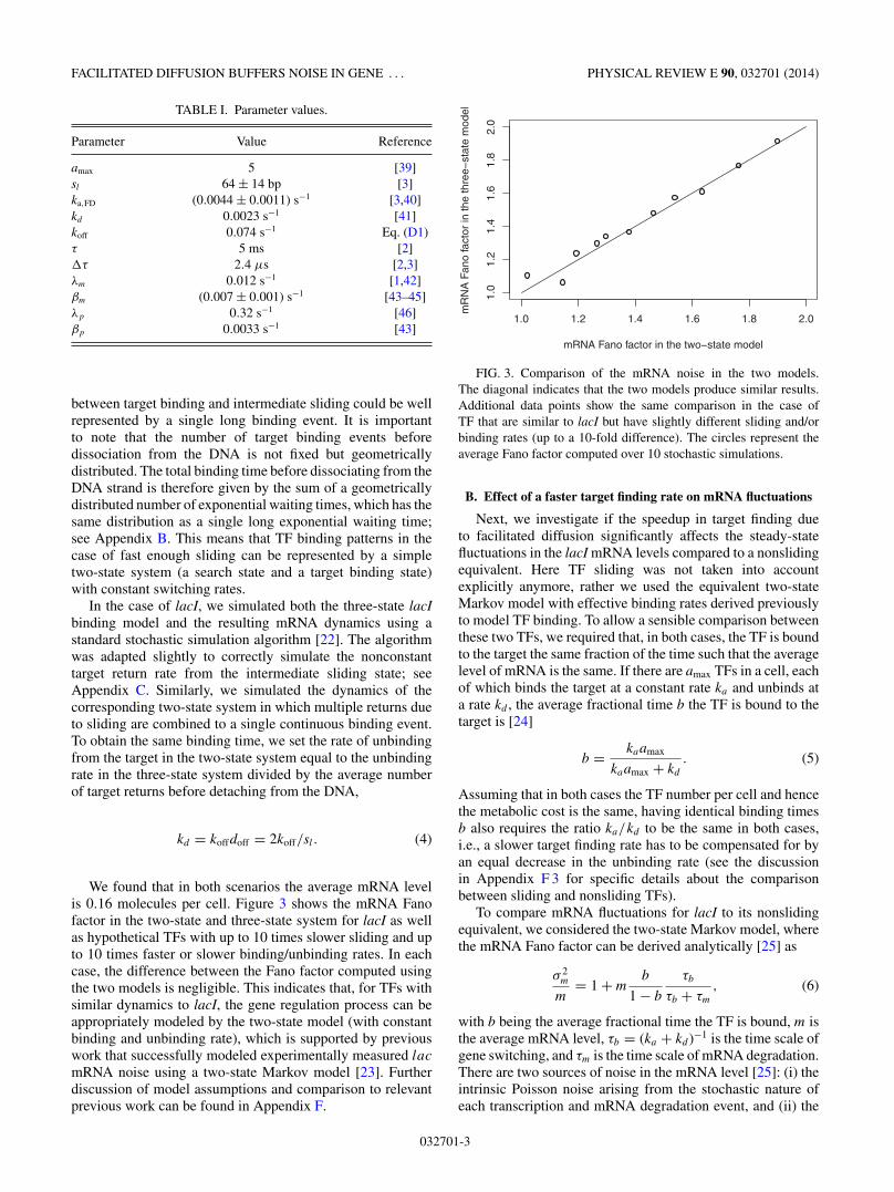
We found that in both scenarios the average mRNA levelis 0.16 molecules per cell. Figure 3 shows the mRNA Fanofactor in the two-state and three-state system for lacI as wellas hypothetical TFs with up to 10 times slower sliding and upto 10 times faster or slower binding/unbinding rates. In eachcase, the difference between the Fano factor computed usingthe two models is negligible. This indicates that, for TFs withsimilar dynamics to lacI, the gene regulation process can beappropriately modeled by the two-state model (with constantbinding and unbinding rate), which is supported by previouswork that successfully modeled experimentally measured lac
mRNA noise using a two-state Markov model [23]. Furtherdiscussion of model assumptions and comparison to relevantprevious work can be found in Appendix F.
1.0 1.2 1.4 1.6 1.8 2.0
1.0
1.2
1.4
1.6
1.8
2.0
mRNA Fano factor in the two−state model
mR
NA
Fan
o fa
ctor
in th
e th
ree−
stat
e m
odel
FIG. 3. Comparison of the mRNA noise in the two models.The diagonal indicates that the two models produce similar results.Additional data points show the same comparison in the case ofTF that are similar to lacI but have slightly different sliding and/orbinding rates (up to a 10-fold difference). The circles represent theaverage Fano factor computed over 10 stochastic simulations.
B. Effect of a faster target finding rate on mRNA fluctuations
Next, we investigate if the speedup in target finding dueto facilitated diffusion significantly affects the steady-statefluctuations in the lacI mRNA levels compared to a nonslidingequivalent. Here TF sliding was not taken into accountexplicitly anymore, rather we used the equivalent two-stateMarkov model with effective binding rates derived previouslyto model TF binding. To allow a sensible comparison betweenthese two TFs, we required that, in both cases, the TF is boundto the target the same fraction of the time such that the averagelevel of mRNA is the same. If there are amax TFs in a cell, eachof which binds the target at a constant rate ka and unbinds ata rate kd , the average fractional time b the TF is bound to thetarget is [24]
b = kaamax
kaamax + kd
. (5)
Assuming that in both cases the TF number per cell and hencethe metabolic cost is the same, having identical binding timesb also requires the ratio ka/kd to be the same in both cases,i.e., a slower target finding rate has to be compensated for byan equal decrease in the unbinding rate (see the discussionin Appendix F 3 for specific details about the comparisonbetween sliding and nonsliding TFs).
To compare mRNA fluctuations for lacI to its nonslidingequivalent, we considered the two-state Markov model, wherethe mRNA Fano factor can be derived analytically [25] as
σ 2m
m= 1 + m
b
1 − b
τb
τb + τm
, (6)
with b being the average fractional time the TF is bound, m isthe average mRNA level, τb = (ka + kd )−1 is the time scale ofgene switching, and τm is the time scale of mRNA degradation.There are two sources of noise in the mRNA level [25]: (i) theintrinsic Poisson noise arising from the stochastic nature ofeach transcription and mRNA degradation event, and (ii) the
032701-3
ARMIN P. SCHOECH AND NICOLAE RADU ZABET PHYSICAL REVIEW E 90, 032701 (2014)
0 20 40 60 80 100
1.0
1.2
1.4
1.6
1.8
2.0
sliding length (bp)
Fano
fact
or
−
−
−− − −
−−
−
lacI 3D
lacI WT
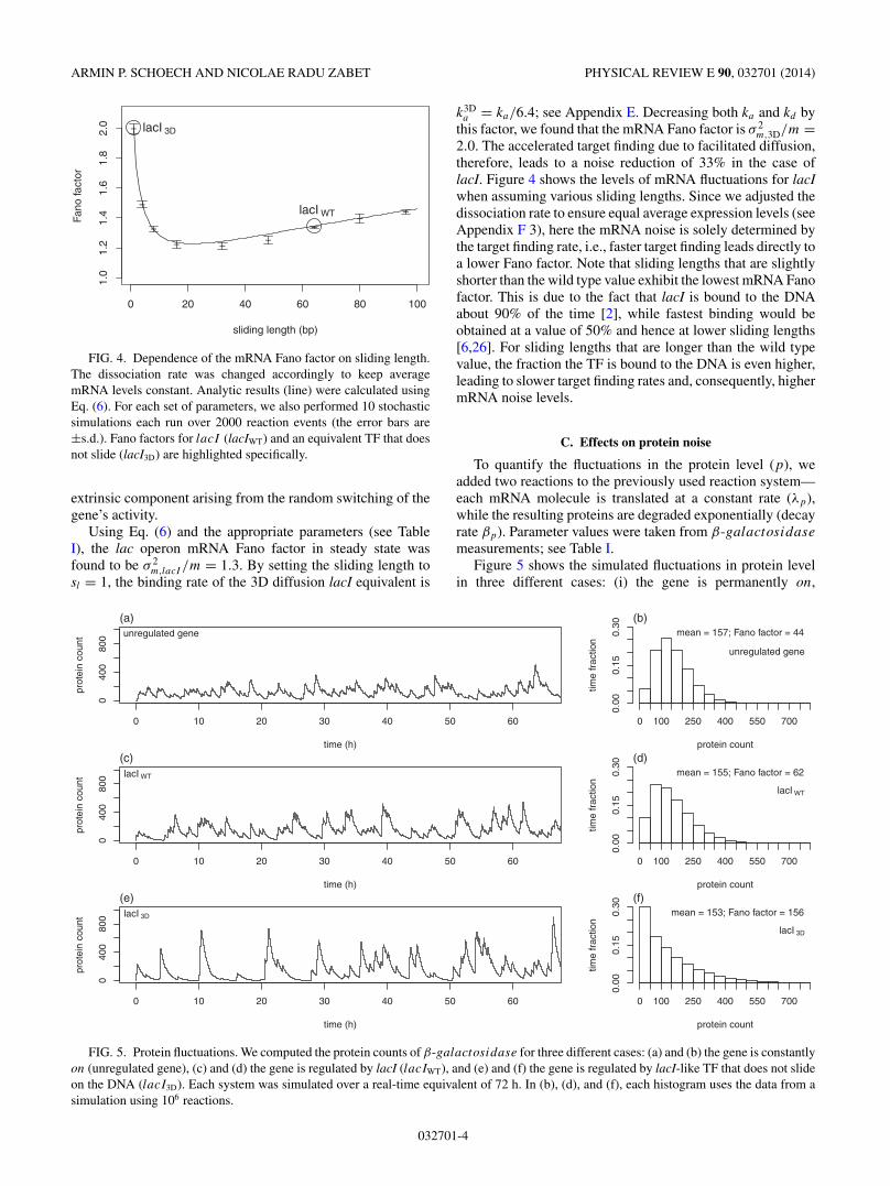
FIG. 4. Dependence of the mRNA Fano factor on sliding length.The dissociation rate was changed accordingly to keep averagemRNA levels constant. Analytic results (line) were calculated usingEq. (6). For each set of parameters, we also performed 10 stochasticsimulations each run over 2000 reaction events (the error bars are±s.d.). Fano factors for lacI (lacIWT) and an equivalent TF that doesnot slide (lacI3D) are highlighted specifically.
extrinsic component arising from the random switching of thegene’s activity.
Using Eq. (6) and the appropriate parameters (see TableI), the lac operon mRNA Fano factor in steady state wasfound to be σ 2
m,lacI /m = 1.3. By setting the sliding length tosl = 1, the binding rate of the 3D diffusion lacI equivalent is
k3Da = ka/6.4; see Appendix E. Decreasing both ka and kd by
this factor, we found that the mRNA Fano factor is σ 2m,3D/m =
2.0. The accelerated target finding due to facilitated diffusion,therefore, leads to a noise reduction of 33% in the case oflacI. Figure 4 shows the levels of mRNA fluctuations for lacIwhen assuming various sliding lengths. Since we adjusted thedissociation rate to ensure equal average expression levels (seeAppendix F 3), here the mRNA noise is solely determined bythe target finding rate, i.e., faster target finding leads directly toa lower Fano factor. Note that sliding lengths that are slightlyshorter than the wild type value exhibit the lowest mRNA Fanofactor. This is due to the fact that lacI is bound to the DNAabout 90% of the time [2], while fastest binding would beobtained at a value of 50% and hence at lower sliding lengths[6,26]. For sliding lengths that are longer than the wild typevalue, the fraction the TF is bound to the DNA is even higher,leading to slower target finding rates and, consequently, highermRNA noise levels.
C. Effects on protein noise
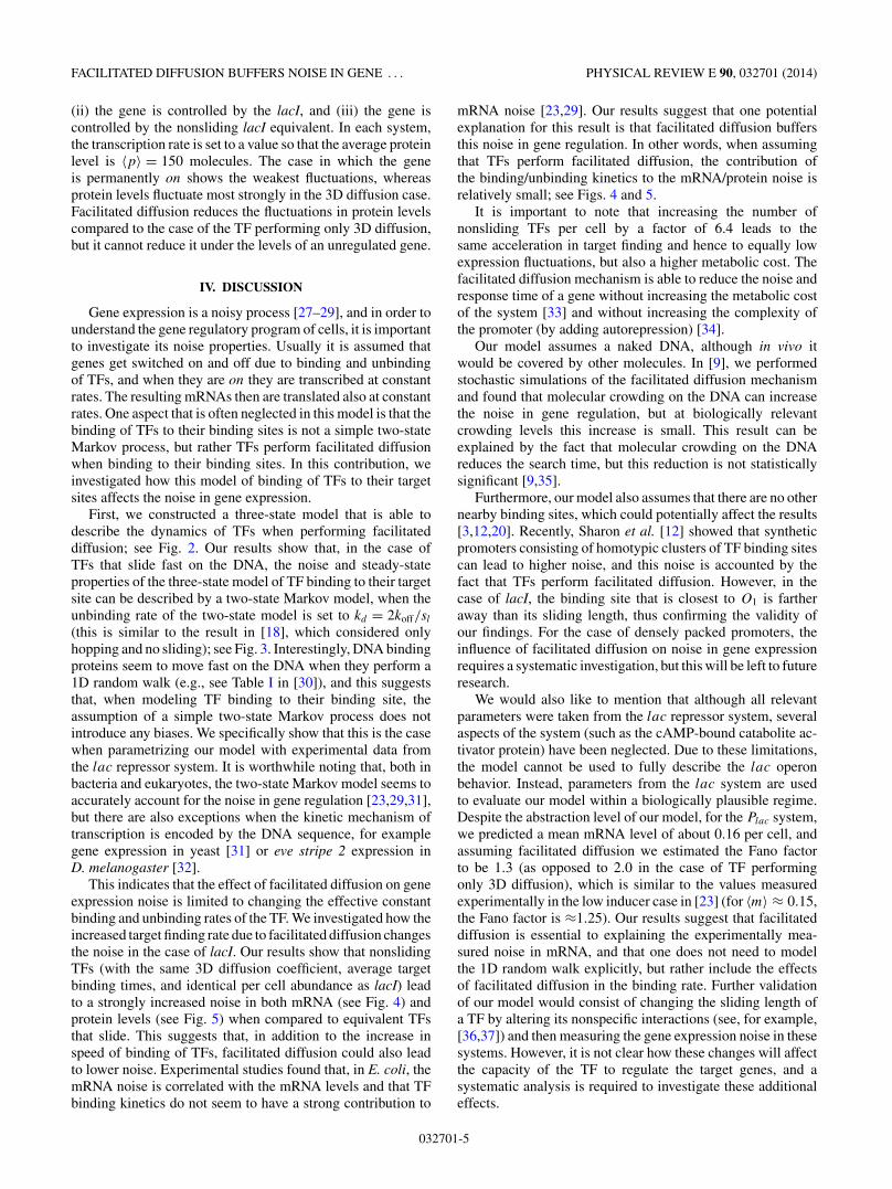
To quantify the fluctuations in the protein level (p), weadded two reactions to the previously used reaction system—each mRNA molecule is translated at a constant rate (λp),while the resulting proteins are degraded exponentially (decayrate βp). Parameter values were taken from β-galactosidase
measurements; see Table I.Figure 5 shows the simulated fluctuations in protein level
in three different cases: (i) the gene is permanently on,
0 10 20 30 40 50 60
040
080
0
time (h)
prot
ein
coun
t
(a)unregulated gene
protein count
time
frac
tion
0.00
0.15
0.30
0 100 250 400 550 700
(b)mean = 157; Fano factor = 44
unregulated gene
0 10 20 30 40 50 60
040
080
0
time (h)
prot
ein
coun
t
(c)lacI WT
protein count
time
frac
tion
0.00
0.15
0.30
0 100 250 400 550 700
(d)mean = 155; Fano factor = 62
lacI WT
0 10 20 30 40 50 60
040
080
0
time (h)
prot
ein
coun
t
(e)lacI 3D
protein count
time
frac
tion
0.00
0.15
0.30
0 100 250 400 550 700
(f)mean = 153; Fano factor = 156
lacI 3D
FIG. 5. Protein fluctuations. We computed the protein counts of β-galactosidase for three different cases: (a) and (b) the gene is constantlyon (unregulated gene), (c) and (d) the gene is regulated by lacI (lacIWT), and (e) and (f) the gene is regulated by lacI-like TF that does not slideon the DNA (lacI3D). Each system was simulated over a real-time equivalent of 72 h. In (b), (d), and (f), each histogram uses the data from asimulation using 106 reactions.
032701-4
FACILITATED DIFFUSION BUFFERS NOISE IN GENE . . . PHYSICAL REVIEW E 90, 032701 (2014)
(ii) the gene is controlled by the lacI, and (iii) the gene iscontrolled by the nonsliding lacI equivalent. In each system,the transcription rate is set to a value so that the average proteinlevel is 〈p〉 = 150 molecules. The case in which the geneis permanently on shows the weakest fluctuations, whereasprotein levels fluctuate most strongly in the 3D diffusion case.Facilitated diffusion reduces the fluctuations in protein levelscompared to the case of the TF performing only 3D diffusion,but it cannot reduce it under the levels of an unregulated gene.
IV. DISCUSSION
Gene expression is a noisy process [27–29], and in order tounderstand the gene regulatory program of cells, it is importantto investigate its noise properties. Usually it is assumed thatgenes get switched on and off due to binding and unbindingof TFs, and when they are on they are transcribed at constantrates. The resulting mRNAs then are translated also at constantrates. One aspect that is often neglected in this model is that thebinding of TFs to their binding sites is not a simple two-stateMarkov process, but rather TFs perform facilitated diffusionwhen binding to their binding sites. In this contribution, weinvestigated how this model of binding of TFs to their targetsites affects the noise in gene expression.
First, we constructed a three-state model that is able todescribe the dynamics of TFs when performing facilitateddiffusion; see Fig. 2. Our results show that, in the case ofTFs that slide fast on the DNA, the noise and steady-stateproperties of the three-state model of TF binding to their targetsite can be described by a two-state Markov model, when theunbinding rate of the two-state model is set to kd = 2koff/sl
(this is similar to the result in [18], which considered onlyhopping and no sliding); see Fig. 3. Interestingly, DNA bindingproteins seem to move fast on the DNA when they perform a1D random walk (e.g., see Table I in [30]), and this suggeststhat, when modeling TF binding to their binding site, theassumption of a simple two-state Markov process does notintroduce any biases. We specifically show that this is the casewhen parametrizing our model with experimental data fromthe lac repressor system. It is worthwhile noting that, both inbacteria and eukaryotes, the two-state Markov model seems toaccurately account for the noise in gene regulation [23,29,31],but there are also exceptions when the kinetic mechanism oftranscription is encoded by the DNA sequence, for examplegene expression in yeast [31] or eve stripe 2 expression inD. melanogaster [32].
This indicates that the effect of facilitated diffusion on geneexpression noise is limited to changing the effective constantbinding and unbinding rates of the TF. We investigated how theincreased target finding rate due to facilitated diffusion changesthe noise in the case of lacI. Our results show that nonslidingTFs (with the same 3D diffusion coefficient, average targetbinding times, and identical per cell abundance as lacI) leadto a strongly increased noise in both mRNA (see Fig. 4) andprotein levels (see Fig. 5) when compared to equivalent TFsthat slide. This suggests that, in addition to the increase inspeed of binding of TFs, facilitated diffusion could also leadto lower noise. Experimental studies found that, in E. coli, themRNA noise is correlated with the mRNA levels and that TFbinding kinetics do not seem to have a strong contribution to
mRNA noise [23,29]. Our results suggest that one potentialexplanation for this result is that facilitated diffusion buffersthis noise in gene regulation. In other words, when assumingthat TFs perform facilitated diffusion, the contribution ofthe binding/unbinding kinetics to the mRNA/protein noise isrelatively small; see Figs. 4 and 5.
It is important to note that increasing the number ofnonsliding TFs per cell by a factor of 6.4 leads to thesame acceleration in target finding and hence to equally lowexpression fluctuations, but also a higher metabolic cost. Thefacilitated diffusion mechanism is able to reduce the noise andresponse time of a gene without increasing the metabolic costof the system [33] and without increasing the complexity ofthe promoter (by adding autorepression) [34].
Our model assumes a naked DNA, although in vivo itwould be covered by other molecules. In [9], we performedstochastic simulations of the facilitated diffusion mechanismand found that molecular crowding on the DNA can increasethe noise in gene regulation, but at biologically relevantcrowding levels this increase is small. This result can beexplained by the fact that molecular crowding on the DNAreduces the search time, but this reduction is not statisticallysignificant [9,35].
Furthermore, our model also assumes that there are no othernearby binding sites, which could potentially affect the results[3,12,20]. Recently, Sharon et al. [12] showed that syntheticpromoters consisting of homotypic clusters of TF binding sitescan lead to higher noise, and this noise is accounted by thefact that TFs perform facilitated diffusion. However, in thecase of lacI, the binding site that is closest to O1 is fartheraway than its sliding length, thus confirming the validity ofour findings. For the case of densely packed promoters, theinfluence of facilitated diffusion on noise in gene expressionrequires a systematic investigation, but this will be left to futureresearch.
We would also like to mention that although all relevantparameters were taken from the lac repressor system, severalaspects of the system (such as the cAMP-bound catabolite ac-tivator protein) have been neglected. Due to these limitations,the model cannot be used to fully describe the lac operonbehavior. Instead, parameters from the lac system are usedto evaluate our model within a biologically plausible regime.Despite the abstraction level of our model, for the Plac system,we predicted a mean mRNA level of about 0.16 per cell, andassuming facilitated diffusion we estimated the Fano factorto be 1.3 (as opposed to 2.0 in the case of TF performingonly 3D diffusion), which is similar to the values measuredexperimentally in the low inducer case in [23] (for 〈m〉 ≈ 0.15,the Fano factor is ≈1.25). Our results suggest that facilitateddiffusion is essential to explaining the experimentally mea-sured noise in mRNA, and that one does not need to modelthe 1D random walk explicitly, but rather include the effectsof facilitated diffusion in the binding rate. Further validationof our model would consist of changing the sliding length ofa TF by altering its nonspecific interactions (see, for example,[36,37]) and then measuring the gene expression noise in thesesystems. However, it is not clear how these changes will affectthe capacity of the TF to regulate the target genes, and asystematic analysis is required to investigate these additionaleffects.
032701-5

ARMIN P. SCHOECH AND NICOLAE RADU ZABET PHYSICAL REVIEW E 90, 032701 (2014)
ACKNOWLEDGMENTS
We would like to thank Boris Adryan and his group foruseful comments and discussions on the manuscript. This workwas supported by the Medical Research Council (G1002110).
APPENDIX A: WAITING TIME DISTRIBUTION WHENSLIDING BACK TO THE TARGET BEFORE
UNBINDING THE DNA
The chance that a TF slides back to the target at a time t afterit slid off it, S(t), is given by the probability of first return to theorigin after time t during a simple unbiased continuous-timerandom walk F (t), times the probability that the TF is stillbound to the DNA at time t , D(t),
S(t) = D(t)F (t). (A1)
The probability that the TF is still bound to the DNA at atime t after unbinding the target decays exponentially withcharacteristic waiting time τ , i.e., D(t ; τ ) = e−t/τ .
Since F (t) is the probability density function of first returnto the origin at time t , it is given by the probability of first returnafter n 1 bp steps, Fn, multiplied by the probability density ofmaking the nth step at time t , φn(t), and then marginalizingover all n:
F (t) =∞∑
n=1
Fnφn(t). (A2)
According to Klafter and Sokolov [38], these probabilitiescan be calculated to be
Fn = 2
n − 1
(n − 1
n/2
)2−n for even n and 0 otherwise (A3)
and
φn(t) = L−1{φn(s)} (A4)
the inverse Laplace transform of φn(s), where φ(s) is in turnthe Laplace transform of the waiting time distribution for asingle base pair step, φ(t). Here we assume that the waitingtime of sliding one step in the neighborhood of the targetis exponentially distributed with a constant characteristic timescale �τ . Therefore, φ(s) = 1
1+s�τand φn(t) can be calculated
to be
φn(t ; �τ ) = ne− t�τ
(t
�τ
)n−1
�τn!. (A5)
Since Fn vanishes for odd n, we can get F (t) by summingover all n = 2m to yield the following expression for therebinding time distribution:
S(t ; τ,�τ ) = e−t/τ
∞∑m=1
2me− t�τ
(t
�τ
)2m−1
�τ (2m)!
× 2
2m − 1
(2m − 1
m
)2−2m. (A6)
Note that this distribution is not normalized, since theprobability of sliding back to the target before unbindingfrom the DNA is smaller than 1. However, the waiting time instate (3) in the TF binding model is the probability density ofreturning at time t given that it does return before unbinding
the DNA. The waiting time, therefore, has to be drawn fromthe corresponding normalized distribution of S(t ; τ,�τ ).
Our model assumes that unbinding directly from the targetsite is negligible. If a TF molecule performs s2
l /2 events duringa 1D random walk, and the probability to unbind is equal fromall positions, then the probability to unbind during any of theseevents is 2/s2
l [8]. Given that on average a TF molecule visitsthe target site sl/2 times during a 1D random walk, then theprobability to dissociate directly from the target site is 1/sl ,which for our model is less than 1.5% and, thus, was neglectedhere.
APPENDIX B: GEOMETRICALLY DISTRIBUTEDNUMBER OF RETURNS LEADS TO AN OVERALL
EXPONENTIALLY DISTRIBUTEDTARGET BINDING TIME
In the case of sufficiently fast sliding, TFs moving on andoff the target multiple times can be approximated by a singlelong target binding event. The length of this effective bindingevent is given by the sum of all individual binding events. Hereeach individual binding time is exponentially distributed. Thenumber of consecutive binding events before DNA detachmentis geometrically distributed since each time the TF leaves thetarget site there is a constant chance doff of not returning to thetarget through sliding. Here we derived the time distribution ofthe overall waiting time as a sum of a geometrically distributednumber of exponential waiting times.
The waiting time distribution of an individual binding eventis
φ(t) = 1
�τe−t/�τ . (B1)
The overall effective waiting time density function giventhat the TF binds the target exactly n consecutive times is
P (t |n) =∫ t2
0
∫ t3
t1
· · ·∫ t
tn−2
φ(t1)
×φ(t2 − t1) · · · φ(t − tn−1)dt1dt2 · · · dtn−1. (B2)
In the Laplace domain, these convolutions turn into a simpleproduct,
P (s|n) = [φ(s)]n, (B3)
with φ(s) = 11+�τs
being the Laplace transform of φ(t).We assume that the number of individual binding events n
is geometrically distributed with constant chance doff of notsliding back. The joint probability is therefore
P (s,n) = P (s|n)(1 − doff)n−1doff (B4)
and hence the return time distribution is
P (s) =∞∑
n=1
[φ(s)]n(1 − doff)n−1doff
= φ(s)doff
1 − φ(s)(1 − doff). (B5)
Substituting φ(s) from above,
P (s) = doff
doff + �τs= 1
1 + N�τs(B6)
032701-6

FACILITATED DIFFUSION BUFFERS NOISE IN GENE . . . PHYSICAL REVIEW E 90, 032701 (2014)
and
P (t) = 1
N�τe−t/N�τ , (B7)
where N = 1/doff is the average number of target bindingsbefore DNA unbinding. We can conclude that in the case of fastenough sliding, multiple returns to the target can be modeledas a single binding event that is exponentially distributed withaverage binding time N�τ .
APPENDIX C: CHANGE TO THE STOCHASTICSIMULATION ALGORITHM
The stochastic simulation algorithm used by Gillespie [22]appropriately simulates reaction systems with exponentialwaiting times, i.e., systems with all possible reactions occur-ring at constant rates for a specific configuration. This is thecase for all reactions in our system apart from the TF slidingback to the target site. When the TF slides off the target, thereturn rate is not constant but decays with time.
To appropriately simulate our system, we adapted slightlythe stochastic simulation algorithm. The original algorithmdraws the time of the next reaction from an exponentialdistribution with a rate equal to the sum of all possible reactionsin the current configuration. Then the specific reaction ischosen according to the individual rates. Here we do the samefor all constant rate reactions in the system, but, in the caseof the TF being in the sliding state, we additionally draw awaiting time from the return time distribution S(t), derivedearlier. If the waiting time drawn from S(t) is smaller thanthe other, the TF returns to the target. If not, a constant ratereaction is carried out accordingly.
APPENDIX D: THE PARAMETERS OF THETHREE-STATE MODEL
The list of parameters for the three-state model is presentedin Table I. Below, we described how some of the parameterswere derived.
1. Number of lacI operons per growing E. coli cell
Although the lac operon only occurs once in the E. coligenome [47], continuous DNA replication during growth canlead to more than one gene being present in a growing cell.Usually, one could observe only one binding spot for lacI,when investigating lacI binding in living and growing cells[2]. Thus, we assumed that there is only about one lac operonpresent in each growing E. coli cell.
2. Total number of lacI molecules per cell
There are 20 lacI monomers per lacI gene in wild typeE. coli [39], and, since there is only one gene per cell (seeabove), we estimate that there are only amax = 5 independentlysearching lac tetramers per cell.
3. Sliding length sl
The root-mean-square deviation during one slide on theDNA was estimated to be sl,RMSD = √
2D1D/kd = (45 ±10) bp [3], where D1D is the 1D diffusion constant and kd
is the DNA dissociation rate. Here, we defined the slidinglength sl as the average number of different base pairs that theTF visits at least once during one slide. Thus, we can computethe sliding rate as sl = √
4D1D/kd = √2sl,RMSD [2,21], and
thus sl = (64 ± 14) bp. Hammar et al. [3] do not discuss ifthis sliding length includes short dissociation events followedby immediate rebinding (hopping) or if the TF unbinds thefirst time on average after scanning 64 bp with a chanceof immediately binding again, performing a new slide onthe DNA. The experimental approach used to determine thesliding length [3] consisted of measuring how the associationrate decreases as additional binding sites near the target areintroduced. Given a median hopping distance of 1 bp and aboutsix hops per 1D random walk [21], it is very unlikely that hopswould by chance overcome the extra binding site. Hoppingis therefore unlikely to significantly alter the experimentalresults, suggesting that sl = (64 ± 14) bp already includesshort hops.
4. The dissociation rate from the binding site
The dissociation rate from the binding site is computedusing the following equation from the main text:
kd = 2koff
sl
⇒
koff = slkd
2= 64 × 0.0023
2= 0.074 s−1. (D1)
Note that we used the following values: sl = 64 bp (see above)and kd = 0.0023 s−1[41]. The latter is similar to the valuemeasured recently (mean bound time of 5.3 ± 0.2) using asingle molecule chase assay [48].
5. β-galactosi dase translation rate
Kennell and Riezman [43] measure one translation initi-ation of a single lacZ mRNA every 2.2 s in exponentiallygrowing cells. However, they state that around 30% ofthe polypeptides are not completed, giving one effectivetranslation every 3.1 s and an effective translation rate ofλp = 0.32 s−1.
6. β-galactosi dase protein decay rate
Mandelstam [46] measured a β-galactosidase degrada-tion rate of 1.4 × 10⁻⁵ s−1. This is much slower than theaverage protein dilution rate of an exponentially growingE. coli cell of 3.3 × 10⁻⁴ s−1 [49]. Thus, the decay ofβ-galactosidase is dominated by dilution, and we approx-imate it by βp = 3.3 × 10⁻⁴ s−1.
APPENDIX E: CHANGING THE ASSOCIATION RATETO A NONSLIDING EQUIVALENT TF
Variations in the extent of facilitated diffusion during targetfinding can be achieved by varying the sliding length. Thishypothetical TF, similar to lacI in all respects but the slidinglength, will have modified association rates. The associationrate can be calculated in closed form, as outlined below. Theassociation rate ka,sl
of a TF with sliding sl is given by the
032701-7
ARMIN P. SCHOECH AND NICOLAE RADU ZABET PHYSICAL REVIEW E 90, 032701 (2014)
following expression [6]:
ka,sl= sl
M∗(t1D,sl
+ t3D)−1
, (E1)
where M∗ is the number of accessible base pairs in the genome,and t1D,sl
and t3D are the average durations of 1D searches(slides and hops on the DNA) and 3D searches (free diffusionin the cytoplasm). It has been experimentally observed thatlacI spends about 90% of the time sliding when searching forthe target site [2], which means that
t1D,lacI = 9t3D. (E2)
To find the dependence of the association rate of the TFon the sliding length from Eq. (E1), we need to calculate themodified t1D,sl
and t3D. Since the 3D search round duration isnot affected by the sliding length of the TF, t3D is identical tothat of lacI and can be calculated by inverting Eq. (E1):
t3D = sl,lacI
10M∗ka,lacI
. (E3)
The average time spent during the 1D slide, t1D,sl, is
proportional to the average number of 1 bp sliding steps N
performed during such a slide. Also, since the transcriptionfactor diffuses along the DNA while sliding, N is proportionalto the square of sl [21] and t1D,sl
∝ s2l . Hence
t1D,sl= t1D,lacI
(sl
sl,lacI
)2
. (E4)
Combining Eqs. (E1), (E2), (E3), and (E4), we find that theassociation rate of a TF with sliding length sl is
ka,sl= 10ka,lacI
sl
sl,lacI
[9
(sl
sl,lacI
)2
+ 1
]−1
, (E5)
where sl,lac = (64 ± 14) bp is the sliding length of lacI [3] andka,lacI = (0.0044 ± 0.0011) s−1 is its association rate [3].
The association rate of an equivalent TF with a differentsliding length can be found by plugging the sliding length sl
into Eq. (E5). The 3D diffusion case can be approached bysetting sl = 1 bp. In the 3D case, the reduced association rateis
ka,3D = ka,lacI
10
sl,lacI
(9
s2l,lacI
+ 1
)−1
= ka,lacI /6.4 = 6.9 × 10⁻⁴ s−1. (E6)
Hence, if lacI was not using facilitated diffusion, it wouldtake on average 6.4 times longer to find its target site.
APPENDIX F: FURTHER CONSIDERATIONSON OUR MODEL
1. Transcription initiation
In our model, we do not model transcription explicitly,but we rather assume that an mRNA molecule is producedat exponentially distributed time intervals when the TF is notbound to the target site. Recently, it was found in Ref. [48]that while this equilibrium model of transcription is accuratefor certain promoters (including lacO1), it fails to explainthe behavior of other promoters (e.g., lacOsym). Nevertheless,
these nonequilibrium binding mechanisms require systematicinvestigation and will be left to further research.
2. Considerations on our three-state model
In this contribution, we proposed a three-state modelthat described the facilitated diffusion mechanism. Pulkkinenand Metzler [11] modeled facilitated diffusion analyticallyassuming a different three-state model, i.e., they assumed thatthe TF molecule can be in the following three states: (i) freein the cytoplasm/nucleoplasm, (ii) bound nonspecifically tothe DNA in the vicinity of the target site, and (iii) bound tothe target site. The transitions between these three states wereassumed to be exponentially distributed.
Crucially, we considered that the TF molecule can be indifferent three states, namely (1) searching for the target usingfacilitated diffusion (at least one DNA detachment beforetarget rebinding), (2) bound to the target site, and (3) slidingon the DNA between two consecutive target binding eventswithout DNA detachment. Note that when sliding off the targetsite, the TF molecule can be in both states (1) and (3), i.e., ifit will return before DNA detachment, the TF is in state (3),while otherwise it is in state (1). Hence, we used well-definedabstract states instead of a purely spatial definition as used in[11]. In other words, we avoided a necessarily approximatedefinition of a “local” search state, which allows us to findthe exact target return time distribution assuming facilitateddiffusion of a TF. Importantly, we find that when sliding on theDNA near the target site, the binding time is not exponentiallydistributed, as is assumed by Pulkkinen and Metzler [11].
Furthermore, Meyer et al. [10] investigated the noise inmRNA assuming that the search takes place in a compactenvironment, and they compared this with the case of thesearch taking place in a noncompact environment. Theyderived a nonexponential return rate to the target site andassumed that facilitated diffusion can be seen as a search ina compact environment. Our approach was different in thesense that we did not assume a distribution of the return times,but rather derived this distribution analytically by assuminga known model of facilitated diffusion. We further used thisdistribution and parameters derived from previous experimentsto understand the influence of facilitated diffusion on the noisein mRNA and protein.
The main focus of our paper is what are the effects offacilitated diffusion on mRNA and protein noise. Pulkkinenand Metzler [11] investigate this problem, but in the case ofcolocalization of the gene encoding for a TF and the targetsite of that TF. This assumption makes their results valid onlyin the context of bacterial systems (where transcription andtranslation are colocalized), while our results are potentiallyvalid even in the context of eukaryotic systems (wheretranslation takes place outside the nucleus). Interestingly, itseems that mRNA noise in animal cells seems to display asimilar level of correlation with the mean expression level asin the case of bacterial cells [31]. This means that assumingthat TFs perform facilitated diffusion in higher eukaryotes[37,50–52], the contribution from binding/unbinding kineticsis potentially small.
It is worthwhile noting that Pedraza and Paulsson [53]proposed a general model to compute noise in mRNA where
032701-8

FACILITATED DIFFUSION BUFFERS NOISE IN GENE . . . PHYSICAL REVIEW E 90, 032701 (2014)
any distribution for the arrival times of the TFs to the target sitecan be assumed. Our model particularizes this type of modelto the case of facilitated diffusion, and we explicitly derive thearrival time distribution as being nonexponential.
Finally, we would like to emphasize that, to our knowledge,no previous work systematically compared nonsliding withsliding TFs and discussed the effects of facilitated diffusion onthe noise in gene expression compared to simple 3D diffusionof TFs.
3. Comparing sliding TFs to their hypotheticalnonsliding equivalents
van Zon et al. [18] investigated a different TF search effect,namely how fast rebinding in the case of a TF that uses only3D diffusion affects transcriptional noise. In our paper, weinvestigate the case of multiple returns due to sliding, and wefind that facilitated diffusion leads to a reduction in the mRNAnoise. This is different from the result of van Zon et al. [18],who find that fast 3D diffusion returns increase transcriptionalnoise. The system investigated in our paper is different inthat, unlike 3D diffusion returns, sliding not only leads tomultiple consecutive binding events but also to a speedup intarget search, hence increasing the TF target finding rate. Mostimportantly, the crucial difference between the two works thatexplains the seemingly contradictory conclusions is due to thedifference in the questions posed. On the one hand, van Zonet al. [18] asked what happens to transcriptional noise if TFsquickly return to the target multiple times through 3D diffusionand hence decrease the effective dissociation rate. On the otherhand, we ask how the effect on gene expression noise couldpose an evolutionary advantage that could play a role in thedevelopment of facilitated diffusion. More specifically, we donot simply ask how a sliding TF compares to another TF thatis identical, except that it is unable to slide along the DNA,
but rather we investigate how the noise in gene expression ina system that has evolved using a sliding TF differs from thenoise in gene expression in a system that uses a nonslidingTF. Thus, we require that both systems have the same averagelevel of repression, and this means that the average time a TFis bound to the target should be identical.
Since we show that target binding dynamics of slidingTFs can be represented as an effective two-state model, anypossible advantage of the facilitated diffusion mechanism interms of noise in gene expression must lie in the effectivebinding and unbinding rates. Here, we compared sliding andnonsliding TFs at equal TF number, and thus at equal metaboliccost. In the case of nonsliding TFs, the overall target findingrate is slower. To keep the average repression level the same,the target dissociation rate for the nonsliding TF is thendecreased accordingly to compensate for the slower targetfinding rate and the effect of multiple fast returns due tosliding. It is worthwhile mentioning that, from an evolutionarypoint of view, changes in dissociation rate could be acquiredrelatively easily via small mutations in target sequence and/orTF DNA-binding domain [54].
We choose the target dissociation rate of the nonslidingTF such that the average mRNA level remains unchanged,and thus we do not consider a decrease in dissociation ratedue to multiple returns of the TF to the binding site as in[18]. The change in the noise in our model is only due to theaccelerated target finding. If we did not correct the dissociationrate, a simple nonsliding lacI equivalent would show both aslower target finding rate as well as a higher effective targetdissociation rate due to the lack of multiple returns. However,such a direct comparison would lead to very different averagemRNA levels. Using our comparison, we are able to showthat the increase in the target finding rate due to facilitateddiffusion can indeed pose an evolutionary advantage for thecell by decreasing the steady-state expression noise of thecontrolled gene for a specific average expression rate.
[1] I. Golding, J. Paulsson, S. M. Zawilski, and E. C. Cox, Cell 123,1025 (2005).
[2] J. Elf, G.-W. Li, and X. S. Xie, Science 316, 1191 (2007).[3] P. Hammar, P. Leroy, A. Mahmutovic, E. G. Marklund, O. G.
Berg, and J. Elf, Science 336, 1595 (2012).[4] J. Paulsson, Nature (London) 427, 415 (2004).[5] N. Friedman, L. Cai, and X. S. Xie, Phys. Rev. Lett. 97, 168302
(2006).[6] L. Mirny, M. Slutsky, Z. Wunderlich, A. Tafvizi, J. Leith, and
A. Kosmrlj, J. Phys. A 42, 434013 (2009).[7] O. Benichou, C. Chevalier, B. Meyer, and R. Voituriez,
Phys. Rev. Lett. 106, 038102 (2011).[8] N. R. Zabet and B. Adryan, Bioinformatics 28, 1517 (2012).[9] N. R. Zabet and B. Adryan, Front. Genet. 4, 197 (2013).
[10] B. Meyer, O. Benichou, Y. Kafri, and R. Voituriez, Biophys. J.102, 2186 (2012).
[11] O. Pulkkinen and R. Metzler, Phys. Rev. Lett. 110, 198101(2013).
[12] E. Sharon, D. van Dijk, Y. Kalma, L. Keren, O.Manor, Z. Yakhini, and E. Segal, Genome Res.,doi:10.1101/gr.168773.113.
[13] A. D. Riggs, S. Bourgeois, and M. Cohn, J. Mol. Biol. 53, 401(1970).
[14] O. G. Berg, R. B. Winter, and P. H. von Hippel, Biochemistry20, 6929 (1981).
[15] Since it is difficult to experimentally distinguish be-tween the 1D translocation modes, by sliding we referto a 1D random walk that includes both sliding andhopping.
[16] H. Kabata, O. Kurosawa, M. W. I Arai, S. Margarson, R. E.Glass, and N. Shimamoto, Science 262, 1561 (1993).
[17] S. Redner, A Guide to First-Passage Processes (CambridgeUniversity Press, New York, 2001).
[18] J. S. van Zon, M. J. Morelli, S. Tanase-Nicola, and P. R. tenWolde, Biophys. J. 91, 4350 (2006).
[19] N. R. Zabet and B. Adryan, Mol. Biosyst. 8, 2815 (2012).
032701-9
ARMIN P. SCHOECH AND NICOLAE RADU ZABET PHYSICAL REVIEW E 90, 032701 (2014)
[20] D. Ezer, N. R. Zabet, and B. Adryan, Nucl. Acids Res. 42, 4196(2014).
[21] Z. Wunderlich and L. A. Mirny, Nucl. Acids Res. 36, 3570(2008).
[22] D. T. Gillespie, J. Phys. Chem. 81, 2340 (1977).[23] L.-h. So, A. Ghosh, C. Zong, L. A. Sepulveda, R. Segev, and
I. Golding, Nat. Genet. 43, 554 (2011).[24] D. Chu, N. R. Zabet, and B. Mitavskiy, J. Theor. Biol. 257, 419
(2009).[25] J. Paulsson, Phys. Life Rev. 2, 157 (2005).[26] M. Slutsky and L. A. Mirny, Biophys. J. 87, 4021 (2004).[27] A. Bar-Even, J. Paulsson, N. Maheshri, M. Carmi, E. O’Shea,
Y. Pilpel, and N. Barkai, Nat. Genet. 38, 636 (2006).[28] J. R. S. Newman, S. Ghaemmaghami, J. Ihmels, D. K. Breslow,
M. Noble, J. L. DeRisi, and J. S. Weissman, Nature (London)441, 840 (2006).
[29] Y. Taniguchi, P. J. Choi, G.-W. Li, H. Chen, M. Babu, J. Hearn,A. Emili, and X. S. Xie, Science 329, 533 (2010).
[30] M. C. DeSantis, J.-L. Li, and Y. M. Wang, Phys. Rev. E 83,021907 (2011).
[31] A. Sanchez and I. Golding, Science 342, 1188 (2013).[32] J. P. Bothma, H. G. Garcia, E. Esposito, G. Schlissel, T. Gregor,
and M. Levine, Proc. Natl. Acad. Sci. USA 111, 10598 (2014).[33] N. R. Zabet and D. F. Chu, J. R. Soc. Interface 7, 945 (2010).[34] N. R. Zabet, J. Theor. Biol. 284, 82 (2011).[35] C. A. Brackley, M. E. Cates, and D. Marenduzzo, Phys. Rev.
Lett. 111, 108101 (2013).[36] D. Vuzman and Y. Levy, Proc. Natl. Acad. Sci. USA 107, 21004
(2010).[37] A. Tafvizi, F. Huang, A. R. Fersht, L. A. Mirny, and A. M. van
Oijen, Proc. Natl. Acad. Sci. USA 108, 563 (2011).[38] J. Klafter and I. M. Sokolov, First Steps in Random Walks,
From Tools to Applications (Oxford University Press, New York,2011).
[39] W. Gilbert and B. Muller-Hill, Proc. Natl. Acad. Sci. USA 56,1891 (1966).
[40] G.-W. Li, O. G. Berg, and J. Elf, Nat. Phys. 5, 294 (2009).[41] M. Fried and D. M. Crothers, Nucl. Acids Res. 9, 6505 (1981).[42] T. Malan, A. Kolb, H. Buc, and W. R. McClure, J. Mol. Biol.
180, 881 (1984).[43] D. Kennell and H. Riezman, J. Mol. Biol. 114, 1 (1977).[44] D. W. Selinger, R. M. Saxena, K. J. Cheung, G. M. Church, and
C. Rosenow, Genome Res. 13, 216 (2003).[45] C. P. Ehretsmann, A. J. Carpousis, and H. M. Krisch, FASEB J.
6, 3186 (1992).[46] J. Mandelstam, Nature (London) 179, 1179 (1957).[47] M. Riley, T. Abe, M. B. Arnaud, M. K. Berlyn, F. R. Blattner,
R. R. Chaudhuri, J. D. Glasner, T. Horiuchi, I. M. Keseler,T. Kosuge, H. Mori, N. T. Perna, G. Plunkett, K. E. Rudd,M. H. Serres, G. H. Thomas, N. R. Thomson, D. Wishart, andB. L. Wanner, Nucl. Acids Res. 34, 1 (2006).
[48] P. Hammar, M. Wallden, D. Fange, F. Persson, O. Baltekin,G. Ullman, P. Leroy, and J. Elf, Nat. Genet. 46, 405 (2014).
[49] H. Bremer and P. P. Dennis, in Escherichia coli and Salmonella:Cellular and Molecular Biology, edited by F. C. Neidhardt,R. Curtiss III, J. L. Ingraham, E. C. C. Lin, K. B. Low, B.Magasanik, W. S. Reznikoff, M. Riley, M. Schaechter, andH. E. Umbarger, 2nd ed. (ASM Press, Washington, DC, 1996),pp. 1553–1569.
[50] G. L. Hager, J. G. McNally, and T. Misteli, Mol. Cell 35, 741(2009).
[51] V. Vukojevic, D. K. Papadopoulos, L. Terenius, W. J. Gehring,and R. Rigler, Proc. Natl. Acad. Sci. USA 107, 4093 (2010).
[52] J. Chen, Z. Zhang, L. Li, B.-C. Chen, A. Revyakin, B. Hajj,W. Legant, M. Dahan, T. Lionnet, E. Betzig, R. Tjian, andZ. Liu, Cell 156, 1274 (2014).
[53] J. M. Pedraza and J. Paulsson, Science 319, 339 (2008).[54] S. J. Maerkl and S. R. Quake, Science 315, 233 (2007).
032701-10