Fabrication and Characterization of a Silk-PCL Based...
47
Fabrication and Characterization of a Silk-PCL Based Scaffold for Ligament Graft A THESIS SUBMITTED FOR PARTIAL FULFILLMENT OF THE REQUIREMENT FOR THE DEGREE OF MASTER OF TECHNOLOGY IN BIOTECHNOLOGY ENGINEERING By: SHENDGE AADESHKUMAR SURESH (Roll no. 211BM2014) Under the guidance of: Dr. BIBHUKALYAN PRASAD NAYAK Department Of Biotechnology And Medical Engineering National Institute Of Technology Rourkela 2013
Transcript of Fabrication and Characterization of a Silk-PCL Based...

Fabrication and Characterization of a Silk-PCL Based Scaffold for Ligament Graft
A THESIS SUBMITTED FOR PARTIAL FULFILLMENT OF THE REQUIREMENT FOR
THE DEGREE OF
MASTER OF TECHNOLOGY
IN
BIOTECHNOLOGY ENGINEERING
By:
SHENDGE AADESHKUMAR SURESH
(Roll no. 211BM2014)
Under the guidance of:
Dr. BIBHUKALYAN PRASAD NAYAK
Department Of Biotechnology And Medical Engineering
National Institute Of Technology Rourkela
2013

ii
National Institute of Technology, Rourkela
Certificate This is to certify that the report entitled, “Fabrication and Characterization of a Silk-PCL Based Scaffold for Ligament Graft” submitted by Mr. Shendge Aadeshkumar Suresh, Roll no.: 211BM2014, M. Tech, Department of Biotechnology & Medical Engineering, National Institute of Technology, Rourkela (Deemed University) is an authentic work carried out by her under my supervision and guidance.
To the best of my knowledge, the matter embodied in the report has not been
submitted to any other University / Institute for the award of any Degree or Diploma.
Date: Dr. B. P. NAYAK
Department of Biotechnology & Medical Engineering
National Institute of Technology
Rourkela – 769008

iii
ACKNOWLEDGEMENT
Apart from the efforts of me, the success of my project depends largely on the
encouragement and guidelines of many others. I take this opportunity to express my
thankfulness and appreciation to those individuals who have been involved in the successful
completion of this project.
I would like to express my deep sense of gratitude and heartfelt thanks to Dr. (Prof.)
Bibhukalyan Prasad Nayak, Department of Biotechnology and Medical Engineering, NIT
Rourkela, for his tremendous support and guidance throughout my project.
I extend my sincere thanks to Prof G.S Hotta, Department of Chemistry, NIT
Rourkela for allowing me to use the lab facilities. My special thanks to research scholar Ms.
Abhipsa Mahapatra and Ms. Shabna patel for their help. I would like to express my
heartily thanks to Ms. Sasmita Majhi for constant help.
I would like to thank Mr. Asif Khurshid, Mr. Akshaya Kumar Padhi , Mr.
Patitapabana Parida, who helped me to solve many problems of my work.
I would like to express my heartily thanks to lab mates and others in the department
for their help and support.
Finally, I would like to express my heartfelt thanks to my family members, for their
blessings, support, and for constant encouragement. And a very special thanks to God for
showering the blessings on me.
Shendge Aadeshkumar Suresh

iv
TABLE OF CONTENTS
CERTIFICATE i
ACKNOWLEDGEMENT ii
LIST OF FIGURES vi
LIST OF TABLES Vii
ABSTRACT viii
Chapter Topic Page No.
CHAPTER 1 INTRODUCTION 1
1.1 Anterior cruciate ligament 2
1.2 Tissue engineering 3
1.3 Scaffold 4
1.3.1 Silk 4
1.3.2 PCL 5
1.4 Electrospinning 6
CHAPTER 2 LITERARURE REVIEW 8
2.1 Anterior cruciate ligament 9
2.1.1 Current treatments and its limitations 9
2.2. Silk 10
2.2.1 Tasar silk 11
2.2.2 Use of silk in ligament tissue engineering 11
2.3 PCL 12
2.3.1 Electrospinning of PCL 13
CHAPTER 3 OBJECTIVES 15
CHAPTER 4 PLAN OF WORK 17
CHAPTER 5 MATERIALS AND METHODS 19
5.1 Fabrication of scaffolds 20
5.1.1 Preparation of Silk Yarn 20
5.1.2 Preparation of knitted silk Scaffold 20

v
5.1.3 Loading of scaffolds on wire frame 21
5.1.4 Morphological characterization of knitted
scaffold
22
5.2 Preparation of PCL Mesh 22
5.2.1 Preparation of PCL solution 22
5.2.2 Electrospinning of PCL 23
5.3 Degradation and Mechanical Testing of Scaffold 24
5.4 Cell Culture 24
5.5 Biocompatibility testing 25
5.5.1 Scaffolds Sterilization 25
5.5.2 Cell Seeding 25
5.5.3 Cell Adhesion Test 25
5.5.4 MTT Assay 26
5.5.5 Staining of Cells with Ethidium Bromide 26
CHAPTER 6 RESULTS AND DISCUSSIONS 27
6.1 SEM Analysis of Scaffold 28
6.2 In Vitro Degradation of Silk Scaffold 29
6.3 Cell Seeding and Proliferation Assay 30
CHAPTER 7 CONCLUSION 33
CHAPTER 8 REFERENCES 34

vi
LIST OF FIGURES
Figure No. Description Page No.
Figure 1.1 Anatomy of knee joint 3
Figure 1.2 Flow in electrospinning 6
Figure 2.1 Chemical Structure of PCL 13
Figure 5.1 Brother Knitting Machine (Model no. KH830) 20
Figure 5.2 Knitted scaffold loaded on wire loop 21
Figure 5.3 TA-HD Plus Texture Analyser 24
Figure 6.1 SEM image of knitted silk scaffold 28
Figure 6.2 SEM image of electro-spun PCL mesh 29
Figure 6.3 Mechanical testing of scaffolds for ultimate tensile strength of knitted silk scaffold
29
Figure 6.4 Cell culture 30
Figure 6.5 Standard curve of MTT assay 31
Figure 6.6 Cells stained with ethidium bromide 31

vii
LIST OF TABLES
Table No. Description Page No.
Table 2.1 Different type of scaffold fabrication methods 12
Table 5.1 Parameters used for electrospinning 23

viii
ABSTRACT
Ligament gets damaged very often in cutting and pivoting sports. Current gold standards for
ligament replacement are based on autografts which has limitations of inadequate strength
and donor site morbidity. Thus, ligament tissue engineering is promising strategy for
replacing severely damaged ligaments beyond repair. The objective of current study is to
fabricate a hybrid scaffold for ligament graft with high tensile strength which will support
cell proliferation. Briefly, knitted silk scaffold was made by use of wild type silk from
Antheraea mylitta. Polycaprolactone (PCL) was electrospun on these knitted scaffold to
facilitate cell growth. Degradation study and mechanical testing of scaffolds were carried out
at five time points (0, 3, 7, 14, 21 d). Mouse fibroblasts (L929) were cultured on these nano-
micro scaffolds to investigate the cell adhesion and proliferation potential. Cell proliferation
was visualized under fluorescent microscopy and was analysed by MTT assay. Hybrid
scaffolds showed slow degradation rate and high tensile strength, 22.75 ± 0.43 N at end of
day 21. Cell adhesion efficiency was determined to be 92.28±0.61%. L929 cells grew
profusely on the hybrid scaffold as confirmed from fluorescent microscopy and MTT assay.
Silk-PCL based hybrid scaffold promises to be a better platform for ligament tissue
engineering with optimal biocompatibility and mechanical properties.
Keywords -: Ligament tissue engineering, Electrospinning, Antheraea mylitta,
Polycaprolactone, Knitted scaffold

1
CHAPTER 1
INTRODUCTION

2
1. Introduction
Anterior cruciate ligament (ACL) is a ligament in knee joint which connects femur and
tibia. The role of ligament is to shift loads between bones on both sides of ligament.
Ligament keeps the both bones in place in joints by proper alignment and avoids
dislocation. Ligament tissue is composed of ligament fibroblast embedded into
extracellular matrix. Ligament tissue is mostly made up of mostly extracellular matrix
with low density of fibroblast. Ligaments have very limited requirement for nutrients and
oxygen. When ligament experiences more forces than it can bear, permanent damage will
be caused to ligament. Limited supply of nutrient and oxygen provide very limited
capacity for healing and regeneration. Thus medical intervention is necessary in case of
damage to ligament. Tissue engineering is promising approach for ligament tissue repair
and regeneration.
Currently different approaches are being tested in various laboratories. In recent
years, integration of newly made ligament tissue with bones is found to be a limitation.
Integration of ligament with bone is recent focus for ligament tissue engineering work.
Combinations of different suitable biomaterial with properties are being used for
fabricating competent scaffold.
1.1. Anterior Cruciate Ligament
Anterior cruciate ligament is the cruciate ligament of knee. Knee joint consist of four
ligaments, anterior cruciate ligament (ACL), posterior cruciate ligament (PCL), medial
collateral ligament (MCL) and lateral collateral ligament (LCL), as shown in fig 1.1. The
anterior cruciate ligament is present on anterior side of knee. The anterior ligament limits
forward motion of femur with respect to tibia in knee joint. The anterior cruciate ligament
is dependable for stabilizing turning movements at the knee that takes place during

3
cutting and pivoting activities. It gets injured more than any other ligament. ACL injuries
are most common ligament associated injuries in cutting or pivoting sports such as
football, rugby etc. Healing potential of ACL is very limited as compared to other
ligaments of knee.
Fig. 1.1 Anatomy of knee joint
1.2. Tissue engineering
Tissue engineering is a combination of cells, materials, and suitable biochemical factors to
replace biological functions in an effort to improve clinical procedures for the repair of
damaged tissues and organs. It is a combination of biological principle and engineering
principles. Tissue engineering approach uses biodegradable and biocompatible
biomaterials with sufficient structural and mechanical properties to imitate the
organization of the native tissue.

4
Tissue engineering is a multidisciplinary approach to the production of living
tissue in vitro and in vivo that proposes innovative solutions for the treatment of organ
and tissue damage. Tissue engineering has gain a great interest for replacement or repair
of musculoskeletal system. Tissue engineering approach is currently being used in
musculoskeletal system for cartilage, bone, ligament and tendon.
1.3. Scaffold
In tissue engineering, scaffold act as template for growth of cells to form functional tissue
or organ. It is assumed that, cell only be them self cannot form the tissue or organ. Cells
need a preformed structure template or scaffold to form tissue or organ. Cells adhere to the
scaffold and micro-environment in scaffold allows cells to proliferate. Scaffold provide
appropriate environment for cell growth and proliferation. Scaffold should able to provide
suitable properties for graft implantation such as high mechanical strength and
interconnected channel. Ideally scaffold should provide support for cell interaction, cell
proliferation and cell differentiation. Scaffold should be biocompatible. The
biodegradation of scaffold should be compared to that of native tissue. Scaffold should
provide strength for neo tissue and during initial period of post-implantation. Scaffold
should be able to provide versatile application for different modification.
Over past century, various biocompatible materials have been used for implantation.
Different materials such as polymers, ceramics and metals have been investigated.
Polymeric biomaterial provides flexibility for the scaffold which advantageous in some
tissue.
1.3.1. Silk
A variety of insects and spiders produces silk. Silk is fibrous material made-up of two
different proteins, sericin and fibroin. Fibroin act like core of filamentous structure while

5
sericin functions as glue. Silk serves for different purpose for different insects. It has
been used in versatile way in nature. Different type of silk shows different type of
properties. Many insects are shown to produce more than one type of silk. Functions of
silks shows varied functional range from web structure and catching prey, safety line to
cocoons for protection. Silkworm silks are contains mainly of fibroin, while the chief
protein of spider silks is spidroin. Silk has been used for medical application as sutures
for centuries. Currently it’s being exploited as a biomaterial for scaffold in tissue
engineering application. Silk from most common silkworm Bombyx mori have been
extensively studied. It has shown potential in biomedical application. In recent years, wild
type silk has been of great interest. Wild type silk have shown better cell adhesion than
mulberry type silk. More extensive studies are needed for exploration of true potential of
wild type silk in tissue engineering application.
Ideally scaffold should provide support for cell interaction, cell proliferation and cell
differentiation. Scaffold should be biocompatible. The biodegradation of scaffold should
be compared to that of native tissue. Scaffold should provide strength for neo tissue and
during initial period of post-implantation. Scaffold should be able to provide versatile
application for different modification.
1.3.2. PCL
Polycaprolactone is linear aliphatic polyester. PCL shows a slow biodegradation. In
human body, PCL is degraded by hydrolysis of ester linkage. It is biocompatible,
bioresorbable synthetic polymer. PCL is available in different molecular weight and
influence its properties. Thus provide diverse option for suitable properties. It has been
approved by Food and drug administration. It has been extensively used in slow drug
release device and suture material. PCL do not have any isoform as that of PLA thus
PCL shows uniformity with materials. PCL is hydrophobic materials. Hydrophobic nature

6
of PCL avoids cell attachment. The material can be modified to allow cell adhesion and
proliferation.
1.4 Electrospinning
Fig 1.2 Flow in electrospinning. Diagram shows formation of Taylor’s cone ohmic flow
and convective flow during elctrospinning
Electrospun fibers are formed after applying a high voltage (Potential difference) between a
liquid solution and a collector. A polymeric solution is used for the elctrospinning. When
high voltage is applied, polymeric solution overcome surface tension and forms a cone. This
cone is called as Taylor’s cone. The solution kept in syringe will flow through needle and
forms nano-fibers. This nano-fiber gets accelerated toward oppositely charged collector. The
collector collects the fibers. When a stationary collector is used, fibers will form a ‘mesh’ of
randomly aligned fibers.

7
As shown in figure 1.2 after application of voltage Taylor’s cone will be formed at spinneret.
The flow can be divided into two types of flows, ohmic flows and convective flows. It
depends on the voltage applied. Elctrospinning depends upon various parameters. Parameters
such as applied voltage, temperature, humidity, solution viscosity etc. can affect morphology
and structure of fibers. Electrospinning has been used extensively for tissue engineering
application. Electro-spun fibres resemble as that of extra cellular matrix of tissue, specially
like collagen.

8
CHAPTER 2
LITERATURE REVIEW

9
2. Literature Review
2.1 Anterior Cruciate Ligament
The anterior cruciate ligament (ACL) of is one of the strongest ligament. Anterior cruciate
ligament consists of extracellular matrix (80%) and fibroblast (20%). Extracellular matrix
is mainly consists of collagen with small amount of elastin, proteoglycan and
glycoprotein1. Collagen present in ACL is mainly of type I. Small amount of collagen type
III is also present.
Ligament acquires tensile strength and elasticity mainly from triple-helical type collagen
type I. Collagen molecules organizes themselves into parallel fibrils. Nano scale fibrils
assemble to form micro scale fibers. The fibers are aligned in parallel direction to the load
i.e. along with major axis of the ligament. Diameter of the micro scale fibers increases
with increasing age2, 3. Ligament fibroblasts interact with each via connexion 32 and 43
positive gap junction made by extended cell processes. ACL injuries occur mostly at the
interface between ligament and bone. Interface at femoral site is more prone to get injured
than interface at tibial-site4, 5. ACL has shown to have very limited healing potential,
which intensify need of therapeutic involvement.
2.1.1 Current treatment and its limitation
Biological tissue autograft reconstruction using the patellar tendon has become the most
popular procedure in surgical treatment of a ruptured ACL. Autologous hamstring are
progressively utilized for ACL reconstruction due to donor site morbidity associated with
bone-patellar tendon-bone grafts (BPTB) 6. Capability to integrate with bone via its bony
ends makes the BPTB graft gold standard. Moreover, it possesses intact insertion sites that
can serve as functional transitions between ligament and bone. The tendon grafts have to

10
be fixed mechanically within the bone tunnel. The graft-to-bone interface is not formed as
the native interface site, instead nonmineralized soft tissue is found in bone tunnel7. Thus
graft fixation at the tibial and femoral tunnels, instead of the isolated strength of the graft,
represents the weakest point during the early postoperative healing period8. The surgical
procedures for ACL replacement are found be associated for pain, muscular weakness or
knee instability. Outcomes after anterior cruciate ligament reconstruction have been
disappointing9, 10.
2.2 Silk
Silk is a natural protein fibre produced by insects and spiders. Silk consist of two measure
protein, fibroin and sericin. Fibroin is the fibrous protein while sericin acts as ‘glue’.
Initial immunogenic activity of silk was traced back to sericin, thus will be eliminated
from silk for its use as biomaterial.
Silk has been used in medical application for more than 3000 years. Silk has shown
excellent biocompatibility. Silk fibres have shown to provide high tensile strength as
compared to other materials. Tissue engineering applications such as ligament tissue
engineering needs fibrous material with high tensile strength. Thus Silk can be used
efficiently as scaffold material for ligament tissue engineering.
Silk is broadly categorised into two types
Mulberry Silk
Non-mulberry Silk (Wild type Silk)
Mulberry silk can obtained from Bombex mori, which feeds on mulberry leaves. It has
been cultivated at large scale due to its important in textile industry. This silk has been
exploited over years for biomedical application.

11
Other type of silk produced from different insect are referred collectively as Non-mulberry
or wild type silk. This group consist of different types of silk. In India, three different kind
of wild silk are found Tasar silk, Muga silk and Eri silk. Wild types of silks are still being
investigated for exploiting its entire potential.
2.2.1 Tasar Silk
Tasar silk is subdivided into Tropical Tasar and Oak Tasar. Tropical tasar is produced by
Antheraea mylitta while Oak tasar is produced by Antheraea proyeli. Tropical tasar is
produced only in India. Production of Tasar has increased by 36% in year 2011-12 over
previous year (Central silk board Annual Report 2011-12). Developing additional
application of tasar silk in tissue engineering application will help to develop its market
value.
Antheraea mylitta silk has been used in very few tissue engineering studies. Still now all
the studies have been reported from same research group. Kundu et al used tropical tasar
silk as substrate for in vitro growth cella. They have studied its potential as scaffold in
case of articular cartilage tissue engineering 11. They have also used tasar silk based
scaffold for cardiac tissue engineering. Antheraea mylitta silk found to be better than the
Bombex mori silk 12. These results may be due to presence of RGD sequence.
2.2.2 Use of silk as biomaterial in Ligament Tissue engineering
Various groups have already used mulberry silk as biomaterial for various type of tissue
engineering work. Goh et al (2008) have described use of mulberry silk based scaffold for
the ligament tissue engineering in their various research papers. Hybrid Scaffolds were
prepared by lyophilizing silk fibroin solution on knitted silk to form micro-porous sponge.
The scaffolds were seeded with mesenchymal stem cells. Cell showed abundant deposition

12
of collagens, prominent component of ligament extra cellular matrix13. This hybrid
scaffold shown to provided suitable environment for proliferation of ligament fibroblast14.
Slow degradation of in vivo implanted scaffold has been shown. In recent study, they have
shown tri-lineage culture of fibroblasts, bone marrow stem cells and Osteoblasts on hybrid
silk scaffold. They co-cultured cells on different scaffold and joined them with stitches15.
Differentiation of bone marrow stem cells into fibrocartilage lineage has been confirmed
with gene expression study by RT-PCR. Chen X et al used knitted silk scaffolds
incorporated with collagen. In vitro studies showed higher expression of collagen than
only silk scaffold16.
Silk fibroin can be used in different forms such as woven, braided, knitted and non woven.
The features of each type have been summarized into table 2.1.
Woven Knitted Braided Non-Woven
Composition Yarn Yarn Yarn Fiber
Formation Interlace Inter loop Intertwine Bond or entangled
Porosity High Very high High High
Mobility Limited Tremendous Limited Very slight
Table 2.1 Different type of scaffold fabrication methods
2.3 PCL
Polycaprolactone (PCL) is biodegradable polyester. PCL is slowly degrading aliphatic
polyester. It has been to show that degradation of PCL is about only 2% after 110 weeks
17. PCL has been approved for implantation for few applications by the Food and Drug
Administration 18. PCL have shown adaptability for scaffold preparation for different

13
methods. Various methods such as Electrospinning 19, 20 and fused deposition modelling 21
have been used widely for fabrication of scaffolds.
Fig 2.1 Chemical Structure of PCL
PCL has been used as scaffolds in many tissue culture applications. PCL has been used in
Nerve tissue engineering, Bone tissue engineering etc. In case of ligament bone interface
tissue engineering, PCL has been used in combination of other biomaterials such as PGA
22, chitosan23. Slow degradation of PCL provides time for tissue restoration. Use of
electro-spun fibers with knitted silk scaffold holds the cells on scaffold and allows the
cells to multiply and adhere to silk fibers.
2.3.1 Electro-spinning of PCL
PCL can be fabricated by different methods such as electrospinning, gravity spinning24,
phase separation, solid freeform fabrication25. Electrospinning of PCL has particular
advantage as the morphology of electro-spun fibrous mesh resembles as that of extra
cellular matrix. High surface area of PCL allows better attachment of cell on the mesh26.
Electrospinning of PCL has been performed with different solution system. Different
solvents such as chloroform, methanol, hexafluoroisopropanol (HFIP), tetrahydrofuran
(THF), dimethylformamide (DMF) and dichloromethane (DCM) have been utilized for
electrospinning.

14
Use of diverse solvents and their combinations are extensively studied over the years. The
morphology and uniformity of fibers highly depends on the use of solvent. Supaphol et al
(2006)27 used combination of DCM/DMF for preparation of PCL solution. DCM/DMF has
been used in ratio of 1:1. Concentration of PCL used is 12%. Similarly Mavis et al
(2009)28 used combination of DCM/DMF at ratio of 1:1 for preparation of 8 % and 12%
solution of PCL. Different operating parameters have been used for electrospinning of
PCL. Use of operating parameter depends on the solution parameter such as viscosity,
conductivity etc. The parameters of the different elctrospinning setup have to be optimised
separately.

15
CHAPTER 3
OBJECTIVES

16
3. Objectives
Recent development in the area to tissue engineering and regenerative medicine has made
possible to regeneration and restoration of musculoskeletal tissue which is under high stress.
The regeneration of tissue requires scaffold that provides similar properties of that of native
tissue. Novel biomaterials have been investigated for tissue engineering. Among the novel
materials which have been utilized recently wild type silk seems to be promising for ligament
tissue engineering. This study investigates use of this novel biomaterial for the ligament
tissue engineering.
Objectives of current research work are:
1. To fabricate the knitted-electrospun scaffold with wild type of silk produced by
Antheraea mylitta and polycaprolactone for ligament tissue engineering.
2. To study degradation and tensile strength of knitted silk scaffold. The scaffold should
able provide similar properties that of native ligament.
3. To investigate cells adhesion and biocompatibility of hybrid scaffold by culturing
cells on scaffold. For successful use of scaffold in tissue engineering it must be
biocompatible.

17
CHAPTER 4
PLAN OF WORK

18
Plan of Work
Wild type silk provide novel biomaterial for the scaffold. The hybrid scaffold with knitted
silk and electrospun PCL mesh was fabricated. Hybrid scaffold was tested for
biocompatibility by in vitro cell culture. The present project work was carried out by
following work plan.
Degradation study
Mechanical Testing
Knitting of silk
Electrospinning of PCL
Hybrid Scaffold
MTT assay Fluorescent Microscopy
Cell culture
Cell Seeding

19
CHAPTER 5
MATERIALS AND
METHODS
20
5. Materials and Methods
5.1 Fabrication of scaffold
5.1.1 Preparation of Silk Yarn
Silk was procured from Raw Tasar Silk Depot, Chiabasa, Jharkhand, India. Different types
of silk fibroin were purchased. The Silk yarn fibroin having 12 fibres were used to make
yarn. By using 10 fibroin yarns were prepared. Yarn was made with 120 filaments. These
yarns were slightly twisted to give approximately circular cross-section.
5.1.2 Preparation of knitted silk Scaffold
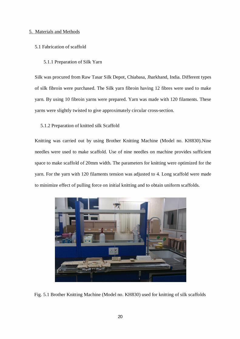
Knitting was carried out by using Brother Knitting Machine (Model no. KH830).Nine
needles were used to make scaffold. Use of nine needles on machine provides sufficient
space to make scaffold of 20mm width. The parameters for knitting were optimized for the
yarn. For the yarn with 120 filaments tension was adjusted to 4. Long scaffold were made
to minimize effect of pulling force on initial knitting and to obtain uniform scaffolds.
Fig. 5.1 Brother Knitting Machine (Model no. KH830) used for knitting of silk scaffolds

21
Briefly, knitting was carried out pushing five alternate needles were pushed to ‘Out’
position. The yarn was loaded to K-carriage and pulled to right end. All five needles got
shifted to ‘Working’ position. The ‘Claw’ weights were used to give uniform pull for
knitting process. Remaining four needs were pushed to ‘Working’ position. K-carriage
moved to and fro from right end to left end with counting. Knitting was finished by cutting
the yarn about 10 cm away from last needle and then K-carriage was moved over knitted
silk. Knitted silk was collected and cut in to 6 cm pieces. They were loaded on wire frame
immediately to avoid any damage to knitting.
The scaffolds were cut at distance of 60 mm with closing the ends with silk fibres.
5.1.3 Loading of scaffolds on wire frame
The scaffolds were loaded on wire frame made by aluminium wire of diameter 1mm to
keep scaffold in planar conformation. The frame was made about same size that of
scaffold. The scaffolds loaded on the wire frame were used for elctrospinning. Figure 5.1
shows scaffold load on the wire loop.
Fig. 5.2 Knitted scaffold loaded on wire loop

22
5.1.4 Morphological characterization of knitted scaffold
The morphology of the knitted fibers was analyzed under optical scanning electron
microscope. A JEOL JSM- 6480LV SEM was used in the experiment for morphology of
fibers at an accelerating voltage of 20 KV. Scaffolds were cut into small square about 1
cm side. Scaffolds were mounted on the sample holder with the help of carbon tape. Each
sample was coated with a thick layer of platinum by a JEOL JFC -1600 auto fine coater.
The operating parameters were maintained at 20 mA for 90 seconds.
5.2 Preparation of PCL Mesh
5.2.1 Preparation of PCL solution
Poly (ε-caprolactone) [Molecular weight Mn=80,000] was purchased from Sigma-Aldrich.
It is available in pellet form. The solvents to dissolve the PCL were selected after the
literature review. Dichloromethane and Di-methyl formamide were used to make solution
of PCL. The solvents were purchased from Merck Co. and used without further
purification. Solutions for electrospinning were prepared in the quantity of 5 gram (wt).
The chemical being used are toxic. All the solution preparation was done in fume hood.
Polymer concentration of 10% wt/wt was used for electrospinning. For preparation of
solution 0.5 gram of PCL pellets was weighted in small glass bottle. After that equal
amount of dichloromethane and dimethylformamide were added to make final solution of
5 gram. The solution was kept for stirring on magnetic stirrer for 24 hours at low rotation
speed. The glass bottle was capped and sealed properly. The solution was used for
elctrospinning within 2 days of preparation of solution.

23
5.2.2 Electrospinning of PCL
The electrospinning setup was used for carrying out electrospinning of 10% PCL solution..
The parameters were fixed from previous work or by trial method. The solution was
loaded into 5 ml glass syringe. Needle with inner diameter of 0.7 mm was used for
electrospinning. Syringe was fixed with a syringe pump which allows controlled
continuous flow of solution. Flow rate was maintained at 2 mL/hr. Distance between the
tip of needle and collector was fixed to 10 cm. This distance was adjusted by rotating
screw attached to collector stage. The collector plate was checked to make it horizontally
levelled. Aluminium foil was kept on the stage. The Aluminium foil was connected to
high voltage supply. A high voltage of 12 kV was applied between needle and collector.
Process Parameter Values used
Distance between Tip and collector 10 cm
Applied voltage 12 kV
Flow rate 2 ml/hr
Table 5.1 Parameters used for electrospinning
Knitted scaffolds, which were previously made, were used for collection of electrospun
mesh. As mentioned earlier, scaffolds were loaded on wire frame. For elctrospinning,
two scaffolds were kept on the collector. Positions of scaffolds were such that they are
not directly under need nor too far from needle. Scaffolds were moved intermediately
to maintain uniformity in mesh formation. Scaffolds were flipped after one hour of
electrospinning and kept for one more hour. After electrospinning for total two hours,
scaffold were removed and used for biocompatibility testing.

24
5.3 Degradation and Mechanical Testing of Scaffold
Mechanical strength of construct is most important property for the ligament tissue
engineering. Degradation of scaffolds was studied by keeping them in phosphate buffered
saline at 37°C with pH 7.4. The pH was measured regular interval and maintained by
addition of 1N HCl or 1N NaOH, if necessary.
Scaffolds degradation was analyzed based on ultimate failure load. Scaffolds were tested
by use of TA-HD Plus Texture Analyser. Gauge length is set at 20mm for all scaffolds.
The cross head speed is kept at 10 mm/min. The ultimate failure load determined for all
the type of scaffold at day 0, 3, 7 and 14.
Fig. 5.3 TA-HD Plus Texture Analyser
5.4 Cell Culture
Cells were obtained from National Centre for Cell Sciences (NCCS), Pune. Mouse
fibroblast cell line L-929 was obtained for its fibroblast nature and wide acceptance. The
cells were cultured on DMEM/D-12 media. Powdered media was purchased for Hi-Media.
It was reconstituted with 800 ml of double distilled water. 1.2 gms of Sodium bicarbonate

25
was added to media. It was then made to one litre. pH was adjusted to 7.2 with 1N NaOH
and 1N HCl. Media was then filtered with filtration unit by using nitrocellulose membrane
of 0.2µm pore size.
Media was supplemented with 5% serum and Antimicrobial -antifungal agents. Cells were
cultured in T75 flask. Approximately 10 ml of media was used in every flask. The media
was changed after every second day. Cells were sub-cultured after about 80% confluence
or at 7th day. The distribution ratio of 1:4 was used, as recommended by NCCS, Pune. The
cells from third passage were used for seeding on scaffolds.
5.5 Biocompatibility Testing
5.5.1 Scaffold sterilization
Scaffolds were kept in separate 100mm2 Petri-plates. Scaffolds were sterilized with
formaldehyde vapours for one hour and kept in bio-safety cabinet for 24 hrs to remove
traces of formaldehyde vapour. The sterilised scaffolds were washed with 10 ml of
sterilized PBS for three times. 10 ml of culture media was added to scaffolds to neutralize.
After 24 hr, media was removed and scaffolds were used for cell seeding.
5.5.2 Cell seeding
Cells were seeded at concentration 2.5 X 10⁶ cells/ml. 1 ml of cell containing media was
used to seed the cells on scaffold. After 1 hr, 10 ml of DMEM/D-12 media supplemented
with 5 % of serum was added.
5.5.3 Cell Adhesion Study
For cell adhesion studies scaffolds with 60mmX20mm were used (n=3). Cells were seeded
on scaffolds. After 24 hrs, scaffolds were removed from Petri-plates. Media was collected
in 15 ml centrifuge tube. The petri-plates were treated with 2 ml of trypsin to remove cell

26
attached to plate’s surface. Trypsin with cells was transferred to tube containing media
from petri-plates. Tubes were centrifuged at 4000 rpm for 15 min to separate cells from
the media. The media was decanted. 1 ml of fresh media was added to pellet. Cell count
was performed with haemocytometer. To find number of cells adhered to scaffold, number
of cells from pellet were subtracted from initial number of cell seeded. Cell adhesion was
expressed in terms percentage of cells adhered to scaffold.
5.5.4 MTT Assay
MTT assay was carried out be adding MTT reagent to the cell seeded scaffold. It was
incubated for 4 hrs in presence of 5% CO2 at 37°C. To dissolve formazan DMSO was
added. Absorbance was taken at 630nm. Standard graph for MTT assay was plotted by
adding MTT reagent to known cell densities.
5.5.5 Staining of cells with ethidium bromide
Stock solution of ethidium bromide of 2mg/ml was prepared. 100µl of ethidium bromide
was added to 10 ml of cell culture medium for staining cells. Cells were incubated for 20
minutes with ethidium bromide. Further washed with PBS and observed under fluorescent
microscope.

27
CHAPTER 6 RESULTS AND DISCUSSION

28
6. Result and Discussion
6.1 SEM analysis of scaffold
Knitting process involves use of knitting machine. Silk yarn may get damaged during
process. Damaged scaffold will show lower tensile strength. To analyze damage to silk
fibers SEM images were examined, as shown in figure 6.1. Knitted silk scaffold have not
shown any type of damaged caused by knitting. In case of ligament tissue engineering, it is
very important to have full strength of scaffold. Surface of the filaments provide
information about intensity degumming process. Properly degummed silk fibres appear
smooth. The yarn appears to smooth, suggesting silk has been properly degummed.
Fig. 6.1 SEM image of knitted silk scaffold
Electro-spun mesh of PCL was also analysed by SEM. Figure 6.2 shows the morphology
of electrospun PCL mesh. It shows non-uniform distribution of fiber diameter. The fiber
diameter is in range of 0.1 µm to 1 µm. The fibers resemble that of collagen morphology
which forms assembly of similar sizes. The mesh of PCL provide good surface for cells to
for attachment.

29
Fig. 6.2 SEM image of electro-spun PCL mesh
6.2 In vitro degradation of silk scaffold
Fig. 6.3 Mechanical testing of scaffolds for ultimate tensile strength of knitted silk
scaffold at five different time point
The tensile strength of silk scaffold decreased with degradation of silk over study period.
The decrease in tensile strength of silk is found to be high as compared to PLGA29. Silk
scaffold showed loss of 41.36% tensile strength over the period of 21 days. This decrease
38.836.62
33.3
27.5522.75
05
1015202530354045
0 3 6 9 12 15 18 21
Bre
akin
g Lo
ad (N
)
Days

30
in strength is slower than many other which were used previously. Knitted silk scaffold
will able to provide high tensile strength over the long period. The strength of knitted silk
scaffold could be improved by using thicker yarn. Use of thicker yarn will reduce area for
the secreted extracellular collagen fibers. Collagen and silk, both are among the strongest
natural fibers. Using the minimal yarn diameter to provide required strength will be
advantageous as it will allow deposition and direction orientation of collagen.
6.3 Cell seeding and proliferation assay
Fig 6.4 Cell culture. (A) Cells in at first subculture. (B) Cells at 3rd passage
Mouse fibroblast L929 shows the elongated morphology during the cell culture. The cells at
first subculture are shown in fig 6.4A while 6.4B shows cells in third passage. Cells from
third passage were seeded on scaffold.
Cells seeding efficiency is found to be 92.28 % ± 0.611. The cell seeding efficiency is found
to better than scaffold29. High cell seeding efficiency allows overlooking of other method of
cell seeding such as fibrin glue for cell seeding on scaffold. The cell seeding efficiency high
may because of high surface area provided by electrospun PCL mesh. This mesh allows
adherence of cells on scaffold.
A B

31
Fig. 6.5 Standard curve of MTT assay
Mouse fibroblast has shown proliferation on silk-PCL scaffold. Initially 1 X 10⁵ cells were
seeded on scaffold for proliferation assay. After day 3, number cells quantified by MTT assay
is 2.7 X 10⁵ cells. And after day 7, cell estimated to be 9.3 X 10⁵ cells. MTT assay shows
proliferation of cells on silk-PCL scaffold. This scaffold found to be suitable for cell growth.
Fig 6.6 Cells stained with ethidium bromide, observed under fluorescent microscope at
day 3 (A) and day 7 (B)
Fluorescent microscopy images taken by staining cell nucleus with ethidium bromide
supports the result obtain from the MTT assay .The fluorescent microscopic images show
R² = 0.9979
0
0.2
0.4
0.6
0.8
1
1.2
1.4
1.6
1.8
0 0.2 0.4 0.6 0.8 1 1.2
Abso
rban
ce
X106 Cells
A B

32
proliferation of cells from day 3 to day 7. Very few cells are visible in the day 3 image. In
day 7 image proliferated cells can be observed clearly. The silk-PCL scaffold provides
suitable microenvironment for growth of mouse fibroblast L929. Use of this biocompatible
scaffold in ligament tissue engineering provides required tensile strength with high cell
seeding efficiency.

33
CHAPTER 7 CONCLUSION

34
7. Conclusion
This research work for first time demonstrated use of wild type of silk produced from
Antheraea mylitta for ligament tissue engineering. Wild type of silk have gain great
interest as a biomaterial for tissue engineering in recent years.
The research also demonstrated successful use of knitted silk scaffold with electrospun
PCL mesh. This combination of materials has been studied for first time. Results show
that combinations of knitted silk with PCL mesh provides competent substrate for cell
attachment and growth.
Ligament is one of the most tensile stress bearing tissue in the body. Tissue engineered
scaffold should provide the tensile strength during healing. The degradation study of silk
has shown slow loss of tensile strength. After 21 day of in vitro degradation, Silk scaffolds
retained 60% of initial strength. Thus silk can provide the strength during in vitro cell
culture and in vivo during early period of post- implantation.
The use of silk provides strength to scaffold while PCL mesh allows higher adhesion of
cells. Results have shown 92.28 ± 0.61% cell adhesion efficiency. The wild type silk is
allows better adhesion of cells due to presence of RGD sequence. This RGD sequence is
absent in the mulberry silk. The competent adhesion of cells to scaffold eliminates need of
any special cell seeding method. The results have shown that this hybrid scaffold supports
proliferation of cells in vitro.
Thus from this study it can be concluded that hybrid scaffold made with wild silk and PCL
is biocompatible and shows desirable properties for ligament tissue engineering. Use of
silk-PCL scaffold in ligament tissue engineering will improve properties of ligament graft.

35
CHAPTER 8
REFERENCES

36
8. References
1. Carlstedt C, Nordin M (1989). “Biomechanics of tendons and ligaments,” in Basic
Biomechanics of the Musculoskeletal System, pp. 59–74.
2. McBride DJ, Trelstad RL, Silver FH (1988). Structural and mechanical assessment of
developing chick tendon. Int J Biol Macromol 10:194-200.
3. Silver FH, Freeman JW, Seehra GP (2003). Collagen self-assembly and the
development of tendon mechanical properties. J Biomech 36:1529-1553.
4. Benjamin M, Evans EJ, Copp L (1986). The histology of tendon attachments to bone
in man. J Anat 149:89–100.
5. Niyibizi C, Visconti CS, Kavalkovich K, Woo SL (1995). Collagens in an adult
bovine medial collateral ligament: immunofluorescence localization by confocal
microscopy reveals that type XIV collagen predominates at the ligament-bone
junction. Matrix Biol 14:743–751.
6. Beynnon BD, Johnson RJ, Fleming BC, Kannus P, Kaplan M, Samani J (2002).
Anterior cruciate ligament replacement: comparison of bone-patellar
tendonbonegrafts with two-strand hamstring grafts. A prospective, randomized study.
J Bone Joint Surg Am 84-A: 1503–1513.
7. Blickenstaff KR, Grana WA, Egle D (1997). Analysis of a semi-tendinosus autograft
in a rabbit model. Am J Sports Med.; 25:554–559.
8. Kurosaka M, Yoshiya S, Andrish JT (1987). A biomechanical comparison of different
surgical techniques of graft fixation in anterior cruciate ligament reconstruction. Am J
Sports Med. 15:225–229.
9. Fu FH, Bennett CH, Lattermann C, Ma CB (1999). Current trends in anterior cruciate
ligament reconstruction. Part I: Biology and biomechanics of reconstruction. Am J
Sports Med 27:821-830.

37
10. Fu FH, Bennett CH, Ma CB, Menetrey J, Lattermann C (2000). Current trends in
anterior cruciate ligament reconstruction. Part II. Operative procedures and clinical
correlations. Am J Sports Med 28:124-130.
11. Talukdar S, Nguyen QT, Chen AC, Sah RL, Kundu SC (2011) Effect of initial cell
seeding density on 3D-engineered silk fibroin scaffolds for articular cartilage tissue
engineering. Biomaterials 32: 8927–8937.
12. Patra C, Talukdar S, Novoyatleva T, Velagala SR, Mühlfeld C, Kundu B, Kundu SC,
Engel FB (2012). Silk protein fibroin from Antheraea mylitta for cardiac tissue
engineering. Biomaterials 33:2673–2680.
13. Hongbin F, Haifeng L, Wong J, Toh S, Goh JC (2008). In vivo study of anterior
cruciate ligament regeneration using mesenchymal stem cells and silk scaffold.
Biomaterials 29:3324–3337.
14. Hongbin F, Haifeng L, Toh SL, Goh JC (2009). Anterior cruciate ligament
regeneration using mesenchymal stem cells and silk scaffold in large animal model.
Biomaterials 30: 4967-4977.
15. He P, Ng KS, Toh SL, Goh JC (2012). In vitro ligament-bone interface regeneration
using a trilineage coculture system on a hybrid silk scaffold. Biomacromolecules
13:2692-703.
16. Chen X, Qi Y, Wang LL, Yin ZG, Zou XH, Ouyang HW (2008). Ligament
regeneration using a knitted silk scaffold combined with collagen matrix.
Biomaterials 29:3683-3692.
17. Huang MH, Li SM, Hutmacher DW, Coudane J, Vert M (2006). Degradation
characteristics of poly (€-caprolactone)- based copolymers and blends. J Appl Polym
Sci 102:1681-1687.

38
18. Rohner D, Hutmacher DW, Cheng TK, Oberholzer M, Hammer B (2003). In vivo
efficacy of bone-marrow-coated polycaprolactone scaffolds for the reconstruction of
orbital defects in the pig. J Biomed Mater Res B Appl Biomater 66:574-580.
19. Chew SY, Wen Y, Dzenis Y, Leong KW (2006). The role of electrospinning in the
emerging field of nanomedicine. Curr Pharm Des 12:4751-1470.
20. Cipitria A, Skelton A, Dargaville TR, Dalton PD, Hutmacher DW(2011). Design,
fabrication and characterization of PCL electrospun scaffolds—a review. J. Mater.
Chem. 21:9419-9453.
21. Zein I, Hutmacher DW, Tan KC, Teoh SH (2002). Fused deposition modelling of
novel scaffold architectures for tissue engineering applications. Biomaterials 23:1169-
1185.
22. Park CH, Rios HF, Jin Q, Bland ME, Flanagan CL, Hollister JH, Giannobile WV
(2010). Biomimetic hybrid scaffolds for engineering human tooth-ligament interfaces.
Biomaterials 31:5945-5952.
23. Shao HJ, Lee YT, Chen CS, Wang JH, Young TH (2010). Modulation of gene
expression and collagen production of anterior cruciate ligament cells through cell
shape changes on polycaprolactone/chitosan blends. Biomaterials 31:4695–4705.
24. Williamson MR, Coombes AG (2004). Gravity spinning of polycaprolactone fibers
for applications in tissue engineering. Biomaterials 25: 459–465.
25. Hutmacher DW, Sittinger M, Risbud MV (2004). Scaffold-based tissue engineering:
rationale for computer-aided design and solid free-form fabrication systems. Trends
Biotechnol. 22:354–362.
26. Li WJ, Laurencin CT, Caterson EJ, Tuan RS, Ko FK (2002). Electrospun nanofibrous
structure: a novel scaffold for tissue engineering, J. Biomed. Mater. Res. 60:613–621.

39
27. Wutticharoenmongkol P, Sanchavanakit N, Pavasant P, Supaphol P (2006).
Preparation and characterization of novel bone scaffolds based on electrospun
polycaprolactone fibers filled with nanoparticles. Macromol Biosci, 6:70–77.
28. Mavis B, Demirtaş TT, Gümüşderelioğlu M, Gündüz G, Çolak U (2009). Synthesis,
characterization and osteoblastic activity of polycaprolactone nanofibers coated with
biomimetic calcium phosphate. Acta Biomaterialia, 5:3098–3111.
29. Vaquette C, Slimani S, Kahn CJ, Tran N, Rahouadj R, Wang X (2010). A poly(lactic-
co-glycolic acid) knitted scaffold for tendon tissue engineering: an in vitro and in vivo
study. J Biomater Sci Polym 21:1737-1760.