Comparative analysis of gene regulation in mouse rat and human

4100 Regent Street, Suite M-2 Columbus, OH 43219 USA Tel: +1 (866) 466 0100 • Fax: +1 (614) 436 6489 [email protected] • www.phylogenyinc.com
Expression of GENE in the Mouse Brain and Eye Robert Researcher, Ph.D. Southern University March 8, 2006

4100 Regent St • Suite M-2 • Columbus, OH 43219 Tel: +1 (866) 466 - 0100 • Fax: +1 (614) 436 - 6489 [email protected] • www.phylogenyinc.com
V2.0.1 i
Study Information
Title: Expression of GENE in the Mouse Brain and Eye
Investigator: Robert Researcher, PhD
Phylogeny: Adel Mikhail, PhD ([email protected])
Date: March 8, 2006
Gene: GENE
Tissues: Experimental Mouse Tissues Brain (Cortical Stab Wound)
Eye (Retinal Tear)
Eye (Crushed Optic Nerve)
Normal Adult Mouse Tissues Brain
Testis
Thymus
Lung
Liver
Pancreas
GI Tract
Prostate Gland
Lymph Node
Bone Marrow
Skeletal Muscle

Expression of GENE in the Mouse Brain and Eye
ii
Table of Contents
Final Report Summary 1
Detailed Findings 2
Methods 14
Table of Figures 15
CD-ROM (Images & Report)
Hybridized Slides
Please reference Phylogeny as having performed this study in any publications or presentations.

Expression of GENE in the Mouse Brain and Eye
1
Summary
Enclosed are slides exposed to emulsion for one and three weeks from an in situ hybridization (ISH) study using GENE to probe sections of injured mouse eye and brain as well as normal adult mouse whole body sections.
In summary, in normal adult mouse sagittal sections, GENE mRNA was detected at the highest levels in the bone marrow, lymph nodes and thymus. In decreasing order of expression, GENE gene transcripts were detected in the brain, pancreas, gut epithelium, prostate gland, liver, lung and skeletal muscle. GENE expression in normal adult tissues was low and required a long (three-week) exposure to emulsion. These expression patterns correlated well with the information on GENE expression in Gene Cards, http://www.genecards.org/cgi-bin/carddisp?GENE&search=Lipin.
In the experimental tissue sections, GENE mRNA did not appear to be elevated. GENE expression was detected in cells of these tissues (neurons and skeletal muscle) in a manner that was consistent with the results of the normal adult mouse sections. The antisense (AS) probe detected the mRNA. The sense (S) probe did not hybridize, but showed the background level of silver grains for the assay.
The image labels indicate the glass slide number, which slides were used in the validation assay, the probe used and whether the tissue was from cortical stab wound, retinal tear, crushed optic nerve or adult mouse sections. All images were acquired using darkfield illumination.
It should be mentioned here that the expected result, namely the elevation of a low level of ubiquitous expression of GENE, may be difficult to detect through ISH. This assay is more effective as a means of localizing gene expression than as a means of quantifying it. In cases where there is a concentrated signal from a cell-specific change in the gene expression pattern, ISH can prove to be a valuable tool. However, ISH detection of global changes in expression patterns with relatively low concentrations of transcripts can be difficult to distinguish from the background labeling that tends to occur during this assay.
If a similar study were to be conducted in the future, it might be helpful to include tissue samples from more animals in the assay, including normal controls from the same colony. It might also be helpful to assay not only the injured tissues, but also the contralateral tissues for comparative purposes.
To validate the integrity of mRNA in the tissue samples provided by the client, Phylogeny performed an ISH on sections of the tissue, using a probe for vonWillebrand factor, an endothelial cell marker.

Expression of GENE in the Mouse Brain and Eye
2
Detailed Findings injured tissues
Figure 1: von Willebrand Factor mRNA in the Mouse Brain with a Cortical Stab Wound
A ISH signal from the von Willebrand factor antisense probe is seen in blood vessels (arrows) of the brain. This result shows that the coronal brain sections of the experimental animal with a cortical stab wound contained intact mRNA. The region shown is the site of the cortical stab wound.
B Corresponding sense control result for (A).
Figure 2: von Willebrand Factor mRNA in the Mouse Eye with a Retinal Tear
A ISH signal from the von Willebrand factor antisense probe is seen in blood vessels (arrows) of the retina in the retinal tear preparation. This result shows that the eye sections of the experimental animal with a retinal tear contained intact mRNA.
B Corresponding sense control result for (A).
Expression of GENE in the Mouse Brain and Eye
3
Detailed Findings injured and control t issues
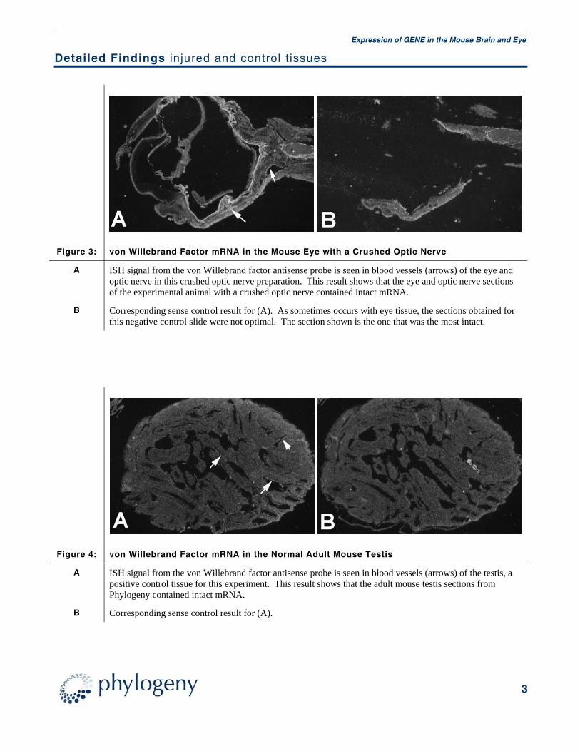
Figure 3: von Willebrand Factor mRNA in the Mouse Eye with a Crushed Optic Nerve
A ISH signal from the von Willebrand factor antisense probe is seen in blood vessels (arrows) of the eye and optic nerve in this crushed optic nerve preparation. This result shows that the eye and optic nerve sections of the experimental animal with a crushed optic nerve contained intact mRNA.
B Corresponding sense control result for (A). As sometimes occurs with eye tissue, the sections obtained for this negative control slide were not optimal. The section shown is the one that was the most intact.
Figure 4: von Willebrand Factor mRNA in the Normal Adult Mouse Testis
A ISH signal from the von Willebrand factor antisense probe is seen in blood vessels (arrows) of the testis, a positive control tissue for this experiment. This result shows that the adult mouse testis sections from Phylogeny contained intact mRNA.
B Corresponding sense control result for (A).

Expression of GENE in the Mouse Brain and Eye
4
Detailed Findings normal t issues
Figure 5: GENE mRNA in the Normal Adult Mouse Testis
A ISH signal from the GENE antisense probe is seen in the seminiferous tubules of the testis. This result validates the design of the GENE probes and shows that the GENE mRNA was expressed at a high level in the testis epithelium.
B Corresponding sense control result for (A).
Figure 6: GENE mRNA in the Normal Adult Mouse Brain
A ISH signal from the GENE antisense probe is seen in neurons of the cerebellum, hippocampus, cerebral cortex and other regions of the brain. The background level of signal was higher in the brain than in other tissues.
B Corresponding sense control result for (A).
Abbreviations: ce – cerebellum; cx – cerebral cortex; hi – hippocampus.

Expression of GENE in the Mouse Brain and Eye
5
Detailed Findings normal t issues
Figure 7: GENE mRNA in the Normal Adult Mouse Thorax
A ISH signal from the GENE antisense probe is seen in the ribs, thymus and lung, but not the heart.
B Corresponding sense control result for (A).
Abbreviations: h – heart; lu – lung; r – rib; th – thymus.
Figure 8: GENE mRNA in the Normal Adult Mouse Abdomen
A ISH signal from the GENE antisense probe is seen at a low level in the liver and at higher levels in the pancreas and epithelium lining the stomach, small intestine and colon.
B Corresponding sense control result for (A).
Abbreviations: co – colon; li – liver; p – pancreas; si – small intestine; st – stomach.

Expression of GENE in the Mouse Brain and Eye
6
Detailed Findings normal t issues
Figure 9: GENE mRNA in the Normal Adult Mouse Abdomen
A ISH signal from the GENE antisense probe is seen in the bone marrow of the hip, a lymph node of the colon, the prostate gland, pancreas, stomach and small intestine.
B Corresponding sense control result for (A).
Abbreviations: bm – bone marrow; co – colon; ln – lymph node; p – pancreas; pr – prostate; si – small intestine; st – stomach.
Figure 10: GENE mRNA in the Normal Adult Mouse Hip Region
A ISH signal from the GENE antisense probe is seen at a high level in the bone marrow of the hip and at a low level in the skeletal muscle.
B Corresponding sense control result for (A).
Abbreviations: bm – bone marrow; co – colon; sk – skeletal muscle.

Expression of GENE in the Mouse Brain and Eye
7
Detailed Findings brain with cortical stab wound
Figure 11: GENE mRNA in the Mouse Brain with a Cortical Stab Wound
A ISH signal from the GENE antisense probe is seen following a one-week exposure of a cortical stab wound (arrow) to a mouse brain. The stab wound penetrated through the cortex to the lateral ventricle. A portion of the choroid plexus is seen in the lateral ventricle and in the third ventricle. The arrowhead points to the habenular nucleus, which expressed GENE mRNA at a slightly elevated level compared to other regions of the brain in these images. The ISH signal was reduced in the immediate vicinity of the stab wound, perhaps due to the loss of neuron cell bodies. An elevated ISH signal for GENE was not detected in the region of the wound. Parallel sections were exposed to emulsion for three weeks in an attempt to detect an increased, but low level expression of GENE mRNA (see Figure 12).
B A second section from the same region as (A) hybridized to the GENE antisense probe.
C Corresponding sense control result for (A) and (B) acquired from between these two sections.
Abbreviations: 3 – third ventricle; cp – choroid plexus; cx – cortex.
Expression of GENE in the Mouse Brain and Eye
8
Detailed Findings brain with cortical stab wound
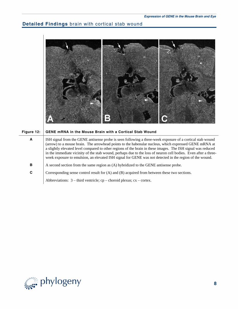
Figure 12: GENE mRNA in the Mouse Brain with a Cortical Stab Wound
A ISH signal from the GENE antisense probe is seen following a three-week exposure of a cortical stab wound (arrow) to a mouse brain. The arrowhead points to the habenular nucleus, which expressed GENE mRNA at a slightly elevated level compared to other regions of the brain in these images. The ISH signal was reduced in the immediate vicinity of the stab wound, perhaps due to the loss of neuron cell bodies. Even after a three-week exposure to emulsion, an elevated ISH signal for GENE was not detected in the region of the wound.
B A second section from the same region as (A) hybridized to the GENE antisense probe.
C Corresponding sense control result for (A) and (B) acquired from between these two sections.
Abbreviations: 3 – third ventricle; cp – choroid plexus; cx – cortex.

Expression of GENE in the Mouse Brain and Eye
9
Detailed Findings eye with retinal tear
Figure 13: GENE mRNA in the Mouse Eye with a Retinal Tear
A ISH signal from the GENE antisense probe is seen following a one-week exposure of a section of eye from a mouse with a retinal tear wound. This was the best section available on the slide. All sections were torn on this slide. An elevated ISH signal for GENE was not detected in these sections. Parallel sections were exposed to emulsion for three weeks in an attempt to detect an increased, but low level expression of GENE mRNA (see Figure 14).
B A second, intact section from the same region as (A) hybridized to the GENE antisense probe.
C Corresponding sense control result for (A) and (B) acquired from between these two sections.
Abbreviations: co – cornea; l – lens; re – retina.

Expression of GENE in the Mouse Brain and Eye
10
Detailed Findings eye with retinal tear
Figure 14: GENE mRNA in the Mouse Eye with a Retinal Tear
A ISH signal from the GENE antisense probe is seen following a three-week exposure of a section of eye from a mouse with a retinal tear wound. The site of the retinal tear was not obvious. Even after a three-week exposure to emulsion, an elevated ISH signal for GENE was not detected in these sections.
B A second section from the same region as (A) hybridized to the GENE antisense probe.
C Corresponding sense control result for (A) and (B) acquired from between these two sections.
Abbreviations: c – cornea; l – lens; r - retina.

Expression of GENE in the Mouse Brain and Eye
11
Detailed Findings eye with crushed optic nerve
Figure 15: GENE mRNA in the Mouse with a Crushed Optic Nerve
A ISH signal from the GENE antisense probe is seen following a one-week exposure of a section of eye from a mouse with a crushed optic nerve wound. The site of the crushed nerve was not obvious. ISH signal for GENE was detected in these sections in two areas. The first was in adipose connective tissue (fat), while the second was in skeletal muscle. These results can also be viewed at a higher magnification (see Figure 16). No clear elevated ISH signal for GENE was detected in these sections. Parallel sections were exposed to emulsion for three weeks in an attempt to detect an increased, but low level expression of GENE mRNA (see Figure 17).
B A second section from the same region as (A) hybridized to the GENE antisense probe.
C Corresponding sense control result for (A) and (B) acquired from between these two sections.
Abbreviations: f – fat; on – optic nerve; re – retina; sk – skeletal muscle.

Expression of GENE in the Mouse Brain and Eye
12
Detailed Findings eye with crushed optic nerve
Figure 16: GENE mRNA in the Mouse Eye with a Crushed Optic Nerve
A ISH signal from the GENE antisense probe is seen following a one-week exposure of a section of eye from a mouse with a crushed optic nerve wound. This is a higher magnification of Figure 15A acquired using phase contrast settings. Even at higher magnification, no clear elevated ISH signal for GENE was detected in these sections. Parallel sections were exposed to emulsion for three weeks in an attempt to detect an increased, but low level expression of GENE mRNA (see Figure 17).
B A second section from the same region as (A) hybridized to the GENE antisense probe. This is a higher magnification of Figure 15B acquired using darkfield settings.
C Corresponding sense control result for (A) and (B) acquired from between these two sections.
Abbreviations: fat – fat; on – optic nerve; re – retina; sk – skeletal muscle.

Expression of GENE in the Mouse Brain and Eye
13
Detailed Findings eye with crushed optic nerve
Figure 17: GENE mRNA in the Mouse Eye with a Crushed Optic Nerve
A ISH signal from the GENE antisense probe is seen following a three-week exposure of a section of eye from a mouse with a crushed optic nerve wound. Even after a three-week exposure to emulsion, the ISH results were consistent with the one-week data. ISH signal for GENE was detected in adipose connective tissue (fat) and skeletal muscle, but did not appear elevated as a result of the crush injury.
B A second section from the same region as (A) hybridized to the GENE antisense probe.
C Corresponding sense control result for (A) and (B) acquired from between these two sections.
Abbreviations: on – optic nerve; re – retina; sk – skeletal muscle.

Expression of GENE in the Mouse Brain and Eye
14
Methods
Tissue Fixation, Embedding and Pretreatment Tissues from the client's experimental animals were fixed in 4% paraformaldehyde in phosphate buffered saline (PBS) overnight, dehydrated and infiltrated with paraffin. 5 to 7 micron serial sections were mounted on gelatinized slides. 1 to 3 sections were mounted on each slide, deparaffinized in xylene, rehydrated and post-fixed. The sections were digested with proteinase K, post-fixed, treated with triethanolamine/acetic anhydride, washed and dehydrated. Mouse whole body sections provided by Phylogeny were frozen cut into 10 to 12 micron thick sections, mounted on gelatin-coated slides and stored at –80C. Before ISH they were fixed in 4% formaldehyde (freshly made from paraformaldehyde) in phosphate buffered saline (PBS), treated with triethanolamine/acetic anhydride, washed and dehydrated with a series of ethanol.
cRNA Probe Preparation The cRNA transcripts were synthesized according to manufacturer's conditions (Ambion) and labeled with 35S-UTP (>1000 Ci/mmol; Amersham). cRNA transcripts larger than 200 nucleotides were subjected to alkali hydrolysis to give a mean size of 70 bases for efficient hybridization.
Hybridization and Washing Procedures Sections were hybridized overnight at 52°C in 50% deionized formamide, 0.3 M NaCl, 20 mM Tris-HCl pH 7.4, 5 mM EDTA, 10 mM NaH2PO4, 10% dextran sulfate, 1X Denhardt's, 50 µg/ml total yeast RNA, and 50-75,000 cpm/µl 35S-labeled cRNA probe. The tissue was subjected to stringent washing at 65°C in 50% formamide, 2X SSC, 10 mM DTT and washed in PBS before treatment with 20 µg/ml RNAse A at 37°C for 30 minutes. Following washes in 2X SSC and 0.1X SSC for 10 minutes at 37°C, the slides were dehydrated and dipped in Kodak NTB-2 nuclear track emulsion and exposed for one week in light-tight boxes with desiccant at 4°C.
Imaging Photographic development was carried out in Kodak D-19. Slides were counterstained lightly with toluidine blue and analyzed using both light- and darkfield optics of a Zeiss Axiophot microscope. Sense control cRNA probes (identical to the mRNAs) always gave background levels of hybridization signal. Enclosed are TIFF image files labeled with the tissue and whether the result was with the antisense or sense probe; a, b, c, etc. indicate a different region of the section on the same glass slide. All micrographs are of one-week exposures to emulsion. A scan of a 1cm micrometer, taken with the same objective used for the 10X figures, is provided if a scale bar is required. The small lines on the micrometer mark millimeters.
Storage and Rehydration Any “crystallized” section may be repaired by allowing the coverslip to fall off the slide after soaking in xylene for 24-48 hours. Rehydrate the slide to 70% EtOH. Redehydrate the slide in 80%, 95% and 2x 100% EtOH for 2 minutes each. After three changes of xylene, mount the coverslip with Cytoseal (VWR Scientific) or other comparable mounting medium. Using the same method, coverslips can be removed for histological staining to take brightfield micrographs. Histological stains that require acidic conditions may dissolve silver grains. Overstaining may obscure the silver grains. Any excess mounting medium or residual emulsion on the back of the slide can be removed with a single-edged razor. Dry the recoverslipped slides flat for 24 hours. These slides can be stored indefinitely at room temperature.
Viewing Original Slides The results are best viewed with darkfield illumination, but with a 20x or 40x phase-contrast objective, the silver grains can be localized over particular cell groups. The antisense probe (AS) detects the mRNA and the sense control probe (S) shows the background level of silver grains for the experiments.

Expression of GENE in the Mouse Brain and Eye
15
Table of Figures
Figure Panel Slide Tissue Probe Total Magnification Photo Filename
1 A CS Val:19 Brain with Cortical Stab Wound vWF AS 25 F1_V19_Brain_vWF_AS B CS Val:20 Brain with Cortical Stab Wound vWF S 25 F2_V20_Brain_vWF_S
2 A RT Val:09 Eye with Retinal Tear vWF AS 25 F5_V09_RT_vWF_AS B RT Val:10 Eye with Retinal Tear vWF S 25 F6_V10_RT_vWF_S
3 A ON Val:04 Eye with Crushed Optic Nerve vWF AS 25 F3_V04_ON_vWF_AS B ON Val:05 Eye with Crushed Optic Nerve vWF S 25 F4_V05_ON_vWF_S
4 A 205 Val:08 Normal Adult Testis vWF AS 25 F7_V08_Testis_vWF_AS B 205 Val:09 Normal Adult Testis vWF S 25 F8_V09_Testis_vWF_S
5 A 205 Val:10 Normal Adult Testis GENE AS 25 F9_V10_Testis_GENE_AS B 205 Val:11 Normal Adult Testis GENE S 25 F10_V11_Testis_GENE_S
6 A Ad Mouse:169 Normal Adult Brain GENE AS 16.25 F29_Brain_GENE_AS_3wks B Ad Mouse:113 Normal Adult Brain GENE S 16.25 F34_Brain_GENE_S_3wks
7 A Ad Mouse:169 Normal Adult Thorax GENE AS 16.25 F30_Thorax_GENE_AS_3wks B Ad Mouse:113 Normal Adult Thorax GENE S 16.25 F35_Thorax_GENE_S_3wks
8 A Ad Mouse:169 Normal Adult Abdomen GENE AS 16.25 F31_LiverGut_GENE_AS_3wks B Ad Mouse:113 Normal Adult Abdomen GENE S 16.25 F36_LiverGut_GENE_S_3wks
9 A Ad Mouse:169 Normal Adult Abdomen GENE AS 16.25 F32_Abdomen_GENE_AS_3wks B Ad Mouse:113 Normal Adult Abdomen GENE S 16.25 F37_Abdomen_GENE_S_3wks
10 A Ad Mouse:169 Normal Adult Hip Region GENE AS 16.25 F33_HipMuscle_GENE_AS_3wks B Ad Mouse:113 Normal Adult Hip Region GENE S 16.25 F38_HipMuscle_GENE_S_3wks
11 A CS:21 Brain with Cortical Stab Wound GENE AS 25 F11_CS21_GENE_AS_1wk B CS:23 Brain with Cortical Stab Wound GENE AS 25 F13_CS23_GENE_AS_1wk C CS:22 Brain with Cortical Stab Wound GENE S 25 F12_CS22_GENE_S_1wk
12 A CS:25 Brain with Cortical Stab Wound GENE AS 25 F14_CS25_GENE_AS_3wks B CS:27 Brain with Cortical Stab Wound GENE AS 25 F16_CS27_GENE_AS_3wks C CS:26 Brain with Cortical Stab Wound GENE S 25 F15_CS26_GENE_S_3wks
13 A RT:11 Eye with Retinal Tear GENE AS 25 F17_RT11_GENE_AS_1wk B RT:13 Eye with Retinal Tear GENE AS 25 F19_RT13_GENE_AS_1wk C RT:12 Eye with Retinal Tear GENE S 25 F18_RT12_GENE_S_1wk
14 A RT:15 Eye with Retinal Tear GENE AS 25 F20_RT15_GENE_AS_3wks B RT:17 Eye with Retinal Tear GENE AS 25 F22_RT17_GENE_AS_3wks C RT:16 Eye with Retinal Tear GENE S 25 F21_RT16_GENE_S_3wks

Expression of GENE in the Mouse Brain and Eye
16
Table of Figures continued
Figure Panel Slide Tissue Probe Total Magnification Photo Filename
15 A ON:08 Eye with Crushed Optic Nerve GENE AS 25 F23_ON08_GENE_AS_1wk B ON:10 Eye with Crushed Optic Nerve GENE AS 25 F25_ON10_GENE_AS_1wk C ON:09 Eye with Crushed Optic Nerve GENE S 25 F24_ON09_GENE_S_1wk
16 A ON:08 Eye with Crushed Optic Nerve GENE AS 50 F23a_ON08_GENE_AS_1wk_10x B ON:10 Eye with Crushed Optic Nerve GENE AS 50 F23b_ON08_GENE_AS_1wk_10x_DF C ON:09 Eye with Crushed Optic Nerve GENE S 50 F24a_ON06_GENE_S_1wk_10x_DF
17 A ON:12 Eye with Crushed Optic Nerve GENE AS 25 F26_ON12_GENE_AS_3wks B ON:14 Eye with Crushed Optic Nerve GENE AS 25 F28_ON14_GENE_AS_3wks C ON:13 Eye with Crushed Optic Nerve GENE S 25 F27_ON13_GENE_S_3wks