Filtration rate of the blue mussel, Mytilus edulis, during exposure to various diet treatments
Upload
sara-tedescoCategory
view
212download
0
Comparative Biochemistry and Physiology, Part C 151 (2010) 167–174
Contents lists available at ScienceDirect
Comparative Biochemistry and Physiology, Part C
j ourna l homepage: www.e lsev ie r.com/ locate /cbpc
Exposure of the blue mussel, Mytilus edulis, to gold nanoparticles and thepro-oxidant menadione
Sara Tedesco a, Hugh Doyle b, Julian Blasco c, Gareth Redmond b, David Sheehan a,⁎a Proteomics Research Group, Department of Biochemistry, Environmental Research Institute, University College Cork, Cork, Irelandb Nanotechnology Research Group, Tyndall National Institute, Cork, Irelandc Instituto de Ciencias Marinas Andalucia (CSIC), Puerto Real (Cadiz) 11510, Spain
Abbreviations: BSA, Bovine serum albumin; 1DE, On2D, Two dimensional; DMSO, Dimethyl sulfoxide; DTNB,acid; DTT, Dithiothreitol; GNP, Gold nanoparticles; GSHOxidized glutathione; IAF, 5′-iodoacetamide fluoresceinplasma-optical emission spectroscopy; IEF, Isoelectric fgradient; PAGE, Polyacrylamide gel electrophoresis;fluoride; ROS, Reactive oxygen species; SDS, Sodium dosion electron microscopy; TNB, 5-thio-2-nitrobenzoic ac⁎ Corresponding author. Tel.: +353 21 4904207; fax:
E-mail address: [email protected] (D. Sheehan).
1532-0456/$ – see front matter © 2009 Elsevier Inc. Aldoi:10.1016/j.cbpc.2009.10.002
a b s t r a c t
a r t i c l e i n f oArticle history:Received 1 January 2000Received in revised form 6 October 2009Accepted 9 October 2009Available online 17 October 2009
Keywords:Mytilus edulisNanoparticlesMenadioneOxidative stressThiol proteinsToxicityGlutathioneProteomicGold
Relatively little is known about how gold nanoparticles (GNP) might interact in vivo with marine organisms.Mytilus edulis was exposed (24 h) to ~15 nm GNP, menadione and both compounds simultaneously (GNP/menadione). GNP was detected by inductively coupled plasma-optical emission spectroscopy mainly indigestive gland of samples exposed to GNP though not GNP/menadione, perhaps due to impaired feeding.Thioredoxin reductase activity and malondialdehyde levels were determined in all tissues. Thioredoxinreductase inhibitionwas detected only in digestive gland exposed tomenadionewhilstmalondialdehyde levelsdid not vary in response to treatment in all tissues. GNP caused a decrease in the reduced/oxidized glutathioneratio in digestive gland, but no difference was found in other tissues or for other treatments. One dimensionalelectrophoresis of proteins containing thiol groups was performed in all tissues and revealed a reduction inprotein thiols for all treatments in digestive gland. Two dimensional electrophoresis of digestive gland extracts,from GNP and control groups, showed decreased levels of thiol proteins in response to GNPwhich we attributeto oxidation. Our results suggest that GNP causes a modest level of oxidative stress sufficient to oxidize thiols inglutathione and proteins but without causing lipid peroxidation or induction of thioredoxin reductase activity.
e dimensional electrophoresis;5,5′-dithiobis(2-nitrobenzoic), Reduced glutathione; GSSG,; ICP-OES, Inductively coupledocusing; IPG, Immobilized pHPMSF, Phenylmethylsulfonyldecyl sulfate; TEM, Transmis-id.+353 21 4274034.
l rights reserved.
© 2009 Elsevier Inc. All rights reserved.
1. Introduction
Engineered nanoparticles have one dimension <100 nm and havealways been present on Earth (Laurent and Petit, 2005). They have verylarge surface to volume ratios which may underlie toxicity (Stoegeret al., 2006). Carbon nanotubes, which closely resemble asbestos, causeasbestosis-like systems in mice (Poland et al., 2008). Physicalpenetration of eukaryote cells by nanoparticles has been reportedbut, even where they are unlikely physically to enter cells, they havepotential to alter cell–cell signaling and to interact with cell surfaces.Recent reviews (Donaldson and Seaton, 2007; Moore, 2006; Nel et al.,2006; Oberdörster et al., 2007; Unfried et al., 2007) have focusedstrongly on nanomaterials' potential to cause oxidative stress.
Oxidative stress (Halliwell andGutteridge, 2007) is a feature ofmuchpathology, ageing and toxicity mechanisms of some environmental
pollutants (Dowling et al., 2006). It occurswhen reactive oxygen species(ROS) exceed cellular defences (Halliwell and Gutteridge, 2007). Theseinclude enzymes like catalase, superoxide dismutase and peroxidasebut also lowMr thiols such as glutathione (GSH;Meister and Anderson,1983). GSH is predominantly present in cells in its reduced form (GSH)but, on oxidation, forms oxidized glutathione (GSSG). Normally, cellsmaintain a high GSH/GSSG ratio. ROS-mediated oxidation of GSH hasbeen shown in marine bivalves exposed to organophosphate com-pounds (Peña-Llopis et al., 2002). Oxidative damage affecting sulphur-containing amino acids is reversed by the thioredoxin/thioredoxinreductase system. Inhibition or genetic suppression of thioredoxinresults in increased oxidant stress, apoptosis and increased sensitivityto oxidants (Chen et al., 2006).While GSH and the GSH/GSSG ratio havereceived extensive research interest in ecotoxicology, protein thiolgroups have been poorly-studied. They contribute significantly toantioxidant defense and are more quantitatively important redoxbuffers than GSH (Hansen et al., 2009). Reversible oxidation of proteinthiols to disulfides or sulphenic acid is important in signal transductionand couples redox status to protein function (Eaton, 2006). The presentpaper focuses on effects of gold nanoparticles (GNP) on thiols of GSHand proteins as measures of mild oxidative stress.
Nanoparticles contribute to oxidative stress at several biologically-relevant levels (Unfried et al., 2007). Some produce ROS as a con-sequence of their large proportional surface area (Stoeger et al., 2006).Others disrupt cell structures such as mitochondria (Nel et al., 2006).

168 S. Tedesco et al. / Comparative Biochemistry and Physiology, Part C 151 (2010) 167–174
Coating of nanoparticles with a “corona” of proteins offers a generalparadigm for biospecific functionalisation of nanoparticles, perhapsproviding routes of passage through biomembranes (e.g. via receptors)(Cedervall et al., 2008). There is agreement on some general principlesregarding toxicological implications of nanomaterials: 1. Smaller nano-particles pose greater threat than larger. 2. Nanoparticles can cross anddamage biomembranes in eukaryotes. 3. They often cause oxidativestress. 4. Their chemical composition is a component of toxicity. 5. Ineukaryotes, nanoparticles can enter cells and sometimes organelleswhilst in prokaryotes they often remain outside the cell.
Environmental concentrations of manufactured nanoparticles haveyet to be routinely measured, but there are serious concerns thatnanoparticles will be released over their lifetime (e.g., by erosion ordeliberate introduction during environmental remediation) or thatproduct applications could generate waste-containing nanomaterials(e.g., household products). These novel materials could be released inlarge quantities into the environment and can be toxic to bacteria,algae, invertebrates and fish, as well as mammals. However, much ofthe currently-available ecotoxicological data are limited to species usedin regulatory testing or freshwater organisms (Handy et al., 2008). Forexample, nanoparticles can act as contaminant carriers for co-existingpollutants, altering the toxicity of specific chemicals towards inverte-brates such as Daphnia magna (Baun et al., 2008) or having effects ontheir locomotor behavior (Lovern et al., 2007).
Gold nanoparticles (GNP) are widely-use in electronics, catalysis,cosmetics (EWG, 2006), food quality control (Mannino and Scampic-chio, 2007) and in cancer detection (Medley et al., 2008). Althoughsupposedly an inert material, nanoscale gold exhibits properties thatchange according to size, surface coating, shape, oxidation state andsynthesis method. Studies have appeared on the biodistribution andtoxicity of Au (I) and Au (III) compounds (Eisler, 2004) but few studieshave evaluated in vivo toxicology of colloidal nanoscale Au (0).Furthermore, relatively few studies have been carried out on filter-feeding aquatic organisms such as mussels exposed to GNP (Renaultet al., 2008; Tedesco et al., 2008) andmechanisms bywhich theymightcause oxidative stress are still poorly-understood. GNP can reactdirectly with thiol-containing amino acids such as homocysteine andcysteine (Lim et al., 2007); whilst pro-oxidants like menadione canoxidizeM. edulis protein thiol groups (McDonagh and Sheehan, 2007).GNP react directly with thiol-containingmolecules such as GSH ormayindirectly cause an imbalance in the GSH/GSSG ratio during oxidativestress (Renault et al., 2008).
Bivalve molluscs, are popular sentinel organisms in environmentalmonitoring (Goldberg, 1986). They need to open their valves tofacilitate free circulation of water through their gills allowing them torespire and feed (Jorgensen, 1996). Metals and organic contaminantsmay affect valve movement, thus reducing filtration rates (Anguianoet al., 2007).Mussels can absorb xenobiotics present in thewater phasedirectly through their gills and indirectly through the digestive systemwhen xenobiotics are adsorbed on the small grain-size fraction ofparticles (Baumard et al., 1999).
Citrate-capped GNP (2 nm and 10 nm) enter cultured human cellsdue to lipid peroxidation (Panessa-Warren et al., 2008) and can causeother forms of cell damage (e.g.: nuclear localization (2 nm-Au);intracellular membrane damage (10 nm-Au) (Panessa-Warren et al.,2008). Gold compounds have been used to treat disease, nowadays invarious forms of arthritis. They can have toxic side-effects includingproteinuria, diarrhea, bone marrow suppression and skin rashes(Eisler, 2003). Auranofin is a gold compound that potently inhibitsthioredoxin reductase, a possible mechanism for its therapeutic be-nefit (Omata et al., 2006).
GNP increased M. edulis catalase activity and protein ubiquitina-tion and carbonylation in one dimensional electrophoresis (1DE)separations (Tedesco et al., 2008). Here, we extend these date bystudying bioaccumulation and effects of GNP on GSH, GSH/GSSG ratioand protein thiols. This is an early report on effects of oxidative stress
on protein thiols in an ecotoxicology context. Our results suggest thatcertain proteins are targets for thiol oxidation which is a good earlymarker for oxidative stress.
2. Materials and methods
2.1. Synthesis of gold nanoparticles and TEM analysis
Gold nanoparticles (GNP) were prepared using the method ofGrabar et al. (1995). All reagents and solvents were purchased fromSigma-Aldrich and used as received. Nanopure H2O (18.2 MΩcm),purified using an Elgastat Prima purification system, was employedduring all experiments. All synthetic glassware was first cleaned withAqua Regia (3 HCl: 1 HNO3), and then thoroughly rinsed withdeionised water. In a typical synthesis, 500 mL of a 1 mM HAuCl4solution was heated to reflux with vigorous stirring. Rapid addition of50 mL of an aqueous solution of sodium citrate (38.8 mM) resulted ina colour change from pale yellow to deep red within 1min. Thenanoparticle dispersion was stirred under reflux for 15 min, allowedto cool to room temperature, and filtered through a 0.45 µm nylonmembrane filter (Gelman).
Transmission electron microscopy (TEM) images were recordedusing a Jeol JEM-2011 TEM equippedwith a Gatan DualVision 600 CCD.Samples were prepared by drop casting a 2.5 µL aliquot of the NPdispersion onto a 300 mesh carbon-coated copper grid, which wasallowed to evaporate under ambient conditions. Fig. 1(a) shows arepresentative TEM image of the citrate-stabilised gold nanoparticles(GNP), showing the range of nanoparticle sizes and morphologiesobserved. Fig. 1(b) shows the histogram of GNP diameters, determinedby analysis of TEM images of ca. 200 nanoparticles located at differentregions of the grid. A log-normal distribution typical of this syntheticmethod is observed, with an average diameter of 15.6 nm and astandard deviation of 5.0 nm.
2.2. Exposure of mussels and sample preparation
M. edulis (approx. 5.5 cm shell length) were sampled from a cleanarea in Cork Harbour, Ireland (McDonagh and Sheehan, 2006).Mussels were collected at low tide, acclimated for a week in 50 Ltanks containing natural aerated seawater collected from Bantry Bay,Cork, Ireland. They were kept in 12 h light/dark cycle, a temperatureof 15–16 °C, fed and water changing (48 h of intervals Phytoplex TM
phytoplankton feed, Kent Marine Inc., Acworth, GA, USA). Fortymussels for each treatment were exposed (24 h) to: 750 ppb goldcitrate nanoparticles (GNP, gold 0); 1 mM menadione (as menadionesodium bisulfite, Sigma-Aldrich Ltd.) and GNP plus 1 mM menadione(GNP/menadione). The same number of mussels was used for thecontrol group and they were treated identically but not exposed toany treatment. Compounds were poured directly into the tank and nomortality was observed under any of the conditions used. Musselswere subsequently dissected and digestive gland, gill and mantlewere removed, pooled (five animals), frozen in liquid nitrogen andstored at −70 °C. Samples were homogenized in 10 mM Tris/HCl, pH7.2, 500 mM sucrose, 1 mM ethylendiaminetetraacetic acid, 1 mMphenylmethylsulfonyl fluoride, centrifuged at 20,000 ×g and storedat −70 °C. These were utilized for all analyses except for measure-ment of lipid peroxidation bymalondialdehyde levels. Protein contentwas calculated by method of Bradford (1976) with bovine serumalbumin (BSA) as a standard. All measurements described belowwereperformed at least in triplicate.
2.3. Chemical analyses by ICP-OES
Metal levels were determined in gill, digestive gland and mantletissues using inductively coupled plasma-optical emission spectros-copy (ICP-OES). The procedure is briefly described: wet sample

Fig. 1. a) Representative TEM image of GNP. b) Histogram of GNP diameters determinedby TEM.
169S. Tedesco et al. / Comparative Biochemistry and Physiology, Part C 151 (2010) 167–174
tissues were digested according to the procedure described by Amiardet al. (1987) slightly modified. Wet sample tissues were digested withnitric acid, 2 mL for 60 min at 95 °C and later with hydrogen peroxide,0.5 mL for 30 min at the same temperature. Samples were made up to5 mL with Milli-Q water. All reagents were Suprapure quality. Theresults were expressed as µg/g wet weight. Quality data were ensuredby performing metal analysis on reference material of mussel tissue(CRM 278). Good agreement was obtained between certified andanalyzed values for copper, zinc and cadmium.
2.4. Spectra analyses
Spectra of compounds utilized in the experimentwere analyzed in aHewlett Packard Vectra XM Spectrophotometer. GNP, menadione andboth compounds' spectra were obtained utilizing de-ionized water forblank and dilutions and read in quartz cuvettes. Spectra were observedbetween 300 and 800 nm at intervals of 30 min and 24 h.
2.5. Thioredoxin reductase activity
Thioredoxin reductase activity was measured with an assay kitfrom Sigma-Aldrich and following a modification of the method ofAerner et al. (1999). It is based on the reduction of 5,5′-dithiobis(2-nitrobenzoic) acid (DTNB) with NADPH to 5-thio-2-nitrobenzoic acid(TNB) that produces a strong yellow colour measured at 412 nm.
Enzyme activities such as GSH reductase and GSH peroxidase alsoreduce DTNB and will increase the observed rate of DTNB reduction.The contribution of these activities to total DTNB reduction can beestimated by using a specific thioredoxin reductase inhibitor. Todetermine DTNB reduction due only to thioredoxin reductase activitya double assay is performed: Total DTNB reduction by the sample ismeasured first followed by DTNB reduction in the presence ofthioredoxin reductase inhibitor. The difference between the tworates is the true thioredoxin reductase activity. Activity was measuredfor 5 min at 25 °C in 96-well microplates. Readings were performed intriplicate for each sample. Average values were expressed as Unit/µgprotein where 1 Unit=A412(thioredoxin reductase ) *1000/min*sample dilution factor.
2.6. Determination of GSH/GSSG ratio
The GSH/GSSG ratio was measured spectrofluorimetrically in alltissues using the methods of Hissin and Hilf (1976). GSH and GSSGwere calculated from a calibration curve with GSH and GSSG asstandards. Fluorescence at 420 nm was determined with excitation at350 nm. From the results of GSH and GSSG (nmol/mg protein), theGSH:GSSG ratio was calculated as GSH/(GSSG/2).
2.7. Labeling of proteins and electrophoresis methods
Thiol groups in protein extracts of all tissues were labeled byadding 5′-iodoacetamide fluorescein (5′-IAF) and dimethyl sulfoxide(DMSO) to a final concentration of 800 µM and incubating at roomtemperature for 2 h in the dark. Proteins were then resolved using1DE on 12% polyacrylamide gels (Laemmli, 1970). Gels were scannedin a Typhoon9400 scanner (GE Healthcare, UK; EXmax 490–495 nm;EMmax 515–520 nm) and were subsequently stained with CoomassieG250. Equal amounts of protein were loaded in 12 wells (3 replicatesfor each treatment) and repeated at least 3 times.
2D-SDS PAGE analysis was performed on protein extracts ofdigestive gland for control and GNP-treated samples incubated with5′-IAF as described above. Samples (180 µg) were then precipitatedwith TCA/acetone and re-suspended in rehydration buffer containing5 M urea, 2 M thiourea, 2% CHAPS, 4% ampholyte (Pharmalyte 3-10,Amersham-Pharmacia Biotech, Little Chalfont, Bucks., UK), 1%Destreak reagent (Amersham-Pharmacia Biotech) and trace amountsof bromophenol blue. A final volume of 125 µl was loaded on 7 cm IPGstrips pH 3–10NL on the bench overnight. Proteins were focused on aProtean IEF Cell (Bio-rad) with linear voltage increases: 250 V for15 min: 4000 V for 2 h; then up to 20,000 Vh. After focusing, stripswere equilibrated for 15 min in equilibration buffer (6 M urea,0.375 M Tris, pH 8.8, 2% SDS, 20% glycerol) containing 2% DTT andthen for 15 min in equilibration buffer containing 2.5% iodoacetamide.Equilibrated strips were electrophoresed on 12% SDS PAGE gels at aconstant voltage (150 V) at 4 °C. 2D Gels were scanned (Typhoon9400scanner) as described above to reveal thiol-containing proteinslabeled by 5′-IAF. Gels were then visualized by silver staining(Rabilloud, 1992) which revealed total protein. 2D-SDS PAGE gelswere performed in digestive gland at least three times respectively forrepresentative samples of control and GNP treatment.
2.8. Quantification of proteins
For each 1DE gel, all bands detected by Typhoon9400 scanner,were subsequently analyzed by Quantity One image analysis software(Bio-Rad, Hercules, CA, USA) measuring the total intensity for eachlane. They were quantified as arbitrary units (A.U.). All 1DE gelsstained with Coomassie blue G250 were scanned in a GS-800calibrated densitometer (Bio-Rad Laboratories) and the opticaldensity from each lane was measured by Quantity One Image analysissoftware as described above. Total optical densities for each lane were

Fig. 2. Thioredoxin reductase activity in digestive gland, gill and mantle of M. edulisexposed to GNP, menadione, GNP/menadione. Values shown are means±S.D.,*p<0.05.
170 S. Tedesco et al. / Comparative Biochemistry and Physiology, Part C 151 (2010) 167–174
normalized with those from Coomassie staining from the same gel. Anaverage of at least three replicates from three different extracts foreach treatment and tissue studied was determined.
2.9. Lipid peroxidation
Lipid peroxidation products (as content of malondialdehyde) wereanalyzed in digestive gland, gill and mantle tissues as described byShaw et al. (2004). After homogenization in 20 mM Tris-HCl, pH 7.4,samples were centrifuged at 3000 ×g for 20 min and then derivatizedin 1 mL reaction mixture containing 10.3 mM 1-methyl-2-phenylin-dole (dissolved in acetonitrile/methanol 3:1) with 32% HCl, calibratedagainst a malondialdehyde standard curve and expressed as nmol/gwet wt.
2.10. Data analyses
Values were expressed as means±standard error (S.E.) performedin triplicate. Tissue sample of gill, mantle and digestive gland fromtreated animals were compared by one-way analysis of variance(ANOVA). The homogeneity of variance was analyzed by Cochran Cfollowed by Post hoc comparison test (Newman–Keuls) to discriminatebetween groups of means. Statistical analyses of data were performedusing the Software Statistica 7.0 (Stat Soft, Tulsa, USA).
3. Results
3.1. Gold bioaccumulation
Chemical analyses showed predominant accumulation of gold indigestive gland with modest accumulation in gill and negligible inmantle for mussels exposed to GNP alone (Table 1). In contrast, notrace of gold was found in tissues from animals co-exposed to GNP/menadione. This may be a consequence of menadione toxicity whichmay have impaired accumulation of GNP during feeding. To excludeeventual reaction between menadione and GNP, the absorbancespectra of GNP, menadione and both were studied. The results did notreveal any reaction between menadione and GNP in de-ionized water(data not shown).
3.2. Thioredoxin reductase activity
In the present study, this activity was found not to vary in all tissuestreated with GNP and GNP/menadione (Fig. 2). These results suggestthatGNPdidnot induce or inhibit this important redox-related enzyme.
Table 1Levels of gold, zinc and copper in tissues of Mytilus edulis exposed to gold, menadioneor a combination of the two.
Au (µg/g tissue) Zn (µg/g tissue) Cu (µg/g tissue)
Digestive glandControl 0 105.93±60.69 5.19±1.69GNP 61.05±12.50 124.28±27.35 6.36±0.68Menadione 0 323.76±178.62 8.42±0.86GNP/menadione 0 255.57±118.60 8.03±0.68
GillControl 0 91.06±29.64 5.44±1.56GNP 0.52±0.80 131.74±96.18 3.94±0.85Menadione 0 116.05±41.66 6.48±0.44GNP/menadione 0 187.21±153.11 4.68±2.62
MantleControl 0 41.26±8.68 5.60±1.91GNP 0.02±0.03 37.75±2.35 5.94±4.04Menadione 0 53.25±12.69 5.82±1.93GNP/menadione 0 58.05±8.57 7.12±5.30
Thioredoxin reductase activity was decreased in digestive gland(p<0.05) exposed to menadione alone by almost 2-fold (Fig. 2).
3.3. GSH/GSSG ratio
The GSH/GSSG ratio showed a statistically significant decrease(p<0.05) only in digestive gland exposed to GNP (mean values:control, 1.99±0.28; GNP, 1.28±0.08; menadione, 1.22±0.38; GNP/menadione, 1.11±0.32). No effect was observed in gill or mantlewhich showed similar values in the ratio GSH/GSSG in all treatments(Fig. 3).
3.4. Protein thiols
One dimensional electrophoresis revealed that protein thiolgroups decreased (p<0.05) in digestive gland of menadione andGNP/menadione groups (p=0.05; Fig. 4). In the gill and mantle nosignificant differences were observed between control and differenttreatments. This general effect observed in digestive gland, wasexplored further in this tissue with 2D-SDS PAGE of M. edulis forcontrol and GNP treatment (Fig. 5). 2D-SDS PAGE analysis of controland GNP-treated digestive gland samples revealed that many spots,especially those at lower molecular weight, disappeared or decreasedin intensity in GNP-treated samples (Fig. 5B) although overall proteinloadings were comparable (Fig. 5C and D). This was especially true of
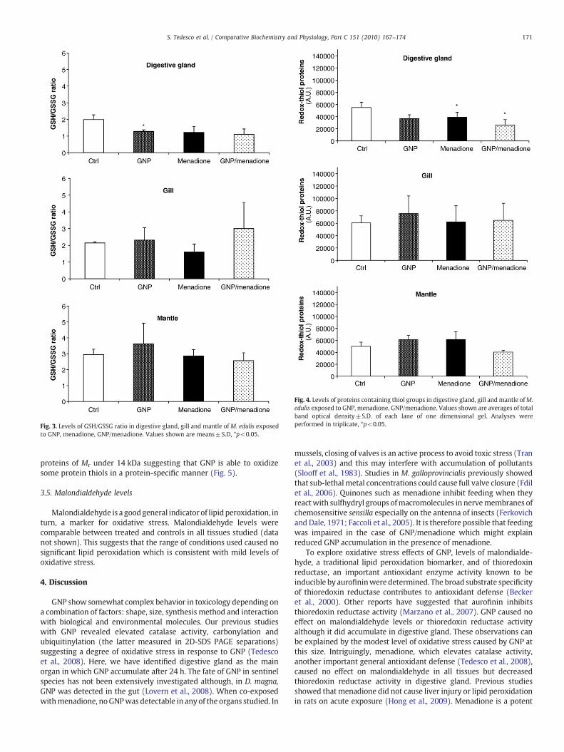
Fig. 3. Levels of GSH/GSSG ratio in digestive gland, gill and mantle of M. edulis exposedto GNP, menadione, GNP/menadione. Values shown are means±S.D, *p<0.05.
Fig. 4. Levels of proteins containing thiol groups in digestive gland, gill and mantle ofM.edulis exposed to GNP, menadione, GNP/menadione. Values shown are averages of totalband optical density±S.D. of each lane of one dimensional gel. Analyses wereperformed in triplicate, *p<0.05.
171S. Tedesco et al. / Comparative Biochemistry and Physiology, Part C 151 (2010) 167–174
proteins of Mr under 14 kDa suggesting that GNP is able to oxidizesome protein thiols in a protein-specific manner (Fig. 5).
3.5. Malondialdehyde levels
Malondialdehyde is a goodgeneral indicator of lipidperoxidation, inturn, a marker for oxidative stress. Malondialdehyde levels werecomparable between treated and controls in all tissues studied (datanot shown). This suggests that the range of conditions used caused nosignificant lipid peroxidation which is consistent with mild levels ofoxidative stress.
4. Discussion
GNP show somewhat complex behavior in toxicology depending ona combination of factors: shape, size, synthesis method and interactionwith biological and environmental molecules. Our previous studieswith GNP revealed elevated catalase activity, carbonylation andubiquitinylation (the latter measured in 2D-SDS PAGE separations)suggesting a degree of oxidative stress in response to GNP (Tedescoet al., 2008). Here, we have identified digestive gland as the mainorgan in which GNP accumulate after 24 h. The fate of GNP in sentinelspecies has not been extensively investigated although, in D. magna,GNP was detected in the gut (Lovern et al., 2008). When co-exposedwithmenadione, noGNPwas detectable in anyof the organs studied. In
mussels, closing of valves is an active process to avoid toxic stress (Tranet al., 2003) and this may interfere with accumulation of pollutants(Slooff et al., 1983). Studies in M. galloprovincialis previously showedthat sub-lethalmetal concentrations could cause full valve closure (Fdilet al., 2006). Quinones such as menadione inhibit feeding when theyreactwith sulfhydryl groups ofmacromolecules in nervemembranes ofchemosensitive sensilla especially on the antenna of insects (Ferkovichand Dale, 1971; Faccoli et al., 2005). It is therefore possible that feedingwas impaired in the case of GNP/menadione which might explainreduced GNP accumulation in the presence of menadione.
To explore oxidative stress effects of GNP, levels of malondialde-hyde, a traditional lipid peroxidation biomarker, and of thioredoxinreductase, an important antioxidant enzyme activity known to beinducible by aurofininwere determined. The broad substrate specificityof thioredoxin reductase contributes to antioxidant defense (Beckeret al., 2000). Other reports have suggested that aurofinin inhibitsthioredoxin reductase activity (Marzano et al., 2007). GNP caused noeffect on malondialdehyde levels or thioredoxin reductase activityalthough it did accumulate in digestive gland. These observations canbe explained by the modest level of oxidative stress caused by GNP atthis size. Intriguingly, menadione, which elevates catalase activity,another important general antioxidant defense (Tedesco et al., 2008),caused no effect on malondialdehyde in all tissues but decreasedthioredoxin reductase activity in digestive gland. Previous studiesshowed that menadione did not cause liver injury or lipid peroxidationin rats on acute exposure (Hong et al., 2009). Menadione is a potent
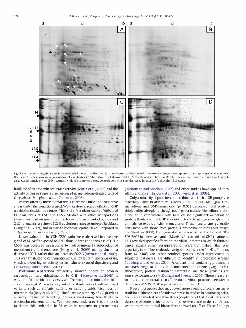
Fig. 5. Two dimensional gels of soluble 5′-IAF-labeled proteins in digestive gland. A) Control. B) GNP-treated. Fluorescence images were captured using Typhoon 9400 scanner (GEHealthcare). Gels shown are representative of 4 replicates. C) Silver stained gel shown in A). D) Silver stained gel shown in B). The black arrows show the control spots whichdisappeared completely on GNP treatment while white arrows denote control spots which are decreased in intensity (although still present).
172 S. Tedesco et al. / Comparative Biochemistry and Physiology, Part C 151 (2010) 167–174
inhibitor of thioredoxin reductase activity (Morin et al., 2008) and theactivity of this enzyme is also repressed in menadione-treated cells ofCorynebacterium glutamicum (Choi et al., 2008).
As measured by these biomarkers, GNP caused little or no oxidativestress under the conditions used. We therefore assessed effects of GNPon thiol antioxidant defences. This is the first observation of effects ofGNP on levels of GSH and GSSG. Studies with other nanoparticles(single wall carbon nanotubes, carbonaceous nanoparticles, SiO2 andZnOnanoparticles) showedGSHdepletion inmouse embryofibroblasts(Yang et al., 2009) and in human bronchial epithelial cells exposed toTiO2 nanoparticles (Park et al., 2008).
Lower values in the GSH/GSSG ratio were observed in digestivegland of M. edulis exposed to GNP alone. A transient decrease of GSH/GSSG was observed in response to hydroquinone (a subproduct ofmenadione) and menadione (Hong et al., 2009) mainly due to adecrease of GSH rather than an increase of GSSG (Amoroso et al., 2009).This was attributed to consumption of GSH by glutathione transferase,which showed higher activity in menadione-exposed digestive gland(McDonagh and Sheehan, 2006).
Proteomic separations previously showed effects on proteincarbonylation and ubiquitination by GNP (Tedesco et al., 2008). Itwas therefore decided to assess GNP effects on protein thiols. The thiol-specific reagent IAF reacts only with free thiols but not with oxidizedvariants such as sulfenic, sulfinic or sulfonic acids, disulfides ornitrosothiols (Baty et al., 2002). The fluorescein moiety of IAF providesa ready means of detecting proteins containing free thiols inelectrophoretic separations. We have previously used this approachto detect thiol oxidation in M. edulis in response to pro-oxidants
(McDonagh and Sheehan, 2007) and other studies have applied it toplants and mice (Hancock et al., 2005; Pérez et al., 2009).
Only a minority of proteins contain thiols and their−SH groups areespecially liable to oxidation (Davies, 2005). In 1DE, GNP (p=0.05)menadione and GNP/menadione (p<0.05) decreased total proteinthiols in digestive gland, thoughnot in gill ormantle.Menadione, eitheralone or in combination with GNP caused significant oxidation ofprotein thiols, even if GNP was not detectable in digestive gland inanimals co-exposed with menadione. These results are generallyconsistent with those from previous proteomic studies (McDonaghand Sheehan, 2008). This general effect was explored further with 2D-SDS PAGE in digestive gland ofM. edulis for control and GNP treatment.This revealed specific effects on individual proteins in which fluores-cence signals either disappeared or were diminished. This wasespecially true of lowmolecularweight proteins under 14 kDa. Proteinsfrom M. edulis and other sentinel species, under-represented insequence databases, are difficult to identify in proteomic screens(Dowling and Sheehan, 2006). Abundant thiol-containing proteins inthe mass range of < 14 kDa include metallothioneins (Kägi, 1993),thioredoxin, protein disulphide isomerase and these proteins aresensitive to stressors (McDonagh and Sheehan, 2007). These measure-ments underline the fact that effects on individual proteins are easier todetect in 2-D SDS PAGE separations rather than 1DE.
Proteomics approaches may reveal more specific effects than moretraditional biomarkers of oxidative stress in studies in sentinel species.GNP caused modest oxidative stress (depletion of GSH/GSSG ratio anddecrease of protein thiol groups) in digestive gland under conditionswhere more traditional biomarkers showed no effect. These findings

173S. Tedesco et al. / Comparative Biochemistry and Physiology, Part C 151 (2010) 167–174
support the view that low levels of nanoparticles can cause subtle sub-toxic effects in filter-feeding species (Peyrot et al., 2009) which couldpotentially impact on their long-term health and ability to cope withtraditional chemical pollutants. We are now developing this experi-mental system by probing effects of GNP of smaller particle diameter.
5. Conclusions
We have studied GNP effects in the sentinel, M. edulis. Theseparticles accumulate mainly in digestive gland, although low levelswere also measured by ICP-OES in the gill and mantle. Co-incubationwith menadione led to no accumulation of GNP which may be due toeffects of menadione on feeding. No effects were observed in levels ofmalondialdehyde or on the GSH/GSSG ratio. However, GNP diddecrease levels of protein thiols in both 1DE and 2D SDS PAGE,consistent with targeting of protein thiols by ROS under theconditions used. GNP is therefore a modest pro-oxidant which canaffect proteins and this may explain our previously-reported effectson protein carbonylation and ubiquitination on GNP exposure(Tedesco et al., 2008). Further studies based on longer exposure-times and/or smaller GNP diameters are necessary to explore theeffects of these nanoparticles further. This study underlines theadvantages of using filter-feeding organisms in future investigationsof environmental effects of manufactured nanomaterials.
Acknowledgements
Our laboratory is supported by the Programme for Research inThird Level Institutions of the Higher Education Authority of Ireland.ST is supported by an EMBARK fellowship of the Irish Research Councilfor Science Engineering and Technology.
References
Aerner, E., Zhong, L., Holmgren, A., 1999. Preparation and assay of mammalianthioredoxin and thioredoxin reductase. Methods Enzymol. 300, 226–239.
Amiard, J.C., Pineau, A., Boiteau, H.L., Metayer, C., Amiardriquet, C., 1987. Application ofatomic-absorption spectrophotometry using Zeeman effect to the determination of8 trace-elements (Ag, Cd, Cr, Cu, Mn, Ni, Pb and Se) in biological-materials. WaterRes. 21, 693–697.
Amoroso, A., Mancilla, R.A., González, B., Vicuña, R., 2009. Hydroquinone and H2O2
differentially affect the ultrastructure and expression of ligninolytic genes in thebasidiomycete Ceriporiopsis subvermispora. FEMS Microbiol. Lett. 294, 232–238.
Anguiano, G., Llera-Herrera, R., Rojas, E., Vazquez-Boucard, C., 2007. Subchronicorganismal toxicity, cytotoxicity, genotoxicity, and feeding response of Pacificoyster (Crassostrea gigas) to lindane (gamma-HCH) exposure under experimentalconditions. Environ. Toxicol. Chem. 26, 2192–2197.
Baty, J.W., Hampton, M.B., Winterbourn, C.C., 2002. Detection of oxidant sensitive thiolproteins by fluorescence labelling and two dimensional electrophoresis. Proteo-mics 2, 1261–1266.
Baumard, P., Budzinski, H., Garrigues, P., Dizer, H., Hansen, P.D., 1999. Polycyclicaromatic hydrocarbons in recent sediments and mussels (Mytilus edulis) from theWestern Baltic Sea: occurrence, bioavailability and seasonal variations. Mar.Environ. Res. 47, 17–47.
Baun, A., Hartmann, N.B., Grieger, K., Kusk, K.O., 2008. The ecotoxicology and chemistryof manufactured nanoparticles. Ecotoxicology 17, 387–395.
Becker, K., Gromer, S., Schirmer, R.H., Muller, S., 2000. Thioredoxin reductase as apathophysiological factor and drug target. Eur. J. Biochem. 267, 6118–6125.
Bradford, M.M., 1976. A rapid and sensitive method for the quantification of microgramquantities of protein, utilizing the principle of protein–dye binding. Ann. Rev.Biochem. 72, 248–254.
Cedervall, T., Lynch, I., Lindman, S., Berggard, T., Thulin, E., Nilsson, H., Dawson, K.A.,Linse, S., 2008. Understanding the nanoparticle-protein corona using methods toquantify exchange rates and affinities of proteins for nanoparticles. Proc. Natl. Acad.Sci. U. S. A. 104, 2050–2055.
Chen, Y., Cai, J., Jones, D.P., 2006. Mitochondrial thioredoxin in regulation of oxidant-induced cell death. FEBS Lett. 580, 6596–6602.
Choi, W.W., Park, S.D., Lee, S.M., Kim, H.B., Kim, Y., Lee, H.S., 2008. ThewhcA gene plays anegative role in oxidative stress response of Corynebacterium glutamicum. FEMSMicrobiol. Lett. 290, 32–38.
Davies, M.J., 2005. The oxidative environment and protein damage. Biochim. Biophys.Acta 1703, 93–109.
Donaldson, K., Seaton, A., 2007. The janus faces of nanoparticles. J. Nanosci. Nanotechnol.7, 4607–4611.
Dowling, V.A., Sheehan, D., 2006. Proteomics as a route to identification of toxicitytargets in ecotoxicology. Proteomics 6, 5597–5604.
Dowling, V., Hoarau, P., Romeo, M., O'Halloran, J.F., van Pelt, F.N.A.M., O'Brien, N.M.,Sheehan, D., 2006. Protein carbonylation and heat shock response in Ruditapesdecussatus following p,p′-dichlorodiphenyldichloroethylene (DDE) exposure: aproteomic approach reveals DDE causes oxidative stress. Aquat. Toxicol. 77, 11–18.
Eaton, P., 2006. Protein thiol in health and disease: techniques for measuring disulfidesand related modifications in complex protein mixtures. Free Radic. Biol Med 40,1889–1899.
Eisler, R., 2003. Chrysotherapy: a synoptic review. Inflamm. Res. 52, 487–501.Eisler, R., 2004. Mammalian sensitivity to elemental gold (Au degrees). Biol. Trace Elem.
Res. 100, 1–17.EWG. (Environmental Working Group), 2006. Comments to FDA on Nano-scale
Ingredients in Cosmetics. Accessed at website http:// www.ewg.org/node/21738.Faccoli, M., BlaŽenec, M., Schlyter, F., 2005. Feeding response to host and non-host
compounds by males and females of the spruce bark beetle Ips typographus in atunneling microassay. J. Chem. Ecol. 31, 745–759.
Fdil, M.A., Mouabad, A., Outzourhit, A., Benhra, A., Maarouf, A., Pihan, J.C., 2006. Valvemovement response of the mussel Mytillus galloprovincialis to metals (Cu, Hg, Cdand Zn) and phosphate industry effluents from Moroccan Atlantic coast.Ecotoxicology 15, 477–486.
Ferkovich, S.M., Dale, M.N., 1971. Naphthoquinone inhibitors of Periplaneta americanaand Scolytus multistriatus feeding: ultraviolet difference spectra of reactions ofjuglone, menadione and 1, 4-naphthoquinone with amino acids and the indicatedmechanism of feeding inhibition. Chem. Biol. Interact. 4, 23–30.
Goldberg, E.G., 1986. The mussel watch concept. Environ. Monit. Assess. 7, 91–103.Grabar, K.C., Freeman, R.G., Hommer, M.B., Natan, M.J., 1995. Preparation and
characterization of Au colloid monolayers. Anal. Chem. 67, 735–743.Halliwell, B., Gutteridge, J.M.C., 2007. Free Radicals in Biology and Medicine, 4th ed.
Oxford University Press, p. 704.Hancock, J.T., Henson, D., Nyirenda, M., Desikan, R., Harrison, J., Lewis, M., Hughes, J.,
Neil, S.J., 2005. Proteomic identification of glyceraldehyde 3-phosphate dehydro-genase as an inhibitory target of hydrogen peroxide in Arabidopsis. Plant Physiol.Biochem. 43, 828–835.
Handy, R.D., Valsami-Jones, E., Owen, R., 2008. The ecotoxicology of nanoparticles andnanomaterials: current status, knowledge gaps, challenges, and future needs.Ecotoxicology 17, 315–325.
Hansen, R.E., Roth, D., Winther, J.R., 2009. Quantifying the global cellular thiol-disulfidestatus. Proc. Natl. Acad. Sci. U. S. A. 106, 422–427.
Hissin, P.J., Hilf, R., 1976. A fluorometric method for determination of oxidized andreduced glutathione in tissues. Anal. Biochem. 74, 214–226.
Hong, J.-Y., Lebofsky, M., Farhood, A., Jaeschke, H., 2009. Oxidant stress-induced liverinjury in vivo: role of apoptosis, oncotic necrosis, and c-Jun NH2-terminal kinaseactivation. Am. J. Physiol. Gastrointest. Liver Physiol. 296, G572–581.
Jorgensen, C.B., 1996. Bivalve filter feeding revisited. MariEcol-Progr. Ser. 142, 287–302.Kägi, J.H.R., 1993. Evolution, structure and chemical activity of class I metallothionein:
an overview. In: Suzuki, K.T., Imura, N., Kimura, M. (Eds.), Metallothionein III.Birkhäuser Verlag, Basel/Switzerland, pp. 29–55.
Laemmli, U.K., 1970. Cleavage of structural proteins during the assembly of the head ofbacteriophage T4. Nature 227, 680–685.
Laurent, L., Petit, J.C., 2005. Nanosciences and its convergence with other technologies—new golden age or apocalypse? Hyle 11, 45–76.
Lim, I.I.S., Ip, W., Crew, E., Njoki, P.N., Mott, D., Zhong, C.J., Pan, Y., Zhou, S., 2007.Homocysteine-mediated reactivity and assembly of gold nanoparticles. Langmuir 23,826–833.
Lovern, S.B., Strickler, J.R., Klaper, R., 2007. Behavioral and physiological changes inDaphnia magna when exposed to nanoparticle suspensions (titanium dioxide,nano-C-60, and C(60)HxC(70)Hx). Environ. Sci. Technol. 41, 4465–4470.
Lovern, S.B., Owen, H.A., Klaper, R., 2008. Electron microscopy of gold nanoparticleintake in the gut of Daphnia magna. Nanotoxicology 2, 43–48.
Mannino, S., Scampicchio, M., 2007. Nanotechnology and food quality control. Vet. Res.Commun. 31, 149–151.
Marzano, C., Gandin, V., Folda, A., Scutari, G., Bindoli, A., Rigobello, M.P., 2007. Inhibitionof thioredoxin reductase by auranofin induces apoptosis in cisplatin-resistanthuman ovarian cancer cells. Free Radic. Biol. Med. 42, 872–881.
McDonagh, B., Sheehan, D., 2006. Redox proteomics in the blue mussel Mytilus edulis:carbonylation is not a pre-requisite for ubiquitination in acute free radical-mediated oxidative stress. Aquat. Toxicol. 79, 325–333.
McDonagh, B., Sheehan, D., 2007. Effects of oxidative stress on protein thiols in the bluemussel Mytilus edulis: proteomic identification of target proteins. Proteomics 7,3395–3403.
McDonagh, B., Sheehan, D., 2008. Effects of oxidative stress on protein thiols anddisulphides in Mytilus edulis revealed by proteomics: actin and protein disulphideisomerase are redox targets. Mar. Environ. Res. 66, 193–195.
Medley, C.D., Smith, J.E., Tang, Z., Wu, Y., Bamrungsap, S., Tan, W., 2008. Goldnanoparticle-based colorimetric assay for the direct detection of cancerous cells.Anal. Chem. 80, 1067–1072.
Meister, A., Anderson, M.E., 1983. Glutathionine. Annu. Rev. Biochem. 52, 711–760.Moore, M.N., 2006. Do nanoparticles present ecotoxicological risks for the health of the
aquatic environment? Environ. Int. 32, 967–976.Morin, C., Besset, T., Moutet, J.C., Fayolle, M., Brückner, M., Limosin, D., Becker, K.,
Davioud-Charvet, E., 2008. The aza-analogues of 1, 4-naphthoquinones are potentsubstrates and inhibitors of plasmodial thioredoxin and glutathione reductases andof human erythrocyte glutathione reductase. Org. Biomolec. Chem. 6, 2731–2742.
Nel, A., Xia, T., Madler, L., Li, N., 2006. Toxic potential of materials at the nanolevel.Science 311, 622–627.
Oberdörster, G., Stone, V., Donaldson, K., 2007. Toxicology of nanoparticles: a historicalperspective. Nanotoxicology 1, 2–25.

174 S. Tedesco et al. / Comparative Biochemistry and Physiology, Part C 151 (2010) 167–174
Omata, Y., Folan, M., Shaw, M., Messer, R.L., Lockwood, P.E., Hobbs, D., Bouillaguet, S.,Sano, H., Lewis, J.B., Wataha, J.C., 2006. Sublethal concentrations of diverse goldcompounds inhibit mammalian cytosolic thioredoxin reductase (TrxR1). Toxicol. InVitro 20, 882–890.
Panessa-Warren, B.J., Warren, J.B., Maye, M.M., Van der Lelie, D., Gang, O., Wong, S.S.,Ghebrehiwet, B., Tortora, G.T., Misewich, J.A., 2008. Human epithelial cellprocessing of carbon and gold nanoparticles. Int. J. Nanotechnol. 5, 55–91.
Park, E., Yi, J., Chung, K., Ryu, D., Choi, J., Park, K., 2008. Oxidative stress and apoptosis inducedby titanium dioxide nanoparticles in cultured BEAS-2B cells. Toxicol. Lett. 180, 222–229.
Peña-Llopis, S., Ferrando, M.D., Peña, J.B., 2002. Impaired glutathione redox status isassociated with decreased survival in two organophosphate-poisoned marinebivalves. Chemosphere 47, 485–497.
Pérez, V.I., Buffenstein, R., Masamsetti, V., Leonard, S., Salmon, A.B., Mele, J., Andziak, B.,Yang, T., Edrey, Y., Friguet, B., Ward, W., Richardson, A., Chaudhuri, A., 2009. Proteinstability and resistance to oxidative stress are determinants of longevity in thelongest-living rodent, thenakedmole-rat. Proc.Natl. Acad. Sci.U. S. A. 106,3059–3064.
Peyrot, C., Gagnon, C., Gagné, F., Willkinson, K.J., Turcotte, P., Sauvé, S., 2009. Effects ofcadmium telluride quantum dots on cadmium bioaccumulation and metallothio-nein production to the freshwater mussel, Elliptio complanata. Comp. Biochem.Physiol. C: Toxicol. Pharmacol. 150, 246–251.
Poland, C.A., Duffin, R., Kinloch, I., Maynard, A., Wallace, W.A.H., Seaton, A., Stone, V.,Brown, S., MacNee, W., Donaldson, K., 2008. Carbon nanotubes introduced into theabdominal cavity of mice show asbestos-like pathogenicity in a pilot study. Natl.Nanotechnol. 3, 423–428.
Rabilloud, T., 1992. A comparison between low background silver diammine and silvernitrate protein stains. Electrophoresis 13, 429–439.
Renault, S., Baudrimont, M., Mesmer-Dudons, N., Gonzalez, P., Mornet, S., Brisson, A.,2008. Impacts of gold nanoparticle exposure on two freshwater species: aphytoplanktonic alga (Scenedesmus subspicatus) and a benthic bivalve (Corbiculafluminea). Gold Bull. 41, 116–126.
Shaw, J.P., Large, A.T., Donkin, P., Evans, S.V., Staff, F.J., Livingstone, D.R., Chipman, J.K.,Peters, L.D., 2004. Seasonal variation in cytochrome P450 immunopositive proteinlevels, lipid peroxidation and genetic toxicity in digestive gland of the musselMytilus edulis. Aquat. Toxicol. 67, 325–336.
Slooff, W., de Zwart, D., Marquenie, J.M., 1983. Detection limits of a biologicalmonitoring system for chemical water pollution based on mussel activity. Bull.Environ. Contam. Toxicol. 30, 400–405.
Stoeger, T., Reinhard, C., Takenaka, S., Schroeppel, A., Karg, E., Ritter, B., Heyder, J.,Schulz, H., 2006. Instillation of six different ultrafine carbon particles indicates asurface area threshold dose for acute lung inflammation in mice. Environ. HealthPerspect. 114, 328–333.
Tedesco, S., Doyle, H., Redmond, D., Sheehan, D., 2008. Gold nanoparticles and oxidativestress in Mytilus edulis. Mar. Environ. Res. 66, 131–133.
Tran, D., Ciret, P., Ciutat, A., Durrieu, G., Massabuau, J.C., 2003. Estimation of potentialand limits of bivalve closure response to detect contaminants: application tocadmium. Environ. Toxicol. Chem. 22, 914–920.
Unfried, K., Albrecht, C., Klotz, L.O., Mikecz, A.V., Grether-Beck, S., Schins, R.P.F., 2007.Cellular responses to nanoparticles: target structures and mechanisms. Nanotox-icology 1, 52–71.
Yang, H., Yang, L.C., Zhang, D., Zhuge, H.X., 2009. Comparative study of cytotoxicity,oxidative stress and genotoxicity induced by four typical nanomaterials: the role ofparticle size, shape and composition. J. Appl. Toxicol. 29, 69–78.

![Becoming symbiotic - the symbiont acquisition and the ... · 09.10.2020 · Mytilus edulis [18]. The names of their late larval stages have been used interchangeably in the past](https://static.fdocuments.in/doc/165x107/605e5acec20a2c154c4f8c88/becoming-symbiotic-the-symbiont-acquisition-and-the-09102020-mytilus.jpg)