EXPLORATION OF POTENTIAL BIOACTIVE COMPOUNDS OF … · testing of active compounds produced by gall...
14
EXPLORATION OF POTENTIAL BIOACTIVE COMPOUNDS OF ENDOPHYTIC MICROBIAL CULTURE ISOLATED FROM GALL RUST SENGON (FALCATARIA MOLUCCANA) Barneby & J.W Grimes) Noor Rahmawati * , Sopandi Sunarya, Alfi Rumidatul SITH ITB Jl Ganesha No 10 Bandung corresponding author: [email protected]. ac.id Abstract Aim: The objective of this research was to investigate the endophytic fungi isolated from gall rust sengon that produced some antimicrobial and antioxidant compounds. Methods: Thirty-four endophytic fungus were isolated from gall rust sengon, screened and evaluated their ability to produce antimicrobial and antioxidant compounds. Results: Antimicrobial activities were found in at least one or more pathogenic microbial (bacteria and fungi) tested. The inhibitions of Bacillus subtilis were detected at 5-46 mm and 24-82 mm for methanol and ethyl acetate extracts subsequently. While, the inhibitions of Pseudomonas aeruginosa were 10-63 mm for methanol extract; 39-103 mm for ethyl acetate extract and 10-65 mm for n-hexane extract. Antifungal activity against fusarium sp was showed by the inhibition growth of 14%, 60% and 4.5% for methanol, ethyl acetate and n-hexane extracts, respectively. While, these activity against Schlerotium sp was showed by the growth inhibition of 18.6%, 8.9% and 20.3% for methanol, ethyl acetate and n-hexane extracts, respectively. Antioxidant activity of methanol and ethyl acetate extracts of fungal culture showed the (DPPH) radical inhibition activity as 44.92% for methanol and 71.48% for ethyl acetate extract. Meanwhile, n-hexane extract showed only 28.69% of inhibition for DPPH radicals. Conclusion: Endophytic fungi isolated from gall rust sengon were potential for producing antimicrobial and antioxidant compounds from its culture. Keywords: Antimicrobial, antioxidant, endophyte, falcataria moluccana, fungi, gall rust INTRODUCTION The rust fungi Uromycladium Tepperianum often attacks sengon and produces gall rust in the attacked plant. These fungi are mostly obligate parasites which requires a living host to grow [1,2], secretes hormones that stimulate excessive cell formation which causes abnormal growth like a tumor [3], and attacked from seeds, young plants to stands in the field. Tumor rust disease in sengon showed typical symptoms, namely hyperplasia (more growth) in the affected part of the plant. Symptoms of the disease is started by formation of local swelling (tumefaksi) in the affected part of the plant, and eventually, turns into lumps which then becomes small nodules or called gall. Galls that arises from the infected plant has varying shapes ranging from round to irregular with diameters range from a few millimetres to greater than 10 cm. The gall can be a group or spread to the affected part [4]. Gall in sengon plants is suspected to have endophytic microbes as a form of defence of singing plants against Uromycladium Tepperianum attack. The secondary metabolites produced by endophytic microbes have potential to stimulate plant growth or provides defence against plant pathogens, also become active compounds whose activity and benefits are similar to those of their host plants, such as antioxidant and antimicrobial compounds. The results of several study prove that extraction of endophytic fungal culture produces bioactive compounds with potential use for drugs, agriculture, and modern industry. Noor Rahmawati et al ISPST 2018 ISSN: 0975-1459 JPSR-November(S)2018 156
Transcript of EXPLORATION OF POTENTIAL BIOACTIVE COMPOUNDS OF … · testing of active compounds produced by gall...
EXPLORATION OF POTENTIAL BIOACTIVE COMPOUNDS OF ENDOPHYTIC MICROBIAL CULTURE ISOLATED FROM GALL
RUST SENGON (FALCATARIA MOLUCCANA) Barneby & J.W Grimes)
Noor Rahmawati*, Sopandi Sunarya, Alfi Rumidatul
SITH ITB Jl Ganesha No 10 Bandung corresponding author: [email protected]. ac.id
Abstract Aim: The objective of this research was to investigate the endophytic fungi isolated from gall rust sengon that produced some antimicrobial and antioxidant compounds.Methods: Thirty-four endophytic fungus were isolated from gall rust sengon, screened and evaluated their ability to produce antimicrobial and antioxidant compounds. Results: Antimicrobial activities were found in at least one or more pathogenic microbial (bacteria and fungi) tested. The inhibitions of Bacillus subtilis were detected at 5-46 mm and 24-82 mm for methanol and ethyl acetate extracts subsequently. While, the inhibitions of Pseudomonas aeruginosa were 10-63 mm for methanol extract; 39-103 mm for ethyl acetate extract and 10-65 mm for n-hexane extract. Antifungal activity against fusarium sp was showed by the inhibition growth of 14%, 60% and 4.5% for methanol, ethyl acetate and n-hexane extracts, respectively. While, these activity against Schlerotium sp was showed by the growth inhibition of 18.6%, 8.9% and 20.3% for methanol, ethyl acetate and n-hexane extracts, respectively. Antioxidant activity of methanol and ethyl acetate extractsof fungal culture showed the (DPPH) radical inhibition activity as 44.92% for methanoland 71.48% for ethyl acetate extract. Meanwhile, n-hexane extract showed only 28.69% ofinhibition for DPPH radicals.Conclusion: Endophytic fungi isolated from gall rust sengon were potential for producingantimicrobial and antioxidant compounds from its culture.Keywords: Antimicrobial, antioxidant, endophyte, falcataria moluccana, fungi, gall rust
INTRODUCTION
The rust fungi Uromycladium Tepperianum often attacks sengon and produces gall rust in the attacked plant. These fungi are mostly obligate parasites which requires a living host to grow [1,2], secretes hormones that stimulate excessive cell formation which causes abnormal growth like a tumor [3], and attacked from seeds, young plants to stands in the field. Tumor rust disease in sengon showed typical symptoms, namely hyperplasia (more growth) in the affected part of the plant. Symptoms of the disease is started by formation of local swelling (tumefaksi) in the affected part of the plant, and eventually, turns into lumps which then becomes small nodules or called gall. Galls that arises from the infected plant has varying shapes ranging from round to irregular with diameters range from a few millimetres to greater than 10 cm. The gall can be a group or spread to the affected part [4]. Gall in sengon plants is suspected to have endophytic microbes as a form of defence of singing plants against Uromycladium Tepperianum attack. The secondary metabolites produced by endophytic microbes have potential to stimulate plant growth or provides defence against plant pathogens, also become active compounds whose activity and benefits are similar to those of their host plants, such as antioxidant and antimicrobial compounds. The results of several study prove that extraction of endophytic fungal culture produces bioactive compounds with potential use for drugs, agriculture, and modern industry.
Noor Rahmawati et al ISPST 2018 ISSN: 0975-1459
JPSR-November(S)2018 156
Antibiotic, antimitotic, immunosuppressant and anti-cancer compounds are some examples of compounds that have been extracted from culture of endophytic fungal broth with some organic solvent [5]. Endophytic microbes are microbes that live in plant tissues without causing disease symptoms in plants and can be isolated from the tissues. Endophytic microbes can live in parts of plants such as roots, stems, leaves, and fruit. Plant tissues colonized by endophytic microbe provide nutrients for endophytic fungi and on the contrary, the endophytic fungi serve protection for their host plants [6]. Each plant contains several endophytic microbes that are capable in producing secondary metabolites which are similar to their host plants that are thought to be due to coevolution or genetic transfer (genetic recombination) from their host plants into endophytic microbes [7]. The ability of endophytic microbes to produce secondary metabolites in accordance with their host plants is a very large opportunity and can be relied upon to produce secondary metabolites from endophytic microbes isolated from the host plant. Of the approximately 300,000 types of plants scattered on this earth, each plant contains one or more endophytic microbes consisting of bacteria and fungi [5]. Various types of endophytes have been isolated from their host plants, and successfully cultured in suitable medium. Likewise, secondary metabolites produced by endophytic microbes have been isolated and purified and their molecular structures have been elucidated. Some of them are: (1) Endophytic microbes that produce broad-spectrum antibiotics called munumbicin, which are produced by endophytic Streptomyces spp. NRRL 30562 strain isolated from Kennedia nigriscans can inhibit the growth of Bacillus anthracis and multiresistant Mycobacterium tuberculosis against various anti-tuberculosis drugs [8]. Other endophytes that also produced broad-spectrum antibiotics are endophytic microbes isolated from the Grevillea pteridifolia produced kakadumycin which has antibacterial activity same as munumbicin D, and also efficacious as anti-malaria [9]. (2) Endophytes that produced antioxidants Pestacin and isopestacin, which are secondary metabolites produced by Pestalotiopsis microspora endophytes. Endophytes were successfully isolated from Terminalia morobensis, which grows in Papua New Guinea. Both pestacin and isopestacin are efficacious as antioxidants, where this activity was thought to be due to its molecular structure similar to flavonoids [10]. Schulz [11] states that almost 80% of endophytic fungi produced metabolites that function as antibacterial compounds, natural fungicides and herbicides. Pestacin and isopestacin are secondary metabolites produced by endophytic fungi P microspora isolated from Terminalia morobensis, which grows in Papua New Guinea and uses as an antioxidant [5]. Endophytic fungi produces metabolites which function as antibiotics (antifungal/antibacterial), antiviral, anticancer, antidiabetic, antimalarial, antioxidant, antiimunosupressive [5] growth regulating substances [7] and hydrolytic enzymes such as amylase, cellulose, xylanase, ligninase, chitinase [12]. Chemotherapeutic compounds, called antimicrobial compounds used to eradicate microorganisms that cause infection in humans, animals and plants must be selective in their toxicity. It means that they must be toxic to microorganisms but relatively non-toxic to the host body [13]. Antibacterial and antifungal are included in antimicrobial. Antibacterial is a substance that inhibited bacterial growth and used generally to treat the infections. Antibacterial are classified into two groups, namely bactericidal (kills bacterial cells but do not cause lysis/rupture the cells) and bacteriostatic (inhibits the bacterial growth but do not kill them) [14]. Antifungal compounds are used for the treatment of infectious diseases caused by fungi). Antifungal has two meanings, namely fungicidal (kills the fungi) and fungi static (inhibits the fungi growth without killing them). Antimicrobial activity is influenced by
Noor Rahmawati et al ISPST 2018 ISSN: 0975-1459
JPSR-November(S)2018 157
environmental factors, such as concentration or intensity of antimicrobial substances, temperature, number and species of microorganisms, the presence of organic matter and pH. Based on the mechanism of action, antimicrobial is divided into five groups [15], namely: (1) Antimicrobial that interferes the microbial cell metabolism, for example sulphonamides, p-amino salicylic acids (PAS) and sulfone; (2) Antimicrobial that inhibits microbial cell membrane synthesis, for example penicillin, cephalosporin, bacitracin, vancomycin and cycloserine; (3) Antimicrobials that interferes the microbial cell membrane permeability, for example polymyxin; (4) Antimicrobial that inhibits the microbial cell protein synthesis, for example tetracycline and chloramphenicol; (5) Antimicrobial that inhibits the synthesis or affects the microbial cell nucleic acids, for example rifampicin. The world development of health and medicine are starting to look for and utilize the natural ingredients. The study of the resources of bioactive compounds was continuously carried out along with the increasing number of new diseases, ranging from infectious diseases caused by microorganisms, cancer, and several other degenerative diseases. Bioactive compounds can be obtained from several sources, including plants, animals and microorganisms. The new paradigm in the world of modern medicine has a tendency to return to the concept of back to nature, namely to utilize the natural ingredients optimally in order to maintain health and treatment [16]. Until now, there is no publication found regarding to the isolation and the testing of active compounds produced by gall rust tumor in sengon plants. Studies on endophytic microbes from gall rust tumor in sengon are required to determine the potential metabolites produced by endophytic fungi, especially to be developed as an alternative for new antimicrobial and antioxidant compounds. Many studies had been reported on the potential of endophytic fungi in producing bioactive compounds from various plants. Furthermore, there have never been reported the presence of endophytic microbes from gall rust tumors in sengon which are attacked by Uromycladium tepperianum fungi especially for the bioactive metabolite. Therefore, it is necessary to conduct the research including isolation and identification of endophytic microbes in order to explore the potential of their metabolites in producing bioactive compounds for the antimicrobial and antioxidant activities. MATERIALS AND METHODS Materials The study was conducted in the microbiology laboratory of the ITB Bioscience and Biotechnology Research Center, and held from January - October 2018. The materials used in this study was a gall rust tumor from the sengon plant obtained from ITB campus, Jatinangor. Other materials used were Nutrient Agar (NA), Nutrient Broth (NB), Potato Dextro Agar (PDA), Potato Dextrose Broth (PDB), gram-positive bacteria (Bacillus sp.), gram-negative bacteria (Pseudomonas sp.), phytopathogenic fungi of Fusarium sp and Schloerotium sp. Organic solvents used in this research were n-hexane, ethyl acetate and methanol. DPPH was used for antioxidant activity test. Isolation of Endophytic Fungi Endophytic fungi were isolated from gall rust tumor from sengon plant. Gall was cut approximately 1 cm long and rinsed with distilled water for surface sterilization and then sequentially washed with 70% of ethanol (1 minute), 5.25% of sodium hypochlorite (15 minutes) and rinsed again with 70% of ethanol for 1 minute followed by rinsing with sterile aquadest for three times. Gall was cut in the middle and then placed on PDA medium which had been added with antibiotics (200 µg of chloramphenicol) and then incubated at 28°±1°C (room temperature) for about 1-3 weeks. The mycelium derived from the sample specimens was purified and cultured under the same conditions [17].
Noor Rahmawati et al ISPST 2018 ISSN: 0975-1459
JPSR-November(S)2018 158
Fermentation and Extraction of Endophytic Fungi The fungal endophytes were cultivated on PDB by placing 4 agar blocks of pure culture (10 mm in diameter) of actively growing culture in 250 mL of Erlenmeyer flasks containing 100 mL of the medium. The flasks were incubated in static condition for 1 weeks at 27±1°C. The culture was filtered through filter paper to remove the mycelial mats. The liquid broth was collected and extracted with equal volume of n-hexane, ethyl acetate and methanol in a separating funnel by vigorous shaking for one hour. The cell mass was separated and weight to obtain weight of mycelium. The solvent was evaporated and the resultant compound was dried with MgSO4 and concentrated to yield the crude extracts. The crude extracts were then dissolved in methanol for antimicrobial bioassay and antioxidant test [17].
Antioxidants Activity Test (2,2-Diphenyl-1-pikrilhidrazil (DPPH) Scavenging Tests) DPPH radical scavenging test was carried out as described by Miliauskas et al. (2004) with some modifications. The samples of extracts (300 µL) at different concentrations was added to 250 µL DPPH (1 mM). After reacting for 30 minutes, the absorbance of samples was read at 517 nm. The DPPH radical scavenging was calculated using the following equation: DPPH radical scavenging (%) = [(A1 - A2) / A1] x 100 where A1 is the absorbance of the control (containing all the reagents except sample extract), and A2 is the absorbance of reagent and the sample extract [17].
Antimicrobial bioassay N-hexane, ethyl acetate and methanol extracts from endophytic fungal culture wereindividually tested against panels of disease-causing microorganisms including gram-positivebacteria (Bacillus subtillis) and gram-negative bacteria (Pseudomonas sp). Tests were alsocarried out on phytopathogenic fungi (Fusarium sp and Schlerotium sp). The inhibitory effectof extracts obtained from endophytic fungal culture on phytopagenic bacteria and fungi wastested by paper disc method. Disc paper (10 mm in diameter) was dipped in a solution ofendophytic microbial culture extract with a concentration of 10 mg / mL (dry residue /volumeof methanol), then dried and placed on PDA medium which had been applied to the fungi orNA medium for bacteria to be tested. The amount of inhibitory activity of endophytic fungiculture extracts against pathogenic microbes was assessed by measuring the diameter (in mm)of the zone of inhibition relative to positive and negative controls. Chloramphenicol was usedas a positive control in treatment with pathogenic bacteria, while for negative controlsmethanol was used. For treatment with pathogenic fungi, antifungal drugs were used aspositive control and methanol as a negative control. Four-disc paper replications were usedon each petri disk. Petri disks that had been inoculated with fungi and bacteria were incubatedat 27° C for 24 hours for clinical bacterial strains, and 96 hours for filament fungi.Antimicrobial activity was evaluated by measuring the clear zone of the test organism. Eachtest in this experiment was repeated three times.
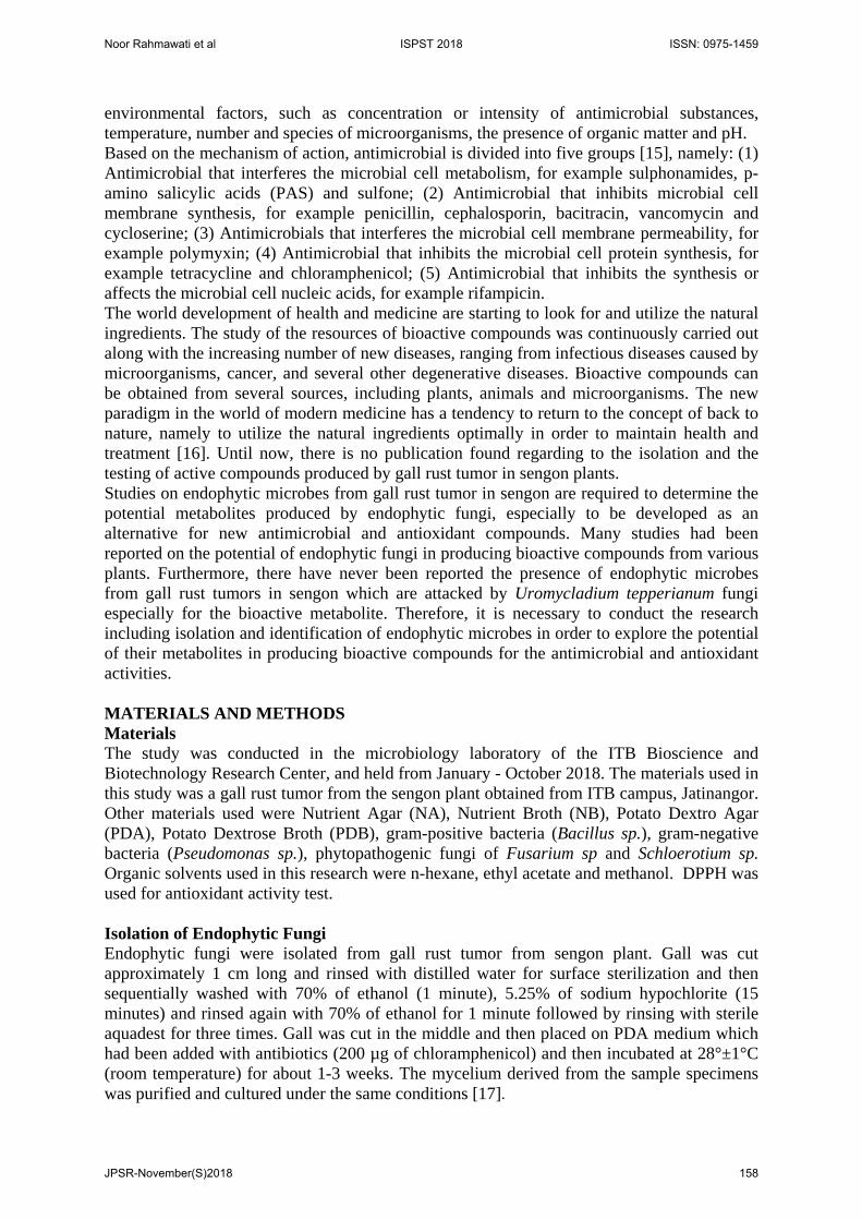
RESULTS AND DISCUSSION In this study, 34 of endophytic fungi were successfully isolated from gall rust sengon. These isolates obtained from gall rust sengon were vary based on macroscopic performance like mycelium, color of pigmented media and color of mycelium. Some of endophytic fungal pictures were displayed at Figure 1 below. The molecular identification of that fungi will be held in the next experiment. Endophytic fungi are microorganisms living inside plants and are considered a promising source of novel and natural biologically active compounds [5]. Endophytic fungi could be found in every plant, because no studies had shown the presence of plant species without endophytes. High species diversity is another
Noor Rahmawati et al ISPST 2018 ISSN: 0975-1459
JPSR-November(S)2018 159
characteristic of endophytic microbes. The large number of isolates of endophytic fungi that can be isolated from gall rust sengon reinforce the results of [5] studies suggesting that all plants found are harbors of some endophytic fungi. All endophytic fungi have many different functions for plants as a consequence of mutualistic symbiosis between endophytic fungi and plants. The abundance of the composition of the endophytic fungi community was strongly influenced by environmental factors (temperature and humidity), chemical variation, anatomy and maturity of the colonized host organ. How the endophytic fungus could enter the host plant without causing the disease symptoms, could be explained by Sieber [18], who explained that the initial step of endophytic fungi went into the host plant was through recognition, germination and penetration. A similar process was also experienced by infections of plant pathogenic fungi. Along the path of the process, endophytic fungi must overcome the defence mechanism of the plant. Spores of fungi often recognized host plants through molecules such as lectins. After germination, the fungus would penetrate into the plant tissue by softening the cuticle and epidermal cell wall or damaging the cuticle with mechanical strength. Once the fungus could penetrate into the plant tissue, and it would change to a latent state, the host's defence mechanisms were no longer activated. This phenomenon was explained by the Gene-for-Gene (GFG) model, in which the avirulence genes (AVR) of the endophytes were encoded into an elicitor and recognized by the product of the resistance gene (R) of the host plant as well as the hypersensitive reaction of the host plant and then the serenity the next occurs through the signal transduction path. Instead, in pathogenic fungi did not contain the AVR gene, thus product R was not produced and symptoms of the disease would develop [18]. The interaction of endophytic fungi with host plants resulted in a compromise between mutualism and antagonism to create a harmonious symbiotic system. Plants could limit the growth of endophytes, and thus endophytes could use various mechanisms to survive. Endophytes not only described some plant metabolites with ecto enzyme to take important nutrients and energy to survive, but also produce beneficial compounds and /or support or promote the growth of host plants to achieve a balanced environment [18].
Figure 1: Endophytic fungi isolated from gall rust sengon
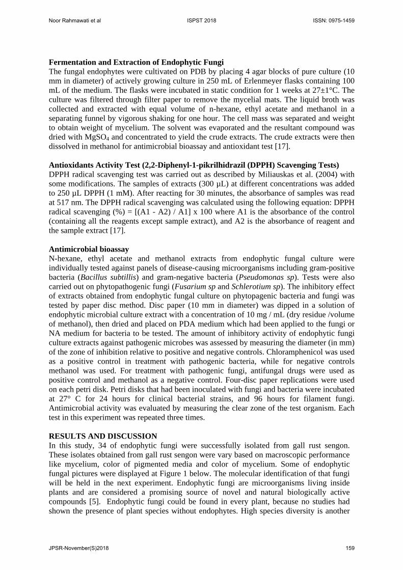
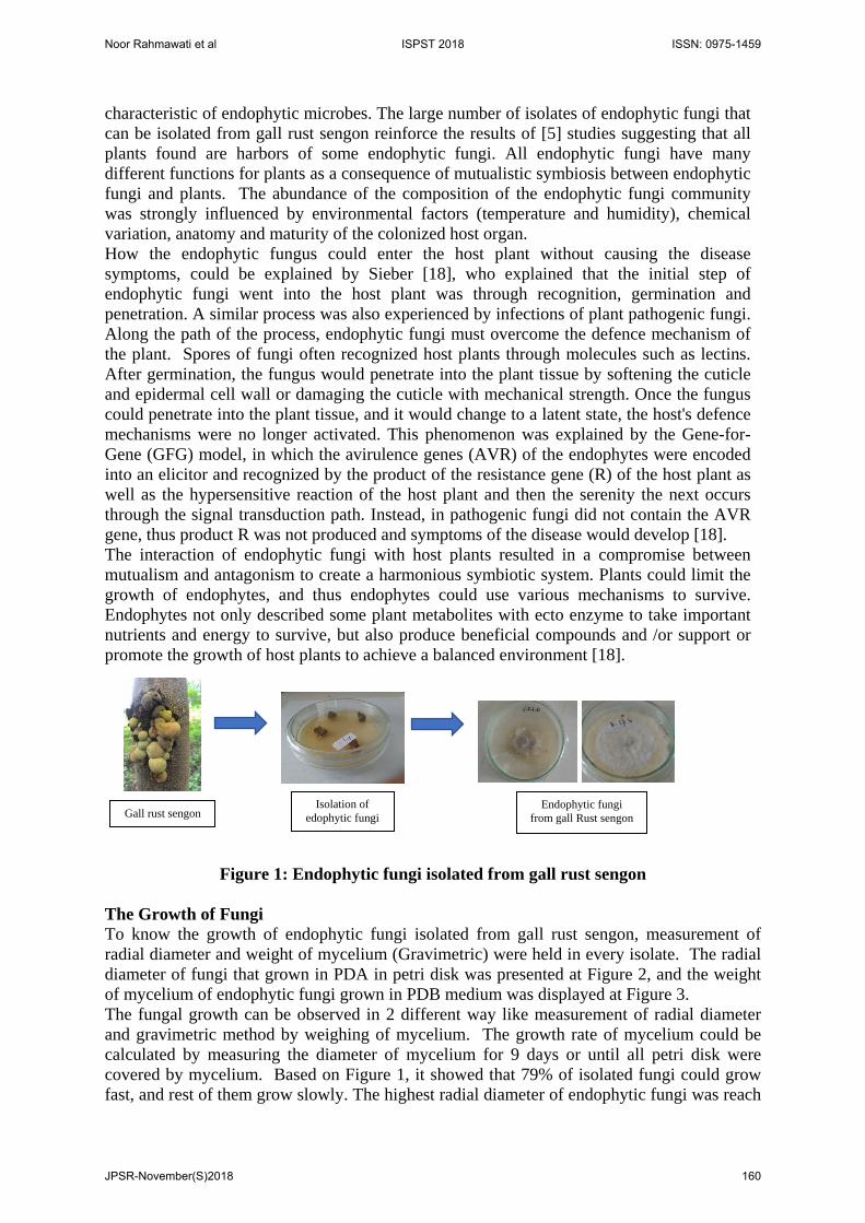
The Growth of Fungi To know the growth of endophytic fungi isolated from gall rust sengon, measurement of radial diameter and weight of mycelium (Gravimetric) were held in every isolate. The radial diameter of fungi that grown in PDA in petri disk was presented at Figure 2, and the weight of mycelium of endophytic fungi grown in PDB medium was displayed at Figure 3. The fungal growth can be observed in 2 different way like measurement of radial diameter and gravimetric method by weighing of mycelium. The growth rate of mycelium could be calculated by measuring the diameter of mycelium for 9 days or until all petri disk were covered by mycelium. Based on Figure 1, it showed that 79% of isolated fungi could grow fast, and rest of them grow slowly. The highest radial diameter of endophytic fungi was reach
Gall rust sengon Isolation of
edophytic fungi Endophytic fungi
from gall Rust sengon
Noor Rahmawati et al ISPST 2018 ISSN: 0975-1459
JPSR-November(S)2018 160
at 8.7 cm for 9 days growing for isolate B3.3.d1. The other fast growth fungi were reached at diameter 8.4 cm for isolate B11b and B12c (Figure 2).
Figure 2: The radial diameter of endophytic fungi from gall rust sengon
Figure 3: The weight of mycelium endophytic fungi cultured in PDB medium
The isolated of endophytic fungi were also grown on PDB medium (pH 5.5), temperature 28 ± 2°C, static condition for 7 days to determine mycelium growth. Dry weight of mycelium was used as a parameter of endophytic fungal growth. The growth of the mycelium of endophytic fungi during 7 days fermentation could be seen in Figure 3. The weight of mycelium of endophytic fungi of gall rust on PDB medium reached average 1.5 g of dry weight, although 2 isolates of them B3.2b and B33(d1) reached almost 2 times (0.32 g). This data showed that the endophytic fungi isolated from gall rust could grow well in the PDB medium, so hopefully they also produced secondary metabolite well. Production of Antioxidant Compounds from Endophytic Fungi Culture The production of secondary metabolites in fungi can be affected by medium composition, pH, temperature, agitation and lighting [19]. Factors affecting secondary metabolite production in fungi are geographical factors, dehydration, lighting, drought, growth medium, nutrient availability, pH, genes expression and carbon sources. According to Deduke, et al (2012), Environmental factors that affect secondary metabolite production such as aflatoxin production in aspergillus fungi are pH, carbon and nitrogen source [20].
0
5
10
H0 H3 H4 H5 H6 H7 H8 H9
Dia
met
er (c
m)
B11a B1.1b B1,1 C B1.2a B1,2.B B1.2C
B1.2D B1.3 A B1.3 b(1) B1.3b(2) B1.3.c B1.3d
B2.1 B2.3a B2,3 b (1) B 2.3 b (2) B3.1A(1) B.3.1.A(2)
B3.1b B3.1c1 B3.1.C2(1) B3.1 C2(2) B3.1 d B3.2a
B 3.2.B B3,2 c B3.2d B3.3a(1) B3.3 a (2) B3.3 B (1)
B3.3B(2) B33C B3,3 D1(1) B3.3D1(2)
0
0.05
0.1
0.15
0.2
0.25
0.3
0.35
Biom
ass(
g)
Noor Rahmawati et al ISPST 2018 ISSN: 0975-1459
JPSR-November(S)2018 161
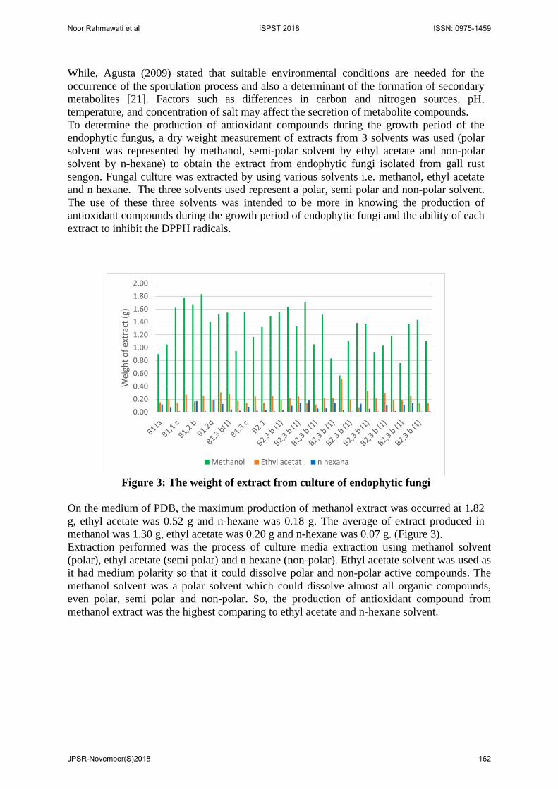
While, Agusta (2009) stated that suitable environmental conditions are needed for the occurrence of the sporulation process and also a determinant of the formation of secondary metabolites [21]. Factors such as differences in carbon and nitrogen sources, pH, temperature, and concentration of salt may affect the secretion of metabolite compounds. To determine the production of antioxidant compounds during the growth period of the endophytic fungus, a dry weight measurement of extracts from 3 solvents was used (polar solvent was represented by methanol, semi-polar solvent by ethyl acetate and non-polar solvent by n-hexane) to obtain the extract from endophytic fungi isolated from gall rust sengon. Fungal culture was extracted by using various solvents i.e. methanol, ethyl acetate and n hexane. The three solvents used represent a polar, semi polar and non-polar solvent. The use of these three solvents was intended to be more in knowing the production of antioxidant compounds during the growth period of endophytic fungi and the ability of each extract to inhibit the DPPH radicals.
Figure 3: The weight of extract from culture of endophytic fungi
On the medium of PDB, the maximum production of methanol extract was occurred at 1.82 g, ethyl acetate was 0.52 g and n-hexane was 0.18 g. The average of extract produced in methanol was 1.30 g, ethyl acetate was 0.20 g and n-hexane was 0.07 g. (Figure 3). Extraction performed was the process of culture media extraction using methanol solvent (polar), ethyl acetate (semi polar) and n hexane (non-polar). Ethyl acetate solvent was used as it had medium polarity so that it could dissolve polar and non-polar active compounds. The methanol solvent was a polar solvent which could dissolve almost all organic compounds, even polar, semi polar and non-polar. So, the production of antioxidant compound from methanol extract was the highest comparing to ethyl acetate and n-hexane solvent.
0.000.200.400.600.801.001.201.401.601.802.00
Wei
ght o
f ext
ract
(g)
Methanol Ethyl acetat n hexana
Noor Rahmawati et al ISPST 2018 ISSN: 0975-1459
JPSR-November(S)2018 162
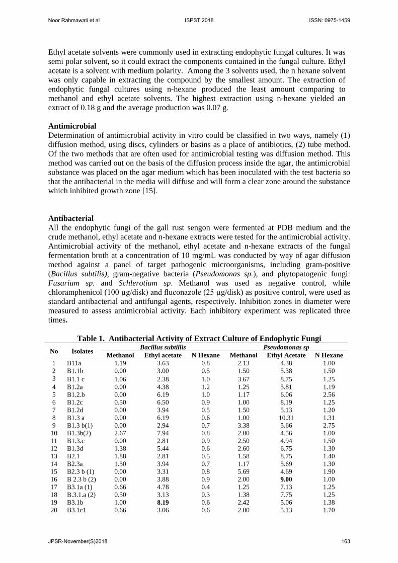
Ethyl acetate solvents were commonly used in extracting endophytic fungal cultures. It was semi polar solvent, so it could extract the components contained in the fungal culture. Ethyl acetate is a solvent with medium polarity. Among the 3 solvents used, the n hexane solvent was only capable in extracting the compound by the smallest amount. The extraction of endophytic fungal cultures using n-hexane produced the least amount comparing to methanol and ethyl acetate solvents. The highest extraction using n-hexane yielded an extract of 0.18 g and the average production was 0.07 g. Antimicrobial Determination of antimicrobial activity in vitro could be classified in two ways, namely (1) diffusion method, using discs, cylinders or basins as a place of antibiotics, (2) tube method. Of the two methods that are often used for antimicrobial testing was diffusion method. This method was carried out on the basis of the diffusion process inside the agar, the antimicrobial substance was placed on the agar medium which has been inoculated with the test bacteria so that the antibacterial in the media will diffuse and will form a clear zone around the substance which inhibited growth zone [15]. Antibacterial All the endophytic fungi of the gall rust sengon were fermented at PDB medium and the crude methanol, ethyl acetate and n-hexane extracts were tested for the antimicrobial activity. Antimicrobial activity of the methanol, ethyl acetate and n-hexane extracts of the fungal fermentation broth at a concentration of 10 mg/mL was conducted by way of agar diffusion method against a panel of target pathogenic microorganisms, including gram-positive (Bacillus subtilis), gram-negative bacteria (Pseudomonas sp.), and phytopatogenic fungi: Fusarium sp. and Schlerotium sp. Methanol was used as negative control, while chloramphenicol (100 µg/disk) and fluconazole (25 µg/disk) as positive control, were used as standard antibacterial and antifungal agents, respectively. Inhibition zones in diameter were measured to assess antimicrobial activity. Each inhibitory experiment was replicated three times.
Table 1. Antibacterial Activity of Extract Culture of Endophytic Fungi No Isolates Bacillus subtillis Pseudomonas sp
Methanol Ethyl acetate N Hexane Methanol Ethyl Acetate N Hexane 1 B11a 1.19 3.63 0.8 2.13 4.38 1.00 2 B1.1b 0.00 3.00 0.5 1.50 5.38 1.50 3 B1.1 c 1.06 2.38 1.0 3.67 8.75 1.25 4 B1.2a 0.00 4.38 1.2 1.25 5.81 1.19 5 B1.2.b 0.00 6.19 1.0 1.17 6.06 2.56 6 B1.2c 0.50 6.50 0.9 1.00 8.19 1.25 7 B1.2d 0.00 3.94 0.5 1.50 5.13 1.20 8 B1.3 a 0.00 6.19 0.6 1.00 10.31 1.31 9 B1.3 b(1) 0.00 2.94 0.7 3.38 5.66 2.75
10 B1.3b(2) 2.67 7.94 0.8 2.00 4.56 1.00 11 B1.3.c 0.00 2.81 0.9 2.50 4.94 1.50 12 B1.3d 1.38 5.44 0.6 2.60 6.75 1.30 13 B2.1 1.88 2.81 0.5 1.58 8.75 1.40 14 B2.3a 1.50 3.94 0.7 1.17 5.69 1.30 15 B2.3 b (1) 0.00 3.31 0.8 5.69 4.69 1.90 16 B 2.3 b (2) 0.00 3.88 0.9 2.00 9.00 1.00 17 B3.1a (1) 0.66 4.78 0.4 1.25 7.13 1.25 18 B.3.1.a (2) 0.50 3.13 0.3 1.38 7.75 1.25 19 B3.1b 1.00 8.19 0.6 2.42 5.06 1.38 20 B3.1c1 0.66 3.06 0.6 2.00 5.13 1.70
Noor Rahmawati et al ISPST 2018 ISSN: 0975-1459
JPSR-November(S)2018 163
21 B3.1.c2(1) 4.58 3.13 0.8 1.00 6.00 1.17 22 B3.1 c2(2) 3.83 3.94 0.7 1.00 7.00 2.00 23 B3.1 d 0.00 4.00 0.5 3.25 6.13 2.25 24 B3.2a 0.00 3.25 0.6 1.25 7.19 2.20 25 B 3.2.b 1.50 7.50 0.5 6.25 5.06 6.50 26 B3.2 c 4.38 5.44 0.4 2.88 4.56 2.20 27 B3.2d 2.08 4.50 0.7 1.69 3.91 2.00 28 B3.3a(1) 0.96 3.69 0.5 1.33 8.69 2.13 29 B3.3 a (2) 2.31 7.31 0.8 3.83 4.63 2.42 30 B3.3 b (1) 0.50 5.69 0.4 1.08 6.56 1.25 31 B3.3b(2) 1.25 8.19 0.6 5.00 7.00 2.00 32 B33c 1.81 6.31 0.4 3.58 5.81 1.00 33 B3.3 d1(1) 1.17 2.69 0.7 5.25 4.28 1.25 34 B3.3d1(2) 1.81 3.06 0.5 1.75 8.00 2.17
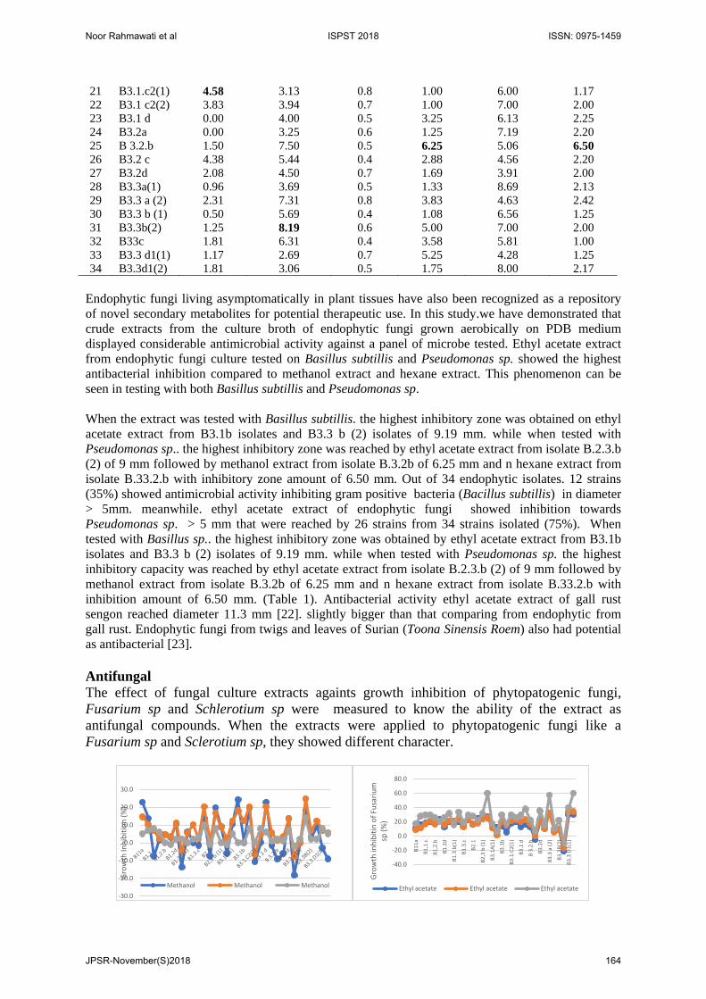
Endophytic fungi living asymptomatically in plant tissues have also been recognized as a repository of novel secondary metabolites for potential therapeutic use. In this study.we have demonstrated that crude extracts from the culture broth of endophytic fungi grown aerobically on PDB medium displayed considerable antimicrobial activity against a panel of microbe tested. Ethyl acetate extract from endophytic fungi culture tested on Basillus subtillis and Pseudomonas sp. showed the highest antibacterial inhibition compared to methanol extract and hexane extract. This phenomenon can be seen in testing with both Basillus subtillis and Pseudomonas sp. When the extract was tested with Basillus subtillis. the highest inhibitory zone was obtained on ethyl acetate extract from B3.1b isolates and B3.3 b (2) isolates of 9.19 mm. while when tested with Pseudomonas sp.. the highest inhibitory zone was reached by ethyl acetate extract from isolate B.2.3.b (2) of 9 mm followed by methanol extract from isolate B.3.2b of 6.25 mm and n hexane extract from isolate B.33.2.b with inhibitory zone amount of 6.50 mm. Out of 34 endophytic isolates. 12 strains (35%) showed antimicrobial activity inhibiting gram positive bacteria (Bacillus subtillis) in diameter > 5mm. meanwhile. ethyl acetate extract of endophytic fungi showed inhibition towards Pseudomonas sp. > 5 mm that were reached by 26 strains from 34 strains isolated (75%). When tested with Basillus sp.. the highest inhibitory zone was obtained by ethyl acetate extract from B3.1b isolates and B3.3 b (2) isolates of 9.19 mm. while when tested with Pseudomonas sp. the highest inhibitory capacity was reached by ethyl acetate extract from isolate B.2.3.b (2) of 9 mm followed by methanol extract from isolate B.3.2b of 6.25 mm and n hexane extract from isolate B.33.2.b with inhibition amount of 6.50 mm. (Table 1). Antibacterial activity ethyl acetate extract of gall rust sengon reached diameter 11.3 mm [22]. slightly bigger than that comparing from endophytic from gall rust. Endophytic fungi from twigs and leaves of Surian (Toona Sinensis Roem) also had potential as antibacterial [23]. Antifungal The effect of fungal culture extracts againts growth inhibition of phytopatogenic fungi, Fusarium sp and Schlerotium sp were measured to know the ability of the extract as antifungal compounds. When the extracts were applied to phytopatogenic fungi like a Fusarium sp and Sclerotium sp, they showed different character.
-30.0
-20.0
-10.0
0.0
10.0
20.0
30.0
Gro
wth
Inhi
bitio
n (%
)
Methanol Methanol Methanol
-40.0
-20.0
0.0
20.0
40.0
60.0
80.0
B11a
B1,1
c
B1,2
.b
B1.2
d
B1.3
b(1
)
B1.3
.c
B2.1
B2,3
b (1
)
B3.1
A(1)
B3.1
b
B3.1
.C2(
1)
B3.1
d
B 3.
2.b
B3.2
d
B3.3
a (2
)
B3.3
B(2)
B3,3
D1(
1)
Gro
wth
inhi
bitin
of F
usar
ium
sp
(%)
Ethyl acetate Ethyl acetate Ethyl acetate
Noor Rahmawati et al ISPST 2018 ISSN: 0975-1459
JPSR-November(S)2018 164
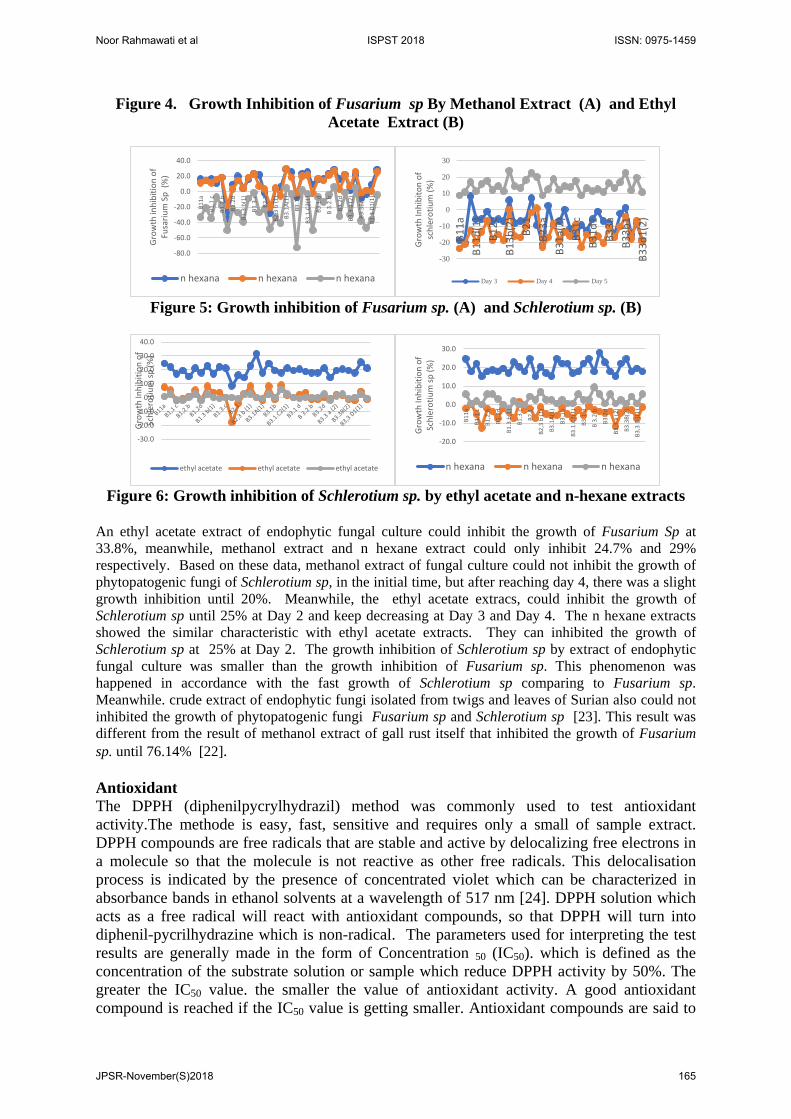
Figure 4. Growth Inhibition of Fusarium sp By Methanol Extract (A) and Ethyl Acetate Extract (B)
Figure 5: Growth inhibition of Fusarium sp. (A) and Schlerotium sp. (B)
Figure 6: Growth inhibition of Schlerotium sp. by ethyl acetate and n-hexane extracts
An ethyl acetate extract of endophytic fungal culture could inhibit the growth of Fusarium Sp at 33.8%, meanwhile, methanol extract and n hexane extract could only inhibit 24.7% and 29% respectively. Based on these data, methanol extract of fungal culture could not inhibit the growth of phytopatogenic fungi of Schlerotium sp, in the initial time, but after reaching day 4, there was a slight growth inhibition until 20%. Meanwhile, the ethyl acetate extracs, could inhibit the growth of Schlerotium sp until 25% at Day 2 and keep decreasing at Day 3 and Day 4. The n hexane extracts showed the similar characteristic with ethyl acetate extracts. They can inhibited the growth of Schlerotium sp at 25% at Day 2. The growth inhibition of Schlerotium sp by extract of endophytic fungal culture was smaller than the growth inhibition of Fusarium sp. This phenomenon was happened in accordance with the fast growth of Schlerotium sp comparing to Fusarium sp. Meanwhile. crude extract of endophytic fungi isolated from twigs and leaves of Surian also could not inhibited the growth of phytopatogenic fungi Fusarium sp and Schlerotium sp [23]. This result was different from the result of methanol extract of gall rust itself that inhibited the growth of Fusarium sp. until 76.14% [22]. Antioxidant The DPPH (diphenilpycrylhydrazil) method was commonly used to test antioxidant activity.The methode is easy, fast, sensitive and requires only a small of sample extract. DPPH compounds are free radicals that are stable and active by delocalizing free electrons in a molecule so that the molecule is not reactive as other free radicals. This delocalisation process is indicated by the presence of concentrated violet which can be characterized in absorbance bands in ethanol solvents at a wavelength of 517 nm [24]. DPPH solution which acts as a free radical will react with antioxidant compounds, so that DPPH will turn into diphenil-pycrilhydrazine which is non-radical. The parameters used for interpreting the test results are generally made in the form of Concentration 50 (IC50). which is defined as the concentration of the substrate solution or sample which reduce DPPH activity by 50%. The greater the IC50 value. the smaller the value of antioxidant activity. A good antioxidant compound is reached if the IC50 value is getting smaller. Antioxidant compounds are said to
-80.0
-60.0
-40.0
-20.0
0.0
20.0
40.0
B11a
B1,1
c
B1,2
.b
B1.2
d
B1.3
b(1
)
B1.3
.c
B2.1
B2,3
b (1
)
B3.1
A(1)
B3.1
b
B3.1
.C2(
1)
B3.1
d
B 3.
2.b
B3.2
d
B3.3
a (2
)
B3.3
B(2)
B3,3
D1(
1)
Gro
wth
inhi
bitio
n of
Fu
sariu
m S
p (%
)
n hexana n hexana n hexana
-30
-20
-10
0
10
20
30
B11a
B11d
(1)
B12c
B13b
(2)
B21
B23a
B31a
(2)
B31c
B31d
1B3
3aB3
3b1
B33d
1(2)
Gro
wth
Inhi
bito
n of
sc
hler
otiu
m (%
)
Day 3 Day 4 Day 5
-30.0
-20.0
-10.0
0.0
10.0
20.0
30.0
40.0
Gro
wth
Inhi
bitio
n of
Sc
hler
otiu
m sp
(%)
ethyl acetate ethyl acetate ethyl acetate
-20.0
-10.0
0.0
10.0
20.0
30.0
B11a
B1,1
c
B1,2
.b
B1.2
d
B1.3
b(1
)
B1.3
.c
B2.1
B2,3
b (1
)
B3.1
A(1)
B3.1
b
B3.1
.C2(
1)
B3.1
d
B 3.
2.b
B3.2
d
B3.3
a (2
)
B3.3
B(2)
B3,3
D1(
1)
Gro
wth
Inhi
bitio
n of
Sc
hler
otiu
m sp
(%)
n hexana n hexana n hexana
Noor Rahmawati et al ISPST 2018 ISSN: 0975-1459
JPSR-November(S)2018 165
be very strong if they have IC50 values of less than 0.05 mg / ml. strong for IC50 between 0.05-0.10 mg / ml. moderate for IC50 between 0.10-0.15 mg / ml and weak if IC50 is worth between 0.150-0.20 mg /ml [24].
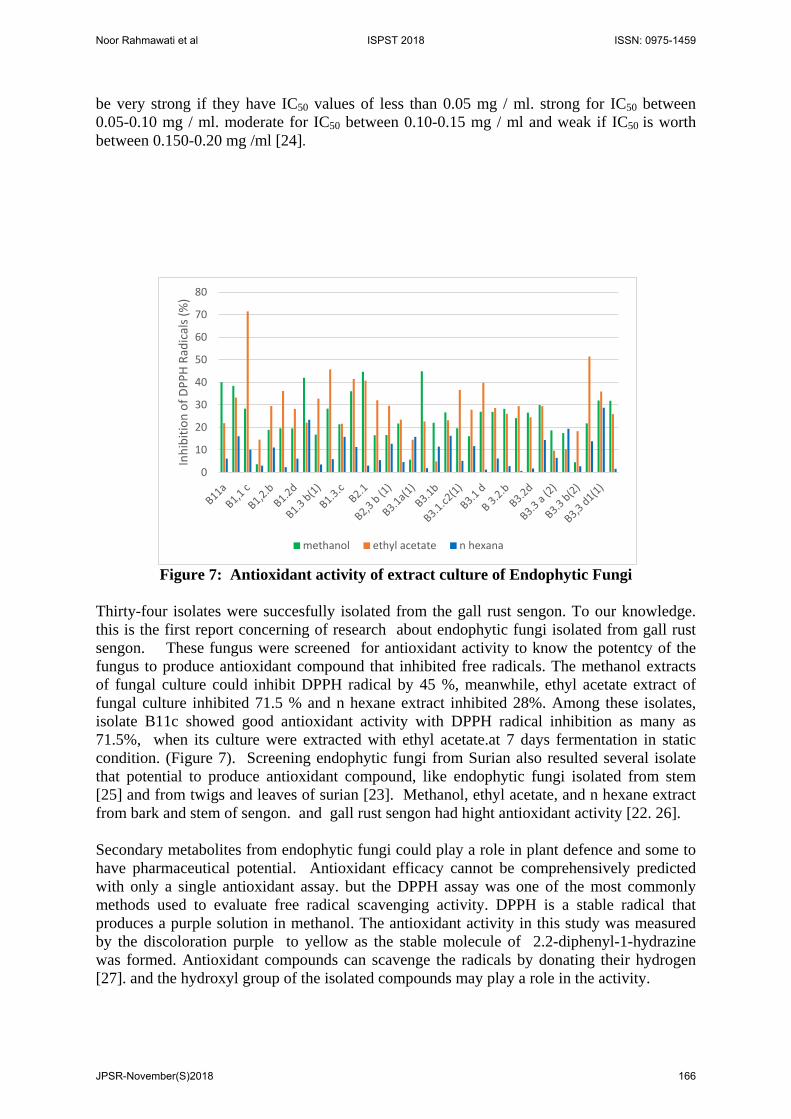
Figure 7: Antioxidant activity of extract culture of Endophytic Fungi
Thirty-four isolates were succesfully isolated from the gall rust sengon. To our knowledge. this is the first report concerning of research about endophytic fungi isolated from gall rust sengon. These fungus were screened for antioxidant activity to know the potentcy of the fungus to produce antioxidant compound that inhibited free radicals. The methanol extracts of fungal culture could inhibit DPPH radical by 45 %, meanwhile, ethyl acetate extract of fungal culture inhibited 71.5 % and n hexane extract inhibited 28%. Among these isolates, isolate B11c showed good antioxidant activity with DPPH radical inhibition as many as 71.5%, when its culture were extracted with ethyl acetate.at 7 days fermentation in static condition. (Figure 7). Screening endophytic fungi from Surian also resulted several isolate that potential to produce antioxidant compound, like endophytic fungi isolated from stem [25] and from twigs and leaves of surian [23]. Methanol, ethyl acetate, and n hexane extract from bark and stem of sengon. and gall rust sengon had hight antioxidant activity [22. 26]. Secondary metabolites from endophytic fungi could play a role in plant defence and some to have pharmaceutical potential. Antioxidant efficacy cannot be comprehensively predicted with only a single antioxidant assay. but the DPPH assay was one of the most commonly methods used to evaluate free radical scavenging activity. DPPH is a stable radical that produces a purple solution in methanol. The antioxidant activity in this study was measured by the discoloration purple to yellow as the stable molecule of 2.2-diphenyl-1-hydrazine was formed. Antioxidant compounds can scavenge the radicals by donating their hydrogen [27]. and the hydroxyl group of the isolated compounds may play a role in the activity.
0
10
20
30
40
50
60
70
80
Inhi
bitio
n of
DPP
H Ra
dica
ls (%
)
methanol ethyl acetate n hexana
Noor Rahmawati et al ISPST 2018 ISSN: 0975-1459
JPSR-November(S)2018 166
In the DPPH radical scavenging assay, antioxidants compounds donate an electron or hydrogen radical to the DPPH radical to become a stable molecule causing discoloration of the DPPH radical. [28] Endophytic fungi from E. sylvestris. Pseudocercospora sp. ESL 02 showed the highest antioxidant activity with the antioxidant compound Terreic acid (1) and 6-methylsalicylic acid (2). with terreic acid having strong antioxidant activity. This study also complements the study of the antioxidant potency of E. sylvestris as the host plant of the fungus [29]. Ethyl acetate extract of endophytic fungal culture Aspergilus. niger and Alternaria. alternate isolated from different organs of Tabebuia argentea showed an antioxidant capacity of 4.29-5.28 umol/L and total phenol of 2.5-2.6 mg/100 mL cultures equivalent to the highest phenolic acid of endophytic fungal cultures [30]. Crude extracts of endophytic fungi alternaria alternate. also showed antioxidant activity. Analysis of 292 endophytic fungi isolated from 29 traditional Chinese herbs showed that the antioxidant capacity of endophytic fungal cultures was significantly correlated with the total phenol [31]. In addition to the extensive research on bioactive compounds from plants, research on the biodiversity of fungal endophytes had also received much attention. Endophytic fungi may also produce other general secondary metabolites. such as altersolanol derivative produced by Nigrospora oryzae isolated from the leaves of Combretum dolichopetalum [32]. and adenosine. which exhibited potential 1.1-diphenyl-2-picrylhydrazyl (DPPH) radical scavenging activities. was isolated from Penicillium sp. YY-20, an endophytic fungus isolated from Ginkgo biloba [33] In the present study. the endophytic fungi from E. sylvestris were isolated and evaluated for their antioxidant potency. Furthermore. secondary metabolites having antioxidant activity were isolated and identified from the fungus that exhibited the highest antioxidant potency. This study can complement the research on the antioxidant potency of E. sylvestris as the host plant of the endophytes[29]. CONCLUSION As many as 34 isolates of endophytic fungus were succesfully isolatated from gall rust sengon. Every isolate was distinguished by differences of macroscopic morphological by looking at the color of mycelium colony and the change of medium color caused by the pigment. Screening of antimicrobial activity against gram positif bacteria (Basillus subtillis); gram negatif bacteria (Pseudomonas sp) and 2 phytopatogenic fungi ( Fusarium sp and Schlerotium sp) resulted several isolate that potential on producing antimicrobial compounds. Crude extract of endophytic fungal culture could inhibited DPPH radical. So that, it could be potential for source of natural antioxidant, and ethyl acetate was the best solvent that extracted bioactive compounds from endophytic fungal culture.
ACKNOWLEDGEMENTS The authors would like to acknowledge The Ministry of Research. Technology and High Education for the research Grant of Number: 127/SP2H/PTNBH/DRPM/2018. REFERENCES [1] Triharso. Dasar-Dasar Perlindungan Tanaman. Gadjah Mada University Press.
Yogyakarta 2004. [2] Rahayu, S; Lee, S.S; Nor, A. S. Uromycladium tepperianum, The gall rust fungus from
Falcataria moluccana in Malaysia and Indonesia. J Mycos. 2010, 51, 2, 149 – 153. [3] Rahayu, S. Penyakit Karat Tumor pada Sengon. Makalah Workshop Penanggulangan
Serangan Karat Puru pada Tanaman Sengon 19 November 2008. Balai Besar Penelitian Bioteknologi dan Pemuliaan Tanaman Hutan. Yogyakarta. Indonesia. 2008.
Noor Rahmawati et al ISPST 2018 ISSN: 0975-1459
JPSR-November(S)2018 167

[4] Anggraeni, I. dan E, Santoso. Penyakit Karat Puru pada Sengon (Paraserianthes falcataria) di Pulau Seram. Buletin Penelitian Hutan. Puslitbang Hutan dan Konservasi Alam, Bogor. 2003, 636.
[5] Strobel, G. A. & Deasy. Endophytes As Sources Of Bioactive Products. Microbes and Infection. 2003, 5, 535-544.
[6] Bacon, C.W; Hinton, D.M. Bacterial edophytes. The endophytic niche, its occupants. and its utility. Gnanamanickam S. S. (editor).in Plant-Associated Bacteria. Netherland (NL), Springer. 2007, 155-195.
[7] Tan R, X, and W, X, Zou. Endophytes: A Rich Source of Functional Metabolites. Nat. Prod. Rep. 200,18, 448–459.
[8] Castillo. U.F; G.A. Strobel; E.J. Ford; W.M Hess; H. Poter; J.B. Jenson; H. Albert; R. Robinson; M.A. Condron; D.B. Teplow; D. Stevens, and D. Yaver. Munumbicins. wide spectrum antibiotics produced by Streptomyces NRRL 30562. Endophytic on Kennedia nigriscans. Microbiology. 2002, 148, 2675-2685.
[9] Castillo, U.J; K. Harper; G.A. Strobel; J. Sears; K. Alesi; E. Ford; J. Lin; M. Hunter; M. Maranta; H. Ge; D. Yaver; J.B. Jensen; H. Porter; R. Robinson; D. Millar; W.M. Hess; M. Condron, and D. Teplow. Kakandumycins. Novel Antibiotics from Streptomyces sp. NRRL 30566. An endophyte of Grevillea pteridifolia. FEMS Lett. 2003), 24, 183-190.
[10] Strobel, G, A; E.Dirkse; J.Sears, and C. Markworth. Volatile Antimicrobials From Muscodor Albus. A Novel Endophytic Fungus The ‘Mycofumigation’ Effects Of M. Albus. Microbiology.2001, 147, 2943–2950.
[11] Schulz, Barbara ; Christine, Boyle; Siegfried, Draeger; . Anne-Katrin; R,O, Mmert; and Karsten, Krohn. Review Endophytic fungi: a source of novel biologically active secondary metabolites. Mycol. Res. 2002,106, 9, 996-1004.
[12] Zinniel, D,K; Lambrecht, P; Harris, B.N; Feng, Z; Kuczmarski, D; Higley, P; Ishimaru, C,A; Arunakumari, A; Barletta, R,G; Vidaver, A, K. Isolation and characterization of endophytic colonizing bacteria from agronomic crops and prairie plants. Applied and Environmental Microbiology. 2002, 68, 5, 2198-2208.
[13] Xia, E; Deng, G; Guo, Y; & Li,H. Biological activities of polyphenols from Grapes. International Journal of Molecular Sciences. 2010, 11, 622 – 646.
[14] Dzen dan Sjoekoer, M. Bakteriologik medik. Malang. Bayumedia. 2003. [15] Jawetz; Melnick; Adelberg; Brooks; Butel. And Ornston. Mikrobiologi kedokteran.
EGC. Jakarta. 2005, Edisi ke-20. [16] Mangaan. Cara Bijak Menaklukan Kanker. Agromedia Pustaka. Jakarta.2003. [17] Zeng, P,Y; J, G, Wu; L, M, Liao; T,Q, Chen; J, Z, Wu; and K, H, Wong; In Vitro
Antioxidant Activities Of Endophytic Fungi Isolated From The Liverwort Scapania verrucos. Genetics and Molecular Research. 2011, 10, 4, 3169-3179.DOI http://dx.doi.org/10.4238/2011.December.20.1
[18] Sieber,T,N. Endophytic fungi in forest trees, are they mutualistis? Fungal Biologi Reviews. 2007. 21, 75-89.
[19] Venugopalan, A; & S, Srivastava. Research review . Endophytes as in vitro production platforms of high value plant secondary metabolites. Biotechnology Advances. 2015, 33, 873–887.
[20] Deduke, C; B,Timsina; and M, D, Pierce-Normore. Effect of Environmental Change on Secondary Metabolite Production in Lichen Forming Fungi. Chapter from the book internasional Perspective on Global Environmental Change. 2012. Downloaded from http//www.intechopen.com/books/international-perspective-on. globa;-environmental-change.
[21] Agusta, A. Biologi dan Kimia Jamur Endofit. Penerbit. ITB P. 2009, 11-27.
Noor Rahmawati et al ISPST 2018 ISSN: 0975-1459
JPSR-November(S)2018 168
[22] Rumidatul, A. Potensi medik metabolit tanaman sengon (Falcataria moluccana (miq)Barneby & J.W. Grimes) yang terserang penyakit karat tumor. Disertasi Program Doktor.Institut Teknologi Bandung. 2018.
[23] Rahmawati, N; D, I, Astuti; & P, Aditiawati. Aktivitas antimikroba dan Antioksidanekstrak kultur Fungi Endofit Dari Daun dan Ranting Toona sinensis . Prosiding seminarNasional Biologi 3 Universitas Islam Negeri Sunan Gunung Djati Bandung. 2018. ISBN978-602-582-302-2 -48-56
[24] Molyneux, P. The use of stable free radical Diphenylpicrylhydrazyl (DPPH) forestimating antioksidan activity. Journal Science Technology. 2004, 26, 2, 211 -219.
[25] Rahmawati, N; A.R. Isfandito; D.I.Astuti; and P, Aditiawati. Endophytic fungi fromsurian (Toona sinensis Roem) and antioxidant potency from its culture. Asian J. PlantSci. 2016, 15, 8-15.
[26] Rumidatul, A; Sulistyawati, E; Aryantha, I.N.P. Potensi ekstrak ranting sengon(falcataria moluccana (miq) sebagai sumber antioksidan alami. Prosiding seminarNasional Biologi 3 Universitas Islam Negeri Sunan Gunung Djati Bandung. ISBN. 2018.978-602, 582-302, 2, 97-108.
[27] El-Haci, I. A; Bekkara, F.A; Mazari, W; Gherib, M. Phenolics content and antioxidantactivity of some organic extracts of endemic medicinal plant Anabasis aretioides Coss. &Moq. from Algerian Sahara. Pharmacon J. 2013, 5, 108-112.
[28] Kedare, S.B; Singh, R.P. Genesis and development of DPPH method of antioxidantassay. J Food Sci Technol. 2011, 48, 412-22.
[29] Prihantini, A.I; & S, Tachibana. Antioxidant compounds produced by Pseudocercosporasp. ESL 02. an endophytic fungus isolated from Elaeocarpus sylvestris. Asian Pac JTrop Biomed. 2017; 7, 2, 110–115.
[30] Sadananda, T. S; Nirupama, R; Chaithra, K; Govindappa, M; Chandrappa, C, P; and .Vinay Raghavendra. Antimicrobial And Antioxidant Activities Of Endophytes FromTabebuia Argentea And Identification Of Anticancer Agent (Lapachol). Journal ofMedicinal Plants Research Vol. (2011) : 5(16). pp. 3643-3652.
[31] Huang, W.Y; Yr-Zhong Cai; Jie Xing; H. Corke; and Mei Sun. A Potential AntioxidantResource : Endophytic Fungi From Medicinal Plants. Economic Botany. The new yorkbotanical Garden Press. Bronk. NY10458-5126.USA. 2007, 61, 1, 14-30.
[32] Uzor, P.F; Ebrahim,W; Osadebe, P.O; Nwodo, J.N; Okoye, F.B; Muller, W. E.Metabolites from ¨Combretum dolichopetalum and its associated endophytic fungusNigrospora oryzae – evidence for a metabolic partnership. Fitoterapia. 2015, 105, 147-50.
[33] Yuan, Y; Tian, J.M; Xiao, J; Shao, Q; Gao, J.M. Bioactive metabolites isolated fromPenicillium sp. YY-20. the endophytic fungus from Ginkgo biloba. Nat Prod Res. 2014,28, 278-81.
Noor Rahmawati et al ISPST 2018 ISSN: 0975-1459
JPSR-November(S)2018 169