Mixed cutaneous round cells tumorin a cock ( Gallus ...
7
Iranian Journal of Veterinary Medicine IJVM (2012), 6(2):123-128 123 Mixed cutaneous round cells tumor in a cock (Gallus domesticus): A case report Dezfoulian, O. 1 , Razmyar, J. 2 , Peighambari, S.M. 3* , Rajabi, Z. 4 , Sadeghi, M.R. 5 , Jahanzad, I. 6 1 Department of Pathobiology, School of Veterinary Medicine, Lorestan University, Khorramabad, Iran. 2 Department of Clinical Sciences, Faculty of Veterinary Medicine, Ferdowsi Univerisity of Mashhad, Mashhad, Iran. 3 Department of Clinical Sciences, Faculty of Veterinary Medicine, Univerisity of Tehran, Tehran, Iran. 4 Department of Clinical Sciences, Faculty of Veterinary Medicine, Univerisity of Tabriz, Tabriz, Iran. 5 Junior School of Veterinary Medicine, BU-Ali Sina University, Hamedan, Iran. 6 Department of Pathology, Faculty of Medicine, Tehran University of Medical Sciences, Tehran, Iran. Case history Cutaneous round cell tumors have been classified as mast cell tumor (MCT), histiocytoma (HCT), lymphosarcoma, undifferentiated round cell tumors and occasionally rhabdomyosarcoma (Fernandez- Bellon et al., 2003; Fernandez et al., 2005) in veterinary medicine. Lymphosarcoma has been subdivided into two groups as T-cell lymphosarcoma (TCLA) and B- cell lymphosarcoma (BCLA). The plasmacytoma (PCT) is also known a subgroup of the BCLA. However, the definite diagnosis of similar histo- pathologic morphology without application of dif- ferential diagnostic tests like histochemical staining and immunohistochemistry, especially in poorly differentiated tumors, is almost impossible. Most of the antibodies now utilized in immunohisto- chemistry of animal samples have been originat-ed from human sources but have shown reliable and valuable results in differential diagnosis of animal round skin tumors (Copie-Bergman et al., 1998; Fernandez et al., 2005). Key words: B-cell lymphoma, histiocytoma, cu- taneous tumor, cock, round cell tumor. Correspondence Peighambari, S.M. Department of Clinical Sciences Faculty of Veterinary Medicine University of Tehran Tehran, Iran. Tel: +98(21) 61117150 Fax: +98(21) 66933222 Email: [email protected] Received: 19 December 2011 Accepted: 1 February 2012 Abstract: Cutaneous round cell tumors have been classified as mast cell tumor (MCT), histiocytoma (HCT), lymphosarcoma, undif- ferentiated round cell tumors and occasionally rhabdomyosarcoma in veterinary medicine. An adult cock (Gallus domesticus) showing a large solitary integument mass raised on dorso bilateral of cervical part, extending to intercapsular and cranial mid part of the back was referred to the birds clinic of the faculty of veterinary medicine at the university of Tehran. Microscopic examination revealed she- eted cells with large, round to oval nuclei with each one containing one or more prominent nucleoli with scant cytoplasm. The myofibrils of the neck were degenerated by aggressive tumor cells. The condition was differentiated from other round cell tumors by electron microscope, histochemical staining, as well as the application of a large panel of antibodies. Polymerase chain re- action failed to confirm the involvement of both Marek's disease virus and avian leukosis virus subgroup-J. It was concluded that the tumor cells were consistent with both B-cell lymphocytes and histiocytes that unusually covered the entire dermal layer of dorsal neck skin. This unusual cutaneous lymphoma was named as lymphoblastic histiocytoma.
Transcript of Mixed cutaneous round cells tumorin a cock ( Gallus ...

Iranian Journal of Veterinary Medicine
IJVM (2012), 6(2):123-128 123
Mixed cutaneous round cells tumor in a cock (Gallus domesticus):Acase report Dezfoulian, O.1, Razmyar, J.2, Peighambari, S.M.3*, Rajabi, Z.4, Sadeghi, M.R.5, Jahanzad, I.6
1Department of Pathobiology, School of Veterinary Medicine, Lorestan University, Khorramabad, Iran.2Department of Clinical Sciences, Faculty of Veterinary Medicine, Ferdowsi Univerisity of Mashhad, Mashhad,Iran.3Department of Clinical Sciences, Faculty of Veterinary Medicine, Univerisity of Tehran, Tehran, Iran.4Department of Clinical Sciences, Faculty of Veterinary Medicine, Univerisity of Tabriz, Tabriz, Iran.5Junior School of Veterinary Medicine, BU-Ali Sina University, Hamedan, Iran.6Department of Pathology, Faculty of Medicine, Tehran University of Medical Sciences, Tehran, Iran.
Case history
Cutaneous round cell tumors have been classifiedas mast cell tumor (MCT), histiocytoma (HCT),lymphosarcoma, undifferentiated round cell tumorsand occasionally rhabdomyosarcoma (Fernandez-Bellon et al., 2003; Fernandez et al., 2005) in veterinarymedicine. Lymphosarcoma has been subdivided intotwo groups as T-cell lymphosarcoma (TCLA) and B-cell lymphosarcoma (BCLA). The plasmacytoma(PCT) is also known a subgroup of the BCLA.
However, the definite diagnosis of similar histo-pathologic morphology without application of dif-ferential diagnostic tests like histochemical stainingand immunohistochemistry, especially in poorlydifferentiated tumors, is almost impossible. Most ofthe antibodies now utilized in immunohisto-chemistry of animal samples have been originat-edfrom human sources but have shown reliable andvaluable results in differential diagnosis of animalround skin tumors (Copie-Bergman et al., 1998;Fernandez et al., 2005).
Key words:
B-cell lymphoma, histiocytoma, cu-taneous tumor, cock, round cell tumor.
Correspondence
Peighambari, S.M.Department of Clinical Sciences Facultyof Veterinary Medicine University ofTehran Tehran, Iran.Tel: +98(21) 61117150Fax: +98(21) 66933222Email: [email protected]
Received: 19 December 2011Accepted: 1 February 2012
Abstract:
Cutaneous round cell tumors have been classified as mast celltumor (MCT), histiocytoma (HCT), lymphosarcoma, undif-ferentiated round cell tumors and occasionally rhabdomyosarcomain veterinary medicine. An adult cock (Gallus domesticus) showinga large solitary integument mass raised on dorso bilateral of cervicalpart, extending to intercapsular and cranial mid part of the back wasreferred to the birds clinic of the faculty of veterinary medicine atthe university of Tehran. Microscopic examination revealed she-eted cells with large, round to oval nuclei with each one containingone or more prominent nucleoli with scant cytoplasm. Themyofibrils of the neck were degenerated by aggressive tumor cells.The condition was differentiated from other round cell tumors byelectron microscope, histochemical staining, as well as theapplication of a large panel of antibodies. Polymerase chain re-action failed to confirm the involvement of both Marek's diseasevirus and avian leukosis virus subgroup-J. It was concluded that thetumor cells were consistent with both B-cell lymphocytes andhistiocytes that unusually covered the entire dermal layer of dorsalneck skin. This unusual cutaneous lymphoma was named aslymphoblastic histiocytoma.

Clinical presentation
A15-months-old cock (Gallus domesticus) with awidespread and raised surface mass on the dorsal partof neck and back was referred to our birds clinic in thefaculty of veterinary medicine, university of Tehran.The cock was being kept with several other chickensin a free-range grazing environment and fed with asupplementary diet of pellets and millet. The bird'sgeneral condition was good, only an unfeathered,large solitary, firm, and raised (~ 3 cm) dermal masswas noticed. According to the owner, other birds werenot suffering from such description. On cut surface,the tumor mass was intradermal or subcutaneous,well-encapsulated, and non-adherent to adjacentepithelial tissues. It was a firmed, slight rubbery, andglistening with gray/white colour covered by un-feathered thickened epidermis.
Diagnostic testing
Pathology. A biopsy of thick section was fixed in10% neutral buffered formalin, routinely processedand embedded in paraffin, sectioned at 5 µm, andstained with hematoxylin and eosin (HE), Toluidineblue (TB), and Giemsa. For immunohistochemicalanalysis, the following antibodies were applied inappropriate dilutions on the sample sections: mousemonoclonal antibodies (MoAb) to CD1a (Dako,Glostrup, Denmark), CD20 (Dako), CD45 (Leuco-cyte common antigen, Dako), CD68 (Dako), CD79a(Dako), IgG (clone A57H, Dako), IgM (clone R1/69,Dako), desmin (clone D33, Dako), myogenin (Dako),vimentin (clone V9, Dako) and rabbit polyclonalantibodies (PoAb) to alpha-1-antitrypsin (Dako),lysozyme (Dako) and S100 (Dako), using the labeledstrptavidin-biotin method (LSABTM Kit, Dako).The slides were counterstained with Mayer'shematoxylin. The specificity of CD20 and CD79aantisera was ascertained in normal thymus and bursaof Fabricius of chicken. For transmission electronmicroscopy (TEM), small pieces of paraffin embed-ded tissues were post-fixed in 2.5% glutaraldehyde in0.1 M phosphate buffer (pH 7.3) and 1% Osmiumtetroxide in 0.1 M phosphate buffer (pH 7.3),embedded in epon. The thin sections were stainedwith uranyl acetate and lead citrate and examinedwith a Philips 208S electron microscope. All
immunohistochemical and electron microscopicexaminations were performed in the Cancer Instituteof Tehran's Imam Khomeini Hospital.
PCR for Marek's disease virus (MDV) andavian leukosis virus-J (ALV-J): for detection ofMDV, vaccine strains of CVI988 (serotype enseigne-ment-MDV1) and non-pathogenic turkeys HVT FC126 (serotype 3- MDV3) were obtained from com-mercial vaccine vials. These viruses were used for theextraction of total cellular DNA. A piece of tissuesample was also used for DNA extraction. Totalextracted DNAs were used for PCR to amplify a partof glycoprotein A gene using primers describedpreviously (Zhu et al., 1992). PCR reaction mixture(50 µl) included 5 µl of 10 x PCR buffer containing1.5 mM MgCl2 (Roche, Germany), 250 ng of eachprimer (TIB, Germany), 2mM of dNTPs (Roche), 0.5U of Taq DNA polymerase (Roche), and steriledinonized water. Template DNA was added to thereaction mixture at a concentration of 0.5-2 µg. Theamplification process was performed in a thermo-cycler (Eppendorf, Germany) and consisted of onecycle at 94°C for 3 min, 94°C for 30 s and 72°C for 30s, repeated for 30 cycles, and followed by a cycle at72°C for 10 min. The Amplified products wereelectrophoresed, stained with ethidium bromide,visualized by UV transluminator, and photographed(Sambrook et al., 2001). For the detection of ALV-J,the procedures described by Rajabi et al., 2009 wasfollowed.
Assessments
In histopathological examination, the tumor wasfound as diffuse infiltrate of closely packed, largepleomorphic cells, most of which had a thin rim ofcytoplasm with well-defined margin and strikinglypale round or ovoid vesicular nuclei. The chromatinwas more concentrated at the nuclear membrane andmost of the nuclei contained a single large prominentcentral but ocassionally eccentric nucleolus (Figures1,2). Mitotic figures were numerous. The disintegrat-ed dense collagen bundles dispersed throuought thelesion. Most of the thin-walled blood vessels wereremarkabley dilated but without any evidence of thepresence of tumoric cells. The skeletal muscles of theneck were splitted by invasive tumor cells. In the deepportion of dermal layer (Figure 2), there were
A case report of mixed cutaneous round cells... Peighambari, S.M.
IJVM (2012), 6(2):123-128124

myxoma-like structures, rimmed by tumor cells(Figure 3). The tumor cells of this pattern werescattered in transparent mucinous stroma.
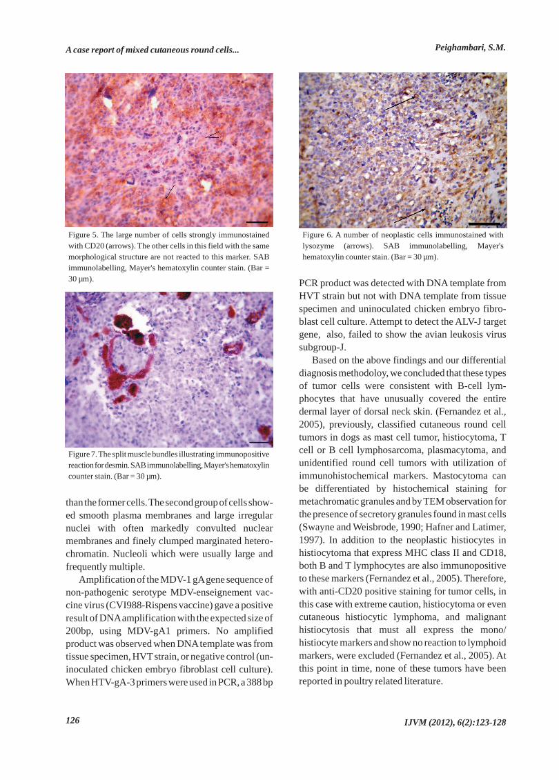
Histochemical results showed that both Toluidineblue and Giemsa stains were negative for the presenceof metachromatic cytoplasmic granules. Immuno-histochemical staining of normal thymus and bursa ofFabricius revealed that thymic cells were negative forboth anti CD79a and CD20. Lymphoid cells in thebursa stained strongly with CD20 (Figure4), but werenegative for CD79a. Most of neoplastic cells inten-sively reacted with anti-CD20 antibody (Figure 5). Alow population of neoplastic cells, dispersed all overthe tissue, also immunolabelled against anti lysozyme(Figure 6). No immunoreaction was found in the third
population of cells for any markers. The splittedmuscles were noticed in histopathological examin-ation labeled for both desmin and myogenin asinternal control (Figure 7).
In TEM, it was found that the tumor wascomposed of two distinct but inter mingled cellpopulations. The first group that included almostmajority of the cells, were characterized by lack ofintercellular junctions, basal lamina, and tonofila-ment. Cytoplasm showed few organelles and theirnuclei contained mainly disaggregated chromatinwith a minimal heterochromatin mostly associatedwith the inner nuclear membrane with one prominentnucleolus. The second group of cells were also non-cohesive and conatined more abundant cytoplasm
A case report of mixed cutaneous round cells...Peighambari, S.M.
IJVM (2012), 6(2):123-128 125
Figure 1. Broad sheet of closely packed tumor cells, with well-defined cytoplasmic borders (HE Bar = 200 µm). Inset: Thesame micrograph under higher magnification. The tissueconsists of variable shaped of tumor cells with well-definedboundaries. Mitotic figures are also present. (HE Bar = 30 µm).
Figure 2. Compact pleomorphic tumor cells, with large roundor oval vesicular nuclei each containing one or more basophilicnucleoli. The amount of pale eosinophilic cytoplasm is smalland well-defined. They infiltrated into dermal layer of skin andsplitted the muscle fibers (arrows). (HE Bar = 30 µm).
Figure 3. The myxoma-like structure area bordered by largepopulation of neoplastic cells (astrisk).(HE Bar = 200 µm).
Figure 4. Control positive immunohistochemical staining forCD20 in normal bursa of Fabricius. SAB immunolabelling,Mayer's hematoxylin counter stain. (Bar = 200 µm).
than the former cells. The second group of cells show-ed smooth plasma membranes and large irregularnuclei with often markedly convulted nuclearmembranes and finely clumped marginated hetero-chromatin. Nucleoli which were usually large andfrequently multiple.
Amplification of the MDV-1 gAgene sequence ofnon-pathogenic serotype MDV-enseignement vac-cine virus (CVI988-Rispens vaccine) gave a positiveresult of DNAamplification with the expected size of200bp, using MDV-gA1 primers. No amplifiedproduct was observed when DNAtemplate was fromtissue specimen, HVTstrain, or negative control (un-inoculated chicken embryo fibroblast cell culture).When HTV-gA-3 primers were used in PCR, a 388 bp
PCR product was detected with DNA template fromHVT strain but not with DNA template from tissuespecimen and uninoculated chicken embryo fibro-blast cell culture. Attempt to detect the ALV-J targetgene, also, failed to show the avian leukosis virussubgroup-J.
Based on the above findings and our differentialdiagnosis methodoloy, we concluded that these typesof tumor cells were consistent with B-cell lym-phocytes that have unusually covered the entiredermal layer of dorsal neck skin. (Fernandez et al.,2005), previously, classified cutaneous round celltumors in dogs as mast cell tumor, histiocytoma, Tcell or B cell lymphosarcoma, plasmacytoma, andunidentified round cell tumors with utilization ofimmunohistochemical markers. Mastocytoma canbe differentiated by histochemical staining formetachromatic granules and by TEM observation forthe presence of secretory granules found in mast cells(Swayne and Weisbrode, 1990; Hafner and Latimer,1997). In addition to the neoplastic histiocytes inhistiocytoma that express MHC class II and CD18,both B and T lymphocytes are also immunopositiveto these markers (Fernandez et al., 2005). Therefore,with anti-CD20 positive staining for tumor cells, inthis case with extreme caution, histiocytoma or evencutaneous histiocytic lymphoma, and malignanthistiocytosis that must all express the mono/histiocyte markers and show no reaction to lymphoidmarkers, were excluded (Fernandez et al., 2005). Atthis point in time, none of these tumors have beenreported in poultry related literature.
A case report of mixed cutaneous round cells... Peighambari, S.M.
IJVM (2012), 6(2):123-128126
Figure 5. The large number of cells strongly immunostainedwith CD20 (arrows). The other cells in this field with the samemorphological structure are not reacted to this marker. SABimmunolabelling, Mayer's hematoxylin counter stain. (Bar =30 µm).
Figure 6. A number of neoplastic cells immunostained withlysozyme (arrows). SAB immunolabelling, Mayer'shematoxylin counter stain. (Bar = 30 µm).
Figure 7. The split muscle bundles illustrating immunopositivereaction for desmin. SAB immunolabelling, Mayer's hematoxylincounter stain. (Bar = 30 µm).

Diffuse immunolabeling of lysozyme, which wasdemonstrable in many cells, revealed that many cellscharacteristically were consistent with histiocytes.But it was impossible to determine wether the cellsthat stained for lysozyme were infiltrating histiocytesor neoplastic cells. Lysozyme and alpha-1-anti-trypsin are both well characterized immune-markersof mononuclear phagocyte differentiotion in humanbeings. Moore and Rossin (1986), in a retrospectivecase series of malignant histiocytosis of dogs,demonstrated the reaction of lysozyme with allneoplastic histiocytes whereas only half of the tumorcells were identified for alpha-1-antitrypsin. Thismeans that lysozyme is more reliable for evaluationof normal or tumor histiocytes than alpha-1-antitrypsin.
Although we used anti-CD1a, the most applicablemarker for identifying of histiocytic cells in formalin-fixed, paraffin-embedded (FFPE) tissue, this markeris exclusively valuable in human samples and was notable to detect any canine cells expected to be CD1apositive in animals (Fernandez et al., 2005).
Lymphoproliferative lesions in Marek's diseasemay also be misdiagnosed with the type of lesion inthis report. However, our PCR results were negativefor MDV and on the other hand, in MD, mainly, thefeather follicles are involved and contain intranuclearinclusions in the epithelium of feather follicles (Silva,1992; Zhu et al., 1992; Schat and Venugopal, 2008).
Other tumors that are histologically very similarto the neoplastic round cells and should be excluded,are rhabdomyosarcoma that might be seen assubcutaneous mass (Fernandez-Bellon et al., 2003)and plasmacytoma (Fernandez et al., 2005). How-ever, the tumor cells in the former are negative forboth myogenin and desmin, and in the later, most ofthe cells have been shown to be CD79a positive indogs (Fernandez et al., 2005). Furthermore, theultrastructural features such as eccentric nuclei andprominent rough endoplasmic reticulum (rER)which are found in normal plasma cells, and also PCT,were not obsereved in our diagnostic electron micros-copy (Dardick et al., 1996)
The tumor cells that were negative to all stainingsmay indicate an undifferentiated or poorly stage ofdevelopment. Three weeks after skin biopsy, the cockdied and it was not submitted to our clinic fornecropsy procedures to examine the possible
metastasis to other organs. Determination of the origin of the tumor cells is
essential for the diagnosis of histiocytoma andlymphoma. In the literature, several descriptions forthe classification of monocyte-macrophage lineagesor lymphoma, are presented as follows: histiocyticlymphoma (HL) is recognized as lymphocytic lym-phoma, lymphoblastic lymphoma and non Hodgkin'slymphoma which is not a tumor of the macrophagesystem. In this tumor, large lymphoid cells havecytoplasmic vacuoles and are similar to macrophagetumors (Wilson and Armitage, 2008). True his-tiocytic lymphoma (THL) is a type of neoplasm thatreacts with monocyte/histiocyte associated markerslike CD68, lysozyme, alpha-1-antitrypsin and CD45,and is negative for CD1a, epithelial, and B- and T-celllineage-specific markers (Copie-Bergman et al.,1998). Hemophagocytic lymphohistiocytosis (HLH)is a reactive process resulting from prolonged andexcessive activation of antigen-presenting cells(macrophages, histiocytes) and CD8+ cells (Filipovich,2009). In our case, the tumor consisted of two distinctcell populations of lymphoblasts and histiocytes.Therefore, to name this unusual cutaneous lym-phoma, we prefer to apply the combined term,lymphoblastic histiocytoma, which indicate thepresence of two different precursor cells (lym-phoblast and histiocyte) in one lesion. Using otherdefinitions, as mentioned above, is a misnomer andcauses diversion of the original meaning.
In conclusion, the population of neoplastic cellsrevealed immunopositive staining only with CD20,which serves as a diagnostic marker for lymphoma,and with lysozyme, which serves as one of the well-known definitive markers for histiocytoma. Con-sidering the above findings, it may be better to call thepresent unusual cutaneous lymphoma as lympho-blastic histiocytoma.
A case report of mixed cutaneous round cells...Peighambari, S.M.
IJVM (2012), 6(2):123-128 127
Copie-Bergman, C., Wotherspoon, A.C., Norton,
A.J., Diss, T.C., Isaacson, P.G. (1998) True histiocytic
lymphoma: A morphologic, immunohistochemical,
and molecular genetic study of 13 cases. Am. J. Surg.
Pathol. 22: 1386-1392.
1. References

A case report of mixed cutaneous round cells... Peighambari, S.M.
IJVM (2012), 6(2):123-128128
Fernandez-Bellon, H., Martorell, J., Rabanal, R.,
Ramis, A. (2003) Rhabdomyosarcoma in a racing
pigeon (Columba livia). Avian. Pathol. 32: 613-616.
Fernandez, N.J., West, K.H., Jackson, M.L., Kidney,
B.A. (2005) Immunohistochemical and histo-
chemical stains for differentiating canine cutaneous
round cell tumors. Vet. Pathol. 42: 437-445.
Filipovich, A.H. (2009) Hemophagocytic lympho-
histiocytosis (HLH) and related disorders. Hemato-
logy. 2009: 127-131.
Dardick, I., Eyden, B., Federman, M., Hammar, S.,
Lajoie, G., Malott, R., et al. (1996) Hand book of
diagnostic electron microscopy for pathologists-in-
training. Igaku-Shoin Medical Publishers, Inc, New
York, USA.
Hafner, S., Latimer, K. (1997) Cutaneous mast cell
tumors with pulmonary metastasis in a hen. Avian
Pathol. 26: 657-663.
Moore, P.F., Rosin, A. (1986) Malignant histiocytosis
of Bernese mountain dogs. Vet. Pathol. 23: 1-10.
Rajabi, Z., Bozorgmehrifard, M.H., Peighambari,
S.M. (2009) Molecular studies on avian leukosis
virus subgroup J (ALV-J) infections in grandparent
flocks in Iran. J. Vet. Res. 64: 7-14.
Silva, R.F. (1992) Differentition of pathogenic and
non-pathogenic serotype 1 Marek's disease viruses
(MDVs) by the polymerase chain reaction amplifica-
tion of the tandem direct repeats within the MDV
genome. Avian. Dis. 36: 521-528.
Swayne, D.E., Weisbrode, S.E. (1990) Cutaneous
mast cell tumor in a great horned Owl (Bubo
virginianus). Vet. Pathol. 27: 124-126.
Schat, K.A., Venugopal, N. (2008) Marek's disease.
In: Diseases of Poultry. Saif, Y.M., Fadly, A.M.,
Glisson, J.R., McDougald, L.R., Nolan, L.K.,
Swayne, D.E. (eds.). (12th ed.) Blackwell Publishing
Professionals, Ames, Iowa, USA. p. 452-514.
Wilson, W.H., Armitage, J.O. (2008) Non-Hodgkin's
lymphoma. In: Abellof 's Clinical Oncology. Abeloff,
M.D., Armitage, J.O., Niederhauber, J.E., Kastan,
M.B., McKenna, W.G. (eds.). (4th ed.) Elsevier
Churchill Livingstone, Philadelphia, Pa, USA. p.112.
Zhu, G.S., Ojima, T., Hironaka, T., Ihara, T.,
Mizukoshi, N., Kato, A., et al. (1992) Differentition
of oncogenic and non oncogenic strains of Marek's
disease virus type 1 by using polymerase chain
reaction amplification. Avian Dis. 36: 637-645.
2.
3.
4.
5.
6.
7.
8.
9.
10.
11.
12.
13.

ìXéú |ÆI kAìþ AüpAó, 1931, kôoû 6, yíBoû 2, 821-321
OõìõoWélÿ ìhPéÈ ðByþ Aquéõë|øBÿ âpk koüà gpôx |)sucitsemod sullaG(
Aìýl kqÖõèýBó1
Wízýl oqï üBo2
|uýl ì¿Ç×þ KýÓíHpÿ3*|
môAè×ÛBooWHþ4
ìdíl oÂB ¾BkÚþ5
|Îývþ WùBðrAk6
1) âpôû KBOõGýõèõsÿ, kAðzßlû kAìLryßþ kAðzãBû èpuPBó, gpï @GBk, AüpAó.2) âpôû Îéõï koìBðãBøþ, kAðzßlû kAìLryßþ kAðzãBû Öpkôuþ ìzùl, ìzùl, AüpAó.
3) âpôû Îéõï koìBðãBøþ, kAðzßlû kAìLryßþ kAðzãBû OùpAó, OùpAó, AüpAó.4) âpôû Îéõï koìBðãBøþ, kAðzßlû kAìLryßþ kAðzãBû OHpür, OHpür, AüpAó.
5) âpôû ìýßpôGýõèõsÿ, kAðzßlû KýpA kAìLryßþ kAðzãBû GõÎéþ uýñB øílAó, øílAó, AüpAó. | |6) âpôû KBOõèõsÿ, kAðzßlû Kryßþ kAðzãBû Îéõï Kryßþ OùpAó, OùpAó, AüpAó.
|(||koüBÖQ ìÛBèú: 21 | @moìBû 0931 , | Knüp} ðùBüþ: 42| Gùíò ìBû 0931)| ||
|̂ßýlû
üà ÚÇÏú gpôx GBèÔ ðtAk æoÿ Gú ÎéQ Oõoï qüpWélÿ upAupÿ Aq AGPlAÿ ðBcýú Kw upôAÚÐ koKzQ âpkó OB ðBcýú WlôâBû Gú Þéýñýà
KpðlâBó kAðzßlû kAìLryßþ kAðzãBû OùpAó AoWBÑ âpkül. Kw Aq AðXBï GýõKvþ, cÃõo¾×dBR uéõèþ âpk GB øvPú|øBÿ Þpôÿ üB GýÃõÿ
Gƒroå Gƒú øíƒpAû üà üB ̂ñl øvPú GpWvPú koôó øvPú|øB ôìÛBküpÞî uýPõKçuî koGpouþ ìýßpôußõKýà WéI ðËpìþ|ðíõk. Aq küãp
ìzBølAR oürGýñþ Aq Gýò oÖPò oyPú øBÿ ìBøý̀ú Aÿ ìvPÛpkoðBcýú ÎÃçR âpkó OõuÈ OùBWî uéõë|øBÿ Oõìõoÿ Gõk. GB AuP×Bkû Aq Oßñýà
ìýßpôußõN AèßPpôðþ, oðä @ìýrÿ øývPõyýíþ ôÞBoGpk ôuýÐ ìBoÞpøBÿ AüíõðõøývPõyýíþ, ̂ùpû AÖPpAÚþ Gýò AðõAÑ Oõìõouéõë|øBÿ âpk
GpÚpAoyl. Oßñýà ôAÞñ{ qðXýpû Aÿ Kéþ ìpAq AGPç Gú GýíBoÿ ìBoá ôèõÞõq KpðlâBó Aq qüpâpôû J |oA OBCüýl ðßpk. koðùBüQ Oßñýà|øBÿ ÖõÝ ðzBó
kAk yƒƒBÞéú Oõkû ìrGõoGB ìñzBC kôâpôû ìP×BôR uéõèþ yBìê èñ×õuýQ|øBÿ BôøývPýõuBüQ ìþ|GByl. GB yñBuBüþ Aüò kôOýM Aq WíÏýQ
uéõë|øBÿ Oõìõoÿ Aüò ÂBüÏú Gú ðBï èñ×õGçuPýà øývPýõuBüPõìB ðBï ânAoÿ yl.
ôAsû øBÿÞéýlÿ:| |èñ×õuýQ|øBÿ B||, øývPõuBüPõìB, OõìõoWélÿ, gpôx,|Oõìõouéõë|øBÿ âpk.
∗)ðõüvñlû ìvõöôë: Oé×ò: 05171116 (12)89+ ðíBGp: 22233966 (12)89+ | ||ri.ca.tu@mahgiepm||:liamE|
Abstracts in Persian Language
134