Saccadic Selectivity During Visual Search: The Influence of

of April 11, 2019.This information is current as Anti-CD20 Therapy
in TNF-Transgenic Mice and Are Targets of the Onset of Inflammatory-Erosive ArthritisInflamed Lymph Nodes Are Associated with
B Cells inhi/CD21+Expanded CD23
Iñaki Sanz, Edward M. Schwarz and Andrea BottaroProulx, Lianping Xing, Denise Skrombolas, Robert Dunn, Jie Li, Igor Kuzin, Safiehkhatoon Moshkani, Steven T.
http://www.jimmunol.org/content/184/11/6142doi: 10.4049/jimmunol.0903489April 2010;
2010; 184:6142-6150; Prepublished online 30J Immunol
MaterialSupplementary
9.DC1http://www.jimmunol.org/content/suppl/2010/04/30/jimmunol.090348
Referenceshttp://www.jimmunol.org/content/184/11/6142.full#ref-list-1
, 12 of which you can access for free at: cites 43 articlesThis article
average*
4 weeks from acceptance to publicationFast Publication! •
Every submission reviewed by practicing scientistsNo Triage! •
from submission to initial decisionRapid Reviews! 30 days* •
Submit online. ?The JIWhy
Subscriptionhttp://jimmunol.org/subscription
is online at: The Journal of ImmunologyInformation about subscribing to
Permissionshttp://www.aai.org/About/Publications/JI/copyright.htmlSubmit copyright permission requests at:
Email Alertshttp://jimmunol.org/alertsReceive free email-alerts when new articles cite this article. Sign up at:
Print ISSN: 0022-1767 Online ISSN: 1550-6606. Immunologists, Inc. All rights reserved.Copyright © 2010 by The American Association of1451 Rockville Pike, Suite 650, Rockville, MD 20852The American Association of Immunologists, Inc.,
is published twice each month byThe Journal of Immunology
by guest on April 11, 2019
http://ww
w.jim
munol.org/
Dow
nloaded from
by guest on April 11, 2019
http://ww
w.jim
munol.org/
Dow
nloaded from

The Journal of Immunology
Expanded CD23+/CD21hi B Cells in Inflamed Lymph NodesAre Associated with the Onset of Inflammatory-ErosiveArthritis in TNF-Transgenic Mice and Are Targets ofAnti-CD20 Therapy
Jie Li,*,†,1 Igor Kuzin,‡,1 Safiehkhatoon Moshkani,†,‡ Steven T. Proulx,* Lianping Xing,*
Denise Skrombolas,† Robert Dunn,x Inaki Sanz,†,‡ Edward M. Schwarz,*,† and
Andrea Bottaro†,‡
Anti-CD20 B cell depletion therapy (BCDT) is very effective for some patients with rheumatoid arthritis (RA); however the path-
ogenic role of B lymphocytes in RA and the primary targets of BCDTare unknown. The human TNF transgenic (hTNF-Tg) mouse
model of RA displays a chronic, progressive disease that spreads from distal to proximal joints and is generally considered to be
adaptive immune system independent. We have previously reported that knee arthritis in hTNF-Tg mice is accompanied by struc-
tural and functional changes of the adjoining popliteal lymph node (PLN), detectable by contrast-enhanced magnetic resonance
imaging. To better understand these changes, in this paper we show that onset of knee synovitis and focal erosions are paralleled by
PLN contraction and accumulation of large numbers of B cells in the lymphatic sinus spaces within the node. Flow cytometry from
TNF-Tg mice 2, 4–5, and 8–12 mo old demonstrated that B cell accumulation in the PLN follows ankle arthritis, but commences
before knee disease, and involves early expansion of CD21hi, CD23+, IgMhi, CD1d+, activation marker-negative, polyclonal B cells
that are found to be specifically restricted to lymph nodes draining inflamed, arthritic joints. The same B cell population also
accumulates in PLNs of K/BxN mice with autoantigen-dependent arthritis. Strikingly, we show that BCDT ameliorates hTNF-Tg
disease and clears follicular and CD21hi, CD23+ B cells from the PLNs. On the basis of these findings, we propose a model whereby
B cells contribute to arthritis in mice, and possibly RA, by directly affecting the structure, composition, and function of joint-
draining lymph nodes. The Journal of Immunology, 2010, 184: 6142–6150.
Rheumatoid arthritis (RA) is a chronic, progressive in-flammatory-erosive autoimmune disease of the joints thataffects as many as 1% of the population, predominantly
females. Although major progress has been made in recent years inunderstanding the mechanisms of disease, many questions aboutRA pathogenesis remain unanswered. Clearly, autoimmunity andultimately tissue destruction in RA are the result of the complexinteraction of multiple contributing mechanisms. Proinflammatory
cytokines, such as TNF-a, IL-1, and IL-6 play a critical, possiblyprimary, role in disease (1). In particular, TNF-a has emerged asa key cytokine exerting pleiotropic effects in driving the arthritisprocess by regulating other proinflammatory cytokines, promotingosteoclastogenesis, recruiting leukocytes to inflamed sites, anddirectly driving expression of enzymes responsible for tissuedamage such as metalloproteinases and oxygenases (2, 3). Asa result, TNF antagonists have become common in the clinicaltreatment of RA (4). T cell involvement in RA is highlighted bystrong genetic associations with MHC haplotypes, synovial andjoint infiltration by activated T cells, and the recognized role ofT cells in murine models of disease, such as collagen-inducedarthritis (5). In contrast, the contribution of B cells to disease hasbeen more controversial: Although production of autoantibodies(rheumatoid factor, anti-cyclic citrullinated peptide Abs) and ac-cumulation of immune complexes and of ectopic germinal center-like structures in the joint and synovium are common in RA pa-tients, they are not universal features of the disease (6–8).Despite these uncertainties, B cell depletion therapy (BCDT)with
anti-CD20 Abs, originally developed for the treatment of B cellmalignancies, has emerged in recent years as an effective strategy toameliorate disease in patients who do not respond to more con-ventional therapy (9, 10). Although disease amelioration by BCDTunderscores the importance of B cells in RA, clinical improvementdoes not always correlate with reduction of serum autoantibodylevels, indicating that B cells may exert additional pathogeneticfunctions (reviewed in Ref. 11). B cells have the potential to pro-mote autoimmune pathology by a number of Ab-independent ef-fector mechanisms, in their role as APC, or by secreting cytokines
*Center for Musculoskeletal Research and ‡Division of Allergy, Immunology, andRheumatology, Department of Medicine, University of Rochester Medical Center;†Department of Microbiology and Immunology, University of Rochester School ofMedicine and Dentistry, Rochester, NY 19642; and xBiogen Idec, San Diego, CA92122
1J.L. and I.K. contributed equally to this work.
Received for publication November 3, 2009. Accepted for publication March 26,2010.
This work was supported in part by Centocor and National Institutes of Health PublicHealth Service Awards AR46545, AR48697, AR54041, and AR56702.
Address correspondence and reprint requests to Andrea Bottaro, Department of Med-icine, University of Rochester Medical Center, URMC695, 601 Elmwood Avenue,Rochester, NY 14642. E-mail address: [email protected]
The online version of this article contains supplemental material.
Abbreviations used in this paper: ALN, axillary lymph node; AU, arbitrary unit;BCDT, B cell depletion therapy; Bin, B cell in inflamed node; CE, contrast enhance-ment; FoB, follicular B cell; hTNF-Tg, human TNF transgenic; ILN, iliac lymphnode; LN, lymph node; LNcap, LN capacity; mCT, microcomputerized tomography;MLN, mesenteric LN; MRI, magnetic resonance imaging; MZB, marginal zone B;PLN, popliteal LN; RA, rheumatoid arthritis; RAG, recombination activating gene;SynVol, synovial volume; WT, wild-type.
Copyright� 2010 by TheAmericanAssociation of Immunologists, Inc. 0022-1767/10/$16.00
www.jimmunol.org/cgi/doi/10.4049/jimmunol.0903489
by guest on April 11, 2019
http://ww
w.jim
munol.org/
Dow
nloaded from

with proinflammatory activity (TNF, IFN-g, IL-12p40, and so on)and chemotactic factors (MIP1-a and -b, CCL1, RANTES) (12, 13;reviewed in Ref. 14). B cells also play a critical role in the formationof ectopic lymphoid tissue structures, which are commonly ob-served in the inflamed synovium of RA patients and are thought toplay a role in local pathogenesis (15, 16; reviewed in Ref. 17).Human TNF-a transgenic (hTNF-Tg) mouse strains develop
a disease that closely resembles RA, which, although variable infeatures and timing based on transgene type and expression levels, ischaracterized by spontaneous, chronic, progressive inflammatory-erosive joint disease, generally starting in the hind paws and ad-vancing cephalically to the knees and fore limbs (18). The initiatingrole of TNF-a in this model, the lack of significant lymphocyticinfiltration in the joints and synovium, the absence of detectableserum rheumatoid factor, and the finding that recombination acti-vating gene 1 (RAG1)-deficient mice, in which the endogenous TNFgene was upregulated by gene targeting, develop ankle arthritis in-distinguishable from that of their RAG-competent counterparts, hasled to the conclusion that TNF-induced arthritis in Tg mice is likelyB and T cell-independent (18–20; reviewed in Refs. 21, 22).Using imaging techniques such as contrast-enhanced magnetic
resonance imaging (CE-MRI) and microcomputerized tomography(mCT), we have previously shown that in the single-copy numberhTNF-Tg strain Tg3647 disease progression is paralleled closelyby changes in the popliteal lymph node (PLN) (23, 24). Thesestudies developed precise metrics that allow the longitudinal studyof disease progression patterns in vivo. Among the most strikingof the biomarkers identified is the observation that disease pro-gression from the ankle (starting at around 1–2 mo of age) to theknee (4–5 mo) is accompanied by a dramatic increase in PLN sizeand CE values (i.e., fluid content) (23, 24). Anti-TNF treatment inthese mice reversed both arthritis and the associated changes inPLN structure, strongly suggesting a functional link between thetwo phenomena (23, 24).In this paper, we extend these findings by analyzing later changes
in PLN structure and organization associated with the progressionof inflammatory-erosive disease in the knee; demonstrate an in-volvement of PLN B cells in these changes from the earliest stagesof disease, and in particular of a CD21-high, CD23+, CD1d+ subsetof B cells that accumulate specifically in inflamed nodes; identifysimilar B cell changes in the lymph nodes (LNs) of K/BxN mice;and show that, unexpectedly, B cell depletion significantly amel-iorates disease in the hTNF-Tg model.
Materials and MethodsAnimals and anti-CD20 treatment
The 3647 line of TNF-Tg mice in a C57BL/6 background were obtainedfrom Dr. George Kollias (Institute of Immunology, Alexander FlemingBiomedical Sciences Research Center, Vari, Greece) (18). All animalstudies were performed under protocols approved by the University ofRochester Committee for Animal Resources. Starting at 3 mo of age,hTNF-Tg mice received CE-MRI bimonthly, as described (23, 24; seebelow), until PLN collapse was detected, at which time they receivedbaseline mCT (23, 24). Mouse anti-mouse CD20 mAbs (18B12 IgG2a) ornonspecific placebo IgG2a Abs (2B8) (Biogen Idec, San Diego, CA) weredosed at 10 mg/kg i.p. every 2 wk for 6 wk, with continuous CE-MRIevery 2 wk, followed by posttreatment mCT scan. Knee joints were sub-jected to histologic examination, and cells from PLN and iliac lymph node(ILN) were subjected to flow cytometry.
(KRN 3 NOD)F1 Tg mice were obtained by crossing KRN Tg males ina C57BL/6 genetic background (kindly provided by Dr. C. Benoist, HarvardMedical School, Boston, MA) (25) with female NODmice (purchased fromThe Jackson Laboratory, Bar Harbor, ME). Offspring were bled at day 21,and those expressing the aVb6-TCR KRN transgene were identified byflow cytometry. These TCR transgene-positive mice were named K/BxNmice, and non-TCR Tg littermates were used as controls. All K/BxN micedeveloped severe ankle joint inflammation around 1 mo of age, and the
joint tissue damage progressed thereafter. The K/BxN mice and littermatesused in this work were 1 y old.
CE-MRI and MR data analysis
Detailed methods of CE-MRI have been previously described (23, 24).Briefly, anesthetized micewere positioned with the knee inserted in a custom-designedmousekneecoil.MRimageswere obtainedona3TeslaSiemensTrioMRI (SiemensMedical Solutions, Erlangen, Germany). Amira 3.1 (TGSUnit,Mercury Computer Systems, San Diego, CA) was used for analysis of high-resolution CE-MRI data. For segmentation of the LN, regions of interest aremanually drawn on a postcontrast three-dimensional stack of images andthresholded based on signal intensity $1500 arbitrary units to define theboundary between the LN and the fat pad surrounding the node. The TissueStatistics module is used to quantify the volume of the LN and the value ofCEof this tissue andof surroundingmuscle.LNcapacity (LNCap) is defined asthe LN CE divided by muscle CE and multiplied by LN volume.
mCT and data analysis
Bone volume analysis was performed by scanning the knee joint in a Vivamicro-CT 40 imaging system (Scanco, Bassersdorf, Switzerland). Patellarbone volume determination and three-dimensional reconstruction of theknee joint were performed usingAmira 3.1, as previously described (23, 24).
Histology and immunohistochemistry
Knee joints were fixed in 4.5% phosphate-buffered formalin and decalcifiedin 14%EDTA for 7 d. Histology sectionswere stainedwithOrangeG/AlcianBlue (H&E). LNs were processed using two different protocols. For im-munohistochemistry, PLNs were dissected and fixed in 10% neutralizedformalin. Tissues were embedded in paraffin wax, and deparaffined sectionswere quenched with 3% hydrogen peroxide and treated for Ag retrieval for30 min. Sections were then stained with anti-B220 Ab (BioLegend, SanDiego, CA). For multicolor immunofluorescence microscopy, fresh-frozenPLNs were cut into 7-mm-thick sections. PLN sections were fixed with 4%paraformaldehyde, rehydrated in PBS, blocked with rat serum, and stainedwith PE-conjugated anti-IgM (eBioscience, San Diego, CA) and FITC-conjugated anti-CD3ε (BioLegend).
Flow cytometry
Single-cell suspensions were prepared from lymphoid organs at definedstages of disease, and were analyzed for expression of surface markers withcombinations of the following fluorochrome-labeled Abs: APC-Alexa 750anti-B220 (clone RA3-6B2; eBioscience); PE anti-IgM (clone II/41;eBioscience); Alexa Fluor 700 anti-CD19 (clone 6D5; BioLegend); AlexaFluor 647 anti-IgD (clone 11-26c.2a; BioLegend); FITC anti-CD93, (cloneAA4.1; eBioscience); Pacific Blue anti-CD21/35 (clone 7E9; BioLegend);PE-Cy7 anti-CD23 (clone B3B4; BioLegend); biotin anti-CD24 (clone M1/69; eBioscience); PE-anti-CD1d (clone 1B1; BD Pharmingen, San Diego,CA); PE-Cy5 anti-CD5 (clone 53-7.3; BioLegend); PE-Cy5 anti-CD80(clone 16-10A1; BioLegend); Pacific Blue anti-CD86 (clone GL-1;BioLegend); biotin anti-CD69 (clone H1.2F3; BD Pharmingen); PerCP-Cy5.5 anti-CD25a (clone PC61; BioLegend); FITC anti-GL7 (clone GL-7;BD Pharmingen); PE-Cy7 anti-CD4 (clone GK1.5; eBioscience); FITCanti-CD3ε (clone 17A2; BioLegend); PE-Cy5 anti CD8a (clone 53-6.7;BioLegend); Alexa Fluor 647 anti-CCR6 (clone 140706; BD Pharmingen);PE anti-CXCR3 (clone 220803; R&D Systems, Minneapolis, MN); biotinanti-CXCR5 (clone 2G8; BD Pharmingen); PE-Cy5 anti-CCR7 (clone4B12; BioLegend); rabbit anti-mouse Ki-67 (clone SP6; Epitomics, Bur-lingame, CA), followed by secondary Ab PE-Goat anti-rabbit Ig(H+L)(Invitrogen, Carlsbad, CA); and PE-Texas Red streptavidin (Invitrogen).Samples were run on an LSRII cytometer and analyzed by FlowJo soft-ware (BD Pharmingen). To control for nonspecific Ab binding, isotypecontrol experiments were conducted and resulted in nonsignificant back-ground stains.
CDR3 spectratyping
Total RNA from the indicated sources was isolated using TRIzol reagent(Invitrogen), and cDNAs were generated using random primers and Su-perscript III M-MLV reverse transcriptase (Invitrogen). The cDNA sampleswere subjected to PCR to amplify the CDR3 region, using a VHall primer(26) and the CmR primer listed below, which maps at the 39 end of the Cm1exon. The PCR products were subjected to seminested PCR using VHallprimer and an internal FAM (6FAM)-labeled CmP primer. The seminestedPCR products were run on an Applied Biosystems (Foster City, CA) 3730Genetic Analyzer at the University of Rochester Functional GenomicCenter, and the resulting chromatograms were analyzed by Peak Scannersoftware version 1 (Applied Biosystems).
The Journal of Immunology 6143
by guest on April 11, 2019
http://ww
w.jim
munol.org/
Dow
nloaded from
Primers sequences (59–39) are as follows: CmP, CAGCCCATGGC-CACCAGATTCTTATCAGAC (59 6FAM labeled); CmR, AATGGTGC-TGGGCAGGAAGT; VHall, AGGTSMARCTGCAGSAGTCWGG.
Statistical analysis
Linear mixed-effects regression models, with mouse as a random effect andtime (treated as a continuous covariate) as a fixed effect, were used to assesschanges over time based on longitudinal data. Differences between groupsin synovial volume, LN volume, LNCap, and disease progression over timewere tested by two-sided t test. The p values ,0.05 were consideredsignificant.
ResultsPLN changes associated with knee arthritis progression inhTNF-Tg mice
We have previously shown how progression of knee synovitis inhTNF-Tg mice can be followed noninvasively via CE-MRI toquantify synovial volume (SynVol), and correlated these findingswith mCT and histological results (23, 24). Although this worklargely corroborated findings from cross-sectional studies dem-onstrating that arthritis initiates in the distal joints (e.g., ankle) andspreads to proximal joints (e.g., knee) over time (18–20), wediscovered that increased knee SynVol is paralleled by an increasein volume and CE (i.e., fluid content) of the adjacent PLN,yielding parameters LNvol and LN-CE, respectively, which can becombined in a single functional biomarker, LN capacitance(LNcap = LNvol 3 LN-CE) (23, 24), which correlates with
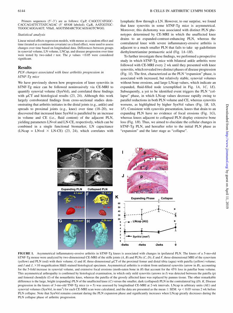
lymphatic flow through a LN. However, to our surprise, we foundthat knee synovitis in some hTNF-Tg mice is asymmetrical.Moreover, this dichotomy was associated with distinct PLN phe-notypes determined by CE-MRI in which the unaffected kneedrains to an expanded-contrast-enhancing PLN, whereas thecontralateral knee with severe inflammatory-erosive arthritis isadjacent to a much smaller PLN that fails to take up gadoliniumdiethylenetriamine pentaacetic acid (Fig. 1A–1H).To further investigate these findings, we performed a prospective
study in which hTNF-Tg mice with bilateral ankle arthritis werefollowed with CE-MRI every 2 wk until they presented with kneesynovitis, which revealed two distinct phases of disease progression(Fig. 1I). The first, characterized as the PLN “expansion” phase, isassociated with increased, but relatively stable, synovial volumeswithout bone erosions, and large LNcap values, which indicate anexpanded, fluid-filled node (exemplified in Fig. 1A, 1C, 1E).Subsequently, a yet to be identified event triggers the PLN “col-lapse” phase, in which LNcap values decrease rapidly owing toparallel reductions in both PLN volume and CE, whereas synovitisworsens, as highlighted by higher SynVol values (Fig. 1B, 1D,1F). Consistent with synovitis presentation, knees that drain to anexpanding PLN have no evidence of focal erosions (Fig. 1G),whereas knees adjacent to collapsed PLN display extensive boneloss (Fig. 1H). Thus, we aimed to elucidate the cellular changes inhTNF-Tg PLN, and hereafter refer to the initial PLN phase as“expansion” and the later stage as “collapse”.
FIGURE 1. Asymmetrical inflammatory-erosive arthritis in hTNF-Tg knees is associated with changes in ipsilateral PLN. The knees of a 5-mo-old
hTNF-Tg mouse were analyzed by two-dimensional CE-MRI of the stifle joints (A, B) and PLNs (C, D); E and F, three-dimensional MRI of the synovium
(yellow) and PLN (red) with their volume; G and H, three-dimensional mCT of the proximal femur and distal tibia (aqua) with patella (yellow) volume;
and I and J, 310 magnification H&E-stained histological specimen. Asymmetrical arthritis is evident from unilateral synovitis (arrow in B), accounting
for the 5-fold increase in synovial volume, and extensive focal erosions (moth-eaten bone in H) that account for the 45% loss in patellar bone volume.
This asymmetrical arthropathy is confirmed by histological examination, in which only mild synovitis (arrows in I) was detected between the patella (p)
and femoral chondyle (f) of the nonarthritic knee, whereas the patella of the grossly affected knee was replaced by pannus tissue. The other remarkable
difference is the large, bright (expanding) PLN of the unaffected knee (C) versus the smaller, dark (collapsed) PLN in the contralateral leg (D). K, Disease
progression in the knees of 3-mo-old TNF-Tg mice (n = 5) was assessed by longitudinal CE-MRI at 2-wk intervals. LNcap in arbitrary units (AU) and
synovial volumes (SynVol, in mm3) for each CE-MRI scan were calculated, and the data are presented as the mean6 SEM. pp, 0.05 versus 2 wk before
PLN collapse. Note that SynVol remains constant during the PLN expansion phase and significantly increases when LNcap greatly decreases during the
PLN collapse phase of arthritic progression.
6144 B CELLS IN ARTHRITIC LYMPH NODES
by guest on April 11, 2019
http://ww
w.jim
munol.org/
Dow
nloaded from
B cell accumulation and migration during PLN expansion andcollapse
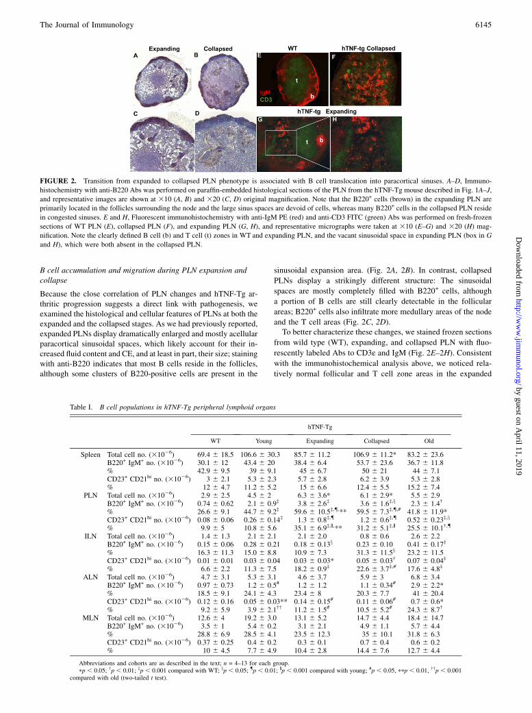
Because the close correlation of PLN changes and hTNF-Tg ar-thritic progression suggests a direct link with pathogenesis, weexamined the histological and cellular features of PLNs at both theexpanded and the collapsed stages. As we had previously reported,expanded PLNs display dramatically enlarged and mostly acellularparacortical sinusoidal spaces, which likely account for their in-creased fluid content and CE, and at least in part, their size; stainingwith anti-B220 indicates that most B cells reside in the follicles,although some clusters of B220-positive cells are present in the
sinusoidal expansion area. (Fig. 2A, 2B). In contrast, collapsed
PLNs display a strikingly different structure: The sinusoidal
spaces are mostly completely filled with B220+ cells, although
a portion of B cells are still clearly detectable in the follicular
areas; B220+ cells also infiltrate more medullary areas of the node
and the T cell areas (Fig. 2C, 2D).To better characterize these changes, we stained frozen sections
from wild type (WT), expanding, and collapsed PLN with fluo-
rescently labeled Abs to CD3ε and IgM (Fig. 2E–2H). Consistent
with the immunohistochemical analysis above, we noticed rela-
tively normal follicular and T cell zone areas in the expanded
FIGURE 2. Transition from expanded to collapsed PLN phenotype is associated with B cell translocation into paracortical sinuses. A–D, Immuno-
histochemistry with anti-B220 Abs was performed on paraffin-embedded histological sections of the PLN from the hTNF-Tg mouse described in Fig. 1A–J,
and representative images are shown at 310 (A, B) and 320 (C, D) original magnification. Note that the B220+ cells (brown) in the expanding PLN are
primarily located in the follicles surrounding the node and the large sinus spaces are devoid of cells, whereas many B220+ cells in the collapsed PLN reside
in congested sinuses. E and H, Fluorescent immunohistochemistry with anti-IgM PE (red) and anti-CD3 FITC (green) Abs was performed on fresh-frozen
sections of WT PLN (E), collapsed PLN (F), and expanding PLN (G, H), and representative micrographs were taken at 310 (E–G) and 320 (H) mag-
nification. Note the clearly defined B cell (b) and T cell (t) zones in WT and expanding PLN, and the vacant sinusoidal space in expanding PLN (box in G
and H), which were both absent in the collapsed PLN.
Table I. B cell populations in hTNF-Tg peripheral lymphoid organs
hTNF-Tg
WT Young Expanding Collapsed Old
Spleen Total cell no. (31026) 69.4 6 18.5 106.6 6 30.3 85.7 6 11.2 106.9 6 11.2* 83.2 6 23.6B220+ IgM+ no. (31026) 30.1 6 12 43.4 6 20 38.4 6 6.4 53.7 6 23.6 36.7 6 11.8% 42.9 6 9.5 39 6 9.1 45 6 6.7 50 6 21 44 6 7.1CD23+ CD21hi no. (31026) 3 6 2.1 5.3 6 2.3 5.7 6 2.8 6.2 6 3.9 5.3 6 2.8% 12 6 4.7 11.2 6 5.2 15 6 6.6 12.4 6 5.5 15.2 6 7.4
PLN Total cell no. (31026) 2.9 6 2.5 4.5 6 2 6.3 6 3.6* 6.1 6 2.9* 5.5 6 2.9B220+ IgM+ no. (31026) 0.74 6 0.62 2.1 6 0.9‡ 3.8 6 2.6‡ 3.6 6 1.6‡,x 2.3 6 1.4†
% 26.6 6 9.1 44.7 6 9.2‡ 59.6 6 10.5‡,{,** 59.5 6 7.3‡,{,# 41.8 6 11.9*CD23+ CD21hi no. (31026) 0.08 6 0.06 0.26 6 0.14‡ 1.3 6 0.8‡,{ 1.2 6 0.6‡,{ 0.52 6 0.23‡,x
% 9.9 6 5 10.8 6 5.6 35.1 6 6.9‡,‖,** 31.2 6 5.1‡,‖ 25.5 6 10.1†,{
ILN Total cell no. (31026) 1.4 6 1.3 2.1 6 2.1 2.1 6 2.0 0.8 6 0.6 2.6 6 2.2B220+ IgM+ no. (31026) 0.15 6 0.06 0.28 6 0.21 0.18 6 0.13x 0.23 6 0.10 0.41 6 0.17‡
% 16.3 6 11.3 15.0 6 8.8 10.9 6 7.3 31.3 6 11.5x 23.2 6 11.5CD23+ CD21hi no. (31026) 0.01 6 0.01 0.03 6 0.04 0.03 6 0.03* 0.05 6 0.03† 0.07 6 0.04‡
% 6.6 6 2.2 11.3 6 7.5 18.2 6 0.9‡ 22.6 6 3.7‡,# 17.6 6 4.8‡
ALN Total cell no. (31026) 4.7 6 3.1 5.3 6 3.1 4.6 6 3.7 5.9 6 3 6.8 6 3.4B220+ IgM+ no. (31026) 0.97 6 0.73 1.2 6 0.5# 1.2 6 1.2 1.1 6 0.34# 2.9 6 2.2*% 18.5 6 9.1 24.1 6 4.3 23.4 6 8 20.3 6 7.7 41 6 20.4CD23+ CD21hi no. (31026) 0.12 6 0.16 0.05 6 0.03** 0.14 6 0.15# 0.11 6 0.06# 0.7 6 0.6*% 9.2 6 5.9 3.9 6 2.1†† 11.2 6 1.5# 10.5 6 5.2# 24.3 6 8.7†
MLN Total cell no. (31026) 12.6 6 4 19.2 6 3.0 13.1 6 5.2 14.7 6 4.4 18.4 6 14.7B220+ IgM+ no. (31026) 3.5 6 1 5.4 6 0.2 3.1 6 2.1 4.9 6 1.1 5.7 6 4.4% 28.8 6 6.9 28.5 6 4.1 23.5 6 12.3 35 6 10.1 31.8 6 6.3CD23+ CD21hi no. (31026) 0.37 6 0.25 0.4 6 0.2 0.3 6 0.1 0.7 6 0.4 0.6 6 0.2% 10 6 4.5 7.7 6 4.9 10.4 6 2.8 14.4 6 7.6 12.7 6 4.4
Abbreviations and cohorts are as described in the text; n = 4–13 for each group.pp , 0.05; †p , 0.01; ‡p , 0.001 compared with WT; xp , 0.05; {p , 0.01; ‖p , 0.001 compared with young; #p , 0.05, ppp , 0.01, ††p , 0.001
compared with old (two-tailed t test).
The Journal of Immunology 6145
by guest on April 11, 2019
http://ww
w.jim
munol.org/
Dow
nloaded from

PLNs, with occasional clusters of IgM-bright cells in both thefollicles and the paracortical area (Fig. 2G–2H). However, in col-lapsed PLNs, the node structure was completely disrupted, IgM-high cells have extensively invaded the central areas of the node,and the integrity of the T cell zone is lost (Fig. 2F). Together, thesefindings strongly point to B cells as key participants in the dramaticstructural and histological changes observed during PLN expan-sion and collapse, which accompany arthritis progression.
Changes in LN B cell populations during arthritic progression
To elucidate the nature of the B cell populations involved in theobserved PLN changes, we conducted an extensive analysis ofPLNs and other peripheral lymphoid organs (spleen, as well asaxillary, iliac, and mesenteric LNs—ALNs, ILNs, and MLNs,respectively) from hTNF-Tg mice and WT controls by flow cy-tometry (summarized in Table I). hTNF-Tg mice were selectedfrom several age groups corresponding to different stages of dis-ease: “Young,” 4–8 wk old, displayed initial signs of ankle ar-thritis, but no detectable changes in PLNs or knees by CE-MRI;“expanded” samples were from mice with abnormally large(.5mm3) PLNs with high CE values (.3), as described above (inmice with asymmetrical PLNs, the ipsilateral ILNs draining thesame leg were also included in the “expanded” group for statis-tical analysis); “collapsed” samples were PLNs from mice inwhich a remarkable decrease in LNvol (.1 mm3) and LNCap
(.5) were observed over 2 wk via CE-MRI, usually accompaniedby exacerbation of knee arthritis (ipsilateral ILNs, spleens, MLNs,and ALNs from mice with at least one collapsed PLN were alsoincluded in the “collapsed” category for statistical analysis); and“old” Tg mice were 8–12 mo of age, with advanced hind limbdisease and detectable signs of ongoing arthritis in the forepaws.The samples were analyzed by 11-color flow cytometry with
a large panel of Abs to B cell markers, as well as markers to othercell types (seeMaterials and Methods). Fig. 3A shows the result ofa representative set of flow cytometry plots for the key markersB220, IgM, CD21, and CD23 obtained from PLNs of a cohort ofmice at the various age/disease groups. The complete set of datafor these markers in all examined organs is summarized in Table I.The results indicate a clear expansion of B220+ B cells, the vastmajority of which are IgM+, starting from the young Tg PLNsamples. The absolute numbers of PLN total B cells are, on av-erage, 3- to 5-fold higher in hTNF-Tgs compared with WT con-trols, accounting for an increase in total cellularity of the nodefrom 1.5 to .2.2-fold. When the B220+ cells were analyzed forexpression of CD23 and CD21, it became apparent that anabundant subset of B cells, coexpressing high levels of CD21 andCD23, were selectively expanded in the PLNs of hTNF-Tg mice.Analysis of the other lymphoid organs revealed a similar picture in
theILNs,whichareknowntoalsodrain theposterior leg (27)(L.Xing,E.M. Schwarz, and A. Bottaro, unpublished observations), but not in
FIGURE 3. Expansion of a CD21-high CD23+
B cell population in hTNF-Tg PLNs at various
stages of disease. A, PLN cells were harvested
from controls and hTNF-Tg mice at defined
stages of PLN progression and were analyzed by
multicolor flow cytometry using Abs to B220,
IgM, IgD, CD1d, CD5, CD19, CD21, CD23,
CD24, and CD93 (AA4.1). Initial gating was
performed on live, B220+ cells (left panels), and
the resulting B cell population was then sub-
divided into three regions (lower left panel)
based on CD21 versus CD23 staining (right
panels). These three regions correspond to
CD21-low/CD23+ follicular B cells (FoB, region
1); CD21-high/CD232 MZB-like cells (MZB,
region 2); and CD21-high/CD23+ Bin cells (re-
gion 3). Numbers indicate the percentage of
gated cells in each region. B, Cells gated through
the B cell subset regions listed above were ana-
lyzed for expression of the additional indicated B
cell markers, and the results are displayed as
histograms.
6146 B CELLS IN ARTHRITIC LYMPH NODES
by guest on April 11, 2019
http://ww
w.jim
munol.org/
Dow
nloaded from

theMLNs or spleens of hTNF-Tgmice (Table I). Interestingly,ALNsshowed significant accumulation of CD21-high, CD23+ B cells onlyin oldermice, inwhich disease had spread to the fore limbs, but not inyounger hTNF-Tgs, regardless of knee disease stage. Thus, CD21-high, CD23+ B cells appear to selectively accumulate in LN drainingsites of arthritic inflammation, but not other nodes, and hereafter arereferred to as B cells in inflamed nodes (Bin).We then analyzed marker expression profiles on B cells gated
according to CD21 and CD23 expression: CD21-low, CD23+
conventional follicular B cells (FoB), CD21-high, CD23-lowmarginal zone B cell (MZB)–like cells (this region was definedbased on gating of MZB cells in the spleen, although these cellsare virtually absent from normal LNs), and the expanded CD23+,CD21-high Bin population (Fig. 3B). The Bin population differsfrom FoB cells because of higher expression of CD1d, IgM, CD5,and CD24, and from MZB-like cells because of lower IgM andCD1d expression, but higher IgD (Fig. 3B). According to All-man’s classification of peripheral B cell subsets (28), these cellsdo not match the phenotype of the T1–T3 transitional subsets,owing to their lack of AA4.1/CD93 expression, and appear moresimilar, although not identical, because of lower IgM levels, to theMZB-precursor population that is normally restricted to the spleen(28, 29; reviewed in Ref. 30).Because of their extrafollicular localization and high IgM
expression levels, we tested the possibility that Bin may corre-spond to an expanded, activated plasmablast-like population.However, we found that they do not express significant levels ofany typical activation, germinal center, or plasma cell markers,including CD80, CD86, CD69, GL7, CD138, CD27, and CD25a,and they do not appear proliferative based on Ki-67 expression(Supplemental Figs. 1A, 1B, 2). In addition, no significant con-sistent increase in expression of Blimp-1 or AID is observed inPLNs of hTNF-Tg mice, arguing against ongoing B cell activa-tion (Supplemental Fig. 1C). Analysis of IgH CDR-3 segmentlengths using spectratyping also showed that no mono- or oli-goclonal expansion is observed in mRNA from total B cells orsorted Bin from hTNF-Tg PLNs, indicating that the rapid, earlyaccumulation of these cells is unlikely to be driven by reactivityto one or a few autoantigens (Fig. 4).
Accumulation of Bin and LN structure disruption are alsoobserved in K/BxN LNs
An important question regarding significance of Bin is whetherthey represent a unique population restricted to the hTNF-Tgmodelor whether they are more generally associated with autoimmuneinflammatory arthritis. To answer this question, we characterizedB cell populations in the PLNs, ILNs, MLNs, and spleen of K/BxNmice, another mouse model that develops spontaneous B cell- andT cell-dependent arthritis within 2 mo of age, owing to expressionof a self-reactive I-A–restricted TCR transgene to a peptide fromthe glucose-6-phosphate-isomerase enzyme (25, 31, 32). We an-alyzed samples from four arthritic K/BxN mice, together with fourlittermates with no detectable arthritis in their hind legs. As in thehTNF-Tg mice, a very significant increase in cellularity, B cell andBin absolute numbers, and frequency was observed in the PLNsof diseased K/BxN mice compared with their healthy littermates(Fig. 5, Supplemental Table I). A similar tendency was observedin the ILNs, but not in the spleen, whereas MLNs from arthriticanimals displayed a small relative decrease in Bin cells comparedwith those from healthy littermates (Supplemental Table I).Consistent with published data (33), comparison of the structureof PLNs in WT, diseased K/BxN, and hTNF-Tg mice by immu-nofluorescence also showed a significant expansion and distortionof the node’s histologic structure in K/BxN mice similar to that
observed in hTNF-Tg PLNs, although not as severe (not shown).Altogether, we conclude that the key observations regardinghTNF-Tg PLN structure and cellular composition are shared withthe K/BxN model.
BCDT effectively clears B cells from hTNF-Tg PLNs andameliorates knee disease
It was previously reported that onset of ankle arthritis in anotherstrain of TNF-overexpressing (gene-targeted TNF DARE 3RAG12/2) animals does not require the presence of B or T lym-phocytes (19), although inflammatory arthritis in the proximal jointsof these mice was not noted. The results discussed above, however,clearly implicate B cells in the dramatic PLN changes associatedwith disease progression in the hTNF-Tg strain we used in ourstudies. We therefore tested the hypothesis that Bin cells are targetsof anti-CD20 BCDT in hTNF-Tg mice experiencing knee flare dueto collapse of the draining PLN, and whether this treatment effec-tively ameliorates arthritic progression.
FIGURE 4. Polyclonal B cell expansion in hTNF-Tg PLN. IgM CDR3
length spectratyping was performed on RNA from PLN B cells from WT
mice, a WT mouse immunized with OVA in the foot pad (day 14), hTNF-
Tg mice at the young and expanded PLN stage, or flow-sorted Bin cells
from the same hTNF-Tg mice. Total RNA was isolated, reverse tran-
scribed, and amplified by seminested PCR with primers to VH segments
and Cm. Representative histograms are shown. Only OVA-immunized
B cells contain a clonally skewed population, indicating oligoclonal ex-
pansion of Ag-specific B cells, whereas the other samples show Gaussian-
like distributions typical of nonselected, polyclonal B cell populations.
FIGURE 5. Bin cell expansion in the K/BxN mouse model of in-
flammatory-erosive arthritis. PLNs were harvested from K/BxN mice with
gross inflammatory arthritis in their ankles and from their healthy littermates
and were analyzed by flow cytometry, as described in Fig. 3. Representative
plots are shown; the comprehensive analysis of four mice per group is sum-
marized in Supplemental Table I.
The Journal of Immunology 6147
by guest on April 11, 2019
http://ww
w.jim
munol.org/
Dow
nloaded from

We first established that Bin cells indeed express CD20 based onflow cytometry analysis (Supplemental Fig. 3). To test BCDTefficacy, a cohort of 10 hTNF-Tg mice with established anklearthritis and collapsed PLN were treated with anti-CD20 Absevery 2 wk for 6 wk, and the progression of knee synovitis in theseanimals during the treatment period was compared with pro-gression in a cohort of four hTNF-Tg animals treated with a pla-cebo Ab. Fig. 6A shows the extent of B cell depletion in PLN,ILN, and spleen of a representative hTNF-Tg mouse that com-
pleted BCDT. B cells were significantly depleted from the PLNs(.85% decrease in absolute numbers compared with controls),although at somewhat lower level than in spleen (.95% re-duction). Interestingly, both the FoB and the Bin populations wereequally reduced, whereas the MZB-like CD232/CD21-high cellsrepresented the bulk of the residual cells after treatment. Strik-ingly, disease progression was essentially arrested in the BCDTcohort, with SynVol stabilizing over the 6 wk. In contrast, weobserved a significant increase in SynVol over time (1 mm3/week;p , 0.002) in the untreated control group, which culminated ina significant (p , 0.05) increase versus the BCDT cohort at 6 wk(Fig. 6L). Fig. 6C illustrates an extreme case in which SynVolactually decreased following BCDT; note that the increase in CEand LNcap for the PLN suggests a “reopening” of the lymphaticflow through the node. These results show that BCDT is effectivein the treatment of arthritic flare in the hTNF-Tg mouse, stronglysuggesting a pathogenetic role for B cells in disease progression.
DiscussionAlthough the identification of autoantibodies in the serum of RApatients dates back to the 1950s, the role these autoantibodies and Bcells may play in the pathogenetic processes of the disease is stillpoorly understood. One of the main reasons for this uncertainty isthe underlying heterogeneity of the human patient population,which provides a strong rationale for the use of genetically andetiologically homogeneous mouse models of disease to tease outpossible contributing factors. Among the many available arthritismodels, the hTNF-Tg strain stands out for sharing several im-portant characteristics with human RA, including the spontaneous,progressive nature of the disease and the well-recognized patho-genetic role of TNF-a. Although experiments with the TNF-overexpressing TNF DARE strain in a RAG-deficient backgroundindicated that B and T lymphocytes are not required for arthritisonset (19), the experiments detailed above highlight several keyfeatures accompanying arthritis progression in hTNF-Tg mice thatimplicate B cells in at least some aspects of pathogenesis.First, we show that onset of arthritic disease is paralleled by
a dramatic increase in the B cell component of the draining LNs,which involves most markedly a population with a unique CD23+,CD21-high, IgM-high, IgD+, CD1d+ phenotype. These B cells arepreferentially restricted to LNs draining arthritic tissues, suggestingthat their accumulation is dependent on signals coming fromthe affected joints. However, expansion of the same population inK/BxNmice clearly indicates that thisBin population is not a uniquefeature of the TNF-Tg microenvironment. The rapid and significantexpansion of B cells, and particularly of the Bin population, in theearly stages of disease in hTNF-Tg mice does not appear to be de-pendent on one or a few autoantigens.Whether Bin cell expansion isequally polyclonal in K/BxN mice, in which a large proportion ofLNB cells are known to be expressing anti-gpi Abs (33), remains tobe determined. Our clonality analysis cannot rule out the possibilitythat hTNF-Tg PLN B cells are more broadly autoreactive (beyondthe limit of detection of oligoclonal expansion by spectratyping) orthat clonal populations may be selected as disease progresses, butthe lack of expression of activation and plasma cell markers on thesecells implies that, regardless of their Ag specificity, they are notdirectly involved in conventional immune responses within thenode. Thus, it seems more likely that Bin cells arise as a polyclonal,possibly Ag-independent, population that is associated with ar-thritis, regardless of the primary cause and nature of autoantigen,and may exert additional roles in the context of disease progression.Interestingly, B cells with a range of phenotypes that resemble
marginal zone precursors, are CD1d+, and in some cases CD21-highhave been defined as a regulatory, anti-inflammatory subset in
FIGURE 6. Effective B cell depletion with anti-CD20 therapy converts
collapsed PLN to expanding PLN and ameliorates inflammatory-erosive
arthritis. A group of 3-mo-old hTNF-Tg mice received CE-MRI every 2 wk
until they displayed the collapsed PLN phenotype described in Fig. 1, after
which baselinemCT scans were performed and themice received anti-CD20
(n = 5; 10 knees) (10 mg/kg i.v. every 2 wk) or placebo (n = 4; 4 knees) for 6
wk, with continuous CE-MRI every 2 wk. A, Representative flow cytometry
plot analyses of total B cells (B220 versus IgM) and the Bin population
(CD21 versus CD23) in PLN, ILN, and spleen from anti-CD20–treated and
placebo-treated mice are shown; the B cell subsets are gated as described in
Fig. 3. B–G, Representative CE-MRI of the stifle joint (B, C) and the PLN
(D, E) of an hTNF-Tg mouse before (B, D) and after 6 wk of anti-CD20
therapy (C, E), with three-dimensional analyses of synovial and PLN vol-
ume (F, G). Note the remarkable decrease in synovitis (red in B, C) and
increased PLN CE (D versus E), which account for the dramatic decrease in
synovial volume. H–K, Three-dimensional mCT (H, I) and 320 magnifi-
cation H&E-stained histological specimen (J, K) confirms amelioration of
erosive arthritis, as evidenced by the increased patellar volume, decreased
synovitis (#), and lack of focal erosions (arrows). L, Longitudinal analyses
of the CE-MRI data demonstrate that anti-CD20 significantly decreased
synovial volume (pp = 0.02) at 6 wk and synovitis over time (&p = 0.0003),
versus the placebo group, which demonstrated a significant (#p = 0.0001)
0.81 mm3/wk increase in synovitis.
6148 B CELLS IN ARTHRITIC LYMPH NODES
by guest on April 11, 2019
http://ww
w.jim
munol.org/
Dow
nloaded from

a number of murine autoimmune conditions, including arthritismodels (34-37; reviewed in Refs. 38, 39). The feature common tothese B-regulatory subsets is their ability to produce IL-10, but ac-cording to our preliminary observations, hTNF-Tg CD23+ CD21-high B cells do not seem to be capable to prominently express thiscytokine or proinflammatory cytokines such as TNF-a (human ormouse) and IFN-g (Supplemental Fig. 3). Thus, although it istempting to speculate that Bin cells may be a regulatory subsetspecifically recruited/differentiated at sites of ongoing inflammation,further analysis will be required to directly address this possibility.Interestingly, it was recently reported that T-regulatory cells are in-herently unstable and can transition to a pathogenetic state and ac-cumulate at inflammation sites (40), highlighting the fluid nature ofregulatory populations and their potential to contribute to patho-genesis. If Bin cells do play a regulatory role in pathogenesis, how-ever, a central function in the progression of inflammatory processesat the LN level seems more likely than local effects at the inflamedsites, because minimal, if any, lymphoid infiltrates are known to bepresent in the arthritic joints of these mice (20, 41, 42).The second key observation we have made in this paper is that
a close correlation exists between the exacerbation of knee diseasein the hTNF-Tg strain and significant changes in the structure of theipsilateral PLN, with a marked reduction in node capacitance anda massive migration of B cells into the expanded lymphatic spacesin the node (“collapse” phase). In support of this correlation, wehave observed hTNF-Tg knees of 1-y-old mice with expandingPLN .20mm3 (103 WT) that never developed inflammatory-erosive arthritis. However, this correlation is not absolute, as wehave also observed some hTNF-Tg knees (∼20%) with expandingPLN and inflammatory-erosive arthritis. Thus, PLN collapse ap-pears to be a prominent, but not necessary, component for theinitiation of inflammatory-erosive arthritis of the knee in mice. Tobetter understand lymphatics in this model, we have reported thatarthritis in mice is accompanied by an increase in lymphoangio-genesis and that lymphangiogenesis and lymphatic drainage arereciprocally related to the severity of joint lesions during the de-velopment of chronic arthritis (43, 44). These results are consis-tent with expansion of the sinusoids in the draining LNs and thehigher PLNcap that we have shown by CE-MRI and histologicstudy, which is associated with earlier stages of disease (23) (Fig.1). Thus, we hypothesize that the reduction in LNcap and an opensinusoidal space caused by B cell migration would correlate witha reduction in the lymphatic flow capacity of the draining LNs, withresulting reduction in the clearance of inflammatory cells and factorsfrom the drained sites. In the case of knee arthritis in hTNF-Tgmice,this would result in the “flare” in synovitis and bone erosion that isobserved in the PLN collapse phase. Because human RA is wellknown to alternate between moderate stages of inflammation andacute flares, the origin of which is yet unexplained, this finding mayalso represent an intriguing candidate for a more general mecha-nism of disease behavior.Two critical issues with regard to the collapse process are the
nature of the signals that induce migration of B cells from thefollicular sites to the sinusoidal spaces, and whether B cell mi-gration is causal to the collapse or simply associated with it. On thebasis of immunohistological analysis, the migrating cells arepreferentially of an IgM-high phenotype, suggesting they may bethe same CD23+, CD21-high cells that are observed accumulatingduring the expansion stage. However, only adoptive transfer ex-periments of purified, identifiable cells of the various subsets cananswer the question of direct lineage relationship. Regardless, thedistinct phenotype of the migrating population renders it amenableto specific functional studies aimed at identifying the potentialchemotactic signals responsible for their unusual localization.
Finally, we have shown that BCDT is effective in amelioratingdisease in hTNF-Tgmice. This is a startling observation, because ofthe commonly accepted paradigm that arthritis in TNF-over-expressing mouse models does not require adaptive immunity.Several possibilities can reconcile these findings. First, it is likelythat the levels of TNF overexpression in the Tg3647 strain used inour study are lower than those in the TNF DARE mice used byKontoyannis and coworkers (19), making disease in Tg3647 micemore dependent on additional mechanisms. Certainly, TNF DAREmice display a far more aggressive disease phenotype than doTg3647 mice, and die by 3 mo of age (18). An additional andmore interesting possibility is that we are looking at two differentstages of disease, with potentially different proximal causalmechanisms. Both Tg3647 and TNF DARE first develop arthritisin the ankle, where disease commences with infiltration of thetendon sheaths by granulocytes and macrophages, and the for-mation of osteoclasts next to the inflamed tendon sheaths (20).Then, the tenosynovitis rapidly progresses into pannus-like tissuelargely devoid of lymphocytes, with osteoclasts mediating focalerosions. Although this earlier stage is dominated by innate im-munity components, we would like to suggest in this paper thatthere exists a second stage, associated with knee “flare” and PLNcollapse, which is B cell dependent. If this is the case, the vari-ability in clinical effectiveness of BCDT in RA patients may be inpart due to the type/stage of disease primarily responsible for thatpatient’s symptoms. Future preclinical and clinical studies pro-spectively designed to assess the cause–effect relationship ofBCDT on lymphatic flow are warranted to test this hypothesis.
AcknowledgmentsWe thank Dr. Edmund Kwok and Patricia Weber for technical assistance
with the MRI, Michael Thullen for technical assistance with the mCT,
Abbie Turner for assistance with animal breeding and genotyping, Ryan
Tierney for technical assistance with the histology, and Dr. Jennifer Anolik
for helpful discussions.
DisclosuresR.D. is an employee of Biogen Idec.
References1. Brennan, F. M., and I. B. McInnes. 2008. Evidence that cytokines play a role in
rheumatoid arthritis. J. Clin. Invest. 118: 3537–3545.2. Feldmann, M., F. M. Brennan, and R. N. Maini. 1996. Rheumatoid arthritis. Cell
85: 307–310.3. Firestein, G. S. 2003. Evolving concepts of rheumatoid arthritis. Nature 423:
356–361.4. Feldmann, M., and R. N. Maini. 2003. Lasker Clinical Medical Research Award.
TNF defined as a therapeutic target for rheumatoid arthritis and other autoim-mune diseases. Nat. Med. 9: 1245–1250.
5. Cope, A. P. 2008. T cells in rheumatoid arthritis.Arthritis Res. Ther. 10(Suppl 1): S1.6. Kotzin, B. L. 2005. The role of B cells in the pathogenesis of rheumatoid ar-
thritis. J. Rheumatol. Suppl. 73: 14–18, discussion 29–30.7. Mandik-Nayak, L., N. Ridge, M. Fields, A. Y. Park, and J. Erikson. 2008. Role of
B cells in systemic lupus erythematosus and rheumatoid arthritis. Curr. Opin.Immunol. 20: 639–645.
8. O’Neill, S. K., T. T. Glant, and A. Finnegan. 2007. The role of B cells in animalmodels of rheumatoid arthritis. Front. Biosci. 12: 1722–1736.
9. Cohen, S. B., P. Emery, M. W. Greenwald, M. Dougados, R. A. Furie,M. C. Genovese, E. C. Keystone, J. E. Loveless, G. R. Burmester, M. W. Cravets,et al; REFLEX Trial Group. 2006. Rituximab for rheumatoid arthritis refractoryto anti-tumor necrosis factor therapy: Results of a multicenter, randomized,double-blind, placebo-controlled, phase III trial evaluating primary efficacy andsafety at twenty-four weeks. Arthritis Rheum. 54: 2793–2806.
10. Keystone, E., P. Emery, C. G. Peterfy, P. P. Tak, S. Cohen, M. C. Genovese,M. Dougados, G. R. Burmester, M. Greenwald, T. K. Kvien, et al. 2009. Rit-uximab inhibits structural joint damage in patients with rheumatoid arthritis withan inadequate response to tumour necrosis factor inhibitor therapies. Ann.Rheum. Dis. 68: 216–221.
11. Leandro, M. J., and I. de la Torre. 2009. Translational Mini-Review Series onB Cell-Directed Therapies: The pathogenic role of B cells in autoantibody-associated autoimmune diseases—lessons from B cell-depletion therapy. Clin.Exp. Immunol. 157: 191–197.
The Journal of Immunology 6149
by guest on April 11, 2019
http://ww
w.jim
munol.org/
Dow
nloaded from
12. Krzysiek, R., E. A. Lefevre, W. Zou, A. Foussat, J. Bernard, A. Portier,P. Galanaud, and Y. Richard. 1999. Antigen receptor engagement selectivelyinduces macrophage inflammatory protein-1 alpha (MIP-1 alpha) and MIP-1beta chemokine production in human B cells. J. Immunol. 162: 4455–4463.
13. Harris, D. P., L. Haynes, P. C. Sayles, D. K. Duso, S. M. Eaton, N. M. Lepak,L. L. Johnson, S. L. Swain, and F. E. Lund. 2000. Reciprocal regulation of po-larized cytokine production by effector B and T cells. Nat. Immunol. 1: 475–482.
14. Lund, F. E. 2008. Cytokine-producing B lymphocytes-key regulators of immu-nity. Curr. Opin. Immunol. 20: 332–338.
15. Schroder, A. E., A. Greiner, C. Seyfert, and C. Berek. 1996. Differentiation ofB cells in the nonlymphoid tissue of the synovial membrane of patients withrheumatoid arthritis. Proc. Natl. Acad. Sci. USA 93: 221–225.
16. Takemura, S., A. Braun, C. Crowson, P. J. Kurtin, R. H. Cofield, W. M. O’Fallon,J. J. Goronzy, and C. M. Weyand. 2001. Lymphoid neogenesis in rheumatoidsynovitis. J. Immunol. 167: 1072–1080.
17. Goronzy, J. J., and C. M. Weyand. 2005. Rheumatoid arthritis. Immunol. Rev.204: 55–73.
18. Keffer, J., L. Probert, H. Cazlaris, S. Georgopoulos, E. Kaslaris, D. Kioussis, andG. Kollias. 1991. Transgenic mice expressing human tumour necrosis factor:a predictive genetic model of arthritis. EMBO J. 10: 4025–4031.
19. Kontoyiannis, D., M. Pasparakis, T. T. Pizarro, F. Cominelli, and G. Kollias.1999. Impaired on/off regulation of TNF biosynthesis in mice lacking TNF AU-rich elements: implications for joint and gut-associated immunopathologies.Immunity 10: 387–398.
20. Hayer, S., K. Redlich, A. Korb, S. Hermann, J. Smolen, and G. Schett. 2007.Tenosynovitis and osteoclast formation as the initial preclinical changes ina murine model of inflammatory arthritis. Arthritis Rheum. 56: 79–88.
21. Douni, E., K. Akassoglou, L. Alexopoulou, S. Georgopoulos, S. Haralambous,S. Hill, G. Kassiotis, D. Konotoyiannis, M. Pasparakis, D. Plows, et al. 1996.Transgenic and knockout analyses of the role of TNF in immune regulation anddisease pathogenesis. J. Inflamm. 47: 27–38.
22. Li, P., and E. M. Schwarz. 2003. The TNF-alpha transgenic mouse model ofinflammatory arthritis. Springer Semin. Immunopathol. 25: 19–33.
23. Proulx, S. T., E. Kwok, Z. You, C. A. Beck, D. J. Shealy, C. T. Ritchlin,B. F. Boyce, L. Xing, and E. M. Schwarz. 2007. MRI and quantification ofdraining lymph node function in inflammatory arthritis. Ann. N. Y. Acad. Sci.1117: 106–123.
24. Proulx, S. T., E. Kwok, Z. You, M. O. Papuga, C. A. Beck, D. J. Shealy,C. T. Ritchlin, H. A. Awad, B. F. Boyce, L. Xing, and E. M. Schwarz. 2007.Longitudinal assessment of synovial, lymph node, and bone volumes in in-flammatory arthritis in mice by in vivo magnetic resonance imaging and mi-crofocal computed tomography. Arthritis Rheum. 56: 4024–4037.
25. Kouskoff, V., A. S. Korganow, V. Duchatelle, C. Degott, C. Benoist, andD. Mathis. 1996. Organ-specific disease provoked by systemic autoimmunity.Cell 87: 811–822.
26. Wu, G. 1997. Monitoring V(D)J rearrangement. In Immunology MethodsManual. I. Lefkovits, ed. Academic Press, New York, p. 235–282.
27. Harrell, M. I., B. M. Iritani, and A. Ruddell. 2008. Lymph node mapping in themouse. J. Immunol. Methods 332: 170–174.
28. Srivastava, B., W. J. Quinn, 3rd, K. Hazard, J. Erikson, and D. Allman. 2005.Characterization ofmarginal zone B cell precursors. J. Exp. Med. 202: 1225–1234.
29. Loder, F., B. Mutschler, R. J. Ray, C. J. Paige, P. Sideras, R. Torres,M. C. Lamers, and R. Carsetti. 1999. B cell development in the spleen takesplace in discrete steps and is determined by the quality of B cell receptor-derivedsignals. J. Exp. Med. 190: 75–89.
30. Pillai, S., A. Cariappa, and S. T. Moran. 2005. Marginal zone B cells. Annu. Rev.Immunol. 23: 161–196.
31. Korganow, A. S., H. Ji, S. Mangialaio, V. Duchatelle, R. Pelanda, T. Martin,C. Degott, H. Kikutani, K. Rajewsky, J. L. Pasquali, et al. 1999. From systemicT cell self-reactivity to organ-specific autoimmune disease via immunoglobulins.Immunity 10: 451–461.
32. Matsumoto, I., A. Staub, C. Benoist, and D. Mathis. 1999. Arthritis provoked bylinked T and B cell recognition of a glycolytic enzyme. Science 286: 1732–1735.
33. Mandik-Nayak, L., B. T. Wipke, F. F. Shih, E. R. Unanue, and P. M. Allen. 2002.Despite ubiquitous autoantigen expression, arthritogenic autoantibody responseinitiates in the local lymph node. Proc. Natl. Acad. Sci. USA 99: 14368–14373.
34. Mizoguchi, A., E. Mizoguchi, R. N. Smith, F. I. Preffer, and A. K. Bhan. 1997.Suppressive role of B cells in chronic colitis of T cell receptor alpha mutantmice. J. Exp. Med. 186: 1749–1756.
35. Mauri, C., D. Gray, N. Mushtaq, and M. Londei. 2003. Prevention of arthritis byinterleukin 10-producing B cells. J. Exp. Med. 197: 489–501.
36. Evans, J. G., K. A. Chavez-Rueda, A. Eddaoudi, A. Meyer-Bahlburg,D. J. Rawlings, M. R. Ehrenstein, and C. Mauri. 2007. Novel suppressivefunction of transitional 2 B cells in experimental arthritis. J. Immunol. 178:7868–7878.
37. Yanaba, K., J. D. Bouaziz, K. M. Haas, J. C. Poe, M. Fujimoto, and T. F. Tedder.2008. A regulatory B cell subset with a unique CD1dhiCD5+ phenotype controlsT cell-dependent inflammatory responses. Immunity 28: 639–650.
38. Mauri, C., and M. R. Ehrenstein. 2008. The ‘short’ history of regulatory B cells.Trends Immunol. 29: 34–40.
39. Bouaziz, J. D., K. Yanaba, and T. F. Tedder. 2008. Regulatory B cells as in-hibitors of immune responses and inflammation. Immunol. Rev. 224: 201–214.
40. Zhou, X., S. L. Bailey-Bucktrout, L. T. Jeker, C. Penaranda, M. Martınez-Llordella, M. Ashby, M. Nakayama, W. Rosenthal, and J. A. Bluestone. 2009.Instability of the transcription factor Foxp3 leads to the generation of pathogenicmemory T cells in vivo. Nat. Immunol. 10: 1000–1007.
41. Gortz, B., S. Hayer, K. Redlich, J. Zwerina, M. Tohidast-Akrad, B. Tuerk,C. Hartmann, G. Kollias, G. Steiner, J. S. Smolen, and G. Schett. 2004. Arthritisinduces lymphocytic bone marrow inflammation and endosteal bone formation.J. Bone Miner. Res. 19: 990–998.
42. Proulx, S. T., E. Kwok, Z. You, M. O. Papuga, C. A. Beck, D. J. Shealy,L. M. Calvi, C. T. Ritchlin, H. A. Awad, B. F. Boyce, et al. 2008. Elucidatingbone marrow edema and myelopoiesis in murine arthritis using contrast-enhanced magnetic resonance imaging. Arthritis Rheum. 58: 2019–2029.
43. Zhang, Q., Y. Lu, S. T. Proulx, R. Guo, Z. Yao, E. M. Schwarz, B. F. Boyce, andL. Xing. 2007. Increased lymphangiogenesis in joints of mice with inflammatoryarthritis. Arthritis Res. Ther. 9: R118.
44. Guo, R., Q. Zhou, S. T. Proulx, R. Wood, R. C. Ji, C. T. Ritchlin, B. Pytowski,Z. Zhu, Y. J. Wang, E. M. Schwarz, and L. Xing. 2009. Inhibition of lym-phangiogenesis and lymphatic drainage via vascular endothelial growth factorreceptor 3 blockade increases the severity of inflammation in a mouse model ofchronic inflammatory arthritis. Arthritis Rheum. 60: 2666–2676.
6150 B CELLS IN ARTHRITIC LYMPH NODES
by guest on April 11, 2019
http://ww
w.jim
munol.org/
Dow
nloaded from