Ex vivo PD-L1/PD-1 pathway blockade reverses dysfunction...
29
1 Ex vivo PD-L1/PD-1 pathway blockade reverses dysfunction of circulating CEA specific T cells in pancreatic cancer patients. Yuan Chen 1 , Shao-An Xue 1,2 , Shahriar Behboudi 3 , Goran H. Mohammad 4,5 , Stephen P. Pereira 6* and Emma C. Morris 1* . *E. Morris and S. Pereira are Joint Senior Authors Affiliation Details: 1 Institute of Immunity and Transplantation, University College London, Royal Free Campus, Pond St, London, NW3 2PF, UK. 2 Genetic Engineering Laboratory, School of Biological & Environmental Engineering, Xi’An University.Xi’An 710065. P. R. China 3The Pirbright Institute, Woking, Pirbright, GU24 0NF, UK. 4Department of Physiology, Anatomy and Genetics, University of Oxford, OX1 3PT, UK. 5Chemistry Department, College of Science, University of Sulaimani, Sulaimanyah, Kurdistan Region, Iraq 6 Institute for Liver and Digestive Health, University College London, Royal Free Campus, Pond St, London, NW3 2PF, UK. Running title: Ex vivo Ag-specific T cells in pancreatic cancer patients Keywords: Pancreatic cancer, Carcinoembryonic antigen, cytotoxic T lymphocytes, PD-1/PD-L1, TIM-3 Financial support information: ECM is supported by the UCLH NIHR Biomedical Research Centre, the CRUK UCL Experimental Cancer Medicine Centre, Bloodwise and the Medical Research Council. SPP was supported by National Institutes of Health (Grant: PO1 CA084203) and by the National Institute for Health Research University College London Hospitals Biomedical Research Centre. SAX was supported by the LLR program grant. SB was supported by BBSRC grant (BB/N002598/1 and BBS/E/I/00001825). Research. on February 10, 2020. © 2017 American Association for Cancer clincancerres.aacrjournals.org Downloaded from Author manuscripts have been peer reviewed and accepted for publication but have not yet been edited. Author Manuscript Published OnlineFirst on July 14, 2017; DOI: 10.1158/1078-0432.CCR-17-1185
Transcript of Ex vivo PD-L1/PD-1 pathway blockade reverses dysfunction...
1
Ex vivo PD-L1/PD-1 pathway blockade reverses dysfunction of circulating CEA specific T cells in pancreatic cancer
patients.
Yuan Chen1, Shao-An Xue1,2, Shahriar Behboudi3, Goran H. Mohammad4,5, Stephen P. Pereira6* and Emma C.
Morris1*.
*E. Morris and S. Pereira are Joint Senior Authors
Affiliation Details:
1 Institute of Immunity and Transplantation, University College London, Royal Free Campus, Pond St, London, NW3
2PF, UK.
2 Genetic Engineering Laboratory, School of Biological & Environmental Engineering, Xi’An University.Xi’An 710065.
P. R. China
3The Pirbright Institute, Woking, Pirbright, GU24 0NF, UK.
4Department of Physiology, Anatomy and Genetics, University of Oxford, OX1 3PT, UK.
5Chemistry Department, College of Science, University of Sulaimani, Sulaimanyah, Kurdistan Region, Iraq
6 Institute for Liver and Digestive Health, University College London, Royal Free Campus, Pond St, London, NW3 2PF,
UK.
Running title: Ex vivo Ag-specific T cells in pancreatic cancer patients
Keywords: Pancreatic cancer, Carcinoembryonic antigen, cytotoxic T lymphocytes, PD-1/PD-L1, TIM-3
Financial support information:
ECM is supported by the UCLH NIHR Biomedical Research Centre, the CRUK UCL Experimental Cancer Medicine
Centre, Bloodwise and the Medical Research Council. SPP was supported by National Institutes of Health (Grant:
PO1 CA084203) and by the National Institute for Health Research University College London Hospitals Biomedical
Research Centre. SAX was supported by the LLR program grant. SB was supported by BBSRC grant (BB/N002598/1
and BBS/E/I/00001825).
Research. on February 10, 2020. © 2017 American Association for Cancerclincancerres.aacrjournals.org Downloaded from
Author manuscripts have been peer reviewed and accepted for publication but have not yet been edited. Author Manuscript Published OnlineFirst on July 14, 2017; DOI: 10.1158/1078-0432.CCR-17-1185

2
Corresponding author:
Professor Emma C Morris, BA (Hons), MA (Cantab), MB BChir, MRCP, FRCPath, PhD.
Professor Clinical Cell and Gene Therapy, UCL institute of Immunity and Transplantation, Royal Free London Hospital,
Rowland Hill Street, London, United Kingdom NW3 2PF.
Email: [email protected]
Tel: +44 (0)207 794 0500 ext 22475.
Research. on February 10, 2020. © 2017 American Association for Cancerclincancerres.aacrjournals.org Downloaded from
Author manuscripts have been peer reviewed and accepted for publication but have not yet been edited. Author Manuscript Published OnlineFirst on July 14, 2017; DOI: 10.1158/1078-0432.CCR-17-1185

3
Statement of translational relevance:
Our manuscript describes the isolation of functional CEA691-specificCD8+ T cells from the peripheral blood and/or
draining lymph nodes of 18 consecutive patients with carcinoma of the pancreas. We demonstrate for the first time
that the antigen-specific function of these self-restricted tumor reactive T cells can be enhanced by PD1/PDL1
pathway blockade. These findings support the clinical development of T cell mediated therapies in combination with
checkpoint inhibitors for patients with an extremely poor prognosis malignancy.
Research. on February 10, 2020. © 2017 American Association for Cancerclincancerres.aacrjournals.org Downloaded from
Author manuscripts have been peer reviewed and accepted for publication but have not yet been edited. Author Manuscript Published OnlineFirst on July 14, 2017; DOI: 10.1158/1078-0432.CCR-17-1185

4
ABSTRACT
Purpose: Carcinoembryonic antigen (CEA) is a candidate target for cellular immunotherapy of pancreatic cancer (PC).
In this study, we havecharacterised the antigen-specific function of autologous cytotoxic T lymphocytes (CTL) specific
for the HLA-A2 restricted peptide, pCEA691-699, isolated fromthe peripheral T cell repertoire of PC patients and
sought to determine if ex vivoPD-L1 & TIM3 blockade could enhance CTLfunction. Experimental Design: CD8+ T cell
lines were generated from peripheral blood mononuclear cells (PBMCs) of 18 HLA-A2+ patients with PC and from 15
healthy controls. In vitro peptide specific responses were evaluated by flow cytometry after staining for intracellular
cytokine production and CSFE cytotoxicity assays using pancreatic cancer cell lines as targets. Results: Cytokine
secreting functional CEA691-specific CTL lines were successfully generated from 10 of 18PC patients, with two CTL
lines able to recognise and kill both CEA691 peptide-loaded T2 cells and CEA+ HLA-A2+ pancreatic cancer cell lines.In
the presence of ex vivo PD-L1 blockade, functional CEA691-specific CD8+ T cell responses,including IFN- secretion
and proliferation,were enhanced and this effect was more pronounced on Ag-specific T cells isolated from tumor
draining lymph nodes. Conclusion:These data demonstrate that CEA691-specific CTLcan be readily expanded from
the self-restricted T cell repertoire of PC patients and that their functioncan be enhanced by PD-L1 blockade.
Research. on February 10, 2020. © 2017 American Association for Cancerclincancerres.aacrjournals.org Downloaded from
Author manuscripts have been peer reviewed and accepted for publication but have not yet been edited. Author Manuscript Published OnlineFirst on July 14, 2017; DOI: 10.1158/1078-0432.CCR-17-1185
5
INTRODUCTION
Pancreatic cancer (PC) remains a highly aggressive and difficult to treat malignancy.As such, it is one of the leading
causes of cancer deaths worldwide (1). At the time of diagnosis approximately 30% of patients have locally advanced
disease and a further 50% already have evidence of metastatic disease. A minority of patients (approximately 15%)
are eligible forpotentially curative surgery (such as pancreatico-duodenectomy, or the Whipple procedure), but the
majority will die from recurrent disease(2, 3). Current 5-year overall survival rates are in the order of 5% with only
marginal improvements in prognosishaving been achieved in the last few decades(4-6).
For patients with un-resectable PC, gemcitabine and folfirinox-based chemotherapeutic regimens are the treatment
of choice; however, efficacy is limited, and drug resistance and disease relapse are very common (7-9). There is
therefore an urgent need to develop alternative therapeutic approaches for the treatment of PC.
Targeted immunotherapeutic approaches aim toimprove the therapeutic index by maximising tumor cell deathwhilst
minimising side effects. Promising results have recently been demonstrated for various advanced cancers, including
melanoma, metastatic non-small-cell lung cancer, renal cell cancer and ovarian cancer (10) using a number of
strategies including monoclonal antibodies, checkpoint inhibitors, vaccination and genetically modified immune cells
(11-13). Cytotoxic T lymphocytes (CTL) play a pivotal role incellular immunity and, by recognizing tumor-associated
antigen (TAA)-derived short peptide epitopes presented by MHC-class I molecules, they can mediate target cell
killing and induce protective anti-tumor immune responses(14).
Whilst a number of TAAs have been identified in the context of PC, including mesothelin (MSLN) (15) and the
carcinoembryonic antigen (CEA) (16), both of which are widely expressed in PC cells, pancreatic tumors are generally
considered to be non-immunogenic and resistant to immunotherapies (10, 17, 18). Additionally, pancreatic cancer
cells actively contribute to local immune suppression in the tumor microenvironment through the production of
anti-inflammatory cytokines such as TGF-, IL-10 and IL-6 and/or the expression of negative regulatory molecules
(18). Specifically, pancreatic cancer cells express high levels of programmed death ligand-1 (PD-L1) (19), an
immunosuppressive molecule that, upon engagement with its receptor programmed death-1 (PD-1) on the surface
of CTLs delivers inhibitory signals impairing T cell effector function(20).In murine models, it has been shown that
anti-PDL1 and anti-CTLA-4 blocking antibodies, commonly referred to as checkpoint inhibitors,improved T cell
mediated anti-tumor immunity and prolong survival(21-23). Althoughsupporting evidence from human clinical
Research. on February 10, 2020. © 2017 American Association for Cancerclincancerres.aacrjournals.org Downloaded from
Author manuscripts have been peer reviewed and accepted for publication but have not yet been edited. Author Manuscript Published OnlineFirst on July 14, 2017; DOI: 10.1158/1078-0432.CCR-17-1185

6
trialsis currently lacking(24), it is expected that T cell-based immunotherapies in combination with checkpoint
inhibitors may circumvent the immunosuppressive properties of the pancreatic tumor microenvironment, and such
combinatorial approaches are likely to be required.
In this study we examined the ex vivofunctional and phenotypic properties of CEA-specific T cells isolated from 18
consecutive HLA-A2+ pancreatic cancer patients.
MATERIALS AND METHODS
Patients and samples
This study was approved by the Central London Research Ethics Committee (Study no 06/Q0512/106)and conducted
in accordance with the Declaration of Helsinki. Written, informed consent was obtained from all patients.
Peripheral blood samples were collected from PC patients at three central hospitals: University College London
Hospitals NHS Foundation Trust (UCLH); Royal Free London Hospital NHS Foundation Trust (RFH); and Charing Cross
Hospital - Imperial College Healthcare NHS Foundation Trust. Detailed patient demographicsand tumor
characteristics are summarised in Table 1. In all cases the diagnosis of pancreatic carcinoma was confirmed by
standard cytopathology or histopathology after biopsy, and the clinical stage was assigned using staging criteria
described in the WHO histological classification of tumors of the exocrine pancreas (25). Anonymised peripheral
blood mononuclear cells (PBMCs) were obtained from the National Blood Service from healthy controls.
Peripheral blood mononuclear cells were isolated by density gradient centrifugation using standard methodology
(Ficoll,Lymphoprep-Apogent Discoveries, Wilmslow, UK). Where possible, lymph node samples were collected from
patients undergoing surgery and then mechanically disrupted to generate a single cell suspension prior to PBMC
isolation.
Patient HLA-A2 status was determined by flow cytometric analysis after staining with a PE-conjugated, anti-HLA-A2
antibody (clone BB7.2; BD BioSciences, UK).
Peptides and tetramers
The HLA-A2 restricted peptides CEA691 (IMIGVLVGV), CMV pp65 (NLVPMVATV) and Telomerase540 (ILAKFLHWL)
were synthesized by Mimotopes Pty Limited (Clayton Victoria, Australia) and dissolved in PBS at a stock
concentration of 2 mol/L prior to use. APC-labelled pCEA691/HLA-A*0201 tetramers (TCMetrix, Zurich, Switzerland)
Research. on February 10, 2020. © 2017 American Association for Cancerclincancerres.aacrjournals.org Downloaded from
Author manuscripts have been peer reviewed and accepted for publication but have not yet been edited. Author Manuscript Published OnlineFirst on July 14, 2017; DOI: 10.1158/1078-0432.CCR-17-1185
7
were used to detect CEA-specific T cells. Other HLA-A2 restricted peptides used in this study are detailed in
supplementary Table 1 and were also synthesized by Mimotopes Pty Ltd.
Short-term T-cell expansion cultures
Short-term primary T-cell lines were generated as previously described (26). Briefly, isolated PBMC were cultured at
1.5×10⁶ cells/mL in normal growth medium (NGM) consisting of RPMI 1640 (Invitrogen, Paisley, United Kingdom)
supplemented with 2 mM glutamine, 1% penicillin/streptomycin (Sigma-Aldrich, St. Louis, MO), and 10% heat-
inactivated FCS (BioWest, Ringmer, United Kingdom). Peptide stimulation was performed at a final concentration of
2µM, in the presence of rhIL-2 (20 U/mL), and the cells were harvested after 9-10 days of culture.
Establishment of primary T-cell lines
PBMC were expanded over four rounds of peptide-specific stimulation, with cells being analysed by flow cytometry
at the end of rounds one and four. Briefly, 3x10⁶ PBMC were initially resuspended in NGM at 1.5×10⁶ cells/mL in a
24-well plate, in the presence of rhIL-2 (20 U/mL, Roche, Basel, Switzerland), rhIL-7 (2 ng/ml, R&D Systems,
Abingdon, Oxfordshire, UK), rhIL-15 (5 ng/ml, R&D Systems) and rmIL-21 (0.5 ng/ml, R&D Systems). Peptide-specific
stimulation was performed by adding pCEA691 or CMV pp65 directly into specific wells, at a final concentration of
10µM. After a first round of 7-9 days duration, cells were moved to re-stimulation. Three rounds of re-stimulation
were performed as described below, each of which were 7-9 days duration.
Re-stimulation was conducted by re-suspending 5×10⁵ cells in 2 ml of cytokine-supplemented NGM in a new 24-well
plate, and stimulating them with irradiated (70 Gy) T2 cells (2×10⁵) pulsed with the appropriate peptide, at a final
concentration of 10µM. Irradiated (35 Gy) autologous PBMCs or PBMC from healthy HLA-A2+ donors obtained from
the National Blood Service (Colindale, UK) were used as feeder cells (2×10⁶).
Cell lines
Six pancreatic cancer cell lines (MiaPaca-2, PK-45, Panc-1, KLM-1, Bx-Pc-3, and PK-1) were obtained from PIKEN
BioResourcecentre (PIKEN BRC, Tsukuba, Japan).FITC-CEA antibody (clone B1.1, BD Sciences)was used to detect CEA
expression on the surface of these cell lines, which was analysed by flow cytometry. Among them, Panc-1(HLA-A2+,
CEA+), MiaPaca2(HLA-A2-, CEA-), PK-1(HLA-A2+, CEA+++), and Bx-Pc-3 (HLA-A2+ CEA+++) were used as target cells in
cytotoxicity assays. Apart from MiaPaca2, all cell lines were maintained in normal growth medium (NGM) containing
RPMI 1640 (Invitrogen, Paisley, United Kingdom) supplemented with 2 mmol/L glutamine, 1% penicillin plus
Research. on February 10, 2020. © 2017 American Association for Cancerclincancerres.aacrjournals.org Downloaded from
Author manuscripts have been peer reviewed and accepted for publication but have not yet been edited. Author Manuscript Published OnlineFirst on July 14, 2017; DOI: 10.1158/1078-0432.CCR-17-1185

8
streptomycin (Sigma-Aldrich, St. Louis, MO), and 10% heat-inactivated FCS (BioWest, Ringmer, United Kingdom).
MiaPaca2 cells were cultured in DMEM (Invitrogen, Paisley, United Kingdom) with 1% penicillin plus streptomycin
and 10% heat-inactivated FCS. The HLA-A2 positive T2 cell line was loaded with specific peptides and used as target
cells were indicated. TAP deficient T2 cells have impaired presentation of HLA molecules with endogenous peptide,
but can be efficiently loaded with exogenously peptides (27). The T2 cell line was maintained in NGM.
Cytotoxicity Assays
A carboxy fluorescein succinimydyl ester (CFSE) cytotoxicity assay was used to determine the antigen-specific
cytotoxicity of expanded T cell lines. pCEA691 peptide-loaded T2 cells or HLA-A2 positive pancreatic cancer cell lines
(known to express CEA, CEA+) were used as target cells. T2 cellspulsed with irrelevant peptides and HLA-A2 negative
pancreatic cancer cell lines were used as controltarget cells. 1 x 10⁶ target cells were suspended in PBS/1%FCS at a
concentration of 106/mL and then stained with CFSE. For sensitive targets, 0.5l of CFSE stock solution (5M) was
added to 1 ml of cell suspension, while for control targets, 0.5l of diluted CFSE at 500M was used. After 4 minutes’
incubation at room temperature, 9mL of PBS/1%FCS was added to stop the reaction. The cells were washed with PBS
then re-suspended at 5×10⁴ cells/mL prior to setting up co-cultures with the effector cells. T cells (effectors) were
added to round-bottomed 96-well plates to obtain a total volume of 200L/well (28). Various E:T (effector : target)
ratios were tested, including 100:1, 50:1, 20:1, 10:1, 5:1, 2.5:1, 1.25:1 and 0.625:1 respectively. Assay plates were
incubated for 4 hours at 37° C, 5% CO2. Cells were washed in PBS prior to FACS analysis (FACSCalibur). For peptide
titration assays, CFSE-stained T2 cells were loaded with variable concentrations of peptides, at 10-5, 10-6, 10-7, 10-8,
10-9, 10-10, 10-11, 10-12 M, respectively (29).
In vitro PD-L1 and TIM-3 blockade
PBMC from 11 PC patients (where sufficient cells were available) and lymph node derived lymphocytes from one PC
patientwere cultured in the presence of CEA691 peptides and rh-IL-2 as described above, at a concentration of
2×10⁶/mL in 200 µL of NGM. On day one, anti-PD-L1 and anti-TIM-3 antibodies (Mouse IgG, eBioscience,Hatfield,
United Kingdom) were added to the wells, either separately, or in combination, at a final concentration of 10 μg/mL.
After 7 days of incubation at 37̊C, the cells were harvested for functional analysis using intracellular cytokine
staining.
Research. on February 10, 2020. © 2017 American Association for Cancerclincancerres.aacrjournals.org Downloaded from
Author manuscripts have been peer reviewed and accepted for publication but have not yet been edited. Author Manuscript Published OnlineFirst on July 14, 2017; DOI: 10.1158/1078-0432.CCR-17-1185

9
Flow cytometry
The following antibody-fluorochrome combinations were used: CD3-PE-Cy7, CD8-Horizon v450, CD4-Horizon v500,
IFN-γ-FITC, PD-1-PE, CD45RO-BV650 (all from BD Biosciences, Oxford, UK), CD62L-APC-Cy7 (eBioscience, Hatfield,
UK); LAG-3-FITC (R&D Systems, Abingdon, UK), and TIM-3-AF700 (eBioscience, Hatfield, UK).Ex vivo surface staining
was performed on 1×10⁶ freshly isolated PBMC. Briefly, one microliter of a 1:50 dilution of each antibody was added
to the cells and incubated for 30 minutes, at 4ºC, in the dark. Cells were washed twice with PBS/1% FCS and then
resuspended in 200 µL PBS/1% FCS for data acquisition. Flow cytometry data acquisition was performed using a
FACSCalibur. Propidium iodide (10 μg/mL) was added immediately prior to acquisition to discriminate dead cells
from viable cells. Data analysis was performed using FlowJo (Treestar Inc., San Carlos, CA, version v10).
Intracellular cytokine staining assay
Intracellular cytokine staining was performed on cultured cells, either aftershort-term stimulation, or after four
rounds of antigen-specific stimulation during the primary T-cell line establishment protocol (described above). Upon
harvest, cultured cells were re-stimulated with 10mol/L of relevant peptide for a further 5 hours in the presence of
10g/mL Brefeldin A. Cells stimulated with an irrelevant peptide were used as negative controls, and cells
stimulated with PMA (50ng/ml) + Ionomycin (500ng/ml) were used as positive controls. Cells were surface stained
with anti-CD3, anti-CD4 and anti-CD8 antibodies, as described above, then permeabilized and fixed using FACS
fix/perm solution (Invitrogen, Paisley, United Kingdom) prior to staining for intracellular cytokines with FITC-
conjugated anti-IFN-γ, for 20 minutes, at 4ºC in the dark. Cells were washedwith PBS/1% FCS and then resuspended
in 200 µL PBS/1% FCS.
An immunological ‘response/responder’ was defined as a two-fold increase in the frequency of cytokine-producing
cells in relation to that observed with the irrelevant peptide (Telomerase540). For example, if the frequency of IFN-
producing CD8+ T cells induced by CEA691 doubled that stimulated by control peptide at the end of 4 rounds
pCEA691 specific stimulation, the response was defined as positive (i.e. a ‘responder’).
Statistical analysis
Statistical analysis was conducted using the SPSS software (SPSS for windows, version 21). Data sets were first tested
for parametric distribution using the Skewness–Kurtosis and the homogeneity of variance tests. For parametric data,
the T test was used to determine statistical significance; for non-parametric data distributions, the Mann–Whitney U
Research. on February 10, 2020. © 2017 American Association for Cancerclincancerres.aacrjournals.org Downloaded from
Author manuscripts have been peer reviewed and accepted for publication but have not yet been edited. Author Manuscript Published OnlineFirst on July 14, 2017; DOI: 10.1158/1078-0432.CCR-17-1185
10
test was applied. When comparing data sets between more than two groups, the one-way ANOVA test (for
parametric data) or Kruskal–Wallis test (for nonparametric data) were used. Whenever an overall P value was
statistically significant, post-hoc pairwise comparisons were performed with the Tukey Honest Significant Difference
(HSD) method. P values < 0.05 were considered statistically significant.For survival analyses, the Mann-Witney U test
was applied.
RESULTS
CEA691-specific CTL responses can be generated from PBMCs of PC patients
We previously investigated antigen-specific CTL responses generated by short-term culture of PBMCs isolated from
13 PC patients (Table S1) and 10 healthy controls.These were stimulated with 3 CEA HLA-A2-restricted peptides
(CEA605-613 YLSGANLNL, CEA691-699 IMIGVLVGV, and CEA694-702 GVLVGVALI) and one irrelevant peptide
Telomerase540-548 ILAKFLHWL over 9-10 days. Antigen-specific IFN-γ production was most frequently observed in
CEA691-responsive CD8+ T cells, with a higher percentage of both patients and healthy controls generating
functional responses than to other tested epitopes (supplemental Figure 1A). In responders, the frequency of IFN-γ
producing cells within the CD3+CD8+ T cell subset was not significantly higher in PC patients than in healthy controls
(supplemental Figure 1B and 1C).This data was in agreement with previous findings indicating that immune
tolerance to particular self-peptides may be incomplete (30).
Here, we demonstrate in Figure 1A the gating strategy for analysis of PBMCs from a single pancreatic cancer patient
(CA11) after 4 rounds of expansion in vitro. An increase in the percentage of antigen-specific IFN-producing CD8+ T
cells was observed. After the 4th round of peptide stimulation, IFN- producing CD8+ T cells were detectable in 5 out
of 15 healthy controls and 10 out of 18 PC patients, with 4 examples of each shown in Figure 1B (healthy controls
H01, H04, H07, H13) and Figure 1C (cancer patients CA07, CA11, CA12, CA18), respectively.
PC disease progression is associated with impaired CEA691-specific CTL responses
Six patients recruited for this study were classified as having stage IV PC (metastatic disease at presentation), whilst
the rest were classified stages II (N=10) and III (N=2) PC (Figure 2A). CEA691-specific CTL responses were detected in
9 out of 12 patients with stage II-III disease, whilst only 1 out of 6 patients with stage IV disease had demonstrable
functional CEA691-specific T cells responses as determined by IFN-γ secretionupon 4-round antigen specific
Research. on February 10, 2020. © 2017 American Association for Cancerclincancerres.aacrjournals.org Downloaded from
Author manuscripts have been peer reviewed and accepted for publication but have not yet been edited. Author Manuscript Published OnlineFirst on July 14, 2017; DOI: 10.1158/1078-0432.CCR-17-1185
11
stimulation (Figure 2B). Relative frequencies of CEA691-specific CD8+ T cells were also significantly lower than that in
patients with earlierstage disease II-III (Figure 2B). PC patients who had been treated with chemotherapy (Figure 2C)
and patients with inoperable tumors (Figure 2D) also had lower frequencies of CEA691-specific CTLs compared to the
non-chemo and surgical patient groups respectively.
No statistically significant difference in median overall survival (OS)from date of recruitment to the study was
observed between patients who generated functionalCD8+ T cell responses to CEA-691-699 and those with no
responses. Median OS was20.5 months (range 1-31 months) for responders versus 14 months (range 1 to 24 months)
for non-responders, p= 0.079 (Table 1). However,the median OS of the three patients with more than 20% of
functional CD8+ T cells at the end of stimulation (CA07, CA11 and CA18) was significantly longer than the other
patients(28 versus 14 months, p=0.004). When only patients treated with surgical resection were analysed (n=10),
those with functional CEA-specific CTL had a significantly longer median OS of 38.5 months (23-59 months, n=6)
compared to 22 months (6-31 months, n=4), p= 0.039.
CEA691-Specific CTL from pancreatic cancer patients can recognize and kill pancreatic cancer cell lines in vitro
T cell lines were generated by stimulating the PBMCs of PC patients using pCEA691 loaded T2 cells for 4 rounds. As
described above, over 30% of CD8+ T cells from 3 patients (CA07, CA11 and CA18) produced cytokines in response to
pCEA691 re-stimulation after 4rounds of expansion (Figures1C). To examine their ability to recognize and induce
tumor cell death, we utilized CFSE killing assays from T cell lines generated from CA07, CA 11 and CA18.
T2 cells loaded with 1mM (10-6M) pCEA691 peptide (CFSEhi) or irrelevant peptide loaded T2 cells (CFSElo) were co-
culture with CTL at different E:T ratios ranging from 100:1 to 0.6:1. Unstimulated HLA-A2+ PBMCs were used as
control effector cells (Supplemental Figure2A). The results demonstrate that CEA691-specific CTLsgenerated from all
these three patients (CA07, 11, 18) were able to recognize and killCEA691 loaded T2 cells (Supplemental Figure2B).
Moreover, T cells from CA11 were able to kill T2 cells loaded with 10-9M CEA691 (1nM), at a E:T ratio = 20:1, in
peptide titration assay, in which T cell lines were stimulated with T2 cells loaded with 10–5 to 10–12M (10mM to 1pM)
CEA691 peptide for 4 h (E:T=20:1) (Supplemental Figures2C and 2D). The T cell line generated from CA11 was shown
to have moderate to high aviditywith recognition ofnano-molar (10-9M) concentrations of CEA, which are
comparable to the concentration of TAAexpression on the surface oftumor cells(31).
Research. on February 10, 2020. © 2017 American Association for Cancerclincancerres.aacrjournals.org Downloaded from
Author manuscripts have been peer reviewed and accepted for publication but have not yet been edited. Author Manuscript Published OnlineFirst on July 14, 2017; DOI: 10.1158/1078-0432.CCR-17-1185

12
Subsequently, we investigated the cytotoxic effector function of T cell lines following stimulation with pancreatic
cancer cell lines in vitro. The expression of CEA and HLA-A2 in six pancreatic cancer cell lines was determined after
staining with specific monoclonal antibodies and analysed by flow cytometry. MiaPaca-2 was negative for both CEA
and HLA-A2; the Panc-1 cell line was HLA-A2 positive, but expressed low levels of CEA; Bx-Pc-3 and PK-1 were HLA-A2
positive cell lines with high expression of CEA (Figure3A). Therefore, MiaPaca-2 was used as the negative control for
both HLA-A2 and CEA, and PK-1, Bx-Bc-3 (HLA-A2+ and CEA+), together with Panc-1 (HLA-A2+ and CEA low) were
used as specific targets. Figure 3B shows the relative reduction in the frequencies of PK-1 after co-culture with the
CTL line (from patient CA11)and MiaPaca-2 cells, at different E:T ratios. T cell lines generated from CA07 and CA11
patients lysed PK-1 and Bx-Pc-3 cancer cell lines, but not Panc-1, demonstrating their CEA691-specific cytotoxicity.
(Figure3C). No cytotoxicity against pancreatic cancer cell lines was observed using unstimulated PBMCs from the
same donors (data not shown). These data suggest that functional CEA-specific CTLs can be generated in pancreatic
cancer patients and that these cells may have the potential to control tumor growth.
Inhibitory pathways play a role in the modulation of antigen-specific CTL responses in PC patients
Possible explanations for the failure of anti-tumorT cell responses in patients include inadequate T cell priming and
insufficient duration of the effector phrase, both of which can be regulated by the co-inhibitory receptors, including
CTLA-4, PD-1, TIM3 and LAG-3(32). For example, PD-1/PD-L1 signaling inhibits T cell function via suppressing TCR-
dependent activation of both CD4+ and CD8+ T cells, particularly, through the inhibition of their proliferation and
cytokine production, including IFNγ, TNF- and IL-2 (33). PD-L1 expression by tumor cells has been associated with a
poorer prognosis or advanced disease (20, 34-36). About 80% of PC cases express PD-L1, of which 20% have up-
regulated expression of PD-L1 (compared to normal pancreatic tissue) and tend to be highly invasive and
recurrent(37, 38).
In order to investigate the relationship between CEA691-specific CTL responses and the expression of negative
regulatory molecules on T cells, ex vivophenotypic examination of PC patients’ peripheral T cells was performed prior
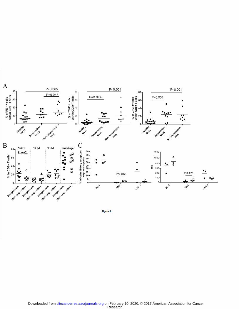
tostimulation with CEA691-peptide. As shown in Figure 4A, the frequency of PD-1, TIM-3 and LAG-3expressing CD8+
T cells was significantly higher in the non-responder group of PC patientsthanin the healthy controls.
It is known that T cell activation and differentiation status influences the expression of some cell surface markers,
such as PD-1. In order to examine the T cell phenotype in more detail, we identified naïve, central memory (TCM),
Research. on February 10, 2020. © 2017 American Association for Cancerclincancerres.aacrjournals.org Downloaded from
Author manuscripts have been peer reviewed and accepted for publication but have not yet been edited. Author Manuscript Published OnlineFirst on July 14, 2017; DOI: 10.1158/1078-0432.CCR-17-1185

13
effector memory (TEM), end-stage/effector phenotypes based on CD62L and CD45RO expression (i.e., TCM was
defined as CD62L+CD45RO+ T cells, TEM as CD62L-CD45RO+T cells, Naïve T cells as CD62L+CD45RO-, and end-
stage/effector T cells as CD62L-CD45RO+). As shown in Figure 4B, there was no statistically significant difference in
the percentage of gated CD4+ or CD8+ T cells at various differentiation stages between PC responders and non-
responders.The percentage of naïve T cells, however, in responders was observed to be higher than in non-
responders, but this difference did not achieve statistically difference (p=0.051).
CD8+ T-cell priming and activation takes place in draining lymph nodes where, upon interaction with antigen-
presenting cells, naïve T cells are primed and differentiate into fully-functional antigen-specific CTLs. We were able
to analyse the expression of PD-1, TIM-3 and LAG-3 in matched peripheral- and lymph node-derived CD8+ T cells
isolated from three of the PC patients(see Table 1). The proportion of lymph node-derived CD8+ T cells expressing
PD-1 and TIM-3, was higher than that observed in peripheral T cells, whereas more LAG-3 positive cells were
identified within the peripheral T ell repertoire than in the draining lymph nodes (Figure 4C, left panel). In addition,
similar differences were observed when analysing the expression levels of PD-1, TIM-3 and LAG-3, as determined by
mean fluorescence intensity, MFI (Figure 4C, right panel).
Blockade of the PD-1/PD-L1 pathway improves tumor antigen-specific CD8+ T-cell responses
To assess whether blockade of the PD-1/PD-L1 negative regulatory pathway enhanced the function of patient
derived, self-restricted CEA691-specific CTL, PBMCs from patients with PC (and LN cells from one patient) were
cultured for seven days in the presence of pCEA691 or the control peptide, rIL-2, with or without anti-PD-L1 and/or
anti-TIM-3 antibodies. In total, the frequency of peptide-specific cytokine producing CD8+ T cells was evaluated in 11
patients with PC (see Table 2 for patient details). In addition, the frequency of CEA691 tetramer positive CD8+ T cells
was determined in 8 of the 11 patients (where sufficient samples were available for analysis).
As shown in Figure 5A, a significant increase in the frequency of CEA691 tetramer-binding CD8+ T cells was observed
in cells cultured in the presence of anti-PD-L1 antibody (p=0.030, n=8) and the combination of anti-PD-L1+anti-TIM-3
antibodies (p=0.045, n=8), compared to non-treated cells. These findings suggest that PD-L1 and TIM-3 blockade can
enhance the proliferation of PC patient derived CEA691-specific CTLs in vitro. Similarly, the frequency of IFNγ-
producing CD8+ T cells was significantly higher in the cells treated with anti-PD-L1 or anti-TIM-3, alone or in
combination (Figures 5A, 5B and 5C). Even though both PD-1/PD-L1 and TIM-3 regulatory pathways were observed
Research. on February 10, 2020. © 2017 American Association for Cancerclincancerres.aacrjournals.org Downloaded from
Author manuscripts have been peer reviewed and accepted for publication but have not yet been edited. Author Manuscript Published OnlineFirst on July 14, 2017; DOI: 10.1158/1078-0432.CCR-17-1185

14
to be involved in the modulation of CTL function, our data did not reveal a significant synergistic effect when PD-
1/PD-L1 and TIM-3 pathways were blocked at the same time.
To determine the relative function of negative regulatory pathways in the inhibition of antigen-specific CTL
responses in the lymph nodes (LN) ofpatients with PC, MNCs isolated from lymph nodes were cultured with the
pCEA691 peptide in the presence or absence of anti-PD-L1 and/or anti-TIM-3 blocking antibodies. Blockade of the
PD-1/PD-L1 pathway, but not of the TIM-3 pathway, led to an increase in the frequency of IFNγ producing cells in
both circulating and lymph node-derived CTLs, suggesting a role for the PD-1/PD-L1 axis in the modulation of IFNγ
responses in patients with PC (Fig. 5C and 5D). We observed that the expression of PD-1 and TIM-3 was higher in
lymph node-derived CD8+ T cells than that in peripheral T cells, andblockade of these inhibitory molecules led to a
more significant recovery of peptide-specific IFNγ production by CD8+ T cells.
Our findings suggest that the expression level of negative regulatory molecules, in circulating and lymph node-
derived CTLs, may influence the magnitude of the CTL mediated anti-tumorresponse in different tissues.
Discussion
The prognosis for patients with pancreatic cancer remains extremely poor and novel treatments such as
immunotherapy are appealing alternatives to improve survival. However, there is currently little evidence from large
clinical trialsto demonstrate that pancreatic cancer is sensitive to checkpoint inhibition, and vaccine based
immunotherapieshave largely failed to generate significant clinical results(10, 17, 18, 24). Meanwhile, some animal
experiments have suggested that the combination of checkpoint inhibitors with other immunotherapies, such as
therapeutic vaccines, may improve outcomes in pancreatic cancer (24). Our study, has focused on exploring
potential epitopes for targeted immunotherapy in pancreatic cancer patients, and has evaluated the influence of
blockade of PD-1/PD-L1 pathway or TIM-3 pathway in anti-tumor T cell responses.
Carcinoembryonic antigen (CEA) is an 180 kDa glycosylated membrane protein that is over-expressed in more than
90% of PC cases (16). As a self-antigen, CEA-specific T cells are expected to be subject to immunological tolerance.
Although previous studies demonstrated that tolerance to CEA691 is incomplete (30), little is known about the
function of CEA691-specific CTL isolated from patients with pancreatic cancer.Here, we identified the most
immunogenic CEA-derived epitopes using short-term in vitro PBMC stimulationfollowed by prolonged antigen
Research. on February 10, 2020. © 2017 American Association for Cancerclincancerres.aacrjournals.org Downloaded from
Author manuscripts have been peer reviewed and accepted for publication but have not yet been edited. Author Manuscript Published OnlineFirst on July 14, 2017; DOI: 10.1158/1078-0432.CCR-17-1185

15
stimulation to expand primary CEA691-specific T-cell lines. After one round of in vitro peptide stimulation we
observed that the CEA691 peptide-specific IFN producing T cell responses were more readily detectable in PC
patients compared to healthy controls and that responding patients had higher percentages of IFN-producing T cells
than those stimulated by other peptide epitopes. After a total of 4 rounds of in vitro stimulation, the CEA691-specific
CTLs from PC patients were also able to produce TNF and kill relevant target cells.
Despite the small sample size we were able to demonstrate thatT cells isolated from patients presenting with more
advanced disease were less likely togenerate effective antigen-specific cytokine-producing responses. It is possible
that prolonged TAA exposure in the tumor microenvironment may induce exhaustion of antigen-specific T cell
responses. Unfortunately, we were unable to test PBMCs at different time points over the course of PC progression
for individual patients. Thus, the evolution of phenotypic and functional changes in the anti-CEA691T cell responses
in our patients may be related to multiple factors including prior treatment, lymphopenia in advanced patients, co-
morbidities including age and/or the use of immunosuppressive drugs. Similarly, PC patients without demonstrable
CEA691-specific T cell responses may have presented with more aggressive and/or quickly progressing cancers,hence
having more advanced disease at diagnosis. To address these issues, a prospective longitudinal study with a larger
sample size is needed.
Interestingly, our preliminary findings suggest that surgical treatment, which had occurred prior to patients being
evaluatedin the study (and therefore prior to T cell isolation), may also modulate CD8+ T cell responses, suggesting
different modes of treatment received may also impact on TAA-specific immune responsesin PC.The interpretation
of this data should take into account that this study was not designed to look at differences in clinical outcome and
that the treatment modalities used were heterogeneous. In our previous study the influence of medical
interventions on T cell response was observed demonstratingthat embolization improved AFP-specific CD4+ T cell
responses in patients with hepatocellular carcinoma (39).Again, to confirm these findings a much larger study would
be required to control for all the relevant variables.
Activation of the inhibitory PD-1/PD-L1 pathway has been linked to the poor prognosis of PC (40). Our data has also
shown that patients not responding to pCEA691 had larger numbers of PD-1 expressing T cells in their peripheral T
cell compartment, compared to the T cells isolated from responding patients or healthy controls. Also, T cells
isolated from the tumor draining LNs were enriched for PD-1 and TIM-3 expressing cellscompared to T cells isolated
Research. on February 10, 2020. © 2017 American Association for Cancerclincancerres.aacrjournals.org Downloaded from
Author manuscripts have been peer reviewed and accepted for publication but have not yet been edited. Author Manuscript Published OnlineFirst on July 14, 2017; DOI: 10.1158/1078-0432.CCR-17-1185
16
from the peripheral blood. Not surprisingly, these findings support the existence of an immunosuppressive tumor
microenvironment in PC patients. It is therefore likely that combination therapies with checkpoint inhibitors and
antigen specific T cells may be required to optimize immunotherapeutic approaches to PC.
Recently, the efficacy of anti-CTLA-4 and anti-PD1/PDL1 antibodies in treating several types of cancers has been
demonstrated in early phase clinical trials(41). However, these checkpoint inhibitors are considered most effective in
treating cancers with high mutational loads, e.g., melanoma and lung cancer, because such cancers typically
generate more neoantigens and checkpoint inhibitors reverse the inhibition of tumor infiltrating neoantigen-specific
T cells (42).On average, the neoantigen repertoire of pancreatic cancer is 1 mutation per megabase, which is much
less than that observed in melanoma and lung cancer (with more than 10 mutations per megabase on average) (43).
In theory, therefore, pancreatic cancer should not be very sensitive to checkpoint blockade. To date there have been
a number of clinical trialsassessing the safety and efficacy of checkpoint inhibitors in PC patients(44), but no positive
results have yet been published.
However, ourin vitro results suggest that the PD-1/PD-L1 pathway may bean important factor in the inhibitionof anti-
tumor functions of CD8+ T cells from PC patients. Following the addition of PD-L1 blocking antibodies, antigen-
specific T cellproliferation and cytokine production (IFN and TNF) was improved, with a greater effect observed on
T cells isolated from the tumor draining LNs than from the peripheral blood. In addition, the percentage of T cells
expressing PD-1 in LNs was higher than that observed in the peripheral blood. Thus, the limited impactto date of
checkpoint inhibition in PC may be due to the relatively small neoantigen repertoire, and/or thatTAA-specific T cells
less readily enter the tumormicroenvironment due to associateddesmoplasia. Finally, we did not observe a
synergistic effect when combining PD-L1 blockadewith TIM-3 blockade. This may have been because TIM-3 levels
were generally low in freshly isolated T cells. Little is known about the expression and role of TIM-3 (45, 46) and LAG-
3 (47) in T cells from pancreatic cancer patients. In our study, the percentage of TIM-3 and LAG-3 positive CD4+ and
CD8+ T cells was higher in pancreatic cancer patients, compared to healthy donors.Although the ex-vivo culture of
patient-derived effector T cells with PD1 and Tim3 blocking antibodies has experimental limitations, which cannot
reflect the complexity of in vivo environment, we believe it provides useful preliminary data on which to base further
studies.
Research. on February 10, 2020. © 2017 American Association for Cancerclincancerres.aacrjournals.org Downloaded from
Author manuscripts have been peer reviewed and accepted for publication but have not yet been edited. Author Manuscript Published OnlineFirst on July 14, 2017; DOI: 10.1158/1078-0432.CCR-17-1185

17
In conclusion, here we describe, the in vitrostimulation and expansion ofself-restricted, autologous, functional
CEA691-specificT cells, isolated from the peripheral blood and draining LNs of patients with pancreatic
adenocarcinoma.This raises the possibility that expansion and infusion of autologous CEA691-specific T cells may be
an effective immunotherapeutic approach to PC. Whilst alternative CEA-based immunotherapies are still being
designed and optimised for PC (12, 48, 49), including CEA-specific engineered T cells, which have been shown to
eradicate PC tumors without inducing toxicity in mice (50, 51), in another study the administration of autologous T
lymphocytes genetically engineered to express a murine TCR specific for CEA691 to colorectal cancer patients was
shown to induce severe transient colitis (52) resulting in suspension of the clinical trial, raising questions about the
suitability of this particular epitope for the design of novel TCR-based therapeutic approaches. Compared to the
CEA691 specific high-affinity TCR generated by others (52-54), our CEA691 specific T cell lines were isolated from
patients with pancreatic cancer and it is possible that their anti-tumor efficacy can be improved by the in vivo
reversalof dysfunctionmediated by increased expression of PD-1 and TIM-3.
ACKNOWLEDGEMENTS
We thank Mr Kito Fusai for performing lymph node resections and providing clinical care for surgically treated
patients. ECM is supported by the UCLH NIHR Biomedical Research Centre, the CRUK UCL Experimental Cancer
Medicine Centre, Bloodwise and the Medical Research Council. SPP is supported by National Institutes of Health and
by the National Institute for Health Research University College London Hospitals Biomedical Research Centre. SAX is
supported by the LLR program grant. SB is supported by BBSRC grant.
Research. on February 10, 2020. © 2017 American Association for Cancerclincancerres.aacrjournals.org Downloaded from
Author manuscripts have been peer reviewed and accepted for publication but have not yet been edited. Author Manuscript Published OnlineFirst on July 14, 2017; DOI: 10.1158/1078-0432.CCR-17-1185

18
REFERENCES
1. Ferlay J, Soerjomataram, I., Ervik, M., et al. GLOBOCAN 2012 v1.0, Cancer Incidence and Mortality Worldwide: IARC CancerBase No.11 2013 [cited 2013; Available from: http://globocan.iarc.fr 2. Cress RD, Yin D, Clarke L, Bold R, Holly EA. Survival among patients with adenocarcinoma of the pancreas: a population-based study (United States). Cancer Causes Control. 2006;17:403-9. 3. O'Reilly EM, Lowery MA. Postresection surveillance for pancreatic cancer performance status, imaging, and serum markers. Cancer J. 2012;18:609-13. 4. Siegel R, Naishadham D, Jemal A. Cancer statistics, 2013. CA Cancer J Clin. 2013;63:11-30. 5. Malvezzi M, Arfe A, Bertuccio P, Levi F, La Vecchia C, Negri E. European cancer mortality predictions for the year 2011. Ann Oncol. 2011;22:947-56. 6. Coupland VH, Konfortion J, Jack RH, Allum W, Kocher HM, Riaz SP, et al. Resection rate, hospital procedure volume and survival in pancreatic cancer patients in England: Population-based study, 2005-2009. Eur J Surg Oncol. 2016;42:190-6. 7. Bayraktar S, Bayraktar UD, Rocha-Lima CM. Recent developments in palliative chemotherapy for locally advanced and metastatic pancreas cancer. World J Gastroenterol. 2010;16:673-82. 8. Vulfovich M, Rocha-Lima C. Novel advances in pancreatic cancer treatment. Expert Rev Anticancer Ther. 2008;8:993-1002. 9. Conroy T, Desseigne F, Ychou M, Bouche O, Guimbaud R, Becouarn Y, et al. FOLFIRINOX versus gemcitabine for metastatic pancreatic cancer. N Engl J Med. 2011;364:1817-25. 10. Brahmer JR, Tykodi SS, Chow LQ, Hwu WJ, Topalian SL, Hwu P, et al. Safety and activity of anti-PD-L1 antibody in patients with advanced cancer. N Engl J Med. 2012;366:2455-65. 11. Maher J, Davies ET. Targeting cytotoxic T lymphocytes for cancer immunotherapy. Br J Cancer. 2004;91:817-21. 12. Osada T, Patel SP, Hammond SA, Osada K, Morse MA, Lyerly HK. CEA/CD3-bispecific T cell-engaging (BiTE) antibody-mediated T lymphocyte cytotoxicity maximized by inhibition of both PD1 and PD-L1. Cancer Immunol Immunother. 2015;64:677-88. 13. Pei Q, Pan J, Zhu H, Ding X, Liu W, Lv Y, et al. Gemcitabine-treated pancreatic cancer cell medium induces the specific CTL antitumor activity by stimulating the maturation of dendritic cells. Int Immunopharmacol. 2014;19:10-6. 14. Finn OJ. Cancer immunology. N Engl J Med. 2008;358:2704-15. 15. Hassan R, Laszik ZG, Lerner M, Raffeld M, Postier R, Brackett D. Mesothelin is overexpressed in pancreaticobiliary adenocarcinomas but not in normal pancreas and chronic pancreatitis. Am J Clin Pathol. 2005;124:838-45. 16. Yamaguchi K, Enjoji M, Tsuneyoshi M. Pancreatoduodenal carcinoma: a clinicopathologic study of 304 patients and immunohistochemical observation for CEA and CA19-9. J Surg Oncol. 1991;47:148-54. 17. Royal RE, Levy C, Turner K, Mathur A, Hughes M, Kammula US, et al. Phase 2 trial of single agent Ipilimumab (anti-CTLA-4) for locally advanced or metastatic pancreatic adenocarcinoma. J Immunother. 2010;33:828-33. 18. Koido S, Homma S, Takahara A, Namiki Y, Tsukinaga S, Mitobe J, et al. Current immunotherapeutic approaches in pancreatic cancer. Clin Dev Immunol. 2011;2011:267539. 19. Geng L, Huang D, Liu J, Qian Y, Deng J, Li D, et al. B7-H1 up-regulated expression in human pancreatic carcinoma tissue associates with tumor progression. J Cancer Res Clin Oncol. 2008;134:1021-7. 20. Nomi T, Sho M, Akahori T, Hamada K, Kubo A, Kanehiro H, et al. Clinical significance and therapeutic potential of the programmed death-1 ligand/programmed death-1 pathway in human pancreatic cancer. Clin Cancer Res. 2007;13:2151-7. 21. Feig C, Jones JO, Kraman M, Wells RJ, Deonarine A, Chan DS, et al. Targeting CXCL12 from FAP-expressing carcinoma-associated fibroblasts synergizes with anti-PD-L1 immunotherapy in pancreatic cancer. Proc Natl Acad Sci U S A. 2013;110:20212-7. 22. Winograd R, Byrne KT, Evans RA, Odorizzi PM, Meyer AR, Bajor DL, et al. Induction of T-cell Immunity Overcomes Complete Resistance to PD-1 and CTLA-4 Blockade and Improves Survival in Pancreatic Carcinoma. Cancer Immunol Res. 2015;3:399-411. 23. Soares KC, Rucki AA, Wu AA, Olino K, Xiao Q, Chai Y, et al. PD-1/PD-L1 blockade together with vaccine therapy facilitates effector T-cell infiltration into pancreatic tumors. J Immunother. 2015;38:1-11.
Research. on February 10, 2020. © 2017 American Association for Cancerclincancerres.aacrjournals.org Downloaded from
Author manuscripts have been peer reviewed and accepted for publication but have not yet been edited. Author Manuscript Published OnlineFirst on July 14, 2017; DOI: 10.1158/1078-0432.CCR-17-1185
19
24. Foley K, Kim V, Jaffee E, Zheng L. Current progress in immunotherapy for pancreatic cancer. Cancer Lett. 2016;381:244-51. 25. Hamilton SR AL, eds. Pathology and Genetics of Tumors of the Digestive System. WHO Classification of Tumors. Lyon: IARC Press; 2000. 26. Chen Y, Ayaru L, Mathew S, Morris E, Pereira SP, Behboudi S. Expansion of anti-mesothelin specific CD4+ and CD8+ T cell responses in patients with pancreatic carcinoma. PLoS One. 2014;9:e88133. 27. DeMars R, Chang CC, Shaw S, Reitnauer PJ, Sondel PM. Homozygous deletions that simultaneously eliminate expressions of class I and class II antigens of EBV-transformed B-lymphoblastoid cells. I. Reduced proliferative responses of autologous and allogeneic T cells to mutant cells that have decreased expression of class II antigens. Human immunology. 1984;11:77-97. 28. Nakagawa Y, Watari E, Shimizu M, Takahashi H. One-step simple assay to determine antigen-specific cytotoxic activities by single-color flow cytometry. Biomedical research. 2011;32:159-66. 29. Sadovnikova E, Zhu X, Collins SM, Zhou J, Vousden K, Crawford L, et al. Limitations of predictive motifs revealed by cytotoxic T lymphocyte epitope mapping of the human papilloma virus E7 protein. Int Immunol. 1994;6:289-96. 30. Keogh E, Fikes J, Southwood S, Celis E, Chesnut R, Sette A. Identification of new epitopes from four different tumor-associated antigens: recognition of naturally processed epitopes correlates with HLA-A*0201-binding affinity. J Immunol. 2001;167:787-96. 31. Bossi G, Gerry AB, Paston SJ, Sutton DH, Hassan NJ, Jakobsen BK. Examining the presentation of tumor-associated antigens on peptide-pulsed T2 cells. Oncoimmunology. 2013;2:e26840. 32. Driessens G, Kline J, Gajewski TF. Costimulatory and coinhibitory receptors in anti-tumor immunity. Immunol Rev. 2009;229:126-44. 33. Freeman GJ, Long AJ, Iwai Y, Bourque K, Chernova T, Nishimura H, et al. Engagement of the PD-1 immunoinhibitory receptor by a novel B7 family member leads to negative regulation of lymphocyte activation. J Exp Med. 2000;192:1027-34. 34. Hamanishi J, Mandai M, Iwasaki M, Okazaki T, Tanaka Y, Yamaguchi K, et al. Programmed cell death 1 ligand 1 and tumor-infiltrating CD8+ T lymphocytes are prognostic factors of human ovarian cancer. Proc Natl Acad Sci U S A. 2007;104:3360-5. 35. Nakanishi J, Wada Y, Matsumoto K, Azuma M, Kikuchi K, Ueda S. Overexpression of B7-H1 (PD-L1) significantly associates with tumor grade and postoperative prognosis in human urothelial cancers. Cancer Immunol Immunother. 2007;56:1173-82. 36. Zou W, Chen L. Inhibitory B7-family molecules in the tumor microenvironment. Nat Rev Immunol. 2008;8:467-77. 37. Wang X, Teng F, Kong L, Yu J. PD-L1 expression in human cancers and its association with clinical outcomes. Onco Targets Ther. 2016;9:5023-39. 38. Birnbaum DJ, Finetti P, Lopresti A, Gilabert M, Poizat F, Turrini O, et al. Prognostic value of PDL1 expression in pancreatic cancer. Oncotarget. 2016;7:71198-210. 39. Ayaru L, Pereira SP, Alisa A, Pathan AA, Williams R, Davidson B, et al. Unmasking of alpha-fetoprotein-specific CD4(+) T cell responses in hepatocellular carcinoma patients undergoing embolization. J Immunol. 2007;178:1914-22. 40. Song X, Liu J, Lu Y, Jin H, Huang D. Overexpression of B7-H1 correlates with malignant cell proliferation in pancreatic cancer. Oncol Rep. 2014;31:1191-8. 41. Kyi C, Postow MA. Checkpoint blocking antibodies in cancer immunotherapy. FEBS Lett. 2014;588:368-76. 42. Lu YC, Robbins PF. Cancer immunotherapy targeting neoantigens. Semin Immunol. 2016;28:22-7. 43. Schumacher TN, Schreiber RD. Neoantigens in cancer immunotherapy. Science. 2015;348:69-74. 44. Mizugaki H, Yamamoto N, Murakami H, Kenmotsu H, Fujiwara Y, Ishida Y, et al. Phase I dose-finding study of monotherapy with atezolizumab, an engineered immunoglobulin monoclonal antibody targeting PD-L1, in Japanese patients with advanced solid tumors. Invest New Drugs. 2016;34:596-603. 45. Tong D, Zhou Y, Chen W, Deng Y, Li L, Jia Z, et al. T cell immunoglobulin- and mucin-domain-containing molecule 3 gene polymorphisms and susceptibility to pancreatic cancer. Mol Biol Rep. 2012;39:9941-6. 46. Farren MR, Mace T, Geyer S, Mikhail S, Wu C, Ciombor KK, et al. Systemic immune activity predicts overall survival in treatment naive patients with metastatic pancreatic cancer. Clin Cancer Res. 2015.
Research. on February 10, 2020. © 2017 American Association for Cancerclincancerres.aacrjournals.org Downloaded from
Author manuscripts have been peer reviewed and accepted for publication but have not yet been edited. Author Manuscript Published OnlineFirst on July 14, 2017; DOI: 10.1158/1078-0432.CCR-17-1185

20
47. Kouo T, Huang L, Pucsek AB, Cao M, Solt S, Armstrong T, et al. Galectin-3 Shapes Antitumor Immune Responses by Suppressing CD8+ T Cells via LAG-3 and Inhibiting Expansion of Plasmacytoid Dendritic Cells. Cancer Immunol Res. 2015;3:412-23. 48. Alters SE, Gadea JR, Philip R. Immunotherapy of cancer. Generation of CEA specific CTL using CEA peptide pulsed dendritic cells. Adv Exp Med Biol. 1997;417:519-24. 49. Geynisman DM, Zha Y, Kunnavakkam R, Aklilu M, Catenacci DV, Polite BN, et al. A randomized pilot phase I study of modified carcinoembryonic antigen (CEA) peptide (CAP1-6D)/montanide/GM-CSF-vaccine in patients with pancreatic adenocarcinoma. Journal for immunotherapy of cancer. 2013;1:8. 50. Chmielewski M, Hahn O, Rappl G, Nowak M, Schmidt-Wolf IH, Hombach AA, et al. T cells that target carcinoembryonic antigen eradicate orthotopic pancreatic carcinomas without inducing autoimmune colitis in mice. Gastroenterology. 2012;143:1095-107 e2. 51. Chmielewski M, Rappl G, Hombach AA, Abken H. T cells redirected by a CD3zeta chimeric antigen receptor can establish self-antigen-specific tumor protection in the long term. Gene Ther. 2013;20:177-86. 52. Parkhurst MR, Yang JC, Langan RC, Dudley ME, Nathan DA, Feldman SA, et al. T cells targeting carcinoembryonic antigen can mediate regression of metastatic colorectal cancer but induce severe transient colitis. Molecular therapy : the journal of the American Society of Gene Therapy. 2011;19:620-6. 53. Parkhurst MR, Joo J, Riley JP, Yu Z, Li Y, Robbins PF, et al. Characterization of genetically modified T-cell receptors that recognize the CEA:691-699 peptide in the context of HLA-A2.1 on human colorectal cancer cells. Clin Cancer Res. 2009;15:169-80. 54. Zhou H, Luo Y, Mizutani M, Mizutani N, Becker JC, Primus FJ, et al. A novel transgenic mouse model for immunological evaluation of carcinoembryonic antigen-based DNA minigene vaccines. The Journal of clinical investigation. 2004;113:1792-8.
Research. on February 10, 2020. © 2017 American Association for Cancerclincancerres.aacrjournals.org Downloaded from
Author manuscripts have been peer reviewed and accepted for publication but have not yet been edited. Author Manuscript Published OnlineFirst on July 14, 2017; DOI: 10.1158/1078-0432.CCR-17-1185

21
TABLES
Table 1 Patient demographic information (PBMC samples, N=18; LN samples, N=3))
ID of PC patients
Age (year)
Sex Histological diagnosis Stage Treatment OS since recruitment
(months)
PBMC samples
CA01 51 M AC IV Untreated 14 CA02 37 M PDAC IIB Untreated* 24 CA03 86 F AC III Untreated 1 CA04 56 F Poorly differentiated AC IV Resected + FOLFOX 3 CA05 67 M AC IV GemCap 8 CA06 66 M Moderately differentiated PDAC IIB Resected, Pre-chem 15 CA07 60 M Ductal AC IIA Resected, Pre-chem 23 CA08 73 F Mucinous carcinoma IIA Resected, Gemcitabine 25 CA09 45 M AC IIB FOLFIRINOX 18 CA10 72 F AC IIB Resected + Gemcitabine 14 CA11 68 F Moderately differentiated AC IIB Resected, pre-chem 28 CA12 69 F Adenocarcinoma IIB Resected + GemCap 20 CA13 60 M Adenocarcinoma IV Untreated 6 CA14 46 M Adenocarcinoma III Untreated 21 CA15 52 M AC IV Gemcitabine 1 CA16 60 M AC IV Laparotomy, GemCap 14 CA17 45 F Adenocarcinoma IIB FOLFIRINOX* 16 CA18 55 M Moderately differentiated AC III Resected, pre-chem 31
LN samples
ID of PC patients
Age (year)
Sex Histological diagnosis Stage HLA-A2
CA13 60 M Adenocarcinoma IIB + CA19 52 M Moderately differentiated AC IIB - CA20 51 F Adenocarcinoma IIB -
*Status of patients when PBMC were collected. 8 patients had already undergone surgical resection and one
laparotomy alone, when the blood was taken. Patients CA02 and CA17had PBMC collected prior to surgical resection
resection of tumor.OS: Overall survival. PDAC: pancreatic ductal adenocarcinoma. AC: adenocarcinoma. chem:
chemotherapy. FOLFOX: Folinic acid, Fluorouracil and Oxaliplatin. GemCap: Gemcitabine and capecitabine.
FOLFIRINOX: FOLFOX+Irinotecan.
Research. on February 10, 2020. © 2017 American Association for Cancerclincancerres.aacrjournals.org Downloaded from
Author manuscripts have been peer reviewed and accepted for publication but have not yet been edited. Author Manuscript Published OnlineFirst on July 14, 2017; DOI: 10.1158/1078-0432.CCR-17-1185

22
FIGURELEGENDS
Figure 1. CEA691 specific T cells isolated form pancreatic cancer patients and healthy controls produce type 1
cytokines.
(A) A typical example of T cells expanded during stimulation with pCEA691 loaded T2 cells. Between the second (2R)
and fourth round (4R) of stimulation, the percentage of INF-producing CD8+ T cells gradually increased. By the end
of the 4th round stimulation, 77.9% of CD8+ T cells were INF-secreting, compared to 2% after 2 rounds.
Dot blots representing IFN-and TNF-producing CD8 T cells cultured with CEA691 pulsed T2 cells and control
peptide-loaded T2 cells after 4 rounds of stimulation (Gated on CD8 T cells) in four out of 15 healthy controls (B) and
four out of 18 cancer patients (C) are shown. The percentages of IFN-+TNF-+ CD8 T cells are shown in the upper-
right quadrants.
Figure 2. Higher frequencies of CEA691-specific CD8+ T-cell responsesare detectedin earlier stages of pancreatic
cancer.
(A) Pie chart illustrates the frequency of pancreatic cancer patients at different disease stages (II-IV). Frequencies of
IFN-γ+ cells within the CD8+ T-cell population are shown for patients stratified according to (B) disease stage, (C)
prior administration of chemotherapy, and (D) submission to surgery. Each symbol represents one individual and
horizontal bars represent median. P values <0.5 were considered statistically significant and are shown in the graphs.
Figure 3 Cytotoxic activity of CTL lines against pancreatic cancer cell lines.
(A) CEA and HLA-A2 expression of 6 pancreatic cancer cell lines. The results were analysed by FACS and presented in
histogram (upper). Isotype antibodies were used to determine the background. Percentage and MFI of HLA-A2/CEA
expressions by different PC cell lines was also shown (bottom). N.B. The unstained cells were gated out. (B) FACS
analysis of CA11 T cells killing activity in response to recognise and kill PK-1 cell line (HLA-A2+, CEA+, labelled with
high dose CFSE), and MiaPaca-2 cell line (HLA-A2-, CEA-, labelled with low dose CFSE), at different E:T ratios. (C) The
percentage of relative killing of PK-1, Panc-1 and Bx-Pc-3 by CTLs from CA07 or CA11, compared to MiaPaca-2, at
different E:T ratios. All the experiments were repeated twice and the mean of the results was shown in the figure.
Research. on February 10, 2020. © 2017 American Association for Cancerclincancerres.aacrjournals.org Downloaded from
Author manuscripts have been peer reviewed and accepted for publication but have not yet been edited. Author Manuscript Published OnlineFirst on July 14, 2017; DOI: 10.1158/1078-0432.CCR-17-1185

23
Figure 4. Expression of negative regulatory molecules in pancreatic cancer.
PBMC were isolated from pancreatic cancer (PC) patients and healthy controls, and surface stained to assess (A) the
expression of PD1, TIM-3 and LAG-3 molecules; as well as (B) the relative proportions of naïve/memory subsets
(based on the expression of CD45RO and CD62L), within the CD8+ T-cell population. Pancreatic cancer patients were
stratified according to their ability to mount CEA691-specific CD8+ T-cell responses (responders vs. non-responders).
Each symbol represents one individual and horizontal bars represent mean. P values <0.5 were considered
statistically significant and are shown in the graphs. (C)T cells were obtained from matching samples of peripheral
blood and lymph nodes from three pancreatic cancer patients. Graphs illustrate the levels of negative regulatory
molecule expression within the CD8+ population, both in terms of frequency and number of molecules per cell,
expressed as mean fluorescence intensity (MFI). Each symbol represents one individual and horizontal bars
represent mean. P values <0.5 were considered statistically significant and are shown in the graph.
Figure 5. PD-1/PD-L1 and TIM-3 blockade in pancreatic cancer.
(A) The percentage of CEA tetramer binding CD8+ T cells after seven days of culture with CEA691 in the presence or
absence of anti-PD-L1 and/or anti-TIM-3 blocking antibodies. (B) CEA691 CD8+ T cells from 11 pancreatic cancer (PC)
patients were expanded for seven days with or without anti-PD-L1 and/or anti-TIM-3 blocking antibodies, and
intracellular stained to assess the levels of IFN-γ production. The graph shows frequencies of IFN-γ+ cells within the
CD8+ T-cell population. Each symbol represents one individual and horizontal bars represent mean. P values <0.5
were considered statistically significant and are shown in the graph. PBMCs and MNCs isolated from lymph nodes of
(CA13) were also stimulated for seven days by pCEA691 with or without anti-PD-L1 and/or anti-TIM-3 blocking
antibodies, and assessed for levels of IFN-γ production. A representative example of IFN-γ production is shown in the
dot-plots (C). (D) The graph shows the frequency of IFN-γ+ cells within the CD8+ T-cell population. Experiments are
duplicated and symbols indicate mean and SD.
Research. on February 10, 2020. © 2017 American Association for Cancerclincancerres.aacrjournals.org Downloaded from
Author manuscripts have been peer reviewed and accepted for publication but have not yet been edited. Author Manuscript Published OnlineFirst on July 14, 2017; DOI: 10.1158/1078-0432.CCR-17-1185

Published OnlineFirst July 14, 2017.Clin Cancer Res Yuan Chen, Shao-An Xue, shahriar behboudi, et al. circulating CEA specific T cells in pancreatic cancer patients.Ex vivo PD-L1/PD-1 pathway blockade reverses dysfunction of
Updated version
10.1158/1078-0432.CCR-17-1185doi:
Access the most recent version of this article at:
Material
Supplementary
http://clincancerres.aacrjournals.org/content/suppl/2017/07/14/1078-0432.CCR-17-1185.DC1
Access the most recent supplemental material at:
Manuscript
Authorbeen edited. Author manuscripts have been peer reviewed and accepted for publication but have not yet
E-mail alerts related to this article or journal.Sign up to receive free email-alerts
Subscriptions
Reprints and
To order reprints of this article or to subscribe to the journal, contact the AACR Publications
Permissions
Rightslink site. Click on "Request Permissions" which will take you to the Copyright Clearance Center's (CCC)
.http://clincancerres.aacrjournals.org/content/early/2017/07/19/1078-0432.CCR-17-1185To request permission to re-use all or part of this article, use this link
Research. on February 10, 2020. © 2017 American Association for Cancerclincancerres.aacrjournals.org Downloaded from
Author manuscripts have been peer reviewed and accepted for publication but have not yet been edited. Author Manuscript Published OnlineFirst on July 14, 2017; DOI: 10.1158/1078-0432.CCR-17-1185