Ewa Paluch Institut Curie/CNRS, Paris Cortical actomyosin gel breakage triggers shape oscillations...
37
Ewa Paluch nstitut Curie/CNRS, Paris Cortical actomyosin gel breakage triggers shape oscillations in cells and cell fragments Present address: Max-Planck-Institute-CBG, Dresden
-
date post
18-Dec-2015 -
Category
Documents
-
view
219 -
download
0
Transcript of Ewa Paluch Institut Curie/CNRS, Paris Cortical actomyosin gel breakage triggers shape oscillations...

Ewa Paluch
Institut Curie/CNRS, Paris
Cortical actomyosin gel breakage triggers shape oscillations in cells and cell fragments
Present address:Max-Planck-Institute-CBG, Dresden

Cell crawling
[M. Abercrombie, Proc. R. Soc. Lond. B, 1980]
[B. Alberts et al., Molecular Biology of the Cell, 2002]

nucleus
Actin Myosin Microtubules
Cell crawling - actin
[D. Bray, Cell Movements, 2001]
Cortex intrinsic dynamics???

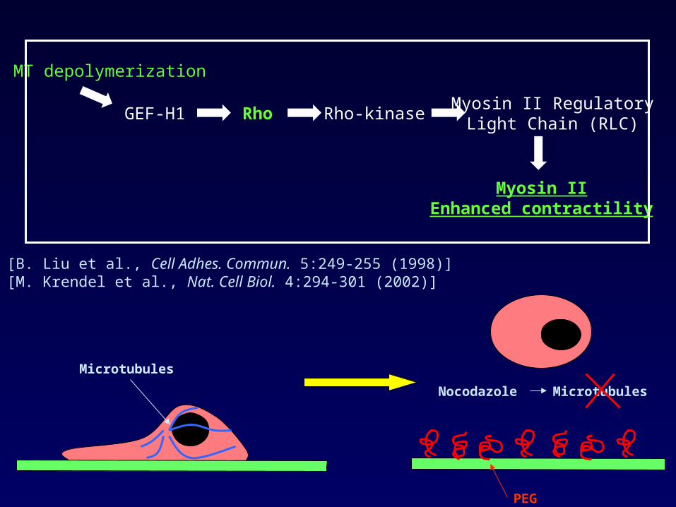
Dynamics of the actomyosin cortex in suspension cells
Microtubules
L929 fibroblast

Microtubules
PEG
Dynamics of the actomyosin cortex in suspension cells

Microtubules
Nocodazole Microtubules
PEG
Dynamics of the actomyosin cortex in suspension cells
Microtubules
Nocodazole Microtubules
PEG
[B. Liu et al., Cell Adhes. Commun. 5:249-255 (1998)][M. Krendel et al., Nat. Cell Biol. 4:294-301 (2002)]
Myosin IIEnhanced contractility
Rho Rho-kinase
MT depolymerization
GEF-H1Myosin II Regulatory
Light Chain (RLC)
PEG
Microtubules
Nocodazole Microtubules
Dynamics of the actomyosin cortex in suspension cells
Lymphoblasts: [M. Bornens, M. Paintrand, C. Celati, J. Cell Biol. 109:1071-1083 (1989)]

L929 fibroblasts
Centrifugation after microfilaments and
microtubules depolymerization
Cytoplast
Fragments
Nucleus
[E. Paluch, M. Piel, J. Prost, M. Bornens, C. Sykes, Biophys. J., 89:724-733]
Fragments of L929 fibroblastsMovie: http://www.biophysj.org/content/vol0/issue2005/images/data/biophysj.105.060590/DC1/Paluch-Movie1.mov

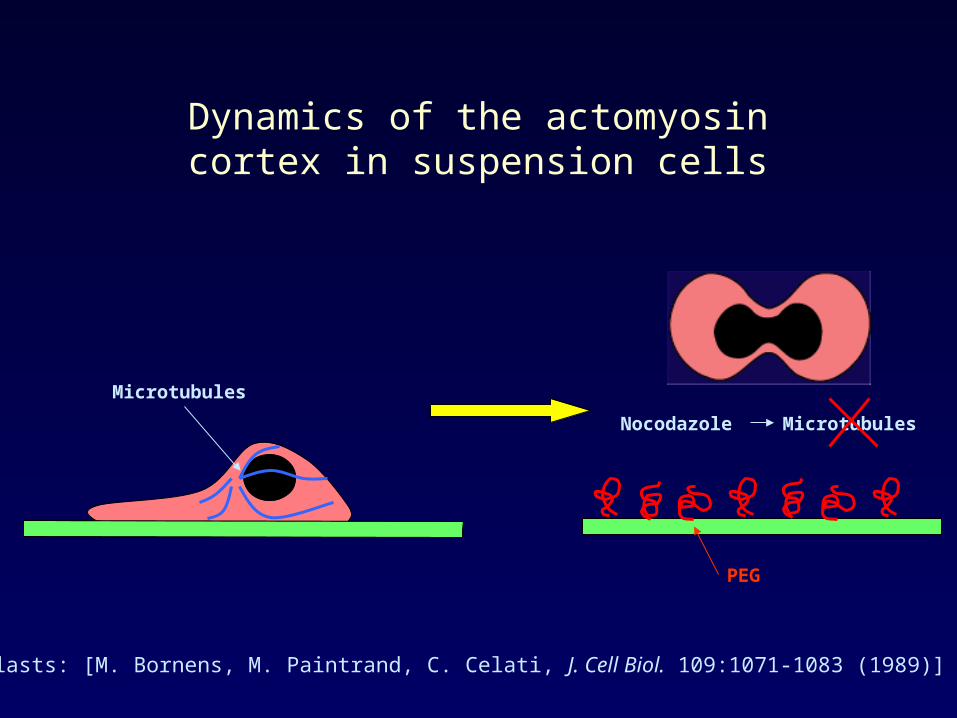
1. Actin and myosin II during the oscillation
2. The mechanism of the oscillation
3. Spontaneous cortical ruptures and cell motility

1. Actin and myosin II during the oscillation
Dynamic characterization of actin during the oscillation
Cell fragment
5 µm
Movie: http://www.biophysj.org/content/vol0/issue2005/images/data/biophysj.105.060590/DC1/Paluch-Movie3.mov
[E. Paluch et al., Biophys. J., 89:724-733]
1. Actin and myosin II during the oscillation
Dynamic characterization of actin during the oscillation
5 µm
[E. Paluch et al., Biophys. J., 89:724-733]

1. Actin and myosin II during the oscillation
Dynamic characterization of myosin II during the oscillation
5 µm
Movie: http://www.biophysj.org/content/vol0/issue2005/images/data/biophysj.105.060590/DC1/Paluch-Movie4.mov
[E. Paluch et al., Biophys. J., 89:724-733]

1. Actin and myosin II during the oscillation
Two-steps mechanism:
1. The actomyosin shell breaks
2. A bulge is expelled and grows
2. The mechanism of the oscillation
3. Spontaneous cortical ruptures and cell motility
1. Actin and myosin II during the oscillation
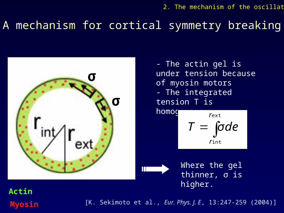
A mechanism for cortical symmetry breaking
σ
σ
- The actin gel is under tension because of myosin motors- The integrated tension T is homogenous:
∫=ext
int
r
r
deT σ
Where the gel thinner, σ is higher.
[K. Sekimoto et al., Eur. Phys. J. E, 13:247-259 (2004)]
Actin
Myosin
2. The mechanism of the oscillation
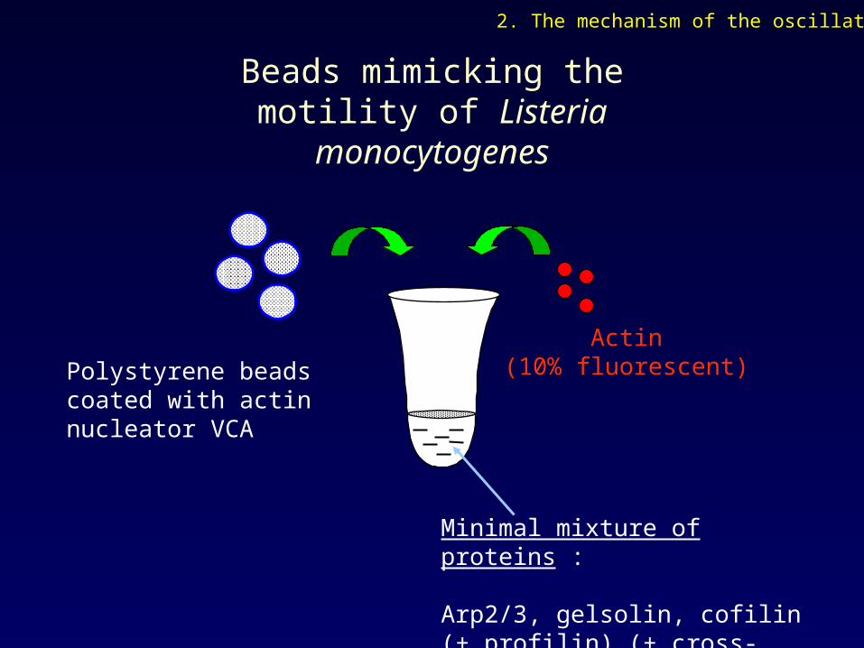
Beads mimicking the motility of Listeria monocytogenes
Polystyrene beads coated with actin nucleator VCA
Actin(10% fluorescent)
Minimal mixture of proteins :
Arp2/3, gelsolin, cofilin (+ profilin) (+ cross-linkers)
2. The mechanism of the oscillation

Actin gel
Actin nucleator
bead
Beads mimicking the motility of Listeria monocytogenes
2. The mechanism of the oscillation
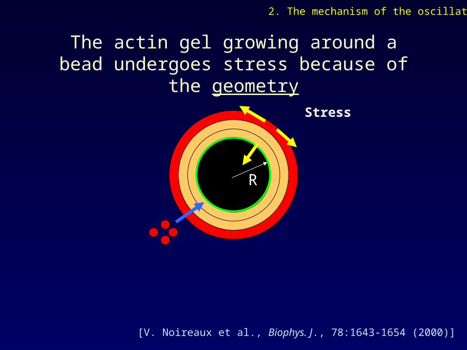
[V. Noireaux et al., Biophys. J., 78:1643-1654 (2000)]
The actin gel growing around a bead undergoes stress because of the geometry
R
Stress
2. The mechanism of the oscillation

Observation of symmetry breaking
2. The mechanism of the oscillation
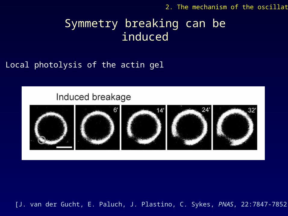
[J. van der Gucht, E. Paluch, J. Plastino, C. Sykes, PNAS, 22:7847-7852 (2005)]
Symmetry breaking can be induced
Local photolysis of the actin gel
2. The mechanism of the oscillation

t = 13’ t = 15’ t = 17’
[J. van der Gucht, E. Paluch, J. Plastino, C. Sykes, PNAS, 22:7847-7852 (2005)]
Hole formation is reminiscent of a fracture
2 µm
2. The mechanism of the oscillation
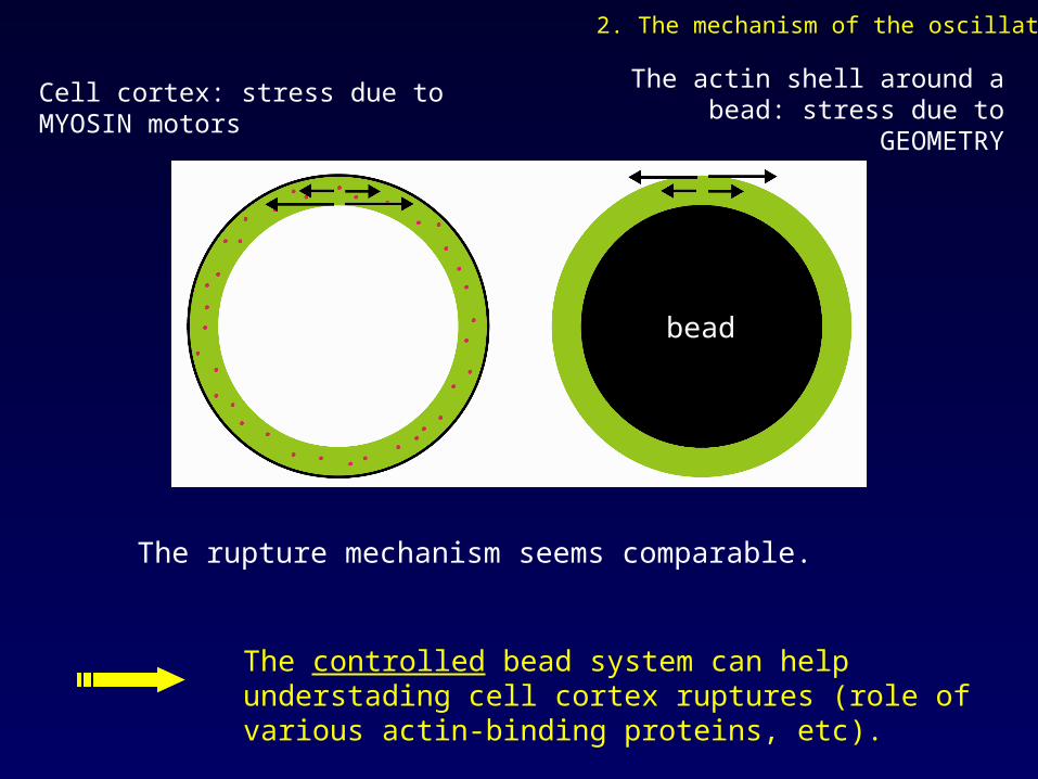
The rupture mechanism seems comparable.
The controlled bead system can help understading cell cortex ruptures (role of various actin-binding proteins, etc).
The actin shell around a bead: stress due to GEOMETRY
Cell cortex: stress due to MYOSIN motors
bead
2. The mechanism of the oscillation
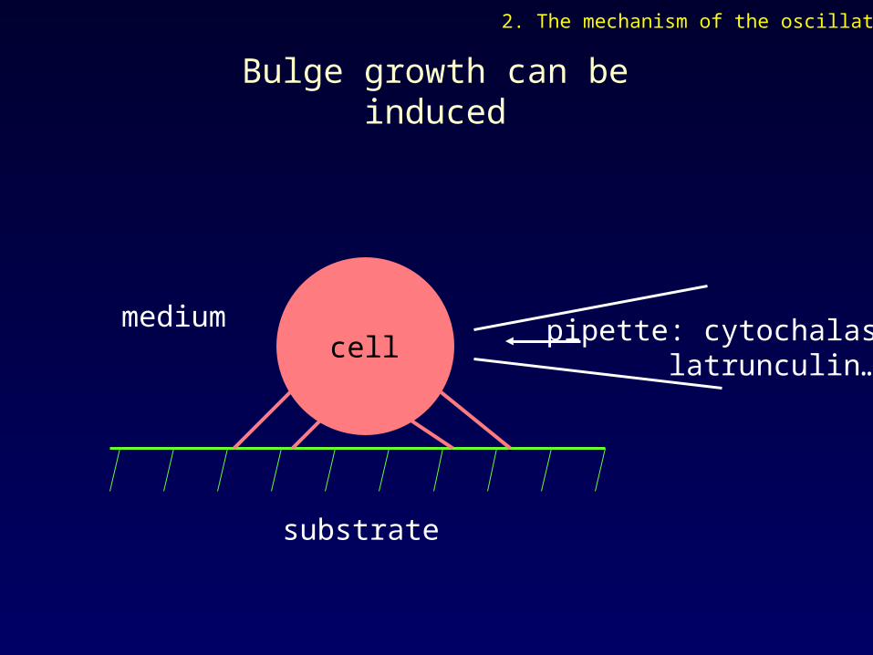
Bulge growth can be induced
pipette: cytochalasin latrunculin…
substrate
cellmedium
2. The mechanism of the oscillation

Local stress application induces bulge growth
P = 200 Pa
flow of medium
pipette
cell
2. The mechanism of the oscillation
[E. Paluch et al., Biophys. J., 89:724-733]
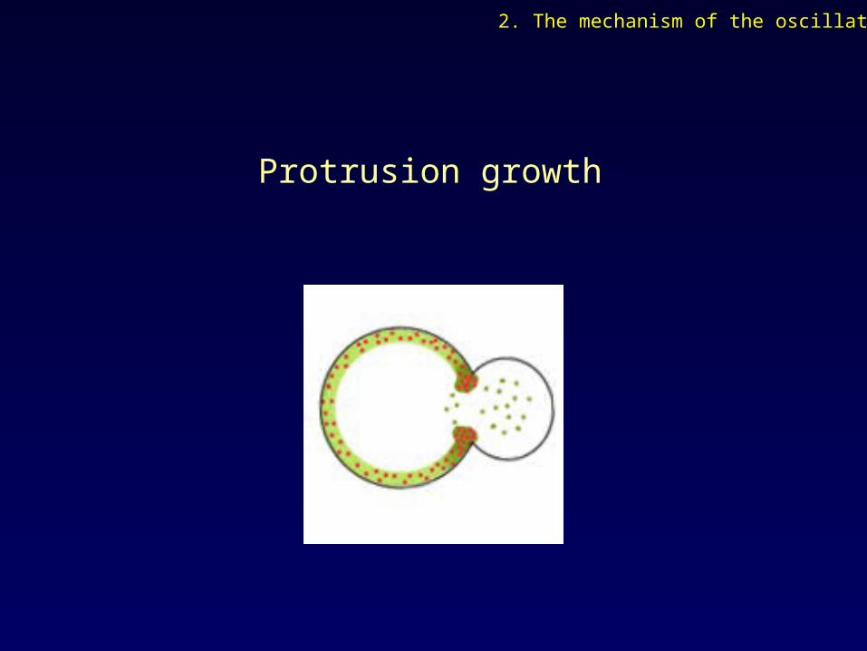
Protrusion growth
2. The mechanism of the oscillation
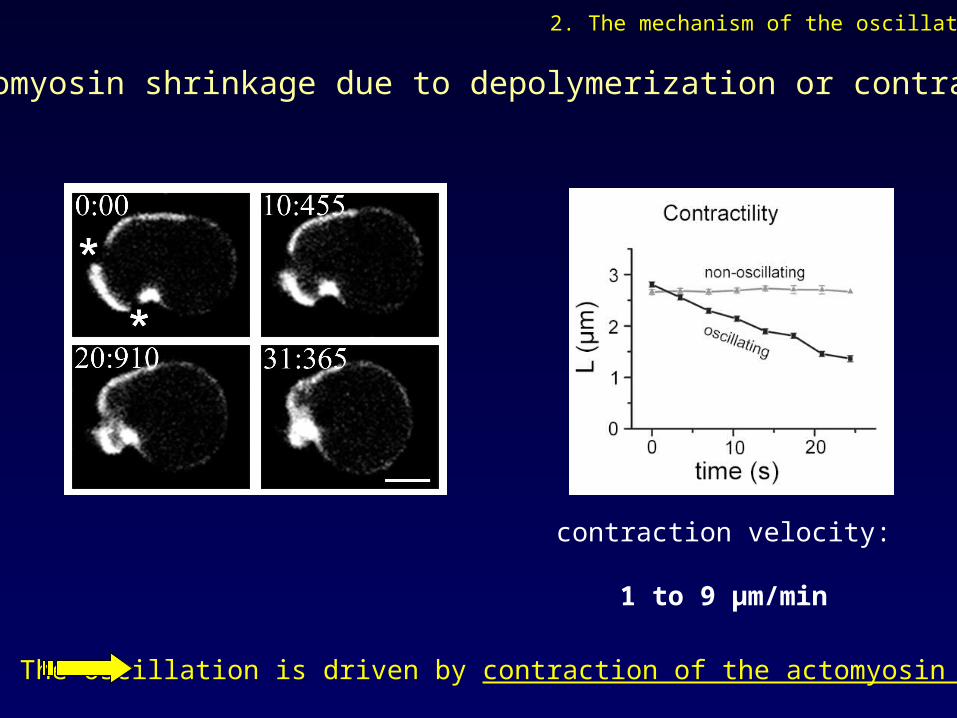
The oscillation is driven by contraction of the actomyosin cortex
contraction velocity:
1 to 9 µm/min
2. The mechanism of the oscillation
Is actomyosin shrinkage due to depolymerization or contraction?

A mechanism for the oscillation
[E. Paluch, M. Piel, J. Prost, M. Bornens, C. Sykes, Biophys. J., 89:724-733]
Actin
Myosin
2. The mechanism of the oscillation

3. Spontaneous cortical ruptures and cell motility
2. The mechanism of the oscillation
1. Actin and myosin II during the oscillation

… + microtubules
reduce myosin II activity
Actin
Myosin
Microtubules
3. Spontaneous cortical ruptures and cell motility
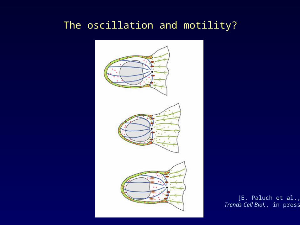
The oscillation and motility?
[E. Paluch et al., Trends Cell Biol., in press]
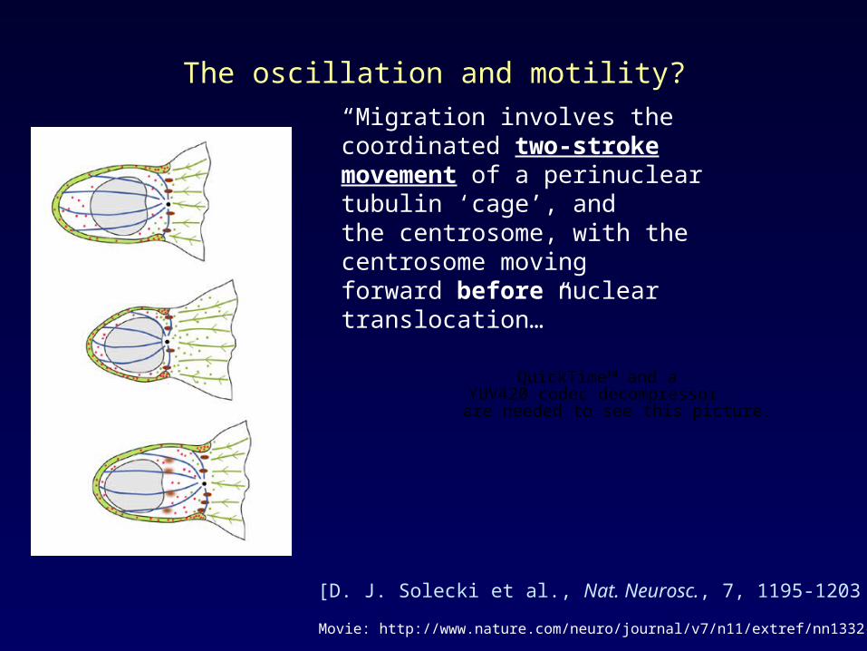
The oscillation and motility?
[D. J. Solecki et al., Nat. Neurosc., 7, 1195-1203 (2004)]
“Migration involves the coordinated two-strokemovement of a perinuclear tubulin ‘cage’, andthe centrosome, with the centrosome movingforward before nuclear translocation…”
QuickTime™ and aYUV420 codec decompressor
are needed to see this picture.
Movie: http://www.nature.com/neuro/journal/v7/n11/extref/nn1332-S8.mpg
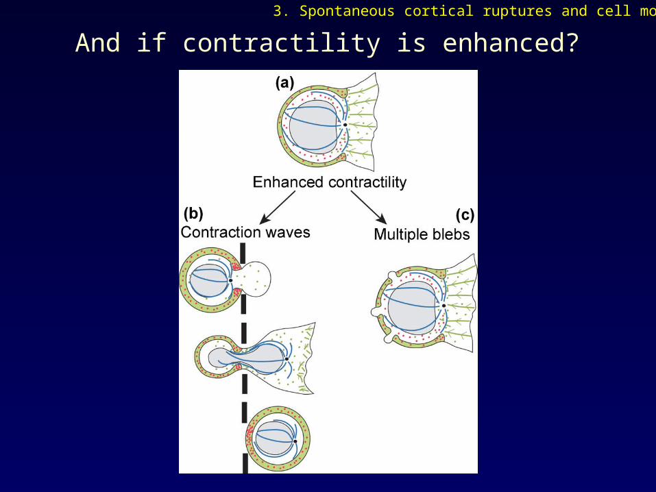
And if contractility is enhanced?
3. Spontaneous cortical ruptures and cell motility

[K. Wolf et al., J. Cell Biol., 160:267-277 (2003)]
I. Contraction waves
[E. Sahai, C. Marshall, Nat. Cell Biol., 5:711-719 (2003)]
II. Multiple blebs
And if contractility is enhanced?
3. Spontaneous cortical ruptures and cell motility
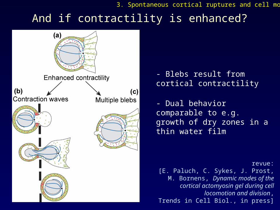
And if contractility is enhanced?
- Blebs result from cortical contractility
- Dual behavior comparable to e.g. growth of dry zones in a thin water film
revue: [E. Paluch, C. Sykes, J. Prost, M. Bornens, Dynamic modes of the cortical actomyosin
gel during cell locomotion and division,Trends in Cell Biol., in press]
3. Spontaneous cortical ruptures and cell motility

Summary
• Cortical oscillation is a general phenomenon resulting from elastic gel properties of the actomyosin cortex
• Bleb formation reveals the level of cortical contractility
• Spontaneous cortical ruptures (and blebs) can be used by cells or remain a side-product of cortex contractions
• Cortex breakage in cells // symmetry breaking of gels around beads

Biology group
Michel Bornens
Matthieu Piel
Physics group
Cécile Sykes
Jasper van der Gucht
Julie Plastino
Theorists
Jean-François JoannyJacques Prost
- GFP constructs: Beat Imhof (University of Geneva) Rex Chisholm (Northwestern University, Chicago)
- Deconvolution: Jean-Baptiste Sibarita (Institut Curie)