Evolution of Nuclear Drug Delivery
24
1 Evolution of Nuclear Drug Delivery Yick Chong Lam* Department of Chemistry, University of Rochester, Rochester, NY 14627. March 22, 2012 Introduction to Nuclear Drug Delivery Research on nuclear drug delivery strategies is constantly evolving to enhance therapeutic and pharmaceutical treatments. Effective drugs have previously been synthesized that display therapeutic activity, but are impractical due to difficulties in human delivery. 1 Drug delivery methods aim to successfully transport these molecules to subcellular compartments, such as the nucleus, for their operations. 2 Extensive research has been conducted to exploit the numerous applications and benefits of drug delivery. 3 Drug delivery research primarily establishes the potential to administer old and new pharmaceutical molecules more efficiently. New delivery methods may also be used to experiment future treatments and to determine whether continuous or pulsatile therapies elicit the desired response. Furthermore, modern drugs, such as synthetic recombinant proteins, are more complex than conventional small molecule drugs and thus, require novel delivery systems for successful transport. 3 Among various subcellular targeting strategies, nuclear drug delivery strategies have been constantly advancing due to ingenious manipulations of biological pathways and physiological conditions. The primary advantage of nuclear drug delivery over other subcellular targeting strategies is the ability to directly influence the sensitive nucleus to alter its control on signaling pathways and DNA replication. 4 Pharmaceutical molecules are able to directly bind to malfunctioning DNA to alleviate damage. Anticancer drugs and drugs that target nuclear proteins, such as those involved in DNA replication and transcription, may also be potent therapeutic agents. 5 As a result, nuclear drug delivery strategies are at the vanguard of therapeutic and pharmaceutical treatments. In this review, we discuss the meticulous manipulations of biological pathways and considerations of physiological properties that are required for the functionality and development of novel nuclear drug delivery strategies. 2, 5 We also discuss recent nuclear drug delivery strategies employing chemical modification, peptide, nanoparticle, and inorganic systems that have the potential to revolutionize current therapies. Biological Pathways Manipulated by Nuclear Drug Delivery Systems Cellular Internalization Nuclear drug delivery systems are required to manipulate biological mechanisms of endogenous molecular transportation into and out of the nucleus. Several biological mechanisms may be exploited, but only some are plausible for nuclear drug delivery. The first step in delivering pharmaceutical cargo, such as synthetic drugs, natural products, or DNA, to the nucleus is internalization through the plasma cell membrane by endocytic mechanisms. 2 One common cellular internalization mechanism for macromolecules is clathrin-dependent endocytosis (Figure 1a). 6 Initially, adaptin proteins identify cargo outside of the plasma membrane that
Transcript of Evolution of Nuclear Drug Delivery

1
Evolution of Nuclear Drug Delivery
Yick Chong Lam*
Department of Chemistry, University of Rochester, Rochester, NY 14627.
March 22, 2012
Introduction to Nuclear Drug Delivery
Research on nuclear drug delivery strategies is constantly evolving to enhance therapeutic and pharmaceutical treatments. Effective drugs have previously been synthesized that display therapeutic activity, but are impractical due to difficulties in human delivery.1 Drug delivery methods aim to successfully transport these molecules to subcellular compartments, such as the nucleus, for their operations.2 Extensive research has been conducted to exploit the numerous applications and benefits of drug delivery.3 Drug delivery research primarily establishes the potential to administer old and new pharmaceutical molecules more efficiently. New delivery methods may also be used to experiment future treatments and to determine whether continuous or pulsatile therapies elicit the desired response. Furthermore, modern drugs, such as synthetic recombinant proteins, are more complex than conventional small molecule drugs and thus, require novel delivery systems for successful transport.3 Among various subcellular targeting strategies, nuclear drug delivery strategies have been constantly advancing due to ingenious manipulations of biological pathways and physiological conditions. The primary advantage of nuclear drug delivery over other subcellular targeting strategies is the ability to directly influence the sensitive nucleus to alter its control on signaling pathways and DNA replication.4 Pharmaceutical molecules are able to directly bind to malfunctioning DNA to alleviate damage. Anticancer drugs and drugs that target nuclear proteins, such as those involved in DNA replication and transcription, may also be potent therapeutic agents.5 As a result, nuclear drug delivery strategies are at the vanguard of therapeutic and pharmaceutical treatments. In this review, we discuss the meticulous manipulations of biological pathways and considerations of physiological properties that are required for the functionality and development of novel nuclear drug delivery strategies.2, 5 We also discuss recent nuclear drug delivery strategies employing chemical modification, peptide, nanoparticle, and inorganic systems that have the potential to revolutionize current therapies. Biological Pathways Manipulated by Nuclear Drug Delivery Systems Cellular Internalization
Nuclear drug delivery systems are required to manipulate biological mechanisms of endogenous molecular transportation into and out of the nucleus. Several biological mechanisms may be exploited, but only some are plausible for nuclear drug delivery. The first step in delivering pharmaceutical cargo, such as synthetic drugs, natural products, or DNA, to the nucleus is internalization through the plasma cell membrane by endocytic mechanisms.2
One common cellular internalization mechanism for macromolecules is clathrin-dependent endocytosis (Figure 1a).6 Initially, adaptin proteins identify cargo outside of the plasma membrane that

2
must be transported into the nucleus. Adaptin and other extracellular cargo receptors bind to the cargo while clathrin binds to the inner membrane. Clathrin then polymerizes around the cargo with the aid of amphiphysin and endophilin proteins to form a transport vesicle endosome. The endosome is excised from the plasma membrane by the GTPase dynamin. Once in the cytosol, heat shock protein hsc70 removes the clathrin coating and the acidic environment causes the cargo receptors to undergo conformational changes to release the cargo-loaded endosome. The endosome may then fuse with lysosomes for cargo degradation or lyse to release the cargo into the cytoplasm (Figure 1b).4 Lysosomes are often targets for therapeutic treatments against lysosomal storage diseases, which are caused by enzyme deficiency, poor degradation, and toxic accumulation.7 Primarily, the majority of drugs aim to escape from the endosome by membrane diffusion and enter the nucleus for nuclear therapies.
Figure 1. (a) Clathrin Dependent Endocytosis. Reproduced from: Rappaport, J. S. Focusing on Clathrin-Mediated Endocytosis. Biochem. J., 2008, 412, 415-423. (b) Endocytic Mechanisms to Lysosomal Degradation or Escape. Reproduced from: Wagstaff, K. M.; Jans, D. A. Nuclear Drug Delivery to Target Tumour Cells. Eur. J. Pharmacol., 2009, 625, 174-180. Another common mechanism for internalization into the cell through the plasma membrane is caveolae-mediated endocytosis.8 Caveolae are flask-shaped invaginations with 60-90 nm diameters found on the plasma membranes of an abundance of cells including endothelial, fibroblast, and muscle cells. Caveolae are biosynthesized by the protein caveolin-1 and are responsible for cellular uptake of both endogenous and exogenous molecules. When a molecule approaches the plasma membrane, a molecular ligand, such as folic acid or cholesterol binds to a receptor in the caveolar invaginations (Figure 2).7, 9 Once the cargo is bound to the caveolae, the protein cavin folds the plasma membrane into a caveolar vesicle. Lastly, the protein GTPase dynamin excises the vesicle from the membrane with assistance from protein and tyrosine kinases.8 The resulting caveolae coated vesicles then fuse with caveosomes by the synaptosome-associated protein. The caveosomes are ultimately able to deliver molecular cargo to the Golgi complex or endoplasmic reticulum (ER).9 In this manner, caveolae-mediated endocytosis evades internalization into lysosomes, preventing lysosomal degradation of cargo. Once therapeutic drugs undergo these endocytic mechanisms, they must escape from their internalization vesicles and enter the targeted nucleus for executing their influence.
a b

3
Figure 2. Caveolae-Mediated Endocytosis into Caveosomes. Reproduced from: Parton, R. G.; Simons, K. The Multiple Faces of Caveolae. Nat. Rev. Mol. Cell Bio., 2007, 8, 185-194. Nuclear Localization and Import
Several biological mechanisms exist to transport endogenous molecules and therapeutic drugs into the nucleus. The conventional mechanism of nuclear transportation involves nuclear localization sequences (NLSs) and nuclear export sequences (NESs), importin (IMP) and exportin (EXP) transport receptors, nucleoporins of the nuclear pore complex (NPC) on the nuclear envelope, and the GTP-binding protein Ran (Figure 3).10, 11 Transportation into and out of the nucleus occurs on the double membrane nuclear envelope at the NPCs, which consists of nucleoporins that have hydrophobic phenylalanine-glycine (FG) repeats.10 The NLSs are amino acid targeting signal sequences that are recognized by IMPα-β1 or IMPβ1 transport receptors, which have histidine, glutamate, alanine, and threonine (HEAT) repeats.11 When NLSs are simultaneously bound to cargo, such as proteins or molecules, and to the IMP transport receptors, they permeate through the hydrophobic NPC using the hydrophobicity of IMPβ1.10 The HEAT repeats of IMPβ1 transport receptors facilitate the passage of cargo by associating and dissociating with the FG repeats of NPC nucleoporins.11 Once the IMP-NLS-cargo complex is inside the nucleus, Ran becomes activated by binding to GTP.10 The activated RanGTP binds to IMPβ1 to dissociate the transport complex, releasing the cargo into the nucleus. On the contrary, NESs are amino acid targeting signal sequences that are recognized by EXP receptors for nuclear exportation.10 Initially, NESs are bound simultaneously to cargo and the EXP receptor. When Ran is activated by binding to GTP, the activated RanGTP binds to the EXP receptor to facilitate exportation of the EXP-NES-cargo-RanGTP complex through the NPC. Exportation through the NPC is also mediated by the association and dissociation of amino acid residues between the NPC nucleoporins and the EXP receptor.10 Outside of the nucleus, when RanGTPase-activating protein catalyzes RanGTP hydrolysis at the gamma phosphate to form RanGDP, the cargo is released into the cytoplasm. These nuclear transportation mechanisms also depend on other conditions such as phosphorylation and dephosphorylation reactions, which can stimulate or inhibit nuclear transportation. Numerous cargo molecules, including p53, GAL4, and cyclin D are regulated by kinase and phosphatase activity for IMP and EXP receptor-mediated nuclear transportation.10

4
Figure 3. Conventional Mechanism for Nuclear Importation and Exportation. Reproduced from: Poon, I. K. H.; Jans, D. A. Regulation of Nuclear Transport: Central Role in Development and Transformation? Traffic, 2005, 6, 173-186.
Other mechanisms of nuclear localization also exist with the potential to be exploited by nuclear
drug delivery strategies. First, adenovirus has been observed to move along intracellular microtubules with the aid of dynein toward the NPCs. Adenovirus binds to the NPC and hsc70 decoats the viral capsid to release the viral DNA, which probably then diffuses into the nucleus.12 Similarly, the simian virus 40 (SV40) has been observed to escape caveosomes and traffic on microtubules to the ER. SV40 can then either migrate through the ER membrane to the inner nuclear membrane or escape the ER, followed by nuclear entry through the NPCs.12 Other alternate, nonviral mechanisms of nuclear localization involve DNA binding proteins, transcription factors, HMG box proteins, and histones.11 However, all of these proteins possess specific NLSs that are recognized by IMPα-β1 or IMPβ1 transport receptors to mediate nuclear entry through the NPCs. Though cellular internalization and nuclear localization mechanisms are abundant, nuclear drug delivery strategies must be carefully designed.
Nuclear Drug Delivery Requirements: Overview Natural biological pathways must be accurately manipulated for successful nuclear drug
delivery. Nuclear drug delivery requires a general protocol for drug importation into the nucleus. First, the drug must enter the body and bind to plasma membrane surface receptors. Drug delivery strategies are able to specifically target one cell type by incorporating ligands or moieties that are only recognized by the surface receptors of targeted cells.7 The targeted plasma membrane surface receptors must not be abundant on undesired cell types since the drug will be localized to any receptor that recognizes the ligands or moieties. Second, endocytic internalization mechanisms must facilitate the passage of pharmaceutical molecules into the cell. Several endocytic internalization mechanisms, such as clathrin- or caveolin-dependent receptor mediated endocytosis, may be manipulated. These endocytic mechanisms enclose the pharmaceutical cargo in endosomes or caveosomes within the cytosol. Third, the pharmaceutical cargo must escape from the endosomes, caveosomes, or other vesicles to prevent degradation. In endosomes or lysosomes, escape may occur by membrane diffusion or by membrane disruption as a result of the acidic environment.4 Fourth, after endosomal or lysosomal escape into the cytoplasm, pharmaceutical molecules must be imported into the nucleus. Several nuclear importation mechanisms exist, with the most common being importin receptor-mediated nuclear import through the NPCs. Once the IMP-NLS-cargo complex is inside the nucleus, activated RanGTP binds to IMPβ1 to release the cargo into the nucleoplasm to exert its influence. By manipulating the variety of nuclear transportation mechanisms, nuclear drug delivery strategies have emerged with the potential to revolutionize current therapeutic treatments.

5
Physiological and Chemical Conditions for Nuclear Drug Delivery Several physiological conditions and physical chemical characteristics of pharmaceutical molecules must be considered when engineering nuclear drug delivery strategies. One significant consideration for successful nuclear drug delivery is physiological solubility.13 Drug solubility is dependent on the molecular weight, hydrogen bonding interactions, and atomic charges of the drug. Drugs with higher molecular weights, more hydrogen bonding interactions, and more charges tend to be less soluble. On the contrary, drugs with lower molecular weights, less hydrogen bonds, and fewer charges are relatively more soluble. The rule of five states that poor absorption generally results when a drug possesses more than five hydrogen bond donors, more than ten hydrogen bond acceptors, and a molecular mass over 500 Daltons.14 With the maximum absorbable dose (MAD) in mind, the minimum acceptable solubility (MAS) for permeability through the intestinal wall is:
𝑀𝐴𝑆(𝑝𝐻 6.5) =𝑀𝐴𝐷
𝐾𝑎 × 𝑆𝐼𝑊𝑉 × 𝑆𝐼𝑇𝑇
where 𝐾𝑎 is the transintestinal absorption rate constant, SIWV is the small intestinal water volume, and SITT is the small intestinal transit time.13 Typically, the intestinal water volume SIWV is approximately 250 mL and the drug residence time SITT is approximately 270 minutes. Drugs generally aim for a dose of 1 𝑚𝑔
𝑘𝑔, signifying a MAD of 65 mg for a 65 kg patient. Therefore, drug permeability 𝐾𝑎 can be
increased to decrease MAS, signifying improved intestinal solubility. Some methods for improving drug solubility include decreasing hydrogen bond donors and acceptors, decreasing molecular weight without sacrificing therapeutic activity, and increasing lipophilicity while maintaining solubility in the physiological environment.13
Another major consideration for nuclear drug delivery is the selectivity and specificity of pharmaceutical molecules for nuclei substrates.15 Many cell surface and transmembrane proteins, such as receptors, lipids, and glycans share common active sites, binding sites, or ligands. As a result, drugs that bind to proteins on one cell type may also bind to proteins on off-target cells. This leads to eventual influence on off-target, healthy nuclei. The similarities in cell surface and transmembrane proteins make the difficulties of pharmaceutical target specificity and selectivity apparent. Since many human cells share similar protein binding sites, one cell type is extremely difficult to be specifically targeted.15 Extensive research is currently being conducted to improve drug target specificity for select cells and thus, select nuclei. By considering physiological conditions and characteristics of pharmaceutical molecules such as solubility and target specificity, the nuclear drug delivery efficiency will improve. Chemical Modification for Nuclear Drug Delivery
Historically, primary methods of nuclear drug delivery emphasized the synthesis of new drugs and the chemical modification of current drugs.3 Various types of drugs, from small molecules to macromolecules, have been chemically modified to improve drug solubility, binding specificity, and activity. Chemical modifications may include, but are not limited to the attachment of lipophilic groups to improve physiological solubility, addition of functional groups to enhance binding, or conjugation to moieties that assist in nuclear delivery. Often, several chemical modifications are incorporated into a pharmaceutical molecule to enhance the overall nuclear delivery and therapeutic activity.
The pharmaceutical molecule doxorubicin has been extensively studied and modified to improve
its anticancer activity. Doxorubicin (DOX), an antibiotic of the anthracycline class, possesses chemotherapeutic activity against leukemia and breast carcinoma among other cancers.16 Recently, lipophilic derivatives of doxorubicin succinate were synthesized and assessed for anticancer activity and nuclear localization.17 The free amine at the 3’ carbon atom of DOX was first protected with a fluorenylmethyloxycarbonyl (F-moc) group to prevent cross reaction 1 (Figure 4).17 DOX was then

6
chemically modified at the 14’ carbon atom by addition of succinic anhydride to form the succinate 2. The DOX succinates 2 then underwent esterification or amidation, followed by F-moc deprotection to generate the respective alkyl esters 12, 13 or amides 8, 9, 10, 11 in the form of lipophilic fatty acyl-doxorubicin conjugates. Acyl substituents of various lengths were incorporated into DOX succinates 2 to assess their influences on nuclear delivery and therapeutic activity in human ovarian adenocarcinoma (SK-OV-3), leukemia (CCRF-CEM), breast adenocarcinoma (MDA-MB-468, MDA-MB-361), and colon adenocarcinoma (HT-29) cells.
O
O
O
OH
OHH
O O
OHNH2
OOH
OHi
O
O
O
OH
OHH
O O
OHHN
OOH
OH
Fmoc
ii
O
O
O
OH
OHH
O O
OHHN
OO
OH
Fmoc
OOH
O
iiiO
O
O
OH
OHH
O O
OHHN
OO
OH
Fmoc
OX
OCnH2n+1
3 X=NH, n=104 X=NH, n=125 X=NH, n=146 X=NH, n=167 X=O, n=14
O
O
O
OH
OHH
O O
OHNH2
OO
OHO
OH
O
vi, vii vi, vii
O
O
O
OH
OHH
O O
OHNH2
OO
OHO
X
OCnH2n+1
8 X=NH, n=109 X=NH, n=1210 X=NH, n=1411 X=NH, n=1612 X=O, n=14
Dox 1
2
13
In vitro antiproliferative experiments determined that the myristoyl amide-DOX 10 possessed more antiproliferative activity than the myristoyl ester-DOX 12 (Figure 5a).17 For each cell line, 10 was observed to have relatively low cell proliferation activity among the other fatty acyl-doxorubicin conjugates, comparable to the cell proliferation activity in the presence of non-conjugated doxorubicin. As a result, 10 was assessed for nuclear localization in SK-OV-3 cells. Since DOX fluoresces in the visible electromagnetic region, the subcellular localization of 10 in SK-OV-3 was monitored by fluorescence-activated cell sorter analysis. The myristoyl amide-DOX 10 was observed to enhance cellular uptake in SK-OV-3 cells by approximately a factor of 3 relative to the uptake of non-conjugated DOX. When 10 is localized in the cell membrane, the labile succinate ester or amide bonds at the 14’ carbon atom are hydrolyzed to release the doxorubicin into the nucleus for DNA intercalation (Figure 5b). As a result, topoisomerase II is unable to cleave the DNA for replication, which inhibits cell proliferation. This was observed for 10 at concentrations greater than 15 µM. The general increased lipophilicity of fatty acyl-doxorubicin conjugates was concluded to improve antiproliferative activity in various human cancer cells, with the myristoyl amide-doxorubicin 10 having the most efficient nuclear localization. Thus, 10 was concluded to possess the optimal lipophilic chain length for enhanced permeability through the plasma membrane and the optimal balance of positive and negative charges.
Figure 4. Chemical Synthesis of Fatty Acyl-DOX Conjugates (8-13). Reagent and conditions: (i) Fmoc-OSu, DIPEA, DCM. (ii) Succinic anhydride, DIPEA, DMF. (iii) CnH2n+1–X, X = NH2 or OH (n = 10, 12, 14, or 16), HBTU, DIPEA, DMF. (iv) Piperidine/DMF (5%), RT, 10 min. (v) HOAc, TFA. (vi) Piperidine/DMF (8%), RT, 10 min. (vii) TFA. Reproduced from: Chhikara, B. S.; Mandal, D.; Parang, K. Synthesis, Anticancer Activities, and Cellular Uptake Studies of Lipophilic Derivatives of Doxorubicin Succinate. J. Med. Chem., 2012, 55, 1500-1510.

7
Figure 5. (a) Antiproliferative Activity of Fatty Acyl-DOX Conjugates. (b) Nuclear Localization of Non-Conjugated DOX and Myristoyl Amide-DOX 10 at 5µM in SK-OV-3 Cells. Reproduced from: Chhikara, B. S.; Mandal, D.; Parang, K. Synthesis, Anticancer Activities, and Cellular Uptake Studies of Lipophilic Derivatives of Doxorubicin Succinate. J. Med. Chem., 2012, 55, 1500-1510.
Modular Transporters for Nuclear Drug Delivery
The original method of chemical modification for improving nuclear drug delivery has recently been superseded by the design of modular transporter systems. Nuclear modular transporters integrate several chemically engineered modalities, such as engineered polypeptides with interchangeable parts or nanotechnology, for nuclear transport into one system.18 By combining nuclear transport modalities, the efficiency and activity of drugs delivered to the nucleus may be enhanced. Successful modular transporters require several components (Figure 6).19 They first require a ligand that specifically binds to receptors on the plasma membrane of the target cell, but not on non-target cells. The binding of the ligand to the receptor causes internalization into endosomes by receptor mediated endocytosis, which leads to molecular degradation. Therefore, modular transporters also require endosomolytic modules for endosomal escape. Once the modular transporters escape into the cytoplasm, they must be transported into the nucleus. Thus, another requirement is the presence of a nuclear localization sequence (NLS) of amino acids that is recognized by importin proteins for nuclear import through NPCs. Lastly, modular transporters require a carrier module that binds to a load, such as a pharmaceutical molecule, for release into the nucleus. By designing modular transporters with these requirements, cell and nuclear specificity can be achieved, which will ultimately lead to improved therapeutic activity.
Figure 6. Modular Transporter Scheme for Nuclear Delivery of Pharmaceutical Molecules. Reproduced from: Rosenkranz, A. A.; Vaidyanathan, G.; Pozzi, O. R.; Lunin, V. G.; Zalutsky, M. R.; Sobolev, A. S. Engineered Modular Recombinant Transporters: Application of New Platform for Targeted Radiotherapeutic Agents to Alpha-Particle Emitting 211 At. Int. J. Radiat. Oncol. Biol. Phys., 2008, 72, 193-200.
b
a

8
Peptide Systems for Nuclear Drug Delivery
An abundance of novel peptide systems have emerged as modular transporters for nuclear drug delivery.20 Peptide systems have been known to undergo internalization into cells and nuclear importation with extensively studied mechanisms.21, 22 However, some mechanisms remain unknown.23 This activity of peptides has been exploited by several research groups for enhancing nuclear drug delivery.
The mechanism of peptide-based nuclear drug delivery has recently been visualized by quantum
dots.24 Quantum dots were coated with polyethylene glycol (PEG) for physiological solubility and conjugated with NLS peptides derived from adenovirus (CGGFSTSLRARKA), SV40 large TAG antigen (CGGGPKKKRKVGG), HIV-1 TAT protein (CGGRKKRRQRRRAP), and endocytic receptors (CKKKKKKSEDEYPYVPN). The NLS peptides facilitated receptor mediated endocytosis and nuclear import. The trajectory of the peptide-conjugated quantum dots was investigated in Chinese hamster ovary (CHO), mouse embryonic fibroblast (NIH 3T3), monkey kidney fibroblast (CV-1), and liver hepatocellular carcinoma (Hep G2) cell lines. At 8 nM concentrations, most of the Tag-NLS peptide-conjugated quantum dots were internalized in the Hep G2 cytoplasm while quantum dots without peptide coating remained out of the cell (Figure 7). However, the TAG-NLS peptide-conjugated quantum dots at 8 nM aggregated in the cytoplasm, which hindered entrance into the nucleus. At the lower concentration of 0.08 nM, cytoplasmic quantum dot aggregation was minimized, which allowed TAG- and TAT-NLS peptide-conjugated quantum dots to successfully enter the nucleus (Figure 8). The successful nuclear delivery and mechanistic study of NLS peptide-conjugated quantum dots perpetuate the opportunity of conjugating NLS peptides to drug carrier modules for nuclear drug delivery.
Figure 7. Confocal Images of (a) 8 nM TAG-NLS Peptide-Conjugated Quantum Dots in Hep G2 Cells with bar: 20 µm and (b) 8 nM Quantum Dots Without Conjugated Peptides in CV-1 Cells with bar: 50 µm. Reproduced from: Kuo, C.; Chueh, D.; Singh, N.; Chien, F.; Chen, P. Targeted Nuclear Delivery Using peptide-Coated Quantum Dots. Bioconjugate Chem., 2011, 22, 1073-1080.
Figure 8. TIRFM images of (a) 0.08 nM TAT-NLS Peptide-Conjugated Quantum Dots in NIH 3T3 Cells with bar: 5 µm, (b) 0.08 nM TAG-NLS Peptide-Conjugated Quantum Dots in CV-1 Cells with bar: 5 µm, and (c) 0.08 nM TAG-NLS Peptide-Conjugated Quantum Dots in NIH 3T3 Cells with bar: 20 µm. Reproduced from: Kuo, C.; Chueh, D.; Singh, N.; Chien, F.; Chen, P. Targeted Nuclear Delivery Using peptide-Coated Quantum Dots. Bioconjugate Chem., 2011, 22, 1073-1080.
a
b
a b c
a b

9
The TAT-NLS-peptide (YGRKKRRQRRR) from the HIV-1 TAT protein is known to traverse the nuclear membrane into the nucleoplasm and has been conjugated to several drug delivery systems.26 The TAT-peptide is easily recognized by the importin transport receptors IMPα-β1 or IMPβ1 for nuclear entry through the NPCs.25 Current research conjugates the TAT-peptides on to mesoporous silica nanoparticles (MSNs-TAT), which are able to deliver and release pharmaceutical molecules, such as doxorubicin, into the nucleus (Figure 9A).26 MSNs-TAT of diameters 25, 50, 67, and 105 nm were synthesized by TAT-peptide esterification on the surface of MSNs for nuclear delivery in cervical cancer HeLa cells. Nuclear internalization was monitored by the green fluorescein isothiocyanate FITC conjugated on the TAT peptide. After 24h, the 67 and 105 nm MSNs-TAT did not enter the nucleus. They remained primarily on the nuclear membrane with their larger diameters hindering entry through the NPC. However, the 25 and 50 nm MSNs-TAT entered the nucleus within 4 h. Furthermore, doxorubicin loaded MSNs-TAT (DOX@MSNs-TAT) of diameter 25 nm were observed to enter the nuclei of HeLa cells successfully (Figure 9B, red fluorescence). Free doxorubicin and DOX@MSNs without the TAT-peptide had significantly lower nuclear entry. Transmission electron microscopy (TEM) images reinforce the nuclear entry of 25 and 50 nm MSNs-TAT, but do not display nuclear entry by 67 and 105 nm MSNs-TAT (Figure 9C). As a control, MSNs without the TAT peptide, at the varying sizes, were internalized into the cytoplasm, but were not found in the nucleoplasm. This indicates that the TAT peptide is a crucial NLS for binding to the importin transport receptors, allowing transport through the nucleoporins of the NPCs.26 Research has also found that TAT-peptides deliver genes to the nucleus, which confirms the nuclear delivery efficiency extended by the TAT-peptide.27 With further studies, the TAT-peptide may be employed in conjunction with other NLS peptides to improve nuclear delivery.
Figure 9. (A) Nuclear Import of DOX@MSNs-TAT through NPCs. (B) Confocal Laser Scanning Microscopy Images and Line Scan Profiles of Fluorescence Intensity of (a) free doxorubicin, (b) DOX@MSNs, and (c) DOX@MSNs-TAT in HeLa cells at 4h. (C) Bio-TEM Images of 0.1 mg/mL MSNs-TAT of diameter (a) 25 nm, (b) 50 nm, (c) 67 nm, and (d) 105 nm; c=cytoplasm, n=nucleoplasm. Reproduced from: Pan, L.; He, Q.; Lui, J.; Chen, Y.; Ma, M.; Zhang, L.; Shi, J. Nuclear-Targeted Drug Delivery of TAT Peptide-Conjugated Monodisperse Mesoporous Silica Nanoparticles. J. Am. Chem. Soc., 2012, 134, 5722-5725.
A
B
C
10
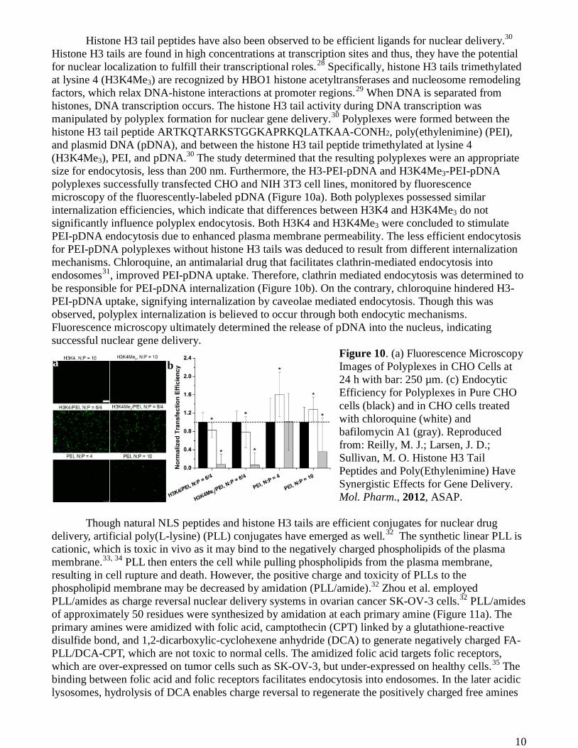
Histone H3 tail peptides have also been observed to be efficient ligands for nuclear delivery.30 Histone H3 tails are found in high concentrations at transcription sites and thus, they have the potential for nuclear localization to fulfill their transcriptional roles.28 Specifically, histone H3 tails trimethylated at lysine 4 (H3K4Me3) are recognized by HBO1 histone acetyltransferases and nucleosome remodeling factors, which relax DNA-histone interactions at promoter regions.29 When DNA is separated from histones, DNA transcription occurs. The histone H3 tail activity during DNA transcription was manipulated by polyplex formation for nuclear gene delivery.30 Polyplexes were formed between the histone H3 tail peptide ARTKQTARKSTGGKAPRKQLATKAA-CONH2, poly(ethylenimine) (PEI), and plasmid DNA (pDNA), and between the histone H3 tail peptide trimethylated at lysine 4 (H3K4Me3), PEI, and pDNA.30 The study determined that the resulting polyplexes were an appropriate size for endocytosis, less than 200 nm. Furthermore, the H3-PEI-pDNA and H3K4Me3-PEI-pDNA polyplexes successfully transfected CHO and NIH 3T3 cell lines, monitored by fluorescence microscopy of the fluorescently-labeled pDNA (Figure 10a). Both polyplexes possessed similar internalization efficiencies, which indicate that differences between H3K4 and H3K4Me3 do not significantly influence polyplex endocytosis. Both H3K4 and H3K4Me3 were concluded to stimulate PEI-pDNA endocytosis due to enhanced plasma membrane permeability. The less efficient endocytosis for PEI-pDNA polyplexes without histone H3 tails was deduced to result from different internalization mechanisms. Chloroquine, an antimalarial drug that facilitates clathrin-mediated endocytosis into endosomes31, improved PEI-pDNA uptake. Therefore, clathrin mediated endocytosis was determined to be responsible for PEI-pDNA internalization (Figure 10b). On the contrary, chloroquine hindered H3-PEI-pDNA uptake, signifying internalization by caveolae mediated endocytosis. Though this was observed, polyplex internalization is believed to occur through both endocytic mechanisms. Fluorescence microscopy ultimately determined the release of pDNA into the nucleus, indicating successful nuclear gene delivery.
Figure 10. (a) Fluorescence Microscopy Images of Polyplexes in CHO Cells at 24 h with bar: 250 µm. (c) Endocytic Efficiency for Polyplexes in Pure CHO cells (black) and in CHO cells treated with chloroquine (white) and bafilomycin A1 (gray). Reproduced from: Reilly, M. J.; Larsen, J. D.; Sullivan, M. O. Histone H3 Tail Peptides and Poly(Ethylenimine) Have Synergistic Effects for Gene Delivery. Mol. Pharm., 2012, ASAP.
Though natural NLS peptides and histone H3 tails are efficient conjugates for nuclear drug delivery, artificial poly(L-lysine) (PLL) conjugates have emerged as well.32 The synthetic linear PLL is cationic, which is toxic in vivo as it may bind to the negatively charged phospholipids of the plasma membrane.33, 34 PLL then enters the cell while pulling phospholipids from the plasma membrane, resulting in cell rupture and death. However, the positive charge and toxicity of PLLs to the phospholipid membrane may be decreased by amidation (PLL/amide).32 Zhou et al. employed PLL/amides as charge reversal nuclear delivery systems in ovarian cancer SK-OV-3 cells.32 PLL/amides of approximately 50 residues were synthesized by amidation at each primary amine (Figure 11a). The primary amines were amidized with folic acid, camptothecin (CPT) linked by a glutathione-reactive disulfide bond, and 1,2-dicarboxylic-cyclohexene anhydride (DCA) to generate negatively charged FA-PLL/DCA-CPT, which are not toxic to normal cells. The amidized folic acid targets folic receptors, which are over-expressed on tumor cells such as SK-OV-3, but under-expressed on healthy cells.35 The binding between folic acid and folic receptors facilitates endocytosis into endosomes. In the later acidic lysosomes, hydrolysis of DCA enables charge reversal to regenerate the positively charged free amines
a b

11
of the PLL. The positively charged PLL then escapes by lysing the lysosomal membrane and enters the nucleus. The glutathione-reactive disulfide bond between CPT and PLL is also cleaved in the reducing environment. The CPT binds to both DNA topoisomerase I and DNA to inhibit DNA replication, ultimately inducing apoptosis.36 These conclusions were supported by confocal fluorescence microscopy of FITC conjugated on to FA-PLL/DCA-CPT (Figure 11b).
HO
O
SPh
PhPh
CPT-OH1) EDC/DMAP
2) CF3COOH/SiEt3
NN
O
OO
O
HS
CPT-SH
O HN
1) Folate acid/EDC
2) SPDP
N S S
O
ONO O
O HN
ONH
O HN
NH2
HN
O
NH2
HN
O OH
OHN O
HN
NHN
NN
NH2
SS
N HO
FA-PLL-PDP
Folate acid
CPT-SH
O HN
ONH
O HN
NH2
HN
O
HN
O OH
OHN O
HN
NHN
NN
NH2
SS
HO
FA-PLL-CPT
Folate acidO O
O
O
N
N
DCA
O
O
O O HN
ONH
O HN
NH
HN
O
HN
O OH
OHN O
HN
NHN
NN
NH2
SS
HO
FA-PLL/DCA-CPT
Folate acidO O
O
O
N
N
O
O
OH
DCA
CPTCPT
We have currently observed that by conjugating a variety of linear peptides, such as the TAT peptide or the histone H3 tail, to drug delivery systems, pharmaceutical cargo can be efficiently delivered to the nucleus. However, cyclic peptides are now emerging as another competent nuclear drug delivery strategy.37 Among the pioneers in this research, Mandal et al. is exploring cyclic peptides that are able to deliver molecules without covalent conjugation and required lysosomal degradation for nuclear release.38 Mandal et al. synthesized 11 amphipathic homochiral L-cyclic peptides consisting of the lipophilic amino acid residues tryptophan W, phenylalanine F, alanine A, and leucine L, and the positively charged amino acid residues lysine K, arginine R, and glutamic acid E (Figure 12).38 The research aimed to determine the optimal parity between lipophilicity and positive charge for efficient nuclear delivery of cyclic peptides encapsulated with pharmaceutical molecules.
a
b
Figure 11. (a) Chemical Synthesis of Charge Reversal Conjugate FA-PLL/DCA-CPT. (b) Confocal Microscopy Images of FA-PLL/DCA-FITC with Expanded Region. Reproduced from: Zhou, Z.; Shen, Y.; Tang, J.; Fan, M.; Van Kirk, E. A.; Murdoch, W. J.; Radosz, M. Charge-Reversal Drug Conjugate for Targeted Cancer Cell Nuclear Drug Delivery. Adv. Funct. Mater., 2009, 19, 3580-3589.

12
NH
NH
NH
NH
HN
HN
HNNH
O
O
O
OO
O
O
O
HN
NH
NH2
NH
NH NH2
NH
HN
HNNH
H2N
HN
HN
HN
H2N
NH
[WR]4
NH
NH
NH
NH
HN
HN
HNNH
O
O
O
OO
O
O
O
[FK]4
NH2
H2N
H2N
NH2
NH
NH
NH
NH
HN
HN
HNNH
O
O
O
OO
O
O
O
[EL]4
OOH
O
OH
OHO
O
HO NH
NH
NH
NH
HN
HN
HNNH
O
O
O
OO
O
O
O
HN
NH
NH2
HNNH
H2N[RFEF]4
O
OH
O
HO
NH
NH
NH
NH
HN
HN
HNNH
O
O
O
OO
O
O
O
[AK]4
NH2
H2N
H2N
NH2
NH
NH
NH
NH
HN
HN
HNNH
O
O
O
OO
O
O
O
[EK]4
NH2
H2N
H2N
NH2
OOH
OH
O
HOO
HO
O
NH
NH
NH
NH
HN
HN
HNNH
O
O
O
OO
O
O
O
HN
NH
NH2
NH NH2
NH
HNNH
H2N
HN
HN
H2N
[ER]4
OHO
OH
O
HOO
HO
O
NH
NH
NH
NH
HN
HN
HNNH
O
O
O
OO
O
O
O
HN
NH
NH2
NH NH2
NH
HNNH
H2N
HN
HN
H2N
[FR]4
H2O
NH
NH
NHHN
HN
HN
HNHN
O
O
O
O
O
O
O
O
HN
NH
NH2
[RFE]3
O
HO O
OH
HN
NHH2N
NH
NH
NH2
O
NH
OOH
NHNH
HN
HNNHHN
OO
O
OO
O
NH
HN NH2
NH
HN
NH2
NHHN
HN
NH
H2N
NH
[WR]3
NH
NH
NH
HN
HNHNH
NNH
O
O
O
OO
O
O
O
HNNH
NH2
NH
HN
NH2
NHHNHN
NHH2N
HN
HN
HN
H2N
NH
[WR]5
OHN
ONH
NH
NH
NH
H2N
Figure 12. Structures of Synthesized Cyclic Peptides. Reproduced from: Mandal, D.; Shirazi, A. N.; Parang, K. Cell-Penetrating Homochiral Cyclic Peptides as Nuclear-Targeting Molecular Transporters. Angew. Chem. Int. Ed., 2011, 50, 9633-9637.
Each cyclic peptide (CP) was loaded with carboxyfluorescein (F) conjugated-lamivudine (3TC), a nucleoside reverse transcriptase inhibitor that prevents HIV and hepatitis B virus transcription to DNA.38 Cellular uptake of F-3TC-CPs was monitored in leukemia CCFM-CEM cells by fluorescence-activated cell sorting (FACS) and fluorescence microscopy. Only F-3TC-[WR]4 and F-3TC-[WR]5 were observed to have the greatest fluorescence, indicating efficient cellular internalization (Figure 13a). The negatively charged phosphopeptide GpYEEI, which binds to positively charged amino acid residues in protein binding sites39, was then conjugated with carboxyfluorescein and loaded on to [WR]4. Though GpYEEI is negatively charged and thus, has difficulty traversing the negatively charged phospholipid membrane, F-GpYEEI-[WR]4 was successfully internalized in breast carcinoma BT-20 cells. This result indicates that [WR]4 and [WR]5 are efficient transporters that do not require covalent conjugation. Binding affinity studies determined that pharmaceutical molecules are loaded on to cyclic peptides by intermolecular interactions. Mandal et al. then compared nuclear delivery of the cyclic peptides F-[W4R3K] and F-[W5R4K] to the linear peptide F-KR3W4.38 The cyclic F-[W4R3K] and F-[W5R4K] were observed to possess over 2-fold fluorescence intensity compared to the linear F-KR3W4 in CCRF-CEM cells by FACS, which signifies that the cyclic structure enhances cellular permeability (Figure 13b). Furthermore, F-[W5R4K] was observed to localize in the nucleus of SK-OV-3 cells by FITC fluorescence. DOX-F-[W5R4K] also efficiently delivered doxorubicin to SK-OV-3 nuclei compared to free doxorubicin, which supports the enhancement of nuclear entry by cyclic peptides (Figure 13c). Lastly, Mandal et al. concluded that F-[W5R4K] cellular internalization did not occur through endocytic

13
mechanisms. In CCRF-CEM cells with endocytic inhibitors such as chlorpromazine, chloroquine, and nystatin, cellular uptake of F-[W5R4K] did not change significantly, which suggests that endocytic mechanisms are not involved (Figure 15d). The cyclic nature and the lipophilicity of the cyclic peptides were deduced to non-covalently load drugs and to enhance plasma membrane and nuclear membrane permeability, respectively. Since covalent drug conjugation, endocytic cellular internalization, and lysosomal escape are not necessary, along with increased delivery efficiency, cyclic peptides are emerging as novel nuclear delivery systems.
Figure 13. (a) Cellular Uptake of F-3TC-CPs in CCRF-CEM cells. (b) FACS Images of Cellular Uptake of F-[W4R3K] and F-[W5R4K] in CCRF-CEM cells. (c) Fluorescence Microscopy Images of DOX and DOX-F-[W5R4K] in SK-OV-3 cells. (d) Cellular Internalization in CCRF-CEM cells In Presence of Endocytic Inhibitors. Reproduced from: Mandal, D.; Shirazi, A. N.; Parang, K. Cell-Penetrating Homochiral Cyclic Peptides as Nuclear-Targeting Molecular Transporters. Angew. Chem. Int. Ed., 2011, 50, 9633-9637. Nanoparticle Transporter Systems for Nuclear Drug Delivery Nanoparticle transporters and drug carriers have been found to be effective nuclear delivery systems of pharmaceutical molecules.5 Similar to other drug delivery strategies, nanoparticle carriers must overcome numerous logistical obstacles. Nanoparticles must be able to have long blood circulation times and thus, require the appropriate size to evade filtration mechanisms and enzymes.5 Nanoparticle drug carriers must then specifically target the cancerous cells by binding to overexpressed receptors, such as the folic receptor. By attaching a ligand to nanoparticle carriers, such as folic acid, nanoparticles are able to selectively target the cancerous cells for endocytosis.35 Once internalized into endosomes or lysosomes, nanoparticle carriers must escape the endosomal or lysosomal membrane. After escape into the cytoplasm, nanoparticle carriers must deliver pharmaceutical cargo into the nucleus through the NPCs for therapeutic influence. Various nanoparticle transporter systems have been synthesized that carefully consider each of these obstacles for nuclear delivery.
a b
c d

14
Nanoparticle carriers are able to deliver a variety of pharmaceutical molecules to nuclei. Immense research has focused on nonviral gene delivery as genes are able to be expressed within nuclei, resulting in proteins that exhibit therapeutic activity.40 Recently, nonviral gene delivery with chitosan-DNA nanoparticles has been achieved.40 The chitosan (Ch) oligosaccharide was first conjugated with folic acid (FA), by a PEG linker, to selectively target the overexpressed folic receptors of human epidermoid carcinoma (KB) cells. Since the chitosan has free amines, Ch-PEG-FA is positively charged in physiological conditions. As a result, it readily electrostatically conjugates with negatively charged DNA phosphates. The Ch-PEG-FA-DNA nanoparticles were synthesized at the molecular weights of 5, 25, and 50 kDa to determine molecular weight influences on gene expression in KB cells. Ch25kDa-PEG-FA-DNA was observed to have much higher gene expression than the low molecular weight (LMW) Ch5kDa-PEG-FA-DNA and the higher molecular weight (HMW) Ch50kDa-PEG-FA-DNA nanoparticles (Figure 14a, 14b). The low gene expression from the LMW Ch5kDa-PEG-FA-DNA was attributed to the weak binding to DNA due to the lower valence of the free amines, interrupting gene delivery. This observation supports earlier findings that LMW chitosan nanoparticles have a lower DNA binding affinity, higher DNA decomplexation rate, and cannot completely condense DNA for transport.41 However, the low gene expression from the HMW Ch50kDa-PEG-FA-DNA was attributed to stable DNA-chitosan complex formation. The high valence of the free amines of chitosan generates stable electrostatic interactions with the DNA. As a result, DNA separation from chitosan is difficult, hindering gene expression in the nucleus. The research concluded that Ch-PEG-FA-DNA nanoparticles must be of the appropriate size for efficient nuclear gene delivery to cancer cells with overexpressed folic receptors.
Figure 14. (a) Fluorescence Microscopy Images. 1: Negative Control KB cells; 2: Naked DNA; 3: 5 kDa Ch-DNA; 4: 5 kDa Ch-PEG-FA-DNA; 5: 25 kDa Ch-DNA; 6: 25 kDa Ch-PEG-FA-DNA; 7: 50 kDa Ch-DNA; 8: 50 kDa Ch-PEG-FA-DNA. (b) Confocal Microscopy Images of Plasmid DNA Gene in Nuclei (blue) of KB cells. Reproduced from: Jreyssaty, C.; Shi, Q.; Wang, H.; Qiu, X.; Winnik, F. M.; Zhang, X.; Dai, K.; Benderdour, M.; Fernandes, J. C. Efficient Nonviral Gene Therapy Using Folate-Targeted Chitosan-DNA Nanoparticles In Vitro. ISRN Pharmaceutics, 2012, ASAP. Extensive research has also been conducted to improve the nuclear delivery of non-gene pharmaceutical molecules by nanoparticle transporter systems. Recombinant modular nanotransporters (MNT) composed of multifunctional polypeptides have recently been designed for optimizing in vivo nuclear delivery.42 The MNTs are composed of four essential polypeptide moieties: a ligand that targets cancer cell receptors for receptor-mediated endocytosis, an endosomolytic module that allows endosomal or lysosomal escape into the cytoplasm, an NLS peptide for nuclear entry through NPCs, and a transporter module for attaching the therapeutic cargo (Figure 15A). Slastnikova et al. synthesized MNTs consisting of the E. coli hemoglobin-like protein (HMP) for loading the pharmaceutical cargo, the amphipathic diphtheria toxin translocation domain (DTox) for endosomal or lysosomal escape, and the NLS from the SV40 large TAG antigen for nuclear entry.42 One MNT possessed the α-melanocyte-stimulating hormone (αMHS) ligand that binds to overexpressed αMHS receptors on melanoma cells (DTox-HMP-NLS-αMHS) and the second MNT possessed the epidermal growth factor (EGF) that binds to EGF receptors on glioblastoma and breast carcinoma cells (DTox-HMP-NLS-EGF). The two MNTs were then labeled with 125I for monitoring cellular and nuclear localization in mice inoculated with cancer cells. Immunofluorescence confocal laser scanning microscopy determined that both MNTs
a b

15
specifically targeted cells overexpressed with cancerous receptors, followed by receptor-mediated endocytosis. Furthermore, nuclei localization was observed by DTox-HMP-NLS-EGF in human epidermoid carcinoma A431 cells (Figure 15Ba). Nuclei localization by DTox-HMP-NLS-αMHS was also observed by B16-F1 and Cloudman S91 melanoma cells (Figure 15Bb, 15Bc). The photosensitizers bacteriochlorin p and chlorin e6 were then conjugated to the MNTs for observing antitumor activity. Bacteriochlorin p-DTox-HMP-NLS-αMHS nanoparticles resulted in 89% tumor inhibition in B16-F1 melanoma cells and 98% tumor inhibition in Cloudman S91 melanoma cells (Figure 16a, 16b). Chlorin e6-DTox-HMP-NLS-EGF nanoparticles resulted in 98% tumor inhibition in A431 cells (Figure 18c). In conclusion, by adjusting moieties, MNTs are novel platforms for cell-specific nuclear delivery of diverse pharmaceutical molecules.
Figure 15. (A) Structure of MNTs and Nuclear Localization Pathway. (B) Merged Immunofluorescence Microscopy Images at 3 h Post-Intravenous MNT Injection: (A) DTox-HMP-NLS-EGF in mice inoculated with A431 cells, (A’) Saline in control mice inoculated with A431 cells. (B) DTox-HMP-NLS-αMHS in mice inoculated with B16-F1 cells, (B’) Saline in control mice inoculated with B16-F1 cells. (C) DTox-HMP-NLS-αMHS in mice inoculated with Cloudman S91 cells, (C’) Saline in control mice inoculated with Cloudman S91 cells. DAPI (blue) indicate nuclei and red indicates MNT. Reproduced from: Slastnikova, T. A.; Rosenkranz, A. A.; Gulak, P. V.; Schiffelers, R. M.; Lupanova, T. N.; Khramtsov, Y. V.; Zalutsky, M. R.; Sobolev, A. S. Modular Nanotransporters: A Multipurpose in Vivo Working Platform for Targeted Drug Delivery. Int. J. Nanomedicine, 2012, 7, 467-482.
Figure 16. Tumor Inhibition by: (a) Bacteriochlorin p-DTox-HMP-NLS-αMHS in B16-F1 cells, (b) Bacteriochlorin p-DTox-HMP-NLS-αMHS in Cloudman S91 cells, and (c) Chlorin e6-DTox-HMP-NLS-EGF in A431 cells. Reproduced from: Slastnikova, T. A.; Rosenkranz, A. A.; Gulak, P. V.; Schiffelers, R. M.; Lupanova, T. N.; Khramtsov, Y. V.; Zalutsky, M. R.; Sobolev, A. S. Modular Nanotransporters: A Multipurpose in Vivo Working Platform for Targeted Drug Delivery. Int. J. Nanomedicine, 2012, 7, 467-482. Polypeptide-derived nanotransporters are continuously evolving to enhance nuclear drug delivery. In addition to MNTs, many research groups have found interest in biodegradable polypeptide delivery systems.43 Han et al. have synthesized amphiphilic biodegradable co-polymer nanotransporters comprising of poly(L-aspartic acid-co-lactic acid) and 1,2-dipalmitoyl-sn-glycero-3-
a b c
a
a’
b
b’ c’
c
A B

16
phosphoethanolamine [poly(AA-co-LA)/DPPE] (Figure 17).43 Doxorubicin (DOX) was then encapsulated on to poly(AA-co-LA)/DPPE by double emulsion-extraction and nanoprecipitation. The DOX-poly(AA-co-LA)/DPPE was observed to have significantly longer lifetimes than free DOX in human lung adenocarcinoma (A549) and cervical carcinoma (HeLa) cells (Figure 18). Furthermore, trafficking microscopy studies determined that the DOX-poly(AA-co-LA)/DPPE nanotransporters internalized into the cells by receptor-mediated endocytosis44, degraded in the acidic lysosomes to release DOX conjugates into the cytoplasm, followed by nuclear entry of DOX. The DOX-poly(AA-co-LA)/DPPE displayed similar activity to free DOX with respect to tumor inhibition, but was much less lethal to mice. All experimental mice inoculated with free DOX at 5 mg/kg died within one month, but no mice died when inoculated DOX-poly(AA-co-LA)/DPPE. By employing poly(AA-co-LA)/DPPE nanoparticles to deliver other chemotherapeutic molecules, therapies will have significantly reduced lethality and toxicity.
H2N
OH
O
O
OHNO2
O O
Cl
Pyridine, DMAP
O
OO
POH
OO
NH2
O
OTriethylamine
O
OO
POH
OO
HN
O
O
O
O
O
HN
N
O
O
O
O
O
OO
O
HOHO
Poly(AA-co-LA)/DPPE
O
O
O
O
Figure 18. Confocal Microscopy Images of the Nuclear Localization by (a) Free DOX in A549 Cells, (b) DOX-poly(AA-co-LA)/DPPE in A549 Cells, (c) Free DOX in HeLa Cells, and (d) DOX-poly(AA-co-LA)/DPPE in HeLa Cells. Red indicates DOX fluorescence, C represents cytoplasm, N indicates nuclei, bar: 10 µm. Reproduced from: Han, S.; Liu, Y.; Nie, X.; Xu, Q.; Jiao, F.; Li, W.; Zhao, Y.; Wu, Y.; Chen, C. Efficient Delivery of Antitumor Drug to the Nuclei of Tumor Cells by Amphiphilic Biodegradable Poly(L-Aspartic Acid-co-Lactic Acid)/DPPE Co-Polymer Nanoparticles. Small Nano. Micro., 2012, ASAP. Nanoparticle transporters have proven to be effective at delivering pharmaceutical cargo to the nucleus, but delivery trajectories are still ambiguous.45 To further visualize and understand the mechanism of nanoparticle transporter nuclear delivery, Kang et al. labeled single-walled chitosan-coated carbon nanotubes (CS-SWNTs) with FITC and loaded them with fluorescent DOX by π-stacking (Figure 19a).45 The green FITC fluorescence displayed the FITC-CS-DOX-SWNTs internalizing into the lysosomes of endothelial progenitor cells (EPC) by receptor mediated endocytosis.46 The acidic lysosomes disrupt the π-stacking between DOX and SWNTs, releasing DOX into the nucleus. The red fluorescence of DOX was observed in the nucleus while the green fluorescence of SWNTs was not present in the nucleus, but remained in the cytoplasm (Figure 19b, 19c). The ability to visualize drug
a
b
c
d
Figure 17. Chemical Synthesis of poly(AA-co-LA)/DPPE. Reproduced from: Han, S.; Liu, Y.; Nie, X.; Xu, Q.; Jiao, F.; Li, W.; Zhao, Y.; Wu, Y.; Chen, C. Efficient Delivery of Antitumor Drug to the Nuclei of Tumor Cells by Amphiphilic Biodegradable Poly(L-Aspartic Acid-co-Lactic Acid)/DPPE Co-Polymer Nanoparticles. Small Nano. Micro., 2012, ASAP.

17
delivery trajectories by fluorescence provides insight into internalization mechanisms, which is crucial in designing improved nuclear drug delivery strategies.
Figure 19. (a) Structure of FITC-CS-DOX-SWNTs. Confocal Microscopy Images of (b) FITC-CS-DOX-SWNTs in EPC Cells and (c) DOX Release from FITC-CS-SWNTs into EPC Nuclei. FITC fluorescence is green, DOX fluorescence is red, DAPI nuclei stain fluorescence is blue. Reproduced from: Kang, B.; Li, J.; Chang, S.; Dai, M.; Ren, C.; Dai, Y.; Chen, D. Subcellular Tracking of Drug Release from Carbon Nanotube Vehicles in Living Cells. Small Nano. Micro., 2012, 8, 777-782. Inorganic Systems for Nuclear Drug Delivery Innumerable nuclear drug delivery systems are currently founded on biological organic chemistry, employing peptides, nucleic acids, and carbohydrates. These organic nuclear delivery strategies employ molecules that are recognized by human body systems. However, recent successes have been observed for nuclear delivery systems employing inorganic metallochemistry.47 Inorganic nuclear delivery systems are effective as many drugs can be loaded on to the central metal.47 However, much consideration is required regarding the toxicity of incorporated metals. Metals that are generally not found in the body may react with body systems unfavorably, resulting in adverse side effects. It is thus essential to ascertain the benefits and risks of employing metals as nuclear delivery strategies. Several inorganic nuclear drug delivery systems have been discovered that carefully consider these aspects. Gold nanoparticles (AuNPs) have gained prominence as inorganic systems that deliver analogues of cisplatin to the nucleus.48 The metallodrug oxaliplatin, a derivative of cisplatin, is extensively used in treatments against many cancers, including testicular, ovarian, and lung cancers.49 Oxaliplatin binds to DNA, which inhibits DNA replication and transcription, resulting in apoptosis. However, free oxaliplatin is not cell-specific and attacks both cancerous and healthy cells to induce apoptosis.48 As a result, side effects include nephrotoxicity, neurotoxicity, and ototoxicity. Graham et al. aimed to overcome oxaliplatin nonspecificity with oxaliplatin-tethered AuNPs that would be specifically delivered to cancerous cells.50 AuNPs are ideal drug delivery vehicles as they are nonimmunogenic, biocompatible, and are easily modified with pharmaceutical molecules or ligands.51 Initially, a PEG linker was generated with a carboxylate terminus and a cyclic disulfide terminus (Figure 20a).50 The cyclic disulfides of the PEG linkers were then conjugated to the AuNPs while the carboxylate end chelated to the active component of oxaliplatin, [Pt(1R,2R-diaminocyclohexane]2+ ([Pt(R,R-dach)]). The resulting [Pt(R,R-dach)]-PEG-AuNPs had an average of 280 (±20) [Pt(R,R-dach)] molecules per AuNP. An increase of AuNP size by 4.5-fold was also observed, but was attributed to crosslinking of AuNP nanoparticles (Figure 20a, 20b). However, the larger [Pt(R,R-dach)]-PEG-AuNPs with a diameter of 176 (±25) nm still fulfilled the size criterion, of diameters under 200 nm52, for optimal nuclear targeting. The [Pt(R,R-dach)]-PEG-AuNPs were ultimately observed to enter the nuclei of A549 cells (Figure 21a). However, the PEG-AuNPs were not able to enter the nuclei. Graham et al. concluded that nuclear entry
15 min 30 min 1 h 3h
a b
c

18
was facilitated by [Pt(R,R-dach)]-mediated internalization. Furthermore, cytotoxicity studies determined that the [Pt(R,R-dach)]-PEG-AuNPs possessed lower inhibitory concentrations IC50 than free oxaliplatin, with respect to platinum concentration (Figure 21b). Thus, a lower dose is required for [Pt(R,R-dach)]-PEG-AuNPs compared to free oxaliplatin, which is essential for reducing adverse side effects. With further in vivo experimentation and clinical trials on targeting specificity, [Pt(R,R-dach)]-PEG-AuNPs may eventually be employed in many chemotherapies.
OHO OO 5 eq. DMAP
DCM, 60oC, 6 hrsO
OOH
O
NH2O
ONH241
5 eq. DIC
DCM, r.t., 24 hrsO
O HN
OO
ONH2
41
HO
O
S S
5 eq. DIC
DCM, r.t., 24 hrsO
O HN
OO
ONH
41
O
S S
10 % TFA
DCM, r.t., 3 hrsHO
O HN
OO
ONH
41
O
S S
AuCl
Cl Cl
Cl_
Sodium CItrateAu
Au
DIPEA
H2N
NH2
PtH2O
H2O
2+
AgNO3
H2N
NH2
PtCl
Cl
Au
Pt
Au
NHHN
Au Pt
H2N
NH2
Figure 20. (a) Chemical Synthesis of [Pt(R,R-dach)]-PEG-AuNPs. (b) Scanning Electron Microscopy Images of (A) AuNPs, (B) PEG-AuNPs, and (C) [Pt(R,R-dach)]-PEG-AuNPs. Reproduced from: Brown, S. D.; Nativo, P.; Smith, J.; Stirling, D.; Edwards, P. R.; Venugopal, B.; Flint, D. J.; Plumb, J. A.; Graham, D.; Wheate, N. J. Gold Nanoparticles for the Improved Anticancer Drug Delivery of the Active Component of Oxaliplatin. J. Am. Chem. Soc., 2010, 132, 4678-4684.
a b

19
Figure 21. (a) TEM Image of Nuclear Uptake of [Pt(R,R-dach)]-PEG-AuNPs in A549 cells: (1) extracellular AuNP, (2) intracellular AuNP, (3) intranuclear AuNP, (4) contracted cell wall, (5) nuclear membrane, (6) endosome, and (7) lysosome. (b) Inhibitory Concentration IC50 of [Pt(R,R-dach)]-PEG-AuNPs and free Oxaliplatin with respect to Platinum Concentration in varying cell lines. Reproduced from: Brown, S. D.; Nativo, P.; Smith, J.; Stirling, D.; Edwards, P. R.; Venugopal, B.; Flint, D. J.; Plumb, J. A.; Graham, D.; Wheate, N. J. Gold Nanoparticles for the Improved Anticancer Drug Delivery of the Active Component of Oxaliplatin. J. Am. Chem. Soc., 2010, 132, 4678-4684. Research on nuclear drug delivery by gold nanoparticles currently focuses on chemical modifications to improve nuclear internalization and cell-specificity. Since AuNPs have low toxicity and are easily modified, they are ideal for delivering pharmaceutical cargo to the nucleus.48 Scarí et al. conjugated the (GC)2 peptide and the RGD-(GC)2 peptide to AuNPs to target integrin αvβ3 receptors on cancerous cells (Figure 22a, 22b).53 The cysteine residues of the (GC)2 peptide chelates to AuNPs and prevent crosslinking between AuNPs while the RGD moiety is known to bind specifically to tumor integrin αvβ3 receptors.54, 55 Both (GC)2-AuNPs and RGD-(GC)2-AuNPs were observed to internalize into glioblastoma U87 cells that overexpress αvβ3 receptors. As expected, RGD-(GC)2-AuNPs displayed higher uptake in U87 cells by receptor-mediated endocytosis of the RGD motif. On the contrary, (GC)2-AuNPs displayed minute uptake in U87 cells. TEM confirmed that the RGD-(GC)2-AuNPs internalized into U87 cells by receptor mediated endocytosis, followed by release into the cytoplasm (Figure 22cA, 22cB). The RGD-(GC)2-AuNPs were also observed in the nucleus, by trafficking through the NPCs, and in the nucleolus (Figures 22cC, 22cD). The RGD and GC moieties were deduced to increase nuclear membrane permeability for nuclear import. Chemical modification of AuNPs has found immense success in nuclear importation by targeting receptors on cancer cells. However, pharmaceutical cargo has yet to be loaded on to RGD-(GC)2-AuNPs for determining in vivo cytotoxicity.
IC50 Platinated
Nanoparticles
Cell Line
wrt [Au] (nM)
wrt [Pt] (µM)
Oxaliplatin control (µM)
Fold increase
HCT116 2.66 (±0.27)
0.734 (±0.044)
0.728 (±0.048)
1.0
HCT15 2.36 (±0.34)
0.652 (±0.093)
2.97 (±0.21)
4.6
HT29 1.29 (±0.03)
0.357 (±0.007)
2.00 (±0.15)
5.6
RKO 0.842 (±0.127)
0.233 (±0.035)
0.295 (±0.021)
1.3
A549 0.495 (±0.062)
0.135 (±0.008)
0.775 (±0.057)
5.7
b a

20
Figure 22. Chemical Structures of (a) (GC)2-AuNPs and (b) RGD-(GC)2-AuNPs. (c) TEM Images of RGD-(GC)2-AuNPs in U87 Cells: (A) at the plasma membrane, (B) inside an endosome, (C) inside the nucleus, and (D) inside the nucleolus. Reproduced from: Scarí, G.; Porta, F.; Fascio, U.; Avvakumova, S.; Dal Santo, V.; De Simone, M.; Saviano, M.; Leone, M.; Del Gatto, A.; Pedone, C.; Zaccaro, L. Gold Nanoparticles Capped by a GC-Containing Peptide Functionalized with an RGD Motif for Integrin Targeting. Bioconjugate Chem., 2012, 23, 340-349.αβ Numerous attempts have been made to carefully monitor the mechanisms of nuclear internalization by inorganic drug delivery systems. Odom et al. recently visualized the interaction between drug-loaded gold nanostars (AuNS) and the nuclei of cancer cells.47 AuNSs were synthesized by reducing AuIIIClO3 with 4-(2-hydroxyethyl)-1-piperazineethanesulfonic acid (HEPES). The aptamer AS1411 (Apt) was then conjugated to the AuNSs. AS1411 is known to bind to nucleolin, a phosphoprotein present in nuclei of normal cells, but overexpressed in the cytoplasm and plasma membrane of cancer cells.56 The resulting Apt-AuNSs can therefore specifically target cancer cells by binding to the overexpressed nucleolin. Each AuNS was determined to be conjugated to approximately 950 AS1411 aptamers with an average size of 33 nm. The Apt-AuNSs were observed to enter HeLa nuclei by confocal fluorescence microscopy of Cy5-labeled AS1411 aptamers (Figure 23a). On the contrary, the control cApt-AuNSs with cysteine rich aptamers, which have a low affinity for nucleolin, did not enter the nucleus. This indicates that nucleolin binding is essential for nuclear uptake by the Apt-AuNSs. Furthermore, Odom et al. observed cytotoxicity induced by the Apt-AuNSs in the form of nuclear envelope invaginations (figure 23b, 23c).47 Nuclear invaginations were more prominent when AS1411 aptamers were released from the AuNSs by femtosecond laser pulses.57 The cApt-AuNSs, however, did not cause nuclear envelope invaginations. Caspase activity, indicative of cellular apoptosis, was also greater for light-released AS1411 aptamers (Figure 23d, 23e). The influences on the nuclear phenotype by the Apt-AuNSs and by the light-triggered release of AS1411 provide insight on new methods of effective nuclear delivery.
a
b
c

21
Figure 23. (a) Confocal Fluorescence Microscopy Images of Cy5-Apt-AuNSs and Cy5-cApt-AuNSs in HeLa cells nuclei at 5 h and 24 h. TEM Images of Nuclear Envelope Invaginations Caused by (b) Apt-AuNSs and (c) Light-Triggered Release of AS1411 Aptamers from Apt-AuNSs in HeLa nuclei. Confocal Fluorescence Microscopy Images of Double-Stranded DNA Breaks Caused by (d) Apt-AuNSs and (e) Light-Triggered Release of AS1411 Aptamers from Apt-AuNSs in HeLa nuclei. Green FITC fluorescence indicates caspase activity and apoptosis. Reproduced from: Dam, D. H. M.; Lee, J. H.; Sisco, P. N.; Co, D. T.; Zhang, M.; Wasielewski, M. R.; Odom, T. W. Direct Observation of Nanoparticle-Cancer Cell Nucleus Interactions. ACS Nano., 2012, ASAP. Future of Nuclear Drug Delivery Several advancements in nuclear drug delivery have been witnessed in the recent years, providing the potential for revolutionizing current therapeutic treatments. Nuclear drug delivery is a vast and constantly expanding field that includes chemical modification, modular transporter, peptide, nanoparticle, and inorganic systems. With the appropriate manipulations of biological pathways and considerations of physiological conditions, the variety of nuclear drug delivery strategies have found success in not only in vitro, but also in vivo studies. Research has determined that nuclear delivery requires cellular penetration by endocytosis, endosomal or lysosomal escape into the cytoplasm, translocation to the nuclear envelope, and nuclear entry through the NPCs.4 Currently, nuclear drug delivery strategies manipulate these pathways for nuclear importation. However, other research suggests possible different mechanisms for nuclear entry.38 By discovering other possible delivery mechanisms, new nuclear drug delivery strategies may be designed. Though numerous nuclear drug delivery strategies have been successful in vitro and in vivo, clinical studies have yet to be conducted to determine the extent of their influences on humans. Research on nuclear drug delivery strategies will certainly expand in the future with the impending possibility of drug resistance. With continued research on nuclear drug delivery and the design of new nuclear drug delivery strategies, the potency of current therapeutic treatments will be dramatically enhanced.
a
b c
d e

22
References Cited
1 Lewis, A. L.; Dreher, M. R. Locoregional Drug Delivery Using Image-Guided Intra-Arterial Drug Eluting Beat Therapy. J. Control Release, 2011, ASAP. 2 Rajendran, L.; Knölker, H.; Simons, K. Subcellular Targeting Strategies for Drug Design and Delivery. Nat. Rev. Drug Discov., 2010, 9, 29–42. 3 Langer, R. New Methods of Drug Delivery. Science, 1990, 249, 1527–1533. 4 Wagstaff, K. M.; Jans, D. A. Nuclear Drug Delivery to Target Tumour Cells. Eur. J. Pharmacol., 2009, 625, 174–180. 5 Sui, M.; Liu, W.; Shen, Y. Nuclear Drug Delivery for Cancer Chemotherapy. J. Control Release, 2011, 155, 227–236. 6 Rappaport, J. S. Focusing on Clathrin-Mediated Endocytosis. Biochem. J., 2008, 412, 415-423. 7 Bareford, L. M.; Swaan, P. W. Endocytic Mechanisms for Targeted Drug Delivery. Adv. Drug Deliv. Rev., 2007, 59, 748–758. 8 Sahay, G.; Alakhova, D. Y.; Kabanov, A. V. Endocytosis of Nanomedicines. J. Control Release, 2010, 145, 182-195. 9 Parton, R. G.; Simons, K. The Multiple Faces of Caveolae. Nat. Rev. Mol. Cell Bio., 2007, 8, 185–194. 10 Poon, I. K. H.; Jans, D. A. Regulation of Nuclear Transport: Central Role in Development and Transformation? Traffic, 2005, 6, 173–186. 11 Pouton, C. W.; Wagstaff, K. M.; Roth, D. M.; Moseley, G. W.; Jans, D. A. Targeted Delivery to the Nucleus. Adv. Drug Deliv. Rev., 2007, 59, 698–717. 12 Mudhakir, D.; Harashima, H. Learning from the Viral Journey: How to Enter Cells and How to Overcome Intracellular Barriers to Reach the Nucleus. AAPS Journal, 2009, 2, 65–77. 13 Curatolo, W. Physical Chemical Properties of Oral Drug Candidates in the Discovery and Exploratory Development Settings. Pharm. Sci. Technol. To., 1998, 1, 387–393. 14 Lipinski, C. A. Annual Meeting of the American Association of Pharmaceutical Scientists; Biotech/PDD Symposium on Tools for Oral Absorption, 1995. 15 Deu, E.; Verdoes, M.; Bogyo, M. New Approaches for Dissecting Protease Functions to Improve Probe Development and Drug Discovery. Nat. Struct. Mol. Biol., 2012, 19, 9–16. 16 Vincenzi, B.; Frezza, A. M.; Santini, D.; Tonini, G. New Therapies in Soft Tissue Sarcoma. Expert Opin. Emerg. Dr., 2010, 15, 237−248. 17 Chhikara, B. S.; Mandal, D.; Parang, K. Synthesis, Anticancer Activities, and Cellular Uptake Studies of Lipophilic Derivatives of Doxorubicin Succinate. J. Med. Chem., 2012, 55, 1500–1510. 18 Sobolev, A. S. Novel Modular Transporters Delivering Anticancer Drugs and Foreign DNA to the Nuclei of Target Cancer Cells. J. Buon., 2009, 14, 33–42. 19 Rosenkranz, A. A.; Vaidyanathan, G.; Pozzi, O. R.; Lunin, V. G.; Zalutsky, M. R.; Sobolev, A. S. Engineered Modular Recombinant Transporters: Application of New Platform for Targeted Radiotherapeutic Agents to Alpha-Particle Emitting 211 At. Int. J. Radiat. Oncol. Biol. Phys., 2008, 72, 193–200. 20 Wender, P. A.; Mitchell, D. J.; Pattabiraman, K.; Pelkey, E. T.; Steinman, L.; Rothbard, J. B. The Design, Synthesis, and Evaluation of Molecules That Enable or Enhance Cellular Uptake: Peptoid Molecular Transporters. Proc. Natl. Acad. Sci. U.S.A., 2000, 97, 13003–13008. 21 Lindgren, M.; Hallbrink, M.; Prochiantz, A.; Langel, U. Cell-Penetrating Peptides. Trends Pharmacol. Sci., 2000, 21, 99–103. 22 Nagahara, H.; Vocero-Akbani, A. M.; Snyder, E. L.; Ho, A.; Latham, D. G.; Lissy, N. A.; Becker Hapak, M.; Ezhevsky, S. A.; Dowdy, S. F. Transduction of Full-Length TAT Fusion Proteins Into Mammalian Cells: TAT-p27 Kip1 Induces Cell Migration. Nat. Med., 1998, 4, 1449–1452. 23 Schröder, T.; Niemeier, N.; Afonin, S.; Ulrich, A. S.; Krug, H. F.; Bräse, S. Peptoidic Amino- and Guanidinium-Carrier Systems: Targeted Drug Delivery into the Cell Cytosol or the Nucleus. J. Med. Chem., 2008, 51, 376–379.

23
24 Kuo, C.; Chueh, D.; Singh, N.; Chien, F.; Chen, P. Targeted Nuclear Delivery Using Peptide-Coated Quantum Dots. Bioconjugate Chem., 2011, 22, 1073–1080. 25 Alber, F.; Dokudovskaya, S.; Veenhoff, L. M.; Zhang, W.; Kipper, J.; Devos, D.; Suprapto, A.; Karni-Schmidt, O.; Williams, R.; Chait, B. T. The Molecular Architecture of the Nuclear Pore Complex. Nature, 2007, 450, 695–701. 26 Pan, L.; He, Q.; Lui, J.; Chen, Y.; Ma, M.; Zhang, L.; Shi, J. Nuclear-Targeted Drug Delivery of TAT Peptide-Conjugated Monodisperse Mesoporous Silica Nanoparticles. J. Am. Chem. Soc., 2012, 134, 5722–5725. 27 Yi, W.; Yang, J.; Li, C.; Wang, H.; Liu, C.; Tao, L.; Cheng, S.; Zhuo, R.; Zhang, X. Enhanced Nuclear Import and Transfection Efficiency of TAT Peptide-Based Gene Delivery Systems Modified by Additional Nuclear Localization Signals. Bioconjugate Chem., 2012, 23, 125–134. 28 Schneider, R.; Bannister, A. J.; Myers, F. A.; Thorne, A. W.; Crane-Robinson, C.; Kouzarides, T. Histone H3 Lysine-4 Methylation Patters in Higher Eukaryotic Genes. Nat. Cell Biol., 2004, 6, 73–77. 29 Palacios, A.; Moreno, A.; Oliveira, B. L.; Rivera, T.; Prieto, J.; Garcia, P.; Fernandez-Fernandez, M. R.; Bernado, P.; Palmero, I.; Blanco, F. J. The Dimeric Structure and the Bivalent Recognition of H3K4ME3 by the Tumor Suppressor ING4 Suggests a Mechanism for Enhanced Targeting of the HBO1 Complex to Chromatin. J. Mol. Biol., 2010, 396, 1117–1127. 30 Reilly, M. J.; Larsen, J. D.; Sullivan, M. O. Histone H3 Tail Peptides and Poly(Ethylenimine) Have Synergistic Effects for Gene Delivery. Mol. Pharm., 2012, ASAP. 31 Parton, R. G.; Howes, M. T. Revisiting Caveolin Trafficking: The End of the Caveosome. J. Cell Biol., 2010, 191, 439–441. 32 Zhou, Z.; Shen, Y.; Tang, J.; Fan, M.; Van Kirk, E. A.; Murdoch, W. J.; Radosz, M. Charge-Reversal Drug Conjugate for Targeted Cancer Cell Nuclear Drug Delivery. Adv. Funct. Mater., 2009, 19, 3580–3589. 33 Mecke, A.; Lee, D.; Ramamoorthy, A.; Orr, B. G.; Holl, M. M. B. Synthetic and Natural Polycationic Polymer Nanoparticles Interact Selectively with Fluid-Phase Domains of DMPC Lipid Bilayers. Langmuir, 2005, 21, 8588–8590. 34 Fischer, D.; Li, Y.; Ahlemeyer, B.; Krieglstein, J.; Kissel, T. In Vitro Cytotoxicity Testing of Polycations: Influence of Polymer Structure on Cell Viability and Hemolysis. Biomaterials, 2003, 24, 1121–1131. 35 Sheff, D. Endosomes as a Route for Drug Delivery in the Real World. Adv. Drug Deliv. Rev., 2004, 56, 927–930. 36 Crowe, D. L. Mechanisms of DNA Damage and Repair in Cancer Chemotherapy. Recent Research Developments in Cancer, 2002, 4, 65–72. 37 Goldberg, M.; Langer, R.; Jia, X. Nanostructured materials for Applications in Drug Delivery and Tissue Engineering. J. Biomater. Sci. Polym. Ed., 2007, 18, 241-268. 38 Mandal, D.; Shirazi, A. N.; Parang, K. Cell-Penetrating Homochiral Cyclic Peptides as Nuclear-Targeting Molecular Transporters. Angew. Chem. Int. Ed., 2011, 50, 9633–9637. 39 Machida, K.; Mayer, B. J. The SH2 Domain: Versatile Signaling Module and Pharmaceutical Target. Biochim. Biophys. Acta Proteins and Proteomics, 2005, 1747, 1–25. 40 Jreyssaty, C.; Shi, Q.; Wang, H.; Qiu, X.; Winnik, F. M.; Zhang, X.; Dai, K.; Benderdour, M.; Fernandes, J. C. Efficient Nonviral Gene Therapy Using Folate-Targeted Chitosan-DNA Nanoparticles In Vitro. ISRN Pharmaceutics, 2012, ASAP. 41 Turan, K.; Nagata, K. Chitosan-DNA Nanoparticles: The Effect of Cell Type and Hydrolysis of Chitosan on In Vitro DNA Transfection. Pharm. Dev. Technol., 2006, 11, 503–512. 42 Slastnikova, T. A.; Rosenkranz, A. A.; Gulak, P. V.; Schiffelers, R. M.; Lupanova, T. N.; Khramtsov, Y. V.; Zalutsky, M. R.; Sobolev, A. S. Modular Nanotransporters: A Multipurpose in Vivo Working Platform for Targeted Drug Delivery. Int. J. Nanomedicine, 2012, 7, 467–482.

24
43 Han, S.; Liu, Y.; Nie, X.; Xu, Q.; Jiao, F.; Li, W.; Zhao, Y.; Wu, Y.; Chen, C. Efficient Delivery of Antitumor Drug to the Nuclei of Tumor Cells by Amphiphilic Biodegradable Poly(L-Aspartic Acid-co-Lactice Acid)/DPPE Co-Polymer Nanoparticles. Small Nano. Micro., 2012, ASAP. 44 Rejman, J.; Oberle, V.; Zuhorn, I.; Hoekstra, D. Size-Dependent Internalization of Particles Via the Pathways of Clathrin- and Caveolae-Mediated Endocytosis. Biochem. J., 2004, 377, 159–169. 45 Kang, B.; Li, J.; Chang, S.; Dai, M.; Ren, C.; Dai, Y.; Chen, D. Subcellular Tracking of Drug Release from Carbon Nanotube Vehicles in Living Cells. Small Nano. Micro., 2012, 8, 777–782. 46 Kang, B.; Yu, D. C.; Chang, S. Q.; Chen, D.; Dai, Y. D.; Ding, Y. T. Intracellular Uptake, Trafficking and Subcellular Distribution of Folate Conjugated Single Walled Carbon Nanotubes Within Living Cells. Nanotechnology 2008, 19, 1–8. 47 Dam, D. H. M.; Lee, J. H.; Sisco, P. N.; Co, D. T.; Zhang, M.; Wasielewski, M. R.; Odom, T. W. Direct Observation of Nanoparticle-Cancer Cell Nucleus Interactions. ACS Nano., 2012, ASAP. 48 Craig, G. E.; Brown, S. D.; Lamprou, D. A.; Graham, D.; Wheate, N. J. Cisplatin-Tethered Gold Nanoparticles That Exhibit Enhanced Reproducibility, Drug Loading, and Stability: a Step Closer to Pharmaceutical Approval? Inorg. Chem., 2012, 51, 3490–3497. 49 Kelland, L. The Resurgence of Platinum-Based Cancer Chemotherapy. Nat. Rev. Cancer, 2007, 7, 573–584. 50 Brown, S. D.; Nativo, P.; Smith, J.; Stirling, D.; Edwards, P. R.; Venugopal, B.; Flint, D. J.; Plumb, J. A.; Graham, D.; Wheate, N. J. Gold Nanoparticles for the Improved Anticancer Drug Delivery of the Active Component of Oxaliplatin. J. Am. Chem. Soc., 2010, 132, 4678–4684. 51 Jelveh, S.; Chithrani, D. B. Gold Nanostructures as a Platform for Combinatorial Therapy in Future Cancer Therapeutics. Cancers, 2011, 3, 1081–1110. 52 Gu, F. X.; Karnik, R.; Wang, A. Z.; Alexis, F.; Levy-Nissenbaum, E.; Hong, S.; Langer, S.; Farokhzad, O. C. Targeted Nanoparticles for Cancer Therapy. Nano. Today, 2007, 2, 14–21. 53 Scarí, G.; Porta, F.; Fascio, U.; Avvakumova, S.; Dal Santo, V.; De Simone, M.; Saviano, M.; Leone, M.; Del Gatto, A.; Pedone, C.; Zaccaro, L. Gold Nanoparticles Capped by a GC-Containing Peptide Functionalized with an RGD Motif for Integrin Targeting. Bioconjugate Chem., 2012, 23, 340–349. 54 Krpetic, Z.; Nativo, P.; Porta, F.; and Brust, M. A Multidentate Peptide for Stabilization and Facile Bioconjugation of Gold Nanoparticles. Bioconjugate Chem. 2009, 20, 619−624. 55 Cai, W.; Chen, X. Multimodality Imaging of Integrin αvβ3 Expression. Anticancer Agents Med. Chem., 2006, 6, 135–148. 56 Soundararajan, S.; Wang, L.; Sridharan, V.; Chen, W.; Courtenay-Luck, N.; Jones, D.; Spicer, E. K.; Fernandes, D. J. Plasma Membrane Nucleolin is a Receptor for the Anticancer Aptamer AS1411 in MV4-11 Leukemia Cells. Mol. Pharmacol., 2009, 76, 984–991. 57 Jain, P. K.; Qian, W.; El-Sayed, M. A. Ultrafast Cooling of Photoexcited Electrons in Gold Nanoparticle-Thiolated DNA Conjugates Involves the Dissociation of the Gold–Thiol Bond J. Am. Chem. Soc., 2006, 128, 2426–2433.