Evaluation of marginal discrepancy on All-ceramic crowns...
48
Universidade de Lisboa Faculdade de Medicina Dentária Evaluation of marginal discrepancy on All- ceramic crowns manufactured by CAD/CAM versus Conventional methods Pedro Nuno Pimenta Dissertação Mestrado Integrado em Medicina Dentária 2015
Transcript of Evaluation of marginal discrepancy on All-ceramic crowns...
Universidade de Lisboa
Faculdade de Medicina Dentária
Evaluation of marginal discrepancy on All-
ceramic crowns manufactured by CAD/CAM
versus Conventional methods
Pedro Nuno Pimenta
Dissertação
Mestrado Integrado em Medicina Dentária
2015
Universidade de Lisboa
Faculdade de Medicina Dentária
Evaluation of marginal discrepancy on All-
ceramic crowns manufactured by CAD/CAM
versus Conventional methods
Pedro Nuno Pimenta
Dissertação orientada pelo Prof. Doutor Tiago Mourão
Mestrado Integrado em Medicina Dentária
2015
Evaluation of marginal discrepancy on All-ceramic crowns manufactured by CAD/CAM versus Conventional methods
i | P a g e
AGRADECIMENTOS
Não posso deixar de fazer uma menção especial a determinado conjunto de pessoas que
sem elas este trabalho teria sido impossível de realizar
Ao Professor Doutor Tiago Mourão pela simpatia, conhecimentos e disponibilidade
demonstrada ao longo de todo este trabalho. Este trabalho é o culminar de anos de
transmissão de conhecimentos que ficarão para sempre na minha memória.
Aos doutores Pedro Rabaço, Leonel Gonzalez, Manuel Marques e Diogo Viegas pelos
conhecimentos clínicos que me transmitiram e pela eterna disponibilidade para
aprofundar os conhecimentos.
À minha colega e namorada Catarina Leonardo por me fazer sorrir todos os dias. O teu
apoio foi essencial para esta conclusão e agradeço-o todos os dias.
À minha colega Sara Domingos por dois anos de trabalho na clínica da faculdade cheios
de altos e baixos mas sempre com a capacidade de trazer o de bom no que á de mau.
Aos meus colegas Luís Braz e Bárbara Menezes pelo apoio e inúmeras conversas que
tanto ajudam a trazer a força necessária nos momentos de maior cansaço.
Aos meus amigos, a vossa amizade enche-me de felicidade e faz-me ser melhor todos os
dias.
Aos meus pais e avós por estarem sempre disponíveis para qualquer dificuldade.
Sempre cheios de amor e carinho em todas as fases da minha vida. São o sustento do
que eu sou e serei.
Um muito obrigado
Evaluation of marginal discrepancy on All-ceramic crowns manufactured by CAD/CAM versus Conventional methods
ii | P a g e
Table of Contents:
Agradecimentos ............................................................................................................... i
Table of Contents ............................................................................................................ ii
Abstract .......................................................................................................................... iv
Resumo ............................................................................................................................ v
Chapter 1: Introduction ................................................................................................. 1
1. Ceramics used for the fabrication of permanent dental crowns ................... 2
1.1. Glass Ceramics ............................................................................................. 2
1.1.1. Feldspathic Glass ceramics .................................................................. 2
1.1.2. Leucite-reinforced glass ceramics ....................................................... 3
1.1.3. Lithium disilicate glass ceramics ......................................................... 4
1.2. Yttrium-tetragonal zirconia polycrystal ceramics (Y-TZP)..................... 5
1.3. Bi-layered materials ..................................................................................... 6
1.3.1. Aluminium oxide ceramics ................................................................... 6
1.3.2. Zirconia Ceramics ................................................................................. 7
2. CAD/CAM technology ....................................................................................... 7
2.1. History ........................................................................................................... 7
2.2. CAD/CAM Components .............................................................................. 8
2.2.1. Scanner .................................................................................................. 8
2.2.2. Design Software ..................................................................................... 9
2.2.3. Processing devices ................................................................................. 9
2.2.3.1. Chairside system ........................................................................... 10
2.2.3.2. Lab-side system ............................................................................. 10
2.2.4. CAD/CAM technique ......................................................................... 10
2.2.4.1. Substractive technique ................................................................. 11
2.2.4.2. Additive technique ........................................................................ 11
3. Conventional Methods ..................................................................................... 11
3.1. Slip Casting ................................................................................................. 11
3.2. Heat-Pressing .............................................................................................. 12
Evaluation of marginal discrepancy on All-ceramic crowns manufactured by CAD/CAM versus Conventional methods
iii | P a g e
4. Marginal Fit ........................................................................................................... 13
Chapter 2: Objective / Materials and Methods ......................................................... 14
1. Objective ............................................................................................................ 15
2. Materials and Methods .................................................................................... 15
Chapter 3: Results ........................................................................................................ 17
Chapter 4: Discussion and Conclusions ..................................................................... 26
References...................................................................................................................... 31
Evaluation of marginal discrepancy on All-ceramic crowns manufactured by CAD/CAM versus Conventional methods
iv | P a g e
Abstract:
Introduction: The marginal integrity of a dental prosthesis can determine
longevity and predictability. This gap is important because the amount of space will
determine the amount of possible cement dissolution. Margin inaccuracy can lead to the
accumulation of plaque and bacteria, the dissolution of luting material, and the
introduction of unfavorable inflammation or the periodontal tissues. With the
introduction of new technologies to fabricate dental ceramic crowns (CAD/CAM),
marginal fit is a valuable way to determine the prognosis of the restoration in
comparison to the more conventional methods.
Objectives: The objective of this work was to review the current literature in
regards of the marginal gap/fit of all-ceramic crowns manufactured by conventional
methods (Heat-Pressing and Slip-Casting) versus digital methods (CAD/CAM).
Materials and Methods: A research on PubMed electronic database was
conducted for articles with the following combination of key words: (discrepancy or fit
or gaps or adaptation) and (disilicate or ceramic) and (copings or crowns). The studies
considered for this research were in English from peer-reviewed publications that
focused on the evaluation of the marginal fit in ceramic single crowns.
Results: An overall review of the data retrieved for marginal gap showed that
86.8% of the values measured were less than or equal to 120 µm described by McLean
and Von Fraunhofer. The widest marginal gap measured was 180 µm, and the smallest
was 17 µm. CAD/CAM ceramic crowns showed, an overall, better marginal fit than
conventional crowns.
Conclusion: Based on the results obtained, the digital method seems to be a
legitimate alternative to the traditional methods. Analysis of the results of this study
suggested that the digital method exceeds the standards of clinical acceptability and can
sometimes surpass the vertical marginal fit of conventionally fabricated crowns.
Keywords: marginal fit; marginal discrepancy; marginal gap; ceramics;
CAD/CAM; coping; disilicate
Evaluation of marginal discrepancy on All-ceramic crowns manufactured by CAD/CAM versus Conventional methods
v | P a g e
Resumo:
Introdução: A utilização de qualquer prótese cerâmica em tratamentos
restauradores tornou-se popular e muitas destas restaurações podem ser fabricadas por
ambos os métodos laboratoriais tradicionais e de CAD/CAM. Os métodos tradicionais
de fabricação de cerâmica têm sido descritos como sendo especialmente exigentes em
termos de tempo de laboratório bem como tempo clínico, técnica sensível e
imprevisível, devido a diversas variáveis . Assim os sistemas de CAD/CAM podem ser
uma boa alternativa tanto para os dentistas bem como para os laboratórios. A tecnologia
CAD/CAM pode também reduzir o tempo de fabricação de cerâmica de alta resistência,
tais como InCeram (Vita Zahnfabrik, Bad Sackingen, Alemanha), até 90%. Além disso,
os blocos fabricados industrialmente apresentam-se mais homogéneos traduzindo-se em
menor número de defeitos intrínsecos. Podemos, assim dizer, que os avanços na
tecnologia CAD/CAM são fundamentais para a pesquisa e desenvolvimento de
cerâmicas de alta resistência, tais como o dióxido de zircônio estabilizado que não
poderiam ser praticamente processado por métodos laboratoriais tradicionais. Estes
materiais tornaram possível a utilização de coroas cerâmicas bem como pontes em
espaços posteriores com elevadas cargas oclusais.
A integridade marginal de uma prótese dentária pode determinar a longevidade e
a sua previsibilidade a longo prazo, logo a sua mensuração requer avaliação precisa e
quantificação dos parâmetros marginais. Holmes et al. definiram geometricamente a
relação da linha cavo-superficial da preparação dentária com a margem da prótese em
termos de oito variáveis: diferença interna, “gap marginal”, discrepância marginal
vertical e horizontal, margem sobre-extendidas, margem sob-extendida, discrepância
marginal absoluta e discrepância de adaptação. A adaptação marginal foi descrita em
estudos “in vitro” e estudos “in vivo” como a discrepância marginal, vertical ou na
horizontal. Esta diferença é importante porque a quantidade de espaço irá determinar a
quantidade de dissolução possível de cimento. Imprecisão ao nível da margem da
restauração pode levar à acumulação de placa bacteriana, dissolução do material de
cimentação, e aparecimento de inflamação nos tecidos periodontais. McLean e Von
Fraunhofer descreveram discrepâncias marginais clinicamente aceitáveis de ≤ 120 µm.
No entanto, de acordo com a American Dental Association (ADA) Especificação N ° 8,
o espaço marginal de restaurações cimentadas deve estar entre os 25-40 µm para
Evaluation of marginal discrepancy on All-ceramic crowns manufactured by CAD/CAM versus Conventional methods
vi | P a g e
permitir a espessura adequada para a cimentação, no entanto este intervalo raramente é
realizável.
Objectivos: O objectivo deste trabalho foi realizar uma revisão da literatura
sobre a adaptação marginal de coroas cerâmicas totais fabricadas por métodos
convencionais, em comparação com coroas em cerâmica total fabricadas por métodos
digitais (CAD/CAM).
Materiais e Métodos: Uma pesquisa na base de dados electrónica PubMed foi
realizada com a seguinte combinação de palavras-chave: (discrepancy or fit or gaps or
adaptation) and (disilicate or ceramic) and (copings or crowns). A última pesquisa foi
realizada a 25 de abril de 2015. Os estudos considerados para esta pesquisa foram em
Inglês a partir de publicações revistas cientificamente, que abordavam a avaliação da
adaptação marginal ou coroas individuais totalmente em cerâmica. Estudos “in vivo” e
“in vitro” foram incluídos. Artigos que incidiam sobre a adaptação marginal de
restaurações não-cerâmicas não foram considerados para esta revisão. Foram ainda
excluídos estudos que incluíssem próteses parciais fixas, facetas, “inlays”, “onlays”,
coroas parciais, restaurações diretas, coroas metalo-cerâmicas e restaurações implanto-
suportadas. Estudos que mediram a adaptação marginal de coroas cerâmicas fabricadas
por sistemas menos populares ou desatualizados foram excluídos. Após a leitura e
análise do resumo de uma artigo possível para ser incluído, o texto integral do artigo foi
revisto e sujeito aos critérios de inclusão e exclusão. A pesquisa eletrônica também foi
complementada por uma pesquisa manual através das referências dos artigos
selecionados. Os seguintes dados foram extraídas de cada artigo: tipo de sistema
estudado, estágio de conclusão da restauração, tamanho da amostra, tipo de limite da
preparação dentária, a ocorrência de cimentação, método de exame da adaptação
marginal e os valores da discrepância marginal absoluta ou “gap” marginal.
Resultados: Entre 525 relatórios identificados através da pesquisa eletrônica, 15
foram selecionados, todos publicados entre 2005 e Abril de 2015. Todos os estudos
foram realizados “in vitro”. Uma análise global dos dados obtidos para a adaptação
marginal mostrou que 86,8% dos valores medidos foram inferiores ou iguais a 120 µm
como descrito por McLean e Von Fraunhofer. A maior diferença marginal medida foi
de 180 µm, e a menor foi de 17 µm. Quatro estudos afirmam que o método
Evaluation of marginal discrepancy on All-ceramic crowns manufactured by CAD/CAM versus Conventional methods
vii | P a g e
convencional demonstra melhores resultados de adaptação marginal, dois estudos
mostraram existir nenhuma diferença significativa entre os métodos convencionais e de
CAD/CAM e nove estudos mostraram melhores resultados para os grupos de
CAD/CAM.
Discussão: O ajuste marginal ao nível das coroas cerâmicas é fundamental para
o sucesso da restauração; coroas com ajuste deficiente estão propensas a falhas devido a
micro-infiltração, dissolução do cimento e cárie dentária. Neste trabalho, a adaptação
das coroas foi avaliada com base na medição da discrepância marginal vertical, que foi
escolhido como o fator mais crítico de fenda marginal, no entanto será o fator menos
suscetível à manipulação de pós-fabricação, conforme indicado por Holmes et al.
Discrepâncias horizontais, como as saliências da coroa, podem ser modificadas até certo
ponto intra-oralmente, no entanto, a discrepância marginal vertical, apenas pode ser
fechado com cimento de cimentação, o que é propenso a dissolution. Por esta razão, este
tipo de adaptação vertical tem a maior relevância clínica e deve ser considerado como o
factor mais importante na avaliação da margem da coroa.
Restaurações em CAD/CAM estão atualmente a ser utilizadas por um grande
número de dentistas em todo o mundo; no entanto, a precisão desses sistemas ainda é
questionável, apresentando bons resultados em alguns estudos. No entanto, a precisão
da aquisição de dados varia de acordo com várias tecnologias impressão óptica do
sistema e do fabricante. A precisão do software de design bem como a tecnologia de
fresagem também sofrem de diferenças entre os sistemas. Além disso, dentro do mesmo
sistema, podem haver diferenças substanciais entre os valores de medição que podem
ser explicadas por diferentes protocolos experimentais utilizados em cada estudo. A
utilização do método convencional de fabricação de coroas tem sido utilizado durante
décadas com resultados comprovados de longo prazo, tanto para a sobrevivência e
longevidade. A seleção cuidadosa de materiais e procedimentos de fabricação
meticulosos são necessários para compensar as expansões e contração dos diferentes
materiais envolvidos na criação de uma coroa. No entanto, a impossibilidade de
controlar todas as variáveis, combinada com a propensão para o erro humano, pode
resultar em má adaptação marginal ou mesmo desajuste. O uso de um método digital
parece diminuir a margem de erro. A intervenção humana no fabrico da coroa pode
desempenhar um papel relativo à perícia do técnico de laboratório dentário sendo uma
variável difícil de controlar. O número de passos envolvidos no processo é um outro
Evaluation of marginal discrepancy on All-ceramic crowns manufactured by CAD/CAM versus Conventional methods
viii | P a g e
elemento importante porque a probabilidade de erro aumenta consideravelmente com
cada passo adicional necessário. Por exemplo, os sistemas não-CAD / CAM não
apresentam a necessidade de aplicação de um espaçador por parte de um técnico, e o
tradicional sistema In-Ceram foi descrito como técnica singularmente sensível.
Conclusão: Com base nos resultados obtidos, o método digital parece ser uma
alternativa legítima para os métodos tradicionais. A análise dos resultados deste estudo
sugeriram que o método digital excede as normas de aceitação clínica e, por vezes, pode
superar a adaptação marginal vertical de coroas convencionalmente fabricadas.
Palavras-chave: adaptação marginal; discrepância marginal; cerâmicas;
CAD/CAM; sistemas digitais; disilicato

Evaluation of marginal discrepancy on All-ceramic crowns manufactured by CAD/CAM versus Conventional methods
1 | P a g e
Chapter 1: Introduction
Evaluation of marginal discrepancy on All-ceramic crowns manufactured by CAD/CAM versus Conventional methods
2 | P a g e
Dental crowns have been used for many years to restore compromised or heavily
restored dentition, and for esthetic changes and improvements. New CAD/CAM
materials and systems have been developed and evolved in the last decade for
fabrication of all-ceramic restorations. Dental CAD/CAM technology is gaining
popularity because of its benefits in terms of manufacturing time, material savings,
standardization of the fabrication process, predictability of the restorations and
economic value. When the CAD/CAM manufacturing process is employed, the number
of steps required for the fabrication of a restoration is less compared to traditional
methods, which can bring fewer errors to the process. Another benefit of CAD/CAM
dentistry includes the use of contemporary materials and data acquisition instruments;
which represents a non-destructive method of saving impressions, restorations and
information that are saved on a computer and constitutes an extraordinary
communication tool for evaluation. Cooper (2011) stated that: “CAD/CAM technology
is an efficient and effective point for critical evaluation of the proposed restorations
prior to its fabrication”. The incorporation of dental technology has not only brought a
new range of manufacturing methods and material options but also some concerns about
the processes involving restorations fit, quality, accuracy, short and long-term prognosis
(MIYAZAKI et al. 2009).
1. Ceramics used for the fabrication of permanent dental crowns
1.1. Glass Ceramics
1.1.1. Feldspathic Glass ceramics
Feldspathic glass ceramics are silica-based ceramics with low to moderate
crystalline leucite filler (K2O.AL2O3.4SiO2) with around 5-25% of volume, which is
created by firing feldspar at 1150ºC ( DENRY et al. 1996; GIORDANO and
MCLAREN 2010). This high glass content in the feldspathic ceramics results in
excellent aesthetic properties resembling the natural tooth substance (PJETURSSON et
al. 2007). Leucite particles are used to provide high translucency and alter the
coefficient of thermal expansion, as well as to improve the material strength by
inhibiting crack propagation. However, the original feldsphatic ceramics have a random
distribution and large size of leucite particles, which contributes to the material’s low
fracture strength (FISCHER et al. 2008), so they are commonly used as a veneering
Evaluation of marginal discrepancy on All-ceramic crowns manufactured by CAD/CAM versus Conventional methods
3 | P a g e
ceramic for metal-ceramic restorations (GIORDANO and MACLADEN 2010). In 1985,
Vita Mark I blocks (VITA Zahnfabrik, Bad Säckingen, Germany) were developed,
becoming the first glass ceramics for the Sirona CAD/CAM system (Sirona Dental
System, Bensheim, Germany). This material had a flexural strength of around 120MPa
(GIORDANO et al. 1995) and was intended to be used for fabrication of inlays, onlays
and veneers. New generations with around 30% by volume fine grain (10µm to 20µm)
and very evenly distributed particles we developed (TINSCHERT et al. 2000,
GIORDANO and MCLAREN 2010) in 1991 as Vita Mark II block (VITA Zahnfabrik,
Bad Säckingen, Germany). The fine crystal microstructure and the CAD/CAM
processing technique produce the enamel-like abrasion characteristic of this material
(KREJCI et al. 1994). According to the manufacturer data, this material is suitable for
the fabrication of inlays, onlays and monolithic anterior crowns and veneers (POSSELT
and KERSCHBAUM 2003, FRADEANI et al. 2005). This material can also be etched
with hydrofluoric acid to create micromechanical retention for adhesive cementation
(FASBINDER 2002, OTTO 2004).
1.1.2. Leucite-reinforced glass ceramics
The glass matrix in leucite-reinforced ceramic is based on an alumino-silicate
glass. A high proportion of leucite crystal ranging from 35% to 45% by volume
(DEANY 1996), is used to reinforce the glass ceramic and improve its biomechanical
properties. Adding more leucite can increase the flexural strength of glass ceramic up to
105-120 MPa (SEGHI et al. 1990). Leucite reinforced are highly translucent
(HEFFERNAN et al. 2002). The type of ceramic was first introduced as VITA VMK 68
ceramic (VITA Zahnfabrik, Bad Säckingen, Germany) in 1968 in powder/liquid form as
metal-ceramic veneering material (GUESS el at. 2011). To improve this powder/liquid
ceramics in terms of micro-porosity and shrinkage, the IPS Empress ceramic (Ivoclar
Vivadent, Schaan, Liechtenstein) was introduced in 1990 and must be the most widely
used leucite-reinforced pressable ceramic (GIORDANO and MCLAREN 2010). The
ceramic ingots, supplied by the manufacturer in a variety of shades, can be pressed
under heat (1050-1080ºC) and pressure (0.3-0.4 MPa) (GONZAGA et al. 2008). The
produced ceramic microstructure consists of uniformly distributed leucite crystals in a
glassy matrix with a size range between 1-5µm (ET 2008). Fine leucite crystals and
heat-pressing techniques have contributed to the increased material flexural strength of
160-180MPa (GROTEN and PROBSTER 1997). This ceramic material is indicated for
Evaluation of marginal discrepancy on All-ceramic crowns manufactured by CAD/CAM versus Conventional methods
4 | P a g e
inlays, onlays, veneers or crown restorations in anterior teeth (FRADEANI and
REDEMAGNI 2002). IPS Empress CAD (Ivoclar Vivadent, Schaan, Liechtenstein) is
the CAD/CAM machinable version. It was introduced in 2006 with flexural strength of
around 160 MPa and designed to be used either with chairside or in lab-side CEREC
systems to fabricate veneers, inlays, onlays and anterior crowns (GIORDANO and
MCLAREN 2010).
1.1.3. Lithium disilicate glass ceramics
In order to construct anterior three-unit all-ceramic bridge restorations, a glass
ceramic based on lithium disilicate (Li2Si2O5) was developed. Arranged in a dense way
lithium disilicate crystals had a concentration of 70% by volume (GUAZZATO et al.
2004) with a length of 4µm and a diameter of 0.5µm, and are uniformly distributed in a
glass matrix. This interlocking structure prevents crack propagation and elevates the
flexural strength of lithium disilicate ceramic to 300-400 MPA, which is more than
twice the strength of leucite-reinforced glass ceramic (DENRY and HOLLOWAY
2010). Ivoclar Vivadent introduced the first lithium disilicate ceramic (IPS Empress II,
Ivoclar Vivadent, Schaan, Lichtenstein) in 1998 as an ingot form to be used with the
press technique at approximately 920ºC. Further improvement in physical properties
and translucency of lithium disilicate glass ceramics was provided with the introduction
of IPS e.max Press (Ivoclar Vivadent, Schaan, Liechtenstein) (STAPPERT et al. 2006).
Pressable ingots are available in a variety of opacities, from high opacity to high
translucency. This material is recommended in the fabrication of monolithic inlays,
onlays and posterior crowns, or as a core for crowns and anterior 3-unit fixed dental
prostheses (FDPs) (HOLLAND et al. 2000). As CAD/CAM production of dental
restorations has become more common, a new innovation in lithium disilicate glass
ceramics was developed in 2005 as IPS e.max CAD (Ivoclar Vivadent, Schaan,
Liechtenstein) for milling techniques. The IPS e.max CAD block is a partially
crystallized block consisting of 40% lithium meta-silicate crystals, allowing the material
to be easily milled. After processing the block, a recrystallization process takes place at
850ºC for 10 minutes, through which the lithium meta-silicate is transformed into
lithium disilicate crystals. This transformation provides the restoration with its final
mechanical and aesthetic properties. According to the manufacturer’s data, the flexural
strength of a fully crystallized IPS e.max CAD is about 360 MPA. This material is
indicated for the fabrication of monolithic inlays, onlays, single crowns, and anterior
Evaluation of marginal discrepancy on All-ceramic crowns manufactured by CAD/CAM versus Conventional methods
5 | P a g e
FDPs, but also for short posterior FDPs (HOLLAND et al. 2008), with either
conventional or adhesive cementation (BINDL et al. 2006).
1.2. Yttrium-tetragonal zirconia polycrystal ceramics (Y-TZP)
Zirconia in a pure state is polymorphic and exhibits three crystallographic
phases at different temperatures: a cubic phase (c), stable from 2680ºC to 2370ºC, a
tetragonal phase (t) stable from 2370ºC to 1170ºC and a monocyclic phase (m), stable
from 1170ºC to room temperature (DENRY and KELLY 2008). This transformation is
associated with substantial volume increase (4%), and causes high internal stress, which
can induce severe cracking (GUAZZATO et al. 2005). Addition of minor components
such as magnesium oxide (MgO), calcium oxide (CaO), or yttrium oxide (Y2O3) to pure
zirconia provides formation of multiphase materials known as partially stabilized
zirconia (PSZ) at room temperature (CATTANI-LORENTE et al. 2011). Advances in
CAD/CAM technology enable the use of zirconia in dentistry. Two CAD/CAM
processing techniques are available, hard processing and soft processing of zirconia
blanks (VAGKOPOULOU et al. 2009). The first method is involves milling fully
sintered zirconia blanks to the desired framework shape and diminution. Unfortunately,
fully sintered zirconia requires special milling equipment and long processing times
(GIORDANO and MCLAREN 2010). The second method is based on milling partially-
sintered zirconia blanks. Enlarged frameworks are designed and fabricated using
CAD/CAM technology to compensate for about 20-25% material shrinkage after final
sinter firing at 1300-1500ºC for around 2-6 hours (SUTTOR et al. 2001). The most
frequently used CAD/CAM systems for the processing of pre-sintered zirconia include
CERCON (Dentsply Friadent, Mannheim, Germany), CEREC (Sirona, Bensheim,
Germany), LAVA (3M ESPE, Seefeld, Germany), and Procera (Nobel Biocare,
Gothenburg, Sweden). Recently, monolithic, fully anatomic zirconia ceramic
restorations have been introduced to serve in high stress-loading posterior teeth, to
avoid chipping failure as with veneering glass-ceramic. Lava all-Zirconia (3M ESPE,
Seefeld, Germany), Zircon Zahn (ZIRCONZAHN GMBH, Bruneck, Italy), and BruxZir
Solid Zirconia (Gildewell laboratories, California, USA) have been introduced to the
market as all-zirconia monolithic restorations. According to the manufacturers, all-
zirconia monolithic restorations are indicated for fabrication of posterior single crowns
restorations in patients with parafunctional habits or limited occlusal space. However, as
zirconia is a high value with opaque material, staining the restoration prior to sintering
Evaluation of marginal discrepancy on All-ceramic crowns manufactured by CAD/CAM versus Conventional methods
6 | P a g e
develops the desired tooth shade. Due to the inferior aesthetic properties of the
monolithic zirconia ceramic, its application is restricted to the less aesthetically
demanding posterior area (HOLT and BOKSMAN 2012, GRIFFIN 2013).
1.3. Bi-layered materials
Despite the superior aesthetic appearance of ceramic restorations, brittleness and
susceptibility to fracture in high-stress areas are the common disadvantages of ceramic
materials. In order to overcome this problems high-strength core materials were needed.
These high strength ceramics tend to be opaque and therefore require veneering with
glass ceramic to achieve a natural aesthetic look.
1.3.1. Aluminium oxide ceramics
The first application of aluminium oxide (Al2O3) in all-ceramic dental
restorations was with the development of In-ceram Alumina (VITA Zahnfabrik,
Badsäckingen, Germany) in 1989 (HASELTON et al. 2000). A infrastructure is
produced by sintering a slurry of densely packed aluminium oxide (70-80%) at 1120ºC
for 10 hours, followed in a second stage by infiltration of a lanthanum silicate glass at
1100ºC for 4 hours (XIAO-PING et al. 2002). Either traditional slip casting or
CAD/CAM processing of pre-sintered blocks with CEREC (Sirona dental system,
Charlotte, NC) can be used for fabrication of the ceramic core (BINDL and
MORMANN 2002; GIORDANO and MCLAREN 2010). The aesthetic appearance of
the restoration is achieved by veneering the core with feldspathic porcelain
(HASELTON et al. 2000). The produced material has a high flexural strength of around
450 MPa and moderate translucency, making it suitable for fabricating anterior and
posterior single crowns (GIORDANO and MCLAREN 2010). Increased attention to
improve the ceramic core materials led to the development of Procera In-Ceram
Alumina crowns (Nobel Biocare, Gothenburg, Sweden) in 1993. Densely sintered pure
alumina consisting of 99.9% alumina oxide of 5 µm grain size is formed by compacting
alumina powder into an enlarged die under high pressure, and then sintering at about
1600ºC (ANDERSSON and ODEN 1993). This technique compensates for about 20%
shrinkage of the alumina core, which is the veneered with feldspathic porcelain
(ANDERSSON and ODEN 1993). Flexural strength of approximately 490-700 MPa
were reported (RAIGRODSKI 2004). In-Ceram Spinell (VITA Zahnfabrik,
Badsäckingen, Germany), is an oxide ceramic based on a magnesium-aluminum mixed
Evaluation of marginal discrepancy on All-ceramic crowns manufactured by CAD/CAM versus Conventional methods
7 | P a g e
oxide, and was first introduced in 1993 with an improved dentin-like translucency
(WASSERMANN et al. 2006). In-Ceram Zirconia (VITA Zahnfabrik, Badsäckingen,
Germany), is an alumina oxide ceramic reinforced with 35% partially stabilized
zirconium oxide. It was introduced in 1999 with a flexural strength of 420-800 MPa
(GUAZZATO et al. 2002). Due to its high strength the material is suitable for
fabrication of posterior single crown restorations and 3-unit FDPs (WASSERMANN et
al. 2006). In-Ceram zirconia frameworks can be made either by using conventional slip
casting or CAD/CAM processing techniques (TINSCHERT et al. 2000).
1.3.2. Zirconia Ceramics
Zirconium dioxide (ZrO2), is a glass-free ceramic material formed by the
addition of oxygen to the pure, elemental zirconium metal (PICONI and MACCAURO
1999). Zirconia in a polycrystalline form is a white opaque material with high flexural
strength ranging from 900 to 1200 MPa and a high fracture toughness (MANICONE et
al. 2007). The absence of a glassy-phase in zirconia impairs the effectiveness of the
traditional hydrofluoric acid etching to aid adhesion (DERAND et al. 2005). Therefore,
several surface treatments, especially airborne abrasion and selective infiltration
etching, have been reported to facilitate the bond strength between resin cement and
zirconia ceramics (ABOUSHELIB et al. 2007). A weak point in bi-layered zirconia
CAD/CAM fabricated dental restorations is their need to be veneered with low strength
glass ceramics. Chipping of the veneer ceramic layer is the most widely reported failure
mode with this system (BEUER et al. 2009; GUESS et al. 2010). By using soft or hard
CAD/CAM machining, dental restoration frameworks can be fabricated. Examples of
(YTZP) blocks include Lava Frame (3M ESPE), Everest ZS and ZH (KaVo), In-Ceram
YZ (VITA), Zerion (Straumann), and Cercon Smart Ceramics (DeguDent) (BEUER et
al. 2008).
2. CAD/CAM technology
2.1. History
The acronym CAD/CAM is the abbreviation for computer aided
design/computer aided manufacturing. The technology of computer aided design (CAD)
applies the use of computer systems to assist in the creation, modification, analysis or
optimization of a design (ET 2008), and computer aided manufacturing (CAM) applies
Evaluation of marginal discrepancy on All-ceramic crowns manufactured by CAD/CAM versus Conventional methods
8 | P a g e
the use of the computer systems that plan, manage and control the manufacturing
operations (PICHLER et al. 2000). This technology was first developed in the 1960s
and used in aircraft industries. A decade later, Dr. Francois Duret was the first to
develop a dental CAD/CAM device known as the Sopha System (Sopha Bioconcept,
Inc.Los Angeles USA) (DURET and PRESTON 1991). However, due to high cost and
complexity of use, the Sopha System was unsuccessful in the dental market. In the early
1980s, Dr. Mörmann and his team succeeded in developing the first dental chairside
CAD/CAM system known as the CEREC system (MÖRMANN 2004). Digital
impression of an inlay prepared cavity was performed using optical intra-oral camera,
and digitized data was used to design and fabricate the first single visit chairside
CAD/CAM inlay restoration. In 1983, a CAD/CAM technology for fabricating
composite veneered restorations was introduced. This systems was later known as the
Procera System (ANDERSSON and ODEN 1993). Since then many different systems
have been introduced to the dental market.
2.2. CAD/CAM Components
Every developed dental CAD/CAM systems are composed of three basic
components (BEUER et al. 2008);
2.2.1. Scanner
The scanner is one of the most critical components of any dental CAD/CAM
system, since the accuracy of the design is limited by the accuracy of the captured and
imported data (FASBINDER 2010). A digital scanner collects 3-dimensional data of the
prepared teeth, neighboring structures and opposing teeth either intra-orally or extra-
orally from cast models. Following image acquisition, the final data is either used for
chairside fabrication of restorations or digitally transmitted to a laboratory. Today, there
are many different scanning devices. The most widely used is the optical scanner, in
which a laser or white light source is used based on a triangulation procedure to capture
several static or video images of the prepared tooth surfaces (CEREC, Lava Scan,
Everest Scan) (BEUER et al. 2008). To enhance the intra-oral scanning quality,
application of a high reflective oxide powder to the scanned tooth surfaces is required in
some optical scanner systems (RONALD et al. 2011). The first intra-oral digital scanner
(CEREC) was introduced in the early 1980s by Dr. Mörmann and Brandstinian, and has
since been upgraded (MORMANN 2006). Whereas the earlier versions of CEREC were
Evaluation of marginal discrepancy on All-ceramic crowns manufactured by CAD/CAM versus Conventional methods
9 | P a g e
powered by and infrared camera, advances in the performance of short-wavelength blue
light (BlueCam, Sirona dental system, Bensheim, Germany) have surpassed the quality
of longer-wavelength infrared cameras. According to manufacturer data, the shorter-
wavelength intense blue light allows for higher precision in the captured optical image.
However, it does continue to require an optical powder to properly image the desired
area. Recently the CEREC Omnicam scanner (Sirona dental system, Bensheim,
Germany) was introduced in the market. This system provided the advantages of the
unrivalled handling and powder-free scanning with precise 3D images in natural colors.
2.2.2. Design Software
Design software can be defined as a computer unit equipped with software
programs for visualization of the scanned data, planning and designing 3D dental
restorations (ET 2008). A variety of dental restorations can be designed, including
inlays, onlays, single crowns, copings and fixed dental prostheses. Software engineer
Alain Ferru in cooperation with Mörmann designed the first dental software. Using the
anatomy of natural tooth and the collected intra-oral preparation data, the CEREC 1
software was able to design the first chairside CAD/CAM inlay restoration
(MORMANN 2006). The design was displayed two-dimensionally. With subsequent
development of CEREC 2 software in 1994, the dentist was able to design and fabricate
full crown restorations and copings. However, the design was still displayed in a 2D
format. Partially due to recent improvements in computer speed and memory, the
CEREC 3, with 3D capability, was introduced in 2005 (FASBINDER 2010). The
software has become much simpler and enables automatic virtual occlusal adjustment
(MORMANN 2006). This generated 3D data can be transformed into various data
formats. Standard transformation language (STL) is used with open systems and allows
free choice among different CAM processing systems (WITKOWSKI 2005). However,
many closed systems are linked through the specific data format of the user (BEUER et
al. 2008).
2.2.3. Processing devices
Virtual restorations provided by CAD software systems are converted to dental
restorations using computer-controlled milling devices. A variety of prefabricated
material blocks, such as ceramics, composites and metals can be machined in different
axes to produce the desired restoration (RONALD et al. 2011). Final manual correction,
Evaluation of marginal discrepancy on All-ceramic crowns manufactured by CAD/CAM versus Conventional methods
10 | P a g e
polishing and staining must be carried out by the dental technician. Two processing
CAD/CAM systems can be defined.
2.2.3.1. Chairside system
With this system, all the components of the CAD/CAM system are located in the
dental office, which offers the dentist the ability to fabricate a tooth-colored restoration
in one appointment (BEUER et al. 2008). Different dental ceramic material blocks for
chairside milling are available. Currently there are two chairside CAD/CAM systems,
CEREC (Sirona dental system, Bensheim, Germany) and E4D (D4D Technologies,
Texas, USA) in the market. The CEREC system is the most widely used and it is found
to be well documented. CEREC AC (Sirona dental system, Bensheim, Germany) is the
newest version of CEREC, which provide the ability to fabricate chairside dental
restorations in one visit. Through the Sirona digital network (CEREC Connect) the
optical impression can be also send by email to the dental laboratory. Recently, CEREC
AC, powered by Omnicam, was introduced in the market. This systems provides the
advantage of powder-free scanning and wide indication spectrum to fabricate chairside
inlays, onlays, veneers, single crowns, bridges and surgical guides.
2.2.3.2. Lab-side system
All the component and production steps of a CAD/CAM system are located in
the laboratory (BEUER et al. 2008). To generate 3D data of the preparation, a
conventional dental impression is used to produce a master cast, which is later digitally
scanned, or chairside digitally scanned data can be sent or mailed from the dental office
to a laboratory. Many of the lab-side systems, such as Lava (3M ESPE, St.Paul, USA),
Everest (KaVo, Biberach, Riss) or Cerec InLab (Sirona, Bensheim, Germany), produce
monolithic restorations and copings and frameworks, which later require veneering with
either manual or CAD/CAM techniques.
2.2.4. CAD/CAM technique
There are two types of processing techniques capable of generating a desired
geometry of a dental restoration.
Evaluation of marginal discrepancy on All-ceramic crowns manufactured by CAD/CAM versus Conventional methods
11 | P a g e
2.2.4.1. Substractive technique
The traditional CAD/CAM manufacturing of dental restorations is based on the
subtractive technique in which sharp machine tools such as diamond drills are used to
cut sintered or pre-sintered material blocks to the desired geometry. Computer programs
are used to control all steps of the manufacturing (VAN NOORT 2012). Although this
technique allows manufacturers to fabricate a more sophisticated dental restoration,
considerable waste in the raw material drives manufacturers to save costs by using the
additive technique (EBERT et al. 2009).
2.2.4.2. Additive technique
Instead of machining sintered or pre-sintered ceramic blocks, a 3D component
can be built up layer-by-layer using a computerized numerical control (CNC) machine
(SILVA et al. 2011b). Five additive manufacturing processes are present (EBERT et al.
2009): 1. Stereolithgraphy; 2. 3D printing; 3. Selective laser sintering; 4.Selective laser
melting; 5. Direct inject printing. Additive manufacturing can produce complex shapes
with little or no waste of materials, however it should be remembered that the materials
currently being used are not suitable for medical industries (VAN NOORT 2012).
3. Conventional Methods
3.1. Slip Casting
Slip-cast ceramics for dental restorations were introduced in mid 1990s. A
porous infrastructure is produced by slip-casting, sintered, and later infiltrated with a
lanthanum-based glass, producing two interpenetrating continuous networks, one
composed of the glassy phase and the other being the crystalline infrastructure. Three
crystalline phases are available, namely alumina (Al2O3), spinel (MgAl2O4) and
zirconia-alumina (12 Ce-TZP-Al2O3). Alumina-based slip-cast ceramics contain 68 vol
% alumina, 27 vol % glass and 5 vol % porosity (GUAZZATO et al. 2004). The
microstructure consists of blocky alumina grains of various sizes and shapes. Evidence
of grain pull-out, bridging and crack deflection was reported with this type of ceramic
(GUAZZATO et al. 2004), indicative of efficient crystalline reinforcement, and
accounting for mechanical properties in the range of heat-pressed lithium disilicate
glass-ceramics. It has also been suggested that the coefficient of thermal expansion
mismatch between the alumina crystals and the infiltration glass could contribute to
Evaluation of marginal discrepancy on All-ceramic crowns manufactured by CAD/CAM versus Conventional methods
12 | P a g e
strengthening due thermal residual stresses. The presence of large alumina crystals with
a high refractive index, and a non-negligible amount of porosity, account for some
degree of opacity in this all-ceramic system. Spinel-based slip-cast ceramics offer better
translucency (HEFFERNAN et al. 2002), similar to that of lithium disilicate heat-
pressed ceramics, at the expense of mechanical properties (JUNG et al. 1999)
3.2. Heat-Pressing
The popularity of heat-pressed ceramics relies on the ability to use the lost-wax
technique to produce dental ceramic restorations. Dental technicians are usually familiar
with this technique, commonly used to cast dental alloys. In addition, the equipment
needed to heat-press dental ceramics is relatively inexpensive. The first generation of
heat-pressed dental ceramics contains leucite as reinforcing crystalline phase. The
second generation is lithium disilicate-based. First generation heat-pressed ceramics
contain between 35 and 45 vol % leucite as crystalline phase (DENRY et al. 1995).
Flexural strength and fracture toughness values that are about two times higher than
those of feldspathic porcelains (SEGHI et al. 1995). This increase in strength and
toughness was explained by dispersion of fine leucite crystals from the heat pressing
process (GUAZZATO et al. 2004). It should be noted, however, that coalescence of
micro cracks can also cause decoupling of the crystals from the matrix and lead to
degradation in strength and fracture toughness (MACKERT et al. 1996). The presence
of about 9% porosity should also be considered, when analyzing the mechanical
properties of this system (GUAZZATO et al. 2004). Further work revealed that the
flexural strength of these ceramics was significantly improved after additional firings,
due to additional leucite crystallization (DONG et al. 1992).
Second generation heat-pressed ceramics contain about 65 vol % lithium disilicate as
the main crystalline phase, with about 1% porosity (GUAZZATO et al. 2004). Lithium
disilicate glass-ceramics have been extensively studied (HÖLAND et al. 2006). All
studies seem to agree that the mechanisms leading to the crystallization of lithium
disilicate in these systems are somewhat complex, due to the presence of nanosized
crystal phases (BOROM et al. 1975). High temperature X-ray diffraction studies
revealed that both lithium metasilicate (Li2SiO3) and cristobalite (SiO2) form during
the crystallization process, prior to the growth of lithium disilicate (Li2Si2O5) crystals
(HÖLAND et al. 2006). The final microstructure consists of highly interlocked lithium
Evaluation of marginal discrepancy on All-ceramic crowns manufactured by CAD/CAM versus Conventional methods
13 | P a g e
disilicate crystals, 5 μm in length, 0,8 μm in diameter. The interlocked microstructure
and layered crystals are also likely to contribute to strengthening. Crack propagation is
easy along the cleavage planes, but more difficult across the planes, leading to multiple
crack deflections due to an array of crystal orientations.
4. Marginal Fit
The marginal integrity of a dental prosthesis can determine longevity and
predictability, and its measurement requires accurate assessment and quantification of
marginal parameters so as to differentiate fir from misfit. Holmes et al. defined
geometrically the relation of the cavosurface finish line to the prosthesis margin and
defined fir in terms of eight variables: internal gap, marginal gap, vertical marginal
discrepancy and horizontal marginal discrepancy, overextended margin, under extended
margin, absolute marginal discrepancy and seating discrepancy (HOLMES et al. 1989).
Fit has been described in both in vitro and in vivo studies as the marginal discrepancy,
either vertically or horizontally (GARDNER et al. 1982). This gap is important because
the amount of space will determine the amount of possible cement dissolution. Margin
inaccuracy could lead to the accumulation of plaque and bacteria (GRASSO et al.
1985), the dissolution of luting material (JACOBS et al. 1991), and the introduction of
unfavorable inflammation or the periodontal tissues (JANENKO et al. 1979). McLean
and Von Fraunhofer described clinically acceptable marginal gaps of ≤ 120 µm
(MCLEAN and VON FRAUNHOFER 1971). However, according to the American
Dental Association (ADA) specification No. 8, the marginal fir of cemented restorations
should be in the range of 25-40 µm to allow for luting cement thickness, however this
range is rarely achievable (MAY et al. 1998).
Evaluation of marginal discrepancy on All-ceramic crowns manufactured by CAD/CAM versus Conventional methods
14 | P a g e
Chapter 2: Objective / Materials and
Methods
Evaluation of marginal discrepancy on All-ceramic crowns manufactured by CAD/CAM versus Conventional methods
15 | P a g e
1. Objective
The objective of this work was to review the current literature in regards of the
marginal gap/fit of all-ceramic crowns manufactured by conventional methods (Heat-
Pressing and Slip-Casting) versus digital methods (CAD/CAM).
2. Materials and Methods
A research on PubMed electronic database was conducted for articles with the
following combination of key words: (discrepancy or fit or gaps or adaptation) and
(disilicate or ceramic) and (copings or crowns). The last search was conducted on April
25th of 2015. The studies considered for this research were in English from peer-
reviewed publications that focused on the evaluation of the marginal fit or ceramic
single crowns. Both in vivo and in vitro studies were included. Articles that focused on
the marginal fit of restorations other than ceramic restorations were not considered for
this review. This excluded studies of partial fixed dental prostheses, veneers, inlays,
onlays, partial crowns, direct restorations, cast crowns, metal ceramic crowns, studies
that focused only on the marginal adaptation of conventionally manufactured ceramic
crowns/copings implant-supported restorations and studies that focused only on the
marginal adaptation of ceramics manufactured by digital systems. Studies that measured
the marginal fit of ceramic crowns manufactured by less popular or outdated systems
were excluded. After the identification of an abstract for possible inclusion, the full text
of the article was reviewed, and subject to the inclusion and exclusion criteria. The
electronic search was also supplemented by manual searching through the references of
selected articles.
The following data was extracted from each article: type of system studied, stage
of completion of the restoration, sample size, type of finish line, occurrence of
cementation, examination method and value of the absolute marginal discrepancy or
marginal gap measured.
Evaluation of marginal discrepancy on All-ceramic crowns manufactured by CAD/CAM versus Conventional methods
16 | P a g e
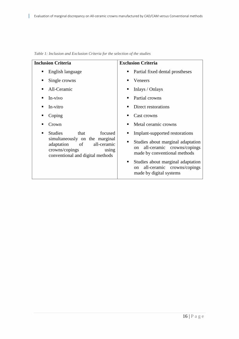
Table 1: Inclusion and Exclusion Criteria for the selection of the studies
Inclusion Criteria
English language
Single crowns
All-Ceramic
In-vivo
In-vitro
Coping
Crown
Studies that focused
simultaneously on the marginal
adaptation of all-ceramic
crowns/copings using
conventional and digital methods
Exclusion Criteria
Partial fixed dental prostheses
Veneers
Inlays / Onlays
Partial crowns
Direct restorations
Cast crowns
Metal ceramic crowns
Implant-supported restorations
Studies about marginal adaptation
on all-ceramic crowns/copings
made by conventional methods
Studies about marginal adaptation
on all-ceramic crowns/copings
made by digital systems

Evaluation of marginal discrepancy on All-ceramic crowns manufactured by CAD/CAM versus Conventional methods
17 | P a g e
Chapter 3: Results
Evaluation of marginal discrepancy on All-ceramic crowns manufactured by CAD/CAM versus Conventional methods
18 | P a g e
Among 525 reports identified through the electronic search, 15 were selected, all
published between 2005 and April 2015. All the studies were conducted in vitro. An
overall review of the data retrieved for marginal gap showed that 86.8% of the values
measured were less than or equal to 120 µm described by McLean and Von Fraunhofer.
The widest marginal gap measured was 180 µm, and the smallest was 17 µm. The
selected articles displayed a significant heterogeneity in terms of experimental
protocols, which led to different discrepancies being measured, sometimes even for the
same system. Four reports stated that the conventional method showed better MG
values (PELEKANOS et al. 2009; MOUSLY et al. 2014; SULAIMAN et al. 1997;
BESCHIT and STRUB 1999). Two studies reported no significant difference between
the conventional methods and CAD/CAM (RINKE et al. 1995, ANADIOTI et al.
2013). Nine studies showed better results for the CAD/CAM groups (YEO et al. 2003;
QUINTAS et al. 2004; BINDL and MORMANN 2005; KORKUT et al. 2011;
YUKSEL and ZAIMOGLU 2011; ASAPVAPANUMAS et al. 2013; NG et al. 2014;
DEMIR et al. 2014; PIMENTA et al. 2015).
Seven studies measured the MG on crowns (RINKE et al. 1995; BESCHIT and
STRUB 1999; YEO et al. 2003; ANADIOTI et al. 2013; NG et al. 2014; MOUSLY et
al. 2014; DEMIR et al. 2014), one did not report where they made the measurements
(SULAIMAN et al. 1997) and seven measured on copings (QUINTAS et al. 2004;
BINDL and MORMANN 2005; PELEKANOS et al. 2009; KORKUT et al. 2011;
YUKSEL and ZAIMOGLU 2011; ASAPVAPANUMAS et al. 2013; PIMENTA et al.
2015). Five studies used a finish line in shoulder (RINKE et al. 1995; BESCHIT and
STRUB 1999; YEO et al. 2003; ASAPVAPANUMAS et al. 2013; DEMIR et al. 2014),
seven used a chamfer (BINDL and MORMANN 2005; PELEKANOS et al. 2009;
KORKUT et al. 2011; YUKSEL and ZAIMOGLU 2011; NG et al. 2014; MOUSLY et
al. 2014; PIMENTA et al. 2015) and 3 didn’t report the kind of finish line in their
preparations (SULAIMAN et al. 1997; QUINTAS et al. 2004; ANADIOTI et al. 2013).
Nine studies presented their results without cementing the crowns/copings
(RINKE et al. 1995; SULAIMAN et al. 1997; YEO et al. 2003; PELEKANOS et al.
2009; ASAPVAPANUMAS et al. 2013; ANADIOTI et al. 2013; NG et al. 2014;
MOUSLY et al. 2014; PIMENTA et al. 2015), three studies presented their results after
cementation (BINDL and MORMANN 2005; KORKUT et al. 2011; YUKSEL and
Evaluation of marginal discrepancy on All-ceramic crowns manufactured by CAD/CAM versus Conventional methods
19 | P a g e
ZAIMOGLU 2011) and three studies reported values of MG before and after
cementation (BESCHIT and STRUB 1999; QUINTAS et al. 2004; DEMIR et al. 2014).
Four studies used direct examination with optical microscope (RINKE et al.
1995; SULAIMAN et al. 1997; YEO et al. 2003; QUINTAS et al. 2004), four studies
used scanning with micro-XCT technology (PELEKANOS et al. 2009; MOUSLY et al.
2014; DEMIR et al. 2014; PIMENTA et al. 2015), two studies used cross-sectioning of
the copings/crowns prior to the use of optical microscopes (KORKUT et al. 2011;
YUKSEL and ZAIMOGLU 2011), two studies used scanning with stereomicroscope
and photographs (ASAPVAPANUMAS et al. 2013; NG et al. 2014), one study used
direct examination with SEM (BINDL and MORMANN 2005), one study used an
epoxy replica of the marginal area and then measured the values using an optical
microscope (BESCHIT and STRUB 1999) and finally, one study did not state the type
of method used to make the measurement (ANADIOTI et al. 2013).
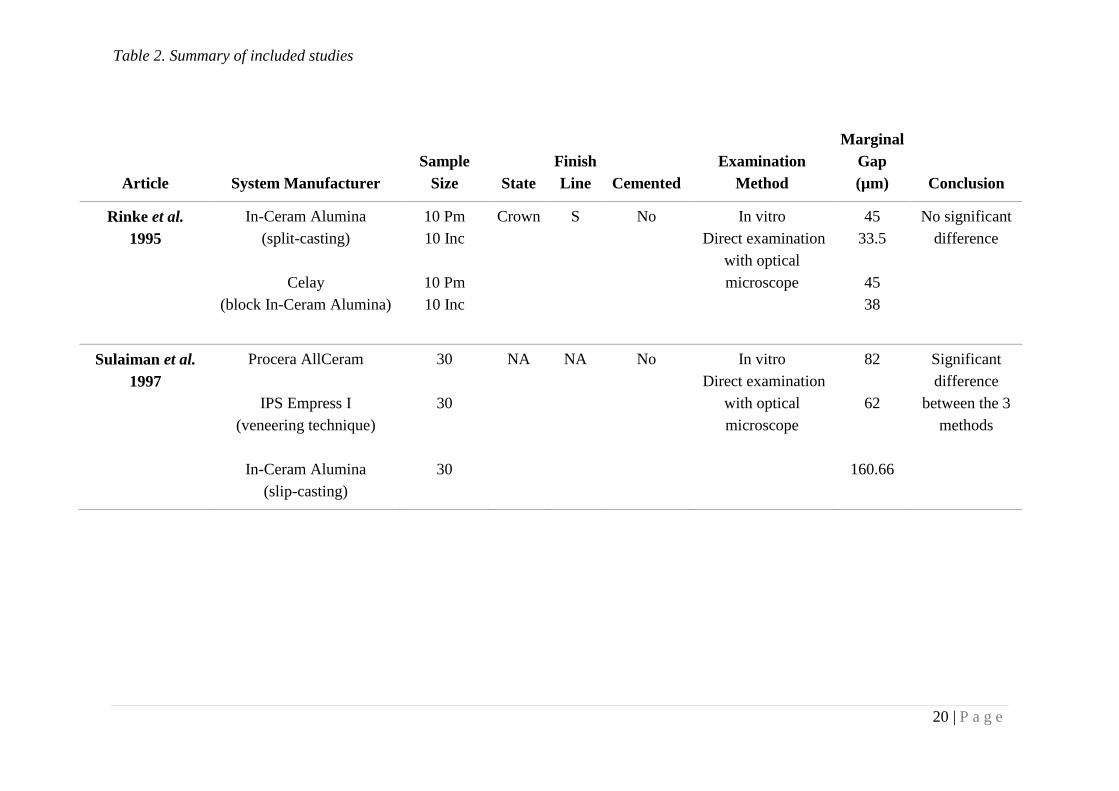
Table 2. Summary of included studies
20 | P a g e
Article System Manufacturer
Sample
Size State
Finish
Line Cemented
Examination
Method
Marginal
Gap
(µm) Conclusion
Rinke et al.
1995
In-Ceram Alumina
(split-casting)
Celay
(block In-Ceram Alumina)
10 Pm
10 Inc
10 Pm
10 Inc
Crown S No In vitro
Direct examination
with optical
microscope
45
33.5
45
38
No significant
difference
Sulaiman et al.
1997
Procera AllCeram
IPS Empress I
(veneering technique)
In-Ceram Alumina
(slip-casting)
30
30
30
NA NA No In vitro
Direct examination
with optical
microscope
82
62
160.66
Significant
difference
between the 3
methods
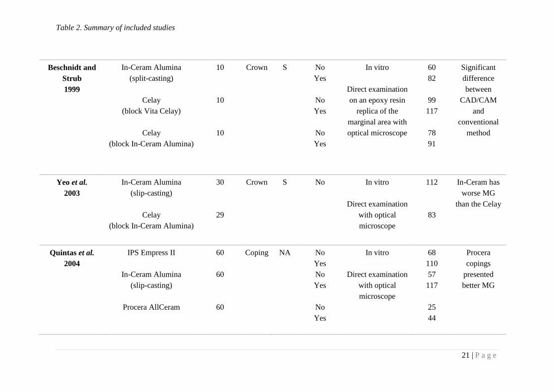
Table 2. Summary of included studies
21 | P a g e
Beschnidt and
Strub
1999
In-Ceram Alumina
(split-casting)
Celay
(block Vita Celay)
Celay
(block In-Ceram Alumina)
10
10
10
Crown S No
Yes
No
Yes
No
Yes
In vitro
Direct examination
on an epoxy resin
replica of the
marginal area with
optical microscope
60
82
99
117
78
91
Significant
difference
between
CAD/CAM
and
conventional
method
Yeo et al.
2003
In-Ceram Alumina
(slip-casting)
Celay
(block In-Ceram Alumina)
30
29
Crown S No In vitro
Direct examination
with optical
microscope
112
83
In-Ceram has
worse MG
than the Celay
Quintas et al.
2004
IPS Empress II
In-Ceram Alumina
(slip-casting)
Procera AllCeram
60
60
60
Coping NA No
Yes
No
Yes
No
Yes
In vitro
Direct examination
with optical
microscope
68
110
57
117
25
44
Procera
copings
presented
better MG

Table 2. Summary of included studies
22 | P a g e
Bindl and
Mormann
2005
In-Ceram Zirconia
(Slip-casting)
IPS Empress II
Cerec InLab
(block In-Ceram Zirconia)
DCS Précident
Decim
Procera AllCeram
12
12
12
12
12
12
Coping C Yes In vitro
Direct examination
with SEM
25
44
43
33
23
17
Procera
copings
presented
better MG
All crowns
were within
clinical
acceptable
values
Pelekanos et al.
2009
WolCeram
(in-Ceram Alumina)
In-Ceram Alumina
(slip-casting)
Cerec inLab
(block In-Ceram Alumina)
Celay
(block In-Ceram Alumina)
4
4
4
4
Coping C No In vitro
Micro-XCT
34.86
21.08
55.09
139.27
Conventional
methods
presented the
best results
Cerec inLab
presented
clinically
acceptable
values
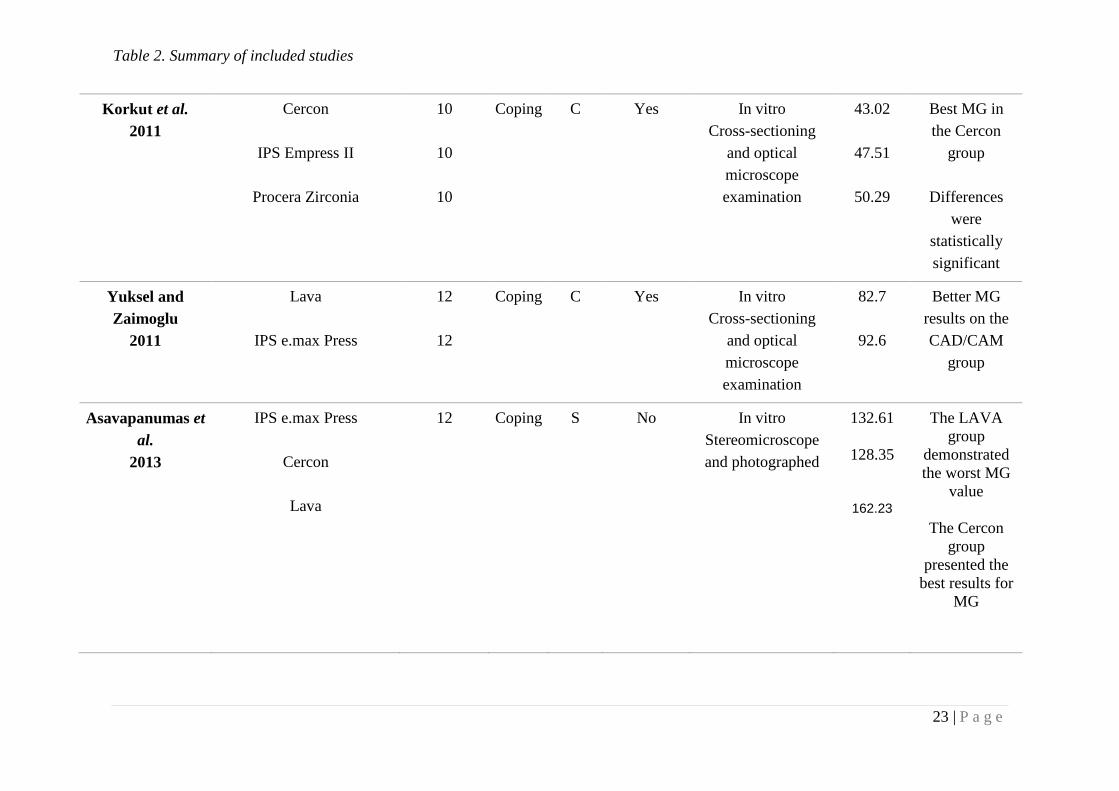
Table 2. Summary of included studies
23 | P a g e
Korkut et al.
2011
Cercon
IPS Empress II
Procera Zirconia
10
10
10
Coping C Yes In vitro
Cross-sectioning
and optical
microscope
examination
43.02
47.51
50.29
Best MG in
the Cercon
group
Differences
were
statistically
significant
Yuksel and
Zaimoglu
2011
Lava
IPS e.max Press
12
12
Coping C Yes In vitro
Cross-sectioning
and optical
microscope
examination
82.7
92.6
Better MG
results on the
CAD/CAM
group
Asavapanumas et
al.
2013
IPS e.max Press
Cercon
Lava
12 Coping S No In vitro
Stereomicroscope
and photographed
132.61
128.35
162.23
The LAVA
group
demonstrated
the worst MG
value
The Cercon
group
presented the
best results for
MG
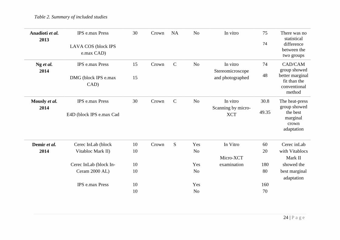
Table 2. Summary of included studies
24 | P a g e
Anadioti et al.
2013
IPS e.max Press
LAVA COS (block IPS
e.max CAD)
30 Crown NA No In vitro
75
74
There was no
statistical
difference
between the
two groups
Ng et al.
2014
IPS e.max Press
DMG (block IPS e.max
CAD)
15
15
Crown C No In vitro
Stereomicroscope
and photographed
74
48
CAD/CAM
group showed
better marginal
fit than the
conventional
method
Mously et al.
2014
IPS e.max Press
E4D (block IPS e.max Cad
30 Crown C No In vitro
Scanning by micro-
XCT
30.8
49.35
The heat-press
group showed
the best
marginal
crown
adaptation
Demir et al.
2014
Cerec InLab (block
Vitabloc Mark II)
Cerec InLab (block In-
Ceram 2000 AL)
IPS e.max Press
10
10
10
10
10
10
Crown S
Yes
No
Yes
No
Yes
No
In Vitro
Micro-XCT
examination
60
20
180
80
160
70
Cerec inLab
with Vitablocs
Mark II
showed the
best marginal
adaptation
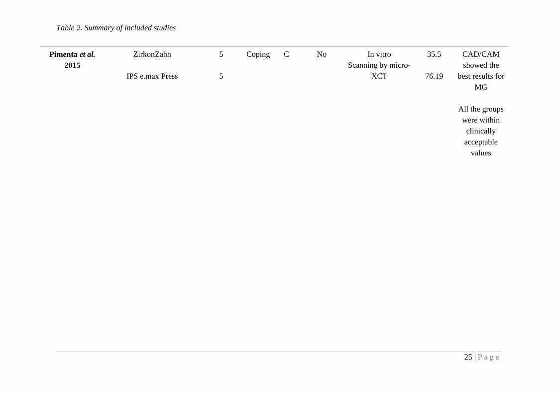
Table 2. Summary of included studies
25 | P a g e
Pimenta et al.
2015
ZirkonZahn
IPS e.max Press
5
5
Coping C No In vitro
Scanning by micro-
XCT
35.5
76.19
CAD/CAM
showed the
best results for
MG
All the groups
were within
clinically
acceptable
values

26 | P a g e
Chapter 4: Discussion and Conclusions
27 | P a g e
Crown marginal fit is critical for success of the restoration; crowns with poor fit
(marginal gap) are prone to failure due to micro-leakage, cement dissolution, and dental
caries. In this paper, the fit of crowns was assessed based on the vertical gap
measurement which was selected as the most critical factor of marginal gap while being
the least susceptible to manipulation post-fabrication, as indicated by Holmes et al. in
1989. Horizontal discrepancies, such as crown overhangs, can be adjusted to some
degree intra-orally, however, vertical MG can only be closed with luting cement, which
is prone to dissolution (NG et al. 2014). For this reason, the vertical MG has the most
clinical relevance and should be regarded as the most critical in crown margin
evaluation.
Chairside CAD/CAM restorations are currently being used by a large number of
dentists all-over the world; however, the accuracy of these systems is still questionable.
The coping, and sometimes even the crown, could be completed without the use of a die
through intraoral impression. However, the accuracy of the data acquisition varies
according to the system’s various optical impression technologies and manufacturer’s
(CONTREPOIS et al. 2013). Software technology and milling accuracy also suffers
from differences between systems. In addition even for the same system, there can be
substantial differences and variations among the measured values which can be
explained by different experimental protocols used in each study as can be showed in
this results.
The use of the conventional method of crown fabrication has been used for
decades with proven long-term results for both longevity and survival. Careful selection
of materials and meticulous fabrication procedures are necessary to compensate for
expansions and contraction of the different materials involved in creating an accurately
fitting crown (NG et al. 2014). However, the impossibility of controlling all the
variables, combined with the propensity for human error, can result in poor marginal
adaptation or even misfit. The use of a digital method seems to decrease the margin of
error. Human intervention in the manufacturing of the crown could play a role
according to the skill of the dental laboratory technician and the relative importance of
his contribution (PELEKANOS et al. 2009).
28 | P a g e
The number of steps involved in the process is another important element
because the probability of error increased with each additional step required For
example, non-CAD/CAM systems required the use of a die spacer applied by a
technician, and the traditional In-Ceram slip-casting system was described as singularly
technique sensitive. (BINDL and MORMANN 2005).
Different measurement methods were used among the various studies, and this
could have impacted results significantly. The first and most widely used method
involved direct microscopic examination of the marginal area. Unfortunately, this
method has two great disadvantages. First, identifying reference points to measure may
prove difficult. Second, it may lead to projection errors (GROTEN et al. 2000). In the
second method, cemented specimens were cross-sectioned, and the marginal area was
then examined under a microscope. However, only a limited number of sections could
be cut on any one specimen. These two techniques were also sometimes used to
measure an epoxy resin replica of the marginal area instead of the area itself. This
technique does not provide accurate results (CONTREPOIS et al. 2013). A third
method involved creating a light bodied silicone replica of the gap between the crown
and the tooth. This replica was then sectioned, and the zone that corresponded to the
marginal area was observed by microscopy. This provided only a limited number of
marginal gap measurements (BESCHIT and STRUB 1999) .The last technique used
was x-ray microtomography. This innovative and nondestructive technique, which
delivers 2-dimensional and 3-dimensional imaging of the space between the
reconstitution and the die, and it can provide very close sections of the marginal area,
which allows for a great number of measurement sites and for easy recognition of the
critical distances. (PELEKANOS et al. 2009). This method has several advantages over
other technologies including the 3-dimensional evaluation of the marginal and internal
gaps. Furthermore, it is easy to perform, nondestructive, and more time efficient and
accurate than other methods. The main disadvantages are radiation artifacts, which are
caused by the differences in the coefficient of radiation absorption among the different
materials and the difficulty in using luting agents because they have some radiopacity,
which might affect the evaluation of the marginal gap. (MOUSLY et al. 2014).
Another important factor is that, a better approximation of clinical conditions
may be reached by conducting measurements upon completion of the crown
29 | P a g e
(CONTREPOIS et al. 2013). In addition, measuring fit at the crown stage is necessary
to compare single-layer crowns and multi-layer crowns.
Some studies also measured the marginal gap before and after cementation of
the crown or coping. Measurements made solely after cementation do not allow for the
determination of the relative impact on the marginal fit of cementation and of a system’s
intrinsic precision (GROTEN et al. 2000) It is also more convenient to conduct
measurements without cementing the crown as most studies did. Further evaluation of
the capacity of all-ceramic conventional methods and CAD/CAM systems marginal
adaptation after cementation, should be studied with different objectives and not mixed
with non-cementing studies.
The type of finish line used in the studies varied from shoulder or chamfer. This
variable also could have been responsible for the different results obtained between the
conventional and digital methods or even inside the same system. Finish lines made in
in vitro studies should be prepared in accordance to the most realistic clinical
conditions. So with this in mind, the use of models that bear no relation to an actual
tooth anatomy should be discontinued. Furthermore, finish lines that present some
degree of curvature should be preferred since they can better simulate the presence of a
gingival margin (CONTREPOIS et al. 2013).
Analysis of the results of this study suggests that more studies support the idea
that digitally made crowns/copings can have better marginal adaptation values.
However, most results seem to be well within clinical acceptable values (≤120 µm),
which means digital or conventional made crowns are still two well supported options
for fixed rehabilitation. Better protocols should be implemented to study the adaptation
or CAD/CAM ceramic crowns versus conventional ceramic crowns. Although there are
many studies made all over the years regarding marginal discrepancy on all-ceramic
crowns, little has been done to clearly compare the digital method and the conventional
method. This fact could be appointed to the fact that CAD/CAM is a relatively new
technology that is slowly making its way to medical office because of its high costs,
making the conventional method still a very low cost/benefit method, still preferred my
most dentists in the world.
30 | P a g e
Conclusions:
Based on the results obtained, the digital method seems to be a legitimate
alternative to the traditional methods. Analysis of the results of this study suggested that
the digital method exceeds the standards of clinical acceptability and can sometimes
surpass the vertical marginal fit of conventionally fabricated crowns.
Further studies are encouraged using standardized protocols as well as systems
and techniques, in order to better evaluate the capabilities of this digital systems.
31 | P a g e
References:
1. Aboushelib M.N., Kleverlaan C.J.,Feilzer A.J. (2007). "Selective infiltration-
etching technique for a strong and durable bond of resin cements to zirconia-
based materials." J Prosthet Dent 98(5): 379-388.
2. Anadioti E, Aquilino SA, Gratton DG, Holloway JA, Denry I, Thomas GW,
Quian F. 3D and 2D marginal fit of pressed and CAD/CAM lithium disilicate
crowns made from digital and conventional impressions. Journal of
Prosthodontics 23 2013 610-617.
3. Andersson M.,Oden A. (1993). "A new all-ceramic crown. A dense-sintered,
high-purity alumina coping with porcelain." Acta Odontol Scand 51(1): 59-64.
4. Asavapanumas C, Leevailoj C. The influence of finish line curvature on the
marginal gap width of ceramic copings. J Prosthet Dent 2013;109:226-233.
5. Beschnidt SM, Strub JR. Evaluation of the marginal accuracy of different all-
ceramic crown systems after simulation in the artificial mouth. J oral Rehabil
1999;26:582-93.
6. Beuer F., Edelhoff D., Gernet W.,Sorensen J.A. (2009a). "Three-year clinical
prospective evaluation of zirconia-based posterior fixed dental prostheses
(FDPs)." Clin Oral Investig 13(4): 445-451
7. Beuer F., Schweiger J.,Edelhoff D. (2008). "Digital dentistry: an overview of
recent developments for CAD/CAM generated restorations." Br Dent J 204(9):
505-511.
8. Bindl A, Mormann WH. Marginal and internal fit of all-ceramic CAD/CAM
crown-copings on chamfer preparations. J Oral Rehabil 2005;32:441-7.
9. Bindl A., Luthy H.,Mormann W.H. (2006). "Strength and fracture pattern of
monolithic CAD/CAM-generated posterior crowns." Dent Mater 22(1): 29-36.
10. Bindl A.,Mormann W.H. (2002). "An up to 5-year clinical evaluation of
posterior in-ceram CAD/CAM core crowns." Int J Prosthodont 15(5): 451-456.
11. Borom, M.P.; Turkalo, A.M.; Doremus, R.H. Strength and microstructure in
lithium disilicate glass-ceramics. J. Am. Ceram. Soc. 1975, 58, 385–391.
32 | P a g e
12. Cattani-Lorente M., Scherrer S.S., Ammann P., Jobin M.,Wiskott H.W. (2011).
"Low temperature degradation of a Y-TZP dental ceramic." Acta Biomater 7(2):
858-865.
13. Contrepois M, Soenen A, Bartala M, Laviole O. Marginal adaptation of ceramic
crowns: a systematic review. J Prosthet Dent 2013;110:447-454.
14. Cooper, L. Direct ceramic restoration using digital technologies. International
dentistry Australian edition VOL. 7, NO. 1. 2011. 32-35.
15. Deany I.L. (1996). "Recent advances in ceramics for dentistry." Crit Rev Oral
Biol Med 7(2): 134-143.
16. Demir N, Ozturk AN, Malkoc MA. Evaluation of marginal fit of full ceramic
crowns by microcomputed tomography (micro-CT) technique. Eur J Dent
2014;8:437-44
17. Denry I.,Holloway J. (2010). "Ceramics for Dental Applications: A Review."
Materials 3(1): 351-368.
18. Denry I.,Kelly J.R. (2008). "State of the art of zirconia for dental applications."
Dent Mater 24(3): 299-307.
19. Denry I.,Kelly J.R. (2008). "State of the art of zirconia for dental applications."
Dent Mater 24(3): 299-307.
20. Denry, I.L.; Rosenstiel, S.F. Phase transformations in feldspathic dental
porcelains. In Bioceramics: Materials and Applications; Fischman, G., Clare,
A., Hench, L., Eds.; The American Ceramic Society: Westerville, OH, USA,
1995; pp. 149–156.
21. Derand T., Molin M.,Kvam K. (2005). "Bond strength of composite luting
cement to zirconia ceramic surfaces." Dent Mater 21(12): 1158-1162.
22. Dong, J.K.; Luthy, H.; Wohlwend, A. Heat-pressed ceramics: Technology and
strength. Int. J. Prosthodont. 1992, 5, 9–16.
23. Duret F.,Preston J.D. (1991). "CAD/CAM imaging in dentistry." Curr Opin Dent
1(2): 150-154.
24. Ebert J., Ozkol E., Zeichner A., Uibel K., Weiss O., Koops U., Telle R.,Fischer
H. (2009). "Direct inkjet printing of dental prostheses made of zirconia." J Dent
Res 88(7): 673-676.
33 | P a g e
25. Et L.N. (2008). Computer Aided Design And Manufacturing, Prentice-Hall Of
India Pvt. Limited
26. Fasbinder D.J. (2002). "Restorative material options for CAD/CAM
restorations." Compend Contin Educ Dent 23(10): 911-916, 918, 920 passim;
quiz 924.
27. Fasbinder D.J. (2010). "Digital dentistry: innovation for restorative treatment."
Compend Contin Educ Dent 31 Spec No 4: 2-11; quiz 12
28. Fischer J., Stawarczyk B.,Hammerle C.H. (2008). "Flexural strength of
veneering ceramics for zirconia." J Dent 36(5): 316-321.
29. Fradeani M., Redemagni M.,Corrado M. (2005). "Porcelain laminate veneers: 6-
to 12-year clinical evaluation--a retrospective study." Int J Periodontics
Restorative Dent 25(1): 9-17.
30. Fradeani M.,Redemagni M. (2002). "An 11-year clinical evaluation of leucite-
reinforced glass-ceramic crowns: a retrospective study." Quintessence Int 33(7):
503-510.
31. Gardner FM. Margins of complete crowns—Literature review. The Journal of
prosthetic dentistry 1982;48:396-400.
32. Giordano R.,McLaren E.A. (2010). "Ceramics overview: classification by
microstructure and processing methods." Compend Contin Educ Dent 31(9):
682-684, 686, 688 passim; quiz 698, 700.
33. Gonzaga C.C., Cesar P.F., Okada C.Y., Fredericci C., Beneduce Neto
F.,Yoshimura H.N. (2008). "Mechanical properties and porosity of dental glass-
ceramics hot-pressed at different temperatures." Materials Research 11: 301-
306.
34. Grasso JE, Nalbandian J, Sanford C, Bailit H. Effect of restoration quality on
periodontal health. The Journal of prosthetic dentistry 1985;53:14.
35. Griffin J.D., Jr. (2013). "Tooth in a bag: same-day monolithic zirconia crown."
Dent Today 32(1): 124, 126-131.
36. Groten M, Girthofer S, Probster L. Marginal fit consistency of copy-milled all-
ceramic crowns during fabrication by light and scanning electron microscopic
analysis in vitro. J Oral Rehabil 1997;24:871-81
34 | P a g e
37. Groten M.,Probster L. (1997). "The influence of different cementation modes on
the fracture resistance of feldspathic ceramic crowns." Int J Prosthodont 10(2):
169-177.
38. Guazzato M., Albakry M., Ringer S.P.,Swain M.V. (2004). "Strength, fracture
toughness and microstructure of a selection of all-ceramic materials. Part I.
Pressable and alumina glass-infiltrated ceramics." Dent Mater 20(5): 441-448.
39. Guazzato M., Albakry M., Swain M.V.,Ironside J. (2002). "Mechanical
properties of In-Ceram Alumina and In-Ceram Zirconia." Int J Prosthodont
15(4): 339-346.
40. Guazzato M., Quach L., Albakry M.,Swain M.V. (2005). "Influence of surface
and heat treatments on the flexural strength of Y-TZP dental ceramic." J Dent
33(1): 9-18.
41. Guazzato, M.; Albakry, M.; Ringer, S.P.; Swain, M.V. Strength, fracture
toughness and microstructure of a selection of all-ceramic materials. Part I.
Pressable and alumina glassinfiltrated ceramics. Dent. Mater. 2004, 20, 441–
448.
42. Guess P.C., Schultheis S., Bonfante E.A., Coelho P.G., Ferencz J.L.,Silva N.R.
(2011). "All-ceramic systems: laboratory and clinical performance." Dent Clin
North Am 55(2): 333-352, ix.
43. Guess P.C., Zavanelli R.A., Silva N.R., Bonfante E.A., Coelho P.G.,Thompson
V.P. (2010). "Monolithic CAD/CAM lithium disilicate versus veneered Y-TZP
crowns: comparison of failure modes and reliability after fatigue." Int J
Prosthodont 23(5): 434-442.
44. Haselton D.R., Diaz-Arnold A.M.,Hillis S.L. (2000). "Clinical assessment of
high-strength all-ceramic crowns." J Prosthet Dent 83(4): 396-401.
45. Heffernan M.J., Aquilino S.A., Diaz-Arnold A.M., Haselton D.R., Stanford
C.M.,Vargas M.A. (2002). "Relative translucency of six all-ceramic systems.
Part I: core materials." J Prosthet Dent 88(1): 4-9.
46. Heffernan, M.J.; Aquilino, S.A.; Diaz-Arnold, A.M.; Haselton, D.R.; Stanford,
C.M.; Vargas,M.A. Relative translucency of six all-ceramic systems. Part I:
Core materials. J. Prosthet. Dent. 2002, 88, 4–9.
35 | P a g e
47. Holand W., Schweiger M., Frank M.,Rheinberger V. (2000). "A comparison of
the microstructure and properties of the IPS Empress 2 and the IPS Empress
glass-ceramics." J Biomed Mater Res 53(4): 297-303.
48. Holand W., Schweiger M., Watzke R., Peschke A.,Kappert H. (2008).
"Ceramics as biomaterials for dental restoration." Expert Rev Med Devices 5(6):
729-745.
49. Höland, W.; Apel, E.; van't Hoen, C.; Rheinberger, V. Studies of crystal phase
formations in highstrength lithium disilicate glass-ceramics. J. Non-Cryst. Solids
2006, 352, 4041–4050.
50. Holmes JR, Bayne SC, Holland GA, Sulik WD. Considerations in measurement
of marginal fit. The Journal of prosthetic dentistry 1989;62:405-408.
51. Holt L.R.,Boksman L. (2012). "Monolithic zirconia: minimizing adjustments."
Dent Today 31(12): 78, 80-71.
52. Jacobs MS, Windeler AS. An investigation of dental luting cement solubility as
a function of the marginal gap. The Journal of prosthetic dentistry 1991;65:436-
442
53. Janenko C, Smales R. Anterior crowns and gingival health. Australian dental
journal 1979;24:225-230.
54. Jung, Y.-G.; IM, P.; A, P.; BR., L. Contact damage resistance and strength
degradation of glassinfiltrated alumina and spinell ceramics. J. Dent. Res. 1999,
78, 804–814.
55. Korkut L, Cotert HS, Kurtulmus H. Marginal, internal fit and microleakage of
zirconia infrastructures: an in-vitro study. Operative Dentistry 2011;36-1, 72-79.
56. Krejci I., Lutz F.,Reimer M. (1994). "Wear of CAD/CAM ceramic inlays:
restorations, opposing cusps, and luting cements." Quintessence Int 25(3): 199-
207.
57. Mackert, J.R., Jr.; Williams, A.L. Microcracks in dental porcelains and their
behavior during multiple firing. J. Dent. Res. 1996, 75, 1484–1490.
58. Manicone P.F., Rossi Iommetti P.,Raffaelli L. (2007). "An overview of zirconia
ceramics: basic properties and clinical applications." J Dent 35(11): 819-826.
59. May KB, Russell MM, Razzoog ME, Lang BR. Precision of fit: the Procera
AllCeram crown. The Journal of prosthetic dentistry 1998;80:394-404.
36 | P a g e
60. McLean JW, von Fraunhofer JA. The estimation of cement film thickness by an
in vivo technique. British dental journal 1971;131:107-111.
61. Miyazaki T, Hotta Y, Kunii J, Kuriyama S, Tamaki Y. A review of dental
CAD/CAM: current status and future perspectives from 20 years of experience.
Dent Mat Journal 2009; 28: 44-56.
62. Mormann W.H. (2004). "The origin of the Cerec method: a personal review of
the first 5 years." Int J Comput Dent 7(1): 11-24.
63. Mormann W.H. (2006). "The evolution of the CEREC system." J Am Dent
Assoc 137 Suppl: 7S-13S.
64. Mously HA, Finkelman M, Zandparsa R, Hirayama H. Marginal and internal
adaptation of ceramic crown restorations fabricated with CAD/CAM technology
and the heat-press technique. J Prosthet Dent 2014,112:249-256.
65. Ng J, Ruse D, Wyatt C. A comparison of the marginal fit of crowns fabricated
with digital and conventional methods. J Prosthet Dent 2014;112:555-560.
66. Otto T. (2004). "Computer-aided direct all-ceramic crowns: preliminary 1-year
results of a prospective clinical study." Int J Periodontics Restorative Dent
24(5): 446-455.
67. Pelekanos S, Koumanou M, Koutayas SO, Zinelis S, Eliades G. Micro-CT
evaluation of the marginal fit of different In-Ceram alumina copings. Eur J
Esthet Dent 2009;4:278-92.
68. Pichler F., Moreno-Diaz R.,Kopacek P. (2000). Computer Aided Systems
Theory - EUROCAST'99: A Selection of Papers from the 7th International
Workshop on Computer Aided Systems Theory Vienna, Austria, September 29 -
October 2, 1999 Proceedings, Springer.
69. Piconi C.,Maccauro G. (1999). "Zirconia as a ceramic biomaterial." Biomaterials
20(1): 1-25.
70. Pimenta MA, Frasca LC, Lopes R, Rivaldo E. Evaluation of marginal and
internal fit of ceramic and metallic crown copings using x-ray microtomography
(micro-CT) technology. J Prosthet Dent 2015.
71. Pjetursson B.E., Sailer I., Zwahlen M.,Hammerle C.H. (2007). "A systematic
review of the survival and complication rates of all-ceramic and metal-ceramic
37 | P a g e
reconstructions after an observation period of at least 3 years. Part I: Single
crowns." Clin Oral Implants Res 18 Suppl 3: 73-85.
72. Posselt A.,Kerschbaum T. (2003). "Longevity of 2328 chairside Cerec inlays
and onlays." Int J Comput Dent 6(3): 231-248.
73. Quintas AF, Oliveira F, Bottino MA. Vertical marginal discrepancy of ceramic
copings with different ceramic materials, finish lines and luting agents: an in
vitro evaluation. J Prosthet Dent 2004;92:250-7.
74. Raigrodski A.J. (2004). "Contemporary all-ceramic fixed partial dentures: a
review." Dent Clin North Am 48(2): viii, 531-544.
75. Rinke S, Huls A, JahnL. Marginal accuracy and fracture strength of
conventional and copy-milled all-ceramic crowns. Int j Prosthodont 1995;8:303-
10
76. Ronald L. Sakaguchi P.D.,John M. Powers P.D. (2011). Craig's Restorative
Dental Materials, Elsevier/Mosby.
77. Seghi R.R., Crispin B.C.,Mito W. (1990). "The effect of ion exchange on the
flexural strength of feldspathic porcelains." Int J Prosthodont 3(2): 130-134.
78. Seghi, R.; Sorensen, J. Relative flexural strength of six new ceramic materials.
Int. J. Prosthodont. 1995, 8, 239–246.
79. Stappert C.F., Att W., Gerds T.,Strub J.R. (2006). "Fracture resistance of
different partial-coverage ceramic molar restorations: An in vitro investigation."
J Am Dent Assoc 137(4): 514-522.
80. Sulaiman F, Chai J, Jameson LM, Wozniak WT, A comparison of the marginal
fot of In-Ceram, IPS Empress and Procera crowns. Int Prosthodont 1997;10:478-
84.
81. Suttor D., Bunke K., Hoescheler S., Hauptmann H.,Hertlein G. (2001). "LAVA--
the system for all-ceramic ZrO2 crown and bridge frameworks." Int J Comput
Dent 4(3): 195-206.
82. Tinschert J., Zwez D., Marx R.,Anusavice K.J. (2000). "Structural reliability of
alumina-, feldspar-, leucite-, mica- and zirconia-based ceramics." J Dent 28(7):
529-535.
38 | P a g e
83. Vagkopoulou T., Koutayas S.O., Koidis P.,Strub J.R. (2009). "Zirconia in
dentistry: Part 1. Discovering the nature of an upcoming bioceramic." Eur J
Esthet Dent 4(2): 130-151.
84. Van Noort R. (2012). "The future of dental devices is digital." Dent Mater 28(1):
3-12.
85. Wassermann A., Kaiser M.,Strub J.R. (2006). "Clinical long-term results of
VITA In-Ceram Classic crowns and fixed partial dentures: A systematic
literature review." Int J Prosthodont 19(4): 355-363.
86. Witkowski S. (2005). "(CAD-)/CAM in Dental Technology." Quintessence Dent
Technol 28: 169-184.
87. Xiao-ping L., Jie-mo T., Yun-long Z.,Ling W. (2002). "Strength and fracture
toughness of MgO-modified glass infiltrated alumina for CAD/CAM." Dent
Mater 18(3): 216-220.
88. Yeo IS, Yang JH, Lee JB. In vitro marginal fit of three all-ceramic crown
systems. J Prosthet Dent 2003;90:459-64.
89. Yuksel E, Zaimoglu A. Influence of marginal fit and cement types on
microleakage of all-ceramic crown systems. Braz Oral Res. 2011;25(3):261-6.