evaluating the effect of polytetrafluoroethylene and … the effect of polytetrafluoroethylene and...
6
Plast Surg Vol 23 No 1 Spring 2015 9 ©2015 Canadian Society of Plastic Surgeons. All rights reserved ORIGINAL ARTICLE Evaluating the effect of polytetrafluoroethylene and extractum cepae-heparin-allantoin gel in peripheral nerve injuries in a rat model Ahmet Kahraman MD, Ramazan Kahveci MD Department of Plastic Reconstructive and Aesthetic Surgery, Mustafa Kemal University, Antakya, Turkey Correspondence: Dr Ahmet Kahraman, Mustafa Kemal University,Tayfur Ata Sökmen Kampüsü Serinyol, Hatay, Turkey. Telephone 90-0326-22710-00-3309, fax 90-0326-245-5654, e-mail [email protected] P eripheral nerves can be injured by congenital, mechanical, thermal or chemical causes. Peripheral nerve injuries are increasing in fre- quency, particularly in countries that are becoming more industrial- ized. Nerve and extremity injuries result in work loss and high treatment costs, and lead to separation of patients from their social environment. Failure of nerve repair causes muscle functional losses, sensory losses and painful neuropathies. Depending on injury severity, structural and functional changes occur in nerves. The segment distal to the injury undergoes Wallerian degeneration, which prepares the tube for axonal regeneration. During the healing process, collagen synthesis due to fibroblastic activity increases in the endoneurium and the perineurium. Scar tissue forma- tion just outside the injured nerve causes adhesion of nerve fibres to neighbouring tissues, decreases mobility and causes vasospasm and traction injuries in the nerve vessels. Furthermore, it decreases nutri- tion by diffusion, inhibits nerve repair, slows axoplasmic flow and negatively effects nerve degeneration, resulting in ischemia and irreversible injury to the nerve. During the healing process, scar tissue forming on the incision line blocks axonal growth by forming a mech- anical barrier. Scar tissue formation is a component of the wound healing process (1,2). If the re-innervation is prolonged, collagen becomes denser and the endoneural tube narrows further, thus slowing progression of regeneration to the distal site of the injury; this is referred to as ‘delayed scar’. For primary peripheral nerve injuries in which the severed nerve endings can be easily reapproximated, primary suturing is the most common first-line repair method. However, despite highly developed surgical techniques and postoperative programs, intra- and extraneural adhesions following nerve surgery remain an important problem. To prevent irreversible nerve injury, and to increase axonal regeneration and prevent adhesions, many methods and materials such as anti- transforming growth factor-beta anti-core (3,4), aprotinin (5), hyal- uronic acid (6,7), human amniotic membrane (8,9), citicoline (10) and ADCON-T/N (11) have been used. Although these agents have inhibited the formation of adhesions, they have not been able to elim- inate them completely (12,13). The present study used a rat model to compare the effects of poly- tetrafluoroethylene (PTFE) and PTFE-extractum cepae-heparin- allantoin (PTFE-EHA) gel compound on nerve histopathology and functional recovery after total peripheral nerve injury repaired by primary suture, and the prevention of adhesions and scar tissue for- mation. PTFE is a synthetic biomaterial that is inert, nonadhesive, A Kahraman, R Kahveci. Evaluating the effect of polytetrafluoroethylene and extractum cepae- heparin-allantoin gel in peripheral nerve injuries in a rat model. Plast Surg 2015;23(1):9-14. BACKGROUND: Peripheral nerves can be injured by congenital, mechanical, thermal or chemical causes. Peripheral nerve injuries are increasing in frequency, particularly in countries that are becoming more industrialized. Nerve and extremity injuries result in work loss and high treatment costs, and can lead to separation of patients from their social environment. Failure of nerve repair causes muscle functional losses, sen- sory losses and painful neuropathies. OBJECTIVES: To compare the effects of condensed polytetrafluoroeth- ylene (cPTFE) and cPTFE-extractum cepae-heparin-allantoin (cPTFE- EHA) gel compound on nerve and functional recovery, and the prevention of adhesion and scar tissue formation after total peripheral nerve injury repaired by primary suture in a rat model. RESULTS: cPTFE alone and cPTFE-EHA gel was found to provide better functional recovery and nerve regeneration compared with primary repair only. In the macroscopic evaluation, the cPTFE-EHA gel was found to have no negative effect on wound healing and, despite increasing extra- neural scar tissue and adhesions, it had no negative effect on nerve func- tion; in addition, it facilitated functional recovery. CONCLUSIONS: Compared with the cPTFE application alone, the application of perineural cPTFE-EHA gel during peripheral nerve surgery appeared to provide better functional recovery without causing any sig- nificant changes in epineural and extraneural scar tissue formation. Key Words: Extractum cepae-heparin-allantoin gel; Perineural scar; Peripheral nerve injury; Regeneration Évaluer l’effet du polytétrafluoréthylène et d’un gel contenant de l’extrait d’oignon, de l’héparine et de l’allantoïne sur les lésions des nerfs périphériques d’un modèle de rat HISTORIQUE : Les nerfs périphériques peuvent être soumis à des lésions congénitales, mécaniques, thermiques ou chimiques. Les lésions des nerfs périphériques sont de plus en plus fréquentes, surtout dans les pays qui s’industrialisent. Les lésions des nerfs et des membres s’associent à des pertes d’emploi et à des coûts de traitement élevés. Elles peuvent égale- ment écarter les patients de leur environnement social. L’échec de la répa- ration nerveuse cause une perte fonctionnelle des muscles, des pertes sensorielles et des neuropathies douloureuses. OBJECTIFS : Comparer les effets du polytétrafluoréthylène condensé (PTFEc) et d’un composé de PTFEc et de gel contenant de l’extrait d’oignon, de l’héparine et de l’allantoïne (PTFEc-EHA) sur le rétablisse- ment nerveux et fonctionnel et sur la prévention de la formation d’adhérences et de tissus cicatriciels après une lésion totale des nerfs péri- phériques réparée par une suture primaire chez un modèle de rat. RÉSULTATS : Il a été établi que le PTFEc seul et le PTFEc-EHA assurait un meilleur rétablissement fonctionnel et une meilleure régénération nerveuse que la réparation primaire seule. Lors de l’évaluation macroscopique, le PTFEc-EHA n’avait pas d’effet négatif sur la cicatrisation de la plaie et, malgré la formation d’adhérences et de tissus cicatriciels extraneuraux, il ne nuisait pas à la fonction nerveuse. Par ailleurs, il facilitait le rétablisse- ment fonctionnel. CONCLUSIONS : Par rapport à la seule application de PTFEc, l’application de PTFEc-EHA périneurale pendant une chirurgie des nerfs périphériques semblait assurer un meilleur rétablissement fonctionnel sans vraiment aggraver la formation de tissus cicatriciels extraneuraux.
-
Upload
phungkhanh -
Category
Documents
-
view
220 -
download
0
Transcript of evaluating the effect of polytetrafluoroethylene and … the effect of polytetrafluoroethylene and...
Plast Surg Vol 23 No 1 Spring 2015 9©2015 Canadian Society of Plastic Surgeons. All rights reserved
original article
evaluating the effect of polytetrafluoroethylene and extractum cepae-heparin-allantoin gel in peripheral
nerve injuries in a rat modelAhmet Kahraman MD, Ramazan Kahveci MD
Department of Plastic Reconstructive and Aesthetic Surgery, Mustafa Kemal University, Antakya, TurkeyCorrespondence: Dr Ahmet Kahraman, Mustafa Kemal University,Tayfur Ata Sökmen Kampüsü Serinyol, Hatay, Turkey.
Telephone 90-0326-22710-00-3309, fax 90-0326-245-5654, e-mail [email protected]
Peripheral nerves can be injured by congenital, mechanical, thermal or chemical causes. Peripheral nerve injuries are increasing in fre-
quency, particularly in countries that are becoming more industrial-ized. Nerve and extremity injuries result in work loss and high treatment costs, and lead to separation of patients from their social environment. Failure of nerve repair causes muscle functional losses, sensory losses and painful neuropathies.
Depending on injury severity, structural and functional changes occur in nerves. The segment distal to the injury undergoes Wallerian degeneration, which prepares the tube for axonal regeneration. During the healing process, collagen synthesis due to fibroblastic activity increases in the endoneurium and the perineurium. Scar tissue forma-tion just outside the injured nerve causes adhesion of nerve fibres to neighbouring tissues, decreases mobility and causes vasospasm and traction injuries in the nerve vessels. Furthermore, it decreases nutri-tion by diffusion, inhibits nerve repair, slows axoplasmic flow and negatively effects nerve degeneration, resulting in ischemia and irreversible injury to the nerve. During the healing process, scar tissue forming on the incision line blocks axonal growth by forming a mech-anical barrier. Scar tissue formation is a component of the wound healing process (1,2). If the re-innervation is prolonged, collagen
becomes denser and the endoneural tube narrows further, thus slowing progression of regeneration to the distal site of the injury; this is referred to as ‘delayed scar’.
For primary peripheral nerve injuries in which the severed nerve endings can be easily reapproximated, primary suturing is the most common first-line repair method. However, despite highly developed surgical techniques and postoperative programs, intra- and extraneural adhesions following nerve surgery remain an important problem. To prevent irreversible nerve injury, and to increase axonal regeneration and prevent adhesions, many methods and materials such as anti-transforming growth factor-beta anti-core (3,4), aprotinin (5), hyal-uronic acid (6,7), human amniotic membrane (8,9), citicoline (10) and ADCON-T/N (11) have been used. Although these agents have inhibited the formation of adhesions, they have not been able to elim-inate them completely (12,13).
The present study used a rat model to compare the effects of poly-tetrafluoroethylene (PTFE) and PTFE-extractum cepae-heparin-allantoin (PTFE-EHA) gel compound on nerve histopathology and functional recovery after total peripheral nerve injury repaired by primary suture, and the prevention of adhesions and scar tissue for-mation. PTFE is a synthetic biomaterial that is inert, nonadhesive,
A Kahraman, R Kahveci. Evaluating the effect of polytetrafluoroethylene and extractum cepae-heparin-allantoin gel in peripheral nerve injuries in a rat model. Plast Surg 2015;23(1):9-14.
BACKgROUND: Peripheral nerves can be injured by congenital, mechanical, thermal or chemical causes. Peripheral nerve injuries are increasing in frequency, particularly in countries that are becoming more industrialized. Nerve and extremity injuries result in work loss and high treatment costs, and can lead to separation of patients from their social environment. Failure of nerve repair causes muscle functional losses, sen-sory losses and painful neuropathies. OBJECTIVES: To compare the effects of condensed polytetrafluoroeth-ylene (cPTFE) and cPTFE-extractum cepae-heparin-allantoin (cPTFE-EHA) gel compound on nerve and functional recovery, and the prevention of adhesion and scar tissue formation after total peripheral nerve injury repaired by primary suture in a rat model. RESULTS: cPTFE alone and cPTFE-EHA gel was found to provide better functional recovery and nerve regeneration compared with primary repair only. In the macroscopic evaluation, the cPTFE-EHA gel was found to have no negative effect on wound healing and, despite increasing extra-neural scar tissue and adhesions, it had no negative effect on nerve func-tion; in addition, it facilitated functional recovery. CONCLUSIONS: Compared with the cPTFE application alone, the application of perineural cPTFE-EHA gel during peripheral nerve surgery appeared to provide better functional recovery without causing any sig-nificant changes in epineural and extraneural scar tissue formation.
Key Words: Extractum cepae-heparin-allantoin gel; Perineural scar; Peripheral nerve injury; Regeneration
Évaluer l’effet du polytétrafluoréthylène et d’un gel contenant de l’extrait d’oignon, de l’héparine et de l’allantoïne sur les lésions des nerfs périphériques d’un modèle de rat
HISTORIQUE : Les nerfs périphériques peuvent être soumis à des lésions congénitales, mécaniques, thermiques ou chimiques. Les lésions des nerfs périphériques sont de plus en plus fréquentes, surtout dans les pays qui s’industrialisent. Les lésions des nerfs et des membres s’associent à des pertes d’emploi et à des coûts de traitement élevés. Elles peuvent égale-ment écarter les patients de leur environnement social. L’échec de la répa-ration nerveuse cause une perte fonctionnelle des muscles, des pertes sensorielles et des neuropathies douloureuses. OBJECTIFS : Comparer les effets du polytétrafluoréthylène condensé (PTFEc) et d’un composé de PTFEc et de gel contenant de l’extrait d’oignon, de l’héparine et de l’allantoïne (PTFEc-EHA) sur le rétablisse-ment nerveux et fonctionnel et sur la prévention de la formation d’adhérences et de tissus cicatriciels après une lésion totale des nerfs péri-phériques réparée par une suture primaire chez un modèle de rat. RÉSULTATS : Il a été établi que le PTFEc seul et le PTFEc-EHA assurait un meilleur rétablissement fonctionnel et une meilleure régénération nerveuse que la réparation primaire seule. Lors de l’évaluation macroscopique, le PTFEc-EHA n’avait pas d’effet négatif sur la cicatrisation de la plaie et, malgré la formation d’adhérences et de tissus cicatriciels extraneuraux, il ne nuisait pas à la fonction nerveuse. Par ailleurs, il facilitait le rétablisse-ment fonctionnel. CONCLUSIONS : Par rapport à la seule application de PTFEc, l’application de PTFEc-EHA périneurale pendant une chirurgie des nerfs périphériques semblait assurer un meilleur rétablissement fonctionnel sans vraiment aggraver la formation de tissus cicatriciels extraneuraux.
Kahraman and Kahveci
Plast Surg Vol 23 No 1 Spring 201510
nonfrictional and induces almost no inflammatory reaction (14,15). Condensed PTFE (cPTFE, MotifMesh, MacroPorous NW Implant, Proxy Biomedical Ltd, Ireland) is composed of pores with a depth of 150 μm and a width of 2350 μm. This permeable structure allows the passage of biologically active materials necessary for cell nutrition, and prevents damage in live tissue psychodynamics (16). It has been demon-strated that EHA (Contractubex gel, 100 g [10 g extractum cepae, 40 mg (5000 IU) heparin sodyum, 1 g allantoin], Merz Pharmaceuticals GmbH, Germany) prevents fibrosis and scar formation in the skin (17,18).
METHODSThe present study was performed in the Animal Laboratory of the Application and Research Center, Uludağ University Faculty of Medicine (Bursa, Turkey), with the approval of Uludağ University Local Animal Experiments Ethics Committee. A total of 24 Sprague Dawley rats weighing between 200 g and 300 g were used. The animals were separated into four groups with six rats in each group, and had ad libitum access to a standard diet and water. Room temperature was adjusted to a mean (± SD) 24±1°C and a light/dark cycle of 12 h/12 h.
Surgical techniqueAccess to food and water was eliminated 24 h before the surgical pro-cedure. For anesthesia, 30 mg/kg thiopental sodium (Pental Sodium, 0.5 Flacon İ. E. Ulagay Pharmaceuticals, Turkey) was administered intraperitoneally. The extremities of the animals were fixed to the appli-cation table in the prone position. Povidone iodine (Isosol IE, 1000 mL solution, İ. E. Ulagay Pharmaceuticals, Turkey) was applied to the right leg, which was covered with an appropriate green surgical drape and opening. A vertical cutaneous incision approximately 3 cm in length was made along the right hip and thigh, and the connection line and the surrounding fascia of the right gluteus and biceps femoris muscles were separated with a sharp dissection and the sciatic nerve was accessed.
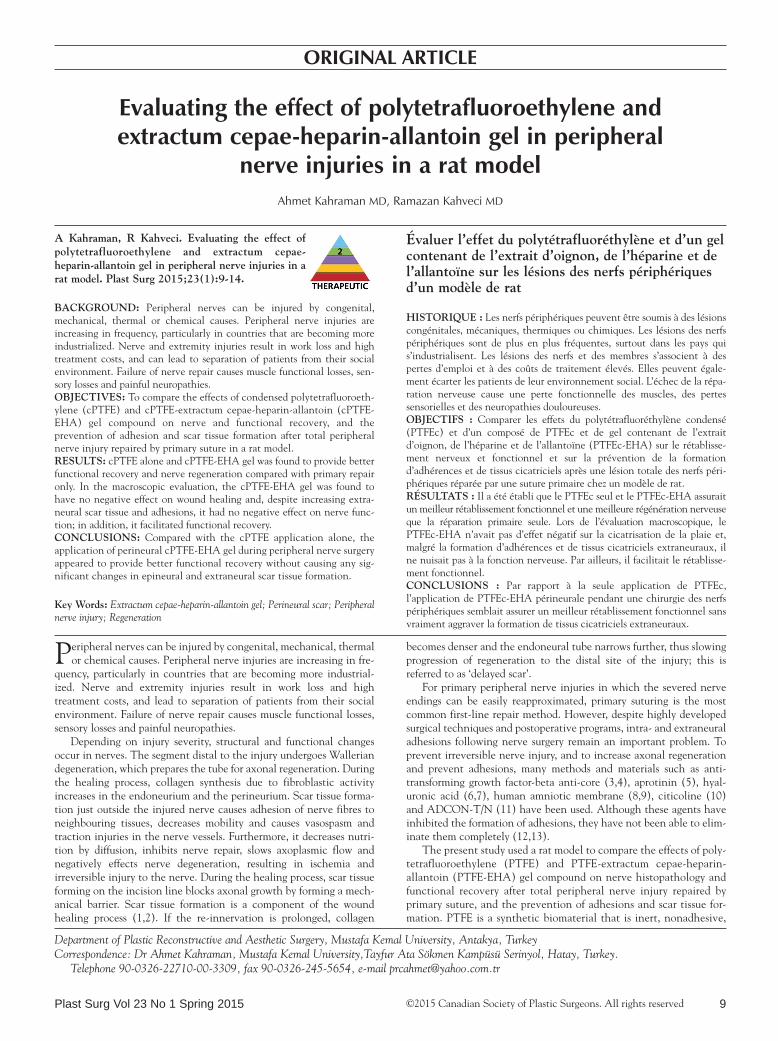
The nerve was separated from the surrounding tissues by scraping the covering membranous tissues from the sciatic foramen to the point where the tibial and the peroneal branches are separated (Figure 1). With the sciatic nerve slightly elevated, using an extractor, from the distal part of the sciatic foramen, the cut was performed smoothly with microscissors in a single attempt. All surgical procedures were performed using microsurgical methods and a loupe under 4× magnification. For each group, the following interventions were performed: •Group C: The nerve was not cut, only nerve dissection was
performed (Figure 1); •GroupP:afterthesciaticnervewasseparatedfromthesurrounding
tissues, it was slightly elevated from the 10 mm distal part of the
sciatic foramen and cut smoothly with microscissors in a single attempt. The severed nerve was coaptated end-to-end using the epineuronal suturing technique with 8/0 polypropylene suture (Prolene, Ethicon Ltd, USA) (Figure 2);
•GroupM: after cutting the sciatic nerve and performing primaryrepair (as in group P), the nerve was enclosed at the coaptation level by a cPTFE mesh tube, constructed by suturing two sides of a 10 mm × 10 mm section of cPTFE mesh with 7/0 polypropylene suture, thus fabricating a tube that would not exert any pressure on the nerve (Figure 3);
•GroupMC:similartogroupM,thesuturednervewasenclosedatthe coaptation level by a 10 mm × 10 mm cPTFE mesh tube and 0.5 mL EHA gel was applied to the area (Figure 4). For all interventions, the cutaneous tissues were primarily sutured
with 4/0 polyglactin absorbable suture (Vicrly, Ethicon Ltd, USA). After eight weeks, which is considered to be the critical time for nerve recovery in this model, the animals were evaluated for functional and electrophysiological recovery.
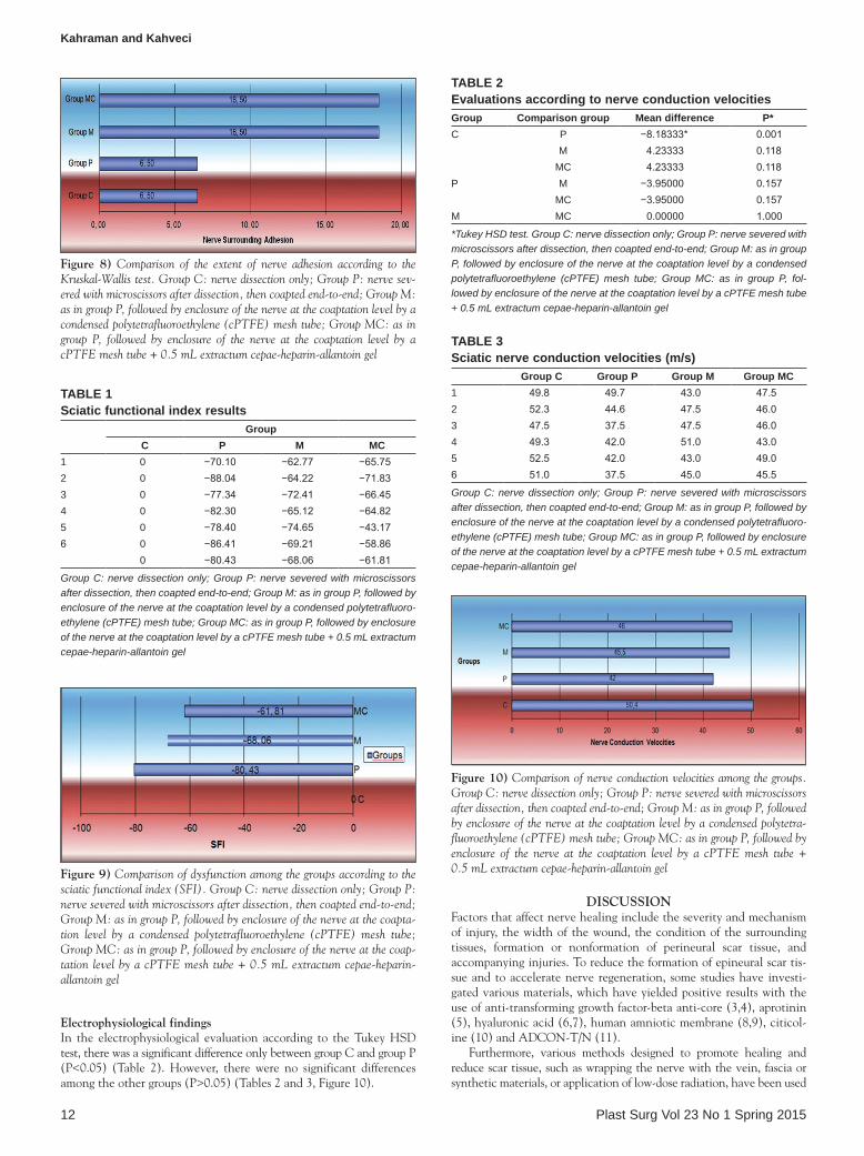
Functional analysisThe sciatic functional index (SFI) is a method used to evaluate func-tional and clinical recovery. Characteristic walking templates com-prise this reliable and repeatable method in which measures are evaluated according to nerve lesions (peroneal, tibial and sciatic) in rats. Because walking is organized cortically, it requires sensorial feed-back and complicated motor unit reinnervations; when SFI is com-pared with other tests, it is believed to be the best method for the evaluation of clinical status. In calculating the SFI footprint exten-sion, the first and the second toe space, the second and the fourth toe space values are used (19-21) (Figure 5). For the walking track analy-sis, after having dipped both the hind legs into stamping ink, all ani-mals were walked on 10 cm × 10 cm × 100 cm wooden platform covered with absorbent paper. SFI values were calculated using the obtained footprint measurements (Figure 6).
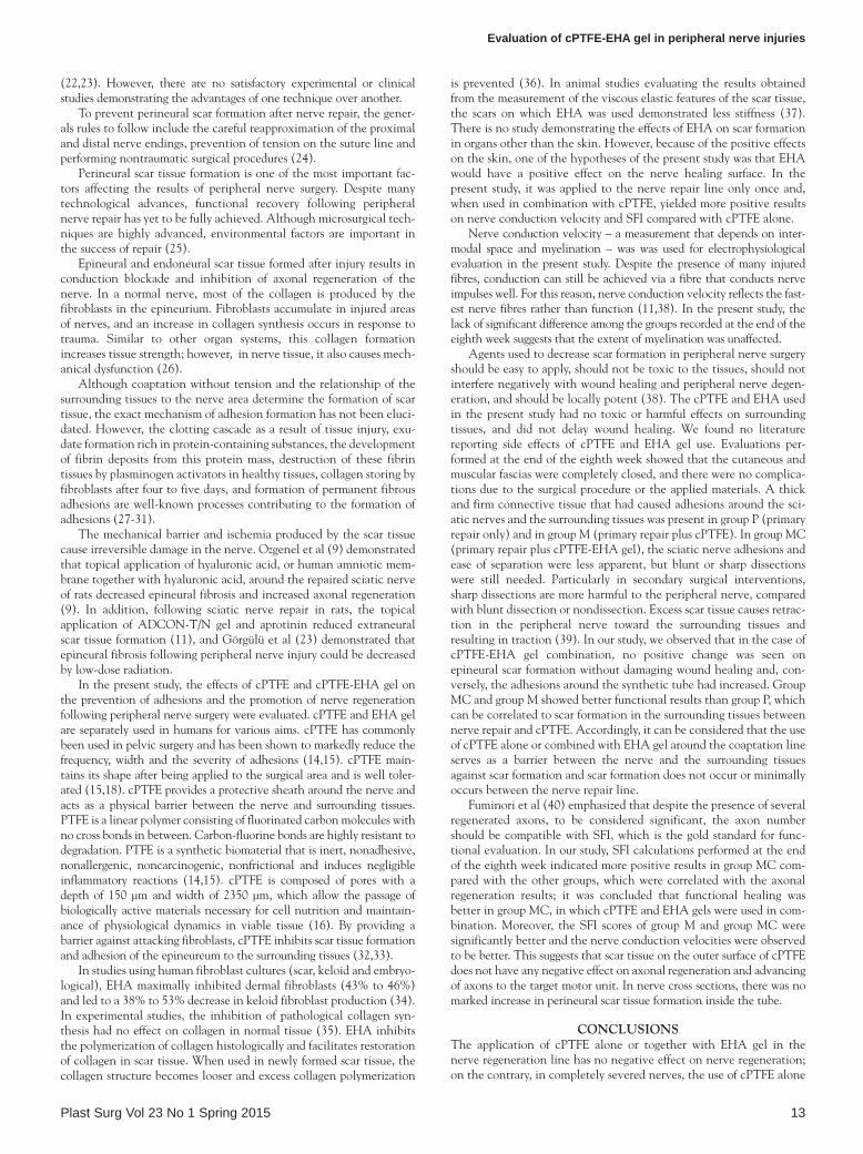
Macroscopic evaluation of perineuronal adhesionsFollowing the walking analysis, the animals were anesthetized by ether inhalation; the incision sites were reopened under anesthesia, and the adhesions of the sciatic nerve to the surrounding tissues and ease of separation from these tissues were evaluated (Figure 7). Closure of the skin and muscle fascia, adhesion of the sciatic nerve to the surrounding tissues and the ease of separation of the sciatic nerve from these tissues were evaluated according to a numerical grading scale described by Peterson et al (11): for adhesion of the sciatic
Figure 1) Anatomical appearance of the rat sciatic nerve after dissection. The sciatic nerve of animals in group C was only dissected
Figure 2) The sciatic nerve of animals in group P was cut after dissection and then primarily sutured
Figure 3) Following primary repair in animals in group M, the coapted nerve was enclosed in a condensed polytetrafluoroethylene mesh tube
Evaluation of cPTFE-EHA gel in peripheral nerve injuries
Plast Surg Vol 23 No 1 Spring 2015 11
nerve to the surrounding tissues and its separation ability: grade 1 – the nerve was free or a minimally blunt dissection was required for separation; grade 2 – moderate or strong blunt dissection was required for separation; and grade 3 – strong sharp dissection was required for separation.
Electrophysiological evaluationFor each animal, The MP100 Data Acquisition and Analysis (BIOPAC Systems Inc, USA) system was used to measure the conduction vel-ocity of the sciatic nerve once it was exposed. One of the electrodes was placed under the sciatic nerve proximal to the injury (0.7 mm of the remaining intact part), and the other electrode was placed just above the bifurcation area of the tibial and the peroneal nerves (0.7 mm of part left intact).
A supramaximal stimulus (7 V, 0.5 ms) produced by the MP100 stimulator was used to stimulate the nerves, and the distance between the electrodes was measured. The BSL Pro v.3.6.6 program (BIOPAC Systems Inc, USA) was used to evaluate the records in the computer medium. The nerve conduction rates were determined by dividing the potential values by the latency between the two electrodes (m/s).
Statistical analysisThe statistical significance of macroscopic evaluation, the functional evaluation (SFI) and the electrophysiological evaluation results were assessed using the Mann-Whitney U, the Kruskal Wallis and the Tukey HSD tests using SPSS verson 13.0 (IBM Corporation, USA) for Windows (Microsoft Coporation, USA). All of the quantitative results were denoted as an arithmetical mean (± SD); P<0.05 was considered to be statistically significant.
RESULTSMacroscopic evaluationAccording to the scale described by Peterson et al (11), closure of the cutaneus and muscular fascia, the adhesion of the nerve tissue to the surrounding tissues and the ease of separation were evaluated. On comparison of the cutaneous and muscular fascia closure, no statistically significant difference was noted among the groups (P>0.05). On assessment of the wound healing according to cutaneus closure and muscular fascia closure, the cutaneous and muscular fascia of all the animals were observed to be completely closed. When adhesion of the nerve was assessed by dissection, macroscopic-ally, groups P and C were determined to have a Peterson et al (11) scale mean of grade 1, and groups M and MC a mean of grade 3. When adhesions were evaluated according to the Kruskal-Wallis
test, group C and group P were significantly different from group M and group MC (P=0.002) (Figure 8).
Walking track analysisThere were significant differences among the groups (P<0.05) according to SFI results eight weeks after repair. However, there were no significant differences between group M and group MC (P>0.05) (Table 1, Figure 9).
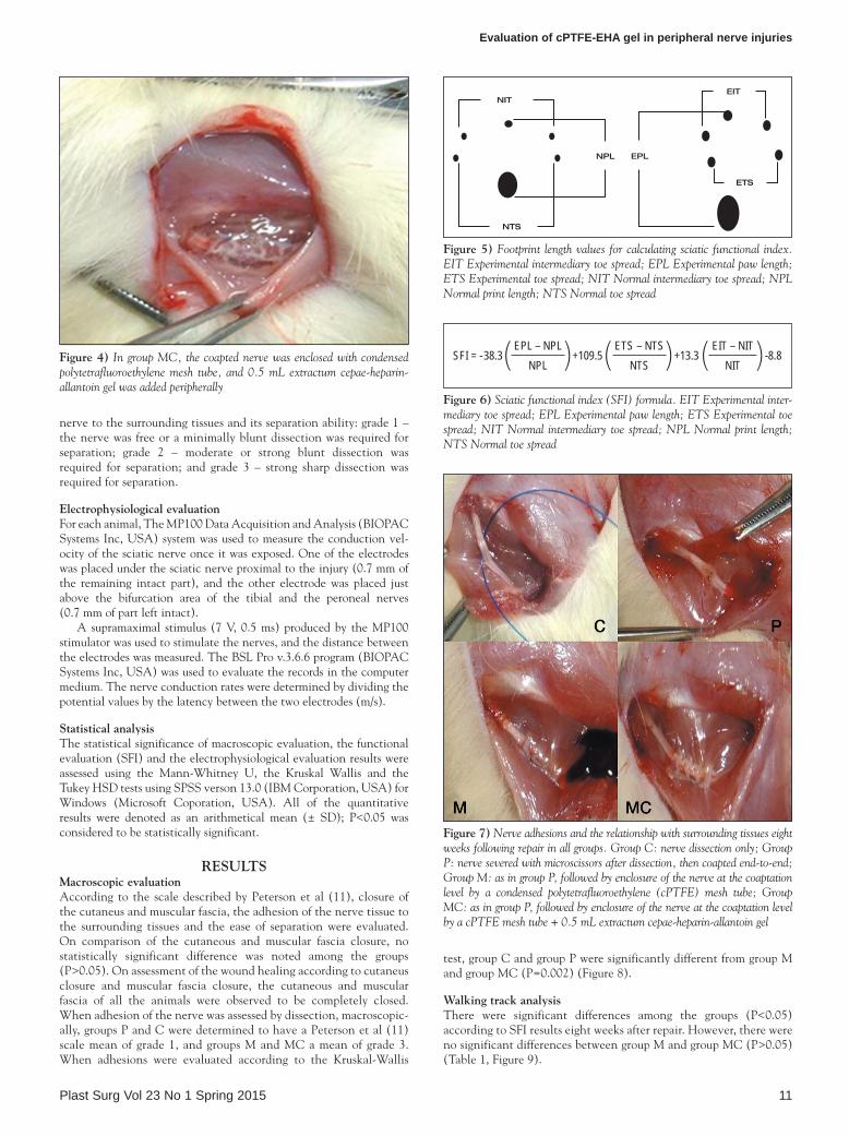
Figure 4) In group MC, the coapted nerve was enclosed with condensed polytetrafluoroethylene mesh tube, and 0.5 mL extractum cepae-heparin-allantoin gel was added peripherally
Figure 7) Nerve adhesions and the relationship with surrounding tissues eight weeks following repair in all groups. Group C: nerve dissection only; Group P: nerve severed with microscissors after dissection, then coapted end-to-end; Group M: as in group P, followed by enclosure of the nerve at the coaptation level by a condensed polytetrafluoroethylene (cPTFE) mesh tube; Group MC: as in group P, followed by enclosure of the nerve at the coaptation level by a cPTFE mesh tube + 0.5 mL extractum cepae-heparin-allantoin gel
Figure 5) Footprint length values for calculating sciatic functional index. EIT Experimental intermediary toe spread; EPL Experimental paw length; ETS Experimental toe spread; NIT Normal intermediary toe spread; NPL Normal print length; NTS Normal toe spread
Figure 6) Sciatic functional index (SFI) formula. EIT Experimental inter-mediary toe spread; EPL Experimental paw length; ETS Experimental toe spread; NIT Normal intermediary toe spread; NPL Normal print length; NTS Normal toe spread
SFI = -38.3( EPL – NPLNPL )+109.5( ETS – NTS
NTS )+13.3( EIT – NITNIT )-8.8
NIT
NPL
NTS
EIT
EPL
ETS
Kahraman and Kahveci
Plast Surg Vol 23 No 1 Spring 201512
Electrophysiological findingsIn the electrophysiological evaluation according to the Tukey HSD test, there was a significant difference only between group C and group P (P<0.05) (Table 2). However, there were no significant differences among the other groups (P>0.05) (Tables 2 and 3, Figure 10).
DISCUSSIONFactors that affect nerve healing include the severity and mechanism of injury, the width of the wound, the condition of the surrounding tissues, formation or nonformation of perineural scar tissue, and accompanying injuries. To reduce the formation of epineural scar tis-sue and to accelerate nerve regeneration, some studies have investi-gated various materials, which have yielded positive results with the use of anti-transforming growth factor-beta anti-core (3,4), aprotinin (5), hyaluronic acid (6,7), human amniotic membrane (8,9), citicol-ine (10) and ADCON-T/N (11).
Furthermore, various methods designed to promote healing and reduce scar tissue, such as wrapping the nerve with the vein, fascia or synthetic materials, or application of low-dose radiation, have been used
TAblE 2Evaluations according to nerve conduction velocitiesGroup Comparison group Mean difference P*C P −8.18333* 0.001
M 4.23333 0.118MC 4.23333 0.118
P M −3.95000 0.157MC −3.95000 0.157
M MC 0.00000 1.000
*Tukey HSD test. Group C: nerve dissection only; Group P: nerve severed with microscissors after dissection, then coapted end-to-end; Group M: as in group P, followed by enclosure of the nerve at the coaptation level by a condensed polytetrafluoroethylene (cPTFE) mesh tube; Group MC: as in group P, fol-lowed by enclosure of the nerve at the coaptation level by a cPTFE mesh tube + 0.5 mL extractum cepae-heparin-allantoin gel
Figure 8) Comparison of the extent of nerve adhesion according to the Kruskal-Wallis test. Group C: nerve dissection only; Group P: nerve sev-ered with microscissors after dissection, then coapted end-to-end; Group M: as in group P, followed by enclosure of the nerve at the coaptation level by a condensed polytetrafluoroethylene (cPTFE) mesh tube; Group MC: as in group P, followed by enclosure of the nerve at the coaptation level by a cPTFE mesh tube + 0.5 mL extractum cepae-heparin-allantoin gel
TAblE 1Sciatic functional index results
GroupC P M MC
1 0 −70.10 −62.77 −65.752 0 −88.04 −64.22 −71.833 0 −77.34 −72.41 −66.454 0 −82.30 −65.12 −64.825 0 −78.40 −74.65 −43.176 0 −86.41 −69.21 −58.86
0 −80.43 −68.06 −61.81
Group C: nerve dissection only; Group P: nerve severed with microscissors after dissection, then coapted end-to-end; Group M: as in group P, followed by enclosure of the nerve at the coaptation level by a condensed polytetrafluoro-ethylene (cPTFE) mesh tube; Group MC: as in group P, followed by enclosure of the nerve at the coaptation level by a cPTFE mesh tube + 0.5 mL extractum cepae-heparin-allantoin gel
Figure 9) Comparison of dysfunction among the groups according to the sciatic functional index (SFI). Group C: nerve dissection only; Group P: nerve severed with microscissors after dissection, then coapted end-to-end; Group M: as in group P, followed by enclosure of the nerve at the coapta-tion level by a condensed polytetrafluoroethylene (cPTFE) mesh tube; Group MC: as in group P, followed by enclosure of the nerve at the coap-tation level by a cPTFE mesh tube + 0.5 mL extractum cepae-heparin-allantoin gel
TAblE 3Sciatic nerve conduction velocities (m/s)
Group C Group P Group M Group MC1 49.8 49.7 43.0 47.52 52.3 44.6 47.5 46.03 47.5 37.5 47.5 46.04 49.3 42.0 51.0 43.05 52.5 42.0 43.0 49.06 51.0 37.5 45.0 45.5
Group C: nerve dissection only; Group P: nerve severed with microscissors after dissection, then coapted end-to-end; Group M: as in group P, followed by enclosure of the nerve at the coaptation level by a condensed polytetrafluoro-ethylene (cPTFE) mesh tube; Group MC: as in group P, followed by enclosure of the nerve at the coaptation level by a cPTFE mesh tube + 0.5 mL extractum cepae-heparin-allantoin gel
Figure 10) Comparison of nerve conduction velocities among the groups. Group C: nerve dissection only; Group P: nerve severed with microscissors after dissection, then coapted end-to-end; Group M: as in group P, followed by enclosure of the nerve at the coaptation level by a condensed polytetra-fluoroethylene (cPTFE) mesh tube; Group MC: as in group P, followed by enclosure of the nerve at the coaptation level by a cPTFE mesh tube + 0.5 mL extractum cepae-heparin-allantoin gel
Evaluation of cPTFE-EHA gel in peripheral nerve injuries
Plast Surg Vol 23 No 1 Spring 2015 13
(22,23). However, there are no satisfactory experimental or clinical studies demonstrating the advantages of one technique over another.
To prevent perineural scar formation after nerve repair, the gener-als rules to follow include the careful reapproximation of the proximal and distal nerve endings, prevention of tension on the suture line and performing nontraumatic surgical procedures (24).
Perineural scar tissue formation is one of the most important fac-tors affecting the results of peripheral nerve surgery. Despite many technological advances, functional recovery following peripheral nerve repair has yet to be fully achieved. Although microsurgical tech-niques are highly advanced, environmental factors are important in the success of repair (25).
Epineural and endoneural scar tissue formed after injury results in conduction blockade and inhibition of axonal regeneration of the nerve. In a normal nerve, most of the collagen is produced by the fibroblasts in the epineurium. Fibroblasts accumulate in injured areas of nerves, and an increase in collagen synthesis occurs in response to trauma. Similar to other organ systems, this collagen formation increases tissue strength; however, in nerve tissue, it also causes mech-anical dysfunction (26).
Although coaptation without tension and the relationship of the surrounding tissues to the nerve area determine the formation of scar tissue, the exact mechanism of adhesion formation has not been eluci-dated. However, the clotting cascade as a result of tissue injury, exu-date formation rich in protein-containing substances, the development of fibrin deposits from this protein mass, destruction of these fibrin tissues by plasminogen activators in healthy tissues, collagen storing by fibroblasts after four to five days, and formation of permanent fibrous adhesions are well-known processes contributing to the formation of adhesions (27-31).
The mechanical barrier and ischemia produced by the scar tissue cause irreversible damage in the nerve. Ozgenel et al (9) demonstrated that topical application of hyaluronic acid, or human amniotic mem-brane together with hyaluronic acid, around the repaired sciatic nerve of rats decreased epineural fibrosis and increased axonal regeneration (9). In addition, following sciatic nerve repair in rats, the topical application of ADCON-T/N gel and aprotinin reduced extraneural scar tissue formation (11), and Görgülü et al (23) demonstrated that epineural fibrosis following peripheral nerve injury could be decreased by low-dose radiation.
In the present study, the effects of cPTFE and cPTFE-EHA gel on the prevention of adhesions and the promotion of nerve regeneration following peripheral nerve surgery were evaluated. cPTFE and EHA gel are separately used in humans for various aims. cPTFE has commonly been used in pelvic surgery and has been shown to markedly reduce the frequency, width and the severity of adhesions (14,15). cPTFE main-tains its shape after being applied to the surgical area and is well toler-ated (15,18). cPTFE provides a protective sheath around the nerve and acts as a physical barrier between the nerve and surrounding tissues. PTFE is a linear polymer consisting of fluorinated carbon molecules with no cross bonds in between. Carbon-fluorine bonds are highly resistant to degradation. PTFE is a synthetic biomaterial that is inert, nonadhesive, nonallergenic, noncarcinogenic, nonfrictional and induces negligible inflammatory reactions (14,15). cPTFE is composed of pores with a depth of 150 μm and width of 2350 μm, which allow the passage of biologically active materials necessary for cell nutrition and maintain-ance of physiological dynamics in viable tissue (16). By providing a barrier against attacking fibroblasts, cPTFE inhibits scar tissue formation and adhesion of the epineureum to the surrounding tissues (32,33).
In studies using human fibroblast cultures (scar, keloid and embryo-logical), EHA maximally inhibited dermal fibroblasts (43% to 46%) and led to a 38% to 53% decrease in keloid fibroblast production (34). In experimental studies, the inhibition of pathological collagen syn-thesis had no effect on collagen in normal tissue (35). EHA inhibits the polymerization of collagen histologically and facilitates restoration of collagen in scar tissue. When used in newly formed scar tissue, the collagen structure becomes looser and excess collagen polymerization
is prevented (36). In animal studies evaluating the results obtained from the measurement of the viscous elastic features of the scar tissue, the scars on which EHA was used demonstrated less stiffness (37). There is no study demonstrating the effects of EHA on scar formation in organs other than the skin. However, because of the positive effects on the skin, one of the hypotheses of the present study was that EHA would have a positive effect on the nerve healing surface. In the present study, it was applied to the nerve repair line only once and, when used in combination with cPTFE, yielded more positive results on nerve conduction velocity and SFI compared with cPTFE alone.
Nerve conduction velocity – a measurement that depends on inter-modal space and myelination – was was used for electrophysiological evaluation in the present study. Despite the presence of many injured fibres, conduction can still be achieved via a fibre that conducts nerve impulses well. For this reason, nerve conduction velocity reflects the fast-est nerve fibres rather than function (11,38). In the present study, the lack of significant difference among the groups recorded at the end of the eighth week suggests that the extent of myelination was unaffected.
Agents used to decrease scar formation in peripheral nerve surgery should be easy to apply, should not be toxic to the tissues, should not interfere negatively with wound healing and peripheral nerve degen-eration, and should be locally potent (38). The cPTFE and EHA used in the present study had no toxic or harmful effects on surrounding tissues, and did not delay wound healing. We found no literature reporting side effects of cPTFE and EHA gel use. Evaluations per-formed at the end of the eighth week showed that the cutaneous and muscular fascias were completely closed, and there were no complica-tions due to the surgical procedure or the applied materials. A thick and firm connective tissue that had caused adhesions around the sci-atic nerves and the surrounding tissues was present in group P (primary repair only) and in group M (primary repair plus cPTFE). In group MC (primary repair plus cPTFE-EHA gel), the sciatic nerve adhesions and ease of separation were less apparent, but blunt or sharp dissections were still needed. Particularly in secondary surgical interventions, sharp dissections are more harmful to the peripheral nerve, compared with blunt dissection or nondissection. Excess scar tissue causes retrac-tion in the peripheral nerve toward the surrounding tissues and resulting in traction (39). In our study, we observed that in the case of cPTFE-EHA gel combination, no positive change was seen on epineural scar formation without damaging wound healing and, con-versely, the adhesions around the synthetic tube had increased. Group MC and group M showed better functional results than group P, which can be correlated to scar formation in the surrounding tissues between nerve repair and cPTFE. Accordingly, it can be considered that the use of cPTFE alone or combined with EHA gel around the coaptation line serves as a barrier between the nerve and the surrounding tissues against scar formation and scar formation does not occur or minimally occurs between the nerve repair line.
Fuminori et al (40) emphasized that despite the presence of several regenerated axons, to be considered significant, the axon number should be compatible with SFI, which is the gold standard for func-tional evaluation. In our study, SFI calculations performed at the end of the eighth week indicated more positive results in group MC com-pared with the other groups, which were correlated with the axonal regeneration results; it was concluded that functional healing was better in group MC, in which cPTFE and EHA gels were used in com-bination. Moreover, the SFI scores of group M and group MC were significantly better and the nerve conduction velocities were observed to be better. This suggests that scar tissue on the outer surface of cPTFE does not have any negative effect on axonal regeneration and advancing of axons to the target motor unit. In nerve cross sections, there was no marked increase in perineural scar tissue formation inside the tube.
CONCLUSIONSThe application of cPTFE alone or together with EHA gel in the nerve regeneration line has no negative effect on nerve regeneration; on the contrary, in completely severed nerves, the use of cPTFE alone
Kahraman and Kahveci
Plast Surg Vol 23 No 1 Spring 201514
or cPTFE-EHA gel combination was found to improve functional recovery and nerve regeneration, compared with primary repair only. In the macroscopic evaluation, the cPTFE-EHA gel combination was found to have no negative effect on wound healing and, despite increasing the extraneural scar tissue and adhesions, it had no nega-tive effect on nerve function; on the contrary, it provided functional recovery. Compared with the cPTFE application alone, the applica-tion of perineural cPTFE-EHA gel combination during peripheral
nerve surgery seems to provide better functional recovery without causing any significant changes on the epineural and extraneural scar tissue formation.
DISCLOSURES: The authors have no financial disclosures or con-flicts of interest to declare.
REFERENCES 1. Lundborg G. A 25-year perspective of peripheral nerve surgery
evolving neuroscientific concepts and clinical cignificance. J Hand Surg 2000;25:391-414.
2. Lin YK, Posnick JC, Vasjar J. Fetal nerve healing: An experimental study. Plast Reconstr Surg 1994;93:1323-32.
3. Santos X, Rodrigo J, Hontanilla B. Evaluation of peripheral nerve regeneration by nerve growth factor locally administered with a novel system. J Neurosci Methods 1998;85:119-27.
4. Farhadieh R, Nicklin S, Yu Y. The role of nerve growth factor and brain derived neurotrophic factor in inferior alveolar nerve regeneration in distraction osteogenesis. J Craniofac Surg 2003;14:859-65.
5. Görgülü A, İmer M, Şimşek O. The effect of aprotinin on extraneural scarring in peripheral nerve surgery. Acta Neurochir 1998;140:1303-07.
6. Ikeda K, Yamauchi D, Osamura N. Hyaluronic acid prevents peripheral nerve adhesion. Br J Plast Surg 2003;56:342-7.
7. Ozgenel GY. Effects of hyaluronic acid an peripheral nerve scarring and regeneration in rats. Microsurg 2003;23:575-81.
8. Ozgenel GY, Filiz G. Combined application of human amniotic membrane wrapping and hyaluronic acid injection in epineurectomized rat sciatic nerve. J Reconstr Microsurg 2004;20:153-7.
9. Ozgenel GY. The effects of a combination of hyaluronic and amniotic membrane on the formation of peritendinous adhesions after flexor tendon surgery in chickens. J Bone Joint Surg Br 2004;86:301-7.
10. Özay R, Bekar A, Kocaeli H. Citicoline improves functional recovery, promotes nerve regeneration, and reduces postoperative scarring after peripheral nerve surgery in rats. Surg Neurol 2007;68:615-22.
11. Peterson J, Russel L, Andrus K. Reduction of extraneural scarring by ADCON-T/N after surgical intervention. Neurosurg 1996;38:976-84.
12. Hatano I, Suga T, Diao E. Adhesions from flexor tendon surgery: An animal study comparing surgical techniques. J Hand Surg 2000;25:252-9.
13. Loescher AR, Robinson PP. The effect of surgical medicaments on peripheral nerve function. Br J Oral Maxillofac Surg 1998;36:327-32.
14. Gabriela V, Analiz R. Macroporous condensed poly(tetra fluoro-ethylene). In vivo effect on adhesion formation and tissue integration. J Biomed Mater Res A 2007;82:426-35.
15. Gabriela V, Peter H.,Macroporous condensed poly(tetrafluoroethylene). In vivo inflammatory response and healing characteristics. J Biomed Mater Res A 2006;76:234-42.
16. Yang S, Leong KF, Du Z, et al. The design of scaffolds for use in tissue engineering. Part I. Traditional factors. Tissue Eng 2001;7:679-89.
17. Koliadenko VH, Stepanenko VI, Bardov PV. New approaches to the treatment and prophylaxis of the cicatrix formation. Klin Khir 2001;2:24-7.
18. Willital GH, Heine H. Efficacy of Contractubex gel in the treatment of fresh scars after thoracic surgery in children and adolescents.Int J Clin Pharmacol Res 1994;14:193-202.
19. Hare GM, Evans PJ, Mackinnon SE. Walking track analysis: A long term assesment of peripheral nerve recovery. Plast Reconstr Surg 1992;89:251-8.
20. Varejao A, Meek M, Ferreira A. Functional evaluation of peripheral nerve regeneration in the rat: Walking track analysis. J Neurosci Methods 2001;108:1-9.
21. Brown C, Mackinnon S, Evans P. Self evaluation of walking track measurement using a sciatic function index. Microsurg 1989;10:226-8.
22. Yavuzer R, Ayhan S, Latifoğlu O. Turnover epineural sheath tube in primary repair of peripheral nerves. Ann Plast Surg 2002;48:392-400.
23. Görgülü A, Uzal C, Doganay L. The effect of low dose external beam radiation on extraneural scarring after peripheral nerve surgery in rats. Neurosurg 2003;53:1389-96.
24. Lee SK, Wolfe SW. Peripheral nerve injury and repair. J Am Acad Orthop Surg 2000;8:243-52.
25. Payne SH. Nerve repair and grafting in the upper extremity. J South Orthop Assoc 2001;10:173-89.
26. Abe Y, Doi K, Kawai S. An experimental model of peripheral nerve adhesions in rabbits. Br J Plast Surg 2005;58:533-40.
27. Smit X, Neck J, Hovius S. Reduction of neural adhesions by biodegradable autocrosslinked hyaluronic acid gel after injury of peripheral nerves: An experimental study. J Neurosurg 2004;101:648-52.
28. Himeda Y, Yanagi S, Kakema T. Adhesion prevention effect of a novel hyaluronic acid gel film in rats. J Int Med Res 2003;31:509-16.
29. Isla A, Martinez J, Perez L. A reservable antiadhesion barrier gel reduces the perineural adhesions in rats after anastomosis. J Neurosurg Sci 2003;47:195-9.
30. Meislin R, Wiseman D, Alexander H. A biomechanical study of tendon adhesion reduction using a bidegradable barrier in a rabbit model. J Appl Biomater 1990;1:13-9.
31. Al-Jaroudi D, Tulandi T. Adhesion prevention in gynecologic surgery. Obstet Gynecol 2004;59:360-7.
32. Miloro M, Macy JM. Expanded polytetrafluoroethylene entubulation of the rabbit inferior alveolar nerve. Oral Surg Oral Med Oral Pathol Oral Radiol Endod 2000;89:292-8.
33. Vasconcelos BC, Gay Escoda C, Vasconcellos RJ, et al. Conduction velocity of the rabbit facial nerve: A noninvasive functional evaluation. Pesqui Odontol Bras 2003;17:126-31.
34. Majewski S, Chadzynska M. Effect of heparin, allantoin and cepae extract on the proliferation of keloid fibroblasts and other cell in vitro. Posterprasantation MS–No 330/P16. Weltkongress für Dermatologie 1987;17:24-9.
35. Janick S, Sznitowska M. Effect of ointments for treating scars and keloids on metabolism of collagen in scars and healthy skin. Eur J Pharm Biopharm 1991;37:188-91.
36. Beuth J, Hunzelmann N, Van Leendert R, et al. Safety and efficacy of local administration of contractubex to hypertrophic scars in comparison to corticosteroid treatment. Results of a multicenter, comparative epidemiological cohort study in Germany. In Vivo 2006;20:277-83
37. Janicki S, Sznitowska M, at all.Studies on the viscous-elastic properties of the skin in evaluating ointments to improve the elasticity of scars. Przegl Dermatol 1988;75:352-7.
38. Dam-Hieu P, Lacroix C, Said G. Reduction of postoperative perineural adhesions by hyaloglide gel: An experimental study in the rat sciatic nerve. Neurosurg 2005;56:425-33.
39. Diamond M, Linsky C, Decherney A. Synergistic effects of Interceed and heparin in reducing adhesion formation in the rabbit uterine horn model. Fertil Steril 1991;55:389-94.
40. Fuminori K, John C, Warren C. Nerve conduction tests, muscle contraction and axon morphometry as indicators of regeneration. Plast Reconstr Surg 1996;98:1264-71.

![Solubility and solubilizing capabilities ... - Herba Polonica et al [1].pdf · Vol. 62 No. 4 2016 51 Solubility and solubilizing capabilities of aqueous solutions of Extractum Taraxaci](https://static.fdocuments.in/doc/165x107/5c9398cb09d3f206488cd9a1/solubility-and-solubilizing-capabilities-herba-et-al-1pdf-vol-62-no.jpg)