ESTIMATION OF VERTICAL DIMENSION OF … · Facial and intra-oral measurements ... non-invasive and...
9
individuals and in a single individual at different times, because the individual's capacity is unknown (Rivera-Morales 4 and Mohl, 1991). Many methods have been proposed to determine the correct Vertical Dimension of Occlusion that include- i. Vertical dimension of rest (VDR) (Thompson & Brodie, 1942) ii. Speaking method (Silverman, 1953) iii. Cephalometric radiographs (Pyott & Schaeffer, 1954) iv. Pre-extraction records (Turner, 1969; Smith, 1971) v. Maximal bite force (Boos, 1940) vi. Facial and intra-oral measurements (Willis, 1935; McGee, 1947). ESTIMATION OF VERTICAL DIMENSION OF OCCLUSION IN EDENTULEOUS PATIENTS USING CEPHALOMETRIC ANALYSIS 1 2 3 4 5 Ritu Batra , Sanjay Kalra , Ajay Bansal , Siddharth Nerula , Rajat Dang 1 Reader, Bhojia Dental College and Hospital, Haryana, India 2 Senior Prosthodontist, Haryana, India 3 Reader, Bhojia Dental College and Hospital, Haryana, India 4 Professor & HOD, Rajisthan Dental College, Rajasthan, India 5 Professor, Mullana Dental College, Haryana, India Corresponding Author: Ritu Batra E-mail: [email protected] th Received: 14 January 2017 th Accepted: 20 April 2017 th Online: 20 May 2017 ORIGINAL ARTICLE www.djas.co.in ISSN No-2321-1482 DJAS 5(I), 30-38, 2017 All rights are reserved Dental JOURNAL of A d v a n c e S t u d i e s ABSTRACT “The best of friends fall out with time and so do teeth.” Thus, there is a need to replace the lost teeth and the supporting structures for the patient's social demands and functional rehabilitation. Prosthetic treatment with complete dentures is a very common treatment modality; the biggest challenge in its fabrication is to duplicate the normal vertical dimension. Failure can be avoided by completing the treatment without changing the vertical dimension and obtaining an optimal facial proportion. There are many methods to record VD. Radiographic cephalometry has been used as a diagnostic tool in Prosthodontics for over five decades and numerous authors, like Ricketts (1981), McNamara (1984) and Slavicek (1984) developed and computerized these techniques to co-relate and record VD in patients. However cephalometric analysis can help to visualize skeletal and facial proportion relation. The present study was done to use the lateral radiographs with cephalometric analysis, as it could be a simple, non- technique sensitive, non-invasive and atraumatic way to determine VD for complete denture patients and also to compare physiologic methods (swallowing / phonetics) with cephalometric method to record lower facial height. Key words : Cephalometrics, Legan-Burstone analysis, Mc Namara analysis, Vertical dimension of rest, Vertical dimension of occlusion, Postural Rest Position. INTRODUCTION 1 Boucher et al stated that “An adequate interocclusal distance is absolutely essential for complete denture patients”. The constancy of rest vertical dimension is important because the vertical dimension of occlusion is 2 dependent on it. Smith claims that eventually “the lips fold inward, furrows and wrinkles are formed and the face becomes prematurely old in 3 appearance”. The value of the lower facial height is routinely estimated by aesthetic analysis and clinical determination of the rest position. Atwood, 1956; Tallgren, 1972; Rugh & Drago 1981 stated that the rest position is an area rather than a point. The width of comfort zone may vary among 30
Transcript of ESTIMATION OF VERTICAL DIMENSION OF … · Facial and intra-oral measurements ... non-invasive and...

individuals and in a single individual at different times, because the individual's capacity is unknown (Rivera-Morales
4and Mohl, 1991).
Many methods have been proposed to determine the correct Vertical Dimension of Occlusion that include-
i. Vertical dimension of rest (VDR)
(Thompson & Brodie, 1942)
ii. Speaking method (Silverman, 1953)
iii. Cephalometric radiographs (Pyott &
Schaeffer, 1954)
iv. Pre-extraction records (Turner, 1969; Smith, 1971)
v. Maximal bite force (Boos, 1940)
vi. Facial and intra-oral measurements (Willis, 1935; McGee, 1947).
ESTIMATION OF VERTICAL DIMENSION OF OCCLUSION IN EDENTULEOUS PATIENTS USING CEPHALOMETRIC ANALYSIS
1 2 3 4 5Ritu Batra , Sanjay Kalra , Ajay Bansal , Siddharth Nerula , Rajat Dang1Reader, Bhojia Dental College and Hospital, Haryana, India2Senior Prosthodontist, Haryana, India 3Reader, Bhojia Dental College and Hospital, Haryana, India4Professor & HOD, Rajisthan Dental College, Rajasthan, India5Professor, Mullana Dental College, Haryana, India
Corresponding Author: Ritu Batra
E-mail: [email protected]
thReceived: 14 January 2017thAccepted: 20 April 2017th Online: 20 May 2017
ORIGINAL ARTICLEwww.djas.co.in ISSN No-2321-1482
DJAS 5(I), 30-38, 2017All rights are reserved
Dental JOURNAL of A d v a n c e S t u d i e s
ABSTRACT
“The best of friends fall out with time and so do teeth.”
Thus, there is a need to replace the lost teeth and the supporting structures for the patient's social demands and
functional rehabilitation. Prosthetic treatment with complete dentures is a very common treatment modality;
the biggest challenge in its fabrication is to duplicate the normal vertical dimension. Failure can be avoided by
completing the treatment without changing the vertical dimension and obtaining an optimal facial proportion.
There are many methods to record VD. Radiographic cephalometry has been used as a diagnostic tool in
Prosthodontics for over five decades and numerous authors, like Ricketts (1981), McNamara (1984) and
Slavicek (1984) developed and computerized these techniques to co-relate and record VD in patients.
However cephalometric analysis can help to visualize skeletal and facial proportion relation. The present
study was done to use the lateral radiographs with cephalometric analysis, as it could be a simple, non-
technique sensitive, non-invasive and atraumatic way to determine VD for complete denture patients and also
to compare physiologic methods (swallowing / phonetics) with cephalometric method to record lower facial
height.
Key words : Cephalometrics, Legan-Burstone analysis, Mc Namara analysis, Vertical dimension of rest,
Vertical dimension of occlusion, Postural Rest Position.
INTRODUCTION 1
Boucher et al stated that “An adequate interocclusal distance is absolutely essential for complete denture patients”. The constancy of rest vertical dimension is important because the vertical dimension of occlusion is
2 dependent on it. Smith claims that eventually “the lips fold inward, furrows and wrinkles are formed and the face b e c o m e s p r e m a t u r e l y o l d i n
3appearance”.
The value of the lower facial height is routinely estimated by aesthetic analysis and clinical determination of the rest position. Atwood, 1956; Tallgren, 1972; Rugh & Drago 1981 stated that the rest position is an area rather than a point. The width of comfort zone may vary among
30
However, none has been shown to be scientifically more valid than any other (Rivera- Morales & Moh, 1991) and a lot of them are inexact because they do not consider physiological, age-related
5facial changes (Koller et al., 1992). They found that none of them have a sufficient reliability and reproducibility to ensure that the practitioner has recorded the right Vertical Dimension of Occlusion. The soft tissue reference points are not stable and definite; therefore, the use of bony reference points increases the accuracy of the measurements.
The advantages to use cephalometric method for 6recording VD:
1. Measurement are made on bony points
2. No manipulation of the patient's face is required,
once the patient is seated comfortably
2. Concentration is made on the patient and his state
of rest, rather than on the measuring device.
3. Permanent record and permanent reference points
are available for months or years later.
4. Cephalometric analysis can provide not only the
Vertical Dimension of Occlusion but also the
orientation of the occlusal plane, the curve of Spee,
the anterior teeth position and the anterior
guidance (Ismail and Bowman, 1968; L' Estrange
and Vig. 1975; Monteith, 1985). 7, 8,9
MATERIAL & METHOD
In order to conduct the study twenty two patients between the age group of 50-80 years were randomly selected, from those attending the O.P.D. of the Department of Prosthodontics at B.R.S. Dental College. Thorough case history was recorded and clinical examination was done.
The following criteria were used to include the patients for this study:
1. Edentulous patients were selected at random.
2. Patient's consent was taken before making him a
part of the study.
3. Patient who was not an old denture wearer was
selected, so that he is not adapted to the old vertical
dimensions.
4. Patients with TMJ pain and any mandibular
deviation from normal opening pattern were 10
eliminated from the study.
11,125. Patients only with class I relation were selected.
For making radiographic assessment and measuring recordings
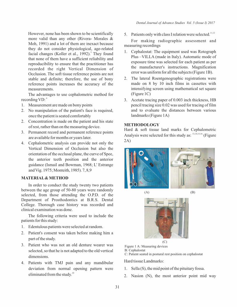
1. Cephalostat: The equipment used was Rotograph Plus –VILLA (made in Italy). Automatic mode of exposure time was selected for each patient as per the manufacturer's instructions. Magnification error was uniform for all the subjects (Figure 1B).
2. The lateral Roentgenographic registrations were made on 8 by 10 inch films in cassettes with intensifying screen using mathematical set square (Figure 1C)
3. Acetate tracing paper of 0.003 inch thickness, HB pencil tracing size 0.02 was used for tracing of film and to evaluate the distances between various landmarks (Figure 1A)
METHODOLOGYHard & soft tissue land marks for Cephalometric
13,14,15,16. Analysis were selected for this study as: (Figure
2A)
Figure 1 A: Measuring devicesB: CephalostatC: Patient seated in postural rest position on cephalostat
(A) (B)
(C)
Hard tissue Landmarks:
1. Sella (S), the mid point of the pituitary fossa.
2. Nasion (N), the most anterior point mid way
Dental Journal of Advance Studies Vol. 5 (Issue I) 2017
31
between the frontal and nasal bones on the
nasofrontal suture in the midsagittal plane.
3. Facial centre (FC), the intersection of the Frankfurt
plane and the perpendicular through the posterior
wall of the pterygomaxillary fissure.
4. Subspinale (A), the deepest point in the mid-
sagittal plane between the anterior nasal spine and
alveolar crest, usually around the level of and
anterior to the apex of the maxillary central
incisors.
5. Pogonion (Pg), the most anterior point of the
contour of the bony chin in the mid-sagittal plane.
6. Supramentale (B), the deepest point in the
midsagittal plane between infradentale and Pg,
usually anterior to and slightly below the apices of
the mandibular incisors.
7. Anterior nasal spine (ANS), the most anterior point
of the nasal floor, the tip of the premaxilla in the
midsagittal plane.
8. Menton (Me), the most inferior mid line point on
the contour of the mandibular symphysis.
9. Gnathion (Gn), the most anteroinferior point on
symphysis. The midpoint between Pg and Me,
located by bisecting the facial line N-Pg and the
mandibular plane (lower border).
10. Posterior nasal spine (PNS), the intersection of a
cont inua t ion of the an te r ior wal l o f
pterygopalatine fossa and the floor of nose,
marking the distal limit of the maxilla (the most
posterior point on the contour of the palate).
11. Porion (Po), the midpoint on the upper edge of the
external auditory meatus.
12. Condylon(Co), most superior posterior point on
condyle
13. Orbitale (Or), the lowest point on the margin of the
orbit(Or)
14. Suprapogonion (Pm), point where curvature of the
anterior contour of the symphysis changes from
concave to convex.
15. Inferior facial height (Xi), the point placed on the
center of the mandibular ascending ramus,
determined by the Frankfurt plane and
pterygomaxillary fissure.
Soft tissue landmarks:
1. Glabella (G), the most prominent point in the
midsagittal plane of the forehead.
2. Subnasion (Sn),the point at which the nasal septum
merges with the upper cutaneous lip in the
midsagittal plane
3. Soft tissue menton (Me'), lowest point on the
contour of the soft tissue chin; found by dropping a
perpendicular from horizontal plane through
menton.
Reference Planes:
1. Palatal plane: The line through ANS-PNS.
2. Anterior cranial base: The line through N-S.
3. Co-Gn: constructed by a line through the condylon
and gnathion obtaining the mandible's length.
Height of mandible was correlated accurately for
radiographic evaluation as Condylon in posterior 30
compartment shows less distortion.
4. ANS-Xi-Pm: between ANS, Xi, and Pm.
5. Frankfurt's plane: constructed from the lowest
point on the margin of the orbit (Or) to the midpoint
on the upper edge of the external auditory meatus
(Po).
6. Mandibular plane (MP): A plane constructed from
menton to the angle of the mandible Go (Downs).
7. Horizontal plane (HP), which is a surrogate
Frankfurt plane, constructed by drawing a line 7
degree from the line S to N. Most measurements
were made from projections either parallel to HP or
perpendicular to HP. All the measurements were 26,28made parallel to HP. (The baseline for
comparison of most of the data in this analysis is a
constructed plane called the Horizontal plane).
8. N-Fc-A: constructed between nasion, facial center
and A, used in the determination of the maxillary
height.
9. N-ANS-Me: constructed by a vertical line from
nasion perpendicular HP to menton.
Dental Journal of Advance Studies Vol. 5 (Issue I) 2017
32
10. G-Sn-Me': constructed by a vertical line from
Glabella to Menton, perpendicular to HP.
Lateral cephalometric radiographs were obtained
at two stages of the study-
1. Before beginning of the treatment
2. After making jaw relation recordings
Step I
The selected patients were examined and their
consent was taken to be a part of the study. Patients
were explained the method before beginning of
treatment. Every effort was made to prevent the
patients from becoming “jaw conscious” or 'rest
position conscious. The patients were not
“conditioned” through exercises or premedication. A
conscious effort was made for the patients, that they are
not tensed because, more tense the patient, the less the 6,17
freeway space tends to be. The first lateral
roentgenograph was taken on cephalostat without bite 18
blocks in postural rest position. The lateral
reoentgenographic registrations were made on 8 by 10
inch films in cassettes with intensifying screen.
Patients without the dentures in there mouth were made
to swallow, wet lips with the tongue and to be perfectly
relaxed (Figure 2C)
While in the Cephalostat with the ear plugs lightly
placed in the ears. Patients were positioned at postural
rest position and were oriented in such a way that FH 2,3,19plane was parallel to floor.
Long barrel version of Orbitale indicator was used
to ensure horizontal alignment of Orbitale reference 20
with earpieces of ear bow. In order to control the
cumulative tracing errors; tracings were done using
semi-transparent acetate paper 0.003 inch. The
measurements were made directly on the
reoentgenographic film. When the mandible assumed a
resting position, the distance from nasion to menton on
the rest position films determined vertical dimension at
rest measuring the distance between various
landmarks. Soft tissue landmarks included upper facial
height and lower facial heights were also measured 19
using cephalometric analysis.
Step II
After making preliminary impressions, secondary
impressions were made. Denture bases were adapted
on casts using self cure acrylic resin (sprinkle on
method) and stability of denture bases was checked 8intraorally. Patients were made to sit in upright
position with the head unsupported and the Frankfurt
horizontal plane parallel to the floor. The upper rim was
adjusted until it appeared parallel to the camper's plane
(centre of the tragus-subnasal point) and the
interpupillary axis using a Fox plane and a metal bar.
Occlusal plane was adjusted for every patient keeping 8it parallel to inter pupillary line and ala tragus line. For
registration of vertical dimensions, the phonetic and
swallowing method were used in this study because
these techniques require the patient to perform simple
physiological actions. These methods were readily 3,18understood by the patients and easily interpreted.
Vertical dimension of rest was recorded by asking
patient to keep lips in moderate contact when occlusal 21rims are out of contact (Nagle and Sears). and
accordingly vertical dimension of occlusion was
adjusted. Bite blocks and temporary denture bases
were delineated for visibility on radiographs by
attaching radio opaque (gutta percha) markers at four
places in the second radiograph (Figure 2B).
The marks were placed at
• The midline of upper rim occlusal plane.
• The midline of lower rim at occlusal plane.
• Deepest part of palate in acrylic base of upper rim.
• On the occlusal plane posteriorly.
For the measurements of the vertical dimension of
occlusion using functional method second radiograph
was taken with bite blocks intraorally and asking the
patient to close in centric relation. The vertical
dimension of occlusion was measured by measuring
the distance from nasion to menton when the maxillary 18and mandibular bite blocks were in contact. (Figure 3A).
As Pleasure (1951) stated that Vertical dimension of
occlusion is approximately 2 mm lower than vertical
dimension of rest. To make the two x-ray films
compatible for measurements of lower facial height
(vertical dimension of occlusion) 2mm was reduced
Dental Journal of Advance Studies Vol. 5 (Issue I) 2017
33
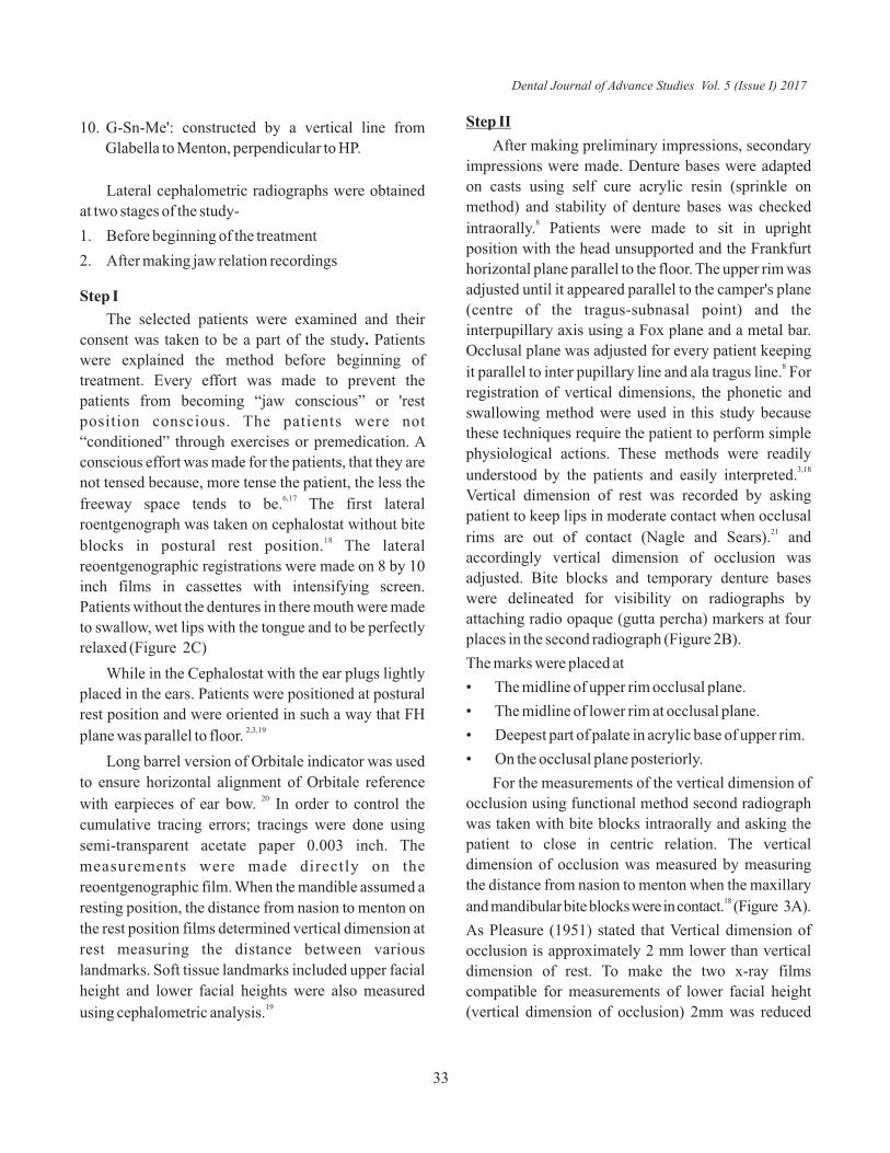
from the measurements in the first x-ray film. The soft
tissue facial profile is closely related to and dependent
on the underlying skeletal structure, so soft tissue as
well as skeletal measurements were done
simultaneously as per Rickets, McNamara (for 22
skeletal), Legan-Burstone analysis (for soft tissue).
RESULTS AND DISCUSSION
Statistical Analysis was done to evaluate the difference between the cephalometric method and functional method, and also to evaluate the reliability of cephalometric method. The observations were compared with the normal values given by McNamara analysis. On every individual two observations were recorded:
CRwor: cephalometric postural rest position without stocclusal rims (1 method).
ndFwr: functional method with occlusal rims (2 method).
Data was collected on twenty two individuals giving us twenty two paired observations. The data was statistically evaluated; box plot of the data was made in SPSS to see how the skeletal and soft tissue proportions for these two methods were distributed.
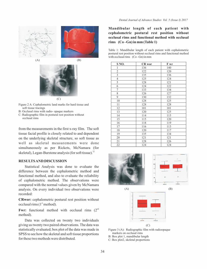
Mandibular length of each patient with cephalometric postural rest position without occlusal rims and functional method with occlusal rims (Co –Gn) in mm (Table 1)
(A) (B)
(C)
Figure 2 A: Cephalometric land marks for hard tissue and soft tissue tracingsB: Occlusal rims with radio- opaque markersC: Radiographic film in postural rest position without occlusal rims
(A) (B)
(C)
Figure 3 (A): Radiographic film with radioopaque markers on occlusal rims B: Box plot 1, mandibular lengthC: Box plot2, skeletal proportions
S NO. CR wor F wr 1 136 140 2 122 120 3 135 136 4 125 124 5 128 121 6 124 122 7 133 134 8 126 127 9 130 132 10 128 125 11 128 128 12 101 101 13 120 125 14 114 115 15 115 120 16 118 119 17 116 113 18 120 117 19 135 134 20 121 121 21 128 126 22 124 124
Table 1: Mandibular length of each patient with cephalometric postural rest position without occlusal rims and functional method with occlusal rims (Co –Gn) in mm
Dental Journal of Advance Studies Vol. 5 (Issue I) 2017
34
stSkeletal proportions for the 1 method were found
ndto be more around 0.8 as compared to the 2 method. As st such 1 method seems to be close to McNamara
Analysis.
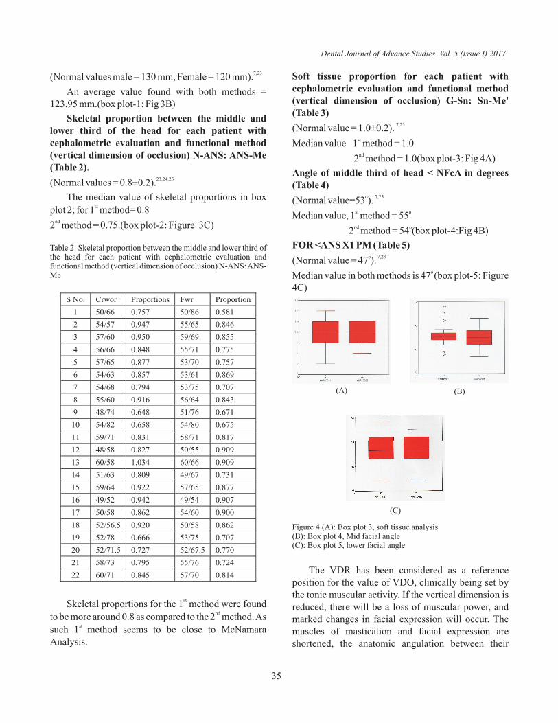
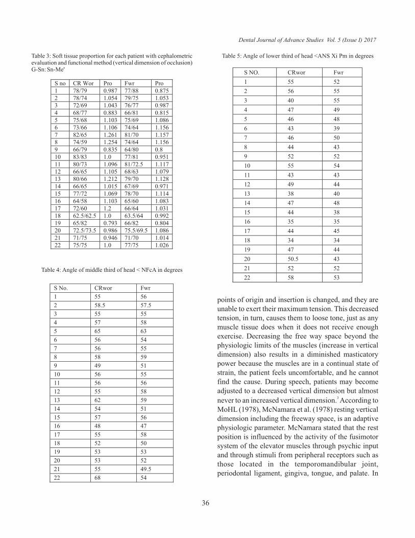
Soft tissue proportion for each patient with cephalometric evaluation and functional method (vertical dimension of occlusion) G-Sn: Sn-Me' (Table 3)
7,23(Normal value = 1.0±0.2). stMedian value 1 method = 1.0 nd
2 method = 1.0(box plot-3: Fig 4A)
Angle of middle third of head < NFcA in degrees (Table 4)
o 7,23(Normal value=53 ). st oMedian value, 1 method = 55 nd o
2 method = 54 (box plot-4:Fig 4B)
FOR <ANS X1 PM (Table 5)o 7,23 (Normal value = 47 ).
o Median value in both methods is 47 (box plot-5: Figure 4C)
S No. Crwor Proportions Fwr Proportion
1 50/66 0.757 50/86 0.581
2 54/57 0.947 55/65 0.846
3 57/60 0.950 59/69 0.855
4 56/66 0.848 55/71 0.775
5 57/65 0.877 53/70 0.757
6 54/63 0.857 53/61 0.869
7 54/68 0.794 53/75 0.707
8 55/60 0.916 56/64 0.843
9 48/74 0.648 51/76 0.671
10 54/82 0.658 54/80 0.675
11 59/71 0.831 58/71 0.817
12 48/58 0.827 50/55 0.909
13 60/58 1.034 60/66 0.909
14 51/63 0.809 49/67 0.731
15 59/64 0.922 57/65 0.877
16 49/52 0.942 49/54 0.907
17 50/58 0.862 54/60 0.900
18 52/56.5 0.920 50/58 0.862
19 52/78 0.666 53/75 0.707
20 52/71.5 0.727 52/67.5 0.770
21 58/73 0.795 55/76 0.724
22 60/71 0.845 57/70 0.814
7,23(Normal values male = 130 mm, Female = 120 mm).
An average value found with both methods = 123.95 mm.(box plot-1: Fig 3B)
Skeletal proportion between the middle and lower third of the head for each patient with cephalometric evaluation and functional method (vertical dimension of occlusion) N-ANS: ANS-Me (Table 2).
23,24,25(Normal values = 0.8±0.2).
The median value of skeletal proportions in box stplot 2; for 1 method= 0.8
nd2 method = 0.75.(box plot-2: Figure 3C)
Table 2: Skeletal proportion between the middle and lower third of the head for each patient with cephalometric evaluation and functional method (vertical dimension of occlusion) N-ANS: ANS-Me
(A) (B)
(C)
Figure 4 (A): Box plot 3, soft tissue analysis(B): Box plot 4, Mid facial angle(C): Box plot 5, lower facial angle
The VDR has been considered as a reference position for the value of VDO, clinically being set by the tonic muscular activity. If the vertical dimension is reduced, there will be a loss of muscular power, and marked changes in facial expression will occur. The muscles of mastication and facial expression are shortened, the anatomic angulation between their
Dental Journal of Advance Studies Vol. 5 (Issue I) 2017
35
points of origin and insertion is changed, and they are unable to exert their maximum tension. This decreased tension, in turn, causes them to loose tone, just as any muscle tissue does when it does not receive enough exercise. Decreasing the free way space beyond the physiologic limits of the muscles (increase in vertical dimension) also results in a diminished masticatory power because the muscles are in a continual state of strain, the patient feels uncomfortable, and he cannot find the cause. During speech, patients may become adjusted to a decreased vertical dimension but almost
3never to an increased vertical dimension. According to MoHL (1978), McNamara et al. (1978) resting vertical dimension including the freeway space, is an adaptive physiologic parameter. McNamara stated that the rest position is influenced by the activity of the fusimotor system of the elevator muscles through psychic input and through stimuli from peripheral receptors such as those located in the temporomandibular joint, periodontal ligament, gingiva, tongue, and palate. In
Table 3: Soft tissue proportion for each patient with cephalometric evaluation and functional method (vertical dimension of occlusion) G-Sn: Sn-Me'
S no CR Wor Pro Fwr Pro 1 78/79 0.987 77/88 0.875 2 78/74 1.054 79/75 1.053 3 72/69 1.043 76/77 0.987 4 68/77 0.883 66/81 0.815 5 75/68 1.103 75/69 1.086 6 73/66 1.106 74/64 1.156 7 82/65 1.261 81/70 1.157 8 74/59 1.254 74/64 1.156 9 66/79 0.835 64/80 0.8 10 83/83 1.0 77/81 0.951 11 80/73 1.096 81/72.5 1.117 12 66/65 1.105 68/63 1.079 13 80/66 1.212 79/70 1.128 14 66/65 1.015 67/69 0.971 15 77/72 1.069 78/70 1.114 16 64/58 1.103 65/60 1.083 17 72/60 1.2 66/64 1.031 18 62.5/62.5 1.0 63.5/64 0.992 19 65/82 0.793 66/82 0.804 20 72.5/73.5 0.986 75.5/69.5 1.086 21 71/75 0.946 71/70 1.014 22 75/75 1.0 77/75 1.026
Table 4: Angle of middle third of head < NFcA in degrees
S No. CRwor Fwr
1 55 56
2 58.5 57.5
3 55 55
4 57 58
5 65 63
6 56 54
7 56 55
8 58 59
9 49 51
10 56 55
11 56 56
12 55 58
13 62 59
14 54 51
15 57 56
16 48 47
17 55 58
18 52 50
19 53 53
20 53 52
21 55 49.5
22 68 54
S NO. CRwor Fwr
1 55 52
2 56 55
3 40 55
4 47 49
5 46 48
6 43 39
7 46 50
8 44 43
9 52 52
10 55 54
11 43 43
12 49 44
13 38 40
14 47 48
15 44 38
16 35 35
17 44 45
18 34 34
19 47 44
20 50.5 43
21 52 52
22 58 53
Table 5: Angle of lower third of head <ANS Xi Pm in degrees
Dental Journal of Advance Studies Vol. 5 (Issue I) 2017
36

the present study radiographs taken at two stages showed a difference between both readings representing the free way space, but did not affect the facial proportions, and allowed for appropriate phonetics and esthetics. This suggests that the functional method traditionally used complimented the cephalometric method to determine VDO in edentulous patients.
LIMITATIONS:
1. The main limitation of using the rest position to determine the vertical dimension of occlusion is that the jaw postural position is not constant but
8 varies continuously.
2. The cephalometric method for positioning the anterior teeth in complete denture is not suitable for routine use. Incisor position is seen outside2 standard deviation, from average Ricketts' data for
10 dentate subjects.
3. The patients with class II, class III relations were eliminated from the study because they show positive correlations between vertical height, mandibular morphology, mandibular plane
19angulation which is high.
4. The increase in the Nasolabial angle (NLA) is associated closely with increase in the vertical dimension of the face. The NLA increases with the
22amount of maxillary incisor retraction.
5. The mandibular rest position is influenced by the presence or absence of dentures. There is lowering of rest position of mandible upon insertion of dentures or patients may raise the mandibular rest position after insertion. The resting vertical dimension is often different depending on whether
6 21the dentures are in or out of the mouth. ,
CONCLUSION
In this study, there was stability in the skeletal vertical dimension, corroborating that the proportion of 0.8±0.2 was present between N-ANS and ANS-Me. Statistically no significant difference was found when
st ndcomparing the measurement of 1 method with the 2 method. In the second method the lower facial height was observed to be on the higher side in comparison
stwith 1 method but the increase in vertical dimension of ndocclusion after insertion of occlusal rims in the 2
method could lead to the increased lower facial 6,27,28
height. It was found that soft tissue proportion was maintained at around 1±0.2mm, and this was observed in both methods. Both the physiological and functional methods showed no difference statistically at+5% level of significance. Almost all observations were close to McNamara Analysis in our study patients, hence proving the reliability of cephalometric method and its acceptance for evaluating vertical dimensions in edentulous patients. These findings support the claim that the cephalometric method is a reliable and appropriate method for estimating the vertical dimensions in edentulous patients and can be used routinely by prosthodontist to confirm the vertical dimensions in combination with other methods.
REFERENCES
1. Zarb-Bolender et al. Prosthodontic treatment for edentulous
patients.12th ed. 2004, Mosby, West line Industril
Drive,St.Louis MO:268-97.
2. Jayashree V Dikshit and Firoze D Mirza. Muscle relaxant and
rest position – A cephalometric Study. J Prosthet Dent
1979;42:579-83.
3. Alex Coulouriotes. A.B. Free Way Space. J Prosthet Dent
1955; 5:194-9.
4. J.D. Orthlieb, M Laurent and O Laplanche. Cephalometric
estimation of Vertical Dimension of Occlusion. J Oral Rehabil
2000;27:802-7.
5. C Millet, C Jeannin, B Vincent & G Malquarti. Report on the
determination of occlusal vertical dimension and centric
relation using swallowing in edentulous patients. J Oral
Rehabil 2003;30:1118-22
6. Douglas Allen Atwood. A cephalometric study of the clinical
rest position of the mandible. Part I. The Variability of the
Clinical Rest Position following the Removal of Occlusal
Contacts. J Prosthet Dent 1955;10:504-9.
7. Brian D Monteith. A cephalometric method to determine the
angulation of the occlusal plane in edentulous patients. J
Prosthet Dent 1985;54:81-7.
8. M M Koller, L Merlini, G Spandre and S Palla. A comparative
Study of two methods for the orientation of the occlusal plane
and the determination of the vertical dimension of occlusion in
edentulous patients. J Oral Rehabil 1992;19:413-25.
9. Jay P Fitzgerald, Ram S Nanda, G Frans Currier. An evaluation
of the nasolabial angle and the relative inclinations of the nose
and upper lip. Am J Orthod Dentofac Orthop 1992;102:328-
Dental Journal of Advance Studies Vol. 5 (Issue I) 2017
37

34.
10. Ales Obrez, Jens C Turp. The effect of musculosketal facial
pain on registration of maxillomandibular relationships and
treatment planning: A synthesis of the literature. J Prosthet
Dent 1998;79:194-9.
11. H W Preiskel. Some observations on the postural position of
the mandible. J Prosthet Dent 1965;15:625-33.
12. Robert S Freeman. Adjusting A-N-B Angles to Reflect the
Effect of Maxillary Position. Angle Orthod 1981;51(2):162-
71.
13. Charles J Burstone, Randal B James, H Legan, G A Murphy,
and Louis A Norton. Cephalometrics for Orthognathic
surgery. J Oral Surg1978;36:269-77.
14. Dr. S I Bhalajhi. Orthodontics, The Art and Science. 2nd ed.
Arya (Medi) Publishing house, Darya Ganj, New Delhi
2001;148-50.
15. Athanasios E Athanasiou. Orthodontic Cephalometry.1995
Mosby-Wolfe, Times Mirror International Publishers
Limited.
16. Thomas Rakosi. An Atlas and Manual of Cephalometric
Radiography. 1982 Lea & Febiger- 600 Washington Square,
Philadelphia, Pennsylvania 19106 USA
17. Glenn L Gittelson. Vertical Dimension of Occlusion in
Implant Dentistry: Significance and Approach. Implant
Dentistry 2002;2:33-8.
18. Herbert Swerdlow, B A, Bethesda. Roentgencephalometric
Study of Vertical Dimension Change in Immediate Denture
patients. J Prosthet Dent 1964;14:635-49.
19. H W Preiskel. Some observations on the postural position of
the mandible. J Prosthet Dent 1965;15:625-33.
20. Brian D Monteith. Cephalometrically programmed adjustable
plane: A new concept in occlusal plane orientation for
complete denture patients. J Prosthet Dent 1985;54:388-94.
21. Allen M Kleinman and Irving M Sheppard. Mandibular rest
levels with and without dentures in place in edentulous and
complete denture wearing subjects. J Prosthet Dent 1972;
28:478-83.
22. Franklin D Lo and W Stuart Hunter. Changes in Nasolabial
angle related to maxillary incisor retraction. Am J Orthod
1982;82:384-90.
23. D Brzoza, N Barrera, G Contasti and A Hernandez. Predicting
vertical dimension with cephalograms,for edentulous
patients. Gerodontology 2005;22:98-103.
24. Charles J Burstone, Randal B James, H Legan, G A Murphy,
and Louis A Norton. Cephalometrics for Orthognathic
surgery. J Oral Surg1978;36:269-77.
25. Robert M Ricketts. Perspective in the Clinical Application of
Cephalometrics: The first fifty years. The Angle Orthod
1981;51:115-47.
26. F Bassi, A Rizzatti, G Schierano & G Pret. Evaluation of the
utility of cephalometric parameters in constructing complete
denture. Part-II: placement of anterior teeth. J Oral Rehabil
2001;28:349-453.
27. Geroge A Wessberg, Michael C Washburn, Frace N Epker and
Kent O Dana. Evaluation of mandibular rest position in
subjects with diverse dentofacial morphology. J Prosthet Dent
1982;48:451-9.
28. Franklin D Lo and W Stuart Hunter. Changes in Nasolabial
angle related to maxillary incisor retraction. Am J Orthod
1982;82:384-90.
Dental Journal of Advance Studies Vol. 5 (Issue I) 2017
Source of Support: Nil, Conflict of Interest: None Declared
38