ESCHERICHIA COLI IN BOVINE CALF SCOURS A Paper
37
ESCHERICHIA COLI IN BOVINE CALF SCOURS A Paper Submitted to the Graduate Faculty of the North Dakota State University of Agriculture and Applied Science By Crystal Nicole Starr In Partial Fulfillment of the Requirements for the Degree of MASTER OF SCIENCE Major Program: Food Safety July 2014 Fargo, North Dakota
Transcript of ESCHERICHIA COLI IN BOVINE CALF SCOURS A Paper

ESCHERICHIA COLI IN BOVINE CALF SCOURS
A Paper
Submitted to the Graduate Faculty
of the
North Dakota State University
of Agriculture and Applied Science
By
Crystal Nicole Starr
In Partial Fulfillment of the Requirements
for the Degree of
MASTER OF SCIENCE
Major Program:
Food Safety
July 2014
Fargo, North Dakota

North Dakota State University Graduate School
Escherichia coli in Bovine Calf Scours
By
Crystal Nicole Starr
The Supervisory Committee certifies that this disquisition complies with North
Dakota State University’s regulations and meets the accepted standards for the
degree of
MASTER OF SCIENCE
SUPERVISORY COMMITTEE:
Dr. Penelope Gibbs
Chair
Dr. Neil Dyer
Dr. Clifford Hall
12/17/2014
Dr. Paul Schwarz
Date Department Chair

iii
ABSTRACT
Scours is caused by inflammation of the intestinal tract of ruminants leading to significant
mortality and morbidity rates. It is predominately found in neonatal ruminants where the disease
can occur 36 hours after birth. One of the most common infectious agents linked to scours is
pathogenic Escherichia coli. Therefore, it is important to understand the virulence factors, diagnostic
assays, age of the animals infected, and the co-factors associated with an E. coli scours outbreak.
These factors are important in both scours disease pathogenesis and potential food safety-related
postharvest pathogens. Using the most frequently identified virulence factors, a new scours
diagnostic assay could be created to detect and prevent disease in cattle. The present study
determined that virulence factors astA, fimC, fimH, int1, int2, irp2, papC were identified over 15%
percent of the time and could be implemented into a more specific multiplex PCR test for pathogenic
E. coli.

iv
TABLE OF CONTENTS
ABSTRACT ................................................................................................................................................. iii
LIST OF TABLES ....................................................................................................................................... vi
LIST OF FIGURES ................................................................................................................................... vii
INTRODUCTION……………………………………….……………………………………………………….….1
LITERATURE REVIEW ............................................................................................................................. 2
Infectious Disease Pathogenesis ............................................................................................................. 2
Colibacillosis ............................................................................................................................................ 3
Bovine Calf Scours ................................................................................................................................... 4
Escherichia coli ........................................................................................................................................ 4
Enteropathogenic E. coli (EPEC)……...……………………………………….……………………...………5
Enterotoxigenic E. coli (ETEC) ............................................................................................................... 5
Enteroinvasive E. coli (EIEC) and Enterohemorrhagic (EHEC) E. coli .............................................. 6
Uropathogenic E. coli (UPEC) ................................................................................................................ 6
Virulence Gene Classification ................................................................................................................. 6
Adhesion Genes........................................................................................................................................ 7
Toxin Genes .............................................................................................................................................. 7
Iron-Regulating Genes ............................................................................................................................ 8
Antibiotic Resistance Genes .................................................................................................................... 9
Preventative Measures .......................................................................................................................... 10
Food Safety Distresses .......................................................................................................................... 11
MATERIALS AND METHODS ................................................................................................................ 14
Polymerase Chain Reaction Analysis Testing ..................................................................................... 16
Gel Amplification ................................................................................................................................... 18
Phylogenetic Typing Method ................................................................................................................ 19
RESULTS AND DISCUSSION ................................................................................................................ 20
CONCLUSION........................................................................................................................................... 26

v
REFERENCES .......................................................................................................................................... 28

vi
LIST OF TABLES
Table Page
1. Vaccine Recommendations for Bred Cows and Calves........................................................................ 11
2. List of Primers and Their Gene Products Used in This Study ........................................................... 17
3. Phylogenetic Typing Results Identified ............................................................................................... 20
4. Adhesion Gene Percent Positive Results ............................................................................................. 21
5. Iron Regulating Gene Percent Positive Results .................................................................................. 22
6. Toxin Gene Percent Positive Results ................................................................................................... 23
7. Presence of Integrons Percent Positive Results .................................................................................. 24

vii
LIST OF FIGURES
Figure Page
1. Map of North Dakota with Positive Scours Cases Highlighted .......................................................... 14
2. Infection of Calves Per Month Breakdown .......................................................................................... 15
3. Phylogenetic Typing Diagram Clarification ........................................................................................ 19

1
INTRODUCTION
Many deceased bovine calves presented at the NDSU Veterinary Diagnostic Laboratory
(NDSU-VDL) with similar antemortem symptoms led the pathologist to request a scours panel to
check for pathogens. Results of these tests indicate the fecal samples from some of the post-mortem
specimens contained Escherichia coli (E. coli) resistant to many of the commonly used antibiotics
such as ampicillin, ceftiofur, chlortetracycline, clindamycin, erythromycin, florfenicol, neomycin,
oxytetracycline, penicillin, spectinomycin, sulphachlorpyridazine, sulphathiazole, tiamulin,
tilmicosin, trimethoprim, and tylosin. These specimens were negative for E. coli virulence factors
routinely tested for at the NDSU-VDL. Since the animal was sick it seemed inconsistent to have no
pathogenic E. coli genes found. Our study investigated this inconsistency further.
In the present study, samples collected in 2004 were tested for 22 additional virulent E. coli
strains to determine which of these were being detected in multi-drug resistant samples. These
samples also showed no positive results for the six E. coli virulence factors tested for in the NDSU-
VDL. A supposition was made that an E. coli virulence factor not included on the test was present in
the calves and making them resistant to antibiotics and susceptible to disease. Testing for the 22
additional virulence factors would help define which other virulence genes were present in North
Dakota and bordering Minnesota farms.
In addition to determining the specific gene present, additional genes could be added to the
current NDSU-VDL test for E. coli virulence factors. The results of this study will not only help the
NDSU-VDL provide more definitive results, but also help the producer and veterinarian narrow
down a treatment and/or preventative option for the already ill or highly susceptible herds.

2
LITERATURE REVIEW
Infectious Disease Pathogenesis
All bacteria go through steps of survival inside a host which may lead to disease. For bacteria
to survive inside a host, they must first find and recognize their specific host. If host attachment
receptors are specific for a bacterium, the bacteria will attach and colonize the host and can lead to
disease. If the host’s immunity is not compromised or vaccines against a bacterium are given, the
attachment and replication of the bacteria may be blocked. Thus, no disease will be caused. In the
case of neonatal calf scours, many strains of E. coli cannot be considered harmless (1). If the host is
immunosuppressed or they have not been treated with an effective vaccine, E. coli are able to attach,
colonize and grow, thus leading to disease expression such as scours (11).
Once E. coli attach they need to obtain nutrients in order to survive and replicate. Their
mechanisms for acquiring nutrients frequently cause disease in the host (7). E. coli is able to acquire
specific nutrients such as amino acids, iron, trace elements, and a carbon source from their host.
This is done by utilizing the host’s nutrient supply. The primary nutrient is iron. If the iron is
present, but not tightly bound, E. coli that produce iron acquisition genes that are more likely to
access it and create an iron deficiency in the host (2). Bacteria growing in aerobic environments need
iron to perform various tasks such as the oxygen decrease for synthesis of ATP, the decrease of the
precursors of ribotides of DNA, and the creation of heme, to name a few. Because these are essential
factors for the colonization of bacteria, the microorganism must obtain some form of iron for survival.
This is done by requiring the microorganism to form molecules to compete with the hydroxyl ion of
iron in its ferric state. If the iron is unavailable, siderophores will be dispersed and will carry the
less tightly bound iron to the bacteria. The availability of the siderophores is governed by the gene
fur (Ferric Uptake Regulation) and thus the two will compromise to bind the gene to the operon,
usually Fe (II). Once binded, the siderophores are able to “carry” the iron back to the depleted
bacteria (24).

3
As nutrients become available, E. coli will replicate and reproduce asexually. This is done by
cell division, which form clones of themselves every time division occurs. A reoccurrence of all these
steps happens with each clone, thus increasing the bacterial numbers to a capacity that eventually
overwhelms the host, leading to a full blown scours disease (33). Following damage and death of the
host’s cells, the E. coli can be passed to a new host via infective feces, blood, respiratory secretions,
feed and bedding (6).
Colibacillosis
Colibacillosis is a diarrheal disease caused by the Gram-negative bacteria, E. coli.
Colibacillosis has been linked to clinical symptoms, such as dehydration, diarrhea, fatigue, fever,
malaise, and depression, in the calf between 3-5 days old. While E. coli is a common resident of the
intestinal tract of calves, there are certain strains that are pathogenic. Pathogenic E. coli may cause
diarrhea in ruminant neonates. If left untreated, the calf could develop colisepticemia, when the
bacterial infection spreads into the bloodstream of the host (6). This could ultimately lead to death.
Garcia-Migura, et al. (11) performed research using a private herd in Toronto, Canada, and
discovered the criteria for identifying a colibacillosis positive calf from those not infected. Their
research identified a larger amount of E. coli in the small intestine along with lymph node
involvement in calves displaying colibacillosis symptoms. Additionally, an absence of other
commonly identified disease causing bacteria was also discovered in those calves. This study was
used to determine a vaccine for the cows that would provide them with antibodies to combat the
disease. The researchers found that cows who are vaccinated using specific E. coli K and O antigens
have a lower mortality and morbidity rate in their offspring. This may indicate that the E. coli found
in cases positive for colibacillosis in calves could possibly be of K or O serotype.
E. coli isolates are serologically separated according to their major surface antigens. The
three major surface antigens are the O (somatic), H (flagellar), and K (capsulated) antigens.
Escherichia coli can also be separated into categories based on properties specific to their
characteristics (22). These include: virulence factors, specific clinical symptoms, and their distinct
O:K:H serotypes (14). The H antigen is also commonly found in pathogenic E. coli, as well. This type

4
uses its flagella to incorporate movement of the bacteria so they are able to gain as much nutrients
as they need. The K serotype is a polysaccharide capsule that envelopes the bacteria whereas the O
serotype is an actual part of the lipopolyssacharide molecule (12).
Bovine Calf Scours
Calf scours is a multifactorial disease that involves the dam, the calf, the calving facility, and
the overall management of the herd. It is vital for a pregnant cow to receive optimum nutrition in
order to ensure a healthy calf at birth. Their diet should include ample amounts of proteins and
calories in order to guarantee a sufficient amount of brown fat in the offspring to supply energy that
will increase calf vigor upon birth. Higher vigor, along with a healthy calf, will provide the energy
needed for the calf to want to stand and nurse. Once the calf has received colostrum, the calf will
obtain the acquired immunity that will assist in combating many pathogenic bacteria (3). It can
affect up to 70 percent of a herd’s offspring, with up to a 50 percent mortality rate. Because scours
predominately affects calves, it is the cow-calf operations that are impacted the hardest. A scours
outbreak can lead to a financial disaster for ranchers and may even lead to their closure (15).
Escherichia coli
Infectious agents can be bacteria, viruses, protozoans, parasites, or fungi. In scours, the most
common infectious agent is pathogenic E. coli. A ruminant can become exposed to pathogenic E. coli
as early as passage through the birth canal. Lactobacilli and enterococci are considered to be good
bacteria and found in the first 30-40 feet of the small intestine and abomasum of a calf. If an
abnormal presentation occurs along with trauma, the producer needs to supplement the calf with the
dam’s colostrum in order to safeguard its health so the acquired immunity enters the calf as soon as
possible. The calf may not want to nurse due to the stress it endured therefore esophageal tubing
should be performed (35). During the first few hours of a calf’s life, it is crucial for them to receive
adequate amounts of colostrum (37). Once the calf starts breathing, the small intestine starts to
mature, beginning intestinal cell turnover, abosomasal acidity, and development of intestinal
secretions and intra-epithelial vacuoles used in digestion. The intestine only absorbs macromolecules
for 24 hours before the closure begins. This initial process is known as passive immunity. This is why
5
it is so important for the calf to receive immunoglobulins that are found in colostrum as soon as
possible (31).
Colostrum is filled with immunoglobulins which play a role in limiting the growth and the
colonization rates of E. coli as well as other infectious causes such as Salmonella, Coronavirus,
Rotavirus, and Cryptosporidium (37). If the animal does not have maternal antibodies and has been
exposed to pathogenic E. coli or other infectious agents the small intestine will quickly become
colonized with the pathogen. This is also known as failure of passive transfer (31). The overwhelming
pathogenic bacterial load results in increased financial losses for the producer. This is due to reduced
weight gain, escalation of medical costs, or death of the infected calves (34).
Diarrheagenic E. coli cause diarrhea as well as extra-intestinal infections in both humans
and animals of almost all species of warm-blooded vertebraes. All these groups shed E. coli into the
feces of the infected host which aids in spreading to other hosts. There are six categories of E. coli
that are known to cause disease. These six categories are: Enteropathogenic E. coli (EPEC),
enterotoxigenic E. coli (ETEC), enteroinvasive E. coli (EIEC), enteroaggregative E. coli (EAEC),
enterohemorrhagic E. coli (EHEC) and uropathogenic E. coli (UPEC) (6,22).
Enteropathogenic E. coli (EPEC)
Enteropathogenic E. coli (EPEC) are able to create lesions in intestinal epithelial cells in
warm blooded mammals. EPEC uses humans as its primary host and is the number one killer in
children under five years of age in developing countries. This is due to its ability to cause severe life-
threatening diarrhea in the third world. Bacteria in this category do not produce shiga toxins and
are not associated with hemolytic uremic syndrome (26). Despite humans being the primary host,
this particular E. coli are a cause of disease and death in calves, piglets, rabbits, and both wild and
domestic birds (26).
Enterotoxigenic E. coli (ETEC)
Enterotoxigenic E. coli (ETEC) are the main causative agent of traveler’s diarrhea. The
bacteria in this subcategory are known to affect the small intestine, and produce a heat-stable or
heat-labile enterotoxin to create buildup of fluid and loose fecal matter (21). This particular type of

6
E. coli causes watery diarrhea in its host. It secretes toxins into the intestines. The most common
genes produced are the heat-stable and heat-labile toxins (ST and LT)(19).
Enteroinvasive (EIEC) and Enterohemorrhagic (EHEC) E. coli
Enteroinvasive E. coli (EIEC) are associated with Shigella spp. Like Shigella spp., the EIEC
are associated with the possession of a large plasmid, which encrypts the outer membrane proteins
involved in the process. Bacteria categorized as EIEC are known to invade the colon and disrupt and
grow inside epithelial cells which ultimately lead to cell death (21).
Enterohemorrhagic E. coli (EHEC) causes bloody intestine in its host. These bacterial strains
secrete verotoxins which are lethal to African green kidney (vero) cells. They are also known as
shigatoxins, due to the activity of the Stx (shiga toxin) gene in the Shigella bacteria. Thus, the EHEC
producing strains can also be referred to as Stx-producing E. coli. The multiple strains placed into
this subcategory are linked to infections that can be quite severe or even fatal. There are over 200
serotypes belonging to this EHEC subgroup (21).
Uropathogenic Escherichia coli (UPEC)
Uropathogenic E. coli (UPEC) are known to cause Urinary Tract Infections. They do this by
infecting the kidney of the host and utilizing their expressed fimbriae to attach and colonize. The
main fimbriae of concern is that of the P-fimbriae, which is encrypted by the pap gene, and is known
to be an adhesion resistant to mannose. This fimbriae uses its adhesion unit to attach to the
uroepithelial cells. Once adhered, it can successfully cause renal failure leading to a urinary tract
infection. Along with the P-fimbriae, there are other virulence strains expressed. These include the
fimbrial adhesion I, hemolysin, cytotoxic necrotonizing factor and the S fimbriae (18).
Virulence Gene Classification
Virulence genes are known to facilitate a microorganisms ability to establish itself on or
within a host; in this instance, a calf. This will allow the pathogen to express toxins, adhesins, and
enzymes enhancing the ability of the bacterium to cause disease. In this study, each virulence gene
was categorized into one of these four categories: 1) Adhesion, 2) Toxin, 3) Iron Regulating, and 4)
Antibiotic Resistance. The listed categories are explained in more depth below.

7
Adhesion Genes
Bacterial adhesion genes are bacteria that express adhesion proteins that reside inside a
biofilm (26). This allows the bacterial population to adhere to each other and to surfaces. The specific
bacteria that form biofilms must have flagella or another form of motility. They utilize their forms of
motility to move along the surface of the intestine to find a nutrient rich area. Type 1 pili, required
for the initial surface attachment, is encoded by the gene fim and has specific phenotypes that aid in
its flagellar production. The fim gene clusters that are located on the operon, fimH, have lesions that
affect the growth of the tip of the Type 1 pili. This will cause this particular gene to function as an
adhesin specific to mannose and therefore cooperate with residues of mannose on the cells to enable
cystitis (28). Other genes tested that produce adhesion operons are that of papC, ibeA, sfa, fimC, afa,
tsh, est1, and neuC.
Toxin Genes
Toxin genes are known to cause bacterial death and significant defects in bacterial growth
leading up to a decreased gene expression in each cell (28). This affects normal growth of the E. coli
which can, in turn, cause a new, highly resistant strain of E. coli to form in the intestine. Toxin
activity can be caused by a cytolethal distending toxin (CDT) due to its deadly effect on cells. This
particular protein is said to distend the cells prior to killing them off. However, this protein is heat
sensitive and causes differing distentions based on whether the toxins are heat-labile, heat-stable, or
shiga-like (30).
E. coli capable of producing CDT will attach and efface the microvilli (AE) causing lesions via
establishment of microcolonies, destruction of microvilli, and have massive amounts of actin
microfilaments at their attachment site. Because of the large quantities of actin, a fluorescent stain
can be used to determine the attachment and effacement activity. Performing this type of procedure
has identified the presence of the eae toxin (25).
Shiga-like toxin producing E. coli (STEC) are known to cause food poisoning as well as
severe/possibly fatal illness. Shiga-like toxin producing strains known to be pathogenic contain the
phage-encoded cytotoxins called shiga-toxins (stx1 and stx2) also known as verotoxins, and a protein

8
called intimin. Intimin is responsible for the AE in the intestinal mucosa. The genes important to
test were the shiga-toxins (stx1 and stx2) as well as eae (Intimin)(27). These were tested along with
other toxin-producing genes, astA, vat, cnf, and elt1.
Iron-Regulating Genes
Iron is an essential mineral that is utilized in a calf’s metabolism and plays an important
role in many of their systems. Nitrogen fixation and oxygen transport are just a few examples of
what this mineral contributes to. The iron-dependent repression is facilitated by the Ferric Uptake
Regulation (FUR), which uses genes involved in the previously mentioned systems. The bacterium
creates a transport system through the use of siderophores (22). According to K.D Krewulak’s
research, siderophores solubilize the iron prior to transport then use a combination of receptors and
transporters to create iron metabolism for the bacteria. This creates an advantage to the bacteria
because it can utilize iron that was produced at another organism’s expense or utilize other iron-
acquisition systems that could otherwise steal iron in an inappropriate form. E. coli contains. The
other way microorganisms will deal with iron deficiency is to obtain the iron extracellularly from
lactoferrin, transferrin, and precipitated ferric hydroxides or by from hemoglobin intracellularly. The
iron uptake process is established using the permeable outer membrane of Gram-negative bacteria.
The trimeric β-barrel proteins housed in the outer membrane allow for solutes less than 600 Da.
Because the lactoferrin, and transferrin are more than the 600 Da, they must find a way to execute
the iron externally. The external binding of these molecules is done utilizing the proteins,
TbpB/TbpA and LbpB/LbpA, by an N-terminal lipid anchor. The protein TbpB will perform as the
first site for binding of the transferrin and make the additional binding to the TbpA go much easier.
The TbpA and TbpB have large loops at the surface that separate the region surrounding the binding
sites of the transferrin and lactoferrin to separate and release any present iron into the periplasm.
Once in the periplasm, the iron is bound to the FbpA protein and transported to the cytoplasmic
membrane and then into the cytoplasm using the cytoplasmic membrane transporters, FbpB and
FbpC. Once out, the iron can be utilized by the bacteria for survival procedures. The iron regulating
genes tested for in this study include: irp1, irp2, iucD, and Hly.

9
Antibiotic Resistance Genes
Antibiotics were founded by Alexander Fleming in 1928. Further research has found that the
bacteria have been developing resistance to antibiotics. The bacteria can develop the resistance buy
mutating and/or using horizontal gene transfer to swap DNA from other bacteria. Normally, the
antibiotic will secure itself to a protein making the protein unable to function correctly. However, if
the bacteria have some sort of mutation in their DNA that code for that protein, the particular
antibiotic will be unable to fasten itself to the abnormal mutated protein and thus the mutated
bacteria will live and keep reproducing more of the mutant bacteria. Because these mutants can live
in the environment where the antibiotic is present, the bacteria will thrive and rapidly reproduce
when the antibiotic is present and the illness will persevere (29).
Many physicians, veterinarians, and producers utilize antibiotic therapy in order to treat
clinically sick neonates, which can in turn lead to an increased chance of antibiotic resistant
bacteria. It has been noted that bacterial plasmids keep virulence factor encoding genes. These genes
are used to encrypt virulence factors for scours. These genes can also be used in bacterial conjugation
as well as antibiotic resistance. Presently, a number of the scours-related E. coli strains have
developed resistance to numerous antibiotics, including but not limited to streptomycin,
sulfisoxazole, ceftiofur, and tetracycline. Those particular antibiotics are screened using a sensitivity
test in the NDSU-VDL Microbiology laboratory. Antibiotic resistant strains have the ability to
inhabit the intestinal tract of antibiotic treated animals. Some bacteria live in biofilms, increasing
the prevalence of antibiotic resistance (26).
Environmental changes to the surface of the intestinal cell are accepted by the biofilm to
activate changes to permit the bacteria to undertake interactions to create antibiotic resistance.
Newer commercially used antibiotics, such as aminoglycosides, fluoroquinolones, and expanded-
spectrum cephalosporins, have been found to be quite effective against E. coli that cause scours in ill
calves. However, because none of these antibiotics carry a label claim for use to treat enteric disease
in calves, they can only be given as Extra Label Care Use in the United States under the Animal
Medicinal Drug Use Act (36). Ceftiofur is another antimicrobial drug approved to treat respiratory

10
disease in bovine and swine but does not have the label claim to treat scours. However, it is still
often used to treat enteric diseases caused by multiresistant E. coli strains (36).
Preventative Measures
Two cattle producers in north central North Dakota found combating colibacillosis to be a
difficult task (20, 34). Local veterinarians were able to help with preventative vaccination including
ScourGuard 4KC (Zoetis, Kalamazoo, MI). This was administered to the cattle and heifers two to
three weeks prior to their calving date. ScourGuard 4KC contains killed viruses of the serotypes G1
and G10 bovine rotavirus, coronavirus, enterotoxigenic strains of E. coli particularly those with K99
pili adhesion factors, and Clostridium perfringens type C. This vaccine is given to cows prior to
calving ensuring the colostrum has these specific antibodies to help the nursing offspring build
immunity to the previous mentioned toxins (Dr. Richard Lagasse, Rugby Veterinary Service, Rugby,
ND, Personal Communication).
Cathy Balsiger, the lead herdsman/calf specialist at a local dairy barn in west-central
Minnesota, has administered the vaccine Guardian (Merck, Summit, NJ), into her herd to decrease
the chance of scours in the calves. She gives this vaccine, composed of a cell-free extract of K99 pilus
of E. coli, two inactivated coronaviruses, two G serotypes of rotaviruses, and a bacterin-toxoid of
Clostridium perfringens Types C and D to her 1350 dairy cows subcutaneously at dry off time,
which is 50-60 days prior to calving, to create a stronger immunity to the above toxins in the
offspring. This vaccine has reduced the prevalence of scours she had in the past, to less than 1
percent death loss (Cathy Balsiger, BGR Dairy, Lake Park, MN, Personal Communication). Dr.
Richard Lagasse recommends Scour Bos 4 (Novartis Animal Health, Larchwood, IA) and Scour Bos 9
(Novartis) vaccinations to his clientele. Scour Bos 9 is given intramuscularly (IM) in the neck 8-16
weeks prior to calving. A revaccination with Scour Bos 4 should be given IM in the neck one month
prior to calving to prevent infection. These vaccinations contain killed viruses of rotavirus,
coronavirus, and bacterial species Clostridium perfringens Type C, and K99 pili E. coli (Dr. Richard
Lagasse, Rugby Veterinary Service, Rugby, ND, Personal Communication) (Table 1). There are also
vaccinations and oral medications to administer to newborns and include: Calf Guard (Zoetis),

11
Bovine Ecolizer + C20 (Novartis Animal Health, Larchwood, IA), Bovine Ecolizer (Novartis), and
Bar-Guard 99 (Boehringer-Ingelheim, St. Joseph, MO). There are other medications that aid in the
prevention of scours, as well. The vaccinations and medications listed below were recommended by
the attending veterinarian to prevent infection (Dr. Richard Lagasse, Rugby Veterinary Service,
Rugby, ND, Personal Communication) (Table 1).
Table 1. Vaccine Recommendations for Bred Cows and Calves.
Vaccinations given to Bred Cows Vaccinations Given to Calves
Scour Guard 4KC Calf Guard
Guardian Bovine Ecolizer +C20
Scour Bos Bovine Ecolizer
Bar Guard 99
Calf Guard (Zoetis) can be given to both cows and calves to protect against rotavirus and
coronavirus. Cows receive two doses 3-6 weeks apart in late gestation. Calves are to receive an oral
dose within 4 hours of birth. Bovine Ecolizer + C20 (Novartis) is another oral medicine given to
calves immediately after birth, no later than four hours and protects against Clostridium perfringens
Type C as well as K99 E. coli. Bovine Ecolizer (Novartis), an oral medication that contains K99 E.
coli antibodies, is given to calves within 12 hours of birth to prevent scours. Bar-Guard 99
(Boehringer Ingelheim) is an oral medicine that contains whole culture K99 pili hyperimmunized
blood from horses and should be given within 12 hours of birth for optimal results (Dr. Richard
Lagasse, Rugby Veterinary Service, Rugby, ND, Personal Communication)(Table 1).
Food Safety Distresses
If a calf raised for veal becomes infected and is given antibiotics, a potential food safety
threat is created since antibiotic resistance remains a large problem. If the calf displays severe
infection after the administration of antibiotics, the producer may choose to slaughter early. After
slaughter, leftover antibiotic residue may be present in the meat, posing a direct threat to the

12
consumer. This is against FDA regulations. Therefore, the producer, who assumes the risk, could
receive a fine if proven guilty (13).
Conjugative plasmids are said to not carry genes fundamental to host cell growth in
conditions that are not under stress. However, Wang, et al (39) found that almost all plasmids
bestow particular phenotypes including but not limited to antibiotic resistance genes. Plasmids are
very adaptable to DNA and will transfer the genetic information found in DNA. He noted in his
discovery that prior to his research, E. coli isolates that had the extended-spectrum β-lactamase-
encoding genes mapped on plasmids had been found in food-producing animals and humans that
were in good health. His research supported this finding (39). This indicates that plasmids can pose
a problematic source for individuals who are found to be healthy implying that there is a risk in
normal flora.
Escherichia coli contamination from the herd of cattle is a food safety concern as well. To
prevent the shedding of E. coli, some producers add chlorine to animals’ drinking water to help
reduce any spread of the pathogen to other animals. The FSIS has no law prohibiting this procedure,
since it is not harmful to the meat supply (9). Due to recent drought in some areas of North Dakota
and Minnesota, additional drugs have been used to help livestock gain weight faster. Problems can
arise since safety experts may not test for all of the drugs when the animal is slaughtered leading to
meat that contains drug residue potentially causing harm to the consumer.
Overall, food safety plays an integral role in the livestock industry, since it remains an
important part of the economy. An important aspect of food safety involves keeping the cattle
healthy and disease-free in order to prevent costly outbreaks to humans. The latest outbreak known
by the Center of Disease Control was in June of 2014 where people were sickened in four different
states. Of these 12, 58 percent were hospitalized. Once the undercooked ground beef from various
restaurants from the Wolverine Packing Company was found to be the source, 1.8 million pounds of
it was indicated to be contaminated and a recall was released. Recalls such as this can be quite costly
but precautions as simple as cooking ground beef to 160 degrees Fahrenheit should always be taken
to decrease the chance of becoming ill. By reducing the amount of illness in the herd and cooking raw

13
meat to appropriate temperatures, one can safeguard a safe, enjoyable, consumable product at the
table.

14
MATERIALS AND METHODS
Seventy-five highly resistant isolates obtained from clinical cases submitted to the North
Dakota State University-Veterinary Diagnostic laboratory (NDSU-VDL) from the year 2004 were
chosen for study. The age of the calf, location of farm or ranch (county and state), month in which
infected, final diagnosis, and any other statistics were noted. This data was compiled and analyzed.
The following figures show the geographical location where the isolates were located (Figure 1).
Figure 1. Map of North Dakota with Positive Scours Cases Highlighted.
Further analysis indicated a high percentage of calves being affected in March which is
consistent with the spring calving season that is found in North Dakota (Figure 2). Due to the
extreme low temperatures, many cows and older calves are housed in the same building/area as cows

15
and newborn calves. The housing unit can become dirty very quickly and the producer must clean
the area often (38).
Figure 2. Infection of Calves Per Month Breakdown.
The NDSU-VDL currently performs the polymerase chain reaction (PCR) test for only six
virulence factors, including: K99, F41, Sta, Intimin, stx-1, and stx-2 (8). Many of these virulence
factors were not amplified during PCR despite E. coli being isolated from the sample. This may be
due to the overwhelming amount of other pathogenic virulence factors that may be present in the
animal. If other factors are shown to be useful in the typing of E. coli, a new diagnostic test utilizing
these factors could be developed.
The 2004 NDSU-VDL isolates were screened for the following virulence genes: astA, cnf, hly
EHEC, hlyA, papC, eaeA, vat, irp1, int1, ibeA, fimH, stx1, stx2, sfa, fimC, afa, tsh, est1, elt1, iucD,
and neuC (8). These virulence factors have been shown to have significance in the bovine and avian

16
intestine, as well as a source for foodborne illness in consumption of raw meat. Screening provides
evidence as to which of the virulence is potentially pathogenic. Phylogenetic grouping was completed
to determine the group each isolate falls into. Shiga toxins (stx-1 and stx-2) have been shown to be
harmful to public health.
The above virulence factors were screened using PCR. Each isolate was performed in
duplicate in order to decrease the potential for human error. Positive strains, negative strains, and
water were used as controls in each case. In order to determine the sizes of the amplified bands a 100
base pair ladder was used. Each isolate ran on a thermal cycler with parameters for the selected
virulence factor.
Polymerase Chain Reaction Analysis Testing
The isolates were plated onto MacConkley agar and incubated at 37°C for 24 hours. Lennox
broth (900 µL) was placed into 1.5 mL tubes and a separate colony of E. coli was added to each tube
and incubated for 24 hours at 37°C with shaking. After 24 hours, 600 µL of glycerol was added to
each tube, vortexed, and frozen in an -80°C freezer until needed for the addition of the individual
primer master mix for the thermal cycler.
The PCR master mix consisted of 5 µL of 5x Green GoTaq Reaction Buffer (Promega,
Madison, Wisconsin), 0.2 µL dNTP mix (Promega), 0.25 µL Forward Primer (Sigma Aldrich, St.
Louis, Missouri), 0.25 µL Reverse Primer (Sigma Aldrich), 5.0 µL 5x GoTaq DNA Polymerase
(Promega), and 17.175 sterile water. A master mix with the corresponding primers was made for all
22 virulence factors.

17
Table 2. List of Primers and Their Gene Products Used in This Study.
Primer Direction
(5’-> 3’)
Gene Bp size Reference
Chua.1
Chua.2
Forward
Reverse
chuA
279 3
yjaa.1 Forward yjaA 211 3
TspE4C2.1
TspE4C2.2
Forward
Reverse
TspE4C2 152 3
Int1-F
Int1-R
Forward
Reverse
Int1 280 10
Slt-F
Slt-R
Forward
Reverse
Stx1 614 17
SltII-F
SltII-R
Forward
Reverse
Stx2 779 17
Sfa/foc-F
Sfa/foc-R
Forward
Reverse
Sfa/foc 410 41
Afa-F
Afa-R
Forward
Reverse
afa 750 41
IbeA-F
IbeA-R
Forward
Reverse
ibeA 171 41
Irp1.A
Irp1.B
Forward
Reverse
Irp1 1691 2
St-1B
St-1C
Forward
Reverse
Est-1 a/b 123 33
LT-I/1
LT-I/2
Forward
Reverse
Elt-1 a/b 365 33
EAST-1F
EAST-1R
Forward
Reverse
astA 110 33
EHLY1
EHLY2
Forward
Reverse
hlyEHEC 889 33
EHLYA-F
EHLYA-R
Forward
Reverse
hlyA 534 41
AERA-F
AERA-R
Forward
Reverse
iucD 692 33
TSH-F
TSH-R
Forward
Reverse
tsh 804 33
PAPC-F
PAPC-R
Forward
Reverse
papC 482 33
FIMC-F
FIMC-R
Forward
Reverse
fimC 476 33
EAEA-F
EAEA-R
Forward
Reverse
eaeA 384 41
VAT-F
VAT-R
Forward
Reverse
vat 981 17
fimH-F
fimH-R
Forward
Reverse
fimH 508 41
neuC-F
neuC-R
Forward
Reverse
neuC 675 41
CNF-F
CNF-R
Forward
Reverse
cnf 693 43

18
Corresponding to each program run on the thermal cycler, a series of temperature and time
adjustments were completed (Table 2). All of these programs have steps that denature the template,
anneal the primer, and extend the primer. The first step in all the programs begins at a temperature
of 94°C or higher for 15 seconds to 2 minutes to denature the template. This step separates the DNA
strands from one another, which then gives one the single-stranded DNA to replicate using the
thermostable DNA polymerase. The next step drops the temperature to 40-60°C to allow for
oligonucleotide primers to form associations with the denatured target DNA. These function as
primers for the DNA polymerase. This step takes approximately 15-60 seconds. Lastly, the synthesis
of new DNA starts as the temperatures is raised to the optimum for the DNA polymerase. This
varies between programs but generally lasts about 1-2 minutes. The program will return to 94°C
until stopped and samples are removed.
Gel Amplification
Following the thermocycler reaction, 10 microliters of sample from each reaction tube was
placed in a 2% agarose gel. The gel was placed in a gel-casting tray in 5X TBE (trio/borate/EDTA)
electrophoresis apparatus. The electrical field was adjusted to 150 volts and ran for 30 to 60 minutes
depending on the size of the gel. The presence of bands and their sizes, which corresponded to the
amp icon size expected using the specific primers was observed. The presence of virulence factors
and the percentage of positive isolates were calculated. Those virulence factors found in many
samples were deemed reasonable for use on a diagnostic test. Each step of the cycle was optimized
for each template and primer pair combination. If the temperature during the annealing and
extension processes was similar, the two steps can be combined and both the annealing and
extending will takes place at the same time. After 20-40 cycles, the amplified band may be run on a
gel to find particular band patterns. These band patterns signify whether the sample was positive or
negative for that particular factor.

19
Figure 3. Phylogenetic Typing Diagram Clarification. (5)
Phylogenetic Typing Method
Determination of which gene goes where is done by a triplex PCR technique using genes,
chuA and yjaA, along with an unknown DNA fragment. The technique for this test was done just like
the others, using chuA as the initial determination gene. Those that produced a positive result were
rerun with the same technique using primers for the gene, yjaA. A positive result in this step
determined a final identification of group B2, whereas a negative result was determined as group D.
A negative chuA gene result was rerun with primers detecting a region of DNA denoted and a new
reaction was rerun using TspE4.C2(5). If the TspE4.C2 reaction was positive the resulting group was
B1; if the isolate was negative for TspE4.C2 the resulting isolated was placed in group A (Figure 3).
chuA
+
B2 or D
yjaA
+
B2
-
D
-
B1 or A
TspE4.C2
+ -
0
B1
11
A
20
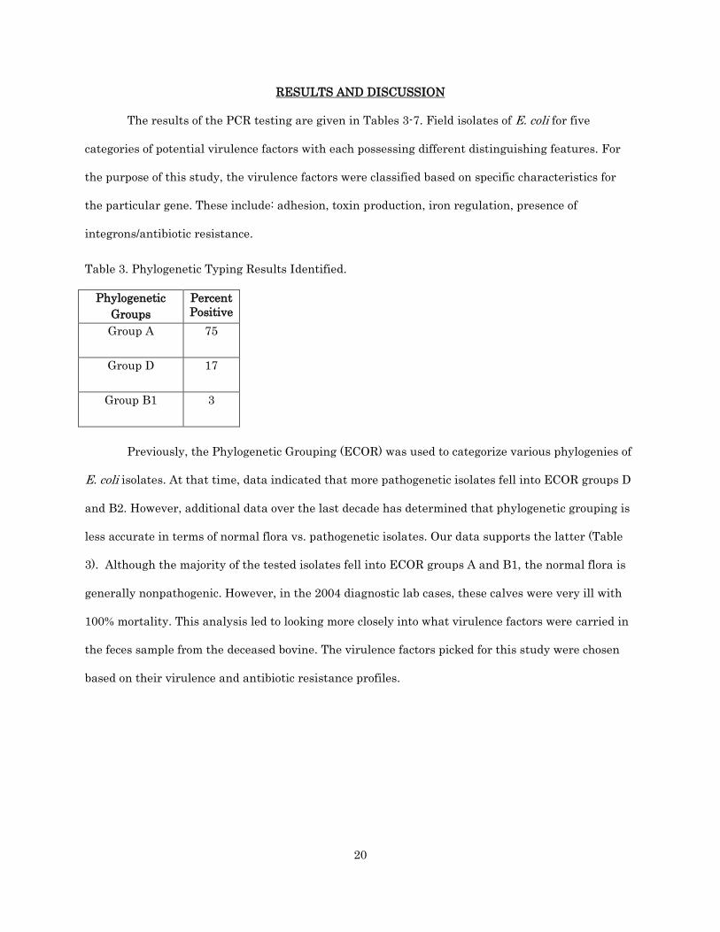
RESULTS AND DISCUSSION
The results of the PCR testing are given in Tables 3-7. Field isolates of E. coli for five
categories of potential virulence factors with each possessing different distinguishing features. For
the purpose of this study, the virulence factors were classified based on specific characteristics for
the particular gene. These include: adhesion, toxin production, iron regulation, presence of
integrons/antibiotic resistance.
Table 3. Phylogenetic Typing Results Identified.
Phylogenetic
Groups
Percent
Positive
Group A 75
Group D 17
Group B1 3
Previously, the Phylogenetic Grouping (ECOR) was used to categorize various phylogenies of
E. coli isolates. At that time, data indicated that more pathogenetic isolates fell into ECOR groups D
and B2. However, additional data over the last decade has determined that phylogenetic grouping is
less accurate in terms of normal flora vs. pathogenetic isolates. Our data supports the latter (Table
3). Although the majority of the tested isolates fell into ECOR groups A and B1, the normal flora is
generally nonpathogenic. However, in the 2004 diagnostic lab cases, these calves were very ill with
100% mortality. This analysis led to looking more closely into what virulence factors were carried in
the feces sample from the deceased bovine. The virulence factors picked for this study were chosen
based on their virulence and antibiotic resistance profiles.

21
Table 4. Adhesion Gene Percent Positive Results.
Adhesion
Genes
Percent
Positive
hly EHEC 2.67
hlyA 5.3
est1 10.67
papC 36
int1 66.67
int2 41.3
The adhesion virulence factors genes tested were neuC,,tsh, sfa, ibeA, hlyEHEC, hlyA, est1,
papC, fimC, and fimH. The first four genes (neuC, tsh, sfa, and ibeA) were negative in the samples
tested and not put into the above table. The other genes had between 2.67% positive to 92% positive.
The lowest percent positive was found in the gene hly EHEC, whereas the highest percent positive
was fimH. The others had the following percent positives: hlyA with 5.3%, est1 with 10.67%, papC
with 36%, and fimC with 85.3% (Table 4).
These results indicate that fimH was the most prevalent factor found. This is in large part
due to the genes ability to utilize its type 1 fimbriae to facilitate mannose binding to target host cells.
It is most commonly found in urinary E. coli where it adheres to a host cell and gains replacements
of amino acids which in turn increase the tropism for the uroepithelium and other components of the
lower membrane. These replacement acid chains are monomannose and therefore have a mutation
that helps the bacteria to colonize in the urinary tract and extraintestinal virulence of E. coli. This
process helps the gene to become highly pathogenic and resistant to many antibiotics (35). Showing a
second high of percent positives was that of fimC. This gene encodes for fimbriae that allow for
attachment, however they do not utilize mannose. They create tight attachments to the host
receptors and cause disease. Thirdly, the percent positives of papC were noted to be high in virulent

22
isolates. This virulence factor is known for its P pilus (pyelonephritis-associated pilus) and utilizes
this pili to adhere to kidney tissues and allow for gram-negative bacteria to set up house-keeping to
create mutations that allow for colonization (Table 4)(38).
Table 5. Iron Regulating Gene Percent Positive Results.
Iron Regulating Genes Percent Positive
iucD 8
irp1 44
irp2 42.67
The Iron Regulating genes that were tested were iucD, irp1, and irp2. Of these three genes,
irp1 had the highest amount of positives found (44%) closely followed by irp2 with 42.67%, and
coming up the lowest was iucD with 8% positives (Table 5). Iron is the bacterial growth-limiting
factor in all bacteria. The results of the iron regulating genes indicated many calves had these
bacteria present. Iron that is not tightly bound allows the bacteria to multiply significantly causing a
bacterial overload. The bacteria will then multiply and colonize. In this study, irp1 was found to be
the most significant iron acquisition gene.
During depletion or low iron, iron acquisition genes will bind to mRNA to reduce the
translation of iron metabolism genes. The irp1 will bind to an iron-sulfur cluster to conserve the
energy production whereas the irp2 gene will be corrupted. The other iron-regulating gene found to
carry a significant amount of positives was that of the gene, irp2. This gene is less likely to be found
in great abundance in cells due to its ability to be degraded. It functions the same as irp1 but does
not bind to an iron sulfuric cluster to regulate energy (16). Lastly, the iron-regulating gene found
was iucD. This particular gene is known to be categorized as aerobactin that is formed when iron is
being depleted in the tissues and fluids of the host. This gene is normally found on plasmids and is
known for its product’s ability to sift ferric acid from transferrin. The enzyme, aerobactin, is used to
catalyze the initial step of the iron pathway (16). These genes all deplete the host of iron and energy,
leaving the host drained and more susceptible to infection (Table 5).

23
Table 6. Toxin Gene Percent Positive Results.
Toxin Genes Percent Positive
Stx1 4
Stx2 1.3
eaeA 5.3
Cnf 6.67
astA 22.67
The toxin genes analyzed were vat, stx1, stx2, eaeA, cnf, and astA. The gene with the most
percent positives at 22.67% was astA. Following with 7% positive was cnf and lastly at 5.5% was
eaeA. Toxin genes are known to cause host cell damage and/or death. Of the toxin genes tested, the
gene astA showed the highest positive samples (Figure 4). According to Jacek Osek, the protein astA
is found to have the presence of the colonization of factor F4, also known as the porcine fimbrial
factor. This is known as the adhesion in ETEC to hold the most significance in pigs, but can also be
found in other species, such as humans and cattle. The second high toxin gene found was the
cytotoxic necrotizing factor (cnf). This gene’s product is known to cause acute infections in the
bladder and kidneys. It also triggers Rho GTPases which create mutations that increase the capacity
of the particular E. coli strain. It will kill the neutrophils in the kidneys leaving the site open for
toxic infection (31). The third most prevalent toxin gene was that of eaeA. This gene is called the
attachment and effacement protein. Along with attaching to the intestine, it’s product creates a
mutation in the target gene that will kill off the microvilli. This causes a lack of absorption in the
intestine leading to diarrhea (Table 6)(41).

24
Table 7. Presence of Integrons Percent Positive Results.
Integrons Percent Positive
Int1 66.67
Int2 41.3
Two genes (int1 and int2) were tested that are categorized this way. Of these two genes, int1
had 66.67% positive while int2 had 41.3% positive (Table 7). Integrons can be found on transposons
and plasmids. Conjugation can also allow for antibiotic resistance. This is done by undergoing a
mating process with another bacterium or by receiving resistant characteristics by the head of a
virus (13). Two DNA components (int1 and int2) were tested that are categorized this way. These
two carry multidrug resistant E. coli strains. Knowing how notorious these specific DNA elements
are for providing antibiotic resistance mechanism to bacteria and explains why they are both fairly
high for percent positives (Table 7).
NDSU-VDL currently cultures the fecal matter of presented specimens to determine if E. coli
is present. If the sample is hemolytic, it will go to the PCR lab to type the specific E. coli present. The
E.coli test looks for only six virulence factors including K99, F41, Sta, Intimin, Stx-1, and Stx-2.
Because assays for these factors often yielded negative results, it seemed pertinent to look into what
factors other labs across the Midwest are using.
University of Minnesota Veterinary Diagnostic Lab performs aerobic culture to detect E. coli.
If sample is hemolytic, the PCR lab is contacted and the sample is then sent through a PCR typing
screen testing for the following virulence factors: eae (Intimin), F18 (cDNA encoding equine), K88
(marker for ETEC strains), F41 (adhesion), K99 (ETEC fimbriae), LT (Heat-Labile Toxin), STa
(Heat-labile enterotoxin), STb(Heat-stabile enterotoxin), Stx2e (Shiga toxin 2e). The eae gene is the
most common gene detected in the specimens that are tested. Another test this particular diagnostic
lab performs is a full fecal work-up to rule out group A and B rotaviruses (using PCR), bovine
coronavirus (using PCR) and Salmonella (using enrichment cultures) (Dr. Jeremy Schefers,
University of Minnesota Veterinary Diagnostic Lab, St. Paul, MN, Personal Communication).

25
Iowa State University Veterinary Diagnostic lab has a calf diarrhea panel that tests for K99 E. coli,
Salmonella, Coronavirus, Rotavirus and Cryptospiridia. This is done using a fecal sample from the
sick animal. If addition testing is requested by the pathologist, a culture may be performed. For E.
coli, they do not look into other virulence factors other than K99 (Dr. Timothy Frana, Iowa State
University Veterinary Diagnostic Lab, Ames, IA, Personal Communication).
The present study could be used to identify virulent strains of E. coli that could potentially
be included in a vaccine to be given to a pregnant cow to prevent scours. If the correct virulence
factors are placed into an injectable or oral form of vaccine, producers would be able to administer
this to their pregnant herd as a preventative measure to protect the fetus against some of the types
of E. coli that can cause scours.

26
CONCLUSION
The present study utilized samples received from North Dakota and Northwestern
Minnesota that had been sent in to the NDSU-VDL to define a cause of death in scouring calves.
Many farms had more than one calf lost indicating a possible outbreak. This testing was performed
to determine what other E. coli virulence factors could be in regional isolates that would enhance the
detection capabilities of an E. coli multiplex PCR.
Iowa State University and University of Minnesota-Veterinary Diagnostic laboratories typed
their E. coli samples using eaeA, F18, F41, K99, LT, Sta, Stb, and Stx2. NDSU tests for K99, F41,
Sta, Stx1, and Stx2. Upon analyzing all the factors that are being tested in the three labs, one
virulence factor was common. This factor was K99. North Dakota and Minnesota both test for eaeA,
Sta, Stx2, and F41. This indicates that these genes are the most prevalent in the Midwest.
Any virulence factors noted in this study showing more than 15 percent positives could be
added to a more specific diagnostic test. The reason for including those isolates positive per 15% of
the time was due to the fact that a multiple PCR is only able to test a limited number of genes. The
factors found to be more than 15 percent positives are: astA, fimC, fimH, int1, irp1, irp2, and papC.
These factors were noted in only North Dakota and Northwestern Minnesota isolates; other
geographical areas may have different virulence factors as other areas have miscellaneous bacterial
growth. An example of this would be that of weather adaptations. In North Dakota, we have winter,
therefore pathogens can freeze and become fewer and/or become more resistant to cold. In Texas, it is
warmer and does not get as cold, therefore, those bacteria are more accustomed to warm
temperatures.
Based on this study, it would be pertinent to add fimC and fimH to the current diagnostic
test used by the NDSU Veterinary Diagnostic Lab as these two showed the highest percent positives
indicating a high percent are multi-drug resistant and pathogenic. Possible future studies could
implement more virulence factors into preventative medicines to help lower fatalities in North
Dakota and northwestern Minnesota cattle herds. This would help to lessen incidence of scours by

27
preventing the problem from occurring and would also reduce the resistance of antibiotics since the
calves would be protected from infections.
In addition to the previously mentioned future studies, another implementation could be
taking the virulence factors mentioned above that were identified 15% percent of the time, culture
them, and place the isolates directly into the calf’s milk. This would be best done with bottle calves
as it would be easier to administer orally. If the calf became ill a fecal sample should be taken to
analyze for the presence of pathogens.

28
REFERENCES
1. Alberts B, Johnson A, Lewis J, et al. Molecular Biology of the Cell. 4th edition. New York: Garland
Science; 2002. Cell Biology of Infection. Available from:
http://www.ncbi.nlm.nih.gov/books/NBK26833/
2. Bahrani-Mougeot, Farah, et al. "Type 1 fimbriae and extracellular polysaccharides are preeminent
uropathogenic Escherichia coli in the murine urinary tract." Molecular Microbiology. 45.4 (2002):
1079-1093.
3. BAMN. (2001). A guide to colostrum and colostrum management for dairy calves. In R. Seller
(Ed.), AFIA Arlington: AFIA.
4. Bird, Lina, Violaine Bonnefoy, et al. "Bioenergenetic challenges of microbial iron metabolisms."
Trends in Microbiology. 19.7 (2011): n. page.
5.Clermont, Olivier. "Rapid and Simple Determination of the escherichia coli Phylogenetic Group."
Applied and Environmental Microbiology 66: 4555-4557.
6. Duda-madej, A, G Gosciniak, et al. "Association of untypeable enteropathogenic Escherichia coli
(EPEC) strains with persistent diarrhea in children from the region of lower Silesia in Poland."
Polish Journal of Microbiology. 62.4 (2013): 641-644.
7. Enterohemorrhagic Escherichia coli Infections. (2009, January 1). Retrieved February 11, 2013,
from www.cfsph.iastate.edu
8. Franck, Sophia, Bosworth, Brad, et al. “Multiplex PCR for Enterotoxigenic, Attaching and
Effacing, and Shiga Toxi-producing Escherichia coli strains from calves.” Journal of Clinical
Microbiology, 36.6 (1998): 1795-1797
9. FSIS. "Pre-Harvest Management Controls and Intervention Options for Reducing Shiga Toxin-
Producing Escherichia coli Shedding in Cattle: An Overview of Current Research ." 01 Aug 2014.
U.S. Department of Agriculture, Online Posting to Food Safety and Inspection Service. Web. 9 Sep.
2014. <fsis.usda.gov>.
10. Gannon, Victor. “Rapid and Sensitive method for detection of shiga-like toxin-producing
Escherichia coli in ground beef using polymerase chain reaction.” Applied and Environmental Microbiology. 58.12 (1992): 3809-3815.
11. Garcia-Migura, et al. “Antimicrobial resistance of zoonotic and commensual bacteria in Europe:
The missing link between consumption and resistance in veterinary medicine.” Veterinary Microbiology. 170. (2014): 1-9.
12. Guerra, B., et al. (2003). Phenotypic and genotypic characterization of antimicrobial resistance in
german escherichia coli isolates from cattle, swine and poultry. Journal of Antimicrobial Chemotherapy, 52, 489-492.
13. Gunther, Andrew. “Dairy Cattle Residue Review.” 27 September 2010. National Milk Producers
Association, Online Posting to Animal Welfare Approved. Web. 9 Sep 2014. http://www.nmpf.org/latest-news/press-releases/nov-2010/dairy-cattle-antibiotic-residue-prevention-manual-available-free.

29
14. Güler, L. (2007). Virulence properties of Escherichia coli isolated from clinical bovine mastitis.
Turk. J. Vet. Anim. Science, 31(5), 361-365.
15. Hansen, Don. "Calf Scours: Causes and Treatment” Extension Beef Cattle Resource Committee. Beef Cattle Handbook, 20 Apr 2014. Web. 21 Apr 2014.
16. Herrero, Marta, Victor de Lorenzo, and J.B. Neilands. "Nucleotide Sequence of the iucD Gene of
the pColV-K30." Journal of Bacteriology. 170.1 (1988): 56-64.
17. Janβen, T., C. Schwarz, et al. “Virulence-associated genes in avian pathogenic Escherichia coli
(APEC) isolated from internal organs of poultry having died from colibacillosis.” Braz. J. Vet. Res. Anim. Sci. 291 (2001): 371-378.
18. Lane, M., & Mobley, H. (2007). Role of p-fimbrial-mediated adherence in pyelonephritis and
persistence of uropathogenic escherichia coli (UPEC) in the mammalian kidney. International Society of Nephrology, 72, 19-25.
19. Lasaro, M.A., J.F. Rodrigues, et al. "Genetic Diversity of Heat-Labile Toxin Expressed by
Enterotoxigenic Escherichia coli Strains Isolated from Humans." Journal of Bacteriology. 190.7
(2008): 2400-2410.
20. McDougall, E. (2013, June 29). Interview by CN Starr. Treatment protocol for scouring calves on
personal herds.
21. Montville, T. (2008). Food microbiology: An introduction. (2nd ed., pp. 124-128). Washington, DC:
ASM Press.
22. Multistate Outbreak of Shiga toxin-producing Escherichia coli O157:H7 Infections linked to
Ground Beef (Final Update) (2014, June 20) Retrieved September 9, 2014 from
www.cdc.gov/ecoli/2014/O157H7-05-14/index.html.
23. Neilands, J.B. "Siderophores: Structure and Function of Microbial Iron Transport Compounds."
Journal of Biological Compounds. 270. (1995): 26723-26726.
24. Oliveria, Leonor, and Jean-Claude Drapier. "Down-regulation of iron regulatory protein 1 gene."
PNAS. 97.12 (2000): 6550–6555.
25. Oswald, E., Schmidt H, et al. "Typing of Intimin Genes in Human and Animal
Enterrohemorrhagic and Enteropathogenic Escherichia coli; Characterization of a new Intimin
Variant." American Society for Microbiology. 68.1 (2000): 64-71.
26. Otto, Karen, and Silhavy, Thomas. "Surface sensing and adhesion of Escherichia coli controlled
by the Cpx-signaling pathway." PNAS. 99.4 (2002): 2287-2292.
27. Paton, Adrienne, and Paton, James C. "Detection and characterization of Shiga toxigenic
Escherichia coli by using multiplex pcr assays for stx1, stx2, eaeA, Enterohemorrhagic E. coli hlyA,
rfbO111, and rfbO157." Journal of Clinical Microbiology. 36.2 (1998): 598-602.
28. Pratt, Leslie and Roberto, Kolter. "Genetic Analysis of Escherichia coli biofilm formation: roles of
flagella, motility, chemotaxis and type 1 pili." Molecular Microbiology. 30.2 (1998): 285-293.

30
29. Purdom, G. (2007, July 10). Antibiotic resistance of bacteria: An example of evolution in action.
Retrieved from http://answersingenesis.org/natural-selection/antibiotic-resistance/
30. Quigley, Jim. "University of Minnesota Extension Service. "The role of oral immunoglobulins in systemic and intestinal immunity of neonatal calves. Diamond V Mills, n.d. Web. 20 Apr 2014.
<http://www.extension.umn.edu/agriculture/dairy/beef/the-role-of-oral-immunoglobulins.pdf>.
31. Rippere-Lampe, K., et al. (2001). Mutation of the gene encoding cytotoxic necrotizing factor type
1 (cnf1) attenuates the virulence of uropathogenic escherichia coli. Infection and Immunity, 69(6),
3954-3964.
32. Rohmer, Laurence. “Are pathogenic bacteria just looking for food? Metabolism and Microbial
pathogenesis.” Trends in Mirobiology. 19.7 (2011): 341-348.
33. Reisinger, R. (1965). Pathogenesis and prevention of infectious diarrhea (scours) of newborn
calves. Journal of the American Veterinary Medical Association, 1417(12). (1965): 1377-1386.
34. Starr, J. S. (2013, June 29). Interview by Cn Starr. Treatment protocol for scouring calves on
personal herds.
35. Stahlhut, Steen G., Sujay Chattopadhyay, et al. "Population Variability of the FimH Type 1
Fimbrial Adhesin in." Journal of Bacteriology. 191.6 (2009): 1941–1950.
36. Stoltenow, C. (2003). Calf scours. NDSU Extension Service. 37. "The Ins and Outs of Extra-Label Drug Use in Animals: A Resource for Veterinarians." 26 Jun
2014. U.S. Food and Drug Administration, Online Posting to Animal & Veterinary. Web. 30 Sep.
2014. <http://www.fda.gov/AnimalVeterinary/ResourcesforYou/ucm380135.htm>.
38. Volkan, E., et al. (2012). Domain activities of papc usher reveal the mechanism of action of an
Escherichia coli molecular machine. PNAS, 109(24), 9563-9568.
39. Wang, J., et al. (2013). Molecular characterization of blaesbl-harboring conjugative plasmids
identified in multi-drug resistant Escherichia coli isolated from food-producing animals and healthy
humans. frontiers in microbiology, 4(188), 1-7.
40. Zhu, C., J. Harel, et al. "Virulence Properties and Attaching-Effacing Activity of Escherichia coli
O45 from Swine Postweaning Diarrhea." Infection and Immunity. 62.10 (1994): 4153-4159.